US8450086B2 - Bacterial membrane protein secretion - Google Patents
Bacterial membrane protein secretion Download PDFInfo
- Publication number
- US8450086B2 US8450086B2 US13/509,287 US201013509287A US8450086B2 US 8450086 B2 US8450086 B2 US 8450086B2 US 201013509287 A US201013509287 A US 201013509287A US 8450086 B2 US8450086 B2 US 8450086B2
- Authority
- US
- United States
- Prior art keywords
- phage
- protein
- exoprotein
- vector
- domain
- Prior art date
- Legal status (The legal status is an assumption and is not a legal conclusion. Google has not performed a legal analysis and makes no representation as to the accuracy of the status listed.)
- Expired - Fee Related
Links
- 230000028327 secretion Effects 0.000 title claims abstract description 51
- 230000001580 bacterial effect Effects 0.000 title claims abstract description 40
- 102000018697 Membrane Proteins Human genes 0.000 title description 2
- 108010052285 Membrane Proteins Proteins 0.000 title description 2
- 108090000623 proteins and genes Proteins 0.000 claims abstract description 129
- 102000004169 proteins and genes Human genes 0.000 claims abstract description 118
- 108090000765 processed proteins & peptides Proteins 0.000 claims abstract description 87
- 102000004196 processed proteins & peptides Human genes 0.000 claims abstract description 68
- 239000002245 particle Substances 0.000 claims abstract description 51
- 101710125418 Major capsid protein Proteins 0.000 claims abstract description 49
- 101710132601 Capsid protein Proteins 0.000 claims abstract description 48
- 101710094648 Coat protein Proteins 0.000 claims abstract description 48
- 102100021181 Golgi phosphoprotein 3 Human genes 0.000 claims abstract description 48
- 101710141454 Nucleoprotein Proteins 0.000 claims abstract description 48
- 101710083689 Probable capsid protein Proteins 0.000 claims abstract description 48
- 229920001184 polypeptide Polymers 0.000 claims abstract description 48
- 101100136076 Aspergillus oryzae (strain ATCC 42149 / RIB 40) pel1 gene Proteins 0.000 claims abstract description 43
- 101150040383 pel2 gene Proteins 0.000 claims abstract description 43
- 101150050446 pelB gene Proteins 0.000 claims abstract description 43
- 238000000034 method Methods 0.000 claims abstract description 35
- 241000724791 Filamentous phage Species 0.000 claims abstract description 27
- 101100295756 Acinetobacter baumannii (strain ATCC 19606 / DSM 30007 / JCM 6841 / CCUG 19606 / CIP 70.34 / NBRC 109757 / NCIMB 12457 / NCTC 12156 / 81) omp38 gene Proteins 0.000 claims abstract description 21
- 101150042295 arfA gene Proteins 0.000 claims abstract description 21
- 101150087557 omcB gene Proteins 0.000 claims abstract description 21
- 101150115693 ompA gene Proteins 0.000 claims abstract description 21
- 239000012528 membrane Substances 0.000 claims abstract description 12
- 235000018102 proteins Nutrition 0.000 claims description 98
- 239000013598 vector Substances 0.000 claims description 55
- 230000027455 binding Effects 0.000 claims description 49
- 108020001507 fusion proteins Proteins 0.000 claims description 28
- 102000037865 fusion proteins Human genes 0.000 claims description 28
- 102000005962 receptors Human genes 0.000 claims description 21
- 108020003175 receptors Proteins 0.000 claims description 21
- 108091033319 polynucleotide Proteins 0.000 claims description 12
- 102000040430 polynucleotide Human genes 0.000 claims description 12
- 239000002157 polynucleotide Substances 0.000 claims description 12
- 108091026890 Coding region Proteins 0.000 claims description 10
- 239000003446 ligand Substances 0.000 claims description 9
- 150000007523 nucleic acids Chemical group 0.000 claims description 8
- 235000001014 amino acid Nutrition 0.000 claims description 7
- 102200123673 rs104894481 Human genes 0.000 claims description 7
- 230000001939 inductive effect Effects 0.000 claims description 5
- 108700010839 phage proteins Proteins 0.000 claims description 4
- 102000008102 Ankyrins Human genes 0.000 claims description 3
- 108010049777 Ankyrins Proteins 0.000 claims description 3
- 125000002924 primary amino group Chemical group [H]N([H])* 0.000 claims description 3
- 230000005945 translocation Effects 0.000 claims description 3
- 102100026120 IgG receptor FcRn large subunit p51 Human genes 0.000 claims description 2
- 101710177940 IgG receptor FcRn large subunit p51 Proteins 0.000 claims description 2
- 101100321993 Drosophila melanogaster pix gene Proteins 0.000 claims 4
- 101710143509 Pre-histone-like nucleoprotein Proteins 0.000 claims 4
- 101710193132 Pre-hexon-linking protein VIII Proteins 0.000 claims 2
- 125000003275 alpha amino acid group Chemical group 0.000 claims 2
- 125000000151 cysteine group Chemical group N[C@@H](CS)C(=O)* 0.000 claims 2
- 108020001756 ligand binding domains Proteins 0.000 claims 2
- 230000005714 functional activity Effects 0.000 claims 1
- 238000006467 substitution reaction Methods 0.000 claims 1
- 230000004927 fusion Effects 0.000 abstract description 19
- 230000001976 improved effect Effects 0.000 abstract description 9
- 238000004519 manufacturing process Methods 0.000 abstract description 7
- 241000894006 Bacteria Species 0.000 abstract description 4
- 230000015572 biosynthetic process Effects 0.000 abstract description 4
- 241001515965 unidentified phage Species 0.000 abstract description 4
- 230000001590 oxidative effect Effects 0.000 abstract 1
- 210000004027 cell Anatomy 0.000 description 36
- 238000002965 ELISA Methods 0.000 description 27
- 108010076504 Protein Sorting Signals Proteins 0.000 description 20
- 239000000427 antigen Substances 0.000 description 20
- 108091007433 antigens Proteins 0.000 description 20
- 102000036639 antigens Human genes 0.000 description 20
- 108090000176 Interleukin-13 Proteins 0.000 description 19
- 102000003816 Interleukin-13 Human genes 0.000 description 19
- 108020004414 DNA Proteins 0.000 description 14
- 230000035772 mutation Effects 0.000 description 14
- 241000588724 Escherichia coli Species 0.000 description 13
- 102220005268 rs33912272 Human genes 0.000 description 13
- 102000002260 Alkaline Phosphatase Human genes 0.000 description 12
- 108020004774 Alkaline Phosphatase Proteins 0.000 description 12
- 108020004705 Codon Proteins 0.000 description 11
- 238000004091 panning Methods 0.000 description 11
- 150000001413 amino acids Chemical class 0.000 description 10
- 101000998146 Homo sapiens Interleukin-17A Proteins 0.000 description 9
- 108060003951 Immunoglobulin Proteins 0.000 description 9
- 239000012634 fragment Substances 0.000 description 9
- 102000018358 immunoglobulin Human genes 0.000 description 9
- 230000012743 protein tagging Effects 0.000 description 9
- BWGNESOTFCXPMA-UHFFFAOYSA-N Dihydrogen disulfide Chemical compound SS BWGNESOTFCXPMA-UHFFFAOYSA-N 0.000 description 8
- 102000009109 Fc receptors Human genes 0.000 description 8
- 108010087819 Fc receptors Proteins 0.000 description 8
- 102100033461 Interleukin-17A Human genes 0.000 description 8
- 238000002823 phage display Methods 0.000 description 7
- 230000000694 effects Effects 0.000 description 6
- 230000006870 function Effects 0.000 description 6
- 239000013642 negative control Substances 0.000 description 6
- 238000012360 testing method Methods 0.000 description 6
- 108010001336 Horseradish Peroxidase Proteins 0.000 description 5
- 230000008901 benefit Effects 0.000 description 5
- 239000011230 binding agent Substances 0.000 description 5
- 238000010367 cloning Methods 0.000 description 5
- LOKCTEFSRHRXRJ-UHFFFAOYSA-I dipotassium trisodium dihydrogen phosphate hydrogen phosphate dichloride Chemical compound P(=O)(O)(O)[O-].[K+].P(=O)(O)([O-])[O-].[Na+].[Na+].[Cl-].[K+].[Cl-].[Na+] LOKCTEFSRHRXRJ-UHFFFAOYSA-I 0.000 description 5
- 238000002703 mutagenesis Methods 0.000 description 5
- 231100000350 mutagenesis Toxicity 0.000 description 5
- 239000002953 phosphate buffered saline Substances 0.000 description 5
- 238000002360 preparation method Methods 0.000 description 5
- 108091008146 restriction endonucleases Proteins 0.000 description 5
- 238000003118 sandwich ELISA Methods 0.000 description 5
- 101100286681 Homo sapiens IL13 gene Proteins 0.000 description 4
- 239000004365 Protease Substances 0.000 description 4
- FAPWRFPIFSIZLT-UHFFFAOYSA-M Sodium chloride Chemical compound [Na+].[Cl-] FAPWRFPIFSIZLT-UHFFFAOYSA-M 0.000 description 4
- 108010090804 Streptavidin Proteins 0.000 description 4
- 238000013459 approach Methods 0.000 description 4
- 238000003556 assay Methods 0.000 description 4
- FPPNZSSZRUTDAP-UWFZAAFLSA-N carbenicillin Chemical compound N([C@H]1[C@H]2SC([C@@H](N2C1=O)C(O)=O)(C)C)C(=O)C(C(O)=O)C1=CC=CC=C1 FPPNZSSZRUTDAP-UWFZAAFLSA-N 0.000 description 4
- 229960003669 carbenicillin Drugs 0.000 description 4
- 238000010276 construction Methods 0.000 description 4
- 230000006872 improvement Effects 0.000 description 4
- 230000003993 interaction Effects 0.000 description 4
- 108020004707 nucleic acids Proteins 0.000 description 4
- 102000039446 nucleic acids Human genes 0.000 description 4
- 239000008188 pellet Substances 0.000 description 4
- 238000012216 screening Methods 0.000 description 4
- 230000009870 specific binding Effects 0.000 description 4
- NFGXHKASABOEEW-UHFFFAOYSA-N 1-methylethyl 11-methoxy-3,7,11-trimethyl-2,4-dodecadienoate Chemical compound COC(C)(C)CCCC(C)CC=CC(C)=CC(=O)OC(C)C NFGXHKASABOEEW-UHFFFAOYSA-N 0.000 description 3
- FWMNVWWHGCHHJJ-SKKKGAJSSA-N 4-amino-1-[(2r)-6-amino-2-[[(2r)-2-[[(2r)-2-[[(2r)-2-amino-3-phenylpropanoyl]amino]-3-phenylpropanoyl]amino]-4-methylpentanoyl]amino]hexanoyl]piperidine-4-carboxylic acid Chemical compound C([C@H](C(=O)N[C@H](CC(C)C)C(=O)N[C@H](CCCCN)C(=O)N1CCC(N)(CC1)C(O)=O)NC(=O)[C@H](N)CC=1C=CC=CC=1)C1=CC=CC=C1 FWMNVWWHGCHHJJ-SKKKGAJSSA-N 0.000 description 3
- 101710192393 Attachment protein G3P Proteins 0.000 description 3
- 102100033183 Epithelial membrane protein 1 Human genes 0.000 description 3
- 241001524679 Escherichia virus M13 Species 0.000 description 3
- HVLSXIKZNLPZJJ-TXZCQADKSA-N HA peptide Chemical compound C([C@@H](C(=O)N[C@@H](CC(O)=O)C(=O)N[C@@H](C(C)C)C(=O)N1[C@@H](CCC1)C(=O)N[C@@H](CC(O)=O)C(=O)N[C@@H](CC=1C=CC(O)=CC=1)C(=O)N[C@@H](C)C(O)=O)NC(=O)[C@H]1N(CCC1)C(=O)[C@@H](N)CC=1C=CC(O)=CC=1)C1=CC=C(O)C=C1 HVLSXIKZNLPZJJ-TXZCQADKSA-N 0.000 description 3
- 108010021625 Immunoglobulin Fragments Proteins 0.000 description 3
- 102000008394 Immunoglobulin Fragments Human genes 0.000 description 3
- 108091028043 Nucleic acid sequence Proteins 0.000 description 3
- 108010008281 Recombinant Fusion Proteins Proteins 0.000 description 3
- 102000007056 Recombinant Fusion Proteins Human genes 0.000 description 3
- 230000010056 antibody-dependent cellular cytotoxicity Effects 0.000 description 3
- 102000025171 antigen binding proteins Human genes 0.000 description 3
- 108091000831 antigen binding proteins Proteins 0.000 description 3
- 238000005119 centrifugation Methods 0.000 description 3
- 238000003776 cleavage reaction Methods 0.000 description 3
- 230000000295 complement effect Effects 0.000 description 3
- 230000001086 cytosolic effect Effects 0.000 description 3
- 230000009977 dual effect Effects 0.000 description 3
- 108010008594 epithelial membrane protein-1 Proteins 0.000 description 3
- 238000002474 experimental method Methods 0.000 description 3
- 238000000338 in vitro Methods 0.000 description 3
- 238000011534 incubation Methods 0.000 description 3
- 239000000203 mixture Substances 0.000 description 3
- 238000004806 packaging method and process Methods 0.000 description 3
- 108010087558 pectate lyase Proteins 0.000 description 3
- 239000013641 positive control Substances 0.000 description 3
- 230000007017 scission Effects 0.000 description 3
- 239000000758 substrate Substances 0.000 description 3
- 238000004448 titration Methods 0.000 description 3
- 230000014616 translation Effects 0.000 description 3
- 108700010070 Codon Usage Proteins 0.000 description 2
- 102000053602 DNA Human genes 0.000 description 2
- 102000004190 Enzymes Human genes 0.000 description 2
- 108090000790 Enzymes Proteins 0.000 description 2
- 108010021468 Fc gamma receptor IIA Proteins 0.000 description 2
- 108010021472 Fc gamma receptor IIB Proteins 0.000 description 2
- 102000016359 Fibronectins Human genes 0.000 description 2
- 108010067306 Fibronectins Proteins 0.000 description 2
- DHMQDGOQFOQNFH-UHFFFAOYSA-N Glycine Chemical compound NCC(O)=O DHMQDGOQFOQNFH-UHFFFAOYSA-N 0.000 description 2
- 108010093488 His-His-His-His-His-His Proteins 0.000 description 2
- 102000006496 Immunoglobulin Heavy Chains Human genes 0.000 description 2
- 108010019476 Immunoglobulin Heavy Chains Proteins 0.000 description 2
- 102000013463 Immunoglobulin Light Chains Human genes 0.000 description 2
- 108010065825 Immunoglobulin Light Chains Proteins 0.000 description 2
- OUYCCCASQSFEME-QMMMGPOBSA-N L-tyrosine Chemical compound OC(=O)[C@@H](N)CC1=CC=C(O)C=C1 OUYCCCASQSFEME-QMMMGPOBSA-N 0.000 description 2
- 102100029204 Low affinity immunoglobulin gamma Fc region receptor II-a Human genes 0.000 description 2
- 102100029185 Low affinity immunoglobulin gamma Fc region receptor III-B Human genes 0.000 description 2
- 108091034117 Oligonucleotide Proteins 0.000 description 2
- 108090000526 Papain Proteins 0.000 description 2
- 241000588701 Pectobacterium carotovorum Species 0.000 description 2
- 108091005804 Peptidases Proteins 0.000 description 2
- 102100037486 Reverse transcriptase/ribonuclease H Human genes 0.000 description 2
- IQFYYKKMVGJFEH-XLPZGREQSA-N Thymidine Chemical compound O=C1NC(=O)C(C)=CN1[C@@H]1O[C@H](CO)[C@@H](O)C1 IQFYYKKMVGJFEH-XLPZGREQSA-N 0.000 description 2
- JLCPHMBAVCMARE-UHFFFAOYSA-N [3-[[3-[[3-[[3-[[3-[[3-[[3-[[3-[[3-[[3-[[3-[[5-(2-amino-6-oxo-1H-purin-9-yl)-3-[[3-[[3-[[3-[[3-[[3-[[5-(2-amino-6-oxo-1H-purin-9-yl)-3-[[5-(2-amino-6-oxo-1H-purin-9-yl)-3-hydroxyoxolan-2-yl]methoxy-hydroxyphosphoryl]oxyoxolan-2-yl]methoxy-hydroxyphosphoryl]oxy-5-(5-methyl-2,4-dioxopyrimidin-1-yl)oxolan-2-yl]methoxy-hydroxyphosphoryl]oxy-5-(6-aminopurin-9-yl)oxolan-2-yl]methoxy-hydroxyphosphoryl]oxy-5-(6-aminopurin-9-yl)oxolan-2-yl]methoxy-hydroxyphosphoryl]oxy-5-(6-aminopurin-9-yl)oxolan-2-yl]methoxy-hydroxyphosphoryl]oxy-5-(6-aminopurin-9-yl)oxolan-2-yl]methoxy-hydroxyphosphoryl]oxyoxolan-2-yl]methoxy-hydroxyphosphoryl]oxy-5-(5-methyl-2,4-dioxopyrimidin-1-yl)oxolan-2-yl]methoxy-hydroxyphosphoryl]oxy-5-(4-amino-2-oxopyrimidin-1-yl)oxolan-2-yl]methoxy-hydroxyphosphoryl]oxy-5-(5-methyl-2,4-dioxopyrimidin-1-yl)oxolan-2-yl]methoxy-hydroxyphosphoryl]oxy-5-(5-methyl-2,4-dioxopyrimidin-1-yl)oxolan-2-yl]methoxy-hydroxyphosphoryl]oxy-5-(6-aminopurin-9-yl)oxolan-2-yl]methoxy-hydroxyphosphoryl]oxy-5-(6-aminopurin-9-yl)oxolan-2-yl]methoxy-hydroxyphosphoryl]oxy-5-(4-amino-2-oxopyrimidin-1-yl)oxolan-2-yl]methoxy-hydroxyphosphoryl]oxy-5-(4-amino-2-oxopyrimidin-1-yl)oxolan-2-yl]methoxy-hydroxyphosphoryl]oxy-5-(4-amino-2-oxopyrimidin-1-yl)oxolan-2-yl]methoxy-hydroxyphosphoryl]oxy-5-(6-aminopurin-9-yl)oxolan-2-yl]methoxy-hydroxyphosphoryl]oxy-5-(4-amino-2-oxopyrimidin-1-yl)oxolan-2-yl]methyl [5-(6-aminopurin-9-yl)-2-(hydroxymethyl)oxolan-3-yl] hydrogen phosphate Polymers Cc1cn(C2CC(OP(O)(=O)OCC3OC(CC3OP(O)(=O)OCC3OC(CC3O)n3cnc4c3nc(N)[nH]c4=O)n3cnc4c3nc(N)[nH]c4=O)C(COP(O)(=O)OC3CC(OC3COP(O)(=O)OC3CC(OC3COP(O)(=O)OC3CC(OC3COP(O)(=O)OC3CC(OC3COP(O)(=O)OC3CC(OC3COP(O)(=O)OC3CC(OC3COP(O)(=O)OC3CC(OC3COP(O)(=O)OC3CC(OC3COP(O)(=O)OC3CC(OC3COP(O)(=O)OC3CC(OC3COP(O)(=O)OC3CC(OC3COP(O)(=O)OC3CC(OC3COP(O)(=O)OC3CC(OC3COP(O)(=O)OC3CC(OC3COP(O)(=O)OC3CC(OC3COP(O)(=O)OC3CC(OC3COP(O)(=O)OC3CC(OC3CO)n3cnc4c(N)ncnc34)n3ccc(N)nc3=O)n3cnc4c(N)ncnc34)n3ccc(N)nc3=O)n3ccc(N)nc3=O)n3ccc(N)nc3=O)n3cnc4c(N)ncnc34)n3cnc4c(N)ncnc34)n3cc(C)c(=O)[nH]c3=O)n3cc(C)c(=O)[nH]c3=O)n3ccc(N)nc3=O)n3cc(C)c(=O)[nH]c3=O)n3cnc4c3nc(N)[nH]c4=O)n3cnc4c(N)ncnc34)n3cnc4c(N)ncnc34)n3cnc4c(N)ncnc34)n3cnc4c(N)ncnc34)O2)c(=O)[nH]c1=O JLCPHMBAVCMARE-UHFFFAOYSA-N 0.000 description 2
- 230000003213 activating effect Effects 0.000 description 2
- 230000004913 activation Effects 0.000 description 2
- OIRDTQYFTABQOQ-KQYNXXCUSA-N adenosine Chemical compound C1=NC=2C(N)=NC=NC=2N1[C@@H]1O[C@H](CO)[C@@H](O)[C@H]1O OIRDTQYFTABQOQ-KQYNXXCUSA-N 0.000 description 2
- 230000000975 bioactive effect Effects 0.000 description 2
- 230000000903 blocking effect Effects 0.000 description 2
- 238000004113 cell culture Methods 0.000 description 2
- 210000000170 cell membrane Anatomy 0.000 description 2
- 239000011248 coating agent Substances 0.000 description 2
- 238000000576 coating method Methods 0.000 description 2
- 230000004540 complement-dependent cytotoxicity Effects 0.000 description 2
- 230000021615 conjugation Effects 0.000 description 2
- 230000008878 coupling Effects 0.000 description 2
- 238000010168 coupling process Methods 0.000 description 2
- 238000005859 coupling reaction Methods 0.000 description 2
- 230000001419 dependent effect Effects 0.000 description 2
- 238000001514 detection method Methods 0.000 description 2
- VYFYYTLLBUKUHU-UHFFFAOYSA-N dopamine Chemical compound NCCC1=CC=C(O)C(O)=C1 VYFYYTLLBUKUHU-UHFFFAOYSA-N 0.000 description 2
- 238000005516 engineering process Methods 0.000 description 2
- 229940088598 enzyme Drugs 0.000 description 2
- BTCSSZJGUNDROE-UHFFFAOYSA-N gamma-aminobutyric acid Chemical compound NCCCC(O)=O BTCSSZJGUNDROE-UHFFFAOYSA-N 0.000 description 2
- 239000001963 growth medium Substances 0.000 description 2
- 239000000833 heterodimer Substances 0.000 description 2
- 239000000710 homodimer Substances 0.000 description 2
- 230000001900 immune effect Effects 0.000 description 2
- 239000000411 inducer Substances 0.000 description 2
- 230000006698 induction Effects 0.000 description 2
- 208000015181 infectious disease Diseases 0.000 description 2
- 230000002401 inhibitory effect Effects 0.000 description 2
- 230000005764 inhibitory process Effects 0.000 description 2
- 239000002609 medium Substances 0.000 description 2
- 239000002773 nucleotide Substances 0.000 description 2
- 125000003729 nucleotide group Chemical group 0.000 description 2
- 238000005457 optimization Methods 0.000 description 2
- 229940055729 papain Drugs 0.000 description 2
- 235000019834 papain Nutrition 0.000 description 2
- 210000001322 periplasm Anatomy 0.000 description 2
- OXCMYAYHXIHQOA-UHFFFAOYSA-N potassium;[2-butyl-5-chloro-3-[[4-[2-(1,2,4-triaza-3-azanidacyclopenta-1,4-dien-5-yl)phenyl]phenyl]methyl]imidazol-4-yl]methanol Chemical compound [K+].CCCCC1=NC(Cl)=C(CO)N1CC1=CC=C(C=2C(=CC=CC=2)C2=N[N-]N=N2)C=C1 OXCMYAYHXIHQOA-UHFFFAOYSA-N 0.000 description 2
- 230000008569 process Effects 0.000 description 2
- 239000000047 product Substances 0.000 description 2
- 235000019419 proteases Nutrition 0.000 description 2
- 238000000746 purification Methods 0.000 description 2
- 238000005215 recombination Methods 0.000 description 2
- 230000006798 recombination Effects 0.000 description 2
- 238000011084 recovery Methods 0.000 description 2
- 238000010187 selection method Methods 0.000 description 2
- QZAYGJVTTNCVMB-UHFFFAOYSA-N serotonin Chemical compound C1=C(O)C=C2C(CCN)=CNC2=C1 QZAYGJVTTNCVMB-UHFFFAOYSA-N 0.000 description 2
- 239000011780 sodium chloride Substances 0.000 description 2
- 239000000126 substance Substances 0.000 description 2
- 239000006228 supernatant Substances 0.000 description 2
- 230000014621 translational initiation Effects 0.000 description 2
- 230000032258 transport Effects 0.000 description 2
- 239000003656 tris buffered saline Substances 0.000 description 2
- OUYCCCASQSFEME-UHFFFAOYSA-N tyrosine Natural products OC(=O)C(N)CC1=CC=C(O)C=C1 OUYCCCASQSFEME-UHFFFAOYSA-N 0.000 description 2
- OGNSCSPNOLGXSM-UHFFFAOYSA-N (+/-)-DABA Natural products NCCC(N)C(O)=O OGNSCSPNOLGXSM-UHFFFAOYSA-N 0.000 description 1
- XSYUPRQVAHJETO-WPMUBMLPSA-N (2s)-2-[[(2s)-2-[[(2s)-2-[[(2s)-2-[[(2s)-2-[[(2s)-2-amino-3-(1h-imidazol-5-yl)propanoyl]amino]-3-(1h-imidazol-5-yl)propanoyl]amino]-3-(1h-imidazol-5-yl)propanoyl]amino]-3-(1h-imidazol-5-yl)propanoyl]amino]-3-(1h-imidazol-5-yl)propanoyl]amino]-3-(1h-imidaz Chemical compound C([C@H](N)C(=O)N[C@@H](CC=1NC=NC=1)C(=O)N[C@@H](CC=1NC=NC=1)C(=O)N[C@@H](CC=1NC=NC=1)C(=O)N[C@@H](CC=1NC=NC=1)C(=O)N[C@@H](CC=1NC=NC=1)C(O)=O)C1=CN=CN1 XSYUPRQVAHJETO-WPMUBMLPSA-N 0.000 description 1
- HKZAAJSTFUZYTO-LURJTMIESA-N (2s)-2-[[2-[[2-[[2-[(2-aminoacetyl)amino]acetyl]amino]acetyl]amino]acetyl]amino]-3-hydroxypropanoic acid Chemical group NCC(=O)NCC(=O)NCC(=O)NCC(=O)N[C@@H](CO)C(O)=O HKZAAJSTFUZYTO-LURJTMIESA-N 0.000 description 1
- UHDGCWIWMRVCDJ-UHFFFAOYSA-N 1-beta-D-Xylofuranosyl-NH-Cytosine Natural products O=C1N=C(N)C=CN1C1C(O)C(O)C(CO)O1 UHDGCWIWMRVCDJ-UHFFFAOYSA-N 0.000 description 1
- MUGYJXWDGQOBET-UFLZEWODSA-N 5-[(3as,4s,6ar)-2-oxo-1,3,3a,4,6,6a-hexahydrothieno[3,4-d]imidazol-4-yl]pentanoic acid;4-(2-aminoethyl)phenol Chemical compound NCCC1=CC=C(O)C=C1.N1C(=O)N[C@@H]2[C@H](CCCCC(=O)O)SC[C@@H]21 MUGYJXWDGQOBET-UFLZEWODSA-N 0.000 description 1
- 229920001817 Agar Polymers 0.000 description 1
- 241000024188 Andala Species 0.000 description 1
- DWRXFEITVBNRMK-UHFFFAOYSA-N Beta-D-1-Arabinofuranosylthymine Natural products O=C1NC(=O)C(C)=CN1C1C(O)C(O)C(CO)O1 DWRXFEITVBNRMK-UHFFFAOYSA-N 0.000 description 1
- 239000002126 C01EB10 - Adenosine Substances 0.000 description 1
- 108010021064 CTLA-4 Antigen Proteins 0.000 description 1
- 102000008203 CTLA-4 Antigen Human genes 0.000 description 1
- 229940045513 CTLA4 antagonist Drugs 0.000 description 1
- 101710167800 Capsid assembly scaffolding protein Proteins 0.000 description 1
- 101710169873 Capsid protein G8P Proteins 0.000 description 1
- BVKZGUZCCUSVTD-UHFFFAOYSA-L Carbonate Chemical compound [O-]C([O-])=O BVKZGUZCCUSVTD-UHFFFAOYSA-L 0.000 description 1
- 102000019034 Chemokines Human genes 0.000 description 1
- 108010012236 Chemokines Proteins 0.000 description 1
- 208000003322 Coinfection Diseases 0.000 description 1
- 241001429175 Colitis phage Species 0.000 description 1
- 108010002947 Connectin Proteins 0.000 description 1
- 108010025905 Cystine-Knot Miniproteins Proteins 0.000 description 1
- UHDGCWIWMRVCDJ-PSQAKQOGSA-N Cytidine Natural products O=C1N=C(N)C=CN1[C@@H]1[C@@H](O)[C@@H](O)[C@H](CO)O1 UHDGCWIWMRVCDJ-PSQAKQOGSA-N 0.000 description 1
- 230000004543 DNA replication Effects 0.000 description 1
- 238000001712 DNA sequencing Methods 0.000 description 1
- 102000001301 EGF receptor Human genes 0.000 description 1
- 102000056372 ErbB-3 Receptor Human genes 0.000 description 1
- 102000003951 Erythropoietin Human genes 0.000 description 1
- 102100031939 Erythropoietin Human genes 0.000 description 1
- 108090000394 Erythropoietin Proteins 0.000 description 1
- 108010075944 Erythropoietin Receptors Proteins 0.000 description 1
- 102100036509 Erythropoietin receptor Human genes 0.000 description 1
- 108091029865 Exogenous DNA Proteins 0.000 description 1
- XZWYTXMRWQJBGX-VXBMVYAYSA-N FLAG peptide Chemical compound NCCCC[C@@H](C(O)=O)NC(=O)[C@H](CC(O)=O)NC(=O)[C@H](CC(O)=O)NC(=O)[C@H](CC(O)=O)NC(=O)[C@H](CC(O)=O)NC(=O)[C@H](CCCCN)NC(=O)[C@@H](NC(=O)[C@@H](N)CC(O)=O)CC1=CC=C(O)C=C1 XZWYTXMRWQJBGX-VXBMVYAYSA-N 0.000 description 1
- 108010020195 FLAG peptide Proteins 0.000 description 1
- -1 Fab Proteins 0.000 description 1
- 108091006020 Fc-tagged proteins Proteins 0.000 description 1
- 108091006027 G proteins Proteins 0.000 description 1
- 108090000045 G-Protein-Coupled Receptors Proteins 0.000 description 1
- 102000003688 G-Protein-Coupled Receptors Human genes 0.000 description 1
- 102000030782 GTP binding Human genes 0.000 description 1
- 108091000058 GTP-Binding Proteins 0.000 description 1
- 102000000802 Galectin 3 Human genes 0.000 description 1
- 108010001517 Galectin 3 Proteins 0.000 description 1
- WQZGKKKJIJFFOK-GASJEMHNSA-N Glucose Natural products OC[C@H]1OC(O)[C@H](O)[C@@H](O)[C@@H]1O WQZGKKKJIJFFOK-GASJEMHNSA-N 0.000 description 1
- 239000004471 Glycine Substances 0.000 description 1
- 102100026122 High affinity immunoglobulin gamma Fc receptor I Human genes 0.000 description 1
- 101000851181 Homo sapiens Epidermal growth factor receptor Proteins 0.000 description 1
- 101000913074 Homo sapiens High affinity immunoglobulin gamma Fc receptor I Proteins 0.000 description 1
- 101000917826 Homo sapiens Low affinity immunoglobulin gamma Fc region receptor II-a Proteins 0.000 description 1
- 101000917824 Homo sapiens Low affinity immunoglobulin gamma Fc region receptor II-b Proteins 0.000 description 1
- 101000917858 Homo sapiens Low affinity immunoglobulin gamma Fc region receptor III-A Proteins 0.000 description 1
- 101000917839 Homo sapiens Low affinity immunoglobulin gamma Fc region receptor III-B Proteins 0.000 description 1
- 108010073816 IgE Receptors Proteins 0.000 description 1
- 102000009438 IgE Receptors Human genes 0.000 description 1
- 108010073807 IgG Receptors Proteins 0.000 description 1
- 102000018071 Immunoglobulin Fc Fragments Human genes 0.000 description 1
- 108010091135 Immunoglobulin Fc Fragments Proteins 0.000 description 1
- 244000050403 Iris x germanica Species 0.000 description 1
- 102100029205 Low affinity immunoglobulin gamma Fc region receptor II-b Human genes 0.000 description 1
- 239000006142 Luria-Bertani Agar Substances 0.000 description 1
- 239000006137 Luria-Bertani broth Substances 0.000 description 1
- 101710156564 Major tail protein Gp23 Proteins 0.000 description 1
- 230000004988 N-glycosylation Effects 0.000 description 1
- 125000000729 N-terminal amino-acid group Chemical group 0.000 description 1
- 206010028980 Neoplasm Diseases 0.000 description 1
- 102400000058 Neuregulin-1 Human genes 0.000 description 1
- 108090000556 Neuregulin-1 Proteins 0.000 description 1
- 102000004108 Neurotransmitter Receptors Human genes 0.000 description 1
- 108090000590 Neurotransmitter Receptors Proteins 0.000 description 1
- 102000007399 Nuclear hormone receptor Human genes 0.000 description 1
- 108020005497 Nuclear hormone receptor Proteins 0.000 description 1
- 108010079246 OMPA outer membrane proteins Proteins 0.000 description 1
- 102000000470 PDZ domains Human genes 0.000 description 1
- 108050008994 PDZ domains Proteins 0.000 description 1
- 241001494479 Pecora Species 0.000 description 1
- 101800005149 Peptide B Proteins 0.000 description 1
- 229920002593 Polyethylene Glycol 800 Polymers 0.000 description 1
- 229920001213 Polysorbate 20 Polymers 0.000 description 1
- 101710130420 Probable capsid assembly scaffolding protein Proteins 0.000 description 1
- ONIBWKKTOPOVIA-UHFFFAOYSA-N Proline Natural products OC(=O)C1CCCN1 ONIBWKKTOPOVIA-UHFFFAOYSA-N 0.000 description 1
- 108010026552 Proteome Proteins 0.000 description 1
- 102100030086 Receptor tyrosine-protein kinase erbB-2 Human genes 0.000 description 1
- 101710100968 Receptor tyrosine-protein kinase erbB-2 Proteins 0.000 description 1
- 101710100969 Receptor tyrosine-protein kinase erbB-3 Proteins 0.000 description 1
- 101710204410 Scaffold protein Proteins 0.000 description 1
- 238000012300 Sequence Analysis Methods 0.000 description 1
- MTCFGRXMJLQNBG-UHFFFAOYSA-N Serine Natural products OCC(N)C(O)=O MTCFGRXMJLQNBG-UHFFFAOYSA-N 0.000 description 1
- 108091081024 Start codon Proteins 0.000 description 1
- 101710172711 Structural protein Proteins 0.000 description 1
- 108020005038 Terminator Codon Proteins 0.000 description 1
- 102100026260 Titin Human genes 0.000 description 1
- 229960005305 adenosine Drugs 0.000 description 1
- 239000008272 agar Substances 0.000 description 1
- 230000004075 alteration Effects 0.000 description 1
- 150000001408 amides Chemical class 0.000 description 1
- 125000000539 amino acid group Chemical group 0.000 description 1
- 230000003092 anti-cytokine Effects 0.000 description 1
- 230000000890 antigenic effect Effects 0.000 description 1
- 101150010487 are gene Proteins 0.000 description 1
- 230000004888 barrier function Effects 0.000 description 1
- 230000009286 beneficial effect Effects 0.000 description 1
- WQZGKKKJIJFFOK-VFUOTHLCSA-N beta-D-glucose Chemical compound OC[C@H]1O[C@@H](O)[C@H](O)[C@@H](O)[C@@H]1O WQZGKKKJIJFFOK-VFUOTHLCSA-N 0.000 description 1
- IQFYYKKMVGJFEH-UHFFFAOYSA-N beta-L-thymidine Natural products O=C1NC(=O)C(C)=CN1C1OC(CO)C(O)C1 IQFYYKKMVGJFEH-UHFFFAOYSA-N 0.000 description 1
- 238000013357 binding ELISA Methods 0.000 description 1
- 230000004071 biological effect Effects 0.000 description 1
- 230000006287 biotinylation Effects 0.000 description 1
- 238000007413 biotinylation Methods 0.000 description 1
- 239000000872 buffer Substances 0.000 description 1
- 210000004899 c-terminal region Anatomy 0.000 description 1
- 201000011510 cancer Diseases 0.000 description 1
- 239000013592 cell lysate Substances 0.000 description 1
- 230000001413 cellular effect Effects 0.000 description 1
- 230000008859 change Effects 0.000 description 1
- 238000012512 characterization method Methods 0.000 description 1
- 238000004587 chromatography analysis Methods 0.000 description 1
- 210000000349 chromosome Anatomy 0.000 description 1
- 239000013599 cloning vector Substances 0.000 description 1
- 230000001332 colony forming effect Effects 0.000 description 1
- 239000012228 culture supernatant Substances 0.000 description 1
- 238000012258 culturing Methods 0.000 description 1
- 235000018417 cysteine Nutrition 0.000 description 1
- XUJNEKJLAYXESH-UHFFFAOYSA-N cysteine Natural products SCC(N)C(O)=O XUJNEKJLAYXESH-UHFFFAOYSA-N 0.000 description 1
- 108010004073 cysteinylcysteine Proteins 0.000 description 1
- UHDGCWIWMRVCDJ-ZAKLUEHWSA-N cytidine Chemical compound O=C1N=C(N)C=CN1[C@H]1[C@H](O)[C@@H](O)[C@H](CO)O1 UHDGCWIWMRVCDJ-ZAKLUEHWSA-N 0.000 description 1
- 230000009089 cytolysis Effects 0.000 description 1
- 230000003013 cytotoxicity Effects 0.000 description 1
- 231100000135 cytotoxicity Toxicity 0.000 description 1
- 230000003247 decreasing effect Effects 0.000 description 1
- 238000013461 design Methods 0.000 description 1
- 238000010586 diagram Methods 0.000 description 1
- 230000006334 disulfide bridging Effects 0.000 description 1
- 125000002228 disulfide group Chemical group 0.000 description 1
- 229960003638 dopamine Drugs 0.000 description 1
- 239000012636 effector Substances 0.000 description 1
- 230000002708 enhancing effect Effects 0.000 description 1
- 230000002255 enzymatic effect Effects 0.000 description 1
- 229940105423 erythropoietin Drugs 0.000 description 1
- 238000001914 filtration Methods 0.000 description 1
- 229960003692 gamma aminobutyric acid Drugs 0.000 description 1
- 102000034238 globular proteins Human genes 0.000 description 1
- 108091005896 globular proteins Proteins 0.000 description 1
- 239000003862 glucocorticoid Substances 0.000 description 1
- 239000008103 glucose Substances 0.000 description 1
- 239000003102 growth factor Substances 0.000 description 1
- 230000036541 health Effects 0.000 description 1
- 102000053162 human IL17A Human genes 0.000 description 1
- 238000001727 in vivo Methods 0.000 description 1
- 101150109249 lacI gene Proteins 0.000 description 1
- 238000012917 library technology Methods 0.000 description 1
- 230000000670 limiting effect Effects 0.000 description 1
- 239000007788 liquid Substances 0.000 description 1
- 125000003588 lysine group Chemical group [H]N([H])C([H])([H])C([H])([H])C([H])([H])C([H])([H])C([H])(N([H])[H])C(*)=O 0.000 description 1
- 239000003550 marker Substances 0.000 description 1
- 230000001404 mediated effect Effects 0.000 description 1
- MYWUZJCMWCOHBA-VIFPVBQESA-N methamphetamine Chemical compound CN[C@@H](C)CC1=CC=CC=C1 MYWUZJCMWCOHBA-VIFPVBQESA-N 0.000 description 1
- 244000005700 microbiome Species 0.000 description 1
- 239000002395 mineralocorticoid Substances 0.000 description 1
- 210000001616 monocyte Anatomy 0.000 description 1
- 108091005763 multidomain proteins Proteins 0.000 description 1
- 230000003551 muscarinic effect Effects 0.000 description 1
- 230000010807 negative regulation of binding Effects 0.000 description 1
- 238000006386 neutralization reaction Methods 0.000 description 1
- 230000003204 osmotic effect Effects 0.000 description 1
- 108010091748 peptide A Proteins 0.000 description 1
- 230000002688 persistence Effects 0.000 description 1
- 239000013612 plasmid Substances 0.000 description 1
- 238000002264 polyacrylamide gel electrophoresis Methods 0.000 description 1
- 229920001223 polyethylene glycol Polymers 0.000 description 1
- 239000000256 polyoxyethylene sorbitan monolaurate Substances 0.000 description 1
- 235000010486 polyoxyethylene sorbitan monolaurate Nutrition 0.000 description 1
- 230000008092 positive effect Effects 0.000 description 1
- 238000001556 precipitation Methods 0.000 description 1
- 210000004777 protein coat Anatomy 0.000 description 1
- 108020001580 protein domains Proteins 0.000 description 1
- 230000012846 protein folding Effects 0.000 description 1
- 230000007398 protein translocation Effects 0.000 description 1
- 230000005180 public health Effects 0.000 description 1
- 230000002829 reductive effect Effects 0.000 description 1
- 238000007634 remodeling Methods 0.000 description 1
- 125000006853 reporter group Chemical group 0.000 description 1
- 239000011347 resin Substances 0.000 description 1
- 229920005989 resin Polymers 0.000 description 1
- 230000000717 retained effect Effects 0.000 description 1
- 238000012552 review Methods 0.000 description 1
- 230000003248 secreting effect Effects 0.000 description 1
- 229940076279 serotonin Drugs 0.000 description 1
- 210000002966 serum Anatomy 0.000 description 1
- 230000035939 shock Effects 0.000 description 1
- 239000007787 solid Substances 0.000 description 1
- 238000000527 sonication Methods 0.000 description 1
- 241000894007 species Species 0.000 description 1
- 238000011895 specific detection Methods 0.000 description 1
- 230000002269 spontaneous effect Effects 0.000 description 1
- 238000010561 standard procedure Methods 0.000 description 1
- 239000012134 supernatant fraction Substances 0.000 description 1
- 238000003786 synthesis reaction Methods 0.000 description 1
- 229940104230 thymidine Drugs 0.000 description 1
- 210000001519 tissue Anatomy 0.000 description 1
- 230000002103 transcriptional effect Effects 0.000 description 1
- 238000013519 translation Methods 0.000 description 1
- 238000011282 treatment Methods 0.000 description 1
- 238000011144 upstream manufacturing Methods 0.000 description 1
- 238000005406 washing Methods 0.000 description 1
- 238000001262 western blot Methods 0.000 description 1
Images


Classifications
-
- C—CHEMISTRY; METALLURGY
- C07—ORGANIC CHEMISTRY
- C07K—PEPTIDES
- C07K14/00—Peptides having more than 20 amino acids; Gastrins; Somatostatins; Melanotropins; Derivatives thereof
- C07K14/37—Peptides having more than 20 amino acids; Gastrins; Somatostatins; Melanotropins; Derivatives thereof from fungi
- C07K14/39—Peptides having more than 20 amino acids; Gastrins; Somatostatins; Melanotropins; Derivatives thereof from fungi from yeasts
- C07K14/395—Peptides having more than 20 amino acids; Gastrins; Somatostatins; Melanotropins; Derivatives thereof from fungi from yeasts from Saccharomyces
-
- A—HUMAN NECESSITIES
- A61—MEDICAL OR VETERINARY SCIENCE; HYGIENE
- A61P—SPECIFIC THERAPEUTIC ACTIVITY OF CHEMICAL COMPOUNDS OR MEDICINAL PREPARATIONS
- A61P43/00—Drugs for specific purposes, not provided for in groups A61P1/00-A61P41/00
-
- C—CHEMISTRY; METALLURGY
- C07—ORGANIC CHEMISTRY
- C07K—PEPTIDES
- C07K2319/00—Fusion polypeptide
- C07K2319/01—Fusion polypeptide containing a localisation/targetting motif
- C07K2319/03—Fusion polypeptide containing a localisation/targetting motif containing a transmembrane segment
Landscapes
- Chemical & Material Sciences (AREA)
- Life Sciences & Earth Sciences (AREA)
- Health & Medical Sciences (AREA)
- Organic Chemistry (AREA)
- Mycology (AREA)
- Medicinal Chemistry (AREA)
- General Health & Medical Sciences (AREA)
- Microbiology (AREA)
- Biophysics (AREA)
- Genetics & Genomics (AREA)
- Biochemistry (AREA)
- Molecular Biology (AREA)
- Gastroenterology & Hepatology (AREA)
- Proteomics, Peptides & Aminoacids (AREA)
- Nuclear Medicine, Radiotherapy & Molecular Imaging (AREA)
- Chemical Kinetics & Catalysis (AREA)
- Public Health (AREA)
- Animal Behavior & Ethology (AREA)
- Pharmacology & Pharmacy (AREA)
- Engineering & Computer Science (AREA)
- General Chemical & Material Sciences (AREA)
- Veterinary Medicine (AREA)
- Bioinformatics & Cheminformatics (AREA)
- Peptides Or Proteins (AREA)
- Micro-Organisms Or Cultivation Processes Thereof (AREA)
- Preparation Of Compounds By Using Micro-Organisms (AREA)
- Medicines That Contain Protein Lipid Enzymes And Other Medicines (AREA)
- Medicines Containing Antibodies Or Antigens For Use As Internal Diagnostic Agents (AREA)
- Medicines Containing Material From Animals Or Micro-Organisms (AREA)
Abstract
Improved bacterial secretion signals derived from pelB and ompA are provided. The improved variants enhance bacterial membrane secretion are thus useful for production of proteins secreted from bacteria including proteins displayed on filamentous phage particles, and, in particular, proteins requiring oxidative formation of covalent bonds, such as disulfide bonds within or between polypeptide chains in order to form a correctly folded and functional protein structure. Described herein are methods for the multivalent display of complex dimeric proteins on the surface of a bacteriophage particle and combinatorial synthetic libraries of such proteins displayed as a fusion polypeptide with filamentous phage pIX coat protein. Heterodimeric or more complex interchain bonded structures may also be displayed using the method of the invention.
Description
This application claims the benefit of International Application Number PCT/US2010/056680, filed 15 Nov. 2010, which claims the benefit of U.S. Provisional Application No. 61/261,768, filed 17 Nov. 2009. The entire contents of each of the aforesaid applications are incorporated herein by reference in their entirety.
This application claims priority to U.S. application No. 61/261,768, filed Nov. 17, 2009, which is entirely incorporated by reference.
1. Field of the Invention
The invention relates to improved methods for secretion of proteins through the bacterial membrane including the use of filamentous phage coat proteins for display or exogenous proteins including libraries of variants which may be monomeric, dimeric, or heterodimeric or multimeric proteins including complete antibodies and antibody fragments, or other disulfide linked multimeric constructs by coupling a mutant pelB or mutant ompA secretion signal to the exoprotein construct.
2. Discussion of the Field
Filamentous phage display is a widely used technology for affinity-based selection of proteins as each phage particle links the nucleic acid encoding the polypeptide fused to the N-terminus of its coat protein together in the selection process. M13 bacteriophage encodes five coat proteins with approximately five copies of the minor coat proteins pIII and pVI at one end of the phage and the same number of pVII and pIX at other end of the phage. The phage DNA is encapsulated by approximately 3000 copies of the major coat protein, pVIII. Although the display of foreign polypeptides has been accomplished with each of the coat proteins of M13, pIII and pVIII are by far the most common fusion partners. Using this technique, libraries of peptides, Fabs, scFvs and other protein binders have been constructed and found use in diverse applications and with great commercial value.
The pIII coat protein has been favored over the other proteins is due to its length and conformation. The pIII minor coat protein is a 402 amino acid, 42 kD protein responsible for phage infection into E. coli comprising two domains connected by a flexible hinge. Fusions to the pIII N-terminus tether the displayed protein away from the phage particle reducing its ability to interfere with required pIII functions and further providing adequate access for ligand binding. In contrast, pVII and pIX are short helical proteins of 33 and 32 aa, respectively, closely packed in the phage particle. Nevertheless, there have been reports describing the display of scFv libraries on pIX (Gao, C. et al. Proc Natl Acad Sci USA 99, 12612-12616, 2002) and heterodimeric display of Fv or Fab libraries by taking advantage of the ability to fuse different polypeptides to both pVII and the closely adjacent pIX (Gao, et al. 1999 Proc Nat Acad Sci 96: 6025-6030 and Janda U.S. Oat. No. 7,078,166). Phage displaying pVII fusions have also been reported (Kwasnikowski, et al. 2005. J Immunol Methods 307:135). Using a hybrid phage vector or phagemid vectors peptides, Fab, and other proteins can be displayed on phage fused to the pIX coast protein (WO2009/085462, WO2009/085464, and WO2009/085468). An alternative approach in which exoproteins encoded by the phage or phagemid vector are not fused to the coat protein but rather covalently attach to re-engineered coat proteins pIII and pIX with through disulfide bonding has also been described (U.S. Pat. No. 6,753,136).
Escherichia coli is one of the most widely used hosts for the production of recombinant proteins. However, there are often problems in recovering substantial yields of correctly folded proteins. One approach to solve these problems is to have recombinant proteins secreted into the periplasmic space or culture medium. The secretory production of recombinant proteins has several advantages, such as simplicity of purification, avoidance of protease attack and N-terminal Met extension, and a better chance of correct protein folding.
After DNA replication and expression of phage proteins, M13 and other filamentous phage are assembled at the bacterial host cell membrane. The transport of the phage structural proteins into the membrane is a crucial step for phage assembly. In the practice of phage display, bacterial signal peptides have been widely used in constructs to help transport fusion proteins through the membrane and, have been critical to achieving display of certain of types of fusion proteins. The signal peptide of pectate lyase B (pelB) from Erwinia carotovora has been widely utilized in for the purpose of both enhancing protein production using bacterial cells as well as in phage display.
The ability to display a more diverse and complex proteins such as dimeric proteins by bacterial culture or on the surface of a phage particle using bacterial host cells and in a combinatorial library format is advantageous in being able to perform selections of modified and re-engineered complex protein structures. There is a continuing need to advance the art for generating highly efficient protein production methods as well as high throughput methods of screening variants of complex proteins such as that of the human IgG, which is a homodimer of heavy and light chain pairs (heterodimers) connected via intermolecular disulfide bonds.
The present invention provides an improved method for the expression, translocation, and assembly of exogenous proteins in bacterial membranes, such as on phage particles being assembled in bacterial hosts, by incorporating nucleic acids encoding a mutant pelB signal sequence operably fused to the nucleic acids encoding the amino acid sequence of the peptide or polypeptide sequence desired to be expressed and/or displayed, the mutant pelB signal of the sequence MKYLLSTAAAGLLLLAAQPAMA which is the P6S variant of (SEQ ID NO: 1). In one aspect of the invention, the sequence encoding the mutant pelB signal may be the mutant SEQ ID NO: 2 in which the base at position 16 is thymidine. In another embodiment of the invention, the secretion signal encoded is a mutant ompA of the sequence MKKTAIAIAVPLAGFATVAQA which is the A11P variant of the wild type sequence (SEQ ID NO: 3). In one aspect, the sequence encoding the mutant ompA secretion signal may be the mutant SEQ ID NO: 4 in which the base at position 31 is cytidine. In a method of the invention, the secretion signal P6S of SEQ ID NO: 1 is used to enhance protein translocation in a bacterial cell culture.
The invention provides improved methods for multivalent display of proteins on phage particles and means for display of dimeric, or multimeric proteins on phage particles fused to phage coat proteins. In one embodiment, the multivalent display is of an exoprotein fused to the N-terminus of the phage pVII or pIX minor coat protein. The embodiments further encompass the expression and display of two different exoproteins simultaneously on a phage particle where the first protein is fused to pVII and the second protein is fused to pIX by fusing a sequence encoding the secretion signal P6S variant of pelB (SEQ ID NO: 1) to the exoprotein encoding region of the pIX or pVII fusion protein which is a phage or phagemid vector. In one aspect, the polypeptides fused to pVII or pIX may form interchain associations to form higher order structures, such as dimeric or multimeric pVII-tethered proteins or dimeric or multimeric pIX-tethered proteins on phage, or dimeric or multimeric structures in which one or more exoproteins is fused to one phage coat protein and the same or different exoprotein is fused to an different phage coat protein. The multimeric proteins may form disulfide linkages between the polypeptide chains fused to the phage coat protein or form more complex structures as by association with a separately encoded and expressed exoprotein. In one example of a dimeric, disulfide-bonded structure, the protein formed comprises an antibody Fc domain with interchain disulfide bonds and intrachain disulfide bonded domains capable of binding to Fc-receptors, protein A, or complement factor C1q. In one aspect, the protein structure formed from coat protein fused polypeptide chains further associate with an antibody light chain to form a full IgG molecule. In another embodiment, two different polypeptides are displayed as pVII and pIX fusion, where the polypeptides may form interchain associations on the filamentous phage of a noncovalent or covalent nature to form dimeric or multimeric protein structures.
The invention provides for the enhanced growth of phage expressing an exoprotein fused to a coat protein selected from the group consisting of pIII, pVII, pVIII, or pIX in culture by incorporating a secretion signal encoding region selected from SEQ ID NO: 2 and 4 or a sequence encoding the peptide of SEQ ID NO: 1 or 3 fused to the exoprotein encoding region.
The invention provides a replicable vector coding for at least one fusion protein, having an nucleic acid sequence encoding an exogenous polypeptide fused to a sequence encoding a phage coat protein selected from the group consisting of pIII, pVII, pVIII, or pIX wherein a secretion signal encoding sequence encoding the P6S variant of SEQ ID NO: 1 or the [A11P] variant of SEQ ID NO: 3 is fused to the exoprotein encoding sequence. In an embodiment of the invention, the vector encoding the protein phage coat protein fusion includes additional sequence interposed between the protein encoding a protein tag recognizable by a ligand or antibody, a protease cleavage site, or a electrostatically charge region, or a region capable of chemical conjugation such as a lysine residue. In one aspect, the additional sequence is an HA sequence (SEQ ID NO: 5), a Flag (SEQ ID NO: 6) region, or hexahistidine (His6, SEQ ID NO: 7).
In another aspect of using the secretion signals of the present invention, the signals are used to construct phage or phagemid vectors with variegated regions with the exoprotein encoding sequences for the purpose of displaying a library of variant proteins. In a preferred embodiment the exoprotein portion of the fusion protein is an antibody or fragment thereof and the library so formed is an antibody or antibody fragment, e.g. scFv or Fab, library. In one aspect of the invention the variant secretion signals are used to construct libraries of a homodimer-forming polypeptide chain, such as an Fc-fusion protein. In another embodiment, the homodimeric structure may further associate with a heteropolypeptide to form a more complex structure. In one aspect, the display of both an antibody heavy chain polypeptide and a light chain polypeptide in a single phage molecule results in the assembly of a functional antibody molecule at the surface of the phage particle, such as, but not limited to a complete IgG molecule. Included in the invention are host cells containing the replicable vector and a phage particle which is capable of displaying the fusion polypeptide on the surface of the phage as a dimeric disulfide-linked protein. The libraries so generated may display a de novo library useful for assembly, screening and such other interrogative techniques as are practiced in the art, for selection and improvement of antibody compositions.
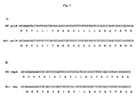
| SEQ ID | |||
| NO: | | Features | |
| 1 | pelB including | P6S | |
| 2 | pelB coding sequence for WT and | C16T | |
| 3 | ompA including discovered variant | [A11P] | |
| 4 | ompA coding sequence for WT and | G31C | |
| 5 | |
||
| 6 | FLAG | ||
| 7 | 6xHis | ||
| 8 | PHPEP 190 | ||
| 9 | PHPEP 97 | ||
Abbreviations
ADCC=antibody-dependent cell-mediated cytotoxicity, ADMC=antibody-dependent monocyte-mediated cytotoxicity, c1q=complement factor 1q, EPO=recombinant erythropoietin, FcR=Fc receptor; Ig=immunoglobulin; Hc=heavy chain; Lc=light chain; IPTG=isopropylthio-β-galactoside; ompA=gene encoding the E. coli outer membrane protein A; pelB=gene encoding the pectate lyase gene of E. caratovora
Definitions
As used herein, unless otherwise indicated or clear from the context, antibody domains, regions and fragments are accorded standard definitions as are well known in the art. The proteins of the invention are derived from, or incorporate portions of antibodies of one or more immunoglobulin classes. Immunoglobulin classes include IgG, IgM, IgA, IgD, and IgE isotypes and, in the case of IgG and IgA, their subtypes, e.g. IgG1, IgG2, IgG3, and IgG4.
By “cistron” is meant a sequence of nucleotides in a DNA molecule coding for an amino acid sequence and including upstream and downstream DNA expression control elements.
By “exogenous polypeptide” or “exogenous protein” or “exoprotein” is meant a protein not normally encoded by the wild-type filamentous phage genome, but rather is foreign to the normal phage protein. A typical exogenous polypeptide is any polypeptide of interest, including an antibody immunoglobulin heavy chain (Hc) domain or immunoglobulin light chain (Lc) domain, an immunoglobulin heavy chain variable domain (VH), an immunoglobulin light chain variable domain (VL), natural or synthetic polypeptides, a single chain antibody (scFv), or a sequence or combination of immunoglobulin domains such as they occur in nature especially as an Fc domain which may include CH3, CH2, a hinge region and/or a CH1 domain or fragment thereof.
By “Fc”, a label given the crystallizable cleavage fragment of a papain digested IgG; is meant a functional fragment of an antibody comprising a dimeric structure of polypeptide chains derived from antibody constant domains and having interchain linkages of disulfide bonds. In human IgG1, papain creates a fragment C-terminal to Cys226 (numbered using the EU index as in Kabat et al., Sequences of Proteins of Immunological Interest, 5th Ed. Public Health Service, National Institutes of Health, Bethesda, Md. (1991), which is expressly incorporated herein by reference. The “EU index as in Kabat” refers to the residue numbering of the human IgG1 EU antibody. Although the definition of N-terminal residue of the Fc may vary, it is generally appreciated to include at least residue 223 in the Kabat numbering system, which is the third residue N-terminal to the first interchain bonding cysteine (C226 in the Kabat system). The Fc portion of the molecule is not directly involved in contact of the antibody with its specific target antigen, but mediates effector functions. These functions are of two types: (1) functions that require binding of the antibody to an antigen, such as C1q binding and/or complement dependent cytotoxicity (CDC) activity or ADCC and ADMC following Fc-receptor gamma-type binding for IgG, Fc-receptor epsilon binding for IgE, and Fc-receptor alpha binding for IgA; and (2) functions that are independent of antigen binding such as persistence in the circulation by the ability to bind FcRn and be transcytosed across cellular and tissue barriers (such as the gut). The ability to significantly increase the serum half-life of antibody molecules or other molecules via the fusion of an Fc, in particular, is highly advantageous. Longer lived molecules may reduce the amount needed in clinical treatments, thereby reducing and frequency of administration.
The terms “Fc receptor” or “FcR” are used to describe a receptor that binds to the Fc region of an antibody. FcR include FcγRI, FcγRII, and FcγRIII subclasses, including allelic variants and alternatively spliced forms of these receptors. FcγRII receptors include FcγRIIA (an “activating receptor”) and FcγRIIB (an “inhibiting receptor”), which have similar amino acid sequences that differ primarily in the cytoplasmic domains thereof. Activating receptor FcγRIIA contains an immunoreceptor tyrosine-based activation motif (FAM) in its cytoplasmic domain. Inhibiting receptor FcγRIIB contains an immunoreceptor tyrosine-based inhibition motif (ITIM) in its cytoplasmic domain (see review in Daëron, Annu. Rev. Immunol., 1997, 15:203-234; FcRs are reviewed in Ravetch and Kinet, Annu. Rev. Immunol., 1991, 9:457-92; Capel et al., Immunomethods, 1994, 4:25-34; and de Haas et al., J. Lab. Clin. Med., 1995, 126:330-41, each of which is incorporated herein by reference).
By “fusion polypeptide” or “fusion protein” is meant a fusion polypeptide (protein) comprising first and second polypeptides encoded by first and second nucleic acid sequences, respectively, which are operatively linked (fused). As exemplified herein, fusion proteins displayed on phage particles are fusions of an exoprotein, a protein not native to the phage proteome, and a phage coat protein.
The term “library” denotes a collection of encoded proteins which are variants, that is, where certain regions are the same or similar and other regions vary. The variation regions may be by directed or random variation (stochastic or non-stochastic changes). A library or variants can be described in terms of number of different variants or “size” of the library. A useful de novo antibody library has high diversity (>1010), amenable to alteration, easy to assemble, and have a low background of undesired sequences. Coupling the following methods accelerates library assembly and leads to low background: (a) Kunkel-based single-stranded mutagenesis; (b) palindromic loop with restriction site and; (c) use of a megaprimer approach.
A “dimeric protein” or “multimeric protein” as used herein refers to more than one separate polypeptide or protein chain associated with each other to form a single globular protein in vitro or in vivo. The multimeric protein may consist of more than one polypeptide of the same kind to form a “homodimer” or a “homomultimer.” Alternatively, the multimeric protein may also be composed of more than one polypeptide of distinct sequences to form a “heterodimer” or “heteromultimer.” Thus, a “heteromultimer” is a molecule comprising at least a first polypeptide and a second polypeptide, wherein the second polypeptide differs in amino acid sequence from the first polypeptide by at least one amino acid residue. The heteromultimer can comprise a “heterodimer” formed by the first and second polypeptide or can form higher order tertiary structures where more than two polypeptides are present. Exemplary structures for the heteromultimer include heterodimers (e.g. Fab, Fc fragments, and diabodies), trimeric G-proteins, heterotetramers (e.g. F(ab′)2 fragments and IgG) and further oligomeric structures.
A “phagemid” or “phage vector” is a replicable cloning vector that contains components derived from both phage chromosomes and exogenous DNA such as that from plasmids. As the phagemid contains a portion of a phage genome, upon co-infection of the host with a helper phage, it can be packaged into phage particles. A phagemid of the invention can be packaged into phage M13 particles.
The term “surface of a phage particle” refers to the part of a bacteriophage particle which is in contact with the medium in which the particle is contained. The surface of the phage particle is determined by the coat protein assembly (the assembled members of the protein coat of the particle) and inner face of the coat protein assembly bounds the region containing nucleic acids of the phage replicated during production of phage in an appropriate host cell.
By “phage coat protein” is meant those proteins forming the phage coat of naturally occurring bacteriophages. In filamentous bacteriophage, such as fl, fd, and M13, the coat proteins are gene III protein (pIII), gene VI protein (pVI), gene VII protein (pVII), gene VIII protein (pVIII), and gene IX protein (pIX). The sequences of the coat proteins of M13 as well as the differences between the closely related members of the filamentous bacteriophages are well known to one of ordinary skill in the art (see, e.g., Kay, B. K., Winter, J. & McCafferty, J., eds. (1996). Phage display of peptides and proteins: a laboratory manual. Academic Press, Inc., San Diego).
Overview
In creating fusion proteins to phage coat proteins, the exoprotein molecule can be large or of diverse chemical characteristics relative to the phage coat protein, particularly so with respect to pIX or pVII, and may interfere with assembly of recombinant phage particles. In order to avoid assembly interference, a phagemid system has often been used whereby the phage particles assemble in the presence of a helper phage and therefore contain both expressed wild-type and exoprotein-coat protein fusions. Phagemid systems are more cumbersome because of the extra steps required and result in much less than the full complete complement of coat protein, if any, displaying the exoprotein. For the latter reason, phagemid display systems are typically considered monovalent. On the other hand, use of a phage vector system to achieve multivalent display often causes low titer phage growth.
The present invention is based on the discovery that a single nucleotide mutation in the coding sequence for the pectate lyase B secretion signal from Erswinia carotovora (pelB) of C for T in the first base of the codon for the sixth residue of the sequence, resulting in a proline to serine change in the encoded signal (SEQ ID NO: 1), causes an improvement in the secretion of several proteins from a bacterial host membrane including proteins displayed on M13 phage coat proteins presumably by improving the secretion of peptides and proteins to the periplasmic region. Secondly, a mutation in the ompA secretion signal, the A11P variant of SEQ ID NO: 3, has been shown to enhance the titer of phage displaying peptide-coat protein fusions.
Using the P6S pelB variant secretion signal, applicants have demonstrated that peptides; including heavily positive-charged, negative-charged or disulfide bond constrained peptides, may be successfully displayed on pIX and pVII phage coat proteins and that antibody fragments and, more recently, dimeric antibody structures including whole IgG can be successfully displayed as a fusion protein of the pIX coat protein using a phagemid system. In addition, as exemplified herein, using the P6S pelB and the ompA, a variety of different peptides and a small protein domain, can be multivalently displayed fused to pVII using a phage vector as described herein. The successful and efficient display of large and multimeric proteins on pIX was undertaken using the presently described mutant pelB secretion signal (SEQ ID NO: 1). The simultaneous multivalent display of proteins on pVII and pIX described herein employed a mutant pelB secretion signal (SEQ ID NO: 1) and an ompA secretion signal (SEQ ID NO: 3), however, either the pelB secretion signal or the ompA secretion signal may be used in conjunction with both proteins to be multivalently displayed and phage coat protein fusions.
These results indicate that the system encompassing the mutant pelB signal sequence discovered and reduced to practice using exoprotein display on pIX or pVII coat proteins using a phagemid vector or a phage vector, can be used to display biochemically diverse peptides and proteins. In addition, other proteins which comprise globular scaffolds such as the fibronectin domains of fibronectin, titin and the like; Z-domain of protein-A, Ankyrin repeat, PDZ-domains, Knottins, CTLA-4 extracellular domain, and the like may be displayed using this system. Monomeric, dimeric, or multimeric receptor proteins such as those that bind to growth factors (e.g. the erythropoietin receptor and the ERBB family of receptors including heregulin), neurotransmitter receptors (e.g. γ-Aminobutyric acid), and other organic or inorganic small molecule receptors (e.g. mineralocorticoid, glucocorticoid) are examples of exoproteins that may be displayed using vectors encoding the mutant sections signals selected from SEQ ID NO: 1 and 3 coupled to the coding sequences for the polypeptide chains comprising the wild type, mutant, or truncated receptor polypeptide chains. Preferred heterodimeric receptors are nuclear hormone receptors (Belshaw et al. (1996) Proc. Natl. Acad. Sci. U.S.A 93 (10):4604-4607), erbB3 and erbB2 receptor complexes, and G-protein-coupled receptors including but not limited to opioid (Gomes et al. (2000) J Neuroscience 20 (22): RC110); Jordan et al. (1999) Nature 399:697-700), muscarinic, dopamine, serotonin, adenosine, and GABAB families of receptors. Further, using standard techniques for randomization and variegation, the construction of display vectors for the above mentioned monomeric, dimeric, or multimeric proteins can serve as the basis for construction of libraries of variant molecules displayed on phage. Such libraries can be useful tools for the exploration of protein remodeling and re-engineering based on these motifs and scaffolds.
The applicants of the present invention have unexpectedly successfully displayed antibody components as described herein a fusion protein to pIX or pVII coat protein on the surface of a filamentous phage particle in as a homodimeric disulfide linked protein displaying at least one of the known biologic activities of the Fc-domain of a natural antibody, such as Fc-receptor binding, and, when in the form of a bivalent antigen-binding protein, capable of specific binding to target antigen. The ability to express large, multidomain proteins tethered to phage coat proteins has heretofore not been reported and is believed to be solely or in part due to the use of the mutant pelB secretion signal in the exoprotein construct.
Method of Making the Invention
In the fusion protein displayed on a filamentous phage particle, the “fusion” between the exogenous polypeptide and the filamentous phage coat protein, and in particular the pVII or pIX coat protein, may be directly linked by a amide linkage, or may comprise a linker polypeptide (i.e., a “linker”). Any of a variety of linkers may be used which are typically a stretch of about 5 to 50 amino acids in length. Particularly preferred linkers provide a high degree of mobility to the fusion protein at the point of the linker. Linkers devoid of secondary structure such as those comprised of predominantly glycine (G, Gly) residues, such as those having G4S (Gly-Gly-Gly-Gly-Ser) repeats or G3S (Gly-Gly-Gly-Ser) where the number of repeats is typically from one to twelve, may be used for this purpose.
The first polypeptide is an exogenous protein and the second polypeptide is a filamentous phage coat protein, such as pVII or pIX protein, whereby the exogenous protein is fused to the amino terminus of the filamentous phage protein. Further, when the fusion protein is in the immature form, i.e., where the leader sequence has not been processed (removed), a fusion protein may contain the amino terminal prokaryotic secretion signal variants of pelB or ompA as described herein.
Where it is desired to express an antibody (immunoglobulin) molecule, a polynucleotide molecule encoding the immunoglobulin will comprise: (1) a first cistron comprising a translational initiation region and a secretion signal operably fused to a polynucleotide sequence encoding a heavy chain and (2) a second cistron comprising a translational initiation region and a secretion signal operably fused to a polynucleotide sequence encoding a light chain, wherein upon expression of the light and heavy polypeptide chains in a prokaryotic host cell, the light and heavy chains are folded and assembled to form a biologically active immunoglobulin. Where it is desired to express the antibody or at least the portions of an antibody capable of stably associating to form a dimeric or heterodimeric structure on the surface of a phage particle, one or both of the cistrons will code for a fusion protein comprising a mutant secretion signal of the invention selected from SEQ ID NO: 1 and 3, fused to an immunoglobulin domain, and which is fused to a phage coat protein.
In natural antibodies, the light chain polypeptide and the heavy chain polypeptide chains are encoded and expressed separately. The typical heterodimeric structure of the IgG class of molecules is dependent on the proper assembly of and formation of disulfide linkages among and between the four polypeptide chains, two heavy and two light chains, of the molecule. Thus, in the present invention, the assembly of the dimeric Fc-portion of the antibody and/or the association of the light chains, when present, recapitulates the natural process of antibody formation insofar as the individual domains of the protein self-associate and form disulfide linkages therebetween.
In one embodiment, the Fc-containing protein to be displayed on the surface of the filamentous phage particle is a natural antibody and a dicistronic vector is constructed for the expression of a Fc-construct-pIX or Fc-construct-pVII fusion protein and a separately encoded and expressed antibody Lc or antigen binding domain which will self-associate. Antigen-binding proteins of the invention can have binding sites for any epitope, antigenic site or protein. Preferred antigen-binding proteins neutralize activation of receptor proteins by direct binding to the receptor or by binding and neutralization of their cognate ligand(s). Generally, the antigen binding domain will be formed of an antibody Lc and an antibody Hc variable domain fused to the natural antibody Hc sequence comprising the Fc domains. In another embodiment, the coat protein-fusion protein includes a scFv linked to the Fc-domain. In another aspect of the invention, the antigen binding sites of the heavy and light chains comprising the scFv may be varied to provide two different binding specificities thereby making the self-assembled disulfide linked construct protein displayed in the phage surface a bispecific and bivalent molecule. For example, substituted for the VL and VH domains of an IgG molecule are scFv domains of different specificity such that the resulting molecule, and is capable of binding to two different epitopes simultaneously. Other methods of creating bispecific antibody molecules having multiple variable domain pairs are taught in US20020103345A1 which could be displayed on phage particles using the methods of the present invention incorporated herein by reference.
In one embodiment the antigen binding or receptor binding domain is not derived from an antibody domain but is a known or random peptide sequence fused to the Fc-domain. Applicants co-pending applications WO04/002417; WO04/002424; WO 05/081687; and WO05/032460 describe a structure referred to herein as a MIMETIBODY™ structure, each of which references are entirely incorporated herein by reference, and which structures are included as dimeric disulfide-linked structures which may be fused to the pIX or pVII phage coat protein and displayed on the outer surface of the phage particle.
In one embodiment, the protein displayed on the phage particle comprises a pair of bioactive peptide-linker-hinge-CH2-CH3 polypeptides, the pair linked by association or covalent linkage, specifically, a Cys-Cys disulfide bond. The bioactive peptide may be on any length and be a naturally occurring sequence derived from any species or be an artificial sequence. The peptides will generally be encoded by the phagemid vector and fused to the Fc-portion of the construct for display on the phage particle. Other constructs of similar structure may be expressed using vectors incorporating the mutant secretion signals of the present invention where the peptide has no known bioactivity but it present to function as marker, a tag, an antigen, or provides for conjugation of a reporter group, a chelating group, or the like.
The level of expression of secreted protein or coat protein-fusion protein can additionally be controlled at the transcriptional level. The fusion proteins are under the inducible control of the Lac Z promoter/operator system (see FIG. 1 ). Other inducible promoters can work as well and are known by one skilled in the art. For high levels of surface expression, the suppressor library is cultured in an inducer of the Lac Z promoter such as isopropylthio-β-galactoside (IPTG). Inducible control is beneficial because biological selection against non-functional pIX fusion proteins can be minimized by culturing the library under non-expressing conditions. Expression can then be induced only at the time of screening to ensure that the entire population of antibodies within the library is accurately represented on the phage surface.
The vector encoding the secreted protein or coat protein-fusion protein may include a translational termination codon at the junction of the exoprotein and coat protein coding regions. When expressed in a bacterial cell carrying a corresponding translation termination suppressor, the fusion protein is produced. When expressed in a bacterial cell without the corresponding suppressor, free exoprotein is not produced.
Method of Using the Invention
When the mutant secretion signal sequences of the invention are used in the construction of a vector for protein expression in a bacterial host cell, e.g. E. coli, the polypeptides are secreted into, and recovered from, the periplasm of the host cells. Protein recovery typically involves disrupting the microorganism, generally by such means as osmotic shock, sonication or lysis. Once cells are disrupted, cell debris or whole cells may be removed by centrifugation or filtration. The proteins may be further purified, for example, by affinity resin chromatography. Alternatively, proteins can be transported into the culture media and isolated therein. Cells may be removed from the culture and the culture supernatant being filtered and concentrated for further purification of the proteins produced. The expressed polypeptides can be further isolated and identified using commonly known methods such as polyacrylamide gel electrophoresis (PAGE) and Western blot assay.
When the mutant secretion signal sequences of the invention are used in the construction of a phage vector or phagemid vector for the expression of a fusion protein which is an exoprotein fused to a phage coat protein, the protein is assembled on the surface of a phage particle formed and secreted from a host cell membrane, e.g. an E. coli cell.
Using the phage vectors exemplified herein as a starting point, the proteins or exoprotein-phage coat protein fusion can be variegated at specific, discrete residue positions or at regions such as N-linked glycosylation sequence, commonly referred to as an NXT sequence, using directed mutagenesis to generate a library of molecules. Particularly useful is a modified Kunkel mutagenesis method which can be used to generate billions of E. coli colonies each harboring a different exoprotein sequence. While efficient, the percentage of non-mutagenized parental DNA increases when generating highly complex sequence libraries. In addition, technical limitations of synthesis of long oligonucleotides can reduce the effectiveness of the method when used to make libraries containing sequence diversities in distant regions. To overcome these limitations, additional techniques of generating oligonucleotides greater than 350 bases can be used. These techniques include use of a mega-primer and creation of a stem-loop sequence containing a restriction enzyme recognition site in the mutagenesis template in combination with the standard Kunkel mutagenesis method (Kunkel at al. 1987 Methods Enzymol. 154: 367-382) as described in US20050048617. Compared to other library technologies, such as restriction cloning (Marks et al., 1991 J. Mol. Biol. 222:581-597; Griffiths et al. 1994 EMBO J. 13, 3245-3260; Hoet et al. 2005 Nature Biotechnol 23, 344-348), phage recombination (Gigapack, Invitrogen), and sequence specific recombination, the improved Kunkel based method is significantly more effective in generating a sequence diverse library (greater than 109) and is more versatile for introducing sequence diversity in any location in the targeted DNA.
The display of an Fc-containing protein on filamentous phage is particularly useful where it is desired to screen a large population of such molecules for desired binding characteristics. Bacterial cells expressing the desired protein or protein fusion are infected with an M13 variant which allows for preferential packaging of vector DNA carrying the fusion gene into phage particles. Each resulting phage particle displays one or more copies of the fusion protein and contains a vector which encodes the fusion protein. The population of such phage particles can be enriched for desired binding characteristics by a panning procedure. Typically, desired particles are immobilized on a solid surface coated with an antigen to which the desired phage particles can bind, such as an ELISA plate. The bound particles are collected and used to further infect bacterial cells. The panning procedure is repeated to further enrich for desired binding characteristics.
One approach to screening proteins (e.g., antibody or Fc-containing molecules) recovered from phage libraries is to remove the phage coat protein that is linked to the protein molecule for display. To facilitate removal of the phage coat protein, an enzymatic cleavage site may be encoded between the exoprotein and the coat protein and appropriately positioned to include or exclude the tag, e.g. Flag or hexahis, sequences.
Phage and other antibody display methods afford the opportunity to manipulate selection against the antigen or receptor target in vitro. One particular advantage of in vitro selection methods is the ability to manipulate selection procedures to obtain antibodies binding to diverse sites on the target protein. Alternatively, whole cells may be used to select binders.
Phage libraries simplify the retrieval of genetic material associated with functional attributes; however, multistep panning strategies are required to isolate the best candidate from the library. Domain or epitope directed pannings have become a routine way of selecting antibodies that bind to a target protein. Such selections have primarily been achieved by employing a stepwise selection of antibodies utilizing methods known variously as selective panning, de-selective panning, ligand capture, subtractive panning or pathfinder selection.
In subtractive panning, target(s) with overlapping but not completely identical binding sites can be used to de-select unwanted binders. This strategy has been used to identify binders even to unknown antigens as in the use of normal cells to de-select binders to cancer cells. Alternatively, naturally occurring proteins with some common domains or structure are used in sequential or competition selection to obtain antibodies binding to sites that differ or are common among the related antigens. In some cases, naturally occurring proteins such as related chemokines or a mutated version of a protein can be used in subtractive panning.
Ligand-capture directed panning is analogous to an ELISA sandwich assay in that an immobilized antibody to an irrelevant and non-adjacent epitope is used to capture and present the preferred binding face of the target ligand for phage panning (U.S. Pat. No. 6,376,170). Others have used competing antibodies to selectively mask the antigen at other than the desired target domain (Tsui, P. et al. 2002. J. Immunol. Meth. 263:123-132). Pathfinder technology uses monoclonal and polyclonal antibodies, as well as natural ligands conjugated directly or indirectly to horseradish peroxidase (HRP). In the presence of biotin tyramine these molecules catalyze biotinylation of phage binding in close proximity to the target antigen, allowing specific recovery of ‘tagged’ phage from the total population using streptavidin. In this way, phage binding to the target itself, or in its immediate proximity, are selectively recovered (Osborn, J. K. et al. 1998. Immunotechnol. 3: 293-302). These methods, variations of the methods, and other methods known to those skilled in the art may be employed to query the libraries of exoproteins generated using the expression methods relying on the mutant secretion signals of the present invention.
In one embodiment, the phage library is used to screen variants of the Fc-portion of the molecules for enhanced, decreased, or altered binding to natural or recombinant Fc-receptors, such as FcRgammaIII (CD16), FcRgammaII (CD32), and FcRgammaI (CD64).
While having described the invention in general terms, the embodiments of the invention will be further disclosed in the following examples that should not be construed as limiting the scope of the claims.
To test if pVII can efficiently and stably display multi-copy of different peptides and proteins, a phage genomic display vector was designed and constructed. The pVII vector was constructed using the phage genome M13KE, a derivative of M13mp19 (vectors which are derivatives of the single-stranded, male-specific filamentous DNA bacteriophage M13, NCBI NC—003287, and approximately 7250 bp in length). The pVII and pIX coding sequences lie adjacent to one another in the genome. In this vector, the peptide with a start codon and a GSGGG linker was inserted into the N-terminus of pVII (FIG. 1A ). The BsrG I restriction endonuclease site before the pVII coding region and the BspH I site at the end of pIX coding region were used for cloning.
A series of biochemically diverse peptides including a linear peptide (HA), heavily negative charged peptide (Flag), positively charged (6×His tag), and disulfide bond constrained peptides (PHPEP 190, PHPEP 97) were cloned into this vector and tested (Table 1). The DNA fragment coding for signal sequence, peptide, GS linker, pVII and pIX, and flanking DNA sequences on 5′ and 3′ ends stretching to BsrG I and BspH I recognition sites, was generated by overlap PCR. The gel-purified PCR product was digested with BsrG I and BspH I restriction endonucleases and then was ligated into double-strand M13KE phage DNA digested with the same enzymes. The ligated recombinant phage DNA was transformed into XL1-Blue E. coli competent cells (Stratagene).
| TABLE 1 |
| Peptide sequences displayed on pVII |
| SEQ | ||||
| ID | Peptide | |||
| NO: | Name | |
||
| 5 | | YPYDVPDYA | ||
| 6 | FLAG | DYKDDDDK | ||
| 7 | 6xHis | HHHHHH | ||
| 8 | PHPEP 190 | |
||
| 9 | PHPEP 97 | RECHPQNWTSCSN | ||
The transformed cells were then plated on a lawn of XL1-Blue host cells to isolate single plaques. Phage plaques on XL1-Blue E. Coli lawn were resuspended in Phosphate-buffered saline (PBS) to allow the phage particles to diffuse from top agar. The diffused phage was added to XL1-Blue liquid media and grown to O.D. 0.5 to infect the E. coli cells. The culture was shaken for 5 hours at 37° C. After shaking the bacteria were removed by centrifugation. The phage particles in the supernatant were precipitated by adding one-tenth volume of cold 0.5 M sodium chloride, 4% PEG-800 and incubating the mixture at 4° C. for 3 hours. The mixture was centrifuged and the phage pellet was resuspended in PBS, aliquotted and stored at −20° C.
Assays of whole phage binding to immobilized ligands on microtiter plates (phage ELISA), DNA sequencing and titration of phage titer were used to characterize the phages and evaluate their displaying on the phage surface. Black Maxisorp ELISA plate (NUNC) was coated overnight with 100 μL/well of antibody or protein recognizing displayed peptide at the concentration of 5 ug/mL in the carbonate coating buffer, pH 9.5. The coated plate was washed 3 times with Tris-buffered saline containing 0.1% Tween-20 (TBST) and blocked with Chemiblocker (Chemicon International) at RT for 1 hour. Phage samples, diluted in Chemiblocker, were added to the plate for binding. After 1 hr of incubation at RT, the plate was washed again to remove the unbound phage. The detection antibody, anti-M13/horseradish peroxidase conjugate (GE healthcare), was diluted 1:5,000 in TBST/10% Chemiblocker and added to the plate at 100 ul/well and incubated for 1 hour at RT. After the final wash, the POD chemiluminescent substrate (Roche) was added to the plate, 100 μl/well, for detection and the plate was read immediately on a Tecan plate reader.
Results
The phage ELISA data is shown in FIG. 2A-E , demonstrating that all five peptides were successfully displayed on the phage surface tethered to pVII coat protein, despite having varying structures and biophysical properties.
A phage vs. phagemid display comparison is shown in FIG. 3 , where the penta-valent display phage system (with all five copies of pVII displaying fusion protein) is compared with the result from a hybrid display system (phage vector having both wild type and fused pIX coding sequences, p99, as described in WO2009/085464 and WO2009/085468) on pIX in which only one copy of the peptide in average was displayed. The ELISA result demonstrated that the HA peptides displayed on the phages derived from the pVII phage vector bound to the anti-HA antibody dramatically better than the phages derived from the p99 hybrid system, suggesting that the vector system produced a multivalent phage display.
Wild-type pVII is naturally synthesized without a signal peptide despite the fact that it is an inner membrane protein in the host cell. However, in previous studies of pVII and pIX display systems, a signal peptide was used (Gao, et al. 1999, Proc Natl Acad Sci USA 96-6025; Kwasnikowski et al. 2005 Journal of Immunological Methods 307:135-143). To examine whether the bacterial signal sequence is necessary for phage display on pVII, we created constructs in which pelB was either added or not added in front of the HA peptide followed by GSGGG linker and pVII.
PHPEP 97 (RECHPQNWTSCSN), a disulfide bond constrained 13 amino acid peptide which binds to streptavidin, was among the peptides tested for the pVII phage display (Example 1). In the phage ELISA, two clones of PEP97 (CL #7 and CL #8) showed elevated binding capacity to streptavidin (FIG. 2E ). Sequence analysis of these clones revealed a single amino acid mutation Pro6 to Ser (FIG. 5A ) in the pelB sequence of both clones.
The P6S pelB was then tested for its ability to enhance display of other peptides and improvement of display efficiency was also observed (data is not shown). To test whether the mutation affected protein secretion, pelB or mutated pelB was fused to alkaline phosphatase (AP) and the secretion of AP in DH10B bacteria examined.
The secreted alkaline phosphatase activity in cell culture was determined. The results showed, in FIG. 6A , that indeed the mutation significantly improved the AP secretion into the cell medium (supernatant fraction) although the AP concentration inside of cells was the same (FIG. 6B ). We therefore hypothesized that the mutated pelB improved the translocation of PHPEP97-pVII to the membrane where phage was assembled by this mutation. To our knowledge, there have been no other reports of mutation in pelB that increase the protein secretion.
To multivalently display two different peptides simultaneously, a phage vector was constructed based on the phage genome M13KE, in which the mutant P6S bacterial signal peptide pelB (SEQ ID NO: 2, C16T), peptide A, and the flexible GSGGG linker were inserted to the N-terminus of the pVII gene; the wild type bacterial signal peptide ompA (SEQ ID NO: 4, G31), peptide B and a flexible GSGGG linker were inserted into the N-terminus of pIX (FIG. 1B ). The BsrG I restriction endonuclease site before the pVII coding region and the BspH I site at the end of pIX coding region were used for cloning. The DNA fragment coding for signal sequences, peptides, GS linkers, pVII, pIX genes and flanking DNA sequences on 5′ and 3′ ends stretching to BsrG I and BspH I recognition sites, was generated by overlap PCR. The gel-purified PCR product was digested with BsrG I and BspH I restriction endonucleases and then was ligated into double-strand M13KE phage DNA digested with the same enzymes. The ligated recombinant phage DNA was transformed into XL1-Blue E. coli competent cells (Stratagene) and the transformed cells were plated on a lawn of XL1-Blue host cells to isolate single plaques as before.
The display of both FLAG and HA peptides was evaluated by phage ELISA (FIG. 7 ) which demonstrated that both peptides were displayed on the phage particles. As was the case for the display of PHPEP 97, however, the original plaque size was very small and the phage titer was low.
To select for the spontaneous mutations in the phage that could improve the growth rate, we continuously passed the phage from two individual clones. After five rounds of continuous passages, a significant increase of the phage titer of one clone (#11) was observed over time (FIG. 8 ). A comparison of the sequence of the whole phage genome purified from round 5 and round 10 phages identified only one single amino acid mutation, Ala to Pro, located on the position 11 of the signal peptide ompA (FIG. 5B ). The same mutation was identified from the phages of the other clone (#9). The mutation, however, did not significantly affect the secretion of alkaline phosphatase as had the mutation in pelB (FIG. 6A ).
A. Vector Design
The full IgG display phagemid (vDR47, FIG. 9 ) was constructed starting from the phagemid construct as described in WO2009/085462 (pCNTO Fab IX). Sequences encoding a human IgG1 hinge, CH2 and CH3 domains were added as well as the P6S pelB variant signal sequence, and the vector does not have a lacI gene however the expression could be controlled by a lac promoter.
B. Characterization of Constructs Used for Full IgG Display
A panel of test constructs was made to assess the display of full IgG on pIX. Antibodies to IL13, designated 6-2 and 16-7, and an anti-cytokine antibody 9-4 were chosen as prototypes for constructing the new full IgG molecules. To determine the effect of different codon usage, two constructs were made for each of the anti-IL13 antibodies, one with human codon optimization and one with E. coli codon optimization. Table 1 lists the vector designation for the five full IgG test constructs. Optimized genes were synthesized and assembled into double stranded DNA as described in U.S. Pat. Nos. 6,670,127 and 6,521,427. In addition, an EMP-1 peptide-Fc fusion construct (CNTO530, SEQ ID NO: 88 of U.S. Pat. No. 7,393,662) fused to pIX was included as a control as it contains the human IgG hinge, CH2 and CH3 domains but no light chains.
| TABLE 2 |
| Test constructs for full IgG Display |
| Antigen | ||||
| pDR# | Isotype | Codon Usage | Description | Specificity |
| pDR2129 | huIgG1/HuKappa | Human codon | 6-2 full IgG | Human |
| IL13 | ||||
| pDR2130 | huIgG1/HuKappa | Human codon | 16-7 full IgG | Human |
| IL13 | ||||
| pDR2131 | huIgG1/HuKappa | E. coli codon | 6-2 full IgG | Human |
| IL13 | ||||
| pDR2132 | huIgG1/HuKappa | E. coli codon | 16-7 full IgG | Human |
| IL13 | ||||
| pDR3041 | huIgG1/HuKappa | Human codon | 9-4 full IgG | Human |
| IL17A | ||||
C. Phage Production
The full IgG display constructs described in section B above were transformed into two different F′ E. coli strains, TG-1 and XL-1 blue, according to standard protocols. The reason for testing these two strains is their difference in growth rate, which hypothetically could affect the packaging and display of the full IgG pIX fusion protein. Individual transformants were picked and grown over night in 2XYT media supplemented with Carbenicillin (always used at 100 μg/ml). The overnight culture (500 μl) was then used to inoculate 25 ml 2XYT/Carbenicillin and the culture was grown at 37° C., 250 rpm, until OD (600 nm) reached 0.5. The bacteria were infected with 10^11 pfu/ml of VCSM13 helper phage (Stratagene, La Jolla, Calif.) during a 30 min incubation at 37° C. with no shaking followed by a centrifugation step at 3,000 rpm for 15 minutes. At this step, the standard protocol calls for the induction of the bacterial culture with 2XYT/Carbenicillin/IPTG (1 mM). However, we divided the cultures into two and added 1 mM IPTG to one and not to the other, with the hypothesis that the leakiness of the system would suffice to produce the fusion protein with subsequent phage packaging. In summary, for each construct, four different phage preparations were made: (i) TG-1 with IPTG (ii) TG-1 without IPTG (iii) XL-1 blue with IPTG (iv) XL-1 blue without IPTG. The cultures were grown over night at 30° C. at 250 rpm and the next day, spun down at 3,000 rpm for 15 minutes, followed by the precipitation of the phage supernatant in PEG/NaCl. After 2 h on ice, the precipitated phage were spun down at 10,000 rpm, 15 min, and the phage pellet was resuspended in 2 ml PBS. The phage preparation was further clarified of any remaining bacterial pellet by a spin at 10,000 rpm for 10 min and stored in 2 ml tubes at 4° C.
D. Phage Titers
The phage titers were determined according to standard protocols. Briefly, TG-1 cells were grown in 2XYT until OD (600 nm) reached 0.5. Phage preparations were serially diluted in PBS in a 96 well plate and TG-1 cells were added to the phage and incubated at 37° C. to allow infection. After 30 min, a spot titration was carried out by dispensing 2 ul of each well onto LB agar plates containing 1% glucose and Carbenicillin. The plates were incubated at 37° C. overnight and the phage concentration in terms of colony forming units (cfu) per ml was determined Table 3 shows the results from the phage titration for all of the constructs and culture conditions. All clones produced high phage titers, between 10^11-10^11 cfu/ml which were in the expected range and indicated that phage was produced efficiently.
| TABLE 3 | ||||
| pDR | Lac | |||
| Description | vector | inducer | TG-1 | XL-1 Blue |
| 6-2 IgG Human Codon | 2129 | −IPTG | 1.00E+13 | 2.00E+13 |
| +IPTG | 5.00E+12 | 5.00E+12 | ||
| 16-7 IgG Human Codon | 2130 | −IPTG | 2.00E+13 | 2.00E+13 |
| +IPTG | 2.00E+12 | 2.00E+12 | ||
| 6-2 IgG E Coli Codon | 2131 | −IPTG | 2.00E+13 | 2.00E+13 |
| +IPTG | 2.00E+13 | 2.00E+13 | ||
| 16-7 IgG E Coli Codon | 2132 | −IPTG | 2.00E+12 | 5.00E+12 |
| +IPTG | 1.00E+11 | 5.00E+11 | ||
| EMP-1 | 2467 | −IPTG | 2.00E+13 | 2.00E+13 |
| +IPTG | 2.00E+12 | 2.00E+12 | ||
E. IgG Domain-Specific Sandwich ELISAs to Assess Functional Display
In order to assess the display of the full IgG molecule on phage pIX, a series of sandwich ELISAs were set up. Black maxisorp plates were coated with 1 μg/ml of one of the following capture antibodies diluted in TBS; sheep anti-human IgG (Fd, CH1) antibody (The Binding Site, Birmingham, UK), mouse anti-human kappa light chains (Southern Biotech, Birmingham, Ala.), mouse anti-human IgG (CH2 domain) antibody (AbD Serotec, Raleigh, N.C.), and mouse anti-human IgG (CH3 domain) antibody (AbD Serotec). After blocking the plates with Chemiblocker (Chemicon/Millipore, Billerica, Mass.), plates were washed and phage were added at a concentration of 2×10^11 cfu/ml (diluted in 10% chemiblocker/TBST) and incubated for one hour. Plates were washed and HRP conjugated mouse anti-M13 antibody was added to the plates. After a 30 min incubation, plates were washed and Chemiluminescence substrate was added to the wells and the plates were read in the Envision plate reader. FIGS. 10A-D show the results from the CH1 (FIG. 10A ), Kappa (FIG. 10B ), CH2 (FIG. 10C ) and CH3 (FIG. 10D ) sandwich ELISAs, respectively. Controls used in the ELISAs were phage displaying the Fab-pIX fusion of clone 6-2 in vDR10 (human codon optimized, made in TG-1 cells, with IPTG induction), an nonspecific scaffold protein-pIX fusion, or the CNTO530-pIX fusion. In the CH1 and Kappa ELISAs, the 6-2 Fab serves as a positive control, whereas the EMP-1 construct (CNTO530) molecule serves as a negative control. In the CH2 and CH3 ELISAs, the 6-2 Fab serves as a negative control and the CNTO530 molecule as a positive control. The scaffold protein phage serves as a negative control in all ELISAs since it does carry any antibody domains. The ELISAs assays were also performed with the addition of an anti-IL13 full IgG1 antibody as a soluble competitor at a concentration of 5 ug/ml in order to prevent binding of the phage to the different capture antibodies.
As shown in FIGS. 10A-D , phage can be detected in all of the sandwich ELISAs, providing evidence that the phage were in fact displaying the different antibody domains on the surface. Phage produced in XL-1 blue cells had the highest signals and the addition of IPTG had a positive effect on the binding signal. The binding of phage can be inhibited by the addition of the soluble anti-IL13 antibody, which indicates specific interactions. However, the soluble anti-IL13 antibody could not compete off the interaction between phage and the CH3 domain (FIG. 10D ). This was observed for both the full IgG-pIX fusions as well as for the EMP-1-Fc-pIX fusion (CNTO530).
F. Full IgG pIX Phage Binding to IL13
After demonstrating that all domains of the IgG molecule can be detected on the phage particles by the ELISAs, it was necessary to determine if the constructs also retained the ability to bind to their respective antigen. The IL13 binding ELISA was set up by coating black Maxisorp plates with 1 ug/ml of a commercial anti-IL13 antibody (mouse anti-human IL13, MAB213, R&D Systems). The MAB213 does not compete with 6-2 or 16-7 for binding to IL13 and thus is ideal as a sandwich ELISA capture antibody. After washing and blocking, biotinylated human IL13R130Q human (Peprotech) was added at 100 nM and incubated for one hour. Plates were washed and phage displaying full IgG versions of 6-2 and 16-7 on pIX were added at 2×10¹¹ cfu/ml, either alone or together with a soluble anti-IL13 antibody for competition. Bound phage was detected with HRP-conjugated mouse anti-M13 antibody and chemiluminescence was read in the Envision instrument. FIG. 11 shows the result of the IL13 phage ELISA. Binding is detected in most conditions, with phage produced in XL-1 blue cells with 1 mM IPTG showing the highest signals. The peptide-Fc-pIX and alternative scaffold molecule-pIX fusions were negative, as expected, and the 6-2 Fab pIX control was positive. The binding was inhibited by adding soluble anti-IL13 antibody, showing that the interaction is specific. To further examine the IL13 binding, an ELISA was set up in which the soluble competition antibody was serially diluted from 50 μg/ml-0.01 μg/ml. A control antibody was also included. FIGS. 12A and B show the effect of soluble antibody competition on IL13 binding of 6-2 IgG pIX and 6-2 Fab pIX, respectively. Inhibition of binding is seen for both constructs, with an IC50 of approximately 0.1 μg/ml. However, for the full IgG pIX construct, the inhibition is incomplete even at very high competitor concentrations, suggesting that some level of un-specific interactions is present.
G. Full IgG pIX Phage Binding to IL13 and IL17
A second confirmatory experiment was performed. This was done by cloning a full IgG version of an anti-IL17A antibody. The construct was transformed into XL-1 blue cells and phage was produced as described above. ELISAs were carried out to confirm the display of the IL17 IgG on pIX as well as its binding to human IL17Amut6 antigen as shown in FIG. 13 . For each ELISA (Fd capture, kappa capture, CH2 capture, CH3 capture, IL13 capture, and IL17 capture), the phage is either added alone or together with a soluble anti-IL13 mAb or a soluble anti-IL17A mAb. The addition of competitor mAb shows the specificity of the ELISA. As evident in FIGS. 12A and B above, the IL17 IgG is displayed on pIX, although at lower levels than the IL13 IgG. This is consistent with differences in Fab expression levels between these constructs (data not shown). The specificity of antigen binding can be seen since the anti IL13 IgG on phage does not bind to IL17 and the anti IL17 IgG on phage does not bind to IL13. In addition, the binding of each of the two types of phage can be inhibited by their soluble mAb counterparts.
Additionally, we have demonstrated that Fc and MIMETIBODY™ proteins could be displayed on the phage surface using a pVII phagemid system.
Claims (24)
1. An isolated polynucleotide encoding a bacterial secretion signal for directing translocation of the protein from the bacterial membrane, selected from the P6S variant of pelB of SEQ ID NO: 1 and the A11P variant of ompA of SEQ ID NO: 3.
2. The isolated polynucleotide of claim 1 operably linked to an exoprotein encoding sequence, wherein the exoprotein is to be secreted from the bacterial membrane.
3. The isolated polynucleotide of claim 2 , wherein the exoprotein is further operably linked to a sequence encoding a phage coat protein selected from pIII, pVII, pVIII, and pIX and wherein the fusion protein so formed is to be secreted from a bacterial membrane.
4. The isolated polynucleotide of claim 3 , wherein the exoprotein is selected from the group consisting of an polypeptide comprising a peptide, an antibody heavy chain constant domain, an antibody heavy chain, a mammalian receptor chain, a receptor extracellular domain, a fibronectin-like domain, and an ankyrin repeat domain.
5. A vector comprising nucleic acid sequences encoding a bacterial secretion signal operably linked to a polynucleotide encoding an amino acid sequence derived from an exogenous protein, the encoded secretion signal selected from the P6S variant of pelB of SEQ ID NO: 1 and the A11P variant of ompA of SEQ ID NO: 3, wherein said secretion signal enhances the proportion of exoprotein found translocated from the bacterial membrane as compared to the wild type secretion signal.
6. The vector of claim 5 wherein said encoded exogenous protein encoding sequence is further fused to the sequence encoding a filamentous phage coat protein selected from pIII, pVII, pVIII, and pIX.
7. The vector of claim 6 which is a phagemid vector.
8. The vector of claim 6 which is a phage vector.
9. The vector of claim 8 in which an exoprotein encoding sequence linked to a mutant secretion signal is fused to pVII and an exoprotein sequence linked to a mutant secretion signal is fused to pIX thereby causing the both exoproteins to be present as multiple copies on the surface of the phage particle when the vector is replicated in a host cell.
10. The vector of claim 6 wherein the encoded exoprotein is selected from the group consisting of a peptide, an antibody heavy chain constant domain, an antibody heavy chain, a mammalian receptor chain, a receptor extracellular domain, a fibronectin-like domain, and an ankyrin repeat domain.
11. The phage vector of claim 5 , further comprising an inducible promoter.
12. The phage vector of claim 11 , wherein the inducible promoter is a lac promoter or mutant of lac.
13. A filamentous phage particle encapsulating a polynucleotide according to claim 3 encoding a secretion signal operably linked to an exoprotein, and having said exoprotein on a surface of said phage.
14. The filamentous phage particle of claim 13 , wherein the exoprotein is fused to an amino terminus of a filamentous phage pVII or pIX protein, whereupon when the protein is expressed at the surface of a filamentous phage protein, a cysteine residue within one exoprotein becomes oxidatively bonded to a cysteine residue on a second exoprotein expressed on the surface of the same filamentous phage particle and forms a functional protein structure.
15. The filamentous phage particle of claim 14 , wherein the functional protein structure comprises an antibody Fc region.
16. The filamentous phage particle of claim 15 , wherein the functional activity of the protein structure is FcRn binding.
17. A bacterial host cell comprising a filamentous phage according to claim 13 .
18. A phage library of bacterial host cells comprising a filamentous phage vector according to claim 17 .
19. The phage library of claim 14 , wherein said each polynucleotide encodes an Fc-forming sequence with an amino acid substitution.
20. The phage library of claim 15 wherein said nucleic acid chain wherein the Fc-forming sequence further comprise a ligand binding domain.
21. The phage library of claim 16 wherein the ligand binding domain is selected from the group consisting of a receptor binding ligand, a receptor extracellular domain, an antibody Fab domain comprising a variable domain and a constant domain, and single chain Fv construct.
22. The phage library of claim 17 wherein said Fab domain comprises sequences that vary one from another at specific residues within the binding domain.
23. A method of expressing an exoprotein in a bacterial host cell using a polynucleotide comprising a coding sequence for the P6S variant of pelB of SEQ ID NO: 1, wherein the exoprotein is recoverable from the media supporting the growth of the bacterial host cell.
24. A method of expressing an exoprotein in a bacterial host cell using a polynucleotide comprising a variant of a bacterial secretion signal according to claim 1 , wherein the protein is fused to a filamentous phage coat protein and the titer of phage displaying the protein on the surface of the phage particle is higher than when a wild type bacterial secretion signal is used or when no bacterial secretion signal is used.
Priority Applications (1)
| Application Number | Priority Date | Filing Date | Title |
|---|---|---|---|
| US13/509,287 US8450086B2 (en) | 2009-11-17 | 2010-11-15 | Bacterial membrane protein secretion |
Applications Claiming Priority (3)
| Application Number | Priority Date | Filing Date | Title |
|---|---|---|---|
| US26176809P | 2009-11-17 | 2009-11-17 | |
| PCT/US2010/056680 WO2011062862A1 (en) | 2009-11-17 | 2010-11-15 | Improved bacterial membrane protein secrection |
| US13/509,287 US8450086B2 (en) | 2009-11-17 | 2010-11-15 | Bacterial membrane protein secretion |
Publications (2)
| Publication Number | Publication Date |
|---|---|
| US20120225807A1 US20120225807A1 (en) | 2012-09-06 |
| US8450086B2 true US8450086B2 (en) | 2013-05-28 |
Family
ID=44059935
Family Applications (1)
| Application Number | Title | Priority Date | Filing Date |
|---|---|---|---|
| US13/509,287 Expired - Fee Related US8450086B2 (en) | 2009-11-17 | 2010-11-15 | Bacterial membrane protein secretion |
Country Status (9)
| Country | Link |
|---|---|
| US (1) | US8450086B2 (en) |
| EP (1) | EP2501824A4 (en) |
| JP (1) | JP5955773B2 (en) |
| CN (2) | CN104531734A (en) |
| AU (1) | AU2010322205B2 (en) |
| CA (1) | CA2781099A1 (en) |
| IL (1) | IL219686A (en) |
| MY (1) | MY159680A (en) |
| WO (1) | WO2011062862A1 (en) |
Cited By (2)
| Publication number | Priority date | Publication date | Assignee | Title |
|---|---|---|---|---|
| US11866465B2 (en) | 2017-04-27 | 2024-01-09 | Juno Therapeutics Gmbh | Oligomeric particle reagents and methods of use thereof |
| US11913024B2 (en) | 2015-10-22 | 2024-02-27 | Juno Therapeutics Gmbh | Methods for culturing cells and kits and apparatus for same |
Families Citing this family (6)
| Publication number | Priority date | Publication date | Assignee | Title |
|---|---|---|---|---|
| AU2014210475B2 (en) | 2013-01-24 | 2017-03-02 | Abtlas Co., Ltd. | Protein combination-based Fv library, and preparation method therefor |
| CN103254277B (en) * | 2013-05-03 | 2017-02-08 | 南京工业大学 | Small peptide strengthening mold sequence of strong secretory signal peptide and application thereof |
| JP6421456B2 (en) * | 2013-05-28 | 2018-11-14 | 東ソー株式会社 | Signal peptide and protein production method using the same |
| CN105018507B (en) * | 2014-04-17 | 2018-05-15 | 东北师范大学 | A kind of phage vector and its construction method for being used for double epitope displays |
| WO2022020380A1 (en) * | 2020-07-20 | 2022-01-27 | Massachusetts Institute Of Technology | M13 bacteriophage with a high cysteine content and genetically engineerable hydrogels |
| CN113136372B (en) * | 2021-05-28 | 2023-08-01 | 广西大学 | Construction method of recombinant phage |
Citations (16)
| Publication number | Priority date | Publication date | Assignee | Title |
|---|---|---|---|---|
| US6521427B1 (en) | 1997-09-16 | 2003-02-18 | Egea Biosciences, Inc. | Method for the complete chemical synthesis and assembly of genes and genomes |
| US6670127B2 (en) | 1997-09-16 | 2003-12-30 | Egea Biosciences, Inc. | Method for assembly of a polynucleotide encoding a target polypeptide |
| WO2004002417A2 (en) | 2002-06-28 | 2004-01-08 | Centocor, Inc. | Mammalian ch1 deleted mimetibodies, compositions, methods and uses |
| WO2004002424A2 (en) | 2002-06-28 | 2004-01-08 | Centocor, Inc. | Mammalian epo mimetic ch1 deleted mimetibodies, compositions, methods and uses |
| US6753136B2 (en) | 1999-07-20 | 2004-06-22 | Morphosys Ag | Methods for displaying (poly) peptides/proteins on bacteriophage particles via disulfide bonds |
| US6875590B2 (en) * | 2000-06-05 | 2005-04-05 | Corixa Corporation | Leader peptides for enhancing secretion of recombinant protein from a host cell |
| WO2005032460A2 (en) | 2003-09-30 | 2005-04-14 | Centocor, Inc. | Human epo mimetic hinge core mimetibodies, compositions, methods and uses |
| WO2005081687A2 (en) | 2003-09-30 | 2005-09-09 | Centocor, Inc. | Human hinge core mimetibodies, compositions, methods and uses |
| US20050250131A1 (en) | 2004-02-27 | 2005-11-10 | Institut Pasteur | Methods for obtaining thermostable enzymes, DNA polymerase I variants from Thermus aquaticus having new catalytic activities, methods for obtaining the same, and applications of the same |
| US7078166B2 (en) | 1999-05-25 | 2006-07-18 | The Scripps Research Institute | Methods for display of heterodimeric proteins on filamentous phage using pVII and pIX, compositions, vectors and combinatorial libraries |
| US20060257856A1 (en) | 1991-04-10 | 2006-11-16 | The Scripps Research Institute | Heterodimeric receptor libraries using phagemids |
| US20060286068A1 (en) | 2000-01-10 | 2006-12-21 | Maxygen Holdings, Ltd. | G-CSF Conjugates |
| US7393662B2 (en) | 2004-09-03 | 2008-07-01 | Centocor, Inc. | Human EPO mimetic hinge core mimetibodies, compositions, methods and uses |
| WO2009085464A2 (en) | 2007-12-19 | 2009-07-09 | Centocor, Inc. | Engineered hybird phage vectors for the design and the generation of a human non-antibody peptide or protein phage library via fusion to pix of m13 phage |
| WO2009085462A1 (en) | 2007-12-19 | 2009-07-09 | Centocor, Inc. | Design and generation of human de novo pix phage display libraries via fusion to pix or pvii, vectors, antibodies and methods |
| WO2009085468A2 (en) | 2007-12-19 | 2009-07-09 | Centocor, Inc. | Engineered phage vectors for the design and the generation of a human non-antibody peptide or protein phage library via fusion to pix of m13 phage |
Family Cites Families (3)
| Publication number | Priority date | Publication date | Assignee | Title |
|---|---|---|---|---|
| EP1299419A2 (en) | 2000-05-24 | 2003-04-09 | Imclone Systems, Inc. | Bispecific immunoglobulin-like antigen binding proteins and method of production |
| GB0027782D0 (en) * | 2000-11-14 | 2000-12-27 | Boehringer Ingelheim Int | Methods fro large scale protein productio in prokaryotes |
| JP2008138690A (en) * | 2006-11-30 | 2008-06-19 | Tanashin Denki Co | Attachment mechanism for rotating component |
-
2010
- 2010-11-15 AU AU2010322205A patent/AU2010322205B2/en not_active Ceased
- 2010-11-15 CA CA2781099A patent/CA2781099A1/en not_active Abandoned
- 2010-11-15 EP EP10832035.9A patent/EP2501824A4/en not_active Withdrawn
- 2010-11-15 CN CN201410748438.8A patent/CN104531734A/en active Pending
- 2010-11-15 CN CN201080052575.8A patent/CN102770555B/en not_active Expired - Fee Related
- 2010-11-15 US US13/509,287 patent/US8450086B2/en not_active Expired - Fee Related
- 2010-11-15 JP JP2012539960A patent/JP5955773B2/en not_active Expired - Fee Related
- 2010-11-15 MY MYPI2012002135A patent/MY159680A/en unknown
- 2010-11-15 WO PCT/US2010/056680 patent/WO2011062862A1/en active Application Filing
-
2012
- 2012-05-09 IL IL219686A patent/IL219686A/en not_active IP Right Cessation
Patent Citations (16)
| Publication number | Priority date | Publication date | Assignee | Title |
|---|---|---|---|---|
| US20060257856A1 (en) | 1991-04-10 | 2006-11-16 | The Scripps Research Institute | Heterodimeric receptor libraries using phagemids |
| US6670127B2 (en) | 1997-09-16 | 2003-12-30 | Egea Biosciences, Inc. | Method for assembly of a polynucleotide encoding a target polypeptide |
| US6521427B1 (en) | 1997-09-16 | 2003-02-18 | Egea Biosciences, Inc. | Method for the complete chemical synthesis and assembly of genes and genomes |
| US7078166B2 (en) | 1999-05-25 | 2006-07-18 | The Scripps Research Institute | Methods for display of heterodimeric proteins on filamentous phage using pVII and pIX, compositions, vectors and combinatorial libraries |
| US6753136B2 (en) | 1999-07-20 | 2004-06-22 | Morphosys Ag | Methods for displaying (poly) peptides/proteins on bacteriophage particles via disulfide bonds |
| US20060286068A1 (en) | 2000-01-10 | 2006-12-21 | Maxygen Holdings, Ltd. | G-CSF Conjugates |
| US6875590B2 (en) * | 2000-06-05 | 2005-04-05 | Corixa Corporation | Leader peptides for enhancing secretion of recombinant protein from a host cell |
| WO2004002417A2 (en) | 2002-06-28 | 2004-01-08 | Centocor, Inc. | Mammalian ch1 deleted mimetibodies, compositions, methods and uses |
| WO2004002424A2 (en) | 2002-06-28 | 2004-01-08 | Centocor, Inc. | Mammalian epo mimetic ch1 deleted mimetibodies, compositions, methods and uses |
| WO2005032460A2 (en) | 2003-09-30 | 2005-04-14 | Centocor, Inc. | Human epo mimetic hinge core mimetibodies, compositions, methods and uses |
| WO2005081687A2 (en) | 2003-09-30 | 2005-09-09 | Centocor, Inc. | Human hinge core mimetibodies, compositions, methods and uses |
| US20050250131A1 (en) | 2004-02-27 | 2005-11-10 | Institut Pasteur | Methods for obtaining thermostable enzymes, DNA polymerase I variants from Thermus aquaticus having new catalytic activities, methods for obtaining the same, and applications of the same |
| US7393662B2 (en) | 2004-09-03 | 2008-07-01 | Centocor, Inc. | Human EPO mimetic hinge core mimetibodies, compositions, methods and uses |
| WO2009085464A2 (en) | 2007-12-19 | 2009-07-09 | Centocor, Inc. | Engineered hybird phage vectors for the design and the generation of a human non-antibody peptide or protein phage library via fusion to pix of m13 phage |
| WO2009085462A1 (en) | 2007-12-19 | 2009-07-09 | Centocor, Inc. | Design and generation of human de novo pix phage display libraries via fusion to pix or pvii, vectors, antibodies and methods |
| WO2009085468A2 (en) | 2007-12-19 | 2009-07-09 | Centocor, Inc. | Engineered phage vectors for the design and the generation of a human non-antibody peptide or protein phage library via fusion to pix of m13 phage |
Non-Patent Citations (9)
| Title |
|---|
| Gao, C. et al., "A method for the generation of combinatorial antibody libraries using pIX phage display", Proc Natl Acad Sci USA, vol. 99, pp. 12612-12616 (2002). |
| Gao, C. et al., Making artificial antibodies: A format for phage display of combinatorial heterodimeric arrays, Proc Nat Acad Sci, vol. 96, pp. 6025-6030 (1999). |
| Griffiths et al., "Isolation of high affinity human antibodies directly from large synthetic repertoires", J EMBO, vol. 13, pp. 3245-3260 (1994). |
| Hoet et al., "Generation of high-affinity human antibodies by combining donor-derived and synthetic complementarity-determining region diversity", Nature Biotechnology, vol. 23, pp. 344-348 (2005). |
| International Search Report for PCT/US10/56680 dated Feb. 17, 2011. |
| Klose et al., "The Influence of Amino Substitutions within the Mature Part of an Eschrichia coli Outer Membrane Protein (OmpA) on Assembly of the Polypeptide into Its Membrane", The Journal of Biological Chemistry, vol. 263, No. 26, pp. 13297-13302 (1988). |
| Kwasnikowski, et al., "Multivalent display system on filamentous bacteriophage pVII minor coat protein", Journal of Immunological Methods, vol. 307, p. 135 (2005). |
| Marks et al., By-passing Immunization, Journal of Molecular Biology, vol. 222, pp. 581-597 (1991). |
| Se-Ho Kim et al., An approach for preventing recombination-deletion of the 40-50 anti-digoxin antibody VH gene from the phage display vector pComb3. Gene 241 (2000) 19-25. * |
Cited By (2)
| Publication number | Priority date | Publication date | Assignee | Title |
|---|---|---|---|---|
| US11913024B2 (en) | 2015-10-22 | 2024-02-27 | Juno Therapeutics Gmbh | Methods for culturing cells and kits and apparatus for same |
| US11866465B2 (en) | 2017-04-27 | 2024-01-09 | Juno Therapeutics Gmbh | Oligomeric particle reagents and methods of use thereof |
Also Published As
| Publication number | Publication date |
|---|---|
| WO2011062862A1 (en) | 2011-05-26 |
| AU2010322205B2 (en) | 2015-01-22 |
| MY159680A (en) | 2017-01-13 |
| EP2501824A1 (en) | 2012-09-26 |
| US20120225807A1 (en) | 2012-09-06 |
| JP2013510592A (en) | 2013-03-28 |
| AU2010322205A1 (en) | 2012-06-07 |
| JP5955773B2 (en) | 2016-07-20 |
| IL219686A0 (en) | 2012-07-31 |
| EP2501824A4 (en) | 2013-06-19 |
| CA2781099A1 (en) | 2011-05-26 |
| IL219686A (en) | 2015-04-30 |
| CN102770555B (en) | 2015-01-28 |
| CN102770555A (en) | 2012-11-07 |
| CN104531734A (en) | 2015-04-22 |
Similar Documents
| Publication | Publication Date | Title |
|---|---|---|
| US8450086B2 (en) | Bacterial membrane protein secretion | |
| CN108473987B (en) | Binding members with altered diverse scaffold domains | |
| DK2044117T3 (en) | PROCEDURE FOR MANUFACTURING IMMUNGLOBULINES | |
| US8551715B2 (en) | Methods for identifying and isolating cells expressing a polypeptide | |
| Gram et al. | Phage display as a rapid gene expression system: production of bioactive cytokine-phage and generation of neutralizing monoclonal antibodies | |
| US8728985B2 (en) | Display of disulfide linked dimeric proteins in filamentous phage | |
| CA2905529A1 (en) | An integrated system for library construction, affinity binder screening and expression thereof | |
| WO2018069416A1 (en) | Triple vector for expressing antibody molecules in full therapeutic format | |
| US20200140573A1 (en) | Nucleic acid compositions, methods and kits for rapid pairing of affinity agents | |
| KR20210110600A (en) | mixed binding domains | |
| FUCHS et al. | Primary structure and functional scFv antibody expression of an antibody against the human protooncogen c-myc | |
| KR20120093312A (en) | Antibody mimetic scaffolds | |
| RU2778694C2 (en) | Binding elements with modified diversified backbone domains | |
| US20230331827A1 (en) | Potassium channel inhibitors | |
| CN114245804A (en) | Modified human variable domains | |
| AU2015258309A1 (en) | Methods for identifying and isolating cells expressing a polypeptide |
Legal Events
| Date | Code | Title | Description |
|---|---|---|---|
| STCF | Information on status: patent grant |
Free format text: PATENTED CASE |
|
| FPAY | Fee payment |
Year of fee payment: 4 |
|
| FEPP | Fee payment procedure |
Free format text: MAINTENANCE FEE REMINDER MAILED (ORIGINAL EVENT CODE: REM.); ENTITY STATUS OF PATENT OWNER: LARGE ENTITY |
|
| LAPS | Lapse for failure to pay maintenance fees |
Free format text: PATENT EXPIRED FOR FAILURE TO PAY MAINTENANCE FEES (ORIGINAL EVENT CODE: EXP.); ENTITY STATUS OF PATENT OWNER: LARGE ENTITY |
|
| STCH | Information on status: patent discontinuation |
Free format text: PATENT EXPIRED DUE TO NONPAYMENT OF MAINTENANCE FEES UNDER 37 CFR 1.362 |
|
| FP | Lapsed due to failure to pay maintenance fee |
Effective date: 20210528 |