US8333952B2 - Dose synthesis module for biomarker generator system - Google Patents
Dose synthesis module for biomarker generator system Download PDFInfo
- Publication number
- US8333952B2 US8333952B2 US12/565,544 US56554409A US8333952B2 US 8333952 B2 US8333952 B2 US 8333952B2 US 56554409 A US56554409 A US 56554409A US 8333952 B2 US8333952 B2 US 8333952B2
- Authority
- US
- United States
- Prior art keywords
- radiopharmaceutical
- solution
- radioisotope
- reaction vessel
- production
- Prior art date
- Legal status (The legal status is an assumption and is not a legal conclusion. Google has not performed a legal analysis and makes no representation as to the accuracy of the status listed.)
- Active, expires
Links
- 238000003786 synthesis reaction Methods 0.000 title claims abstract description 34
- 230000015572 biosynthetic process Effects 0.000 title claims abstract description 33
- 239000000090 biomarker Substances 0.000 title abstract description 18
- 239000012217 radiopharmaceutical Substances 0.000 claims abstract description 88
- 229940121896 radiopharmaceutical Drugs 0.000 claims abstract description 88
- 230000002799 radiopharmaceutical effect Effects 0.000 claims abstract description 88
- 238000006243 chemical reaction Methods 0.000 claims abstract description 26
- 239000003153 chemical reaction reagent Substances 0.000 claims abstract description 15
- 238000002414 normal-phase solid-phase extraction Methods 0.000 claims abstract description 13
- 238000012360 testing method Methods 0.000 claims description 37
- OKTJSMMVPCPJKN-BJUDXGSMSA-N carbon-11 Chemical compound [11C] OKTJSMMVPCPJKN-BJUDXGSMSA-N 0.000 claims description 4
- QJGQUHMNIGDVPM-BJUDXGSMSA-N Nitrogen-13 Chemical compound [13N] QJGQUHMNIGDVPM-BJUDXGSMSA-N 0.000 claims description 3
- QVGXLLKOCUKJST-BJUDXGSMSA-N oxygen-15 atom Chemical compound [15O] QVGXLLKOCUKJST-BJUDXGSMSA-N 0.000 claims description 3
- 238000012546 transfer Methods 0.000 claims description 3
- AOYNUTHNTBLRMT-MXWOLSILSA-N 2-Deoxy-2(F-18)fluoro-2-D-glucose Chemical compound OC[C@@H](O)[C@@H](O)[C@H](O)[C@@H]([18F])C=O AOYNUTHNTBLRMT-MXWOLSILSA-N 0.000 claims 1
- YCKRFDGAMUMZLT-BJUDXGSMSA-N fluorine-18 atom Chemical compound [18F] YCKRFDGAMUMZLT-BJUDXGSMSA-N 0.000 claims 1
- 238000002600 positron emission tomography Methods 0.000 abstract description 25
- 238000004519 manufacturing process Methods 0.000 abstract description 20
- 238000000034 method Methods 0.000 abstract description 14
- 239000000203 mixture Substances 0.000 abstract description 8
- 230000002194 synthesizing effect Effects 0.000 abstract description 4
- 239000002699 waste material Substances 0.000 abstract description 4
- 239000000523 sample Substances 0.000 description 55
- 238000003908 quality control method Methods 0.000 description 31
- WEVYAHXRMPXWCK-UHFFFAOYSA-N Acetonitrile Chemical compound CC#N WEVYAHXRMPXWCK-UHFFFAOYSA-N 0.000 description 28
- ZCXUVYAZINUVJD-GLCXRVCCSA-N [18F]fluorodeoxyglucose Chemical compound OC[C@H]1OC(O)[C@H]([18F])[C@@H](O)[C@@H]1O ZCXUVYAZINUVJD-GLCXRVCCSA-N 0.000 description 15
- KRHYYFGTRYWZRS-BJUDXGSMSA-N ac1l2y5h Chemical compound [18FH] KRHYYFGTRYWZRS-BJUDXGSMSA-N 0.000 description 11
- XLYOFNOQVPJJNP-ZSJDYOACSA-N Heavy water Chemical compound [2H]O[2H] XLYOFNOQVPJJNP-ZSJDYOACSA-N 0.000 description 8
- 238000012824 chemical production Methods 0.000 description 8
- 239000002904 solvent Substances 0.000 description 8
- 239000007789 gas Substances 0.000 description 7
- VEXZGXHMUGYJMC-UHFFFAOYSA-N Hydrochloric acid Chemical compound Cl VEXZGXHMUGYJMC-UHFFFAOYSA-N 0.000 description 6
- 239000002245 particle Substances 0.000 description 6
- XLYOFNOQVPJJNP-UHFFFAOYSA-N water Substances O XLYOFNOQVPJJNP-UHFFFAOYSA-N 0.000 description 6
- LFQSCWFLJHTTHZ-UHFFFAOYSA-N Ethanol Chemical compound CCO LFQSCWFLJHTTHZ-UHFFFAOYSA-N 0.000 description 4
- 238000005516 engineering process Methods 0.000 description 4
- 238000003384 imaging method Methods 0.000 description 4
- 239000012535 impurity Substances 0.000 description 4
- 239000011261 inert gas Substances 0.000 description 4
- 239000004615 ingredient Substances 0.000 description 4
- 239000012071 phase Substances 0.000 description 4
- 238000003860 storage Methods 0.000 description 4
- 239000000126 substance Substances 0.000 description 4
- IJGRMHOSHXDMSA-UHFFFAOYSA-N Atomic nitrogen Chemical compound N#N IJGRMHOSHXDMSA-UHFFFAOYSA-N 0.000 description 3
- ZLMJMSJWJFRBEC-UHFFFAOYSA-N Potassium Chemical compound [K] ZLMJMSJWJFRBEC-UHFFFAOYSA-N 0.000 description 3
- VYPSYNLAJGMNEJ-UHFFFAOYSA-N Silicium dioxide Chemical compound O=[Si]=O VYPSYNLAJGMNEJ-UHFFFAOYSA-N 0.000 description 3
- OIBDVHSTOUGZTJ-PEBLQZBPSA-N [(2r,3r,4s,5s,6s)-3,4,6-triacetyloxy-5-(trifluoromethylsulfonyloxy)oxan-2-yl]methyl acetate Chemical compound CC(=O)OC[C@H]1O[C@@H](OC(C)=O)[C@@H](OS(=O)(=O)C(F)(F)F)[C@@H](OC(C)=O)[C@@H]1OC(C)=O OIBDVHSTOUGZTJ-PEBLQZBPSA-N 0.000 description 3
- 201000010099 disease Diseases 0.000 description 3
- 208000037265 diseases, disorders, signs and symptoms Diseases 0.000 description 3
- 239000002243 precursor Substances 0.000 description 3
- 238000013094 purity test Methods 0.000 description 3
- 230000005855 radiation Effects 0.000 description 3
- NWUYHJFMYQTDRP-UHFFFAOYSA-N 1,2-bis(ethenyl)benzene;1-ethenyl-2-ethylbenzene;styrene Chemical compound C=CC1=CC=CC=C1.CCC1=CC=CC=C1C=C.C=CC1=CC=CC=C1C=C NWUYHJFMYQTDRP-UHFFFAOYSA-N 0.000 description 2
- ZCXUVYAZINUVJD-AHXZWLDOSA-N 2-deoxy-2-((18)F)fluoro-alpha-D-glucose Chemical compound OC[C@H]1O[C@H](O)[C@H]([18F])[C@@H](O)[C@@H]1O ZCXUVYAZINUVJD-AHXZWLDOSA-N 0.000 description 2
- 125000002777 acetyl group Chemical group [H]C([H])([H])C(*)=O 0.000 description 2
- PNEYBMLMFCGWSK-UHFFFAOYSA-N aluminium oxide Inorganic materials [O-2].[O-2].[O-2].[Al+3].[Al+3] PNEYBMLMFCGWSK-UHFFFAOYSA-N 0.000 description 2
- 238000002591 computed tomography Methods 0.000 description 2
- 150000003983 crown ethers Chemical class 0.000 description 2
- GPWDPLKISXZVIE-UHFFFAOYSA-N cyclo[18]carbon Chemical compound C1#CC#CC#CC#CC#CC#CC#CC#CC#C1 GPWDPLKISXZVIE-UHFFFAOYSA-N 0.000 description 2
- 238000010586 diagram Methods 0.000 description 2
- 238000009826 distribution Methods 0.000 description 2
- 230000000694 effects Effects 0.000 description 2
- 238000010828 elution Methods 0.000 description 2
- 239000002158 endotoxin Substances 0.000 description 2
- 238000000605 extraction Methods 0.000 description 2
- 238000012061 filter integrity test Methods 0.000 description 2
- 239000000499 gel Substances 0.000 description 2
- 230000036512 infertility Effects 0.000 description 2
- 239000003456 ion exchange resin Substances 0.000 description 2
- 229920003303 ion-exchange polymer Polymers 0.000 description 2
- 125000000311 mannosyl group Chemical group C1([C@@H](O)[C@@H](O)[C@H](O)[C@H](O1)CO)* 0.000 description 2
- 230000002503 metabolic effect Effects 0.000 description 2
- 239000011148 porous material Substances 0.000 description 2
- 229910052700 potassium Inorganic materials 0.000 description 2
- 239000011591 potassium Substances 0.000 description 2
- NROKBHXJSPEDAR-UHFFFAOYSA-M potassium fluoride Chemical compound [F-].[K+] NROKBHXJSPEDAR-UHFFFAOYSA-M 0.000 description 2
- 230000001681 protective effect Effects 0.000 description 2
- 238000013022 venting Methods 0.000 description 2
- AUFVJZSDSXXFOI-UHFFFAOYSA-N 2.2.2-cryptand Chemical compound C1COCCOCCN2CCOCCOCCN1CCOCCOCC2 AUFVJZSDSXXFOI-UHFFFAOYSA-N 0.000 description 1
- KRHYYFGTRYWZRS-UHFFFAOYSA-M Fluoride anion Chemical compound [F-] KRHYYFGTRYWZRS-UHFFFAOYSA-M 0.000 description 1
- YCKRFDGAMUMZLT-UHFFFAOYSA-N Fluorine atom Chemical compound [F] YCKRFDGAMUMZLT-UHFFFAOYSA-N 0.000 description 1
- 206010028980 Neoplasm Diseases 0.000 description 1
- FAPWRFPIFSIZLT-UHFFFAOYSA-M Sodium chloride Chemical compound [Na+].[Cl-] FAPWRFPIFSIZLT-UHFFFAOYSA-M 0.000 description 1
- MJOQJPYNENPSSS-XQHKEYJVSA-N [(3r,4s,5r,6s)-4,5,6-triacetyloxyoxan-3-yl] acetate Chemical compound CC(=O)O[C@@H]1CO[C@@H](OC(C)=O)[C@H](OC(C)=O)[C@H]1OC(C)=O MJOQJPYNENPSSS-XQHKEYJVSA-N 0.000 description 1
- 210000003484 anatomy Anatomy 0.000 description 1
- 201000011510 cancer Diseases 0.000 description 1
- 238000005352 clarification Methods 0.000 description 1
- 238000011109 contamination Methods 0.000 description 1
- 238000001514 detection method Methods 0.000 description 1
- 238000011161 development Methods 0.000 description 1
- 238000002059 diagnostic imaging Methods 0.000 description 1
- 238000002405 diagnostic procedure Methods 0.000 description 1
- 229910001873 dinitrogen Inorganic materials 0.000 description 1
- 230000005611 electricity Effects 0.000 description 1
- 238000001704 evaporation Methods 0.000 description 1
- 229910052731 fluorine Inorganic materials 0.000 description 1
- 239000011737 fluorine Substances 0.000 description 1
- 230000005714 functional activity Effects 0.000 description 1
- 230000003760 hair shine Effects 0.000 description 1
- 238000002372 labelling Methods 0.000 description 1
- 239000007788 liquid Substances 0.000 description 1
- 238000002595 magnetic resonance imaging Methods 0.000 description 1
- 239000000463 material Substances 0.000 description 1
- 238000002493 microarray Methods 0.000 description 1
- 238000012986 modification Methods 0.000 description 1
- 230000004048 modification Effects 0.000 description 1
- 229910052757 nitrogen Inorganic materials 0.000 description 1
- 239000012299 nitrogen atmosphere Substances 0.000 description 1
- 238000009206 nuclear medicine Methods 0.000 description 1
- 210000000056 organ Anatomy 0.000 description 1
- 210000004789 organ system Anatomy 0.000 description 1
- 125000002524 organometallic group Chemical group 0.000 description 1
- QVGXLLKOCUKJST-NJFSPNSNSA-N oxygen-18 atom Chemical compound [18O] QVGXLLKOCUKJST-NJFSPNSNSA-N 0.000 description 1
- 230000010412 perfusion Effects 0.000 description 1
- 239000008363 phosphate buffer Substances 0.000 description 1
- 230000001766 physiological effect Effects 0.000 description 1
- 239000011698 potassium fluoride Substances 0.000 description 1
- 235000003270 potassium fluoride Nutrition 0.000 description 1
- 238000000746 purification Methods 0.000 description 1
- 230000005258 radioactive decay Effects 0.000 description 1
- 239000012857 radioactive material Substances 0.000 description 1
- 239000000700 radioactive tracer Substances 0.000 description 1
- 230000035484 reaction time Effects 0.000 description 1
- 238000011160 research Methods 0.000 description 1
- 239000012488 sample solution Substances 0.000 description 1
- 239000000741 silica gel Substances 0.000 description 1
- 229910002027 silica gel Inorganic materials 0.000 description 1
- 239000000377 silicon dioxide Substances 0.000 description 1
- 239000011780 sodium chloride Substances 0.000 description 1
- 208000024891 symptom Diseases 0.000 description 1
- 238000002604 ultrasonography Methods 0.000 description 1
- 230000000007 visual effect Effects 0.000 description 1
Images


Classifications
-
- A—HUMAN NECESSITIES
- A61—MEDICAL OR VETERINARY SCIENCE; HYGIENE
- A61K—PREPARATIONS FOR MEDICAL, DENTAL OR TOILETRY PURPOSES
- A61K51/00—Preparations containing radioactive substances for use in therapy or testing in vivo
- A61K51/02—Preparations containing radioactive substances for use in therapy or testing in vivo characterised by the carrier, i.e. characterised by the agent or material covalently linked or complexing the radioactive nucleus
- A61K51/04—Organic compounds
- A61K51/0491—Sugars, nucleosides, nucleotides, oligonucleotides, nucleic acids, e.g. DNA, RNA, nucleic acid aptamers
-
- B—PERFORMING OPERATIONS; TRANSPORTING
- B01—PHYSICAL OR CHEMICAL PROCESSES OR APPARATUS IN GENERAL
- B01J—CHEMICAL OR PHYSICAL PROCESSES, e.g. CATALYSIS OR COLLOID CHEMISTRY; THEIR RELEVANT APPARATUS
- B01J19/00—Chemical, physical or physico-chemical processes in general; Their relevant apparatus
- B01J19/0006—Controlling or regulating processes
- B01J19/004—Multifunctional apparatus for automatic manufacturing of various chemical products
-
- B—PERFORMING OPERATIONS; TRANSPORTING
- B01—PHYSICAL OR CHEMICAL PROCESSES OR APPARATUS IN GENERAL
- B01J—CHEMICAL OR PHYSICAL PROCESSES, e.g. CATALYSIS OR COLLOID CHEMISTRY; THEIR RELEVANT APPARATUS
- B01J19/00—Chemical, physical or physico-chemical processes in general; Their relevant apparatus
- B01J19/0093—Microreactors, e.g. miniaturised or microfabricated reactors
-
- B—PERFORMING OPERATIONS; TRANSPORTING
- B01—PHYSICAL OR CHEMICAL PROCESSES OR APPARATUS IN GENERAL
- B01J—CHEMICAL OR PHYSICAL PROCESSES, e.g. CATALYSIS OR COLLOID CHEMISTRY; THEIR RELEVANT APPARATUS
- B01J2219/00—Chemical, physical or physico-chemical processes in general; Their relevant apparatus
- B01J2219/00781—Aspects relating to microreactors
- B01J2219/00788—Three-dimensional assemblies, i.e. the reactor comprising a form other than a stack of plates
-
- B—PERFORMING OPERATIONS; TRANSPORTING
- B01—PHYSICAL OR CHEMICAL PROCESSES OR APPARATUS IN GENERAL
- B01J—CHEMICAL OR PHYSICAL PROCESSES, e.g. CATALYSIS OR COLLOID CHEMISTRY; THEIR RELEVANT APPARATUS
- B01J2219/00—Chemical, physical or physico-chemical processes in general; Their relevant apparatus
- B01J2219/00781—Aspects relating to microreactors
- B01J2219/00873—Heat exchange
-
- B—PERFORMING OPERATIONS; TRANSPORTING
- B01—PHYSICAL OR CHEMICAL PROCESSES OR APPARATUS IN GENERAL
- B01J—CHEMICAL OR PHYSICAL PROCESSES, e.g. CATALYSIS OR COLLOID CHEMISTRY; THEIR RELEVANT APPARATUS
- B01J2219/00—Chemical, physical or physico-chemical processes in general; Their relevant apparatus
- B01J2219/00781—Aspects relating to microreactors
- B01J2219/00891—Feeding or evacuation
-
- B—PERFORMING OPERATIONS; TRANSPORTING
- B01—PHYSICAL OR CHEMICAL PROCESSES OR APPARATUS IN GENERAL
- B01J—CHEMICAL OR PHYSICAL PROCESSES, e.g. CATALYSIS OR COLLOID CHEMISTRY; THEIR RELEVANT APPARATUS
- B01J2219/00—Chemical, physical or physico-chemical processes in general; Their relevant apparatus
- B01J2219/00781—Aspects relating to microreactors
- B01J2219/00905—Separation
Definitions
- This invention concerns a chemical apparatus and process for synthesizing and purifying radiopharmaceuticals for use in positron emission tomography (PET). Specifically, the present invention relates to a system for analyzing a liquid sample of PET biomarker.
- a biomarker is used to interrogate a biological system and can be created by “tagging” or labeling certain molecules, including biomolecules, with a radioisotope.
- a biomarker that includes a positron-emitting radioisotope is required for positron-emission tomography (PET), a noninvasive diagnostic imaging procedure that is used to assess perfusion or metabolic, biochemical and functional activity in various organ systems of the human body.
- PET positron-emission tomography
- PET is similar to other nuclear medicine technologies in which a radiopharmaceutical is injected into a patient to assess metabolic activity in one or more regions of the body.
- PET provides information not available from traditional imaging technologies, such as magnetic resonance imaging (MRI), computed tomography (CT) and ultrasonography, which image the patient's anatomy rather than physiological images.
- MRI magnetic resonance imaging
- CT computed tomography
- ultrasonography ultrasonography
- a positron-emitting radioisotope undergoes radioactive decay, whereby its nucleus emits positrons.
- a positron inevitably travels less than a few millimeters before interacting with an electron, converting the total mass of the positron and the electron into two photons of energy.
- the photons are displaced at approximately 180 degrees from each other, and can be detected simultaneously as “coincident” photons on opposite sides of the human body.
- the modern PET scanner detects one or both photons, and computer reconstruction of acquired data permits a visual depiction of the distribution of the isotope, and therefore the tagged molecule, within the organ being imaged.
- Radiopharmaceutical Most clinically-important positron-emitting radioisotopes are produced in a cyclotron. Cyclotrons operate by accelerating electrically-charged particles along outward, quasi-spherical orbits to a predetermined extraction energy generally on the order of millions of electron volts. The high-energy electrically-charged particles form a continuous beam that travels along a predetermined path and bombards a target. When the bombarding particles interact in the target, a nuclear reaction occurs at a sub-atomic level, resulting in the production of a radioisotope. The radioisotope is then combined chemically with other materials to synthesize a radiochemical or radiopharmaceutical (hereinafter “radiopharmaceutical”) suitable for introduction into a human body.
- radiopharmaceutical radiochemical or radiopharmaceutical
- radiopharmaceuticals for use in PET are synthesized at centralized production facilities. The radiopharmaceuticals then must be transported to hospitals and imaging centers up to 200 miles away. Due to the relatively short half-lives of the handful of clinically important positron-emitting radioisotopes, it is expected that a large portion of the radioisotopes in a given shipment will decay and cease to be useful during the transport phase. To ensure that a sufficiently large sample of active radiopharmaceutical is present at the time of the application to a patient in a PET procedure, a much larger amount of radiopharmaceutical must be synthesized before transport. This involves the production of radioisotopes and synthesis of radiopharmaceuticals in quantities much larger than one (1) unit dose, with the expectation that many of the active atoms will decay during transport.
- the need to transport the radiopharmaceuticals from the production facility to the hospital or imaging center also dictates the identity of the isotopes selected for PET procedures.
- fluorine isotopes, and especially fluorine-18 (or F-18) enjoy the most widespread use.
- the F-18 radioisotope is commonly synthesized into [ 18 F]fluorodeoxyglucose, or [ 18 F]FDG, for use in PET.
- F-18 is widely used mainly because its half-life, which is approximately 110 minutes, allows for sufficient time to transport a useful amount.
- the current system of centralized production and distribution largely prohibits the use of other potential radioisotopes.
- the acidity of the final radiopharmaceutical solution must be within acceptable limits (broadly a pH between 4.5 and 7.5 for [ 18 F]FDG, although this range may be different depending upon the application and the radiopharmaceutical tracer involved).
- the final radiopharmaceutical solution should be tested for the presence and levels of volatile organics, such as ethanol or methyl cyanide, that may remain from synthesis process. Likewise, the solution should be tested for the presence of crown ethers or other reagents used in the synthesis process, as the presence of these reagents in the final dose is problematic. Further, the radiochemical purity of the final solution should be tested to ensure that it is sufficiently high for the solution to be useful. Other tests, such as tests of radionuclide purity, tests for the presence of bacterial endotoxins, and tests of the sterility of the synthesis system, are known in the art.
- a PET biomarker production system includes a radioisotope generator, a radiopharmaceutical production module, and a quality control module.
- PET biomarker production system is designed to produce approximately one (1) unit dose of a radiopharmaceutical biomarker very efficiently.
- the overall assembly includes a small, low-power cyclotron, particle accelerator or other radioisotope generator (hereinafter “accelerator”) for producing approximately one (1) unit dose of a radioisotope.
- the system also includes a microfluidic chemical production module.
- the chemical production module or CPM receives the unit dose of the radioisotope and reagents for synthesizing the unit dose of a radiopharmaceutical.
- the accelerator produces per run a maximum quantity of radioisotope that is approximately equal to the quantity of radioisotope required by the microfluidic chemical production module to synthesize a unit dose of biomarker.
- Chemical synthesis using microreactors or microfluidic chips (or both) is significantly more efficient than chemical synthesis using conventional (macroscale) technology. Percent yields are higher and reaction times are shorter, thereby significantly reducing the quantity of radioisotope required in synthesizing a unit dose of radiopharmaceutical. Accordingly, because the accelerator is for producing per run only such relatively small quantities of radioisotope, the maximum power of the beam generated by the accelerator is approximately two to three orders of magnitude less than that of a conventional particle accelerator.
- the accelerator is significantly smaller and lighter than a conventional particle accelerator, has less stringent infrastructure requirements, and requires far less electricity. Additionally, many of the components of the small, low-power accelerator are less expensive than the comparable components of conventional accelerators. Therefore, it is feasible to use the low-power accelerator and accompanying CPM within the grounds of the site of treatment. Because radiopharmaceuticals need not be synthesized at a central location and then transported to distant sites of treatment, less radiopharmaceutical need be produced, and different isotopes, such as carbon-11, may be used if desired.
- radiopharmaceuticals for PET can be administered to patients almost immediately after synthesis.
- eliminating or significantly reducing the transportation phase does not eliminate the need to perform quality control tests on the CPM and the resultant radiopharmaceutical solution itself.
- the traditional 45 to 60 minutes required for quality control tests on radiopharmaceuticals produced in macro scale is clearly inadequate.
- the accelerator and the CPM are producing a radiopharmaceutical solution that is approximately just one (1) unit dose, it is important that the quality control tests not use too much of the radiopharmaceutical solution; after some solution has been sequestered for testing, enough radiopharmaceutical solution must remain to make up an effective unit dose.
- the sample card and quality control module allow operators to conduct quality control tests in reduced time using micro-scale test samples from the radiopharmaceutical solution.
- the sample card works in conjunction with the CPM to collect samples of radiopharmaceutical solution on the scale of up to 100 microliters per sample.
- the sample card then interacts with the quality control module (or QCM) to feed the samples into a number of test vessels, where the samples undergo a number of automated diagnostic tests. Because the quality control tests are automated and run in parallel on small samples, the quality control testing process may be completed in under 20 minutes.
- a radiopharmaceutical solution would be produced as a batch, and quality control tests would be performed on the entire batch, with each batch producing several doses of radiopharmaceutical.
- the PET biomarker production system produces approximately one unit dose per run, at least some quality control tests may be performed on every dose, rather than on the batch as a whole.
- FIG. 1 is an schematic illustration of one embodiment of the overall PET biomarker production system, including the accelerator, the chemical production module (CPM), the dose synthesis card, the sample card, and the quality control module (QCM);
- CPM chemical production module
- QCM quality control module
- FIG. 2 is another view of the embodiment shown in FIG. 1 , showing the sample card interacting with the quality control module (QCM);
- QCM quality control module
- FIG. 3 is a flow diagram of one embodiment of the chemical production module (CPM), the dose synthesis card, and the sample card;
- CPM chemical production module
- FIG. 4 is a flow diagram of one embodiment of the sample card interacting with one embodiment of the quality control module (QCM); and
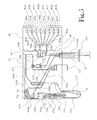
- FIG. 5 is a schematic illustration of one embodiment of the dose synthesis card and the sample card.
- a chemical production module and dose synthesis card for a PET biomarker radiopharmaceutical production system are described more fully hereinafter.
- This invention may, however, be embodied in many different forms and should not be construed as limited to the embodiments set forth herein. Rather, these embodiments are provided to ensure that this disclosure is thorough and complete, and to ensure that it fully conveys the scope of the invention to those skilled in the art.
- the chemical production module, the dose synthesis card and the sample card operate in conjunction with a complete PET biomarker production system.
- this PET biomarker production system comprises an accelerator 10 , which produces the radioisotopes; a chemical production module (or CPM) 20 ; a dose synthesis card 30 ; a sample card 40 ; and a quality control module (or QCM) 50 .
- the accelerator 10 has produced a radioisotope
- the radioisotope travels via a radioisotope delivery tube 112 to the dose synthesis card 30 attached to the CPM 20 .
- the CPM 20 holds reagents and solvents that are required during the radiopharmaceutical synthesis process.
- the radiopharmaceutical solution is synthesized from the radioisotope and then purified for testing and administration. Following synthesis and purification, a small percentage of the resultant radiopharmaceutical solution is diverted into the sample card 40 , while the remainder flows into a dose vessel 200 . As shown in FIG. 2 , once samples of the radiopharmaceutical solution have flowed into the sample card 40 , an operator removes the sample card 40 from the CPM 20 and interfaces it with the QCM 50 , where a number of diagnostic instruments perform automated quality control tests on the samples.
- FIGS. 3 and 4 present a more detailed overview of the complete synthesis and quality control testing processes for one embodiment of the present invention.
- the radioisotope involved is flourine-18 (F-18), produced from the bombardment in a cyclotron of heavy water containing the oxygen-18 isotope.
- F-18 flourine-18
- the sample card and quality control module also work with radiopharmaceutical synthesis systems using other radioisotopes, including carbon-11, nitrogen-13, and oxygen-15.
- the radioisotope enters a reaction chamber or reaction vessel 110 from the radioisotope delivery tube 112 .
- the radioisotope F-18 is still mixed with quantities of heavy water from the biomarker generator.
- a first organic ingredient is introduced to the reaction vessel 110 from a reagent storage compartment 120 by an organic input pump 124 .
- the first organic ingredient includes a solution of potassium complexed to 1,10-diaza-4,7,13,16,21,24-hexaoxabicyclo[8.8.8]hexacosane (commonly called Kryptofix 222TM, hereinafter “kryptofix”) or a similar crown ether.
- the potassium-kryptofix complex or similar organometallic complex is carried by acetonitrile as solvent.
- the potassium activates the F-18 fluoride radioisotope, while the kryptofix binds the potassium atoms and inhibits the formation of a potassium-fluoride complex.
- a gas input 142 fills the reaction vessel 110 with an inert gas such as dry nitrogen, the gas having been stored in a storage area 140 within or near the CPM 20 .
- the mixture in the reaction vessel 110 is heated by the nearby heat source 114 to remove the residual heavy water by evaporating the azeotropic water/acetonitrile mixture.
- a vacuum 150 helps to remove the vaporized water.
- the organic input pump 124 adds a second organic ingredient from a second reagent storage compartment 122 to the mixture in the reaction vessel 110 .
- the second organic ingredient is mannose triflate in dry acetonitrile.
- the solution is then heated at approximately 110 degrees Celsius for approximately two minutes.
- the F-18 has bonded to the mannose to form the immediate precursor for [ 18 F]FDG, commonly 18F-fluorodeoxyglucose tetraacetate (FTAG).
- aqueous acid in many embodiments, aqueous hydrochloric acid—is introduced from a storage compartment 130 through an aqueous input pump 132 .
- the hydrochloric acid removes the protective acetyl groups on the 18 F-FTAG, leaving 18 F-fludeoxyglucose (i.e. [ 18 F]FDG).
- the [ 18 F]FDG having been synthesized, it must be purified before testing and administration.
- the [ 18 F]FDG in solution passes from the reaction vessel 110 through a solid phase extraction column 160 .
- the solid phase extraction column 160 comprises a length filled with an ion exchange resin, a length filled with alumina, and a length filled with carbon-18.
- the [ 18 F]FDG next passes through a filter 170 , which in many embodiments includes a Millipore filter with pores approximately 0.22 micrometers in diameter.
- the filter 170 contains a number of sample vessels 402 a - e , which in some embodiments each hold approximately 10 microliters of solution.
- the number of sample vessels will vary according to the number of quality control tests to be performed for that run, and the system is adapted to operate with different sample cards containing varying numbers of sample vessels.
- the remainder of the radiopharmaceutical solution i.e. all of the solution that is not diverted for quality control testing) flows into the dose vessel 200 , ready for administration to a patient.
- the radiopharmaceutical samples travel from the sample vessels 402 a - e into the test vessels 502 , 602 , 702 , 802 , and 902 within the QCM 50 .
- instruments exist to perform a number of automated quality control tests for each run of radiopharmaceutical produced by the radiopharmaceutical synthesis system.
- a light source 504 shines white light through the sample in the test vessel 502 .
- An electronic eye 506 detects the light that has passed through the sample and measures that light's intensity and color against reference samples.
- pH test device 604 i.e. a pH probe or pH colorstrip, measures the pH of the sample in the sample vessel 602 .
- a heat source 704 heats the sample in the test vessel 702 to approximately 150 degrees Celsius so that the aqueous sample components, now is gas form, enter an adjacent gas chromatograph 706 .
- a gas sensor microarray 708 (informally, an “electronic nose”) then detects the presence and prevalence (e.g. as ppm) of such chemicals as methyl cyanide and ethanol.
- the sample in the test vessel 802 is placed on a gel 804 comprising silica gel with iodoplatinate.
- the sample and gel 804 are then warmed, and a color recognition sensor 806 measures the resultant color of the sample, with a yellow color indicating the presence of kryptofix.
- the sample in the test vessel 902 is eluted through a silica column 904 using a carrier mixture of acetonitrile and water.
- the acetonitrile and water are mixed in a ratio of 9:1.
- a radiation probe 906 measures the activity of the solution as it is eluted. As [ 18 F]FDG has an elution time that can be predicted with accuracy, the probe 906 measures the percentage of the activity that elutes at or very near to the predicted elution time for [ 18 F]FDG. A percentage of 95% or higher indicates acceptable radiochemical purity.
- a filter integrity test is also performed for every dose that is produced. As shown in FIG. 3 , after the radiopharmaceutical solution has gone through the filter 170 , the integrity of the filter 170 is tested by passing inert gas from the inert gas input 142 through the filter 170 at increasing pressure. A pressure sensor 302 measures the pressure of the inert gas upon the filter 170 and detects whether the filter 170 is still intact.
- the filter 170 should be capable of maintaining integrity under pressures of at least 50 pounds per square inch (psi).
- FIG. 5 displays a schematic view of one embodiment of the dose synthesis card 30 ′ together with the attached sample card 40 ′.
- the dose synthesis card 30 ′ includes a reaction vessel 110 a where the radiopharmaceutical solution is synthesized.
- a radioisotope input 112 a introduces the radioisotope F-18 into the reaction vessel 110 a through a radioisotope input channel 1121 .
- the radioisotope is still mixed with quantities of heavy water from the biomarker generator.
- an organic input 124 a introduces a solution of potassium-kryptofix complex in acetonitrile into the reaction vessel 110 a through an organic input channel 1241 .
- a combination nitrogen-input and vacuum 154 pumps nitrogen gas into the reaction vessel 110 a through a gas channel 1540 a and a valve 1541 , which valve is at that time in an open position.
- the mixture A in the reaction vessel 110 a is heated in nitrogen atmosphere to azeotropically remove water from the mixture A, the vaporized water being evacuated through the gas channel 1540 a and the vacuum 154 .
- the organic input 124 a introduces mannose triflate in dry acetonitrile into the reaction vessel 110 a through the organic input channel 1241 .
- the solution is heated at approximately 110 degrees Celsius for approximately two minutes. By this stage, the F-18 has bonded to the mannose to form the immediate precursor for [ 18 F]FDG, FTAG.
- aqueous hydrochloric acid is introduced into the reaction vessel 110 a through an aqueous input 132 a and an aqueous channel 1321 .
- the hydrochloric acid removes the protective acetyl groups on the intermediate 18 F-FTAG, leaving 18 F-fludeoxyglucose (i.e. [ 18 F]FDG).
- the [ 18 F]FDG in solution passes from the reaction vessel 110 a through a post-reaction channel 1101 into a solid phase extraction column 160 a , where some undesirable substances are removed from the solution, thereby clarifying the radiopharmaceutical solution.
- the solid phase extraction (SPE) column 160 a comprises a length with an ion exchange resin, a length filled with alumina, and a length filled with carbon-18.
- the radiopharmaceutical passes through the SPE column 160 a with a mobile phase that in many embodiments includes acetonitrile from the organic input 124 a .
- the mobile phase and impurities emerge from the SPE column 160 a , they pass through a second post-reaction channel 1542 and through a three-way valve 175 and waste channel 1104 into a waste receptacle 210 .
- the radiopharmaceutical solution next passes through the second post-reaction channel 1542 and through the three-way valve 175 into a filter channel 1103 and then through a filter 170 a .
- the filter 170 a removes other impurities (including particulate impurities), thereby further clarifying the radiopharmaceutical solution.
- the filter 170 a includes a Millipore filter with pores approximately 0.22 micrometers in diameter.
- the clarified radiopharmaceutical solution travels via the post-clarification channel 1105 into the sterile dose administration vessel 200 a , which in the illustrated embodiment is incorporated into a syringe 202 .
- the dose administration vessel is filled beforehand with a mixture of phosphate buffer and saline.
- some of the solution B is diverted through an extraction channel 1401 , an open valve 1403 , and a transfer channel 1402 into the sample card 40 ′.
- the sample card 40 ′ contains a number of sample loops 404 a - h , which hold separated aliquots of solution for imminent testing, and a number of valves 408 a - h , which at this stage are closed.
- the sample card 40 ′ is separated from the dose synthesis card 30 ′ and inserted into the QCM, as was shown in FIGS. 2 and 4 .
- the aliquots then travel through the now-open valves 408 a - h into the sample egress ports 406 a - h , from which the aliquots pass into the test vessels, as was shown in FIG. 4 .
- each of the sample loops 404 a - h holds approximately 10 microliters of sample solution.
- the number of sample loops will vary according to the number of quality control tests to be performed for that run, and the system is adapted to operate with different sample cards containing varying numbers of sample loops.
- the CPM 20 holds sufficient amounts of reagents and solvents that are required during the radiopharmaceutical synthesis process to carry out multiple runs without reloading. Indeed, in some embodiments the CPM 20 is loaded with reagents and solvents approximately once per month, with that month's supply of reagents and solvents sufficient to produce several dozen or even several hundred doses of radiopharmaceutical. As the reagents and solvents are stored in the CPM 20 , it is easier than under previous systems to keep the reagents and solvents sterile and uncontaminated. In some embodiments, a sterile environment is supported and contamination inhibited by discarding each dose synthesis card 30 and the sample card 40 after one run; these components of the system are adapted to be disposable.
- each batch of reagents and solvents, loaded periodically into the CPM 20 will supply a batch of multiple doses of radiopharmaceutical, each dose produced in a separate run.
- Some quality control tests are performed for every dose that is produced, while other quality control tests are performed for every batch of doses.
- the filter integrity test, the color and clarity test, the acidity test, the volatile organics test, the chemical purity test, and the radiochemical purity test are performed for every dose.
- some quality control tests need be performed only once or twice per batch, such as the radionuclide purity test (using a radiation probe to measure the half-life of the F-18 in the [ 18 F]FDG), the bacterial endotoxin test, and the sterility test. These tests are performed generally on the first and last doses of each batch. Because these per-batch quality control tests are conducted less frequently, they may not be included in the QCM, but rather may be conducted by technicians using separate laboratory equipment.
Abstract
Description
Claims (4)
Priority Applications (15)
| Application Number | Priority Date | Filing Date | Title |
|---|---|---|---|
| US12/565,544 US8333952B2 (en) | 2009-09-23 | 2009-09-23 | Dose synthesis module for biomarker generator system |
| EP10819146.1A EP2480258B1 (en) | 2009-09-23 | 2010-09-21 | Chemical production module and dose synthesis card for pet biomarker production system |
| NZ599555A NZ599555A (en) | 2009-09-23 | 2010-09-21 | Chemical production module and dose synthesis card for pet biomarker production system |
| MX2012003572A MX2012003572A (en) | 2009-09-23 | 2010-09-21 | Chemical production module and dose synthesis card for pet biomarker production system. |
| RU2012116143/15A RU2541254C2 (en) | 2009-09-23 | 2010-09-21 | Chemical production unit and dose synthesis chart for pet biomarker production system |
| JP2012530859A JP2013505294A (en) | 2009-09-23 | 2010-09-21 | Chemical production module for PET biomarker production system and administration liquid synthesis card |
| BR112012006679-0A BR112012006679B1 (en) | 2009-09-23 | 2010-09-21 | system for synthesis of a microfluidic radiopharmaceutical |
| CA2775034A CA2775034A1 (en) | 2009-09-23 | 2010-09-21 | Chemical production module and dose synthesis card for pet biomarker production system |
| AU2010298725A AU2010298725B2 (en) | 2009-09-23 | 2010-09-21 | Chemical production module and dose synthesis card for pet biomarker production system |
| PCT/US2010/002577 WO2011037615A1 (en) | 2009-09-23 | 2010-09-21 | Chemical production module and dose synthesis card for pet biomarker production system |
| US13/446,334 US20130130309A1 (en) | 2009-09-23 | 2012-04-13 | Radiopharmaceutical Production System and Quality Control System Utilizing High Performance Liquid Chromatography |
| CO12066593A CO6541527A2 (en) | 2009-09-23 | 2012-04-23 | CHEMICAL PRODUCTION MODULE AND DOSAGE SYNTHESIS CARD FOR PET BIOMARKER PRODUCTION SYSTEM |
| US14/618,795 US11135321B2 (en) | 2009-09-23 | 2015-02-10 | Automated radiopharmaceutical production and quality control system |
| US14/618,772 US20150160171A1 (en) | 2009-09-23 | 2015-02-10 | Automated Quality Control System for Radiopharmaceuticals |
| US14/618,732 US10109385B2 (en) | 2009-09-23 | 2015-02-10 | Dose synthesis card for use with automated biomarker production system |
Applications Claiming Priority (1)
| Application Number | Priority Date | Filing Date | Title |
|---|---|---|---|
| US12/565,544 US8333952B2 (en) | 2009-09-23 | 2009-09-23 | Dose synthesis module for biomarker generator system |
Related Parent Applications (1)
| Application Number | Title | Priority Date | Filing Date |
|---|---|---|---|
| US12/565,552 Continuation-In-Part US20110070158A1 (en) | 2009-09-23 | 2009-09-23 | Quality Control Module for Biomarker Generator System |
Related Child Applications (1)
| Application Number | Title | Priority Date | Filing Date |
|---|---|---|---|
| US13/446,334 Continuation-In-Part US20130130309A1 (en) | 2009-09-23 | 2012-04-13 | Radiopharmaceutical Production System and Quality Control System Utilizing High Performance Liquid Chromatography |
Publications (2)
| Publication Number | Publication Date |
|---|---|
| US20110070160A1 US20110070160A1 (en) | 2011-03-24 |
| US8333952B2 true US8333952B2 (en) | 2012-12-18 |
Family
ID=43756796
Family Applications (1)
| Application Number | Title | Priority Date | Filing Date |
|---|---|---|---|
| US12/565,544 Active 2030-09-03 US8333952B2 (en) | 2009-09-23 | 2009-09-23 | Dose synthesis module for biomarker generator system |
Country Status (11)
| Country | Link |
|---|---|
| US (1) | US8333952B2 (en) |
| EP (1) | EP2480258B1 (en) |
| JP (1) | JP2013505294A (en) |
| AU (1) | AU2010298725B2 (en) |
| BR (1) | BR112012006679B1 (en) |
| CA (1) | CA2775034A1 (en) |
| CO (1) | CO6541527A2 (en) |
| MX (1) | MX2012003572A (en) |
| NZ (1) | NZ599555A (en) |
| RU (1) | RU2541254C2 (en) |
| WO (1) | WO2011037615A1 (en) |
Cited By (5)
| Publication number | Priority date | Publication date | Assignee | Title |
|---|---|---|---|---|
| US20150157743A1 (en) * | 2009-09-23 | 2015-06-11 | Abt Molecular Imaging, Inc. | Dose Synthesis Card for Use with Automated Biomarker Production System |
| US20170102391A1 (en) * | 2014-06-06 | 2017-04-13 | The Regents Of The University Of California | Self-shielded, benchtop chemistry system |
| US10005060B2 (en) | 2014-01-31 | 2018-06-26 | Hitachi, Ltd. | Drug provision system and drug provision method |
| US10344314B2 (en) | 2016-05-04 | 2019-07-09 | Curium Us Llc | Systems and methods for sterility testing of radionuclide generator column assemblies |
| US11426489B2 (en) | 2015-06-10 | 2022-08-30 | Globus Medical, Inc. | Biomaterial compositions, implants, and methods of making the same |
Families Citing this family (17)
| Publication number | Priority date | Publication date | Assignee | Title |
|---|---|---|---|---|
| US11135321B2 (en) | 2009-09-23 | 2021-10-05 | Best Medical International, Inc. | Automated radiopharmaceutical production and quality control system |
| WO2013049431A2 (en) * | 2011-09-30 | 2013-04-04 | Ge Healthcare Limited | Reactor for multi-step radiochemistry |
| EP2650681B1 (en) * | 2012-04-13 | 2015-12-30 | ABT Molecular Imaging, Inc. | Radiopharmaceutical production system and quality control system utilizing high performance liquid chromatography |
| US8937287B2 (en) | 2012-04-19 | 2015-01-20 | Abt Molecular Imaging, Inc. | Self-referencing radiation detector for use with a radiopharmaceutical quality control testing system |
| US9192934B2 (en) | 2012-10-25 | 2015-11-24 | General Electric Company | Insert assembly for a microfluidic device |
| US9399216B2 (en) * | 2013-12-30 | 2016-07-26 | General Electric Company | Fluid transport in microfluidic applications with sensors for detecting fluid presence and pressure |
| GB201418893D0 (en) | 2014-10-23 | 2014-12-10 | Univ Hull | Monolithic body |
| GB201418897D0 (en) * | 2014-10-23 | 2014-12-10 | Univ Hull | Methods and apparatus for the analysis of compounds |
| GB201418899D0 (en) | 2014-10-23 | 2014-12-10 | Univ Hull | System for radiopharmaceutical production |
| CN107530455A (en) * | 2015-02-10 | 2018-01-02 | Abt分子成像公司 | With automatic biological label production system associated with dosage synthesize card |
| WO2016130463A1 (en) * | 2015-02-10 | 2016-08-18 | Abt Molecular Imaging, Inc. | Automated radiopharmaceutical production and quality control system |
| EP3192583A1 (en) | 2016-01-12 | 2017-07-19 | ETH Zurich | Apparatus and process for the automated chemical synthesis of compounds |
| KR101903695B1 (en) * | 2016-11-28 | 2018-10-04 | 가천대학교 산학협력단 | F-18 radiosynthesis module with nitrogen heater |
| US20180209921A1 (en) * | 2017-01-20 | 2018-07-26 | Mallinckrodt Nuclear Medicine Llc | Systems and methods for assaying an eluate of a radionuclide generator |
| FR3072301B1 (en) | 2017-10-18 | 2022-01-07 | P M B | MICROFLUIDIC CASSETTE FOR SYNTHESIZING A RADIO-TRACER AND METHOD FOR SYNTHESIZING A RADIO-TRACER WITH SUCH A CASSETTE |
| RU2689399C1 (en) * | 2018-06-19 | 2019-05-28 | ВАВИЛИН Андрей Владимирович | Neutron multiplier |
| CN113447578A (en) * | 2021-05-24 | 2021-09-28 | 华东理工大学 | Magnetic solid-phase extraction micro-fluidic chip-mass spectrometry combined device and application thereof |
Citations (6)
| Publication number | Priority date | Publication date | Assignee | Title |
|---|---|---|---|---|
| US20050232861A1 (en) | 2004-04-20 | 2005-10-20 | Buchanan Charles R | Microfluidic apparatus and method for synthesis of molecular imaging probes including FDG |
| US20080067413A1 (en) | 2006-05-26 | 2008-03-20 | Advanced Biomarker Technologies, Llc | Biomarker generator system |
| US20080233018A1 (en) | 2007-01-23 | 2008-09-25 | Van Dam Robert Michael | Fully-automated microfluidic system for the synthesis of radiolabeled biomarkers for positron emission tomography |
| US20090036668A1 (en) | 2007-04-12 | 2009-02-05 | Siemens Medical Solutions Usa, Inc. | Microfluidic radiosynthesis system for positron emission tomography biomarkers |
| US20100145630A1 (en) * | 2008-12-04 | 2010-06-10 | Siemens Medical Solutions Usa, Inc. | Apparatus and Method for Automated Quality Control |
| US20110178359A1 (en) * | 2007-01-01 | 2011-07-21 | Hirschman Alan D | Systems For Integrated Radiopharmaceutical Generation, Preparation, Transportation and Administration |
Family Cites Families (6)
| Publication number | Priority date | Publication date | Assignee | Title |
|---|---|---|---|---|
| CA2523189A1 (en) * | 2003-04-22 | 2004-11-04 | Molecular Technologies, Inc. | System and method for synthesis of molecular imaging probes including fdg |
| US8206593B2 (en) * | 2004-12-03 | 2012-06-26 | Fluidigm Corporation | Microfluidic chemical reaction circuits |
| GB0520529D0 (en) * | 2005-10-10 | 2005-11-16 | Ge Healthcare Ltd | Automated radiolabelling method |
| US7884340B2 (en) * | 2006-05-26 | 2011-02-08 | Advanced Biomarker Technologies, Llc | Low-volume biomarker generator |
| US7741121B2 (en) * | 2006-08-24 | 2010-06-22 | Siemens Medical Solutions Usa, Inc. | System for purification and analysis of radiochemical products yielded by microfluidic synthesis devices |
| WO2012024663A1 (en) * | 2010-08-20 | 2012-02-23 | Ge Healthcare Limited | Quality control devices and methods for radiopharmaceuticals |
-
2009
- 2009-09-23 US US12/565,544 patent/US8333952B2/en active Active
-
2010
- 2010-09-21 BR BR112012006679-0A patent/BR112012006679B1/en not_active IP Right Cessation
- 2010-09-21 JP JP2012530859A patent/JP2013505294A/en active Pending
- 2010-09-21 WO PCT/US2010/002577 patent/WO2011037615A1/en active Application Filing
- 2010-09-21 AU AU2010298725A patent/AU2010298725B2/en not_active Expired - Fee Related
- 2010-09-21 RU RU2012116143/15A patent/RU2541254C2/en active
- 2010-09-21 EP EP10819146.1A patent/EP2480258B1/en active Active
- 2010-09-21 NZ NZ599555A patent/NZ599555A/en not_active IP Right Cessation
- 2010-09-21 MX MX2012003572A patent/MX2012003572A/en active IP Right Grant
- 2010-09-21 CA CA2775034A patent/CA2775034A1/en not_active Abandoned
-
2012
- 2012-04-23 CO CO12066593A patent/CO6541527A2/en not_active Application Discontinuation
Patent Citations (6)
| Publication number | Priority date | Publication date | Assignee | Title |
|---|---|---|---|---|
| US20050232861A1 (en) | 2004-04-20 | 2005-10-20 | Buchanan Charles R | Microfluidic apparatus and method for synthesis of molecular imaging probes including FDG |
| US20080067413A1 (en) | 2006-05-26 | 2008-03-20 | Advanced Biomarker Technologies, Llc | Biomarker generator system |
| US20110178359A1 (en) * | 2007-01-01 | 2011-07-21 | Hirschman Alan D | Systems For Integrated Radiopharmaceutical Generation, Preparation, Transportation and Administration |
| US20080233018A1 (en) | 2007-01-23 | 2008-09-25 | Van Dam Robert Michael | Fully-automated microfluidic system for the synthesis of radiolabeled biomarkers for positron emission tomography |
| US20090036668A1 (en) | 2007-04-12 | 2009-02-05 | Siemens Medical Solutions Usa, Inc. | Microfluidic radiosynthesis system for positron emission tomography biomarkers |
| US20100145630A1 (en) * | 2008-12-04 | 2010-06-10 | Siemens Medical Solutions Usa, Inc. | Apparatus and Method for Automated Quality Control |
Non-Patent Citations (1)
| Title |
|---|
| Chin et al. J Label. Compd. Radiopharm. 2006, 17-31. * |
Cited By (7)
| Publication number | Priority date | Publication date | Assignee | Title |
|---|---|---|---|---|
| US20150157743A1 (en) * | 2009-09-23 | 2015-06-11 | Abt Molecular Imaging, Inc. | Dose Synthesis Card for Use with Automated Biomarker Production System |
| US10109385B2 (en) * | 2009-09-23 | 2018-10-23 | Abt Molecular Imaging, Inc. | Dose synthesis card for use with automated biomarker production system |
| US10005060B2 (en) | 2014-01-31 | 2018-06-26 | Hitachi, Ltd. | Drug provision system and drug provision method |
| US20170102391A1 (en) * | 2014-06-06 | 2017-04-13 | The Regents Of The University Of California | Self-shielded, benchtop chemistry system |
| US10473668B2 (en) * | 2014-06-06 | 2019-11-12 | The Regents Of The University Of California | Self-shielded, benchtop radio chemistry system with a plurality shielded carriers containing a disposable chip cassette |
| US11426489B2 (en) | 2015-06-10 | 2022-08-30 | Globus Medical, Inc. | Biomaterial compositions, implants, and methods of making the same |
| US10344314B2 (en) | 2016-05-04 | 2019-07-09 | Curium Us Llc | Systems and methods for sterility testing of radionuclide generator column assemblies |
Also Published As
| Publication number | Publication date |
|---|---|
| CO6541527A2 (en) | 2012-10-16 |
| NZ599555A (en) | 2013-10-25 |
| MX2012003572A (en) | 2012-10-09 |
| JP2013505294A (en) | 2013-02-14 |
| WO2011037615A1 (en) | 2011-03-31 |
| RU2541254C2 (en) | 2015-02-10 |
| BR112012006679B1 (en) | 2021-04-20 |
| CA2775034A1 (en) | 2011-03-31 |
| EP2480258B1 (en) | 2021-07-21 |
| RU2012116143A (en) | 2013-10-27 |
| AU2010298725A1 (en) | 2012-04-12 |
| EP2480258A1 (en) | 2012-08-01 |
| BR112012006679A8 (en) | 2018-01-23 |
| EP2480258A4 (en) | 2017-03-22 |
| BR112012006679A2 (en) | 2016-05-10 |
| US20110070160A1 (en) | 2011-03-24 |
| AU2010298725B2 (en) | 2016-11-03 |
Similar Documents
| Publication | Publication Date | Title |
|---|---|---|
| US8333952B2 (en) | Dose synthesis module for biomarker generator system | |
| US20110070158A1 (en) | Quality Control Module for Biomarker Generator System | |
| US20130130309A1 (en) | Radiopharmaceutical Production System and Quality Control System Utilizing High Performance Liquid Chromatography | |
| US9260354B2 (en) | Device and method for the production of radiochemical compounds | |
| US10109385B2 (en) | Dose synthesis card for use with automated biomarker production system | |
| US11135321B2 (en) | Automated radiopharmaceutical production and quality control system | |
| EP2653863B1 (en) | Self-referencing radiation detection method for use with a radiopharmaceutical quality control testing system | |
| Meisenheimer et al. | Gallium-68: radiolabeling of radiopharmaceuticals for PET imaging—A lot to consider | |
| US20150160171A1 (en) | Automated Quality Control System for Radiopharmaceuticals | |
| Lindner et al. | Automated production of [18F] SiTATE on a Scintomics GRP™ platform for PET/CT imaging of neuroendocrine tumors | |
| Zhang et al. | High-yielding radiosynthesis of [68 Ga] Ga-PSMA-11 using a low-cost microfluidic device | |
| Rodnick et al. | Synthesis of 68Ga-radiopharmaceuticals using both generator-derived and cyclotron-produced 68Ga as exemplified by [68Ga] Ga-PSMA-11 for prostate cancer PET imaging | |
| Menzel et al. | FOMSy: 3D-printed flexible open-source microfluidic system and flow synthesis of PET-tracer | |
| EP2650681B1 (en) | Radiopharmaceutical production system and quality control system utilizing high performance liquid chromatography | |
| US9221029B2 (en) | Automatic system for synthesizing 123I-MIBG and automatic device for synthesizing and dispensing 123I-MIBG comprising the same | |
| WO2012064312A1 (en) | Quality control module for biomarker generator system | |
| WO2016130449A1 (en) | Dose synthesis card for use with automated biomarker production system | |
| WO2016130458A1 (en) | Automated quality control system for radiopharmaceuticals | |
| Lisova | Novel economical approaches for the fluorine-18 radiopharmaceuticals production via droplet radiochemistry | |
| WO2016130463A1 (en) | Automated radiopharmaceutical production and quality control system | |
| KR20230077672A (en) | Method of continuous production of f-18 sodium fluoride | |
| Tran et al. | Principles behind Radiopharmacy | |
| JPS6064934A (en) | Apparatus for automatic synthesis of methyl iodide | |
| CN112028914A (en) | A kind of18F-boron trifluoride tyrosine kit and application thereof | |
| Reed | Optimization of Automated 18F Fluoroalkylation: Production of 3 Opioid PET Tracers |
Legal Events
| Date | Code | Title | Description |
|---|---|---|---|
| AS | Assignment |
Owner name: ABT MOLECULAR IMAGING, INC., TENNESSEE Free format text: ASSIGNMENT OF ASSIGNORS INTEREST;ASSIGNORS:NUTT, RONALD;GIAMIS, ANTHONY M;MCFARLAND, AARON;REEL/FRAME:023276/0185 Effective date: 20090923 |
|
| STCF | Information on status: patent grant |
Free format text: PATENTED CASE |
|
| AS | Assignment |
Owner name: SQUARE 1 BANK, NORTH CAROLINA Free format text: SECURITY INTEREST;ASSIGNOR:ABT MOLECULAR IMAGING, INC.;REEL/FRAME:032464/0298 Effective date: 20140318 |
|
| AS | Assignment |
Owner name: ABT MOLECULAR IMAGING, INC., TENNESSEE Free format text: RELEASE BY SECURED PARTY;ASSIGNOR:SQUARE 1 BANK;REEL/FRAME:034090/0551 Effective date: 20141014 |
|
| AS | Assignment |
Owner name: PACIFIC WESTERN BANK, NORTH CAROLINA Free format text: SECURITY INTEREST;ASSIGNOR:ABT MOLECULAR IMAGING, INC.;REEL/FRAME:037391/0210 Effective date: 20151204 |
|
| AS | Assignment |
Owner name: PACIFIC WESTERN BANK, NORTH CAROLINA Free format text: SECURITY INTEREST;ASSIGNOR:ABT MOLECULAR IMAGING, INC.;REEL/FRAME:038110/0538 Effective date: 20151111 Owner name: SWK FUNDING, LLC, TEXAS Free format text: SECURITY INTEREST;ASSIGNOR:ABT MOLECULAR IMAGING, INC.;REEL/FRAME:038110/0847 Effective date: 20151211 |
|
| FPAY | Fee payment |
Year of fee payment: 4 |
|
| AS | Assignment |
Owner name: SWK FUNDING LLC, TEXAS Free format text: SECURITY INTEREST;ASSIGNOR:PACIFIC WESTERN BANK;REEL/FRAME:038845/0812 Effective date: 20160428 |
|
| AS | Assignment |
Owner name: BEST ABT, INC., VIRGINIA Free format text: ASSIGNMENT OF ASSIGNORS INTEREST;ASSIGNOR:ABT MOLECULAR IMAGING, INC.;REEL/FRAME:047511/0829 Effective date: 20181112 |
|
| AS | Assignment |
Owner name: SWK FUNDING LLC, TEXAS Free format text: INTELLECTUAL PROPERTY SECURITY AGREEMENT;ASSIGNOR:BEST ABT INC.;REEL/FRAME:048351/0263 Effective date: 20190213 |
|
| MAFP | Maintenance fee payment |
Free format text: PAYMENT OF MAINTENANCE FEE, 8TH YR, SMALL ENTITY (ORIGINAL EVENT CODE: M2552); ENTITY STATUS OF PATENT OWNER: SMALL ENTITY Year of fee payment: 8 |
|
| MAFP | Maintenance fee payment |
Free format text: PAYMENT OF MAINTENANCE FEE, 12TH YR, SMALL ENTITY (ORIGINAL EVENT CODE: M2553); ENTITY STATUS OF PATENT OWNER: SMALL ENTITY Year of fee payment: 12 |