US8324350B2 - Dual-specific IL-1α/IL-1β antibodies - Google Patents
Dual-specific IL-1α/IL-1β antibodies Download PDFInfo
- Publication number
- US8324350B2 US8324350B2 US12/006,068 US606807A US8324350B2 US 8324350 B2 US8324350 B2 US 8324350B2 US 606807 A US606807 A US 606807A US 8324350 B2 US8324350 B2 US 8324350B2
- Authority
- US
- United States
- Prior art keywords
- seq
- antibody
- antigen
- binding portion
- less
- Prior art date
- Legal status (The legal status is an assumption and is not a legal conclusion. Google has not performed a legal analysis and makes no representation as to the accuracy of the status listed.)
- Expired - Fee Related, expires
Links
Images


Classifications
-
- C—CHEMISTRY; METALLURGY
- C07—ORGANIC CHEMISTRY
- C07K—PEPTIDES
- C07K16/00—Immunoglobulins [IGs], e.g. monoclonal or polyclonal antibodies
- C07K16/46—Hybrid immunoglobulins
- C07K16/468—Immunoglobulins having two or more different antigen binding sites, e.g. multifunctional antibodies
-
- A—HUMAN NECESSITIES
- A61—MEDICAL OR VETERINARY SCIENCE; HYGIENE
- A61K—PREPARATIONS FOR MEDICAL, DENTAL OR TOILETRY PURPOSES
- A61K39/00—Medicinal preparations containing antigens or antibodies
- A61K39/395—Antibodies; Immunoglobulins; Immune serum, e.g. antilymphocytic serum
- A61K39/39533—Antibodies; Immunoglobulins; Immune serum, e.g. antilymphocytic serum against materials from animals
- A61K39/3955—Antibodies; Immunoglobulins; Immune serum, e.g. antilymphocytic serum against materials from animals against proteinaceous materials, e.g. enzymes, hormones, lymphokines
-
- A—HUMAN NECESSITIES
- A61—MEDICAL OR VETERINARY SCIENCE; HYGIENE
- A61K—PREPARATIONS FOR MEDICAL, DENTAL OR TOILETRY PURPOSES
- A61K45/00—Medicinal preparations containing active ingredients not provided for in groups A61K31/00 - A61K41/00
- A61K45/06—Mixtures of active ingredients without chemical characterisation, e.g. antiphlogistics and cardiaca
-
- A—HUMAN NECESSITIES
- A61—MEDICAL OR VETERINARY SCIENCE; HYGIENE
- A61P—SPECIFIC THERAPEUTIC ACTIVITY OF CHEMICAL COMPOUNDS OR MEDICINAL PREPARATIONS
- A61P37/00—Drugs for immunological or allergic disorders
-
- C—CHEMISTRY; METALLURGY
- C07—ORGANIC CHEMISTRY
- C07K—PEPTIDES
- C07K16/00—Immunoglobulins [IGs], e.g. monoclonal or polyclonal antibodies
- C07K16/18—Immunoglobulins [IGs], e.g. monoclonal or polyclonal antibodies against material from animals or humans
- C07K16/24—Immunoglobulins [IGs], e.g. monoclonal or polyclonal antibodies against material from animals or humans against cytokines, lymphokines or interferons
- C07K16/244—Interleukins [IL]
- C07K16/245—IL-1
-
- C—CHEMISTRY; METALLURGY
- C07—ORGANIC CHEMISTRY
- C07K—PEPTIDES
- C07K2317/00—Immunoglobulins specific features
- C07K2317/50—Immunoglobulins specific features characterized by immunoglobulin fragments
- C07K2317/56—Immunoglobulins specific features characterized by immunoglobulin fragments variable (Fv) region, i.e. VH and/or VL
- C07K2317/569—Single domain, e.g. dAb, sdAb, VHH, VNAR or nanobody®
-
- C—CHEMISTRY; METALLURGY
- C07—ORGANIC CHEMISTRY
- C07K—PEPTIDES
- C07K2317/00—Immunoglobulins specific features
- C07K2317/90—Immunoglobulins specific features characterized by (pharmaco)kinetic aspects or by stability of the immunoglobulin
- C07K2317/92—Affinity (KD), association rate (Ka), dissociation rate (Kd) or EC50 value
Definitions
- Cytokines such as interleukin-1 (IL-1) and tumor necrosis factor (TNF), are molecules produced by a variety of cells, such as monocytes and macrophages, which have been identified as mediators of inflammatory processes.
- Interleukin-1 is a cytokine with a wide range of biological and physiological effects, including fever, prostaglandin synthesis (in e.g., fibroblasts, muscle and endothelial cells), T-lymphocyte activation, and interleukin 2 production.
- Antibodies capable of binding of IL-1 ⁇ and IL-1 ⁇ have been described in the art (see US Publication No. 20030040083 and PCT publication WO 07/063,308). However, there exists a need for improved therapeutics to IL-1 ⁇ and IL-1 ⁇ .
- the invention includes an isolated, dual-specific antibody, or an antigen-binding portion thereof, which dissociates from human IL-1 ⁇ with a K D of 3 ⁇ 10 ⁇ 7 M or less; dissociates from human IL-1 ⁇ with a K D of 5 ⁇ 10 ⁇ 5 M or less; and does not bind mouse IL-1 ⁇ or mouse IL-1 ⁇ .
- the antibody, or antigen-binding portion neutralizes human IL-1 ⁇ in a standard in vitro assay with an ND 50 of 800 nM or less.
- the antibody, or antigen-binding portion neutralizes human IL-1 ⁇ in a standard in vitro assay with an ND 50 of 900 nM or less, and neutralizes human IL-1 ⁇ in a standard in vitro assay with an ND 50 of 800 nM or less.
- the antibody, or antigen-binding portion neutralizes human IL-1 ⁇ in a standard in vitro MRC5 assay with an ND 50 of 900 nM or less, and/or neutralizes human IL-1 ⁇ in a standard MRC5 in vitro assay with an ND 50 of 800 nM or less.
- the antibody, or antigen-binding portion dissociates from IL-1 ⁇ with a K D of 1 ⁇ 10 ⁇ 8 M or less; dissociates from IL-1 ⁇ with a K D of 1 ⁇ 10 ⁇ 9 M or less; dissociates from IL-1 ⁇ with a K D of 40-86 nM or less; dissociates from IL-1 ⁇ with a K D of 20-42 nM or less; dissociates from IL-1 ⁇ with a K D of 32-42 nM or less; dissociates from IL-1 ⁇ with a K D of 7-12 nM or less; dissociates from IL-1 ⁇ with a K D of 3.0 ⁇ 10 ⁇ 7 M or less; dissociates from IL-1 ⁇ with a K D of 1.1 ⁇ 10 ⁇ 7 M or less; dissociates from IL-1 ⁇ with a K D of 6.1 ⁇ 10 ⁇ 8 M or less; dissociates from IL-1 ⁇ with a K D of 6 ⁇ 10 ⁇ 8 M or less; dissociates from IL
- the antibody, or antigen-binding portion thereof dissociates from IL-1 ⁇ with a K D of 5.4 ⁇ 10 ⁇ 5 M or less; dissociates from IL-1 ⁇ with a K D of 2.8 ⁇ 10 ⁇ 6 M or less; dissociates from IL-1 ⁇ with a K D of 1.3 ⁇ 10 ⁇ 6 M or less; dissociates from IL-1 ⁇ with a K D of 9.3 ⁇ 10 ⁇ 7 M or less; dissociates from IL-1 ⁇ with a K D of 2 ⁇ 10 ⁇ 7 M or less; dissociates from IL-1 ⁇ with a K D of 1.1 ⁇ 10 ⁇ 7 M or less; or dissociates from IL-1 ⁇ with a K D of 2.8 ⁇ 10 ⁇ 8 M or less.
- the antibody, or antigen-binding portion thereof neutralizes IL-1 ⁇ in a standard in vitro assay with an ND 50 of 10 nM or less. In one embodiment, the antibody, or antigen-binding portion thereof, neutralizes IL-1 ⁇ in a standard in vitro assay with an ND 50 of 200 nM or less. In one embodiment, the antibody, or antigen-binding portion thereof, neutralizes IL-1 ⁇ in a standard MRC5 in vitro assay with an ND 50 of 10 nM or less. In one embodiment, the antibody, or antigen-binding portion thereof, neutralizes IL-1 ⁇ in a standard in vitro MRC5 assay with an ND 50 of 200 nM or less.
- the antibody, or antigen-binding portion thereof, of the invention comprises a heavy chain variable region comprising complementary determining regions (CDRs) as set forth in SEQ ID NO: 16 (ABT1-96) and a light chain variable region comprising CDRs as set forth in SEQ ID NO: 36 (ABT242).
- CDRs complementary determining regions
- the antibody, or antigen-binding portion thereof, of the invention comprises a light chain variable region comprising CDRs as set forth in SEQ ID NO: 24 (ABT1-122) and a heavy chain variable region comprising CDRs as set forth in SEQ ID NO: 52 (ABT2-108).
- the antibody, or antigen-binding portion thereof, of the invention comprises a light chain variable region comprising CDRs as set forth in SEQ ID NO: 28 (ABT1-141) and a heavy chain variable region comprising CDRs as set forth in SEQ ID NO: 52 (ABT2-108).
- the antibody, or antigen-binding portion thereof, of the invention comprises a heavy chain variable region comprising CDRs as set forth in SEQ ID NO: 16 (ABT1-96) and a heavy chain variable region comprising CDRs as set forth in SEQ ID NO: 32 (ABT2-13).
- the antibody, or antigen-binding portion thereof, of the invention comprises a heavy chain variable region comprising CDRs as set forth in SEQ ID NO: 16 (ABT1-96) and a light chain variable region comprising CDRs as set forth in SEQ ID NO: 40 (ABT2-46).
- the antibody, or antigen-binding portion thereof, of the invention comprises a light chain variable region comprising CDRs as set forth in SEQ ID NO: 12 (ABT1-95) and a heavy chain variable region comprising CDRs as set forth in SEQ ID NO: 32 (ABT2-13).
- the antibody, or antigen-binding portion thereof, of the invention comprises a light chain variable region comprising CDRs as set forth in SEQ ID NO: 24 (ABT1-122) and a heavy chain variable region comprising CDRs as set forth in SEQ ID NO: 44 (ABT2-65).
- the antibody, or antigen-binding portion thereof, of the invention comprises a heavy chain variable region comprising CDRs as set forth in SEQ ID NO: 20 (ABT1-98) and a light chain variable region comprising CDRs as set forth in SEQ ID NO: 48 (ABT2-76).
- the antibody, or antigen-binding portion thereof, of the invention comprises a heavy chain variable region comprising CDRs as set forth in SEQ ID NO: 4 (ABT1-6-23) and a second heavy chain variable region comprising CDRs as set forth in SEQ ID NO: 32 (ABT2-13).
- the invention also includes an isolated antibody, or an antigen-binding portion thereof, having dual-specificity for human IL-1 ⁇ and human IL-1 ⁇ comprising a variable light chain comprising complementary determining regions (CDRs) as set forth in an amino acid sequence selected from the group consisting of SEQ ID NO: 12, SEQ ID NO: 24, and SEQ ID NO: 28, or a variable heavy chain comprising CDRS as set forth in an amino acid sequence selected from the group consisting of SEQ ID NO: 4, SEQ ID NO: 8, SEQ ID NO: 16, and SEQ ID NO: 20.
- CDRs complementary determining regions
- the antibody, or antigen-binding portion thereof dissociates from human IL-1 ⁇ with a K D of 5 ⁇ 10 ⁇ 5 M or less; dissociates from IL-1 ⁇ with a K D of 5.4 ⁇ 10 ⁇ 5 M or less; dissociates from IL-1 ⁇ with a K D of 2.8 ⁇ 10 ⁇ 6 M or less; dissociates from IL-1 ⁇ with a K D of 1.3 ⁇ 10 ⁇ 6 M or less; dissociates from IL-1 ⁇ with a K D of 9.3 ⁇ 10 ⁇ 7 M or less; dissociates from IL-1 ⁇ with a K D of 2 ⁇ 10 ⁇ 7 M or less; dissociates from IL-1 ⁇ with a K D of 1.1 ⁇ 10 ⁇ 7 M or less; or dissociates from IL-1 ⁇ with a K D of 2.8 ⁇ 10 ⁇ 8 M or less.
- the antibody, or antigen-binding portion thereof, of the invention neutralizes human IL-1 ⁇ in a standard in vitro assay with an ND 50 of 800 nM or less; neutralizes human IL-1 ⁇ in a standard in vitro MRC5 assay with an ND 50 of 800 nM or less; neutralizes IL-1 ⁇ in a standard in vitro assay with an ND 50 of 200 nM or less; neutralizes IL-1 ⁇ in a standard in vitro MRC5 assay with an ND 50 of 200 nM or less.
- the invention also includes an antibody, or antigen-binding portion thereof, which dissociates from human IL-1 ⁇ with a K D of 5 ⁇ 10 ⁇ 5 M or less and neutralizes human IL-1 ⁇ in a standard in vitro MRC5 assay with an ND 50 of 800 nM or less.
- the antibody, or antigen-binding portion thereof, of the invention comprises variable light chain CDRs as set forth in an amino acid sequence selected from the group consisting of SEQ ID NO: 36, SEQ ID NO: 40, and SEQ ID NO: 48, or variable heavy chain comprising CDRS as set forth in an amino acid sequence selected from the group consisting of SEQ ID NO: 32, SEQ ID NO: 44, SEQ ID NO: 52.
- the invention provides an isolated antibody, or an antigen-binding portion thereof, having dual-specificity for human IL-1 ⁇ and human IL-1 ⁇ comprising a variable light chain comprising CDRs as set forth in an amino acid sequence selected from the group consisting of SEQ ID NO: 36, SEQ ID NO: 40, and SEQ ID NO: 48, or a variable heavy chain comprising CDRS as set forth in an amino acid sequence selected from the group consisting of SEQ ID NO: 32, SEQ ID NO: 44, SEQ ID NO: 52.
- the antibody, or antigen-binding portion thereof, of the invention dissociates from IL-1 ⁇ with a K D of 3.0 ⁇ 10 ⁇ 7 M or less; dissociates from IL-1 ⁇ with a K D of 1.1 ⁇ 10 ⁇ 7 M or less; dissociates from IL-1 ⁇ with a K D of 6 ⁇ 10 ⁇ 8 M or less; dissociates from IL-1 ⁇ with a K D of 4.2 ⁇ 10 ⁇ 8 M or less; dissociates from IL-1 ⁇ with a K D of 1 ⁇ 10 ⁇ 8 M or less; or dissociates from IL-1 ⁇ with a K D of 1 ⁇ 10 ⁇ 9 M or less.
- the antibody, or antigen-binding portion thereof, of the invention dissociates from human IL-1 ⁇ with a K D of 1 ⁇ 10 ⁇ 7 M or less and neutralizes human IL-1 ⁇ in a standard in vitro assay with an ND 50 of 900 nM or less. In one embodiment, the antibody, or antigen-binding portion thereof, neutralizes human IL-1 ⁇ in a standard in vitro MRC5 assay with an ND 50 of 900 nM or less. In another embodiment, the antibody, or antigen-binding portion thereof, neutralizes IL-1 ⁇ in a standard in vitro assay with an ND 50 of 10 nM or less. In still another embodiment, the antibody, or antigen-binding portion thereof, neutralizes IL-1 ⁇ in a standard in vitro MRC5 assay with an ND 50 of 10 nM or less.
- the isolated antibody, or an antigen-binding portion thereof comprises a variable light chain comprising CDRs as set forth in an amino acid sequence selected from the group consisting of SEQ ID NO: 12, SEQ ID NO: 24, and SEQ ID NO: 28, or a variable heavy chain comprising CDRS as set forth in an amino acid sequence selected from the group consisting of SEQ ID NO: 4, SEQ ID NO: 8, SEQ ID NO: 16, and SEQ ID NO: 20.
- the invention describes an isolated antibody, or an antigen-binding portion thereof, having dual-specificity for human IL-1 ⁇ and human IL-1 ⁇ , comprising an IL-1 ⁇ antigen binding region with a light chain variable sequence comprising a CDR3 selected from the group consisting of SEQ ID NO: 11, SEQ ID NO: 23, and SEQ ID NO: 27 or a heavy chain variable sequence comprising a CDR3 selected from the group consisting of SEQ ID NO: 3, SEQ ID NO: 7, SEQ ID NO: 15, SEQ ID NO: 19, and an IL-1 ⁇ antigen binding region with a light chain variable sequence comprising a CDR3 selected from the group consisting of SEQ ID NO: 35, SEQ ID NO: 39, and SEQ ID NO: 47 or a heavy chain variable sequence comprising a CDR3 selected from the group consisting of SEQ ID NO: 31, SEQ ID NO: 43, and SEQ ID NO: 51.
- the light chain variable sequence of the IL-1 ⁇ antigen binding region further comprises a CDR2 selected from the group consisting of SEQ ID NO: 10, SEQ ID NO: 22, and SEQ ID NO: 26.
- the light chain variable sequence of the IL-1 ⁇ antigen binding region further comprises CDR1 selected from the group consisting of SEQ ID NO: 9, SEQ ID NO: 21, and SEQ ID NO: 25.
- the light chain variable sequence of the IL-1 ⁇ antigen binding region further comprises a CDR2 selected from the group consisting of SEQ ID NO: 34, SEQ ID NO: 38, and SEQ ID NO: 46.
- the light chain variable sequence of the IL-1 ⁇ .antigen binding region further comprises CDR1 selected from the group consisting of SEQ ID NO: 33, SEQ ID NO: 37, and SEQ ID NO: 45.
- the heavy chain variable sequence of the IL-1 ⁇ antigen binding region further comprises a CDR2 selected from the group consisting of SEQ ID NO: 2, SEQ ID NO: 6, SEQ ID NO: 14, and SEQ ID NO: 18.
- the heavy chain variable sequence of the IL-1 ⁇ antigen binding region further comprises CDR1 selected from the group consisting of SEQ ID NO: 1, SEQ ID NO: 5, SEQ ID NO: 13, and SEQ ID NO: 17.
- the heavy chain variable sequence of the IL-1 ⁇ .antigen binding region further comprises a CDR2 selected from the group consisting of SEQ ID NO: 30, SEQ ID NO: 43, and SEQ ID NO: 50.
- the heavy chain variable sequence of the IL-1 ⁇ .antigen binding region further comprises CDR1 selected from the group consisting of SEQ ID NO: 29, SEQ ID NO: 41, and SEQ ID NO: 49.
- the invention also provides a dual-specific, isolated antibody, or antigen-binding portion thereof, comprising an IL-1 ⁇ antigen binding region and an IL-1 ⁇ antigen binding region, wherein the antibody, or antigen-binding portion thereof, comprises a heavy chain variable region and a light chain variable region combination selected from the group consisting of a heavy chain variable region comprising CDRs as set forth in SEQ ID NO: 16 (ABT1-96) and a light chain variable region comprising CDRs as set forth in SEQ ID NO: 40 (ABT2-46); light chain variable region comprising CDRs as set forth in SEQ ID NO: 24 (ABT1-122) and a heavy chain variable region comprising CDRs as set forth in SEQ ID NO: 52 (ABT2-108); a light chain variable region comprising CDRs as set forth in SEQ ID NO: 28 (ABT1-141) and a heavy chain variable region comprising CDRs as set forth in SEQ ID NO: 52 (ABT2-108); a light chain variable region comprising CDR
- the invention provides antibody, or antigen-binding portion thereof, comprising an IL-1 ⁇ antigen binding region and/or an IL-1 ⁇ antigen binding region, wherein the antibody comprises SEQ ID NO: 4, SEQ ID NO: 8, SEQ ID NO: 12, SEQ ID NO: 16, SEQ ID NO: 20, SEQ ID NO: 24, SEQ ID NO: 28, SEQ ID NO: 32, SEQ ID NO: 36, SEQ ID NO: 40, SEQ ID NO: 44, SEQ ID NO: 48, SEQ ID NO: 52, SEQ ID NO: 53, SEQ ID NO: 54, SEQ ID NO: 55, SEQ ID NO: 56, SEQ ID NO: 57, SEQ ID NO: 58, SEQ ID NO: 59, SEQ ID NO: 60, SEQ ID NO: 61, SEQ ID NO: 62, SEQ ID NO: 63 SEQ ID NO: 64, SEQ ID NO: 65, SEQ ID NO: 66, SEQ ID NO: 67, SEQ ID NO: 68, SEQ ID NO
- the antibody, or antigen-binding portion thereof, of the invention has an IgG1 or an IgG4 heavy chain constant region.
- the antibody, or antigen-binding portion thereof, of the invention is an antibody fragment selected from the group consisting of a Fab, a Fab′, a Fab 2 , a Fab′ 2 , an Fd, an Fd′, a single chain Fv (scFv), an scFv a , and a domain antibody (dAb).
- the antibody, or antigen-binding portion thereof, of the invention is human.
- the invention also provides a pharmaceutical composition
- a pharmaceutical composition comprising the antibody, or antigen-binding portion thereof, of the invention, and a pharmaceutically acceptable carrier.
- the pharmaceutical composition of the invention further comprises at least one additional therapeutic agent for treating a disorder in which IL-1 ⁇ /IL-1 ⁇ activity is detrimental.
- the invention also provides a method for inhibiting human IL-1 ⁇ /IL-1 ⁇ activity comprising contacting human IL-1 ⁇ and IL-1 ⁇ with the antibody, or antigen-binding portion thereof, of the invention such that human IL-1 ⁇ /IL-1 ⁇ activity is inhibited.
- the invention includes a method for inhibiting human IL-1 ⁇ /IL-1 ⁇ activity in a human subject suffering from a disorder in which IL-1 ⁇ /IL-1 ⁇ activity is detrimental, comprising administering to the human subject the antibody, or antigen-binding portion thereof, of the invention such that human IL-1 ⁇ /IL-1 ⁇ activity in the human subject is inhibited.
- the disorder in which IL-1 ⁇ /IL-1 ⁇ activity is detrimental is selected from the group consisting of an autoimmune disease, an intestinal disorder, a skin disorder, a neurological disorder, and a metabolic disorder.
- the disorder in which IL-1 ⁇ /IL-1 ⁇ activity is detrimental is selected from the group consisting of rheumatoid arthritis, Crohn's disease, multiple sclerosis, insulin dependent diabetes, mellitus, and psoriasis.
- the antibody, or antigen binding portion thereof, of the invention is administered to the subject with an additional therapeutic agent.
- the invention describes a single domain antibody (dAb) comprising an amino acid sequence selected from the group consisting of SEQ ID NO: 4, SEQ ID NO: 8, SEQ ID NO: 12, SEQ ID NO: 16, SEQ ID NO: 20, SEQ ID NO: 24, SEQ ID NO: 28, SEQ ID NO: 32, SEQ ID NO: 36, SEQ ID NO: 40, SEQ ID NO: 44, SEQ ID NO: 48, SEQ ID NO: 52, SEQ ID NO: 53, SEQ ID NO: 54, SEQ ID NO: 55, SEQ ID NO: 56, SEQ ID NO: 57, SEQ ID NO: 58, SEQ ID NO: 59, SEQ ID NO: 60, SEQ ID NO: 61, SEQ ID NO: 62, SEQ ID NO: 63 SEQ ID NO: 64, SEQ ID NO: 65, SEQ ID NO: 66, SEQ ID NO: 67, SEQ ID NO: 68, SEQ ID NO: 69, SEQ ID NO: 70, SEQ ID NO: 71, SEQ
- the invention also describes an isolated nucleic acid encoding a light chain CDR3 domain selected from the group consisting of SEQ ID NO: 11, SEQ ID NO: 23, and SEQ ID NO: 27.
- the nucleic acid of the invention encodes an antibody light chain variable region.
- the isolated nucleic acid of the invention comprises a CDR2 domain of the light chain variable region comprises the amino acid sequence selected from the group consisting of SEQ ID NO: 10, SEQ ID NO: 22, and SEQ ID NO: 26.
- the isolated nucleic acid of the invention comprises the CDR1 domain of the light chain variable region comprises the amino acid sequence selected from the group consisting of SEQ ID NO: 9, SEQ ID NO: 21, and SEQ ID NO: 25.
- the invention also describes an isolated nucleic acid encoding a heavy chain CDR3 domain selected from the group consisting of SEQ ID NO: 3, SEQ ID NO: 7, SEQ ID NO: 15, and SEQ ID NO: 19.
- the nucleic acid encodes an antibody heavy chain variable region.
- the CDR2 domain of the heavy chain variable region comprises the amino acid sequence selected from the group consisting of SEQ ID NO: 2, SEQ ID NO: 6, SEQ ID NO: 14, and SEQ ID NO: 18.
- the CDR1 domain of the heavy chain variable region comprises the amino acid sequence selected from the group consisting of SEQ ID NO: 1, SEQ ID NO: 5, SEQ ID NO: 13, and SEQ ID NO: 17.
- the invention also provides an isolated nucleic acid encoding a light chain CDR3 domain selected from the group consisting of SEQ ID NO: 35, SEQ ID NO: 39, and SEQ ID NO: 47.
- the nucleic acid encodes an antibody light chain variable region.
- the CDR2 domain of the light chain variable region comprises the amino acid sequence selected from the group consisting of SEQ ID NO: 34, SEQ ID NO: 38, and SEQ ID NO: 46.
- the CDR1 domain of the light chain variable region comprises the amino acid sequence selected from the group consisting of SEQ ID NO: 33, SEQ ID NO: 37, and SEQ ID NO: 45.
- the invention provides an isolated nucleic acid encoding a heavy chain CDR3 domain selected from the group consisting of SEQ ID NO: 31, SEQ ID NO: 43, and SEQ ID NO: 51.
- the nucleic acid encodes an antibody heavy chain variable region.
- the CDR2 domain of the heavy chain variable region comprises the amino acid sequence selected from the group consisting of SEQ ID NO: 30, SEQ ID NO: 42, and SEQ ID NO: 50.
- the CDR1 domain of the heavy chain variable region comprises the amino acid sequence selected from the group consisting of SEQ ID NO: 29, SEQ ID NO: 41, and SEQ ID NO: 49.
- the invention describes an isolated nucleic acid encoding a CDR3 domain comprising an amino acid sequence selected from the group consisting of SEQ ID NO: 3, SEQ ID NO 7, SEQ ID NO: 11, SEQ ID NO: 15, SEQ ID NO: 19, SEQ ID NO: 23, SEQ ID NO: 27, SEQ ID NO: 31, SEQ ID NO: 35, SEQ ID NO: 39, SEQ ID NO: 43, SEQ ID NO: 47, and SEQ ID NO: 51.
- the invention also provides an isolated nucleic acid encoding an antibody light chain variable region comprising the amino acid sequence selected from the group consisting of SEQ ID NO: 12, SEQ ID NO: 24, SEQ ID NO: 28, SEQ ID NO: 36, SEQ ID NO: 40, and SEQ ID NO: 48.
- the nucleic acid encodes the antibody light chain variable region and an antibody light chain constant region
- the invention also provides an isolated nucleic acid encoding an antibody heavy chain variable region comprising the amino acid sequence selected from the group consisting of SEQ ID NO: 4, SEQ ID NO 8, SEQ ID NO: 16, SEQ ID NO: 20, SEQ ID NO: 32, SEQ ID NO: 44, and SEQ ID NO: 52.
- the nucleic acid encodes the antibody heavy chain variable region and an antibody heavy chain constant region.
- the invention also provides a recombinant expression vector encoding an antibody light chain having a variable region comprising an amino acid sequence selected from the group consisting of SEQ ID NO: 12, SEQ ID NO: 24, and SEQ ID NO: 28; and an antibody heavy chain having a variable region comprising an amino acid sequence selected from the group consisting of SEQ ID NO: 32, SEQ ID NO: 44, and SEQ ID NO: 52.
- the invention also provides a recombinant expression vector encoding an antibody heavy chain having a variable region comprising an amino acid sequence selected from the group consisting of SEQ ID NO: 4, SEQ ID NO: 8, and SEQ ID NO: 16; and an antibody light chain having a variable region comprising an amino acid sequence selected from the group consisting of SEQ ID NO: 36, SEQ ID NO: 40, and SEQ ID NO: 48.
- the invention provides an isolated nucleic acid encoding an antibody light chain variable region comprising the amino acid sequence selected from the group consisting of SEQ ID NO: 12, SEQ ID NO: 24, SEQ ID NO: 28, SEQ ID NO: 36, SEQ ID NO: 40, SEQ ID NO: 48, SEQ ID NO: 94, SEQ ID NO: 95, SEQ ID NO: 96, SEQ ID NO: 97, SEQ ID NO: 98, SEQ ID NO: 99, SEQ ID NO:100, SEQ ID NO: 101, SEQ ID NO: 102, SEQ ID NO: 103, SEQ ID NO: 104, SEQ ID NO: 105, SEQ ID NO: 106, SEQ ID NO: 107, SEQ ID NO: 108, SEQ ID NO: 109
- the invention provides an isolated nucleic acid encoding an antibody heavy chain variable region comprising the amino acid sequence selected from the group consisting of SEQ ID NO: 4, SEQ ID NO 8, SEQ ID NO: 16, SEQ ID NO: 20, SEQ ID NO: 32, SEQ ID NO: 44, SEQ ID NO: 52, SEQ ID NO: 54, SEQ ID NO: 55, SEQ ID NO: 56, SEQ ID NO: 57, SEQ ID NO: 58, SEQ ID NO: 59, SEQ ID NO: 60, SEQ ID NO: 61, SEQ ID NO: 62, SEQ ID NO: 63 SEQ ID NO: 64, SEQ ID NO: 65, SEQ ID NO: 66, SEQ ID NO: 67, SEQ ID NO: 68, SEQ ID NO: 69, SEQ ID NO: 70, SEQ ID NO: 71, SEQ ID NO: 72, SEQ ID NO: 73, SEQ ID NO: 74, SEQ ID NO: 75, SEQ ID NO: 76, SEQ ID NO:
- the invention provides a recombinant expression vector comprising a stuffer sequence and a nucleic acid sequence encoding a heavy or light chain constant region.
- the gene of interest is an antibody heavy or light chain variable region.
- Such vectors may be used to express full length heavy or light chains of an antibody, by cloning a heavy or light chain variable sequence into the insertion site of the vector, which is operably linked to a constant region.
- the vector comprises an antibody heavy chain constant region is murine or human, e.g., murine or human lambda, human kappa human IgG3, IgA, IgE, IgM, murine IgG2b, murine IgG3, and an Fc domain.
- the antibody heavy constant region further comprises an alanine mutation at position 234, 235, 237, or any combination thereof.
- the antibody light chain constant region is either a human kappa isotype or a human lambda isotype.
- the antibody heavy constant region is selected from the group consisting of gamma1, z, a; gamma1, z, non-a; gamma2, n+; gamma2, n ⁇ ; and gamma 4.
- the vector of the invention may be used to express a fusion protein, e.g., an Fc fusion protein.
- the invention also provides an expression vector comprising an episomal origin of replication; an insertion site for inserting a nucleic acid sequence encoding a gene of interest; a stuffer sequence at the insertion site; and a nucleic acid encoding an antibody heavy or light chain constant region.
- the stuffer sequence comprises the restriction enzyme sites NruI, FspAI, or a combination thereof at the 5′ end of the stuffer sequence.
- the stuffer sequence comprises the restriction enzyme sites AfeI, SnaBI, BsiWI, HpaI, SalI, or a combination thereof at the 3′ end of the stuffer sequence.
- the stuffer sequence comprises a nucleic acid sequence having at least 80%, 90%, 95%, 98%, or 99% identity to the sequence set forth at nucleotides 124 to 1100 of SEQ ID NO: 844.
- the vector of the invention may also include certain features desirable for protein expression.
- the vector may includes an episomal origin of replication is an SV40 origin of replication.
- the vector comprises a promoter operably linked to the insertion site, wherein the promoter is an EF-1 ⁇ promoter.
- the invention provides a nucleic acid comprising the recombinant expression vector, e.g., SEQ ID NOs: 844 to 855. Sequences having 80%, 85%, 90%, 95%, 99% identity to the sequences described in SEQ ID NO: 844, SEQ ID NO: 845, SEQ ID NO: 846, SEQ ID NO: 847, SEQ ID NO: 848, SEQ ID NO: 849, SEQ ID NO: 850, SEQ ID NO: 851, SEQ ID NO: 852, SEQ ID NO: 853, SEQ ID NO: 854, and SEQ ID NO: 855 are also included in the invention.
- the invention also provides a pEF-BOS recombinant expression vector comprising a stuffer sequence which is inbetween an upstream signal sequence and a downstream Ig constant region sequence.
- the invention also includes a host cell into which the recombinant expression vector of the invention has been introduced.
- the invention further provides a method of synthesizing a human antibody that binds human IL-1 ⁇ and IL-1 ⁇ comprising culturing the host cell of the invention in a culture medium until a human antibody that binds human IL-1 ⁇ and IL-1 ⁇ is synthesized by the cell.
- FIG. 1 shows a schematic of a plasmid map of pYDs-TEV-2-108/1-122.
- FIG. 2 shows the sequence randomization of HCDRs in the 30 small libraries.
- FIG. 2 discloses the “HCDR1” sequences as SEQ ID NOS 49 and 856-859, the “HCDR2” sequences as SEQ ID NOS 50 and 860-874 and the “HCDR3” sequences as SEQ ID NOS 51 and 875-885, all respectively, in order of appearance.
- FIG. 3 shows a schematic and sequences used for CDR recombination of clone 2-108. Bold amino acids highlight differences between clone 2-108 and the affinity matured clones.
- FIG. 3 discloses the “CDR1” sequences as SEQ ID NOS 49 and 1165-1168, the “CDR2” sequences as SEQ ID NOS 50 and 1172, 1174, 1176-1177 and 1181 and the “CDR3” sequences as SEQ ID NOS 51 and 1184, 1186-1187, 1190 and 1194, all respectively, in order of appearance.
- FIG. 4 shows K D on yeast surface of single CDR affinity matured and parental 2-108 clones.
- FIG. 5 shows IL-1 ⁇ neutralization potency of single CDR affinity matured vs parental 2-108 clones as IgG.
- FIG. 6 shows K D on yeast surface of CDR recombination vs. single CDR affinity matured 2-108 clones.
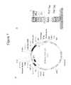
- FIG. 7 shows a plasmid map ( 7 A) and schematic representation of Fab expression on yeast surfaces ( 7 B).
- FIGS. 8A and 8B show Fab library sorting for ABT2-108 affinity maturation.
- FIG. 9 shows analysis of 2-108 clones on yeast surface.
- FIG. 10 shows sorting results for ABT 2-122 affinity maturation.
- FIG. 11 shows the plasmid map of pYDs-TEV-2-108-538x/1-95-15.
- FIG. 12 provides a diagram of the yeast CDR spiking libraries construction.
- FIG. 13 shows ABT-95-15 CDR recombination library construction.
- FIG. 14 provides results of shows K D analysis of ABT2-108-538x/ABT1-95-15 scFv on yeast surface.
- FIG. 15 provides off-rate analysis if the single CDR mutants on yeast surface.
- FIG. 16 describe an on rate and off rate analysis of affinity-matured ABT1-95-15 clones on yeast surface.
- FIG. 17 describes an analysis of affinity-matured ABT1-95-15 clones on yeast surface.
- FIGS. 18A and 18B show the design and use of mouse and human pBOS templates, respectively.
- FIG. 18A discloses SEQ ID NOS 925, 927, 926, 928, 929, 931, 930 and 932, respectively, in order of appearance and
- FIG. 18B discloses SEQ ID NOS 925, 933, 926, 934, 935, 937, 936, 938, 929, 939, 930 and 940, respectively, in order of appearance.
- FIG. 19 describes a representative plasmid map of an scFv fusion construct (pBOS-1-98/2-108-538x-scFc).
- FIG. 20 shows combined MRC5 neutralisation data for IL-1 ⁇ and IL-1 ⁇ of the ABT1-95-A2 and ABT2-65-166 dAb molecules and the IgG resulting from the combination of these two dAb variable domains.
- FIGS. 21A to 21D show sequences of ABT1-95 (SEQ ID NOS 93, 12, 941, 942, 127, 943-946, 121, 130, 132, 133, 122 and 124-126, respectively, in order of appearance), ABT1-122 (SEQ ID NOS 93, 24, 948, 97 and 111, respectively, in order of appearance), ABT1-141 (SEQ ID NOS 93, 1292, 113 and 949, 950, respectively, in order of appearance), and ABT2-65 (SEQ ID NOS 53, 44, 86 and 83, respectively, in order of appearance), respectively, clones as maturation has progressed.
- ABT1-95 SEQ ID NOS 93, 12, 941, 942, 127, 943-946, 121, 130, 132, 133, 122 and 124-126, respectively, in order of appearance
- ABT1-122 SEQ ID NOS 93, 24, 948, 97 and 111, respectively,
- FIG. 22 shows simultaneous binding of rhIL-1 alpha followed by rhIL-1 beta to ABT 108-620x/ABT1-95-A3—showing non interfering independent binding
- FIG. 23 shows IL-6 inhibition using an in vivo study to determine the efficacy of a dual specific antibody for inhabiting IL-1 ⁇ and IL-1 ⁇ activity.
- Key for figure reads top of key to left of figure, e.g., hIgG (250 ⁇ g) coincides to days 7, 4, and 1 beginning at left of x axis.
- IL-1 refers to interleukin-1.
- IL-1 is intended to include both IL-1 ⁇ and IL-1 ⁇ .
- antibody as referred to herein includes whole antibodies and any antigen binding fragment (i.e., “antigen-binding portion”) or single chains thereof.
- An “antibody” refers to a glycoprotein comprising at least two heavy (H) chains and two light (L) chains inter-connected by disulfide bonds, or an antigen binding portion thereof.
- Each heavy chain is comprised of a heavy chain variable region (abbreviated herein as V H ) and a heavy chain constant region.
- the heavy chain constant region is comprised of three domains, CH1, CH2 and CH3.
- Each light chain is comprised of a light chain variable region (abbreviated herein as V L ) and a light chain constant region.
- the light chain constant region is comprised of one domain, CL.
- V H and V L regions can be further subdivided into regions of hypervariability, termed complementarity determining regions (CDR), interspersed with regions that are more conserved, termed framework regions (FR).
- CDR complementarity determining regions
- FR framework regions
- Each V H and V L is composed of three CDRs and four FRs, arranged from amino-terminus to carboxy-terminus in the following order: FR1, CDR1, FR2, CDR2, FR3, CDR3, FR4.
- the variable regions of the heavy and light chains contain a binding domain that interacts with an antigen.
- the constant regions of the antibodies may mediate the binding of the immunoglobulin to host tissues or factors, including various cells of the immune system (e.g., effector cells) and the first component (Clq) of the classical complement system.
- antibody portion refers to one or more fragments of an antibody that retain the ability to specifically bind to an antigen (e.g., IL-1 ⁇ , IL-1 ⁇ ). It has been shown that the antigen-binding function of an antibody can be performed by fragments of a full-length antibody.
- binding fragments encompassed within the term “antigen-binding portion” of an antibody include (i) a Fab fragment, a monovalent fragment consisting of the V L , V H , CL and CH1 domains; (ii) a F(ab′) 2 fragment, a bivalent fragment comprising two Fab fragments linked by a disulfide bridge at the hinge region; (iii) a Fd fragment consisting of the V H and C H1 domains; (iv) a Fv fragment consisting of the V L and V H domains of a single arm of an antibody, (v) a dAb fragment (Ward et al., (1989) Nature 341:544-546), which consists of a V H or V L domain; and (vi) an isolated complementarity determining region (CDR).
- a Fab fragment a monovalent fragment consisting of the V L , V H , CL and CH1 domains
- a F(ab′) 2 fragment a bivalent fragment comprising
- the two domains of the Fv fragment, VL and V H are coded for by separate genes, they can be joined, using recombinant methods, by a synthetic linker that enables them to be made as a single protein chain in which the VL and VH regions pair to form monovalent molecules (known as single chain Fv (scFv); see e.g., Bird et al. (1988) Science 242:423-426; and Huston et al. (1988) Proc. Natl. Acad. Sci. USA 85:5879-5883).
- single chain Fv single chain Fv
- Such single chain antibodies are also intended to be encompassed within the term “antigen-binding portion” of an antibody.
- the antibody fragment is selected from the group consisting of a Fab, an Fd, an Fd′, a single chain Fv (scFv), an scFv a , and a domain antibody (dAb).
- an antibody or antigen-binding portion thereof may be part of a larger immunoadhesion molecules, formed by covalent or noncovalent association of the antibody or antibody portion with one or more other proteins or peptides.
- immunoadhesion molecules include use of the streptavidin core region to make a tetrameric scFv molecule (Kipriyanov et al. (1995) Human Antibodies and Hybridomas 6:93-101) and use of a cysteine residue, a marker peptide and a C-terminal polyhistidine tag to make bivalent and biotinylated scFv molecules (Kipriyanov et al. (1994) Mol. Immunol. 31:1047-1058).
- Antibody portions such as Fab and F(ab′) 2 fragments, can be prepared from whole antibodies using conventional techniques, such as papain or pepsin digestion, respectively, of whole antibodies. Moreover, antibodies, antibody portions and immunoadhesion molecules can be obtained using standard recombinant DNA techniques.
- Two antibody domains are “complementary” where they belong to families of structures which form cognate pairs or groups or are derived from such families and retain this feature.
- a VH domain and a VL domain of an antibody are complementary; two VH domains are not complementary, and two VL domains are not complementary.
- Complementary domains may be found in other members of the immunoglobulin superfamily, such as the V ⁇ and V ⁇ (or gamma and delta) domains of the T-cell receptor
- domain refers to a folded protein structure which retains its tertiary structure independently of the rest of the protein. Generally, domains are responsible for discrete functional properties of proteins, and in many cases may be added, removed or transferred to other proteins without loss of function of the remainder of the protein and/or of the domain.
- single antibody variable domain is meant a folded polypeptide domain comprising sequences characteristic of antibody variable domains.
- variable domains and modified variable domains, for example in which one or more loops have been replaced by sequences which are not characteristic of antibody variable domains, or antibody variable domains which have been truncated or comprise N- or C-terminal extensions, as well as folded fragments of variable domains which retain at least in part the binding activity and specificity of the full-length domain.
- Variable domains of the invention may be combined to form a group of domains; for example, complementary domains may be combined, such as VL domains being combined with VH domains. Non-complementary domains may also be combined. Domains may be combined in a number of ways, involving linkage of the domains by covalent or non-covalent means.
- a “dAb” or “domain antibody” refers to a single antibody variable domain (V H or V L ) polypeptide that specifically binds antigen.
- an “isolated dual-specific antibody”, or an “isolated antibody,” as used herein, is intended to refer to an antibody that is substantially free of other antibodies having different antigenic specificities (e.g., an isolated antibody that specifically binds IL-1 ⁇ /IL-1 ⁇ that is substantially free of antibodies that specifically bind antigens other than IL-1 ⁇ ). Moreover, an isolated antibody may be substantially free of other cellular material and/or chemicals.
- the terms “dual-specific antibody” or “an antibody having dual-specificity” means an antibody comprising two antigen-binding sites or regions, a first binding site or region having affinity for a first antigen or epitope and a second binding site or region having binding affinity for a second antigen or epitope that is distinct from the first.
- the heavy chain variable domain comprises one antigen binding region, i.e., IL-1 ⁇ or IL-1 ⁇
- the light chain variable domain comprises a further antigen binding region, i.e., IL-1 ⁇ or IL-1 ⁇ , such that the antibody has dual-epitope specificity for IL-1 ⁇ and IL-1 ⁇ .
- the invention also includes within its scope dual-specific V H /V L combinations and V L /V L combinations.
- the term “antigen binding region” or “antigen binding site” refers to the portion(s) of an antibody molecule, or antigen binding portion thereof, which contains the amino acid residues that interact with an antigen and confers on the antibody its specificity and affinity for the antigen.
- the antibody of the invention comprises two antigen binding regions, one which is specific for IL-1 ⁇ and another which is specific for IL-1 ⁇ .
- epitope is meant to refer to that portion of any molecule capable of being recognized by and bound by an antibody at one or more of the antibody's antigen binding regions.
- first and second “epitopes” are understood to be epitopes which are not the same and are not bound by a single monospecific antibody, or antigen-binding portion thereof.
- the first and second epitopes are advantageously on different antigens, IL-1 ⁇ or IL-1 ⁇ .
- the first and second antigens are advantageously not the same.
- a “neutralizing antibody,” as used herein, is intended to refer to an antibody whose binding to a particular antigen(s), i.e., IL-1 ⁇ or IL-1 ⁇ , results in inhibition of the biological activity of the antigen. Inhibition of the biological activity of IL-1 ⁇ or IL-1 ⁇ can be assessed by measuring one or more indicators of IL-1 ⁇ or IL-1 ⁇ activity, such as IL-1 ⁇ or IL-1 ⁇ -induced cytotoxicity (either in vitro or in vivo), or IL-1 ⁇ or IL-1 ⁇ binding to IL-1 ⁇ or IL-1 ⁇ receptors. These indicators of IL-1 ⁇ /IL-1 ⁇ activity can be assessed by one or more standard in vitro or in vivo assays known in the art (for example see Example 5). In one embodiment, the ability of a dual-specific antibody to neutralize both IL-1 ⁇ and IL-1 ⁇ activity is assessed by inhibition of IL-1 ⁇ and IL-1 ⁇ in human embryonic lung fibroblasts, MRC-5 cells.
- ND 50 value of an antibody, or antigen-binding portion thereof refers to the concentration of antibody resulting in a one-half maximal inhibition of the given cytokine activity on a responsive cell line.
- the term “EC50” is defined as the concentration of an antibody, or antigen-binding portion thereof, that results in 50% of a measured biological effect.
- the EC50 of a therapeutic agent having a measurable biological effect may comprise the value at which the agent displays 50% of the biological effect.
- the term “IC50” is defined as the concentration of an antibody, or antigen-binding portion thereof, that results in 50% inhibition of a measured effect.
- surface plasmon resonance refers to an optical phenomenon that allows for the analysis of real-time biospecific interactions by detection of alterations in protein concentrations within a biosensor matrix, for example using the BIAcore system (Pharmacia Biosensor AB, Uppsala, Sweden and Piscataway, N.J.).
- BIAcore Pharmaacia Biosensor AB, Uppsala, Sweden and Piscataway, N.J.
- K off is intended to refer to the off rate constant for dissociation of an antibody from the antibody/antigen complex.
- K d or “K D ” as used herein, is intended to refer to the dissociation constant of a particular antibody-antigen interaction, which is obtained from the ratio of k d to k a (i.e., k d /k a ) and is expressed as a molar concentration (M).
- K D values for antibodies can be determined using methods well established in the art. A preferred method for determining the K D of an antibody is by using surface plasmon resonance, preferably using a biosensor system such as a Biacore® system.
- a “monoclonal antibody” as used herein is intended to refer to an antibody obtained from a population of substantially homogeneous antibodies, i.e., the individual antibodies comprising the population are identical except for possible naturally occurring mutations that may be present in minor amounts. Monoclonal antibodies are highly specific. Furthermore, in contrast to polyclonal antibody preparations that typically include different antibodies directed against different determinants (epitopes), each monoclonal antibody is directed against a single determinant on the antigen. The modifier “monoclonal” is not to be construed as requiring production of the antibody by any particular method.
- recombinant antibody refers to antibodies that are prepared, expressed, created or isolated by recombinant means, such as antibodies expressed using a recombinant expression vector transfected into a host cell, antibodies isolated from a recombinant, combinatorial antibody library, antibodies isolated from an animal (e.g., a mouse) that is transgenic for human immunoglobulin genes (see e.g., Taylor et al. (1992) Nucl. Acids Res. 20:6287-6295) or antibodies prepared, expressed, created or isolated by any other means that involves splicing of particular immunoglobulin gene sequences (such as human immunoglobulin gene sequences) to other DNA sequences.
- recombinant antibodies include chimeric, CDR-grafted and humanized antibodies.
- human antibody refers to antibodies having variable and constant regions corresponding to, or derived from, human germline immunoglobulin sequences as described by, for example, Kabat et al. (See Kabat, et al. (1991) Sequences of Proteins of Immunological Interest, Fifth Edition, U.S. Department of Health and Human Services, NIH Publication No. 91-3242).
- the human antibodies of the invention may include amino acid residues not encoded by human germline immunoglobulin sequences (e.g., mutations introduced by random or site-specific mutagenesis in vitro or by somatic mutation in vivo), for example in the CDRs and in particular CDR3.
- Recombinant human antibodies of the invention have variable regions, and may also include constant regions, derived from human germline immunoglobulin sequences (See Kabat et al. (1991) Sequences of Proteins of Immunological Interest, Fifth Edition, U.S. Department of Health and Human Services, NIH Publication No. 91-3242).
- such recombinant human antibodies are subjected to in vitro mutagenesis (or, when an animal transgenic for human Ig sequences is used, in vivo somatic mutagenesis) and thus the amino acid sequences of the VH and VL regions of the recombinant antibodies are sequences that, while derived from and related to human germline VH and VL sequences, may not naturally exist within the human antibody germline repertoire in vivo.
- such recombinant antibodies are the result of selective mutagenesis or backmutation or both.
- backmutation refers to a process in which some or all of the somatically mutated amino acids of a human antibody are replaced with the corresponding germline residues from a homologous germline antibody sequence.
- the heavy and light chain sequences of a human antibody of the invention are aligned separately with the germline sequences in the VBASE database to identify the sequences with the highest homology. Differences in the human antibody of the invention are returned to the germline sequence by mutating defined nucleotide positions encoding such different amino acid.
- the role of each amino acid thus identified as candidate for backmutation should be investigated for a direct or indirect role in antigen binding and any amino acid found after mutation to affect any desirable characteristic of the human antibody should not be included in the final human antibody.
- those amino acid positions found to be different from the closest germline sequence but identical to the corresponding amino acid in a second germline sequence can remain, provided that the second germline sequence is identical and colinear to the sequence of the human antibody of the invention for at least 10, preferably 12 amino acids, on both sides of the amino acid in question.
- Backmutation may occur at any stage of antibody optimization.
- chimeric antibody refers to antibodies which comprise heavy and light chain variable region sequences from one species and constant region sequences from another species, such as antibodies having murine heavy and light chain variable regions linked to human constant regions.
- CDR-grafted antibody refers to antibodies which comprise heavy and light chain variable region sequences from one species but in which the sequences of one or more of the CDR regions of VH and/or VL are replaced with CDR sequences of another species, such as antibodies having murine heavy and light chain variable regions in which one or more of the murine CDRs (e.g., CDR3) has been replaced with human CDR sequences.
- humanized antibody refers to antibodies which comprise heavy and light chain variable region sequences from a non-human species (e.g., a mouse) but in which at least a portion of the VH and/or VL sequence has been altered to be more “human-like”, i.e., more similar to human germline variable sequences.
- a non-human species e.g., a mouse
- human CDR-grafted antibody in which human CDR sequences are introduced into non-human VH and VL sequences to replace the corresponding nonhuman CDR sequences.
- nucleic acid molecule is intended to include DNA molecules and RNA molecules.
- a nucleic acid molecule may be single-stranded or double-stranded, but preferably is double-stranded DNA.
- isolated nucleic acid molecule as used herein in reference to nucleic acids encoding antibodies or antibody portions (e.g., VH, VL, CDR3) that bind IL-1 ⁇ and IL-1 ⁇ , is intended to refer to a nucleic acid molecule in which the nucleotide sequences encoding the antibody or antibody portion are free of other nucleotide sequences encoding antibodies or antibody portions that bind antigens other than hTNF ⁇ , which other sequences may naturally flank the nucleic acid in human genomic DNA.
- an isolated nucleic acid of the invention encoding a VH region of an anti-TNF ⁇ antibody contains no other sequences encoding other VH regions that bind antigens other than IL-1 ⁇ and IL-1 ⁇ .
- vector is intended to refer to a nucleic acid molecule capable of transporting another nucleic acid to which it has been linked.
- plasmid refers to a circular double stranded DNA loop into which additional DNA segments may be ligated.
- viral vector Another type of vector is a viral vector, wherein additional DNA segments may be ligated into the viral genome.
- Certain vectors are capable of autonomous replication in a host cell into which they are introduced (e.g., vectors may have a bacterial origin of replication and/or an episomal origin of replication (generally derived from a viral sequence, e.g., SV40).
- vectors e.g., non-episomal mammalian vectors
- vectors can be integrated into the genome of a host cell upon introduction into the host cell, and thereby are replicated along with the host genome.
- certain vectors are capable of directing the expression of genes to which they are operatively linked.
- Such vectors are referred to herein as “recombinant expression vectors” (or simply, “expression vectors”).
- expression vectors of utility in recombinant DNA techniques are often in the form of plasmids.
- plasmid and vector may be used interchangeably as the plasmid is the most commonly used form of vector.
- the invention is intended to include such other forms of expression vectors, such as viral vectors (e.g., replication defective retroviruses, adenoviruses and adeno-associated viruses), which serve equivalent functions.
- episomally replicating vector refers to a vector which is typically and very preferably not integrated into the genome of the host cell, but exists in parallel.
- An episomally replicating vector is replicated during the cell cycle and in the course of this replication the vector copies are distributed statistically in the resulting cells depending on the number of the copies present before and after cell division.
- the episomally replicating vector may take place in the nucleus of the host cell, and preferably replicates during S-phase of the cell cycle.
- the episomally replicating vector is replicated at least once, i.e. one or multiple times, in the nucleus of the host cell during S-phase of the cell cycle. In a very preferred embodiment, the episomally replicating vector is replicated once in the nucleus of the host cell during S-phase of the cell cycle.
- oil of replication sequences or “origin of replication,” used interchangeably herein, refer to sequences which, when present in a vector, initiate replication.
- An origin of replication may be recognized by a replication initiation factor or, alternatively, by a DNA helicase.
- the “gene of interest” as used herein, refers to an exogenous DNA sequence which is added to the vector of the invention.
- the gene of interest for example, may comprise a coding sequence which can be either spaced by introns or which is a cDNA encoding the open reading frame.
- the region of the vector to which the gene of interest is cloned is referred to herein as an “insertion site.”
- the gene of interest comprises a portion of the antibody or fusion protein that is expressed using a vector of the invention.
- the heavy chain variable region of the antibody ABT2-108 i.e., the gene of interest, may cloned into the vector of the invention which comprises a heavy chain constant region
- the vector comprises an antibody light or heavy chain constant region which is 3′ to the insertion site for the gene of interest and is operably linked thereto.
- the gene of interest is a variable region of a light or heavy chain of an antibody which is operably linked to the antibody light or heavy chain constant region encoded in the vector of the invention.
- promoter includes any nucleic acid sequence sufficient to direct transcription in a eukaryotic cell, including inducible promoters, repressible promoters and constitutive promoters.
- a promoter includes elements that are sufficient to render promoter-dependent gene expression controllable in a cell type-specific, tissue-specific or temporal-specific manner, or inducible by external signals or agents. Such elements can be located in the 5′ or 3′ or intron sequence regions of a particular gene.
- gene expression will be constitutive, although regulatable promoters can be employed in the present invention if desired. Gene expression can also be controlled by transcription-regulation using heat, light, or metals, such as by the use of metallothionine genes or heat shock genes.
- Upstream and downstream are terms used to describe the relative orientation between two elements present in a nucleotide sequence or vector.
- An element that is “upstream” of another is located in a position closer to the 5′ end of the sequence (i.e., closer to the end of the molecule that has a phosphate group attached to the 5′ carbon of the ribose or deoxyribose backbone if the molecule is linear) than the other element.
- An element is said to be “downstream” when it is located in a position closer to the 3′ end of the sequence (i.e., the end of the molecule that has an hydroxyl group attached to the 3′ carbon of the ribose or deoxyribose backbone in the linear molecule) when compared to the other element.
- the term “stuffer sequence” refers to a nucleic acid sequence, preferably in a vector, which is flanked by restriction enzyme sites at both the 5′ and 3′ ends.
- the stuffer sequence is located in a vector at the insertion site for the nucleic acid encoding the gene of interest.
- the stuffer sequence is digested away from the vector using the appropriate restriction enzymes, and the nucleic acid encoding the gene of interest is ligated or homologously recombined into the vector at the former position of the stuffer sequence.
- the stuffer sequence is large enough to provide sufficient distance between the 5′ and 3′ restriction enzyme sites so that the restriction enzyme can efficiently cut the vector.
- the length of the stuffer sequence is different than the size of the nucleic acid encoding the gene of interest, e.g., a stuffer sequence of about 300 base pairs or less or about 400 base pairs or more may be used for a nucleic acid encoding the gene of interest which is about 350 base pairs. In another embodiment, the stuffer sequence is about 1 kb in size.
- recombinant host cell (or simply “host cell”), as used herein, is intended to refer to a cell into which a recombinant expression vector has been introduced. It should be understood that such terms are intended to refer not only to the particular subject cell but to the progeny of such a cell. Because certain modifications may occur in succeeding generations due to either mutation or environmental influences, such progeny may not, in fact, be identical to the parent cell, but are still included within the scope of the term “host cell” as used herein.
- oire refers to a collection of diverse variants, for example polypeptide variants which differ in their primary sequence.
- a library used in the present invention will encompass a repertoire of polypeptides comprising at least 1000 members.
- library refers to a mixture of heterogeneous polypeptides or nucleic acids.
- the library is composed of members, each of which have a single polypeptide or nucleic acid sequence. To this extent, library is synonymous with repertoire. Sequence differences between library members are responsible for the diversity present in the library.
- the library may take the form of a simple mixture of polypeptides or nucleic acids, or may be in the form of organisms or cells, for example bacteria, viruses, animal or plant cells and the like, transformed with a library of nucleic acids.
- each individual organism or cell contains only one or a limited number of library members.
- the nucleic acids are incorporated into expression vectors, in order to allow expression of the polypeptides encoded by the nucleic acids.
- a library may take the form of a population of host organisms, each organism containing one or more copies of an expression vector containing a single member of the library in nucleic acid form which can be expressed to produce its corresponding polypeptide member.
- the population of host organisms has the potential to encode a large repertoire of genetically diverse polypeptide variants.
- the invention provides isolated, dual-specific antibodies, or antigen binding portions thereof, that bind human IL-1 ⁇ and IL-1 ⁇ with high affinity and neutralizing capacity.
- the antibody, or antigen-binding fragment thereof is human.
- the antibodies of the invention are recombinant, neutralizing anti-IL-1 ⁇ and anti-IL-1 ⁇ .
- the invention provides an isolated, dual-specific antibody, or an antigen-binding portion thereof, which dissociates from human IL-1 ⁇ with a K D of 1 ⁇ 10 ⁇ 7 M or less and neutralizes human IL-1 ⁇ in a standard in vitro MRC5 assay with an ND 50 of 900 nM or less, and dissociates from human IL-1 ⁇ with a K D of 5 ⁇ 10 ⁇ 5 M or less, and neutralizes human IL-1 ⁇ in a standard in vitro MRC5 assay with an ND 50 of 800 nM or less.
- the IL-1 ⁇ /IL-1 ⁇ dual-specific antibody, or antigen-binding portion thereof binds human IL-1 ⁇ and IL-1 ⁇ (hIL-1 ⁇ and hIL-1 ⁇ ), but does not bind mouse IL-1 ⁇ or mouse IL-1 ⁇ .
- the variable domains are selected from V-gene repertoires selected for instance using phage display technology as herein described, then these variable domains can comprise a universal framework region, such that is they may be recognised by a specific generic ligand as herein defined.
- the use of universal frameworks, generic ligands and the like is described in WO99/20749.
- reference to phage display includes the use of both phage and/or phagemids.
- the dual-specific antibodies of the invention may comprise variable regions which are derived from antibodies directed against IL-1 ⁇ and IL-1 ⁇ .
- the antibodies of the invention may be derived from a repertoire of single antibody domains such as those expressed on the surface of filamentous bacteriophage. Selection may be performed as described below in section III and in the Examples provided herein.
- the invention provides single domain antibodies which are have neutralizing and affinity properties specific for IL-1 ⁇ or IL-1 ⁇ , as summarized in Tables 8 and 61 to 64, as well as the Examples.
- Dual-specific antibodies, or antigen-binding portions thereof, according to the present invention preferably comprise combinations of heavy and light chain domains.
- the dual specific antibody, or antigen-binding portion thereof may comprise a V H domain and a V L domain, which may be linked together in the form of an scFv.
- the antibody, or antigen-binding portion thereof may comprise one or more C H or C L domains.
- the antibody, or antigen-binding portion thereof may comprise a C H 1 domain, C H 2 or C H 3 domain, and/or a C L domain, C ⁇ 1, C ⁇ 2, C ⁇ 3 or C ⁇ 4 domains, or any combination thereof.
- a hinge region domain may also be included.
- Such combinations of domains may, for example, mimic natural antibodies, such as IgG or IgM, or fragments thereof, such as Fv, scFv, Fab or F(ab′) 2 molecules.
- Other structures such as a single arm of an IgG molecule comprising V H , V L , C H 1 and C L domains, are envisaged.
- the dual specific antibody, or antigen-binding portion thereof, of the invention comprises only two variable domains although several such antibodies, or antigen-binding portions thereof, may be incorporated together into the same protein, for example two such antibodies, or antigen-binding portions thereof, can be incorporated into an IgG or a multimeric immunoglobulin, such as IgM.
- a plurality of dual specific ligands are combined to form a multimer. For example, two different dual specific ligands are combined to create a tetra-specific molecule.
- variable regions of a dual-specific antibodies, or antigen-binding portions thereof, produced according to the methods described herein and known in the art may be on the same polypeptide chain, or alternatively, on different polypeptide chains.
- variable regions are on different polypeptide chains, then they may be linked via a linker, generally a flexible linker (such as a polypeptide chain), a chemical linking group, or any other method known in the art.
- the present invention provides dual specific antibodies, or antigen-binding portions thereof, which comprise at least two complementary variable domains, e.g., a VH and a VL domain.
- the present invention provides dual specific antibodies, or antigen-binding portions thereof, which comprise at least two non-complementary variable domains.
- the antibody, or antigen-binding portion thereof may comprise a pair of VH domains or a pair of VL domains.
- the domains are of non-camelid origin; preferably they are human domains or comprise human framework regions (FWs) and one or more heterologous CDRs.
- CDRs and framework regions are those regions of an immunoglobulin variable domain as defined in the Kabat database of Sequences of Proteins of Immunological Interest.
- Dual-specific antibodies of the invention may include variable heavy and light chain amino acid regions described in SEQ ID NOs: 4, 8, 12, 16, 20, 24, 28, 32, 36, 40, 44, 48, and 52 (see Table 61 and Example 3 for a summary).
- Dula specific antibodies may also include any of the variable heavy and light chain amino acid regions described in SEQ ID NOs: 53 to 133.
- Pairings of the single domain antibodies described in SEQ ID NOs: 4, 8, 12, 16, 20, 24, 28, 32, 36, 40, 44, 48, and 52, as well as any of the variable sequences described in SEQ ID NOs: 53 to 133, are also included in the invention.
- Examples of dual-specific IL-1 ⁇ and IL-1 ⁇ antibodies are described in the Examples, including Example 5 and Table 9.
- the invention provides a dual-specific, isolated antibody, or antigen-binding portion thereof, comprising an IL-1 ⁇ antigen binding region and an IL-1 ⁇ antigen binding region, wherein the antibody, or antigen-binding portion thereof, comprises a heavy chain variable region and a light chain variable region combination comprising a heavy chain variable region comprising CDRs as set forth in SEQ ID NO: 16 (ABT1-96) and a light chain variable region comprising CDRs as set forth in SEQ ID NO: 40 (ABT2-46); a light chain variable region comprising CDRs as set forth in SEQ ID NO: 24 (ABT1-122) and a heavy chain variable region comprising CDRs as set forth in SEQ ID NO: 52 (ABT2-108); a light chain variable region comprising CDRs as set forth in SEQ ID NO: 28 (ABT1-141) and a heavy chain variable region comprising CDRs as set forth in SEQ ID NO: 52 (ABT2-108); a light chain variable region comprising CDRs
- the invention pertains to antibodies, including human antibodies, that have slow dissociation kinetics for association with either IL-1 ⁇ or IL-1 ⁇ and that have light and heavy chain CDR3 domains that structurally are identical to or related to those identified herein, including those CDR3 domains described in SEQ ID NOs: 11, 23, 27, 3, 7, 15, and 19.
- the invention pertains to a dual-specific antibody, or antigen-binding portion thereof, of the invention is preferably selected to have desirable binding kinetics (e.g., high affinity, low dissociation, slow off-rate, strong neutralizing activity) for IL-1 ⁇ and IL-1 ⁇ , to which the antibody specifically binds.
- desirable binding kinetics e.g., high affinity, low dissociation, slow off-rate, strong neutralizing activity
- the dual-specific antibody, or portion thereof may bind IL-1 ⁇ and IL-1 ⁇ with a k off rate constant of 0.1 s ⁇ 1 or less, more preferably a k off rate constant of 1 ⁇ 10 ⁇ 2 s ⁇ 1 or less, even more preferably a k off rate constant of 1 ⁇ 10 ⁇ 3 s ⁇ 1 or less, even more preferably a k off rate constant of 1 ⁇ 10 ⁇ 4 s ⁇ 1 or less, or even more preferably a k off rate constant of 1 ⁇ 10 ⁇ 5 s ⁇ 1 or less, as determined by surface plasmon resonance.
- a k off rate constant of 0.1 s ⁇ 1 or less more preferably a k off rate constant of 1 ⁇ 10 ⁇ 2 s ⁇ 1 or less, even more preferably a k off rate constant of 1 ⁇ 10 ⁇ 3 s ⁇ 1 or less, even more preferably a k off rate constant of 1 ⁇ 10 ⁇ 4 s ⁇ 1 or less, or even more preferably
- the isolated, dual-specific antibody, or an antigen-binding portion thereof dissociates from human IL-1 ⁇ with a K D of about 1 ⁇ 10 ⁇ 7 M to about 1 ⁇ 10 ⁇ 8 M or less and dissociates from human IL-1 ⁇ with a K D of about 5 ⁇ 10 ⁇ 5 M to about 1 ⁇ 10 ⁇ 9 M or less.
- Ranges intermediate to the above recited constants, e.g., 4.0 ⁇ 10 ⁇ 8 M, are also intended to be part of this invention. For example, ranges of values using a combination of any of the above recited values as upper and/or lower limits are intended to be included.
- the antibody, or antigen-binding portion thereof dissociates from IL-1 ⁇ with a K D of 5.4 ⁇ 10 ⁇ 5 M or less; dissociates from IL-1 ⁇ with a K D of 2.8 ⁇ 10 ⁇ 6 M or less; dissociates from IL-1 ⁇ with a K D of 1.3 ⁇ 10 ⁇ 6 M or less; dissociates from IL-1 ⁇ with a K D of 9.3 ⁇ 10 ⁇ 7 M or less; dissociates from IL-1 ⁇ with a K D of 2 ⁇ 10 ⁇ 7 M or less; dissociates from IL-1 ⁇ with a K D of 1.1 ⁇ 10 ⁇ 7 M or less; or dissociates from IL-1 ⁇ with a K D of 2.8 ⁇ 10 ⁇ 8 M or less.
- Ranges intermediate to the above recited constants e.g., K D of 1.7 ⁇ 10 ⁇ 7 M or less, are also intended to be part of this invention.
- K D 1.7 ⁇ 10 ⁇ 7 M or less
- ranges of values using a combination of any of the above recited values as upper and/or lower limits are intended to be included.
- the antibody, or antigen-binding portion dissociates from IL-1 ⁇ with a K D of 1 ⁇ 10 ⁇ 8 M or less; dissociates from IL-1 ⁇ with a K D of 1 ⁇ 10 ⁇ 9 M or less; dissociates from IL-1 ⁇ with a K D of 40-86 nM or less; dissociates from IL-1 ⁇ with a K D of 20-42 nM or less; dissociates from IL-1 ⁇ with a K D of 32-42 nM or less; dissociates from IL-1 ⁇ with a K D of 7-12 nM or less; dissociates from IL-1 ⁇ with a K D of 3.0 ⁇ 10 ⁇ 7 M or less; dissociates from IL-1 ⁇ with a K D of 1.1 ⁇ 10 ⁇ 7 M or less; dissociates from IL-1 ⁇ with a K D of 6.1 ⁇ 10 ⁇ 8 M or less; dissociates from IL-1 ⁇ with a K D of 6 ⁇ 10 ⁇ 8 M or less; dissociates from IL
- Ranges intermediate to the above recited constants e.g., K D of 1.7 ⁇ 10 ⁇ 7 M or less, are also intended to be part of this invention.
- K D 1.7 ⁇ 10 ⁇ 7 M or less
- ranges of values using a combination of any of the above recited values as upper and/or lower limits are intended to be included.
- affinity properties may also apply to single domain antibodies of the invention which bind and neutralize IL-1 ⁇ or IL-1 ⁇ .
- Surface plasmon resonance analysis may be used for determining kinetic values, including K D and k off values.
- the IL-1 ⁇ /IL-1 ⁇ dual-specific antibody, or antigen-binding portion thereof, of the invention may exhibit a strong capacity to neutralize both hIL-1 ⁇ and hIL-1 ⁇ activity, as determined using a standard in vitro or in vivo assay (see also Examples, including Example 5).
- IL-1 ⁇ /IL-1 ⁇ antibodies neutralize human IL-1 ⁇ in a standard in vitro assay with ND 50 values of about 900 nM to about 10 nM or less.
- the antibodies of the invention are also able to neutralize human IL-1 ⁇ in a standard in vitro assay with ND 50 values of about 800 nM to about 200 nM or less.
- a dual-specific antibody, or antigen binding portion thereof may inhibit the activity of one, and more preferably both, of IL-1 ⁇ and/or IL-1 ⁇ with an ND 50 of 1 ⁇ 10 ⁇ 6 M or less, even more preferably with an ND 50 of 1 ⁇ 10 ⁇ 7 M or less, even more preferably with an ND 50 of 1 ⁇ 10 ⁇ 8 M or less, even more preferably with an ND 50 of 1 ⁇ 10 ⁇ 9 M or less, even more preferably with an ND 50 of 1 ⁇ 10 ⁇ 10 M or less, or even more preferably with an ND 50 of 1 ⁇ 10 ⁇ 11 M or less.
- the antibody, or antigen-binding portion neutralizes human IL-1 ⁇ in a standard in vitro assay with an ND 50 of 900 nM or less, and/or neutralizes human IL-1 ⁇ in a standard in vitro assay with an ND 50 of 800 nM or less. In one embodiment, the antibody, or antigen-binding portion thereof, neutralizes IL-1 ⁇ in a standard in vitro assay with an ND 50 of 10 nM or less. In one embodiment, the antibody, or antigen-binding portion thereof, neutralizes IL-1 ⁇ in a standard in vitro assay with an ND 50 of 200 nM or less.
- the antibody, or antigen-binding portion thereof neutralizes IL-1 ⁇ in a standard in vitro assay with an ND 50 of 10 nM or less. In one embodiment, the antibody, or antigen-binding portion thereof, neutralizes IL-1 ⁇ in a standard in vitro assay with an ND 50 of 200 nM or less. Ranges intermediate to the above recited values, e.g., ND 50 of 80 nM or less, ND 50 of 4.0 ⁇ 10 ⁇ 10 M or less, are also intended to be part of this invention. For example, ranges of values using a combination of any of the above recited values as upper and/or lower limits are intended to be included.
- neutralization properties of an antibody, or antigen-binding portion thereof may be determined using an in vitro MRC-5 cell assay.
- neutralization properties may also apply to single domain antibodies of the invention which bind and neutralize IL-1 ⁇ or IL-1 ⁇ .
- an antibody of the invention comprises heavy and light chain variable regions comprising amino acid sequences that are homologous to the amino acid sequences of the preferred antibodies described herein, and wherein the antibodies retain the desired functional properties of the IL-1 ⁇ /IL-1 ⁇ dual-specific antibodies of the invention.
- the invention provides an isolated antibody, or antigen binding portion thereof, comprising a heavy chain variable region and a light chain variable region, wherein (a) the heavy chain variable region comprises an amino acid sequence that is at least 80% homologous to an amino acid sequence selected from the group consisting of SEQ ID NOs: 4, 16, 20, 32, 44, 52, or 53 to 92; (b) the light chain variable region comprises an amino acid sequence that is at least 80% homologous to an amino acid sequence selected from the group consisting of SEQ ID NOs: 8, 12, 24, 28, 36, 40, 48, or 93 to 133; and (c) the antibody specifically binds to IL-1 ⁇ /IL-1 ⁇ and exhibits at least one of the functional properties described herein, preferably several of the functional properties described herein. Also included in the invention is an isolated antibody, or antigen binding portion thereof, comprising two heavy (or two light) chain variable regions that specifically binds to IL-1 ⁇ /IL-1 ⁇ .
- the VH and/or VL amino acid sequences may be 85%, 90%, 95%, 96%, 97%, 98% or 99% homologous to the sequences set forth herein, including SEQ ID NOs: 52 to 133.
- An antibody having VH and VL regions having high (i.e., 80% or greater) homology to the VH and VL regions of the sequences set forth above, can be obtained by mutagenesis (e.g., site-directed or PCR-mediated mutagenesis) of nucleic acid molecules encoding SEQ ID NOs: 4, 8, 12, 16, 20, 24, 28, 32, 36, 40, 44, 48, and 52 to 133, followed by testing of the encoded altered antibody for retained function (i.e., affinity and neutralization properties) using the functional assays described herein.
- mutagenesis e.g., site-directed or PCR-mediated mutagenesis
- the invention provides nucleic acid sequences which may be 85%, 90%, 95%, 96%, 97%, 98% or 99% homologous to the sequences set forth herein, including SEQ ID NOs: 134 to 843.
- the percent homology between two amino acid sequences is equivalent to the percent identity between the two sequences.
- the comparison of sequences and determination of percent identity between two sequences can be accomplished using a mathematical algorithm, as described in the non-limiting examples below.
- the percent identity between two amino acid sequences can be determined using the algorithm of Meyers and Miller (Comput. Appl. Biosci., 4:11-17 (1988)) which has been incorporated into the ALIGN program (version 2.0), using a PAM120 weight residue table, a gap length penalty of 12 and a gap penalty of 4. In addition, the percent identity between two amino acid sequences can be determined using the Needleman and Wunsch (J. Mol. Biol.
- the protein sequences of the present invention can further be used as a “query sequence” to perform a search against public databases to, for example, identify related sequences.
- Such searches can be performed using the XBLAST program (version 2.0) of Altschul et al. (1990) J. Mol. Biol. 215:403-10.
- Gapped BLAST can be utilized as described in Altschul et al. (1997) Nucleic Acids Res. 25(17):3389-3402.
- the default parameters of the respective programs e.g., XBLAST and NBLAST
- the default parameters of the respective programs e.g., XBLAST and NBLAST
- an antibody of the invention comprises a heavy chain variable region comprising CDR1, CDR2 and CDR3 sequences and a light chain variable region comprising CDR1, CDR2 and CDR3 sequences, wherein one or more of these CDR sequences comprise specified amino acid sequences based on the preferred antibodies described herein, or conservative modifications thereof, and wherein the antibodies retain the desired functional properties of the anti-IL-1 ⁇ /IL-1 ⁇ dual-specific antibodies of the invention.
- the invention provides an isolated antibody, or antigen binding portion thereof, comprising a heavy chain variable region comprising CDR1, CDR2, and CDR3 sequences and a light chain variable region comprising CDR1, CDR2, and CDR3 sequences, wherein: (a) the heavy chain variable region CDR3 sequence comprises an amino acid sequence selected from the group consisting of amino acid sequences of SEQ ID NOs: 3, 7, 15, 19, 31, 43, and 51, and conservative modifications thereof; (b) the light chain variable region CDR3 sequence comprises an amino acid sequence selected from the group consisting of amino acid sequences of SEQ ID NOs: 11, 23, 27, 35, 39, and 47, and conservative modifications thereof, and (c) the antibody specifically binds to IL-1 ⁇ /IL-1 ⁇ and exhibits at least one of the functional properties described herein, more preferably several of the functional properties described herein, i.e., high affinity and neutralizing for both IL-1 ⁇ /IL-1 ⁇ .
- the heavy chain variable region CDR2 sequence comprises an amino acid sequence selected from the group consisting of amino acid sequences of SEQ ID NOs: 2, 6, 14, 18, 30, 42, and 50, and conservative substitutions thereof
- the light chain variable region CDR2 sequence comprises an amino acid sequence selected from the group consisting of amino acid sequences of SEQ ID NOs: 10, 22, 26, 34, 38, and 46, and conservative modifications thereof.
- the heavy chain variable region CDR1 sequence comprises an amino acid sequence selected from the group consisting of amino acid sequences of SEQ ID NOs: 1, 5, 13, 17, 29, 41, and 49, and conservative modifications thereof
- the light chain variable region CDR1 sequence comprises an amino acid sequence selected from the group consisting of amino acid sequences of SEQ ID NOs: 9, 21, 25, 33, 37, and 45, and conservative modifications thereof.
- a “conservative amino acid substitution” or a “conservative substitution”, as used herein, is one in which one amino acid residue is replaced with another amino acid residue having a similar side chain.
- Families of amino acid residues having similar side chains have been defined in the art, including basic side chains (e.g., lysine, arginine, histidine), acidic side chains (e.g., aspartic acid, glutamic acid), uncharged polar side chains (e.g., glycine, asparagine, glutamine, serine, threonine, tyrosine, cysteine), nonpolar side chains (e.g., alanine, valine, leucine, isoleucine, proline, phenylalanine, methionine, tryptophan), beta-branched side chains (e.g., threonine, valine, isoleucine) and aromatic side chains (e.g., tyrosine, phenylalanine, tryp
- substitutions and modifications can be introduced into an antibody of the invention by standard techniques known in the art, such as site-directed mutagenesis and PCR-mediated mutagenesis.
- one or more amino acid residues within the CDR regions of an antibody of the invention can be replaced with other amino acid residues from the same side chain family and the altered antibody can be tested for retained function, e.g., affinity or neutralization characteristics, using the functional assays described herein.
- another embodiment of the invention pertains to an isolated dual-specific antibody, or antigen binding portion thereof, comprising a heavy chain variable region comprising CDR1, CDR2, and CDR3 sequences comprising an amino acid sequence selected from the group consisting of SEQ ID NOs: 1, 5, 13, 17, 29, 41, and 49, SEQ ID NOs: 2, 6, 14, 18, 30, 42, and 50 and SEQ ID NOs: 3, 7, 15, 19, 31, 43, and 51, respectively, and a light chain variable region comprising CDR1, CDR2, and CDR3 sequences comprising an amino acid sequence selected from the group consisting of SEQ ID NOs: 9, 21, 25, 33, 37, and 45, SEQ ID NOs: 10, 22, 26, 34, 38, and 46 and SEQ ID NOs: 11, 23, 27, 35, 39, and 47, respectively.
- such antibodies contain the VH and/or VL CDR sequences of antibodies described herein, yet may contain different framework sequences from these antibodies.
- Preferred human framework regions are those encoded by germline gene segments DP47 and DPK9.
- FW1, FW2 and FW3 of a VH or VL domain have the sequence of FW1, FW2 or FW3 from DP47 or DPK9.
- the human frameworks may optionally contain mutations, for example up to about 5 amino acid changes or up to about 10 amino acid changes collectively in the human frameworks used in the ligands of the invention.
- Such framework sequences can be obtained from public DNA databases or published references that include germline antibody gene sequences.
- germline DNA sequences for human heavy and light chain variable region genes can be found in the “VBase” human germline sequence database (available on the Internet at www.mrc-cpe.cam.ac.uk/vbase), as well as in Kabat et al. (1991) Sequences of Proteins of Immunological Interest, Fifth Edition, U.S. Department of Health and Human Services, NIH Publication No. 91-3242; Tomlinson, I. M., et al. (1992) “The Repertoire of Human Germline V H Sequences Reveals about Fifty Groups of V H Segments with Different Hypervariable Loops” J. Mol.
- Preferred framework sequences for use in the antibodies of the invention are those that are structurally similar to the framework sequences used by selected antibodies of the invention.
- VH CDR1, 2 and 3 sequences of SEQ ID NOs: 1, 2, 3, 5, 6, 7, 13, 14, 15, 17, 18, 19, 29, 30, 31, 41, 42, 43 49, 50, and 51 and the VL CDR1, 2 and 3 sequences of SEQ ID NOs: 9, 10, 11, 21, 22, 23, 25, 26, 27, 33, 34, 35, 37, 38, 39, 45, 46, and 47, can be grafted onto framework regions that have the same sequence as that found in the germline immunoglobulin gene from which the framework sequence derive, or the CDR sequences can be grafted onto framework regions that contain one or more mutations as compared to the germline sequences.
- an antibody of the invention may be prepared using an antibody having one or more of the V H and/or V L sequences disclosed herein as starting material to engineer a modified antibody, which modified antibody may have altered properties from the starting antibody.
- An antibody can be engineered by modifying one or more residues within one or both variable regions (i.e., V H and/or V L ), for example within one or more CDR regions and/or within one or more framework regions. Additionally or alternatively, an antibody can be engineered by modifying residues within the constant region(s), for example to alter the effector function(s) of the antibody.
- CDR grafting One type of variable region engineering that can be performed is CDR grafting.
- Antibodies interact with target antigens predominantly through amino acid residues that are located in the six heavy and light chain complementarity determining regions (CDRs). For this reason, the amino acid sequences within CDRs are more diverse between individual antibodies than sequences outside of CDRs. Because CDR sequences are responsible for most antibody-antigen interactions, it is possible to express recombinant antibodies that mimic the properties of specific naturally occurring antibodies by constructing expression vectors that include CDR sequences from the specific naturally occurring antibody grafted onto framework sequences from a different antibody with different properties (see, e.g., Riechmann et al. (1998) Nature 332:323-327; Jones. et al.
- variable region modification is to mutate amino acid residues within the VH and/or VL CDR1, CDR2 and/or CDR3 regions to thereby improve one or more binding properties (e.g., affinity) of the antibody of interest.
- Site-directed mutagenesis or PCR-mediated mutagenesis can be performed to introduce the mutation(s) and the effect on antibody binding, or other functional property of interest, can be evaluated in in vitro or in vivo assays as described herein and provided in the Examples.
- Preferably conservative modifications are introduced.
- the mutations may be amino acid substitutions, additions or deletions, but are preferably substitutions.
- typically no more than five residues are altered within a CDR region are altered.
- Engineered antibodies of the invention include those in which modifications have been made to framework residues within V H and/or V L , e.g. to improve the properties of the antibody. Typically such framework modifications are made to decrease the immunogenicity of the antibody.
- one approach is to “backmutate” one or more framework residues to the corresponding germline sequence. More specifically, an antibody that has undergone somatic mutation may contain framework residues that differ from the germline sequence from which the antibody is derived. Such residues can be identified by comparing the antibody framework sequences to the germline sequences from which the antibody is derived. To return the framework region sequences to their germline configuration, the somatic mutations can be “backmutated” to the germline sequence by, for example, site-directed mutagenesis or PCR-mediated mutagenesis.
- Another type of framework modification involves mutating one or more residues within the framework region, or even within one or more CDR regions, to remove T cell epitopes to thereby reduce the potential immunogenicity of the antibody. This approach is also referred to as “deimmunization” and is described in further detail in U.S. patent Publication No. 20030153043 by Carr et al.
- dual-specific antibodies of the invention may be engineered to include modifications within the Fc region, typically to alter one or more functional properties of the antibody, such as serum half-life, complement fixation, Fc receptor binding, and/or antigen-dependent cellular cytotoxicity.
- an antibody of the invention may be chemically modified (e.g., one or more chemical moieties can be attached to the antibody) or be modified to alter it's glycosylation, again to alter one or more functional properties of the antibody.
- the numbering of residues in the Fc region is that of the EU index of Kabat.
- the hinge region of CH1 is modified such that the number of cysteine residues in the hinge region is altered, e.g., increased or decreased.
- This approach is described further in U.S. Pat. No. 5,677,425 by Bodmer et al.
- the number of cysteine residues in the hinge region of CH1 is altered to, for example, facilitate assembly of the light and heavy chains or to increase or decrease the stability of the dual-specific antibody.
- the Fc hinge region of the antibody of the invention is mutated to decrease the biological half life of the antibody. More specifically, one or more amino acid mutations are introduced into the CH2-CH3 domain interface region of the Fc-hinge fragment such that the antibody has impaired Staphylococcyl protein A (SpA) binding relative to native Fc-hinge domain SpA binding.
- SpA Staphylococcyl protein A
- the antibody of the invention is modified to increase its biological half life.
- Various approaches are possible. For example, one or more of the following mutations can be introduced: T252L, T254S, T256F, as described in U.S. Pat. No. 6,277,375 to Ward.
- the antibody may be altered within the CH1 or CL region to contain a salvage receptor binding epitope taken from two loops of a CH2 domain of an Fc region of an IgG, as described in U.S. Pat. Nos. 5,869,046 and 6,121,022 by Presta et al.
- the Fc region is altered by replacing at least one amino acid residue with a different amino acid residue to alter the effector function(s) of the dual-specific antibody.
- one or more amino acids selected from amino acid residues 234, 235, 236, 237, 297, 318, 320 and 322 can be replaced with a different amino acid residue such that the antibody has an altered affinity for an effector antibody but retains the antigen-binding ability of the parent antibody.
- the effector antibody to which affinity is altered can be, for example, an Fc receptor or the C1 component of complement. This approach is described in further detail in U.S. Pat. Nos. 5,624,821 and 5,648,260, both by Winter et al.
- one or more amino acids selected from amino acid residues 329, 331 and 322 can be replaced with a different amino acid residue such that the dual-specific antibody has altered C1q binding and/or reduced or abolished complement dependent cytotoxicity (CDC).
- CDC complement dependent cytotoxicity
- one or more amino acid residues within amino acid positions 231 and 239 are altered to thereby alter the ability of the antibody to fix complement. This approach is described further in PCT Publication WO 94/29351 by Bodmer et al.
- the Fc region is modified to increase the ability of the dual-specific antibody to mediate antibody dependent cellular cytotoxicity (ADCC) and/or to increase the affinity of the antibody for an Fc ⁇ receptor by modifying one or more amino acids at the following positions: 238, 239, 248, 249, 252, 254, 255, 256, 258, 265, 267, 268, 269, 270, 272, 276, 278, 280, 283, 285, 286, 289, 290, 292, 293, 294, 295, 296, 298, 301, 303, 305, 307, 309, 312, 315, 320, 322, 324, 326, 327, 329, 330, 331, 333, 334, 335, 337, 338, 340, 360, 373, 376, 378, 382, 388, 389, 398, 414, 416, 419, 430, 434, 435, 437, 438 or 439.
- ADCC antibody dependent cellular cytotoxicity
- the glycosylation of an antibody is modified.
- an aglycoslated antibody can be made (i.e., the antibody lacks glycosylation).
- Glycosylation can be altered to, for example, increase the affinity of the antibody for antigen.
- carbohydrate modifications can be accomplished by, for example, altering one or more sites of glycosylation within the antibody sequence.
- one or more amino acid substitutions can be made that result in elimination of one or more variable region framework glycosylation sites to thereby eliminate glycosylation at that site.
- Such aglycosylation may increase the affinity of the antibody for antigen.
- an antibody can be made that has an altered type of glycosylation, such as a hypofucosylated antibody having reduced amounts of fucosyl residues or an antibody having increased bisecting GlcNac structures.
- altered glycosylation patterns have been demonstrated to increase the ADCC ability of antibodies.
- carbohydrate modifications can be accomplished by, for example, expressing the antibody in a host cell with altered glycosylation machinery. Cells with altered glycosylation machinery have been described in the art and can be used as host cells in which to express recombinant antibodies of the invention to thereby produce an antibody with altered glycosylation.
- glycoprotein-modifying glycosyl transferases e.g., beta(1,4)-N-acetylglucosaminyltransferase III (GnTIII)
- GnTIII glycoprotein-modifying glycosyl transferases
- an antibody can be pegylated to, for example, increase the biological (e.g., serum) half life of the antibody.
- the antibody, or fragment thereof typically is reacted with polyethylene glycol (PEG), such as a reactive ester or aldehyde derivative of PEG, under conditions in which one or more PEG groups become attached to the antibody or antibody fragment.
- PEG polyethylene glycol
- the pegylation is carried out via an acylation reaction or an alkylation reaction with a reactive PEG molecule (or an analogous reactive water-soluble polymer).
- polyethylene glycol is intended to encompass any of the forms of PEG that have been used to derivatize other proteins, such as mono (C1-C10) alkoxy- or aryloxy-polyethylene glycol or polyethylene glycol-maleimide.
- the antibody to be pegylated is an aglycosylated antibody. Methods for pegylating proteins are known in the art and can be applied to the antibodies of the invention. See for example, EP 0 154 316 by Nishimura et al. and EP 0 401 384 by Ishikawa et al.
- the present invention provides a composition
- a composition comprising a dual-specific antibody, or antigen-binding portion thereof, obtainable by a method described herein or known in the art, and a pharmaceutically acceptable carrier, diluent or excipient.
- the present invention provides a method for the treatment and/or prevention of disease using a dual-specific antibody, or antigen-binding portion thereof, or a composition according to the present invention.
- Dual-specific antibodies of the invention may be prepared according to established techniques used in the field of antibody engineering. Techniques for the preparation of antibodies, and in particular dual-specific antibodies, are, for example, described in the following reviews and the references cited therein: Winter & Milstein, (1991) Nature 349:293-299; Plueckthun (1992) Immunological Reviews 130:151-188; Wright et al., (1992) Crit. Rev. Immunol. 12:125-168; Holliger, P. &Winter, G. (1993) Curr. Op. Biotechn. 4, 446-449; Carter, et al. (1995) J. Hematother. 4, 463-470; Chester, & Hawkins (1995) Trends Biotechn.
- variable domains against two different antigens or epitopes i.e., IL-1 ⁇ or IL-1 ⁇
- IL-1 ⁇ or IL-1 ⁇ two different antigens or epitopes
- VH and/or VL libraries may be selected against target antigens or epitopes separately, in which case single domain binding is directly selected for, or together.
- a preferred method for making a dual-specific antibody according to the present invention comprises using a selection system in which a repertoire of variable domains is selected for binding to a first antigen or epitope (i.e., IL-1 ⁇ or IL-1 ⁇ ) and a repertoire of variable domains is selected for binding to a second antigen or epitope (i.e., IL-1 ⁇ or IL-1 ⁇ ).
- the selected variable first and second variable domains are then combined and the dual-specific antibody selected for binding to both first and second antigen or epitope.
- the antibody or antigen-binding portion thereof is identified through a method comprising the general steps of: (a) selecting a first variable domain by its ability to bind to a first epitope (i.e., IL-1 ⁇ or IL-1 ⁇ ), (b) selecting a second variable region by its ability to bind to a second epitope (i.e., IL-1 ⁇ or IL-1 ⁇ ), (c) combining the variable domains; and (d) selecting the antibody, or antigen-binding portion thereof, by its ability to bind to said first epitope and to said second epitope.
- a first variable domain by its ability to bind to a first epitope (i.e., IL-1 ⁇ or IL-1 ⁇ )
- a second variable region by its ability to bind to a second epitope (i.e., IL-1 ⁇ or IL-1 ⁇ )
- c combining the variable domains
- selecting the antibody, or antigen-binding portion thereof by its ability to bind to said first epitope
- variable regions are selected from single domain V gene repertoires.
- the repertoire of single antibody domains is displayed on the surface of filamentous bacteriophage.
- each single antibody domain is selected by binding of a phage repertoire to antigen.
- each single variable domain may be selected for binding to its target antigen or epitope in the absence of a complementary variable region.
- the single variable domains may be selected for binding to its target antigen or epitope in the presence of a complementary variable region.
- the first single variable domain may be selected in the presence of a third complementary variable domain
- the second variable domain may be selected in the presence of a fourth complementary variable domain.
- the complementary third or fourth variable domain may be the natural cognate variable domain having the same specificity as the single domain being tested, or a non-cognate complementary domain—such as a “dummy” variable domain.
- Phage display technology provides an approach for the selection of antibody polypeptides which bind a desired target from among large, diverse repertoires of antibody polypeptides.
- Phage display libraries may contain synthetic libraries whereby germline V gene segments are ‘rearranged’ in vitro (Hoogenboom and Winter (1992) J Mol Bio 227:381; Nissim et al. (1994) EMBO J., 13: 692; Griffiths et al. (1994) EMBO J., 13: 3245; De Kruif et al. (1995) J. Mol.
- the antibody polypeptides displayed on the phage comprise antigen-binding antibody fragments.
- the antibody polypeptides displayed on the phage are single domains (dAbs).
- Bacteriophage lambda expression systems may be screened directly as bacteriophage plaques or as colonies of lysogens, both as previously described (Huse et al. (1989) Science, 246: 1275; Caton and Koprowski (1990) Proc. Natl. Acad. Sci. U.S.A., 87; Mullinax et al. (1990) Proc. Natl. Acad. Sci. U.S.A., 87: 8095; Persson et al. (1991) Proc. Natl. Acad. Sci. U.S.A., 88: 2432) and are of use in the invention. Whilst such expression systems can be used to screen up to 10 6 different members of a library, they are not really suited to screening of larger numbers (greater than 10 6 members).
- selection display systems which enable a nucleic acid to be linked to the polypeptide it expresses.
- a selection display system is a system that permits the selection, by suitable display means, of the individual members of the library by binding the generic and/or target ligands.
- Selection protocols for isolating desired members of large libraries are known in the art, as typified by phage display techniques.
- Such systems in which diverse peptide sequences are displayed on the surface of filamentous bacteriophage (Scott and Smith (1990) Science, 249: 386), have proven useful for creating libraries of antibody fragments (and the nucleotide sequences that encoding them) for the in vitro selection and amplification of specific antibody fragments that bind a target antigen (McCafferty et al., WO 92/01047).
- the nucleotide sequences encoding the VH and VL regions are linked to gene fragments which encode leader signals that direct them to the periplasmic space of E.
- phage-based display systems An advantage of phage-based display systems is that, because they are biological systems, selected library members can be amplified simply by growing the phage containing the selected library member in bacterial cells. Furthermore, since the nucleotide sequence that encode the polypeptide library member is contained on a phage or phagemid vector, sequencing, expression and subsequent genetic manipulation is relatively straightforward.
- RNA molecules are selected by alternate rounds of selection against a target antibody and PCR amplification (Tuerk and Gold (1990) Science, 249: 505; Ellington and Szostak (1990) Nature, 346: 818).
- a similar technique may be used to identify DNA sequences which bind a predetermined human transcription factor (Thiesen and Bach (1990) Nucleic Acids Res., 18: 3203; Beaudry and Joyce (1992) Science, 257: 635; WO92/05258 and WO92/14843).
- in vitro translation can be used to synthesize polypeptides as a method for generating large libraries.
- These methods which generally comprise stabilized polysome complexes, are described further in WO88/08453, WO90/05785, WO90/07003, WO91/02076, WO91/05058, and WO92/02536.
- Alternative display systems which are not phage-based, such as those disclosed in WO95/22625 and WO95/11922 (Affymax) use the polysomes to display polypeptides for selection.
- a still further category of techniques involves the selection of repertoires in artificial compartments, which allow the linkage of a gene with its gene product.
- a selection system in which nucleic acids encoding desirable gene products may be selected in microcapsules formed by water-in-oil emulsions is described in WO99/02671, WO00/40712 and Tawfik & Griffiths (1998) Nature Biotechnol 16(7), 652-6.
- Genetic elements encoding a gene product having a desired activity are compartmentalized into microcapsules and then transcribed and/or translated to produce their respective gene products (RNA or protein) within the microcapsules.
- Genetic elements which produce gene product having desired activity are subsequently sorted. This approach selects gene products of interest by detecting the desired activity by a variety of means.
- Libraries intended for selection may be constructed using techniques known in the art, for example as set forth above, or may be purchased from commercial sources. Libraries which are useful in the present invention are described, for example, in WO99/20749.
- PCR polymerase chain reaction
- PCR may be performed using template DNA (at least 1 fg; more usefully, 1-1000 ng) and at least 25 pmol of oligonucleotide primers; it may be advantageous to use a larger amount of primer when the primer pool is heavily heterogeneous, as each sequence is represented by only a small fraction of the molecules of the pool, and amounts become limiting in the later amplification cycles.
- a typical reaction mixture includes: 2 ⁇ l of DNA, 25 pmol of oligonucleotide primer, 2.5 W of 10 ⁇ PCR buffer 1 (Perkin-Elmer, Foster City, Calif.), 0.4 ⁇ l of 1.25 ⁇ M dNTP, 0.15 ⁇ l (or 2.5 units) of Taq DNA polymerase (Perkin Elmer, Foster City, Calif.) and deionized water to a total volume of 25 ⁇ l.
- Mineral oil is overlaid and the PCR is performed using a programmable thermal cycler. The length and temperature of each step of a PCR cycle, as well as the number of cycles, is adjusted in accordance to the stringency requirements in effect.
- Annealing temperature and timing are determined both by the efficiency with which a primer is expected to anneal to a template and the degree of mismatch that is to be tolerated; obviously, when nucleic acid molecules are simultaneously amplified and mutagenised, mismatch is required, at least in the first round of synthesis.
- An annealing temperature of between 30° C. and 72° C. is used.
- Initial denaturation of the template molecules normally occurs at between 92° C. and 99° C. for 4 minutes, followed by 20-40 cycles consisting of denaturation (94-99° C.
- Vector constructs or libraries of vectors containing polynucleotide molecules as described herein can be introduced to selected host cells by any of a number of suitable methods known in the art.
- vector constructs may be introduced to appropriate bacterial cells by infection, in the case of bacteriophage vector particles such as lambda or M13, or any of a number of transformation methods for plasmid vectors or for bacteriophage DNA.
- a phage displayed repertoire of V H or V L domains is screened by panning against either IL-1 ⁇ or IL-1 ⁇ .
- IL-1 ⁇ or IL-1 ⁇ specific domains useful in the invention may be combined by a variety of methods known in the art, including by covalent and non-covalent methods.
- Preferred methods include the use of polypeptide linkers, as described, for example, in connection with scFv molecules (Bird et al., (1988) Science 242:423-426). Discussion of suitable linkers is provided in Bird et al. Science 242, 423-426; Hudson et al, Journal Immunol Methods 231 (1999) 177-189; Hudson et al, Proc Nat Acad Sci USA 85, 5879-5883. Linkers are preferably flexible, allowing the two single domains to interact.
- the linkers used in diabodies, which are less flexible, may also be employed (Holliger et al., (1993) PNAS (USA) 90:6444-6448).
- the linker employed is not an immunoglobulin hinge region.
- Variable domains may be combined using methods other than linkers.
- disulphide bridges provided through naturally-occurring or engineered cysteine residues, may be exploited to stabilise VH-VH, VL-VL or VH-VL dimers (Reiter et al., (1994) Protein Eng. 7:697-704) or by remodelling the interface between the variable domains to improve the “fit” and thus the stability of interaction (Ridgeway et al., (1996) Protein Eng. 7:617-621; Zhu et. al., (1997) Protein Science 6:781-788).
- binding of an antibody to its specific antigens or epitopes can be tested by methods which will be familiar to those skilled in the art and include ELISA.
- binding of the single domain of the invention is tested using monoclonal phage ELISA according to standard methods.
- phage produced at each round of selection can be screened for binding by ELISA to the selected antigen or epitope, to identify “polyclonal” phage antibodies. Phage from single infected bacterial colonies from these populations can then be screened by ELISA to identify “monoclonal” phage antibodies. It is also desirable to screen soluble antibody fragments for binding to antigen or epitope, and this can also be undertaken by ELISA using reagents, for example, against a C- or N-terminal tag (see for example Winter et al. (1994) Ann. Rev. Immunology 12, 433-55 and references cited therein.
- the diversity of the selected phage monoclonal antibodies may also be assessed by gel electrophoresis of PCR products (Marks et al. 1991, supra; Nissim et al. 1994 supra), probing (Tomlinson et al., 1992) J. Mol. Biol. 227, 776) or by sequencing of the vector DNA.
- Assays for detecting IL-1 are known in the art and may be used to determine the ability of a dual-specific antibody for neutralizing IL-1 ⁇ and/or IL-1 ⁇ .
- an antibody is herein defined as an antibody (for example IgG, IgM, IgA, IgA, IgE) or fragment (Fab, Fv, disulphide linked Fv, scFv, diabody) which comprises at least one heavy and a light chain variable domain, at least two heavy chain variable domains or at least two light chain variable domains.
- An antibody may be at least partly derived from any species naturally producing an antibody, or created by recombinant DNA technology; whether isolated from serum, B-cells, hybridomas, transfectomas, yeast or bacteria).
- the dual-specific antibody comprises at least one single heavy chain variable domain of an antibody and one single light chain variable domain of an antibody, or two single heavy or light chain variable domains.
- the antibody may comprise a VH/VL pair, a pair of VH domains, or a pair of VL domains.
- the first and the second variable domains of such an antibody may be on the same polypeptide chain. Alternatively they may be on separate polypeptide chains. In the case that they are on the same polypeptide chain they may be linked by a linker, which is preferentially a peptide sequence, as described above.
- the first and second variable domains may be covalently or non-covalently associated.
- the covalent bonds may be disulphide bonds.
- variable domains are selected from V-gene repertoires selected for instance using phage display technology as herein described, then these variable domains comprise a universal framework region, such that is they may be recognised by a specific generic ligand as herein defined.
- the use of universal frameworks, generic ligands and the like is described in WO99/20749.
- variable domains are preferably located within the structural loops of the variable domains.
- the polypeptide sequences of either variable domain may be altered by DNA shuffling or by mutation in order to enhance the interaction of each variable domain with its complementary pair.
- DNA shuffling is known in the art and taught, for example, by Stemmer, 1994, Nature 370: 389-391 and U.S. Pat. No. 6,297,053, both of which are incorporated herein by reference.
- Other methods of mutagenesis are well known to those of skill in the art.
- a protein skeleton according to the invention is an immunoglobulin skeleton.
- immunoglobulin skeleton refers to a protein which comprises at least one immunoglobulin fold and which acts as a nucleus for one or more epitope binding domains, as defined herein.
- Preferred immunoglobulin skeletons as herein defined includes any one or more of those selected from the following: an immunoglobulin molecule comprising at least (i) the CL (kappa or lambda subclass) domain of an antibody; or (ii) the CH1 domain of an antibody heavy chain; an immunoglobulin molecule comprising the CH1 and CH2 domains of an antibody heavy chain; an immunoglobulin molecule comprising the CH1, CH2 and CH3 domains of an antibody heavy chain; or any of the subset (ii) in conjunction with the CL (kappa or lambda subclass) domain of an antibody.
- a hinge region domain may also be included.
- Such combinations of domains may, for example, mimic natural antibodies, such as IgG or IgM, or fragments thereof, such as Fv, scFv, Fab or F(ab′) 2 molecules. Those skilled in the art will be aware that this list is not intended to be exhaustive.
- Linking of the skeleton to the epitope binding domains may be achieved at the polypeptide level, that is after expression of the nucleic acid encoding the skeleton and/or the epitope binding domains. Alternatively, the linking step may be performed at the nucleic acid level.
- Methods of linking a protein skeleton according to the present invention, to the one or more epitope binding domains include the use of protein chemistry and/or molecular biology techniques which will be familiar to those skilled in the art and are described herein.
- Skeletons may be based on immunoglobulin molecules or may be non-immunoglobulin in origin as set forth above.
- Preferred immunoglobulin skeletons as herein defined includes any one or more of those selected from the following: an immunoglobulin molecule comprising at least (i) the CL (kappa or lambda subclass) domain of an antibody; or (ii) the CH1 domain of an antibody heavy chain; an immunoglobulin molecule comprising the CH1 and CH2 domains of an antibody heavy chain; an immunoglobulin molecule comprising the CH1, CH2 and CH3 domains of an antibody heavy chain; or any of the subset (ii) in conjunction with the CL (kappa or lambda subclass) domain of an antibody.
- a hinge region domain may also be included.
- Such combinations of domains may, for example, mimic natural antibodies, such as IgG or IgM, or fragments thereof, such as Fv, scFv, Fab or F(ab′) 2 molecules. Those skilled in the art will be aware that this list is not intended to be exhaustive.
- the dual-specific antibody is a single chain Fv fragment.
- the dual-specific antibody consists of a Fab format.
- the present invention provides nucleic acid encoding a dual-specific antibody as herein defined.
- the invention provides a nucleic acid encoding a dAb identified herein.
- both antigens or epitopes may bind simultaneously to the same antibody molecule. Alternatively, they may compete for binding to the same antibody molecule.
- both variable domains of a dual-specific antibodies are able to independently bind their target epitopes. Where the domains compete, the one variable domain is capable of binding its target, but not at the same time as the other variable domain binds its cognate target; or the first variable domain is capable of binding its target, but not at the same time as the second variable domain binds its cognate target.
- variable regions may be derived from antibodies directed against target antigens or epitopes. Alternatively they may be derived from a repertoire of single antibody domains such as those expressed on the surface of filamentous bacteriophage. Selection may be performed as described above and in the Examples provided herein.
- nucleic acid molecules and vector constructs required for the performance of the present invention may be constructed and manipulated as set forth in standard laboratory manuals, such as Sambrook et al. (1989) Molecular Cloning: A Laboratory Manual, Cold Spring Harbor, USA.
- nucleic acids useful in the present invention is typically carried out in recombinant vectors.
- the present invention provides a vector comprising nucleic acid encoding at least a dual-specific antibody as herein defined.
- the invention provides a vector comprising a nucleic acid encoding a dAb identified herein.
- vectors are publicly available, including bacterial plasmids, bacteriophage, artificial chromosomes and episomal vectors. Such vectors may be used for simple cloning and mutagenesis; alternatively gene expression vector is employed.
- a vector of use according to the invention may be selected to accommodate a polypeptide coding sequence of a desired size, typically from 0.25 kilobase (kb) to 40 kb or more in length
- kb kilobase
- a suitable host cell is transformed with the vector after in vitro cloning manipulations.
- Each vector contains various functional components, which generally include a cloning (or “polylinker”) site, an origin of replication and at least one selectable marker gene. If given vector is an expression vector, it additionally possesses one or more of the following: enhancer element, promoter, transcription termination and signal sequences, each positioned in the vicinity of the cloning site, such that they are operatively linked to the gene encoding an antibody according to the invention.
- Both cloning and expression vectors generally contain nucleic acid sequences that enable the vector to replicate in one or more selected host cells.
- this sequence is one that enables the vector to replicate independently of the host chromosomal DNA and includes origins of replication or autonomously replicating sequences.
- origins of replication or autonomously replicating sequences are well known for a variety of bacteria, yeast and viruses.
- the origin of replication from the plasmid pBR322 is suitable for most Gram-negative bacteria, the 2 micron plasmid origin is suitable for yeast, and various viral origins (e.g. SV 40, adenovirus) are useful for cloning vectors in mammalian cells.
- the origin of replication is not needed for mammalian expression vectors unless these are used in mammalian cells able to replicate high levels of DNA, such as COS cells.
- a cloning or expression vector may contain a selection gene also referred to as selectable marker.
- This gene encodes a protein necessary for the survival or growth of transformed host cells grown in a selective culture medium. Host cells not transformed with the vector containing the selection gene will therefore not survive in the culture medium.
- Typical selection genes encode proteins that confer resistance to antibiotics and other toxins, e.g. ampicillin, neomycin, methotrexate or tetracycline, complement auxotrophic deficiencies, or supply critical nutrients not available in the growth media.
- an E. coli -selectable marker for example, the ⁇ -lactamase gene that confers resistance to the antibiotic ampicillin, is of use.
- E. coli plasmids such as pBR322 or a pUC plasmid such as pUC18 or pUC19.
- Expression vectors usually contain a promoter that is recognised by the host organism and is operably linked to the coding sequence of interest. Such a promoter may be inducible or constitutive.
- operably linked refers to a juxtaposition wherein the components described are in a relationship permitting them to function in their intended manner.
- a control sequence “operably linked” to a coding sequence is ligated in such a way that expression of the coding sequence is achieved under conditions compatible with the control sequences.
- Promoters suitable for use with prokaryotic hosts include, for example, the ⁇ -lactamase and lactose promoter systems, alkaline phosphatase, the tryptophan (trp) promoter system and hybrid promoters such as the tac promoter. Promoters for use in bacterial systems will also generally contain a Shine-Delgarno sequence operably linked to the coding sequence.
- the preferred vectors are expression vectors that enable the expression of a nucleotide sequence corresponding to a polypeptide library member.
- selection with the first and/or second antigen or epitope can be performed by separate propagation and expression of a single clone expressing the polypeptide library member or by use of any selection display system.
- the preferred selection display system is bacteriophage display.
- phage or phagemid vectors may be used, eg pIT1 or pIT2.
- Leader sequences useful in the invention include pelB, stII, ompA, phoA, bla and pelA.
- phagemid vectors which have an E.
- the vector contains a ⁇ -lactamase gene to confer selectivity on the phagemid and a lac promoter upstream of a expression cassette that consists (N to C terminal) of a pelB leader sequence (which directs the expressed polypeptide to the periplasmic space), a multiple cloning site (for cloning the nucleotide version of the library member), optionally, one or more peptide tag (for detection), optionally, one or more TAG stop codon and the phage protein pIII.
- a pelB leader sequence which directs the expressed polypeptide to the periplasmic space
- a multiple cloning site for cloning the nucleotide version of the library member
- one or more peptide tag for detection
- TAG stop codon optionally, one or more TAG stop codon and the phage protein pIII.
- the vector is able to replicate as a plasmid with no expression, produce large quantities of the polypeptide library member only or produce phage, some of which contain at least one copy of the polypeptide-pIII fusion on their surface.
- the invention provides improved vectors for expressing an antibody heavy or light chain, or an antigen-binding portion thereof.
- the vectors of the invention may also be used for library screening, as they provide efficient cloning for screening purposes.
- the invention provides an improved recombinant expression vector comprising a stuffer sequence, e.g., 1 kb, which is in between an upstream signal sequence and a downstream Ig constant region sequence.
- a stuffer sequence e.g., 1 kb
- the stuffer sequence in the chosen master template (or vector) is removed by restriction enzyme digestion followed by inserting the desired antibody V domain sequence (e.g., VH, V ⁇ , or V ⁇ ).
- VH, V ⁇ , or V ⁇ the desired antibody V domain sequence
- SEQ ID NO: 844 SEQ ID NO: 845, SEQ ID NO: 846, SEQ ID NO: 847, SEQ ID NO: 848, SEQ ID NO: 849, SEQ ID NO: 850, SEQ ID NO: 851, SEQ ID NO: 852, SEQ ID NO: 853, SEQ ID NO: 854, and SEQ ID NO: 855.
- nucleic acid sequences that are 80% identical, 90% identical, 95% identical, 98% identical, and 99% identical to SEQ ID NO: 844, SEQ ID NO: 845, SEQ ID NO: 846, SEQ ID NO: 847, SEQ ID NO: 848, SEQ ID NO: 849, SEQ ID NO: 850, SEQ ID NO: 851, SEQ ID NO: 852, SEQ ID NO: 853, SEQ ID NO: 854, and SEQ ID NO: 855 are also contemplated as part of the invention.
- An example of the vector of the invention is also provided in FIG. 19 .
- the size of the stuffer sequence will vary depending on the gene of interest being ligated or recombined into the vector. Generally, the stuffer sequence cannot be too small, i.e., less than 100 bp, as it should be large enough to visualize on an analytical agarose gel to confirm the excision of the stuffer. In theory, however, the theoretical lower limitation may be about 10 base pairs, as this size may provide enough distance between the two sites for efficient enzyme cutting (without confirmation by gel electrophoresis). While there is no upper limit in size to the stuffer sequence, it is not desirable to have a stuffer sequence which is larger than the vector itself, especially for distinguishing between the stuffer and linear cut vector on an agarose gel.
- the stuffer sequence is less than 3 kilobases, but larger than 10 base pairs. In another embodiment, the stuffer sequence is less than 3 kilobases, but larger than 50 base pairs. In another embodiment, the stuffer sequence is less than 3 kilobases, but larger than 80 base pairs. In another embodiment, the stuffer sequence is less than 3 kilobases, but larger than 100 base pairs. In one embodiment, the stuffer sequence is less than 1 kilobase, but larger than 10 base pairs. In another embodiment, the stuffer sequence is less than 1 kilobase, but larger than 50 base pairs. In another embodiment, the stuffer sequence is less than 1 kilobase, but larger than 80 base pairs.
- the stuffer sequence is less than 1 kilobases, but larger than 100 base pairs.
- stuffer sequences are described in Table 36, and include nucleotides 124 to 1103 of SEQ ID NO: 855, 124 to 1100 of SEQ ID NOs: 844 to 849, and 132 to 1100 of SEQ ID NO: 850.
- nucleic acids having high homology to the stuffer sequences described herein i.e., nucleic acid sequences that are 80%, 90%, 95%, 98%, or 99% identical to the stuffer sequences described herein.
- the stuffer sequence used in the vector of the invention contains certain restriction enzyme sites at the 5′, 3′ or both the 5′ and 3′ ends.
- restriction enzyme sites include, but are not limited to, NruI, FspAI, or a combination thereof at the 5′ end of the stuffer sequence and AfeI, SnaBI, BsiWI, HpaI, SalI, or a combination thereof at the 3′ end of the stuffer sequence.
- Stuffer sequences having advantageous restriction sites at the 5′ and 3′ ends are also described in FIG. 18 .
- the vector of the invention contains heavy or light chain constant region sequences which may be operably linked to an insertion site for a nucleic acid encoding a variable chain region or another protein (e.g., to form an Fc fusion protein).
- the vector may contain any isotype of antibody, e.g., IgG, IgA, IgE, IgM, and IgD.
- the constant region corresponds to a murine heavy or light chain sequence, including murine lambda, murine IgG2b, and murine IgG3.
- the constant region may be human, as described in the vector of Example 4.
- the vector comprises an episomal origin of replication which is an SV40 origin of replication.
- the SV40 (Simian Virus 40) origin of replication (described in FIG. 19 ) requires a single viral protein, the large T-antigen, for initiation of replication of the vector via this origin.
- the SV40 origin of replication may be used in episomal vectors to replicate and maintain said vector (see Calos (1996) Trends Genetics 12: 462; Harrison et al. (1994) J Virol 68:1913; Cooper et al. (1997) PNAS 94:6450; and Ascenziono et al. (1997) Cancer Lett 118:135).
- the vector of the invention may be used to express an Fc fusion protein.
- linked used or “fusion” are used interchangeably. These terms refer to the joining together of two more elements or components, by whatever means including chemical conjugation or recombinant means.
- An “in-frame fusion” or “operably linked” refers to the joining of two or more open reading frames (ORFs) to form a continuous longer ORF, in a manner that maintains the correct reading frame of the original ORFs.
- the resulting recombinant fusion protein is a single protein containing two or more segments that correspond to polypeptides encoded by the original ORFs (which segments are not normally so joined in nature.) Although the reading frame is thus made continuous throughout the fused segments, the segments may be physically or spatially separated by, for example, an in-frame linker sequence.
- an Fc region includes amino acid sequences derived from the constant region of an antibody heavy chain.
- an Fc region includes a polypeptide comprising the constant region of an antibody excluding the first constant region immunoglobulin domain.
- Fc fusion or “Fc fusion protein”, as used herein, include a protein wherein one or more proteins, polypeptides or small molecules is operably linked to an Fc region or derivative thereof.
- Fc fusion as used herein is intended to be synonymous with terms such as “Ig fusion”, “Ig chimera”, and “receptor globulin” (sometimes with dashes) as used in the prior art (Chamow et al., 1996 , Trends Biotechnol 14:52-60; Ashkenazi et al., 1997 , Curr Opin Immunol 9:195-200).
- An Fc fusion combines one or more Fc regions, or variant(s) thereof, of an immunoglobulin with a fusion partner, which in general can be any protein, polypeptide, peptide, or small molecule.
- a fusion partner which in general can be any protein, polypeptide, peptide, or small molecule.
- the role of the non-Fc part of an Fc fusion, i.e., the fusion partner may be to mediate target binding, and thus it can be functionally analogous to the variable regions of an antibody.
- vectors encoding antibodies employs conventional ligation techniques. Isolated vectors or DNA fragments are cleaved, tailored, and religated in the form desired to generate the required vector. If desired, analysis to confirm that the correct sequences are present in the constructed vector can be performed in a known fashion. Suitable methods for constructing expression vectors, preparing in vitro transcripts, introducing DNA into host cells, and performing analyses for assessing expression and function are known to those skilled in the art.
- telomere sequence The presence of a gene sequence in a sample is detected, or its amplification and/or expression quantified by conventional methods, such as Southern or Northern analysis, Western blotting, dot blotting of DNA, RNA or protein, in situ hybridisation, immunocytochemistry or sequence analysis of nucleic acid or protein molecules. Those skilled in the art will readily envisage how these methods may be modified, if desired.
- sequence identity also is known in the art. Typically, such techniques include determining the nucleotide sequence of the mRNA for a gene and/or determining the amino acid sequence encoded thereby, and comparing these sequences to a second nucleotide or amino acid sequence. In general, “identity” refers to an exact nucleotide-to-nucleotide or amino acid-to-amino acid correspondence of two polynucleotides or polypeptide sequences, respectively.
- Two or more sequences can be compared by determining their “percent identity.”
- the percent identity of two sequences, whether nucleic acid or amino acid sequences is the number of exact matches between two aligned sequences divided by the length of the shorter sequences and multiplied by 100.
- An approximate alignment for nucleic acid sequences is provided by the local homology algorithm of Smith and Waterman, Advances in Applied Mathematics 2:482-489 (1981). This algorithm can be applied to amino acid sequences by using the scoring matrix developed by Dayhoff, Atlas of Protein Sequences and Structure, M. O. Dayhoff ed., 5 suppl.
- Two nucleic acid fragments are considered to “selectively hybridize” as described herein.
- the degree of sequence identity between two nucleic acid molecules affects the efficiency and strength of hybridization events between such molecules.
- a partially identical nucleic acid sequence will at least partially inhibit a completely identical sequence from hybridizing to a target molecule. Inhibition of hybridization of the completely identical sequence can be assessed using hybridization assays that are well known in the art (e.g., Southern blot, Northern blot, solution hybridization, or the like, see Sambrook et al., 1989, Molecular Cloning: A Laboratory Manual, Cold Spring Harbor Laboratories, New York; or Ausubel et al.
- Such assays can be conducted using varying degrees of selectivity, for example, using conditions varying from low to high stringency. If conditions of low stringency are employed, the absence of non-specific binding can be assessed using a secondary probe that lacks even a partial degree of sequence identity (for example, a probe having less than about 30% sequence identity with the target molecule), such that, in the absence of non-specific binding events, the secondary probe will not hybridize to the target.
- a partial degree of sequence identity for example, a probe having less than about 30% sequence identity with the target molecule
- Stringent hybridization conditions typically allow detection of target nucleic acid sequences of at least about 10-14 nucleotides in length having a sequence identity of greater than about 90-95% with the sequence of the selected nucleic acid probe.
- Hybridization conditions useful for probe/target hybridization where the probe and target have a specific degree of sequence identity can be determined as is known in the art (see, for example, Nucleic Acid Hybridization: A Practical Approach, editors B. D. Hames and S. J. Higgins, (1985) Oxford; Washington, D.C.; IRL Press).
- stringency conditions for hybridization it is well known in the art that numerous equivalent conditions can be employed to establish a particular stringency by varying, for example, the following factors: the length and nature of probe and target sequences, base composition of the various sequences, concentrations of salts and other hybridization solution components, the presence or absence of blocking agents in the hybridization solutions (e.g., formamide, dextran sulfate, and polyethylene glycol), hybridization reaction temperature and time parameters, as well as, varying wash conditions.
- the selection of a particular set of hybridization conditions is selected following standard methods in the art (see, for example, see Sambrook, et al., supra or Ausubel et al., supra).
- the invention also provides a kit containing one or more vectors of the invention in a suitable vessel such as a vial.
- the expression vectors can contain at least one cloning site for insertion of a selected sequence of interest, or can have a specific gene of interest already present in the vector.
- the vector an be provided in a dehydrated or lyophilized form, or in an aqueous solution.
- the kit can include a buffer for reconstituting the dehydrated polynucleotide.
- Other reagents can be included in the kit, e.g., reaction buffers, positive and negative control vectors for comparison.
- the kit will also include instructions for use of the reagents therein.
- the members of the immunoglobulin superfamily all share a similar fold for their polypeptide chain.
- antibodies are highly diverse in terms of their primary sequence
- comparison of sequences and crystallographic structures has revealed that, contrary to expectation, five of the six antigen binding loops of antibodies (H1, H2, L1, L2, L3) adopt a limited number of main-chain conformations, or canonical structures (Chothia and Lesk (1987) J. Mol. Biol., 196: 901; Chothia et al. (1989) Nature, 342: 877).
- Analysis of loop lengths and key residues has therefore enabled prediction of the main-chain conformations of H1, H2, L1, L2 and L3 found in the majority of human antibodies (Chothia et al. (1992) J.
- H3 region is much more diverse in terms of sequence, length and structure (due to the use of D segments), it also forms a limited number of main-chain conformations for short loop lengths which depend on the length and the presence of particular residues, or types of residue, at key positions in the loop and the antibody framework (Martin et al. (1996) J. Mol. Biol., 263: 800; Shirai et al. (1996) FEBS Letters, 399: 1).
- the dual-specific antibodies of the present invention are advantageously assembled from libraries of domains, such as libraries of V H domains and/or libraries of V L domains. Moreover, the dual-specific antibodies of the invention may themselves be provided in the form of libraries.
- libraries of dual-specific antibodies and/or domains are designed in which certain loop lengths and key residues have been chosen to ensure that the main-chain conformation of the members is known.
- these are real conformations of immunoglobulin superfamily molecules found in nature, to minimise the chances that they are non-functional, as discussed above.
- Germline V gene segments serve as one suitable basic framework for constructing antibody or T-cell receptor libraries; other sequences are also of use. Variations may occur at a low frequency, such that a small number of functional members may possess an altered main-chain conformation, which does not affect its function.
- Canonical structure theory is also of use to assess the number of different main-chain conformations encoded by ligands, to predict the main-chain conformation based on antibody sequences and to choose residues for diversification which do not affect the canonical structure. It is known that, in the human V ⁇ domain, the L1 loop can adopt one of four canonical structures, the L2 loop has a single canonical structure and that 90% of human V ⁇ domains adopt one of four or five canonical structures for the L3 loop (Tomlinson et al. (1995) supra); thus, in the V ⁇ domain alone, different canonical structures can combine to create a range of different main-chain conformations.
- V ⁇ domain encodes a different range of canonical structures for the L1, L2 and L3 loops and that V ⁇ and V ⁇ domains can pair with any VH domain which can encode several canonical structures for the H1 and H2 loops
- the number of canonical structure combinations observed for these five loops is very large. This implies that the generation of diversity in the main-chain conformation may be essential for the production of a wide range of binding specificities.
- by constructing an antibody library based on a single known main-chain conformation it has been found, contrary to expectation, that diversity in the main-chain conformation is not required to generate sufficient diversity to target substantially all antigens.
- the single main-chain conformation need not be a consensus structure—a single naturally occurring conformation can be used as the basis for an entire library.
- the dual-specific ligands of the invention possess a single known main-chain conformation.
- the single main-chain conformation that is chosen is preferably commonplace among molecules of the immunoglobulin superfamily type in question.
- a conformation is commonplace when a significant number of naturally occurring molecules are observed to adopt it.
- the natural occurrence of the different main-chain conformations for each binding loop of an immunoglobulin domain are considered separately and then a naturally occurring variable domain is chosen which possesses the desired combination of main-chain conformations for the different loops. If none is available, the nearest equivalent may be chosen.
- the desired combination of main-chain conformations for the different loops is created by selecting germline gene segments which encode the desired main-chain conformations. It is more preferable, that the selected germline gene segments are frequently expressed in nature, and most preferable that they are the most frequently expressed of all natural germline gene segments.
- the incidence of the different main-chain conformations for each of the six antigen binding loops may be considered separately.
- H1, H2, L1, L2 and L3 a given conformation that is adopted by between 20% and 100% of the antigen binding loops of naturally occurring molecules is chosen.
- its observed incidence is above 35% (i.e. between 35% and 100%) and, ideally, above 50% or even above 65%.
- the natural occurrence of combinations of main-chain conformations is used as the basis for choosing the single main-chain conformation.
- the natural occurrence of canonical structure combinations for any two, three, four, five or for all six of the antigen binding loops can be determined.
- the chosen conformation is commonplace in naturally occurring antibodies and most preferable that it observed most frequently in the natural repertoire.
- dual-specific antibodies according to the invention or libraries for use in the invention may be constructed by varying the binding site of the molecule in order to generate a repertoire with structural and/or functional diversity. This means that variants are generated such that they possess sufficient diversity in their structure and/or in their function so that they are capable of providing a range of activities.
- the desired diversity is typically generated by varying the selected molecule at one or more positions.
- the positions to be changed can be chosen at random or are preferably selected.
- the variation can then be achieved either by randomisation, during which the resident amino acid is replaced by any amino acid or analogue thereof, natural or synthetic, producing a very large number of variants or by replacing the resident amino acid with one or more of a defined subset of amino acids, producing a more limited number of variants.
- H3 region of a human tetanus toxoid-binding Fab has been randomised to create a range of new binding specificities (Barbas et al. (1992) Proc. Natl. Acad. Sci. USA, 89: 4457). Random or semi-random H3 and L3 regions have been appended to germline V gene segments to produce large libraries with unmutated framework regions (Hoogenboom & Winter (1992) J. Mol. Biol., 227: 381; Barbas et al. (1992) Proc. Natl. Acad. Sci. USA, 89: 4457; Nissim et al.
- loop randomisation has the potential to create approximately more than 10 15 structures for H3 alone and a similarly large number of variants for the other five loops, it is not feasible using current transformation technology or even by using cell free systems to produce a library representing all possible combinations.
- 6 ⁇ 10 10 different antibodies which is only a fraction of the potential diversity for a library of this design, were generated (Griffiths et al. (1994) supra).
- the binding site for the target is most often the antigen binding site.
- the invention provides libraries of or for the assembly of dual-specific antibodies in which only those residues in the antigen binding site are varied. These residues are extremely diverse in the human antibody repertoire and are known to make contacts in high-resolution antibody/antigen complexes. For example, in L2 it is known that positions 50 and 53 are diverse in naturally occurring antibodies and are observed to make contact with the antigen. In contrast, the conventional approach would have been to diversify all the residues in the corresponding Complementarity Determining Region (CDR1) as defined by Kabat et al. (1991, supra), some seven residues compared to the two diversified in the library for use according to the invention. This represents a significant improvement in terms of the functional diversity required to create a range of antigen binding specificities.
- CDR1 Complementarity Determining Region
- antibody diversity is the result of two processes: somatic recombination of germline V, D and J gene segments to create a naive primary repertoire (so called germline and junctional diversity) and somatic hypermutation of the resulting rearranged V genes.
- somatic hypermutation spreads diversity to regions at the periphery of the antigen binding site that are highly conserved in the primary repertoire (see Tomlinson et al. (1996) J. Mol. Biol., 256: 813).
- This complementarity has probably evolved as an efficient strategy for searching sequence space and, although apparently unique to antibodies, it can easily be applied to other polypeptide repertoires.
- the residues which are varied are a subset of those that form the binding site for the target. Different (including overlapping) subsets of residues in the target binding site are diversified at different stages during selection, if desired.
- an initial ‘naive’ repertoire is created where some, but not all, of the residues in the antigen binding site are diversified.
- the term “naive” refers to antibody molecules that have no pre-determined target. These molecules resemble those which are encoded by the immunoglobulin genes of an individual who has not undergone immune diversification, as is the case with fetal and newborn individuals, whose immune systems have not yet been challenged by a wide variety of antigenic stimuli.
- This repertoire is then selected against a range of antigens or epitopes. If required, further diversity can then be introduced outside the region diversified in the initial repertoire. This matured repertoire can be selected for modified function, specificity or affinity.
- Two different naive repertoires of binding domains may be used for the construction of dual-specific ligands, or a naive library of dual-specific antibodies, in which some or all of the residues in the antigen binding site are varied.
- the “primary” library mimics the natural primary repertoire, with diversity restricted to residues at the centre of the antigen binding site that are diverse in the germline V gene segments (germline diversity) or diversified during the recombination process (junctional diversity).
- Those residues which are diversified include, but are not limited to, H50, H52, H52a, H53, H55, H56, H58, H95, H96, H97, H98, L50, L53, L91, L92, L93, L94 and L96.
- residues that are diversified during the recombination process include, but are not limited to: H31, H33, H35, H95, H96, H97, H98, L30, L31, L32, L34 and L96. All the residues listed above as suitable for diversification in these libraries are known to make contacts in one or more antibody-antigen complexes. Since in both libraries, not all of the residues in the antigen binding site are varied, additional diversity is incorporated during selection by varying the remaining residues, if it is desired to do so. It shall be apparent to one skilled in the art that any subset of any of these residues (or additional residues which comprise the antigen binding site) can be used for the initial and/or subsequent diversification of the antigen binding site.
- diversification of chosen positions is typically achieved at the nucleic acid level, by altering the coding sequence which specifies the sequence of the polypeptide such that a number of possible amino acids (all 20 or a subset thereof) can be incorporated at that position.
- the most versatile codon is NNK, which encodes all amino acids as well as the TAG stop codon.
- the NNK codon is preferably used in order to introduce the required diversity.
- Other codons which achieve the same ends are also of use, including the NNN codon, which leads to the production of the additional stop codons TGA and TAA.
- a feature of side-chain diversity in the antigen binding site of human antibodies is a pronounced bias which favors certain amino acid residues. If the amino acid composition of the ten most diverse positions in each of the V H , V ⁇ and V ⁇ regions are summed, more than 76% of the side-chain diversity comes from only seven different residues, these being, serine (24%), tyrosine (14%), asparagine (11%), glycine (9%), alanine (7%), aspartate (6%) and threonine (6%).
- This bias towards hydrophilic residues and small residues which can provide main-chain flexibility probably reflects the evolution of surfaces which are predisposed to binding a wide range of antigens or epitopes and may help to explain the required promiscuity of antibodies in the primary repertoire.
- the distribution of amino acids at the positions to be varied preferably mimics that seen in the antigen binding site of antibodies.
- Such bias in the substitution of amino acids that permits selection of certain polypeptides (not just antibody polypeptides) against a range of target antigens is easily applied to any polypeptide repertoire.
- There are various methods for biasing the amino acid distribution at the position to be varied including the use of tri-nucleotide mutagenesis, see WO97/08320), of which the preferred method, due to ease of synthesis, is the use of conventional degenerate codons.
- libraries are constructed using either the DVT, DVC or DVY codon at each of the diversified positions.
- Domains identified from library screening are then subjected to mutagenesis and further screened in order to produce and select variants with improved characteristics.
- the dual-specific antibodies, or portions thereof can be used to detect IL-1 ⁇ and/or IL-1 ⁇ (e.g., in a biological sample, such as serum or plasma), using a conventional immunoassay, such as an enzyme linked immunosorbent assays (ELISA), an radioimmunoassay (RIA) or tissue immunohistochemistry.
- a conventional immunoassay such as an enzyme linked immunosorbent assays (ELISA), an radioimmunoassay (RIA) or tissue immunohistochemistry.
- the invention provides a method for detecting IL-1 ⁇ and/or IL-1 ⁇ in a biological sample comprising contacting a biological sample with an antibody, or antibody portion, of the invention and detecting either the antibody (or antibody portion) bound to IL-1 ⁇ and/or IL-1 ⁇ or unbound antibody (or antibody portion), to thereby detect IL-1 ⁇ and/or IL-1 ⁇ in the biological sample.
- the antibody is directly or indirectly labeled with a detectable substance to facilitate detection of the bound or unbound antibody. Suitable detectable substances include various enzymes, prosthetic groups, fluorescent materials, luminescent materials and radioactive materials.
- suitable enzymes include horseradish peroxidase, alkaline phosphatase, ⁇ -galactosidase, or acetylcholinesterase;
- suitable prosthetic group complexes include streptavidin/biotin and avidin/biotin;
- suitable fluorescent materials include umbelliferone, fluorescein, fluorescein isothiocyanate, rhodamine, dichlorotriazinylamine fluorescein, dansyl chloride or phycoerythrin; an example of a luminescent material includes luminol; and examples of suitable radioactive material include 125I, 131I, 35S or 3H.
- IL-1 ⁇ and/or IL-1 ⁇ can be assayed in biological fluids by a competition immunoassay utilizing IL-1 ⁇ and/or IL-1 ⁇ standards labeled with a detectable substance and an unlabeled anti-IL-1 ⁇ and/or anti-IL-1 ⁇ antibody.
- the biological sample, the labeled IL-1 ⁇ and/or IL-1 ⁇ standards and the anti-IL-1 ⁇ and/or anti-IL-1 ⁇ antibody are combined and the amount of labeled IL-1 ⁇ and/or IL-1 ⁇ standard bound to the unlabeled antibody is determined.
- the amount of IL-1 ⁇ and/or IL-1 ⁇ in the biological sample is inversely proportional to the amount of labeled IL-1 ⁇ and/or IL-1 ⁇ standard bound to the anti-IL-1 ⁇ and/or anti-IL-1 ⁇ antibody.
- the dual-specific antibodies and antibody portions of the invention are capable of neutralizing IL-1 ⁇ and IL-1 ⁇ activity. Accordingly, the antibodies and antibody portions of the invention can be used to inhibit IL-1 ⁇ and/or IL-1 ⁇ activity, e.g., in a cell culture containing IL-1 ⁇ and/or IL-1 ⁇ , in human subjects.
- the invention provides a method for inhibiting IL-1 ⁇ and IL-1 ⁇ activity comprising contacting IL-1 ⁇ and IL-1 ⁇ with an antibody or antibody portion of the invention such that IL-1 ⁇ and IL-1 ⁇ activity is inhibited.
- the IL-1 ⁇ and hIL-1 ⁇ is human IL-1 ⁇ and IL-1 ⁇ .
- an antibody or antibody portion of the invention can be added to the culture medium to inhibit hIL-1 ⁇ and hIL-1 ⁇ activity in the culture.
- the invention provides a method for inhibiting IL-1 ⁇ and IL-1 ⁇ activity in a subject suffering from a disorder in which that IL-1 ⁇ and IL-1 ⁇ activity is detrimental.
- the invention provides methods for inhibiting IL-1 ⁇ and IL-1 ⁇ activity in a subject suffering from such a disorder, which method comprises administering to the subject a dual-specificity antibody or antibody portion of the invention such that IL-1 ⁇ and IL-1 ⁇ activity in the subject is inhibited.
- the IL-1 ⁇ and IL-1 ⁇ is human IL-1 ⁇ and IL-1 ⁇ and the subject is a human subject.
- An antibody of the invention can be administered to a human subject for therapeutic purposes.
- an antibody of the invention can be administered to a non-human mammal expressing IL-1 ⁇ and IL-1 ⁇ with which the antibody binds for veterinary purposes or as an animal model of human disease.
- animal models may be useful for evaluating the therapeutic efficacy of antibodies of the invention (e.g., testing of dosages and time courses of administration).
- a disorder in which IL-1 activity is detrimental or the phrase “a disorder in which IL-1 ⁇ and IL-1 ⁇ is detrimental,” is intended to include diseases and other disorders in which the presence of IL-1 (which encompasses both IL-1 ⁇ and IL-1 ⁇ ) in a subject suffering from the disorder has been shown to be or is suspected of being either responsible for the pathophysiology of the disorder or a factor that contributes to a worsening of the disorder. Accordingly, a disorder in which IL-1 activity is detrimental is a disorder in which inhibition of IL-1 activity (i.e., either or both of IL-1 ⁇ and IL-1 ⁇ ) is expected to alleviate the symptoms and/or progression of the disorder.
- Such disorders may be evidenced, for example, by an increase in the concentration of IL-1 in a biological fluid of a subject suffering from the disorder (e.g., an increase in the concentration of IL-1 in serum, plasma, synovial fluid, etc. of the subject), which can be detected, for example, using an anti-IL-1 antibody as described above.
- an increase in the concentration of IL-1 in a biological fluid of a subject suffering from the disorder e.g., an increase in the concentration of IL-1 in serum, plasma, synovial fluid, etc. of the subject
- an anti-IL-1 antibody as described above.
- Interleukin 1 plays a critical role in the pathology associated with a variety of diseases involving immune and inflammatory elements. These diseases include, but are not limited to, rheumatoid arthritis, osteoarthritis, juvenile chronic arthritis, Lyme arthritis, psoriatic arthritis, reactive arthritis, spondyloarthropathy, systemic lupus erythematosus, Crohn's disease, ulcerative colitis, inflammatory bowel disease, insulin dependent diabetes mellitus, thyroiditis, asthma, allergic diseases, psoriasis, dermatitis scleroderma, graft versus host disease, organ transplant rejection, acute or chronic immune disease associated with organ transplantation, sarcoidosis, atherosclerosis, disseminated intravascular coagulation, Kawasaki's disease, Grave's disease, nephrotic syndrome, chronic fatigue syndrome, Wegener's granulomatosis, Henoch-Schoenlein purpurea, microscopic vasculitis of the kidneys
- the IL-1 ⁇ and IL-1 ⁇ dual-specific antibodies of the invention or antigen-binding portions thereof are used to treat rheumatoid arthritis, Crohn's disease, multiple sclerosis, insulin dependent diabetes, mellitus and psoriasis.
- An IL-1 ⁇ and IL-1 ⁇ dual-specific antibody, or antibody portion, of the invention also can be administered with one or more additional therapeutic agents useful in the treatment of autoimmune and inflammatory diseases.
- IL1-related disorders include both the adult and juvenile forms of the disease where appropriate. It is also understood that all of the above-mentioned disorders include both chronic and acute forms of the disease.
- Antibodies of the invention, or antigen binding portions thereof may be used alone or in combination to treat such diseases. It should be understood that the antibodies of the invention or antigen binding portion thereof can be used alone or in combination with an additional agent, e.g., a therapeutic agent, said additional agent being selected by the skilled artisan for its intended purpose.
- the additional agent can be a therapeutic agent art-recognized as being useful to treat the disease or condition being treated by the antibody of the present invention.
- the additional agent also can be an agent which imparts a beneficial attribute to the therapeutic composition e.g., an agent which effects the viscosity of the composition.
- the combinations which are to be included within this invention are those combinations useful for their intended purpose.
- the agents set forth below are illustrative for purposes and not intended to be limited.
- the combinations which are part of this invention can be the antibodies of the present invention and at least one additional agent selected from the lists below.
- the combination can also include more than one additional agent, e.g., two or three additional agents if the combination is such that the formed composition can perform its intended function.
- Preferred combinations are non-steroidal anti-inflammatory drug(s) also referred to as NSAIDS which include drugs like ibuprofen and COX-2 inhibitors.
- Other preferred combinations are corticosteroids including prednisolone; the well known side-effects of steroid use can be reduced or even eliminated by tapering the steroid dose required when treating patients in combination with the anti-IL-1 antibodies of this invention.
- Non-limiting examples of therapeutic agents for rheumatoid arthritis with which an antibody, or antibody portion, of the invention can be combined include the following: cytokine suppressive anti-inflammatory drug(s) (CSAIDs); antibodies to or antagonists of other human cytokines or growth factors, for example, TNF, LT, IL-2, IL-6, IL-7, IL-8, IL-12, IL-15, IL-16, IL-18, EMAP-II, GM-CSF, FGF, and PDGF.
- CSAIDs cytokine suppressive anti-inflammatory drug
- Antibodies of the invention, or antigen binding portions thereof, can be combined with antibodies to cell surface molecules such as CD2, CD3, CD4, CD8, CD25, CD28, CD30, CD40, CD45, CD69, CD80 (B7.1), CD86 (B7.2), CD90, or their ligands including CD154 (gp39 or CD40L).
- cell surface molecules such as CD2, CD3, CD4, CD8, CD25, CD28, CD30, CD40, CD45, CD69, CD80 (B7.1), CD86 (B7.2), CD90, or their ligands including CD154 (gp39 or CD40L).
- TNF antagonists like chimeric, humanized or human TNF antibodies, D2E7 (adalimumab; PCT Publication No. WO 97/29131), CA2 (RemicadeTM), CDP 571, CDP 870, Thalidamide and soluble p55 or p75 TNF receptors, derivatives, thereof, (p75TNFR1gG (EnbrelTM) or p55TNFR1gG (Lenercept), and also TNF ⁇ converting enzyme (TACE) inhibitors; similarly IL-1 inhibitors (Interleukin-1-converting enzyme inhibitors, IL-1RA etc.) may be effective for the same reason.
- TNF antagonists like chimeric, humanized or human TNF antibodies, D2E7 (adalimumab; PCT Publication No. WO 97/29131), CA2 (RemicadeTM), CDP 571, CDP 870, Thalidamide and soluble p55 or p75 TNF receptors, derivatives, thereof, (p75TN
- Interleukin 11 Other preferred combinations include Interleukin 11.
- IL-12 and/or IL-18 antagonists including IL-12 and/or IL-18 antibodies or soluble IL-12 and/or IL-18 receptors, or IL-12 and/or IL-18 binding proteins. It has been shown that IL-12 and IL-18 have overlapping but distinct functions and a combination of antagonists to both may be most effective.
- IL-12 and IL-18 have overlapping but distinct functions and a combination of antagonists to both may be most effective.
- Yet another preferred combination are non-depleting anti-CD4 inhibitors.
- Yet other preferred combinations include antagonists of the co-stimulatory pathway CD80 (B7.1) or CD86 (B7.2) including antibodies, soluble receptors or antagonistic ligands.
- the antibodies of the invention, or antigen binding portions thereof, may also be combined with agents, such as methotrexate, 6-MP, azathioprine sulphasalazine, mesalazine, olsalazine chloroquinine/hydroxychloroquine, pencillamine, aurothiomalate (intramuscular and oral), azathioprine, cochicine, corticosteroids (oral, inhaled and local injection), beta-2 adrenoreceptor agonists (salbutamol, terbutaline, salmeteral), xanthines (theophylline, aminophylline), cromoglycate, nedocromil, ketotifen, ipratropium and oxitropium, cyclosporin, FK506, rapamycin, mycophenolate mofetil, leflunomide, NSAIDs, for example, ibuprofen, corticosteroids such as prednisolone,
- IL-1 ⁇ converting enzyme inhibitors IL-1 ⁇ converting enzyme inhibitors
- TACE TNF ⁇ converting enzyme
- T-cell signalling inhibitors such as kinase inhibitors, metalloproteinase inhibitors, sulfasalazine, azathioprine, 6-mercaptopurines, angiotensin converting enzyme inhibitors, soluble cytokine receptors and derivatives thereof (e.g.
- soluble p55 or p75 TNF receptors and the derivatives p75TNFRIgG EnbrelTM and p55TNFRIgG (Lenercept)
- sIL-1RI sIL-1RII
- sIL-6R antiinflammatory cytokines
- Preferred combinations include methotrexate or leflunomide and in moderate or severe rheumatoid arthritis cases, cyclosporine.
- Non-limiting examples of therapeutic agents for inflammatory bowel disease with which an antibody, or antibody portion, of the invention can be combined include the following: budenoside; epidermal growth factor; corticosteroids; cyclosporin, sulfasalazine; aminosalicylates; 6-mercaptopurine; azathioprine; metronidazole; lipoxygenase inhibitors; mesalamine; olsalazine; balsalazide; antioxidants; thromboxane inhibitors; IL-1 receptor antagonists; anti-IL-1 ⁇ monoclonal antibodies; anti-IL-6 monoclonal antibodies; growth factors; elastase inhibitors; pyridinyl-imidazole compounds; antibodies to or antagonists of other human cytokines or growth factors, for example, TNF, LT, IL-2, IL-6, IL-7, IL-8, IL-12, IL-15, IL-16, IL-18, EMAP-II, GM
- Antibodies of the invention can be combined with antibodies to cell surface molecules such as CD2, CD3, CD4, CD8, CD25, CD28, CD30, CD40, CD45, CD69, CD90 or their ligands.
- the antibodies of the invention, or antigen binding portions thereof may also be combined with agents, such as methotrexate, cyclosporin, FK506, rapamycin, mycophenolate mofetil, leflunomide, NSAIDs, for example, ibuprofen, corticosteroids such as prednisolone, phosphodiesterase inhibitors, adenosine agonists, antithrombotic agents, complement inhibitors, adrenergic agents, agents which interfere with signalling by proinflammatory cytokines such as TNF ⁇ or IL-1 (e.g.
- IL-1 ⁇ converting enzyme inhibitors IL-1 ⁇ converting enzyme inhibitors
- TNF ⁇ converting enzyme inhibitors T-cell signalling inhibitors such as kinase inhibitors, metalloproteinase inhibitors, sulfasalazine, azathioprine, 6-mercaptopurines, angiotensin converting enzyme inhibitors, soluble cytokine receptors and derivatives thereof (e.g. soluble p55 or p75 TNF receptors, sIL-1RI, sIL-1RII, sIL-6R) and antiinflammatory cytokines (e.g. IL4, IL-10, IL-11, IL-13 and TGF ⁇ ).
- IL4 IL-10 antiinflammatory cytokines
- TNF antagonists for example, anti-TNF antibodies, D2E7 (adalimumab; PCT Publication No. WO 97/29131), CA2 (RemicadeTM), CDP 571, TNFR-Ig constructs, (p75TNFRIgG (EnbrelTM) and p55TNFRIgG (Lenercept)) inhibitors and PDE4 inhibitors.
- Antibodies, or antigen binding portions thereof, of the invention or antigen binding portions thereof can be combined with corticosteroids, for example, budenoside and dexamethasone, Antibodies of the invention or antigen binding portions thereof, may also be combined with agents such as sulfasalazine, 5-aminosalicylic acid and olsalazine, and agents which interfere with synthesis or action of proinflammatory cytokines such as IL-1, for example, IL-1 ⁇ converting enzyme inhibitors and IL-1ra. Antibodies of the invention or antigen binding portion thereof may also be used with T cell signaling inhibitors, for example, tyrosine kinase inhibitors 6-mercaptopurines. Antibodies of the invention or antigen binding portions thereof, can be combined with IL-11.
- Non-limiting examples of therapeutic agents for multiple sclerosis with which an antibody, or antibody portion, of the invention can be combined include the following: corticosteroids; prednisolone; methylprednisolone; azathioprine; cyclophosphamide; cyclosporine; methotrexate; 4-aminopyridine; tizanidine; interferon- ⁇ 1a (Avonex; Biogen); interferon- ⁇ 1b (Betaseron; Chiron/Berlex); Copolymer 1 (Cop-1; Copaxone; Teva Pharmaceutical Industries, Inc.); hyperbaric oxygen; intravenous immunoglobulin; clabribine; antibodies to or antagonists of other human cytokines or growth factors, for example, TNF, LT, IL-2, IL-6, IL-7, IL-8, IL-12, IL-15, IL-16, IL-18, EMAP-II, GM-CSF, FGF, and PDGF.
- Antibodies of the invention, or antigen binding portions thereof, can be combined with antibodies to cell surface molecules such as CD2, CD3, CD4, CD8, CD25, CD28, CD30, CD40, CD45, CD69, CD80, CD86, CD90 or their ligands.
- cell surface molecules such as CD2, CD3, CD4, CD8, CD25, CD28, CD30, CD40, CD45, CD69, CD80, CD86, CD90 or their ligands.
- the antibodies of the invention, or antigen binding portions thereof, may also be combined with agents, such as methotrexate, cyclosporine, FK506, rapamycin, mycophenolate mofetil, leflunomide, NSAIDs, for example, ibuprofen, COX-2 inhibitors, corticosteroids such as prednisolone, phosphodiesterase inhibitors, adensosine agonists, antithrombotic agents, complement inhibitors, adrenergic agents, agents which interfere with signalling by proinflammatory cytokines such as TNF ⁇ or IL-1 (e.g.
- agents such as methotrexate, cyclosporine, FK506, rapamycin, mycophenolate mofetil, leflunomide, NSAIDs, for example, ibuprofen, COX-2 inhibitors, corticosteroids such as prednisolone, phosphodiesterase inhibitors, adensosine agonists, antithrombotic agents,
- IL-1 ⁇ converting enzyme inhibitors TACE inhibitors
- T-cell signalling inhibitors such as kinase inhibitors, metalloproteinase inhibitors, sulfasalazine, azathioprine, 6-mercaptopurines, angiotensin converting enzyme inhibitors, soluble cytokine receptors and derivatives thereof (e.g. soluble p55 or p75 TNF receptors, sIL-1RI, sIL-1RII, sIL-6R) and antiinflammatory cytokines (e.g. IL4, IL-10, IL-13 and TGF ⁇ ).
- interferon- ⁇ for example, IFN ⁇ 1a and IFN ⁇ 1b
- copaxone corticosteroids
- IL-1 inhibitors for example, TNF inhibitors, and antibodies to CD40 ligand and CD80.
- compositions of this invention may be in a variety of forms. These include, for example, liquid, semi-solid and solid dosage forms, such as liquid solutions (e.g., injectable and infusible solutions), dispersions or suspensions, tablets, pills, powders, liposomes and suppositories.
- liquid solutions e.g., injectable and infusible solutions
- dispersions or suspensions tablets, pills, powders, liposomes and suppositories.
- the preferred form depends on the intended mode of administration and therapeutic application. Typical preferred compositions are in the form of injectable or infusible solutions, such as compositions similar to those used for passive immunization of humans with other antibodies.
- the preferred mode of administration is parenteral (e.g., intravenous, subcutaneous, intraperitoneal, intramuscular).
- the antibody is administered by intravenous infusion or injection.
- the antibody is administered by intramuscular or subcutaneous injection.
- compositions typically must be sterile and stable under the conditions of manufacture and storage.
- the composition can be formulated as a solution, microemulsion, dispersion, liposome, or other ordered structure suitable to high drug concentration.
- Sterile injectable solutions can be prepared by incorporating the active compound (i.e., antibody or antibody portion) in the required amount in an appropriate solvent with one or a combination of ingredients enumerated above, as required, followed by filtered sterilization.
- dispersions are prepared by incorporating the active compound into a sterile vehicle that contains a basic dispersion medium and the required other ingredients from those enumerated above.
- the preferred methods of preparation are vacuum drying and spray-drying that yields a powder of the active ingredient plus any additional desired ingredient from a previously sterile-filtered solution thereof.
- the proper fluidity of a solution can be maintained, for example, by the use of a coating such as lecithin, by the maintenance of the required particle size in the case of dispersion and by the use of surfactants.
- Prolonged absorption of injectable compositions can be brought about by including in the composition an agent that delays absorption, for example, monostearate salts and gelatin.
- the antibodies and antibody-portions of the present invention can be administered by a variety of methods known in the art, although for many therapeutic applications, the preferred route/mode of administration is subcutaneous injection, intravenous injection or infusion. As will be appreciated by the skilled artisan, the route and/or mode of administration will vary depending upon the desired results.
- the active compound may be prepared with a carrier that will protect the compound against rapid release, such as a controlled release formulation, including implants, transdermal patches, and microencapsulated delivery systems.
- a carrier such as a controlled release formulation, including implants, transdermal patches, and microencapsulated delivery systems.
- Biodegradable, biocompatible polymers can be used, such as ethylene vinyl acetate, polyanhydrides, polyglycolic acid, collagen, polyorthoesters, and polylactic acid.
- an antibody or antibody portion of the invention may be orally administered, for example, with an inert diluent or an assimilable edible carrier.
- the compound (and other ingredients, if desired) may also be enclosed in a hard or soft shell gelatin capsule, compressed into tablets, or incorporated directly into the subject's diet.
- the compounds may be incorporated with excipients and used in the form of ingestible tablets, buccal tablets, troches, capsules, elixirs, suspensions, syrups, wafers, and the like.
- To administer a compound of the invention by other than parenteral administration it may be necessary to coat the compound with, or co-administer the compound with, a material to prevent its inactivation.
- an antibody or antibody portion of the invention is coformulated with and/or coadministered with one or more additional therapeutic agents that are useful for treating disorders in which IL-1 activity is detrimental.
- an anti-hIL-1 antibody or antibody portion of the invention may be coformulated and/or coadministered with one or more additional antibodies that bind other targets (e.g., antibodies that bind other cytokines or that bind cell surface molecules).
- one or more antibodies of the invention may be used in combination with two or more of the foregoing therapeutic agents.
- Such combination therapies may advantageously utilize lower dosages of the administered therapeutic agents, thus avoiding possible toxicities or complications associated with the various monotherapies.
- a lower dosage of antibody may be desirable than when the antibody alone is administered to a subject (e.g., a synergistic therapeutic effect may be achieved through the use of combination therapy which, in turn, permits use of a lower dose of the antibody to achieve the desired therapeutic effect).
- compositions of the invention may include a “therapeutically effective amount” or a “prophylactically effective amount” of an antibody or antibody portion of the invention.
- a “therapeutically effective amount” refers to an amount effective, at dosages and for periods of time necessary, to achieve the desired therapeutic result.
- a therapeutically effective amount of the antibody or antibody portion may vary according to factors such as the disease state, age, sex, and weight of the individual, and the ability of the antibody or antibody portion to elicit a desired response in the individual.
- a therapeutically effective amount is also one in which any toxic or detrimental effects of the antibody or antibody portion are outweighed by the therapeutically beneficial effects.
- a “prophylactically effective amount” refers to an amount effective, at dosages and for periods of time necessary, to achieve the desired prophylactic result. Typically, since a prophylactic dose is used in subjects prior to or at an earlier stage of disease, the prophylactically effective amount will be less than the therapeutically effective amount.
- Dosage regimens may be adjusted to provide the optimum desired response (e.g., a therapeutic or prophylactic response). For example, a single bolus may be administered, several divided doses may be administered over time or the dose may be proportionally reduced or increased as indicated by the exigencies of the therapeutic situation. It is especially advantageous to formulate parenteral compositions in dosage unit form for ease of administration and uniformity of dosage.
- Dosage unit form as used herein refers to physically discrete units suited as unitary dosages for the mammalian subjects to be treated; each unit containing a predetermined quantity of active compound calculated to produce the desired therapeutic effect in association with the required pharmaceutical carrier.
- An exemplary, non-limiting range for a therapeutically or prophylactically effective amount of an antibody or antibody portion of the invention is 0.1-20 mg/kg, more preferably 1-10 mg/kg. It is to be noted that dosage values may vary with the type and severity of the condition to be alleviated. It is to be further understood that for any particular subject, specific dosage regimens should be adjusted over time according to the individual need and the professional judgment of the person administering or supervising the administration of the compositions, and that dosage ranges set forth herein are exemplary only and are not intended to limit the scope or practice of the claimed composition.
- the following examples describes methods and compositions for making a dual-specific antibody which is specific for and can neutralize both IL-1 ⁇ and IL-1 ⁇ .
- the following examples detail how to select IL-1 ⁇ and IL-1 ⁇ domain antibodies (dAbs), how to affinity mature the identified dAbs to improve desired characteristics (affinity and neutralization properties), and how to construct a dual-specific antibody which retains the affinity and neutralization properties of the parent antibody.
- the first part of the name relates to its specificity (IL-1 ⁇ represented by ABT1; and IL-1 ⁇ represented by ABT2), followed by the first number which relates to the lineage from which that clone is derived and a second number which is the unique identifier for that clone.
- Human phage antibody libraries containing either variable heavy or variable light single human frameworks were used in the screen to identify specific IL-1 ⁇ and IL-1 ⁇ domain antibodies (dAbs).
- the dAb libraries used in the screen were based on the DP47 or DPk9 germline sequences, i.e., a single human framework for VH (V3-23 [locus] DP47 [V Base Entry] and JH4b) and V L (012/02 [locus] DP ⁇ 9 [V Base Entry] and J ⁇ 1) with side chain diversity incorporated at positions in the antigen binding site that make contacts with antigen in known molecular structures. Importantly, these positions are also highly diverse in the mature repertoire.
- the canonical structure (VH: 1-3, VL: 2-1-1) encoded by these frameworks are the most common in the human antibody repertoire.
- the CDR3 of the heavy chain was designed to be as short as possible yet still able to form an antigen binding surface.
- the libraries were selected and affinity matured without knowing the sequence of selected clones.
- the library format used was dAb displayed on phage (single or multiple copies of a human variable VH or V ⁇ domain displayed on a phage virus particle as gene III fusions).
- the VH library size was 1.6 ⁇ 10 10 and the V ⁇ library size was 1.7 ⁇ 10 10 .
- the libraries were phage libraries; therefore all the genes required to generate functional phage plus the geneIII-dab gene fusion were encoded on the phage vector (pDOM4 vector).
- Three selection rounds were performed with clones screened from the outputs of selections rounds 2 and 3. Selection type was in solution using biotinylated antigen. Increase in phage output titre was used as an indication of specific phage enrichment during the selection rounds. Monoclonal phage were tested at rounds 2 and 3 for binding to antigen coated or captured by ELISA prior to screening the soluble dAb from these selection outputs. There were no antibody positive controls used during initial screening; however MAb200 and MAb201 were used as positive inhibiting IgG in the functional cell assay. Each single domain antibody was chosen by binding of a phage repertoire to the antigen, i.e., either human IL-1 ⁇ or human IL-1 ⁇ . Variable heavy and variable light dAbs to both IL-1 ⁇ and IL-1 ⁇ were identified.
- DNA purification was by QIAprep Spin Miniprep Kit; QIAfilter Plasmid Midi Kit or QIAfilter Plasmid Maxi Kit as appropriate for the amount of DNA required (kits by QIAGEN Ltd., QIAGEN House, Fleming Way, Crawley, Westshire, RH10 9NQ), using the standard protocols supplied with the kits.
- Affinity maturation of the dAbs identified in the library screen described in Example 1 was performed using different affinity maturation processes, as described below.
- ABT1-95 One of the domain antibodies identified in the library screen, i.e., ABT1-95, was affinity matured by Emulsion selection, improving neutralization from an ND 50 of 1 ⁇ M to an ND 50 of 50 pM for ABT1-95-15. This process of affinity maturation was done in three incremental stages. In the first stage, two ABT1-95 derived clones were isolated: ABT1-95-1 and ABT1-95-2. By recombining these two mutants the clone ABT1-95-3 was created and was determined to have an ND 50 of 50 nM.
- ABT1-95 derived dAbs (Y88H named ABT1-95-1 (which contained a Y88H mutation), and a mutant that contained the same CDR2 as ABT1-141 named ABT1-95-2) were isolated from an ABT1-122 library selection. As shown in the table below, both clones had moderate increases in affinity over the parental ABT1-95. However, when these changes were combined by site directed mutagenesis, the new clone, ABT1-95-3, showed a marked increase in affinity (7-12 nM). Below are the BIAcore kinetics using an IL-1 ⁇ coated chip with purified dAb flowed over the surface.
- CDR1-1 Pro Ile NNS NNS NNS Leu NNS with template ABT1-95-3*-k1 (“Pro Ile NNS NNS NNS Leu NNS” disclosed as SEQ ID NO: 1159)
- NNS Ser Ser Ser Ser NNS . . . NNS Tyr Arg Trp Pro NNS with template ABT1-95-3*-k3 (“NNS Ser Ser Ser NNS” disclosed as SEQ ID NO: 1161 and “NNS Tyr Arg Trp Pro NNS” disclosed as 1162)
- the six ABT1-95-3 libraries were subjected to nine rounds of emulsion selection.
- Libraries 1, 2, 3 and 4 went through 3 rounds of selection at relatively high IL-1 ⁇ concentrations (50, 20, 10 nM), were then mixed 1:1:1:1, and subjected to 6 rounds of off-rate selection.
- cold IL-1 ⁇ concentration was increased from 300 to 1000 nM, while ‘hot’ IL-1a was kept at 10 nM.
- All off-rate selections were performed for short periods of time (i.e. 5-20 min.) and in the presence of free Ter operator (3 nM, 2000-fold excess) to limit rebinding of Tus-dAb complexes to non-corresponding DNA.
- Output at this stage was digested with BtsI to remove a known contaminant.
- the NNS library was kept separate while the error-prone libraries were pooled.
- IL-1 Receptor Binding Assay The majority was at least as good as ABT1-95-8 (1 nM), while 7 clones were better. These seven were tested in the cell assay and 6 out of 7 were better than 95-8. Summary of characterization is shown in Table 2 below. BIAcore was also performed on purified clones, but values were not very reliable, especially since the Koff values tend to be influenced by drift of the baseline.
- Error-prone libraries one pg of template DNA was PCR amplified using GenemorphII for 35 cycles with the primer set OA16/17n, followed by a restriction digest with SalI and NotI. The clean fragments were ligated in the vector pIE7t 3 T using T4 DNA ligase. Small aliquots of the ligation product were amplified in the presence of competitor DNA to establish that the number of ligation events exceeded 10 9 .
- the libraries were PCR amplified from the ligation reaction using Platinum pfx and primers AS12/18 (Tus libraries). For targeted diversification libraries, oligonucleotides containing NNS codons at the indicated positions were used and the dAb was assembled by overlap PCR.
- the assembly reaction was PCR amplified with OA16/17n oligonucleotides using PfuUltra DNA polymerase and cloned SalI/NotI in the respective IVT vector.
- In vitro expression and emulsification For in vitro transcription/translation of the library of PCR fragments the following reaction mixture was used: 1.5 ⁇ l 100 mM oxidized glutathione, 2 ⁇ l 5 mM Methionine, 0.5 ⁇ l DNA (5.0 ⁇ 10 8 molecules), 10 ⁇ l H 2 O, biotinylated antigen at varying concentrations, and 35 ⁇ l of EcoPro T7 (Novagen) coupled transcription-translation extract.
- the extract was added to 0.7 ml of light white mineral oil containing 4.5% (v/v) Span-80 and 0.5% (v/v) Triton X-100.
- Emulsification was carried out by spinning a magnetic stirrer for 5 min at 2000 rpm in a 5 ml glass vial. Microscopic analysis of droplet formation confirmed that droplets were ⁇ 2 ⁇ m in diameter.
- the emulsion was incubated for 60 min at 25° C. to allow expression and formation of the protein-DNA complex to take place.
- Breaking of the emulsion was performed by adding 0.25 ml of PBS/1% BSA/biotinylated antigen and 0.5 ml hexane/20% (v/v) mineral oil, followed by brief vortex and centrifugation for 1 min at 13 k rpm. After removal of the oil phase, 1 ml of hexane/mineral oil was added and the procedure repeated three times. The last extraction was performed using only hexane. Selection and amplification. Selection for binders was performed by incubating the extracted aqueous phase for 30 min in the presence 50-5 nM of biotinylated antigen (See previous information for exact conditions used per round).
- ABT1-95 was affinity matured by Emulsion selection from an ND 50 of 1 ⁇ M to an ND 50 of 50 pM for ABT1-95-15. This was done in three incremental stages, as described above. Generally, in the first stage, two ABT1-95 derived clones were isolated: ABT1-95-1 and ABT1-95-2. By recombining these two mutants the clone ABT1-95-3 was created and was determined to have an ND 50 of 50 nM.
- NNS screening individual NNS libraries at every diversified position in each CDR (Q27, P28, V30, R31, N32, R34, Y49, S50, S51, Y53, E55, P56, Q89, G91, Y92, R93, W94, V96) were created and screened, with mutations that led to activity improvements combined to create ABT1-95-174 (ND 50 of 2 pM).
- ABT1-95-15 was also matured using yeast display with each CDR being individually diversified followed by sorting of the libraries using FACS, then recombination of the outputs followed by further sorting to create ABT1-95-A3, ABT1-95-A5, and ABT1-95-A6.
- FIG. 21A and Table 3 show the changes in both sequence, neutralising activity and affinity that occurred as the maturation of ABT1-95 progressed.
- ABT1-122 was affinity matured by phage display using both libraries diversified by using NNS codons in either CDR1, CDR2 or CDR3 and error-prone PCR with selection taking place under both equilibrium and off-rate conditions. Analyses of the outputs from these selections lead to the identification of ABT1-122-18 which has an ND 50 of 5 nM compared to the parental ND 50 of 50 nM.
- each CDR was again individually diversified followed by sorting of the libraries using FACS, then recombination of the outputs followed by further sorting to create ABT1-122-750X which has an ND 50 in the MRC5 assay as a dAb of 934 pM and ND 50 of 20 nM in the MRC5 assay when paired as an IgG with ABT2-65-17.
- FIG. 21B shows the sequences of clones ABT1-122 as maturation progressed. In addition to the sequences described in FIG.
- clone ABT1-122-751x was also created (CDR1 RASQHIWTELN (SEQ ID NO: 951)/CDR2 GSASRQK (SEQ ID NO: 952)/CDR3 KQFAYYPNT (SEQ ID NO: 953)).
- Table 4 describes the inhibitory activity of the clones.
- ABT1-141 was affinity matured by phage display using both libraries diversified using NNS codons in either CDR1, CDR2 or CDR3 and error-prone PCR with selection taking place under both equilibrium and off-rate conditions ( FIG. 21C shows the sequences of the maturation process). Analyses of the outputs from these selections identified ABT1-141-25, which has an ND 50 of 5 nM compared to the parental ND 50 of 70 nM.
- ABT2-65 was affinity matured by phage display using both libraries diversified by using NNS codons in either CDR1, CDR2 or CDR3 and error-prone PCR with selection taking place under both equilibrium and off-rate conditions.
- a number of clones with improved activity in the MRC5 assay were identified from the library where the inner residues in CDR3 had been diversified, the best of which was ABT2-65-17 which has an ND 50 of 2 nM compared to 266 nM for the parental clone.
- One framework mutation and an N-linked glycosylation site were present in ABT2-65 and a further framework mutation was present in ABT2-65-17. These were all removed to create the clone ABT2-65-166.
- Clone sequences are described in FIG. 21D , and inhibitory activity is described in Table 6.
- ABT2-108 was identified as an anti-human IL-1 ⁇ VH dAb (see Table 60 and Example 1).
- ABT2-108 has ⁇ 10-100 nM IL-1 ⁇ neutralizing potency as a dAb and paired IgG.
- affinity maturation of ABT2-108 was performed in the scFv format by yeast display, using the small scanning library and CDR recombination methods.
- the objective of the study was to affinity-mature ABT2-108 by yeast display in the scFv format by the use of small scanning CDR libraries and CDR recombination in order to obtain ⁇ 200 pM IL-1 ⁇ neutralization potency when converted into the paired IgG format.
- Gapped vectors for yeast recombination were generated by long PCR using the pYDs-TEV-2-108/1-122 plasmid as a template and 7 sets of gapped primer pairs (Table 7).
- Gapped Vector Primers (SEQ ID NOS 954-967, respectively, in order of appearace) Gap Forward Reverse 1 2-108-Gap1Fwd 2-108-Gap1Rev TGG GTC CGC CAG GCT CCA AAA GGT GAA TCC GGA G GGC TG 2 2-108-Gap2Fwd 2-108-Gap2Rev AAT ACA TAC TAC GCA GAC TGA GAC CCA CTC GAG TCC GTG ACC 3 2-108-Gap3Fwd 2-108-Gap3Rev GAC TCC GTG AAG GGC CGG ATC CTG CCC AAT ACG TGA G 4 2-108-Gap4Fwd 2-108-Gap4Rev CGG TTC ACC ATC TCC CGC GTA TGT ATT CTT ACC ATC CTG CCC 5 2-108-Gap5Fwd 2-108-Gap5Rev CAT CAT CTT TTT GAC TTT C
- FIG. 2 shows the randomization of HCDRs in the 30 small libraries.
- Yeast libraries generation 30 small yeast libraries were generated via homologous recombination by co-transforming yeast (EBY100) with Gapped vectors and their respective long oligos.
- Libraries output sizes were estimated by plating aliquots of transformed cells on SD(-UT) plates.
- Yeast libraries labeling and sorting Expression of scFv fragments on the surface of yeast was induced by growing libraries in SRG(-UT) medium for 48 hours at 20° C.
- yeast cells were labeled with antigen (human IL-1 ⁇ ) at a concentration of 6 nM followed by staining with detection antibody (biotinylated goat polyclonal anti-IL-1 ⁇ antibody, R&D Systems, Cat# BAF201, at concentration 5 ⁇ g/ml) and then Streptavidin-PE (Jackson ImmunoResearch Laboratories, Inc, Cat #016-110-034).
- ScFv expression was determined by staining libraries with anti-V5 mAb (Invitrogen, Cat# R96025) followed by anti-mouse IgG-FITC (Molecular Probes, Cat # F11021).
- the outputs were analyzed for sequence diversity, and individual clones were assayed for IL-1 ⁇ binding on the surface of yeast. The best clones were converted into IgG and tested for IL-1 ⁇ neutralization potency in an MRC-5 assay.
- a 2-108 recombination library was generated by amplifying the HCDR fragments from the best affinity matured clones as well as 2-108 parental for each CDR and then performing homologous recombination in yeast with all the fragments ( FIG. 3 ).
- One round of sorting was performed on the recombination library using 2 different selection strategies, equilibrium sorting with 60 pM.
- IL-1 ⁇ and on-rate sorting with 600 pM IL-1 ⁇ for 20 minutes labeling. Both outputs were analyzed for sequence diversity, and individual clones were assayed for IL-1 ⁇ binding on the surface of yeast. The best clones were converted into IgG and tested for IL-1 ⁇ neutralization potency in a MRC-5 assay.
- the MRC-5 cell line is a human lung fibroblast cell line that produces IL-8 in response to human IL-1 ⁇ and IL-1 ⁇ in a dose-dependent manner.
- MRC-5 cells were originally obtained from ATCC and subcultured in 10% FBS complete MEM and grown at 37 C in a 5% CO2 incubator.
- 4 ⁇ concentrated Ab 50 ul was added to a 96 well plate and pre-incubated with 50 ul of 4 ⁇ concentrated IL-1a or IL-1b for 1 hr at 37 C, 5% CO2.
- CDR1 sequences disclosed as SEQ ID nos 49, 1164-1168, 1192, 1167, 1167, 1166, 1167 and 1167
- CDR2 sequence disclosed as SEQ ID NOS 50, 1169-1183, 50, 1172, 50, 50, 1181 and1172
- CDR3 sequence disclosed as SEQ ID NOS 51, 1184-1191, 1193-1195, 1184, 1194, 1194, 1184, 1184, and 1184, all respectively, in order of appearance
- CDR1 CDR2 CDR3 Clone# ABT-2108 EYTMM RIGQDGKNTYYADSVKG YTGRVGVHHLFDY WT -501 WEG MM Apr.
- EC50 values for the clones and wild type were as follows (clone name (EC50 value)): 2-108/1-122 WT (69.29); 504 (1.054); 509 (5.318); 511 (1.848); 518 (4.404); 522 (2.963); 524 (3.103); and 527 (2.596).
- EC50 values for the clones and wild type were as follows (clone name (EC50 value)): 533x (0.04322); 534x (0.2354); 535x (0.04475); 536x (0.07300); 537x (0.03390); 538x (0.03956); and 521 (0.5222).
- the best CDR recombination clones when converted into an IgG format paired with 1-122 showed greater than 1 log improvement in IL-1 ⁇ neutralization potency vs. one of the best single CDR 2-108 affinity matured clones in a MRC-5 assay.
- EC50 (nM) values were determined as follows (clone name (EC50 value)): 2-108-533x/1-122 (0.03525); 2-108-534x/1-122 (0.02724); 2-108-537x/1-122 (0.006324); 2-108-538x/1-122 (0.005366); 2-108-521/1-122 (0.1878); and MAB201 (0.001218).
- the EC50 data showed the IL-1 ⁇ neutralization potency of CDR recombination vs. single CDR affinity matured 2-108 clones as IgG.
- ABT1-95 was previously identified as an anti-human IL-1 ⁇ V ⁇ dAb (see Example 1 and Table 60). ABT1-95 had low IL-1 ⁇ neutralizing potency as a dAb and insignificant potency when converted into IgG. Using an emulsion technology and CDR mutagenesis, an improved ABT1-95-15 clone (dAb IC 50 in the low pM range) was generated (see above Example 2.1), with confirmed its improved potency (IC 50 ⁇ 5 nM) in the IgG format with pairing heavy chains ABT2-108 or ABT2-65 lineages (anti-IL-1 ⁇ VH dabs).
- affinity maturation of ABT1-95-15 was performed in the scFv format by yeast display.
- the objective of this study was to affinity-mature ABT1-95-15 by yeast display in the scFv format by large CDR spiking libraries to sub-nM affinity and potency in the IgG format.
- the pYDs-TEV plasmid was linearized with Sfi I and Not I restriction enzymes and purified.
- ABT2-108-538x/ABT1-95-15 scFv fragment DNA was produced by overlapping PCRs with ABT1-95-15 framework primers, pYD vector specific primers, and long oligonucleotide primers containing mutations in each of the three ABT1-95-15 CDRs.
- the mutations were introduced by synthesizing the CDR region sequences at nucleotide ratio 70-10-10-10 (70%—original nucleotide, the other three nucleotides—10% each), (Table 10).
- Libraries output sizes were estimated by plating aliquots of transformed cells on SD(-UT) plates. Libraries diversity was verified by sequencing analysis of 24 individual colonies from each library.
- Yeast libraries labeling and sorting Expression of scFv fragments on the surface of yeast was induced by growing libraries in SRG(-UT) medium for 48 hours at 20° C.
- yeast cells (5 ⁇ 10 8 cells for each library at first round) were labeled with antigen (human IL-1 ⁇ ) at a concentration of 660 pM followed by staining with detection antibody (biotinylated goat polyclonal anti-IL-1 ⁇ antibody, R&D Systems, Cat# BAF200, at concentration 5 ⁇ g/ml) and then Streptavidin-PE (Jackson ImmunoResearch Laboratories, Inc, Cat #016-110-034). ScFv expression was determined by staining libraries with anti-V5 mAb (Invitrogen, Cat# R96025) followed by anti-mouse IgG-FITC (Molecular Probes, Cat# F11021).
- Round 2 outputs (8 ⁇ 20 ⁇ 10 6 cells) were subsequently sorted for round 3 as described above, except that competition time was 100 min.
- CDR1 fragments from ABT1-95-15 L1 round 3 output, CDR2 fragments from ABT1-95-15 L2 round 3 output, and CDR3 fragments from ABT1-95-15 L3 round 3 output were mixed with linearized pYDs-Tev vector and transformed into EBY100 yeast cells to generate ABT1-95-15 CDR recombination library ( FIG. 13 )
- Half life values were determined as follows (clone name (half life): 1-95-15 (19.77); L1-2B (162.7); L1-3B (52.56); L1-7 (31.56); L2-3 (38.26); L24 (39.47); L2-10 (21.43); L3-9 (46.10); L3-12 (23.62); and L3-13 (32.08).
- ABT1-95-15 CDR Recombined Library Round 3 output (ABT1-95-A1, -A2, -A3, A4, A5 and -A6) were selected for further analysis on yeast surface based on their frequency and frequencies of their individual CDRs.
- On-rate, off-rate and K D analysis of these clones on yeast surface is presented in FIGS. 16 and 17 .
- a 10 to 30 fold improvement was observed in K D on yeast surface for affinity matured clones.
- On rate analysis half life was determined to be the following (clone name (half life)): 1-95-A3 (2.817); 1-95-A5 (3.244); 1-95-A6 (4.485); 1-95-15 (6.924); amd 1-95-A1 (9.449).
- CDR1 sequences disclosed as SEQ ID NOS 1196, 1232, 1232, 1232, 1232, 1232, 1232, 1232-1233, 1233-1234, 1234, 1234, 1234, 1216, 1216, 1199, 1235, 1235 and 1235-1244
- CDR2 sequences disclosed as SEQ ID NOS 1197, 1219, 1219, 1219, 1245, 1245, 1221, 1246, 1219, 1222, 1247, 1247-1248, 1219, 1249, 1248, 1250, 1247-1248, 1219, 1251-1253, 1219, 1254, 1219, 1255-1256, 1197 and 1221
- CDR3 sequences dis- closed as SEQ ID NOS 11, 1257, 11, 1258-1259, 11, 11, 11, 1257, 1260, 1257, 11, 1260, 1257, 1260, 1257, 11, 1257-1258, 11, 11, 11, 11, 11, 11, 11, 11, 11, 11, 11, 11, 11, 12,57, 1260, 1257, 1260, 1257, 11, 12
- clones ABT1-95-A3, -A5 and -A6 were selected for conversion into IgG format with a pairing heavy chain containing V H of ABT2-108 and ABT2-65 lineages.
- MRC-5 assays demonstrated up to 1000-fold improvement in IL-1 ⁇ neutralization potency for the affinity-matured clones.
- the above example demonstrates that it is possible to affinity mature an anti-IL-1 ⁇ V ⁇ domain antibody ABT1-95-15 to low pM potency by yeast display in the scFv format.
- the three CDRs were mutated by spiking in 30% of mutating nucleotides in the CDR regions, which resulted in three very large yeast libraries. These libraries were selected for improved clones through three rounds of sorting, followed by the generation of a large CDR recombination library to combine all CDR mutations present in the round 3 selection outputs.
- This approach maximizes the chance to identify CDR mutations that are beneficial to the antigen affinity without pre-determining the mutation positions within the CDRs as they are determined by random and low frequency mutagenesis.
- the subsequent CDR recombination library approach seeks to identify individual CDR mutations that are compatible with one another that as a whole further the affinity improvement of these mutants. This method is time efficient.
- Two domain antibodies (anti-IL-1 ⁇ VH dAb ABT2-108 and anti-IL-1 ⁇ V ⁇ dAb ABT1-122), identified from single domain antibody library selections (described above in Example 1) were chosen for affinity maturation by yeast display. Both Fab and scFv expression systems were employed to carry out the affinity maturation works. This example focuses on the affinity maturation works by the Fab expression system.
- the objective of the study was to develop a yeast surface Fab expression system for the affinity maturation of two IL-1 dAb leads.
- the pFab-B vector was based on the pESC-Trp vector (Stratagene) for expressing Fab proteins on yeast surface. Both ABT2-108 and ABT1-122 were subcloned into the pFab-B vector by standard molecular cloning methods. The completed pFab-B-2-108/1-122 vector is shown in FIG. 9 . Construction of mini-libraries. Gapped Fab vectors were generated by PCR amplification using Platinum HiFi DNA polymerase (Invitrogen) and ‘gap primers’ that flanks each CDRs.
- the PCR amplification produced ‘gapped vectors’ in which 15 to 21 nucleotides were deleted from pFabB-2-108/1-122, leaving a gap across the CDR of interest. Seven gapped vectors were generated for ABT 2-108 (1 for CDR1, 3 for CDR2, and 3 for CDR3) and five gapped vectors for ABT 1-122 (2 for CDR1, 1 for CDR2, and 2 for CDR3). Primers used for gapped vectors generation are listed below in Table 13.
- oligos Thirty homologous recombination oligos were made for ABT2-108 and twenty-one oligos were made for ABT1-122. These oligos contained the 15-21 nucleotides removed in the gapped vectors as well as about 40 nucleotide-flanking sequences both up- and downstream of the gap. Each homologous recombination oligos randomizes three consecutive amino acids in the CDR gap.
- Libraries output sizes were estimated by plating aliquots of transformed cells on SD(-UT) plates. Libraries diversity was verified by sequencing analysis of 5 individual colonies from each library.
- Yeast libraries labeling and sorting Expression of Fab fragments on the surface of yeast was induced by growing libraries in SRG(-UT) medium for 48 hours at 20° C.
- ABT2-108 libraries were individually sorted for Round 1 at antigen (IL-1 ⁇ ) concentration 220 pM.
- R1 outputs were sorted individually and in pools at antigen concentration 220 pM (Round 2).
- Pooled R2 outputs were sorted at antigen concentration 220 pM and 110 pM (Round 3).
- Ten clones from each pooled R3 output were analyzed by sequencing
- ABT1-122 libraries were individually sorted for Round 1 at antigen (IL-1 ⁇ ) concentration 100 nM.
- R1 outputs were sorted individually and in pools at antigen concentration 100 nM (Round 2).
- Pooled R2 outputs were sorted at antigen concentration 50 nM (Round 3)
- Pooled R3 outputs were sorted at antigen concentration 20 nM (Round 4).
- Ten clones from each pooled R3 and R4 output were analyzed sequencing.
- Identified clones were analyzed for their KD on the surface of yeast. Clones with improved KD were converted into full-length IgG and expressed in COS cells. ABT2-108 CDR recombination libraries generation and sorting
- CDR1 fragments were PCR amplified from ABT2-108, -601, -602, and -618 using primers 2-108-CDR1F (Table 14) and 2-108Gap2Rev-new.
- CDR2 fragments were PCR amplified from ABT2-108, -603, -604, -605, -606, -607, -607, -609, -610, -611, -615, and -619 using primers 2-108-CDR2F and 2-108-CDR2R.
- CDR3 fragments were amplified from ABT2-108, -612, -613, -614, -616, and -617 using primers 2-108Gap4Fwd-new and 2-108-CDR3R.
- Overlapping CDR fragments were transformed into and homologously recombined in EBY100 cells with either ABT1-122 or ABT1-122-511 pairing light chain to generate CDR recombination libraries.
- the estimated library output sizes are 2.2 ⁇ 10 5 for ABT1-122-paired library and 3.4 ⁇ 10 5 for ABT1-122-511-paired library. Twelve colonies from each library were sequenced for QC.
- Round 1 was selected by labeling library with 500 pM biotinylated IL-1 ⁇ for 15 minutes incubation.
- Round 2 was selected with both 250 pM and 500 pM biotinylated IL-1 ⁇ for 15 minutes incubation.
- Eight clones from the 500 pM R2 sorts and 4 clones from the 250 pM R2 sorts were sequenced from each library.
- An clone ABT2-108-620x identified from the R2 selection was chosen for conversion into IgG and tested by MRC-5 assay.
- EC50 values of the clones were as follows (clone name (EC50 nM): 2-108 (5.778); 2-108-601 (0.5225); 2-108-602 (0.4741); 2-108-603 (1.349); 2-108-604 (1.348); 2-108-605 (0.2096); 2-108-606 (0.3432); 2-108-608 (0.3657); 2-108-611 (0.3508); 2-108-612 (0.2674); 2-108-613 (0.5273); 2-108-614 (1.010); 2-108-615 (0.2145); 2-108-616 (0.3237); and 2-108-617 (0.2236).
- MRC-5 analysis of these clones showed significant improvement in neutralization potency (by at least 20-100 fold when compared with the parental ABT2-108) for clones ABT2-108-605, -612, -613 and -617.
- Sorting results for ABT1-122 affinity maturation are shown in FIG. 10 . None of the pooled ABT1-122 libraries showed significant improvement in IL-1 ⁇ binding after 3 rounds of sorting. Many clones in these libraries showed lack of the staining with anti-human C ⁇ antibody, presumably due to frame-shifting mutations occurred during libraries construction.
- CDR1 sequences disclosed as SEQ ID NOS 21 and 1280-1281, “CDR2” sequences disclosed as SEQ ID NOS 50 and 1264-1274 and “CDR3” sequences disclosed as SEQ ID NOS 1286-1289, all respctively, in order of appearance
- Clone # CDR1 CDR2 CDR3 Frequency ABT1-122 RASQPIWTELN GSSSLQS QQFAYFPATF -501 R F S Y PIWTELN 2 -502 RA VYL IWTELN 1 -503 E QFAYFPATF 2 -504 K QFAYF S ATF 1 -505 K QFAYFPATF 9 -506 GS MHT QS 1 -507 R SSSLQS 1 -508 GSSSLQ R 1 -509 G HAE LQS 8 None of the identified clones, when analyzed on the surface of yeast, showed improvements in KD, even though they were much
- ABT1-122-505, -508 and -510 Three of the identified Fab clones (ABT1-122-505, -508 and -510) were converted into IgG constructs and expressed in COS cells with pairing ABT2-108 heavy chain. None of these clones showed improvements in IL-1 ⁇ neutralization potency. Mutations from ABT1-122-505 and -508 were subsequently combined and produced the ABT1-122-511 clone. This clone, when converted into IgG with several of the improved anti-IL-1 ⁇ ABT2-108 heavy chains showed improvements in its IL-1 ⁇ neutralization potency by 4 to 5 folds. Representative IC50 data for ABT2-108/1-122-511 IgG is described in Table 9.
- ABT2-108-601 was the dominant CDR1 sequence seen, and ABT2-108-612 was the most frequent CDR3 sequence observed. CDR2 sequences in the output showed more diversity, with ABT2-108-605 being represented the most.
- ABT2-108-620x 601-605-612 was converted in IgG with ABT1-122 and ABT1-122-511 light chains and tested in MRC-5 assays. Both pairs of ABT2-108-620x showed IL-1 ⁇ neutralization potencies in the low pM range (see Table 18 below).
- domain antibodies can be affinity matured, and, furthermore, that yeast Fab display system can be used for their affinity maturation.
- Clone ABT1-6-23 is an affinity matured variant; created by combining CDR1 from ABT1-6-15 with CDR2 from ABT1-6-3 (these three clones are all affinity matured derivatives of the parental clone ABT1-6). The CDR3 for all three affinity matured clones is unchanged from the parental clone ABT1-6.
- the CDR amino acid sequences are detailed in Table 20.
- Amino Acid Sequence (diversified residues are indicated in bold; the CDRs are underlined. A single V to A framework 2 mutation is highlighted in parentheses)
- Amino Acid Sequence (diversified residues are indicated in bold; the CDRs are underlined. A single N to D framework 3 mutation is highlighted in parentheses).
- Tables 62 and 63 provide an overview of the characteristics of the IL-1 ⁇ (ABT1) dAbs and IL-1 ⁇ dAbs, respectively.
- ABT1 IL-1 ⁇
- Table 64 the sequences of all of the dAb variable heavy and light chains identified herein are provided in Table 64.
- a panel of expression vectors also referred to herein as master template plasmids or master templates, for the expression of recombinant human and mouse IgG were constructed from pre-existing antibody constructs.
- the purpose of making these master templates was to generate a panel of improved expression vectors, i.e., improved pEF-BOS-based cloning vectors, for mammalian expression of recombinant human and mouse IgGs in transiently transfected COS cells.
- the pEF-BOS vector was originally described in Mizushima and Nagata, pEF-BOS, a powerful mammalian expression vector. Nucleic Acids Res, 1990. 18(17): p. 5322.
- the constructed mammalian expression vectors described herein all have a 1-kb stuffer sequence in between an upstream signal peptide leader sequence and a downstream Ig constant region sequence.
- the stuffer sequence in the chosen master template is removed by restriction enzyme digestion followed by inserting the desired antibody V domain sequence (VH, V ⁇ , or V ⁇ ).
- VH, V ⁇ , or V ⁇ the desired antibody V domain sequence
- the resulting plasmid constructs are easily proprigated in and purified from E. coli and can be used to express antibody by co-transfecting both a heavy chain and a light chain construct in COS cells.
- Master templates for mouse IgGs were constructed by first introducing both FspA I and Afe I sites in between the signal peptide leader sequences and the constant regions and subclone the amplified products into the Srf I and Not I sites of pEF-BOS vector backbone.
- the four primers used for pBOS-mC ⁇ overlapping PCR were pEF-BOS-Fwd, mCkappa-Rev, mCkappa-Fwd, and pEF-BOS-Rev using pA534 and pA246 DNA as templates.
- the primers were pEF-BOS-Fwd, mIgG1-Rev, mIgG1-Fwd, pEF-BOS-Rev and the templates was pA245.
- the primers for pBOS-mC ⁇ 2a were pEF-BOS-Fwd, mIgG2a-Rev, mIgG2a-Fwd, and pEF-BOS-Rev using templates pA245 and pA239.
- a blunt-end stuffer sequence which was amplified by LambdaStuffer-Fwd and -Rev primers from ⁇ /Hind III digested DNA markers, was inserted into these constructs linearized by FspA I and Afe I to create the final mouse master templates.
- Human heavy chain master templates were constructed by one of two methods. For pBOS-hC ⁇ 1,z,non-a and pBOS-hC ⁇ 1,z,non-a,mut(234,235) constructs, a heavy chain leader sequence was linked to the stuffer sequence by overlapping PCR using primers pEF-BOS-Fwd, SHS-Rev, SHS-Fwd, and hCGamma1-Rev and the template vectors. The resulting PCR products were than inserted into pBOS backbone vectors, via Srf I and Sal I sites.
- a stuffer sequence was amplified by SHS-Fwd and hCGamma1-Rev primers, digested by Sal I, and subcloned into Nru I and Sal I-digested vector.
- the pBOS-hC ⁇ 1,z,a, pBOS-hC ⁇ 4, and pBOS-hC ⁇ 2(n ⁇ ) were made by subcloning the Sal I and Not I digested constant regions from constant chain containing vectors, and an RT-PCR product from transiently transfected COS cells expressing IgG2 into pBOS-hC ⁇ 1,z,non-a,mut(234,237) linearized by Sal I and Not I.
- a GVE to GME amino acid change was introduced into the IgG2 Fc region in the pBOSAB198G2WT template via overlapping PCR using primers 5′Sal198, 3Sal198, 5′GME198, and 3′BSR198.
- Product from this overlapping PCR served as template for the next round of overlapping PCR to introduce a PSS to TSS amino acid change in the IgG2 Fc region using primers 5′Sal198, 3′TSS198, 5′BSR198, and 3′BSR198.
- This PCR product was then inserted back into the pBOSAB198G2WT via BsrG I and Sal I sites.
- the IgG2(n+) constant region from this vector was excised and inserted into pBOS-hC ⁇ 2(n ⁇ ) via Sal I and Not I sites to generate the pBOS-hC ⁇ 2(n+) construct.
- the human ⁇ light chain master template was made by two overlapping PCRs linking a ⁇ leader sequence to the stuffer sequence then to the human ⁇ constant region before cloning the final product into Srf I and Not I-lineared pEF-BOS-based backbone.
- primers pEF-BOS-Fwd, LambdaSignal-Rev, LambdaSignal-Fwd, hCLambda-Rev, hCLambda-fwd and pEF-BOS-Rev were used to amplify ⁇ light chain leader sequence, the stuffer sequence, and human ⁇ light chain constant region sequence together using templates in two overlapping PCRS.
- the final product was cloned into Srf I and Not I-lineared pEF-BOS-based backbone.
- PCR primers used in the construction of the vectors are described in Table 34.
- PCRs were carried out in an MJ Research DNA engine tetrad 2 thermal cycler equipped with stainless steel heating blocks.
- the proofreading Pfu Turbo Hotstart DNA polymerase (Stratagene) and its accompanying buffer were used for DNA amplification to minimize undesired sequence mutation and generate blunt-ended products.
- Typical thermal cycling condition was an initial DNA template denaturation at 94° C. for 2 minutes; followed by 30 thermal cycles of 94° C. for 20 seconds, 55° C. for 20 seconds, and 72° C. for 1 minute per kb; finished up by an additional 5 minutes at 72° C. and kept at 4° C. forever.
- mice master templates Three mouse master templates were made: pBOS-mC ⁇ , pBOS-mC ⁇ 1, and pBOS-mC ⁇ 2a. Sequences for the murine vectors are described in SEQ ID NOs 844 to 855 and provided in Table 36.
- the pBOS-mC ⁇ may be used for cloning mouse V ⁇ region sequence for expressing a mouse Ig light chain
- pBOS-mC ⁇ 1 and pBOS-mC ⁇ 2a may be used for cloning mouse V H sequence for expressing a heavy chain with a ⁇ 1 and ⁇ 2a constant region, respectively.
- the 1-kb ⁇ stuffer sequence in all three master templates can be released from the master template DNA by FspA I and Afe I (or Eco47 III) restriction enzymes. There is an Nru I site upstream of the FspA I site that can be used also.
- the resulting linearized vectors will have blunt ends at both 5′ and 3′ ends and are ready for ligation with blunt-ended V region sequences.
- V region amplification forward primer should have additional 30 nucleotides at the 5′ end that overlaps with the 3′ end of leader sequence in the vector
- reverse primer should have additional 30 nucleotides at the 3′ end that overlaps with the 5′ end of the constant region sequence.
- the amplified V region products are gel-purified and mixed with the linearized vector DNA at greater than 5 to 1 molar ratio and co-transformed into a recombination-capable competent cells.
- the DH5 ⁇ competent cells sold by Invitrogen work well for this purpose.
- a schematic showing the design and use of mouse pBOS templates is described in FIG. 18 .
- Table 37 lists the eight human pBOS master templates.
- the pBOS-hC ⁇ template can be linearized by FspA I and BsiW I enzymes to remove the ⁇ stuffer sequence and ligated to a V ⁇ sequence for kappa light chain expression (see FIG. 18B ).
- Alternative 5′ Nru 1 and 3′ SnaB I restriction enzyme sites are available for cloning purpose if FapA I or BsiW I site is present in the V ⁇ sequence and thus prevents their use in cloning work.
- the pBOS-hC ⁇ should be linearized by Nru I and Hpa I and ligated to V ⁇ sequence ( FIG. 18B ).
- the linearized vector can be homologously recombined with the V ⁇ sequence in bacteria as described in 5.1 to avoid the use of less efficient blunt-end ligation process or the presence of either Nru I or Hpa I site in the V ⁇ sequence.
- the creation of the Hpa I site at the 5′ end of C ⁇ requires the creation of a C to T point mutation and this mutation must be corrected back to a C in the V ⁇ sequence to be ligated to the vector backbone. Keeping this mutation in the constructed plasmid will result in truncated open reading frame as the C to T mutation created a termination codon at the 5′ end of C ⁇ .
- All six human Ig heavy chain pBOS master templates can be used by the same cloning strategy. They all can be lineared by FspA I and Sal I restriction enzyme digestions and ligated to a VH sequence that has compatible ends ( FIG. 18B ). Again, there is an available Nru I site upstream of the FspA I site as an alternative cloning site.
- the pBOS-mC ⁇ was submitted for complete double-strand sequencing using primers listed in the methods section. A sequence contig was assembled and used as the template to generate all pBOS templates' electronic sequence file in Vector NTI program. Other than several single base-pair differences and difference in the length of a poly-A region, there was no major difference from the old pEF-BOS vector sequence.
- the new vector backbone sequence is a perfect match to the pUC119 plasmid sequence from which the pEF-BOS was derived (Mizushima and Nagata, pEF-BOS, a powerful mammalian expression vector. Nucleic Acids Res, 1990. 18(17): p. 5322).
- the master templates described herein provide a 1-kb stuffer sequence unrelated to any Ig sequence in the vector.
- the 1-kb stuffer facilitates restriction enzyme digestion, easy removal by gel purification, and identifying correct clones from background colonies after transformation by either colony PCR or restriction digestion of miniprep DNAs.
- the overall objective of the study was to generate a dual-specific antibody that can bind and neutralize both IL-1 ⁇ and ⁇ .
- Previous studies combined separate VH or V L dAbs with affinities for either IL-1 ⁇ or IL-1 ⁇ into a dual-specific IgG or IgG-like molecule that binds and neutralizes both IL-1 ⁇ and ⁇ .
- Initial attempts to pair V H /V H or V L /V L to generate IgG-like proteins had poor yields and unpaired heavy/light chains using a COS cell expression system.
- This example provides an alternative format for expressing V H /V H or V L /V L single chain-Fc fusion proteins by directly pairing two dAbs with an inflexible linker and fused to a human IgG1 Fc region.
- scFv-Fc fusion constructs could be made by linking an IL-1 ⁇ and an IL-1 ⁇ -specific V H dAb together, and if these constructs could maintain high neutralization potencies for both IL-1 ⁇ and IL-1 ⁇ in a bioassay.
- Constructs pairing ABT2-108-538x (an IL-1 ⁇ specific V H dAb) with ABT1-207 or ABT1-98 (IL-1 ⁇ specific V H dabs) were generated and analyzed to test this approach.
- V H dAbs ABT2-108-538, ABT1-207, ABT1-98 were amplified from their corresponding pBOS plasmid constructs via PCR using primers SCFCVH 5′ and GSBAMVH 3′, resulting in the addition of a 3′ (Gly 4 Ser) 3 linker (SEQ ID NO: 1150), and gel purified.
- the Fc region was amplified from pBOS-hCg1,z, non-a via PCR using primers FCBAMB 5′ and 1926 3′, resulting in the addition of an overlapping 5′ (Gly 4 Ser) 3 linker (SEQ ID NO: 1150), and gel purified.
- V H -FC fusions were inserted into vectors pBOS-ABT1-98, pBOS-ABT1-207, and pBOS-ABT2-108-538x via Not I and Sal I sites to generate constructs pBOS-1-98/2-108-538x-scFc, pBOS-1-207/2-108-538x-scFc, pBOS-2-108-538x/1-98-scFc, and pBOS-2-108-538x/1-207-scFc ( FIG. 19 ).
- the purified scFc protein for all 4 constructs produced well in COS cells, and gave one uniform band of slightly more than 100 kDa unreduced and 50 kDa reduced by SDS-PAGE. Protein yields are described below in Table 39.
- Protein yields of scFc constructs Protein yield ScFc Construct (per L supernatant) ABT1-98/2-108-538 6.2 mg ABT1-207/2-108-538 4.6 mg ABT2-108-538/1-98 4.4 mg ABT2-108-538/1-207 1 mg
- Table 40 show the results of the MRC-5 assays, using MAB200 and MAB201 as reference antibodies, to measure IL-1 ⁇ and ⁇ neutralization potencies of the scFc proteins
- Table 41 describes 10 Fab molecules that were specific to IL-1 ⁇ /IL-1 ⁇ and were also able neutralizing in a standard MRC5 in vitro bioassay.
- Fab molecule ABT1-6-23/ABT2-13 was effective at neutralizing with respect to IL-1 ⁇ , however the molecule was not been tested at a sufficient concentration to determine a value for IL-1 ⁇ neutralisation.
- the first part its name relates to its specificity (IL-1 ⁇ represented by ABT1; and IL-1 ⁇ represented by ABT2).
- Clone ABT1-6-23 is an affinity matured clone derived from the parental ABT1-6.
- Fab pairing dAb type Fab IL-1 ⁇ ND 10 Fab IL-1 ⁇ ND 10 ABT1-96/ABT2-42 VH/VK 3 nM 10 nM ABT1-122/ABT2-108 VK/VH 3 nM 50 nM ABT1-141/ABT2-108 VK/VH 10 nM 15 nM ABT1-96/ABT2-13 VH/VH 0.9 nM 80 nM ABT1-6-23/ABT2-46 VH/VK 4 nM 150 nM ABT1-141/ABT2-65 VK/VH 80 nM 300 nM ABT1-96/ABT2-46 VH/VK 6 nM 400 nM ABT1-95/ABT2-13 VK/VH 200
- ND 50 and estimated ND 10 values for the dAb and Fab molecules Clone Name ND 10 IL-1 ⁇ ND 50 IL-1 ⁇ ND 10 IL-1 ⁇ ND 50 IL-1 ⁇ dAb 3 nM 30 nM — — ABT1-6-23 dAb ABT2-13 — — 100 nM 800 nM Fab 40 nM 200 nM >500 nM >500 nM
- phage display and emulsion technologies and yeast display together with rational mutagenesis, produced a range of improved dAbs to IL-1 ⁇ and IL-1 ⁇ (some of which appeared to exceed the potency of the cell based assay, indicating that they have monomeric potencies in the very low picomolar or high femtomolar range).
- dAbs were then used these to create a panel of dual targeting IgGs.
- Examples 6.1-6.2 provide experiments that show that using dAbs identified and affinity matured, IgGs could be constructed that have sub-200 pM potencies to BOTH IL-1 ⁇ and IL-1 ⁇ , thus demonstrating that a fully human IgG can be generated that neutralises two separate targets as well as the best murine antibodies that are only able to neutralise one or the other target. Furthermore, in vitro these molecules inhibited IL-1 signalling better than the natural antagonist (IL-1ra) and yet did not bind to it, suggesting that in vivo the dual targeting IL-1 IgG should act in concert with IL-1ra.
- IL-1ra the natural antagonist
- Example 6 The goal of the experiments in Example 6 was to provide a panel of IgGs that met the following criteria:
- IgGs or Fabs containing the lead clones have also been tested in the Receptor Binding Assay (RBA) and the kinetics of their interaction with IL-1 ⁇ and IL-1 ⁇ were analysed using BIAcore.
- RBA Receptor Binding Assay
- Table 53 contains a summary of the IgG pairings that were analysed in the MRC5 assay as the affinity maturation of each dAb progressed. This culminated in the identification of dual specific pairings that met the above criteria (ND 50 of ⁇ 200 pM)—these are highlighted in bold in Table 53.
- Receptor Binding Assay ABT1 clone MRC5 IL-1 ⁇ ABT2 clone MRC5 IL-1 ⁇ 1-95-15 540 pM 2-108-538X 71 pM 1-95-A2 67 pM 2-65-166 73 pM 1-95-A3 83 pM 2-65-166 709 pM 1-95-A6 108 pM 2-65-166 537 pM
- the ND 50 values determined from the MRC5 IL-1 ⁇ and IL-1 ⁇ neutralisation assay data from 24 are summarised in Table 55, as well as FIG. 20 .
- the potency of a dual specific IgG i.e., ABT2-108-620x/ABT1-95-A3, was determined using the MRC-5 neutralization assay.
- the MRC5 neutralization assay is based on the amount of IL-8 in the supernatant of human lung fibroblast cells having activated IL-1 receptor.
- antibody is pre-incubated with IL-1 ⁇ or IL-1 ⁇ .
- the antibody (bound to IL-1 ⁇ or IL-1 ⁇ is added to human lung fibroblast cells, which are trypsinized and plated in a 96 well plate.
- the plate is then incubated for about 20 hours at 37° C. with 5% CO 2 .
- supernatant from the cells is taken and the IL-8 level is measured using standard ELISA.
- the binding of IL-1 ⁇ or IL-1 ⁇ to IL-1 receptor on cells produces IL-8.
- the results of the MRC-5 neutralization assay showed that the potency (IC50) of ABT2-108-620x/ABT1-95-A3 was better than 1 pM antibody concentration (versus an EC50 of 0.1447 for control MAD200).
- the results also showed that the dual specific IgG was able to inhibit IL-8 production, where ABT2-108-620x/ABT1-95-A3 had an IC50 value of 0.005585 nM and the control antibody (MAB201) had an IC50 value of 0.003586.
- Biacore analysis was performed on ABT2-108-620x/ABT1-95-A3 to determine the binding affinity of the dual specific antibody. This analysis was done according to the following:
- the antibody (1-5 ug/ml) was first captured on a goat anti human IgG FC surface. Then individual binding of the antigens at 100 nM for recombinant human IL-1 alpha (rhIL-1 alpha) and 200 nM for recombinant human IL-1 beta (rhIL-1 beta) was determined. rhIL-1 alpha (100 nM) was injected followed by rhIL-1 beta(200 nM). The analysis of this injection showed that the stoichiometry for rhIL-1 alpha was 2 and for rhIL-1 beta was 0.9 (see FIG. 22 ). In the next cycle, rhIL-1 beta (200 nM) was injected first followed by rhIL-1 alpha, and, in this case, the stoichiometry for the antigens remained the same as before.
- rhIL-1 alpha and rhIL-1 beta were injected over the captured antibody (1-5 ug/ml) using goat anti hu IgG FC capture. The surface was regenerated using 10 mM Glycine pH 1.5.
- Results from the kinetic assay (bound IL-1 alpha or IL-1 beta to captured antibody) are provided in Table 59.
- the following example provides an in vivo experiment using the dual specific antibody, ABT2-108-620x/ABT1-95-A3, to inhibit IL-1 ⁇ or IL-1 ⁇ .
- C57B1/6N mice at 8 weeks of age were dosed with 200 ⁇ l of ABT2-108-620x/ABT1-95-A3 via intra-peritoneal injection (200 ⁇ g for IL-1 ⁇ challenged animals and 50 ⁇ g for IL-1 ⁇ challenged animals).
- mice were then challenged with 40 ng of rhIL-1 ⁇ cytokine (Roche) or 60 ng of rhIL-1 ⁇ cytokine (R&D Systems) in 100 ⁇ l by subcutaneous injection in the scruff of the neck. At 2 hours post cytokine challenge, animals were euthanized and terminally bled via cardiac puncture for plasma.
- treatment of animals with 50 ⁇ g of the anti-IL-1 ⁇ / ⁇ , antibody ABT2-108-620x/ABT1-95-A3 significantly inhibited IL-6 production in response to rhIL-1 ⁇ up to 7 days prior to challenge.
- IL-6 production in response to rhIL-1 ⁇ was significantly inhibited by treatment with 200 ⁇ g of antibody 1 day prior to challenge, but not 4 or 7 days prior to challenge.
- Tables 60 to 64 relate to the above examples.
Landscapes
- Health & Medical Sciences (AREA)
- Chemical & Material Sciences (AREA)
- Medicinal Chemistry (AREA)
- Immunology (AREA)
- Life Sciences & Earth Sciences (AREA)
- Organic Chemistry (AREA)
- General Health & Medical Sciences (AREA)
- Public Health (AREA)
- Animal Behavior & Ethology (AREA)
- Veterinary Medicine (AREA)
- Proteomics, Peptides & Aminoacids (AREA)
- Molecular Biology (AREA)
- Pharmacology & Pharmacy (AREA)
- Genetics & Genomics (AREA)
- Biophysics (AREA)
- Biochemistry (AREA)
- Epidemiology (AREA)
- Bioinformatics & Cheminformatics (AREA)
- Engineering & Computer Science (AREA)
- Mycology (AREA)
- Microbiology (AREA)
- Endocrinology (AREA)
- Nuclear Medicine, Radiotherapy & Molecular Imaging (AREA)
- General Chemical & Material Sciences (AREA)
- Chemical Kinetics & Catalysis (AREA)
- Peptides Or Proteins (AREA)
- Medicines Containing Antibodies Or Antigens For Use As Internal Diagnostic Agents (AREA)
Priority Applications (2)
| Application Number | Priority Date | Filing Date | Title |
|---|---|---|---|
| US12/006,068 US8324350B2 (en) | 2006-12-29 | 2007-12-28 | Dual-specific IL-1α/IL-1β antibodies |
| US13/591,027 US20130171146A1 (en) | 2006-12-29 | 2012-08-21 | Dual-specific il-1a/ il-1b antibodies |
Applications Claiming Priority (2)
| Application Number | Priority Date | Filing Date | Title |
|---|---|---|---|
| US87816506P | 2006-12-29 | 2006-12-29 | |
| US12/006,068 US8324350B2 (en) | 2006-12-29 | 2007-12-28 | Dual-specific IL-1α/IL-1β antibodies |
Related Child Applications (1)
| Application Number | Title | Priority Date | Filing Date |
|---|---|---|---|
| US13/591,027 Continuation US20130171146A1 (en) | 2006-12-29 | 2012-08-21 | Dual-specific il-1a/ il-1b antibodies |
Publications (2)
| Publication Number | Publication Date |
|---|---|
| US20090291081A1 US20090291081A1 (en) | 2009-11-26 |
| US8324350B2 true US8324350B2 (en) | 2012-12-04 |
Family
ID=39589136
Family Applications (2)
| Application Number | Title | Priority Date | Filing Date |
|---|---|---|---|
| US12/006,068 Expired - Fee Related US8324350B2 (en) | 2006-12-29 | 2007-12-28 | Dual-specific IL-1α/IL-1β antibodies |
| US13/591,027 Abandoned US20130171146A1 (en) | 2006-12-29 | 2012-08-21 | Dual-specific il-1a/ il-1b antibodies |
Family Applications After (1)
| Application Number | Title | Priority Date | Filing Date |
|---|---|---|---|
| US13/591,027 Abandoned US20130171146A1 (en) | 2006-12-29 | 2012-08-21 | Dual-specific il-1a/ il-1b antibodies |
Country Status (3)
| Country | Link |
|---|---|
| US (2) | US8324350B2 (fr) |
| EP (1) | EP2114443A4 (fr) |
| WO (1) | WO2008082651A2 (fr) |
Cited By (3)
| Publication number | Priority date | Publication date | Assignee | Title |
|---|---|---|---|---|
| US8475766B2 (en) | 2000-06-29 | 2013-07-02 | Abbvie Inc. | Dual specificity antibodies and methods of making and using |
| US8889130B2 (en) | 2011-02-08 | 2014-11-18 | Abbvie Inc. | Treatment of osteoarthritis and pain |
| US12054537B2 (en) * | 2017-05-10 | 2024-08-06 | The Wistar Institute Of Anatomy And Biology | Optimized nucleic acid antibody constructs encoding anti-respiratory syncytial virus (RSV) antibodies |
Families Citing this family (27)
| Publication number | Priority date | Publication date | Assignee | Title |
|---|---|---|---|---|
| JP4986370B2 (ja) | 2000-12-22 | 2012-07-25 | マックス−プランク−ゲゼルシャフト・ツア・フェルデルング・デア・ヴィッセンシャフテン・エー・ファオ | Rgmおよびそのモジュレーターの用途 |
| EP2216653A1 (fr) | 2005-08-02 | 2010-08-11 | XBiotech, Inc | Diagnostic, traitement et prévention de troubles vasculaires au moyen d'auto-anticorps dirigés contre l'IL-1 alpha |
| ES2542501T3 (es) | 2005-09-30 | 2015-08-06 | Abbvie Deutschland Gmbh & Co Kg | Dominios de unión de proteínas de la familia de proteínas de moléculas de orientación repulsiva (RGM) y fragmentos funcionales de las mismas, así como su uso |
| EP2114443A4 (fr) | 2006-12-29 | 2011-08-10 | Abbott Lab | Anticorps il-1a/il-1b a double specificite |
| US8962803B2 (en) | 2008-02-29 | 2015-02-24 | AbbVie Deutschland GmbH & Co. KG | Antibodies against the RGM A protein and uses thereof |
| KR101603917B1 (ko) | 2008-05-09 | 2016-03-17 | 애브비 인코포레이티드 | 최종 당화 산물의 수용체(rage)에 대한 항체 및 이의 용도 |
| ES2797126T3 (es) | 2008-05-30 | 2020-12-01 | Xbiotech Inc | Usos de anticuerpos IL-1 alfa |
| CN102209557A (zh) | 2008-09-12 | 2011-10-05 | 埃克斯生物科技公司 | 靶向病原性单核细胞 |
| WO2011053707A1 (fr) | 2009-10-31 | 2011-05-05 | Abbott Laboratories | Anticorps pour récepteur pour produits terminaux de glycation avancée (rage) et utilisations de ceux-ci |
| CN102656190A (zh) | 2009-12-08 | 2012-09-05 | 雅培股份有限两合公司 | 用于在视网膜神经纤维层变性治疗中使用的针对rgm a蛋白质的单克隆抗体 |
| CA2797846A1 (fr) * | 2010-05-07 | 2011-11-10 | Xoma Technology Ltd. | Methodes de traitement d'affections liees a l'il-1.beta. |
| DK2571532T3 (en) | 2010-05-14 | 2017-08-28 | Abbvie Inc | IL-1 BINDING PROTEINS |
| AU2016222339B2 (en) * | 2010-05-14 | 2017-11-23 | Abbvie Inc. | IL-1 binding proteins |
| CN102946904A (zh) | 2010-06-18 | 2013-02-27 | 埃克斯生物科技公司 | 关节炎治疗 |
| CN107281488B (zh) | 2010-08-23 | 2021-02-09 | 詹森生物科技公司 | 对肿瘤疾病的治疗 |
| WO2012135812A2 (fr) * | 2011-04-01 | 2012-10-04 | Xbiotech, Inc. | Traitement de pathologies dermatologiques |
| US9724409B2 (en) | 2011-04-01 | 2017-08-08 | Xbiotech, Inc. | Treatment of inflammatory skin disease |
| CA2849710C (fr) | 2011-09-23 | 2020-04-28 | Xbiotech Inc. | Traitement de la cachexie |
| BR112014012121A2 (pt) * | 2011-11-21 | 2019-09-24 | Abbvie Inc | proteínas ligantes |
| CN107880124B (zh) | 2012-01-27 | 2021-08-13 | 艾伯维德国有限责任两合公司 | 用于诊断和治疗与神经突变性相关的疾病的组合物和方法 |
| TW201402608A (zh) | 2012-07-12 | 2014-01-16 | Abbvie Inc | Il-1結合蛋白質 |
| US9545441B2 (en) | 2012-09-18 | 2017-01-17 | Xbiotech, Inc. | Treatment of diabetes |
| EP2938637A2 (fr) * | 2012-12-28 | 2015-11-04 | AbbVie Inc. | Compositions protéiques à liaison multivalente |
| US11542332B2 (en) | 2016-03-26 | 2023-01-03 | Bioatla, Inc. | Anti-CTLA4 antibodies, antibody fragments, their immunoconjugates and uses thereof |
| SG11201907159SA (en) | 2017-02-16 | 2019-09-27 | Xbiotech Inc | Treatment of hidradenitis suppurativa |
| WO2020150633A1 (fr) | 2019-01-18 | 2020-07-23 | Orthobio Therapeutics, Inc. | Édition de gène pour améliorer la fonction articulaire |
| WO2024130541A1 (fr) * | 2022-12-20 | 2024-06-27 | 成都优洛生物科技有限公司 | ANTICORPS CIBLANT L'IL-1α OU FRAGMENT DE LIAISON À L'ANTIGÈNE DE CELUI-CI ET LEUR UTILISATION |
Citations (29)
| Publication number | Priority date | Publication date | Assignee | Title |
|---|---|---|---|---|
| EP0218531A2 (fr) | 1985-10-02 | 1987-04-15 | Merck & Co. Inc. | Peptides immunogéniques de l'interleukine-1 humaine et les anticorps anti-peptide correspondants |
| EP0436597A1 (fr) | 1988-09-02 | 1991-07-17 | Protein Eng Corp | Production et selection de proteines de liaison diversifiees de recombinaison. |
| WO1992001047A1 (fr) | 1990-07-10 | 1992-01-23 | Cambridge Antibody Technology Limited | Procede de production de chainon de paires a liaison specifique |
| WO1992002551A1 (fr) | 1990-08-02 | 1992-02-20 | B.R. Centre Limited | Procedes de production de proteines presentant une fonction souhaitee |
| WO1992009690A2 (fr) | 1990-12-03 | 1992-06-11 | Genentech, Inc. | Methode d'enrichissement pour des variantes de l'hormone de croissance avec des proprietes de liaison modifiees |
| WO1992020791A1 (fr) | 1990-07-10 | 1992-11-26 | Cambridge Antibody Technology Limited | Methode de production de chainons de paires de liaison specifique |
| US5168062A (en) | 1985-01-30 | 1992-12-01 | University Of Iowa Research Foundation | Transfer vectors and microorganisms containing human cytomegalovirus immediate-early promoter-regulatory DNA sequence |
| US5179017A (en) | 1980-02-25 | 1993-01-12 | The Trustees Of Columbia University In The City Of New York | Processes for inserting DNA into eucaryotic cells and for producing proteinaceous materials |
| US5580717A (en) | 1990-05-01 | 1996-12-03 | Affymax Technologies N.V. | Recombinant library screening methods |
| WO1997008320A1 (fr) | 1995-08-18 | 1997-03-06 | Morphosys Gesellschaft Für Proteinoptimierung Mbh | Banques de proteines/(poly)peptides |
| US5622701A (en) | 1994-06-14 | 1997-04-22 | Protein Design Labs, Inc. | Cross-reacting monoclonal antibodies specific for E- and P-selectin |
| WO1997029131A1 (fr) | 1996-02-09 | 1997-08-14 | Basf Aktiengesellschaft | ANTICORPS HUMAINS SE FIXANT AU FACTEUR NECROSANT DES TUMEURS DE TYPE $g(a) |
| WO1998020159A1 (fr) | 1996-11-07 | 1998-05-14 | Ramot University Authority For Applied Research & Industrial Development Ltd. | Representations d'interactions bimoleculaires |
| US5756095A (en) | 1992-05-22 | 1998-05-26 | The Research And Development Institute, Inc. | Antibodies with specificity for a common epitope on E-selectin and L-selectin |
| WO1998031700A1 (fr) | 1997-01-21 | 1998-07-23 | The General Hospital Corporation | Selection de proteines a l'aide de fusions arn-proteine |
| WO1998047343A2 (fr) | 1997-04-04 | 1998-10-29 | Biosite Diagnostics, Inc. | Bibliotheques polyvalentes et polyclonales |
| WO1998049286A2 (fr) | 1997-05-01 | 1998-11-05 | Board Of Regents, The University Of Texas System | Evolution orientee d'enzymes et d'anticorps |
| US5885793A (en) | 1991-12-02 | 1999-03-23 | Medical Research Council | Production of anti-self antibodies from antibody segment repertoires and displayed on phage |
| WO1999036569A1 (fr) | 1998-01-20 | 1999-07-22 | The Board Of Trustees Of The University Of Illinois | Proteines pour la presentation de la surface d'une cellule de levure et leurs utilisations |
| US5939598A (en) | 1990-01-12 | 1999-08-17 | Abgenix, Inc. | Method of making transgenic mice lacking endogenous heavy chains |
| WO1999045962A1 (fr) | 1998-03-13 | 1999-09-16 | Genpharm International, Inc. | Animaux transgeniques capables de fabriquer des anticorps heterologues |
| US6036978A (en) | 1994-06-24 | 2000-03-14 | Immunex Corporation | Controlled release polypeptide compositions and methods of treating inflammatory bowel disease |
| WO2000056772A1 (fr) | 1999-03-25 | 2000-09-28 | Knoll Gmbh | Anticorps humains se liant a l'interleukine-12 humaine et procedes de production de ces derniers |
| WO2002002773A2 (fr) | 2000-06-29 | 2002-01-10 | Abbott Laboratories | Anticorps a double specificite, procedes de fabrication et d"utilisation |
| WO2003002609A2 (fr) | 2001-06-28 | 2003-01-09 | Domantis Limited | Ligand |
| US20060106203A1 (en) | 2002-06-28 | 2006-05-18 | Domantis Limited | Ligand |
| WO2007063308A2 (fr) | 2005-12-01 | 2007-06-07 | Domantis Limited | Formats d'anticorps a domaine non competitif qui lient le recepteur type 1 d'interleukine 1 |
| US7270816B2 (en) | 1998-09-18 | 2007-09-18 | Schering Corporation | IL-1-like cytokine antibodies |
| WO2008082651A2 (fr) | 2006-12-29 | 2008-07-10 | Abbott Laboratories | Anticorps il-1a/il-1b à double spécificité |
Family Cites Families (1)
| Publication number | Priority date | Publication date | Assignee | Title |
|---|---|---|---|---|
| GB0001448D0 (en) * | 2000-01-21 | 2000-03-08 | Novartis Ag | Organic compounds |
-
2007
- 2007-12-28 EP EP07868125A patent/EP2114443A4/fr not_active Withdrawn
- 2007-12-28 WO PCT/US2007/026482 patent/WO2008082651A2/fr active Search and Examination
- 2007-12-28 US US12/006,068 patent/US8324350B2/en not_active Expired - Fee Related
-
2012
- 2012-08-21 US US13/591,027 patent/US20130171146A1/en not_active Abandoned
Patent Citations (36)
| Publication number | Priority date | Publication date | Assignee | Title |
|---|---|---|---|---|
| US5179017A (en) | 1980-02-25 | 1993-01-12 | The Trustees Of Columbia University In The City Of New York | Processes for inserting DNA into eucaryotic cells and for producing proteinaceous materials |
| US5168062A (en) | 1985-01-30 | 1992-12-01 | University Of Iowa Research Foundation | Transfer vectors and microorganisms containing human cytomegalovirus immediate-early promoter-regulatory DNA sequence |
| EP0218531A2 (fr) | 1985-10-02 | 1987-04-15 | Merck & Co. Inc. | Peptides immunogéniques de l'interleukine-1 humaine et les anticorps anti-peptide correspondants |
| EP0436597A1 (fr) | 1988-09-02 | 1991-07-17 | Protein Eng Corp | Production et selection de proteines de liaison diversifiees de recombinaison. |
| US5939598A (en) | 1990-01-12 | 1999-08-17 | Abgenix, Inc. | Method of making transgenic mice lacking endogenous heavy chains |
| US5580717A (en) | 1990-05-01 | 1996-12-03 | Affymax Technologies N.V. | Recombinant library screening methods |
| WO1992020791A1 (fr) | 1990-07-10 | 1992-11-26 | Cambridge Antibody Technology Limited | Methode de production de chainons de paires de liaison specifique |
| EP0589877A1 (fr) | 1990-07-10 | 1994-04-06 | Cambridge Antibody Tech | Procede de production de chainon de paires a liaison specifique. |
| US5969108A (en) | 1990-07-10 | 1999-10-19 | Medical Research Council | Methods for producing members of specific binding pairs |
| WO1992001047A1 (fr) | 1990-07-10 | 1992-01-23 | Cambridge Antibody Technology Limited | Procede de production de chainon de paires a liaison specifique |
| WO1992002551A1 (fr) | 1990-08-02 | 1992-02-20 | B.R. Centre Limited | Procedes de production de proteines presentant une fonction souhaitee |
| US5627052A (en) | 1990-08-02 | 1997-05-06 | B.R. Centre, Ltd. | Methods for the production of proteins with a desired function |
| WO1992009690A2 (fr) | 1990-12-03 | 1992-06-11 | Genentech, Inc. | Methode d'enrichissement pour des variantes de l'hormone de croissance avec des proprietes de liaison modifiees |
| US5885793A (en) | 1991-12-02 | 1999-03-23 | Medical Research Council | Production of anti-self antibodies from antibody segment repertoires and displayed on phage |
| US5756095A (en) | 1992-05-22 | 1998-05-26 | The Research And Development Institute, Inc. | Antibodies with specificity for a common epitope on E-selectin and L-selectin |
| US5622701A (en) | 1994-06-14 | 1997-04-22 | Protein Design Labs, Inc. | Cross-reacting monoclonal antibodies specific for E- and P-selectin |
| US6036978A (en) | 1994-06-24 | 2000-03-14 | Immunex Corporation | Controlled release polypeptide compositions and methods of treating inflammatory bowel disease |
| WO1997008320A1 (fr) | 1995-08-18 | 1997-03-06 | Morphosys Gesellschaft Für Proteinoptimierung Mbh | Banques de proteines/(poly)peptides |
| WO1997029131A1 (fr) | 1996-02-09 | 1997-08-14 | Basf Aktiengesellschaft | ANTICORPS HUMAINS SE FIXANT AU FACTEUR NECROSANT DES TUMEURS DE TYPE $g(a) |
| WO1998020159A1 (fr) | 1996-11-07 | 1998-05-14 | Ramot University Authority For Applied Research & Industrial Development Ltd. | Representations d'interactions bimoleculaires |
| WO1998031700A1 (fr) | 1997-01-21 | 1998-07-23 | The General Hospital Corporation | Selection de proteines a l'aide de fusions arn-proteine |
| WO1998047343A2 (fr) | 1997-04-04 | 1998-10-29 | Biosite Diagnostics, Inc. | Bibliotheques polyvalentes et polyclonales |
| WO1998049286A2 (fr) | 1997-05-01 | 1998-11-05 | Board Of Regents, The University Of Texas System | Evolution orientee d'enzymes et d'anticorps |
| WO1999036569A1 (fr) | 1998-01-20 | 1999-07-22 | The Board Of Trustees Of The University Of Illinois | Proteines pour la presentation de la surface d'une cellule de levure et leurs utilisations |
| WO1999045962A1 (fr) | 1998-03-13 | 1999-09-16 | Genpharm International, Inc. | Animaux transgeniques capables de fabriquer des anticorps heterologues |
| US7270816B2 (en) | 1998-09-18 | 2007-09-18 | Schering Corporation | IL-1-like cytokine antibodies |
| WO2000056772A1 (fr) | 1999-03-25 | 2000-09-28 | Knoll Gmbh | Anticorps humains se liant a l'interleukine-12 humaine et procedes de production de ces derniers |
| US7491516B2 (en) * | 2000-06-29 | 2009-02-17 | Abbott Laboratories | Dual specificity antibodies and methods of making and using |
| WO2002002773A2 (fr) | 2000-06-29 | 2002-01-10 | Abbott Laboratories | Anticorps a double specificite, procedes de fabrication et d"utilisation |
| US20030040083A1 (en) | 2000-06-29 | 2003-02-27 | Albert Collinson | Dual specificity antibodies and methods of making and using |
| US20090232736A1 (en) | 2000-06-29 | 2009-09-17 | Albert Collinson | Dual specificity antibodies and methods of making and using |
| WO2003002609A2 (fr) | 2001-06-28 | 2003-01-09 | Domantis Limited | Ligand |
| US20060106203A1 (en) | 2002-06-28 | 2006-05-18 | Domantis Limited | Ligand |
| WO2007063308A2 (fr) | 2005-12-01 | 2007-06-07 | Domantis Limited | Formats d'anticorps a domaine non competitif qui lient le recepteur type 1 d'interleukine 1 |
| WO2008082651A2 (fr) | 2006-12-29 | 2008-07-10 | Abbott Laboratories | Anticorps il-1a/il-1b à double spécificité |
| US20090291081A1 (en) | 2006-12-29 | 2009-11-26 | Chung-Ming Hsieh | Dual-specific IL-1A/IL-1b antibodies |
Non-Patent Citations (72)
Cited By (3)
| Publication number | Priority date | Publication date | Assignee | Title |
|---|---|---|---|---|
| US8475766B2 (en) | 2000-06-29 | 2013-07-02 | Abbvie Inc. | Dual specificity antibodies and methods of making and using |
| US8889130B2 (en) | 2011-02-08 | 2014-11-18 | Abbvie Inc. | Treatment of osteoarthritis and pain |
| US12054537B2 (en) * | 2017-05-10 | 2024-08-06 | The Wistar Institute Of Anatomy And Biology | Optimized nucleic acid antibody constructs encoding anti-respiratory syncytial virus (RSV) antibodies |
Also Published As
| Publication number | Publication date |
|---|---|
| EP2114443A2 (fr) | 2009-11-11 |
| US20090291081A1 (en) | 2009-11-26 |
| WO2008082651A2 (fr) | 2008-07-10 |
| US20130171146A1 (en) | 2013-07-04 |
| EP2114443A4 (fr) | 2011-08-10 |
| WO2008082651A3 (fr) | 2009-04-09 |
Similar Documents
| Publication | Publication Date | Title |
|---|---|---|
| US8324350B2 (en) | Dual-specific IL-1α/IL-1β antibodies | |
| US20200071393A1 (en) | Antigen Binding Proteins Capable of Binding Thymic Stromal Lymphopoietin | |
| EP1590369B1 (fr) | Antagonistes de l'il-1 beta humaine | |
| KR101111103B1 (ko) | 사람 인터류킨-18에 결합하는 항체 및 이를 제조하고 사용하는 방법 | |
| TWI339209B (en) | Human antibodies that bind human il-12 and methods for producing | |
| TW201041594A (en) | Compositions and methods for increasing muscle growth | |
| JP6358763B2 (ja) | インターロイキン−20に対するヒト化抗体および炎症性疾患のための治療 | |
| JP2005518802A (ja) | 抗インターロイキン−1ベータ類縁体 | |
| JP2019510474A (ja) | トロンビン抗体、その抗原結合フラグメント及び医薬用途 | |
| AU2014277673A1 (en) | Antigen Binding Proteins Capable of Binding Thymic Stromal Lymphopoietin | |
| CN113166239B (zh) | 抗il-17a抗体及其应用 | |
| JP7307720B2 (ja) | Il-5抗体、その抗原結合フラグメント、およびそれらの医薬適用 | |
| KR20200095455A (ko) | 폴리펩티드 및 폴리펩티드에 결합되는 항체 | |
| AU2016404449A1 (en) | Trispecific antibodies against IL-17A, IL-17F and another proinflammatory molecule | |
| CN112513078B (zh) | 抗il-17a抗体及其用途 | |
| RU2816204C2 (ru) | Антитело против il-17a и его применение | |
| WO2023161448A1 (fr) | Protéines de liaison à une cible de type humain | |
| CN115677855A (zh) | 抗tigit抗体 | |
| OA19281A (en) | Trispecific antibodies against II-17A, II-17F and another proinflammatory molecule. |
Legal Events
| Date | Code | Title | Description |
|---|---|---|---|
| AS | Assignment |
Owner name: ABBOTT LABORATORIES, ILLINOIS Free format text: ASSIGNMENT OF ASSIGNORS INTEREST;ASSIGNORS:HSIEH, CHUNG-MING;MCRAE, BRADFORD;KUTSKOVA, YULIYA;AND OTHERS;SIGNING DATES FROM 20080528 TO 20080630;REEL/FRAME:028631/0702 |
|
| AS | Assignment |
Owner name: ABBVIE INC., ILLINOIS Free format text: ASSIGNMENT OF ASSIGNORS INTEREST;ASSIGNOR:ABBOTT LABORATORIES;REEL/FRAME:030235/0856 Effective date: 20120801 |
|
| REMI | Maintenance fee reminder mailed | ||
| LAPS | Lapse for failure to pay maintenance fees | ||
| STCH | Information on status: patent discontinuation |
Free format text: PATENT EXPIRED DUE TO NONPAYMENT OF MAINTENANCE FEES UNDER 37 CFR 1.362 |
|
| FP | Lapsed due to failure to pay maintenance fee |
Effective date: 20161204 |