US8315809B2 - Bio-expression system with module for creating the standard Drosophila brain model and its coordinate system - Google Patents
Bio-expression system with module for creating the standard Drosophila brain model and its coordinate system Download PDFInfo
- Publication number
- US8315809B2 US8315809B2 US12/222,813 US22281308A US8315809B2 US 8315809 B2 US8315809 B2 US 8315809B2 US 22281308 A US22281308 A US 22281308A US 8315809 B2 US8315809 B2 US 8315809B2
- Authority
- US
- United States
- Prior art keywords
- standard
- bio
- model
- individual
- neuropil
- Prior art date
- Legal status (The legal status is an assumption and is not a legal conclusion. Google has not performed a legal analysis and makes no representation as to the accuracy of the status listed.)
- Active, expires
Links
- 210000004556 brain Anatomy 0.000 title claims abstract description 74
- 241000255581 Drosophila <fruit fly, genus> Species 0.000 title description 23
- 238000000034 method Methods 0.000 claims abstract description 70
- 210000004179 neuropil Anatomy 0.000 claims abstract description 51
- 230000009466 transformation Effects 0.000 claims abstract description 22
- PXFBZOLANLWPMH-UHFFFAOYSA-N 16-Epiaffinine Natural products C1C(C2=CC=CC=C2N2)=C2C(=O)CC2C(=CC)CN(C)C1C2CO PXFBZOLANLWPMH-UHFFFAOYSA-N 0.000 claims abstract description 16
- 238000012935 Averaging Methods 0.000 claims description 20
- 210000002569 neuron Anatomy 0.000 claims description 13
- 238000001514 detection method Methods 0.000 claims description 4
- 230000001131 transforming effect Effects 0.000 claims description 3
- 230000002123 temporal effect Effects 0.000 claims description 2
- 238000012800 visualization Methods 0.000 claims 1
- 230000008569 process Effects 0.000 description 27
- 108090000623 proteins and genes Proteins 0.000 description 19
- 238000004422 calculation algorithm Methods 0.000 description 12
- 230000001413 cellular effect Effects 0.000 description 10
- 238000005516 engineering process Methods 0.000 description 10
- 230000014509 gene expression Effects 0.000 description 10
- 210000002686 mushroom body Anatomy 0.000 description 9
- 241000255588 Tephritidae Species 0.000 description 8
- 201000010099 disease Diseases 0.000 description 8
- 208000037265 diseases, disorders, signs and symptoms Diseases 0.000 description 8
- 230000006870 function Effects 0.000 description 8
- 230000001537 neural effect Effects 0.000 description 7
- 230000015654 memory Effects 0.000 description 6
- 238000013528 artificial neural network Methods 0.000 description 5
- 210000004027 cell Anatomy 0.000 description 5
- 230000003287 optical effect Effects 0.000 description 5
- 238000005192 partition Methods 0.000 description 5
- 230000005477 standard model Effects 0.000 description 5
- 238000010276 construction Methods 0.000 description 4
- 210000003238 esophagus Anatomy 0.000 description 4
- 230000002068 genetic effect Effects 0.000 description 4
- 102000004169 proteins and genes Human genes 0.000 description 4
- 241001465754 Metazoa Species 0.000 description 3
- 238000004458 analytical method Methods 0.000 description 3
- WZSUOQDIYKMPMT-UHFFFAOYSA-N argon krypton Chemical compound [Ar].[Kr] WZSUOQDIYKMPMT-UHFFFAOYSA-N 0.000 description 3
- 238000009826 distribution Methods 0.000 description 3
- 230000010354 integration Effects 0.000 description 3
- 238000012986 modification Methods 0.000 description 3
- 230000004048 modification Effects 0.000 description 3
- 238000012545 processing Methods 0.000 description 3
- 238000011160 research Methods 0.000 description 3
- 238000012549 training Methods 0.000 description 3
- 238000011282 treatment Methods 0.000 description 3
- 239000013598 vector Substances 0.000 description 3
- 208000024827 Alzheimer disease Diseases 0.000 description 2
- XKRFYHLGVUSROY-UHFFFAOYSA-N Argon Chemical compound [Ar] XKRFYHLGVUSROY-UHFFFAOYSA-N 0.000 description 2
- 102000004868 N-Methyl-D-Aspartate Receptors Human genes 0.000 description 2
- 108090001041 N-Methyl-D-Aspartate Receptors Proteins 0.000 description 2
- 230000002159 abnormal effect Effects 0.000 description 2
- 210000003484 anatomy Anatomy 0.000 description 2
- 238000013459 approach Methods 0.000 description 2
- 230000006399 behavior Effects 0.000 description 2
- 230000008901 benefit Effects 0.000 description 2
- 230000008827 biological function Effects 0.000 description 2
- 210000003850 cellular structure Anatomy 0.000 description 2
- 230000001419 dependent effect Effects 0.000 description 2
- 230000004064 dysfunction Effects 0.000 description 2
- 238000000605 extraction Methods 0.000 description 2
- 238000010353 genetic engineering Methods 0.000 description 2
- 238000001727 in vivo Methods 0.000 description 2
- 230000013016 learning Effects 0.000 description 2
- 230000008506 pathogenesis Effects 0.000 description 2
- 238000000513 principal component analysis Methods 0.000 description 2
- 238000005070 sampling Methods 0.000 description 2
- 239000007787 solid Substances 0.000 description 2
- 238000003860 storage Methods 0.000 description 2
- 101150022210 tim gene Proteins 0.000 description 2
- 238000013519 translation Methods 0.000 description 2
- 230000000007 visual effect Effects 0.000 description 2
- 108700028369 Alleles Proteins 0.000 description 1
- 241000256844 Apis mellifera Species 0.000 description 1
- 108091006146 Channels Proteins 0.000 description 1
- 241000255601 Drosophila melanogaster Species 0.000 description 1
- 241000238631 Hexapoda Species 0.000 description 1
- 241000124008 Mammalia Species 0.000 description 1
- 241000699666 Mus <mouse, genus> Species 0.000 description 1
- 241000699660 Mus musculus Species 0.000 description 1
- 230000009471 action Effects 0.000 description 1
- 101150010487 are gene Proteins 0.000 description 1
- 229910052786 argon Inorganic materials 0.000 description 1
- 230000004888 barrier function Effects 0.000 description 1
- 230000003542 behavioural effect Effects 0.000 description 1
- 230000015572 biosynthetic process Effects 0.000 description 1
- 230000003925 brain function Effects 0.000 description 1
- 238000004364 calculation method Methods 0.000 description 1
- 238000004113 cell culture Methods 0.000 description 1
- 230000019771 cognition Effects 0.000 description 1
- 230000001427 coherent effect Effects 0.000 description 1
- 239000013065 commercial product Substances 0.000 description 1
- 230000000052 comparative effect Effects 0.000 description 1
- 238000004624 confocal microscopy Methods 0.000 description 1
- 238000007596 consolidation process Methods 0.000 description 1
- 230000001186 cumulative effect Effects 0.000 description 1
- 230000003111 delayed effect Effects 0.000 description 1
- 238000003745 diagnosis Methods 0.000 description 1
- 238000002059 diagnostic imaging Methods 0.000 description 1
- 238000010586 diagram Methods 0.000 description 1
- 230000000694 effects Effects 0.000 description 1
- 239000003623 enhancer Substances 0.000 description 1
- 210000004602 germ cell Anatomy 0.000 description 1
- 239000011521 glass Substances 0.000 description 1
- 238000000338 in vitro Methods 0.000 description 1
- 230000005764 inhibitory process Effects 0.000 description 1
- 238000007689 inspection Methods 0.000 description 1
- 230000003993 interaction Effects 0.000 description 1
- 238000011835 investigation Methods 0.000 description 1
- 238000002372 labelling Methods 0.000 description 1
- 230000007787 long-term memory Effects 0.000 description 1
- 238000004519 manufacturing process Methods 0.000 description 1
- 238000013507 mapping Methods 0.000 description 1
- 239000011159 matrix material Substances 0.000 description 1
- QSHDDOUJBYECFT-UHFFFAOYSA-N mercury Chemical compound [Hg] QSHDDOUJBYECFT-UHFFFAOYSA-N 0.000 description 1
- 229910052753 mercury Inorganic materials 0.000 description 1
- 210000002241 neurite Anatomy 0.000 description 1
- 238000000399 optical microscopy Methods 0.000 description 1
- 230000008520 organization Effects 0.000 description 1
- 230000001575 pathological effect Effects 0.000 description 1
- 230000035515 penetration Effects 0.000 description 1
- 238000002360 preparation method Methods 0.000 description 1
- 230000005180 public health Effects 0.000 description 1
- 230000011218 segmentation Effects 0.000 description 1
- 238000004088 simulation Methods 0.000 description 1
- 238000001228 spectrum Methods 0.000 description 1
- 210000000225 synapse Anatomy 0.000 description 1
- 230000009897 systematic effect Effects 0.000 description 1
- 238000012546 transfer Methods 0.000 description 1
- 238000011820 transgenic animal model Methods 0.000 description 1
- 230000009261 transgenic effect Effects 0.000 description 1
- 238000011830 transgenic mouse model Methods 0.000 description 1
- 230000001052 transient effect Effects 0.000 description 1
- 230000005945 translocation Effects 0.000 description 1
Images


Classifications
-
- G—PHYSICS
- G09—EDUCATION; CRYPTOGRAPHY; DISPLAY; ADVERTISING; SEALS
- G09B—EDUCATIONAL OR DEMONSTRATION APPLIANCES; APPLIANCES FOR TEACHING, OR COMMUNICATING WITH, THE BLIND, DEAF OR MUTE; MODELS; PLANETARIA; GLOBES; MAPS; DIAGRAMS
- G09B23/00—Models for scientific, medical, or mathematical purposes, e.g. full-sized devices for demonstration purposes
- G09B23/36—Models for scientific, medical, or mathematical purposes, e.g. full-sized devices for demonstration purposes for zoology
Definitions
- the present invention relates to a system of bio-expression, and more specifically, to a system having module for creating the standard drosophila brain model and the coordinate system.
- the brain of fruit flies has been used to investigate the pathogenesis of Alzheimer disease.
- Drosophila melanogaster Drosophila melanogaster
- a Drosophila model of Alzheimer's disease dissecting the pathological roles of A ⁇ 42 and A ⁇ 40”, to K. Iijima, Proc. Natl. Acad. Sci. USA, vol. 101, 6623-6628, 2004.
- studies on early detection and treatment of numerous diseases may become more efficient in the future if a good correlation among genes, cellular structures and diseases can be established successfully in a fly model. The benefit resulted will be not only on science but also on public health that many new treatments with better accuracy can be found for diseases, especially for those are gene related.
- the fruit fly has become one of the prime model systems in brain research. Its brain (about 600 ⁇ 250 ⁇ 150 micrometers) consists of about 200,000 neurons. Given this relatively small brain, the fly shows a surprisingly complex repertoire of behaviors, e.g. orientation, courtship, learning and memory. The whole brains were dissected from heads, sliced and labeled fluorescently for inspections. However, in this way and in all prior methods, the whole neural circuitry in the fly brain is impossible to be reconstructed reasonably due to its physical damages from tissue slicing and the limited depth of view in each observation. Our invention provides a complete and novel resolution to overcome this barrier.
- Virtual reality technology has progressed into practical and useful applications. These applications have found utility in a wide variety of fields and industries.
- One application is known as training and researching applications.
- Virtual reality training applications allow users to develop important skills and experience without subjecting them to the hazards or costs of training.
- Virtual reality is a computer-generated environment in which a user is immersed. Actions of the user are translated by a computer into inputs that effect the virtual environment (VE).
- Virtual reality systems may stimulate naturally occurring senses, so that a user can navigate through a virtual environment as if in the real world. However, never a virtual reality system has been used to explore the cellular networks in a biological tissue at high resolution (in the range of few micrometers).
- a probabilistic atlas has been proposed as a representation for the Drosophila brain. See R. Brandt, T. Rohlfing, J. Rybak, S. Krofczik, A. Maye, M. Westerhoff, H.-C. Hege, and R. Menzel “Three-dimensional average-shape atlas of the honeybee brain and its applications,” Journal of Comparative Neurology, vol. 492, no. 1, pp. 1-19, 2005. It provides only a boundary for statistical confidence instead of an absolute anatomic shape and position.
- the probabilistic atlas is versatile and suitable for distinguishing normal brains from abnormal ones but it is not suitable in serving as a common coordinate system.
- Heisenberg et al. proposed a standard Drosophila brain model which is also a voxel-based probabilistic atlas. Please refer to K. Rein, M. Zockler, M. T. Mader, C. Grubel, and M. Heisenberg, “The Drosophila standard brain” Current Biology, 12, pp. 227-231, 2002. It is constructed from the superposition of rigid-registered neuropils but no coordinates.
- a deterministic reference template is necessary instead of the probabilistic atlas. Since the result of warping is highly dependent on the disparity between the template and the individual, a good reference template should have as small disparity to all the individuals as possible on average.
- the object of the present invention is to disclose a system of bio-expression including a module for creating the standard drosophila brain model and the coordinate system.
- a further object of the present invention is to provide a method including a module for creating the standard drosophila brain model and the coordinate system.
- a method of generating standard surface and neuropils model from a bio-expression system comprises performing steps to input a first individual model and a second individual model to the bio-expression system; processing the input first individual model and second individual model by a coarse-level model averaging; transforming the first individual model and a second individual model to corresponding pseudo-average models; and creating a signed distance field from the pseudo-average models, wherein the signed distance field records the distance from each voxel to its nearest voxel on the surface of the pseudo-average models.
- the method further comprises a step of extracting a surface of a final standard model after cumulating the signed distance field of all pseudo-average models.
- the global characteristics are averaged during the step of the coarse-level model averaging, wherein the global characteristics includes orientations, positions, sizes, and angles between axial structures.
- the final standard model is obtained by determining the shape average of the pseudo-average model.
- a method for generating standard brain model from a bio-expression system comprises determining a global coordinate to present the entire standard brain model; determining a local coordinate to present a sub-structure of the standard brain model; and determining characteristics of the local coordinate with respect to the global coordinate.
- the origin of the global coordinate is first defined. Other characteristics include position of the local coordinate to the origin, the orientation of the local coordinate and the angle between axial structures of the local coordinate within the global coordinate.
- a method of generating standard brain model from a bio-expression system comprises performing steps of registration on input standard surface and individual surface through affine registration; recording a transformation parameters from the affine registration; performing steps of inputting an individual neuropil and transform parameters for affine transformation; applying the same affine transformation to transform individual neuropils to achieve the transformed individual neuropils; and performing a step of rigid transformation to register a standard neuropil to the transformed individual neuropil to achieve a resulting transformation, wherein the resulting transformation can be output as one possible position and orientation of standard neuropil within the standard surface.
- the standard brain includes the standard surface and the standard neuropil.
- the standard neuropil is located within the standard surface with averaged position and orientation, calculated by averaging all possible positions and orientations from individual datasets.
- the standard brain model is obtained after all the standard neuropil are processed and located into the standard surface.
- FIG. 1 illustrates the system diagram according to the present invention.
- FIG. 2 is the flow chart according to the present invention.
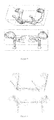
- FIG. 3 shows the wireframe model of the mushroom body.
- FIG. 4 shows the principal axes and wireframe of the same model of FIG. 3 .
- FIG. 5 shows the local coordinate system of the set of principal axes.
- FIG. 6 illustrates an example of the generated 3D sub-structures and neural networks in the fly brain.
- FIG. 7 demonstrates an average model of a fruit fly brain with some of its major sub-structures.
- FIG. 8 demonstrates the distribution of some genes within neural networks in a fly brain.
- FIG. 9 demonstrates the flow charts for determining the global and local coordinates of the present invention.
- FIG. 10 demonstrates the flow charts for two level model averaging of the present invention.
- FIG. 11 demonstrates the flow charts for registration of the standard neuropil of the present invention.
- the present invention provides cellular networks database and system for gene expression in the bio-organization, one preferred example is gene expression in fruit fly brains.
- Such an expressing system should be modular to allow expansion for multiple types of gene with different functions.
- the bio-expression system 10 of the present invention includes a computing process system 100 used to process and compute the data and information under certain instruction.
- the bio-expression system collects and presents the bio-characters.
- the system may allow to analyze and define a neural circuit involved in any scientific investigation, medical-related diagnosis or artistic exhibition.
- the high-performance computer with advance CPU could be employed to achieve the goal.
- An average model generating module 200 is embedded in the process system 100 to transfer the input two-dimension image data such as a set of individual model sections to a three-dimension image.
- one commercial product application or software AMIRA v3.1, Mercury Computer Systems Inc. USA
- the image data could be calculated by the process system 100 introducing the average model generating module 200 to generate reconstructed three-dimension stereoscopic image.
- the input data is prepared by the sample preparation system 500 and the image data generating system 400 .
- the sampling system 500 l is employed to generate the target sample for the bio expression system 10 .
- the fruit fly brain (about 600 ⁇ 250 ⁇ 150 micrometers in its adult brain) is used for illustration.
- the foregoing preferred brain organization of the fly embodiment is illustrative of the present invention rather than limiting the present invention.
- the whole tissue of fly brain can be obtained by the well-know manner, followed by increasing its degree of light transparency to about 0.15 mm or deeper with a technique invented by one of our inventors, please refer to the U.S. Pat. No. 6,472,216 B1, filed on Oct.
- Fluorescent marking or labeling molecules are implanted to label a predetermined portion of the target sample such as some neurons underlie a specific function.
- the procedure could be achieved by means of genetic engineering, as well known in the art.
- the system 400 includes a laser scanning microscope that is equipped with a plurality of laser light sources. During the procedure, the sample with label is scanned by the laser to activate fluorescent molecules. The cross-sections of the sample at different depth are scanned entirely (or partially) with the laser according to a predetermined sequence. Therefore, the scanned image data include pluralities of surface images at different depth. Images from various parts of the same cross-section can be stitched to its entirety with the help of computer software such as AMIRA. Thereafter, the generated image data are fed in to the process system 100 for subsequent process. As aforementioned, the average model generating module 200 is coupled to the process system 100 to process the input data for generating the three-dimension image or an average model.
- the generated average model or three-dimension image data could be stored in the database 600 .
- the database may include a plurality of sub-database such as bio-express sub-database 610 , cellular network sub-database 620 and bio-fine structure sub-database 630 .
- bio-express sub-database 610 a plurality of sub-database
- cellular network sub-database 620 a plurality of sub-databases 630 .
- the bio-expression sub-database 610 includes the data of bio-character such as gene (or protein) expressions.
- the database 610 also includes standard bio-geographic index sub-database, which allows the calibration and comparison between different individuals. All of the data can be classified in accordance with gene (or protein) expressions, individual growth, developmental, disease or experience dependent procedures.
- Cellular network sub-database 620 establishes a functional linkage between certain functions (or dysfunctions) and cellular entities. For example, the transient inhibition of NMDA receptors in neurons disrupts a certain type of memory formation, establishing a function link between memory and neurons identified by the Feb170 enhancer trap allele. Please refer to the article “Specific requirement of NMDA receptors for long-term memory consolidation in Drosophila ellipsoid body” in Nature Neuroscience, 2007 December;10(12):1578-86. The article is incorporated herein for reference.
- the fine structure sub-database 630 includes the data of bio-fine structures.
- FIG. 6 illustrates an example of the generated 3D fly brain incorporated herein for reference.
- the figure shows a 3D image illustrating the distribution of the neurons which express the C133 gene in a brain of a male fruit fly.
- the green color portion represents the expression of C133 gene resulted from the fluorescent marking molecule.
- the larger spots are neurons with a width at about 10 micrometers, while smallest dots are believed to be the synapses, at the size of micron range.
- the brown color portion is the mushroom body.
- the averaged standard mushroom body is constructed according to the algorithm used in the average-model module in the process system.
- FIG. 7 shows the location of neurons expressing the GH146 gene (orange) in a standard fly brain model constructed according to its mushroom body (purple). The yellow part is the optical lobe where the visual signal is collected. The blue part is called the central complex.
- FIG. 8 several cellular networks expressing different genes in one brain is illustrated, GH146 (green), tim (brown), and 201Y (purple). The tim gene is related to the biological clock in the fruit fly.
- the bio-structure image can be obtained by means of the present invention.
- the bio-network images can be stored in a storage medium.
- the present invention discloses a computer readable storage medium being available to store an image of bio-network generated by performing the following scanning a sample with label by a laser scanning microscope to activate fluorescent molecules in the sample, wherein the laser scanning microscope is equipped with a plurality of laser light sources; during the scanning procedure, at least a part of the sample is scanned by the laser light and cross-sections at different depth are scanned according to a predetermined sequence, thereby obtaining scanned image data including pluralities of surface images at different depth.
- the images from various parts of the same cross-section can be stitching to its entirety.
- the present invention further comprises the following steps before scanning the sample: preparing the sample and implanting molecules of fluorescence generating potential in the sample to label a predetermined portion of a target sample by means of genetic engineering. Then, the degree of light transparency of the sample is increased to about 0.15 mm or deeper.
- a proposed process flow is to build an average model (3D atlas) from a group of initial individual models.
- the method consists of three main steps, individual model construction and two model-averaging procedures at different levels.
- the first step is to construct 3D wireframe model for each individual dataset.
- the second step is the coarse level model averaging.
- a partition step is performed to partition each individual model into several significant parts (submodels) with a user interface. For each individual model, a set of principal axes is extracted and can be referred to as skeleton of the model.
- the individual model is fed into the process system and the system may process the partition procedure under the input instruction of the user.
- Each individual wireframe model is partition into several significant parts manually, and then the corresponding principal axis for each submodel is found by the technology of PCA disclosed by Ian T Jolliffe, “Principal Component Analysis”, Springer-Verlag, New York, 1986. By calculating eigenvalues and the corresponding eigenvectors of the following sample covariance matrix, the direction of the principal axis is determined:
- ⁇ ( t ) A+t ⁇ D, t min ⁇ t ⁇ t max
- A is a point on the principal axis, and be set to ⁇ x .
- the boundary, t min and t max can be determined by projection of all submodel vertices to the principal axis. For each individual model, a set of principal axes is extracted and can be referred to as skeleton of the model.
- Averaging procedures are carried out by calculating the average position of the middle points, the average direction and the average length of the principal axis, performed on the computer or the process system of the present invention. Consequently, the average principal axis can be represented as:
- v ⁇ ( t ) ⁇ M + t ⁇ ⁇ D , t min _ ⁇ t ⁇ t max _
- k ′ - t min ⁇ ,
- FIG. 3 shows the wireframe model of the mushroom body.
- FIG. 4 shows the principal axes and wireframe of the same model of FIG. 3 .
- FIG. 5 shows the local coordinate system of the set of principal axes.
- the next procedure after the average skeleton of original datasets being generated is to warp the stored individual skeleton dataset in three dimensions by the process system 100 of the present invention.
- the calculation performed on the process system 100 is able to warp each individual model to its corresponding pseudo-average model.
- the warping function processed by the computer is defined as:
- W k (p) is the warped position of p using a single principal axis pair.
- the weighting of the k-th principal axis of W(p) is defined as:
- l k is the length of the principal axis and its importance is adjusted by the constant c.
- d k is the distance from a point p to the principal axis.
- the constant a means the adherence of the principal axis, and the constant b can be seen as the concentration of the strength of the principal axis.
- the pseudo-average model is generated by the process system 100 based on the above model. Then, the pseudo-average models are registered according to the common average skeleton. The final average model can be obtained by determining the geometric median of these pseudo-average models, on the process system 100 .
- the triangle patches are converted into volumetric voxels by sampling.
- a 3D seed-fill algorithm is applied to convert this volumetric hollow object to a volumetric solid object.
- Each pseudo-average model will generate a volumetric solid object to indicate the volumetric voxels it possesses.
- a cumulative volume of voxel values from 1 to N is obtained with N pseudo-average models superimposed.
- the geometric median is located at where the voxel value is N/2.
- a full colored three dimensional stereo neuron graphic can be seen and manipulated with the facilities.
- several facilities are used for the data generating system 400 .
- a Zeiss LSM 510 confocal microscope is equipped with 4 laser light sources including an argon laser (emission at 364 nm), an argon-krypton laser (458, 488, or 514 nm), and two HeNe lasers (543 and 633 nm). The system allows for simultaneous detection of four fluorescence signals and a transmitted image.
- Zeiss LSM 510 META confocal two-photon microscope system is equipped with 4 laser light sources including an argon-krypton laser (458, 488, or 514 nm), two HeNe lasers (543 and 633 nm), and with a Coherent Mira femtosecond T-Sapphire laser for nonlinear optical microscopy (2-photon) which is capable of 700-1000 nm single optics set tuning. This is designed for in vivo observation of fluorescence signals in thick living tissues.
- Zeiss LSM 510 META confocal microscope is equipped with 3 laser light sources including an argon-krypton laser (458, 488, or 514 nm), and two HeNe lasers (543 and 633 nm).
- the system has 3 photomultipliers and a META detector allowing simultaneous collection of full spectrum fluorescence signals. It does not have transmitted light detector. It has an automated stage scanner for image montage and optical system for IR light.
- a stereoscopic projecting system 300 is coupled to the process system 100 .
- the process system 100 may access the database under the input instruction and send the image to a video card with multiple graphic outputs (such as NVIDIA Quadro4-980 or better).
- the CPU in the process system 100 can be a 32-bit or 64-bit (or better) unit(s), with sufficient memories for image data processing.
- the image from the multiple outputs is individually fed into multiple projectors so that a front or back projection can be implemented for stereoscopic presentation and manipulation.
- the procedure can be controlled by (but not limited to) commercially available software (such as AMIRA v.3.1) and hardware (such as a 3D mouse).
- Special glasses as known in the art should be provided for generating the virtual three dimension image. It is well-known in the art, the description is omitted.
- the module 700 also includes a module 700 for generating standard brain model and coordinate system coupled to the processing system.
- the module 700 may process the procedures to generate the standard Drosophila brain model with coordinate information.
- a deterministic reference framework is pursued for data integration and comparison.
- a shape averaging algorithm will be provided for creating the standard template including axial and non-axial structures.
- the standard Drosophila brain comprises the standard surface and the standard neuropils.
- the standard neuropils are located within the standard surface with the average position and orientations.
- a global coordinate system of the brain surface and several local coordinate systems of the specific neuropils compose the coordinate system of the Drosophila standard brain.
- the standard surface and neuropils can be obtained by performing shape-averaging algorithm to the constructed 3D surface models from individual brains.
- the neuropils can be separated into two categories, axial and non-axial.
- the axial neuropils such as the mushroom bodies, are easy to find the correspondence between each other through the axial structures.
- the model-averaging of the axial neuropils can be performed by employing the axial features.
- the non-axial neuropils such as the optical lobes and the brain surface, the transformation relationship between them can only be found by a surface registration process.
- a global coordinate is defined in the initial step.
- the global coordinate may define entire circuits for the whole brain.
- the original of the global coordinates is also defined within the step.
- a local coordinate is defined to stand for each sub-element, for example, neuropils.
- the distance and the orientation of the local coordinate with respect to the global coordinate can be determined.
- the global and local characteristics of the models such as orientations, positions, sizes, and angles between axial structures, are determined. Therefore, each location of the neuron is defined for subsequent observation.
- the technique to construct the standard model is described as follow.
- a two-level model averaging technique is introduced for the construction of the standard template.
- the two level hierarchy of the algorithm is shown in FIG. 10 .
- the individual 3D models are first processed with a coarse-level model averaging procedure.
- we average the global characteristics of the models such as orientations, positions, sizes, and angles between axial structures.
- two individual models i.e. individual model 1 and individual model 2 are input to the system and the input data are processed by coarse-level model averaging procedure.
- a reference model must be created from the individual models first.
- the candidate of the reference model is chosen to be the individual model which has the volume size closest to the average volume size. After scaling the candidate model to the average volume size, we can obtain the reference model.
- a 3D distance field is generated from the reference model to facilitate the registration procedure. It records the distance from each voxel to its nearest surface voxel on the reference model.
- the position vector is denoted as X, and DF (x) indicates the 3D distance field.
- An affine transformation mapping from position x to position y is defined as
- the set of transform parameters is the solution which minimize s the following objective function
- each individual model For axial structures, we partition each individual model into several significant parts with a user interface, and compute principal axes of these parts by the technique of principal component analysis. For each individual model, a set of principal axes is extracted and can be referred to as the skeleton of the model. By taking the skeletons of all models into account, we can compute an average skeleton. The pseudo-average models of the individual models are generated after applying 3D field based warping to all individual models from original skeletons to the average skeleton.
- each individual model is transformed to a corresponding pseudo-average model.
- the final standard model can be obtained by determining the shape average of the pseudo-average models.
- Each pseudo-average model can create a signed distance field that records the distance from each voxel to its nearest voxel on the surface of the pseudo-average model. The value is set to positive if the voxel is inside the pseudo-average model, and vise versa.
- the surface of the final standard model can be extracted by zero-crossing detection of the cumulateive distance field.
- the further aspect of the present invention is to create the standard brain model by the module 700 as shown in FIG. 1 .
- One of the examples is to create the Drosophila brain model.
- the standard brain comprises the standard surface and the standard neuropils.
- the standard neuropils are located within the standard surface with the average position and orientation.
- the flowchart for registration of the standard neuropil is depicted in FIG. 11 .
- the individual datasets needed consist of a surface model and their corresponding neuropil models.
- the individual surface of an individual dataset is first affine registered to the standard surface.
- the standard neuropil is then rigidly registered to the transformed individual neuropil.
- the resulting transformation can be regarded as a suggestion about the position and orientation of the standard neuropil within the standard surface.
- the position and orientation in the standard surface can be calculated by averaging the suggestions from the individual datasets.
- the average parameter of the translation can be obtained by
- T i is the parameter of translation suggested by the ith individual dataset.
- Y is the corresponding point of x
- m is the number of points in both sets.
- R i is the parameter of rotation suggested by the ith individual dataset.
- the Drosophila standard brain model can be obtained after all the standard neuropils are processed and located into the standard surface.
- the global coordinate system of the Drosophila brain surface is constructed based on two features: the principal axis of the whole brain surface and the esophagus, which nearly cross the brain surface horizontally.
- the surface of the esophagus is first constructed, with the same algorithm for the brain surface, and the center of the esophagus is then determined as origin of the whole brain;
- the z-axis (anterior positive) is the major principal axis of the esophagus;
- the y-axis is along the line (ventral positive) perpendicular to the z-axis and the major principal axis of the brain at the esophageal center.
- the x-axis is at the intersection of the two axes and normal to the y-z plane (right positive).
- For the local coordinate system of the specific neuropil we have constructed the coordinate system of the mushroom bodies. The local coordinate system is constructed based on the six lobes of the mushroom bodies. We can obtain the global and local coordinate system of the standard Drosophila brain by introducing the algorithms to the standard brain surface and standard neuropils.
- the individual dataset contains two channels of image stack, one for the brain surface and the other for the mushroom bodies.
- Each image stack consists of about 60 image slices acquired by the confocal microscopy.
- the present invention develops procedures for generating the standard Drosophila brain with coordinate information.
- the standard Drosophila brain comprises the standard surface and the standard neuropils.
- the present invention proposes a shape-averaging algorithm to create the axial and non-axial templates. Afterwards, an algorithm is proposed to determine the average positions and orientations of each neuropils within the standard surface.
- the global and local coordinate systems are constructed for the standard Drosophila brain.
- the present invention can generate the 3D virtual Drosophila brain after applying the proposed system with a large amount of individual 3D neuronal image.
- the standard Drosophila brain with coordinate information enables the integration of the whole brain circuits. Finally, the information about the spatial and temporal relationship among neurons can be achieved.
Landscapes
- Physics & Mathematics (AREA)
- Engineering & Computer Science (AREA)
- General Physics & Mathematics (AREA)
- Zoology (AREA)
- Mathematical Optimization (AREA)
- General Health & Medical Sciences (AREA)
- Algebra (AREA)
- Computational Mathematics (AREA)
- Animal Behavior & Ethology (AREA)
- Mathematical Analysis (AREA)
- Health & Medical Sciences (AREA)
- Mathematical Physics (AREA)
- Pure & Applied Mathematics (AREA)
- Business, Economics & Management (AREA)
- Life Sciences & Earth Sciences (AREA)
- Educational Administration (AREA)
- Educational Technology (AREA)
- Theoretical Computer Science (AREA)
- Image Processing (AREA)
Abstract
Description
ν(t)=A+t·D, t min ≦t≦t max
ν(t)=M+t·D, t′ min ≦t≦t′ max
x1=(1,0,0)′
x2=(0,1,0)′
x3=(0,0,1)′
Claims (10)
Priority Applications (2)
| Application Number | Priority Date | Filing Date | Title |
|---|---|---|---|
| US12/222,813 US8315809B2 (en) | 2005-06-30 | 2008-08-18 | Bio-expression system with module for creating the standard Drosophila brain model and its coordinate system |
| TW098113807A TWI381283B (en) | 2008-08-18 | 2009-04-24 | Bio-expression system with module for creating the standard drosophila brain model and its coordinate system |
Applications Claiming Priority (2)
| Application Number | Priority Date | Filing Date | Title |
|---|---|---|---|
| US11/169,890 US7742878B2 (en) | 2005-06-30 | 2005-06-30 | Bio-expression system and the method of the same |
| US12/222,813 US8315809B2 (en) | 2005-06-30 | 2008-08-18 | Bio-expression system with module for creating the standard Drosophila brain model and its coordinate system |
Related Parent Applications (1)
| Application Number | Title | Priority Date | Filing Date |
|---|---|---|---|
| US11/169,890 Continuation-In-Part US7742878B2 (en) | 2005-06-30 | 2005-06-30 | Bio-expression system and the method of the same |
Publications (2)
| Publication Number | Publication Date |
|---|---|
| US20090063119A1 US20090063119A1 (en) | 2009-03-05 |
| US8315809B2 true US8315809B2 (en) | 2012-11-20 |
Family
ID=40408818
Family Applications (1)
| Application Number | Title | Priority Date | Filing Date |
|---|---|---|---|
| US12/222,813 Active 2027-08-01 US8315809B2 (en) | 2005-06-30 | 2008-08-18 | Bio-expression system with module for creating the standard Drosophila brain model and its coordinate system |
Country Status (1)
| Country | Link |
|---|---|
| US (1) | US8315809B2 (en) |
Families Citing this family (2)
| Publication number | Priority date | Publication date | Assignee | Title |
|---|---|---|---|---|
| WO2014020428A2 (en) * | 2012-08-02 | 2014-02-06 | MAX-PLANCK-Gesellschaft zur Förderung der Wissenschaften e.V. | Method and computing system for modelling a primate brain |
| US9558568B2 (en) * | 2014-06-27 | 2017-01-31 | Siemens Healthcare Gmbh | Visualization method for a human skeleton from a medical scan |
Citations (1)
| Publication number | Priority date | Publication date | Assignee | Title |
|---|---|---|---|---|
| US6381562B2 (en) * | 1998-07-13 | 2002-04-30 | John A. Keane | Configurable bio-transport system simulator |
-
2008
- 2008-08-18 US US12/222,813 patent/US8315809B2/en active Active
Patent Citations (1)
| Publication number | Priority date | Publication date | Assignee | Title |
|---|---|---|---|---|
| US6381562B2 (en) * | 1998-07-13 | 2002-04-30 | John A. Keane | Configurable bio-transport system simulator |
Also Published As
| Publication number | Publication date |
|---|---|
| US20090063119A1 (en) | 2009-03-05 |
Similar Documents
| Publication | Publication Date | Title |
|---|---|---|
| CN114693933B (en) | Medical image segmentation device based on generative adversarial network and multi-scale feature fusion | |
| Guibas et al. | Synthetic medical images from dual generative adversarial networks | |
| MacLeod | Morphometrics: History, development methods and prospects | |
| Ziegler et al. | Accelerated acquisition, visualization, and analysis of zoo-anatomical data | |
| Mamdouh et al. | A New Model for Image Segmentation Based on Deep Learning. | |
| Qu et al. | 3-D registration of biological images and models: registration of microscopic images and its uses in segmentation and annotation | |
| Nattkemper | Multivariate image analysis in biomedicine | |
| US8315809B2 (en) | Bio-expression system with module for creating the standard Drosophila brain model and its coordinate system | |
| CN114494132B (en) | Disease classification system based on deep learning and spatial statistical analysis of fiber tracts | |
| Setiawardani et al. | The role of artificial intelligence in 3D development–facial reconstruction of skull bones as a forensic investigation solution: A comprehensive review | |
| US7742878B2 (en) | Bio-expression system and the method of the same | |
| TWI291630B (en) | Bio-expression system and the method of the same | |
| Tawfeeq et al. | Predication of Most Significant Features in Medical Image by Utilized CNN and Heatmap. | |
| Pereanu et al. | Digital three-dimensional models of Drosophila development | |
| TWI381283B (en) | Bio-expression system with module for creating the standard drosophila brain model and its coordinate system | |
| Song et al. | Abdominal multi-organ segmentation using multi-scale and context-aware neural networks | |
| Teo et al. | An automated 3D modeling pipeline for constructing 3D models of MONOGENEAN HARDPART using machine learning techniques | |
| Sundaresan et al. | Insight Into Various Algorithms For Medical Image Analyzes Using Convolutional Neural Networks (Deep Learning) | |
| Muniz et al. | Generative design applied to cloud modeling | |
| Borno et al. | Percept-diff: Innovations in stable diffusion for high-fidelity ihc image generation in her2 breast cancer incorporating perceptual loss | |
| Reinitz et al. | Three-dimensional visualization of the distribution of melanin-concentrating hormone producing neurons in the mouse hypothalamus | |
| Wiet et al. | Integration of high-resolution data for temporal bone surgical simulations | |
| Yousefi | Multi-Objective Optimisation of Growth-Mechanics Problem in the Developing Mammalian Heart | |
| Rasti | Contributions to Deep Learning for Life Science Applications | |
| Bagga et al. | Computer-assisted anthropology |
Legal Events
| Date | Code | Title | Description |
|---|---|---|---|
| AS | Assignment |
Owner name: NATIONAL TSING HUA UNIVERSITY, TAIWAN Free format text: ASSIGNMENT OF ASSIGNORS INTEREST;ASSIGNORS:CHIANG, ANN-SHYN;CHEN, YUNG-CHANG;CHANG, HSIU-MING;REEL/FRAME:021472/0845;SIGNING DATES FROM 20080731 TO 20080808 Owner name: NATIONAL TSING HUA UNIVERSITY, TAIWAN Free format text: ASSIGNMENT OF ASSIGNORS INTEREST;ASSIGNORS:CHIANG, ANN-SHYN;CHEN, YUNG-CHANG;CHANG, HSIU-MING;SIGNING DATES FROM 20080731 TO 20080808;REEL/FRAME:021472/0845 |
|
| STCF | Information on status: patent grant |
Free format text: PATENTED CASE |
|
| FPAY | Fee payment |
Year of fee payment: 4 |
|
| MAFP | Maintenance fee payment |
Free format text: PAYMENT OF MAINTENANCE FEE, 8TH YR, SMALL ENTITY (ORIGINAL EVENT CODE: M2552); ENTITY STATUS OF PATENT OWNER: SMALL ENTITY Year of fee payment: 8 |
|
| MAFP | Maintenance fee payment |
Free format text: PAYMENT OF MAINTENANCE FEE, 12TH YR, SMALL ENTITY (ORIGINAL EVENT CODE: M2553); ENTITY STATUS OF PATENT OWNER: SMALL ENTITY Year of fee payment: 12 |