CROSS-REFERENCE TO RELATED APPLICATIONS
The present application is a divisional of U.S. Ser. No. 10/531,543, now abandoned, filed Oct. 28, 2005, which is a National Stage Filing of, and claims priority to, PCT Patent Application No. PCT/KR/2004/002190, filed Aug. 31, 2004. U.S. Ser. No. 10/531,543 and PCT/KR/2004/002190 are incorporated herein by reference in their entireties.
BACKGROUND OF THE INVENTION
1. Field of the Invention
The present invention relates, in general, to a composition for treating a viral infection comprising Jab1. More particularly, the present invention relates to a composition for treating or preventing a viral infection comprising a Jab1 (Jun-activation binding protein 1) protein, a nucleic acid having a nucleotide sequence coding for a Jab1 protein or a recombinant virus expressing a Jab1 protein.
2. Description of the Related Art
Flavivirus and pestivirus belong to the Flaviviridae family which possesses a single-stranded positive sense RNA genome and causes various diseases in vertebrate hosts. West Nile virus (WNV) (Burt et al., Emerg Infect Dis., 8(8):820-826, 2002; Asnis et al., Clin Imfect Dis 30(3): 413-418, 2000) causes diseases including fever, rash, arthralgia and myalgia when infecting susceptible hosts. Apoptosis in wild-type WNV-infected brain cells is induced in a Bax-dependent manner (Parquet et al., FEBS Lett., 500(1-2):17-24. 2001), and the apoptosis is induced by the capsid protein of WNV through the mitochondrial/caspase-9 pathway (Yang et al., Emerg Infect Dis., 8(12):1379-1384, 2002). However, the intracellular pathological mechanism of West Nile virus infection has not been completely understood.
Immunoglobulins and antiviral agents such as interferon alpha-2b and ribavirin were conventionally used for preventing and treating West Nile virus infection (Agrawal and Petersen., J Infect Dis, 188(1):1-4, 2003; Morrey et al., Antiviral Res., 55(1):107-116, 2002; Anderson et al., Emerg Infect Dis., 8(1):107-108, 2002), but they have low therapeutic effects. At present, there is no effective drug for treating or preventing West Nile virus infection. Thus, there is a need for the development of such effective drugs.
On the other hand, Jab1 (Jun-activation binding protein 1) was initially known as a coactivator of AP-1 (Jun/Fos proto-oncogene) protein and has the following, various functions. Jab1 is a component (CSN5) of the COP9 signalosome (CSN) (Wei et al., Annu Rev Cell Dev Biol., 19:261-286, 2003), and Jab1/CSN5 exists in a wide spectrum of organisms, ranging from yeasts to plants and animals. Overexpression of Jab1 causes the translocation of cyclin dependent kinase inhibitor p27/Kip1 from the nucleus to the cytoplasm, accelerates the Ub-26S proteasome-dependent degradation, and participates in the G1-S transition of the cell cycle, mediated by p27/Kip1 (Tomoda et al., Nature, 398(6723):160-165, 1999). In addition, Jab1 involves the nuclear translocation of PGP9.5 that is overexpressed in primary lung cancer cells (Caballero et al., Oncogene, 21(19):3003-3010, 2002). Jab1 interacts with p53, Smad4 and lutropin/choriogonadotropin receptor and stimulates degradation of these proteins (Bech-Otschir et al., EMBO J., 20(6):1630-1639, 2001; Li et al. J Biol Chem., 275(18):13386-13393, 2000; Wan et al., EMBO J., 3(2):171-176, 2002). Taken together, Jab1 translocates proteins from the nucleus to the cytoplasm by interaction with intracellular proteins and thus stimulates protein degradation in a proteasome-dependent manner.
However, there is no report for interaction between Jab1 and viral proteins upon flavivirus infection.
Based on this background, the present inventors identified Jab1 as a protein interacting with the capsid protein of flavivirus, and found that Jab1 inhibits apotosis by accelerating degradation of the capsid protein and that Jab1 is useful for treating or preventing a viral infection, thereby leading to the present invention.
SUMMARY OF THE INVENTION
It is therefore an object of the present invention to provide a composition for treating or preventing a flavivirus or pestivirus infection, which comprises a Jab1 (Jun-activation binding protein 1) protein.
It is another object of the present invention to provide a composition for treating or preventing a flavivirus or pestivirus infection, which comprises a nucleic acid having a nucleotide sequence coding for a Jab1 protein.
It is a further object of the present invention to provide a composition for treating or preventing a flavivirus or pestivirus infection, which comprises a recombinant virus expressing a Jab1 protein.
It is yet another object of the present invention to provide a method of treating or preventing a flavivirus or pestivirus infection, which is based on administering a pharmaceutically effective amount of a Jab1 protein to a subject requiring treatment or prevention of a viral infection.
It is still another object of the present invention to provide a method of treating or preventing a flavivirus or pestivirus infection, which is based on administering a pharmaceutically effective amount of a nucleic acid having a nucleotide sequence coding for a Jab1 protein to a subject requiring treatment or prevention of a viral infection.
It is still another object of the present invention to provide a method of treating or preventing a flavivirus or pestivirus infection, which is based on administering a pharmaceutically effective amount of a recombinant virus expressing a Jab1 protein to a subject requiring treatment or prevention of a viral infection.
It is still another object of the present invention to provide a method of assaying a substance stimulating expression of a Jab1 protein.
BRIEF DESCRIPTION OF THE DRAWINGS
The above and other objects, features and other advantages of the present invention will be more clearly understood from the following detailed description taken in conjunction with the accompanying drawings, in which:
FIG. 1 shows the results of immunofluorescence analysis for expression patterns of the capsid (Cp) protein of West Nile virus (WNV) in three tumor cell lines;
FIG. 2 shows the results of immunofluorescence analysis for expression patterns of WNV-Cp in SK-N-SH cells;
FIG. 3 shows the results of an annexin assay, displaying apoptosis induction by WNV-Cp in two tumor cell lines;
FIG. 4 shows the results of FACS analysis, displaying apoptosis induction by WNV-Cp in 293T cells;
FIG. 5 shows the procedure of a yeast two hybrid assay resulting in obtainment of clones expressing proteins interacting with WNV-Cp;
FIG. 6 shows the results of immunofluorescence analysis, revealing that Jab1 translocates WNV-Cp from the nucleolus to the cytoplasm;
FIG. 7 shows the results of immunoprecipitation, revealing that Jab1 interacts with WNV-Cp;
FIG. 8 shows that co-expression of Jab 1 and WNV-Cp leads to a decrease in caspase activity, wherein FIG. 8A shows a graph for the results of measuring caspase activity to each protein, and FIG. 8B refers to the western blotting results for the expression of protein;
FIG. 9 shows the results of Western blotting, displaying that degradation of WNV-Cp by Jab1 is remarkably suppressed in the presence of a 26S proteasome inhibitor LLnL;
FIG. 10 shows the results of FACS analysis, displaying an apoptosis rate of normal cells not transfected with a C2-Cp gene;
FIG. 11 shows the results of FACS analysis, displaying an apoptosis rate of cells transfected with a pEGFP-C2-Cp plasmid;
FIG. 12 shows the results of FACS analysis, displaying an apoptosis rate of cells transfected with a pEGFP-N1 plasmid;
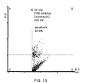
FIG. 13 shows the results of FACS analysis, displaying an apoptosis rate of cells transfected with a pEGFP-C2-Cp plasmid and treated with 200 nM of a PI3K inhibitor;
FIG. 14 shows the results of FACS analysis, displaying an apoptosis rate of cells transfected with a pEGFP-C2-Cp plasmid and treated with 5 μM of an Akt inhibitor;
FIG. 15 shows the results of FACS analysis, displaying an apoptosis rate of cells transfected with a pEGFP-C2-Cp plasmid and treated with 50 μM of an Akt inhibitor;
FIG. 16 shows the results of Western blotting, displaying that p53 expression decreases with increasing concentrations of Jab1;
FIG. 17 shows the determining of an expression of a vector for establishing a Jab1 adenovirus stable cell line, wherein FIG. 17A refers to the results for western blotting for pShuttle-IRES-hrGFP and Jab1, FIG. 17B shows the data for pShuttle-Jab1 and pShuttle-Cp. And, FIG. 17C represents western blotting data for Jab1 isolated by Pad enzyme after transformation; and
FIG. 18 shows the results of Western blotting, demonstrating that a NIH3T3 Jab1 retrovirus stable cell line is successfully established.
DETAILED DESCRIPTION OF THE INVENTION
In one aspect, the present invention provides a composition for treating or preventing a flavivirus or pestivirus infection comprising a Jab1 protein.
Viral infections and associated diseases intended to be treated or prevented according to the present invention are flavivirus and pestivirus infections. Flavivirus and pestivirus according to the classification of International Committee on Taxonomy of viruses, belong to the Flaviviridae family, which possesses a positive-stranded single strand RNA genome and has a natural host range including vertebrates and arthropods. Flavivirus and pestivirus virions consist of an envelope and a nucleocapsid. Flavivirus virions are spherical and 40-50 nm in diameter, and pestivirus virions are spherical to pleomorphic and 40-60 nm in diameter. Flavivirus and pestivirus have a very similar structure and infection mechanism and induce apoptosis of infected cells.
Flavivirus includes the mammalian tick-borne virus group, seabird tick-borne virus group, Aroa virus group, Dengue virus group, Japanese encephalitis virus group, Ntaya virus group, Kokobera virus group, Spondweni virus group, Yellow fever virus group, Entebbe virus group, Modac virus group and Rio Bravo virus group. The present composition may be preferably applied to an infection with the Japanese encephalitis virus group. The Japanese encephalitis virus group includes Cacipacore virus, koutango virus, Japanese encephalitis virus, Murray Valley encephalitis virus, St. Louis encephalitis virus, Usutu virus, West Nile virus, and Yaounde virus.
Pestivirus includes Border disease virus, bovine viral diarrhea virus 1, Bovine viral diarrhea virus 2, and Classical swine fever virus.
The present inventors, via a yeast two hybrid assay, found that Jab1 is a protein directly interacting with the capsid (Cp) protein of West Nile virus, which induces apoptosis in WNV-infected cells, and investigated the effect of Jab1 on the capsid protein. As a result, Jab1 was found to directly interact with the capsid protein, translocate the capsid protein from the nucleus to the cytoplasm and stimulate degradation of the capsid protein, thereby remarkably inhibiting apoptosis mediated by the viral capsid protein.
The Capsid denotes the protein shell that encloses the viral nucleic acid and is formed by multiple copies of a single major structural subunit protein. The structural subunit protein forming the capsid is called the capsid protein. With respect to the objects of the present invention, the capsid protein is the flavivirus or pestivirus capsid protein to which the Jab1 protein binds. The complete genome sequence of West Nile virus including the nucleic acid sequence of the capsid protein of West Nile virus, a member of flavivirus, is available from GenBank under accession numbers AF206518, AF196835, AF202541 and M12294. The nucleic acid sequences of capsid proteins of other members of flavivirus and pestivirus are also available from GenBank, for example, for JEV, under accession numbers M18370, D90194 and D90195; for SLEV, under accession number M16614; for YFV, under accession numbers AF094612, U17067, U17066, U54798, U21055, U21056 and X03700; for DENV, accession numbers M23027, U88535, U88536 and U88537); and for BVDV, accession number M31182.
The homology of the capsid protein between flavivirus and pestivirus, which possess the capsid protein capable of binding the Jab1 protein, is about 90%.
The Jab1 protein used in the present composition includes all Jab1 proteins derived from yeasts, plants and animals, which include a wild-type Jab1 protein and, as long as the function of binding to the flavivirus or pestivirus capsid protein and stimulating degradation of the capsid protein is retained, variants of the Jab1 protein made by deletions, insertions, non-conserveative or conservative substitutions, or combinations thereof. In one embodiment, the Jab1 protein may have an amino acid sequence designated as SEQ ID No. 2, and substitution, insertion and deletion variants of this amino acid sequence may be useful in the present composition.
The variant of Jab1 means the protein that has a sequence in which one or more amino acid residues differ from a wild-type amino acid sequence. An insertion is typically made by the addition of a consecutive amino acid sequence of about 1 to 20 amino acids, or may be made with a longer sequence. A deletion is typically in the range of about 1 to 30 amino acid residues, or may be in part made in a longer sequence such as the absence of one domain. Such variants may be prepared by a chemical peptide synthesis method or a DNA sequence-based recombinant method, which are known in the art (Sambrook et al., Molecular Cloning, Cold Spring Harbour Laboratory Press, New York, USA, 2d Ed., 1989). Amino acid exchanges in proteins and peptides which do not generally alter the activity of the protein or peptide are known in the art (H. Neurath, R. L. Hill, The Proteins, Academic Press, New York, 1979). The most commonly occurring exchanges are Ala/Ser, Val/Ile, Asp/Glu, Thr/Ser, Ala/Gly, Ala/Thr, Ser/Asn, Ala/Val, Ser/Gly, Thy/Phe, Ala/Pro, Lys/Arg, Asp/Asn, Leu/Ile, Leu/Val, Ala/Glu and Asp/Gly, in both directions.
In addition, the Jab1 protein, if desired, may be modified by phosphorylation, sulfation, acrylation, glycosylation, methylation, farnesylation, and the like.
The variant or modified product may have the biological activity functionally identical to its natural form, or, if desired, may be made by altering the property of the natural form. It is preferably a protein that is improved in enhanced structural stability against heat, pH, etc., and protein activity by alteration and modification of its amino acid sequence.
The Jab1 protein may be obtained by extraction and purification from nature according to a method well known in the art (Merrifleld, J. Amer. chem. Soc. 85:2149-2156, 1963), or may be obtained using a genetic recombination technique.
When the protein is prepared by chemical synthesis, a polypeptide synthesis method well known in the art may be used.
In the case of using the genetic recombination technique, the Jab1 protein may be obtained by a process including inserting a nucleic acid coding for Jab1 into a suitable expression vector, transforming a host cell with the vector, cultivating the host cell to allow Jab1 to express and recovering expressed Jab1 from the cultured host cell.
As the expression vector for expressing the Jab1 protein, all common expression vectors may be used. Since expression levels and modification of proteins differ according to host cells, the most suitable host cell may be selected according to the intended use. Available host cells include, but are not limited to, prokaryotic cells such as Escherichia coli, Bacillus subtilis, Streptomyces, Pseudomonas, Proteus mirabilis or Staphylococcus. Among them, E. coli is most commonly used. In addition, useful as host cells are lower eukaryotic cells, such as fungi (e.g., Aspergillus) and yeasts (e.g., Pichia pastoris, Saccharomyces cerevisiae, Schizosaccharomyces, Neurospora crassa), insect cells, plant cells, and cells derived from higher eukaryotes including mammals.
After a protein is expressed in a selected host cell, it may be isolated and purified by a general biochemical isolation technique, for example, treatment with a protein precipitating agent (salting out), centrifugation, ultrasonic disruption, ultrafiltration, dialysis, and various chromatographies, such as molecular sieve chromatography (gel filtration), adsorption chromatography, ion exchange chromatography and affinity chromatography. Typically, these techniques are used in combinations of two or more to obtain highly pure isolation of a protein (Maniatis et al., Molecular Cloning: A Laboratory Manual, Cold Spring Harbor Laboratory, Cold Spring Harbor, N.Y. (1982); Sambrook et al., Molecular Cloning: A Laboratory Manual, 2d Ed., Cold Spring Harbor Laboratory Press (1989); Deutscher, M., Guide to Protein Purification Methods Enzymology, vol. 182. Academic Press. Inc., San Diego, Calif. (1990)).
In another aspect, the present invention provides a composition for treating or preventing a flavivirus or pestivirus infection comprising a nucleic acid having a nucleotide sequence coding for a Jab1 protein.
The Jab1-encoding nucleotide sequence in the present composition, which encodes the Jab1 protein in the form of a wild type or a variant as described above, may be altered by substitutions, deletions, insertions or combinations thereof of one or more bases, and may be naturally occurring or chemically synthesized.
The chemical synthesis of the Jab1-encoding nucleotide sequence may be carried out by a synthesis method well known in the art, for example, as described in the literature: Engels and Uhlmann, Angew Chem IntEd Engl., 37:73-127, 1988. Examples of the synthesis method include triester, phosphite, phosphoramidate and H-phosphate methods, PCR and other autoprimer methods, and oligonucleotide synthesis methods on solid phase supports.
In an embodiment, the Jab1-encoding nucleotide sequence is exemplified as a nucleotide sequence encoding the amino acid sequence of SEQ ID No. 2, preferably a nucleotide sequence designated as SEQ ID No. 1.
A nucleic acid having the aforementioned nucleotide sequence may be single-stranded or double-stranded, and may be DNA (genome, cDNA or synthetic) or RNA molecules.
In a preferred aspect, the Jab1-encoding nucleotide sequence is operably linked to a vector to provide a recombinant expression vector expressing the nucleotide sequence.
The term “vector”, as used herein, means a vehicle for introducing a nucleic acid sequence (e.g., DNA, RNA, etc.) coding for a target gene into a host cell. Also, the term “expression vector”, as used herein, which is a vector capable of expressing a target protein or target RNA in a suitable host cell, refers to a genetic construct that comprises essential regulatory elements to which a gene insert is operably linked thereto in such a manner as to be expressed in a host cell.
The term “operably linked”, as used herein, refers to a functional linkage between a nucleic acid expression control sequence (such as a promoter) and a second nucleic acid sequence coding for a target protein or RNA in a manner that allows general functions. For example, when a nucleic acid sequence coding for a protein or RNA is operably linked to a promoter, the promoter may affect the expression of a coding sequence. The operable linkage to a recombinant vector may be prepared using a genetic recombinant technique well known in the art, and site-specific DNA cleavage and ligation may be carried out using enzymes generally known in the art.
The vector useful in the present invention includes plasmid vectors, cosmid vectors and viral vectors. A suitable expression vector includes expression regulatory elements, such as a promoter, an operator, an initiation codon, a stop codon, a polyadenylation signal and an enhancer, and a signal sequence or leader sequence for membrane targeting or secretion, and may be prepared in various constructs according to the intended use. The initiation and stop codons are generally considered to be a portion of a nucleotide sequence encoding an immunogenic target protein. Also, the initiation and stop codons are necessary to be functional in an individual to whom a genetic construct has been administered, and must be in frame with the coding sequence. The promoter of the vector may be constitutive or inducible. Also, the expression vector includes a selectable marker for selecting a host cell containing a vector, and, in the case of being replicable, includes a replication origin. The vector may be self-replicated or integrated into host DNA.
In a more preferred aspect, the present invention provides a composition comprising a recombinant viral vector carrying a nucleotide sequence coding for a Jab1 protein.
The term “recombinant viral vector”, as used herein, typically denotes a viral vector that contains one or more exogenous genes, and, in the present invention, means a viral vector carrying a Jab1 gene. The viral vector is preferably a replication-defective vector that lacks a replicon.
Non-limiting examples of the recombinant vector include retrovirus, which is exemplified by HIV (Human Immunodeficiency Virus) MLV (Murine Leukemia Virus), ASLV (Avian Sarcoma/Leukosis Virus), SNV (Spleen Necrosis Virus), RSV (Rous Sarcoma Virus) and MMTV (Mouse mammary tumor virus), and recombinant viral vectors, which are exemplified by adenovirus, adeno-associated virus and herpes simplex virus.
The nucleic acid having a nucleotide sequence coding for a Jab1 protein may be delivered into target cells of a patient for treating or preventing a viral infection by a method known in the art, for example, direct injection of a vector in naked DNA form (Wolff et al., Science, 247:1465-8, 1990: Wolff et al., J Cell Sci. 103:1249-59, 1992), or using liposomes, cationic polymers, and the like. Liposomes are phospholipid membranes made by mixing cationic phospholipids such as DOTMA or DOTAP for gene delivery. When cationic liposomes are mixed with anionic nucleic acids in a predetermined ratio, nucleic acid-liposome complexes are formed. These complexes are internalized into cells by endocytosis and stay in the endosome (Schaefer-Ridder M et al., Science. 215(4529):166-168, 1982; Hodgson et al., Nat Biotechnol., 14(3):339-342, 1996). Release of an internalized gene from the endosome into the cytoplasm and transport of the endosomally released gene from the cytoplasm to the nucleus determine the efficiency for gene transfer and therapy. This gene transfer allows repeated administration and ensures high safety due to low immunogenicity, but has a disadvantage of providing low efficiency in gene expression. Cationic polymers used in gene transport include poly-L-lysine, spermine, polyethylenimine (PEI) and chitosan (Hashida, Br J Cancer., 90(6):1252-1258, 2004; Wiseman, Gene Ther., 10(19):1654-1662, 2003; Koping-Hoggard, Gene Ther., 8(14):1108-1121, 2001). When a gene is administered into the body in a complex form with a cationic polymer, in vivo detention time and expression duration of the gene remarkably increased in comparison with the case of being administered in naked DNA form.
In a further aspect, the present invention provides a composition for treating or preventing a flavivirus or pestivirus infection comprising a recombinant virus expressing a Jab1 protein.
Since the infection of cells of a patient with infective viral particles manipulated to express Jab1 in infected cells results in an increase in the expression efficiency of Jab1, this method provides a highly therapeutic effect.
Non-limiting examples of recombinant viruses useful in the present composition comprising a recombinant virus include retroviruses, adenoviruses, adeno-associated viruses and herpes simplex virus. Preferred are retroviruses and adenoviruses, and more preferred are adenoviruses.
Retroviruses have an advantage of providing long-lasting gene expression because they are irreversibly fused to the host chromosome. Adenoviruses, which are the most frequently used system in general gene therapy studies, are applicable to a wide spectrum of mammalian cells. Adeno-associated viruses have advantages of having a broad range of host cells where a therapeutic gene is delivered, fewer side effects on the immune system upon repeated administration and a long duration of gene expression. Herpes simplex virus is a highly neurotropic virus, which infects neural cells where its genome remains as a stable episomal element within the nucleus of neural cells without disturbing normal function of neural cells. When a replication-deficient herpes simplex virus was used for gene delivery, expression of a reporter gene in the nervous system was found to be sustained for a period of over one year.
In yet another aspect, the present invention provides a method of treating or preventing a flavivirus or pestivirus infection, which is base on administering, to a subject requiring treatment or prevention of a viral infection, a pharmaceutically effective amount of a Jab1 protein, a nucleic acid having a nucleotide sequence coding for a Jab1 protein or a recombinant virus expressing a Jab1 protein.
The Jab1 protein, nucleic acid having a nucleotide sequence coding for a Jab1 protein or recombinant virus expressing a Jab1 protein, used in the treatment method of the present invention, is the same as described above.
The treatment method of the present invention is provided for preventing or treating a viral infection in vertebrates, which includes mammals such as humans and livestock.
The pharmaceutical composition of the present invention, comprising the aforementioned Jab1 protein, nucleic acid having a nucleotide sequence coding for a Jab1 protein or recombinant virus expressing a Jab1 protein, is used for treating or preventing an infection of a virus belonging to the Flaviviridae family, preferably flavivirus or pestivirus. In particular, the present composition may be preferably used for treating or preventing a flavivirus infection. The aforementioned viruses are known to cause fever, rash, bleeding, jaundice, arthralgia, myalgia, encephalitis and meningitis (Watt et al., Am J Trop Med Hyg., 68(6):704-706, 2003; Anninger et al., Clin Infect Dis., 38(7):55-56, 2004). The pharmaceutical composition of the present invention may be used for suppressing or treating the incidence of the aforementioned diseases.
The present composition may include a pharmaceutically acceptable carrier. Examples of the pharmaceutically acceptable carrier may include, for oral administration, binders, lubricants, disintegrators, excipients, solubilizing agents, dispersing agents, stabilizing agents, suspending agents, pigments and aromatics; for injectable preparations, buffering agents, preservatives, analgesics, solubilizing agents, tonic adjusting agents and stabilizing agents; and for topical administration, bases, excipients, lubricants and preservatives. The pharmaceutical composition of the present invention may be formulated into a variety of dosage forms in combination with the aforementioned pharmaceutically acceptable carrier. For example, for oral administration, the pharmaceutical composition may be formulated into tablets, troches, capsules, elixirs, suspensions, syrups or wafers. For injectable preparations, the pharmaceutical composition may be formulated into a unit dosage form, such as a multidose container or an ampoule as a single-dose dosage form.
The pharmaceutical composition of the present invention may be administered via any of the common routes, if it is able to reach a desired tissue. Therefore, the present composition may be administered topically, orally, parenterally, intranasally, intravenously, intramuscularly, subcutaneously, intraocularly and intradermally, and may be formulated into solutions, suspensions, tablets, pills, capsules and sustained release preparations. Injectable preparations are preferred. Injection may be carried out subcutaneously, intramuscularly and intravenously.
The present composition may be administered in a therapeutically or preventively effective amount. The dosage may vary according to the patient's age and sex, type and severity of the illness, administration routes, target cells and expression levels, and may be easily determined by an expert in the art.
In still another aspect, the present invention relates to a method of screening a compound stimulating expression of a Jab1 protein, comprising: (a) culturing a cell expressing the Jab1 protein; (b) contacting the cell cultured at (a) with candidate compounds for stimulating expression of the Jab1 protein; (c) comparing an expression level of the Jab1 protein at (b) with that in a control not contacted with the candidate compounds; and (d) identifying a compound increasing expression levels of the Jab1 protein.
In still another aspect, the present invention relates to a method of screening a compound stimulating interaction between a Jab1 protein and a capsid (Cp) protein, comprising: (a) culturing a cell transformed with both a recombinant vector expressing the Jab1 protein and another recombinant vector expressing the Cp protein of flavivirus or pestivirus; (b) contacting the cell cultured at (a) with candidate compounds for stimulating interaction between the Jab1 protein and the Cp protein; (c) comparing an expression level of the Cp protein at (b) with that in a control not contacted with the candidate compounds; and (d) identifying a compound reducing expression levels of the Cp protein.
In the above screening method, the Cp protein of flavivirus or pestivirus, and preferably the Cp protein of West Nile virus, may be used.
Decreased or increased expression levels of the Jab1 and Cp proteins may be detected in protein or mRNA levels.
Protein expression levels may be detected by electorphoresis where each protein is loaded onto a gel, and preferably by immunoassay where the amount of formed antigen-antibody complexes are assayed using an antibody to the Jab1 or Cp protein. Examples of these analysis methods include Western blotting, RIA and immunoprecipitation assay.
In the above detection method, the amount of antigen-antibody complexes formed may be quantitatively analyzed based on the size of signals of a detection label. The detection label may be selected from the group consisting of enzymes, fluorescent materials, ligands, luminescent materials, microparticles, redox molecules and radioisotopes, but the present invention is not limited to these examples.
The antigen-antibody complex formation may be detected using one selected from the group consisting of a colorimetric method, an electrochemical method, a fluorimetric method, luminometry, a particle counting method, visual assessment and a scintillation counting method, but the present invention is not limited to the examples.
mRNA expression levels may be detected by a method using primers specific for the Jab1 or Cp protein. Examples of the method include RT-PCR and Northern blotting. Preferred is RT-PCR, a simple analysis method that allows quantitative analysis of transcription of Jab1 or Cp to mRNA by analysis of band patterns and intensity.
The present invention will be explained in more detail with reference to the following examples in conjunction with the accompanying drawings. However, the following examples are provided only to illustrate the present invention, and the present invention is not limited to the examples.
EXAMPLE 1
Expression of the WNV-Cp Protein in Human Cells
To investigate the expression patterns of the WNV capsid (WNV-Cp) protein in various human cell lines, human kidney 293T cells (ATCC), osteosarcoma U2OS cells (ATCC), HeLa cells (ATCC) and human neuroblastma SK-N-SH cells (ATCC) were transfected with a vector carrying a WNV-Cp gene using a Lipofectamine reagent, and were subjected to immunofluorescent staining.
Primarily, a WNV-Cp gene was amplified by PCR using pcDNA3.1WNV-Cp as a template and primers designated as SEQ ID Nos. 3 and 4, and the WNV-Cp DNA was digested with EcoRI and XhoI and inserted into a pcDNA3HA plasmid, thus generating pcDNA3-HA/WNV-Cp. 293T, U2OS, HeLa and SK-N-SH cells were transfected with the pcDNA3-HA/WNV-Cp. After 24 hrs, the transfected cells were fixed and subjected to immunofluorescent staining using a primary HA-mouse monoclonal antibody (1:100 diluted; Santa Cruz) and a secondary fluorescein isothiocyanate (FITC)-conjugated antibody (1:100 diluted; Sigma). Then, the expression of WNV-Cp (green) was observed using a UV confocal microscope. Nucleus was stained with DAPI (blue). The WNV-Cp protein was found to exist in the nucleolus in 293T, U2OS and HeLa cells (FIG. 1) and in the cytoplasm in SK-N-SH cells (FIG. 2).
The existence in the cytoplasm of WNV-Cp present mainly in the nucleolus indicates that WNV-Cp interacts with some intracellular proteins.
EXAMPLE 2
Apoptosis Induction by WNV-Cp in Human Cells
The WNV-Cp protein is known to induce apoptosis by previous studies revealing that the WNV-Cp protein, in HeLa cells, induces nuclear condensation that is a typical feature of cells undergoing apoptosis, and such apoptosis occurs via the capase-9 pathway. In this test, these facts were confirmed by annexin-V staining and PI staining.
Primarily, a WNV-Cp gene was amplified by PCR using pcDNA3.1WNV-Cp as a template and primers designated as SEQ ID Nos. 3 and 7. The amplified WNV-Cp DNA was digested with EcoRI and BamHI and inserted into a pEGFP-C2 plasmid, thus generating pEGFP-WNV-Cp. Then, 293T and U2OS cells were individually transfected with a GFP expression vector, pEGFP-C2 (control vector; CLONTECH) and the pEGFP-WNV-Cp. After 24 hrs, the cells were stained with annexin-V (red) to bind annexin-V to an apoptosis indicator, phosphatidyl serine that is externalized upon apoptosis, and were observed under a Carl Zeiss vision microscope. As a result, apoptosis occurred in the cells with transfected the pEGFP-WNV-Cp (FIG. 3).
Separately, 293T cells were transfected with pEGFP-C2 (control vector) and pEGFP-WNV-Cp. After 48 hrs, cell lysates were collected, stained with PI (propidium iodide) to measure apoptosis, and subjected to FACS analysis. The transfection with the control vector pEGFP resulted in an apoptosis rate of 15.97%, and the transfection with the pEGFP-WNV-Cp resulted in an apoptosis rate of 27.03% (FIG. 4).
EXAMPLE 3
Screening for Proteins Interacting with WNV-Cp
A possible mechanism of the apoptosis induction by WNV-Cp involves direct or indirect interaction of the capsid protein with regulators capable of causing apoptosis. In this regard, to better understand the apoptosis induction by the capsid protein, the regulators interacting with WNV-Cp need to be screened. For screening the regulators, a yeast two hybrid assay was performed using a cDNA library from human brain tissue that is a major infection site of West Nile virus.
Primarily, a WNV-Cp gene (450 bp) was amplified by PCR using pcDNA3.1 WNV-Cp as a template and primers designated as SEQ ID Nos. 3 and 4, below. The amplified WNV-Cp gene was cloned into EcoRI/SalI sites of a pGBK-T7 vector containing a TRP1 marker and a Ga14-DNA binding domain, thus generating a pGBK-T7 WNV-Cp construct.
| Forward primer (SEQ ID No. 3): |
| 5'-CCG GAA TTC TCT AAA AAA CCA GGT GGC CCC GG-3' |
| |
| Reverse primer (SEQ ID No. 4): |
| 3'-CCG CTC GAG CTA CGC GCC CAC GCT GGC GAT CAG-5' |
The yeast two hybrid assay was carried out using the pGBK-T7 WNV-Cp plasmid as a bait and, as a prey, a human brain cDNA library (Clontech) carrying the LEU2 marker and fused to the downstream of the Gal4 activation domain. A yeast strain AH109 was transfected with the bait plasmid pGBK-T7 WNV-Cp by a lithium acetate method (Gietz et al. 1995), mixed for mating with another yeast strain Y187 transfected with 1 ml of the human brain cDNA library, and smeared onto fifty 150-mm SD plates lacking adenine, leucine, histidine and tryptophan. 945 colonies were obtained (the a of FIG. 5), and candidates to have the potential to interact with the capsid protein were selected on the same plate (the b of FIG. 5). For second screening, replica plating was carried out on the selection medium, SD/-Ade-Leu-His-Trp, and blue colonies were obtained (the c of FIG. 5). The blue colonies were tested again, and eventually, eighty clones were obtained (the d of FIG. 5). Yeast plasmid was isolated from the clones by lyticase-based cell disruption and subjected to DNA sequencing using primers designated as SEQ ID Nos. 5 and 6, below, followed by blast searching for identifying corresponding proteins.
| Forward primer |
| (SEQ ID No. 5) |
| 5'-CTA TTC GAT GAT GAA GAT ACC CCA CCA AAC CC-3' |
| |
| Reverse primer |
| (SEQ ID No. 6) |
| 3'-AGT GAA CTT GCG GGG TTT TTC AGT ATC TAC GAT-5' |
Eight proteins were identified, which were Jab1, TPR1, RanBPM (RanBP9), PAP-1BP, Snapin (Synaptosomal-associated protein), Bassoon protein, a likely ortholog of mouse rabphilin3A and CG13214-PA.
EXAMPLE 4
Translocation of WNV-Cp by Jab1
To evaluate the effect of Jab1, identified to interact with WNV-Cp, on the intracellular location of WNV-cp, 293T, U2OS and HeLa cells were cotransfected with the WNV-Cp protein and Jab1. 293T, U2OS and HeLa cells were cotransfected with HA-tagged pcDNA-HA/WNV-Cp and Flag-tagged pCMV Tag2B-Jab1. After 24 hrs, the cells were stained using an anti-HA antibody (green) and an anti-Flag antibody (red) and observed under a confocal microscope. As shown in FIG. 6, like Jab1, the immunofluorescence signal for WNV-Cp appeared in the cytoplasm. A merge of two confocal images shows that WNV-Cp and Jab1 are expressed in the same site, cytoplasm, and PC (phase contrast) displays the whole cell morphology.
Separately, immunoprecipitation (IP) was performed to confirm the interaction between WNV-Cp and Jab1. 293T cells were cotransfected with Flag-Jab1 and HA-WNV-Cp plasmids, and the whole cell lysates were immunoprecipitated with an anti-HA mouse antibody. As a control, IP was carried out with an anti-Myc antibody. Immunoprecipited proteins were run on a 12% SDS-PAGE gel, transferred to a nitrocellulose membrane, and detected with an anti-Flag mouse antibody to visualize immunoprecipitated Jab1 along with WNV-Cp (FIG. 7).
Jab1 was found to be co-immunoprecipitated with WNV-Cp. This result indicates that Jab1 interacts with WNV-Cp in 293T cells and translocates WNV-Cp from the nucleolus to the cytoplasm.
EXAMPLE 5
Inhibition of WNV-Induced Apoptosis by Jab1
WVP-Cp, which is a pathogenic protein, is known to induce apoptosis via the mitochondrial/caspase-9 pathway. In this regard, a caspase activity assay was performed to evaluate the effect of Jab1 on WVP-Cp-induced apoptosis.
293T cells were plated onto 60-mm plates at a density of 3×10⁵ cells, cultured, and transfected with pcDNA3-HA, pcDNA3-HA/Cp, pCMV-tag2B-Jab1, both pcDNA3-HA/Cp and pCMV-tag2B-Jab1, and pcDNA3-Bax, respectively. After 24 hrs, the cells were washed with 1×PBS twice, transferred to 1.5-ml tubes, and lysed with 20 μl of buffer C (25% glycerol, 0.42 M NaCl, 1.5 M MgCl2, 0.2 mM EDTA, 20 mM HEPES, 1 mM DTT, 0.5 mM PMSF, pH7.9). After being incubated for 10 min on ice, the lysed cells were centrifuged. The total protein concentration in each supernatant was measured, and samples of zero to 300 μg of proteins were placed into a 96-well plate. To the 96-well plate, 50 μl of 2× reaction buffer and 5 μl of 4 mM DNA-conjugated substrate, provided in a caspase colorimetric substrate set II plus kit (Biovision), were added. After a 1-hr incubation at 37° C., the activity of caspase-3 and caspase-9 was measured at 410 nm using a microtiter plate reader. The results are given in the A panel of FIG. 8. In cells expressing WNV-Cp, the activity of capase-3 and caspase-9 was similar to that in cells expressing Bax, a member of the pro-apoptotic Bcl-2 family. When cells co-expressed WNV-Cp and Jab1, the caspase activity was remarkably reduced. These results indicate that Jab1 suppresses the activation of caspase-3 and caspase-9 by WNV-Cp. The expression of the proteins used in this caspase activity assay was detected by Western blotting, and the results are given in the B panel of FIG. 8.
Taken together, these results indicate that Jab1 expressed in the cell suppresses WNV-Cp-induced apoptosis through the mitochondrial/caspase-9 pathway by translocating WNV-Cp from the nucleolus to the cytoplasm.
EXAMPLE 6
Degradation of WNV-Cp by Jab1
To determine whether Jab1 stimulates degradation of WNV-Cp, protein levels of WNV-Cp were assessed in cells treated with a 26S proteasome inhibitor, LLnL (Sigma).
293T cells were plated onto 60-mm plates at a density of 3×10⁵ cells, cultured, and transfected with pcDAN3-HA/WNV-Cp alone, and pcDAN3-HA/WNV-Cp and pCMV-tag2B-Jab1 together using an Effectene transfection reagent (Qiagen). In the case of the co-transfection, to equalize the levels of expressed DNA, 12 hrs after transfection, cells were divided into two plates by pipetting. 20 hrs after transfection, the cells were treated for 4 hrs with 20 μM/ml of the proteasome inhibitor LLnL (N-acetyl-L-luecinyl-norleucinal, Sigma). Then, the cells were washed with 1×PBS and collected. Samples of 50 μg protein were loaded onto a SDS-PAGE gel and analyzed by Western blotting using an anti-HA antibody (Santa Cruz) for the detection of WNV-Cp expression and an anti-Flag M2 antibody (Sigma) for the detection of Jab1 expression. Actin was used as a loading control.
Compared to the single expression of WNV-Cp, the co-expression with Jab1 resulted in a large decrease in protein levels of WNV-Cp ( lanes 1 and 2 of FIG. 9). In contrast, upon the treatment with LLnL, the cotransfected cells exhibited increased protein levels of WNV-Cp (lane 3 of FIG. 9). These results indicate that Jab1 accelerates the degradation of WNV-Cp.
Taken together, these results indicate that Jab1 suppresses the function of WNV-Cp by inducing the degradation of WNV-Cp through the ubiquitin proteasome pathway.
EXAMPLE 7
Inhibition of Apoptosis Using Signal Transduction Pathway Regulators
Human neuroblastoma SH-SY5Y cells were transfected with a WNV-Cp gene. To determine an apoptosis rate in cells expressing the capsid protein of WNV, the cells were stained with annexin V-PE and subjected to FACS analysis (BioRAD, Win BRYTE). As a result, the cells were fractionated into four fractions: A, B, C and D. The A fraction indicates a cell population that was not injected with the WNV-Cp gene and stained with annexin V-PE. The B fraction displays a cell population that was injected with the WNV-Cp gene and stained with annexin V-PE. The C fraction displays a cell population that was not injected with the WNV-Cp gene and not stained with annexin V-PE. The D fraction displays a cell population that was injected with the WNV-Cp gene and not stained with annexin V-PE.
Normal cells not injected with a C2-Cp gene displayed an apoptosis rate of 14.0% (FIG. 10), and cells expressing WNV-Cp exhibited an apoptosis rate of 69.6% (FIG. 11). These results confirmed that WNV-Cp greatly increases apoptosis of cells. As a negative control, pEGFP-N-1-injected cells showed an apoptosis rate of 23.8% (FIG. 12), which was higher than as expected. This high apoptosis rate in the negative control is believed to be due to an EGFP signal being very strong and thus cross-linked with a FL2 signal, and may therefore be substantially lower.
Separately, SH-SY5Y cells were transfected with a WNV-Cp gene. After 6 hrs, the cells were treated with 200 nM of a PI3K inhibitor, wortmanin (Sigma), and 5 μM and 50 μM of an Akt inhibitor, calbiochem (CN Biosciences). After 24 hrs, the cells were stained with annexin V-PE that is capable of detecting a step of apoptosis and subjected to FACS analysis (BioRAD, WinBryte) for measuring an apoptosis rate. About 10-30% of the cells were found to be successfully transfected with the WNV-Cp gene. The transfected cells were analyzed on a FL3 channel (green fluorescence) and annexin V-PE binding was analyzed on a FL2 channel. An apoptosis rate in capsid-expressing cells was calculated according to Reaction 1, below. Cells transfected with a pEGFP-N1 plasmid were used as a negative control, and cells transfected with a C2-Cp plasmid and not treated with the inhibitor were used as a positive control.
[Annexin V-PE-positive cells/(all cells expressing C2−Cp)]×100 [Reaction 1]
Cells expressing WNV-Cp displayed an apoptosis rate of 69.6%. In contrast, when treated with 50 μM of the Akt inhibitor calbiochem and 200 nM of the PI3K inhibitor wortmanin, these cells exhibited apoptosis rates of 15.9% and 22.8%, respectively. That is, the treatment of the inhibitors resulted in suppression of apoptosis induced by WMV-Cp (FIGS. 13,14 and 15). These results indicate that the PI3K inhibitor and Akt inhibitor suppress the apoptosis induced by the expression of WNV-Cp.
EXAMPLE 8
Decreased Expression of Endogenous p53 by Jab1
The COP9 signalosome-specific phosphorylation targets the tumor suppressor gene p53 to degradation by the ubiquitin-26S proteasome-dependent pathway (Bech-Otschir et al., EMBO J., 20(7):1630-1639, 2001). On the assumption that Jab1 interacts with p53 because it is a member of the COP9 signalosome, Jab1 was evaluated for its effect on p53 expression.
U2OS cells were transfected with Flag/mdm2 (control) and Flag/Jab1 with various concentrations of 1, 3 and 5 μg using a Lipofectamin/plus reagent (Invitrogen). After 48 hrs, cell lysates were collected, and total protein concentrations were measured by a BSA (PIERCE) method. Samples of 100 μg/ml protein were separated on a 10% SDS-PAGE gel and transferred to a nitrocellulose membrane. The blot was blocked with 5% skim milk for 30 min, and treated with a rabbit anti-HA antibody (Santa Cruz) and a mouse anti-Flag antibody (Sigma) to examine expression levels of p53 according to increased expression of mdm2 and Jab1. Expression of p53 was rarely affected by the control mdm2, but remarkably decreased with increasing concentrations of Jab1 (FIG. 16).
EXAMPLE 9
Establishment of Jab1 Adenovirus Stable Cell Line
A stable cell line producing an adenovirus inducing overexpression of Jab1 was established using an AdEasy XL adenoviral vector system (Stratagene). Jab1 was cloned into BglII/PvuI sites of a pShuttle-IRES-hrGFP vector (the A and B of FIG. 17). The resulting pShuttle-IRES-hrGFP/Jab1 vector was purified by maxi preparation (maxi-prep), digested with PmeI, and transformed into an E. coli strain BJ5183 which contains AD1 (Stratagene) to produce homologous recombinant adenovirus plasmid. Emerged colonies were picked and grown in a culture broth, and plasmid DNA was isolated from the culture. Cloning was found to be successful by restriction mapping with Pad (the C of FIG. 17). The plasmid was then amplified by being transfected into mammalian AD293 cells (Stratagene) using a Lipofectamin/plus reagent (Invitrogen). Produced adenovirus was transfected again into AD293 cells, thus generating a stable cell line producing a recombinant adenovirus carrying a Jab1 gene, that is, a Jab1 adenovirus stable cell line.
The Ad1-Jab1 plasmid used in the production of the adenovirus stable cell line was deposited at an international depositary authority, KCCM (Korean Culture Center of Microorganisms; 2nd Floor, Yourim Building, 361-221, Hongje 1-dong Seodaemun-gu, Seoul, Korea) on Aug. 31, 2004, and assigned accession number KCCM 10593.
EXAMPLE 10
Establishment of NIH3T3 Jab1 Stable Retrovirus Cell Line
A HA/Jab1 fragment excised from the pcDNA3-HA/Jab1 plasmid was subcloned into an EcoRI site of the pLPCX retroviral vector (BD Bioscience) capable of producing retrovirus, thus generating pLPC/HA-Jab1. The pLPC/HA-Jab1 construct carrying a puromycin resistant gene was cotransfected with the pCL packaging plasmid (BD Bioscience) into 293T cells using a Lipofectamine reagent (Invitrogen). After two days, viral particles were purified with a 0.45-μm filter. 1 ml of the viral particles was diluted in 2 ml of medium and supplemented with 4 μg/ml polybren (Sigma) helping viral infection, and infected NIH3T3 cells. After 24 hrs, the cells were selected in a medium containing 2 μg/ml puromycin (Sigma), thereby generating a stable cell line producing a recombinant retrovirus carrying a Jab1 gene, that is, a Jab1 retrovirus stable cell line. A recombinant retrovirus produced by the stable cell line, Retro-Jab1, was deposited at an international depositary authority, KCCM (Korean Culture Center of Microorganisms; 2nd Floor, Yourim Building, 361-221, Hongje 1-dong Seodaemun-gu, Seoul, Korea) on Aug. 31, 2004, and assigned assess number KCCM 10592. In a control cell line not carrying an exogenous Jab1 gene and the retrovirus stable cell line highly expressing Jab1, expression levels of Jab1 and p53 were examined. The retrovirus stable cell line displayed high expression of Jab1 and decreased expression of p53 (FIG. 18).
The decreased expression of p53, shown in FIG. 18, correlated with the results of Example 8. These results indicate that stable gene transfer using a recombinant retrovirus overexpressing Jab1 leads to degradation of a viral capsid protein.
INDUSTRIAL APPLICABILITY
As described hereinbefore, the present composition for treating a viral infection comprising Jab1 is capable of effectively treating diseases caused by flavivirus or pestivirus infections, including fever, rash, bleeding, jaundice, arthralgia, myalgia, encephalitis and meningitis.
DEPOSIT OF BIOLOGICAL MATERIAL
Deposit was made to the Korean Culture Center of Microorganisms (KCCM), 2nd Floor, Yulim Blid., 361-221, Hongje 1-dong, Seodaemun-gu, Seoul, 120-091, Korea on Aug. 34, 2004 (Assession No. KCCM 10592).