US8153610B2 - Sulfoglycolipid antigens, their extraction from Mycobacterium tuberculosis, and their use against tuberculosis - Google Patents
Sulfoglycolipid antigens, their extraction from Mycobacterium tuberculosis, and their use against tuberculosis Download PDFInfo
- Publication number
- US8153610B2 US8153610B2 US10/553,801 US55380104A US8153610B2 US 8153610 B2 US8153610 B2 US 8153610B2 US 55380104 A US55380104 A US 55380104A US 8153610 B2 US8153610 B2 US 8153610B2
- Authority
- US
- United States
- Prior art keywords
- iii
- compound
- tuberculosis
- compounds
- formula
- Prior art date
- Legal status (The legal status is an assumption and is not a legal conclusion. Google has not performed a legal analysis and makes no representation as to the accuracy of the status listed.)
- Expired - Fee Related, expires
Links
- 0 [1*]OC1C(O[2*])C(O)C(CO)C(O)C1OC1OC(CO)C(O)C(O)C1OSOOO Chemical compound [1*]OC1C(O[2*])C(O)C(CO)C(O)C1OC1OC(CO)C(O)C(O)C1OSOOO 0.000 description 7
- RWMAEZAOUXDBSI-UHFFFAOYSA-N C.CCC(=O)OC1C(OC2OC(CO)C(O)C(O)C2OSOOO)OC(CO)C(O)C1OC(=O)C(C)CC(C)C(O)CC Chemical compound C.CCC(=O)OC1C(OC2OC(CO)C(O)C(O)C2OSOOO)OC(CO)C(O)C1OC(=O)C(C)CC(C)C(O)CC RWMAEZAOUXDBSI-UHFFFAOYSA-N 0.000 description 2
- BBRZIMQGWVOOMZ-UHFFFAOYSA-N CCC(=O)OC1C(O)C(CO)OC(OC2OC(CO)C(O)C(O)C2OSOOO)C1OC(=O)C(C)CC(C)C(O)CC Chemical compound CCC(=O)OC1C(O)C(CO)OC(OC2OC(CO)C(O)C(O)C2OSOOO)C1OC(=O)C(C)CC(C)C(O)CC BBRZIMQGWVOOMZ-UHFFFAOYSA-N 0.000 description 2
- QJUJTALPRKRZEI-UHFFFAOYSA-N CCC(=O)OC1C(O)C(CO)OC(OC2OC(CO)C(O)C(O)C2[H]OS(=O)(=O)O)C1OC(=O)C(C)CC(C)C(O)CC Chemical compound CCC(=O)OC1C(O)C(CO)OC(OC2OC(CO)C(O)C(O)C2[H]OS(=O)(=O)O)C1OC(=O)C(C)CC(C)C(O)CC QJUJTALPRKRZEI-UHFFFAOYSA-N 0.000 description 2
- OQGYSRHOQZUOOD-UHFFFAOYSA-N CCC(C)=O.CCC(C)=O.CCC(C)=O.CCC(C)=O.CCC(C)=O.CCC(O)C(C)CC(C)C(C)=O Chemical compound CCC(C)=O.CCC(C)=O.CCC(C)=O.CCC(C)=O.CCC(C)=O.CCC(O)C(C)CC(C)C(C)=O OQGYSRHOQZUOOD-UHFFFAOYSA-N 0.000 description 2
- WSVDIKJUWWGASC-UHFFFAOYSA-N CCCCCCCCCCCCCCCC(=O)OC1C(OC2OC(CO)C(O)C(O)C2OSOOO)OC(CO)C(O)C1OC(=O)C(C)CC(C)C(O)C(C)CCCCCCCCCCCCC.CCCCCCCCCCCCCCCC(O)C(C)CC(C)C(=O)OC1C(OC2OC(CO)C(O)C(O)C2OSOOO)OC(CO)C(O)C1OC(=O)C(C)CCCCCCCCCCCCC Chemical compound CCCCCCCCCCCCCCCC(=O)OC1C(OC2OC(CO)C(O)C(O)C2OSOOO)OC(CO)C(O)C1OC(=O)C(C)CC(C)C(O)C(C)CCCCCCCCCCCCC.CCCCCCCCCCCCCCCC(O)C(C)CC(C)C(=O)OC1C(OC2OC(CO)C(O)C(O)C2OSOOO)OC(CO)C(O)C1OC(=O)C(C)CCCCCCCCCCCCC WSVDIKJUWWGASC-UHFFFAOYSA-N 0.000 description 2
- NWFKAKSREZFNJA-UHFFFAOYSA-N C.C.C.C.C.C.C.C.C.C.C.C.CCCC(C)CC(C)C(=O)OC1C(O)C(OC(=O)C(C)CC(C)C(O)CC)OC(OC2CC(COC(=O)C(C)CC(C)C(O)CC)C(O)C(O)C2OSOOO)C1OC(=O)CC Chemical compound C.C.C.C.C.C.C.C.C.C.C.C.CCCC(C)CC(C)C(=O)OC1C(O)C(OC(=O)C(C)CC(C)C(O)CC)OC(OC2CC(COC(=O)C(C)CC(C)C(O)CC)C(O)C(O)C2OSOOO)C1OC(=O)CC NWFKAKSREZFNJA-UHFFFAOYSA-N 0.000 description 1
- XKGXALJZVJMCCN-UHFFFAOYSA-N C.C.CCC(=O)OC1C(OC2OC(CO)C(O)C(O)C2[H]OS(=O)(=O)O)CC(CO)C(O)C1OC(=O)C(C)CC(C)C(O)CC Chemical compound C.C.CCC(=O)OC1C(OC2OC(CO)C(O)C(O)C2[H]OS(=O)(=O)O)CC(CO)C(O)C1OC(=O)C(C)CC(C)C(O)CC XKGXALJZVJMCCN-UHFFFAOYSA-N 0.000 description 1
- NITMEKCAMUYRBO-UHFFFAOYSA-N C.CCC(=O)OC1C(OC2OC(CO)C(O)C(O)C2[H]OS(=O)(=O)O)OC(CO)C(O)C1OC(=O)C(C)CC(C)C(O)CC Chemical compound C.CCC(=O)OC1C(OC2OC(CO)C(O)C(O)C2[H]OS(=O)(=O)O)OC(CO)C(O)C1OC(=O)C(C)CC(C)C(O)CC NITMEKCAMUYRBO-UHFFFAOYSA-N 0.000 description 1
- QRWHNTATXYUFFV-UHFFFAOYSA-N CCC(O)C(C)CC(C)C(=O)O.OCC1OC(OC2OC(CO)C(O)C(O)C2OSOOO)C(O)C(O)C1O Chemical compound CCC(O)C(C)CC(C)C(=O)O.OCC1OC(OC2OC(CO)C(O)C(O)C2OSOOO)C(O)C(O)C1O QRWHNTATXYUFFV-UHFFFAOYSA-N 0.000 description 1
- HSMZFNPREAWEOC-UHFFFAOYSA-N CCCC(C)CC(C)C(C)=O Chemical compound CCCC(C)CC(C)C(C)=O HSMZFNPREAWEOC-UHFFFAOYSA-N 0.000 description 1
- RZLKGHDKYQAVBX-UHFFFAOYSA-N CCCCCCCCCCCCCCCC(=O)OC1C(O)C(CO)OC(OC2OC(CO)C(O)C(O)C2[H]OS(=O)(=O)O)C1OC(=O)C(C)CC(C)C(O)C(C)CCCCCCCCCCCCC.CCCCCCCCCCCCCCCC(C)C(=O)OC1C(OC2OC(CO)C(O)C(O)C2[H]OS(=O)(=O)O)OC(CO)C(O)C1OC(=O)C(C)CC(C)C(O)CCCCCCCCCCCCCCC.CCCCCCCCCCCCCCCC(O)C(C)CC(C)C(=O)OC1C(O)C(CO)OC(OC2OC(CO)C(O)C(O)C2[H]OS(=O)(=O)O)C1OC(=O)C(C)CCCCCCCCCCCCC.CCCCCCCCCCCCCCCCCC(=O)OC1C(O)C(CO)OC(OC2OC(CO)C(O)C(O)C2[H]OS(=O)(=O)O)C1OC(=O)C(C)CC(C)C(O)C(C)CCCCCCCCCCCCC.CCCCCCCCCCCCCCCCCC(O)C(C)CC(C)C(=O)OC1C(O)C(CO)OC(OC2OC(CO)C(O)C(O)C2[H]OS(=O)(=O)O)C1OC(=O)C(C)CCCCCCCCCCCCC Chemical compound CCCCCCCCCCCCCCCC(=O)OC1C(O)C(CO)OC(OC2OC(CO)C(O)C(O)C2[H]OS(=O)(=O)O)C1OC(=O)C(C)CC(C)C(O)C(C)CCCCCCCCCCCCC.CCCCCCCCCCCCCCCC(C)C(=O)OC1C(OC2OC(CO)C(O)C(O)C2[H]OS(=O)(=O)O)OC(CO)C(O)C1OC(=O)C(C)CC(C)C(O)CCCCCCCCCCCCCCC.CCCCCCCCCCCCCCCC(O)C(C)CC(C)C(=O)OC1C(O)C(CO)OC(OC2OC(CO)C(O)C(O)C2[H]OS(=O)(=O)O)C1OC(=O)C(C)CCCCCCCCCCCCC.CCCCCCCCCCCCCCCCCC(=O)OC1C(O)C(CO)OC(OC2OC(CO)C(O)C(O)C2[H]OS(=O)(=O)O)C1OC(=O)C(C)CC(C)C(O)C(C)CCCCCCCCCCCCC.CCCCCCCCCCCCCCCCCC(O)C(C)CC(C)C(=O)OC1C(O)C(CO)OC(OC2OC(CO)C(O)C(O)C2[H]OS(=O)(=O)O)C1OC(=O)C(C)CCCCCCCCCCCCC RZLKGHDKYQAVBX-UHFFFAOYSA-N 0.000 description 1
- GBBLHYQXNGNECQ-UHFFFAOYSA-N CCCCCCCCCCCCCCCC(=O)OC1C(O)C(CO)OC(OC2OC(CO)C(O)C(O)C2[H]OS(=O)(=O)O)C1OC(=O)C(C)CC(C)C(O)C(C)CCCCCCCCCCCCCCC Chemical compound CCCCCCCCCCCCCCCC(=O)OC1C(O)C(CO)OC(OC2OC(CO)C(O)C(O)C2[H]OS(=O)(=O)O)C1OC(=O)C(C)CC(C)C(O)C(C)CCCCCCCCCCCCCCC GBBLHYQXNGNECQ-UHFFFAOYSA-N 0.000 description 1
- IGNIZBAOZUYSNU-UHFFFAOYSA-N CCCCCCCCCCCCCCCC(=O)OC1C(OC2OC(CO)C(O)C(O)C2OSOOO)OC(CO)C(O)C1OC(=O)C(C)CC(C)C(C)O.CCCCCCCCCCCCCCCCCC(=O)OC1C(OC2OC(CO)C(O)C(O)C2OSOOO)OC(CO)C(O)C1OC(=O)C(C)CC(C)C(O)C(C)CCCCCCCCCCCCC Chemical compound CCCCCCCCCCCCCCCC(=O)OC1C(OC2OC(CO)C(O)C(O)C2OSOOO)OC(CO)C(O)C1OC(=O)C(C)CC(C)C(C)O.CCCCCCCCCCCCCCCCCC(=O)OC1C(OC2OC(CO)C(O)C(O)C2OSOOO)OC(CO)C(O)C1OC(=O)C(C)CC(C)C(O)C(C)CCCCCCCCCCCCC IGNIZBAOZUYSNU-UHFFFAOYSA-N 0.000 description 1
- LRUKPYGPZZLPAT-UHFFFAOYSA-N CCCCCCCCCCCCCCCC(=O)OC1C(OC2OC(CO)C(O)C(O)C2OSOOO)OC(CO)C(O)C1OC(=O)C(C)CC(C)C(C)O.CCCCCCCCCCCCCCCCCC(O)C(C)CC(C)C(=O)OC1C(OC2OC(CO)C(O)C(O)C2OSOOO)OC(CO)C(O)C1OC(=O)C(C)CCCCCCCCCCCCC Chemical compound CCCCCCCCCCCCCCCC(=O)OC1C(OC2OC(CO)C(O)C(O)C2OSOOO)OC(CO)C(O)C1OC(=O)C(C)CC(C)C(C)O.CCCCCCCCCCCCCCCCCC(O)C(C)CC(C)C(=O)OC1C(OC2OC(CO)C(O)C(O)C2OSOOO)OC(CO)C(O)C1OC(=O)C(C)CCCCCCCCCCCCC LRUKPYGPZZLPAT-UHFFFAOYSA-N 0.000 description 1
- AICCOBIYBPZWGH-UHFFFAOYSA-N CCCCCCCCCCCCCCCC(C)C(=O)OC1C(O)C(CO)OC(OC2OC(CO)C(O)C(O)C2OSOOO)C1OC(=O)C(C)CC(C)C(O)CCCCCCCCCCCCCCC.CCCCCCCCCCCCCCCCCC(=O)OC1C(OC2OC(CO)C(O)C(O)C2OSOOO)OC(CO)C(O)C1OC(=O)C(C)CC(C)C(O)C(C)CCCCCCCCCCCCC Chemical compound CCCCCCCCCCCCCCCC(C)C(=O)OC1C(O)C(CO)OC(OC2OC(CO)C(O)C(O)C2OSOOO)C1OC(=O)C(C)CC(C)C(O)CCCCCCCCCCCCCCC.CCCCCCCCCCCCCCCCCC(=O)OC1C(OC2OC(CO)C(O)C(O)C2OSOOO)OC(CO)C(O)C1OC(=O)C(C)CC(C)C(O)C(C)CCCCCCCCCCCCC AICCOBIYBPZWGH-UHFFFAOYSA-N 0.000 description 1
- RNMWZZYAACZJQA-UHFFFAOYSA-N CCCCCCCCCCCCCCCC(C)C(=O)OC1C(O)C(CO)OC(OC2OC(CO)C(O)C(O)C2[H]OS(=O)(=O)O)C1OC(=O)C(C)CCCCCCCCCCCCC Chemical compound CCCCCCCCCCCCCCCC(C)C(=O)OC1C(O)C(CO)OC(OC2OC(CO)C(O)C(O)C2[H]OS(=O)(=O)O)C1OC(=O)C(C)CCCCCCCCCCCCC RNMWZZYAACZJQA-UHFFFAOYSA-N 0.000 description 1
- QFNNJMVDLLXMLB-UHFFFAOYSA-N CCCCCCCCCCCCCCCCCC(=O)OC1C(O)C(CO)OC(OC2OC(CO)C(O)C(O)C2OSOOO)C1OC(=O)CCCCCCCCCCCCCCC Chemical compound CCCCCCCCCCCCCCCCCC(=O)OC1C(O)C(CO)OC(OC2OC(CO)C(O)C(O)C2OSOOO)C1OC(=O)CCCCCCCCCCCCCCC QFNNJMVDLLXMLB-UHFFFAOYSA-N 0.000 description 1
- IMAJDHNEBCXPAP-UHFFFAOYSA-N CCCCCCCCCCCCCCCCCC(O)C(C)CC(C)C(=O)OC1C(OC2OC(CO)C(O)C(O)C2OSOOO)OC(CO)C(O)C1OC(=O)C(C)CCCCCCCCCCCCC Chemical compound CCCCCCCCCCCCCCCCCC(O)C(C)CC(C)C(=O)OC1C(OC2OC(CO)C(O)C(O)C2OSOOO)OC(CO)C(O)C1OC(=O)C(C)CCCCCCCCCCCCC IMAJDHNEBCXPAP-UHFFFAOYSA-N 0.000 description 1
- JFLINCVZZJDGLG-UHFFFAOYSA-N OCC1OC(OC2OC(CO)C(O)C(O)C2OSOOO)C(O)C(O)C1O Chemical compound OCC1OC(OC2OC(CO)C(O)C(O)C2OSOOO)C(O)C(O)C1O JFLINCVZZJDGLG-UHFFFAOYSA-N 0.000 description 1
Images


Classifications
-
- C—CHEMISTRY; METALLURGY
- C07—ORGANIC CHEMISTRY
- C07H—SUGARS; DERIVATIVES THEREOF; NUCLEOSIDES; NUCLEOTIDES; NUCLEIC ACIDS
- C07H13/00—Compounds containing saccharide radicals esterified by carbonic acid or derivatives thereof, or by organic acids, e.g. phosphonic acids
- C07H13/02—Compounds containing saccharide radicals esterified by carbonic acid or derivatives thereof, or by organic acids, e.g. phosphonic acids by carboxylic acids
- C07H13/04—Compounds containing saccharide radicals esterified by carbonic acid or derivatives thereof, or by organic acids, e.g. phosphonic acids by carboxylic acids having the esterifying carboxyl radicals attached to acyclic carbon atoms
- C07H13/06—Fatty acids
-
- A—HUMAN NECESSITIES
- A61—MEDICAL OR VETERINARY SCIENCE; HYGIENE
- A61P—SPECIFIC THERAPEUTIC ACTIVITY OF CHEMICAL COMPOUNDS OR MEDICINAL PREPARATIONS
- A61P31/00—Antiinfectives, i.e. antibiotics, antiseptics, chemotherapeutics
- A61P31/04—Antibacterial agents
-
- A—HUMAN NECESSITIES
- A61—MEDICAL OR VETERINARY SCIENCE; HYGIENE
- A61P—SPECIFIC THERAPEUTIC ACTIVITY OF CHEMICAL COMPOUNDS OR MEDICINAL PREPARATIONS
- A61P31/00—Antiinfectives, i.e. antibiotics, antiseptics, chemotherapeutics
- A61P31/04—Antibacterial agents
- A61P31/06—Antibacterial agents for tuberculosis
-
- C—CHEMISTRY; METALLURGY
- C07—ORGANIC CHEMISTRY
- C07H—SUGARS; DERIVATIVES THEREOF; NUCLEOSIDES; NUCLEOTIDES; NUCLEIC ACIDS
- C07H11/00—Compounds containing saccharide radicals esterified by inorganic acids; Metal salts thereof
-
- C—CHEMISTRY; METALLURGY
- C07—ORGANIC CHEMISTRY
- C07H—SUGARS; DERIVATIVES THEREOF; NUCLEOSIDES; NUCLEOTIDES; NUCLEIC ACIDS
- C07H17/00—Compounds containing heterocyclic radicals directly attached to hetero atoms of saccharide radicals
- C07H17/04—Heterocyclic radicals containing only oxygen as ring hetero atoms
Definitions
- the present invention relates to new sulfoglycolipid antigens, a process to extract them from Mycobacterium tuberculosis , and their use in the treatment or the prophylaxis of tuberculosis.
- tuberculosis is the estimated cause of 3 million deaths.
- the causative agent of this disease is a bacterium, Mycobacterium tuberculosis , by which one of every three people is infected worldwide. It is transmitted through the air by sneezes or coughs of infected persons.
- the bacterium may be harboured in an inactive state by infected persons who will never develop the disease. However, under certain conditions, such as age or depression of the immune system, the bacterium may become active and cause the onset of tuberculosis.
- mycobacterial envelope accounts for an important part of the virulence of bacteria of the Mycobacterium genus. Indeed, up to 40% of mycobacteria dry weight is constituted by lipids. Among those lipids, some of them seem restricted to Mycobacterium tuberculosis , such as sulfoglycolipids for instance (Vergne I. and Daffe M. Frontiers in Bioscience (1998) 3:865-876).
- This family of glycolipids is typified by a sulfate substituent on position 2′ of a trehalose unit (i.e. ⁇ -D-glucopyranosyl-(1-1′)- ⁇ ′-D-glucopyranoside) (A).
- the fatty acids substituents notably comprise palmitic acid, stearic acid, phthioceranoic acid and hydroxyphthioceranoic acid.
- the latter two fatty acids are characteristic of sulfoglycolipids.
- the most abundant sulfoglycolipid is SL-I (Goren et al. Biochemistry (1976) 15:2728-2735) (B).
- the vaccine used for the prophylaxis of tuberculosis consists of a live attenuated bacterium of the Mycobacterium bovis species. It is named BCG, Bacillus of Calmette and Guerin, after the two scientists who first devised it, at the beginning of the 20 th century. In addition to its flaw as being a live vaccine, which precludes its use in immunodepressed patients, its protection against tuberculosis is controversial. Thus, BCG protective efficacy in adults range from 0% to 80% according to varying studies, besides, it fails to protect against pulmonary tuberculosis, the most prevalent disease form in adults.
- the invention notably provides new sulfoglycolipid antigens, specific of M. tuberculosis , which have been shown to stimulate CD1-restricted T lymphocytes in vitro.
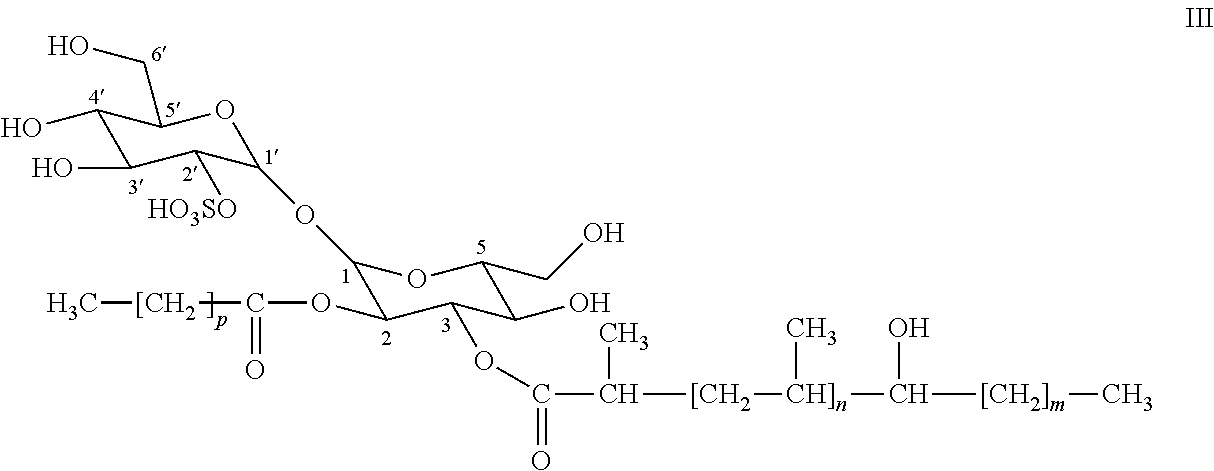
- the invention relates to compounds of the following general formula (I):
- R 1 and R 2 are fatty acyl groups.
- Fatty acyl groups are derived from fatty acid groups which have been esterified to the hydroxyl groups in position 2 and 3 of the trehalose-2′-sulfate unit.
- Fatty acid groups are aliphatic carboxylic acids which can be linear or ramified, saturated or unsaturated, unsubstituted or substituted by groups such as hydroxyl, or ketone.
- the invention particularly relates to compounds of formula I, wherein R 1 and R 2 independently from each other represent a fatty acyl group containing from 16 to 60 carbon atoms, and more particularly wherein R 1 and R 2 are selected from the group comprising:
- n is an integer from 2 to 10
- n is an integer from 2 to 10
- the fatty acyl groups notably comprise linear and saturated fatty acyl groups such as groups according to formula —OC—(CH 2 ) k —CH 3 wherein k is an integer from 14 to 58.
- the invention more particularly relates to compounds of formula I, wherein R 1 and R 2 are selected from the group comprising palmitic acyl and stearic acyl, namely compounds of following formulae:
- the invention further relates to compounds of formula I, wherein at least one of R 1 and R 2 represents a hydroxyphthioceranoic acyl group.
- the invention more specifically relates to compounds of formula I, wherein R 1 or R 2 represents a hydroxyphthioceranoic acyl group.
- the invention more particularly relates to compounds of formula I, wherein:
- the invention more specifically relates to compounds of following formulae:
- the invention relates to compounds of formulae III-21, III-22 or III-23.
- the invention relates to a composition comprising at least two different compounds of formula I such as defined above.
- the invention further relates to a composition characterized in that it comprises a mixture of compounds selected from the compounds of formulae II and III, preferably those of formulae II-1 to II-36 and of formulae III-1 to III-36, and more preferably those of formulae II-21, II-22, II-23, III-21, III-22 and III-23.
- the invention also relates to a composition
- a composition comprising a mixture of compounds selected from the compounds of formula III, preferably those of formulae III-1 to III-36, and more preferably those of formulae III-21, III-22 and III-23.
- the invention relates in particular to a composition as defined above, wherein compounds selected from the compounds of formulae II-21, II-22, II-23, III-21, III-22 and III-23 represent from about 20% to about 100%, more particularly about 30%, of the total amount of compounds of formula I of said composition.
- the invention also relates to a composition as defined above, wherein compounds selected from the compounds of formulae III-21, III-22 and III-23 represent from about 20% to about 100%, more particularly about 30%, of the total amount of compounds of formula I of said composition.
- the invention notably relates to a composition as defined above, wherein at least one compound, selected from the list consisting of compounds of formulae II-21, II-22, II-23, III-21, III-22 and III-23, represents from about 20% to about 100%, notably 30% of the total amount of compounds of formula I of said composition.
- the percentage used to characterize the contents of the above described compositions is a molar percentage.
- the invention also relates to a pharmaceutical composition
- a pharmaceutical composition comprising at least one compound as defined above, or a composition as defined above, in association with a pharmaceutically acceptable vehicle.
- a pharmaceutically acceptable carrier comprises in particular liposomes.
- composition may also comprise vaccine adjuvants.
- vaccine adjuvants for use in human individuals or animals are well known to the man skilled in the art, a list of such salts can be found for instance in “A compendium of vaccine adjuvants and excipients” 2 nd edition, Vogel et al.
- Particular vaccine adjuvants notably comprise aluminum salts or M59, for example.
- the invention particularly relates to a pharmaceutical composition as defined above, characterized in that it is presented in a form intended for administration by oral or injectable route.
- the invention more particularly relates to a pharmaceutical composition as defined above, characterized in that it comprises one or more other products useful for the treatment or the prophylaxis of tuberculosis, such as BCG or mycobacterial proteins.
- BCG stands for Bacillus of Calmette and Guerin, the different strains of BCG currently used for vaccination are notably described in Behr M. A. et al. Science (1999) 284:1520-1523.
- mycobacterial proteins refers to proteins, or fragments thereof, which are encoded by the genome of bacteria of the Mycobacterium genus and notably by the genome of Mycobacterium tuberculosis , such proteins may be advantageously recombinant. According to a preferred embodiment said mycobacterial proteins are antigens of M. tuberculosis.
- kits for the treatment or the prophylaxis of tuberculosis notably comprise immunomodulators, such as cytolines, DNA fragments encoding M. tuberculosis antigens, live M. tuberculosis deletion mutants, such as mutants in which virulence genes have been deleted, or live recombinant BCG, such as BCG expressing antigens of M. tuberculosis.
- immunomodulators such as cytolines, DNA fragments encoding M. tuberculosis antigens, live M. tuberculosis deletion mutants, such as mutants in which virulence genes have been deleted, or live recombinant BCG, such as BCG expressing antigens of M. tuberculosis.
- tuberculosis refers to the disease caused in humans by the bacterium Mycobacterium tuberculosis , but also to the corresponding disease in animals.
- the invention relates to products comprising:
- At least one other product useful for the treatment or the prophylaxis of tuberculosis such as BCG or mycobacterial proteins
- the invention also relates to the use of at least one compound as defined above, or of an above mentioned composition, for the preparation of a medicament, notably a vaccine, intended for the treatment or the prophylaxis of tuberculosis.
- the vaccine may comprise a vaccine adjuvant such as described above.
- the invention relates to the use of at least one compound as defined above, or of a composition according as defined above, as an immune reaction activator, and more particularly an inflammatory reaction activator.
- immune reaction activator is meant a compound which has the ability to activate components or processes of the immune reaction, in vitro or in vivo, in particular cells of the immune system such as T lymphocytes, B lymphocytes, antigen presenting cells (APCs), such as dendritic cells or macrophages, monocytes or granulocytes.
- T lymphocytes T lymphocytes
- B lymphocytes B lymphocytes
- antigen presenting cells APCs
- dendritic cells or macrophages monocytes or granulocytes.
- inflammatory reaction activator is meant a compound which has the ability to activate components or processes of the inflammatory reaction, in vitro or in vivo, such as diapedesis, capillary permeabilization, macrophage activation or fever onset for instance.
- the invention also relates to the use of at least one compound as defined above, or of a composition as defined above, to induce the activation of T lymphocytes, notably CD1-restricted T lymphocytes.
- T lymphocytes notably CD1-restricted T lymphocytes.
- the activation can proceed in vitro or in vivo.
- T lymphocytes can be assessed by several methods, such as measuring cell multiplication or cytokine production such as IFN- ⁇ (interferon- ⁇ ), IL-2 (interleukine-2), IL-4 (interleukine-4), or TNF- ⁇ (tumor necrosis factor ⁇ ), for instance.
- IFN- ⁇ interferon- ⁇
- IL-2 interleukine-2
- IL-4 interleukine-4
- TNF- ⁇ tumor necrosis factor ⁇
- CD1-restricted T lymphocytes are T lymphocytes which are activated by antigens presented by CD1 molecules.
- the invention also relates to the use of at least one compound as defined above, or of a composition as defined above, to induce the production of IFN- ⁇ , TNF- ⁇ , IL-4 or granulysin.
- IFN- ⁇ , TNF- ⁇ , IL-4 or granulysin can be measured for instance by immunoassays, such as ELISA (enzyme linked immunosorbent assay) or EIA (enzyme immunoassay).
- immunoassays such as ELISA (enzyme linked immunosorbent assay) or EIA (enzyme immunoassay).
- the invention relates to a process for generating T cell clones, characterized in that it comprises the following stages:
- the invention further relates to T cell clones such as obtained by the abovementioned process.
- T cell clone is described in the examples as Z4B27.
- the invention relates to a process for screening products, such as sulfoglycolipids extracted from Mycobacterium tuberculosis , characterized in that it comprises the following stages:
- the invention also relates to a process for the extraction of compounds as defined above, or of a composition as defined above, from Mycobacterium tuberculosis , characterized in that it comprises the following stages:
- FIG. 1 represents the amount of IFN- ⁇ (in pg/ml, horizontal axis) produced by T cell clone Z4B27 in response to stimulation (vertical axis) by the culture medium (medium), by sulfoglycolipids alone (SL), by dendritic cells alone (DC) or by dendritic cells loaded with sulfoglycolipids (DC+SL)
- FIG. 2 represents the negative MALDI (Matrix Assisted Laser Desorption/Ionisation) mass spectrum of the purified preparation of sulfoglycolipids extracted from M. tuberculosis which activates the T-cell clone Z4B27.
- the horizontal axis represents the m/z ratio of the compounds contained in the preparation.
- the vertical axis represents the peak intensity as a percentage of maximum peak intensity. The major peaks are identified by their respective m/z ratio.
- FIG. 3 represents the 1-D 1 H NMR (Nuclear Magnetic Resonance) spectrum of the purified preparation of sulfoglycolipids extracted from M. tuberculosis which activates the T-cell clone Z4B27.
- the vertical axis represents the intensity of resonance and the horizontal axis the chemical shifts in ppm (parts per million).
- a close-up of the spectrum between 4.7 and 5.4 ppm is inserted in the left part of the spectrum.
- the peak designed as CDCl 3 corresponds the solvent.
- FIG. 4 represents a diagram showing the evolution of the number of M. tuberculosis CFUs (colony forming units) infecting dendritic cells ( ⁇ 10 ⁇ 6 , vertical axis) in response to the addition of increasing amounts of sulfoglycolipid specific T cell clones, represented as T cells:dendritic cells ratios on the right of the figure, between day 1 and day 5 of the experiment (horizontal axis).
- Immature dendritic cells were prepared from peripheral blood of healthy donors. Plastic-adherent cells were cultured in RPMI-1640 medium containing 10% FCS (foetal calf serum), 50 ng/ml recombinant human GM-CSF, 1000 U/ml recombinant human IL-4, and were used after 5 days. Before each experiment the percentage of dendritic cells was monitored by immunofluorescence analysis using mAbs specific for CD1b (WM-25, ImmunoCAM, Lugano, Switzerland), CD1a (OKT6) and CD1c (L161) (Instrumentation Laboratory, Schlieren, Switzerland) and was always >90%.
- CD1b WM-25, ImmunoCAM, Lugano, Switzerland
- CD1a OKT6
- CD1c L161
- cells were suspended in PBS containing 1% BSA and 0.04% NaN 3 and incubated with the primary mAbs (10 ⁇ g/ml) for 40 min, washed twice, and then further incubated with FITC-conjugated goat anti-mouse Ig for another 40 min at 4° C. After washing, cells were analyzed on a FACScan flow cytometer (Becton Dickinson).
- Mycobacterial glycolipids were extracted from Mycobacterium tuberculosis cell wall. Killed cells were treated by a mixture of chloroform and methanol, 1/1, v/v and filtered, giving rise to delipidated cells and a supernatant containing cell wall glycolipids. The cells were extracted four times in the same way to yield a M. tuberculosis glycolipid extract.
- T cell lines were established by culturing freshly isolated peripheral blood mononuclear cells (PBMC) with autologous DCs. DCs were incubated for 2 h with the above mentioned glycolipids (20 ⁇ g/ml) before addition of PBMCs. After 3 weeks, growing T cells were restimulated with heterologous DCs and the same preparation of glycolipids. An identical restimulation was performed after additional 3 weeks, cells were then tested for their antigen specificity.
- PBMC peripheral blood mononuclear cells
- T cells from glycolipid-specific T cell lines were cloned by limiting dilution in Terasaki plates. Cloning was performed using RPMI-1640 medium added with 5% human AB serum (Swiss Red Cross, Bern), 100 U/ml recombinant human IL-2, 1% PHA (Wellcome), 2 mM L-Glutamine (Gibco, 21051-024), 1 mM Na Pyruvate (Gibco, 11840-048), 1% Non-essential Aminoacids (Gibco, 11140-035), and 100 ⁇ g/ml Kanamycin (Gibco, 15160-021).
- T cell clones Twelve days after plating, wells containing growing cells were scored and the plating efficiency was calculated according to Poisson's distribution. Cells were transferred into 96 wells plates and further expanded for one week before screening for their glycolipid specificity. The glycolipid specificity of T cell clones was detected by measuring the IFN- ⁇ released in the supernatant 48 hr after stimulation with dendritic cells and glycolipids, using the human IFN- ⁇ ELISA kit purchased from BD Phaminigen. The assay was performed using the protocol suggested by the dealer. Glycolipid-specific clones, such as Z4B27 ( FIG. 1 ), were further expanded and characterized.
- DC 4 ⁇ 10 4 /well in triplicate
- RPMI-1640 medium containing 10% FCS
- glycolipid antigen (1-50 ⁇ g/ml)
- TNF- ⁇ , IFN- ⁇ and IL-4 released into culture supernatants were assayed by ELISA using commercial kits (Instrumentation Laboratory, Schlieren, Switzerland).
- CIR cells (6 ⁇ 10 4 /well in triplicate) transfected with CD1a, b, c, d or mock transfected were used in some assays.
- Inhibition of T cell activation was performed by adding different doses of purified anti-CD3s TR66 mAb Fab fragments or purified anti-CD1b WM-25 mAbs together with glycolipid-pulsed APC. After 36 h, supernatants were harvested and tested in ELISA for the presence of TNF- ⁇ , IFN- ⁇ and IL-4. Two of the glycolipid-specific T cell clones tested, Z4B26 and Z4B27, were found to be TCR ⁇ + , CD8 + and were restricted by CD1b. Clone Z4B27 was chosen in order to characterize the glycolipid antigens responsible for its activation.
- the above mentioned mycobacterial extract was concentrated and partitioned between water and chloroform.
- the chloroform phase contained most of the envelope lipids and glycolipids as well as the sulfoglycolipids. It was evaporated, and the residue was dissolved in the minimum of chloroform, then acetone was added and the mixture was kept overnight at 4° C. A precipitate was observed and by centrifugation at 4° C. for 15 min (3000 ⁇ g) an “acetone-insoluble” phase and an “acetone-soluble” phase were obtained. Most of the sulfoglycolipids were found in the “acetone soluble” phase together with other lipids.
- the sulfoglycolipids were characterized by their behaviour on silica gel thin layer chromatography using chloroform/methanol, 9/1, v/v as migration solvent and were visualized with orcinol staining. In addition, their presence in the “acetone-soluble” phase was unambiguously established from MALDI-Time of Flight-mass spectrometry analysis (MALDI-Tof-MS) in negative mode. From the mass spectrum, recorded from 700 to 5000 mass units, mainly three families of sulfoglycolipids were characterized differing by their acylation degree and the fatty acyl appendage structures.
- the low mass range was mainly dominated by ⁇ - ⁇ -D-trehalose-2′-sulfate (A) containing two fatty acyl appendages including one hydroxyphthioceranoic acid residue (C) and either one palmitic (HOOC—(CH 2 ) 14 —CH 3 ) or stearic acid (HOOC—(CH 2 ) 16 —CH 3 ) residue. Beside this sulfoglycolipid, other acyl-forms were observed containing one and two additional hydroxyphthioceranoic acids corresponding to the two other sulfoglycolipids families noticed above.
- the “acetone-soluble” phase was then first fractionated on silicic acid column irrigated by chloroform, and chloroform containing 10, 20 and 30% of methanol.
- the bioactivity i.e. the capacity to activate T cell clone Z4B27, was mainly restricted to the fraction eluted with a mixture of chloroform containing 20% of methanol.
- FIG. 2 shows the MALDI-Tof spectrum recorded in negative mode using HABA [2-(4-hydroxyphenylazo)-benzoic acid] as matrix. The peaks were assigned to deprotonated molecular ions (M-H) ⁇ thus allowing the characterization of the different molecular species summarized in Table 1.
- the desulfatation of the sulfoglycolipid antigens was performed and the bioactivity of the resulting glycolipids was investigated.
- the sulfoglycolipids (1 mg) dissolved in 1 ml of methanol were mixed with 50 mM of HCl at room temperature during 16 h.
- To the reaction mixture 3 ml of chloroform, 0.5 ml of methanol and 1 ml of 0.2% sodium acetate were added.
- the lower phase was washed with 2 ⁇ 2 ml of chloroform/methanol/water 3/48/47, v/v dried and tested on silica gel high performance thin layer chromatography plates.
- glycolipids were visualized by orcinol staining and their migration using chloroform/methanol 9/1 as migration solvent was in agreement with the loss of the sulfate group.
- the desulfatation abrogated the capacity of the resulting glycolipid to activate the T cell clone Z4B27, revealing that sulfoglycolipids were the antigens which stimulate the T cell clones.
- the fraction containing the diacylated ⁇ - ⁇ -D-trehalose-2′-sulfate antigens was applied on a C18 Sep-Pak cartridge which was successively irrigated with methanol/water 9/1, methanol and methanol/chloroform, 1/1, v/v.
- the bioactivity was restricted to the fractions eluted with methanol and methanol/chloroform, 1/1, v/v.
- FIG. 3 shows the one dimensional 1 H spectrum. In the anomeric zone, four signals were observed namely I 1 , II 1 , II 2 and II 3 .
- the resonances I 1 and II 1 typified the two anomeric protons of the ⁇ - ⁇ -trehalose core, and the resonances II 2 and II 3 were assigned to the H2 and H3 of the glucose moiety II, respectively.
- the downfield shifts of the H2 and H3 protons indicated that the fatty acyl appendages (hydroxyphthioceranoic acid and palmitic or stearic acids) were located on C2 and C3 of unit II.
- the sulphate residue was located on the C2′ of the glucose I moiety.
- IFN- ⁇ release was measured after stimulation with the purified sulfoglycolipids. 2 ⁇ 10 5 PBMC per/well were incubated for 4 days in the presence of GM-CSF (500 U/ml) and IL-4 (5 ng/ml). Autologous effector T-cells were incubated in 10% human serum during this time. The sulfoglycolipids (10 ⁇ g/ml) were added to the irradiated, CD1-expressing antigen-presenting cells. Finally, effector cells were added (2 ⁇ 10 5 /well) and IFN- ⁇ release was measured by ELISA in the supernatants after 18 hours.
- the IFN- ⁇ ELISA was performed in 96-well immunosorbent plates, which were coated with an IFN- ⁇ capture antibody (2 ⁇ g/ml) overnight. Non-specific binding sites were blocked with PBS containing 1% bovine serum albumin. The supematants were diluted 1:1 and added in a final volume of 100 ⁇ l. Plates were incubated at room temperature for 2 hours and removed by thorough washing (3-4 times). Finally, a biotinylated anti-IFN- ⁇ antibody was added for 1 hour (2 ⁇ g/ml). For detection of immunoreactive IFN- ⁇ , horseradish-peroxidase was added for 30 min. Finally, a chromogenic substrate (TMB, Endogen, Mass., USA) was added. After 20 min.
- TMB Endogen, Mass., USA
- T cell clones which recognize sulfoglycolipids
- DC dendritic cells
- sulfoglycolipids-reactive T cell clones were permeabilised using 0.5% saponin and a polyclonal rabbit serum directed against granulysin was added. Staining was visualized using a FITC-conjugated secondary antibody directed against rabbit immunoglobulins. The number of positive cells was quantitated by analyzing cells in a flow cytometer.
- DCs were generated from peripheral blood monocytes by treatment with GM-CSF and IL-4.
- the cells were then infected with a virulent strain of M. tuberculosis (H37Rv) at a multiplicity of infection of 1.
- H37Rv virulent strain of M. tuberculosis
- Non-phagocytosed bacteria were removed by thorough washing and then T-cells were added in increasing amounts as indicated in FIG. 4 .
- the number of surviving bacteria was determined by plating cell lysates after 5 days of coincubation and counting the number of colonies grown after three weeks.
- sulfoglycolipids-responsive cells express granulysin, the secretion of this antibacterial molecule is most likely responsible for the killing of the pathogen. Therefore, sulfoglycolipids are presented on the surface of human host cells for mycobacteria recognition and killing.
- T-cell clones specific for sulfoglycolipids recognize DC infected with live M. tuberculosis bacilli and kill the pathogen possibly by the secretion of granulysin.
Abstract

Description


wherein R1 and R2 are fatty acyl groups.


-
- R1 represents a hydroxyphthioceranoic acyl group, and R2 represents a palmitic acyl group or a stearic acyl group, namely compounds of following formula (II):

-
- wherein p is 14 or 16, m is 14 or 16 and n is an integer from 2 to 10, or
- R2 represents a hydroxyphthioceranoic acyl group, and R1 represents a palmitic acyl group or a stearic acyl group, namely compounds of following formula (III):

-
- wherein p is 14 or 16, m is 14 or 16 and n is an integer from 2 to 10.
n=2, m=14 and p=14 (II.1);
n=2, m=14 and p=16 (II.2);
n=2, m=16 and p=14 (II.3);
n=2, m=16 and p=16 (II.4);
n=3, m=14 and p=14 (II.5);
n=3, m=14 and p=16 (II.6);
n=3, m=16 and p=14 (II.7);
n=3, m=16 and p=16 (II.8);
n=4, m=14 and p=14 (II.9);
n=4, m=14 and p=16 (II.10);
n=4, m=16 and p=14 (II.11);
n=4, m=16 and p=16 (II.12);
n=5, m=14 and p=14 (II.13);
n=5, m=14 and p=16 (II.14);
n=5, m=16 and p=14 (II.15);
n=5, m=16 and p=16 (II.16);
n=6, m=14 and p=14 (II.17);
n=6, m=14 and p=16 (II.18);
n=6, m=16 and p=14 (II.19);
n=6, m=16 and p=16 (II.20);
n=7, m=14 and p=14 (II.21);
n=7, m=14 and p=16 (II.22);
n=7, m=16 and p=14 (II.23);
n=7, m=16 and p=16 (II.24);
n=8, m=14 and p=14 (II.25);
n=8, m=14 and p=16 (II.26);
n=8, m=16 and p=14 (II.27);
n=8, m=16 and p=16 (II.28);
n=9, m=14 and p=14 (II.29);
n=9, m=14 and p=16 (II.30);
n=9, m=16 and p=14 (II.31);
n=9, m=16 and p=16 (II.32);
n=10, m=14 and p=14 (II.33);
n=10, m=14 and p=16 (II.34);
n=10, m=16 and p=14 (II.35);
n=10, m=16 and p=16 (II.36);
or of formula m, wherein:
n=2, m=14 and p=14 (III.1);
n=2, m=14 and p=16 (III.2);
n=2, m=16 and p=14 (III.3);
n=2, m=16 and p=16 (III.4);
n=3, m=14 and p=14 (III.5);
n=3, m=14 and p=16 (III.6);
n=3, m=16 and p=14 (III.7);
n=3, m=16 and p=16 (III.8);
n=4, m=14 and p=14 (III.9);
n=4, m=14 and p=16 (III.10);
n=4, m=16 and p=14 (III.11);
n=4, m=16 and p=16 (III.12);
n=5, m=14 and p=14 (III.13);
n=5, m=14 and p=16 (III.14);
n=5, m=16 and p=14 (III.15);
n=5, m=16 and p=16 (III.16);
n=6, m=14 and p=14 (III.17);
n=6, m=14 and p=16 (III.18);
n=6, m=16 and p=14 (III.19);
n=6, m=16 and p=16 (III.20);
n=7, m=14 and p=14 (III.21);
n=7, m=14 and p=16 (III.22);
n=7, m=16 and p=14 (III.23);
n=7, m=16 and p=16 (III.24);
n=8, m=14 and p=14 (III.25);
n=8, m=14 and p=16 (III.26);
n=8, m=16 and p=14 (III.27);
n=8, m=16 and p=16 (III.28);
n=9, m=14 and p=14 (III.29);
n=9, m=14 and p=16 (III.30);
n=9, m=16 and p=14 (III.31);
n=9, m=16 and p=16 (III.32);
n=10, m=14 and p=14 (III.33);
n=10, m=14 and p=16 (III.34);
n=10, m=16 and p=14 (III.35);
n=10, m=16 and p=16 (III.36);


-
- incubating antigen presenting cells (APCs), notably dendritic cells, with a Mycobacterium tuberculosis envelope preparation substantially devoid of proteins, to obtain non-protein envelope antigen loaded APCs,
- contacting peripheral blood mononuclear cells with the envelope antigen loaded APCs to obtain proliferating T cells,
- cloning proliferating T cells by limiting dilution and selecting the clones releasing a molecule selected from the group comprising IFN-γ, TNF-α, granulysin or IL-4 when contacted by envelope antigen loaded APCs to obtain T cell clones.
-
- contacting dendritic cells loaded with the product to screen, notably sulfoglycolipids extracted from Mycobacterium tuberculosis, with the above-defined T cell clones,
- detecting a molecule selected from the group comprising IFN-γ, TNF-α, granulysin or IL-4, released by the T cell clones.
-
- treatment of M. tuberculosis bacteria with a mixture of methanol and chloroform to obtain a chloroform/methanol extract,
- concentration of the chloroform/methanol extract followed by its partition between a chloroform phase and an aqueous phase,
- taking of the chloroform phase and evaporation of most of the chloroform, followed by addition of acetone thereto to obtain a precipitate and a soluble acetone phase,
- taking of the soluble acetone phase followed by its concentration, and application of the concentrated soluble acetone phase on a silicic acid column irrigated with mixtures of methanol and chloroform,
- elution of a fraction from the above-mentioned silicic acid column by a mixture of chloroform and approximately 20% methanol, said fraction corresponding to a composition as defined above,
- if necessary purification of the fraction eluted from the silicic acid column to obtain different preparations respectively containing substantially only one of the compounds as defined above.

| TABLE I | |
| Mass | |
| (m/z) | trehalose-2′-sulfate substituents |
| 1039 | Palmitic acid and hydroxyphthioceranoic acid (n = 2, m = 14) |
| 1067 | Stearic acid and hydroxyphthioceranoic acid (n = 2, m = 14), |
| or palmitic acid and hydroxyphthioceranoic acid (n = 2, m = 16) | |
| 1123 | Palmitic acid and hydroxyphthioceranoic acid (n = 4, m = 14) |
| 1151 | Stearic acid and hydroxyphthioceranoic acid (n = 4, m = 14) |
| or palmitic acid and hydroxyphthioceranoic acid (n = 4, m = 16) | |
| 1165 | Stearic acid and hydroxyphthioceranoic acid (n = 5 m = 16) |
| 1179 | Stearic acid and hydroxyphthioceranoic acid (n = 4 m = 16) |
| 1193 | Stearic acid and hydroxyphthioceranoic acid (n = 5 m = 14), |
| or palmitic acid and hydroxyphthioceranoic acid (n = 5 m = 16) | |
| 1207 | Palmitic acid and hydroxyphthioceranoic acid (n = 6 m = 14) |
| 1235 | Stearic acid and hydroxyphthioceranoic acid (n = 6 m = 14), |
| or palmitic acid and hydroxyphthioceranoic acid (n = 6 m = 16) | |
| 1249 | Palmitic acid and hydroxyphthioceranoic acid (n = 7 m = 14) |
| 1263 | Stearic acid and hydroxyphthioceranoic acid (n = 6 m = 16) |
| 1277 | Stearic acid and hydroxyphthioceranoic acid (n = 7 m = 14), |
| or palmitic acid and hydroxyphthioceranoic acid (n = 7 m = 16) | |
| 1291 | Palmitic acid and hydroxyphthioceranoic acid (n = 8 m = 14) |
| 1305 | Stearic acid and hydroxyphthioceranoic acid (n = 7 m = 16) |
| 1319 | Stearic acid and hydroxyphthioceranoic acid (n = 8 m = 14), |
| or palmitic acid and hydroxyphthioceranoic acid (n = 8 m = 16) | |
| 1333 | Palmitic acid and hydroxyphthioceranoic acid (n = 9 m = 14) |
| 1361 | Stearic acid and hydroxyphthioceranoic acid (n = 9 m = 14), |
| or palmitic acid and hydroxyphthioceranoic acid (n = 9 m = 16) | |

Claims (20)


n=2, m=14 and p=14 (II.1);
n=2, m=14 and p=16 (II.2);
n=2, m=16 and p=14 (II.3);
n=2, m=16 and p=16 (II.4);
n=3, m=14 and p=14 (II.5);
n=3, m=14 and p=16 (II.6);
n=3, m=16 and p=14 (II.7);
n=3, m=16 and p=16 (II.8);
n=4, m=14 and p=14 (II.9);
n=4, m=14 and p=16 (II.10);
n=4, m=16 and p=14 (II.11);
n=4, m=16 and p=16 (II.12);
n=5, m=14 and p=14 (II.13);
n=5, m=14 and p=16 (II.14);
n=5, m=16 and p=14 (II.15);
n=5, m=16 and p=16 (II.16);
n=6, m=14 and p=14 (II.17);
n=6, m=14 and p=16 (II.18);
n=6, m=16 and p=14 (II.19);
n=6, m=16 and p=16 (II.20);
n=7, m=14 and p=14 (II.21);
n=7, m=14 and p=16 (II.22);
n=7, m=16 and p=14 (II.23);
n=7, m=16 and p=16 (II.24);
n=8, m=14 and p=14 (II.25);
n=8, m=14 and p=16 (II.26);
n=8, m=16 and p=14 (II.27);
n=8, m=16 and p=16 (II.28);
n=9, m=14 and p=14 (II.29);
n=9, m=14 and p=16 (II.30);
n=9, m=16 and p=14 (II.31);
n=9, m=16 and p=16 (II.32);
n=10, m=14 and p=14 (II.33);
n=10, m=14 and p=16 (II.34);
n=10, m=16 and p=14 (II.35); and
n=10, m=16 and p=16 (II.36);

n=2, m=14 and p=14 (III.1);
n=2, m=14 and p=16 (III.2);
n=2, m=16 and p=14 (III.3);
n=2, m=16 and p=16 (III.4);
n=3, m=14 and p=14 (III.5);
n=3, m=14 and p=16 (III.6);
n=3, m=16 and p=14 (III.7);
n=3, m=16 and p=16 (III.8);
n=4, m=14 and p=14 (III.9);
n=4, m=14 and p=16 (III.10);
n=4, m=16 and p=14 (III.11);
n=4, m=16 and p=16 (III.12);
n=5, m=14 and p=14 (III.13);
n=5, m=14 and p=16 (III.14);
n=5, m=16 and p=14 (III.15);
n=5, m=16 and p=16 (III.16);
n=6, m=14 and p=14 (III.17);
n=6, m=14 and p=16 (III.18);
n=6, m=16 and p=14 (III.19);
n=6, m=16 and p=16 (III.20);
n=7, m=14 and p=14 (III.21);
n=7, m=14 and p=16 (III.22);
n=7, m=16 and p=14 (III.23);
n=7, m=16 and p=16 (III.24);
n=8, m=14 and p=14 (III.25);
n=8, m=14 and p=16 (III.26);
n=8, m=16 and p=14 (III.27);
n=8, m=16 and p=16 (III.28);
n=9, m=14 and p=14 (III.29);
n=9, m=14 and p=16 (III.30);
n=9, m=16 and p=14 (III.31);
n=9, m=16 and p=16 (III.32);
n=10, m=14 and p=14 (III.33);
n=10, m=14 and p=16 (III.34);
n=10, m=16 and p=14 (III.35);and
n=10, m=16 and p=16 (III.36).


Applications Claiming Priority (4)
| Application Number | Priority Date | Filing Date | Title |
|---|---|---|---|
| EP03290965 | 2003-04-18 | ||
| EP03290965A EP1469007A1 (en) | 2003-04-18 | 2003-04-18 | Sulfoglycolipid antigens, their extraction from mycobacterieum tuberculosis, and their use against tuberculosis |
| EP03290965.7 | 2003-04-18 | ||
| PCT/EP2004/003830 WO2004092192A1 (en) | 2003-04-18 | 2004-04-09 | Sulfoglycolipid antigens, their extraction from mycobacterium tuberculosis, and their use against tuberculosis |
Publications (2)
| Publication Number | Publication Date |
|---|---|
| US20080176818A1 US20080176818A1 (en) | 2008-07-24 |
| US8153610B2 true US8153610B2 (en) | 2012-04-10 |
Family
ID=32893003
Family Applications (1)
| Application Number | Title | Priority Date | Filing Date |
|---|---|---|---|
| US10/553,801 Expired - Fee Related US8153610B2 (en) | 2003-04-18 | 2004-04-09 | Sulfoglycolipid antigens, their extraction from Mycobacterium tuberculosis, and their use against tuberculosis |
Country Status (3)
| Country | Link |
|---|---|
| US (1) | US8153610B2 (en) |
| EP (2) | EP1469007A1 (en) |
| WO (1) | WO2004092192A1 (en) |
Families Citing this family (5)
| Publication number | Priority date | Publication date | Assignee | Title |
|---|---|---|---|---|
| WO2005077411A2 (en) * | 2004-02-10 | 2005-08-25 | Innate Pharma | Composition and method for the treatment of carcinoma |
| FR2908658B1 (en) * | 2006-11-20 | 2011-11-11 | Centre Nat Rech Scient | COMPOSITION FOR THE PREVENTION AND / OR TREATMENT OF DISEASES ASSOCIATED WITH OVEREXPRESSION OF TNF AND / OR IL-12 |
| EP1950218A1 (en) * | 2007-01-24 | 2008-07-30 | Centre National de la Recherche Scientifique | Sulfoglycolipid antigens, their process of preparation, and their use against tuberculosis |
| EP2047860A1 (en) * | 2007-10-12 | 2009-04-15 | Centre National De La Recherche Scientifique (Cnrs) | Phamaceutical compositons comprosing actinomycete glycerol acyl derivatives antigens, their process of extractin, and their use against tuberculosis. |
| CA2777400C (en) | 2009-10-12 | 2018-11-27 | The United States Of America, As Represented By The Secretary, Department Of Health And Human Services | Granulysin in immunotherapy |
-
2003
- 2003-04-18 EP EP03290965A patent/EP1469007A1/en not_active Withdrawn
-
2004
- 2004-04-09 US US10/553,801 patent/US8153610B2/en not_active Expired - Fee Related
- 2004-04-09 WO PCT/EP2004/003830 patent/WO2004092192A1/en active Application Filing
- 2004-04-09 EP EP04726714A patent/EP1615939A1/en not_active Withdrawn
Non-Patent Citations (5)
| Title |
|---|
| Besra et al, Biochemistry, 1992, 31, 9832-37. * |
| Desmarais et al, J. Bacteriology, 1997, 3146-53. * |
| I. Vergne, M. Daffe: "Interaction of mycobacterial glycolipids with host cells", Frontiers in Bioscience, vol. 3, 1998, pp. 856-876, XP008023822. |
| The Merck Manual, 1992, pp. 140-141. * |
| Vergne et al, Frontiers in Bioscience, 1998, 3, 865-76. * |
Also Published As
| Publication number | Publication date |
|---|---|
| WO2004092192A1 (en) | 2004-10-28 |
| EP1615939A1 (en) | 2006-01-18 |
| EP1469007A1 (en) | 2004-10-20 |
| US20080176818A1 (en) | 2008-07-24 |
Similar Documents
| Publication | Publication Date | Title |
|---|---|---|
| US8557859B2 (en) | Immunomodulatory compositions | |
| US5158939A (en) | Method of stimulating the immune systems of animals and compositions useful therefor | |
| US8268801B2 (en) | Sulfoglycolipid antigens, their process of preparation, and their use against tuberculosis | |
| EP0025495A1 (en) | Lipophilic muramyl peptides, processes for their preparation and their use | |
| CS205027B2 (en) | Method of producing glucosamine derivatives | |
| EP0038750B1 (en) | Immunologically active dipeptidyl 4-0-, 6-0-acyl-2-amino-2-deoxy-d-glucose derivatives and methods for their preparation | |
| JP2010516748A5 (en) | ||
| US20210177965A1 (en) | Brartemicin analogues | |
| US4315913A (en) | Immunologically active dipeptidyl 2-amino-1,2-dideoxy-D-glucose derivatives and methods of preparation | |
| US8153610B2 (en) | Sulfoglycolipid antigens, their extraction from Mycobacterium tuberculosis, and their use against tuberculosis | |
| US5041427A (en) | Lipid A derivatives | |
| US4310514A (en) | Immunologically active dipeptidyl 5-0,6-0-acyl-2-amino-2-deoxy-D-glucofuranose derivatives and methods of preparation | |
| JP2001515868A (en) | Synthetic antigens for CD1 restricted immune response | |
| US4391800A (en) | Immunologically active peptidyl disaccharides and methods of preparation | |
| EP0014159A1 (en) | Immunologically active dipeptidyl saccharides and methods of preparation | |
| EP0118364B1 (en) | Immunostimulatory dipeptidyl d-glucose derivatives and methods of preparation | |
| EP0668289A1 (en) | Novel disaccharide derivative | |
| WO2018155051A1 (en) | Lipid a | |
| US4698331A (en) | 2,3-diamino-2,3-didesoxyhexose derivatives and their use | |
| AU679970B2 (en) | Novel disaccharide derivative | |
| JPH06206893A (en) | New disaccharide derivative | |
| JPH10152497A (en) | Helicobacter pylori lipid a | |
| KR20230106604A (en) | Novel aminoalkylglucosaminide 4-phosphate derivatives | |
| EP3000820A1 (en) | Synthetic vaccines against Streptococcus pneumoniae serotype 8 | |
| US20100255015A1 (en) | Pharmaceuticals compositions comprising actinomycete glycerol acyl derivatives antigens, their process of extraction, and their use against tuberculosis |
Legal Events
| Date | Code | Title | Description |
|---|---|---|---|
| AS | Assignment |
Owner name: CENTRE NATIONAL DE LA RECHERCHE SCIENTIFIQUE, FRAN Free format text: ASSIGNMENT OF ASSIGNORS INTEREST;ASSIGNORS:PUZO, GERMAIN;GILLERON, MARTINE;STENGER, STEFFEN;AND OTHERS;SIGNING DATES FROM 20050912 TO 20080305;REEL/FRAME:020694/0713 |
|
| AS | Assignment |
Owner name: UNIVERSITY OF ERLANGEN (25 PERCENT INTEREST), GERM Free format text: ASSIGNMENT OF ASSIGNORS INTEREST;ASSIGNOR:CENTRE NATIONAL DE LA RECHERCHE SCIENTIFIQUE (CNRS);REEL/FRAME:021568/0034 Effective date: 20060817 Owner name: UNIVERSITY HOSPITAL OF BASEL (25 PERCENT INTEREST) Free format text: ASSIGNMENT OF ASSIGNORS INTEREST;ASSIGNOR:CENTRE NATIONAL DE LA RECHERCHE SCIENTIFIQUE (CNRS);REEL/FRAME:021568/0034 Effective date: 20060817 |
|
| FEPP | Fee payment procedure |
Free format text: PAYOR NUMBER ASSIGNED (ORIGINAL EVENT CODE: ASPN); ENTITY STATUS OF PATENT OWNER: LARGE ENTITY |
|
| REMI | Maintenance fee reminder mailed | ||
| LAPS | Lapse for failure to pay maintenance fees | ||
| STCH | Information on status: patent discontinuation |
Free format text: PATENT EXPIRED DUE TO NONPAYMENT OF MAINTENANCE FEES UNDER 37 CFR 1.362 |
|
| FP | Lapsed due to failure to pay maintenance fee |
Effective date: 20160410 |