US8073635B2 - Method of quantitation by mass spectrometry - Google Patents
Method of quantitation by mass spectrometry Download PDFInfo
- Publication number
- US8073635B2 US8073635B2 US12/032,263 US3226308A US8073635B2 US 8073635 B2 US8073635 B2 US 8073635B2 US 3226308 A US3226308 A US 3226308A US 8073635 B2 US8073635 B2 US 8073635B2
- Authority
- US
- United States
- Prior art keywords
- sample
- ions
- ion
- calibration
- mass
- Prior art date
- Legal status (The legal status is an assumption and is not a legal conclusion. Google has not performed a legal analysis and makes no representation as to the accuracy of the status listed.)
- Active, expires
Links
Images


Classifications
-
- H—ELECTRICITY
- H01—ELECTRIC ELEMENTS
- H01J—ELECTRIC DISCHARGE TUBES OR DISCHARGE LAMPS
- H01J49/00—Particle spectrometers or separator tubes
- H01J49/0009—Calibration of the apparatus
Definitions
- Quantitation by mass spectrometry is conventionally performed with a triple-quadrupole mass spectrometer using a multiple reaction monitoring (MRM) method that selects certain product and precursor ion combinations to provide the best sensitivity and signal-to-noise.
- MRM multiple reaction monitoring
- a linear dynamic range of three to five orders of magnitude can often be achieved by such a system.
- a triple-quadrupole mass spectrometer with a time-of-flight mass spectrometer replacing the third quadrupole (QqTOF) can also be used for quantitation, with the advantage that much higher mass resolution can be achieved.
- intense product ions can saturate the detector of a QqTOF mass spectrometer, limiting its linear dynamic range to only two to three orders of magnitude.
- FIG. 1 is a block diagram that illustrates a computer system, upon which embodiments of the present teachings may be implemented.
- FIG. 2 is a flowchart showing a method for improving selectivity of a measurement from a mass spectrometer, in accordance with the present teachings.
- FIG. 3 is a flowchart showing a method for determining an extracted ion current (XIC) window to use for a mass spectrometer measurement, in accordance with the present teachings.
- FIG. 4 is a flowchart showing a method for quantitation using data from a mass spectrometer, in accordance with the present teachings.
- FIG. 5 is a schematic diagram of a mass spectrometry system that includes a mass spectrometer and computer system, in accordance with the present teachings.
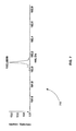
- FIG. 6 is an exemplary product ion mass spectrum from a urine sample, in accordance with the present teachings.
- FIG. 7 is an exemplary expanded view of a product ion mass spectrum from a urine sample, in accordance with the present teachings
- FIG. 8 is an exemplary expanded view of a product ion mass spectrum from a urine sample showing an XIC window with a width of 0.5 atomic mass units (amu), in accordance with the present teachings.
- FIG. 9 is an exemplary plot of the XIC for five samples injected about three minutes apart using the XIC window shown in FIG. 8 , in accordance with the present teachings.
- FIG. 10 is an exemplary expanded view of a product ion mass spectrum from a urine sample showing an XIC window with a width of 0.01 amu, in accordance with the present teachings.
- FIG. 11 is an exemplary plot of the XIC for five samples injected about three minutes apart using the XIC window shown in FIG. 10 , in accordance with the present teachings.
- FIG. 12 is an exemplary plot of a mass peak of interest and an interfering mass peak, showing how it can be advantageous to select a position of the XIC window that is not centered on the true center of the mass of interest, in accordance with the present teachings.
- FIG. 13 is a table showing the linear ranges of the calibration curves of five product ions of an exemplary known compound, in accordance with the present teachings.
- FIG. 14 is a table showing the intensities of five product ions of an exemplary known compound that are found in a sample, in accordance with the present teachings.
- FIG. 1 is a block diagram that illustrates a computer system 100 , upon which embodiments of the present teachings may be implemented.
- Computer system 100 includes a bus 102 or other communication mechanism for communicating information, and a processor 104 coupled with bus 102 for processing information.
- Computer system 100 also includes a memory 106 , which can be a random access memory (RAM) or other dynamic storage device, coupled to bus 102 for determining base calls, and instructions to be executed by processor 104 .
- Memory 106 also may be used for storing temporary variables or other intermediate information during execution of instructions to be executed by processor 104 .
- Computer system 100 further includes a read only memory (ROM) 108 or other static storage device coupled to bus 102 for storing static information and instructions for processor 104 .
- a storage device 110 such as a magnetic disk or optical disk, is provided and coupled to bus 102 for storing information and instructions.
- Computer system 100 may be coupled via bus 102 to a display 112 , such as a cathode ray tube (CRT) or liquid crystal display (LCD), for displaying information to a computer user.
- a display 112 such as a cathode ray tube (CRT) or liquid crystal display (LCD)
- An input device 114 is coupled to bus 102 for communicating information and command selections to processor 104 .
- cursor control 116 is Another type of user input device, such as a mouse, a trackball or cursor direction keys for communicating direction information and command selections to processor 104 and for controlling cursor movement on display 112 .
- This input device typically has two degrees of freedom in two axes, a first axis (e.g., x) and a second axis (e.g., y), that allows the device to specify positions in a plane.
- Computer system 100 can perform the present teachings. Consistent with certain implementations of the present teachings, results are provided by computer system 100 in response to processor 104 executing one or more sequences of one or more instructions contained in memory 106 . Such instructions may be read into memory 106 from another computer-readable medium, such as storage device 110 . Execution of the sequences of instructions contained in memory 106 causes processor 104 to perform the process described herein. Alternatively hard-wired circuitry may be used in place of or in combination with software instructions to implement the present teachings. Thus implementations of the present teachings are not limited to any specific combination of hardware circuitry and software.
- Non-volatile media includes, for example, optical or magnetic disks, such as storage device 110 .
- Volatile media includes dynamic memory, such as memory 106 .
- Transmission media includes coaxial cables, copper wire, and fiber optics, including the wires that comprise bus 102 . Transmission media can also take the form of acoustic or light waves, such as those generated during radio-wave and infra-red data communications.
- Computer-readable media include, for example, a floppy disk, a flexible disk, hard disk, magnetic tape, or any other magnetic medium, a CD-ROM, any other optical medium, punch cards, papertape, any other physical medium with patterns of holes, a RAM, PROM, and EPROM, a FLASH-EPROM, any other memory chip or cartridge, a carrier wave as described hereinafter, or any other medium from which a computer can read.
- Various forms of computer-readable media may be involved in carrying one or more sequences of one or more instructions to processor 104 for execution.
- the instructions may initially be carried on the magnetic disk of a remote computer.
- the remote computer can load the instructions into its dynamic memory and send the instructions over a telephone line using a modem.
- a modem local to computer system 100 can receive the data on the telephone line and use an infra-red transmitter to convert the data to an infra-red signal.
- An infra-red detector coupled to bus 102 can receive the data carried in the infra-red signal and place the data on bus 102 .
- Bus 102 carries the data to memory 106 , from which processor 104 retrieves and executes the instructions.
- the instructions received by memory 106 may optionally be stored on storage device 110 either before or after execution by processor 104 .
- instructions configured to be executed by a processor to perform a method are stored on a computer-readable medium.
- the computer-readable medium can be a device that stores digital information.
- a computer-readable medium can include, but is not limited to, a compact disc read-only memory (CD-ROM) as is known in the art for storing software.
- CD-ROM compact disc read-only memory
- the computer-readable medium is accessed by a processor suitable for executing instructions configured to be executed.
- Triple quadrupole mass spectrometers are widely used to measure the amount or concentration of compounds such as, for example, pharmaceuticals in plasma or urine samples.
- a precursor and product ion combination must be selected in advance when using the multiple reaction monitoring (MRM) method with a triple quadrupole mass spectrometer.
- MRM multiple reaction monitoring
- the mass resolution peak width
- the mass resolution must be tuned and fixed in advance of the data acquisition. It is not possible after the acquisition to change or select the width of the XIC window with a triple quadrupole.
- a product ion or multiple product ions can be selected after acquisition of a sample spectrum.
- QqTOF time-of-flight mass spectrometer
- the measurement of a concentration of an amount of a known compound in a sample is often performed, for example, by acquiring mass spectra continuously during a time period in which the sample elutes from a liquid chromatograph (LC) column.
- the compound can be injected into a flowing liquid stream without an LC column, in a technique called flow injection analysis (FIA).
- Spectra are acquired continuously during a time period, which can be of several minutes in duration, commonly with a frequency of 1 spectrum per second.
- a plurality of spectra acquired during this time period forms a data set which can be processed by calculating an extracted ion current (XIC) for each ion of interest.
- the mass-to-charge width, or width of the XIC window, for each product ion can be selected after the acquisition of a plurality of sample spectra to provide the best signal-to-noise ratio (S/N). For example, a narrow XIC window that corresponds to less than the width of the mass peak can be selected for processing if there is an improvement in the S/N compared to selecting a wider XIC window. Both the center position and the width of the selected window can be selected to provide maximum signal-to-noise. For example, the center of the XIC window can be chosen to be on one side of the actual mass value if there is an interfering mass peak that overlaps on the other side of the mass peak of interest.
- the selected XIC window In order to generate a measurable signal, the selected XIC window must overlap to some degree with the position of the true mass peak of interest. In various embodiments the selection of the width of the XIC window is selected after the acquisition of the plurality of sample spectra, avoiding the necessity of tuning the mass spectrometer for a specific mass resolution before the analysis.
- FIG. 2 is a flowchart showing a method 200 for improving selectivity of a measurement from a mass spectrometer, in accordance with the present teachings.
- the mass spectrometer can include, but is not limited to, a time of flight mass spectrometer or an electrospray ionization time of flight mass spectrometer.
- the measurement can be, for example, a quantitation measurement.
- a plurality of mass spectra of a material are acquired over a period of time.
- the plurality of mass spectra can be, for example, product ion mass spectra.
- a first XIC window is selected and from the plurality of mass spectra a first intensity as a function of time is calculated for an ion using the first XIC window.
- the first XIC window includes a first width and a first center, for example.
- the ion can be, for example, a product ion.
- the first XIC window is selected after acquisition of the plurality of mass spectra, for example.
- a second XIC window is selected and from the plurality of mass spectra a second intensity as a function of time is calculated for the ion using the second XIC window.
- the second XIC window includes a second width and a second center, for example.
- the second XIC window is selected after acquisition of the plurality of mass spectra, for example.
- step 240 a first S/N of the first intensity is compared with a second S/N of the second intensity.
- step 250 if the second S/N is greater than the first S/N, the second intensity as a function of time is used for the measurement.
- the first width is larger than the second width. In various embodiments, the first width and the width have values less than 0.02 atomic mass units. In various embodiments, the first center and the second center are not equal.
- FIG. 3 is a flowchart showing a method 300 for determining an XIC window to use for a mass spectrometer measurement, in accordance with the present teachings.
- the mass spectrometer can include, but is not limited to, a time of flight mass spectrometer or an electrospray ionization time of flight mass spectrometer.
- the measurement can be, for example, a quantitation measurement.
- a plurality of mass spectra of a material are acquired over a period of time.
- the plurality of mass spectra can be, for example, product ion mass spectra.
- an initial XIC window is selected.
- the initial XIC window can be selected after acquisition of the plurality of mass spectra.
- the XIC window is set equal to the initial XIC window and from the plurality of mass spectra an intensity as a function of time is calculated for an ion using the XIC window.
- the ion can be, for example, a product ion.
- step 340 the steps of changing a parameter of the XIC window by an increment, calculating from the plurality of mass spectra a next intensity as a function of time for the ion using the changed parameter of the XIC window, and calculating a next S/N from the next intensity are repeated until a stop condition is reached.
- the stop condition is, for example, the next S/N reaching a maximum S/N.
- the XIC window at the maximum S/N can then be used for the measurement.
- the stop condition is the next S/N becoming greater than or equal to a threshold.
- the threshold can be, for example, 3.
- a parameter of the XIC window includes, but is not limited to, the width or the center. Changing a parameter of the XIC window by an increment can include, but is not limited to, decreasing the parameter by the increment or increasing the parameter by the increment.
- the increment can be, for example, 0.01 atomic units.
- a mass spectrometry system includes a mass spectrometer and a computer system.
- the mass spectrometer can include, but is not limited to, a time-of-flight mass spectrometer or an electrospray ionization time of flight mass spectrometer.
- the computer system is in communication with mass spectrometer.
- the computer system can be, but is not limited to, computer system 100 , shown in FIG. 1 and described above.
- the computer system acquires a plurality of mass spectra of a material over a period of time, selects a first XIC window and calculates from the plurality of mass spectra a first intensity as a function of time for an ion using the first XIC window, selects a second XIC window and calculates from the plurality of mass spectra a second intensity as a function of time for the ion using the second XIC window, compares a first S/N of the first intensity with a second S/N of the second intensity, and, if the second S/N is greater than the first S/N, uses the second intensity as a function of time for the measurement.
- the computer system can select the first XIC window and the second XIC window after acquisition of the plurality of mass spectra, for example.
- a full product ion spectrum can be obtained.
- the spectrum can include a range of product ions, some more intense than others.
- a wide dynamic range can be obtained by using the most intense product ions for quantitation at low concentrations, and using less intense product ions for quantitation at high concentration where the larger-intensity ions are saturated.
- a range of linear response for each product ion can be established from a calibration curve, and multiple product ions can be used to produce a linear calibration curve over a wide range of concentrations.
- An internal standard can be used to compensate for matrix and ionization suppression effects.
- mass spectrometers can also be used to provide a full product ion spectrum and wide dynamic range. These spectrometers include, but are not limited to, a linear ion trap mass spectrometer, an orbitrap mass spectrometer, a Fourier transform mass spectrometer, or a three-dimensional ion trap mass spectrometer.
- calibration curves are constructed for more than one product ion of the same precursor, the acquired product ion spectra of a sample is processed, and the concentration of the sample is measured by selecting the product ion or product ions that are still in the linear portion of the response curve.
- Multiple product ions can be used for the measurement of concentration by combining the measurements in an algorithm that assigns confidence or precision based on statistical criteria. For example, two product ions can be used to measure the concentration, but if the signal-to-noise ratio (S/N) of one product ion is much lower than the other, then the two results can be combined in a statistically relevant method.
- a statistically relevant method can include, but is not limited to, weighting the two results based on their S/Ns.
- a method of using a mass spectrometer for quantitative measurement of an unknown concentration can include producing a response curve from a standard material over a wide range of concentrations, where full product ion spectra are acquired over the range of concentrations, measuring the linearity of response for at least two product ions with different responses, and measuring the response to an unknown sample concentration by acquiring the product ions spectrum, and determining the concentration from the intensity of the product ions that corresponds to a linear portion of the response curve.
- FIG. 4 is a flowchart showing a method 400 for quantitation using data from a mass spectrometer and involving the selection two or more ions within their linear ranges, in accordance with the present teachings.
- the mass spectrometer can include, but is not limited to, a time-of-flight mass spectrometer, a linear ion trap mass spectrometer, an orbitrap mass spectrometer, a Fourier transform mass spectrometer, or a three-dimensional ion trap mass spectrometer.
- a plurality of calibration ion mass spectra are acquired for each of a plurality of known quantities of a material.
- the plurality of calibration ion mass spectra is, for example, a plurality of product ion mass spectra.
- step 420 from the plurality of calibration ion mass spectra a plurality of ions that identify the material is determined and for each ion of the plurality of ions a linear range over which an intensity of each ion varies linearly with quantity and a linear function for the linear range are determined.
- a plurality of sample ion mass spectra is acquired for an unknown quantity of the material.
- a sample ion mass spectrum is, for example, a product ion mass spectrum.
- the resolution of a quadrupole mass filter can be adjusted to transmit a mass window that includes two or more isotopes of the compound. It is common that carbon-13 containing isotopes provide precursor ions of less intensity than the first isotope. Selection of a mass window that includes two or more isotopes of the precursor ion can result in product ions that contain carbon-13 isotopes and are less intense than the first isotope. These less intense product ions can be used for quantitation.
- the unfragmented precursor ion and its isotopes can be used for quantitation.
- quantitation can be done in a time-of-flight mass spectrometry (TOFMS) mode using the precursor ion and its isotopes to provide a range of ion intensities.
- TOFMS time-of-flight mass spectrometry
- quantitation can be done in a TOFMS system using product ions that are created without selecting the precursor ions, for example, by fragmentation in the ion source, in the declustering region, or in the ion optics region before the time-of-flight (TOF).
- step 440 a sample intensity is measured for each ion of the plurality of ions from the sample spectra.
- step 450 after acquiring the sample spectra, one or more ions from the plurality of ions are selected such that a sample intensity of each selected ion of the one or more ions is within a linear range of each selected ion.
- the one or more ions are selected only if the signal to noise ratio of each ion of the one or more ions is greater than or equal to a threshold value.
- a S/N threshold value is 3, for example.
- Signal-to-noise can be determined by methods that are known in the art. For example, signal-to-noise can be determined by calculating the ratio of the peak height to the standard deviation of the background ion signal in a selected time window. Other measurements of signal-to-noise can also be used.
- the unknown quantity is calculated from one or more sample intensities of the one or more ions and one or more linear functions of the one or more ions.
- the one or more ions can include two or more ions.
- calculating the unknown quantity can include averaging two or more quantities of the two or more ions, where each quantity of the two or more quantities is obtained from a sample intensity and a linear function of an ion of the two or more ions.
- calculating the unknown quantity can also include summing two or more weighted quantities of the two or more ions, wherein each weighted quantity of the two or more weighted quantities is obtained from a signal-to-noise weighting factor, a sample intensity, and a linear function of an ion of the two or more ions.
- a sample intensity is measured by measuring the extracted ion current (XIC) as a function of time, and determining the area under a curve that corresponds to a time window that is characteristic of the sample compound.
- the characteristic window can be the time window in which the compound elutes from a liquid chromatography system.
- the XIC is a measurement of intensity consisting of the sum or integral of the ion current measurement within a fixed mass-to-charge window in the mass spectrum.
- a mass peak of a product ion has a characteristic mass peak width that is determined by the resolution of the mass spectrometer.
- XIC window width that corresponds to all or a major portion of the mass peak
- center value for the XIC window that consists of the known mass value (which can be the peak top or centroid of the mass peak).
- the XIC can consist of a single mass peak point, corresponding to the minimum width of the mass scale.
- This minimum width in a time-of-flight mass spectrometer is the minimum time resolution or bin size of the time measurement.
- the intensity of the minimum width peak can also be the same as the peak height in the mass spectrum.
- the width of the XIC window for each product ion can be selected after the acquisition of a sample spectrum to provide the best signal-to-noise ratio (S/N). For example, a narrow XIC window that corresponds to less than the width of the mass peak can be selected for processing if there is an improvement in the S/N compared to selecting a wider XIC window. Both the center position and the width of the selected window can be selected to provide maximum signal-to-noise. For example, the center of the XIC window can be chosen to be on one side of the actual mass value if there is an interfering mass peak that overlaps on the other side of the mass peak of interest. In order to generate a measurable signal, the selected XIC window must overlap to some degree with the position of the true mass peak of interest.
- the width and position of the XIC window for each mass in the spectrum known to be associated with the compound of interest is selected in order to provide a maximum S/N.
- the XIC window width can be different for each selected mass value.
- the XIC window width selected to provide a maximum S/N can be used to calculate a concentration from a calibration curve that is generated from the calibration samples by using the same XIC window width.
- m/z 255.035 is a sample mass and an XIC window width of 0.015 atomic mass units (amu) provides the best S/N for a particular known sample of unknown concentration
- concentration can be calculated from a calibration curve for m/z 255.035 that uses the same width of 0.015 amu for the XIC.
- the calibration curves of a different XIC window width can be generated by the computer before the samples of unknown concentration are run or after the samples are run.
- the position of the XIC window can be selected to provide the best S/N. For example, if the known exact m/z of the sample ion is 255.035, then the XIC window for m/z 255.035 with a width of 0.015 amu can be selected by the computer, and the S/N of the peak at the correct retention time can be calculated. Next, the XIC window for m/z 255.045 can be calculated with a width of 0.015 amu. If the S/N for a peak at the correct retention time is higher for m/z 255.045 than for m/z 255.035, then this XIC window can be used to measure the sample concentration from the calibration curve.
- one or more ions are selected from the plurality of ions.
- the best XIC window width can be determined by measuring the S/N for a range of XIC window widths.
- a calibration curve can be generated for the selected XIC window width from calibration data.
- One or more ions can be selected from the plurality of ions such that a sample intensity of each selected ion of the one or more ions is within a linear range of the calibration curve of each selected ion.
- the center position of the XIC window can be selected in order to provide the best S/N.
- a first XIC window and a second XIC window for at least one of the one or more ions are selected.
- the first XIC window width of the first XIC window is not equal to a second XIC window width of the second XIC window, for example.
- a first XIC window center position of the first XIC window is not equal to a second XIC window center position of the second XIC window.
- a first sample intensity for the at least one of the one or more ions is calculated using the first XIC window and a second sample intensity for the at least one of the one or more ions is calculate using the second XIC window.
- a first S/N of the first sample intensity is calculated and a second S/N of the second sample intensity is calculated. If the second S/N is greater than the first S/N, the second sample intensity is used to calculate the unknown quantity.
- a first XIC window and a second XIC window for at least one of the one or more ions are selected.
- the first XIC window width of the first XIC window is not equal to a second XIC window width of the second XIC window, for example.
- a first XIC window center position of the first XIC window is not equal to a second XIC window center position of the second XIC window.
- a first sample intensity for the at least one of the one or more ions is calculated using the first XIC window and a second sample intensity for the at least one of the one or more ions is calculated using the second XIC window.
- a first relative contribution of a closely eluting compound in the sample to the first sample intensity is calculated and a second relative contribution of the closely eluting compound in the sample to the second sample intensity is calculated.
- a relative contribution of a closely eluting compound in the sample to the sample intensity is a measure of interference between the closely eluting compound in the sample and the material of interest in the compound.
- the relative contribution of the closely eluting compound in the sample to the sample intensity is, for example, the proportion of the sample intensity due to the closely eluting compound relative to the proportion of the sample intensity due to the materiel of interest. If the second relative contribution is less than the first relative contribution, the second sample intensity is used to calculate the unknown quantity.
- the linear range of the calibration curve in terms of sample concentration or sample amount for each of the known ions in the sample, is not dependent on the width of the XIC window selected for the calibration. For example, if the linear range of calibration for the ion of m/z 255.035 is from 10 femto-grams (fg) to 10 pico-grams (pg) as determined from a calibration curve with an XIC window width of 0.02 amu, then the linear range of the calibration curve for an XIC window width of 0.01 amu will still be from 10 fg to 10 pg. For any ion mass, the linear range for any selected XIC window width will all be the same.
- an XIC window width can be selected for each of the known ions of the sample.
- Calibration curves can be determined for each of the known ions, and linear ranges determined for each of the known ions.
- the response for each known ion can be determined by using the XIC window width.
- the ions that have a response within the linear range can be determined.
- the XIC window width and center value can be varied and selected according to the methods described above in order to determine the best XIC window width.
- a new calibration curve can be calculated by using the new selected XIC window width, and the concentration of the sample can be calculated based on the new calibration curve.
- the improved S/N obtained from the new selected XIC window can provide a more accurate measurement of the sample concentration than was obtained from the original XIC window width.
- the best XIC window can be selected before the linear range of the calibration is determined.
- the calibration curve can be produced by using that XIC window to process the data from the plurality of calibration ion mass spectra.
- the linear range of response and a linear function can be determined.
- the unknown quantity is calculated from one or more sample intensities of the one or more ions and one or more linear functions of the one or more ions.
- FIG. 5 is a schematic diagram of a mass spectrometry system 500 that includes mass spectrometer 510 and computer system 520 , in accordance with the present teachings.
- Mass spectrometer 510 can include, but is not limited to, a time-of-flight mass spectrometer, a linear ion trap mass spectrometer, an orbitrap mass spectrometer, a Fourier transform mass spectrometer, or a three-dimensional ion trap mass spectrometer.
- Computer system 520 is in communication with mass spectrometer 510 .
- Computer system 520 can be, but is not limited to, computer system 100 , shown in FIG. 1 and described above.
- Computer system 520 acquires a plurality of calibration ion mass spectra for each of a plurality of known quantities of a material, determines from the plurality of calibration ion mass spectra a plurality of ions that identify the material and for each ion of the plurality of ions a linear range over which an intensity of each ion varies linearly with quantity and a linear function for the linear range, acquires a plurality of sample ion mass spectra for an unknown quantity of the material, measures a sample intensity for each ion of the plurality of ions from the sample spectra, after acquiring the sample spectra, selects one or more ions from the plurality of ions such that a sample intensity of each selected ion of the one or more ions is within a linear range of each selected ion, and calculate
- FIG. 6 is an exemplary product ion mass spectrum 600 from a urine sample, in accordance with the present teachings.
- Spectrum 600 is a product ion spectrum of a precursor with mass-to-charge ratio (m/z) 237 . Many peaks are present, but only a few are associated with the drug of interest (Carbamazapine).
- FIG. 7 is an exemplary expanded view of a product ion mass spectrum 700 from a urine sample, in accordance with the present teachings.
- Spectrum 700 shows that even at m/z 192 product mass, there are several components present. Only 192.0929, however, is due to Carbamazapine.
- FIG. 8 is an exemplary expanded view of a product ion mass spectrum 800 from a urine sample showing an extracted ion current (XIC) window 810 with a width of 0.5 atomic mass units (amu), in accordance with the present teachings.
- Selected XIC window 810 extends from 191.799 to 192.305 Daltons (Da) and has a center at 192.052 Da.
- Selected XIC window 810 includes all components at m/z 192 product mass.
- FIG. 9 is an exemplary plot 900 of the XIC 910 for five samples injected about three minutes apart using XIC window 810 shown in FIG. 8 , in accordance with the present teachings.
- Each sample is slightly different and has multiple peaks. Only one peak is Carbamazapine. The other peaks are potential interferences that are separated by liquid chromatography (LC) in this case, but might not be in other cases.
- LC liquid chromatography
- FIG. 10 is an exemplary expanded view of a product ion mass spectrum 1000 from a urine sample showing an XIC window 1010 with a width of 0.01 atomic mass units (amu), in accordance with the present teachings.
- FIG. 10 shows the same data as shown in FIG. 8 , but now using a narrower XIC window.
- Selected XIC window 1010 extends from 192.079 to 192.089 Da and has a center at 192.084 Da.
- Selected XIC window 1010 does not include all of the components at m/z 192 product mass.
- FIG. 11 is an exemplary plot 1100 of the XIC 1110 for five samples injected about three minutes apart using the XIC window shown in FIG. 10 , in accordance with the present teachings. Comparing FIG. 11 with FIG. 9 shows that selected narrower XIC window 1010 , shown in FIG. 10 and centered at 192.084 Da, provides a better S/N than selected XIC window 810 , shown in FIG. 8 and centered at 192.052 Da.
- the width and center of XIC window 1010 , shown in FIG. 10 , and the width and center of XIC window 810 , shown in FIG. 8 are, for example, chosen after sample data acquisition and selected so that each XIC window includes or is near correct mass value for Carbamazapine. The correct mass value is known from a standard, for example.
- FIG. 12 is an exemplary plot 1200 of a mass peak of interest 1210 and an interfering mass peak 1220 , showing how it can be advantageous to select a position of the XIC window that is not centered on the true center of the mass of interest, in accordance with the present teachings. If XIC window 1225 is selected, the XIC will contain significant contributions from both mass peak of interest 1210 and interfering mass peak 1220 . If XIC window 1215 is selected, the signal from mass peak of interest 1210 is reduced, but the signal from interfering mass peak 1220 is reduced even more, so the S/N is increased.
- a series of standards are run over a range of concentrations. From the series of standards, a calibration curve is constructed for each product ion over the entire range of concentrations. For each calibration curve, the highest concentrations are discarded until a linear function is regression fit to the curve within a threshold value.
- a threshold value for linearity can be determined from a coefficient of determination R 2 >0.995 where R is the coefficient of variations, determined using standard and well-known regression methods, for example. In various embodiments, the threshold value can be greater than or less than 0.995.
- a threshold value can be used below which the deviation from linearity is acceptable, and above which the deviation from linearity is unacceptable.
- a weighted linear regression may be used. In one example of weighted linear regression, a weighting factor of 1/x is applied to each data point where x is the sample concentration. Weighted linear regression is known in the art.
- a linear calibration curve that covers a certain range of concentrations is obtained for each ion.
- These calibration curves are generated and stored prior to running unknown samples.
- the calibration curves can be obtained after running the unknown samples and before processing the data to determine the concentrations of the unknowns.
- calibration curves can be run before an unknown sample and after running an unknown sample, and two or more calibration curves can be combined in a statistically reasonable fashion, for example by averaging the calibration curves together.
- the response for each of the known product ions in the sample is measured.
- the intensity of each product ion in the sample is then compared with its calibration curve to determine if the intensity is within the linear range of that calibration curve. If the intensity is within the linear range, the product ion can be used for quantitation.
- FIG. 13 is a table 1300 showing the linear ranges of the calibration curves of five product ions of the exemplary known compound, in accordance with the present teachings.
- the data shown in table 1300 is consistent with a mass spectrometer that saturates (or becomes non-linear) above 4000 ions and has a detection limit of 10 ions.
- FIG. 14 is a table 1400 showing the intensities of five product ions of an exemplary known compound that are found in the sample, in accordance with the present teachings.
- the concentration of the known compound can be found from the concentrations of the two product ions, for example.
- the concentration of the known compound can also be found by combining the concentrations of the two product ions in a statistically significant fashion.
- An exemplary statistically significant method of combining the concentrations of the two product ions includes averaging the two concentrations together, or in various embodiments by using a weighting factor based on the relative signal-to-noise ratios (S/N's) of the two sample intensity measurements. For example, if the S/N of m/z 122 is measured as 6 and the S/N of m/z 231 is measured as 20, then the calculated concentration is 128 multiplied by 6/26 plus 135 multiplied by 20/26, or 133.3 pg.
- a known compound has a precursor mass-to-charge ratio (m/z) of 287 and product ions with m/z's of 59, 89, 122, 231, and 269.
- the exemplary known compound has known exact m/z values of 287.135 for the precursor ion and 59.035, 89.088, 122.103, 231.145 and 269.201 for the product ions.
- the analysis of the known compound at unknown concentrations in complex biological samples can be made difficult by the presence of interfering compounds with the same or very similar precursor ion mass and the same or very similar product ion masses.
- an exemplary interfering compound has a precursor ion mass 287.155 with a plurality of product ions, one of which is product ion of m/z 122.113.
- the interfering precursor ion can be transmitted through the quadrupole mass filter even when it is set to transmit the sample ion mass of m/z 287.135 because the quadrupole mass filter resolution can only separate ions that differ in mass by approximately 0.7 amu.
- a time-of-flight mass spectrometer with a mass resolution of 10,000 at half-height can partly but not fully separate the mass value of 122.103 from the sample and 122.113 from the interfering compound.
- the interfering compound can interfere with the measurement of the sample. If the XIC window center value is 122.103, and the XIC window width is 0.02 amu, then the XIC window will include the integral of the ion signal between m/z 122.093 and 122.113. If the interfering compounds are slightly separated by the LC, then two peaks can be observed in the chromatogram and the calculation of sample concentration will be difficult due to the second peak in the chromatogram.
- the XIC of the sample ion will appear as a signal that is sitting on top of the constant background signal from the interfering ion. This will result in reduced S/N for the sample measurement. If the XIC window is reduced to 0.01 amu, then the XIC will comprise the integral of the ion signal from m/z 122.098 to 122.108, and a significant portion of the interfering ion signal from m/z 122.113 will be eliminated. This will improve the S/N and the ability to identify the area of the peak in the LC chromatogram.
- a calibration curve for the known compound can be measured from the calibration data by using an XIC window width of 0.01 amu for the calibration data, and the linear range of the calibration curve can be determined using the methods described above.
- the unknown concentration of the sample can be determined by the intensity of the 122 peak with the selected XIC window width if it falls within the linear range of the calibration curve.
- the XIC window width for other product ions in the sample spectrum can be selected by using this method, and the concentration of the sample determined by combining the measurements from linear calibration curves by using statistical methods described above.
- the range of linearity for a known ion can be selected by determining the ratio of adjacent isotopic peaks, and determining when this ratio changes by an amount that indicates that the larger peak is saturating.
- a predetermined ion count can be chosen as a threshold value beyond which the response of the detector is not linear. In various embodiments only those ions with XIC or peak intensities that are less than this threshold value can be selected for quantitation.
- the threshold value can be specified to depend on the XIC window width selected.
- the specification may have presented a method and/or process as a particular sequence of steps.
- the method or process should not be limited to the particular sequence of steps described.
- other sequences of steps may be possible. Therefore, the particular order of the steps set forth in the specification should not be construed as limitations on the claims.
- the claims directed to the method and/or process should not be limited to the performance of their steps in the order written, and one skilled in the art can readily appreciate that the sequences may be varied and still remain within the spirit and scope of the various embodiments.
Abstract
Description
Claims (22)
Priority Applications (5)
| Application Number | Priority Date | Filing Date | Title |
|---|---|---|---|
| US12/032,263 US8073635B2 (en) | 2008-02-15 | 2008-02-15 | Method of quantitation by mass spectrometry |
| CA2713082A CA2713082C (en) | 2008-02-15 | 2009-02-16 | Method of quantitation by mass spectrometry |
| PCT/US2009/034213 WO2009103050A1 (en) | 2008-02-15 | 2009-02-16 | Method of quantitation by mass spectrometry |
| JP2010546951A JP2011512534A (en) | 2008-02-15 | 2009-02-16 | Quantification method by mass spectrometry |
| EP09711367.4A EP2245450B1 (en) | 2008-02-15 | 2009-02-16 | Method of quantitation by mass spectrometry |
Applications Claiming Priority (1)
| Application Number | Priority Date | Filing Date | Title |
|---|---|---|---|
| US12/032,263 US8073635B2 (en) | 2008-02-15 | 2008-02-15 | Method of quantitation by mass spectrometry |
Publications (2)
| Publication Number | Publication Date |
|---|---|
| US20100280764A1 US20100280764A1 (en) | 2010-11-04 |
| US8073635B2 true US8073635B2 (en) | 2011-12-06 |
Family
ID=40957294
Family Applications (1)
| Application Number | Title | Priority Date | Filing Date |
|---|---|---|---|
| US12/032,263 Active 2030-10-07 US8073635B2 (en) | 2008-02-15 | 2008-02-15 | Method of quantitation by mass spectrometry |
Country Status (5)
| Country | Link |
|---|---|
| US (1) | US8073635B2 (en) |
| EP (1) | EP2245450B1 (en) |
| JP (1) | JP2011512534A (en) |
| CA (1) | CA2713082C (en) |
| WO (1) | WO2009103050A1 (en) |
Cited By (4)
| Publication number | Priority date | Publication date | Assignee | Title |
|---|---|---|---|---|
| US20140117219A1 (en) * | 2011-03-07 | 2014-05-01 | Micromass Uk Limited | Dynamic Resolution Correction of Quadrupole Mass Analyser |
| US20160322210A1 (en) * | 2015-04-30 | 2016-11-03 | University Of Washington | Globally optimized targeted mass spectrometry (got-ms) |
| US9734995B2 (en) | 2012-03-19 | 2017-08-15 | Micromass Uk Limited | Time of flight quantitation using alternative characteristic ions |
| US11527393B2 (en) * | 2018-12-05 | 2022-12-13 | Shimadzu Corporation | Spectrum calculation processing device, spectrum calculation processing method, ion trap mass spectrometry system, ion trap mass spectrometry method and non-transitory computer readable medium storing spectrum calculation processing program |
Families Citing this family (13)
| Publication number | Priority date | Publication date | Assignee | Title |
|---|---|---|---|---|
| JP5719433B2 (en) * | 2010-06-10 | 2015-05-20 | インターナショナル・ビジネス・マシーンズ・コーポレーションInternational Business Machines Corporation | Method, computer program and system for analyzing mass spectra |
| CN103081055B (en) * | 2010-09-08 | 2016-04-27 | Dh科技发展私人贸易有限公司 | For the system and method using variable-quality to select window width in Tandem Mass Spectrometry Analysis |
| JP5889402B2 (en) * | 2011-06-03 | 2016-03-22 | ディーエイチ テクノロジーズ デベロップメント プライベート リミテッド | Use of variable XIC width of TOF-MSMS data for determination of background interference in SRM assays |
| EP2715769B1 (en) * | 2011-06-03 | 2021-07-07 | DH Technologies Development Pte. Ltd. | Removal of ions from survey scans using variable window band-pass filtering to improve intrascan dynamic range |
| JP5737419B2 (en) * | 2011-10-28 | 2015-06-17 | 株式会社島津製作所 | Quantitative analysis method using mass spectrometer and mass spectrometer |
| US10297432B2 (en) | 2012-04-02 | 2019-05-21 | Dh Technologies Development Pte. Ltd. | Systems and methods for sequential windowed acquisition across a mass range using an ion trap |
| CN102983056B (en) * | 2012-11-29 | 2015-11-25 | 聚光科技(杭州)股份有限公司 | Mass ions tuning methods |
| WO2014195785A1 (en) * | 2013-06-06 | 2014-12-11 | Dh Technologies Development Pte. Ltd. | Improved data quality after demultiplexing of overlapping acquisition windows |
| JP6463578B2 (en) | 2013-10-16 | 2019-02-06 | ディーエイチ テクノロジーズ デベロップメント プライベート リミテッド | System and method for discriminating precursor ions from product ions using arbitrary transmission window generation |
| JP6237510B2 (en) * | 2014-07-11 | 2017-11-29 | 株式会社島津製作所 | Chromatograph data processing apparatus, data processing method, and chromatographic analysis system |
| GB2544959B (en) * | 2015-09-17 | 2019-06-05 | Thermo Fisher Scient Bremen Gmbh | Mass spectrometer |
| US11442047B2 (en) | 2017-05-08 | 2022-09-13 | Shimadzu Corporation | Chromatograph mass spectrometry data processing device and chromatograph mass spectrometry data processing program |
| EP3879559A1 (en) * | 2020-03-10 | 2021-09-15 | Thermo Fisher Scientific (Bremen) GmbH | Method for determining a parameter to perform a mass analysis of sample ions with an ion trapping mass analyser |
Citations (19)
| Publication number | Priority date | Publication date | Assignee | Title |
|---|---|---|---|---|
| US4008388A (en) * | 1974-05-16 | 1977-02-15 | Universal Monitor Corporation | Mass spectrometric system for rapid, automatic and specific identification and quantitation of compounds |
| US4075475A (en) * | 1976-05-03 | 1978-02-21 | Chemetron Corporation | Programmed thermal degradation-mass spectrometry analysis method facilitating identification of a biological specimen |
| US20020182649A1 (en) * | 2001-02-01 | 2002-12-05 | Ciphergen Biosystems, Inc. | Methods for protein identification, characterization and sequencing by tandem mass spectrometry |
| US20030042414A1 (en) * | 2001-05-30 | 2003-03-06 | Smith Richard D. | Method for calibrating a fourier transform ion cyclotron resonance mass spectrometer |
| US20030121779A1 (en) * | 2001-10-12 | 2003-07-03 | Kidwell David A. | Ion selective electrodes for direct organic drug analysis in saliva, sweat, and surface wipes |
| US20040031917A1 (en) | 2000-11-30 | 2004-02-19 | Hager James W. | Method for improving signal-to-noise ratios for atmospheric pressure ionization mass spectrometry |
| US6787760B2 (en) | 2001-10-12 | 2004-09-07 | Battelle Memorial Institute | Method for increasing the dynamic range of mass spectrometers |
| US20050151073A1 (en) | 2003-12-24 | 2005-07-14 | Yoshiaki Kato | Method for accurate mass determination with ion trap/time-of-flight mass spectrometer |
| US20050261838A1 (en) | 2002-04-12 | 2005-11-24 | Andreev Victor P | Matched filtration with experimental noise determination for denoising, peak picking and quantitation in lc-ms |
| US20060080042A1 (en) | 2000-02-02 | 2006-04-13 | Goodenowe Dayan B | Method of non-targeted complex sample analysis |
| US20060080040A1 (en) | 2004-04-23 | 2006-04-13 | Roche Diagnostics Operations, Inc. | Method and system for processing multi-dimensional measurement data |
| US20060095212A1 (en) | 2004-10-20 | 2006-05-04 | Bruker Daltonik Gmbh | Mass scale alignment of time-of-flight mass spectra |
| US20060255261A1 (en) | 2005-04-04 | 2006-11-16 | Craig Whitehouse | Atmospheric pressure ion source for mass spectrometry |
| US20070023633A1 (en) * | 2005-05-29 | 2007-02-01 | Yongdong Wang | Application of comprehensive calibration to mass spectral peak analysis and molecular screening |
| US20070069121A1 (en) | 2004-05-14 | 2007-03-29 | Hitachi High-Technologies Corporation | Ion trap/time-of-flight mass spectrometer and method of measuring ion accurate mass |
| US20070108391A1 (en) | 2004-03-29 | 2007-05-17 | Toru Shiomitsu | Method of analyzing dioxins |
| US20070176091A1 (en) * | 2004-03-26 | 2007-08-02 | Oliver Lange | Fourier transform mass spectrometer and method for generating a mass spectrum therefrom |
| US20070203652A1 (en) | 2004-03-26 | 2007-08-30 | Horning Stevan R | Method Of Improving A Mass Spectrum |
| US20090134323A1 (en) * | 2002-06-26 | 2009-05-28 | Gross Richard W | Multidimensional mass spectrometry of serum and cellular lipids directly from biologic extracts |
Family Cites Families (6)
| Publication number | Priority date | Publication date | Assignee | Title |
|---|---|---|---|---|
| JPH04233462A (en) * | 1990-12-28 | 1992-08-21 | Idemitsu Petrochem Co Ltd | Immunological quantitative analysis |
| JPH05180761A (en) * | 1991-10-21 | 1993-07-23 | Shimadzu Corp | Atomic absorption spectrometer |
| JP3783766B2 (en) * | 2000-09-06 | 2006-06-07 | セイコーエプソン株式会社 | Greenhouse gas measurement method using infrared absorption spectrometer |
| JP2002297834A (en) * | 2001-04-02 | 2002-10-11 | Ebara Corp | Risk management support system |
| US7223965B2 (en) * | 2002-08-29 | 2007-05-29 | Siemens Energy & Automation, Inc. | Method, system, and device for optimizing an FTMS variable |
| JP4156609B2 (en) * | 2005-04-26 | 2008-09-24 | 株式会社日立製作所 | Ion trap mass spectrometry method |
-
2008
- 2008-02-15 US US12/032,263 patent/US8073635B2/en active Active
-
2009
- 2009-02-16 WO PCT/US2009/034213 patent/WO2009103050A1/en active Application Filing
- 2009-02-16 JP JP2010546951A patent/JP2011512534A/en active Pending
- 2009-02-16 EP EP09711367.4A patent/EP2245450B1/en active Active
- 2009-02-16 CA CA2713082A patent/CA2713082C/en not_active Expired - Fee Related
Patent Citations (19)
| Publication number | Priority date | Publication date | Assignee | Title |
|---|---|---|---|---|
| US4008388A (en) * | 1974-05-16 | 1977-02-15 | Universal Monitor Corporation | Mass spectrometric system for rapid, automatic and specific identification and quantitation of compounds |
| US4075475A (en) * | 1976-05-03 | 1978-02-21 | Chemetron Corporation | Programmed thermal degradation-mass spectrometry analysis method facilitating identification of a biological specimen |
| US20060080042A1 (en) | 2000-02-02 | 2006-04-13 | Goodenowe Dayan B | Method of non-targeted complex sample analysis |
| US20040031917A1 (en) | 2000-11-30 | 2004-02-19 | Hager James W. | Method for improving signal-to-noise ratios for atmospheric pressure ionization mass spectrometry |
| US20020182649A1 (en) * | 2001-02-01 | 2002-12-05 | Ciphergen Biosystems, Inc. | Methods for protein identification, characterization and sequencing by tandem mass spectrometry |
| US20030042414A1 (en) * | 2001-05-30 | 2003-03-06 | Smith Richard D. | Method for calibrating a fourier transform ion cyclotron resonance mass spectrometer |
| US20030121779A1 (en) * | 2001-10-12 | 2003-07-03 | Kidwell David A. | Ion selective electrodes for direct organic drug analysis in saliva, sweat, and surface wipes |
| US6787760B2 (en) | 2001-10-12 | 2004-09-07 | Battelle Memorial Institute | Method for increasing the dynamic range of mass spectrometers |
| US20050261838A1 (en) | 2002-04-12 | 2005-11-24 | Andreev Victor P | Matched filtration with experimental noise determination for denoising, peak picking and quantitation in lc-ms |
| US20090134323A1 (en) * | 2002-06-26 | 2009-05-28 | Gross Richard W | Multidimensional mass spectrometry of serum and cellular lipids directly from biologic extracts |
| US20050151073A1 (en) | 2003-12-24 | 2005-07-14 | Yoshiaki Kato | Method for accurate mass determination with ion trap/time-of-flight mass spectrometer |
| US20070176091A1 (en) * | 2004-03-26 | 2007-08-02 | Oliver Lange | Fourier transform mass spectrometer and method for generating a mass spectrum therefrom |
| US20070203652A1 (en) | 2004-03-26 | 2007-08-30 | Horning Stevan R | Method Of Improving A Mass Spectrum |
| US20070108391A1 (en) | 2004-03-29 | 2007-05-17 | Toru Shiomitsu | Method of analyzing dioxins |
| US20060080040A1 (en) | 2004-04-23 | 2006-04-13 | Roche Diagnostics Operations, Inc. | Method and system for processing multi-dimensional measurement data |
| US20070069121A1 (en) | 2004-05-14 | 2007-03-29 | Hitachi High-Technologies Corporation | Ion trap/time-of-flight mass spectrometer and method of measuring ion accurate mass |
| US20060095212A1 (en) | 2004-10-20 | 2006-05-04 | Bruker Daltonik Gmbh | Mass scale alignment of time-of-flight mass spectra |
| US20060255261A1 (en) | 2005-04-04 | 2006-11-16 | Craig Whitehouse | Atmospheric pressure ion source for mass spectrometry |
| US20070023633A1 (en) * | 2005-05-29 | 2007-02-01 | Yongdong Wang | Application of comprehensive calibration to mass spectral peak analysis and molecular screening |
Cited By (7)
| Publication number | Priority date | Publication date | Assignee | Title |
|---|---|---|---|---|
| US20140117219A1 (en) * | 2011-03-07 | 2014-05-01 | Micromass Uk Limited | Dynamic Resolution Correction of Quadrupole Mass Analyser |
| US9324543B2 (en) * | 2011-03-07 | 2016-04-26 | Micromass Uk Limited | Dynamic resolution correction of quadrupole mass analyser |
| US20160240359A1 (en) * | 2011-03-07 | 2016-08-18 | Micromass Uk Limited | Dynamic Resolution Correction of Quadrupole Mass Analyser |
| US9805920B2 (en) * | 2011-03-07 | 2017-10-31 | Micromass Uk Limited | Dynamic resolution correction of quadrupole mass analyser |
| US9734995B2 (en) | 2012-03-19 | 2017-08-15 | Micromass Uk Limited | Time of flight quantitation using alternative characteristic ions |
| US20160322210A1 (en) * | 2015-04-30 | 2016-11-03 | University Of Washington | Globally optimized targeted mass spectrometry (got-ms) |
| US11527393B2 (en) * | 2018-12-05 | 2022-12-13 | Shimadzu Corporation | Spectrum calculation processing device, spectrum calculation processing method, ion trap mass spectrometry system, ion trap mass spectrometry method and non-transitory computer readable medium storing spectrum calculation processing program |
Also Published As
| Publication number | Publication date |
|---|---|
| EP2245450A1 (en) | 2010-11-03 |
| WO2009103050A1 (en) | 2009-08-20 |
| EP2245450A4 (en) | 2015-11-25 |
| CA2713082A1 (en) | 2009-08-20 |
| US20100280764A1 (en) | 2010-11-04 |
| CA2713082C (en) | 2016-10-04 |
| EP2245450B1 (en) | 2019-09-04 |
| JP2011512534A (en) | 2011-04-21 |
Similar Documents
| Publication | Publication Date | Title |
|---|---|---|
| US8073635B2 (en) | Method of quantitation by mass spectrometry | |
| CA2713085C (en) | Method of improving signal-to-noise for quantitation by mass spectrometry | |
| US7202473B2 (en) | Mass spectrometer | |
| US7645984B2 (en) | Apparatus and method for identifying peaks in liquid chromatography/mass spectrometry data and for forming spectra and chromatograms | |
| EP2024064B1 (en) | Ion detection and parameter estimation for liquid chromatography - ion mobility spectrometry - mass spectrometry data | |
| JP3062251B2 (en) | Methods for reducing interference in elemental mass spectrometers | |
| US11221338B2 (en) | Method for evaluating data from mass spectrometry, mass spectrometry method, and MALDI-TOF mass spectrometer | |
| Kaufmann et al. | Comparison of linear intrascan and interscan dynamic ranges of Orbitrap and ion‐mobility time‐of‐flight mass spectrometers | |
| JPWO2012073322A1 (en) | Mass spectrometry data processor | |
| JP4497455B2 (en) | Mass spectrometer | |
| CA2774337C (en) | Systems and methods for maintaining the precision of mass measurement | |
| US10163613B2 (en) | Deconvolution of mixed spectra | |
| US20130015344A1 (en) | Background noise correction in quadrupole mass spectrometers | |
| US11474087B2 (en) | Automated expected retention time and optimal expected retention time window detection | |
| US11217436B2 (en) | Systems and methods for determining mass of an ion species | |
| US11953478B2 (en) | Agnostic compound elution determination | |
| CN117242543A (en) | Linear quantitative dynamic range extension method |
Legal Events
| Date | Code | Title | Description |
|---|---|---|---|
| AS | Assignment |
Owner name: APPLERA CORPORATION, MASSACHUSETTS Free format text: ASSIGNMENT OF ASSIGNORS INTEREST;ASSIGNORS:THOMSON, BRUCE A.;LE BLANC, YVES;REEL/FRAME:020517/0921 Effective date: 20080124 Owner name: MDS ANALYTICAL TECHNOLOGIES, ONTARIO Free format text: ASSIGNMENT OF ASSIGNORS INTEREST;ASSIGNORS:THOMSON, BRUCE A.;LE BLANC, YVES;REEL/FRAME:020517/0921 Effective date: 20080124 |
|
| AS | Assignment |
Owner name: BANK OF AMERICA, N.A., AS COLLATERAL AGENT, WASHIN Free format text: SECURITY AGREEMENT;ASSIGNOR:APPLIED BIOSYSTEMS, LLC;REEL/FRAME:021940/0920 Effective date: 20081121 |
|
| AS | Assignment |
Owner name: APPLIED BIOSYSTEMS INC., CALIFORNIA Free format text: CHANGE OF NAME;ASSIGNOR:APPLERA CORPORATION;REEL/FRAME:023729/0832 Effective date: 20080701 Owner name: APPLIED BIOSYSTEMS, LLC, CALIFORNIA Free format text: MERGER;ASSIGNOR:APPLIED BIOSYSTEMS INC.;REEL/FRAME:023729/0890 Effective date: 20081121 Owner name: APPLIED BIOSYSTEMS (CANADA) LIMITED, CANADA Free format text: ASSIGNMENT OF ASSIGNORS INTEREST;ASSIGNOR:APPLIED BIOSYSTEMS, LLC;REEL/FRAME:023729/0908 Effective date: 20091221 |
|
| AS | Assignment |
Owner name: APPLIED BIOSYSTEMS, LLC, CALIFORNIA Free format text: RELEASE BY SECURED PARTY;ASSIGNOR:BANK OF AMERICA, N.A.;REEL/FRAME:024160/0955 Effective date: 20100129 |
|
| AS | Assignment |
Owner name: DH TECHNOLOGIES DEVELOPMENT PTE. LTD., SINGAPORE Free format text: ASSIGNMENT OF ASSIGNORS INTEREST;ASSIGNOR:MDS INC.;REEL/FRAME:024218/0603 Effective date: 20100129 |
|
| AS | Assignment |
Owner name: DH TECHNOLOGIES DEVELOPMENT PTE. LTD., SINGAPORE Free format text: ASSIGNMENT OF ASSIGNORS INTEREST;ASSIGNOR:APPLIED BIOSYSTEMS (CANADA) LIMITED;REEL/FRAME:024225/0092 Effective date: 20100129 |
|
| STCF | Information on status: patent grant |
Free format text: PATENTED CASE |
|
| AS | Assignment |
Owner name: APPLIED BIOSYSTEMS, INC., CALIFORNIA Free format text: LIEN RELEASE;ASSIGNOR:BANK OF AMERICA, N.A.;REEL/FRAME:030182/0677 Effective date: 20100528 |
|
| FPAY | Fee payment |
Year of fee payment: 4 |
|
| MAFP | Maintenance fee payment |
Free format text: PAYMENT OF MAINTENANCE FEE, 8TH YEAR, LARGE ENTITY (ORIGINAL EVENT CODE: M1552); ENTITY STATUS OF PATENT OWNER: LARGE ENTITY Year of fee payment: 8 |
|
| MAFP | Maintenance fee payment |
Free format text: PAYMENT OF MAINTENANCE FEE, 12TH YEAR, LARGE ENTITY (ORIGINAL EVENT CODE: M1553); ENTITY STATUS OF PATENT OWNER: LARGE ENTITY Year of fee payment: 12 |