CLAIM OF PRIORITY
This application claims priority under 35 USC §119(e) to U.S. Provisional Application Ser. No. 60/755,709, filed on Dec. 29, 2005, U.S. Provisional Application Ser. No. 60/755,710, filed on Dec. 29, 2005, U.S. Provisional Application Ser. No. 60/844,768, filed on Sep. 15, 2006, and U.S. Provisional Application Ser. No. 60/845,118, filed on Sep. 15, 2006, all of which are incorporated by reference in their entirety herein.
TECHNICAL FIELD
This disclosure relates to compositions containing diagnostic agents that are capable of binding to and thus imaging collagen, and more particularly to the use of such compositions for myocardial imaging and perfusion measurements.
BACKGROUND
Collagens are a class of extracellular matrix proteins that represent 30% of total body protein and shape the structure of tendons, bones, and connective tissues. Abnormal or excessive accumulation of collagen in organs such as the liver, lungs, kidneys, or breasts, and vasculature can lead to fibrosis of such organs (e.g., cirrhosis of the liver), lesions in the vasculature or breasts, collagen-induced arthritis, Dupuytren's disease, rheumatoid arthritis, and other collagen vascular diseases. It would be useful to have both therapeutic and diagnostic agents that could assist in the treatment or diagnosis of such disorders.
Diagnostic imaging techniques, such as magnetic resonance imaging (MRI), X-ray, nuclear radiopharmaceutical imaging, ultraviolet-visible-infrared light imaging, and ultrasound, have been used in medical diagnosis for a number of years. Contrast media additionally have been used to improve or increase the resolution of the image or to provide specific diagnostic information.
Complexes between gadolinium or other paramagnetic ions and organic ligands are widely used to enhance and improve MRI contrast. Gadolinium complexes increase contrast by increasing the nuclear magnetic relaxation rates of protons found in the water molecules that are accessible to the diagnostic compositions during MRI (Caravan, P., et al., Chem. Rev. 99, 2293 (1999)). The relaxation rate of the protons in these water molecules increases relative to protons in other water molecules that are not accessible to the diagnostic composition. This change in relaxation rate leads to improved contrast of the images. In addition, this increase in relaxation rate within a specific population of water molecule protons can result in an ability to collect more image data in a given amount of time. This in turn results in an improved signal to noise ratio.
Imaging may also be performed using light, in which case an optical dye is chosen to provide signal. In particular, light in the 600-1300 nm (visible to near-infrared) range passes relatively easily through biological tissues and can be used for imaging purposes. The light that is transmitted through, or scattered by, reflected, or re-emitted (fluorescence), is detected and an image generated. Changes in the absorbance, reflectance, or fluorescence characteristics of a dye, including an increase or decrease in the number of absorbance peaks or a change in their wavelength maxima, may occur upon binding to a biological target, thus providing additional tissue contrast. In some situations, for example the diagnosis of disease close to the body surface, UV or visible light may also be used.
Ischemic heart disease is a leading cause of death in the developed world. Efforts in the detection of the disease often focus on the patency of major blood vessels such as the coronary arteries, and recent paradigms have emphasized the importance of the coronary microvasculature in providing blood flow, including collateral blood flow, to injured myocardial tissue. Since cardiac catheterization assessing the patency of coronary arteries is an expensive and risky procedure, noninvasive techniques that assess the likelihood of coronary artery disease have flourished, especially nuclear medicine based myocardial perfusion studies.
The most widely used techniques for measuring myocardial perfusion are SPECT (single photon computed tomography) imaging protocols using injectable nuclear agents (e.g., “hot” radiotracers), such as thallium isotope or technetium Sestamibi (MIBI). Frequently the patient is required to undergo a stress test (e.g., a treadmill exercise stress test) to aid in the SPECT evaluation of myocardial perfusion. The cardiac effect of exercise stress can also be simulated pharmacologically by the intravenous administration of a coronary vasodilator. Typically, after injection of the nuclear agent during stress, the myocardium is imaged. A second redistribution rest image is then obtained after an appropriate rest period (approximately 3-4 hours). Alternatively, the patient may be given a second, 2× concentrated dose of the nuclear agent during the rest phase and a second rest image is then acquired. The clinician compares the two image sets to diagnose ischemic areas as “cold” spots on the stress image. SPECT imaging, however, may result in inconclusive perfusion data due to attenuation artifacts and/or from the relatively low spatial resolution compared to other modalities. For instance, subendocardial defects may not be adequately visualized. Moreover, SPECT imaging exposes the patient to ionizing radiation.
Recently, magnetic resonance imaging (MRI) techniques have also been proposed to assess myocardial perfusion. In general, MRI is appealing because of its noninvasive character, ability to provide improved spatial resolution, and ability to derive other important measures of cardiac performance, including cardiac morphology, wall motion and ejection fraction in a single sitting. Current MRI perfusion imaging techniques require rapid imaging of the myocardium during the first pass (after bolus injection) of an extracellular fluid (ECF) or intravascular MR diagnostic composition; this technique is referred to as MRFP (magnetic resonance first pass) perfusion imaging. On T1-weighted images, the ischemic zones appear with a delayed and lower signal enhancement (e.g., hypointensity) as compared with normally perfused myocardium. Myocardial signal intensity versus time curves can then be analyzed to extract perfusion parameters. Intensity differences, however, rapidly decrease as the MR diagnostic composition is diluted in the systemic circulation after the first pass. Furthermore, because of the rapid timing requirement of MRFP perfusion imaging, the patient must undergo pharmacologically-induced stress while positioned inside the MRI apparatus. Rapid imaging may also limit the resolution of the perfusion maps obtained and may result in poor quantification of perfusion.
Because ischemically-injured myocardium contains both reversibly and irreversibly injured regions, accurate characterization of myocardial injury, in particular the differentiation between non-viable, necrotic (necrotic, acutely infarcted myocardium or chronically infarcted myocardium), ischemic, and viable myocardial tissue, is an important factor in proper patient management. This characterization can be aided by an analysis of the perfusion and/or reperfusion state of myocardial tissue adjacent to coronary microvessels either before or after an ischemic event (e.g., an acute myocardial infarction).
SUMMARY
Peptides described herein exhibit an affinity for collagen, and can be used to treat, prevent, ameliorate, or evaluate physiologic functions, manifestations, or disorders where collagens are present in either normal or atypically high concentrations. Examples include the use of collagen-specific agents to treat, prevent, ameliorate, or evaluate fibrosis in the lungs, liver, kidneys, joints, or breasts, or lesions in the vasculature, or heart. Use of such agents can also affect the remodeling of myocardial tissue after an ischemic event. The compositions thus may be useful for both diagnostic and therapeutic purposes.
The disclosure is based on peptides and peptide-targeted diagnostic compositions, including multimeric diagnostic compositions, for MR, optical, SPECT, nuclear medicine, and radionuclide imaging, wherein a peptide can function both as a targeting group and a point of attachment for one or more chelates at one or more of the internal amino acids, N-, and/or C-termini, either directly or via an optional intervening linker. Diagnostic compositions maintain binding affinity for biological targets such as collagen. Diagnostic compositions have a sufficient half-life following in vivo administration such that effective imaging studies can be performed.
The disclosure is also based on the discovery of MR-based methods and diagnostic compositions for measuring myocardial perfusion that provide enhanced anatomical detail and accurate perfusion maps. The methods and diagnostic compositions allow maximum flexibility in the induction of stress in a patient prior to imaging and permit an extended time period for MR signal acquisition post-stress induction. Use of the methods and diagnostic compositions allow the differentiation of ischemia from infarct. Diagnostic compositions are also useful for imaging the myocardium or physiologic states having high concentrations of collagen. Diagnostic compositions can be useful for characterizing atherosclerotic plaque as fibrotic or not, and/or to assess the presence or absence of vulnerable plaque.
A diagnostic composition can include the following formula:
[EMTG]n-[L]m-[C]p
wherein m and p are independently one to ten; n is one to five; C is a physiologically compatible metal chelating group; L is a linker; and EMTG is an Extracellular Matrix Targeting Group.
In other embodiments, a diagnostic composition can include the following formula:
wherein m and p are independently one to ten; n is one to five; C is a physiologically compatible metal chelating group; L is a linker; and EMTG is an Extracellular Matrix Targeting Group.
In certain embodiments, C can be complexed to a paramagnetic metal ion. The paramagnetic metal ion can be selected from the group consisting of: Gd(III), Fe(III), Mn(II), Mn(III), Cr(III), Cu(II), Dy(III), Ho(III), Er(III), Pr(III), Eu(II), Eu(III), Tb(III), and Tb(IV), and the physiologically compatible metal chelating group (C) can include a cyclic or an acyclic organic chelating agent. In some cases, the cyclic or acyclic organic chelating agent can be selected from the group consisting of DTPA, DOTA, HP-DO3A, NOTA, DOTAGA, Glu-DTPA, DTPA-BMA, and derivatives thereof. In other cases, the cyclic or acyclic organic chelating agent comprises Glu-DTPA, DOTAGA, DOTA, or derivatives thereof, and wherein said paramagnetic metal ion complexed to the metal chelate is Gd(III).
In some embodiments, L can include a linear, branched, or cyclic peptide. In specific cases, L can include a linear dipeptide having the sequence G-G or P-P. In other cases, L can include a linear, branched, or cyclic alkane, alkene, or alkyne, or a phosphodiester moiety. Additionally, L can be substituted with at least one functional group selected from the group consisting of ketones, esters, amides, ethers, carbonates, sulfonamides, ureas, and carbamates.
In other embodiments, EMTG can include a cyclic peptide wherein L caps the N-terminus of the peptide as an amide moiety; or EMTG can include a cyclic peptide wherein L caps the C-terminus of said peptide as an amide moiety.
In certain embodiments, m can be one or two; n can be one to four or alternatively, n can be one to two; p can be one to four or p can be one to two.
In other embodiments the EMTG includes any of the cyclic amino acid sequences set forth in Tables 1-16, 18-41, 44, and 45. In further embodiments, the EMTG can include a cyclic peptide including the amino acid sequence W-X1-C-(X2)n-W-X3-C (SEQ ID NO: 806), wherein n is 5-7; X1, X2, and X3 are any amino acid; and wherein the peptide has a length of 11 to 30 amino acids. In some embodiments, n can be 5, 6, or 7. In certain embodiments, X1 is selected from K, Q, Y, T, E, D, L, R, H, I, V, N, M, and A; and X2 is selected from R, E, D, S, H, K, N, Y, M, V, I, Q, and G.
In certain embodiments, the EMTG can include a cyclic peptide including the amino acid sequence W-X1-C-X2-G*-X3-X4-X5-X6-W-X7-C (SEQ ID NO: 807), wherein X1 is selected from any amino acid; X2 is selected from S, V, T, H, R, Y, and D; G* is selected from G and any amino acid in D form; X3 is selected from D and N, independently in D or L form; X4 is selected from any amino acid in D or L form; X5 is selected from any amino acid in D or L form; X6 is selected from T, K, H, D, A, R, Y, and E; and X7 is selected from Y, K, H, V, S, M, and N; wherein the peptide has a total length of 12 to 30 amino acids. In some cases, the cyclic peptide includes the amino acid sequence W-X1-C-X2-G*-X3-X4-X5-X6-W-X7-C-X8-X9 (SEQ ID NO: 808), wherein X8 is selected from N, L, I, R, K, and A; and X9 is selected from Y, F, M, R, and H, independently in D or L form. In other cases, X3 is D; X1 is T; X2 is selected from S, T and V; X4 is selected from E, H, I, S, and A; X5 is selected from Y, K, L, F, A, and P; X6 is T; X7 is selected from H and K; X8 is selected from N, K, and A; and X9 is selected from Y and F. In certain embodiments, the cyclic peptide includes one of the following amino acid sequences W-T-C-S-G-D-E-Y-T-W-H-C (SEQ ID NO: 809); W-T-C-V-G-D-H-K-T-W-K-C (SEQ ID NO: 810); W-Y-C-S-G-D-H-L-D-W-K-C (SEQ ID NO: 811); and W-E-C-H-G-N-E-F-E-W-N-C (SEQ ID NO: 812).
The EMTG can include a cyclic peptide including the amino acid sequence Q-W-H-C-T-T-R-F-P-H-H-Y-C-L-Y-G (SEQ ID NO: 74), wherein the peptide has a total length of 16 to 30 amino acids.
In other embodiments, the EMTG can include a cyclic peptide including the amino acid sequence C-Y-Q-X1-X2-C-W-X3-W (SEQ ID NO: 813), wherein X1 is any amino acid; X2 is any amino acid; X3 is any amino acid; wherein each C, Y, Q, W, X1, X2, or X3, independently, can be in the D form; and wherein the peptide contains 9 to 30 amino acids. In certain cases, X1 is selected from A, G, I, L, V, F, and P; X2 is selected from G, A, I, L, V, F, and P; and X3 is selected from I, A, G, L, V, F, and P. The cyclic peptide can include the amino acid sequence C-Y-Q-A-G-C-W-I-W (SEQ ID NO: 814) in any combination of D or L forms for the individual amino acids; or C-Y-Q-A-G-C-W-I-W (SEQ ID NO: 814) in all L-form.
In certain embodiments, the EMTG can include a cyclic peptide including the amino acid sequence Y-X1-X2-C-Y-Q-X3-X4-C-W-X5-W (SEQ ID NO: 815), wherein X1 is any amino acid; X2 is any amino acid; X3 is any amino acid; X4 is any amino acid; X5 is I, G, L, V, F, or P; and wherein the peptide contains 12 to 30 amino acids. In some embodiments, X1 is selected from H, R, K, E, D, Q, or N; X2 is selected from A, G, I, L, V, F, or P; X3 is selected from A, G, I, L, V, F, or P; X4 is selected from G, A, I, L, V, F, or P; and X5 is selected from I, L, V, or F.
In some embodiments, the EMTG can include a cyclic peptide including the amino acid sequence C*-X1-X2-X3-X4-X5-X6-X7-X8-C* (SEQ ID NO: 816), wherein X1, X2, X3, X4, X5, X6, X7, and X8 are independently any amino acid; C* is C or Pen in D or L form; and wherein the peptide has a length of 10 to 30 amino acids. In certain embodiments, X1 is selected from T, A, K, V, I, S, Y, G, R, P, L, 3-NO2 Y, 4-Pal, 4-CO2H-F, 4-tBu-F, F(4-NH2), Y(Bn, 3-Cl), b-h-S, Y(3-I), or Aib, in D or L form; X2 is selected from T, A, N, S, Y, R, V, I, K, D, G, b-h-G, Orn, or Dpr, in D or L form; X3 is selected from R, A, S, L, Y, D, K, G, P, Aib, Y(3-Cl), I, Cha, Abu, F(4-F), Dopa, Tle, Cit, b-h-D, or K(Boc), in D or L form; X4 is selected from F, A, Y, E, R, L, Bip, F(4-CF3), 4-Pal, I-Nal, F(4-NO2), Hfe, Bpa, F(4-CN), F(4-NH2), F(3,4-OMe), 2-Nal, Y(3-Cl), Aib, or b-h-E, in D or L form; X5 is selected from P, A, Y, D, R, T, P(3-OH), ΔPro, Pip, N-Me-A, P(3-OH), Y(3-I), b-h-Y, or Aib, in D or L form; X6 is selected from H, A, S, K, N, Y, T, D, R, W, P, Aib, or b-h-T, in D or L form; X7 is selected from H, A, S, N, D, Y, W, Aib, Dpr, 2-Pal, 1-Nal, thien-W, W(5-OH), b-h-W, in D or L form; and X8 is selected from Y, A, R, T, V, H, D, S, P, 1-Nal, Bip, DOPA, H-Tyr, H-Tyr(Me), F(3-OMe), Y(3-Cl), Y(2,6-Me2), Dip, F(4-NH2), or Aib, in D or L form. In other cases, X1 is selected from T or S; X2 is selected from T or G; X3 is selected from R or D; X4 is selected from F or E; X5 is selected from P or Y; X6 is selected from H or T; X7 is selected from H or W; and X8 is selected from Y or H. Alternatively, the cyclic peptide can include one of the following amino acid sequences: C-T-T-S-F-P-H-H-Y-C (SEQ ID NO: 817); C-T-T-K-F-P-H-H-Y-C (SEQ ID NO: 818); C-Y-T-Y-F-P-H-H-Y-C (SEQ ID NO: 819); C-T-T-R-F-P-H-H-Y-C (SEQ ID NO: 820); and C-S-G-D-E-Y-T-W-H-C (SEQ ID NO: 821).
In other embodiments, the EMTG can include a cyclic peptide including the amino acid sequence C*-X1-X2-X3-X4-X5-X6-X7-X8-C*-X9-X10-X11 (SEQ ID NO: 822), wherein X1, X2, X3, X4, X5, X6, X7, X8, X9, X10, and X11 are independently any amino acid; C* is C or Pen, in D or L form; and wherein the peptide has a length of 13 to 30 amino acids. In some cases, X1 is selected from T, A, K, V, I, S, Y, G, R, P, L, 3-NO2 Y, 4-Pal, 4-CO2H-F, 4-tBu-F, F(4-NH2), Y(Bn, 3-Cl), b-h-S, Y(3-I), or Aib, in D or L form; X2 is selected from T, A, N, S, Y, R, V, I, K, D, G, b-h-G, Orn, or Dpr, in D or L form; X3 is selected from R, A, S, L, Y, D, K, G, P, Aib, Y(3-Cl), I, Cha, Abu, F(4-F), Dopa, Tle, Cit, b-h-D, or K(Boc), in D or L form; X4 is selected from F, A, Y, E, R, L, Bip, F(4-CF3), 4-Pal, 1-Nal, F(4-NO2), Hfe, Bpa, F(4-CN), F(4-NH2), F(3,4-OMe), 2-Nal, Y(3-Cl), Aib, or b-h-E, in D or L form; X5 is selected from P, A, Y, D, R, T, P(3-OH), ΔPro, Pip, N-Me-A, P(3-OH), Y(3-I), b-h-Y, or Aib, in D or L form; X6 is selected from H, A, S, K, N, Y, T, D, R, W, P, Aib, or b-h-T, in D or L form; X7 is selected from H, A, S, N, D, Y, W, Aib, Dpr, 2-Pal, 1-Nal, thien-W, W(5-OH), b-h-W, in D or L form; X8 is selected from Y, A, R, T, V, H, D, S, P, 1-Nal, Bip, DOPA, H-Tyr, H-Tyr(Me), F(3-OMe), Y(3-Cl), Y(2,6-Me2), Dip, F(4-NH2), or Aib, in D or L form X9 is selected from L, A, I, K, V, F, N, Y, P, Aib, Hse, Hfe, Bpa, 2-Nal, Y(3-Cl), Dip, or F(4-NH2), in D or L form; X10 is selected from Y, A, F, E, Bpa, 2-Nal, Y(3-Cl), Dip, F(4-NH2), or Y(3-I), in D or L form; and X11 is selected from G, E, Y, F, V, Bip, F(4-NH2), or Aib, in D or L form. In other cases, X9 is selected from L or N, preferably L; X10 is Y; and X11 is selected from G or E.
The EMTG can include a cyclic peptide including the amino acid sequence C*-X1-X2-X3-X4-X5-X6-X7-X8-C*-X9-X10-X11-X12 (SEQ ID NO: 823), wherein X1, X2, X3, X4, X5, X6, X7, X8, X9, X10, and X11 are independently any amino acids; X12 is any one or two amino acids; C* is C or Pen, in D or L form; and wherein the peptide has a length of 14 to 30 amino acids. In further embodiments, X1 is selected from T, A, K, V, I, S, Y, G, R, P, L, 3-NO2 Y, 4-Pal, 4-CO2H-F, 4-tBu-F, F(4-NH2), Y(Bn, 3-Cl), b-h-S, Y(3-I), or Aib, in D or L form; X2 is selected from T, A, N, S, Y, R, V, I, K, D, G, b-h-G, Orn, or Dpr, in D or L form; X3 is selected from R, A, S, L, Y, D, K, G, P, Aib, Y(3-Cl), I, Cha, Abu, F(4-F), Dopa, Tle, Cit, b-h-D, or K(Boc), in D or L form; X4 is selected from F, A, Y, E, R, L, Bip, F(4-CF3), 4-Pal, 1-Nal, F(4-NO2), Hfe, Bpa, F(4-CN), F(4-NH2), F(3,4-OMe), 2-Nal, Y(3-Cl), Aib, or b-h-E, in D or L form; X5 is selected from P, A, Y, D, R, T, P(3-OH), ΔPro, Pip, N-Me-A, P(3-OH), Y(3-I), b-h-Y, or Aib, in D or L form; X6 is selected from H, A, S, K, N, Y, T, D, R, W, P, Aib, or b-h-T, in D or L form; X7 is selected from H, A, S, N, D, Y, W, Aib, Dpr, 2-Pal, 1-Nal, thien-W, W(5-OH), b-h-W, in D or L form; X8 is selected from Y, A, R, T, V, H, D, S, P, 1-Nal, Bip, DOPA, H-Tyr, H-Tyr(Me), F(3-OMe), Y(3-Cl), Y(2,6-Me2), Dip, F(4-NH2), or Aib, in D or L form; X9 is selected from L, A, I, K, V, F, N, Y, P, Aib, Hse, Hfe, Bpa, 2-Nal, Y(3-Cl), Dip, or F(4-NH2), in D or L form; X10 is selected from Y, A, F, E, Bpa, 2-Nal, Y(3-Cl), Dip, F(4-NH2), or Y(3-I), in D or L form; X11 is selected from G, E, Y, F, V, Bip, F(4-NH2), or Aib, in D or L form; and X12 is selected from K, KK, Peg K, PEG(1×O), 1,4-AMB, 1,3-AMB, 1,6-Hex, PEG, or GTE, in D or L form. In other cases, X12 is K.
In certain embodiments, the EMTG can include a cyclic peptide including the amino acid sequence X14-X13-C*-X1-X2-X3-X4-X5-X6-X7-X8-C* (SEQ ID NO: 824), wherein X1, X2, X3, X4, X5, X6, X7, X8, X13, and X14 are independently any amino acid; C* is C or Pen, in D or L form; and wherein the peptide has a length of 12 to 30 amino acids. In various embodiments, X1 is selected from T, A, K, V, I, S, Y, G, R, P, L, 3-NO2 Y, 4-Pal, 4-CO2H-F, 4-tBu-F, F(4-NH2), Y(Bn, 3-Cl), b-h-S, Y(3-I), or Aib, in D or L form; X2 is selected from T, A, N, S, Y, R, V, I, K, D, G, b-h-G, Orn, or Dpr, in D or L form; X3 is selected from R, A, S, L, Y, D, K, G, P, Aib, Y(3-Cl), I, Cha, Abu, F(4-F), Dopa, Tle, Cit, b-h-D, or K(Boc), in D or L form; X4 is selected from F, A, Y, E, R, L, Bip, F(4-CF3), 4-Pal, 1-Nal, F(4-NO2), Hfe, Bpa, F(4-CN), F(4-NH2), F(3,4-OMe), 2-Nal, Y(3-Cl), Aib, or b-h-E, in D or L form; X5 is selected from P, A, Y, D, R, T, P(3-OH), ΔPro, Pip, N-Me-A, P(3-OH), Y(3-T), b-h-Y, or Aib, in D or L form; X6 is selected from H, A, S, K, N, Y, T, D, R, W, P, Aib, or b-h-T, in D or L form; X7 is selected from H, A, S, N, D, Y, W, Aib, Dpr, 2-Pal, 1-Nal, thien-W, W(5-OH), b-h-W, in D or L form; X8 is selected from Y, A, R, T, V, H, D, S, P, 1-Nal, Bip, DOPA, H-Tyr, H-Tyr(Me), F(3-OMe), Y(3-Cl), Y(2,6-Me2), Dip, F(4-NH2), or Aib, in D or L form; X13 is selected from H, A, S, K, N, D, Y, T, P, or Aib, in D or L form; and X14 is selected from W, A, Y, 1-Nal, 2-Nal, thien-W, Tic, or W(5-OH), in D or L form. In certain cases, X13 is selected from H or T; and X14 is W.
In some embodiments, the EMTG can include a cyclic peptide including the amino acid sequence X16-X15-X14-X13-C*-X1-X2-X3-X4-X5-X6-X7-X8-C* (SEQ ID NO: 825), wherein X1, X2, X3, X4, X5, X6, X7, X8, X13, and X14 are independently any amino acid; X15 and X16 independently comprise one to three amino acids; C* is C or Pen, in D or L form; and wherein the peptide has a length of 14 to 30 amino acids. In other embodiments, X1 is selected from T, A, K, V, I, S, Y, G, R, P, L, 3-NO2 Y, 4-Pal, 4-CO2H-F, 4-tBu-F, F(4-NH2), Y(Bn, 3-Cl), b-h-S, Y(3-I), or Aib, in D or L form; X2 is selected from T, A, N, S, Y, R, V, I, K, D, G, b-h-G, Orn, or Dpr, in D or L form; X3 is selected from R, A, S, L, Y, D, K, G, P, Aib, Y(3-Cl), I, Cha, Abu, F(4-F), Dopa, Tle, Cit, b-h-D, or K(Boc), in D or L form; X4 is selected from F, A, Y, E, R, L, Bip, F(4-CF3), 4-Pal, 1-Nal, F(4-NO2), Hfe, Bpa, F(4-CN), F(4-NH2), F(3,4-OMe), 2-Nal, Y(3-Cl), Aib, or b-h-E, in D or L form; X5 is selected from P, A, Y, D, R, T, P(3-OH), ΔPro, Pip, N-Me-A, P(3-OH), Y(3-I), b-h-Y, or Aib, in D or L form; X6 is selected from H, A, S, K, N, Y, T, D, R, W, P, Aib, or b-h-T, in D or L form; X7 is selected from H, A, S, N, D, Y, W, Aib, Dpr, 2-Pal, 1-Nal, thien-W, W(5-OH), b-h-W, in D or L form; X8 is selected from Y, A, R, T, V, H, D, S, P, 1-Nal, Bip, DOPA, H-Tyr, H-Tyr(Me), F(3-OMe), Y(3-Cl), Y(2,6-Me2), Dip, F(4-NH2), or Aib, in D or L form; X13 is selected from H, A, S, K, N, D, Y, T, P, or Aib, in D or L form; X14 is selected from W, A, Y, 1-Nal, 2-Nal, thien-W, Tic, or W(5-OH), in D or L form; X15 is selected from Q, G, A, D, S, P, K, GQ, K(G), K(Y.G), K(V.G). K(F.G), K(H.H), KK(K), Dpr, or Aib, in D or L form; and X16 is selected from G, K, PP, GY, GV, GF, GH, GK(G), KK(K), Dpr, EAG, or PPG, in D or L form. In other cases, X15 is selected from Q, D, or K(G); and X16 is G.
In further embodiments, the EMTG can include a cyclic peptide including the amino acid sequence G-Q-W-H-C-T-T-S-F-P-H-H-Y-C-L-Y-G (SEQ ID NO: 264); G-K(G)-W-H-C-T-T-K-F-P-H-H-Y-C-L-Y-Bip (SEQ ID NO: 400); or K-K-W-H-C-Y-T-Y-F-P-H-H-Y-C-V-Y-G (SEQ ID NO: 408).
In additional embodiments, the EMTG can include a cyclic peptide including the amino acid sequence X1-X2-X3-C*-X4-T-X5-X6-P*-X7-H-X8-C-X9-X10-X11 (SEQ ID NO: 826), wherein X1, X2, X3, X4, X5, X6, X7, X8, X9, X10, and X11 are independently any amino acid; C* is C or Pen; P* is P, L-hydroxyproline, piperidine-2-carboxylic acid, or 4-hydroxypiperidine-2-carboxylic acid; and; and wherein the peptide has a length of 16 to 30 amino acids. In certain cases, X1 is selected from any amino acid in L form; X2 is selected from W or W*; X3 is selected from H, A, K, or S; X4 is selected from T, Y, G, K, or Y*; X5 is selected from any amino acid in L form; X6 is selected from F, Y, or Y*; X7 is selected from H, A, or Y; X8 is selected from Y or Y*; X9 is selected from L, V, L*, or Y*; X10 is selected from Y, F, or Y*; and X11 is selected from G, Y, Bip, or Y*; wherein W* is 1-Nal, 2-Nal, Bpa, thien-W, W(5-OH), 7-aza-Trp, 1-methyl-Trp, 5-bromo-Tryp, 5-chloro-Tryp, 5-fluor-Trp, 7-methyl-trp, 6-methyl-Trp, 6-fluoro-Trp, or 6-hydroxy-trp; Y* is F(4-NH2), F(3,4-OMe2), F(3-OMe), F(4-CF3), F(4-CN), F(4-NO2), F(4-F), F(4-NO2), Hfe, 4-tBu-F, 4-CO2H-F, h-Tyr, h-Tyr(Me), Y(2,6-Me2), Y(3-Cl), Y(3-I), Y(Bn, 3-Cl), 2-substituted L-Tyr, 2,3-substituted-L-Tyr, 2,3,5-substituted-L-tyr, 2,5-substituted-L-Tyr, 2,6-substituted-L-Tyr, 2,3,5,6-substituted-L-Tyr, 3-substituted-L-Tyr, 3,5-substituted-L-Tyr, 2-substituted L-Phe, 2,3-substituted-L-Phe, 2,3,5-substituted-L-Phe, 2,5-substituted-L-Phe, 2,6-substituted-L-Phe, 2,3,5,6-substituted-L-Phe, 3-substituted-L-Phe, 3,5-substituted-L-Phe, L-2-pyridylalanine, L-3-pyridylalanine, or L-4-pyridylalanine; L* is I, V, A, L, G, Tle, L-norvaline, L-norleucine, L-dehydroleucine, L-abu (2-aminobutyric acid), L-tert-leucine, beta-cyclohexyl-L-alanine, L-homoleucine, or L-homo-cyclohexylalanine; and the substituent can be independently selected from alkyl, aryl, halogen, alkoxy, cyano, nitro, carboxy, amino, methoxy, or hydroxy. In certain cases, X1 is selected from Q or K(G); and X5 is selected from R, Y, L, D, or K.
The EMTG can include a cyclic peptide including the amino acid sequence X1-X2-X3-C-X4-X5-D-X6-X7-X8-W-X9-C-X10-X11-X12 (SEQ ID NO: 827), wherein X1, X2, X3, X4, X5, X6, X7, X8, X9, X10, X11, and X12 are any amino acid; and wherein the peptide has a length of 16 to 30 amino acids. In certain embodiments, X1 is selected from any amino acid in L form; X2 is selected from W or W*; X3 is selected from T, A, or W; X4 is selected from S, Y, A, V, or Y*; X5 is selected from G or D*; X6 is selected from E, A, or H; X7 is selected from Y, L, or Y*; X8 is selected from T, Y, A, or S; X9 is selected from H, S, or Y; X10 is selected from N or A; X11 is selected from Y or Y*; X12 is selected from any amino acid in L form; W* is 1-Nal, 2-Nal, Bpa, thien-W, W(5-OH), 7-aza-Trp, 1-methyl-Trp, 5-bromo-Tryp, 5-chloro-Tryp, 5-fluor-Trp, 7-methyl-trp, 6-methyl-Trp, 6-fluoro-Trp, or 6-hydroxy-trp; Y* is F(4-NH2), F(3,4-OMe2), F(3-OMe), F(4-CF3), F(4-CN), F(4-NO2), F(4-F), F(4-NO2), Hfe, 4-tBu-F, 4-CO2H-F, h-Tyr, h-Tyr(Me), Y(2,6-Me2), Y(3-Cl), Y(3-I), Y(Bn, 3-Cl), 2-substituted L-Tyr, 2,3-substituted-L-Tyr, 2,3,5-substituted-L-tyr, 2,5-substituted-L-Tyr, 2,6-substituted-L-Tyr, 2,3,5,6-substituted-L-Tyr, 3-substituted-L-Tyr, 3,5-substituted-L-Tyr, 2-substituted L-Phe, 2,3-substituted-L-Phe, 2,3,5-substituted-L-Phe, 2,5-substituted-L-Phe, 2,6-substituted-L-Phe, 2,3,5,6-substituted-L-Phe, 3-substituted-L-Phe, 3,5-substituted-L-Phe, L-2-pyridylalanine, L-3-pyridylalanine, or L-4-pyridylalanine; D* is any amino acid in D form; and the substituent can be independently selected from alkyl, aryl, halogen, alkoxy, cyano, nitro, carboxy, amino, methoxy, or hydroxy. In some embodiments, X1 is selected from Q or D; and X12 is selected from E or G.
In other embodiments, the diagnostic composition can include any of the sequences set forth in Tables 18-41, 44, and 45. In some cases, the diagnostic composition can have a structure set forth in Table 17, 42, or 43. In further embodiments, the diagnostic composition can be selected from Compound ID 800, Compound ID 801, Compound ID 802, Compound ID 803, Compound ID 807, Compound ID 808, Compound ID 816, Compound ID 813, Compound ID 815, Compound ID 1014, Compound ID 1004, or Compound ID 1013.
Also provided herein is a method of distinguishing fibrotic from non-fibrotic pathologies in an animal, the method includes: a) administering to the animal an effective amount of an MR-based diagnostic composition, the diagnostic composition comprising An Extracellular Matrix Targeting Group (EMTG) and a physiologically compatible metal chelating group (C), wherein the EMTG exhibits an affinity for collagen; b) acquiring a T1-weighted image of a tissue of said animal at from about 1 minute to about 10 minutes after administration of the MR-based diagnostic composition; c) acquiring a second T1-weighted image of the tissue of said animal at a time from about 10 minutes to about 2 hours after administration of the MR-based diagnostic composition; and d) evaluating differences between the images acquired in steps b) and c), wherein a non-fibrotic pathology exhibits greater loss in enhancement from the image collected in step b) to that in step c) as compared to a fibrotic pathology.
In another embodiment, the method of distinguishing fibrotic from non-fibrotic pathologies in an animal includes: a) administering to the animal an effective amount of an MR-based diagnostic composition, the diagnostic composition comprising An Extracellular Matrix Targeting Group (EMTG) and a physiologically compatible metal chelating group (C), wherein the EMTG exhibits an affinity for collagen; b) acquiring a series of T1-weighted images of the tissue of said animal for a time from about 1 minute to about 2 hours after administration of the MR-based diagnostic composition; and c) evaluating differences between the images acquired in step b), wherein a non-fibrotic pathology exhibits greater loss in enhancement in an image collected earlier in time compared to an image collected later in time, as compared to a fibrotic pathology.
In the above methods, the pathology can be selected from cancer, liver fibrosis, kidney fibrosis, pulmonary fibrosis, and myocardial infarction. In some cases, the collagen can be type I or type III collagen.
In alternative embodiments, a method of imaging a myocardial infarct in an animal is disclosed. The method includes: a) optionally acquiring a baseline image of the myocardium of said animal; b) administering to the animal an effective amount of an MR-based diagnostic composition, the diagnostic composition comprising An Extracellular Matrix Targeting Group (EMTG) and a physiologically compatible metal chelating group (C), wherein the EMTG exhibits an affinity for collagen; c) acquiring an image of the myocardium during administration to about 2 hours after administration of the MR-based diagnostic composition; and d) identifying infarcted regions of the myocardium by enhancement of signal in the image of step c) as compared to other regions of the myocardium and/or the optional baseline image of step a). In some cases, the MR-based diagnostic composition is administered as a bolus. The image of step c) can be obtained within about 10 minutes, from about 10 minutes to about 40 minutes, or about one hour following administration of the MR-based diagnostic composition.
In some embodiments, a cardiac-gated gradient inversion recovery sequence is used to obtain the image. Enhancement can be at least 10 times greater than that observed in an optional baseline image.
Additionally, a method of distinguishing a benign from a malignant breast tumor in an animal is provided, the method includes: a) administering to the animal an effective amount of an MR-based diagnostic composition, the diagnostic composition comprising An Extracellular Matrix Targeting Group (EMTG) and a physiologically compatible metal chelating group (C), wherein the EMTG exhibits an affinity for the component of an extracellular matrix of the tumor tissue; b) acquiring a series of T1-weighted image of the breast tissue of said animal for a time from about 1 minutes to about 2 hours after administration of the MR-based diagnostic composition; and c) evaluating differences between the images acquired in steps b), wherein a malignant tumor exhibits greater loss in enhancement in an image collected earlier in time compared to an image collected later in time, as compared to a benign tumor.
In another embodiment, the method of distinguishing a benign from a malignant breast tumor in an animal includes: a) administering to the animal an effective amount of an MR-based diagnostic composition, the diagnostic composition comprising An Extracellular Matrix Targeting Group (EMTG) and a physiologically compatible metal chelating group (C), wherein the EMTG exhibits an affinity for the component of an extracellular matrix of the tumor tissue; b) acquiring a T1-weighted image of the breast tissue of said animal at from about 1 minute to about 10 minutes after administration of the MR-based diagnostic composition; c) acquiring a second T1-weighted image of the breast tissue of said animal at a time from about 10 minutes to about 2 hours after administration of the MR-based diagnostic composition; and d) evaluating differences between the images acquired in steps b) and c), wherein a malignant tumor exhibits greater loss in enhancement from the image collected in step b) to that in step c) as compared to a benign tumor.
In an additional embodiment, a method of imaging fibrosis in an animal includes: a) optionally acquiring a baseline image of a tissue of said animal; b) administering to the animal an effective amount of an MR-based diagnostic composition, the diagnostic composition comprising An Extracellular Matrix Targeting Group (EMTG) and a physiologically compatible metal chelating group (C), wherein the EMTG exhibits an affinity for collagen; c) acquiring an image of the tissue during administration to about 2 hours after administration of the MR-based diagnostic composition; d) identifying infarcted regions of fibrosis by enhancement of signal in the image of step c) as compared to other regions of the tissue and/or the optional baseline image of step a).
Unless otherwise defined, all technical and scientific terms used herein have the same meaning as commonly understood by one of ordinary skill in the art to which this invention belongs. Although methods and materials similar or equivalent to those described herein can be used in the practice or testing of the present invention, suitable methods and materials are described below. All publications, patent applications, patents, and other references mentioned herein are incorporated by reference in their entirety. In case of conflict, the present specification, including definitions, will control. In addition, the methods, materials, and examples are illustrative only and not intended to be limiting.
The details of one or more embodiments of the invention are set forth in the accompanying drawings and the description below. Other features, objects, and advantages of the invention will be apparent from the description and drawings, and from the claims.
DESCRIPTION OF DRAWINGS
FIG. 1 is in vivo short-axis images from a mouse heart pre- and post-injection of compound ID 800.
FIG. 2 is a graph demonstrating the signal to noise ratios (SNR) versus time for myocardium and blood generated from the images in FIG. 1.
FIG. 3 is a graph displaying the contrast to noise ratios (CNR) for myocardium versus blood generated from the images in FIG. 1.
FIG. 4 is in vivo short-axis images from a mouse heart with a 7-day old infarction pre- and post-injection of compound ID 800.
FIG. 5 shows a panel of pre- and post compound ID 800 images for mice with 7 day, 40 day, or 210 day infarcts. The images show that compound ID 800 enhances the myocardium relative to the pre-contrast image. The compound ID 800 enhanced images show the infarct zone as hyperintense relative to the normal, viable myocardium. These images demonstrate that the collagen targeted contrast agent can be used to demonstrate viability in infarctions of different ages from relatively acute to chronic.
FIG. 6 shows that the picrosirius stained myocardium correlates very well with the MR image. The collagen rich scar stained darkly by picrosirius red appears hyperenhanced (bright) on the MR image.
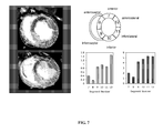
FIG. 7 illustrates example images from the mid-cavity of the heart. Prior to compound ID 1014 injection, the myocardium and ventricles are both dark. Five minutes after injection the ventricles are hyperintense because of contrast agent in the blood and the myocardium shows a dark, ischemic zone in anterior and anteroseptal segments 7 and 8 whereas the inferior and lateral wall is much more enhanced. At 20 minutes, the signal in the blood has decreased but the myocardium remains dark in segments 7 and 8 and brighter in segments 9-12.
FIG. 8 shows example images from the mid-cavity of the heart.
DETAILED DESCRIPTION
Definitions
Commonly used chemical abbreviations that are not explicitly defined in this disclosure may be found in The American Chemical Society Style Guide, Second Edition; American Chemical Society, Washington, D.C. (1997), “2001 Guidelines for Authors” J. Org. Chem. 66(1), 24A (2001), “A Short Guide to Abbreviations and Their Use in Peptide Science” J. Peptide. Sci. 5, 465-471 (1999).
For the purposes of this application, the term “aliphatic” describes any acyclic or cyclic, saturated or unsaturated, branched or unbranched carbon compound, excluding aromatic compounds.
The term “alkyl” includes saturated aliphatic groups, including straight-chain alkyl groups (e.g., methyl, ethyl, propyl, butyl, pentyl, hexyl, heptyl, octyl, nonyl, decyl, etc.), branched-chain alkyl groups (isopropyl, tert-butyl, isobutyl, etc.), cycloalkyl (alicyclic) groups (cyclopropyl, cyclopentyl, cyclohexyl, cycloheptyl, cyclooctyl), alkyl substituted cycloalkyl groups, and cycloalkyl substituted alkyl groups. The term alkyl further includes alkyl groups, which can further include oxygen, nitrogen, sulfur or phosphorous atoms replacing one or more carbons of the hydrocarbon backbone. In certain embodiments, a straight chain or branched chain alkyl has 6 or fewer carbon atoms in its backbone (e.g., C1-C6 for straight chain, C3-C6 for branched chain), and more preferably 4 or fewer. Likewise, preferred cycloalkyls have from 3-8 carbon atoms in their ring structure, and more preferably have 5 or 6 carbons in the ring structure. The term C1-C6 includes alkyl groups containing 1 to 6 carbon atoms.
Moreover, the term “alkyl” includes both “unsubstituted alkyls” and “substituted alkyls,” the latter of which refers to alkyl moieties having substituents replacing a hydrogen on one or more carbons of the hydrocarbon backbone. Such substituents can include, for example, alkenyl, alkynyl, halogen, hydroxyl, alkylcarbonyloxy, arylcarbonyloxy, alkoxycarbonyloxy, aryloxycarbonyloxy, carboxylate, alkylcarbonyl, arylcarbonyl, alkoxycarbonyl, aminocarbonyl, alkylaminocarbonyl, dialkylaminocarbonyl, alkylthiocarbonyl, alkoxyl, phosphate, phosphonato, phosphinato, cyano, amino (including alkylamino, dialkylamino, arylamino, diarylamino, and alkylarylamino), acylamino (including alkylcarbonylamino, arylcarbonylamino, carbamoyl and ureido), amidino, imino, sulfhydryl, alkylthio, arylthio, thiocarboxylate, sulfates, alkylsulfinyl, sulfonato, sulfamoyl, sulfonamido, nitro, trifluoromethyl, cyano, azido, heterocyclyl, alkylaryl, or an aromatic or heteroaromatic moiety. Cycloalkyls can be further substituted, e.g., with the substituents described above. An “arylalkyl” moiety is an alkyl substituted with an aryl (e.g., phenylmethyl(benzyl)). The term “alkyl” also includes the side chains of natural and unnatural amino acids. The term “n-alkyl” means a straight chain (i.e., unbranched) unsubstituted alkyl group.
The term “alkenyl” includes aliphatic groups that may or may not be substituted, as described above for alkyls, containing at least one double bond and at least two carbon atoms. For example, the term “alkenyl” includes straight-chain alkenyl groups (e.g., ethylenyl, propenyl, butenyl, pentenyl, hexenyl, heptenyl, octenyl, nonenyl, decenyl, etc.), branched-chain alkenyl groups, cycloalkenyl(alicyclic) groups (cyclopropenyl, cyclopentenyl, cyclohexenyl, cycloheptenyl, cyclooctenyl), alkyl or alkenyl substituted cycloalkenyl groups, and cycloalkyl or cycloalkenyl substituted alkenyl groups. The term alkenyl further includes alkenyl groups that include oxygen, nitrogen, sulfur or phosphorous atoms replacing one or more carbons of the hydrocarbon backbone. In certain embodiments, a straight chain or branched chain alkenyl group has 6 or fewer carbon atoms in its backbone (e.g., C2-C6 for straight chain, C3-C6 for branched chain). Likewise, cycloalkenyl groups may have from 3-8 carbon atoms in their ring structure, and more preferably have 5 or 6 carbons in the ring structure. The term C2-C6 includes alkenyl groups containing 2 to 6 carbon atoms.
Moreover, the term alkenyl includes both “unsubstituted alkenyls” and “substituted alkenyls,” the latter of which refers to alkenyl moieties having substituents replacing a hydrogen on one or more carbons of the hydrocarbon backbone. Such substituents can include, for example, alkyl groups, alkynyl groups, halogens, hydroxyl, alkylcarbonyloxy, arylcarbonyloxy, alkoxycarbonyloxy, aryloxycarbonyloxy, carboxylate, alkylcarbonyl, arylcarbonyl, alkoxycarbonyl, aminocarbonyl, alkylaminocarbonyl, dialkylaminocarbonyl, alkylthiocarbonyl, alkoxyl, phosphate, phosphonato, phosphinato, cyano, amino (including alkylamino, dialkylamino, arylamino, diarylamino, and alkylarylamino), acylamino (including alkylcarbonylamino, arylcarbonylamino, carbamoyl and ureido), amidino, imino, sulfhydryl, alkylthio, arylthio, thiocarboxylate, sulfates, alkylsulfinyl, sulfonato, sulfamoyl, sulfonamido, nitro, trifluoromethyl, cyano, azido, heterocyclyl, alkylaryl, or an aromatic or heteroaromatic moiety.
The term “alkynyl” includes unsaturated aliphatic groups analogous in length and possible substitution to the alkyls described above, but which contain at least one triple bond and two carbon atoms. For example, the term “alkynyl” includes straight-chain alkynyl groups (e.g., ethynyl, propynyl, butynyl, pentynyl, hexynyl, heptynyl, octynyl, nonynyl, decynyl, etc.), branched-chain alkynyl groups, and cycloalkyl or cycloalkenyl substituted alkynyl groups. The term alkynyl further includes alkynyl groups that include oxygen, nitrogen, sulfur or phosphorous atoms replacing one or more carbons of the hydrocarbon backbone. In certain embodiments, a straight chain or branched chain alkynyl group has 6 or fewer carbon atoms in its backbone (e.g., C2-C6 for straight chain, C3-C6 for branched chain). The term C2-C6 includes alkynyl groups containing 2 to 6 carbon atoms.
In general, the term “aryl” includes groups, including 5- and 6-membered single-ring aromatic groups that may include from zero to four heteroatoms, for example, benzene, phenyl, pyrrole, furan, thiophene, thiazole, isothiaozole, imidazole, triazole, tetrazole, pyrazole, oxazole, isooxazole, pyridine, pyrazine, pyridazine, and pyrimidine, and the like. Furthermore, the term “aryl” includes multicyclic aryl groups, e.g., tricyclic, bicyclic, such as naphthalene, benzoxazole, benzodioxazole, benzothiazole, benzoimidazole, benzothiophene, methylenedioxyphenyl, quinoline, isoquinoline, napthridine, indole, benzofuran, purine, benzofuran, deazapurine, or indolizine. Those aryl groups having heteroatoms in the ring structure may also be referred to as “aryl heterocycles,” “heterocycles,” “heteroaryls,” or “heteroaromatics.” An aryl group may be substituted at one or more ring positions with substituents.
For the purposes of this application, “DTPA” refers to a chemical compound comprising a substructure composed of diethylenetriamine, wherein the two primary amines are each covalently attached to two acetyl groups and the secondary amine has one acetyl group covalently attached according to the following formula:
wherein each X is independently a functional group capable of coordinating a metal cation, preferably COO
−, COOH, C(O)NH
2, C(O)NHR, C(O)NRR′, PO
3 2−, PO
3R
−, P(R)O
2 − or NHR, or OR wherein R is any aliphatic group. When each X group is the tert-butoxy (
tBu) carboxylate ester (COO
tBu), the structure may be referred to as “DTPE” (“E” for ester).
For the purposes of this application, “DOTA” refers to a chemical compound comprising a substructure composed of 1,4,7,11-tetraazacyclododecane, wherein the amines each have one acetyl group covalently attached according to the following formula:
wherein X is defined above.
For the purposes of this application, “NOTA” refers to a chemical compound comprising a substructure composed of 1,4,7-triazacyclononane, wherein the amines each have one acetyl group covalently attached according to the following formula:
wherein X is defined above.
For the purposes of this application, “DO3A” refers to a chemical compound comprising a substructure composed of 1,4,7,11-tetraazacyclododecane, wherein three of the four amines each have one acetyl group covalently attached and the other amine has a substituent having neutral charge according to the following formula:
wherein R
1 is an uncharged chemical moiety, preferably hydrogen, any aliphatic, alkyl group, or cycloalkyl group, and uncharged derivatives thereof. The chelate “HP”-DO3A has R
1=—CH
2(CHOH)CH
3.
In each of the four structures above, the carbon atoms of the indicated ethylenes may be referred to as “backbone” carbons. The designation “bbDTPA” may be used to refer to the location of a chemical bond to a DTPA molecule (“bb” for “back bone”). Note that as used herein, bb(CO)DTPA-Gd means a C═O moiety bound to an ethylene backbone carbon atom of DTPA.
The terms “chelating ligand,” “chelating moiety,” and “chelate moiety” may be used to refer to any polydentate ligand which is capable of coordinating a metal ion, including DTPA (and DTPE), DOTA, DO3A, DOTAGA, Glu-DTPA, or NOTA molecule, or any other suitable polydentate chelating ligand as is further defined herein, that is either coordinating a metal ion or is capable of doing so, either directly or after removal of protecting groups. The term “chelate” refers to the actual metal-ligand complex, and it is understood that the polydentate ligand will eventually be coordinated to a medically useful metal ion.
The term “specific binding affinity” as used herein, refers to the capacity of a peptide or composition to be taken up by, retained by, or bound to a particular biological component to a greater degree than other components. Peptides that have this property are said to be “targeted” to the “target” component. Peptides that lack this property are said to be “non-specific” or “non-targeted” agents. The binding affinity for a target is expressed in terms of the equilibrium dissociation constant “Kd” or as a percentage of the compound bound to the target under a defined set of conditions.
The term “relaxivity” as used herein, refers to the increase in either of the MRI quantities 1/T1 or 1/T2 per millimolar (mM) concentration of paramagnetic ion, contrast agent, therapeutic agent, or diagnostic composition, wherein T1 is the longitudinal or spin-lattice, relaxation time, and T2 is the transverse or spin-spin relaxation time of water protons or other imaging or spectroscopic nuclei, including protons found in molecules other than water. Relaxivity is expressed in units of mM−1s−1.
As used herein, the term “purified” refers to a peptide that has been separated from either naturally occurring organic molecules with which it normally associates or, for a chemically-synthesized peptide, separated from any other organic molecules present in the chemical synthesis. Typically, the polypeptide is considered “purified” when it is at least 70% (e.g., 70%, 80%, 90%, 95%, or 99%), by dry weight, free from any other proteins or organic molecules. The terms “purified” and “isolated” are used interchangeably herein.
As used herein, the term “peptide” refers to a chain of amino acids that is about 2 to about 75 amino acids in length (e.g., 3 to 50 amino acids, 1 to 50 amino acids, 3 to 30 amino acids, 2 to 25 amino acids, 10-25 amino acids, 10-50 amino acids, 15-25 amino acids, 8-20 amino acids, 8-15 amino acids, 16-17 amino acids). All peptide sequences herein are written from the N to C terminus. Additionally, peptides containing two or more cysteine residues can form disulfide bonds under non-reducing conditions. Formation of the disulfide bond can result in the formation of a cyclic peptide. The cyclic peptide may represent all or a portion of the peptide sequence. A peptide as described herein can be branched, e.g., have additional amino acids linked to one or more of the side chains of an amino acid in the chain. For example, a lysine residue having an additional lysine residue off of the ε-amino group, such a functionality is represented as K(K), wherein the group in the parentheses is that which is linked off of a side chain. Where more than one amino acid is bound off of the side chain, it is represented with a period separating the two amino acids, e.g., K(Y.G). In certain embodiments, a chelating group or a metal containing chelating group may be linked to one or more side chains of an amino acid. For example, a lysine residue having a GdDTPA complex off of the ε-amino group, such a functionality is represented as K(GdDTPA), wherein the group in parenthesis is that which is linked off of a side chain.
Additionally, an amino acid can be substituted. Such substituents can include, for example, alkenyl, alkynyl, halogen, hydroxyl, alkylcarbonyloxy, arylcarbonyloxy, alkoxycarbonyloxy, aryloxycarbonyloxy, carboxylate, alkylcarbonyl, arylcarbonyl, alkoxycarbonyl, aminocarbonyl, alkylaminocarbonyl, dialkylaminocarbonyl, alkylthiocarbonyl, alkoxyl, phosphate, phosphonato, phosphinato, cyano, amino (including alkylamino, dialkylamino, arylamino, diarylamino, and alkylarylamino), acylamino (including alkylcarbonylamino, arylcarbonylamino, carbamoyl and ureido), amidino, imino, sulfhydryl, alkylthio, arylthio, thiocarboxylate, sulfates, alkylsulfinyl, sulfonato, sulfamoyl, sulfonamido, nitro, trifluoromethyl, cyano, azido, heterocyclyl, alkylaryl, or an aromatic or heteroaromatic moiety. Cycloalkyls can be further substituted, e.g., with the substituents described above. An “arylalkyl” moiety is an alkyl substituted with an aryl (e.g., phenylmethyl(benzyl)). The term “alkyl” also includes the side chains of natural and unnatural amino acids. The term “n-alkyl” means a straight chain (i.e., unbranched) unsubstituted alkyl group.
As used herein, the term “natural” or “naturally occurring” amino acid refers to one of the twenty most common occurring amino acids. Natural amino acids modified to provide a label for detection purposes (e.g., radioactive labels, optical labels, or dyes) are considered to be natural amino acids. Natural amino acids are referred to by their standard one- or three-letter abbreviations. Natural amino acids can be in their D or L form. As used herein, a lower case one or two letter abbreviation refers to the D-form of an amino acid.
The terms “target binding” and “binding” for purposes herein refer to non-covalent interactions of a peptide with a target. These non-covalent interactions are independent from one another and may be, inter alia, hydrophobic, hydrophilic, dipole-dipole, pi-stacking, hydrogen bonding, electrostatic associations, or Lewis acid-base interactions.
As used herein, all references to “Gd,” “gado,” or “gadolinium” mean the Gd(III) paramagnetic metal ion.
Collagen Binding Peptides
Isolated peptides described herein have an affinity for an extracellular matrix protein, such as collagen, including human collagen type I. In some embodiments, an isolated peptide has a specific binding affinity for an extracellular matrix protein such as collagen relative to serum proteins, such as human serum albumin (HSA) and/or fibrinogen. In these embodiments, the peptide may exhibit a smaller dissociation constant for an extracellular matrix protein relative to the dissociation constant for a serum protein.
Extracellular matrix proteins include soluble and insoluble proteins, polysaccharides, including heteropolysaccharides and polysaccharides covalently bound to proteins, and cell-surface receptors. For example, extracellular matrix proteins can be collagens (Types I, II, III, IV, V, and VI), elastin, decorin, glycosoaminoglycans, and proteoglycans.
Collagens are particularly useful extracellular matrix proteins to target. For example, collagens I and III are the most abundant components of the extracellular matrix of myocardial tissue, representing over 90% of total myocardial collagen and about 5% of dry myocardial weight. The ratio of collagen I to collagen III in the myocardium is approximately 2:1, and their total concentration is approximately 100 μM in the extracellular matrix. Human collagen type I is a trimer of two chains with an [α1(I)]2 [α2(I)] stoichiometry characterized by a repeating G-X-Y sequence motif, where X is most frequently proline and Y is frequently hydroxyproline. Thus, in some embodiments, a peptide has an affinity for human and/or rat collagen type I.
Peptides useful for inclusion in the diagnostic compositions described herein can include natural or unnatural amino acids which may be in the D or L form. In some embodiments, all of the amino acids are natural amino acids. In some embodiments, all of the amino acids are in the L form. The peptides can be synthesized according to standard synthesis methods such as those disclosed in, e.g., WO 01/09188 and WO 01/08712. Charged groups on the peptides can be neutralized if desired. For example, the C-terminal carboxylate moiety can be amidated with an —NH2 group, yielding a C(═O)NH2 moiety. In certain embodiments, the C-terminus is amidated via cleavage of the peptide from the resin; see the Examples, below. For ease of synthesis and cost considerations, it is preferred that the peptides have between 3 to 75 amino acids (e.g., 3 to 50, 1 to 50, 10 to 50, 10 to 30, 3 to 30, 3 to 20, 3 to 15, 5 to 30, 3 to 25, 16 to 17, 5 to 25, 5 to 20, 5 to 15, 11 to 25, 11 to 50, 11 to 40, 10 to 12, 8 to 30, 8 to 20, or 8 to 15 amino acids in length).
Amino acids with many different protecting groups appropriate for immediate use in the solid phase synthesis of peptides are commercially available. Concatemers of peptides (2-5 or more) can increase binding affinity and specificity for an extracellular matrix protein (Verrecchio, A., Germann, M. W., Schick, B. P., Kung, B., Twardowski, T., and San Antonio, J. D. J. Biol. Chem. (2000) 275, 7701-7707).
Peptides can be assayed for affinity to the appropriate extracellular matrix protein by methods as disclosed in WO 01/09188 and WO 01/08712, and as described below. For example, peptides can be screened for binding to an extracellular matrix protein by methods well known in the art, including equilibrium dialysis, affinity chromatography, and inhibition or displacement of probes bound to the matrix protein. For example, peptides can be evaluated for their ability to bind to collagen, such as dried human collagen type I or dried rat collagen type I. In certain cases, a peptide can exhibit a percent binding to dried human collagen type I or dried rat collagen type I (see assays described below) of greater than 10%, e.g., greater than 12%, greater than 15%, greater than 20%, greater than 25%, greater than 30%, greater than 35%, greater than 40%, greater than 45%, greater than 50%, greater than 55%, greater than 60%, greater than 65%, greater than 70%, greater than 75%, greater than 80%, or greater than 85%. In some embodiments, a peptide can exhibit a percent binding to dried human collagen in the range of from about 10% to about 50%, or from about 20% to about 60%, or from about 30% to about 60%, or from about 40% to about 90%. Certain peptides useful for inclusion in the diagnostic compositions herein can exhibit an affinity for collagen. Such peptides can be identified through phage display experiments; see the Examples, below.
Collagen binding peptides can be derivatized with non-metallic radionuclides for PET or SPECT imaging. For instance the tyrosine amino acid can be iodinated with I-123, I-125, or I-131 as described in the Examples. Flourine-18 can be incorporated into the peptide using fluorination and bioconjugation techniques as described in the literature (see e.g. Guenther K J, Yoganathan S, Garofalo R, Kawabata T, Strack T, Labiris R, Dolovich M, Chirakal R, Valliant J F. “Synthesis and in vitro evaluation of 18F- and 19F-labeled insulin: a new radiotracer for PET-based molecular imaging studies.” J Med. Chem. 2006 49:1466-74; de Bruin B, Kuhnast B, Hinnen F, Yaouancq L, Amessou M, Johannes L, Samson A, Boisgard R, Tavitian B, Dolle F. “1-[3-(2-[18F]fluoropyridin-3-yloxy)propyl]pyrrole-2,5-dione: design, synthesis, and radiosynthesis of a new [18F]fluoropyridine-based maleimide reagent for the labeling of peptides and proteins.” Bioconjug Chem. 2005 16:406-20; Chen X, Park R, Hou Y, Khankaldyyan V, Gonzales-Gomez I, Tohme M, Bading J R, Laug W E, Conti P S. “MicroPET imaging of brain tumor angiogenesis with 18F-labeled PEGylated RGD peptide.” Eur J Nucl Med Mol. Imaging. 2004 31:1081-9; Wester H J, Schottelius M, Scheidhauer K, Meisetschlager G, Herz M, Rau F C, Reubi J C, Schwaiger M. “PET imaging of somatostatin receptors: design, synthesis and preclinical evaluation of a novel 18F-labelled, carbohydrated analogue of octreotide.” Eur J Nucl Med Mol. Imaging. 2003 30(1):117-22).
Peptides disclosed herein can include the amino acid sequence W-X1-C-(X2)n-W-X3-C (SEQ ID NO: 806), wherein n=5-7; X1, X2, and X3 are any amino acid; and wherein the peptide has a length of 11 to 50 amino acids. In some embodiments, the peptide can have a length of 11 to 30 amino acids, 11 to 35 amino acids, 11 to 25 amino acids, 11 to 20 amino acids, or 11 to 15 amino acids. In certain embodiments, X1 is selected from K, Q, Y, T, E, D, L, R, H, I, V, N, M, and A. Similarly, X2 is in some cases selected from R, E, D, S, H, K, N, Y, M, V, I, Q, and G. In certain cases, X1 is selected from M, K, Q, T, Y, and R, and X3 is selected from Y, K, H, V, S, N, and M.
A purified peptide can include the amino acid sequence W-X1-C-X2-G*-X3-X4-X5-X6-W-X7-C (SEQ ID NO: 807), wherein X1 is any amino acid; X2 can be S, V, T, H, R, Y, or D; G* is G or any amino acid in D form; X3 can be D or N, independently in D or L form; X4 can be any amino acid in D or L form; X5 can be any amino acid in D or L form; X6 can be T, K, H, D, A, R, Y, or E; and X7 can be Y, K, H, V, S, M, or N, wherein the peptide has a total length of 12 to 50 amino acids. The peptide length can vary, as indicated previously, e.g., 12 to 25 amino acids, 12 to 30 amino acids, 12 to 40 amino acids, 12 to 20 amino acids, and 12 to 15 amino acids. In some cases, G* is selected from G and the D form of the amino acids A, S, R, Y, and L.
In some embodiments, such a purified peptide can include the amino acid sequence: W-X1-C-X2-G*-X3-X4-X5-X6-W-X7-C-X8-X9 (SEQ ID NO: 808), wherein X1 to X6 and G* are as defined above for SEQ ID NO: 807; X8 can be N, L, I, R, K, or A; and X9 can be Y, F, M, R, or H, independently in D or L form. In some cases, X3 can be D. In some embodiments, X1 can be T; X2 can be S, T or V; X4 can be E, H, I, S, or A; X5 can be Y, K, L, F, A, or P; X6 is T; X7 is H or K; X8 is N, K, or A; and X9 is Y or F. In some embodiments, the peptide can include one of the following amino acid sequences:
| |
W-Y-C-S-G-D-H-L-D-W-K-C; |
|
| |
and |
| |
|
A purified peptide can include any of the amino acid sequences in Tables 1-16, 18-41, 44, and 45. In some embodiments, such peptides have a total length of 50 amino acids or less, e.g., 45 amino acids or less, 40 amino acids or less, 35 amino acids or less, 30 amino acids or less, 25 amino acids or less, 20 amino acids or less, or 15 amino acids or less.
A purified peptide can include the amino acid sequence Q-W-H-C-T-T-R-F-P-H-H-Y-C-L-Y-G (SEQ ID NO: 74), wherein the peptide has a total length of 16 to 50 amino acids, e.g., 16 to 40 amino acids, 16 to 30 amino acids, 16 to 20 amino acids, or 16 to 18 amino acids.
In other cases, a purified peptide can include the amino acid sequence C-Y-Q-X1-X2-C-W-X3-W (SEQ ID NO: 813), wherein X1 is any amino acid; X2 is any amino acid; X3 is any amino acid; wherein each C, Y, Q, W, X1, X2, or X3, independently, can be in the D form; and wherein the peptide contains 9 to 50 amino acids, such as 9 to 40 amino acids, 9 to 30 amino acids, 9 to 20 amino acids, or 9 to 15 amino acids. In some cases, X1 is selected from A, G, I, L, V, F, and P; X2 is selected from G, A, I, L, V, F, and P; and X3 is selected from I, A, G, L, V, F, and P. In certain embodiments, the peptide includes the amino acid sequence C-Y-Q-A-G-C-W-1-W (SEQ ID NO: 814) in any combination of D or L forms for the individual amino acids. For example, a peptide can include SEQ ID NO: 814 in all L-form.
A purified peptide can include amino acid sequence Y-X1-X2-C-Y-Q-X3-X4-C-W-X5-W (SEQ ID NO: 815), wherein X1 is any amino acid; X2 is any amino acid; X3 is any amino acid; X4 is any amino acid; X5 is I, G, L, V, F, or P; and wherein the peptide contains 12 to 50 amino acids, such as 12 to 40 amino acids, 12 to 30 amino acids, 12 to 25 amino acids, 12 to 20 amino acids, or 12 to 15 amino acids. In some embodiments, X1 is selected from H, R, K, E, D, Q, or N; X2 is selected from A, G, I, L, V, F, or P; X3 is selected from A, G, I, L, V, F, or P; X4 is selected from G, A, I, L, V, F, or P; and X5 is selected from I, L, V, or F. For example, a purified peptide can include SEQ ID NO:1, SEQ ID NO:132, or SEQ ID NO:135. Other peptides are set forth in the accompanying claims.
A purified peptide can include the amino acid sequence C*-X1-X2-X3-X4-X5-X6-X7-X8-C* (SEQ ID NO: 816), wherein X1, X2, X3, X4, X5, X6, X7, and X8 are any amino acid; C* is C or Pen in D or L form; and the peptide has a length of 10 to 50 amino acids, such as 10 to 20 amino acids, 10 to 30 amino acids, 10 to 40 amino acids, and 10 to 15 amino acids. In some cases, X1 is selected from T, A, K, V, I, S, Y, G, R, P, L, 3-NO2 Y, 4-Pal, 4-CO2H-F, 4-tBu-F, F(4-NH2), Y(Bn, 3-Cl), b-h-S, Y(3-I), and Aib, in D or L form; X2 is selected from T, A, N, S, Y, R, V, I, K, D, G, b-h-G, Orn, and Dpr, in D or L form; X3 is selected from R, A, S, L, Y, D, K, G, P, Aib, Y(3-Cl), I, Cha, Abu, F(4-F), Dopa, Tle, Cit, b-h-D, and K(Boc), in D or L form; X4 is selected from F, A, Y, E, R, L, Bip, F(4-CF3), 4-Pal, 1-Nal, F(4-NO2), Hfe, Bpa, F(4-CN), F(4-NH2), F(3,4-OMe), 2-Nal, Y(3-Cl), Aib, and b-h-E, in D or L form; X5 is selected from P, A, Y, D, R, T, P(3-OH), ΔPro, Pip, N-Me-A, P(3-OH), Y(3-I), b-h-Y, and Aib, in D or L form; X6 is selected from H, A, S, K, N, Y, T, D, R, W, P, Aib, and b-h-T, in D or L form; X7 is selected from H, A, S, N, D, Y, W, Aib, Dpr, 2-Pal, 1-Nal, thien-W, W(5-OH), and b-h-W, in D or L form; and X8 is selected from Y, A, R, T, V, H, D, S, P, 1-Nal, Bip, DOPA, H-Tyr, H-Tyr(Me), F(3-OMe), Y(3-Cl), Y(2,6-Me2), Dip, F(4-NH2), or Aib, in D or L form. For example, a peptide can be C-T-T-S-F-P-H-H-Y-C (SEQ ID NO: 817), C-T-T-K-F-P-H-H-Y-C (SEQ ID NO: 818), C-Y-T-Y-F-P-H-H-Y-C (SEQ ID NO: 819), C-T-T-R-F-P-H-H-Y-C (SEQ ID NO: 820), or C-S-G-D-E-Y-T-W-H-C (SEQ ID NO: 821).
In another embodiment, a purified peptide can include the amino acid sequence C*-X1-X2-X3-X4-X5-X6-X7-X8-C*-X9-X10-X1 (SEQ ID NO: 822), wherein X1, X2, X3, X4, X5, X6, X7, X8, X9, X10, and X11 are any amino acid; C* is C or Pen, in D or L form; and the peptide has a length of 13 to 50 amino acids, such as 13 to 40 amino acids, 13 to 30 amino acids, 13 to 20 amino acids, and 13 to 17 amino acids. In certain embodiments, X1 is selected from T, A, K, V, I, S, Y, G, R, P, L, 3-NO2 Y, 4-Pal, 4-CO2H-F, 4-tBu-F, F(4-NH2), Y(Bn, 3-Cl), b-h-S, Y(3-I), and Aib, in D or L form; X2 is selected from T, A, N, S, Y, R, V, I, K, D, G, b-h-G, Orn, and Dpr, in D or L form; X3 is selected from R, A, S, L, Y, D, K, G, P, Aib, Y(3-Cl), I, Cha, Abu, F(4-F), Dopa, Tle, Cit, b-h-D, and K(Boc), in D or L form; X4 is selected from F, A, Y, E, R, L, Bip, F(4-CF3), 4-Pal, 1-Nal, F(4-NO2), Hfe, Bpa, F(4-CN), F(4-NH2), F(3,4-OMe), 2-Nal, Y(3-Cl), Aib, and b-h-E, in D or L form; X5 is selected from P, A, Y, D, R, T, P(3-OH), ΔPro, Pip, N-Me-A, P(3-OH), Y(3-I), b-h-Y, and Aib, in D or L form; X6 is selected from H, A, S, K, N, Y, T, D, R, W, P, Aib, and b-h-T, in D or L form; X7 is selected from H, A, S, N, D, Y, W, Aib, Dpr, 2-Pal, 1-Nal, thien-W, W(5-OH), and b-h-W, in D or L form; X8 is selected from Y, A, R, T, V, H, D, S, P, 1-Nal, Bip, DOPA, H-Tyr, H-Tyr(Me), F(3-OMe), Y(3-Cl), Y(2,6-Me2), Dip, F(4-NH2), and Aib, in D or L form; X9 is selected from L, A, I, K, V, F, N, Y, P, Aib, Hse, Hfe, Bpa, 2-Nal, Y(3-Cl), Dip, and F(4-NH2), in D or L form; X10 is selected from Y, A, F, E, Bpa, 2-Nal, Y(3-Cl), Dip, F(4-NH2), and Y(3-I), in D or L form; and X11 is selected from G, E, Y, F, V, Bip, F(4-NH2), and Aib, in D or L form.
A purified peptide can include the amino acid sequence C*-X1-X2-X3-X4-X5-X6-X7-X8-C*-X9-X10-X11-X12 (SEQ ID NO: 823), wherein X1, X2, X3, X4, X5, X6, X7, X8, X9, X10, and X11 are any amino acids; X12 is any one or two amino acids; C* is C or Pen, in D or L form; and wherein the peptide has a length of 14 to 50 amino acids, such as 14 to 40 amino acids, 14 to 30 amino acids, 14 to 20 amino acids, and 14 to 17 amino acids. In some embodiments, X1 is selected from T, A, K, V, I, S, Y, G, R, P, L, 3-NO2 Y, 4-Pal, 4-CO2H-F, 4-tBu-F, F(4-NH2), Y(Bn, 3-Cl), b-h-S, Y(3-I), and Aib, in D or L form; X2 is selected from T, A, N, S, Y, R, V, I, K, D, G, b-h-G, Orn, and Dpr, in D or L form; X3 is selected from R, A, S, L, Y, D, K, G, P, Aib, Y(3-Cl), I, Cha, Abu, F(4-F), Dopa, Tle, Cit, b-h-D, and K(Boc), in D or L form; X4 is selected from F, A, Y, E, R, L, Bip, F(4-CF3), 4-Pal, 1-Nal, F(4-NO2), Hfe, Bpa, F(4-CN), F(4-NH2), F(3,4-OMe), 2-Nal, Y(3-Cl), Aib, and b-h-E, in D or L form; X5 is selected from P, A, Y, D, R, T, P(3-OH), ΔPro, Pip, N-Me-A, P(3-OH), Y(3-I), b-h-Y, and Aib, in D or L form; X6 is selected from H, A, S, K, N, Y, T, D, R, W, P, Aib, and b-h-T, in D or L form; X7 is selected from H, A, S, N, D, Y, W, Aib, Dpr, 2-Pal, 1-Nal, thien-W, W(5-OH), and b-h-W, in D or L form; X8 is selected from Y, A, R, T, V, H, D, S, P, 1-Nal, Bip, DOPA, H-Tyr, H-Tyr(Me), F(3-OMe), Y(3-Cl), Y(2,6-Me2), Dip, F(4-NH2), and Aib, in D or L form; X9 is selected from L, A, I, K, V, F, N, Y, P, Aib, Hse, Hfe, Bpa, 2-Nal, Y(3-Cl), Dip, and F(4-NH2), in D or L form; X10 is selected from Y, A, F, E, Bpa, 2-Nal, Y(3-Cl), Dip, F(4-NH2), and Y(3-I), in D or L form; X11 is selected from G, E, Y, F, V, Bip, F(4-NH2), and Aib, in D or L form; and X12 is selected from K, KK, Peg K, PEG(1×O), 1,4-AMB, 1,3-AMB, 1,6-Hex, PEG, and GTE, in D or L form.
In another embodiment, a purified peptide includes the amino acid sequence X14-X13-C*-X1-X2-X3-X4-X5-X6-X7-X8-C* (SEQ ID NO: 824), wherein X1, X2, X3, X4, X5, X6, X7, X8, X13, and X14 are any amino acid; C* is C or Pen, in D or L form; and wherein the peptide has a length of 12 to 50 amino acids, such as 12 to 40 amino acids, 12 to 30 amino acids, 12 to 20 amino acids, and 12 to 17 amino acids. In certain embodiments, X1 is selected from T, A, K, V, I, S, Y, G, R, P, L, 3-NO2 Y, 4-Pal, 4-CO2H-F, 4-tBu-F, F(4-NH2), Y(Bn, 3-Cl), b-h-S, Y(3-I), and Aib, in D or L form; X2 is selected from T, A, N, S, Y, R, V, I, K, D, G, b-h-G, Orn, and Dpr, in D or L form; X3 is selected from R, A, S, L, Y, D, K, G, P, Aib, Y(3-Cl), I, Cha, Abu, F(4-F), Dopa, Tle, Cit, b-h-D, and K(Boc), in D or L form; X4 is selected from F, A, Y, E, R, L, Bip, F(4-CF3), 4-Pal, 1-Nal, F(4-NO2), Hfe, Bpa, F(4-CN), F(4-NH2), F(3,4-OMe), 2-Nal, Y(3-Cl), Aib, and b-h-E, in D or L form; X5 is selected from P, A, Y, D, R, T, P(3-OH), ΔPro, Pip, N-Me-A, P(3-OH), Y(3-I), b-h-Y, and Aib, in D or L form; X6 is selected from H, A, S, K, N, Y, T, D, R, W, P, Aib, and b-h-T, in D or L form; X7 is selected from H, A, S, N, D, Y, W, Aib, Dpr, 2-Pal, 1-Nal, thien-W, W(5-OH), and b-h-W, in D or L form; X8 is selected from Y, A, R, T, V, H, D, S, P, 1-Nal, Bip, DOPA, H-Tyr, H-Tyr(Me), F(3-OMe), Y(3-Cl), Y(2,6-Me2), Dip, F(4-NH2), and Aib, in D or L form; X13 is selected from H, A, S, K, N, D, Y, T, P, and Aib, in D or L form; and X14 is selected from W, A, Y, 1-Nal, 2-Nal, thien-W, Tic, or W(5-OH), in D or L form.
A purified peptide can include the amino acid sequence X16-X15-X14-X13-C*-X1-X2-X3-X4-X5-X6-X7-X8-C* (SEQ ID NO: 825), wherein X1, X2, X3, X4, X5, X6, X7, X8, X13, and X14 are any amino acid; X15 and X16 comprise one to three amino acids; C* is C or Pen, in D or L form; and wherein the peptide has a length of 14 to 50 amino acids, such as 14 to 40 amino acids, 14 to 30 amino acids, 14 to 20 amino acids, and 14 to 17 amino acids. In some embodiments, X1 is selected from T, A, K, V, I, S, Y, G, R, P, L, 3-NO2 Y, 4-Pal, 4-CO2H-F, 4-tBu-F, F(4-NH2), Y(Bn, 3-Cl), b-h-S, Y(3-I), and Aib, in D or L form; X2 is selected from T, A, N, S, Y, R, V, I, K, D, G, b-h-G, Orn, and Dpr, in D or L form; X3 is selected from R, A, S, L, Y, D, K, G, P, Aib, Y(3-Cl), I, Cha, Abu, F(4-F), Dopa, Tle, Cit, b-h-D, and K(Boc), in D or L form; X4 is selected from F, A, Y, E, R, L, Bip, F(4-CF3), 4-Pal, 1-Nal, F(4-NO2), Hfe, Bpa, F(4-CN), F(4-NH2), F(3,4-OMe), 2-Nal, Y(3-Cl), Aib, and b-h-E, in D or L form; X5 is selected from P, A, Y, D, R, T, P(3-OH), ΔPro, Pip, N-Me-A, P(3-OH), Y(3-I), b-h-Y, and Aib, in D or L form; X6 is selected from H, A, S, K, N, Y, T, D, R, W, P, Aib, and b-h-T, in D or L form; X7 is selected from H, A, S, N, D, Y, W, Aib, Dpr, 2-Pal, 1-Nal, thien-W, W(5-OH), and b-h-W, in D or L form; X8 is selected from Y, A, R, T, V, H, D, S, P, 1-Nal, Bip, DOPA, H-Tyr, H-Tyr(Me), F(3-OMe), Y(3-Cl), Y(2,6-Me2), Dip, F(4-NH2), and Aib, in D or L form; X13 is selected from H, A, S, K, N, D, Y, T, P, and Aib, in D or L form; X14 is selected from W, A, Y, 1-Nal, 2-Nal, thien-W, Tic, or W(5-OH), in D or L form; X15 is selected from Q, G, A, D, S, P, K, GQ, K(G), K(Y.G), K(V.G). K(F.G), K(H.H), KK(K), Dpr, and Aib, in D or L form; and X16 is selected from G, K, PP, GY, GV, GF, GH, GK(G), KK(K), Dpr, EAG, and PPG, in D or L form.
In other embodiments, a purified peptide can include the amino acid sequence X1-X2-X3-C*-X4-T-X5-X6-P*-X7-H-X8-C-X9-X10-X11 (SEQ ID NO: 826), wherein X1, X2, X3, X4, X5, X6, X7, X8, X9, X10, and X11 are any amino acid; C* is C or Pen; P* is P in D or L form; and wherein the peptide has a length of 16 to 50 amino acids, such as 16 to 40 amino acids, 16 to 30 amino acids, 16 to 20 amino acids, and 16 to 17 amino acids. In certain embodiments, X1 is selected from any amino acid in L form; X2 is selected from W or W*; X3 is selected from H, A, K, or S; X4 is selected from T, Y, G, K, and Y*; X5 is selected from any amino acid in L form; X6 is selected from F, Y, and Y*; X7 is selected from H, A, and Y; X8 is selected from Y and Y*; X9 is selected from L, V, L*, and Y*; X10 is selected from Y, F, and Y*; and X11 is selected from G, Y, Bip, and Y*; wherein W* is 1-Nal, 2-Nal, Bpa, thien-W, W(5-OH), 7-aza-Trp, 1-methyl-Trp, 5-bromo-Tryp, 5-chloro-Tryp, 5-fluor-Trp, 7-methyl-trp, 6-methyl-Trp, 6-fluoro-Trp, or 6-hydroxy-trp; Y* is F(4-NH2), F(3,4-OMe2), F(3-OMe), F(4-CF3), F(4-CN), F(4-NO2), F(4-F), F(4-NO2), Hfe, 4-tBu-F, 4-CO2H-F, h-Tyr, h-Tyr(Me), Y(2,6-Me2), Y(3-Cl), Y(3-I), Y(Bn, 3-Cl), 2-substituted L-Tyr, 2,3-substituted-L-Tyr, 2,3,5-substituted-L-tyr, 2,5-substituted-L-Tyr, 2,6-substituted-L-Tyr, 2,3,5,6-substituted-L-Tyr, 3-substituted-L-Tyr, 3,5-substituted-L-Tyr, 2-substituted L-Phe, 2,3-substituted-L-Phe, 2,3,5-substituted-L-Phe, 2,5-substituted-L-Phe, 2,6-substituted-L-Phe, 2,3,5,6-substituted-L-Phe, 3-substituted-L-Phe, 3,5-substituted-L-Phe, L-2-pyridylalanine, L-3-pyridylalanine, or L-4-pyridylalanine; and L* is I, V, A, L, G, Tle, L-norvaline, L-norleucine, L-dehydroleucine, L-abu (2-aminobutyric acid), L-tert-leucine, beta-cyclohexyl-L-alanine, L-homoleucine, or L-homo-cyclohexylalanine.
In a further embodiment, a purified peptide can include the amino acid sequence X1-X2-X3-C-X4-X5-D-X6-X7-X8-W-X9-C-X10-X11-X12 (SEQ ID NO: 827), wherein X1, X2, X3, X4, X5, X6, X7, X8, X9, X10, X11, and X12 are any amino acid; and wherein said peptide has a length of 16 to 50 amino acids, such as 16 to 40 amino acids, 16 to 30 amino acids, 16 to 20 amino acids, and 16 to 17 amino acids. In some cases, X1 is selected from any amino acid in L form; X2 is selected from W and W*; X3 is selected from T, A, or W; X4 is selected from S, Y, A, V, and Y*; X5 is selected from G or D*; X6 is selected from E, A, and H; X7 is selected from Y, L, or Y*; X8 is selected from T, Y, A, and S; X9 is selected from H, S, and Y; X10 is selected from N and A; X11 is selected from Y and Y*; X12 is selected from any amino acid in L form; wherein W* is 1-Nal, 2-Nal, Bpa, thien-W, W(5-OH), 7-aza-Trp, 1-methyl-Trp, 5-bromo-Tryp, 5-chloro-Tryp, 5-fluor-Tip, 7-methyl-trp, 6-methyl-Tip, 6-fluoro-Tip, or 6-hydroxy-trp; Y* is F(4-NH2), F(3,4-OMe2), F(3-OMe), F(4-CF3), F(4-CN), F(4-NO2), F(4-F), F(4-NO2), Hfe, 4-tBu-F, 4-CO2H-F, h-Tyr, h-Tyr(Me), Y(2,6-Me2), Y(3-Cl), Y(3-T), Y(Bn, 3-Cl), 2-substituted L-Tyr, 2,3-substituted-L-Tyr, 2,3,5-substituted-L-tyr, 2,5-substituted-L-Tyr, 2,6-substituted-L-Tyr, 2,3,5,6-substituted-L-Tyr, 3-substituted-L-Tyr, 3,5-substituted-L-Tyr, 2-substituted L-Phe, 2,3-substituted-L-Phe, 2,3,5-substituted-L-Phe, 2,5-substituted-L-Phe, 2,6-substituted-L-Phe, 2,3,5,6-substituted-L-Phe, 3-substituted-L-Phe, 3,5-substituted-L-Phe, L-2-pyridylalanine, L-3-pyridylalanine, or L-4-pyridylalanine; and D* is any amino acid in D form.
Any of the peptides described herein can be capable of forming a disulfide bond under non-reducing conditions, as known to those having ordinary skill in the art. In certain cases, any of the peptides described herein include a disulfide bond, and form a cyclized peptide structure. Any of the peptides can exhibit specific binding affinity for collagen, e.g., collagen type I from human or rat.
Specific peptides and peptide linker combinations are also set forth in Tables 1-16, 18-41, 44, and 45 and in the Examples, below.
Diagnostic Compositions
Diagnostic compositions (e.g., diagnostic compositions suitable for MR imaging, nuclear imaging, PET imaging, SPECT imaging, or optical imaging), which can be used for detecting pathologies where abnormal or excessive proliferation of collagen is implicated, are described herein. Typically such diagnostic compositions will include one or more imaging moieties (IEMs) coupled, such as through a linker (L), to an Extracellular Matrix Targeting Group (EMTG).
Extracellular Matrix Targeting Group
Generally, the Extracellular Matrix Targeting Group (EMTG) has an affinity for an extracellular matrix component, such as collagen. For example, the EMTG can bind the extracellular matrix component with a dissociation constant of less than 100 μM (e.g., less than 50 μM, less than 10 μM, less than 5 μM, less than 1 μM, or less than 100 nM). In some embodiments, the EMTG has a specific binding affinity for an extracellular matrix component relative to serum proteins, such as human serum albumin (HSA) and fibrinogen, to result in decreased background signal (e.g., background signal of blood). In these embodiments, the EMTG may exhibit a smaller dissociation constant for an extracellular matrix component relative to the dissociation constant for a serum protein.
Extracellular matrix components of the myocardium include soluble and insoluble proteins, polysaccharides, including heteropolysaccharides and polysaccharides covalently bound to proteins, and cell-surface receptors. For example, extracellular matrix components can be collagens (Types I, II, III, IV, V, and VI), elastin, decorin, glycosoaminoglycans, and proteoglycans.
Collagens are particularly useful extracellular matrix components to target. For example, collagens I and III are the most abundant components of the extracellular matrix of myocardial tissue, representing over 90% of total myocardial collagen and about 5% of dry myocardial weight. The ratio of collagen I to collagen III in the myocardium is approximately 2:1, and their total concentration is approximately 100 μM in the extracellular matrix. Human collagen type I is a trimer of two chains with an [α1(I)]2 [α2(I)] stoichiometry characterized by a repeating G-X-Y sequence motif, where X is most frequently proline and Y is frequently hydroxyproline. Thus, in some embodiments, human, pig, rabbit, mouse, and/or rat collagen type I is targeted.
Another extracellular matrix component suitable for targeting is elastin. The aorta and major blood vessels are 30% by dry weight elastin. Similarly, proteoglycans are also suitable for targeting, including proteoglycans present in the heart and blood vessels. For example, in non-human primates, proteoglycan distribution in the myocardium is approximately 62% heparan sulfates; 20% hyaluronin, and 16% chondroitan/dermatan sulfates. The choindroitan/dermatan sulfate fraction consists exclusively of biglycan and decorin.
In principal, the EMTG can be any compound that exhibits affinity for a component of the extracellular matrix, e.g., an extracellular matrix component of the myocardium, and can include small organic molecules, such as azo dyes or fluorophores, and peptides. Peptides can be particularly useful, both as EMTGs in diagnostic compositions as well as compositions, e.g., for therapeutic and/or diagnostic purposes. A peptide can also be a point of attachment for one or more chelates at one or both peptide termini, or at one or more side chains, optionally through the use of linkers. In some embodiments, a peptide can one described herein. Examples of such peptides are also set forth in the Examples, below.
Imaging Moieties
Diagnostic compositions can be prepared that incorporate any of the EMTGs described previously, including in particular the collagen binding peptides described above. Diagnostic compositions described herein typically include one or more physiologically compatible chelating groups (C) as Imaging Moieties, Extracellular Matrix Targeting Groups (EMTG), and optional linkers (L). The diagnostic compositions thus target an extracellular matrix component (“the target”), e.g., such as collagen present in the extracellular matrix of the myocardium, and bind to it, allowing imaging of collagen and/or the myocardium. In some cases, a diagnostic compositions will include one or more collagen binding peptides as the EMTG, one or more physiologically compatible metal chelating groups (C), and optionally one or more linkers (L) connecting the two (or more) moieties.
The C can be any of the many known in the art, and includes, for example, cyclic and acyclic organic chelating agents such as DTPA, DOTA, HP-DO3A, DOTAGA, NOTA, Glu-DTPA, and DTPA-BMA. For MRI, metal chelates such as gadolinium diethylenetriaminepentaacetate (DTPA.Gd), gadolinium tetraamine 1,4,7,10-tetraazacyclododecane-N,N′,N″,N′″-tetraacetate (DOTA.Gd), gadolinium 1,4,7,10-tetraazacyclododecane-1,4,7-triacetate (DO3A.Gd), and bb(CO)DTPA.Gd are particularly useful. In certain embodiments, DOTAGA may be used. The structure of DOTAGA, shown complexed with Gd(III), is as follows:
In other cases, the C can be GluDTPA, which has the following structure (shown complexed with Gd(III):
For MR applications, the C can be complexed to a paramagnetic metal ion, including Gd(III), Fe(III), Mn(II), Mn(III), Cr(III), Cu(II), Dy(III), Ho(III), Er(III), Pr(III), Eu(II), Eu(III), Tb(III), Tb(IV), Tm(III), and Yb(III). Additional information regarding C groups and synthetic methodologies for incorporating them into diagnostic compositions can be found in WO 01/09188, WO 01/08712, and U.S. patent application Ser. No. 10/209,183, entitled “Peptide-Based Multimeric Targeted Contrast Agents,” filed Jul. 30, 2002.
For radionuclide imaging agents, radionuclides 90Y, 99mTc, 111In, 47Sc, 67Ga, 51Cr, 177Sn, 67Cu, 167Tm, 97Ru, 188Re, 177Lu, 199Au, 203Pb, and 141Ce are particularly useful, and can be complexed to the C's described previously.
Metal complexes with useful optical properties also have been described. See, Murru et al., J. Chem. Soc. Chem. Comm. 1993, 1116-1118. For optical imaging using chelates, lanthanide chelates such as La(III), Ce(III), Pr(III), Nd(III), Pn(III), Sm(III), Eu(III), Gd(III), Tb(III), Dy(III), Ho(III), Er(III), Tm(III), Yb(III) and Ln(III) are suitable. Eu(III) and Tb(III) are particularly useful.
Metal chelates should not dissociate metal to any significant degree during the imaging agent's passage through the body, including while bound to a target tissue.
Linkers
In some embodiments, a peptide and one or more Cs are covalently bound through a linker (L). A linker can be on the C-terminus, the N-terminus, or both, of a peptide. Additionally, a linker can be bound to the side chain of a peptide. If a peptide is bound to multiple Ls, each L can be different. A L can be covalently linked to a side chain of an amino acid, e.g., lysine, glutamine, cysteine, methionine, glutamate, aspartate, asparagine.
In some embodiments an amino acid side chain can serve as the linker. For example the epsilon amino group (ε-NH2) can be used to conjugate to a chelate for instance through an amide or thiourea linkage. Similarly the delta amino group of ornithine (orn), the gamma amino group of diaminobutyric acid (dab), or the beta amino group of diamino proprionic acid (dpr) can also act linkers. These amino acids may be at the C- or N-terminus of the peptide or they may be positioned within the peptide sequence.
An L can include, for example, a linear, branched or cyclic peptide sequence. For example, and L can be a peptide sequence having from 1 to 20, e.g., 1 to 10, or 2 to 5, prolines. Similarly, a L can be a peptide sequence having from 1 to 20, e.g., 1 to 10, or 2 to 5, glycines, Specific examples of L are a single G; the linear dipeptide sequence G-G (glycine-glycine); a single P (proline); the linear dipeptide sequence P-P (proline-proline); —NH(CH2)2—O—(CH2)2NH2 (referred to as PEG-H herein, and typically on the C-terminal end of a peptide), and NH2(CH2)2—O—(CH2)2—O—(CH2)C(O)— (referred to as PEG2O and typically on the N-terminal end). In some cases, the L can cap the N-terminus of the peptide, the C-terminus, or both N- and C-termini, as an amide moiety. Other exemplary L capping moieties include sulfonamides, ureas, thioureas and carbamates. Ls can also include linear, branched, or cyclic alkanes, alkenes, or alkynes, and phosphodiester moieties. The L may be substituted with one or more functional groups, including ketone, ester, amide, ether, carbonate, sulfonamide, thiourea, or carbamate functionalities. Specific Ls contemplated also include —NH(CH2)2—O—(CH2)2)NH—, —NH—CO—NH—; and —CO—(CH2), —NH—, where n=1 to 10; diaminopropionic acid (dpr); diaminobenzidine (dab); —NH-Ph-; —NH—(CH2)n—, where n=1 to 10; —CO—NH—; —(CH2)n—NH—, where n=1 to 10; —CO—(CH2), —NH—, where n=1 to 10; and —CS—NH—. Additional examples of Ls and synthetic methodologies for incorporating them into diagnostic compositions, particularly diagnostic compositions comprising peptides, are set forth in WO 01/09188, WO 01/08712, and U.S. patent application Ser. No. 10/209,183, entitled “Peptide-Based Multimeric Targeted Contrast Agents,” filed Jul. 30, 2002.
In some embodiments, the linker can have the following structure:
Structures of MR Diagnostic Compositions
An MR diagnostic composition (also referred to as an MR chelate or diagnostic composition) may have the following general formula:
[EMTG]n-[L]m-[C]p,
where n can range from 1 to 10, m can range from 0 to 10, and p can range from 1 to 20, and the EMTG, L, and C moieties are as described above. In some embodiments, the EMTG is a collagen binding peptide, as described previously.
In other embodiments, an MR diagnostic composition can have the following general formula:
wherein n, m, p, EMTG, L and C are as defined above.
Examples of MR diagnostic compositions having such structures are set forth in the Examples, below, e.g., Table 17.
An MR diagnostic composition can also have the following general formula:
[C]p-[L]m-[EMTG]n-[L]q-[C]r
where p and r can independently range from 1 to 20; m and q can independently be 0 or 1; and n can range from 1 to 10. For example, a diagnostic composition corresponding to such a generic structure is depicted in Example 2, below.
Table 17, 42, and 43, below, sets forth MR diagnostic compositions having affinity for collagen, e.g., dried human and/or rat collagen.
In certain embodiments, an MR diagnostic composition can also have the following general formula:
[C]p-[L]m-[EMTG]n-{[L]s-[C]x}y-[EMTG]z-[L]q-[C]r
where p, x, and r can independently range from 1 to 20; m, s, and q can independently be 0 or 1; y can range from 1 to 10, and n and z can independently range from 1 to 10. The structure of other MR diagnostic compositions are set forth in the accompanying claims.
Multimeric Structures
MR diagnostic compositions can also exhibit multimeric structures of EMTGs, Cs, and Ls. For example, specifically contemplated herein are diagnostic composition structures as shown in U.S. patent application Ser. No. 10/209,183, entitled PEPTIDE-BASED MULTIMERIC TARGETED CONTRAST AGENTS, filed Jul. 30, 2002, wherein a collagen binding peptide would substitute for the fibrin binding peptides disclosed therein.
Properties of Diagnostic Compositions
Certain diagnostic compositions can be more stable with respect to degradation by endogenous enzymes than the parent peptide (i.e., a collagen binding peptide without any attached chelates). To estimate in vivo stability, test compounds can be incubated with rat liver homogenates. After selected intervals, the reactions can be quenched and centrifuged, and the supernatant can be analyzed by liquid chromatography-mass spectrometry to quantitate the amount of compound remaining. Alternately, plasma samples can be analyzed for metabolites after administration of the test compound.
Diagnostic compositions can also bind an extracellular matrix component, such as collagen. For example, at least 10% (e.g., at least 15%, 20%, 25%, 30%, 35%, 40%, 45%, 50%, 60%, 65%, 70%, 75%, 80%, 85%, 90%, 92%, 94%, or 96%) of the diagnostic composition can be bound to the desired component at physiologically relevant concentrations of diagnostic composition and target. The extent of binding of a diagnostic composition to a target can be assessed by a variety of equilibrium binding methods, e.g., ultrafiltration methods; equilibrium dialysis; affinity chromatography; or competitive binding inhibition or displacement of probe compounds. For example, the binding of a diagnostic composition to collagen can be assessed by monitoring the inhibition of von Willebrand Factor binding to collagen by the diagnostic composition.
In some cases, peptides can be evaluated for their ability to bind to collagen using assays described herein or as indicated in the cross-referenced application, such as dried human collagen or dried rat collagen assays. For example, in certain cases, a peptide can exhibit a percent binding to dried human collagen or dried rat collagen (see assays described in the cross-referenced case) of greater than 10%, e.g., greater than 12%, greater than 15%, greater than 20%, greater than 25%, greater than 30%, greater than 35%, greater than 40%, greater than 45%, greater than 50%, greater than 55%, greater than 60%, greater than 65%, greater than 70%, greater than 75%, greater than 80%, or greater than 85%. In some embodiments, a peptide can exhibit a percent binding to dried human collagen in the range of from about 10% to about 50%, or from about 20% to about 60%, or from about 30% to about 60%, or from about 40% to about 90%.
Alternatively, the extraction of the diagnostic composition into myocardial tissue using a perfused heart model can be assessed. See the Examples, below.
MR diagnostic compositions can exhibit high relaxivity as a result of target binding (e.g., to collagen), which can lead to better image resolution. The increase in relaxivity upon binding is typically 1.5-fold or more (e.g., at least a 2, 3, 4, 5, 6, 7, 8, 9, or fold increase in relaxivity). Targeted MR diagnostic compositions having 7-8 fold, 9-10 fold, or even greater than 10 fold increases in relaxivity are particularly useful. Typically, relaxivity is measured using an NMR spectrometer. The preferred relaxivity of an MRI diagnostic composition at 20 MHz and 37° C. is at least 8 mM−1s−1 per paramagnetic metal ion (e.g., at least 10, 15, 20, 25, 30, 35, 40, or 60 mM−1s−1 per paramagnetic metal ion). MR diagnostic compositions having a relaxivity greater than 60 mM−1s−1 at 20 MHz and 37° C. are particularly useful.
As described herein, targeted MR diagnostic compositions can be taken up selectively by areas in the body having higher concentrations of an extracellular matrix component target (e.g., collagen) relative to other areas. Selectivity of uptake of targeted agents can be determined by comparing the uptake of the agent by myocardium as compared to the uptake by blood. The selectivity of targeted diagnostic compositions also can be demonstrated using MRI and observing enhancement of myocardial signal as compared to blood signal.
Use of Diagnostic Compositions
MR diagnostic compositions prepared according to the disclosure herein may be used in the same manner as conventional MRI diagnostic compositions and are useful for imaging extracellular matrix components of the myocardium. Typically, the MR diagnostic composition is administered to a patient (e.g., an animal, such as a human) and an MR image of the patient is acquired. Generally, the clinician will acquire an image of an area having the extracellular matrix component that is targeted by the agent. For example, the clinician may acquire an image of the heart, a joint, a bone, or an organ if the diagnostic composition targets collagen or locations of abnormal collagen accumulation in a disease state. The clinician may acquire one or more images at a time before, during, or after administration of the MR diagnostic composition.
Certain MR techniques and pulse sequences may be preferred in the methods of the present disclosure. Both 2-dimensional and 3-dimensional T1-weighted acquisitions are desirable. For example spin-echo and fast spin echo sequences with short repetition times (TR), or gradient recalled echo sequences with short TR. Inversion recovery sequences may be particularly useful for highlighting T1 changes, as well as the use of an inversion prepulse combined with a T1-weighted sequence. For cardiac imaging methods of cardiac gating, either prospective or retrospective methods, can be applied to freeze cardiac motion. Similarly artifacts from respiratory motion can be reduced using breath-hold methodologies or free-breathing navigator techniques. In some instances it may be desirable to obtain additional contrast and the T1-weighted sequence can be combined with fat suppression, or blood flow suppression, or by using a magnetization transfer prepulse. Similarly, those of skill in the art will recognize other suitable MR-based methods for detecting infarct, e.g., T2 weighted imaging, delayed hyperenhancement imaging following extracellular contrast agent, and myocardial imaging.
In some embodiments, a contrast-enhancing imaging sequence that preferentially increases a contrast ratio of a magnetic resonance signal of the myocardium having a MR diagnostic composition bound thereto relative to the magnetic resonance signal of background or flowing blood is used. These techniques include, but are not limited to, black blood angiography sequences that seek to make blood dark, such as fast spin echo sequences; flow-spoiled gradient echo sequences; and out-of-volume suppression techniques to suppress in-flowing blood. These methods also include flow independent techniques that enhance the difference in contrast due to the T1 difference between contrast-enhanced myocardium and blood and tissue, such as inversion-recovery prepared or saturation-recovery prepared sequences that will increase the contrast between the myocadium and background tissues. Methods of preparation for T2 techniques may also prove useful. Finally, preparations for magnetization transfer techniques may also improve contrast with MR diagnostic compositions.
Methods may be used that involve the acquisition and/or comparison of contrast-enhanced and non-contrast images and/or the use of one or more additional MR diagnostic compositions. The additional MR diagnostic compositions may also exhibit affinity for an extracellular matrix component of the myocardium, as described herein. For example, a series of images may be obtained with an MR diagnostic composition that binds to collagen, while another series of images may be obtained with an MR diagnostic composition that binds to elastin. Alternatively, an additional MR diagnostic composition may be used that is nonspecific or that may exhibit an affinity for fibrin or HSA. For example, methods as set forth in U.S. patent application Ser. No. 09/778,585, entitled MAGNETIC RESONANCE ANGIOGRAPHY DATA, filed Feb. 7, 2001 and U.S. patent application Ser. No. 10/209,416, entitled SYSTEMS AND METHODS FOR TARGETED MAGNETIC RESONANCE IMAGING OF THE VASCULAR SYSTEM, filed Jul. 30, 2002 may be used. Similarly, fibrin targeted agents are described in U.S. patent application Ser. No. 10/209,183, entitled PEPTIDE-BASED MULTIMERIC TARGETED CONTRAST AGENTS, filed Jul. 30, 2002. Diagnostic compositions for binding HSA are described in WO 96/23526.
In addition, MR diagnostic compositions are useful for monitoring and measuring myocardial perfusion. Certain methods, although not all, include the step of obtaining an MR image of the myocardial tissue of an animal while the animal is in a pre-hyperemic state. As used herein, the term “pre-hyperemic state” refers to a resting physiologic state of the animal. In some methods, peak hyperemia can be induced in the animal, either before or after the step of obtaining a pre-hyperemic MR image. As used herein, the term “peak hyperemia” means the point approaching maximum increased blood supply to an organ or blood vessel for physiologic reasons. Peak hyperemia can be exercise-induced or pharmacologically-induced. Exercise-induced peak hyperemia can be achieved through what is commonly known as a “stress test,” and has several clinically relevant endpoints, including excessive fatigue, dyspnea, moderate to severe angina, hypotension, diagnostic ST depression, or significant arrhythmia. If exercise is used to induce peak hyperemia, the animal can exercise for at least one additional minute before the administration of a diagnostic composition, as described below. The cardiac effect of exercise-induced peak hyperemia can also be simulated pharmacologically (e.g., by the intravenous administration of a coronary vasodilator, such as Dipyridamole (Persantine™)) or adenosine.
After or during the induction of peak hyperemia, an effective amount of an MR-diagnostic composition as described above can be administered to the animal. An MR image of the animal's myocardial tissue after the induction of peak hyperemia can then be acquired. Generally, the acquisition of the image begins at a time frame at least 2 times greater than that required for a first pass distribution of the MR diagnostic composition. In humans, with venous injection of an MR diagnostic composition, the bolus typically passes through the right heart after approximately 12 sec., and through the left heart after about another 12 sec. Thus, from time of injection to the first pass of the MR diagnostic composition through the entire heart, approximately 24-30 seconds have passed usually. The second pass of the MR diagnostic composition usually is seen approximately 45 sec. later. In some embodiments, the MR image of the myocardial tissue of the animal after the induction of peak hyperemia may begin at a time frame at least 5, 10, or 30 times greater than that required for a first pass distribution of the MR diagnostic composition. Typically, the acquisition of the MR image of the myocardial tissue after the induction of peak hyperemia begins in a time period from about 5 to about 60 minutes after the induction of peak hyperemia. For example, in some embodiments, peak hyperemia is induced in the patient outside of an MR scanner, the MR diagnostic composition is injected at or after peak hyperemia, and the patient is put inside the MR scanner to acquire the MR image of the myocardium after peak hyperemia.
In certain embodiments, the MR images of the myocardium, whether at peak or pre-hyperemia, are T1-weighted images. In some embodiments, T2-weighted images of the myocardium in a pre-hyperemic state are obtained. A T2 weighted image of the myocardium at rest (pre-hyperemic) would give an enhancement of infarcted tissue.
In certain cases, the MR image of the myocardial tissue of the animal in the pre-hyperemic state, if obtained, are compared with the MR image of the myocardial tissue after the induction of peak hyperemia in order to evaluate myocardial perfusion. Zones of abnormal, or low, perfusion will be hypointense (less intense) compared to normal myocardium in the peak hyperemia image.
Certain methods employ a second MR diagnostic composition. In these methods, peak hyperemia can be induced in an animal and an effective amount of a first MR-based diagnostic composition, as described herein, is administered. An MR image of the animal's myocardial tissue after the induction of peak hyperemia is acquired, as described previously. An effective amount of a second MR-based diagnostic composition can then be administered. In some embodiments, the first and second MR-based diagnostic compositions are administered together. The second MR diagnostic composition may be any MR-based diagnostic composition including ECF agents or the diagnostic compositions described herein. Suitable examples of Gd(III)-complexed MR diagnostic compositions include Gd(III)-DTPA, Gd(III)-DOTA; Gd(III)-HP-DO3A, Gd(III)-DTPA-BMA, Gd(III)-DTPA-BMEA, Gd(III)-BOPTA, Gd(III)-EOB-DTPA, Gd(III)-MS-325, Gd(III)-Gadomer-17, or the Gd(III)-complex of the first MR diagnostic composition administered in the method. Other examples of useful diagnostic compositions are described in WO 96/23526. The administration of the second MR diagnostic composition can occur after a time frame sufficient to return the animal to a pre-hyperemic state. For example, the animal may immediately return to a pre-hyperemic state, or the administration of the second diagnostic composition can occur on a time frame typically ranging from 15 min. to approximately 4 hours after the induction of peak hyperemia. An MR image of the myocardial tissue of the animal in the pre-hyperemic state is then acquired. As one of skill in the art can recognize, the order of the above-referenced steps can be altered, e.g., the administration of the “second” MR diagnostic composition and acquisition of the pre-hyperemic image can be performed first, while the administration of the “first” MR diagnostic composition and peak hyperemic scan could be acquired second.
An MR image of the myocardial tissue of the animal in the pre-hyperemic state can be compared with the MR image of the myocardial tissue after the induction of peak hyperemia. Zones of abnormal, or low, perfusion will be hypointense compared to normal myocardium in the peak hyperemia image. Both ischemic and infarct zones appear as hypointense in the peak hyperemia image. In the pre-hyperemic image acquired with the second diagnostic composition, however, the ischemic zones appear with normal to hyper-intensity, while infarct zones initially appear as hypointense (e.g., after a short time period after injection of the second diagnostic composition) and then as hyperintense after a longer delay after injection. A comparison of the two images thus allows the characterization of abnormal, or low, perfusion as either ischemia or infarct.
In other methods of evaluating myocardial perfusion, peak hyperemia is induced and an MR diagnostic composition is administered. An MR image of the animal's myocardial tissue after the induction of peak hyperemia is acquired. The animal is allowed to return to a pre-hyperemic state, and the myocardial tissue is imaged again. The two images can then be compared and examined for zones of ischemia and/or infarct.
Administering an MR diagnostic composition as described herein (e.g., a collagen targeted agent) at peak hyperemia should yield an MR image where healthy tissue is bright, while inducibly ischemic and infarcted tissue is dark, for T I weighted scans. If there is a dark (hypointense region), one can distinguish whether it is viable tissue (inducible ischemia) or if it is an infarct by comparing the image to an image of the myocardium obtained using one or more of several other methods. For example, one method would be to acquire a T2-weighted scan of the myocardium at rest (e.g., either before or after the induction of peak hyperemia). Infarct appears bright relative to normal diagnostic composition as described herein (e.g., a collagen targeted MR agent) at rest (pre-hyperemia) and to obtain a pre-hyperemic MR scan of the myocardium, as described previously above; this administration could be performed either before or after the peak hyperemia MR scan. In such a pre-hyperemic scan, normal and inducibly ischemic tissue would enhance, but infarct would not (analogously to nuclear medicine protocols). A third approach would be to administer an extracellular fluid MR diagnostic composition (ECF), e.g., GdDTPA or GdDOTA, or others as known to those having ordinary skill in the art, at pre-hyperemia, and to obtain an MR image of the myocardium from about 2 to about 60 (e.g., 2 to 20, 2 to 10, 5 to 10, 5 to 20, 10 to 30, 5 to 40, or 8 to 50) minutes after administration of the ECF, e.g., a delayed enhancement image. In this case the infarct would enhance, but the ischemic area would not. Finally, a fourth approach would be to administer an ECF agent at pre-hyperemia and to perform a first pass (MRFP) dynamic perfusion exam to determine if hypointense areas as seen in the targeted MR agent hyperemia scans enhance as quickly and intensely as normal myocardium, which would indicate inducible ischemia.
MRI diagnostic compositions containing small organic molecules as IEMs may also be useful as optical diagnostic compositions. Due to the difference in sensitivity between optical and MR techniques, such dual MR/optical diagnostic compositions can be used, for example, to image areas of both high and low concentration of the myocardial extracellular matrix component. Alternatively, a dual agent may be useful to image areas where there is reduced resolution or signal due to an aspect of the alternative imaging modality.
Small organic molecules included in the compositions typically have an optical signal. The optical signal can be any signal that can be detected, including transmission or absorption of a particular wavelength of light (e.g., near-infrared), fluorescence or phosphorescence absorption and emission, reflection, changes in absorption amplitude or maxima, and elastically scattered radiation. Generally, the optical signal is a near-infrared or fluorescence emission spectrum. Methods of detection include catheters equipped with an appropriate optical detector.
The diagnostic compositions of the present disclosure may function to distinguish benign from malignant breast lesions or tumors. The compositions may be small enough to freely extravasate from the blood vessels and into the interstitial space of the lesion. This may allow enhancement of all lesions, akin to that of contrast agents used clinically, such as GdDTPA. Benign lesions such as fibroadenomas and fibrocystic tissue contain significant concentrations of type I collagen. Carcinomas are also collagen rich compared to normal breast tissue, but also contain high collagenase concentrations which serve to degrade collagen.
In certain embodiments, a diagnostic composition of the present disclosure (e.g., compound ID 800) may be used. In some embodiments, a T1-weighted imaging is performed after injection of the diagnostic composition, and a dynamic phase shows all lesions enhanced. The diagnostic composition is retained in the collagen-rich benign lesions, but washes out of the carcinoma. An image is then acquired at a later time point (e.g., 10 minutes or more post injection) and the benign lesion remains enhanced whereas the carcinoma is not enhanced at this late time point.
In another embodiment, the dynamic contrast-enhanced magnetic resonance imaging (DCE-MRI) approach is used with the diagnostic compositions (e.g., compound ID 800). Collagen binding alters the signal intensity vs time curve, especially at later time points where the wash-out from the benign lesion is much slower than from the carcinoma.
Compound ID 800
It is also contemplated that the diagnostic compositions set forth in this disclosure may be useful in the following applications:
1. Atherosclerosis, high risk/vulnerable plaque. It has become established that certain atherosclerotic lesions are at risk for rupture, thereby creating a thrombogenic surface. Plaque rupture leads to thrombosis which can result in myocardial infarction or stroke. The precursor lesion of plaque rupture has been defined (Virmani et al, J Interv Cardiol. 2002, 15:439-46) as “thin-cap fibroatheroma” (TCFA). Morphologically, TCFAs have a necrotic core with an overlying thin fibrous cap (<65 mm) consisting of collagen type I, which is infiltrated by macrophages. These lesions are most frequent in the coronary tree of patients dying with acute myocardial infarction. In TCFAs, necrotic core length is approximately 2-17 mm (mean 8 mm) and the underlying cross-sectional luminal narrowing in over 75% of cases is <75% (<50% diameter stenosis). The area of the necrotic core in at least 75% of cases is ≦3 mm2. Clinical studies of TCFAs are limited as angiography and intravascular ultrasound (IVUS) catheters cannot precisely identify these lesions. Identification of these precursor lesions of plaque rupture is therefore a great unmet medical need.
Stable lesions, on the other hand, have a thick fibrous (collagenous) cap. The ability to identify and distinguish atherosclerotic plaques based on cap thickness would be of great value. A collagen type I targeted imaging agent such as those described in this application, would bind to the fibrous cap in a collagen-dependent manner. Stable plaques would be seen by T1-weighted MRI as hyperenhanced regions in the lumen and vessel wall. Unstable or at risk plaques (the TCFA) would be seen as a thin hyperenhanced complex zone appearing along the vessel wall.
2. Myocardial infarct imaging and myocardial viability. It has been demonstrated that delayed enhancement of infarcted myocardium with GdDTPA enhanced MRI is useful for detecting both transmural and subendocardial infarcts (e.g. Wagner et al. Lancet 2003, 361:374-9). Myocardial infarcts (MI) are typically classified by their EKG response and are grouped into Q-wave MI and non-Q-wave MI. Non-Q-wave infarcts are typically smaller infarcts, however they are associated with a morbidity and mortality associated with larger infarcts. Wagner et al. showed that delayed contrast enhancement MRI was much better at detecting subendocardial infarcts than single photon emission computed tomography (SPECT). Improving the detection of infarct to identify smaller MI would result in a change in treatment for these patients whose MI would otherwise have been missed and would likely improve prognosis. MI results in cardiac remodeling and an increased collagen content. A specific collagen targeted contrast agent would be able to better delineate infarcted regions and improve specificity for infarct.
3. Renal fibrosis—diagnosis, and monitoring response to therapy. Renal fibrosis is a final common process of many chronic renal diseases. It is characterized by overdeposition of the extracellular matrix, notabl collagen, which eventually leads to the end-stage renal disease (ESRD). Several renal disorders such as diabetic nephropathy, chronic glomerulonephritis, tubulointerstitial fibrosis and hypertensive nephrosclerosis can result into ESRD. Early detection of renal fibrosis would be valuable in order to start treatments earlier and improve the likelihood of reversing the disease. Moreover an imaging agent that allows monitoring of fibrosis would be valuable in assessing response to therapy.
4. Pulmonary fibrosis—diagnosis, and monitoring response to therapy. Pulmonary fibrosis is a pathology whereby the lung tissue becomes scarred with deposits of fibrotic (collagen) tissue. As fibrosis increases there is a decrease in the lung's ability to transfer oxygen to the blood resulting in considerable morbidity and a high likelihood of mortality. There are many causes of pulmonary fibrosis: environmental pollutants/toxins such as cigarette smoke, asbestos; diseases such as scleroderma, sarcoidosis, lupus, rheumatoid arthritis; side effects of radiation treatment or chemotherapy (e.g. bleomycin treatment) for cancer. Early detection and accurate characterization of pulmonary fibrosis can improve patient outcomes. Moreover, as new antifibrotic therapies become available there is a need for means of non-invasively monitoring pulmonary fibrosis and the patient's response to therapy.
5. Liver fibrosis—diagnosis, and monitoring response to therapy. Liver fibrosis is a common result of many diseases which attack the liver: hepatitis B and C; non-alcoholic steato hepatitis (NASH); cirrhosis; and occurs in a fraction of patients with fatty liver. Fibrosis in the liver can be diagnosed but only at an advanced stage with current non-invasive procedures. Biopsy can detect fibrosis at an earlier stage but liver biopsy is not well suited to screening/monitoring disease because of its cost, associated morbidity and known lack of accuracy because of sampling variation, Rockey D C, Bissell D M. “Noninvasive measures of liver fibrosis” Hepatology. 2006 43:S113-20. Early detection and accurate characterization of liver fibrosis can improve patient outcomes. For patients with NASH, diet changes can reverse the disease if caught early enough. Moreover, as new antifibrotic therapies become available there is a need for means of non-invasively monitoring pulmonary fibrosis and the patient's response to therapy.
Therapeutic Compositions
Peptides described herein can be included in compositions for treating, ameliorating, preventing, or prophylaxis of pathologies or disorders associated with abnormal or excessive accumulation of collagen or for treating, ameliorating, preventing, or prophylaxis of pathologies or disorders associated with collagen vascular or tissue diseases. For example, a therapeutic composition can include a peptide as shown herein conjugated to a therapeutic agent, such as collagenase, a collagenase activator, an anti-inflammatory, or an antithrombotic (e.g., a platelet gpIIb/IIa inhibitor, a Factor Xa inhibitor, and a thrombin inhibitor). In cases where a collagenase or collagenase activator is conjugated, the therapeutic composition can be useful to alter (e.g., increase or improve) the myocardial remodeling process after a myocardial infarction. Antifibrotics can include inhibitors of transforming growth factor beta-1 (TGF β1), angiotensin converting enzyme (ACE) inhibitors (e.g. captopril), endothelin A receptor antagonists (e.g. LU 135252, Cho J J, Hocher B, Herbst H, Jia J D, Ruehl M, Hahn E G, Riecken E O, Schuppan D. “An oral endothelin-A receptor antagonist blocks collagen synthesis and deposition in advanced rat liver fibrosis”, Gastroenterology. 2000 118:1169-78), antioxidants, PPAR-γ agonists, and integrin antagonists to inhibit activation of TGF-β (e.g. EMD409849, an anti αvβ6 antagonist, Goodman S L, Holzemann G, Sulyok G A, Kessler H., Nanomolar small molecule inhibitors for alphav(beta)6, alphav(beta)5, and alphav(beta)3 integrins” J Med. Chem. 2002 45:1045-51).
Peptides can be linked or fused to a therapeutic agent in known ways, using the linkers discussed below with respect to constructing diagnostic compositions. Conjugation to a therapeutic agent can be achieved by standard chemical techniques including the formation of amide, ester, disulfide, thiourea, and thioether bonds. For example, a peptide can be covalently linked, either directly or through a linker, to a protein by forming an amide bond between the peptide or the linker and the lysine residues on the surface of the protein. Surface lysine residues are usually distant from enzymatic catalytic sites. Therefore, a tethered moiety is less likely to interfere with the enzyme's catalytic activity. In particular, a coupling agent or an activated ester can be used to achieve amide bond formation between a lysine on a protein therapeutic agent and the peptide. Multiple ligation can be achieved in a single step. The ratio of the peptide to the therapeutic agent can be controlled by adjusting the stoichiometry of the ligation chemistry. Multiple ligation is particularly useful in the case of a moderately strongly binding peptide because higher binding affinity can be realized through the so called “avidity” effect. Alternatively, a peptide can be incorporated into the hybrid molecule using recombinant DNA technology.
Pharmaceutical Compositions
Pharmaceutical compositions can include any of the diagnostic or therapeutic compositions described previously, and can be formulated as a pharmaceutical composition in accordance with routine procedures. As used herein, pharmaceutical compositions can include pharmaceutically acceptable salts or derivatives thereof “Pharmaceutically acceptable” means that the agent can be administered to an animal without unacceptable adverse effects. A “pharmaceutically acceptable salt or derivative” means any pharmaceutically acceptable salt, ester, salt of an ester, or other derivative of composition that, upon administration to a recipient, is capable of providing (directly or indirectly) a composition of the present disclosure or an active metabolite or residue thereof. Other derivatives are those that increase the bioavailability when administered to a mammal (e.g., by allowing an orally administered compound to be more readily absorbed into the blood) or which enhance delivery of the parent compound to a biological compartment (e.g., the brain or lymphatic system) thereby increasing the exposure relative to the parent species. Pharmaceutically acceptable salts of the therapeutic or diagnostic compositions or compositions of this disclosure include counter ions derived from pharmaceutically acceptable inorganic and organic acids and bases known in the art, e.g., sodium, calcium, N-methylglutamine, lithium, magnesium, potassium, etc.
Pharmaceutical compositions can be administered by any route, including both oral, intranasal, inhalation, or parenteral administration. Parenteral administration includes, but is not limited to, subcutaneous, intravenous, intraarterial, interstitial, intrathecal, and intracavity administration. When administration is intravenous, pharmaceutical compositions may be given as a bolus, as two or more doses separated in time, or as a constant or non-linear flow infusion. Thus, compositions can be formulated for any route of administration.
Typically, compositions for intravenous administration are solutions in sterile isotonic aqueous buffer. Where necessary, the composition may also include a solubilizing agent, a stabilizing agent, and a local anesthetic such as lidocaine to ease pain at the site of the injection. Generally, the ingredients will be supplied either separately, e.g. in a kit, or mixed together in a unit dosage form, for example, as a dry lyophilized powder or water free concentrate. The composition may be stored in a hermetically sealed container such as an ampule or sachette indicating the quantity of active agent in activity units. Where the composition is administered by infusion, it can be dispensed with an infusion bottle containing sterile pharmaceutical grade “water for injection,” saline, or other suitable intravenous fluids. Where the composition is to be administered by injection, an ampule of sterile water for injection or saline may be provided so that the ingredients may be mixed prior to administration. Pharmaceutical compositions comprise the therapeutic or diagnostic compositions of the present disclosure and pharmaceutically acceptable salts thereof, with any pharmaceutically acceptable ingredient, excipient, carrier, adjuvant or vehicle.
A pharmaceutical composition is preferably administered to the patient in the form of an injectable composition. The method of administering a therapeutic or diagnostic composition is preferably parenterally, meaning intravenously, intra-arterially, intrathecally, interstitially or intracavitarilly. Pharmaceutical compositions can be administered to mammals including humans in a manner similar to other diagnostic or therapeutic agents. The dosage to be administered, and the mode of administration will depend on a variety of factors including age, weight, sex, condition of the patient and genetic factors, and will ultimately be decided by medical personnel subsequent to experimental determinations of varying dosage followed by imaging as described herein. In general, dosage required for diagnostic sensitivity or therapeutic efficacy will range from about 0.001 to 50,000 μg/kg, preferably between 0.01 to 25.0 μg/kg of host body mass. The optimal dose will be determined empirically following the disclosure herein.
EXAMPLES
1. Phage Display Identification of Peptides that Bind to Collage
A. Selection Strategies
Collagen I selections were conducted using cyclic peptide sub-libraries as well as a linear library (Ln20) (Dyax, Inc., Cambridge, Mass.) in the following panning formats:
-
- 1. Biotinylated collagen I on streptavidin (SV) beads selection
- 2. Biotinylated collagen I immobilized on SV beads with human serum in selection
- 3. Collagen I (non-biotinylated) immobilized on immunotubes
- 4. Collagen I (non-biotinylated) immobilized on immunotubes with human serum in selection
- 5. Collagen I (non-biotinylated) immobilized on carboxylic acid (CA) beads
Since cross reactivity with human serum albumin (HSA) was not desired, phage aliquots were depleted against HSA (bound to SV beads) before selecting on collagen I. For each of the selections above, three rounds were performed. Selections 1 and 2 above consisted of 2 arms each, where binding time for phage and targets was varied between either 5- or 60-minutes. All other selections were performed using 60-minutes binding time. Based on pre-screening ELISA on round 2 and round 3 selection outputs, a specific set of selection arms was chosen for high throughput screening. ELISA positive isolates were re-arrayed and sequenced. Unique sequences were re-arrayed, and secondary ELISA with collagen from different species (bovine, rabbit, rat, pig) was performed. Sequence motif analysis was performed on unique sequences.
Target Validation:
Biotinylated collagen and non-biotinylated collagen were analyzed to confirm that target was effectively immobilized prior to phage selection. The analyses included: a.) SDS PAGE verification of SV beads pull-down experiment for biotinylated collagen, b.) ELISA on CA beads coated with non-biotinylated collagen (using anti-collagen antibody), and c.) Immunotubes ELISA after coating tubes with non-biotinylated collagen (using anti-collagen antibody).
Results Summary:
From selection and screening using collagen, the cyclic and linear peptide libraries produced over 200 total unique peptide sequences from all selection modes, none of which cross-reacted in ELISA with HSA. Using sequence alignment and analysis, motifs were identified for the libraries. Cross-species ELISA showed that many clones bound effectively to collagens from rat, rabbit, bovine and pig; and 15 isolates showed binding to all 5 species. Many isolates (226) showed binding to h-Collagen but not HSA in the presence of serum.
ELISA Analyses:
To determine the relative binding affinity and binding specificity of individual phage clones to collagen, individual phage colonies obtained after the phage screen were hand-picked at random for amplification in 96-well plates. The procedure for growing liquid cultures of phage was as follows:
- 1. From an overnight culture of (E. coli) MRF′, dilute 1:100 in NZCYM/12.5 μg/mL tetracycline and grow to mid-log stage (OD 0.5, 600 nm);
- 2. Aliquot mid-log cells into 96-well microtiter plates (200 uL per well)
- 3. Pick plaques (by hand for pre-screening or with automated picker for high-throughput) into individual wells of microtiter plates from step 2 above
- 4. Seal plates using adhesive film seal and incubate with shaking at 37° C. overnight
Amplified phage in liquid culture were then tested for their ability to bind to collagen using a phage ELISA procedure. In this method, biotinylated human collagen was immobilized to the wells of a 96-well microtiter plate coated with streptavidin. Phage were incubated either in the plate in either buffer (PBST) or in human serum. Unbound phage were washed from the plate and the presence of bound phage was detected by anti-M 13 antibody coupled to horse radish peroxidase. Plates were developed with the calorimetric substrates TMB/H2O2 and the absorbance of the plate was measured at 630 nm. High absorbance values were associated with high-binding phage colonies. To determine the specificity of the interaction, phage ELISAs were conducted to determine binding to streptavidin alone or to human serum albumin. In both cases, the protein target was passively adsorbed to the plate in buffer (100 mM bicarbonate, pH 8.5 for streptavidin; PBS for HSA).
The protocol used was as follows:
-
- 1. Coat plates with Streptavidin (2 μg/mL in 100 mM bicarbonate, pH 8.5, 100 uL per well) overnight at 4° C.
- 2. Next morning, block all plates with 1% (w/v) BSA in 100 mM bicarbonate, pH 8.5, 2 hr at 37° C.
- 3. Wash 3×100 uL PBST (PBS with Triton X-100).
- 4. Add biotinylated-collagen at 1 μg/mL (100 μL per well); for background plates use streptavidin and bio-HSA. Incubate 2 hr RT.
- 5. Wash all plates 3×PBST.
- 6. Spin overnight cultures of amplified phage in 96-well plates from step 4 in the previous section at 1200 rpm for 5 min.
- 7. Add 70 μL PBST and 30 μL amplified phage culture to each well; incubate 1.5 hr at RT.
- 8. Wash plates 5×100 uL with PBST.
- 9. Add anti-M13 monoclonal antibody-HRP conjugate (1:5000 dilution in PBST, 100 μL/well). Incubate 1 hr RT
- 10. Wash plates 7×100 uL with PBST.
- 11. Develop with TMB/H2O2.
- 12. Read plates on plate reader at 630 nm.
- 13. For cross-species ELISA: the same procedure was used, wells were coated with designated collagen (rabbit, bovine, rat or pig) at 1 ug/mL in PBST (100 uL per well).
- 14. For ELISA with serum: same procedure is used but serum was added at the same time phage was added; final concentration of serum was 50% (v/v) per well.
- 15. For phage ELISA vs. Streptavidin or HSA, plates were prepared following steps 1 and 2 above.
Approximately 211 peptide sequences were identified from the phage display protocol by DNA sequencing of positive clones.
B. Peptide Sequences
Approximately 140 synthetic peptides were prepared using standard peptide synthesis methods. The percent binding to dried human collagen (assay described below), for certain peptides are set forth below.
| TABLE 1 |
| |
| TN-6 Peptides, all peptides are cyclic with |
|
| disulfide bond between the two cysteines: |
| |
|
Dried |
|
|
| |
SEQ |
Human |
| |
ID |
Collagen |
| |
NO. |
Binding |
Sequence |
| |
|
| |
1 |
56% |
Y H A C Y Q A G C W I W |
| |
|
| TABLE 2 |
| |
| TN-8 Peptides, all peptides are cyclic with |
|
| disulfide bond between the two cysteines: |
| |
|
Dried |
|
|
| |
SEQ |
Human |
| |
ID |
Collagen |
| |
NO. |
Binding |
Sequences |
| |
|
| |
2 |
14% | W G W C E W A Q N N C W N Y | |
|
| |
|
| |
3 |
2% |
P W W C H E M P S M C F G F |
| |
|
| TABLE 3 |
| |
| TN-9 Peptides, all peptides are cyclic with |
|
| disulfide bond between the two cysteines: |
| |
Dried |
|
|
| SEQ |
Human |
| ID |
Collagen |
| NO. |
Binding |
Sequences |
| |
| 4 |
45% | T W M C V D P P L W R C W V Q | |
|
| |
| 5 |
24% | N W K C W G V V K W E C I W A | |
| |
| 6 |
20% | T W Q C S G N Q K W S C E W F | |
| |
| 7 |
11% | N W Y C T G T K S W E C F W K | |
| |
| 8 |
9% | G W Q C F G A S D W H C T W V | |
| |
| 9 |
7% | T W N C Y G V T E W H C Y M I | |
| |
| 10 |
9% | L T V C H P P Y Y G R C N F V | |
| |
| 11 |
9% | P L V C H P P Y S G S C S L H | |
| |
| 12 |
7% |
P M I C H A P Y V G K C N F L |
| |
| TABLE 4 |
| |
| TN-10 Peptides, all peptides are cyclic with |
|
| disulfide bond between the two cysteines: |
| |
Dried |
|
|
| SEQ |
Human |
| ID |
Collagen |
| NO. |
Binding |
Sequences |
| |
| 13 |
78-85% |
Q W T C S G D E Y T W H C N Y E |
|
| |
| 14 |
83% |
D W T C R G D E Y T W H C N Y E |
| |
| 15 |
83% |
D W T C Y G D E Y T W H C N Y E |
| |
| 16 |
83% |
D W T C S G D E Y Y W H C N Y E |
| |
| 17 |
78% |
D W T C S G D E Y T W H C N Y E |
| |
| 18 |
78% |
D W T C S G D E Y T W Y C N Y E |
| |
| 19 |
70% |
D W T C S G D E Y R W H C N Y E |
| |
| 20 |
64% |
P W Y C S G D H L D W K C I Y Q |
| |
| 21 |
57% |
D W T C V G D H K T W K C N F H |
| |
| 22 |
55% |
D W E C H G N E F E W N C L M R |
| |
| 23 |
44% |
A W D C S G N I P T W Y C R R L |
| |
| 24 |
43% |
E W L C V G D S L K W Y C K H S |
| |
| 25 |
39% |
I W L C T G G A A T W N C K F D |
| |
| 26 |
25% |
W R C D G D A H D W H C D W F |
| |
| 27 |
24% |
S W H C F G D N E N W M C N L R |
| |
| 28 |
20% |
S W I C T G D N I D W N C R F A |
| |
| 29 |
16% |
D W I C H G D F D T W K C D L Q |
| |
| 30 |
16% |
G W D C Q G T D N I W E C V R K |
| |
| 31 |
15% |
G W V C G G D H T T W E C H L Q |
| |
| 32 |
12% |
N W V C S G D H A D W S C A L I |
| |
| 33 |
12% |
A W T C V G G E K T W G C V W N |
| |
| 34 |
11% |
M W D C T G N S A E W R C E M Q |
| |
| 35 |
8% |
Y W V C G G D H Q S W H C S H P |
| |
| 36 |
7% |
S W S C G G D H N A W K C Q Y S |
| |
| 37 |
7% |
L W N C H G T D A N W K C V L N |
| |
| 38 |
6% |
G W S C H G D A A D W P C Q W S |
| |
| 39 |
6% |
G W Q C S G D A S V W N C D W I |
| |
| 40 |
3% |
E W R C R G D S S S W L C D Y T |
| |
| 41 |
1% |
V W A C R G G T T N W H C D L |
| |
| 42 |
40% |
T W R C D Q F K G K W V C R G G |
| |
| 43 |
30% |
P W Q C Y S D K T S W V C N L Y |
| |
| 44 |
28% |
G W N C Y E Y D S Q W I C D H L |
| |
| 45 |
25% |
E W Q C T Q Y A N Q W N C K Y N |
| |
| 46 |
23% |
G W V C L Q K G P K W V C D W D |
| |
| 47 |
22% |
P W T C R M T E N T W V C D L N |
| |
| 48 |
22% |
A W S C W I V E G R W N C S D I |
| |
| 49 |
19% |
A W F C S Q K N R L W S C G E T |
| |
| 50 |
18% |
K W F C E L M Q D Q W Q C G S K |
| |
| 51 |
18% |
K W F C E L M H D Q W Q C G S K |
| |
| 52 |
15% |
R W S C W L D E N G W K C D G T |
| |
| 53 |
12% |
G W F C K L V D G N W E C S T K |
| |
| 54 |
12% |
M W N C T M T K S G W R C F E K |
| |
| 55 |
12% |
S W N C H W R N Q G W L C S G G |
| |
| 56 |
11% |
S W N C H M I R N E W R C T G H |
| |
| 57 |
11% |
R W T C D L Q R G D W Q C S T I |
| |
| 58 |
10% |
G W V C M M R E T D W N C S I |
| |
| 59 |
10% |
H W Q C R L T D Y G W N C D E R |
| |
| 60 |
10% |
E W H C V L N D F R W T C G G D |
| |
| 61 |
9% |
K W S C Y M V D H Q W Y C R E F |
| |
| 62 |
9% |
H W S C Y L G D N G W N C H D R |
| |
| 63 |
8% |
N W Y C S Q A L D N W S C K L R |
| |
| 64 |
8% |
T W I C S H N D K G W T C G D Q |
| |
| 65 |
7% |
K W E C V H T K G E W Y C E T K |
| |
| 66 |
6% |
R W S C V L D A D G W V C S D N |
| |
| 67 |
5% |
G W S C H S M D M Q W H C D F S |
| |
| 68 |
5% |
S W H C F L E N H H W M C S D H |
| |
| 69 |
3% |
H W Q C G E K M S F W S C E L V |
| |
| 70 |
3% |
F W R C A L L D G H W Q C T D H |
| |
| 71 |
3% |
S W H C A L M G S R W V C G Q N |
| |
| 72 |
1% |
E W H C V F I Q G D W L C N S G |
| |
| 73 |
1% |
S W H C A L V E N S W Q C S E A |
| |
| 74 |
67-69% |
Q W H C T T R F P H H Y C L Y G |
| |
| 75 |
10% |
E R N C V L N D F R W T C G G D |
| |
| 76 |
1% |
F G A C D I F P T F H T C P G V |
| |
| TABLE 5 |
| |
| TN-11 Peptides, all peptides are cyclic with |
|
| disulfide bond between the two cysteines: |
| |
Dried |
|
|
| SEQ |
Human |
| ID |
Collagen |
| NO. |
Binding |
Sequences |
| |
| 77 |
12% |
G W Q C Q G T D S L D W K C L Y M |
|
| |
| 78 |
8% |
T W A C D L D E Y G G W Q C Y T G |
| |
| 79 |
8% |
F W T C E L D F R Q S W Y C Y D K |
| |
| 80 |
6% |
S W Y C N N G S Y G Q W H C E H R |
| |
| 81 |
3% |
Q W F C E M D E Y G K W N C G M M |
| |
| 82 |
3% |
L W T C S M D R N Y D W V C G E K |
| |
| 83 |
3% |
G W A C N T T S K G D W E C T N L |
| |
| 84 |
3% |
H W S C D L A M D N E W F C S T K |
| |
| 85 |
3% |
G W T C S Q P G A N V W N C T M Q |
| |
| 86 |
2% |
S W Y C D W D D R K G W M C G S D |
| |
| 87 |
1% |
H W T C D Q A K G G A W S C S S T |
| |
| 88 |
1% |
F W T C M R D Q V G E W H C G T E |
| |
| 89 |
1% |
K W H C E L D S H M E W S C S G H |
| |
| 90 |
34% |
T W A C G W T T T G W D N C R W I |
| |
| 91 |
28% |
T W A C G W T T A G W D N C R W I |
| |
| 92 |
34% |
T W A C G W T T T G W D N C R W I |
| |
| 93 |
5% |
W W A C Q K G Q H D W E K C H W L |
| |
| 94 |
21% |
W T D C Q W M D E Q L W T C R W D |
| |
| 95 |
5% |
W T D C Q W M D E Q I W T C R W D |
| |
| 96 |
17% |
W Q L C S S R N D H V A Y C F V S |
| |
| 97 |
13% |
W I S C E S S E E K I S Y C W R A |
| |
| 98 |
5% |
W Q V C A D S P G V I T Y C Y T Y |
| |
| 99 |
3% |
A K K C W Y N D G G H L R C R T L |
| |
| TABLE 6 |
| |
| TN-12 Dried Collagen Binders, all peptides are | |
| cyclic with disulfide bond between the two |
| cysteines: |
| | Dried | | |
| SEQ | Human |
| ID | Collagen |
| NO. | Binding | Sequences |
| |
| 100 | 67% | W W G C R Q G T G E H W S H C M W F | |
| |
| 101 | 56% | W W T C H M T W S G Q W D S C K W H |
| |
| 102 | 31% | W A Y C M T D P S G K Y R Y C Q N W |
| |
| 103 | 2% | Y P A C D D Q T H L W N L A C W P A |
| |
2. Synthesis of Peptides
Peptides described herein were synthesized using the generic protocol described below.
Peptides are synthesized on an automated peptide synthesizer “Symphony” (Rainin Inc.) using 1 to 12 batch reactors loaded with 0.1 mmol of commercially available Rink amide resin (˜0.20 mmol/g). A double coupling cycle is used for each amino acid and a 5-fold excess of amino acids is used per coupling to synthesize the peptide on the resin. Standard Fmoc chemistry is used to elongate the peptide on the resin. The Fmoc is removed with a solution of 20% piperidine in DMF. Each amino acid dissolved in a 0.2 M solution of 1-hydroxybenzotriazole in (N-methylpyrrolidone) NMP is coupled to the peptide using a 0.2 M solution of diisopropylcarbodiimide in NMP. After each deprotection or coupling step the resin is washed alternatively three times with DMF and MeOH. The completed peptide/resin is washed with CH2Cl2 and dried under nitrogen.
After the synthesis of the peptide on the resin is complete, the peptide is cleaved from the resin using the following cleavage cocktail: TFA/TIS/H2O 95:2.5:2.5 (5 mL per 100 μmoles of peptide). The solution of fully deprotected peptide is then concentrated to a tenth of its initial volume and the peptide is precipitated with cold ether (20 mL). The peptide solid is isolated after centrifugation and then re-dissolved in a 1:1 mixture of DMSO/H2O (1 mL per 25 mg of peptide) and the pH is adjusted to 5 with a 1N NaOH solution. The cyclization is monitored by LC-MS (12 to 24 h). The cyclic peptide is purified by reverse phase preparative HPLC on a C-18 column using a gradient of 1% TFA in water to 1% TFA in acetonitrile. The fractions of pure peptide are pooled and lyophilized to give the final peptide moiety.
3. Screening of Phage-Display Identified Peptides
Peptides identified using the phage-display protocol were screened using dried collagen assays (DCA) as described in A.-D. below.
A. Preparation of Human Collagen:
Acid soluble human collagen extracted from placenta (Sigma, cat#C7774, lot#083K375) is dissolved in 15 mM HCl (3.5 mg/ml) by vortexing and gently shaking for 3-4 hours at 4° C. The acid soluble collagen is dissolved against PBS, pH 7.4 (three buffer exchanges are used). The NaH2PO4 protein concentration is determined by the BCA method (Pierce, Cat # 23225) using bovine collagen (Vitrogen, cat #FXP-019) as a reference standard. Percent gelation (fibril formation) of the collagen is determined by incubating 10 μM collagen (3.3 mg/ml) at 37° C. for 6 hours. A typical percent gelation is 60%.
B. Preparation of Rat Collagen:
Rat collagen (acid soluble, type I, rat tail, Upstate USA, Inc, cat# 08-115) is dialyzed against 10 mM Phosphate (NaH2PO4), pH 4.2 with three changes of the dialysis buffer. For the final assay, a 1:10 volume of 10×PBS (100 mM NaH2PO4, 1.5 M NaCl pH 7.4) is added to the collagen solution (final 1×PBS) and incubated at 37° C. for 2 hours. The gelation is typically 90%.
C. Preparation of Microtiter Plate:
Collagen solutions are gelled and dried down in the wells of a 96 well microtiter plate (non-binding polystyrene, VWR, cat# 29445-142) or polypropylene plate (Coaster, cat #29444-100, code 3364). 75 μl of 10 μM human collagen is aliquoted into each well and the plate is incubated at 37° C. for 6 hours to form a gel. The collagen gels are evaporated overnight to dryness at 37° C. Ungelled collagen is removed by washing the collagen films with 200 μL PBS (four times, 15 min per wash). The thin collagen fibril film remains, coating the bottom of each well. The final well content of gelled collagen is 150 μg. After washing by PBS the plate is again dried at 37° C. for 2 hours and is stored at −20° C.
D. Binding Assay:
600 μL of 5 μM peptide solution is prepared in PBS, pH 7.4. 90 μl of the 5 μM peptide solution is added to two collagen containing wells, and in addition, an empty well to control for nonspecific binding to the plate. An additional 90 μL is reserved in a HPLC glass vial as a measure of the total concentration. The plate is then incubated on a shaker table (300 rpm) for 2 hours at room temperature to allow the compound to bind. After 2 hours the supernatant from each well (with or without collagen) is transferred to an HPLC glass vial. The relative amount of free, unbound compound in the sample supernatants and the amount of compound in the reserved (total) sample are determined by HPLC (Agilent, 1100 series). The compounds are chromatographed on a Kromasil C-4 column (AKZONOBEL, cat #E 22840), and eluted use a two buffer system (buffer A, 1% TFA in distilled water, buffer B 1% TFA in Acetonitrile). Each sample (30 μl) is injected onto the column and the compound (peptide or other compound) is eluted by a 10-40% gradient of buffer B (3 min, 5 ml/min). The peak area of the compound in each sample is determined by integration using the ChemStation software. Values for the supernatant samples ([Free]) after incubation with collagen and the total sample are averaged. The percent bound, % B, is calculated from the formula: % B=([Total]−[Free])/[Total].
4. Modification of Peptides and Screening
Peptides identified in the phage display protocol were modified in order to assess the effects of amino acid type and location on binding; the results are shown below.
A. Various Amino Acid Substitutions
| TABLE 7 |
| |
| TN-6 Peptides, all peptides are cyclic with |
|
| disulfide bond between the two cysteines: |
| ID |
Dried |
Dried |
|
|
| NO. |
Human |
Rat |
Sequences |
| |
| 104 |
12 |
19 |
Y H A C Y Q A G′ C W I W |
|
| |
| 105 |
9 |
23 |
Y H A C Y Q A′ G C W I W |
| |
| 106 |
9 |
24 |
Y H A C Y Q A G C W I Y |
| |
| 107 |
9 |
6 |
Y H A C Y Q A G C Y I W |
| |
| 108 |
5 |
12 |
Y H A C Y Q A G C Y I Y |
| |
| 109 |
21 |
37 |
Y S A C Y Q A G C W I W |
| |
| 110 |
10 |
2 |
Y S A C Y Q A G C Y I Y |
| |
| 111 |
0 |
24 |
Y H A S Y Q A G S W I W |
| |
| Note that G′ and A′ are the N-methyl derivatives of G and A, respectively. |
| TABLE 8 |
| |
| TN-9 Peptides, all peptides are cyclic with |
|
| disulfide bond between the two cysteines: |
| |
Dried |
|
|
| SEQ |
Human |
| ID |
Collagen |
| NO. |
Binding |
Sequences |
| |
| 112 |
9% |
A K A C S V H D E F G C L I S |
|
| |
| 113 |
3% |
F S E C V W V N A Y Q C E Y F |
| |
| TABLE 9 |
| |
| TN-10 Peptides, all peptides are cyclic with |
|
| disulfide bond between the two cysteines: |
| ID |
Dried |
Dried |
|
|
| NO. |
Human |
Rat |
Sequences |
| |
| 114 |
19 |
14 |
W T C S G D E Y T W H C N Y E |
|
| |
| 115 |
39 |
23 |
D W T C S G D P Y T W H C N Y E |
| |
| 116 |
58 |
35 |
D W T C S G D H L T W H C N Y G |
| |
| 117 |
38 |
24 |
D W T C S G D H L T W K C N Y G |
| |
| 118 |
68 |
67 |
D W T C S G N H L T W Y C N Y G |
| |
| 119 |
55 |
55 |
D W T C S G D E F T W H C N Y E |
| |
| 120 |
38 |
28 |
D W T C S G D E Y A W H C N Y e |
| |
| 121 |
57 |
72 |
P W T C S G D E Y A W H C N Y e |
| |
| TABLE 10 |
| |
| TN-10 Peptides with the Linker -G- at the |
|
| N terminus (G-peptide); all peptides are |
| cyclic with disulfide bond between the |
| two cysteines: |
| SEQ ID |
Dried |
Dried |
Sequences |
|
| NO. |
Human |
Rat |
(L = G at N-terminus) |
| |
| 122 |
71 |
71 |
Q W T C S G D E Y T W H C N Y E |
|
| |
| 123 |
59 |
72 |
Q W T C S G D E Y T W H C N Y |
| |
| 124 |
24 |
17 |
Q W T C S G D E Y a W H C N A e |
| |
| 125 |
85 |
86 |
Q W T C S G D E Y S W H C N Y e |
| |
| 126 |
68 |
73 |
Q W T C S G D E Y A W H C N Y e |
| |
| 127 |
80 |
73 |
Q W T C S G D E Y T W S C N Y E |
| |
| 128 |
80 |
72 |
Q W T C S G D A Y T W H C A Y E |
| |
| 129 |
87 |
84 |
A W T C S G D E Y T W H C N Y E |
| |
| TABLE 11 |
| |
| TN-11 Peptides, all peptides are cyclic with |
|
| disulfide bond between the two cysteines: |
| SEQ |
Dried Human |
|
|
| ID |
Collagen |
|
| NO. |
Binding |
Sequences |
| |
| 130 |
32% |
W W A C Q K G R H D W E K C R W L |
| |
B. Alanine Scanning
Alanine scanning was used to also probe the effect of amino acid position and type on binding. The results are shown below.
| TABLE 12 |
| |
| TN-6 Peptides, all peptides are cyclic with |
|
| disulfide bond between the two cysteines: |
| SEQ ID |
Dried |
Dried |
|
|
| No. |
Human |
Rat |
Sequences |
| |
| 131 |
7 |
12 |
A H A C Y Q A G C W I W |
|
| |
| 132 |
53 |
37 |
Y A A C Y Q A G C W I W |
| |
| 133 |
4 |
5 |
Y H A C A Q A G C W I W |
| |
| 134 |
14 |
1 |
Y H A C Y A A G C W I W |
| |
| 135 |
72 |
88 |
Y H A C Y Q A A C W I W |
| |
| 136 |
2 |
2 |
Y H A C Y Q A G C A I W |
| |
| 137 |
4 |
7 |
Y H A C Y Q A G C W A W |
| |
| 138 |
2 |
1 |
Y H A C Y Q A G C W I A |
| |
| TABLE 13 |
| |
| TN-10 Peptides, all peptides are cyclic with |
|
| disulfide bond between the two cysteines: |
| SEQ ID |
Dried |
Dried |
Sequences (all have a -G- |
|
| NO. |
Human |
Rat |
Linker at the N-terminus) |
| |
| 139 |
1 |
0 |
Q A T C S G D E Y T W H C N Y E |
|
| |
| 140 |
84 |
77 |
Q W A C S G D E Y T W H C N Y E |
| |
| 141 |
87 |
83 |
Q W T C A G D E Y T W H C N Y E |
| |
| 142 |
9 |
6 |
Q W T C S A D E Y T W H C N Y E |
| |
| 143 |
66 |
51 |
Q W T C S G A E Y T W H C N Y E |
| |
| 144 |
87 |
77 |
Q W T C S G D A Y T W H C N Y E |
| |
| 145 |
68 |
50 |
Q W T C S G D E A T W H C N Y E |
| |
| 146 |
82 |
77 |
Q W T C S G D E Y A W H C N Y E |
| |
| 147 |
5 |
1 |
Q W T C S G D E Y T A H C N Y E |
| |
| 148 |
66 |
73 |
Q W T C S G D E Y T W A C N Y E |
| |
| 149 |
82 |
82 |
Q W T C S G D E Y T W H C A Y E |
| |
| 150 |
16 |
9 |
Q W T C S G D E Y T W H C N A E |
| |
| 151 |
87 |
81 |
Q W T C S G D E Y T W H C N Y A |
| |
C. D-Amino Acid Scanning
Peptides having D-amino acids at certain positions were also prepared and assayed for collagen binding. The results are shown below. Note that a lower-case letter indicates the D-form of the amino acid.
| TABLE 14 |
| |
| TN-6 Peptides, all peptides are cyclic with |
|
| disulfide bond between the two cysteines: |
| SEQ ID |
Dried |
Dried |
|
|
| No. |
Human |
Rat |
Sequences |
| |
| 152 |
|
|
y H A C Y Q A G C W I W |
|
| |
| 153 |
0 |
0 |
Y h A C Y Q A G C W I W |
| |
| 154 |
24 |
21 |
Y H a C Y Q A G C W I W |
| |
| 155 |
15 |
11 |
Y H A c Y Q A G C W I W |
| |
| 156 |
38 |
48 |
Y H A C y Q A G C W I W |
| |
| 157 |
37 |
35 |
Y H A C Y q A G C W I W |
| |
| 158 |
56 |
66 |
Y H A C Y Q a G C W I W |
| |
| 159 |
7 |
27 |
Y H A C Y Q A G c W I W |
| |
| 160 |
20 |
12 |
Y H A C Y Q A G C w I W |
| |
| 161 |
8 |
10 |
Y H A C Y Q A G C W i W |
| |
| 162 |
18 |
14 |
Y H A C Y Q A G C W I w |
| |
| 163 |
37 |
53 |
y h a c y q a G c w i w |
| |
| TABLE 15 |
| |
| TN-10 Peptides, all peptides are cyclic with |
|
| disulfide bond between the two cysteines: |
| SEQ ID |
Dried |
Dried |
Sequences (all have a -G- |
|
| NO. |
Human |
Rat |
Linker at the N-terminus) |
| |
| 164 |
46 |
31 |
q W T C S G D E Y T W H C N Y E |
|
| |
| 165 |
2 |
4 |
Q w T C S G D E Y T W H C N Y E |
| |
| 166 |
2 |
3 |
Q W t C S G D E Y T W H C N Y E |
| |
| 167 |
4 |
0 |
Q W T c S G D E Y T W H C N Y E |
| |
| 168 |
3 |
2 |
Q W T C s G D E Y T W H C N Y E |
| |
| 169 |
11 |
4 |
Q W T C S G d E Y T W H C N Y E |
| |
| 170 |
49 |
35 |
Q W T C S G D e Y T W H C N Y E |
| |
| 171 |
13 |
6 |
Q W T C S G D E y T W H C N Y E |
| |
| 172 |
9 |
8 |
Q W T C S G D E Y t W H C N Y E |
| |
| 173 |
5 |
4 |
Q W T C S G D E Y T w H C N Y E |
| |
| 174 |
5 |
0 |
Q W T C S G D E Y T W h C N Y E |
| |
| 175 |
2 |
2 |
Q W T C S G D E Y T W H c N Y E |
| |
| 176 |
2 |
2 |
Q W T C S G D E Y T W H C n Y E |
| |
| 177 |
30 |
27 |
Q W T C S G D E Y T W H C N y E |
| |
| 178 |
90 |
87 |
Q W T C S G D E Y T W H C N Y e |
| |
| TABLE 16 |
| |
| TN-10 Peptides, all peptides are cyclic with | |
| disulfide bond between the two cysteines: |
| SEQ ID | Dried | Dried | | |
| NO. | Human | Rat | Sequences |
| |
| 179 | 81% | 75% | D W T C S a D E Y T W H C N Y E | |
| |
| 180 | 81% | 72% | D W T C S s D E Y T W H C N Y E |
| |
| 181 | 87% | 86% | D W T C S r D E Y T W H C N Y E |
| |
| 182 | 89% | 86% | D W T C S y D E Y T W H C N Y E |
| |
| 183 | 73% | 63% | D W T C S l D E Y T W H C N Y E |
| |
5. Iodination of SEQ ID NO: 144
SEQ ID NO: 144 was iodinated with radioactive I-125 at GE Healthcare at either tyrosine residue using the lactoperoxidase method of iodination. Two products were identified and purified by HPLC, presumably corresponding to iodination at each tyrosine. The iodinated material was mixed with SEQ ID NO: 144 to a concentration of 5 μM and analyzed using the dried collagen assay; the fraction that bound to collagen as determined using a radiotracer that was similar to the fraction bound for SEQ ID NO: 144 without the radiotracer was used, as set forth below.
6. Biodistribution Analysis of SEQ ID NO: 144 as Compared to GdDTPA:
Conscious male Sprague-Dawley rats ranging in weight from 270 to 320 grams were administered a solution containing SEQ ID NO: 144 (0.5 μmol/kg) with radiolabeled (1-125) SEQ ID NO: 144 (Example 5) (8-10 μCi), GdDTPA (0.5 μmol/kg), and radiolabeled (Tc-99 m) DTPA (8-10 μCi) via tail vein injection. At either one or five minutes post injection the animals were sacrificed and the blood and heart collected. The heart was rinsed in a saline and blotted dry before analysis. The organs were then weighed and the radioactivity measured with a Packard Cobra 5003 Gamma Scintillation counter. The Tc-99 m counts were measured in the window 128-165 keV; the I-125 counts were measured in the window 15-75 keV with a 5% correction for spillover from the technetium. An aliquot of the injection solution was also weighed and counted. Concentration estimates were decay corrected. Studies were performed at least in duplicate.
| |
|
Time |
Heart |
Blood |
|
| Compound |
N |
(min) |
(% ID/g) |
(% ID/g) |
Heart:Blood |
| |
| SEQ ID NO: 144 |
2 |
1 |
0.68 ± 0.01 |
1.8 ± 0.2 |
0.38 |
| GdDTPA |
2 |
1 |
0.32 ± 0.02 |
1.2 ± 0.2 |
0.26 |
| SEQ ID NO: 144 |
3 |
5 |
0.50 ± 0.16 |
0.46 ± 0.07 |
1.08 |
| GdDTPA |
3 |
5 |
0.17 ± 0.08 |
0.67 ± 0.12 |
0.26 |
| |
Conclusion:
The collagen binding peptide (SEQ ID NO: 144) shows positive uptake in the heart relative to the GdDTPA negative control. This collagen binding peptide is retained in the heart at 5 minutes compared to the GdDTPA control.
7. Langendorff Heart Model
A. General Langendorff Preparation
After deep anesthesia with pentobarbital (80 mg/kg ip), the chest cavity of a male Sprague Dawley rat (300 g) was opened, retracted and the heart was removed immediately and placed in an ice-cold normal Krebs-Henseleit (K-H) solution (NaCl, 118 mM; KCl, 4.7 mM; CaCl2, 2.5 mM; MgSO4, 1.2 mM; KH2PO4, 1.2 mM; NaHCO3, 25 mM; glucose, 5.5 mM). A K-H buffer filled 20 Gauge needle was inserted into the apex of the heart penetrating into the bottom of the chamber. This was attached to a pressure transducer used to record and monitor heart function. Perfusion pressure (˜60 mmHg) was monitored using a second transducer. The heart was perfused at a constant flow rate of 10-12 mL/min with 37° C. Krebs-Henseleit buffer saturated with a mixture of 95% O2 and 5% CO2 gas. The heart was paced at 300 beats/min.
B. Equilibrium Binding to Perfused Langendorff Rat Heart
Two peptide test articles, a high collagen binding peptide (SEQ ID NO: 144) and a low collagen binding peptide (SEQ ID NO: 173), are compared to GdDTPA. The appropriate test article was added to the K-H buffer solution to a total concentration of either 3 or 30 μM. Also added to the K-H buffer was a radiotracer analog of each peptide or GdDTPA. For the peptides, the radiotracer was an aliquot of the appropriate I-125 labeled peptide derivative (see Example 5 for protocol). For GdDTPA, the tracer added was Tc-99 m labeled DTPA. The amount of radioactivity added to the buffer solution was 1-5 μCi.
The heart was perfused for a period of 10 minutes and the perfusion solution was recycled through the heart. The total volume of K-H buffer used was 50-60 mL. After 10 min, the heart was removed from the apparatus and any connective tissue was removed. The heart was opened, fluid in the chambers drained, and the interior blotted dry with filter paper. The heart was then weighed and the radioactivity in the heart measured with a Packard Cobra 5003 Gamma Scintillation counter. An aliquot of the K-H buffer was also weighed and counted. Studies were performed at least in duplicate.
Results:
| |
| Compound |
N |
Heart (nmol/g) |
Buffer (μM) |
Heart:Buffer |
| |
| |
| SEQ ID NO: 144 |
5 |
9.8 ± 2.3 |
3.0 |
3.3 ± 0.8 |
| SEQ ID NO: 173 |
3 |
1.1 ± 0.2 |
3.0 |
0.37 ± 0.08 |
| GdDTPA |
6 |
1.3 ± 0.3 |
3.0 |
0.42 ± 0.06 |
| SEQ ID NO: 144 |
3 |
120 ± 50 |
30 |
4.0 ± 1.7 |
| GdDTPA |
2 |
14 ± 1.0 |
30 |
0.46 ± 0.03 |
| |
Conclusion:
GdDTPA is a marker of extracellular space. It is used as a negative control. The amount of GdDTPA in the heart is representative of the buffer present in the heart. SEQ ID NO: 173, a peptide with weak collagen binding, exhibits similar heart concentrations as GdDTPA, indicating no specific uptake. SEQ ID NO: 144, a peptide with good collagen binding, exhibits about 10 times more heart uptake than GdDTPA. This indicates specific heart uptake for the collagen binding peptide.
C. Washout Kinetics of the Collagen Binding Peptide (SEQ ID NO: 144) from Perfused Langendorff Rat Heart
A Langendorff rat heart preparation was perfused with K-H buffer at a rate of 10-12 mL/min. A one mL solution containing SEQ ID NO: 144 (300 μM), radiolabeled (I-125) SEQ ID NO: 144 (1-6 μCi), GdDTPA (300 μM), and radiolabeled (Tc-99m) DTPA (5-8 μCi) was infused into the heart at a rate of 1 mL/min. After the infusion was finished, the heart was either removed or perfusion was allowed to continue for an additional 10 minutes and then the heart was removed. The perfusion buffer was not recirculated through the heart. After removal of any connective tissue, the heart was opened, fluid in the chambers drained, and the interior blotted dry with filter paper. The heart was then weighed and the radioactivity in the heart measured with a Packard Cobra 5003 Gamma Scintillation counter. The Tc-99m counts were measured in the window 128-165 keV; the I-125 counts were measured in the window 15-75 keV with a 5% correction for spillover from the technetium. An aliquot of the K-H buffer was also weighed and counted. Concentration estimates were decay corrected. Studies were performed at least in duplicate.
Results:
| |
|
| |
|
|
Time after |
|
| |
Compound |
N |
infusion (min) |
Heart (% ID/g) |
| |
|
| |
| |
SEQ ID NO: 144 |
3 |
0 |
4.6 ± 1.1 |
| |
GdDTPA |
3 |
0 |
2.9 ± 1.5 |
| |
SEQ ID NO: 144 |
2 |
10 |
3.1 ± 1.6 |
| |
GdDTPA |
2 |
10 |
0.014 ± 0.002 |
| |
|
Conclusion:
The collagen binding peptide (SEQ ID NO: 144) is significantly retained in the heart after perfusion with buffer for 10 minutes. At 10 minutes after infusion of the compounds, 68% of the peptide that was present at 0 minutes post infusion remains, compared to only 0.5% for GdDTPA. This indicates that the collagen binding peptide (SEQ ID NO: 144) binds to and is retained by the heart.
8. Diagnostic Composition Synthesis
A. General Scheme for the Preparation of N-Terminus Chelate-Functionalized Glu-DTPA-Gd Peptides:
B. General Procedure for the Preparation of N-Terminus Chelate-Functionalized Glu-DTPA-Gd Peptides:
Peptide of interest was dissolved in DMF (5-7 mL/100 mg of resin). In a separate vial, 1.5-2 eq of Glu-DTPE, HOBt and PyBop were added to DMF (10% volume of peptide mixture). DIEA was added until pH≈8 (measured with wet pH paper). After 5 to 10 minutes of pre-activation, the DTPA mixture was added to peptide and the pH was adjusted to ˜8 with DIEA. The mixture was agitated at RT for 4-18 hours.
The reaction was monitored by performing a mini-cleavage and global deprotection on a small aliquot of resin. The resin was first washed with DMF (2 times) and ether (3 times). The peptide and the DTPA penta-ester was fully deprotected using a deprotection cocktail (TFA/MeSO3H/Dodecanethiol/Water 85:5:5:5) for 30-120 minutes. The deprotection was monitored by LCMS.
The bulk of the reaction was deprotected after the monitoring showed less than 5% of starting peptide remained using the same deprotection cocktail (5-7 ml/100 mg resin). The linear deprotected peptide ligand was precipitated and triturated in ether to give a white solid.
Crude linear peptide-ligand was dissolved in DMSO (4-7 mL/100 mg solid). Water was added until the solution started to become cloudy and then a little more DMSO was added to clear the solution. The pH was adjusted to ˜7.5 with 1.0 N NaOH. The gadolinium chelate was prepared by adding 1.2 eq GdCl3 (based on initial loading of resin). The pH was adjusted to ˜7.5-8-with 1N NaOH. Completion of reaction was determined by LC/MS (ammonium formate/Acetonitrile). Excess GdCl3 was scavenged with 2 eq of EDTA to scavenge.
Cyclic peptide-chelate was purified by preparative-HPLC using Kromasil C4 or C18 columns and either bufferless conditions or 50 mM Ammonium. Formate/90:10 ACN: 50 mM Ammonium. Formate. The product was characterized by LC-MS.
The following peptides were derivatized on their N-terminus and/or C-terminus using the general procedure:
1. Glu-DTPA-Gd-W.W.T.C.H.M.T.W.S.G.Q.W.D.S.C.K.W.H-CONH2 (Compound ID 1020; —SEQ ID NO: 184) was prepared following the general procedure above to give 0.3 mg of product with the correct molecular mass. The C-terminus is capped with an —NH2.
2. Glu-DTPA-Gd-G.Q.W.T.C.S.G.D.E.Y.T.W.H.C.NY.E-PEG-H (Compound ID 1021; SEQ ID NO: 185), having an N-terminal G linker and PEG-H at the C-terminus, was prepared following the general procedure to give 19.5 mg of product with the correct molecular mass.
3. Glu-DTPA-Gd-G.Q.W.H.C.T.T.R.F.P.H.H.Y.C.L.Y.G-PEG-H (Compound ID 1022; SEQ ID NO: 186), having an N-terminal G linker and PEG-H at the C-terminus, was prepared following the general procedure (see additional details, below) to give 78 mg of product with the correct molecular mass.
4. Glu-DTPA-Gd-G.G.D.W.T.C.V.G.D.H.K.T.W.K.C.N.F.H-CONH2 (Compound ID 1023; SEQ ID NO: 187), having an N-terminal G-G linker and a C-terminus capped with —NH2, was prepared following the general procedure to give 143 mg of product with the correct molecular mass.
C. General Scheme for the Preparation of N- and C-termini Chelate-Functionalized Glu-DTPA-Gd Peptides:
D. Synthesis of MR Phantom Study Contrast Agent, SEQ ID NO: 186
SEQ ID NO: 186, an MR contrast agent having the structure Gd-Glu-DTPA-GQWHCTTRFPHHYCLYG-PEG-H, where G is the N-terminal linker and PEG-His the C-terminal capping moiety, was prepared as described below to give 78 mg of product with the correct molecular mass.
General Scheme
Protected peptide on resin (4.2 g, 0.6 mmol) was suspended in 40 mL of DMF. The pH was adjusted to ≈8.5 with DIEA (wet pH paper test). Glu-DTPE acid (488 mg, 0.65 mmol., 1.1 eq.) dissolved in DMF (2 mL) was added followed by PyBOP (0.33 g, 0.63 mmol., 1.05 eq.) and HOBt mono hydrate (0.10 g, 0.74 mmol., 1.23 eq.). The reaction mixture was shaken on an orbital shaker overnight. The resin was filtered off and was successively washed with DMF (2 times) and ether (3 times). The peptide was cleaved from the resin and globally deprotected with 50 mL of deprotection cocktail (TFA/Methanesulfonic acid/dodecanethiol 90:5:5), at room temperature for 2 h. The resin was filtered off and the peptide was precipitated from the filtrate with ether to give an oily solid. The crude linear peptide was dissolved in a 3:1 mixture of DMSO and H2O (4-7 mL per 100 mg of linear peptide) and the pH was adjusted to 8 with DIEA. The cyclization was monitored by LC-MS.
The pH of the crude ligand solution was adjusted to 7.0 with 6N HCl and the ligand was titrated (Xylenol orange method). Gadolinium chloride (0.18 mmol., 1 eq.) was added and the pH was adjusted to 6.5 with 1N NaOH. The chelation was monitored by LC-MS. The cyclic peptide chelate was purified by reverse phase preparative HPLC using a gradient of 10 to 50% B (A:H2O, B: Acetonitrile).
General Procedure:
Coupling Step:
The peptide on resin was suspended in DMF (5-7 mL/100 mg resin). In a separate vial, 1.5-2 eq of Glu-DTPE acid was activated by addition of HOBt, and PyBOP in DMF (10% of the volume of peptide mixture) for 5-10 min at pH ≈8 obtained by addition of DIEA (measured with wet pH paper). Activated DTPE acid mixture was added to the solution of peptide and the pH was adjusted to ˜8 with DIEA. The mixture was shaken at RT for 4-18 hours.
The reaction was monitored by taking an aliquot of resin in suspension in DMF from the reaction mixture. The resin was washed with DMF (2 times) and with ether (3 times). The peptide was cleaved from the resin and globally deprotected with the deprotection cocktail (TFA, MSA, DDT, Water 85:5:5:5) for 30-120 minutes. The reaction was stopped when less than 5% of the starting peptide was detected by LC-MS. Then the bulk of the reaction mixture was deprotected using the same conditions (5-7 ml of deprotection cocktail/100 mg resin). Crude peptide conjugate was precipitated with ether and triturated several times with ether to give the desired ligand as a white solid.
Cyclization/Chelation Step:
Linear peptide DTPA conjugate was dissolved in DMSO (4-7 mL/100 mg solid). Water was added until the solution became cloudy, and some DMSO was added to clear the solution. The pH was adjusted with 1.0 N NaOH to ˜7.5. Gadolinium chloride was added (1.2 eq., based on initial loading of resin). The pH was maintained to ˜7.5-8 with 1N NaOH during the reaction. Completion of reaction was determined by LC/MS (aqueous ammonium formate/acetonitrile gradient). Excess GdCl3 was scavenged with EDTA (2 eq.) and then the peptide chelate DTPA conjugate was purified by reverse phase preparative HPLC using C-4 or C-18 Kromasil columns with a buffer (50 mM ammonium formate/90:10 acetonitrile/50 mM ammonium formate) or without a buffer. The pure fractions were pooled together based on the LC-MS analysis (neutral buffer method).
E. Preparation of Additional Contrast Agents
Other contrast agents prepared using the methods described above include:
1. Glu-DTPA-Gd-P-P-Q-W-H-C-T-T-R-F-P-H-H-Y-C-L-Y-G (Compound ID 1024; SEQ ID NO: 188), which includes a P-P linker on the N-terminus of the peptide; the C-terminus is capped with —NH2.
2. Glu-DTPA-Gd-G-G-T-W-R-C-D-Q-F-K-G-K-W-V-C-R-G-G (Compound ID 1025; SEQ ID NO: 189), which includes a -G-G-linker on the N-terminus of the peptide; the C-terminus is capped with —NH2.
3. Glu-DTPA-Gd-PEG(20)-G-Q-W-T-C-S-G-D-E-Y-T-W-H-C-N-Y-e (Compound ID 1026; SEQ ID NO: 190), where e is the D-form of E, and which includes the Linker -PEG(2O)-G- on the N-terminus and the C-terminus capping moiety of —NH2.
Other contrast agents that could also be made using the above protocol include:
4. Glu-DTPA-Gd-P-P-Q-W-T-C-S-G-D-E-Y-T-W-H-C-N-Y-E-P-P-Glu-DTPA-Gd (Compound ID 1027; SEQ ID NO: 191), which includes a P-P linker at both the N and C termini.
5. Glu-DTPA-Gd-G-Q-W-H-C-T-T-R-F-P-H-H-Y-C-L-Y-G (Compound ID 1028; SEQ ID NO: 192), which includes a G linker on the N-terminus of the peptide and a C-terminal capping moiety of —NH2.
6. Glu-DTPA-Gd-G-Q-W-T-C-S-G-D-E-Y-T-W-H-C-N-Y-E (Compound ID 1029; SEQ ID NO: 193), which includes a G linker on the N-terminus of the peptide, and a C-terminal capping moiety of —NH2.
7. Glu-DTPA-Gd-G-D-W-T-C-V-G-D-H-K-T-W-K-C-N-F-H (Compound ID 1030; SEQ ID NO: 194), which includes a G linker on the N-terminus of the peptide, and a C-terminal capping moiety of —NH2.
The percent binding to human and rat collagen type I for various chelate-derivatized peptides are set forth below.
| TABLE 17 |
| |
| | % Binding | Chelate Conjugates |
| SEQ ID | Collagen I | | C-terminal |
| NO: | Human | Rat | Chelate | Linker | Peptide Sequence | Moiety |
| |
| 188 | 70 | 55 | Glu- | PP | QWHCTTRFPHHYCLYG | NH2 |
| | | | DTPA- | | | |
| | | | Gd | | | |
| 186 | 36 | 38 | Glu- | G | QWHCTTRFPHHYCLYG | PEG-H |
| | | | DTPA- | | | |
| | | | Gd | | | |
| 187 | 14 | 12 | Glu- | GG | DWTCVGDHKTWKCNFH | NH2 |
| | | | DTPA- | | | |
| | | | Gd | | | |
| 185 | 48 | 13 | Glu- | G | QWTCSGDEYTWHCNYE | PEG-H |
| | | | DTPA- | | | |
| | | | Gd | | | |
| 190 | 28 | 5 | Glu- | PEG(2O)G | QWTCSGDEYTWHCNYe | NH2 |
| | | | DTPA- | | | |
| | | | Gd | | | |
| 184 | 60 | 34 | Glu- | — | WWTCHMTWSGQWDSCKWH | NH2 |
| | | | DTPA- | | | |
| | | | Gd | | | |
| 195 | 16 | 9 | Glu- | — | WWGCRQGTGEHWSHCMWF | PEG-Glu- |
| | | | DTPA- | | | DTPA-Gd |
| | | | Gd | | | |
| 196 | 60 | 34 | Glu- | — | WWTCHMTWSGQWDSCKWH | NH2 |
| | | | DTPA- | | | |
| | | | Gd |
| |
9. MR Phantom Study Collagen Imaging with SEQ ID NO. 186, “Compound ID 1022”
A series of samples were prepared to demonstrate that a collagen binding peptide conjugated to a GdGluDTPA moiety could enhance the signal of collagen in an MR image. Compound ID 1022 was compared with GdDTPA alone to show that the peptide part of Compound ID 1022 was necessary for the contrast enhancement.
Collagen Stock Preparation:
Human Collagen Stock:
Acid soluble human collagen type I extracted from placenta (Sigma, cat#C7774, lot#083K375) was dissolved in 15 mM HCl (3.5 mg/mL) by vortexing and gently shaking for 3-4 hours at 4° C. The acid soluble collagen was dialyzed against PBS (pH 7.4). Protein concentration was determined by the BCA method (Pierce, Cat#23225) using bovine collagen (Vitrogen, cat#FXP-019) as a reference standard. The final collagen concentration for the stock solution was 9 μM.
Rat Collagen Stock:
Rat collagen (acid soluble, type I, rat tail, Upstate USA, Inc. Cat# 08-115) was dialyzed against 10 mM Phosphate buffer (NaH2PO4, pH 4.2). The final collagen concentration for the stock solution was approx. 12 μM.
Samples:
-
- 1. 10 μM Compound ID 1022 in PBS, pH 7.4
- 2. 10 μM Compound ID 1022 in a solution of 5.0 μM rat type I collagen in 10 mM phosphate buffer, pH 5 incubated at 37° C. overnight to form a gel
- 3. 10 μM Compound ID 1022 in a solution of 7.5 μM human type I collagen in PBS, pH 7.4 incubated at 37° C. overnight to form a gel
- 4. Sample prepared as sample 2, but centrifuged to separate insoluble rat collagen gel
- 5. Sample prepared as sample 3, but centrifuged to separate insoluble human collagen gel
- 6. 27 μM GdDTPA solution in PBS, pH 7.4
- 7. 27 μM GdDTPA in a solution of 5.0 μM rat type I collagen in 10 mM phosphate buffer, pH 5 incubated at 37° C. overnight to form a gel
- 8. Sample prepared as in sample 7, but centrifuged to separate insoluble rat collagen gel
- 9. Homogeneous gel of 5 μM rat collagen
- 10. Homogeneous gel of 7.5 μM human collagen
T1 was determined for samples 1, 2, 3, 6, 7, 9, 10 at 0.47 Tesla using a Bruker NMS120 minispec NMR analyzer operating at 37° C. The data are listed below:
| |
|
| |
Sample number |
T1 (s) |
| |
|
| |
1 - Compound ID 1022 in PBS |
2.375 |
| |
2 - Compound ID 1022 in rat collagen gel |
2.370 |
| |
3 - Compound ID 1022 in human collagen gel |
2.100 |
| |
6 - GdDTPA in PBS |
2.437 |
| |
7 - GdDTPA in rat collagen gel |
2.410 |
| |
9 - rat collagen blank |
3.370 |
| |
10 - human collagen blank |
3.120 |
| |
|
The Table shows that the presence of Gd(III) reduces the relaxation times of the samples as compared to the collagen blanks. It also indicates that GdDTPA and Compound ID 1022 samples are matched in terms of their T I values.
Imaging Experiments
Samples for imaging were placed in glass tubes that were in turn placed in tubes containing water. Images were acquired at 4.7 T on a Bruker Biospec Imager using a Multi-Slice Multi-Echo Method with variable Relaxation Delay (MSMEVTR) experiment. The spin echo time and the relaxation delay were set to TE=11.2 ms and TR=500 ms, respectively with a flip angle of 30°. Images were acquired of sample 2, sample 4, sample 7, and sample 8. As compared to the uncentrifuged sample (sample 2), the pellet for sample 4 was much brighter than the supernatant, indicating that Compound ID 1022 is associated with the collagen gel. Sample 7 and 8 show uniform signal intensity. After the collagen gel is separated (sample 8), there is no increased concentration of GdDTPA in the gel relative to the supernatant. This demonstrates the specificity of Compound ID 1022 for collagen.
10. Myocardial Imaging with Compound ID 800
A 28 g C57BL/6 mouse was anesthetized using a 2% mixture of isoflurane in oxygen and anesthesia was maintained with a 1% mixture. The mouse forelimbs were shaved and fitted with pediatric ECG leads (Blue Sensor, BRS-50-K/UJS, Ambu, Inc., Linthicum, Md.). The core body temperature and ECG were monitored with an SAII Model 1025 monitoring and gating system (Small Animal Instruments, Inc., Stony Brook, N.Y.). Temperature was maintained at 37° C. using tubing that contained circulating, thermostated water. An i.v. line was implanted in the tail vein and the mouse was placed in the magnet.
Images were acquired on a Varian 4.7-T Inova scanner. A cardiac-gated gradient echo inversion recovery sequence was used whereby the inversion time was set to null the signal from the myocardium. The inversion pulse was a non-selective sinc pulse with a T1 of 430 ms and TR of 3 seconds. The excitation is slice selective at 90 degrees and 3 to 4 lines were acquired per TR. Scan time was 4-5 minutes.
Baseline images were acquired. Typically 3-4 short-axis slices were acquired. After satisfactory baseline images were obtained, compound ID 800 was administered by i.v. as a bolus at a dose of 25 μmol/kg and imaging commenced immediately post injection. Imaging was repeated out to an hour post injection.
A series of short-axis images are shown in FIG. 1. Immediately post injection the myocardium and the blood pool increased in signal intensity followed by signal washout from the blood and slower washout from the myocardium. To better quantify the images, region of interest (ROI) signal intensity (SI) measurements were made in the myocardium, in the left ventricle, and compared to the standard deviation (SD) of the noise. Four ROIs were measured in the myocardium and in the left ventricle and the average measurements was taken. Signal to noise ratios (SNR) were calculated as signal intensity in myocardium or blood divided by the standard deviation of the noise. FIG. 2 shows SNR curves versus time for the myocardium and blood pool before and after injection of compound ID 800. Contrast to noise ratios (CNR) for myocardium relative to blood pool was also calculated as:
CNR=(SI myocardium −SI blood)/(SD noise).
FIG. 3 shows CNR values versus time before and after injection of compound ID 800. These data show that compound ID 800 provides positive enhancement of the myocardium that persists for at least 1 hour.
11. Myocardial Infarction Imaging Using Compound ID 800
A myocardial infarct (MI) was induced in a 28 g C57BL/6 mouse by a 1-hr occlusion of the left anterior descending coronary artery, followed by reperfusion. The mouse was allowed to recover. Seven days post MI, the mouse was anesthetized using a 2% mixture of isoflurane in oxygen and anesthesia was maintained with a 1% mixture. The mouse forelimbs were shaved and fitted with pediatric ECG leads (Blue Sensor, BRS-50-K/UJS, Ambu, Inc., Linthicum, Md.). The core body temperature and ECG were monitored with an SAII Model 1025 monitoring and gating system (Small Animal Instruments, Inc., Stony Brook, N.Y.). Temperature was maintained at 37° C. using tubing that contained circulating, thermostated water. An i.v. line was implanted in the tail vein and the mouse was placed in the magnet.
Images were acquired on a Varian 4.7-T Inova scanner. A cardiac-gated gradient echo inversion recovery sequence was used whereby the inversion time was set to null the signal from the myocardium. The inversion pulse was a non-selective sinc pulse with a TI of 430 ms and TR of 3 seconds. The excitation is slice selective at 90 degrees and 3 to 4 lines were acquired per TR. Scan time was 4-5 minutes.
Baseline images were acquired. Typically 3-4 short-axis slices were acquired. After satisfactory baseline images were obtained, compound ID 800 was administered by i.v. as a bolus at a dose of 25 μmol/kg and imaging commenced immediately post injection. Imaging was repeated out to an hour post injection.
A series of short-axis images are shown in FIG. 4. Immediately post injection the myocardium and the blood pool increased in signal intensity followed by signal washout from the blood and slower washout from the myocardium. In this case, the myocardium did not enhance uniformly. Infarcted regions of the heart were hyperenhanced as may be expected because of the increased collagen content in infarcted regions.
These data show that compound ID 800 provides positive enhancement of the myocardium and hyperenhancement of infarcted zones and that the enhancement persists for at least 1 hour.
12. Example of Heart Uptake Using Compound ID 800
Male BALB/c mice were anesthetized with pentobarbital (80 mg/kg ip). Following deep anesthesia, a longitudinal incision was made above the base of the abdomen up to just below the sternum. Internal organs were carefully moved out of the body cavity to the left, exposing the mesentery vein. Compound ID 800, at a dose of 10 μmol/kg, was injected directly into the vessel. The animals were sacrificed at 1, 5, 15, or 30 minutes post-injection. The heart and lungs were immediately removed and rinsed in saline, separated from each other and rinsed with saline again. Both were removed and carefully dried before being prepared for analysis. Organs were digested with nitric acid and gadolinium content determined by ICP-MS. Gadolinium concentration in the heart was 23.9±7.8, 33.9±4.5, 37±4.3, and 34.4±2.9 at 1, 5, 15, and 30 minutes post injection, respectively.
This data shows that compound ID 800 delivers gadolinium to the heart and that gadolinium is retained in the heart at least out to 30 minutes.
13. Further Synthesis of Peptides
Additional peptides were synthesized following the general protocol described in Example 2. Peptide sequences are shown in Tables 18-41. Note that lower-case letter indicates the D-form of the amino acid.
| TABLE 18 |
| |
| all peptides are cyclic with disulfide bond |
|
| between the two cysteines: |
| 197 |
G A W H C T T R F P H H Y C L Y G |
|
| |
| 198 |
G Q A H C T T R F P H H Y C L Y G |
| |
| 199 |
G Q W A C T T R F P H H Y C L Y G |
| |
| 200 |
G Q W H C A T R F P H H Y C L Y G |
| |
| 201 |
G Q W H C T A R F P H H Y C L Y G |
| |
| 202 |
G Q W H C T T A F P H H Y C L Y G |
| |
| 203 |
G Q W H C T T R A P H H Y C L Y G |
| |
| 204 |
G Q W H C T T R F A H H Y C L Y G |
| |
| 205 |
G Q W H C T T R F P A H Y C L Y G |
| |
| 206 |
G Q W H C T T R F P H A Y C L Y G |
| |
| 207 |
G Q W H C T T R F P H H A C L Y G |
| |
| 208 |
G Q W H C T T R F P H H Y C A Y G |
| |
| 209 |
G Q W H C T T R F P H H Y C L A G |
| |
| TABLE 19 |
| |
| SEQ ID NO. |
Sequence |
| |
| |
| 210 |
G q W H C T T R F P H H Y C L Y G |
|
| |
| 211 |
G Q w H C T T R F P H H Y C L Y G |
| |
| 212 |
G Q W h C T T R F P H H Y C L Y G |
| |
| 213 |
G Q W H c T T R F P H H Y C L Y G |
| |
| 214 |
G Q W H C t T R F P H H Y C L Y G |
| |
| 215 |
G Q W H C T t R F P H H Y C L Y G |
| |
| 216 |
G Q W H C T T r F P H H Y C L Y G |
| |
| 217 |
G Q W H C T T R f P H H Y C L Y G |
| |
| 218 |
G Q W H C T T R F p H H Y C L Y G |
| |
| 219 |
G Q W H C T T R F P h H Y C L Y G |
| |
| 220 |
G Q W H C T T R F P H h Y C L Y G |
| |
| 221 |
G Q W H C T T R F P H H y C L Y G |
| |
| 222 |
G Q W H C T T R F P H H Y c L Y G |
| |
| 223 |
G Q W H C T T R F P H H Y C l Y G |
| |
| 224 |
G q w h c t t r f p h h y c l y G |
| |
| 225 |
G Q W H C T T R F P H H Y C L y G |
| |
| TABLE 20 |
| |
| SEQ ID NO. |
Sequence |
| |
| |
| 226 |
G Q 1-Nal H C T T R F P H H Y C L Y G |
|
| |
| 227 |
G Q 2-Nal H C T T R F P H H Y C L Y G |
| |
| 228 |
G Q thien-W H C T T R F P H H Y C L Y G |
| |
| 229 |
G Q Y H C T T R F P H H Y C L Y G |
| |
| 230 |
G Tic H C T T R F P H H Y C L Y G |
| |
| 231 |
G Q W(5-OH) H C T T S F P H H Y C L Y G |
| |
| TABLE 21 |
| |
| SEQ ID NO. |
Sequence |
| |
| |
| 232 |
G |
Q |
W |
S |
C |
T |
T |
R |
F |
P |
H |
H |
Y |
C |
L |
Y |
G |
|
| |
| 233 |
G |
Q |
W |
Aib |
C |
T |
T |
R |
F |
P |
H |
H |
Y |
C |
L |
Y |
G |
| |
| 234 |
cbz-G |
Q |
W |
K |
C |
T |
T |
R |
F |
P |
H |
H |
Y |
C |
L |
Y |
G |
| |
| 235 |
G |
Q |
W |
S |
C |
T |
T |
R |
F |
P |
H |
H |
y |
C |
L |
Y |
G |
| |
| 236 |
G |
Q |
W |
N |
C |
T |
T |
L |
F |
P |
H |
H |
Y |
C |
L |
Y |
G |
| |
| 237 |
G |
Q |
W |
D |
C |
T |
T |
L |
F |
P |
H |
H |
Y |
C |
L |
Y |
G |
| |
| 238 |
G |
K(G) |
W |
Y |
C |
T |
T |
Y |
F |
P |
H |
H |
Y |
C |
L |
Y |
G |
| |
| TABLE 22 |
| |
| SEQ |
|
|
| ID NO. |
Sequence |
| |
| |
| 239 |
G |
Q |
W |
H |
C |
Aib |
T |
R |
F |
P |
H |
H |
Y |
C |
L |
Y |
G |
|
| |
| 240 |
cbz-G |
Q |
W |
H |
C |
K |
T |
R |
F |
P |
H |
H |
Y |
C |
L |
Y |
G |
| |
| 241 |
G |
Q |
W |
H |
C |
Aib |
T |
R |
F |
P |
H |
H |
Y |
C |
L |
Y |
G |
| |
| 242 |
G |
Q |
W |
H |
C |
V |
T |
L |
F |
P |
H |
H |
Y |
C |
L |
Y |
G |
| |
| 243 |
G |
Q |
W |
H |
C |
I |
T |
L |
F |
P |
H |
H |
Y |
C |
L |
Y |
G |
| |
| 244 |
G |
Q |
W |
H |
C |
S |
T |
L |
F |
P |
H |
H |
Y |
C |
L |
Y |
G |
| |
| 245 |
G |
Q |
W |
H |
C |
Y |
T |
L |
F |
P |
H |
H |
Y |
C |
L |
Y |
G |
| |
| 246 |
G |
Q |
W |
H |
C |
G |
T |
L |
F |
P |
H |
H |
Y |
C |
L |
Y |
G |
| |
| 247 |
G |
K(G) |
W |
H |
C |
Y(3-I) |
T |
Y |
F |
P |
H |
H |
Y |
C |
L |
Y |
G |
| |
| TABLE 23 |
| |
| SEQ ID NO. |
Sequence |
| |
| |
| 248 |
G |
Q |
W |
H |
C |
T |
n |
R |
F |
P |
H |
H |
Y |
C |
L |
Y |
G |
|
| |
| 249 |
G |
Q |
W |
H |
C |
T |
s |
R |
F |
P |
H |
H |
Y |
C |
L |
Y |
G |
| |
| 250 |
G |
Q |
W |
H |
C |
T |
y |
R |
F |
P |
H |
H |
Y |
C |
L |
Y |
G |
| |
| 251 |
G |
Q |
W |
H |
C |
T |
r |
R |
F |
P |
H |
H |
Y |
C |
L |
Y |
G |
| |
| 252 |
G |
Q |
W |
H |
C |
T |
V |
L |
F |
P |
H |
H |
Y |
C |
L |
Y |
G |
| |
| 253 |
G |
Q |
W |
H |
C |
T |
I |
L |
F |
P |
H |
H |
Y |
C |
L |
Y |
G |
| |
| 254 |
G |
Q |
W |
H |
C |
T |
N |
L |
F |
P |
H |
H |
Y |
C |
L |
Y |
G |
| |
| 255 |
G |
Q |
W |
H |
C |
T |
Y |
L |
F |
P |
H |
H |
Y |
C |
L |
Y |
G |
| |
| 256 |
cbz-G |
Q |
W |
H |
C |
T |
Dpr |
R |
F |
P |
H |
H |
Y |
C |
L |
Y |
G |
| |
| 257 |
G |
Q |
W |
H |
C |
T |
Dpr |
R |
F |
P |
H |
H |
Y |
C |
L |
Y |
G |
| |
| 258 |
cbz-G |
Q |
W |
H |
C |
T |
K |
R |
F |
P |
H |
H |
Y |
C |
L |
Y |
G |
| |
| 259 |
G |
Q |
W |
H |
C |
T |
K |
R |
F |
P |
H |
H |
Y |
C |
L |
Y |
G |
| |
| 260 |
cbz-G |
Q |
W |
H |
C |
T |
Orn |
R |
F |
P |
H |
H |
Y |
C |
L |
Y |
G |
| |
| 261 |
G |
Q |
W |
H |
C |
T |
Orn |
R |
F |
P |
H |
H |
Y |
C |
L |
Y |
G |
| |
| 262 |
G |
Q |
W |
H |
C |
T |
D |
R |
F |
P |
H |
H |
Y |
C |
L |
Y |
G |
| |
| 263 |
G |
K(G) |
W |
H |
C |
Y |
K |
Y |
F |
P |
H |
H |
Y |
C |
L |
Y |
G |
| |
| TABLE 24 |
| |
| SEQ ID NO. |
Sequence |
| |
| |
| 264 |
G |
Q |
W |
H |
C |
T |
T |
S |
F |
P |
H |
H |
Y |
C |
L |
Y |
G |
|
| |
| 265 |
G |
Q |
W |
H |
C |
T |
T |
D |
F |
P |
H |
H |
Y |
C |
L |
Y |
G |
| |
| 266 |
G |
Q |
W |
H |
C |
T |
T |
L |
F |
P |
H |
H |
Y |
C |
L |
Y |
G |
| |
| 267 |
G |
Q |
W |
H |
C |
T |
T |
Y |
F |
P |
H |
H |
Y |
C |
L |
Y |
G |
| |
| 268 |
cbz-G |
Q |
W |
H |
C |
T |
T |
K |
F |
P |
H |
H |
Y |
C |
L |
Y |
G |
| |
| 269 |
G |
Q |
W |
H |
C |
T |
T |
Aib |
F |
P |
H |
H |
Y |
C |
L |
Y |
G |
| |
| 270 |
G |
Q |
W |
H |
C |
T |
T |
Y(3-Cl) |
F |
P |
H |
H |
y |
C |
L |
Y |
G |
| |
| 271 |
G |
Q |
W |
H |
C |
T |
T |
I |
F |
P |
H |
H |
y |
C |
L |
Y |
G |
| |
| 272 |
G |
Q |
W |
H |
C |
T |
T |
Cha |
F |
P |
H |
H |
y |
C |
L |
Y |
G |
| |
| 273 |
G |
Q |
W |
H |
C |
T |
T |
Abu |
F |
P |
H |
H |
Y |
C |
L |
Y |
G |
| |
| 274 |
G |
Q |
W |
H |
C |
T |
T |
F(4-F) |
F |
P |
H |
H |
Y |
C |
L |
Y |
G |
| |
| 275 |
G |
Q |
W |
H |
C |
T |
T |
Dopa |
F |
P |
H |
H |
Y |
C |
L |
Y |
G |
| |
| 276 |
G |
Q |
W |
H |
C |
T |
T |
Tle |
F |
P |
H |
H |
Y |
C |
L |
Y |
G |
| |
| 277 |
G |
Q |
W |
H |
C |
T |
T |
Cit |
F |
P |
H |
H |
Y |
C |
L |
Y |
G |
| |
| TABLE 25 |
| |
| SEQ ID NO. |
Sequence |
| |
| |
| 278 |
G |
Q |
W |
H |
C |
T |
T |
R |
Y |
P |
H |
H |
Y |
C |
L |
Y |
G |
|
| |
| 279 |
G |
Q |
W |
H |
C |
T |
T |
R |
Bip |
P |
H |
H |
Y |
C |
L |
Y |
G |
| |
| 280 |
G |
Q |
W |
H |
C |
T |
T |
R |
F(4-CF3) |
P |
H |
H |
Y |
C |
L |
Y |
G |
| |
| 281 |
G |
Q |
W |
H |
C |
T |
T |
R |
4-Pal |
P |
H |
H |
Y |
C |
L |
Y |
G |
| |
| 282 |
G |
Q |
W |
H |
C |
T |
T |
R |
1-Nal |
P |
H |
H |
Y |
C |
L |
Y |
G |
| |
| 283 |
G |
Q |
W |
H |
C |
T |
T |
R |
F(4-NO2) |
P |
H |
H |
Y |
C |
L |
Y |
G |
| |
| 284 |
G |
Q |
W |
H |
C |
T |
T |
R |
Hfe |
P |
H |
H |
Y |
C |
L |
Y |
G |
| |
| 285 |
G |
Q |
W |
H |
C |
T |
T |
D |
Bpa |
P |
H |
H |
Y |
C |
L |
Y |
G |
| |
| 286 |
G |
Q |
W |
H |
C |
T |
T |
D |
F(4-CN) |
P |
H |
H |
Y |
C |
L |
Y |
G |
| |
| 287 |
G |
Q |
W |
H |
C |
T |
T |
D |
F(4-NH2) |
P |
H |
H |
Y |
C |
L |
Y |
G |
| |
| 288 |
G |
Q |
W |
H |
C |
T |
T |
D |
F(3,4-OMe) |
P |
H |
H |
Y |
C |
L |
Y |
G |
| |
| 289 |
G |
Q |
W |
H |
C |
T |
T |
D |
2-Nal |
P |
H |
H |
Y |
C |
L |
Y |
G |
| |
| 290 |
G |
Q |
W |
H |
C |
T |
T |
D |
Y(3-Cl) |
P |
H |
H |
Y |
C |
L |
Y |
G |
| |
| TABLE 26 |
| |
| SEQ ID NO. |
Sequence |
| |
| |
| 291 |
PP |
Q |
W |
H |
C |
T |
T |
R |
F |
P(3-OH) |
H |
H |
Y |
C |
L |
Y |
G |
|
| |
| 292 |
G |
Q |
W |
H |
C |
T |
T |
S |
F |
ΔPro |
H |
H |
Y |
C |
L |
Y |
G |
| |
| 293 |
G |
Q |
W |
H |
C |
T |
T |
S |
F |
Pip |
H |
H |
Y |
C |
L |
Y |
G |
| |
| 294 |
G |
Q |
W |
H |
C |
T |
T |
R |
F |
N-Me-A |
H |
H |
Y |
C |
L |
Y |
G |
| |
| 295 |
|
D |
W |
S |
C |
T |
T |
D |
Y |
P(3-OH) |
A |
H |
y |
C |
L |
Y |
G |
| |
| TABLE 27 |
| |
| SEQ |
|
|
| ID NO. |
Sequence |
| |
| |
| 296 |
G |
Q |
W |
H |
C |
T |
T |
R |
F |
P |
S |
H |
Y |
C |
L |
Y |
G |
|
| |
| 297 |
cbz-G |
Q |
W |
H |
C |
T |
T |
R |
F |
P |
K |
H |
Y |
C |
L |
Y |
G |
| |
| 298 |
G |
Q |
W |
H |
C |
T |
T |
R |
F |
P |
Aib |
H |
Y |
C |
L |
Y |
G |
| |
| 299 |
G |
Q |
W |
H |
C |
T |
T |
L |
F |
P |
N |
H |
Y |
C |
L |
Y |
G |
| |
| 300 |
|
A |
W |
H |
C |
T |
T |
R |
F |
P |
A |
H |
Y |
C |
L |
Y |
G |
| |
| 301 |
G |
K(G) |
W |
H |
C |
T |
T |
Y |
F |
P |
Y |
H |
Y |
C |
L |
Y |
G |
| |
| 302 |
G |
Q |
W |
H |
C |
T |
T |
R |
F |
P |
H |
S |
Y |
C |
L |
Y |
G |
| |
| 303 |
G |
Q |
W |
H |
C |
T |
T |
R |
F |
P |
H |
Aib |
Y |
C |
L |
Y |
G |
| |
| 304 |
G |
Q |
W |
H |
C |
T |
T |
D |
F |
P |
H |
Dpr |
Y |
C |
L |
Y |
G |
| |
| 305 |
G |
Q |
W |
H |
C |
T |
T |
D |
F |
P |
H |
2-Pal |
Y |
C |
L |
Y |
G |
| |
| 306 |
G |
Q |
W |
H |
C |
T |
T |
L |
F |
P |
H |
N |
Y |
C |
L |
Y |
G |
| |
| 307 |
G |
Q |
W |
H |
C |
T |
T |
L |
F |
P |
H |
D |
Y |
C |
L |
Y |
G |
| |
| 308 |
G |
K(G) |
W |
H |
C |
T |
T |
Y |
F |
P |
H |
Y |
Y |
C |
L |
Y |
G |
| |
| 309 |
G |
K(G) |
W |
H |
C |
T |
T |
Y |
F |
P |
H |
W |
Y |
C |
L |
Y |
G |
| |
| TABLE 28 |
| |
| SEQ |
|
|
| ID NO. |
Sequence |
| |
| |
| 310 |
G |
Q |
W |
H |
C |
T |
T |
R |
F |
P |
H |
H |
1-Nal |
C |
L |
Y |
G |
|
| |
| 311 |
G |
Q |
W |
H |
C |
T |
T |
R |
F |
P |
H |
H |
Bip |
C |
L |
Y |
G |
| |
| 312 |
G |
Q |
W |
H |
C |
T |
T |
R |
F |
P |
H |
H |
r |
C |
L |
Y |
G |
| |
| 313 |
G |
Q |
W |
H |
C |
T |
T |
R |
F |
P |
H |
H |
bip |
C |
L |
Y |
G |
| |
| 314 |
G |
Q |
W |
H |
C |
T |
T |
R |
F |
P |
H |
H |
1-nal |
C |
L |
Y |
G |
| |
| 315 |
G |
Q |
W |
H |
C |
T |
T |
R |
F |
P |
H |
H |
t |
C |
L |
Y |
G |
| |
| 316 |
G |
Q |
W |
H |
C |
T |
T |
L |
F |
P |
H |
H |
1-Nal |
C |
L |
Y |
G |
| |
| 317 |
G |
Q |
W |
H |
C |
T |
T |
S |
F |
P |
H |
H |
Dopa |
C |
L |
Y |
G |
| |
| 318 |
G |
Q |
W |
H |
C |
T |
T |
R |
F |
P |
H |
H |
h-Tyr |
C |
L |
Y |
G |
| |
| 319 |
G |
Q |
W |
H |
C |
T |
T |
R |
F |
P |
H |
H |
h-Tyr(Me) |
C |
L |
Y |
G |
| |
| 320 |
G |
Q |
W |
H |
C |
T |
T |
R |
F |
P |
H |
H |
F(3-OMe) |
C |
L |
Y |
G |
| |
| 321 |
G |
K(G) |
W |
H |
C |
T |
T |
Y |
F |
P |
H |
H |
Bip |
C |
L |
Y |
G |
| |
| 322 |
G |
K(G) |
W |
H |
C |
T |
T |
Y |
F |
P |
H |
H |
Y(3-Cl) |
C |
L |
Y |
G |
| |
| 323 |
G |
K(G) |
W |
H |
C |
T |
T |
Y |
F |
P |
H |
H |
Y(2,6-Me2) |
C |
L |
Y |
G |
| |
| 324 |
G |
K(G) |
W |
H |
C |
T |
T |
Y |
F |
P |
H |
H |
V |
C |
L |
Y |
G |
| |
| 325 |
G |
K(G) |
W |
H |
C |
T |
T |
L |
F |
P |
H |
H |
V |
C |
L |
Y |
G |
| |
| 326 |
G |
K(G) |
W |
H |
C |
T |
T |
Y |
F |
P |
H |
H |
Dip |
C |
L |
Y |
G |
| |
| 327 |
G |
K(G) |
W |
H |
C |
T |
T |
Y |
F |
P |
H |
H |
Dip |
C |
L |
Y |
G |
| |
| 328 |
G |
K(G) |
W |
H |
C |
T |
T |
Y |
F |
P |
H |
H |
F(4-NH2) |
C |
L |
Y |
G |
| |
| 329 |
G |
K(G) |
W |
H |
C |
Y |
T |
Y |
F |
P |
H |
H |
1-Nal |
C |
L |
Y |
G |
| |
| TABLE 29 |
| |
| SEQ ID NO. |
Sequence |
| |
| |
| 330 |
G |
Q |
W |
H |
Pen |
T |
T |
R |
F |
P |
H |
H |
Y |
C |
L |
Y |
G |
|
| |
| 331 |
G |
Q |
W |
H |
C |
T |
T |
R |
F |
P |
H |
H |
Y |
Pen |
L |
Y |
G |
| |
| 332 |
G |
Q |
W |
H |
Pen |
T |
T |
R |
F |
P |
H |
H |
Y |
Pen |
L |
Y |
G |
| |
| TABLE 30 |
| |
| SEQ ID NO. |
Sequence |
| |
| |
| 333 |
cbz-G |
Q |
W |
H |
C |
T |
T |
R |
F |
P |
H |
H |
Y |
C |
K |
Y |
G |
|
| |
| 334 |
G |
Q |
W |
H |
C |
T |
T |
R |
F |
P |
H |
H |
Y |
C |
Aib |
Y |
G |
| |
| 335 |
G |
Q |
W |
H |
C |
T |
T |
L |
F |
P |
H |
H |
Y |
C |
I |
Y |
G |
| |
| 336 |
G |
Q |
W |
H |
C |
T |
T |
L |
F |
P |
H |
H |
Y |
C |
V |
Y |
G |
| |
| 337 |
G |
Q |
W |
H |
C |
T |
T |
L |
F |
P |
H |
H |
Y |
C |
Hse |
Y |
G |
| |
| 338 |
G |
Q |
W |
H |
C |
T |
T |
R |
F |
P |
H |
H |
Y |
C |
F |
Y |
G |
| |
| 339 |
G |
Q |
W |
H |
C |
T |
T |
R |
F |
P |
H |
H |
Y |
C |
Hfe |
Y |
G |
| |
| TABLE 31 |
| |
| SEQ ID NO. |
Sequence |
| |
| |
| 340 |
G |
Q |
W |
H |
C |
T |
T |
D |
F |
P |
H |
H |
Y |
C |
L |
Bpa |
G |
|
| |
| 341 |
G |
Q |
W |
H |
C |
T |
T |
D |
F |
P |
H |
H |
Y |
C |
L |
F |
G |
| |
| 342 |
G |
Q |
W |
H |
C |
T |
T |
D |
F |
P |
H |
H |
Y |
C |
L |
2-Nal |
G |
| |
| 343 |
G |
Q |
W |
H |
C |
T |
T |
D |
F |
P |
H |
H |
Y |
C |
L |
Y(3-Cl) |
G |
| |
| 344 |
G |
Q |
W |
H |
C |
T |
T |
L |
F |
P |
H |
H |
Y |
C |
L |
2-Nal |
G |
| |
| 345 |
G |
K(G) |
W |
H |
C |
T |
T |
Y |
F |
P |
H |
H |
Y |
C |
L |
Dip |
G |
| |
| 346 |
G |
K(G) |
W |
H |
C |
T |
T |
Y |
F |
P |
H |
H |
Y |
C |
L |
F(4-NH2) |
G |
| |
| TABLE 32 |
| |
| SEQ |
|
|
| ID NO. |
Sequence |
| |
| |
| 347 |
G |
Q |
Y |
T |
C |
S |
G |
D |
E |
Y |
T |
W |
H |
C |
N |
Y |
E |
|
| |
| 348 |
G |
Q |
1-Nal |
T |
C |
S |
G |
D |
E |
Y |
T |
W |
H |
C |
N |
Y |
E |
| |
| 349 |
|
D |
thien-W |
T |
C |
S |
G |
D |
E |
Y |
T |
W |
H |
C |
N |
Y |
E |
| |
| 350 |
|
D |
W(5-OH) |
T |
C |
S |
G |
D |
E |
Y |
T |
W |
H |
C |
N |
Y |
E |
| |
| 351 |
G |
Q |
W |
T |
C |
S |
G |
D |
E |
Y |
T |
Y |
H |
C |
N |
Y |
E |
| |
| 352 |
G |
Q |
W |
T |
C |
S |
G |
D |
E |
Y |
T |
1-Nal |
H |
C |
N |
Y |
E |
| |
| 353 |
|
D |
W |
T |
C |
S |
G |
D |
E |
Y |
T |
thien-W |
H |
C |
N |
Y |
E |
| |
| 354 |
|
D |
W |
T |
C |
S |
G |
D |
E |
Y |
T |
W(5-OH) |
H |
C |
N |
Y |
E |
| |
| 355 |
|
D |
W |
T |
C |
S |
G |
D |
E |
Y |
T |
b-h-W |
H |
C |
N |
Y |
E |
| |
| 356 |
|
D |
W |
T |
C |
S |
G |
D |
E |
Y |
T |
H |
H |
C |
N |
Y |
E |
| |
| TABLE 33 |
| |
| SEQ ID NO. |
Sequence |
| |
| |
| 357 |
|
D |
W |
T |
C |
R |
G |
D |
E |
Y |
T |
W |
H |
C |
N |
Y |
E |
|
| |
| 358 |
|
D |
W |
T |
C |
y |
G |
D |
E |
Y |
T |
W |
H |
C |
N |
Y |
E |
| |
| 359 |
|
D |
W |
T |
C |
P |
G |
D |
E |
Y |
T |
W |
H |
C |
N |
Y |
E |
| |
| 360 |
|
D |
W |
T |
C |
Y |
G |
D |
E |
Y |
T |
W |
H |
C |
N |
Y |
E |
| |
| 361 |
|
D |
W |
T |
C |
b-h-S |
G |
D |
E |
Y |
T |
W |
H |
C |
N |
Y |
E |
| |
| 362 |
|
D |
W |
T |
C |
L |
G |
D |
E |
Y |
T |
W |
H |
C |
N |
Y |
E |
| |
| 363 |
|
D |
W |
T |
C |
3-NO2 Y |
G |
D |
E |
Y |
T |
W |
H |
C |
N |
Y |
E |
| |
| 364 |
|
D |
W |
T |
C |
3-NO2 Y |
G |
D |
E |
Y |
T |
W |
H |
C |
N |
Y |
E |
| |
| 365 |
|
D |
W |
T |
C |
4-Pal |
G |
D |
E |
Y |
T |
W |
H |
C |
N |
Y |
E |
| |
| 366 |
|
D |
W |
T |
C |
4-CO2H-F |
G |
D |
E |
Y |
T |
W |
H |
C |
N |
Y |
E |
| |
| 367 |
|
D |
W |
T |
C |
4-tBu-F |
G |
D |
E |
Y |
T |
W |
H |
C |
N |
Y |
E |
| |
| 368 |
|
D |
W |
T |
C |
F(4-NH2) |
G |
D |
E |
Y |
T |
W |
H |
C |
N |
Y |
E |
| |
| 369 |
|
D |
W |
T |
C |
Y(Bn, 3-Cl) |
G |
D |
E |
Y |
T |
W |
H |
C |
N |
Y |
E |
| |
| 370 |
G |
Q |
W |
T |
C |
Y |
G |
D |
E |
Y |
T |
W |
Y |
C |
N |
Y |
E |
| |
| 371 |
|
D |
W |
T |
C |
Aib |
G |
D |
E |
Y |
T |
W |
H |
C |
N |
Y |
E |
| |
| TABLE 34 |
| |
| SEQ ID NO. |
Sequence |
| |
| |
| 372 |
PP |
Q |
W |
H |
C |
T |
T |
R |
F |
P |
H |
H |
Y |
C |
L |
Y |
G |
|
| |
| 373 |
G |
Q |
W |
H |
C |
T |
T |
R |
F |
Y |
T |
W |
H |
C |
N |
Y |
E |
| |
| 374 |
G |
Q |
W |
H |
C |
T |
T |
R |
F |
P |
H |
H |
Y |
C |
L |
Y |
G |
| |
| 375 |
G |
Q |
W |
H |
C |
T |
T |
R |
F |
P |
H |
H |
Y |
C |
L |
Y |
G |
| |
| 376 |
|
|
W |
H |
C |
T |
T |
R |
F |
P |
H |
H |
Y |
C |
L |
Y |
G |
| |
| 377 |
GK(G) |
Q |
W |
H |
C |
T |
T |
Y |
F |
P |
H |
H |
Y |
C |
L |
Y |
G |
| |
| 378 |
G |
Q |
W |
H |
C |
T |
T |
L |
F |
P |
H |
H |
y |
C |
L |
Y |
G |
| |
| 379 |
G |
A |
W |
H |
C |
T |
T |
L |
F |
P |
H |
H |
y |
C |
L |
Y |
G |
| |
| 380 |
|
A |
W |
H |
C |
T |
T |
L |
F |
P |
H |
H |
y |
C |
L |
Y |
G |
| |
| 381 |
G |
D |
W |
H |
C |
T |
T |
L |
F |
P |
H |
H |
y |
C |
L |
Y |
G |
| |
| 382 |
G |
S |
W |
H |
C |
T |
T |
L |
F |
P |
H |
H |
y |
C |
L |
Y |
G |
| |
| 383 |
P |
P |
W |
H |
C |
T |
T |
L |
F |
P |
H |
H |
y |
C |
L |
Y |
G |
| |
| 384 |
G |
Q |
W |
H |
C |
T |
T |
Y |
F |
P |
H |
H |
y |
C |
L |
Y |
G |
| |
| 385 |
G |
A |
W |
H |
C |
T |
T |
Y |
F |
P |
H |
H |
y |
C |
L |
Y |
G |
| |
| 386 |
G. |
K(G) |
W |
H |
C |
T |
T |
L |
F |
P |
H |
H |
Y |
C |
L |
Y |
G |
| |
| 387 |
G |
Abu |
W |
H |
C |
T |
T |
S |
F |
P |
H |
H |
Y |
C |
L |
Y |
G |
| |
| 388 |
G |
Cit |
W |
H |
C |
T |
T |
S |
F |
P |
H |
H |
Y |
C |
L |
Y |
G |
| |
| 389 |
G |
K(G) |
W |
H |
C |
T |
T |
Y |
F |
P |
H |
H |
Y |
C |
V |
Y |
G |
| |
| 390 |
G |
K(G) |
W |
H |
C |
Y |
T |
Y |
F |
P |
H |
H |
Y |
C |
L |
Y |
G |
| |
| 391 |
G |
K(G) |
W |
H |
C |
Y |
T |
Y |
F |
P |
H |
H |
Y |
C |
V |
Y |
G |
| |
| 392 |
G |
K(G) |
W |
H |
C |
Y |
T |
Y |
F |
P |
H |
H |
Y |
C |
V |
Y |
Y |
| |
| 393 |
G |
K(G) |
W |
H |
C |
T |
T |
Y |
F |
P |
H |
H |
Y |
C |
L |
Y |
Y |
| |
| 394 |
G |
K(G) |
W |
H |
C |
T |
T |
Y |
F |
P |
H |
H |
Y |
C |
L |
Y |
BiP |
| |
| 395 |
KK(K) |
GQ |
W |
H |
C |
T |
T |
Y |
F |
P |
H |
H |
Y |
C |
L |
Y |
G |
| |
| 396 |
G |
K(G) |
W |
H |
C |
T |
T |
Y |
F |
P |
T |
H |
Y |
C |
L |
Y |
G |
| |
| 397 |
G |
K(G) |
W |
H |
C |
Y |
T |
Y |
F |
P |
Y |
H |
Y |
C |
V |
Y |
G |
| |
| 398 |
G |
K(G) |
W |
H |
C |
Y |
T |
Y |
F |
P |
Y |
H |
Y |
C |
L |
Y |
G |
| |
| 399 |
G |
K(G) |
W |
H |
C |
T |
T |
Y |
F |
P |
H |
H |
Y |
C |
L |
Y |
F(4-NH2) |
| |
| 400 |
G |
K(G) |
W |
H |
C |
T |
T |
K |
F |
P |
H |
H |
Y |
C |
L |
Y |
BiP |
| |
| TABLE 35 |
| |
| SEQ ID NO. |
Sequence |
| |
| |
| 401 |
G |
K(G) |
W |
H |
C |
Y |
T |
K |
F |
P |
H |
H |
Y |
C |
V |
Y |
G |
|
| |
| 402 |
G |
K(G) |
W |
H |
C |
Y |
T |
K |
F |
P |
H |
H |
Y |
C |
V |
Y |
Y |
| |
| 403 |
G |
K(G) |
W |
H |
C |
T |
T |
K |
F |
P |
H |
H |
Y |
C |
L |
Y |
Y |
| |
| 404 |
GY |
K(Y.G) |
W |
H |
C |
T |
T |
Y |
F |
P |
H |
H |
Y |
C |
L |
Y |
G |
| |
| 405 |
GV |
K(V.G) |
W |
H |
C |
T |
T |
Y |
F |
P |
H |
H |
Y |
C |
L |
Y |
G |
| |
| 406 |
GF |
K(F.G) |
W |
H |
C |
T |
T |
Y |
F |
P |
H |
H |
Y |
C |
L |
Y |
G |
| |
| 407 |
GH |
K(H.G) |
W |
H |
C |
T |
T |
Y |
F |
P |
H |
H |
Y |
C |
L |
Y |
G |
| |
| 408 |
K |
K |
W |
H |
C |
Y |
T |
Y |
F |
P |
H |
H |
Y |
C |
V |
Y |
G |
| |
| 409 |
DPr |
DPr(DPr) |
W |
H |
C |
Y |
T |
Y |
F |
P |
H |
H |
Y |
C |
V |
Y |
G |
| |
| 410 |
|
KK(K) |
W |
H |
C |
Y |
T |
Y |
F |
P |
H |
H |
Y |
C |
V |
Y |
G |
| |
| 411 |
G |
Q |
W |
T |
C |
S |
G |
D |
E |
P |
H |
H |
Y |
C |
L |
Y |
G |
| |
| 412 |
G |
Q |
W |
T |
C |
S |
G |
D |
F |
P |
H |
H |
Y |
C |
L |
Y |
G |
| |
| 413 |
G |
Q |
W |
T |
C |
S |
G |
D |
F |
P |
H |
H |
Y |
C |
L |
Y |
G |
| |
| 414 |
G |
Q |
W |
T |
C |
S |
G |
R |
F |
P |
H |
H |
Y |
C |
L |
Y |
G |
| |
| TABLE 36 |
| |
| SEQ |
|
|
| ID NO. |
Sequence |
| |
| |
| 415 |
cbz-G |
Q |
W |
H |
C |
T |
T |
R |
F |
P |
H |
H |
Y |
C |
L |
Y |
G |
K |
|
| |
| 416 |
cbz-G |
Q |
W |
H |
C |
T |
T |
R |
F |
P |
H |
H |
Y |
C |
L |
Y |
G |
k |
| |
| 417 |
cbz-G |
Q |
W |
H |
C |
T |
T |
R |
F |
P |
H |
H |
Y |
C |
L |
Y |
G |
Peg K |
| |
| 418 |
cbz-G |
Q |
W |
H |
C |
T |
T |
R |
F |
P |
H |
H |
Y |
C |
L |
Y |
G |
KK |
| |
| 419 |
G |
Q |
W |
H |
C |
T |
T |
Y |
F |
P |
H |
H |
Y |
C |
L |
Y |
G |
peg(1xO) |
| |
| 420 |
G |
Q |
W |
H |
C |
Y |
T |
L |
F |
P |
H |
H |
Y |
C |
L |
Y |
G |
1,4-AMB |
| |
| 421 |
G |
Q |
W |
H |
C |
Y |
T |
L |
F |
P |
H |
H |
Y |
C |
L |
Y |
G |
1,4-AMB |
| |
| 422 |
G |
Q |
W |
H |
C |
Y |
T |
L |
F |
P |
H |
H |
Y |
C |
L |
Y |
G |
1,3-AMB |
| |
| 423 |
G |
Q |
W |
H |
C |
T |
T |
Y |
F |
P |
H |
H |
Y |
C |
L |
Y |
G |
1,4-AMB |
| |
| 424 |
G |
Q |
W |
H |
C |
T |
T |
Y |
F |
P |
H |
H |
Y |
C |
L |
Y |
G |
1,3-AMB |
| |
| 425 |
G |
Q |
W |
H |
C |
T |
T |
Y |
F |
P |
H |
H |
Y |
C |
L |
Y |
G |
1,3-AMB |
| |
| 426 |
G |
K(G) |
W |
H |
C |
Y |
T |
Y |
F |
P |
H |
H |
Y |
C |
V |
Y |
G |
K |
| |
| 427 |
G |
K(G) |
W |
H |
C |
Y |
T |
Y |
F |
P |
H |
H |
Y |
C |
V |
Y |
Y |
K |
| |
| 428 |
G |
K(G) |
W |
H |
C |
T |
T |
Y |
F |
P |
H |
H |
Y |
C |
L |
Y |
Y |
K |
| |
| 429 |
G |
K(G) |
W |
H |
C |
T |
T |
Y |
F |
P |
H |
H |
Y |
C |
L |
Y |
Y |
K |
| |
| 430 |
G |
K(G) |
W |
H |
C |
T |
T |
Y |
F |
P |
H |
H |
Y |
C |
L |
Y |
BiP |
K |
| |
| 431 |
G |
K(G) |
W |
H |
C |
Y |
T |
Y |
F |
P |
H |
H |
Y |
C |
V |
Y |
G |
1,4 AMB |
| |
| 432 |
G |
K(G) |
W |
H |
C |
Y |
T |
Y |
F |
P |
H |
H |
Y |
C |
L |
Y |
F |
K |
| |
| 433 |
G |
K(G) |
W |
H |
C |
Y |
T |
Y |
F |
P |
H |
H |
Y |
C |
L |
Y |
Y |
K |
| |
| 434 |
G |
K(G) |
W |
H |
C |
Y |
T |
Y |
F |
P |
H |
H |
Y |
C |
L |
Y |
Y |
K |
| |
| 435 |
G |
K(G) |
W |
H |
C |
Y |
T |
Y |
F |
P |
H |
H |
Y |
C |
L |
Y |
y |
K |
| |
| 436 |
G |
K(G) |
W |
H |
C |
Y |
T |
Y |
F |
P |
H |
H |
Y |
C |
L |
Y |
V |
K |
| |
| 437 |
G |
K(G) |
W |
H |
C |
Y |
T |
Y |
F |
P |
H |
H |
Y |
C |
L |
Y |
V |
K |
| |
| 438 |
G |
Q |
W |
K |
C |
Y |
T |
K |
F |
P |
H |
H |
Y |
C |
L |
Y |
G |
K |
| |
| 439 |
G |
Q |
W |
H |
C |
Y |
T |
Y |
F |
P |
H |
H |
Y |
C |
L |
Y |
G |
K |
| |
| 440 |
G |
K(G) |
W |
H |
C |
Y |
T |
Y |
F |
P |
H |
H |
Y |
C |
L |
Y |
G |
K |
| |
| 441 |
G |
K(G) |
W |
H |
C |
Y |
T |
Y |
F |
P |
H |
H |
Y |
C |
V |
Y |
G |
1,6-Hex |
| |
| 442 |
G |
K(G) |
W |
H |
C |
Y |
T |
Y |
F |
P |
H |
H |
Y |
C |
V |
Y |
G |
PEG |
| |
| TABLE 37 |
| |
| SEQ ID NO. |
Sequence |
| |
| |
| 443 |
|
D |
W |
T |
C |
S |
G |
P |
E |
Y |
T |
W |
H |
C |
N |
Y |
E |
|
| |
| 444 |
|
D |
W |
T |
C |
S |
G |
b-h-D |
E |
Y |
T |
W |
H |
C |
N |
Y |
E |
| |
| 445 |
|
D |
W |
T |
C |
S |
G |
L |
E |
Y |
T |
W |
H |
C |
N |
Y |
E |
| |
| 446 |
G |
Q |
W |
T |
C |
S |
G |
K(Boc) |
E |
Y |
T |
W |
H |
C |
N |
Y |
E |
| |
| 447 |
|
D |
W |
T |
C |
S |
G |
Aib |
E |
Y |
T |
W |
H |
C |
N |
Y |
E |
| |
| TABLE 38 |
| |
| SEQ ID NO. |
Sequence |
| |
| |
| 448 |
D |
W |
T |
C |
S |
G |
D |
E |
D |
T |
W |
H |
C |
N |
Y |
E |
|
| |
| 449 |
D |
W |
T |
C |
S |
G |
D |
E |
R |
T |
W |
H |
C |
N |
Y |
E |
| |
| 450 |
D |
W |
T |
C |
S |
G |
D |
E |
P |
T |
W |
H |
C |
N |
Y |
E |
| |
| 451 |
D |
W |
T |
C |
S |
G |
D |
E |
Y(3-I) |
T |
W |
H |
C |
N |
Y |
E |
| |
| 452 |
D |
W |
T |
C |
S |
G |
D |
E |
b-h-Y |
T |
W |
H |
C |
N |
Y |
E |
| |
| 453 |
D |
W |
T |
C |
S |
G |
D |
E |
Aib |
T |
W |
H |
C |
N |
Y |
E |
| |
| 454 |
D |
W |
T |
C |
S |
G |
D |
E |
Y |
T |
W |
H |
C |
N |
Y(3-I) |
E |
| |
| TABLE 39 |
| |
| SEQ ID NO. |
Sequence |
| |
| |
| 455 |
EAG |
Q |
W |
T |
C |
S |
G |
D |
E |
Y |
T |
W |
H |
C |
N |
Y |
E |
|
|
| |
| 456 |
G |
Q |
W |
T |
C |
S |
G |
D |
E |
Y |
T |
W |
H |
C |
N |
Y |
E |
GTE |
| |
| 457 |
EAG |
Q |
W |
T |
C |
S |
G |
D |
E |
Y |
T |
W |
H |
C |
N |
Y |
E |
GTE |
| |
| 458 |
G |
Q |
W |
T |
C |
S |
G |
D |
E |
Y |
T |
W |
H |
C |
N |
Y |
e |
PEG(1xO) |
| |
| 459 |
G |
Q |
W |
T |
C |
S |
G |
D |
E |
Y |
T |
W |
H |
C |
N |
Y |
e |
K |
| |
| 460 |
PPG |
Q |
W |
T |
C |
S |
G |
D |
E |
Y |
T |
W |
H |
C |
N |
Y |
e |
K |
| |
| TABLE 40 |
| |
| SEQ |
|
|
| ID NO. |
Sequence |
| |
| |
| 461 |
PPG |
Q |
W |
T |
C |
S |
G |
D |
E |
Y |
T |
W |
H |
C |
N |
Y |
e |
|
| |
| 462 |
|
D |
W |
T |
C |
S |
G |
D |
Y |
Y |
T |
W |
H |
C |
N |
Y |
E |
| |
| 463 |
G |
q |
w |
t |
c |
s |
G |
d |
e |
Y |
t |
w |
h |
c |
n |
y |
e |
| |
| 464 |
|
D |
W |
T |
C |
S |
G |
D |
E |
Y |
D |
W |
H |
C |
N |
Y |
E |
| |
| 465 |
|
D |
W |
T |
C |
S |
G |
D |
E |
Y |
R |
W |
H |
C |
N |
Y |
E |
| |
| 466 |
|
D |
W |
T |
C |
S |
G |
D |
R |
Y |
T |
W |
H |
C |
N |
Y |
E |
| |
| 467 |
|
D |
W |
T |
C |
S |
G |
D |
L |
Y |
T |
W |
H |
C |
N |
Y |
E |
| |
| 468 |
G |
Q |
W |
T |
C |
S |
G |
D |
E |
Y |
T |
W |
H |
C |
N |
| |
| 469 |
G |
Q |
W |
T |
C |
S |
G |
D |
E |
Y |
T |
W |
H |
C |
| |
| 470 |
G |
Q |
W |
T |
C |
S |
G |
D |
Y |
T |
W |
H |
C |
N |
Y |
E |
| |
| 471 |
|
D |
W |
T |
C |
S |
G |
D |
E |
Y |
P |
W |
H |
C |
N |
Y |
E |
| |
| 472 |
|
D |
W |
T |
C |
S |
G |
D |
E |
Y |
Y |
W |
H |
C |
N |
Y |
E |
| |
| 473 |
|
D |
W |
T |
C |
S |
G |
D |
E |
Y |
T |
W |
D |
C |
N |
Y |
E |
| |
| 474 |
|
D |
W |
T |
C |
S |
G |
D |
E |
Y |
T |
W |
Y |
C |
N |
Y |
E |
| |
| 475 |
PP |
Q |
W |
T |
C |
S |
G |
D |
E |
Y |
T |
W |
H |
C |
A |
Y |
E |
| |
| 476 |
PP |
Q |
W |
T |
C |
S |
G |
D |
A |
Y |
T |
W |
H |
C |
A |
Y |
E |
| |
| 477 |
G |
Q |
W |
T |
C |
S |
G |
D |
A |
Y |
T |
W |
S |
C |
N |
Y |
E |
| |
| 478 |
|
D |
W |
T |
C |
S |
G |
D |
E |
Y |
T |
W |
P |
C |
N |
Y |
E |
| |
| 479 |
|
D |
W |
T |
C |
S |
G |
D |
E |
Y |
T |
W |
H |
C |
P |
Y |
E |
| |
| 480 |
|
D |
W |
T |
C |
S |
G |
D |
E |
Y |
T |
W |
H |
C |
N |
Y |
E |
| |
| 481 |
|
D |
W |
T |
C |
S |
G |
D |
E |
Y |
L |
W |
H |
C |
N |
Y |
E |
| |
| 482 |
G |
Q |
W |
T |
C |
S |
G |
D |
A |
Y |
T |
W |
S |
C |
N |
Y |
E |
| |
| 483 |
|
D |
W |
P |
C |
S |
G |
D |
E |
Y |
T |
W |
H |
C |
N |
Y |
E |
| |
| 484 |
|
D |
W |
T |
C |
S |
G |
D |
Aib |
Y |
T |
W |
H |
C |
N |
Y |
E |
| |
| 485 |
G |
Q |
W |
T |
C |
S |
k |
D |
E |
Y |
T |
W |
H |
C |
N |
Y |
E |
| |
| 486 |
|
Aib |
W |
T |
C |
S |
G |
D |
E |
Y |
T |
W |
H |
C |
N |
Y |
E |
| |
| 487 |
|
D |
W |
Aib |
C |
S |
G |
D |
E |
Y |
T |
W |
H |
C |
N |
Y |
E |
| |
| 488 |
|
D |
W |
T |
C |
S |
G |
D |
E |
Aib |
T |
W |
H |
C |
N |
Y |
E |
| |
| 489 |
|
D |
W |
T |
C |
S |
G |
D |
E |
Y |
Aib |
W |
H |
C |
N |
Y |
E |
| |
| 490 |
|
D |
W |
T |
C |
S |
G |
D |
E |
Y |
T |
W |
Aib |
C |
N |
Y |
E |
| |
| 491 |
|
D |
W |
T |
C |
S |
G |
D |
E |
Y |
T |
W |
H |
C |
N |
Y |
Aib |
| |
| TABLE 41 |
| |
| SEQ |
|
| ID |
|
| NO. |
Sequence |
| |
| 492 |
D W T C S b-h-G D E Y T W H C N Y E |
| |
| 493 |
D W T C S G D b-h-E Y T W H C N Y E |
| |
| 494 |
D W T C S G D E b-h-Y T W H C N Y E |
| |
| 495 |
D W T C S G D E Y b-h-T W H C N Y E |
| |
The unnatural amino acids listed in Tables 18-41 are abbreviated as:
| |
|
| |
Abbreviation |
Name |
| |
|
| |
1-Nal |
L-1-Naphthylalanine |
| |
2-Nal |
L-2-Naphthylalanine |
| |
2-Pal |
L-2-Pyridylalanine |
| |
3-NO2 Y |
L-3-Nitrotyrosine |
| |
4-CO2H—F |
L-4-carboxyphenylalanine |
| |
4-Pal |
L-4-Pyridylalanine |
| |
4-tBu-F |
L-4-tert-Butylphenylalanine |
| |
Abu |
L-α-Aminobutyic acid |
| |
Aib |
Aminoisobutyric acid |
| |
b-h-D |
L-β-homoaspartic acid |
| |
b-h-S |
L-β-homoserine |
| |
b-h-W |
L-β-homotryptophan |
| |
b-h-Y |
L-β-homotyrosine |
| |
Bip |
L-Biphenylalanine |
| |
bip |
D-Biphenylalanine |
| |
Bpa |
L-4-Benzoylphenylalanine |
| |
Cha |
L-Cyclohexylalanine |
| |
Cit |
L-Citrulline |
| |
Dip |
L-Diphenylalanine |
| |
Dopa |
L-3,4-Dihydroxyphenylalanine |
| |
ΔPro |
L-3,4-Dehydroproline |
| |
F(3,4-OMe2) |
L-3,4-Dimethoxyphenylalanine |
| |
F(3-OMe) |
L-3-Methoxyhenylalanine |
| |
F(4-CF3) |
L-4-Trifluoromethylphenylalanine |
| |
F(4-CN) |
L-4-Cyanophenylalanine |
| |
F(4-F) |
L-4-Fluorophenylalanine |
| |
F(4-NH2) |
L-4-Aminophenylalanine |
| |
F(4-NO2) |
L-4-Nitrophenylalanine |
| |
Hfe |
L-Homophenylalanine |
| |
Hse |
L-Homoserine |
| |
h-Tyr |
L-Homotyrosine |
| |
h-Tyr(Me) |
L-O-methylhomotyrosine |
| |
N-Me-A |
N-Methyl-L-alanine |
| |
Orn |
L-Ornithine |
| |
P(3-OH) |
L-3-Hydroxyproline |
| |
Pen |
L-Penicillamine |
| |
Pip |
L-Pipecolic acid |
| |
thien-W |
L-3-Benzothienylalanine |
| |
Tic |
L-1,2,3,4-Tetrahydroisoquinoline-3-carboxylic acid |
| |
Tle |
L-tert-Leucine |
| |
W(5-OH) |
L-5-Hydroxytryptophan |
| |
Y(2,6-Me2) |
L-2,6-Dimethyltyrosine |
| |
Y(3-Cl) |
L-3-Chlorotyrosine |
| |
Y(3-I) |
L-3-Iodotyrosine |
| |
Y(Bn, 3-Cl) |
L-3-Chloro-O-benzyltyrosine |
| |
|
The full names of the abbreviation of linkers used in Tables 18-41 are given below:
| | |
| | Abbreviation | Name | |
| | |
| | 1,3-AMB | 1,3-Bis(aminomethyl)benzene |
| | 1,4-AMB | 1,4-Bis(aminomethyl)benzene |
| | 1,6-Hex | 1,6-Diaminohexane |
| | PEG | 8-Amino-3,6-dioxaoctanoic acid |
| | peg(1xO) | 2,2′-Oxydiethylamine |
| | PEG(1xO) | 2,2′-Oxydiethylamine |
| | |
All other non-natural amino acids are known to those of ordinary skill in the art.
12. Synthesis of GdDTPA-Peptide Conjugates with Thiourea Linkages
A. Preparation of GdDTPA-ITC solution. The ligand 2-(4-isothiocyanatobenzyl)-diethylenetriaminepentaacetic acid (DTPA-ITC) was purchased from Macrocyclics as the tris hydrochloride salt. DTPA-ITC, 1.72 g (2.65 mmol) was dissolved in 10 mL of distilled deionized water and the pH adjusted to 6 by addition of 1 M NaOH. GdCd3.6H2O, 781 mg (2.1 mmol) was added with stirring and the pH re-adjusted to 6 with 1 M NaOH. Another 186 mg (0.55 mmol) of GdCl3.6H2O was added and the pH re-adjusted to 6. The reaction was complete as determined by LC-MS analysis. The final volume was 43.6 mL resulting in a concentration of 59.6 mM GdDTPA-ITC.
B. General Procedures for GdDTPA-Peptide Conjugates with Thiourea Linkages.
i. Microwave synthesis. Purified cyclized peptide (0.05 mmol) containing N primary amines is suspended in 10 mL pH 7.5 phosphate buffer (200 mM Pi). GdDTPA-ITC solution (59.6 mM) is added in excess (2×N amines×0.05 mmol peptide), typically 1-5 mL of solution. The mixture is heated to 80° C. for 20 min using an Emrys Optimizer microwave synthesizer. The solution is allowed to cool to room temperature and the conjugate purified and isolated by preparative HPLC (Kromasil C18 column) using a gradient of increasing acetonitrile (ACN) into an aqueous ammonium formate (50 mM) mobile phase.
ii. Room temperature synthesis. Purified cyclized peptide (0.05 mmol) containing N primary amines is suspended in 10 mL pH 9 borate buffer (100 mM). GdDTPA-ITC solution (59.6 mM) is added in excess (5×N amines×0.05 mmol peptide). The mixture is stirred at room temperature overnight. The conjugate is purified and isolated by preparative HPLC (Kromasil C18 column) using a gradient of increasing acetonitrile (ACN) into an aqueous ammonium formate (50 mM) mobile phase.
C. Synthesis of Compound ID 800. Peptide, SEQ ID NO. 400 (0.0133 mmol) was suspended in 3 mL of pH 9 borate buffer. Gd-DTPA-ITC (3.0 mL of a 59.6 mM solution, 0.18 mmol) was added and the solution stirred at room temperature for 69 hours. The product was isolated directly by preparative HPLC and elutes at approximately 35% ACN. Pure fractions were combined and salts were removed by loading the compound onto a 5 g C18 SepPak column and eluting the salts with water. The pure compound was eluted with 50% water/ethanol solution. After removal of solvent 16.2 mg of product was obtained with correct molecular weight by LC-MS.
Compound ID 800:
D. Synthesis of Compound ID 801. Peptide, SEQ ID NO. 408 (0.043 mmol) was suspended in a mixture of 10 mL pH 7.5 phosphate buffer (200 mM) and 5 ml ACN in a microwave reaction vessel. Gd-DTPA-ITC (3.4 mL of a 59.6 mM solution, 0.203 mmol) was added. The mixture was heated in the microwave for 20 min at 80° C. A clear solution was obtained. The product was isolated directly by preparative HPLC and elutes between 20-30% acetonitrile. The product was analyzed by LC-MS and gave the correct mass.
Compound ID 801:
14. Synthesis of GdDOTAGA-Peptide Conjugates with Amide Linkages
A. General Procedure for Peptide-Gd-DOTAGA Conjugates.
Coupling: The peptide (0.05 mmol) containing N primary amines is dissolved in DMF (15 ml). t-butyl protected DOTAGA-pentafluorophenylester (2×N primary amines×0.05 mmol) is added and the pH of the reaction mixture adjusted to 7.5 with di-isopropylethylamine (DIEA). The reaction is stirred overnight at room temperature and then the solvent is removed in vacuo. Conversion to product is confirmed by LC-MS and the product is used without further purification.
Deprotection: The crude product, protected DOTAGA-peptide conjugate, is dissolved in a mixture of TFA/methanesulfonic acid/TIS/water/phenol (20 ml, 18:0.5:0.5:0.5:0.5) and stirred for 20 min at room temperature and then poured into ether giving a white precipitate. The precipitate was isolated by centrifugation followed by decanting the solvent. The crude deprotected DOTAGA-peptide conjugate was not purified.
Chelation: The crude ligand is dissolved in H2O and the pH adjusted to 6 with a 1 N NaOH solution. Solid GdCl3.6H2O (1.1×N primary amines×0.05 mmol peptide) is added at RT and the pH re-adjusted to 6.5. The reaction is allowed to proceed overnight and results in a cloudy suspension. The chelation reaction is checked by LC-MS to ensure that it has gone to completion. A solution of 100 mM EDTA (to scavenge the excess gadolinium ions) is added dropwise with stirring until the solution became clear.
Purification: The crude product is purified by preparative HPLC (Kromasil C18, ammonium formate (50 mM)/ACN) and the purified product analyzed by LC-MS.
B. Synthesis of Compound ID 802. See reaction scheme below. Peptide, SEQ ID 408 (0.061 mmol) labeled as 1 in synthesis scheme was dissolved in 15 mL of DMF. t-butyl protected DOTAGA-pentafluorophenylester, 2, (0.366 mmol) was added and DIEA added to adjust the pH to 7.5. After reaction at room temperature overnight, the solvent was removed under reduced pressure. The crude solid, 3, was dissolved in a 20 mL mixture of TFA:methane sulfonic acid:TIS:water:phenol (18:0.5:0.5:0.5:0.5) and stirred for 20 min at room temperature. The deprotected ligand, 4, was obtained after precipitation with ether. The solid was then taken up in 25 mL water and neutralized by addition of 1 M NaOH until the pH was 6.5. Solid GdCl3.6H2O (75 mg, 0.20 mmol) was added at RT and the pH re-adjusted to 6.5. The solution was stirred overnight and the resultant solution was cloudy. Na2H2EDTA solution (0.1 M) was added dropwise with stirring until the solution became clear. The resultant solution was purified by preparative HPLC (Kromasil C18, ammonium formate (50 mM)/ACN) and the product, 5, eluted at 45% ACN. The product was analyzed by LC-MS and gave the correct mass.
Reaction Scheme for Compound ID 802:
15. Synthesis of Compound ID 803—a Dual Peptide, Gadolinium Tetramer
A. Synthesis of bb-DTPE Tetramer Diacid.
Synthesis of Bis-Amide
1,6 diaminohexane and hydroxysuccinimide ester of Boc-Glu(OBn)-OH were dissolved in dichloromethane and the mixture was stirred for 5 h at RT. The solvent was evaporated and the resulting solid was washed with EtOAc to give the desired bis-amide (M+1=755.5) in a 93% yield.
Deprotection of the Boc Groups
The di-Boc protected derivative was dissolved in 2:1 mixture of dichloromethane/TFA and the mixture was stirred for 2 h at RT. The mixture was concentrated to half of the initial volume and the diamine was precipitated with ether as a TFA salt in a 68% yield.
Coupling of Diamine with di-Boc-Diaminopropanoic Acid.
Boc-dap-(Boc)-OH DHCA salt was suspended in 0.5 N KHSO4 and the free acid was extracted with EtOAc. The combined organic fractions were dried over Na2SO4 and evaporated to dryness. The acid and the diamine were dissolved in dichloromethane and HOBt, H2O, DIEA and DIC were successively added under argon at 0° C. The mixture was stirred for 20 hr between 0° C. and RT. The desired product was obtained as a white solid after filtration of the reaction mixture and several washes with 1:1 ether/dichloromethane and then with ether resulting in a 75% yield.
Deprotection of the Boc Groups.
Boc-protected tetramine was dissolved in a 1:2 TFA/dichloromethane and the solution was stirred for 1 h. The mixture was evaporated to dryness and the residue was triturated with 4 M HCl in dioxane and the mixture was concentrated under reduced pressure. The residue was triturated with 4 M HCl in dioxane and the mixture diluted with ether. The desired tetramine was obtained as a tetrahydrochloride salt in a 89% yield.
Coupling of bb-DTPE to Tetramine.
Acid and tetramine were dissolved in a 1:1 mixture of acetonitrile/DMF and the pH was adjusted to 9 (wet pH paper test) with DIEA. DIC and HOBt were added and the pH was adjusted to 9 with DIEA. The reaction mixture was stirred overnight. The reaction mixture was diluted with water and extracted with EtOAc. The organic layers were combined and washed successively with saturated NaHCO3, 0.1 N HCl solution and with brine and then dried over Na2SO4. The desired tetramer was obtained after purification by flash chromatography on silica gel using for eluent a gradient of Hexanes/1-PrOH/DIEA 20:1:0.1 to 15:1:0.1 in a 50% yield.
Deprotection of the Diacid
The di-benzyl ester was dissolved in a 1:1 mixture of EtOAC/dichloromethane. The mixture was shaken overnight in a Parr bottle under 45 psi H2 in the presence of 10% Palladium on carbon (12.5% by weight). The desired acid was obtained after filtration of the catalyst and evaporation of the solvents (M+2=1674.6; M+3=1116.8; M+4=837.8).
Compound ID 803
B. Synthesis of Protected Dual Peptide-Tetramer.
Coupling of the Diacid Tetramer to the N-Terminus of the Peptide.
The diacid, SEQ ID NO. 264 peptide (1.5 eq. per acid) and HOBt, 1H2O were dissolved in DMF. DIC (1.1 eq. per acid) dissolved in DMF was added and the pH was adjusted to 7-8 with DIEA. The reaction was monitored by LC-MS. The protected tetramer was purified by HPLC with a C4 column using a gradient of 0.1% TFA in ACN/H2O.
Deprotection of Dual Peptide-Tetramer
Protected tetramer-dual peptide was dissolved in the deprotection cocktail mixture (10 ml/80 mg of tetramer dual peptide) composed of TFA/methanesulfonic acid/TIS/H2O 87.5:2.5:5:5 and the solution was stirred for 1.5 h at RT. The crude ligand was obtained after precipitation with ether and filtration.
Chelation and Purification of Compound ID 803.
The crude ligand was dissolved in H2O and the pH was adjusted to 6 with 1 N NaOH. Solid GdCl3.6H2O (4.5 eq) was added at RT and the pH re-adjusted to 6.5. The solution was stirred for an hour and the resultant solution was cloudy. Na2H2EDTA solution (0.1 M) was added dropwise with stirring until the solution became clear. The resultant solution was purified by preparative HPLC (Kromasil C18, ammonium formate (50 mM)/ACN). The product was analyzed by LC-MS and gave the correct mass.
16. Langendorff Heart Model
A. General Langendorff Preparation
After deep anesthesia with pentobarbital (80 mg/kg ip), the chest cavity of a male Sprague Dawley rat (300 g) was opened, retracted and the heart was removed immediately and placed in an ice-cold normal Krebs-Henseleit (K-H) solution (NaCl, 118 mM; KCl, 4.7 mM; CaCl2, 2.5 mM; MgSO4, 1.2 mM; KH2PO4, 1.2 mM; NaHCO3, 25 mM; glucose, 5.5 mM). A K-H buffer filled 20 Gauge needle was inserted into the apex of the heart penetrating into the bottom of the chamber. This was attached to a pressure transducer used to record and monitor heart function. Perfusion pressure (˜60 mmHg) was monitored using a second transducer. The heart was perfused at a constant flow rate of 10-12 mL/min with 37° C. Krebs-Henseleit buffer saturated with a mixture of 95% O2 and 5% CO2 gas. The heart was paced at 300 beats/min.
B. Equilibrium Binding to Perfused Langendorff Rat Heart
The dual peptide gadolinium tetramer (Compound ID 803) was compared to GdDTPA. Compound ID 803 and GdDTPA were added to the K-H buffer solution to a total concentration of 3 μM. Also added to the K-H buffer was a radiotracer analog of Compound ID 803 or GdDTPA. For Compound ID 803, the radiotracer was an aliquot of the In-111 labeled compound. For GdDTPA, the tracer added was Tc-99m labeled DTPA. The amount of radioactivity added to the buffer solution was 1-5 μCi.
The heart was perfused for a period of 10 minutes and the perfusion solution was recycled through the heart. The total volume of K-H buffer used was 50-60 mL. After 10 min, the heart was removed from the apparatus and any connective tissue was removed. The heart was opened, fluid in the chambers drained, and the interior blotted dry with filter paper. The heart was then weighed and the radioactivity in the heart measured with a Packard Cobra 5003 Gamma Scintillation counter. An aliquot of the K-H buffer was also weighed and counted. Studies were performed at least in duplicate.
Results:
| |
| Compound |
N |
Heart (nmol/g) |
Buffer (μM) |
Heart:Buffer |
| |
| Comp ID 803 |
2 |
9.1 ± 1.8 |
3.0 |
3.02 ± 0.62 |
| GdDTPA |
3 |
1.4 ± 0.02 |
3.0 |
0.46 ± 0.05 |
| |
Conclusion:
GdDTPA is a marker of extracellular space. It was used as a negative control. The amount of GdDTPA in the heart is representative of the buffer present in the heart. Compound ID 803, with good collagen binding, exhibits about 7 times more heart uptake than GdDTPA. This indicates specific heart uptake for the collagen binding compound.
C. Washout Kinetics of the Collagen Binding Compound (Compound ID NO: 803) from Perfused Langendorff Rat Heart
A Langendorff rat heart preparation was perfused with K-H buffer at a rate of 10-12 mL/min. A one mL solution containing Compound ID NO:803 (300 μM), radiolabeled (In-111) Compound ID NO:803 (1-6 μCi), GdDTPA (300 μM), and radiolabeled (Tc-99m) DTPA (5-8 μCi) was infused into the heart at a rate of 1 mL/min. After the infusion was finished, the heart was either removed or perfusion was allowed to continue for an additional 10 minutes and then the heart was removed. The perfusion buffer was not recirculated through the heart. After removal of any connective tissue, the heart was opened, fluid in the chambers drained, and the interior blotted dry with filter paper. The heart was then weighed and the radioactivity in the heart measured with a Packard Cobra 5003 Gamma Scintillation counter. The Tc-99m counts were measured in the window 128-165 keV; the I-125 counts were measured in the window 15-75 keV with a 5% correction for spillover from the technetium. An aliquot of the K-H buffer was also weighed and counted. Concentration estimates were decay corrected. Studies were performed at least in duplicate.
Results:
| |
|
| |
|
|
Time after |
|
| |
Compound |
N |
infusion (min) |
Heart (% ID/g) |
| |
|
| |
| |
Comp ID 803 |
3 |
0 |
2.03 ± 0.23 |
| |
GdDTPA |
3 |
0 |
2.53 ± 0.50 |
| |
Comp ID 803 |
3 |
10 |
1.09 ± 0.53 |
| |
GdDTPA |
2 |
10 |
0.056 ± 0.011 |
| |
|
Conclusion:
The collagen binding compound (Compound ID No: 803) is significantly retained in the heart after perfusion with buffer for 10 minutes. At 10 minutes after infusion of the compounds, 54% of Compound ID No: 803 that was present at 0 minutes post infusion remains, compared to only 2.2% for GdDTPA. This indicates that the collagen binding compound (Compound ID NO: 803) binds to and is retained by the heart.
17. GdDTPA Substituted Peptides
A. N-Terminal Functionalized Peptide-Chelate Conjugates
| TABLE 42 |
| |
| Examples of peptide-chelate conjugates. |
| |
| |
| |
SEQ |
SEQ |
|
| Comp |
ID |
ID |
|
| ID |
NO |
NO |
Sequence |
| |
| 800 |
496 |
496 |
GdT |
G |
K(G.GdT) |
W |
H |
C |
T |
T |
K(GdT) |
F |
| 801 |
497 |
497 |
GdT |
K(GdT) |
K(GdT) |
W |
H |
C |
Y |
T |
Y |
F |
| 802 |
498 |
498 |
GdD |
K(GdD) |
K(GdD) |
W |
H |
C |
Y |
T |
Y |
F |
| 807 |
499 |
499 |
GdT |
G |
Q |
W |
H |
C |
T |
T |
R |
F |
| 808 |
500 |
500 |
GdT |
G |
Q |
W |
H |
C |
T |
T |
R |
F |
| 813 |
501 |
501 |
GdT |
G |
Q |
W |
H |
C |
T |
T |
K(GdT) |
F |
| 815 |
502 |
502 |
GdT |
G |
Q |
W |
H |
C |
T |
T |
R |
F |
| 816 |
503 |
503 |
GdT |
G |
Q |
W |
H |
C |
T |
T |
Y |
F |
| 820 |
504 |
504 |
GdG |
G |
Q |
W |
T |
C |
S |
G |
D |
A |
| 821 |
505 |
505 |
GdG |
|
D |
W |
T |
C |
S |
r |
D |
E |
| 822 |
506 |
506 |
GdG |
|
D |
W |
T |
C |
R |
G |
D |
E |
| 823 |
507 |
507 |
GdG |
G |
Q |
W |
T |
C |
S |
G |
D |
E |
| 824 |
508 |
508 |
GdG |
|
P |
W |
T |
C |
S |
G |
D |
E |
| 825 |
509 |
509 |
GdT |
|
D |
W |
T |
C |
Y(Bn,3-Cl) |
G |
D |
E |
| 826 |
510 |
510 |
GdT |
G |
Q |
W |
T |
C |
Y |
G |
D |
E |
| 827 |
511 |
511 |
GdT |
|
D |
W |
T |
C |
F(4-tBu) |
G |
D |
E |
| 828 |
512 |
512 |
GdT |
|
D |
W |
T |
C |
F(4-CO2H) |
G |
D |
E |
| 829 |
513 |
513 |
GdT |
|
D |
W |
T |
C |
S |
y |
D |
E |
| 830 |
514 |
514 |
GdT |
G |
A |
W |
T |
C |
S |
G |
D |
E |
| 831 |
515 |
515 |
GdT |
|
D |
W |
T |
C |
S |
G |
D |
E |
| 832 |
516 |
516 |
GdT |
G |
Q |
W |
T |
C |
S |
G |
D |
E |
| 833 |
517 |
517 |
GdT |
G |
Q |
W |
T |
C |
S |
G |
D |
E |
| 834 |
518 |
518 |
GdT |
|
D |
W |
T |
C |
S |
G |
D |
E |
| 835 |
519 |
519 |
GdT |
|
D |
W |
T |
C |
S |
a |
D |
E |
| 836 |
520 |
520 |
GdT |
|
D |
W |
T |
C |
S |
s |
D |
E |
| 837 |
521 |
521 |
GdT |
G |
Q |
W |
T |
C |
S |
G |
D |
E |
| 838 |
522 |
522 |
GdGM |
G |
Q |
W |
T |
C |
S |
G |
D |
A |
| 839 |
523 |
523 |
GdT |
G |
Q |
W |
T |
C |
S |
G |
D |
E |
| 840 |
524 |
524 |
GdT |
G |
Q |
W |
A |
C |
S |
G |
D |
E |
| 841 |
525 |
525 |
GdT |
G |
Q |
W |
T |
C |
S |
G |
D |
E |
| 842 |
526 |
526 |
GdT |
|
D |
W |
T |
C |
S |
G |
D |
E |
| 843 |
527 |
527 |
GdT |
|
D |
W |
T |
C |
Y(3-NO2) |
G |
D |
E |
| 844 |
528 |
528 |
GdT |
|
D |
W |
T |
C |
Y |
G |
D |
E |
| 845 |
529 |
529 |
GdT |
|
D |
W |
T |
C |
S |
G |
D |
E |
| 846 |
530 |
530 |
GdT |
|
D |
W |
T |
C |
S |
G |
D |
E |
| 847 |
531 |
531 |
GdT |
|
D |
W |
T |
C |
S |
d-leu |
D |
E |
| 848 |
532 |
532 |
GdT |
|
D |
W |
T |
C |
S |
G |
D |
E |
| 849 |
533 |
533 |
GdT |
|
D |
W |
T |
C |
4-Pal |
G |
D |
E |
| 850 |
534 |
534 |
GdT |
|
D |
W |
T |
C |
S |
G |
D |
E |
| 851 |
535 |
535 |
GdT |
G |
Q |
W |
H |
C |
T |
T |
D |
F |
| 852 |
536 |
536 |
GdT |
G |
Q |
W |
H |
C |
T |
T |
S |
F |
| 853 |
537 |
537 |
GdT |
G |
Q |
W |
H |
C |
T |
T |
A |
F |
| 854 |
538 |
538 |
GdT |
PP |
Q |
W |
H |
C |
T |
T |
R |
F |
| 855 |
539 |
539 |
GdT |
G |
Q |
W |
H |
C |
T |
T |
R |
F |
| 856 |
540 |
540 |
GdT |
PP |
Q |
W |
H |
C |
T |
T |
R |
F |
| 857 |
541 |
541 |
GdT |
G |
A |
W |
H |
C |
T |
T |
R |
F |
| 858 |
542 |
542 |
GdT |
G |
Q |
W |
H |
C |
T |
T |
R |
F |
| 859 |
543 |
543 |
GdT |
G |
Q |
W |
H |
C |
T |
T |
R |
Y |
| 860 |
544 |
544 |
GdT |
G |
Q |
W |
H |
C |
T |
T |
R |
1-Nal |
| 861 |
545 |
545 |
GdT |
G |
Q |
W |
H |
C |
T |
T |
Y |
F |
| 862 |
546 |
546 |
GdT |
G |
Q |
W |
H |
C |
T |
T |
L |
F |
| 863 |
547 |
547 |
GdT |
G |
Q |
W |
H |
C |
T |
T |
L |
F |
| 864 |
548 |
548 |
GdT |
G |
Q |
W |
H |
C |
T |
T |
R |
F |
| 865 |
549 |
549 |
GdT |
G |
Q |
W |
H |
C |
T |
T |
R |
F |
| 866 |
550 |
550 |
GdT |
G |
q |
W |
H |
C |
T |
T |
R |
F |
| 867 |
551 |
551 |
GdT |
G |
Q |
W |
H |
C |
T |
T |
R |
F |
| 868 |
552 |
552 |
GdT |
G |
Q |
thien-W |
H |
C |
T |
T |
R |
F |
| 869 |
553 |
553 |
GdT |
G |
Q |
W |
H |
C |
T |
T |
S |
F |
| 870 |
554 |
554 |
GdT |
G |
Q |
W |
S |
C |
T |
T |
R |
F |
| 871 |
555 |
555 |
GdT |
G |
Q |
W |
H |
C |
T |
T |
R |
F |
| 872 |
556 |
556 |
GdT |
G |
Q |
2-Nal |
H |
C |
T |
T |
R |
F |
| 873 |
557 |
557 |
GdT |
G |
Q |
W |
H |
C |
T |
T |
R |
F |
| 874 |
558 |
558 |
|
G |
Q |
W |
H |
C |
T |
T |
Y |
F |
| 875 |
559 |
559 |
GdT |
GK(G.GdT) |
Q |
W |
H |
C |
T |
T |
Y |
F |
| 876 |
560 |
560 |
GdT |
G |
Q |
W |
A |
C |
T |
T |
R |
F |
| 877 |
561 |
561 |
GdT |
G |
Q |
W |
H |
C |
A |
T |
R |
F |
| 878 |
562 |
562 |
GdT |
G |
Q |
W |
H |
C |
T |
T |
R |
F |
| 879 |
563 |
563 |
GdT |
G |
Q |
W |
H |
C |
T |
t |
R |
F |
| 880 |
564 |
564 |
GdT |
G |
Q |
W |
H |
C |
T |
T |
R |
F |
| 881 |
565 |
565 |
GdT |
G |
Q |
W |
H |
C |
T |
T |
R |
Bip |
| 882 |
566 |
566 |
GdT |
G |
Q |
W |
H |
C |
T |
T |
D |
Bpa |
| 883 |
567 |
567 |
GdT |
G |
Q |
W |
H |
C |
T |
T |
D |
F(4-CN) |
| 884 |
568 |
568 |
GdT |
G |
Q |
W |
H |
C |
T |
T |
D |
F(4-NH2) |
| 885 |
569 |
569 |
GdT |
G |
Q |
W |
H |
C |
T |
T |
D |
F(4-NH2)(GdT) |
| 886 |
570 |
570 |
GdT |
G |
Q |
W |
H |
C |
T |
T |
D |
F |
| 887 |
571 |
571 |
GdT |
G |
Q |
W |
H |
C |
T |
T |
D |
F |
| 888 |
572 |
572 |
GdT |
G |
Q |
W |
H |
C |
T |
T |
D |
F |
| 889 |
573 |
573 |
GdT |
G |
Q |
W |
H |
C |
T |
T |
D |
F |
| 890 |
574 |
574 |
GdT |
G |
Q |
W |
H |
C |
T |
T |
D |
F |
| 891 |
575 |
575 |
GdT |
G |
Q |
W |
H |
C |
T |
T |
D |
F |
| 892 |
576 |
576 |
GdT |
G |
Q |
W |
H |
C |
T |
T |
D |
F |
| 893 |
577 |
577 |
GdT |
G |
Q |
W |
H |
C |
T |
T |
D |
F(3,4-OMe) |
| 894 |
578 |
578 |
GdT |
G |
Q |
W |
H |
C |
T |
T |
D |
2-Nal |
| 895 |
579 |
579 |
GdT |
G |
Q |
W |
H |
C |
T |
T |
D |
Y(3-Cl) |
| 896 |
580 |
580 |
GdT |
G |
Q |
W |
H |
Pen |
T |
T |
R |
F |
| 897 |
581 |
581 |
GdT |
G |
Q |
W |
H |
C |
T |
n |
R |
F |
| 898 |
582 |
582 |
GdT |
G |
Q |
W |
H |
C |
T |
s |
R |
F |
| 899 |
583 |
583 |
GdT |
G |
Q |
W |
H |
C |
T |
y |
R |
F |
| 900 |
584 |
584 |
GdT |
G |
Q |
W |
H |
C |
T |
r |
R |
F |
| 901 |
585 |
585 |
GdT |
G |
Q |
W |
H |
C |
T |
T |
R |
F |
| 902 |
586 |
586 |
GdT |
G |
Q |
W |
H |
C |
T |
T |
R |
F |
| 903 |
587 |
587 |
GdT |
G |
Q |
W |
H |
C |
T |
T |
R |
F |
| 904 |
588 |
588 |
GdT |
G |
Q |
W |
H |
C |
T |
T |
R |
F |
| 905 |
589 |
589 |
GdT |
G |
Q |
W |
H |
C |
T |
T |
R |
F |
| 906 |
590 |
590 |
GdT |
G |
Q |
W |
H |
C |
T |
T |
R |
F |
| 907 |
591 |
591 |
GdT |
G |
Q |
W |
H |
C |
T |
T |
L |
F |
| 908 |
592 |
592 |
GdT |
G |
A |
W |
H |
C |
T |
T |
L |
F |
| 909 |
593 |
593 |
GdT |
|
A |
W |
H |
C |
T |
T |
L |
F |
| 910 |
594 |
594 |
GdT |
G |
D |
W |
H |
C |
T |
T |
L |
F |
| 911 |
595 |
595 |
GdT |
G |
S |
W |
H |
C |
T |
T |
L |
F |
| 912 |
596 |
596 |
GdT |
P |
P |
W |
H |
C |
T |
T |
L |
F |
| 913 |
597 |
597 |
GdT |
G |
Q |
W |
H |
C |
T |
T |
L |
F |
| 914 |
598 |
598 |
GdT |
G |
Q |
W |
H |
C |
T |
T |
Y |
F |
| 915 |
599 |
599 |
GdT |
G |
A |
W |
H |
C |
T |
T |
Y |
F |
| 916 |
600 |
600 |
GdT |
G |
Q |
W |
H |
C |
T |
T |
Y(3-Cl) |
F |
| 917 |
601 |
601 |
GdT |
G |
Q |
W |
H |
C |
T |
T |
I |
F |
| 918 |
602 |
602 |
GdT |
G |
Q |
W |
H |
C |
T |
T |
Cha |
F |
| 919 |
603 |
603 |
GdG |
G |
Q |
W |
H |
C |
T |
T |
Y |
F |
| 920 |
604 |
604 |
GdG |
G |
Q |
W |
H |
C |
Y |
T |
L |
F |
| 921 |
605 |
605 |
GdT |
G |
Q |
W |
H |
C |
T |
Y |
L |
F |
| 922 |
606 |
606 |
GdT |
G |
Q |
W |
N |
C |
T |
T |
L |
F |
| 923 |
607 |
607 |
GdT |
G |
Q |
W |
H |
C |
T |
T |
L |
F |
| 924 |
608 |
608 |
GdT |
G |
Q |
W |
H |
C |
T |
T |
L |
F |
| 925 |
609 |
609 |
|
cbz-G |
Q |
W |
H |
C |
T |
T |
R |
F |
| 926 |
610 |
610 |
|
cbz-G |
Q |
W |
H |
C |
T |
T |
K(GdT) |
F |
| 927 |
611 |
611 |
GdT |
G |
Q |
W |
H |
C |
T |
T |
L |
F |
| 928 |
612 |
612 |
GdT |
G |
Q |
W |
H |
C |
T |
T |
L |
F |
| 929 |
613 |
613 |
GdT |
G |
Q |
W |
H |
C |
T |
T |
F(4-F) |
F |
| 930 |
614 |
614 |
GdT |
G |
K(G.GdT) |
W |
H |
C |
T |
T |
L |
F |
| 931 |
615 |
615 |
GdT |
G |
Q |
W |
H |
C |
T |
T |
R |
F |
| 932 |
616 |
616 |
GdT |
G |
Q |
W |
H |
C |
T |
T |
R |
F |
| 933 |
617 |
617 |
GdT |
G |
Q |
W |
H |
C |
T |
T |
R |
F |
| 934 |
618 |
618 |
GdT |
G |
Q |
W |
H |
C |
T |
T |
R |
F |
| 935 |
619 |
619 |
GdT |
G |
Q |
W |
H |
C |
T |
T |
R |
F |
| 936 |
620 |
620 |
GdT |
G |
Q |
W |
H |
C |
T |
D |
R |
F |
| 937 |
621 |
621 |
GdT |
|
A |
W |
H |
C |
T |
T |
R |
F |
| 938 |
622 |
622 |
GdT |
G |
Q |
W |
H |
C |
T |
T |
R |
F |
| 939 |
623 |
623 |
|
cbz-G |
Q |
W |
H |
C |
T |
Dpr(GdT) |
R |
F |
| 940 |
624 |
624 |
|
cbz-G |
Q |
W |
H |
C |
T |
K(GdT) |
R |
F |
| 941 |
625 |
625 |
|
cbz-G |
Q |
W |
H |
C |
T |
Orn(GdT) |
R |
F |
| 942 |
626 |
626 |
GdT |
G |
K(G.GdT) |
W |
H |
C |
T |
T |
Y |
F |
| 943 |
627 |
627 |
GdT |
G |
K(G.GdT) |
W |
H |
C |
Y |
T |
Y |
F |
| 944 |
628 |
628 |
GdT |
G |
K(G.GdT) |
W |
H |
C |
Y |
T |
Y |
F |
| 945 |
629 |
629 |
GdT |
G |
K(G.GdT) |
W |
H |
C |
Y |
T |
Y |
F |
| 946 |
630 |
630 |
GdT |
G |
K(G.GdT) |
W |
H |
C |
T |
T |
Y |
F |
| 947 |
631 |
631 |
GdT |
G |
K(G.GdT) |
W |
H |
C |
T |
T |
Y |
F |
| 948 |
632 |
632 |
GdT |
G |
K(G.GdT) |
W |
H |
C |
T |
T |
Y |
F |
| 949 |
633 |
633 |
GdT |
G |
K(G.GdT) |
W |
H |
C |
T |
T |
Y |
F |
| 950 |
634 |
634 |
GdT |
G |
K(G.GdT) |
W |
H |
C |
T |
T |
Y |
F |
| 951 |
635 |
635 |
GdT |
G |
K(G.GdT) |
W |
H |
C |
T |
T |
Y |
F |
| 952 |
636 |
636 |
GdT |
G |
K(G.GdT) |
W |
H |
C |
T |
T |
Y |
F |
| 953 |
637 |
637 |
GdT |
G |
K(G.GdT) |
W |
H |
C |
T |
T |
Y |
F |
| 954 |
638 |
638 |
GdT |
G |
Q |
W |
H |
C |
T |
T |
R |
F |
| 955 |
639 |
639 |
GdT |
G |
Q |
W |
H |
C |
T |
T |
R |
F |
| 956 |
640 |
640 |
GdT |
G |
Q |
W |
K(GdT) |
C |
T |
T |
R |
F |
| 957 |
641 |
641 |
GdT |
G |
Q |
W |
H |
C |
K(GdT) |
T |
R |
F |
| 958 |
642 |
642 |
GdT |
G |
Q |
W |
H |
C |
T |
T |
R |
F |
| 959 |
643 |
643 |
GdT |
G |
Q |
W |
H |
C |
Y |
T |
L |
F |
| 960 |
644 |
644 |
GdT |
K(GdT) |
K(K(GdT)GdT) |
G.Q.W |
H |
C |
T |
T |
Y |
F |
| 961 |
645 |
645 |
GdD |
GK(G.GdD) |
Q |
W |
H |
C |
T |
T |
Y |
F |
| 962 |
646 |
646 |
GdT |
G |
K(G.GdT) |
W |
H |
C |
T |
T |
Y |
F |
| 963 |
647 |
647 |
GdT |
G |
K(G.GdT) |
W |
H |
C |
Y |
T |
Y |
F |
| 964 |
648 |
648 |
GdT |
G |
K(G.GdT) |
W |
H |
C |
Y |
T |
Y |
F |
| 965 |
649 |
649 |
GdT |
G |
K(G.GdT) |
W |
H |
C |
T |
T |
Y |
F |
| 966 |
650 |
650 |
GdT |
G |
K(G.GdT) |
W |
H |
C |
T |
T |
L |
F |
| 967 |
651 |
651 |
GdT |
G |
K(G.GdT) |
W |
H |
C |
T |
T |
Y |
F |
| 968 |
652 |
652 |
GdT |
G |
K(G.GdT) |
W |
H |
C |
T |
T |
Y |
F |
| 969 |
653 |
653 |
GdT |
G |
K(G.GdT) |
W |
H |
C |
T |
T |
Y |
F |
| 970 |
654 |
654 |
GdT |
G |
K(G.GdT) |
W |
H |
C |
T |
T |
Y |
F |
| 971 |
655 |
655 |
GdT |
G |
K(G.GdT) |
W |
H |
C |
T |
T |
Y |
F |
| 972 |
656 |
656 |
GdT |
G |
K(G.GdT) |
W |
H |
C |
T |
T |
Y |
F |
| 973 |
657 |
657 |
GdT |
G |
K(G.GdT) |
W |
H |
C |
Y(3-I) |
T |
Y |
F |
| 974 |
658 |
658 |
GdT |
G |
K(G.GdT) |
W |
H |
C |
Y |
T |
Y |
F |
| 975 |
659 |
659 |
GdT |
G |
K(G.GdT) |
W |
H |
C |
Y |
T |
Y |
F |
| 976 |
660 |
660 |
GdT |
G |
K(G.GdT) |
W |
H |
C |
T |
T |
Y |
F |
| 977 |
661 |
661 |
GdT |
G |
K(G.GdT) |
W |
H |
C |
T |
T |
Y |
F |
| 978 |
662 |
662 |
GdT |
G |
K(G.GdT) |
W |
H |
C |
Y |
T |
K(GdT) |
F |
| 979 |
663 |
663 |
GdT |
G |
K(G.GdT) |
W |
H |
C |
Y |
T |
K(GdT) |
F |
| 980 |
664 |
664 |
GdT |
G |
K(G.GdT) |
W |
H |
C |
T |
T |
K(GdT) |
F |
| 981 |
665 |
665 |
GdT |
G.Y |
K(Y.G.GdT) |
W |
H |
C |
T |
T |
Y |
F |
| 982 |
666 |
666 |
GdT |
G.V |
K(V.G.GdT) |
W |
H |
C |
T |
T |
Y |
F |
| 983 |
667 |
667 |
GdT |
G.F |
K(F.G.GdT) |
W |
H |
C |
T |
T |
Y |
F |
| 984 |
668 |
668 |
GdT |
G.H |
K(H.G.GdT) |
W |
H |
C |
T |
T |
Y |
F |
| 985 |
669 |
669 |
GdT |
G |
K(G.GdT) |
W |
H |
C |
Y |
T |
Y |
F |
| 986 |
670 |
670 |
GdT |
G |
K(G.GdT) |
W |
H |
C |
Y |
T |
Y |
F |
| 987 |
671 |
671 |
GdT |
G |
K(G.GdT) |
W |
H |
C |
Y |
T |
Y |
F |
| 988 |
672 |
672 |
GdT |
G |
K(G.GdT) |
W |
H |
C |
Y |
T |
Y |
F |
| 989 |
673 |
673 |
GdT |
G |
K(G.GdT) |
W |
H |
C |
Y |
T |
Y |
F |
| 990 |
674 |
674 |
GdT |
G |
K(G.GdT) |
W |
Y |
C |
T |
T |
Y |
F |
| 991 |
675 |
675 |
GdT |
G |
K(G.GdT) |
W |
H |
C |
Y |
K(GdT) |
Y |
F |
| 992 |
676 |
676 |
GdT |
G |
K(G.GdT) |
W |
H |
C |
Y |
T |
Y |
F |
| 993 |
677 |
677 |
GdT |
G |
Q |
W |
H |
C |
Y |
T |
K(GdT) |
F |
| 994 |
678 |
678 |
GdT |
G |
Q |
W |
H |
C |
Y |
T |
Y |
F |
| 995 |
679 |
679 |
GdT |
G |
K(G.GdT) |
W |
H |
C |
Y |
T |
K(GdT) |
F |
| 996 |
680 |
680 |
GdT |
G |
K(G.GdT) |
W |
H |
C |
Y |
T |
Y |
F |
| 997 |
681 |
681 |
GdT |
K(GdT) |
K(GdT) |
W |
H |
C |
T |
T |
K(GdT) |
F |
| 998 |
682 |
682 |
GdT |
G. |
K(G.GdT) |
W |
H |
C |
Y |
T |
Y |
F |
| 999 |
683 |
683 |
GdT |
G. |
K(G.GdT) |
W |
H |
C |
Y |
T |
Y |
F |
| 1000 |
684 |
684 |
GdT |
G |
K(G.GdT) |
W |
H |
C |
Y |
T |
Y |
F |
| 1001 |
685 |
685 |
GdD |
G |
K(G.GdD) |
W |
H |
C |
T |
T |
K(GdD) |
F |
| 1002 |
686 |
686 |
GdT |
Dpr(GdT) |
Dpr(Dpr(GdT)GdT) |
W |
H |
C |
Y |
T |
Y |
F |
| 1003 |
687 |
687 |
GdT |
K(GdT) |
K(K(GdT)GdT) |
W |
H |
C |
Y |
T |
Y |
F |
| 1004 |
688 |
688 |
GdT |
G |
K(G.GdT) |
W |
H |
C |
Y |
T |
K(GdT) |
F |
| 1005 |
689 |
689 |
GdT |
G |
K(G.GdT) |
W |
H |
C |
Y |
T |
Dab(GdT) |
F |
| 1006 |
690 |
690 |
GdT |
G |
K(G.GdT) |
W |
H |
C |
Y |
T |
Dpr(GdT) |
F |
| 1007 |
691 |
691 |
GdT |
G |
Dab(G.GdT) |
W |
H |
C |
Y |
T |
K(GdT) |
F |
| 1008 |
692 |
692 |
GdT |
|
K(GdT) |
W |
H |
C |
Y |
T |
K(GdT) |
F |
| 1009 |
693 |
693 |
GdT |
G |
K(G.GdT) |
W |
H |
C |
Y |
T |
K(GdT) |
F |
| 1010 |
694 |
694 |
GdT |
G |
K(G.GdT) |
W |
H |
C |
Y |
T |
K(GdT) |
F |
| 1011 |
695 |
695 |
GdT |
G |
K(G.GdT) |
W |
H |
C |
Y |
T |
K(GdT) |
F |
| 1012 |
696 |
696 |
GdT |
G |
K(G.GdT) |
W |
H |
C |
Y |
T |
K(GdT) |
F |
| 1013 |
697 |
697 |
GdT |
G |
K(G.GdT) |
W |
H |
C |
Y |
T |
K(GdT) |
F |
| 1014 |
698 |
698 |
|
|
ITC(GdD) |
W |
H |
C |
Y |
T |
Y |
F |
| 1015 |
699 |
699 |
GdD |
K(GdD) |
K(GdD) |
G.W |
H |
C |
Y |
T |
Y |
F |
| 1016 |
700 |
700 |
GdD |
K(GdD) |
K(Gd3 D) |
A.W |
H |
C |
Y |
T |
Y |
F |
| 1017 |
701 |
701 |
GdD |
K(GdD) |
K(GdD) |
L.W |
H |
C |
Y |
T |
Y |
F |
| 1018 |
702 |
702 |
GdD |
K(GdD) |
K(GdD) |
Y.W |
H |
C |
Y |
T |
Y |
F |
| 1019 |
703 |
703 |
GdT |
G |
K(G.GdT) |
W |
H |
c |
T |
T |
K(GdT) |
F |
| |
| |
800 |
P |
H |
H |
Y |
C |
L |
Y |
Bip |
|
| |
801 |
P |
H |
H |
Y |
C |
V |
Y |
G |
|
| |
802 |
P |
H |
H |
Y |
C |
L |
Y |
G |
|
| |
807 |
P |
H |
H |
Y |
C |
L |
Y |
G |
K(GdT) |
| |
808 |
P |
H |
H |
Y |
C |
L |
Y |
G |
k(GdT) |
| |
813 |
P |
H |
H |
Y |
C |
L |
Y |
G |
|
| |
815 |
P |
H |
H |
Y |
C |
K(GdT) |
Y |
G |
|
| |
816 |
P |
H |
H |
Y |
C |
L |
Y |
G |
1,4 AMB(GdT) |
| |
820 |
Y |
T |
W |
H |
C |
A |
Y |
E |
|
| |
821 |
Y |
T |
W |
H |
C |
N |
Y |
E |
|
| |
822 |
Y |
T |
W |
H |
C |
N |
Y |
E |
|
| |
823 |
Y |
T |
W |
H |
C |
N |
Y |
|
|
| |
824 |
Y |
A |
W |
H |
C |
N |
Y |
e |
|
| |
825 |
Y |
T |
W |
H |
C |
N |
Y |
E |
|
| |
826 |
Y |
T |
W |
Y |
C |
N |
Y |
E |
|
| |
827 |
Y |
T |
W |
H |
C |
N |
Y |
E |
|
| |
828 |
Y |
T |
W |
H |
C |
N |
Y |
E |
|
| |
829 |
Y |
T |
W |
H |
C |
N |
Y |
E |
|
| |
830 |
Y |
T |
W |
H |
C |
N |
Y |
E |
|
| |
831 |
Y |
T |
W |
H |
C |
N |
Y(3-I) |
E |
|
| |
832 |
Y |
A |
W |
H |
C |
N |
Y |
e |
|
| |
833 |
Y |
T |
W |
A |
C |
N |
Y |
E |
|
| |
834 |
Y |
Y |
W |
H |
C |
N |
Y |
E |
|
| |
835 |
Y |
T |
W |
H |
C |
N |
Y |
E |
|
| |
836 |
Y |
T |
W |
H |
C |
N |
Y |
E |
|
| |
837 |
Y |
T |
W |
H |
C |
N |
Y |
E |
GTE |
| |
838 |
Y |
T |
W |
H |
C |
N |
Y |
E |
|
| |
839 |
Y |
T |
W |
H |
C |
A |
Y |
E |
|
| |
840 |
Y |
T |
W |
H |
C |
N |
Y |
E |
|
| |
841 |
Y |
T |
W |
H |
C |
N |
Y |
A |
|
| |
842 |
Y |
T |
W |
Y |
C |
N |
Y |
E |
|
| |
843 |
Y |
T |
W |
H |
C |
N |
Y |
E |
|
| |
844 |
Y |
T |
W |
H |
C |
N |
Y |
E |
|
| |
845 |
Y |
R |
W |
H |
C |
N |
Y |
E |
|
| |
846 |
Y(3-I) |
T |
W |
H |
C |
N |
Y |
E |
|
| |
847 |
Y |
T |
W |
H |
C |
N |
Y |
E |
|
| |
848 |
Y |
T |
W |
H |
C |
N |
Y |
E |
|
| |
849 |
Y |
T |
W |
H |
C |
N |
Y |
E |
|
| |
850 |
Y |
T |
W |
H |
C |
N |
Y |
Aib |
|
| |
851 |
P |
H |
H |
Y |
C |
L |
Y |
G |
|
| |
852 |
P |
H |
H |
Y |
C |
L |
Y |
G |
|
| |
853 |
P |
H |
H |
Y |
C |
L |
Y |
G |
|
| |
854 |
P |
H |
H |
Y |
C |
L |
Y |
G |
|
| |
855 |
P |
H |
H |
Y |
C |
A |
Y |
G |
|
| |
856 |
HyP |
H |
H |
Y |
C |
L |
Y |
G |
|
| |
857 |
P |
H |
H |
Y |
C |
L |
Y |
G |
|
| |
858 |
P |
H |
H |
Y |
C |
L |
Y |
G |
|
| |
859 |
P |
H |
H |
Y |
C |
L |
Y |
G |
|
| |
860 |
P |
H |
H |
Y |
C |
L |
Y |
G |
|
| |
861 |
P |
H |
H |
Y |
C |
L |
Y |
G |
|
| |
862 |
P |
H |
H |
Y |
C |
L |
Y |
G |
|
| |
863 |
P |
H |
H |
Y |
C |
L |
Y |
G |
|
| |
864 |
P |
H |
H |
y |
C |
L |
Y |
G |
|
| |
865 |
P |
A |
H |
Y |
C |
L |
Y |
G |
|
| |
866 |
P |
H |
H |
Y |
C |
L |
Y |
G |
|
| |
867 |
P |
H |
H |
1-Nal |
C |
L |
Y |
G |
|
| |
868 |
P |
H |
H |
Y |
C |
L |
Y |
G |
|
| |
869 |
P |
H |
H |
Y |
C |
L |
Y |
G |
|
| |
870 |
P |
H |
H |
Y |
C |
L |
Y |
G |
|
| |
871 |
P |
H |
H |
Y |
C |
L |
Y |
G |
|
| |
872 |
P |
H |
H |
Y |
C |
L |
Y |
G |
|
| |
873 |
P |
H |
H |
Bip |
C |
L |
Y |
G |
|
| |
874 |
P |
H |
H |
Y |
C |
L |
Y |
G |
PEG(GdG) |
| |
875 |
P |
H |
H |
Y |
C |
L |
Y |
G |
|
| |
876 |
P |
H |
H |
Y |
C |
L |
Y |
G |
|
| |
877 |
P |
H |
H |
Y |
C |
L |
Y |
G |
|
| |
878 |
P |
H |
A |
Y |
C |
L |
Y |
G |
|
| |
879 |
P |
H |
H |
Y |
C |
L |
Y |
G |
|
| |
880 |
P |
S |
H |
Y |
C |
L |
Y |
G |
|
| |
881 |
P |
H |
H |
Y |
C |
L |
Y |
G |
|
| |
882 |
P |
H |
H |
Y |
C |
L |
Y |
G |
|
| |
883 |
P |
H |
H |
Y |
C |
L |
Y |
G |
|
| |
884 |
P |
H |
H |
Y |
C |
L |
Y |
G |
|
| |
885 |
P |
H |
H |
Y |
C |
L |
Y |
G |
|
| |
886 |
P |
H |
Dpr |
Y |
C |
L |
Y |
G |
|
| |
887 |
P |
H |
Dpr(GdT) |
Y |
C |
L |
Y |
G |
|
| |
888 |
P |
H |
2-Pal |
Y |
C |
L |
Y |
G |
|
| |
889 |
P |
H |
H |
Y |
C |
L |
Bpa |
G |
|
| |
890 |
P |
H |
H |
Y |
C |
L |
F |
G |
|
| |
891 |
P |
H |
H |
Y |
C |
L |
2-Nal |
G |
|
| |
892 |
P |
H |
H |
Y |
C |
L |
Y(3-Cl) |
G |
|
| |
893 |
P |
H |
H |
Y |
C |
L |
Y |
G |
|
| |
894 |
P |
H |
H |
Y |
C |
L |
Y |
G |
|
| |
895 |
P |
H |
H |
Y |
C |
L |
Y |
G |
|
| |
896 |
P |
H |
H |
Y |
C |
L |
Y |
G |
|
| |
897 |
P |
H |
H |
Y |
C |
L |
Y |
G |
|
| |
898 |
P |
H |
H |
Y |
C |
L |
Y |
G |
|
| |
899 |
P |
H |
H |
Y |
C |
L |
Y |
G |
|
| |
900 |
P |
H |
H |
Y |
C |
L |
Y |
G |
|
| |
901 |
P |
H |
H |
f |
C |
L |
Y |
G |
|
| |
902 |
P |
H |
H |
r |
C |
L |
Y |
G |
|
| |
903 |
P |
H |
H |
bip |
C |
L |
Y |
G |
|
| |
904 |
P |
H |
H |
1-nal |
C |
L |
Y |
G |
|
| |
905 |
P |
H |
H |
t |
C |
L |
Y |
G |
|
| |
906 |
P |
H |
H |
Y |
Pen |
L |
Y |
G |
|
| |
907 |
P |
H |
H |
y |
C |
L |
Y |
G |
|
| |
908 |
P |
H |
H |
y |
C |
L |
Y |
G |
|
| |
909 |
P |
H |
H |
y |
C |
L |
Y |
G |
|
| |
910 |
P |
H |
H |
y |
C |
L |
Y |
G |
|
| |
911 |
P |
H |
H |
y |
C |
L |
Y |
G |
|
| |
912 |
P |
H |
H |
y |
C |
L |
Y |
G |
|
| |
913 |
P |
H |
H |
1-Nal |
C |
L |
Y |
G |
|
| |
914 |
P |
H |
H |
y |
C |
L |
Y |
G |
|
| |
915 |
P |
H |
H |
y |
C |
L |
Y |
G |
|
| |
916 |
P |
H |
H |
y |
C |
L |
Y |
G |
|
| |
917 |
P |
H |
H |
y |
C |
L |
Y |
G |
|
| |
918 |
P |
H |
H |
y |
C |
L |
Y |
G |
|
| |
919 |
P |
H |
H |
Y |
C |
L |
Y |
G |
|
| |
920 |
P |
H |
H |
Y |
C |
L |
Y |
G |
|
| |
921 |
P |
H |
H |
Y |
C |
L |
Y |
G |
|
| |
922 |
P |
H |
H |
Y |
C |
L |
Y |
G |
|
| |
923 |
P |
N |
H |
Y |
C |
L |
Y |
G |
|
| |
924 |
P |
H |
N |
Y |
C |
L |
Y |
G |
|
| |
925 |
P |
H |
H |
Y |
C |
L |
Y |
G |
k(GdT) |
| |
926 |
P |
H |
H |
Y |
C |
L |
Y |
G |
|
| |
927 |
P |
H |
H |
Y |
C |
I |
Y |
G |
|
| |
928 |
P |
H |
H |
Y |
C |
V |
Y |
G |
|
| |
929 |
P |
H |
H |
Y |
C |
L |
Y |
G |
|
| |
930 |
P |
H |
H |
Y |
C |
L |
Y |
G |
|
| |
931 |
P |
H |
H |
Y |
C |
F |
Y |
G |
|
| |
932 |
P |
H |
H |
Y |
C |
Hfe |
Y |
G |
|
| |
933 |
P |
H |
H |
h-Tyr |
C |
L |
Y |
G |
|
| |
934 |
P |
H |
H |
h-Tyr(Me) |
C |
L |
Y |
G |
|
| |
935 |
P |
H |
H |
F(3-OMe) |
C |
L |
Y |
G |
|
| |
936 |
P |
H |
H |
Y |
C |
L |
Y |
G |
|
| |
937 |
P |
A |
H |
Y |
C |
L |
Y |
G |
|
| |
938 |
N-Me-A |
H |
H |
Y |
C |
L |
Y |
G |
|
| |
939 |
P |
H |
H |
Y |
C |
L |
Y |
G |
|
| |
940 |
P |
H |
H |
Y |
C |
L |
Y |
G |
|
| |
941 |
P |
H |
H |
Y |
C |
L |
Y |
G |
|
| |
942 |
P |
H |
H |
Y |
C |
V |
Y |
G |
|
| |
943 |
P |
H |
H |
Y |
C |
L |
Y |
G |
|
| |
944 |
P |
H |
H |
Y |
C |
V |
Y |
G |
|
| |
945 |
P |
H |
H |
Y |
C |
V |
Y |
Y |
|
| |
946 |
P |
Y |
H |
Y |
C |
L |
Y |
G |
|
| |
947 |
P |
H |
Y |
Y |
C |
L |
Y |
G |
|
| |
948 |
P |
H |
W |
Y |
C |
L |
Y |
G |
|
| |
949 |
P |
H |
H |
Y |
C |
L |
Y |
Y |
|
| |
950 |
P |
H |
H |
Y |
C |
L |
Y |
Bip |
|
| |
951 |
P |
H |
H |
Bip |
C |
L |
Y |
G |
|
| |
952 |
P |
H |
H |
Y(3-Cl) |
C |
L |
Y |
G |
|
| |
953 |
P |
H |
H |
Y(2,6-Me2) |
C |
L |
Y |
G |
|
| |
954 |
P |
H |
H |
Y |
C |
L |
Y |
G |
PEG(GdT) |
| |
955 |
P |
H |
H |
Y |
C |
L |
Y |
G |
K(K(GdT)GdT) |
| |
956 |
P |
H |
H |
Y |
C |
L |
Y |
G |
|
| |
957 |
P |
H |
H |
Y |
C |
L |
Y |
G |
|
| |
958 |
P |
K(GdT) |
H |
Y |
C |
L |
Y |
G |
|
| |
959 |
P |
H |
H |
Y |
C |
L |
Y |
G |
|
| |
960 |
P |
H |
H |
Y |
C |
L |
Y |
G |
|
| |
961 |
P |
H |
H |
Y |
C |
L |
Y |
G |
|
| |
962 |
P |
T |
H |
Y |
C |
L |
Y |
G |
|
| |
963 |
P |
Y |
H |
Y |
C |
V |
Y |
G |
|
| |
964 |
P |
Y |
H |
Y |
C |
L |
Y |
G |
|
| |
965 |
P |
H |
H |
V |
C |
L |
Y |
G |
|
| |
966 |
P |
H |
H |
V |
C |
L |
Y |
G |
|
| |
967 |
P |
H |
H |
Y |
C |
L |
Dip |
G |
|
| |
968 |
P |
H |
H |
Dip |
C |
L |
Y |
G |
|
| |
969 |
P |
H |
H |
Y |
C |
L |
Y |
F(4-NH2) |
|
| |
970 |
P |
H |
H |
Y |
C |
L |
F(4-NH2) |
G |
|
| |
971 |
P |
H |
H |
Y |
C |
L |
F(4-NH2)(GdT) |
G |
|
| |
972 |
P |
H |
H |
F(4-NH2) |
C |
L |
Y |
G |
|
| |
973 |
P |
H |
H |
Y |
C |
L |
Y |
G |
|
| |
974 |
P |
H |
H |
Y |
C |
V |
Y |
G |
K(GdT) |
| |
975 |
P |
H |
H |
Y |
C |
V |
Y |
Y |
K(GdT) |
| |
976 |
P |
H |
H |
Y |
C |
L |
Y |
Y |
K(GdT) |
| |
977 |
P |
H |
H |
Y |
C |
L |
Y |
Bip |
K(GdT) |
| |
978 |
P |
H |
H |
Y |
C |
V |
Y |
G |
|
| |
979 |
P |
H |
H |
Y |
C |
V |
Y |
Y |
|
| |
980 |
P |
H |
H |
Y |
C |
L |
Y |
Y |
|
| |
981 |
P |
H |
H |
Y |
C |
L |
Y |
G |
|
| |
982 |
P |
H |
H |
Y |
C |
L |
Y |
G |
|
| |
983 |
P |
H |
H |
Y |
C |
L |
Y |
G |
|
| |
984 |
P |
H |
H |
Y |
C |
L |
Y |
G |
|
| |
985 |
P |
H |
H |
Y |
C |
L |
Y |
F |
K(GdT) |
| |
986 |
P |
H |
H |
Y |
C |
L |
Y |
Phg |
K(GdT) |
| |
987 |
P |
H |
H |
Y |
C |
L |
Y |
Y |
K(GdT) |
| |
988 |
P |
H |
H |
Y |
C |
L |
Y |
y |
K(GdT) |
| |
989 |
P |
H |
H |
Y |
C |
L |
Y |
V |
K(GdT) |
| |
990 |
P |
H |
H |
Y |
C |
L |
Y |
G |
|
| |
991 |
P |
H |
H |
Y |
C |
L |
Y |
G |
|
| |
992 |
P |
H |
H |
1-Nal |
C |
L |
Y |
G |
|
| |
993 |
P |
H |
H |
Y |
C |
L |
Y |
G |
K(GdT) |
| |
994 |
P |
H |
H |
Y |
C |
L |
Y |
G |
K(GdT) |
| |
995 |
P |
H |
H |
Y |
C |
L |
Y |
G |
K(GdT) |
| |
996 |
P |
H |
H |
Y |
C |
L |
Y |
G |
K(GdT) |
| |
997 |
P |
H |
H |
Y |
C |
L |
Y |
Bip |
|
| |
998 |
P |
H |
H |
Y |
C |
V |
Y |
G |
PEG(GdT) |
| |
999 |
P |
H |
H |
Y |
C |
V |
Y |
G |
1,6-Hex(GdT) |
| |
1000 |
P |
H |
H |
Y |
C |
V |
Y |
G |
1,4 AMB(GdT) |
| |
1001 |
P |
H |
H |
Y |
C |
L |
Y |
Bip |
|
| |
1002 |
P |
H |
H |
Y |
C |
V |
Y |
G |
|
| |
1003 |
P |
H |
H |
Y |
C |
V |
Y |
G |
|
| |
1004 |
P |
H |
H |
Y |
C |
V |
Y |
Bip |
|
| |
1005 |
P |
H |
H |
Y |
C |
V |
Y |
Bip |
|
| |
1006 |
P |
H |
H |
Y |
C |
V |
Y |
Bip |
|
| |
1007 |
P |
H |
H |
Y |
C |
V |
Y |
Bip |
|
| |
1008 |
P |
H |
H |
Y |
C |
V |
Y |
Bip |
|
| |
1009 |
P |
H |
H |
Y |
C |
V |
Y |
Bip |
R |
| |
1010 |
P |
H |
H |
Y |
C |
V |
Y |
Y |
Y |
| |
1011 |
P |
Y |
H |
Y |
C |
V |
Y |
Y |
|
| |
1012 |
P |
H |
H |
Y |
C |
K(GdT) |
Y |
Bip |
|
| |
1013 |
P |
H |
H |
Y |
C |
K(GdT) |
Y |
Y |
|
| |
1014 |
P |
H |
H |
Y |
C |
V |
Y |
G |
|
| |
1015 |
P |
H |
H |
Y |
C |
T |
Y |
G |
|
| |
1016 |
P |
H |
H |
Y |
C |
V |
Y |
G |
|
| |
1017 |
P |
H |
H |
Y |
C |
V |
Y |
G |
|
| |
1018 |
P |
H |
H |
Y |
C |
V |
Y |
G |
|
| |
1019 |
P |
H |
H |
Y |
C |
L |
Y |
Bip |
| |
| In this table, GdT is GdDTPA-thiourea, GdG is GdDTPA-glutamate (GluDTPA), GdGM is GdDOTA-GlyMe, and GdD is GdDOTAGA. |
B. Examples of N- and C-Terminal GdDTPA Substituted Peptides Linked Via a Thiourea Containing Linkage.
C. Examples of Agents Having Chelates Linked to a Peptide Side Chain
18. Collagen Binding of Peptides and Peptide-Chelate Conjugates
A. Preparation of Human Collagen:
Acid soluble human collagen extracted from placenta (Sigma, cat# C7774, lot# 083K375) is dissolved in 15 mM HCl (3.5 mg/ml) by vortexing and gently shaking for 3-4 hours at 4° C. The acid soluble collagen is dissolved against PBS, pH 7.4 (three buffer exchanges are used). The NaH2PO4 protein concentration is determined by the BCA method (Pierce, Cat # 23225) using bovine collagen (Vitrogen, cat #FXP-019) as a reference standard. Percent gelation (fibril formation) of the collagen is determined by incubating 10 μM collagen (3.3 mg/ml) at 37° C. for 6 hours. A typical percent gelation is 60%.
B. Preparation of Rat Collagen:
Rat collagen (acid soluble, type I, rat tail, Upstate USA, Inc, cat# 08-115) is dialyzed against 10 mM Phosphate (NaH2PO4), pH 4.2 with three changes of the dialysis buffer. For the final assay, a 1:10 volume of 10×PBS (100 mM NaH2PO4, 1.5 M NaCl pH 7.4) is added to the collagen solution (final 1×PBS) and incubated at 37° C. for 2 hours. The gelation is typically 90%.
C. Preparation of Microtiter Plate:
Collagen solutions are gelled and dried down in the wells of a 96 well microtiter plate (non-binding polystyrene, VWR, cat# 29445-142) or polypropylene plate (Coaster, cat #29444-100, code 3364). 75 μl of 10 μM human collagen is aliquoted into each well and the plate is incubated at 37° C. for 6 hours to form a gel. The collagen gels are evaporated overnight to dryness at 37° C. Ungelled collagen is removed by washing the collagen films with 200 μl PBS (four times, 15 min per wash). The thin collagen fibril film remains, coating the bottom of each well. The final well content of gelled collagen is 150 μg. After washing by PBS the plate is again dried at 37° C. for 2 hours and is stored at −20° C.
D. Binding Assay:
600 μl of 5 μM peptide solution is prepared in PBS, pH 7.4. 90 μl of the 5 μM peptide solution is added to two collagen containing wells, and in addition, an empty well to control for nonspecific plastic binding. 90 μl is also reserved in a HPLC glass vial as a sample to measure the total concentration. The plate is then incubated on a shaker table (300 rpm) for 2 hours at room temperature to allow the compound to bind. After 2 hours the supernatant from each well (with or without collagen) is transferred to an HPLC glass vial. The relative amount of free, unbound compound in the sample supernatants and the amount of compound in the reserved (total) sample are determined either by HPLC (Agilent, 1100 series) or for the metal containing compounds by ICP-MS (Agilent 7500). For HPLC analysis, the compounds are chromatographed on a Kromasil C-4 column (AKZONOBEL, cat #E 22840), and eluted use a two buffer system (buffer A, 1% TFA in distilled water, buffer B 1% TFA in Acetonitrile). Each sample (30 μl) is injected onto the column and the compound (peptide or other compound) is eluted by a 10-40% gradient of buffer B (3 min, 5 ml/min). The peak area of the compound in each sample is determined by integration using the ChemStation software. For ICP-MS analysis the gadolinium concentration is determined directly. Values for the supernatant samples ([Free]) after incubation with collagen and the total sample are averaged. The percent bound, % B, is calculated from the formula: % B=([Total]−[Free])/[Total].
| TABLE 43 |
| |
| Collagen binding of Gd-peptide conjugates to human and rat |
| collagen at 5 μM compound and 5 μM collagen, 37° C., pH 7.4 |
| Comp | Human | Rat |
| ID NO. | binding | binding |
| |
| 800 | 85% | 88% |
| 801 | 81% | 77% |
| 802 | 57% | 45% |
| 807 | 48% | 45% |
| 808 | 25% | 19% |
| 813 | 65% | 55% |
| 815 | 66% | 59% |
| 816 | 48% | 48% |
| 820 | 60% | 29% |
| 821 | 56% | 29% |
| 822 | 47% | 20% |
| 823 | 53% | 28% |
| 824 | 12% | 0% |
| 825 | 61% | 63% |
| 826 | 87% | 71% |
| 827 | 41% | 20% |
| 828 | 52% | 20% |
| 829 | 20% | 21% |
| 830 | 26% | 11% |
| 831 | 50% | 21% |
| 832 | 12% | 4% |
| 833 | 13% | 4% |
| 834 | 22% | 9% |
| 835 | 17% | 9% |
| 836 | 13% | 8% |
| 837 | 22% | 10% |
| 838 | 70% | 61% |
| 839 | 30% | 11% |
| 840 | 15% | 3% |
| 841 | 51% | 28% |
| 842 | 23% | 9% |
| 843 | 55% | 36% |
| 844 | 68% | 48% |
| 845 | 36% | 10% |
| 846 | 31% | 9% |
| 847 | 16% | 7% |
| 848 | 35% | 9% |
| 849 | 64% | 42% |
| 850 | 75% | 67% |
| 851 | 50% | 41% |
| 852 | 65% | 57% |
| 853 | 66% | 57% |
| 854 | 64% | 56% |
| 855 | 27% | 15% |
| 856 | 52% | 42% |
| 857 | 67% | 69% |
| 858 | 64% | 46% |
| 859 | 47% | 38% |
| 860 | 60% | 45% |
| 861 | 73% | 79% |
| 862 | 77% | 76% |
| 863 | 67% | 57% |
| 864 | 64% | 50% |
| 865 | 30% | 17% |
| 866 | 60% | 38% |
| 867 | 55% | 48% |
| 868 | 36% | 40% |
| 869 | 46% | 37% |
| 870 | 30% | 28% |
| 871 | 68% | 54% |
| 872 | 41% | 35% |
| 873 | 38% | 33% |
| 874 | 45% | 28% |
| 875 | 64% | 60% |
| 876 | 48% | 26% |
| 877 | 58% | 41% |
| 878 | 8% | 9% |
| 879 | 33% | 0% |
| 880 | 30% | 0% |
| 881 | 19% | 28% |
| 882 | 0% | 13% |
| 883 | 8% | 2% |
| 884 | 24% | 15% |
| 885 | 16% | 9% |
| 886 | 8% | 13% |
| 887 | 0% | 16% |
| 888 | 38% | 30% |
| 889 | 38% | 19% |
| 890 | 38% | 15% |
| 891 | 79% | 70% |
| 892 | 63% | 56% |
| 893 | 4% | 2% |
| 894 | 20% | 15% |
| 895 | 7% | 0% |
| 896 | 50% | 41% |
| 897 | 4% | 1% |
| 898 | 5% | 3% |
| 899 | 20% | 14% |
| 900 | 4% | 1% |
| 901 | 2% | 2% |
| 902 | 7% | 2% |
| 903 | 3% | 5% |
| 904 | 0% | 3% |
| 905 | 6% | 2% |
| 906 | 13% | 5% |
| 907 | 6% | 1% |
| 908 | 20% | 0% |
| 909 | 14% | 0% |
| 910 | 13% | 0% |
| 911 | 15% | 0% |
| 912 | 20% | 0% |
| 913 | 78% | 64% |
| 914 | 19% | 0% |
| 915 | 15% | 0% |
| 916 | 23% | 0% |
| 917 | 12% | 0% |
| 918 | 19% | 0% |
| 919 | 93% | 93% |
| 920 | 67% | 91% |
| 921 | 68% | 35% |
| 922 | 68% | 58% |
| 923 | 28% | 10% |
| 924 | 24% | 1% |
| 925 | 64% | 62% |
| 926 | 81% | 87% |
| 927 | 64% | 63% |
| 928 | 73% | 80% |
| 929 | 27% | 34% |
| 930 | 31% | 42% |
| 931 | 70% | 83% |
| 932 | 59% | 79% |
| 933 | 36% | 24% |
| 934 | 21% | 14% |
| 935 | 59% | 39% |
| 936 | 9% | 5% |
| 937 | 11% | 14% |
| 938 | 16% | 13% |
| 939 | 4% | 5% |
| 940 | 9% | 8% |
| 941 | 8% | 5% |
| 942 | 78% | 67% |
| 943 | 55% | 72% |
| 944 | 91% | 84% |
| 945 | 83% | 82% |
| 946 | 71% | 68% |
| 947 | 64% | 57% |
| 948 | 25% | 17% |
| 949 | 72% | 79% |
| 950 | 78% | 79% |
| 951 | 45% | 32% |
| 952 | 52% | 72% |
| 953 | 50% | 50% |
| 954 | 31% | 22% |
| 955 | 51% | 36% |
| 956 | 39% | 15% |
| 957 | 42% | 39% |
| 958 | 45% | 33% |
| 959 | 58% | 68% |
| 960 | 30% | 34% |
| 961 | 54% | 55% |
| 962 | 20% | 10% |
| 963 | 82% | 91% |
| 964 | 88% | 86% |
| 965 | 26% | 11% |
| 966 | 14% | 8% |
| 967 | 11% | 4% |
| 968 | 77% | 64% |
| 970 | 53% | 54% |
| 971 | 21% | 25% |
| 972 | 3% | 0% |
| 973 | 85% | 92% |
| 974 | 71% | 69% |
| 975 | 70% | 64% |
| 976 | 62% | 57% |
| 977 | 40% | 43% |
| 978 | 64% | 66% |
| 979 | 87% | 86% |
| 980 | 85% | 80% |
| 981 | 73% | 81% |
| 982 | 40% | 47% |
| 983 | 58% | 59% |
| 984 | 67% | 69% |
| 985 | 56% | 68% |
| 986 | 60% | 67% |
| 987 | 62% | 68% |
| 988 | 69% | 71% |
| 989 | 67% | 74% |
| 990 | 59% | 62% |
| 991 | 23% | 11% |
| 992 | 75% | 64% |
| 993 | 75% | 70% |
| 994 | 86% | 84% |
| 995 | 51% | 42% |
| 996 | 76% | 67% |
| 997 | 53% | 55% |
| 998 | 71% | 75% |
| 999 | 65% | 66% |
| 1000 | 75% | 68% |
| 1001 | 83% | 81% |
| 1002 | 44% | 45% |
| 1003 | 48% | 47% |
| 1004 | 91% | 90% |
| 1005 | 90% | 95% |
| 1006 | 89% | 87% |
| 1007 | 87% | 92% |
| 1008 | 94% | 93% |
| 1009 | 93% | 96% |
| 1010 | 92% | 89% |
| 1011 | 94% | 93% |
| 1012 | 91% | 92% |
| 1013 | 81% | 88% |
| 1014 | 72% | 61% |
| 1015 | 29% | 30% |
| 1016 | 48% | 50% |
| 1017 | 25% | 18% |
| 1018 | 53% | 55% |
| 1019 | 14% | 6% |
| |
Binding constant. The binding of Compound ID No. 800 to mouse collagen (5 μM) was measured over the concentration range 1-300 μM of Comp ID No:800. The binding data was fit to a model on N binding sites with equal affinity. This yielded a dissociation constant of 1.8 μM and 8 equivalent binding sites.
19. Binding of Comp ID No 1014 to Other Collagens
Compound ID No 1014 was assayed for binding to type I collagen of different species using the dried collagen assay described above. Under the conditions 6 μM Comp ID No 1014, 5 μM collagen, 37° C., pH 7.4, Comp ID No 1014 was 81.3% bound to human collagen, 73% bound to pig collagen, 68.9% bound to rabbit collagen, 62.9% bound to rat collagen, 47.7% bound to mouse collagen.
This shows that Comp ID No 1014 has affinity for type I collagen from a number of species.
Additional competition studies were carried out. The dried collagen assay was modified to include a soluble competitor protein. In this experiment there was 5 μM insoluble type I human or rat collagen, 5 μM Comp ID No 1014, and 1.6 μM of a competitor protein.
| |
|
| |
|
% bound to |
% bound to |
| |
|
insoluble human |
insoluble rat |
| |
Competitor protein |
collagen |
collagen |
| |
|
| |
None |
70.1 |
58.6 |
| |
Type I human collagen |
60.5 |
21.0 |
| |
Type II human collagen |
68.8 |
54.2 |
| |
Type III human collagen |
66.6 |
46.9 |
| |
Type IV human collagen |
59.9 |
28.1 |
| |
|
There was significant inhibition of binding from soluble type I human collagen and soluble type IV human collagen indicating strong binding of Comp ID No 1014 to these collagens. There was weaker inhibition with soluble type III human collagen and weaker still with type II human collagen. However both of these collagens still inhibited binding and indicated that there is some affinity of Comp ID No 1014 for type III and type II human collagen.
20. 111In Radiolabeling of Comp ID 726
Peptide-chelate conjugate Compound ID 726 (11.0 mg, 2.41 μmol) was dissolved in 200 μL of nanopure water in a glass vial equipped with a Teflon-coated magnetic stir bar. A solution of 111InCl3 in 1M HCl (Perkin Elmer, 8.2 μL, 328 μCi) was then added followed by 100 μL of water. The pH of the resulting solution was checked with pH paper and 1M HCl was added to reach pH 4. The resulting solution was heated at 45° C. and stirred for 1 hour. The solution was removed from the hot plate and left to cool to room temperature. A 2 μL aliquot was taken and added to about 100 μL of 50 mM HEPES buffer (pH 7) for analysis by HPLC using a y detector (C4 column; eluent A: 50 mM ammonium formate, 0.1 mM EDTA in water; eluent B: acetonitrile; gradient of 2 to 45% B in 13 minutes; any unreacted In-111 elutes in the void). Radiochemical purity was >99%. When the reaction was complete, the pH was readjusted to ˜7 by addition of a 1M solution of sodium hydroxide.
21. Additional Synthesized Peptides
Additional peptides were synthesized following the general protocol described in Example 2. Peptide sequences are shown in Tables 44 and 45. Note that a lower-case letter indicates the D-form of the amino acid.,
| Table 44 |
| |
| all peptides are cyclic and cyclized through a disulfide |
| bond between the two cysteines: |
| 704 |
K(H.G) |
W H C |
T |
T Y F P H H |
Y |
C L |
Y |
G |
|
| |
| 705 |
GQ |
W H C |
T |
T Y F P H H |
Y |
C L |
Y |
G |
|
| |
| 706 |
Q |
W H C |
T |
T Y F P H H |
Y |
C L |
Y |
G |
|
| |
| 707 |
K(G) |
W H C |
Y |
T Y F P H H |
Y |
C V |
Y |
G |
1,4 AMB |
| |
| 708 |
K(G) |
W H C |
Y |
T Y F P Y H |
Y |
C V |
Y |
G |
|
| |
| 709 |
K(G) |
W H C |
Y |
T Y F P Y H |
Y |
C V |
Y |
G |
|
| |
| 710 |
K(G) |
W H C |
T |
T Y F P H H |
V |
C L |
Y |
G |
|
| |
| 711 |
K(G) |
W H C |
T |
T L F P H H |
V |
C L |
Y |
G |
|
| |
| 712 |
K(G) |
W H C |
T |
T Y F P H H |
Y |
C L |
Dip |
G |
|
| |
| 713 |
K(G) |
W H C |
T |
T Y F P H H |
Dip |
C L |
Y |
G |
|
| |
| 714 |
K(G) |
W H C |
T |
T Y F P H H |
Dip |
C L |
Y |
G |
|
| |
| 715 |
K(G) |
W H C |
T |
T Y F P H H |
Y |
C L |
Y |
F(4-NH2) |
|
| |
| 716 |
K(G) |
W H C |
T |
T Y F P H H |
Y |
C L |
F(4-NH2) |
G |
|
| |
| 717 |
K(G) |
W H C |
T |
T Y F P H H |
Y |
C L |
F(4-NH2) |
G |
|
| |
| 718 |
K(G) |
W H C |
T |
T Y F P H H |
F(4-NH2) |
C L |
Y |
G |
|
| |
| 719 |
K(G) |
W H C |
Y(3-I) |
T Y F P H H |
Y |
C L |
Y |
G |
|
| |
| 720 |
K(G) |
W H C |
Y(3-I) |
T Y F P H H |
Y |
C L |
Y |
G |
|
| |
| 721 |
K(G) |
W H C |
Y |
T Y F P H H |
Y |
C V |
Y |
G |
K |
| |
| 722 |
K(G) |
W H C |
Y |
T Y F P H H |
Y |
C V |
Y |
Y |
K |
| |
| 723 |
K(G) |
W H C |
Y |
T Y F P H H |
Y |
C V |
Y |
Y |
K |
| |
| 724 |
K(G) |
W H C |
T |
T Y F P H H |
Y |
C L |
Y |
Y |
K |
| |
| 725 |
K(G) |
W H C |
T |
T Y F P H H |
Y |
C L |
Y |
Bip |
K |
| |
| 726 |
K(G) |
W H C |
Y |
T K F P H H |
Y |
C V |
Y |
G |
|
| |
| 727 |
K(G) |
W H C |
Y |
T K F P H H |
Y |
C V |
Y |
Y |
|
| |
| 728 |
K(G) |
W H C |
T |
T K F P H H |
Y |
C L |
Y |
Y |
|
| |
| 729 |
K(G) |
W H C |
T |
T K F P H H |
Y |
C L |
Y |
Bip |
|
| |
| 730 |
K(Y.G) |
W H C |
T |
T Y F P H H |
Y |
C L |
Y |
G |
|
| |
| 731 |
K(V.G) |
W H C |
T |
T Y F P H H |
Y |
C L |
Y |
G |
|
| |
| 732 |
K(F.G) |
W H C |
T |
T Y F P H H |
Y |
C L |
Y |
G |
|
| |
| 733 |
K(H.G) |
W H C |
T |
T Y F P H H |
Y |
C L |
Y |
G |
|
| |
| 734 |
K |
W H C |
Y |
T Y F P H H |
Y |
C V |
Y |
G |
|
| |
| 735 |
K(F.G) |
W H C |
T |
T Y F P H H |
Y |
C L |
Y |
G |
|
| |
| 736 |
K(V.G) |
W H C |
T |
T Y F P H H |
Y |
C L |
Y |
G |
|
| |
| 737 |
K(Y.G) |
W H C |
T |
T Y F P H H |
Y |
C L |
Y |
G |
|
| |
| 738 |
K(G) |
W H C |
T |
T K F P H H |
Y |
C L |
Y |
Bip |
|
| |
| 739 |
K(G) |
W H C |
T |
T K F P H H |
Y |
C L |
Y |
Y |
|
| |
| 740 |
K(G) |
W H C |
Y |
T K F P H H |
Y |
C V |
Y |
Y |
|
| |
| 741 |
K(G) |
W H C |
Y |
T K F P H H |
Y |
C V |
Y |
G |
|
| |
| 742 |
K(G) |
W H C |
T |
T Y F P H H |
Y |
C L |
Y |
Bip |
K |
| |
| 743 |
K(G) |
W H C |
T |
T Y F P H H |
Y |
C L |
Y |
Y |
K |
| |
| TABLE 45 |
| |
| all peptides are cyclic and cyclized through a |
| disulfide bond between the two cysteines: |
| 744 | K(G) | W H C T T | K | F P H H | Y | C L Y | Bip | |
| |
| 745 | K(G) | W H C Y T | Y | F P H H | Y | C L Y | F | K |
| |
| 746 | K(G) | W H C Y T | Y | F P H H | Y | C L Y | F | K |
| |
| 747 | K(G) | W H C Y T | Y | F P H H | Y | C L Y | Y | K |
| |
| 748 | K(G) | W H C Y T | Y | F P H H | Y | C L Y | Y | K |
| |
| 749 | K(G) | W H C Y T | Y | F P H H | Y | C L Y | y | K |
| |
| 750 | K(G) | W H C Y T | Y | F P H H | Y | C L Y | V | K |
| |
| 751 | K(G) | W Y C T T | Y | F P H H | Y | C L Y | G | |
| |
| 752 | K(G) | W H C Y K | Y | F P H H | Y | C L Y | G | |
| |
| 753 | K(G) | W H C Y K | Y | F P H H | Y | C L Y | G | |
| |
| 754 | K(G) | W H C Y T | Y | F P H H | 1-NaI | C L Y | G | |
| |
| 755 | Q | W H C Y T | K | F P H H | Y | C L Y | G | K |
| |
| 756 | Q | W H C Y T | Y | F P H H | Y | C L Y | G | K |
| |
| 757 | Q | W H C Y T | Y | F P H H | Y | C L Y | G | K |
| |
| 758 | K(G) | W H C T T | Y | F P H H | Y | C L Y | Y | K |
| |
| 759 | K(G) | W H C Y T | Y | F P H H | Y | C L Y | V | K |
| |
| 760 | KK(K) | W H C Y T | Y | F P H H | Y | C V Y | G | |
| |
| 761 | Dpr(Dpr) | W H C Y T | Y | F P H H | Y | C V Y | G | |
| |
| 762 | K(G) | W H C Y T | Y | F P H H | Y | C L Y | G | K |
| |
| 763 | K(G) | W H C Y T | Y | F P H H | Y | C L Y | F | K |
| |
| 764 | K(G) | W H C Y T | Y | F P H H | Y | C L Y | Phg | K |
| |
| 765 | K(G) | W H C Y T | Y | F P H H | Y | C L Y | Y | K |
| |
| 766 | K(G) | W H C Y T | Y | F P H H | Y | C L Y | y | K |
| |
| 767 | K(G) | W H C Y T | Y | F P H H | Y | C L Y | V | K |
| |
| 768 | K(G) | W Y C T T | Y | F P H H | Y | C L Y | G | |
| |
| 769 | K(G) | W H C Y K | Y | F P H H | Y | C L Y | G | |
| |
| 770 | K(G) | W H C Y T | Y | F P H H | 1-NaI | C L Y | G | |
| |
| 771 | Q | W H C Y T | K | F P H H | Y | C L Y | G | K |
| |
| 772 | Q | W H C Y T | Y | F P H H | Y | C L Y | G | K |
| |
| 773 | K(G) | W H C Y T | K | F P H H | Y | C L Y | G | K |
| |
| 774 | K(G) | W H C Y T | Y | F P H H | Y | C L Y | G | K |
| |
| 775 | K(G) | W H C T T | Y | F P T H | Y | C L Y | G | |
| |
| 776 | K(G) | W H C Y T | Y | F P Y H | Y | C L Y | G | |
| |
| 777 | K | W H C T T | K | F P H H | Y | C L Y | Bip | |
| |
| 778 | K | W H C T T | K | F P H H | Y | C L Y | Bip | |
| |
| 779 | K(G) | W H C Y T | Y | F P H H | Y | C V Y | G | 1,6-Hex |
| |
| 780 | K(G) | W H C Y T | Y | F P H H | Y | C V Y | G | PEG |
| |
| 781 | K | W H C Y T | Y | F P H H | Y | C V Y | G | |
| |
| 782 | K(G) | W H C Y T | Y | F P H H | Y | C V Y | G | PEG |
| |
| 783 | K(G) | W H C Y T | Y | F P H H | Y | C V Y | G | 1,6-Hex |
| |
| 784 | K(G) | W H C Y T | Y | F P H H | Y | C V Y | G | 1,4 AMB |
| |
| 785 | K(G) | W H C T T | K | F P H H | Y | C L Y | Bip | |
| |
| 786 | Dpr(Dpr) | W H C Y T | Y | F P H H | Y | C V Y | G | |
| |
| 787 | KK(K) | W H C Y T | Y | F P H H | Y | C V Y | G | |
| |
| 788 | K(G) | W H C Y T | K | F P H H | Y | C V Y | Bip | |
| |
| 789 | K(G) | W H C Y T | Dab | F P H H | Y | C V Y | Bip | |
| |
| 790 | K(G) | W H C Y T | Dpr | F P H H | Y | C V Y | Bip | |
| |
| 791 | Dab(G) | W H C Y T | K | F P H H | Y | C V Y | Bip | |
| |
| 792 | K | W H C Y T | K | F P H H | Y | C V Y | Bip | |
| |
| 793 | K(G) | W H C Y T | K | F P H H | Y | C V Y | Bip | R |
| |
| 794 | K(G) | W H C Y T | K | F P H H | Y | C V Y | Y | Y |
| |
| 795 | K(G) | W H C Y T | K | F P Y H | Y | C V Y | Y | |
| |
| 796 | K(G) | W H C Y T | K | F P H H | Y | C K Y | Bip | |
| |
| 797 | K(G) | W H C Y T | K | F P H H | Y | C K Y | Y | |
| |
| 798 | | W H C Y T | Y | F P H H | Y | C V Y | G | |
| |
| 799 | K | W H C Y T | Y | F P H H | Y | C L Y | G | |
| |
| 800 | G | W H C Y T | Y | F P H H | Y | C T Y | G | |
| |
| 801 | A | W H C Y T | Y | F P H H | Y | C V Y | G | |
| |
| 802 | L | W H C Y T | Y | F P H H | Y | C V Y | G | |
| |
| 803 | Y | W H C Y T | Y | F P H H | Y | C V Y | G | |
| |
| 804 | K(G) | W H C T T | K | F P H H | Y | C L Y | Bip | |
| |
| 805 | K(G) | W H C T T | K | F P H H | Y | C L Y | Bip |
| |
22. Synthesis of Compound ID No. 1014
Synthesis of peptide. The peptide having the sequence W-H-C-Y-T-Y-F-P-H-H-Y-C-V-Y-G (SEQ ID No. 828) was synthesized on an automated peptide synthesizer “Symphony” (Rainin Inc.) using 1 to 12 batch reactors loaded with 0.1 mmol of commercially available Rink amide resin (˜0.20 mmol/g). A double coupling cycle is used for each Fmoc protected amino acid and a 5-fold excess of each amino acid is used per coupling to synthesize the peptide on the resin. Standard Fmoc chemistry is used to elongate the peptide on the resin. The Fmoc is removed with a solution of 20% piperidine in dimethylformamide. Each amino acid dissolved in a 0.2 M solution of 1-hydroxybenzotriazole in NMP is coupled to the peptide using a 0.2 M solution of diisopropylcarbodiimide in NMP. After each deprotection or coupling step the resin is washed alternatively three times with DMF and MeOH. The completed peptide/resin is washed with CH2Cl2 and dried under nitrogen.
After the synthesis of the peptide on the resin is complete, the peptide is cleaved from the resin using the following cleavage cocktail: TFA/Anisole/TIS/H2O 88:4:4:4 (10 mL per 100 μmoles of peptide). The solution of fully deprotected peptide is then precipitated with cold ether (40 mL). The peptide solid is isolated after centrifugation and then re-dissolved in a 1:1 mixture of DMSO/40 mM pH 5 Acetate buffer (3 mL per 25 mg of peptide). The cyclization is monitored by LC-MS (12 to 48 h). The cyclic peptide is purified by reverse phase preparative HPLC with UV (280 nm) detection using a mixture of 0.1% trifluoroacetic acid (TFA) and 10% (0.1% TFA in acetonitrile (ACN)) for 5 minutes and then rising from 25 to 40% (0.1% TFA in ACN) over 14 min (20 mL/min, Kromasil C18, 250×20 mm, 10 mm particle size, 100 Å pore size). The fractions of pure peptide are pooled and lyophilized to give the final peptide moiety.
Synthesis of ITC-Gd3
Synthesis of (S)—N1-(2-aminoethyl)-3-(4-nitrophenyl)propane-1,2-diamine trihydrochloride salt
The (S)—N1-(2-aminoethyl)-3-(4-nitrophenyl)propane-1,2-diamine trihydrochloride salt was synthesized following the procedures of Brechbiel and Meares (Brechbiel, M. W. et al. Inorg. Chem. 1986, 25, 2772; Meares, C. F. Bioconjugate Chem. 2000, 11, 292.). The HCl salt of the methyl ester of para-nitrobenzylalanine (p-NO2Bn-Ala-OMe, HCl salt, 13.03 g, 50 mmoles, 1 eq.) was dissolved in methanol (30 mL) and triethylamine (10.5 mL, 75 mmoles, 1.5 eq.) was added. The reaction mixture was stirred for 15 minutes at RT and ether (225 mL) was added. The reaction mixture was cooled to 0° C. with an ice-bath and the precipitate was filtered off and rinsed with ether (20 mL). The filtrate was concentrated to an orange oil which was re-dissolved in methanol (10 mL) and the solution of free amine was added at RT to ethylenediamine (100 mL, 1500 mmoles, 30 eq.) via a syringe pump over 4 h under argon. The mixture was stirred for 11 h and the ethylenediamine and the methanol were evaporated under high vacuum to give the desired crude amide (13.16 g, 104%) as a brown oil which was used directly in the next step without any further purification.
The crude amide was dissolved in anhydrous THF (250 mL) and 1M Borane-THF complex (200 mL, 200 mmoles, 12 eq.) was added dropwise in 3 portions (100 mL, 50 mL and 50 mL) at 0° C. over 4 days. The reaction was stirred at RT between the additions. The reaction was refluxed at 65° C. for 5 h and cooled to 10° C. with an ice-bath. The reaction was quenched with a very slow addition of methanol (10 mL) at 10° C. Another portion of methanol (140 mL) was added more rapidly and the temperature was slowly raised to RT. The solvents were evaporated and the residue was re-dissolved in methanol (50 mL) and the solvent was evaporated and the last 2 operations repeated a second time. The last traces of ethylenediamine were removed under high vacuum overnight. The residue was dissolved in absolute alcohol (200 mL) and 4M HCl in dioxane (50 mL) was added at 0° C. and a gum formed. The mixture was refluxed for 3.5 h and a fine powder formed over time. The reaction was stirred overnight at RT and then cooled at 5° C. in the refrigerator. The precipitate was filtered and rinsed with ether to give 8.53 g of the desired triamine tri-hydrochloride salt (49%) as a slightly yellow solid.
MS: 239 (M+1).
Coupling of DOTA GA to Triamine.
Triamine hydrochloride (n≦3) (2.07 g, ≦5.95 mmoles) purified by prep-HPLC was dissolved in DMF (60 mL) and CH2Cl2 (20 mL). DOTAGA(O-t-Bu)4 (13.76 g, 19.6 mmoles, 3 eq.) and DIEA (62 mL, 35.7 mmoles, 5.5 eq.) were added at once. HBTU (7.45 g, 19.6 mmoles, 3 eq.) was added portionwise at 0° C. under argon and the brown solution was stirred for 24 h at RT. Excess DOTAGA was scavenged with a tris-amine resin (10.0 mmoles). HBTU (1.90 g, 5 mmoles) and DIEA (2 mL, 11.5 mmoles) were added and the mixture stirred for 8 h. The resin was filtered and the solvent was evaporated under high vacuum. The residue was dissolved in EtOAc and washed successively with saturated NaHCO3 and brine. The organic layer was dried over Na2SO4 and the solvent was evaporated. The residue was purified by flash chromatography on silica gel (CH2Cl2/MeOH: 99.5:0.5 to 98:2) to give a pure fraction (0.46 g, 70%) and a lesser pure fraction (1.28 g, 8.5%). MS: (M+3/3) 763.3; (M+2/2) 1144.1.
Reduction of Nitro Group.
p-NO2Bn tris-DOTAGA amide (16.52 g, 7.22 mmoles, 1 eq.) was dissolved in EtOAc (100 mL) and 10% Palladium on carbon (4.0 g) was added under argon. The mixture was shaken under 45 psi Hydrogen. Fresh catalyst was added (2 g after 16 h and 2 g after 23.5 h). The catalyst was filtered after 40 h and the solvent was evaporated to give 16.61 g of crude aniline derivative which was purified by flash chromatography on silica gel (CH2Cl2/MeOH: 99:1 to 98:2) to give 14.32 g (88%) of the desired product. MS: (M+3/3) 753.1; (M+2/2) 1129.1.
Deprotection of Ligand.
Deprotection of Ligand.
A mixture of TFA (280 mL), TIS (16 mL) and H2O (16 mL) was added at 10° C. to the p-NH2Bn tris-DOTAGA amide (16.67 g, 7.4 mmoles, 1 eq.) and the solution was stirred for 10 minutes at 10° C. Methanesulfonic acid (8 mL) was added dropwise over 2 minutes and the solution was stirred at RT for 2 h. The reaction mixture was poured into ether (1.5 L) cooled at 10° C. and the mixture was kept overnight in the refrigerator. The ligand was filtered quickly under argon, rinsed with ether (4×100 mL). The hygroscopic solid was transferred to a round bottom flask and was dried under high vacuum to give the desired ligand (17.26 g, 104% crude yield) as an off-white solid as a methanesulfonate salt. MS: 528.3 (M+3/3); 792.7 (M+2)/2; (M+1) 1584.9.
Chelation of Ligand
Ligand p-NH2-Bn-tris-DOTAGA (17.21 g, max 7.4 mmoles) was dissolved in 25 mL of nanopure water. The solution was stirred at room temperature. The pH was adjusted to 6.5 (pH-meter) by slow addition of first a 4M then a 1M aqueous solution of sodium hydroxide.
The temperature was then increased to about 50° C. Solid gadolinium chloride hexahydrate was added in portions (11.1 mmoles; 3.69 mmoles; 3.69 mmoles then 1.845 mmoles). After each addition, as the solid dissolved over time, the pH decreased as a result of chelation. It was adjusted back to 6.5 by addition of a 1M aqueous solution of sodium hydroxide. The reaction was monitored by HPLC-MS and more GdCl3 was added until only the tris-chelate could be detected. The total amount of salt added at that point was 20.325 mmoles, amounting to 6.775 mmoles tris-chelate.
A 100 mM aqueous solution of EDTA (10 mL) was added and the pH was adjusted back to 6.5. The solution was checked by HPLC-MS and used as is for the next step.
MS: 1025.2 (M+2)/2; 683.5 (M+3)/3 (complex isotopic pattern due to Gd isotopes)
Conversion of Anilino Group to Isothiocyanto Group with Thiophosgene to Give ITC-Gd3.
To an aqueous solution of p-NH2Bn-tris-Gd-DOTAGA amide (6.77 mmoles by ICP) was added CHCl3 (50 mL) and thiophosgene (0.65 mL, 8.47 mmoles, 1.25 eq.) and the heterogeneous mixture was stirred vigorously for 16 h. The reaction was monitored by HPLC. The organic layer was decanted and the last traces of solvent and excess thiophosgen were evaporated (excess thiophosgen was quenched with ethylenediamine before disposal). The aqueous solution was decanted and the grey solid was filtered through a paper filter to give a 27.8 mM solution of desired isothiocyanate chelate (196 ml, 5.45 mmoles, 74% 3 steps). The concentration was determined by ICP. MS: 697.0 (M+3/3); 1045.0 (M+2)/2.
Coupling of the Peptide to ITC-Gd3
The pH of a 38.6 mM solution of ITC-Gd3 (16.0 mL, 0.61 mmole, 1.2 eq.) was adjusted to 6 with 1N NaOH and the peptide (SEQ ID NO: 828) (1.014 g, calculated as 100% purity and 100% potency) was added portionwise. The pH was progressively adjusted to pH=9 with 1N NaOH and the insoluble peptide was continuously re-dissolved with DMF (amine free, 20 mL). The reaction was monitored by HPLC using a neutral pH method (Phosphate pH=7 buffer/ACN) and a C-18 column. After 18 h excess ITC-Gd3 was added (2 ml, 0.15 eq.) was added and the solution was stirred for 23 h. The crude peptide conjugate of Compound ID NO. 1014 was purified by prep-HPLC on 2 inch C-4 column using bufferless conditions (ACN/H2O 2% for 5 min 2 to 23 over 5 min and 23 to 30% over 15 min) to give the desired product (0.87 g calculated as 100% purity and 100% potency, 38% yield). MS 1354.8 (M+3/3); 1016.2 (M+4)/4; 813.2 (M+5)/5.
23. Mouse Model of Chronic Infarction
Myocardial infarction was induced in C57BL/6 mice by occlusion of the left anterior descending coronary artery followed by reperfusion. The mice were anesthetized with an intraperitoneal (ip.) injection of 100 μg, pentobarbital sodium per gram body weight and a thorocotamy was performed. The pericardium was removed and the left anterior artery was sutured with a 7.0 silk suture for 60 minutes after which reperfusion was established.
Imaging was performed on separate animals at 7 days, 40 days, or 210 days following infarction. Imaging was performed using on a Varian 4.7 T MRI system Mice were anesthetized with isoflurane (1 vol. % in oxygen). Three pediatric electrocardiogram (ECG) leads were attached to shaved limbs and a rectal temperature probe was placed. ECG and core body temperature were monitored with a SAII Model 1025 monitoring and gating system (Small Animal Instruments, Inc., Stony Brook, N.Y., USA). Temperature was maintained at 37.0±0.5° C. using circulating hot water. Imaging was performed prior to, and serially (every 5 minutes) post intravenous (tail vein) injection of 25 μmol/kg Compound ID NO. 800. 6-8 short-axis inversion-recovery slices covering the whole heart from base to apex were acquired with TI=430 ms, TR=1000 ms, RE=4.3 ms and 2 averages. All images had a slice thickness of 1 mm with an in-plane resolution of 100×100 μm after zero-filling.
FIG. 5 shows a panel of pre- and post Compound ID NO. 800 images for mice with 7 day, 40 day, or 210 day infarcts. The images show that Compound ID NO. 800 enhances the myocardium relative to the pre-contrast image. The Compound ID NO. 800 enhanced images show the infarct zone as hyperintense relative to the normal, viable myocardium. These images demonstrate that the collagen targeted contrast agent can be used to demonstrate viability in infarctions of different ages from relatively acute to chronic.
A heart with a 40 day old infarct was explanted, thoroughly washed in saline solution and fixed in a 3% by volume isotonic solution of formaldehyde for 12 hrs at 3° C. They were washed in PBS and stored in 70% ETOH before embedding in paraffin. The heart was sectioned at 10 μm thick intervals from base to apex and stained with picrosirius red which stains positive for collagen. FIG. 6 shows that the picrosirius stained myocardium correlates very well with the MR image. The collagen rich scar stained darkly by picrosirius red appears hyperenhanced (bright) on the MR image.
For two mice that had 40 day old infarcts, the hearts were explanted 50 min post-injection of Compound ID NO. 800, thoroughly washed in saline solution and grossly divided in two sections, scar versus non-scar, by visually detecting the white epicardium associated with scar. Each sample was assessed for tissue gadolinium concentration by inductively coupled plasma mass spectrometry. Blood samples were also taken at 50 min post-injection and analyzed for gadolinium. In the two animals, respectively, there was 137 and 122 nmol Gd/g scar; 56.6 and 40.4 nmol Gd/g viable myocardium; and 27.8 and 14.6 nmol Gd/g blood. These results quantitatively confirm the imaging and histology data. These data show that: 1) the collagen targeted agent localizes preferentially in the collagen rich scar; and 2) binding to collagen in the both the viable myocardium and in the infarct zone results in retention of the agent and higher gadolinium levels than in the blood.
24. Pig Model of Perfusion Imaging
Domestic swine (50 kg) were premedicated with 0.5 ml intramuscular atropine, 0.2 ml intramuscular azaperone/kg bodyweight, and 0.1 ml ketamine/kg bodyweight. An aqueous solution of pentobarbital (1:3) was administered intravenously via an ear vein as needed to maintain anesthesia. The animals were intubated, and mechanical ventilation was maintained throughout the entire study.
A critical coronary artery stenosis was created by advancing a 3 mm Smash balloon catheter into the proximal left anterior descending (LAD) artery. After baseline MRI scanning, the balloon was inflated under X-ray guidance. X-ray angiography indicated reduced flow distal to the balloon but the absence of a complete occlusion. Lanthanum labeled microspheres (BioPAL Inc.) were administered into the left ventricle as a marker of blood flow at this point. The pig was placed in the MR scanner and an adenosine infusion (0.25 mg/kg/min) was started. After 8 minutes of adenosine, another microsphere injection was made with ytterbium labeled microspheres (BioPAL Inc.). After 10 minutes of adenosine infusion, a 25 mL bolus of Compound ID NO. 1014 (13 μmol/kg) in 80 mM sucrose solution was administered via an ear vein. The adenosine infusion was maintained for an additional 5 minutes. Steady state imaging was performed at 5, 20, 40, and 60 min post Compound ID NO. 1014. Additional X-ray angiography was performed at 30 minutes post Compound ID NO. 1014 injection to demonstrate that the LAD was still patent. The animal was sacrificed at ca. 70 minutes post Compound ID NO. 1014 and the heart removed and sectioned according to American Heart Association guidelines (MD Cerqueira et al, Circulation, 2002, 105:539-42) and assayed for Gd and microsphere content. TTC staining was applied to rule out infarction of the myocardium.
Imaging was performed on 1.5-T Gyroscan Intera whole body MR system. A radiofrequency spoiled 3D gradient echo sequence was used for the steady state imaging. Five 10 mm slices were acquired in short-axis orientation. Scan parameters were TR=5.0 ms; TE=1.5 ms; flip angle=30°; FOV=260×260 mm; 256×256 matrix.
Example short-axis images from the mid-cavity of the heart are shown in FIG. 7. Prior to Compound ID NO. 1014 injection, the myocardium and ventricles are both dark. Five minutes after injection the ventricles are hyperintense because of contrast agent in the blood and the myocardium shows a dark, ischemic zone in anterior and anteroseptal segments 7 and 8 whereas the inferior and lateral wall is much more enhanced. At 20 minutes, the signal in the blood has decreased but the myocardium remains dark in segments 7 and 8 and brighter in segments 9-12. Microsphere data are expressed in two ways. First blood flow during adenosine stress for the mid-cavity of the heart (segments 7-12) is compared to blood flow at rest, prior to adenosine. Note that perfusion increases significantly by 4-5 fold in segments 9-12, but there is little flow increase in segments 7 and 8. The relative flow in the mid-cavity at stress was also compared to the mean flow in the basal segments of the heart. The base of the heart is proximal to the LAD occlusion and does not suffer from a perfusion deficit. The mean flow in the base was taken as “normal” perfusion at stress. Again, segments 9-12 show flow that is equivalent to flow in the basal segments, i.e. normal. However flow is significantly reduced in segments 7 and 8.
These data demonstrate that the MR images are reflective of perfusion in the myocardium as measured by microspheres. The collagen targeted contrast agent provides positive image contrast in the normally perfused myocardium, whereas the ischemic part of the myocardium is hypointense (dark).
25. Pig Model of Perfusion and Viability Imaging
A Domestic swine (50 kg) was premedicated with 0.5 ml intramuscular atropine, 0.2 ml intramuscular azaperone/kg bodyweight, and 0.1 ml ketamine/kg bodyweight. An aqueous solution of pentobarbital (1:3) was administered intravenously via an ear vein as needed to maintain anesthesia. The animal was intubated, and mechanical ventilation was maintained throughout the entire study.
A critical coronary artery stenosis was created by advancing a 3 mm Smash balloon catheter into the proximal left anterior descending (LAD) artery. After baseline MRI scanning, the balloon was inflated under X-ray guidance. X-ray angiography indicated reduced flow distal to the balloon but the absence of a complete occlusion. Lanthanum labeled microspheres (BioPAL Inc.) were administered into the left ventricle as a marker of blood flow at this point. The pig was placed in the MR scanner and an adenosine infusion (0.25 mg/kg/min) was started. After 8 minutes of adenosine, another microsphere injection was made with gold labeled microspheres (BioPAL Inc.). After 10 minutes of adenosine infusion, a 25 mL bolus of Compound ID NO. 1014 (13 μmol/kg) in 80 mM sucrose solution was administered via an ear vein. The adenosine infusion was maintained for an additional 5 minutes. Steady state imaging was performed at 5, 20, 40, and 60 min post Compound ID NO. 1014. Additional X-ray angiography was performed at 30 minutes post Compound ID NO. 1014 injection to demonstrate that the LAD was still patent. The animal was sacrificed at ca. 70 minutes post Compound ID NO. 1014 and the heart removed and sectioned according to American Heart Association guidelines (MD Cerqueira et al, Circulation, 2002, 105:539-42) and assayed for Gd and microsphere content. TTC staining was applied to identify regions of infarction of the myocardium.
Imaging was performed on 1.5-T Gyroscan Intera whole body MR system. A radiofrequency spoiled 3D gradient echo sequence was used for the steady state imaging. Five 10 mm slices were acquired in short-axis orientation. Scan parameters were TR=5.0 ms; TE=1.5 ms; flip angle=30°; FOV=260×260 mm; 256×256 matrix.
Example short-axis images from the mid-cavity of the heart are shown in FIG. 8. Prior to Compound ID NO. 1014 injection, the myocardium and ventricles are both dark. Five minutes after injection the ventricles are hyperintense because of contrast agent in the blood and the myocardium shows a dark, ischemic zone in anterior and anteroseptal segments whereas the inferior and lateral wall is much more enhanced. At 20 minutes, the signal in the blood has decreased but the myocardium remains dark in segments anteroseptal area and brighter in the inferior and lateral wall. At 40 and 60 minutes, redistribution has occurred and the whole myocardium is of near uniform intensity with a small exception. In the septum, there is a region of hyperenhancement present at 40 min and increasing in intensity at 60 min (arrow). An inversion recovery image obtained at 60 minutes clearly highlights this hyperintense region. Upon autopsy and TTC staining, it was confirmed that there is a small infarction (6×4 mm on TTC staining) in the septum.
These data demonstrate that the collagen targeted contrast agent can provide MR images are reflective of perfusion in the myocardium. The collagen targeted contrast agent provides positive image contrast in the normally perfused myocardium, whereas the ischemic part of the myocardium is hypointense (dark). The collagen targeted agent also provides information on viability. Infarcted tissue appears hyperintense relative to viable and ischemic myocardium on these delayed scans. This is apparent on gradient echo and inversion recovery T1-weighted images.
26. Comparison of Collagen Binding Constant and Heart Uptake with Collagen Binding Contrast Agent and Non-Binding Analog.
The affinity to collagen of two similar compounds (Compound ID No. 800 and Compound ID No. 1019) was assessed over the concentration range 1-300 μM compound at a fixed collagen concentration of 5 μM using the dried collagen assay. Compound ID No. 800 and Compound ID No. 1019 differ only in the chirality of one cysteine. Compound ID No. 1019 has a D-cysteine whereas Compound ID No. 800 has an L-Cys in this position. The binding data was fit to a model on N binding sites with equal affinity. This yielded a dissociation constant, Kd, of 1.8 μM and 8 equivalent binding sites for Compound ID No. 800, whereas the affinity of Compound ID No. 1019 for type I collagen was much lower (Kd=400 μM). This demonstrates the specificity of Compound ID No. 800 for binding to collagen.
In vivo heart uptake of these two compounds were also compared. Compound (either Compound ID 800 or Compound ID No. 1019), at a dose of 10 μmol/kg, was injected into the tail vein of conscious male BALB/c mice (N=4 per compound). The animals were sacrificed at 15 minutes post-injection. The organs were immediately removed and rinsed in saline, and then blotted dry. Organs were digested with nitric acid and gadolinium content determined by ICP-MS. Gadolinium concentrations in the heart were 25.5±2.0, 14.7±1.0, and in the blood 14.3±2.3, 13.3±0.3 nmol Gd/g tissue for Compound ID 800 and Compound ID No. 1019, respectively. These data show that the collagen binder Compound ID 800 is preferentially taken up in a collagen rich organ like the heart, whereas Compound ID No. 1019 is poorly taken up by the heart. Both compounds have similar concentrations in the blood.
27. Relaxivity of Compound ID NO. 1014
The relaxivity of Compound ID NO. 1014 was determined in pig plasma at 37° C. using a Bruker mq60 spectrometer operating at 60 MHz (1.4 tesla). Compound ID NO. 1014 in pig plasma ranged from 0-200 μM. Samples were equilibrated for at least 30 minutes at 37° C. T1 was measured using an inversion recovery sequence. 10 delay times were used and T1 was estimated from the monoexponential change in signal intensity with delay time. Recycle delays were set to at least 5T1. T2 was determined using a CPMG sequence with phase cycling. Typically 400 echoes were collected and T2 estimated from the monoexponential decay in signal. Relaxivities were calculated by subtracting the relaxation rate of the plasma with Gd from the relaxation rate of the plasma sample with Gd and then dividing the result by the concentration of Compound ID NO. 1014. The relaxivities determined this way were r1=63.8±5.6 mM−1s−1; r2=115.6±10.7 mM−1s−1.
A number of embodiments have been described. Nevertheless, it will be understood that various modifications may be made without departing from the spirit and scope of the invention. Accordingly, other embodiments are within the scope of the following claims.