US7728287B2 - Imaging mass spectrometer with mass tags - Google Patents
Imaging mass spectrometer with mass tags Download PDFInfo
- Publication number
- US7728287B2 US7728287B2 US11/713,519 US71351907A US7728287B2 US 7728287 B2 US7728287 B2 US 7728287B2 US 71351907 A US71351907 A US 71351907A US 7728287 B2 US7728287 B2 US 7728287B2
- Authority
- US
- United States
- Prior art keywords
- biological material
- exposing
- analyzing
- recognition element
- mass
- Prior art date
- Legal status (The legal status is an assumption and is not a legal conclusion. Google has not performed a legal analysis and makes no representation as to the accuracy of the status listed.)
- Expired - Fee Related, expires
Links
Images




Classifications
-
- H—ELECTRICITY
- H01—ELECTRIC ELEMENTS
- H01J—ELECTRIC DISCHARGE TUBES OR DISCHARGE LAMPS
- H01J49/00—Particle spectrometers or separator tubes
- H01J49/02—Details
- H01J49/10—Ion sources; Ion guns
- H01J49/14—Ion sources; Ion guns using particle bombardment, e.g. ionisation chambers
- H01J49/142—Ion sources; Ion guns using particle bombardment, e.g. ionisation chambers using a solid target which is not previously vapourised
-
- H—ELECTRICITY
- H01—ELECTRIC ELEMENTS
- H01J—ELECTRIC DISCHARGE TUBES OR DISCHARGE LAMPS
- H01J49/00—Particle spectrometers or separator tubes
- H01J49/0004—Imaging particle spectrometry
-
- Y—GENERAL TAGGING OF NEW TECHNOLOGICAL DEVELOPMENTS; GENERAL TAGGING OF CROSS-SECTIONAL TECHNOLOGIES SPANNING OVER SEVERAL SECTIONS OF THE IPC; TECHNICAL SUBJECTS COVERED BY FORMER USPC CROSS-REFERENCE ART COLLECTIONS [XRACs] AND DIGESTS
- Y10—TECHNICAL SUBJECTS COVERED BY FORMER USPC
- Y10T—TECHNICAL SUBJECTS COVERED BY FORMER US CLASSIFICATION
- Y10T436/00—Chemistry: analytical and immunological testing
- Y10T436/24—Nuclear magnetic resonance, electron spin resonance or other spin effects or mass spectrometry
Definitions
- the present invention relates to mapping of cells and tissue and more particularly to imaging mass spectrometry with mass tags.
- U.S. Pat. No. 6,756,586 for methods and apparatus for analyzing biological samples by mass spectrometry provides the following state of technology information: “A specimen is generated, which may include an energy absorbent matrix. The specimen is struck with laser beams such that the specimen releases proteins. The atomic mass of the released proteins over a range of atomic masses is measured. An atomic mass window of interest within the range of atomic masses is analyzed to determine the spatial arrangement of specific proteins within the sample, and those specific proteins are identified as a function of the spatial arrangement. By analyzing the proteins, one may monitor and classify disease within a sample.”
- Mass spectrometry techniques are highly sensitive tools for chemical analysis of a wide range of materials.
- the applications of mass spectrometry for biological cell analyses are just beginning.
- Applicants' studies show the utility of ToF-SIMS analysis and multivariate statistical techniques for characterizing the origin, developmental stage and disease state of single cells. These methods can detect physical, chemical, or radiation damage in individual cells, with the capability of determining the molecules that are the basis of changes detected.
- the methods enable new discoveries to be made by chemically analyzing single cells.
- the present invention provides a method of analyzing biological material.
- the method includes exposing the biological material to a recognition element, exposing the biological material to a mass tag element, directing an ion beam of a mass spectrometer to the biological material, interrogating at least one region of interest area from the biological material and producing data, and analyzing the data to provide information about the biological material.
- the step of analyzing the biological material includes obtaining known data and comparing said data with said know data.
- the step of analyzing the biological material includes distributing the data in plots indicating measures of similarity.
- the present invention can be used with broad-based mass spectrometry techniques such as time-of-flight secondary ion mass spectrometry (ToF-SIMS), and matrix-assisted laser desorption/ionization mass spectrometry (MALDI-MS) to understand the intracellular localization of tagged molecules and pathway fluxes.
- broad-based mass spectrometry techniques such as time-of-flight secondary ion mass spectrometry (ToF-SIMS), and matrix-assisted laser desorption/ionization mass spectrometry (MALDI-MS) to understand the intracellular localization of tagged molecules and pathway fluxes.
- ToF-SIMS time-of-flight secondary ion mass spectrometry
- MALDI-MS matrix-assisted laser desorption/ionization mass spectrometry
- the present invention can be used for medical diagnostic and prognostic applications and for fundamental studies of biological processes.
- the methods involve individual eukaryotic and prokaryotic cells or multi-cellular tissues.
- the technology will be especially applicable to problems that require localization of known targets and pathways with cells or tissues.
- This method will allow 10-1000 molecular species to be evaluated for classification of cancers for diagnosis and treatment (multiplex analysis).
- Single cell or tissue analysis can be used for mass spectrometry-based medical diagnostics and basic and applied research.
- the present invention can be used for projects in cancer detection, stem cell development, drug studies and environmental analyses.
- FIGS. 1A , 1 B, and 1 C are flow charts illustrating embodiments of methods of the present invention.
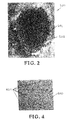
- FIG. 2 shows a mass spectral map of individual cells, tissues, and surrounding materials.
- FIG. 3 shows a mass spectrum from the area of interest 202 of FIG. 2 .
- FIGS. 4-9 illustrate another embodiment of a method of the present invention.
- FIG. 10 illustrates another embodiment of a method of the present invention.
- FIGS. 11A and 11B illustrate yet another embodiment of a method of the present invention.
- FIGS. 12A , 12 B, 12 C, and 12 D illustrate another embodiment of a method of the present invention
- FIG. 1A is a flow chart that illustrates one embodiment of a method of the present invention.
- the method is designated generally by the reference numeral 100 .
- the method 100 provides a method of analyzing biological cells by imaging mass spectrometry coupled with mass tags.
- the method 100 will allow for localization of cellular molecules by specific tagging and then imaging the tags using imaging mass spectrometry.
- Examples of applications of the method 100 include early disease detection in buccal cells, peripheral blood, sputum or urine, disease prognosis in the above-described examples as well as multi-cellular tissues, measurement of in vitro cell response to physical, chemical or radiation exposure, identifying m-RNA expression, proteins and metabolite pathways in single cells, predicting stem cell development, and other applications.
- Clinical and basic science uses of the method 100 will apply to eukaryotic and prokaryotic cells.
- the method 100 will allow multiplex analysis of molecular signatures for cancer classification in single cells by using cleavable mass tags followed by ToF-SIMS imaging.
- biological materials are exposed to detection molecules consisting of a recognition element and a mass tag element. These two elements are cleavable.
- the recognition element binds to a specific chemical or protein structure.
- the mass tags are released by the mass spectrometer ion beam, by photolysis, or other means to make the mass tag detectable by the instrument, thus localizing the distribution and quantity of the chemical or protein identified by the detection element.
- the method 100 includes a series of steps.
- step 101 the biological material is exposed to a recognition element.
- step 102 the biological material is exposed to a mass tag element.
- step 103 an ion beam of a mass spectrometer is directed to the biological material.
- step 103 at least one region of interest area from the biological material is interrogated and data is produced.
- step 104 the data is analyzed to provide information about the biological material.
- the biological materials, fluids, cells or tissues are placed on chips of silicon or other suitable material. Samples are analyzed directly or the cell contents exposed by crushing, freeze-fracturing or other methods. Samples are placed in an imaging mass spectrometer such as a ToF-SIMS, or ToF-SIMS/MALDI.
- an imaging mass spectrometer such as a ToF-SIMS, or ToF-SIMS/MALDI.
- Step 103 uses an ion beam of a mass spectrometer directed to the biological material.
- the ion beam is a finely focused energetic primary-ion beam of a time-of-flight secondary ion mass spectrometer.
- the ion beam is an ion beam of a matrix-assisted laser desorption/ionization mass spectrometer.
- FIG. 1B is a flow chart illustrating another embodiment of a method of the present invention.
- This method is designated generally by the reference numeral 100 B.
- the method 100 B provides a method of analyzing biological cells by imaging mass spectrometry coupled with mass tags.
- the method 100 B will allow for localization of cellular molecules by specific tagging and then imaging the tags using imaging mass spectrometry.
- Examples of applications of the method 100 B include early disease detection in buccal cells, peripheral blood, sputum or urine, disease prognosis in the above-described examples as well as multi-cellular tissues, measurement of in vitro cell response to physical, chemical or radiation exposure, identifying m-RNA expression, proteins and metabolite pathways in single cells, predicting stem cell development, and other applications.
- Clinical and basic science uses of the method 100 B will apply to eukaryotic and prokaryotic cells.
- the method 100 B will allow multiplex analysis of molecular signatures for cancer classification in single cells by using cleavable mass tags followed by ToF-SIMS imaging.
- biological materials are exposed to detection molecules consisting of a recognition element and a mass tag element. These two elements are cleavable.
- the recognition element binds to a specific chemical or protein structure.
- the mass tags are released by the mass spectrometer ion beam, by photolysis, or other means to make the mass tag detectable by the instrument, thus localizing the distribution and quantity of the chemical or protein identified by the detection element.
- the method 100 B includes a series of steps.
- step 101 B the biological material is exposed to a recognition element.
- step 102 B the biological material is exposed to a mass tag element.
- step 103 B an ion beam of a mass spectrometer is directed to the biological material.
- step 103 B at least one region of interest area from the biological material is interrogated and data is produced.
- step 104 B the data is distributed in plots indicating measures of similarity.
- the biological materials, fluids, cells or tissues are placed on chips of silicon or other suitable material. Samples are analyzed directly or the cell contents exposed by crushing, freeze-fracturing or other methods. Samples are placed in an imaging mass spectrometer such as a ToF-SIMS, or ToF-SIMS/MALDI.
- an imaging mass spectrometer such as a ToF-SIMS, or ToF-SIMS/MALDI.
- Step 103 B uses an ion beam of a mass spectrometer directed to the biological material.
- the ion is a finely focused energetic primary-ion beam of a time-of-flight secondary ion mass spectrometer.
- the ion beam is an ion beam of a matrix-assisted laser desorption/ionization mass spectrometer.
- FIG. 1C a flow chart illustrates another embodiment of a method of the present invention.
- the method is designated generally by the reference numeral 100 C.
- the method 100 C provides a method of analyzing biological material by imaging mass spectrometry coupled with mass tags.
- the method 100 C will allow for localization of cellular molecules by specific tagging and then imaging the tags using imaging mass spectrometry.
- Ga and Au ions to chemically map the surface of cells or the interior of crushed or fractured cells can be quite useful in telling one cell from another, but understanding what protein or expressed gene is responsible for the difference requires more specific analysis. This is why using the same imaging technology but putting specific masses attached to ligands that can recognize DNA sequences (oligos) or specific proteins (antibodies) can give the method specificity.
- multiplexing 10-100 of these mass tagged detectors in the same cell would allow analysis of many macromolecules in a pathway or system at the same time. No method exists today that can do this at the single cell level and also image the result.
- biological materials are exposed to detection molecules consisting of a recognition element and a mass tag element. These two elements are cleavable.
- the recognition element binds to a specific chemical or protein structure.
- the mass tags are released by the mass spectrometer ion beam, by photolysis, or other means to make the mass tag detectable by the instrument, thus localizing the distribution and quantity of the chemical or protein identified by the detection element.
- the method 100 C includes a series of steps.
- One form of a reagent has an antibody connected by a UV linker to a molecule with specific mass. This will allow identification of that antibody binding specific from others in the multiplex reagent. UV light will cleave the tag away from the antibody which is hundreds of times larger than the mass tag.
- This method can be used on individual cells or paraffin embedded tissues. It should contribute to cancer prognosis and drug effectiveness determinations.
- a recognition element (Ab) and a mass tag element (Mass 224 ) are connected by a cleavable linker (UV Cleavable linker). This provides the recognition element and a mass tag element connected by a cleavable linker (Ab-Mass 224 ) shown as block 108 C.
- step 109 C the cleavable linker (Ab-Mass 224 ) is cleaved.
- exposure to ultraviolet light cleaves the cleavable linker.
- an ion beam is directed to the biological material.
- an ion beam of a mass spectrometer is directed to the biological material.
- at least one region of interest area from the biological material is interrogated and data is produced.
- step 104 C the data is analyzed to provide information about the biological material.
- the data analysis spectra includes a mass tag from an individual cell.
- the Time-of-Flight Secondary Ion Mass Spectrometry (ToF-SIMS) produces a chemical map of the surface of a biological sample.
- the mass spectral map is designated generally by the reference numeral 200 .
- the mass spectral map shows a region of interest area 202 from individual cells, tissues or surrounding materials 201 .
- the region of interest area 202 from individual cells, tissues or surrounding materials 201 is analyzed and the data recorded.
- the mass spectra from region of interest 202 is exported to statistical analysis software, and the data set for each cell or region are distributed in plots indicating measures of similarity.
- Single cell mass spectral data sets can be compared to known samples to identify a cells' tissue of origin, understand the cells metabolic state, or predict progression to disease state.
- FIG. 3 a mass spectrum from the area of interest 202 of FIG. 2 is shown.
- the mass spectra from the region of interest is exported to statistical analysis software, and the data set for each cell or region are distributed in plots indicating measures of similarity.
- Single cell mass spectral data sets can be compared to known samples to identify a cells' tissue of origin, understand the cells metabolic state, or predict progression to disease state.
- FIGS. 4-9 another embodiment of a method of the present invention is illustrated.
- the use of Ga and Au ions to chemically map the surface of cells or the interior of crushed or fractured cells can be quite useful in telling one cell from another, but understanding what protein or expressed gene is responsible for the difference requires more specific analysis.
- a Time-of-Flight Secondary Ion Mass Spectrometry produces a chemical map of the surface of a biological sample.
- the imaging technology is used together with the step of putting specific masses attached to ligands that can recognize DNA sequences (oligos) or specific proteins (antibodies). This gives the method specificity.
- multiplexing 10-100 of these mass tagged detectors in the same cell allows analysis of many macromolecules in a pathway or system at the same time. No method exists today that can do this at the single cell level and also image the result.
- FIG. 4 shows cells 401 grown on a silicon wafer 400 .
- 2 ⁇ 10 6 cells can be plated in T75 flasks and harvested later, when the cells are 75% confluent.
- 8 ⁇ 10 5 cells can be plated in a 60 mm dish containing 3 to 5 silicon wafers, each about 1 cm square.
- the Si wafers are sterilized by UV irradiation prior to seeding.
- Cells are grown on the polished side of the silicon wafers; no change was observed in cellular growth or morphology as compared to cells grown on the typical plastic-cell-culture ware. Cells grown on wafers were freeze-fractured 48 hr after plating.
- FIG. 5 shows a primary ion beam 500 that desorbs a cloud 501 of secondary ions.
- Biological materials are exposed to detection molecules consisting of a recognition element and a mass tag element. These two elements are cleavable.
- the recognition element binds to a specific chemical or protein structure.
- the mass tags are released by the mass spectrometer ion beam, by photolysis, or other means to make the mass tag detectable by the instrument, thus localizing the distribution and quantity of the chemical or protein identified by the detection element.
- the ion beam 500 in this embodiment is a finely focused energetic primary-ion beam of a time-of-flight secondary ion mass spectrometer that is directed to the small groups of cells or the single cell and tissues or surrounding materials. At least one region of interest is interrogated. At least one region of interest can be an area from individual cells, tissues or surrounding materials.
- Time-of-Flight Secondary Ion Mass Spectrometry is a surface sensitive technique that allows the detection and localization of the chemical composition of sample surfaces. The instrument uses a finely focused ( ⁇ 300 nm), pulsed primary ion beam 500 to desorb and ionize molecular species from a sample surface.
- FIG. 6 shows that secondary ions 601 are detected in a time-of-flight mass spectrometer 600 .
- the secondary ions 601 are accelerated into a mass spectrometer 600 , where they are analyzed for mass by measuring their time-of-flight from the sample surface to the detector. Displaying the mass spectra that were collected from the sample surface generates chemical images.
- the resulting ion images contain a mass spectrum in each pixel of the 256 ⁇ 256 pixels in an image. These mass spectra are used to create secondary ion images that reflect the composition and distribution of sample surface constituents.
- FIG. 7 shows a position-specific mass spectral map that is generated.
- FIG. 8 shows a 65,000 mass spectra 800 and a region of interest 801 .
- FIG. 9 shows selected mass peaks 900 can be imaged.
- the mass spectral map is designated generally by the reference numeral 700 in FIG. 7 .
- the mass spectral map shows regions of interest areas from individual cells, tissues or surrounding materials. The region of interest area from individual cells, tissues or surrounding materials is analyzed and the data recorded.
- the mass spectra from region of interest is exported to statistical analysis software, and the data set for each cell or region are distributed in plots indicating measures of similarity.
- Single cell mass spectral data sets can be compared to known samples to identify a cells' tissue of origin, understand the cells metabolic state, or predict progression to disease state.
- FIG. 10 shows regions of interest MTLn3, MTC, and MCF7 from individual cells, tissues or surrounding materials.
- the region of interest area from individual cells, tissues or surrounding materials is analyzed and the data recorded.
- the mass spectra from region of interest is exported to statistical analysis software, and the data set for each cell or region are distributed in plots indicating measures of similarity.
- Single cell mass spectral data sets can be compared to known samples to identify a cells' tissue of origin, understand the cells metabolic state, or predict progression to disease state.
- Rat mammary cell lines differing in metastatic potential, are well-separated by PCA, but what molecules are responsible for the differences? Mass tag technology can answer that question.
- Rat mammary adenocarcinoma cell lines that were derived from the same tumor. MTLn3 cells have the potential to cause distant metastases, MTC cells do not; model for metastasis.
- MCF-7 is a human breast cancer cell line. It is possible to tell which proteins determine the malignancy of MTLn3 and not MTC.
- FIG. 11A shows a ToF-SIMS image with mass tag 1100 , being a single cell analysis of crushed rat mammary carcinoma cells using antibodies with mass tags.
- FIG. 11A uses Ab-Mass 224 and a UV cleavable linker 1101 .
- FIG. 11B shows a spectra including mass tag from an individual cell.
- UV linker to molecule with specific mass. This allows identification of that antibody binding specific from others in the multiplex reagent. UV cleaves the tag away from the antibody which is hundreds of times larger. This method can be used on individual cells or paraffin embedded tissues. It will contribute to cancer prognosis and drug effectiveness determinations.
- FIGS. 12A , 12 B, 12 C, and 12 D yet another embodiment of a method of the present invention is illustrated.
- FIGS. 12A , 12 B, 12 C, and 12 D illustrate imaging of Expressed RNAs in individual cells hybridized to oligos with mass tags.
- FIG. 12A shows a ToF-SIMS total ion image.
- FIG. 12B shows the total spectrum.
- FIG. 12C shows the nuclear region and uses AGCCG-Mass 184 and a cleavable linker.
- FIG. 12D shows the cytosolic region and uses AGCTGG-Mass 147 and a cleavable linker.
Landscapes
- Physics & Mathematics (AREA)
- Chemical & Material Sciences (AREA)
- Analytical Chemistry (AREA)
- Spectroscopy & Molecular Physics (AREA)
- Engineering & Computer Science (AREA)
- Plasma & Fusion (AREA)
- Other Investigation Or Analysis Of Materials By Electrical Means (AREA)
Abstract
Description
Claims (23)
Priority Applications (2)
| Application Number | Priority Date | Filing Date | Title |
|---|---|---|---|
| US11/713,519 US7728287B2 (en) | 2007-03-01 | 2007-03-01 | Imaging mass spectrometer with mass tags |
| US12/788,146 US8362415B2 (en) | 2007-03-01 | 2010-05-26 | Imaging mass spectrometer with mass tags |
Applications Claiming Priority (1)
| Application Number | Priority Date | Filing Date | Title |
|---|---|---|---|
| US11/713,519 US7728287B2 (en) | 2007-03-01 | 2007-03-01 | Imaging mass spectrometer with mass tags |
Related Child Applications (1)
| Application Number | Title | Priority Date | Filing Date |
|---|---|---|---|
| US12/788,146 Continuation US8362415B2 (en) | 2007-03-01 | 2010-05-26 | Imaging mass spectrometer with mass tags |
Publications (2)
| Publication Number | Publication Date |
|---|---|
| US20080210857A1 US20080210857A1 (en) | 2008-09-04 |
| US7728287B2 true US7728287B2 (en) | 2010-06-01 |
Family
ID=39732415
Family Applications (2)
| Application Number | Title | Priority Date | Filing Date |
|---|---|---|---|
| US11/713,519 Expired - Fee Related US7728287B2 (en) | 2007-03-01 | 2007-03-01 | Imaging mass spectrometer with mass tags |
| US12/788,146 Expired - Fee Related US8362415B2 (en) | 2007-03-01 | 2010-05-26 | Imaging mass spectrometer with mass tags |
Family Applications After (1)
| Application Number | Title | Priority Date | Filing Date |
|---|---|---|---|
| US12/788,146 Expired - Fee Related US8362415B2 (en) | 2007-03-01 | 2010-05-26 | Imaging mass spectrometer with mass tags |
Country Status (1)
| Country | Link |
|---|---|
| US (2) | US7728287B2 (en) |
Cited By (10)
| Publication number | Priority date | Publication date | Assignee | Title |
|---|---|---|---|---|
| US20100255602A1 (en) * | 2007-03-01 | 2010-10-07 | Felton James S | Imaging Mass Spectrometer With Mass Tags |
| US9312111B2 (en) | 2014-04-02 | 2016-04-12 | The Board Of Trustees Of The Leland Stanford Junior University | Apparatus and method for sub-micrometer elemental image analysis by mass spectrometry |
| US9714937B2 (en) | 2009-10-13 | 2017-07-25 | Nanostring Technologies, Inc. | Protein detection via nanoreporters |
| US9766224B2 (en) | 2015-03-25 | 2017-09-19 | The Board Of Trustees Of The Leland Stanford Junior University | Single cell analysis using secondary ion mass spectrometry |
| US9797879B2 (en) | 2015-04-23 | 2017-10-24 | The Board Of Trustees Of The Leland Stanford Junior University | Method for multiplexed sample analysis by photoionizing secondary sputtered neutrals |
| US10041949B2 (en) | 2013-09-13 | 2018-08-07 | The Board Of Trustees Of The Leland Stanford Junior University | Multiplexed imaging of tissues using mass tags and secondary ion mass spectrometry |
| US20190057854A1 (en) * | 2015-10-28 | 2019-02-21 | Duke University | Mass spectrometers having segmented electrodes and associated methods |
| US10501777B2 (en) | 2015-07-17 | 2019-12-10 | Nanostring Technologies, Inc. | Simultaneous quantification of a plurality of proteins in a user-defined region of a cross-sectioned tissue |
| US10640816B2 (en) | 2015-07-17 | 2020-05-05 | Nanostring Technologies, Inc. | Simultaneous quantification of gene expression in a user-defined region of a cross-sectioned tissue |
| US11377689B2 (en) | 2018-02-12 | 2022-07-05 | Nanostring Technologies, Inc. | Chemical compositions and uses thereof |
Families Citing this family (5)
| Publication number | Priority date | Publication date | Assignee | Title |
|---|---|---|---|---|
| US20100273259A1 (en) * | 2009-04-22 | 2010-10-28 | Massachusetts Institute Of Technology | Substrates and methods for culturing stem cells |
| JP2013101101A (en) * | 2011-10-12 | 2013-05-23 | Canon Inc | Mass distribution measurement method and mass distribution measurement device |
| KR20150134373A (en) | 2013-03-22 | 2015-12-01 | 에테하 취리히 | Laser ablation cell |
| CN107389776B (en) * | 2017-07-18 | 2020-06-02 | 宁波瑞瑧生物科技有限公司 | Analysis method for detecting drug distribution and application thereof |
| US10916414B2 (en) | 2018-01-25 | 2021-02-09 | Ionpath, Inc. | Ion beam focus adjustment |
Citations (21)
| Publication number | Priority date | Publication date | Assignee | Title |
|---|---|---|---|---|
| US5633495A (en) * | 1994-05-10 | 1997-05-27 | Ion-Tof Gmbh | Process for operating a time-of-flight secondary-ion mass spectrometer |
| US5808300A (en) | 1996-05-10 | 1998-09-15 | Board Of Regents, The University Of Texas System | Method and apparatus for imaging biological samples with MALDI MS |
| US20020150927A1 (en) * | 1999-04-30 | 2002-10-17 | Matray Tracy J. | Methods for detecting a plurality of analytes by mass spectrometry |
| US20020168691A1 (en) * | 2001-04-03 | 2002-11-14 | Christopher Becker | Methods and kits useful for the simplification of complex peptide mixtures |
| US20020168644A1 (en) * | 2001-05-14 | 2002-11-14 | Aebersold Rudolf H. | Methods for isolation and labeling of sample molecules |
| WO2003014724A1 (en) | 2001-08-03 | 2003-02-20 | Biotron Limited | A novel cancer marker and uses therefor in the diagnosis of cancer |
| WO2003075772A2 (en) | 2002-03-11 | 2003-09-18 | Pawliszyn Janusz B | Micro-devices and analytical procedures for investigation of biological systems |
| WO2004038381A2 (en) | 2002-10-25 | 2004-05-06 | Metabolon, Inc. | Methods for drug discovery, disease treatment, and diagnosis using metabolomics |
| US6740497B2 (en) | 1998-03-06 | 2004-05-25 | The Regents Of The University Of California | Method and apparatus for detecting cancerous cells using molecules that change electrophoretic mobility |
| US6756586B2 (en) | 2001-10-15 | 2004-06-29 | Vanderbilt University | Methods and apparatus for analyzing biological samples by mass spectrometry |
| US20040137420A1 (en) | 2000-03-06 | 2004-07-15 | Akikazu Yasuda | Method for analysis of substances in tissue or in cells |
| US6809315B2 (en) | 2001-02-14 | 2004-10-26 | Picoliter Inc. | Method and system using acoustic ejection for preparing and analyzing a cellular sample surface |
| US20040235062A1 (en) | 2001-06-19 | 2004-11-25 | Terumi Nakajima | Method of analyzing protein occurring in cell or substance interacting with the protein |
| US20050014132A1 (en) | 2000-04-14 | 2005-01-20 | Metabolon, Inc. | Methods for drug discovery, disease treatment, and diagnosis using metabolomics |
| US6855925B2 (en) | 2001-02-14 | 2005-02-15 | Picoliter Inc. | Methods, devices, and systems using acoustic ejection for depositing fluid droplets on a sample surface for analysis |
| US20050048581A1 (en) | 2003-08-25 | 2005-03-03 | Chiu Daniel T. | Method and device for biochemical detection and analysis of subcellular compartments from a single cell |
| US20060134086A1 (en) * | 2000-12-14 | 2006-06-22 | Yiyou Chen | Targeted enzymes |
| US20070114375A1 (en) * | 2005-09-30 | 2007-05-24 | Paul Pevsner | Methods for direct biomolecule identification by matrix-assisted laser desorption ionization (MALDI) mass spectrometry |
| US20080145850A1 (en) * | 2000-03-14 | 2008-06-19 | Gunter Schmidt | Mass Labels |
| US20090156424A1 (en) * | 2004-05-20 | 2009-06-18 | Trillion Genomics Limited | Use of mass labeled probes to detect target nucleic acids using mass spectrometry |
| US20090226913A1 (en) * | 2005-05-04 | 2009-09-10 | Life Technologies Corporation | Identification of cancer biomarkers and phosphorylated proteins |
Family Cites Families (2)
| Publication number | Priority date | Publication date | Assignee | Title |
|---|---|---|---|---|
| US7124033B2 (en) * | 2003-04-30 | 2006-10-17 | Perlegen Sciences, Inc. | Method for identifying matched groups |
| US7728287B2 (en) * | 2007-03-01 | 2010-06-01 | Lawrence Livermore National Security, Llc | Imaging mass spectrometer with mass tags |
-
2007
- 2007-03-01 US US11/713,519 patent/US7728287B2/en not_active Expired - Fee Related
-
2010
- 2010-05-26 US US12/788,146 patent/US8362415B2/en not_active Expired - Fee Related
Patent Citations (21)
| Publication number | Priority date | Publication date | Assignee | Title |
|---|---|---|---|---|
| US5633495A (en) * | 1994-05-10 | 1997-05-27 | Ion-Tof Gmbh | Process for operating a time-of-flight secondary-ion mass spectrometer |
| US5808300A (en) | 1996-05-10 | 1998-09-15 | Board Of Regents, The University Of Texas System | Method and apparatus for imaging biological samples with MALDI MS |
| US6740497B2 (en) | 1998-03-06 | 2004-05-25 | The Regents Of The University Of California | Method and apparatus for detecting cancerous cells using molecules that change electrophoretic mobility |
| US20020150927A1 (en) * | 1999-04-30 | 2002-10-17 | Matray Tracy J. | Methods for detecting a plurality of analytes by mass spectrometry |
| US20040137420A1 (en) | 2000-03-06 | 2004-07-15 | Akikazu Yasuda | Method for analysis of substances in tissue or in cells |
| US20080145850A1 (en) * | 2000-03-14 | 2008-06-19 | Gunter Schmidt | Mass Labels |
| US20050014132A1 (en) | 2000-04-14 | 2005-01-20 | Metabolon, Inc. | Methods for drug discovery, disease treatment, and diagnosis using metabolomics |
| US20060134086A1 (en) * | 2000-12-14 | 2006-06-22 | Yiyou Chen | Targeted enzymes |
| US6855925B2 (en) | 2001-02-14 | 2005-02-15 | Picoliter Inc. | Methods, devices, and systems using acoustic ejection for depositing fluid droplets on a sample surface for analysis |
| US6809315B2 (en) | 2001-02-14 | 2004-10-26 | Picoliter Inc. | Method and system using acoustic ejection for preparing and analyzing a cellular sample surface |
| US20020168691A1 (en) * | 2001-04-03 | 2002-11-14 | Christopher Becker | Methods and kits useful for the simplification of complex peptide mixtures |
| US20020168644A1 (en) * | 2001-05-14 | 2002-11-14 | Aebersold Rudolf H. | Methods for isolation and labeling of sample molecules |
| US20040235062A1 (en) | 2001-06-19 | 2004-11-25 | Terumi Nakajima | Method of analyzing protein occurring in cell or substance interacting with the protein |
| WO2003014724A1 (en) | 2001-08-03 | 2003-02-20 | Biotron Limited | A novel cancer marker and uses therefor in the diagnosis of cancer |
| US6756586B2 (en) | 2001-10-15 | 2004-06-29 | Vanderbilt University | Methods and apparatus for analyzing biological samples by mass spectrometry |
| WO2003075772A2 (en) | 2002-03-11 | 2003-09-18 | Pawliszyn Janusz B | Micro-devices and analytical procedures for investigation of biological systems |
| WO2004038381A2 (en) | 2002-10-25 | 2004-05-06 | Metabolon, Inc. | Methods for drug discovery, disease treatment, and diagnosis using metabolomics |
| US20050048581A1 (en) | 2003-08-25 | 2005-03-03 | Chiu Daniel T. | Method and device for biochemical detection and analysis of subcellular compartments from a single cell |
| US20090156424A1 (en) * | 2004-05-20 | 2009-06-18 | Trillion Genomics Limited | Use of mass labeled probes to detect target nucleic acids using mass spectrometry |
| US20090226913A1 (en) * | 2005-05-04 | 2009-09-10 | Life Technologies Corporation | Identification of cancer biomarkers and phosphorylated proteins |
| US20070114375A1 (en) * | 2005-09-30 | 2007-05-24 | Paul Pevsner | Methods for direct biomolecule identification by matrix-assisted laser desorption ionization (MALDI) mass spectrometry |
Non-Patent Citations (5)
| Title |
|---|
| Baumgart, S., et al., "The contributions of specific amino acid side chains to signal intensities of peptides in matrix-assisted laser desorption/ionization mass spectrometry," Rapid Communications in Mass Spectrometry, 2004, 18, pp. 863-868. |
| Hummon, Aanda B., et al., "Discovering new neuropeptides using single-cell mass spectrometry," Trends in Analytical Chemistry, vol. 22, No. 9, 2003, pp. 515-521. |
| Kulp, Kristen S., et al., "Chemical and Biological Differentiation of Three Human Breast Cancer Cell Types Using Time-of-Flight Secondary Ion Mass Spectrometry," Anal. Chem. 2006, 78, pp. 3651-3658. |
| Li, Lingjun, et al., "Single-cell MALDI: a new tool for direct peptide profiling," TIBTECH Apr. 2000, vol. 18, pp. 151-160. |
| Rubakhin, Stanislav S., et al., "Spatial Profiling with MALDI MS: Distribution of Neuropeptides within Single Neurons," Anal. Chem., 2003, 75, pp. 5374-5380. |
Cited By (20)
| Publication number | Priority date | Publication date | Assignee | Title |
|---|---|---|---|---|
| US20100255602A1 (en) * | 2007-03-01 | 2010-10-07 | Felton James S | Imaging Mass Spectrometer With Mass Tags |
| US8362415B2 (en) * | 2007-03-01 | 2013-01-29 | Lawrence Livermore National Security, Llc | Imaging mass spectrometer with mass tags |
| US9714937B2 (en) | 2009-10-13 | 2017-07-25 | Nanostring Technologies, Inc. | Protein detection via nanoreporters |
| US9995739B2 (en) | 2009-10-13 | 2018-06-12 | Nanostring Technologies, Inc. | Protein detection via nanoreporters |
| US10041949B2 (en) | 2013-09-13 | 2018-08-07 | The Board Of Trustees Of The Leland Stanford Junior University | Multiplexed imaging of tissues using mass tags and secondary ion mass spectrometry |
| US9312111B2 (en) | 2014-04-02 | 2016-04-12 | The Board Of Trustees Of The Leland Stanford Junior University | Apparatus and method for sub-micrometer elemental image analysis by mass spectrometry |
| US9766224B2 (en) | 2015-03-25 | 2017-09-19 | The Board Of Trustees Of The Leland Stanford Junior University | Single cell analysis using secondary ion mass spectrometry |
| US12135300B2 (en) | 2015-03-25 | 2024-11-05 | The Board Of Trustees Of The Leland Stanford Junior University | Single cell analysis using secondary ion mass spectrometry |
| US10114004B2 (en) | 2015-03-25 | 2018-10-30 | The Board Of Trustees Of The Leland Stanford Junior University | Single cell analysis using secondary ion mass spectrometry |
| US10408814B2 (en) | 2015-04-23 | 2019-09-10 | The Board Of Trustees Ofthe Leland Stanford Junior University | Method for multiplexed sample analysis by photoionizing secondary sputtered neutrals |
| US9797879B2 (en) | 2015-04-23 | 2017-10-24 | The Board Of Trustees Of The Leland Stanford Junior University | Method for multiplexed sample analysis by photoionizing secondary sputtered neutrals |
| US10501777B2 (en) | 2015-07-17 | 2019-12-10 | Nanostring Technologies, Inc. | Simultaneous quantification of a plurality of proteins in a user-defined region of a cross-sectioned tissue |
| US10640816B2 (en) | 2015-07-17 | 2020-05-05 | Nanostring Technologies, Inc. | Simultaneous quantification of gene expression in a user-defined region of a cross-sectioned tissue |
| US11708602B2 (en) | 2015-07-17 | 2023-07-25 | Nanostring Technologies, Inc. | Simultaneous quantification of gene expression in a user-defined region of a cross-sectioned tissue |
| US12435361B2 (en) | 2015-07-17 | 2025-10-07 | Bruker Spatial Biology, Inc. | Simultaneous quantification of a plurality of proteins in a user-defined region of a cross-sectioned tissue |
| US12630869B2 (en) | 2015-07-17 | 2026-05-19 | Board Of Regents, The University Of Texas System | Simultaneous quantification of gene expression in a user-defined region of a cross-sectioned tissue |
| US20190057854A1 (en) * | 2015-10-28 | 2019-02-21 | Duke University | Mass spectrometers having segmented electrodes and associated methods |
| US11081331B2 (en) * | 2015-10-28 | 2021-08-03 | Duke University | Mass spectrometers having segmented electrodes and associated methods |
| US11377689B2 (en) | 2018-02-12 | 2022-07-05 | Nanostring Technologies, Inc. | Chemical compositions and uses thereof |
| US11473142B2 (en) | 2018-02-12 | 2022-10-18 | Nanostring Technologies, Inc. | Chemical compositions and uses thereof |
Also Published As
| Publication number | Publication date |
|---|---|
| US8362415B2 (en) | 2013-01-29 |
| US20080210857A1 (en) | 2008-09-04 |
| US20100255602A1 (en) | 2010-10-07 |
Similar Documents
| Publication | Publication Date | Title |
|---|---|---|
| US8362415B2 (en) | Imaging mass spectrometer with mass tags | |
| Tajik et al. | Single-cell mass spectrometry | |
| Cornett et al. | MALDI imaging mass spectrometry: molecular snapshots of biochemical systems | |
| Levenson et al. | Immunohistochemistry and mass spectrometry for highly multiplexed cellular molecular imaging | |
| Schwamborn et al. | Molecular imaging by mass spectrometry—looking beyond classical histology | |
| US12135300B2 (en) | Single cell analysis using secondary ion mass spectrometry | |
| AU772680B2 (en) | LCM (Laser capture microdissection) for cellular protein analysis | |
| Minakshi et al. | Single-cell proteomics: technology and applications | |
| US6969614B1 (en) | Methods for the isolation and analysis of cellular protein content | |
| Smith et al. | Cancer proteomics and its application to discovery of therapy response markers in human cancer | |
| AU2006261624B2 (en) | Method of classifying chemically crosslinked cellular samples using mass spectra | |
| Brožová et al. | Spatial proteomics for the molecular characterization of breast cancer | |
| US9739765B2 (en) | Metal-based covalent viability reagent for single cell analysis | |
| Alessandro et al. | Proteomic approaches in colon cancer: promising tools for new cancer markers and drug target discovery | |
| Bañez et al. | Proteomics in prostate cancer | |
| Mocellin et al. | Molecular oncology in the post-genomic era: the challenge of proteomics | |
| Matharoo‐Ball et al. | Serum biomarker profiling in cancer studies: a question of standardisation? | |
| US7955810B2 (en) | Positive selection of serum proteins for proteomic analysis | |
| Huang et al. | Quantitative fluorescence imaging analysis for cancer biomarker discovery: application to β-catenin in archived prostate specimens | |
| Hachey et al. | Proteomics in reproductive medicine: the technology for separation and identification of proteins | |
| Kumar et al. | Fundamentals of flow cytometry and cytometry by time of flight (cytof) | |
| Conrads et al. | The utility of proteomic patterns for the diagnosis of cancer | |
| Drake et al. | Imaging mass spectrometry | |
| Borowsky | Next-Generation Molecular Histology Using Highly Multiplexed Ion Beam Imaging (MIBI) of Breast Cancer Tissue Specimens for Enhanced Clinical Guidance | |
| Winters et al. | The future of cancer diagnostics: proteomics, immunoproteomics, and beyond |
Legal Events
| Date | Code | Title | Description |
|---|---|---|---|
| AS | Assignment |
Owner name: CALIFORNIA, THE REGENTS OF THE UNIVERSITY OF, CALI Free format text: ASSIGNMENT OF ASSIGNORS INTEREST;ASSIGNORS:FELTON, JAMES S.;WU, KUANG JEN J.;KNIZE, MARK G.;AND OTHERS;REEL/FRAME:019307/0168 Effective date: 20070228 Owner name: CALIFORNIA, THE REGENTS OF THE UNIVERSITY OF,CALIF Free format text: ASSIGNMENT OF ASSIGNORS INTEREST;ASSIGNORS:FELTON, JAMES S.;WU, KUANG JEN J.;KNIZE, MARK G.;AND OTHERS;REEL/FRAME:019307/0168 Effective date: 20070228 |
|
| AS | Assignment |
Owner name: LAWRENCE LIVERMORE NATIONAL SECURITY, LLC, CALIFOR Free format text: ASSIGNMENT OF ASSIGNORS INTEREST;ASSIGNOR:REGENTS OF THE UNIVERSITY OF CALIFORNIA, THE;REEL/FRAME:020012/0032 Effective date: 20070924 Owner name: LAWRENCE LIVERMORE NATIONAL SECURITY, LLC,CALIFORN Free format text: ASSIGNMENT OF ASSIGNORS INTEREST;ASSIGNOR:REGENTS OF THE UNIVERSITY OF CALIFORNIA, THE;REEL/FRAME:020012/0032 Effective date: 20070924 |
|
| AS | Assignment |
Owner name: US DEPARTMENT OF ENERGY, DISTRICT OF COLUMBIA Free format text: ASSIGNMENT OF ASSIGNORS INTEREST;ASSIGNOR:REGENTS OF THE UNIVERSITY OF CALIFORNIA;REEL/FRAME:019939/0322 Effective date: 20070807 Owner name: US DEPARTMENT OF ENERGY,DISTRICT OF COLUMBIA Free format text: ASSIGNMENT OF ASSIGNORS INTEREST;ASSIGNOR:REGENTS OF THE UNIVERSITY OF CALIFORNIA;REEL/FRAME:019939/0322 Effective date: 20070807 |
|
| AS | Assignment |
Owner name: THE REGENTS OF THE UNIVERSITY OF CALIFORNIA, CALIF Free format text: ASSIGNMENT OF ASSIGNORS INTEREST;ASSIGNOR:LAWRENCE LIVERMORE NATIONAL SECURITY, LLC;REEL/FRAME:022701/0275 Effective date: 20090514 Owner name: THE REGENTS OF THE UNIVERSITY OF CALIFORNIA,CALIFO Free format text: ASSIGNMENT OF ASSIGNORS INTEREST;ASSIGNOR:LAWRENCE LIVERMORE NATIONAL SECURITY, LLC;REEL/FRAME:022701/0275 Effective date: 20090514 |
|
| STCF | Information on status: patent grant |
Free format text: PATENTED CASE |
|
| FEPP | Fee payment procedure |
Free format text: PAYOR NUMBER ASSIGNED (ORIGINAL EVENT CODE: ASPN); ENTITY STATUS OF PATENT OWNER: LARGE ENTITY |
|
| FPAY | Fee payment |
Year of fee payment: 4 |
|
| SULP | Surcharge for late payment | ||
| AS | Assignment |
Owner name: U.S. DEPARTMENT OF ENERGY, DISTRICT OF COLUMBIA Free format text: CONFIRMATORY LICENSE;ASSIGNOR:THE REGENTS OF THE UNIVERSITY OF CALIFORNIA;REEL/FRAME:037257/0205 Effective date: 20070807 |
|
| FPAY | Fee payment |
Year of fee payment: 8 |
|
| FEPP | Fee payment procedure |
Free format text: MAINTENANCE FEE REMINDER MAILED (ORIGINAL EVENT CODE: REM.); ENTITY STATUS OF PATENT OWNER: LARGE ENTITY |
|
| LAPS | Lapse for failure to pay maintenance fees |
Free format text: PATENT EXPIRED FOR FAILURE TO PAY MAINTENANCE FEES (ORIGINAL EVENT CODE: EXP.); ENTITY STATUS OF PATENT OWNER: LARGE ENTITY |
|
| STCH | Information on status: patent discontinuation |
Free format text: PATENT EXPIRED DUE TO NONPAYMENT OF MAINTENANCE FEES UNDER 37 CFR 1.362 |
|
| FP | Lapsed due to failure to pay maintenance fee |
Effective date: 20220601 |