CROSS REFERENCE TO RELATED APPLICATIONS
This application is a Continuation-In-Part of application Ser. No. 11/219,698 filed on Sep. 7, 2005, now abandoned, which is a Continuation of application Ser. No. 10/959,996 filed on Oct. 8, 2004, now abandoned, which is a Continuation of application Ser. No. 10/795,402 filed on Mar. 9, 2004, now abandoned, which is a Continuation of application Ser. No. 10/404,841 filed on Apr. 2, 2003, now abandoned, which claims benefit of the filing date of Provisional Application No. 60/369,318 filed Apr. 3, 2002, the entire contents of all of which are hereby incorporated by reference and for which priority is claimed under 35 U.S.C. §120 and §119.
FIELD OF THE INVENTION
The present invention relates to a novel bioactive substance having controlling effects on growth and differentiation of undifferentiated cells.
BACKGROUND OF THE INVENTION
Higher animals have a system for supplementing cells after death of cells that form tissues and organs by apoptosis or injury. For example, the amphibian newt can regenerate the limbs and tail if they are cut off, and birds can easily regenerate the nervous system. Mammals have lost such high regenerative capacity, but their liver can regenerate oneself unless it has suffered a severe damage. Additionally, skin, hair, small intestine and hematopoietic cells are regenerated while a mammalian individual is alive. In these tissues, the cell cycle of new cell birth, differentiation and death is repeated as long as the individual is alive. The regenerative capacity depends on cells known as stem cells (Fuchs and Segre, Cell, 100, 143-155, 2000; Weissman Cell, 100, 157-168, 2000). Of various cells types forming tissues and organs, blood cells, nerve cells, vascular endothelial cells and epithelial cells are known to mature through several stages from the undifferentiated cells known as stem cells.
Stem cells have the ability of self-replication (self-renewal) to reproduce oneself by cell division, and the ability of differentiation into specific mature cells. In stem cells, a delicate balance is struck between the self-replication and the differentiation.
For one mechanism of maintaining the balance, the hereinafter-mentioned systems have been proposed.
The localization sites of stem cells are referred to as niches, in which there is a molecular infrastructure that allows the stem cells to be maintained and reproduced. In niches, the stem cells are typically maintained in a growth arrest phase. Once released from the arrested conditions due for example to a tissue injury, the stem cells enter a growth phase, and form a certain population of cells. In this growth process, heterogeneity arises within the population of cells. Some cells re-enter the cell arrest phase and retain their characteristics as stem cells, whereas others statistically express a transcription factor and thereby become destined to differentiate, and subsequently differentiate to different lineages of mature cells. There are thought to be stromal cells in the niches, which can come in contact with the stem cells and trigger a signal for growth arrest in the stem cells.
In the differentiation process to mature cells from so-called precursor cells (or progenitor cells), wherein the precursor cells has left the niches, expressed the transcription factor, and thereby become destined to differentiate, there is a mechanism which controls the process to allow the differentiation to proceed properly. It is thought that other stromal cells, which are different from the stromal cells that trigger a signal for growth arrest in the stem cells, come in contact with the precursor cells, and the precursor cells are subjected to control by certain molecules expressed by these stromal cells, and then that differentiation proceeds properly.
If the identities of the stem cell growth activating/arresting signals possessed by the stromal cells (or the stromal cells population) present in the niches, or those of the precursor cell differentiation control signals, are ascertained, whereby the methods for keeping stem cells in an undifferentiated state for a long time and for controlling differentiation of stem cells, by controlling the growth and arrest of stem cells, can be provided. These methods have many applications in fields such as regenerative medicine, gene therapy and transplantation. Specifically, the methods can be used in hematopoietic stem cell transplantation for a medical treatment of aplastic anemia, or in neural stem cell transplantation for a medical treatment of Alzheimer's disease, but these are only a few examples.
Various studies have been conducted in the past aimed at ascertaining the identities of stem cell growth activating/arresting signals and of precursor cell differentiation control signals.
Leukemia inhibitory factor (LIF) and transforming growth factor (TGF-β) are known to be cytokines which inhibit the differentiation of stem cells. LIF is known to cause the growth of mouse embryonic stem cells without differentiation, but it does not have such effect on mouse hematopoietic stem cells. Also, LIF does not affect human or monkey embryonic stem cells. For TGF-β, there are many reports regarding its inhibitory effects on various types of cells, but no fixed consensus has been obtained regarding its effect on stem cells. Examples of the molecules that control the differentiation of precursor cells are M-CSF, GM-CSF, G-CSF, SCF, TPO and FLK ligands. However such molecules discovered to date cannot account for the differentiation of various types of cells, which suggests the presence of hitherto unidentified molecules.
Recently, Notch, which is a molecule involved in differentiation control of nerve cells in Drosophila, has been discovered, and homologs of this molecule have been found in a broad spectrum of organisms across the classification of invertebrates and vertebrates (Artavanis-Tsakonas et al., Science 268, 225-232, 1995). In mammals, it has been shown that the mutation of Notch is related to T cell leukemia and lymphoma (Pear et al., J. Exp. Med. 183, 2283-2291, 1996). It has also demonstrated that expression of activated Notch molecule in myeloblast cell lines causes the inhibition of their innate ability to differentiate into neutrophils by G-CSF (Milner et al., Proc. Natl. Acad. Sci. USA 93, 13014-13019), and the Notch molecule are involved in the determination of the fate of CD4/CD8 cells in T cell differentiation (Robey et al., Cell 87, 483-492, 1996). Therefore, Notch molecules have attracted further attention as differentiation control molecules. Moreover, Delta and Serrate, which are ligands of the Notch molecule, have been identified in Drosophila (Kopczynski et al., Genes Dev., 1723-1753, 1988, Thomas et al., Development, 111, 749-761, 1991). X-Delta and D111, homologs of Delta, have been identified in the Xenopus (Chitnis et al., Nature, 375, 761-766, 1995) and mouse (Bettenhausen et al., Development 121, 2431-2418, 1995), respectively, and Jagged, homologs of Serrate, has been identified in rat and human (Luo et al., Mol. Cell. Biol. 17, 6057-6067, 1997).
From these findings, Notch receptor, and ligands thereof (Delta, Serrate and Jagged), are now attracting attention as cell differentiation and growth control molecules.
Comparing the structures of Notch, Delta and Jagged, the repetition of an EGF (Epidermal Growth Factor)-like domain is commonly found in them (Lindsell et al., Cell, 80, 909-917, 1995). The repetition is referred to as EGF-like repeat sequence or EGF-like repeat motif.
The consensus sequence of the EGF-like domain is C-X-C-X(5)-G-X(2)-C (SEQ ID NO:32) or C-X-C-X(2)-[GP]-[FYW]-X(4, 8)-C (SEQ ID NOS: 33 and 34). These domain structures are found in EGF and many extracellular proteins, and are involved in protein interactions or cellular interactions (Campbell and Bork Curr. Opin. Struct. Biol, 3, 385-392, 1993, Rao et al., Cell, 82, 131-141, 1995).
These suggest that the stromal cells in the niches possess a differentiation and growth control molecule, and the molecule belongs to the Notch, Delta and Jagged family. But the previously identified molecules of the family cannot explain the differentiation and growth control mechanism of stem cells. Accordingly, it is thought that there is also a hitherto unidentified functionally similar molecule as the above molecule possessed by the stromal cells.
A transcriptional induction system is known as a common gene expression control mechanism in animals (Nature, 321: 409-413, 1984). A promoter is generally located 5′ upstream to a region that is transcribed into mRNA on a chromosome. Furthermore, through binding or dissociation of a transcription factor to a sequence referred to as the regulatory region within the promoter sequence (transcriptional regulatory sequence), the promoter regulates the transcription level of a gene that is present in the 3′ downstream region of the promoter. Therefore, the gene expression level at the transcription stage can be estimated to some extent by measuring promoter activity. In the meantime, promoter activity is not affected in most cases by the 3′ downstream region thereof. Hence, promoter activity can be measured by inserting an appropriate reporter gene encoding an enzyme protein or the like into a downstream region of the promoter and then detecting the expression of the reporter gene. Very sensitive and convenient promoter activity measurement has become possible with the use of such a reporter, owing to recent technical innovation. Thus, such promoter activity measurement is used for drug screening and examination of biological functions. For example, screening with the promoter of peroxisome proliferator activated receptor γ (PPARγ) that is a transcription factor for adipose cells differentiation, for a compound that controls the expression of PPARγ was reported (Cell, 99: 239-242, 1999).
Production of transgenic non-human animals using promoters has also been performed. In general, it is difficult to examine the functions of genes that are essential for developmental processes or maintenance of living systems, because deletion of such genes is often lethal in mice. Conditional gene targeting techniques have been used as a potential method for addressing the problem, using a Cre-loxP recombination system under control of a promoter.
Cre recombinase is a site-specific recombinase derived from bacteriophage P1 and specifically recognizes a loxP sequence of 34 base pairs. This enzyme mediates recombination between two loxP sequences, and then a DNA fragment flanked by the two loxP sequences is excised in a cyclic form only under conditions where Cre recombinase is expressed, and the DNA fragment is deleted. For example, lck is a gene that is expressed in T cells and is strongly expressed particularly in the thymus where the development and differentiation of T cells take place. Thus, in a mouse in which a Cre recombinase gene ligated to downstream of the promoter of the lck gene has been introduced, Cre recombinase is specifically expressed only in T cells and the gene flanked by loxP sequences is disrupted (Science, 265: 103-106, 1994, Proc. Natl. Acad. Sci. U.S.A., 92: 12070-12074, 1995).
Mice known to have a Cre recombinase gene under control of such a tissue-specific promoter used therein includes: a mouse having a PO promoter that is expressed in neural crest cells (Dev Biol, 212: 191-203, 1999); a mouse having an L7 promoter that is expressed in Purkinje cells (Genesis, 28, 93-8, 2000); a mouse having a keratin 14 promoter that functions in epidermal basal cells (Horm Res, 54: 296-300, 2000); a mouse having an Mx1 promoter whose activity is induced in the presence of interferon (Science, 269: 1427-1429, 1995); and a mouse having a crystallin promoter that functions in the lens of the eyes (Proc. Natl. Acad. Sci. U.S.A., 89: 6232-6236, 1992). Discovery of a new tissue-specific promoter in addition to these promoters may cause further advancement in functional verification of genes by the conditional gene targeting.
Promoters are important also in production of recombinant proteins. When a protein is recombinantly produced using cells, the gene of a target protein is ligated downstream of a promoter and then the resultant is introduced into and expressed by cells. When animal cells are used as hosts, in general, promoters derived from viruses, such as SV40 and CMV (Proc. Natl. Acad. Sci. U.S.A., 78: 1527-1531; 1981, Nature, 329: 840-842, 1987), an actin gene promoter (Gene, 108: 193-200, 1991), and an elongation factor gene promoter (Nucleic Acids Res., 18: 5322, 1990) are used. However, the strength of the activity of these promoters differs depending on the types of proteins to be expressed and host cell types. Hence, it is necessary to examine such combination to select an optimum promoter. Therefore, provision of a new promoter is always desired for more effective production of individual proteins.
SUMMARY OF THE INVENTION
It is an object of the present invention, which was conceived in view of the above technical backgrounds, to provide a novel molecule by discovering a protein molecule which can affect stem cells to trigger the growth arrest signal in stem cells, or a protein molecule which can affect precursor cells to control their differentiation and growth other than Delta and Jagged; and determining the genetic sequence and amino acid sequence of that novel molecule. It is further object of the present invention to provide a pharmaceutical composition which comprises such molecule as an active ingredient for treating diseases caused by cell or tissue damage, based on the differentiation and growth control effect which is one of the features of this molecule. It is a further object of the present invention to provide a method for gene therapy by using the genes of this molecule. It is yet another object of the present invention to provide a method of regenerative medicine by discovering a molecule that controls stem cell or precursor cell growth and differentiation. It is further object of the present invention to provide a novel tissue specific promoter and use thereof.
We have cloned the gene of stem cell/precursor cell differentiation and growth controlling molecules that contain an EGF-like repeat sequence, from the mRNA of stromal cell lines considered to present in the “niches”, by RT-PCR method using primers designed based on the amino acid sequence which is appeared with a relatively high frequency in EGF-like motif sequences. Primers have been designed based on sequence information of the resulting cDNA fragments, and the cDNA which encodes the full amino acid sequence of the novel molecule containing an EGF-like repeat sequence has been successfully isolated by the 3′ and 5′RACE method. By using this cDNA, the cells that express the above gene has been detected, transformed cells have been generated, antibodies have been produced, and in vivo localization of the expression product of the gene have been identified. The protein molecule of the present invention, which contains an EGF-like repeat sequence or EGF-like repeat motif, has been named “stromal cell-derived EGF-like repeat containing factor”, which is abbreviated as SELF. Then, the nucleic acid molecule of the present invention, which encodes such SELF protein, has been called also SELF gene. In addition, we isolated a promoter sequence of SELF gene (SELF promoter) and assayed functions of SELF protein.
The present invention generally relates to SELF protein, SELF gene and SELF promoter, and their use.
One aspect of the present invention is an isolated protein comprising the amino acid sequence as shown in SEQ ID NO: 2, 3, 4 or 24.
Another aspect of the present invention is an isolated protein comprising an amino acid sequence having one or more amino acids deleted, substituted or added in the amino acid sequence as shown in SEQ ID NO: 2, 3, 4 or 24, wherein the protein contains an EGF-like repeat motif and has bioactivity as a growth and differentiation controlling factor.
Another aspect of the present invention is an isolated protein, wherein the protein has at least 80% homology to a protein comprising the amino acid sequence as shown in SEQ ID NO: 2, 3, 4 or 24, contains the EGF-like repeat motif, and has bioactivity as the growth and differentiation controlling factor.
Another aspect of the present invention is an isolated protein, wherein the protein has at least 90% homology to a protein comprising the amino acid sequence as shown in SEQ ID NO: 2, 3, 4 or 24, contains the EGF-like repeat motif, and has bioactivity as the growth and differentiation controlling factor.
Another aspect of the present invention is an isolated nucleic acid which encodes a protein comprising the amino acid sequence as shown in SEQ ID NO: 2, 3, 4 or 24.
Another aspect of the present invention is an isolated nucleic acid comprising the nucleotide sequence as shown in SEQ ID NO: 1.
Another aspect of the present invention is an isolated nucleic acid consisting of the nucleotide sequence of nucleotides 157 to 4365 of SEQ ID NO: 1.
Another aspect of the present invention is an isolated nucleic acid consisting of the nucleotide sequence of nucleotides 1 to 1251 of SEQ ID NO: 1.
Another aspect of the present invention is an isolated nucleic acid consisting of a nucleotide sequence of nucleotides 1624 to 2174 of SEQ ID NO: 1.
Another aspect of the present invention is an isolated nucleic acid comprising a nucleotide sequence as shown in SEQ ID NO: 23.
Another aspect of the present invention is an isolated nucleic acid which hybridizes under stringent conditions with the above nucleic acid, and encodes a protein containing the EGF-like repeat motif and having bioactivity as the growth and differentiation controlling factor.
Another aspect of the present invention is an isolated nucleic acid comprising a nucleotide sequence which has at least 80% homology with the above nucleic acid, and encodes a protein containing the EGF-like repeat motif and having bioactivity as the growth and differentiation controlling factor.
Another aspect of the present invention is an isolated nucleic acid comprising a nucleotide sequence which has at least 90% homology with the above nucleic acid, and encodes a protein containing the EGF-like repeat motif and having bioactivity as the growth and differentiation controlling factor.
Another aspect of the present invention is a recombinant DNA construct, comprising the above nucleic acid or part thereof, and a vector DNA functionally linked thereto wherein the vector can be expressed in a host cell. Preferably, the present invention relates to a recombinant vector comprising the above nucleic acid, for example, a recombinant expression vector capable of expressing the above nucleic acid in a host cell.
Another aspect of the present invention is a cell transformed with the above recombinant vector.
Another aspect of the present invention is a method of producing the above protein comprising culturing the above transformed cell, and recovering a produced protein from the culture medium or cultured cells.
Another aspect of the present invention is an antibody, which specifically binds to the above protein or fragments of the protein. For example, an antibody, which specifically recognizes a protein comprising the amino acid sequence as shown in SEQ ID NO: 2, 3, 4 or 24 is provided.
Another aspect of the present invention is an antibody, which specifically recognizes a protein comprising the amino acid sequence of amino acids 1390 to 1403 of SEQ ID NO: 2.
Another aspect of the present invention is an antibody, which specifically recognizes a protein comprising the amino acid sequence of amino acids 235 to 432 of SEQ ID NO: 2.
Another aspect of the present invention is a method of controlling the growth and differentiation of undifferentiated cells with the above protein. More specifically, the present invention relates to a method for controlling the growth and differentiation of undifferentiated cells, comprising contacting the above protein with undifferentiated cells. The undifferentiated cells are preferably hematopoietic undifferentiated cells.
Further, another aspect of the present invention is a pharmaceutical composition containing the above protein, and/or a recombinant expression vector comprising the above nucleic acid. Preferably, the pharmaceutical composition of the present invention further comprises a vascular endothelial growth factor inhibitor.
Another aspect of the present invention is a pharmaceutical kit which further comprises a vascular endothelial growth factor inhibitor, or a recombinant expression vector encoding a vascular endothelial growth factor inhibitor, together with the above protein, or a recombinant expression vector comprising the above nucleic acid.
Another aspect of the present invention is a method for controlling the growth and differentiation of undifferentiated cells, comprising administering to a subject the above protein, or a recombinant expression vector comprising the above nucleic acid.
More particularly, the present invention relates to a method for stimulating hematopoiesis comprising administering the above protein or a recombinant expression vector comprising the above nucleic acid to a subject. The present invention also relates to a method for treating or preventing hypocythemia due to the hematopoiesis-stimulating effects. Preferably, the hypocythemia is a cytopenic condition in the subject suffering from anaplastic anemia, myelodysplastic syndrome, or leukemia; or following cancer chemotherapy, radiation therapy, or bone marrow transplantation.
Further, the present invention relates to a method for inhibiting the growth and differentiation of smooth muscle cells, a method for inhibiting angiogenesis, and a method for treating or preventing angiogenic disease, by administering the above protein or the recombinant expression vector comprising the above nucleic acid to a subject. The preferred examples of the angiogenic disease include malignant tumors, diabetic retinopathy, retinopathy of prematurity, rubeosis iridis, sickle-cell retinopathy, central retinal vein occlusion, central retinal artery occlusion, branch retinal vein occlusion, age-related macular degeneration, neovascular glaucoma, rheumatoid arthritis, psoriasis, ascites cancer, malignant pleural effusion, Crow-Fukase syndrome, ovarian hyperstimulation syndrome, atherosclerosis, cerebral infarction, cardiac infarction and peripheral artery occlusive disease. In these methods, the effects of inhibiting blood vessel formation can be enhanced by further administering a vascular endothelial growth factor inhibitor or a recombinant expression vector encoding the vascular endothelial growth factor inhibitor to the subject.
Another aspect of the present invention is an isolated promoter comprising a nucleic acid selected from the group consisting of: (a) an isolated nucleic acid consisting of the nucleotide sequence of nucleotides 1 to 3487 of SEQ ID NO: 31; (b) an isolated nucleic acid consisting of at least 114 contiguous nucleotides of SEQ ID NO: 31 wherein said at least 114 contiguous nucleotides comprise nucleotides 3374 to 3487 of SEQ ID NO: 31; (c) an isolated nucleic acid hybridizing under stringent conditions with the nucleic acid of the above (b); (d) an isolated nucleic acid comprising a nucleotide sequence having at least 70% homology to the nucleic acid of the above (b); (e) an isolated nucleic acid comprising a nucleotide sequence having one or more nucleotides deleted, substituted or added in the nucleic acid of the above (b).
Another aspect of the present invention is an isolated promoter according to the above (b) consisting of the nucleotide sequence of nucleotides 3374 to 3487 of SEQ ID NO: 31.
Another aspect of the present invention is an isolated promoter according to the above (b) comprising the nucleotide sequence of nucleotides 3299 to 3487 of SEQ ID NO: 31.
Another aspect of the present invention is an isolated promoter according to the above (b) comprising the nucleotide sequence of nucleotides 2796 to 3487 of SEQ ID NO: 31.
Another aspect of the present invention is a recombinant vector comprising the above promoter. Preferably, the present invention relates to a recombinant vector comprising a structural gene (i.e., an exogenous gene) under the expression control of the above promoter, or a recombinant vector further comprising a viral enhancer sequence inserted adjacent to said promoter. Another aspect of the present invention is a cell transformed with the above recombinant vector. Another aspect of the present invention is a transgenic non-human animal transformed with the above recombinant vector.
Further another aspect of the present invention is a method for screening for a substance that enhances or inhibits a SELF promoter activity comprising containing the above transformed cell with a test substance.
Another aspect of the present invention is a kit for screening for a substance that enhances or inhibits a SELF promoter activity comprising the above transformed cell.
BRIEF DESCRIPTION OF THE DRAWINGS
The patent or application file contains at least one drawing executed in color. Copies of this patent or patent application publication with color drawing(s) will be provided by the office upon request and payment of the necessary fee.
FIG. 1 is a photograph showing SELF mRNA expression in various mouse cell lines. The expression of the SELF gene was examined for various mouse cell lines by the Northern blot method using a SELF cDNA fragment. Lane 1: marker; Lane 2: human fibroblast (DIP2); Lane 3: MC3T3E1 cells; Lane 4: MC3T3E1 cells (cultured for 60 days); Lane 5: MC3T3E1 cells (stimulated with TGF-β); Lane 6: hepatic parenchymal cells (on day 2 of culture); Lane 7: hepatic parenchymal cells (on day 4 of culture); Lane 8: hepatic parenchymal cells (on day 6 of culture); Lane 9: SPB2.4 cells (LGL strain); Lane 10: GRSL cells (T cells); and Lane 11: J774.1 cells (macrophage).
FIG. 2 is a photograph showing the expression of SELF mRNA in mouse fetuses. The expression of the SELF gene was examined for mouse fetuses by Northern blot analysis using a SELF cDNA fragment. Lane 1: marker; Lane 2: fetus on day 7; Lane 3: fetus on day 11; Lane 4: fetus on day 15; and Lane 5: fetus on day 17.
FIG. 3 is a photograph showing the expression of SELF mRNA in human various organs. Each lane indicates samples that were collected from, from left to right, the brain, heart, skeletal muscle, colon, thymus, spleen, kidney, liver, intestine, placenta, lungs, and peripheral blood.
FIG. 4 is a photograph showing the SELF protein produced in the culture supernatants of PA6 cells, as examined by the Western blotting. Lane 1: a supernatant sample of cultured stromal cells PA6 in a serum-free medium, that has been concentrated 50 times with 50% saturated ammonium sulfate; and Lane 2: a supernatant sample prepared by culturing stromal cells in a medium containing 10% FCS.
FIG. 5 is a graph showing the effects of the SELF protein on differentiation-antigen-negative marrow cells as examined in Example 9.4GF: mouse SCF+human IL-6+human IL-11+mouse FLT-3 ligand; and 4GFS: mouse SCF+human IL-6+human IL-11+mouse FLT-3 ligand+mouse SELF.
FIG. 6A and FIG. 6B show photographs showing the effects of the SELF protein on the growth and differentiation of smooth muscle cells as examined in Example 10. Photograph in FIG. 6A: no SELF protein was added. In this case, as shown in the figure, vascular network was formed. Photograph in FIG. 6B: the SELF protein (100 ng/ml) was added to a culture system. In this case, sheet formation of endothelial cells was observed, but no recruitment of smooth muscle cells was found (the site indicated with an arrow). Furthermore, vascular network formation was also inhibited.
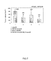
FIG. 7 is a graph showing the in vivo ability of tumorigenesis of colon26 cells, Flt1-Fc gene-expressing colon26 cells, SELF gene-expressing colon26 cells, and cell mixture prepared by mixing Flt1-Fc gene-expressing colon26 cells and SELF gene-expressing colon26 cells at a ratio of 1:1, as examined in Example 11. 1: Colon26 cells; 2: Flt1-Fc gene-expressing colon26 cells; 3: SELF gene-expressing colon26 cells; and 4: Cell mixture prepared by mixing Flt1-Fc gene-expressing colon26 cells and SELF gene-expressing colon26 cells at a ratio of 1:1.
FIG. 8 is a restriction enzyme map showing a SELF genome portion prepared in Example 13.
FIG. 9 shows the structures of vectors constructed in Example 15.
FIG. 10 shows the structures of vectors lacking the SELF promoter, as constructed in Example 16. This figure shows the positions of major transcription factor binding sequences as examined in Example 2. Each numerical value in the figure indicates a nucleotide number of SEQ ID NO: 31.
FIG. 11 is a graph showing luciferase activity as measured in Example 17.
DETAILED DESCRIPTION OF THE INVENTION
The present invention will now be described in detail. The necessary experiments for the present invention, such as preparation of mRNA, production of cDNA, RT-PCR method, RACE method, DNA sequencing and identification of gene expression by Northern blot, can be performed according to methods described in standard laboratory books. An example is “Molecular Cloning, A Laboratory Manual”, 2001, Eds., Sambrook, J & Russell, D W., Cold Spring Harbor Laboratory Press.
1. Obtainment of SELF Gene
The genes of the present invention, which encode the novel protein SELF containing the EGF-like repeat motif, can be cloned by searching any genes for the amino acid sequence of the EGF-like motif found in Notch and Delta, obtaining a sequence appeared in the sequences of the EGF-like motif with a relatively high frequency, and using the obtained sequence as an indicator. Examples of the amino acid sequence of EGF-like motif are CPPGF (SEQ ID NO: 18) and NGGTC (SEQ ID NO: 19), but it is not limited thereto. The genes can be cloned by designing degenerate primers based on the amino acid sequence, synthesizing the primers on a DNA synthesizer, purifying the synthesized primers, and performing RT-PCR with the purified primers.
Stromal cells may be used as the mRNA source for RT-PCR using the primers. The stromal cells may be cell lines that support the growth and differentiation of stem cells or precursor cells, preferably PA6, OP9, ST2, more preferably PA6. mRNA are extracted from the stromal cells, and amplified by RT-PCR.
cDNA fragments which have been amplified from PA6 mRNAs by the RT-PCR method, are cloned in various vectors, and the DNA sequences contained in the clones are determined. By comparing the determined genetic sequences of the DNAs with genetic sequences corresponding to well-known proteins containing the EGF-like repeat sequence such as Notch and Delta, it can be verified that a partial fragment of SELF gene has been cloned.
A coding region of the gene fragment can be cloned by labeling the partially cloned gene as mentioned above with e.g. a radioactive isotope, and screening cDNA library prepared from mRNA extracted from stromal cells using e.g. hybridization method. Alternatively, a coding region of the gene fragment can be also cloned by RACE method using primers which are designed based on the sequence information of the partial cloned gene. RACE method allows one to obtain the full-length sequence of the gene encoding SELF protein containing the EGF-like repeat motif.
The cDNA nucleotide sequence of the invention, which encodes a mouse novel protein SELF containing an EGF-like repeat motif, is shown as SEQ ID NO: 1 in the sequence listing. As a result of homology searching between the DNA sequence shown by SEQ ID NO: 1 and the DNA sequence of known genes, it has been found that the DNA sequence shown by SEQ ID NO: 1 is homologous to the genes encoding the protein containing the EGF-like repeat motif, such as human Tan1, mouse Notch 4, rat Jagged 2 and human Delta.
Further, human SELF cDNA can be obtained by PCR amplification using cDNA derived from human spleen as a template and primers or probes designed from the sequence information of the above mouse SELF DNA, and then sequenced, as described in the after-mentioned Examples. The nucleotide sequence of human SELF cDNA as obtained in this way is typically shown in SEQ ID NO: 23 and the corresponding amino acid sequence is shown in SEQ ID NO: 24.
It has also been found that the SELF gene of the invention is homologous to the nucleotide sequences described in International Publication No. WO 01/32873 A1 (FIG. 4 and FIG. 6 of WO 01/32873 A1; GenBank accession Nos. NM—172463 and XM 059482). The nucleotide sequence shown in FIG. 4A of International Publication No. WO 01/32873A1, which has been derived from rat, shows homology with the nucleotide sequence between the nucleotides 1342 to 4368 of SEQ ID NO: 1 of the present invention. The nucleotide sequence shown in FIGS. 6A and 6B of International Publication No. WO 01/32873A1, which has been derived from human, shows homology with the nucleotide sequence between the nucleotides 1252 to 4368 of SEQ ID NO: 1. However, the physiological function of a protein encoded by the nucleotide sequence of NM—172463 has been unresolved. The nucleotide sequence of XM—059482 contains additional 192 nucleotides within the above shown sequence of human SELF gene, and the physiological function of a protein encoded by the nucleotide sequence of XM—059482 sequence has not yet been determined.
The protein encoded by the gene disclosed in International Publication No. WO 01/32873A1 is an intracellular transcription factor, which regulates insulin signal transduction. On the other hand, the SELF protein encoded by the gene of the present invention is secreted extracellularly, has bioactivity as a growth and differentiation controlling factor, in particular, hematopoiesis-stimulating effects, and/or inhibiting effects on the growth and differentiation of smooth muscle cells. Further, the SELF protein can act directly on cells. Hence, the SELF gene of the present invention is different from any known genes and is therefore a novel gene.
The nucleotide sequence of the polynucleotide which encode the polypeptide comprising the amino acid sequences of SEQ ID NO: 2, 3 and 4 is also shown in SEQ ID NO: 1. The amino acid sequence of SEQ ID NO: 2 corresponds to the nucleotide sequence of nucleotides 157 to 4365 of SEQ ID NO: 1. The amino acid sequence of SEQ ID NO: 3 corresponds to the nucleotide sequence of nucleotides 223 to 4365 of SEQ ID NO: 1. The amino acid sequence of SEQ ID NO: 3 shows a SELF protein devoid of the signal sequence. The amino acid sequence of SEQ ID NO: 4 corresponds to the nucleotide sequence of nucleotides 223 to 1317 of SEQ ID NO: 1. The amino acid sequence of SEQ ID NO: 4 shows a part of the SELF protein devoid of the signal sequence (SEQ ID NO: 3).
It may be often found for the genetic sequence of the present invention that the DNA sequence of its chromosomal DNA or cDNA, which is obtained from nature, is mutated without causing any mutation at the amino acid level because of degeneracy of the genetic code. Also, the DNA sequences of the 5′ untranslated region and 3′ untranslated region may have high variability since the regions are not involved in definition of the amino acid sequence of the protein. The varied nucleotide sequences based on the degeneracy of the genetic code as mentioned above, are also included in the nucleic acids (or polynucleotides) of the present invention. Further, variants of the protein of the invention produced by alternative splicing are also included in the proteins of the present invention, provided that the variants retain the characteristics of the SELF proteins comprising the amino acid sequence of SEQ ID NO: 2, 3, 4 or 24 in the sequence listing.
The present invention further includes the other animal-derived SELF nucleic acids corresponding to the nucleic acids comprising the nucleotide sequences of SEQ ID NOs: 1 and 23, and their partial fragments; and the other animal-derived proteins corresponding to the proteins comprising the amino acid sequences of SEQ ID NOs: 2, 3, 4 and 24 and their partial fragments.
In addition, nucleic acids of the present invention include not only the nucleic acid comprising the nucleotide sequence of SEQ ID NO: 1 (i.e., nucleotides 1 to 5245 of SEQ ID NO: 1), but also the nucleic acid having the nucleotide sequence of nucleotides 1 to 1251 of SEQ ID NO: 1, the nucleic acid having the nucleotide sequence of nucleotides 1624 to 2174 of SEQ ID NO: 1.
Nucleic acids of the present invention also includes nucleic acids which hybridizes under stringent conditions with the DNA comprising the nucleotide sequence of SEQ ID NO: 1, the nucleic acid having the nucleotide sequence of nucleotides 1 to 1251 of SEQ ID NO: 1 or the nucleic acid having the nucleotide sequence of nucleotides 1624 to 2174 of SEQ ID NO: 1 or the nucleic acid comprising the nucleotide sequence of SEQ ID NO: 23 (i.e, nucleotides 1 to 4242 of SEQ ID NO: 23). Such nucleic acids preferably contain the nucleotide sequences encoding EGF-like repeat motifs, and encode proteins having bioactivity as a growth and differentiation controlling factor.
In the present invention, “stringent conditions” means conditions defined by carrying out hybridization at 68° C. in the presence of 0.7-1.0 M NaCl on a DNA-immobilized filter, and subsequently washing the filter at 68° C. with the 0.1-2.0×SSC solution (1×SSC contains 150 mM NaCl and 15 mM sodium citrate), under which conditions detection of DNA of interest can be accomplished, or the substantially equivalent conditions. The “bioactivity as a growth and differentiation controlling factor” means an effect of controlling the growth and differentiation on undifferentiated cells. “Undifferentiated cells” as used herein refers to stem cells or precursor cells. Stem cells are defined as cells which can reproduce themselves and can differentiate into many types of cell lineages. These include myeloid stem cells, neural crest cells, skin stem cells, neural stem cells, muscle stem cells, hematopoietic stem cells and liver stem cells, and each of them has the ability of self-replication and the ability of generating the cell lineages. Precursor cells refer to the cell lineage-committed cells from each stem cell, which have not achieved their final differentiation. Preferably, the undifferentiated cells may be hematopoietic undifferentiated cells, for example, hematopoietic stem cells or hematopoietic progenitor cells. Alternatively, the undifferentiated cells may be preferably myeloid stem cells, neural crest cells, mesenchymal stem cells, smooth muscle progenitor cells or ES cells (embryonic stem cells).
“Controlling effects on the growth and differentiation” means an effect that allows the differentiation and/or growth of undifferentiated cells to be autonomously or heteronomously promoted or inhibited. Specifically, this term means an effect of causing undifferentiated cells to 1) reach a differentiating state, 2) remain in their present state without differentiating, or 3) reach a replicating state. Any molecules having the differentiation and growth control effect may be used, as long as they affect, directly or indirectly, the undifferentiated cells in a body or a culture system and result in showing the effect. For example, such effects of the molecules may be demonstrated by adding the molecules to cultured marrow cells to produce blood cells or osteoclasts.
In the preferred aspect of the invention, the controlling effect on growth and differentiation of undifferentiated cells with respect to SELF protein of the present invention may be controlling effects on growth and differentiation of hematopoietic undifferentiated cells, for example, hematopoiesis-stimulating effects. Alternatively, the controlling effect on growth and differentiation of undifferentiated cells may be inhibiting effects on the growth and differentiation of undifferentiated cells into smooth muscle cells.
The term “SELF,” herein described, refers to SELF protein and/or SELF gene.
Nucleic acids of the present invention further include nucleic acids comprising a nucleotide sequence having at least 70% homology, preferably at least 80% homology, more preferably 90% homology, and still more preferably 95%, 96%, 97%, 98% or 99% homology, with the nucleotide sequence of SEQ ID NO: 1, the nucleotide sequence of nucleotides 1 to 1251 of SEQ ID NO: 1, the nucleotide sequence of nucleotides 1624 to 2174 of SEQ ID NO: 1, or the nucleotide sequence of SEQ ID NO: 23, wherein the homology is calculated using BLAST (e.g., with the default or initial setting parameters of BLAST).
Mutations can be introduced into the genes of the present invention by known techniques such as the Kunkel method or Gapped duplex method or a technique based thereon, e.g., with a mutagenesis kit based on site-specific mutagenesis method (e.g., Mutan™-K (TAKARA) or Mutan™-G (TAKARA), or a kit of TAKARA LA PC™ in vitro Mutagenesis series).
Once the nucleotide sequences of genes of the present invention are determined, the genes of the present invention can then be obtained by chemical synthesis, by PCR using cDNA as a template, or by hybridization using DNA fragments having this nucleotide sequence as a probe.
The recombinant vectors of the present invention can be obtained by ligation (insertion) of a gene of the present invention into a suitable vector. There is no particular limitation on the vectors into which a gene of the present invention is inserted, provided that they are replicable in the host, e.g., plasmid DNA or phage DNA.
Examples of plasmid DNA are plasmids derived from E. coli (e.g., pBR322, pBR325, pUC118, pUC119, pUC18, pUC19), plasmids derived from Bacillus subtilis (e.g., pUB110, pTP5), and plasmids derived from yeast (e.g., YEp13, YEp24, YCp50). Examples of phage DNA are λ phage (Charon 4A, Charon 21A, EMBL 3, EMBL 4, λ gt10, λ gt11, λ ZAP). Further, detoxified DNA viruses or RNA viruses, such as retroviruses, adenoviruses, adeno-associated viruses, herpes viruses, vaccinia viruses, poxviruses, polioviruses, Sinbis virus, Sendai virus, SV40 and Human Immunodeficiency Virus (HIV); animal viruses such as pCI-neo, pcDNA3, or pZeoSV, and insect virus vectors, e.g., baculoviruses, can also be used.
In order to insert the genes of the present invention into the vector, for example, purified DNA is first cleaved with one or more suitable restriction enzymes, and the resulting gene fragment is then inserted and ligated into a restriction enzyme site or a multi-cloning site of a suitable vector DNA.
The genes of the present invention need to be inserted into a vector in a manner that the gene can perform its function. For this purpose, the vectors of the invention may be optionally contain other fragments, including those containing a cis-element such as an enhancer, a splicing signal consisting of a splice donor site on the 5′ terminal side of intron and a splice acceptor site on the 3′ terminal side of intron, a poly A addition signal, a selectable marker or a ribosome binding sequence (SD sequence), in addition to a promoter and the gene of the present invention. Examples of selectable marker include dihydrofolic acid reductase gene, ampicillin resistant gene, and neomycin resistant gene.
The transformants of the present invention, for example transformed cells, can be obtained by introducing a recombinant vector of the present invention into a host such that the gene of interest can be expressed. There is no particular limitation on the host used in the invention provided that the DNA of the present invention can be expressed in the host. Examples of the host include bacteria belonging to the genus Escherichia (such as Escherichia coli), the genus Bacillus (such as Bacillus subtilis), and the genus Pseudomonas (such as Pseudomonas putida); yeasts such as Saccharomyces cerevisiae or Schizosaccharomyces pombe; animal cells such as COS cells or CHO cells; and insect cells such as Sf21.
When Escherichia coli (E. coli) is used as the host, preferably the recombinant vectors of the present invention are autonomously replicable in the host cells, and contain a promoter, a ribosome binding sequence, a gene of the present invention and a transcription termination sequence. The vectors may also contain a gene for controlling the promoter.
Examples of E. coli include Escherichia coli DH1. Example of grass bacillus is Bacillus subtilis. However the host bacteria used in the invention is not limited to these organisms.
There is no limitation on the promoters when bacteria are used as the host, provided that it can be expressed in the host such as E. coli. For example, the promoters derived from E. coli, such as trp promoter, lac promoter, PL promoter and PR promoter, and phage-based promoters can be used. Any artificially modified promoter, such as tac promoter, can also be used.
There is no limitation on the methods of introducing a recombinant vector into bacteria used in the invention, provided that it can introduce DNA into bacteria. Examples of the method are a method using calcium ion [Cohen, S, N. et al.: Proc. Natl. Acad. Sci., USA, 69:2110 (1972)], and electroporation method.
When yeast is used as the host, Saccharomyces cerevisiae, Schizosaccharomyces pombe or Pichia pastoris, etc. can be used. There is no limitation on the promoters when yeast is used as the host, provided that it can be expressed in the yeast, and e.g., gall promoter, ga110 promoter, heat shock protein promoter, MFα1 promoter, PH05 promoter, PGK promoter, GAP promoter, ADH promoter or AOX1 promoter can be used.
There is no limitation on the methods of introducing a recombinant vector into the yeast, provided that it can introduce DNA into yeast. Examples are electroporation method [Becker, D. M. et al.: Methods. Enzymol., 194:182 (1990)], spheroplast method [Hinnen, A. et al.: Proc. Natl. Acad. Sci., USA, 75:1929 (1978)] and lithium acetate method [Itoh, H.: J. Bacteriol., 153:163 (1983)].
When animal cells are used as the host, for example, monkey cell COS-7, Vero, Chinese hamster ovarian cell (CHO cell), mouse L cell, rat GH3 or human FL cell, can be used. In this case, for example, SRα promoter, SV40 promoter, LTR promoter or CMV promoter, can be used as the promoter. The recombinant vectors can be introduced into the animal cells by, e.g., electroporation method, calcium phosphate method or lipofection method.
When insect cells are used as host, for example, Sf21 cells can be used. The recombinant vectors can be introduced into the insect cells by e.g., calcium phosphate method, lipofection method or electroporation method.
In this specification, both “nucleic acid” and “polynucleotide” mean compounds wherein nucleotides are polymerized, and no special distinction is made between them. Moreover, both nucleic acids and polynucleotides include DNA and RNA.
2. Preparation of the Proteins of the Present Invention
The proteins of the present invention are proteins comprising an amino acid sequence encoded by the SELF gene of the present invention; or proteins comprising an amino acid sequence having one or more amino acids deleted, substituted or added in the above amino acid sequences, containing an EGF-like repeat motif, and having bioactivity as a growth and differentiation controlling factor, preferably a hematopoiesis-stimulating effect and an inhibiting effect on growth and differentiation of smooth muscle cells.
The SELF proteins containing the EGF-like repeat motif of the invention have the particular structures as follows. In the amino acid sequence as shown in SEQ ID NO: 2 in the sequence listing, the sequence of amino acids 1 to 22 of SEQ ID NO: 2 is predicted to be a signal peptide region with the method of von Heijin (Nucleic Acids Res. 14, 4683-4690, 1986). The amino acid sequence of amino acids 38 to 40 is cell attachment sequence RGD. The amino acid sequence of amino acids 1081 to 1084 is glycosaminoglycan attachment sequence. There are ten sites where asparagine-linked sugar is added to, which are respectively the amino acid no. 408, no. 484, no. 536, no. 712, no. 886, no. 977, no. 1015, no. 1109, no. 1139, no. 1298 of SEQ ID NO: 2. EGF-like motif is found at 15 sites. The first EGF-like motif sequence is from amino acid no. 278 cysteine to amino acid no. 308 cysteine, the second EGF-like motif sequence is from amino acid no. 315 cysteine to amino acid no. 346 cysteine, the third EGF-like motif sequence is from amino acid no. 353 cysteine to amino acid no. 384 cysteine, the fourth EGF-like motif sequence is from amino acid no. 387 cysteine to amino acid no. 422 cysteine, the fifth EGF-like motif sequence is from amino acid no. 433 cysteine to amino acid no. 464 cysteine, the sixth EGF-like motif sequence is from amino acid no. 472 cysteine to amino acid no. 499 cysteine, the seventh EGF-like motif sequence is from amino acid no. 545 cysteine to amino acid no. 576 cysteine, the eighth EGF-like motif sequence is from amino acid no. 584 cysteine to amino acid no. 615 cysteine, the ninth EGF-like motif sequence is from amino acid no. 623 cysteine to amino acid no. 654 cysteine, the tenth EGF-like motif sequence is from amino acid no. 661 cysteine to amino acid no. 692 cysteine, the eleventh EGF-like motif sequence is from amino acid no. 753 cysteine to amino acid no. 788 cysteine, the twelfth EGF-like motif sequence is from amino acid no. 791 glutamic acid to amino acid no. 826 cysteine, the thirteenth EGF-like motif sequence is from amino acid no. 833 cysteine to amino acid no. 864 cysteine, the fourteenth EGF-like motif sequence is from amino acid no. 871 cysteine to amino acid no. 902 cysteine, and the fifteenth EGF-like motif sequence is from amino acid no. 1310 cysteine to amino acid no. 1341 cysteine.
The fourth EGF-like sequence, the eleventh EGF-like sequence and the twelfth EGF-like sequence are calcium-linked EGF-like sequences and are involved in protein interactions. Also, there is a sequence similar to an EGF-like sequence between the sixth EGF-like sequence and the seventh EGF-like sequence at one location.
Further, the sequence of amino acids 1 to 29 of SEQ ID NO: 24 is predicted to be a signal peptide region by searching with a software SOSUI (http://sosui.proteome.bio.tuat.ac.jp/sosuiframe0.html). The amino acid sequence of amino acids 38 to 40 is cell attachment sequence RGD. There are thirteen sites where asparagine-linked sugar is added to, which are respectively the amino acid no. 145, no. 204, no. 368, no. 408, no. 484, no. 536, no. 712, no. 886, no. 977, no. 1015, no. 1109, no. 1139, no. 1310 of SEQ ID NO: 24. EGF-like motif is found at 15 sites. The first EGF-like motif sequence is from amino acid no. 278 cysteine to amino acid no. 308 cysteine, the second EGF-like motif sequence is from amino acid no. 315 cysteine to amino acid no. 346 cysteine, the third EGF-like motif sequence is from amino acid no. 353 cysteine to amino acid no. 384 cysteine, the fourth EGF-like motif sequence is from amino acid no. 387 cysteine to amino acid no. 422 cysteine, the fifth EGF-like motif sequence is from amino acid no. 433 cysteine to amino acid no. 464 cysteine, the sixth EGF-like motif sequence is from amino acid no. 472 cysteine to amino acid no. 499 cysteine, the seventh EGF-like motif sequence is from amino acid no. 545 cysteine to amino acid no. 576 cysteine, the eighth EGF-like motif sequence is from amino acid no. 584 cysteine to amino acid no. 615 cysteine, the ninth EGF-like motif sequence is from amino acid no. 623 cysteine to amino acid no. 654 cysteine, the tenth EGF-like motif sequence is from amino acid no. 661 cysteine to amino acid no. 692 cysteine, the eleventh EGF-like motif sequence is from amino acid no. 753 cysteine to amino acid no. 788 cysteine, the twelfth EGF-like motif sequence is from amino acid no. 791 glutamic acid to amino acid no. 826 cysteine, the thirteenth EGF-like motif sequence is from amino acid no. 833 cysteine to amino acid no. 864 cysteine, the fourteenth EGF-like motif sequence is from amino acid no. 871 cysteine to amino acid no. 902 cysteine, and the fifteenth EGF-like motif sequence is from amino acid no. 1311 cysteine to amino acid no. 1342 cysteine.
The proteins of the present invention include a protein consisting of the amino acid sequences of SEQ ID NOs: 2, 3, 4 and 24 in the sequence listing, but also include variants based on intraspecies mutations known to occur in nature, mutations such as allelic mutations or point mutations which can be produced artificially, provided that they retain the characteristics of the proteins of SEQ ID NOs: 2, 3, 4 and 24 in the sequence listing.
The proteins of the invention include a protein consisting of an amino acid sequence having one or more amino acids deleted, substituted or added in the amino acid sequence as shown in SEQ ID NO: 2, 3, 4 or 24, wherein the protein contains an EGF-like repeat motif and has bioactivity as the growth and differentiation controlling factor, for example, preferably, the hematopoiesis-stimulating effect and the inhibiting effect on growth and differentiation of smooth muscle cells.
Herein, “one or more amino acids deleted, substituted or added”, means, but are not limited to, that preferably 1-50 amino acids, more preferably one to several amino acids or most preferably 1-3 amino acids are deleted, substituted or added in the given amino acid sequence. Examples of the amino acid sequence having one or more amino acids deleted, substituted or added in the amino acid sequence as shown in SEQ ID NO: 2, 3, 4 or 24 include amino acid sequences having at least 70% homology, preferably at least 80% homology, more preferably 90% homology, and yet more preferably 95%, most preferably 96%, 97%, 98% or 99% homology, with the amino acid sequence as shown in SEQ ID NO: 2, 3, 4 or 24, wherein the homology is calculated using BLAST (e.g., with the default or initial setting parameters of BLAST).
The SELF proteins of the present invention containing the EGF-like repeat motif can be obtained by culturing the aforesaid transformant and recovering the protein from the culture. The term “culture” means a culture supernatant, cultured non-bacterial cells or bacterial cells, or a cell debris of non-bacterial cells or bacterial cells.
The transformants of the present invention are cultured according to usual method for culturing a host.
When the transformants obtained from microorganisms such as E. Coli or yeast as the host are cultured, the culture medium may be natural culture medium or synthetic culture medium, as long as it contains carbon source, nitrogen source and mineral salts utilized by the microorganism and is useful for efficient culturing of the transformants.
Examples of carbon source are carbohydrates such as glucose, fructose, sucrose or starch, organic acids such as acetic acid or propionic acid, and alcohols such as ethanol or propanol.
Examples of nitrogen source are ammonia, and inorganic or organic ammonium salts such as ammonium chloride, ammonium sulfate, ammonium acetate and ammonium phosphate, and other nitrogen-containing compounds, as well as peptone, meat extracts and corn steep liquor.
Examples of mineral salts are monopotassium phosphate, dipotassium phosphate, magnesium phosphate, magnesium sulfate, sodium chloride, ferrous sulfate, manganese sulfate, copper sulfate and calcium carbonate.
The culturing is typically performed aerobically by e.g. shaking culture or aeration stirring culture at 37° C. The pH of the culture is adjusted with an inorganic or organic acid, or alkaline solution.
Antibiotics such as ampicillin or tetracycline may also be added to the culture medium, if necessary.
When microorganisms transformed with an expression vector using an inducible promoter are cultured, inducers may be added to the culture if necessary. For example, to culture a microorganism transformed by an expression vector using the Lac promoter, isopropyl-β-D-thiogalactopyrranoside (IPTG) may be added to the culture. Also, to culture an microorganism transformed by an expression vector using the trp promoter, indoleacetic acid (IAA) may be added to the culture.
When the transformant obtained from animal cells as host are cultured, the commonly-used RPMI1640 medium, DMEM medium, αMEM or the like, or these culture supplemented with fetal calf serum can be used as the culture medium.
The culturing is typically performed in the presence of 5% CO2 at 37° C. for 1-30 days. During the culturing, antibiotics such as kanamycin or penicillin may be added to the culture if necessary.
After the culturing, when SELF protein of the present invention is produced intracellularly in bacterial cells or non-bacterial cells, the protein can be extracted by crushing the bacterial cells or non-bacterial cells. If the protein of the present invention is produced extracellularly by bacterial cells or non-bacterial cells, the culture solution itself may be used, or the bacterial cells or non-bacterial cells may be removed by centrifugation from the culture solution to be used. The protein according to the present invention can be isolated from the culture by subsequent protein isolation/purification process, for example, by using a conventional biochemical method for protein isolation and purification, such as ammonium sulfate precipitation, gel chromatography, ion exchange chromatography or affinity chromatography, alone or in combinations as appropriate.
3. Preparation of Antibodies
Antibodies specifically recognizing (or binding to) the protein of the present invention or fragments thereof can be produced as shown in Example 5 below. They can also be produced by the various methods shown in the printed books (see, e.g., “Antibodies: A Laboratory Manual”, Cold Spring Harbor Laboratory Press). For example, antibodies recognizing SELF protein can be produced as follows: fully immunizing animals, such as mouse, guinea pig, rabbit and goat by inoculating SELF protein several times subcutaneously, intramuscularly, intraperitoneally or intravenously; drawing blood from the animal; and separating the serum from the blood. A suitable adjuvant can also be used in the immunization. Monoclonal antibodies can also be produced by well-known methods. For example, spleen cells from the mouse immunized with SELF protein are fused to mouse myeloma cells to produce hybridoma, and the monoclonal antibodies are prepared from a culture supernatant of the hybridoma, or a peritoneal fluid from a mouse which was administered intraperitoneally with the hybridoma. For use as an immunogen, SELF protein may be native proteins, recombinant proteins, or chemically synthesized proteins. Furthermore, proteins comprising the full amino acid sequence, peptide fragments having a partial structure of the protein, or fusion proteins of the protein and an additional protein, may also be used as the immunogen. The peptide fragment may be a fragment of the protein obtained by proteolysis with an appropriate protease, or a product expressed from a expression vector which incorporates whole or a part of the nucleotide sequence of SEQ ID NO: 1 or 23. The polypeptide fragment may be combined with a suitable carrier protein by chemical bonding and then may be used. The reactivities of the obtained antibodies can be determined by methods well known to those skilled in the art, such as enzyme immunoassay (EIA), radioimmunoassay (RIA), or Western blotting.
The antibodies produced as mentioned above can be applied to the purification, detection and measurement of the present protein, and can be used also as a diagnostic reagent for abnormal cell differentiation associated diseases. Example 6 shows that the antibody produced in Example 5 is used to determine where and on which developmental stages the present protein appears in mouse. As a result, the expression is observed on the fetal whole-mount at both the 9th day and 11th day in the limb buds and throughout the mesenchymal cells of the face, whereas in blood vessels at the 9th day, the expression is strongly observed in the arteria vitellina (omphalomesenteric membrane artery), and, although weakly, in the anterior cardinal veins of the head. It is weakly expressed in the heart on both embryonal days 9 and 11, and on the 9th day heart it is expressed only in the ventricle of heart, indicating that the present protein (the SELF protein) is expressed in the cardiac muscle. In the 11th embryonal day, it is found that the present protein is expressed in the internal epithelial layer of the intestine and outermost coat of the intestine.
4. Methods for Detecting Self Protein or Self Gene, and Detecting Reagents
SELF protein, antibodies against SELF protein and the gene encoding SELF protein (SELF gene) according to the invention, can be used for diagnosis of diseases associated with abnormal cell differentiation or the like.
In the present invention, the probe, which hybridize with the above nucleic acids to specifically recognize the nucleic acids, can be used as a detecting reagent for detection of the genes encoding SELF protein. The probes may be labeled with the commonly used radioactive isotopes (for example, 32P, 35S) or enzymes (for example, digoxygenin, fluorescein), etc., and may be specifically hybridized with the nucleic acids using the conventional blotting analysis or in situ hybridization, and thus may be detected.
Nucleic acids used as a probe in the present invention may be nucleic acids having at least part of the nucleotide sequence as shown in SEQ ID NO: 1 or 23. The length of the probe may be, but are not limited to, the full-length of the sequence of SEQ ID NO: 1, or more preferably 200-300 nucleotides in length.
Primers are also designed and synthesized based on the nucleic acid sequence of the present invention, the synthesized primers are used in gene amplification methods such as PCR, and thereby SELF gene can be detected.
The novel protein containing the EGF-like repeat motif of the present invention, SELF protein, can be detected using the antibodies against SELF protein. The detection may be made by immunological assay methods known in the art, such as EIA or RIA.
5. Activities of SELF Protein and SELF Gene, Pharmaceutical Composition Containing the Same, and Therapeutic and Preventive Method Using the Same
The SELF protein of the present invention has a function of controlling the growth and differentiation of undifferentiated cells. The present invention also relates to a method for controlling the growth and differentiation of undifferentiated cells, comprising causing the SELF protein to come into contact with undifferentiated cells in vitro, ex vivo, or in vivo, for example. Undifferentiated cells herein may be any of the above-described undifferentiated cells. Preferable examples of such undifferentiated cells include: hematopoietic undifferentiated cells such as hematopoietic stem cells and hematopoietic progenitor cells; stem cells or progenitor cells contained in the bone marrow such as peripheral blood stem cells, myeloid stem cells, neural crest cells, neural stem cells, smooth muscle progenitor cells, and mesenchymal stem cells; and ES cells. These undifferentiated cells may be removed and obtained from subjects such as humans or non-human mammals.
The SELF protein has hematopoiesis-stimulating effects. In the present invention, that the SELF protein “has hematopoiesis-stimulating effects” means that, in the presence of the SELF protein, the number of blood cells generated from hematopoietic undifferentiated cells such as hematopoietic stem cells or hematopoietic progenitor cells or generated from cell populations (e.g., marrow cells) containing these undifferentiated cells, is increased by the effects. As described in Example 8, when stromal cells caused to overexpress the SELF gene after introduction of the gene are cocultured with differentiation-antigen-negative bone marrow cells, the SELF protein secreted from stromal cells acts on the bone marrow cells, so as to promote the production of blood cells in the bone marrow cells. Furthermore, as described in Example 9, when bone marrow cells are cultured for a long time in the presence of the SELF protein, their capability for growth and differentiation is maintained for a time longer compared with a case of the culture in the absence of the SELF protein. Moreover, when bone marrow cells are cultured in the presence of the SELF protein, the amount of cell growth (or proliferation) is increased compared with a case of the culture in the absence of the SELF protein. Preferably, most blood cells produced in such bone marrow cells are neutrophils or macrophages. As described above, the SELF protein is capable of supporting the growth of hematopoietic progenitor cells in bone marrow cells.
Various hematopoietic factors such as G-CSF, GM-CSF, M-CSF, and EPO are used for treating hypocythemia. G-CSF is known to form neutrophil colonies in an in vitro colony formation test (Nicola et al., J. Biol. Chem., 258: 9017-9023, 1983). Furthermore, increases in neutrophils have been observed upon administration of G-CSF to mice (Tamura et al., Biochem. Biophys. Res. Commun., 142: 454-460, 1987). Furthermore, it has been shown that similar to the case of such an experiment conducted on mice, increases in neutrophils are also observed in a dose-dependent manner upon administration of G-CSF to humans (Asano et al., Behring Int. Mitt. 83: 222-228, 1988). For example, hematopoietic factors are clinically used and exert effects in: treatment of anaplastic anemia, myelodysplastic syndrome (MDS), or acute myeloid leukemia (AML); recovery of blood cell counts after cancer chemotherapy for non-Hodgkin's lymphoma, breast cancer, ovarian cancer, head and neck cancer, esophageal cancer, small cell lung cancer, urothelial carcinoma, non-small cell lung cancer, neuroblastoma, or the like; or recovery of blood cell counts after bone marrow transplantation performed for patients with acute leukemia, chronic myeloid leukemia, multiple myeloma, malignant lymphoma, anaplastic anemia, myelodysplastic syndrome, or the like (Ogawa, Blood, 81: 2844-2853, 1993; Sonoda et al., Stem Cells, 11: 543-554, 1993; Antin et al., Blood, 72: 707-713, 1988; Akio Urabe et al., Clinical Hematology (Rinsho Ketsueki), 34: 928-936, 1002-1010, 1993; Bessho et al., Br. J. Haematol. 80: 409-411, 1992; Hijiri Kitamura, hematopoietic factor 3: 64-70, 1990; Bessho et al., Stem Cells, 12: 604-615, 1994; Hoelzer et al., Behring Inst. Mitt., 83: 134-138, 1988; Shinpei Furusawa et al., Journal of Clinical and Experimental Medicine (Igaku no ayumi), 171: 851-855, 1994; Estey et al., J. Clin. Oncol. 12: 671-678, 1994; Kazumasa Ogawa, Journal of Clinical and Experimental Medicine (Igaku no ayumi), 171: 847-855, 1994; Tooru Masaoka, Journal of Clinical and Experimental Medicine (Igaku no ayumi), 171: 856-859, 1994). The SELF protein has hematopoiesis-stimulating effects, so that the protein is useful for treating and preventing the above hypocythemia in a manner similar to that in the case of hematopoietic factors.
In the present invention, hematopoiesis can be stimulated in a subject using the SELF protein or the SELF gene. According to the present invention, hypocythemia in a patient with anaplastic anemia, myelodysplastic syndrome, leukemia, or the like, and hypocythemia in a patient after cancer chemotherapy, bone marrow transplantation, or the like can be treated or prevented through administration of the SELF protein or a recombinant expression vector containing the SELF gene, preferably as a pharmaceutical composition containing the protein or the vector. The SELF protein or the SELF gene is preferable for treating leukemia and particularly preferable for treating acute myeloid leukemia. Furthermore, hematopoiesis can be stimulated and hypocythemia can be treated or prevented through administration of the SELF protein or the SELF gene to patients suffering from induced hypocythemia or patients who are expected to develop hypocythemia, after cancer chemotherapy against malignant tumors such as non-Hodgkin's lymphoma, breast cancer, ovarian cancer, head and neck cancer, esophageal cancer, small cell lung cancer, urothelial carcinoma, non-small cell lung cancer, or neuroblastoma, or following bone marrow transplantation therapy that is performed for treating acute leukemia, chronic myeloid leukemia, multiple myeloma, malignant lymphoma, anaplastic anemia, myelodysplastic syndrome, or the like. Moreover, the SELF protein or a recombinant expression vector containing the SELF gene can be administered to a patient, so as to increase peripheral blood stem cells. Thereafter, stem cells for stem cell transplantation can also be recovered from the peripheral blood. Preferably, such the SELF protein or recombinant expression vector containing the SELF gene is used by administering the vector into the bone marrow of a patient, for example.
The SELF protein or a recombinant expression vector containing the SELF gene is added to an in vitro coculture system of stromal cells and bone marrow cells to enhance the production of blood cells in the bone marrow cells. The thus obtained blood cells can also be administered to a patient. In this case, the bone marrow cells may be recovered from the relevant patient or another patient.
The SELF protein also has inhibiting effects on the growth and differentiation into smooth muscle cells from undifferentiated cells (inhibiting effects on the growth and differentiation of smooth muscle cells). The SELF protein can inhibit the growth and differentiation of any smooth muscle cells such as vascular smooth muscle cells, pericytes, gastrointestinal smooth muscle cells, bronchial smooth muscle cells, and urinary bladder smooth muscle cells, and preferably vascular smooth muscle cells. Examples of undifferentiated cells used herein include any undifferentiated cells. Such undifferentiated cells may be preferably hematopoietic stem cells, hematopoietic progenitor cells, myeloid stem cells, peripheral blood stem cells, neural crest cells, neural stem cells, mesenchymal stem cells, or smooth muscle progenitor cells. In the present invention, that the SELF protein has “inhibiting effects on the growth and differentiation of smooth muscle cells” means that under conditions where the growth and differentiation of smooth muscle cells can be induced and in the presence of the SELF protein, the number of smooth muscle cells does not increase or its level of increase decreases compared with a case in the absence of the SELF protein. In the present invention, according to the method of Takakura et al. (Takakura, N. et al., Immunity 9: 677-686, 1998), the para-aortic splanchnopleural mesoderms (P-Sp) of mouse fetuses were cocultured on a stromal cell line OP9 for 10 days in the presence of the SELF protein. When sheet-like structure (vascular bed) formation by vascular endothelial cells is observed but no vascular network formation takes place, it can be confirmed that the growth and differentiation of smooth muscle cells have been inhibited by the SELF protein.
Various diseases are known to be developed in association with elevated levels of the growth and differentiation of smooth muscle cells. For example, excessive growth of smooth muscle cells is observed in the case of glomerulonephritis. Furthermore, excessive growth of vascular smooth muscle cells causes blood vessel wall thickening or the stricture or occlusion of vascular lumina. Such excessive growth induces arteriosclerotic diseases such as atherosclerosis, diabetic vascular disorders, cerebral ischemic stroke, stenocardia, and cardiac infarction (Ross, R. et al., N. Engl. J. Med., 314, p. 488-500, 1986). It is thought that inhibition of the growth and differentiation of vascular smooth muscle cells leads to treatment and prevention of such arteriosclerotic diseases. Treatment for diseases that are associated with elevated levels of the growth and differentiation of smooth muscle cells, such as arteriosclerotic diseases, is under development, which comprises administration of an inhibitor for the growth and differentiation of smooth muscle cells to patients (Gordon, A et al., Science, 253, p. 1129, 1991).
Diseases associated with elevated levels of the growth and differentiation of smooth muscle cells (e.g., arteriosclerotic diseases or glomerulonephritis) can be treated or prevented by administering the SELF protein or a recombinant expression vector containing the SELF gene to a subject, preferably as a pharmaceutical composition containing such protein or vector, to inhibit the growth and differentiation of smooth muscle cells in the subject. For example, through administration of them to a subject with arteriosclerotic disease, the progression of arteriostenosis can be inhibited or its stenosis state can be alleviated.
Moreover, the SELF protein can inhibit angiogenesis through inhibition of the growth and differentiation of smooth muscle cells.
The vascular structures of mature blood vessels are stabilized through lining of vascular endothelial cells via matrices by parietal cells. Parietal cells surrounding vascular endothelial cells are composed of smooth muscle lineage cells such as smooth muscle cells and/or pericytes. Parietal cells function not only for supporting the vascular structure but also for vascular relaxation and contraction, for example. Parietal cells are desorbed from existing blood vessels when subjected to angiogenesis stimulation. Subsequently, bared vascular endothelial cells produce various proteases to digest vascular basement membranes or their surrounding extracellular matrices. Furthermore, the vascular endothelial cells grow and migrate to form a luminal structure. Parietal cells are then recruited to surround the vascular endothelial cells, so as to arrest the migration and the growth of the vascular endothelial cells. New vascular basement membranes are formed, and then mature blood vessels are constructed. Formation of new blood vessels based on such budding from existing blood vessels is generally referred to as angiogenesis.
Whereas angiogenesis is known to play an important role in endometrial formation, follicle formation, wound healing, and the like, abnormal angiogenesis is known to cause various pathological conditions. Folkman and Klagsbrun have proposed to generically designate diseases (a group of diseases) associated with abnormally elevated levels of vascular growth (e.g., malignant tumors, diabetic retinopathy, and psoriasis) as angiogenic diseases (Folkman & Klagsbrun, Science 235: 442-447, 1987). In particular, malignant tumors that are developed due to abnormal cell growth induce angiogenesis (tumor angiogenesis) at tumor-forming sites for supply of oxygen and nutrients, thereby causing tumor growth or metastasis. In recent years, therapeutic methods for angiogenic diseases using various angiogenesis inhibitors produce effects. Examples of angiogenesis inhibitors that have been reported to be useful as antitumor drugs include interferon, a vascular endothelial growth factor (VEGF) inhibitor (e.g., a neutralization antibody against VEGF), NK4, angiostatin, endostatin, and prolactin. For example, angiogenesis inhibitors targeting VEGF have been reported as follows. Inhibition of angiogenesis through administration of an anti-VEGF monoclonal antibody exerts tumor growth inhibitory effects (Kim et al., Nature 362, 841-844, 1993). Inhibition of angiogenesis through administration of an anti-VEGF monoclonal antibody can inhibit cancer metastasis (Melnyk et al., Cancer Research 56, 921-924, 1996). An effect of prolonging the survival time of a cancer patient can be obtained by the use of an anti-VEGF monoclonal antibody Bevacizumab (Avastin™; Genentech) in combination with anticancer agents (3 agents: irinotecan, 5-fluorouracil, and leucovorin) (Hurwitz et al., New Engl. J. Med. 350: 2335-2342, 2004). Tumor growth can be inhibited through inhibition of angiogenesis using an antisense DNA against a VEGF gene or small interfering RNA (siRNA) (Saleh et al., Cancer Research 56, 393-401, 1996). A vector expressing a soluble VEGF receptor protein (that binds to VEGF to inhibit VEGF activity) is introduced into a human ovarian cancer cell line RMG-1 and stably expressed therein, and then administration of the resulting RMG-1 cells to nude mice results in prolonged survival time of the nude mice and inhibited tumor growth in the nude mice, compared with control mice inoculated with an RMG-1 cell line expressing no soluble VEGF receptors (Hasumi et al., Cancer Res. 62: 2019-2031, 2002). When a chimeric protein comprising a soluble VEGF receptor and a human IgG1 constant region (Fc) is administered to nude mice into which a human ovarian cancer cell line OVCAR-3 has been transplanted, the chimeric protein binds to VEGF in a manner similar to that in the case of the soluble VEGF receptor, so as to inhibit the effects, and thereby the weights of tumors generated in vivo in mice are significantly lower than those in a control group to which a human IgG1 Fc alone has been administered (Byrne et al., Clin. Cancer Res. 9: 5721-5728, 2003). These reports show that gene therapy that involves introducing a vector capable of expressing a soluble VEGF receptor into cancer cells has therapeutic effects similar to those exerted by a therapeutic method that involves administering a soluble VEGF receptor protein to a subject. Furthermore, a low molecular weight compound SU5416 that is a tyrosine kinase inhibitor for a VEGF receptor has an effect of improving disease conditions against lung cancer, large bowel cancer, and Kaposi's sarcoma (Rosen et al., Proceedings of 35th ASCO, No. 618, 1999; Fong et al., Cancer Res. 59: 99-106, 1999). However, a case has also been reported wherein sufficient effects of inhibiting angiogenesis cannot be obtained by inhibition of the growth of vascular endothelial cells alone using such VEGF inhibitor.
In the present invention, the growth of vascular smooth muscle cells can be inhibited in a subject so as to inhibit angiogenesis through administration of the SELF protein or a recombinant expression vector containing the SELF gene to the subject. Furthermore, “angiogenesis” in the present invention means not only a process during which new blood vessels bud from existing blood vessels to generate mature blood vessels, but also a process during which mature blood vessels are generated through vasculogenesis that is initiated by undifferentiated cells. According to the present invention, by the use of the SELF protein or the SELF gene, tumor angiogenesis can be inhibited, so that tumor growth can be inhibited. Particularly, according to the present invention, angiogenic diseases in subjects can be treated or prevented by administering the SELF protein or a recombinant expression vector containing the SELF gene of the present invention to subjects, preferably as a pharmaceutical composition containing the protein or the vector. Target angiogenic disease may be any disease associated with abnormally elevated levels of angiogenesis and is not limited. Examples of such angiogenic diseases include malignant tumors such as solid tumors (including primary carcinoma and metastatic carcinoma) including renal cancer, breast cancer, brain tumor, gastrointestinal cancer, ovarian cancer, hepatic cancer, angioma, vascular fibrous tumor, and multiple myeloma; ophthalmic angiogenesis-related diseases such as diabetic retinopathy, retinopathy of prematurity, rubeosis iridis, sickle-cell retinopathy, central retinal vein occlusion, branch retinal vein occlusion, central retinal artery occlusion, age-related macular degeneration, and neovascular glaucoma; chronic inflammatory diseases such as rheumatoid arthritis; dermatologic angiogenesis-related diseases such as psoriasis (e.g., psoriasis vulgaris); diseases associated with abnormally elevated levels of vascular permeability such as ascites cancer, malignant pleural effusion, Crow-Fukase syndrome, and ovarian hyperstimulation syndrome; and ischemic diseases such as atherosclerosis, cerebral infarction, acute cardiac infarction, and peripheral artery occlusive disease. Through the above administration to subjects suffering from angiogenic diseases, the pathological conditions of angiogenic diseases can be improved. For example, tumor growth can be inhibited (that is, arrested or delayed) or tumor regeneration can be induced. Through the use of the SELF protein or a recombinant expression vector containing the SELF gene in combination with a VEGF inhibitor, therapeutic and preventive effects can also be enhanced in angiogenic diseases such as malignant tumors.
For the purpose of inhibiting angiogenesis or treating or preventing angiogenic diseases, the SELF protein or a recombinant expression vector containing the SELF gene may be systemically or topically administered to a subject. Preferably, such vector is administered topically to a site where angiogenesis is taking place or a site where angiogenesis is predicted to take place or to the periphery thereof. For example, it is preferable to directly inject the SELF protein or a recombinant expression vector containing the SELF gene into malignant tumors or to topically administer such protein or vector to the eyes of patients with ophthalmic angiogenesis-related diseases. Alternatively, such the SELF protein or recombinant expression vector containing the SELF gene is systemically administered such that the protein or the vector is delivered in a tissue specific manner to angiogenesis sites or organs containing such sites.
Blood vessel formation initiated by undifferentiated cells can also be inhibited by adding the SELF protein or a recombinant expression vector containing the SELF gene to an in vitro coculture system of stromal cells and undifferentiated cells (e.g., ES cells).
The dose of such the SELF protein or recombinant expression vector containing the SELF gene may differ depending on symptoms, age, and body weight of a subject, and the like. In the case of oral administration, the dose is generally between approximately 0.001 mg and several hundred mg per day. In this case, the protein or the vector is administered once or at several separate times. Furthermore, in the case of perenteral administration, the dose is generally between 0.001 mg and several hundred mg per administration via subcutaneous injection, intramuscular injection, or intravenous injection. In the case of gene therapy, more preferably, the dose is between 0.001 mg and several hundred mg per administration via subcutaneous injection, intramuscular injection, or intravenous injection at intervals of few days, several weeks, or several months.
In the present invention, through administration of the SELF protein or a recombinant expression vector containing the SELF gene in combination with a vascular endothelial growth factor (VEGF) inhibitor, higher inhibitory effects can be obtained against angiogenesis (e.g., tumor angiogenesis). As a result, for example, tumor growth and metastasis can be significantly inhibited.
Examples of such vascular endothelial growth factor (VEGF) inhibitor include, but are not limited to, antibodies such as a neutralization antibody against a vascular endothelial growth factor (VEGF), a neutralization antibody against EGF, a neutralization antibody against angiopoietin-2, Tie-2 antibody, and an antibody against a VEGF receptor; angiogenesis-related proteins such as a VEGF receptor, Tie-2, endostatin, angiostatin, chondromodulin (Hiraki et al., J. Biol. Chem., 272: 32419-32426, 1997), tenomodulin (Shukunami et al., Biochem. Biophys. Res. Commun. 280: 1323-1327, 2001), angiopoietin-2, thrombospondin, and interferon; and nucleic acids encoding such proteins. Moreover, a VEGF inhibitor may be an antisense oligonucleotide or a siRNA against a gene encoding VEGF, a VEGF receptor, FGF, a FGF receptor, angiopoietin-2, or Tie-2. A preferable example of a VEGF inhibitor is a vector mFlt-1-hIgG1 capable of expressing a Flt1-Fc protein that is a chimeric protein of a soluble VEGF receptor and a human IgG1 constant region (Fc) (Hirashima, M., et al., Blood 93: 1253-1263, 1999). In addition, a gene encoding VEGF has been identified in the case of a human (GenBank accession No. X51602), a mouse (GenBank accession No. NM—010228), a rat (GenBank accession No. NM—019306), and the like.
The SELF protein or a recombinant expression vector containing the SELF gene and a VEGF inhibitor may also be administered in mixed state, may be administered separately but simultaneously, or may be sequentially administered. More preferably, they are both directly administered to an angiogenesis site (e.g., tumor). The SELF protein or the recombinant expression vector containing the SELF gene, and the VEGF inhibitor, are more preferably administered at a dose ratio of 1:1, but the ratio is not limited thereto.
In the present invention, “subject” means a human or a non-human animal (e.g., a non-human mammal). Examples of such non-human animals include rodents (e.g., mice, rats, and hamsters), domestic animals (e.g., cattle, horses, and sheep), and companion animals (e.g., dogs and cats).
The present invention also provides a pharmaceutical composition containing an effective dose of the SELF protein or a recombinant expression vector that contains the SELF gene. The pharmaceutical composition of the present invention is useful for stimulating hematopoiesis in a subject, for inhibiting the growth and differentiation of smooth muscle cells, for inhibiting angiogenesis, and for obtaining antitumor effects. Such pharmaceutical composition containing a recombinant expression vector that contains the SELF gene can also be preferably used as a medicament for gene therapy. The pharmaceutical composition containing the recombinant expression vector that contains the SELF gene can also be used for ex vivo gene therapy according to a conventionally known method. A vector to be used for gene therapy in the present invention is preferably a vector that can be expressed in a host to be subjected to administration. For example, when a vector is administered to a human, a vector based on a plasmid, a virus, or the like, for which a mammalian cell is used as a host, is preferably used.
The pharmaceutical compositions of the present invention can be administered to patients in various forms. Examples of such administration forms include an oral administration of a tablet, capsule, granule, powder, syrup, etc., or a parenteral administration of an injection solution, ophthalmic solution, drop, suppository, etc. Such pharmaceutical compositions can be prepared by well-known methods. The pharmaceutical compositions may comprise a pharmaceutically acceptable carrier, diluent and excipient, as usually used in the pharmaceutical industry. For example, lactose or magnesium stearate may be used as the carrier or excipient for tablets. The injection solution is prepared using the solution wherein the SELF or its salt is dissolved, suspended or emulsified in a sterile aqueous or oily solution commonly used for injection. The aqueous solution used for injection solution may be an isotonic solution containing physiological saline, glucose or other auxiliaries, and can be used in combination with a suitable solubilizing agent, e.g., an alcohol or polyalcohol such as propylene glycol, a nonionic surfactant, etc. The oily solution may be sesame oil, soybean oil, etc., and can be used in combination with a solubilizing agent, e.g., benzyl benzoate or benzyl alcohol, etc.
The present invention also relates to a pharmaceutical kit for angiogenesis inhibition or tumor growth inhibition, which contains the SELF protein or a recombinant expression vector containing the SELF gene and a vascular endothelial growth factor inhibitor in combination. Such pharmaceutical kit contains each of these agents in a single dosage unit or in multiple dosage units. The pharmaceutical kit may also contain a syringe, medication instructions, and the like.
6. SELF Promoter
A SELF promoter region of the present invention comprises the nucleotide sequence as shown in SEQ ID NO: 31. “SELF promoter” or “SELF promoter region” in the present invention means a region that substantially affects the transcription of the SELF gene. SELF promoter may contain transcriptional regulatory sequences such as an enhancer and silencer in addition to a region having promoter activity. Furthermore, “promoter activity” means ability of transcription from a gene into mRNA.
As shown by comparison of the SELF promoter sequence shown in SEQ ID NO: 31 with a SELF cDNA sequence shown in SEQ ID NO: 1, the transcription initiation position is at nucleotide 3487 of SEQ ID NO: 31. Furthermore, the portion between nucleotides 3632 and 3848 of SEQ ID NO: 31 is the 5′ terminal portion of a structural gene of the SELF protein. The 1st intron begins from nucleotide 3849 of SEQ ID NO: 31.
A region having basic promoter activity of the SELF gene is the CpG island region between nucleotides 3299 and 3487 in the nucleotide sequence shown in SEQ ID NO: 31. Hence, any fragment of the nucleotide sequence as shown in SEQ ID NO: 31 that contains the nucleotide sequence of nucleotides 3299 to 3487 has SELF promoter activity. For example, DNA consisting of the nucleotide sequence of nucleotides 2360 to 3487 of SEQ ID NO: 31 or DNA consisting of the nucleotide sequence of nucleotides 3374 to 3487 of SEQ ID NO: 31 has SELF promoter activity.
In particular, the region exerting the highest promoter activity is the nucleotide sequence of nucleotides 2796 to 3487 of SEQ ID NO: 31. Therefore, the SELF promoter of the present invention preferably contains at least the region of nucleotides 2796 to 3487 as shown in SEQ ID NO: 31.
Several transcriptional regulatory sequences (sequences to which transcription controlling factors can bind) are present in such SELF promoter region as shown in SEQ ID NO: 31. Specifically, a C/EBPβ (CCAAT/Enhancer Binding Protein β) binding sequence is present between nucleotides 1590 and 1603, between nucleotides 2402 and 2415, between nucleotides 2456 and 2469, or between nucleotides 2744 and 2757 of SEQ ID NO: 31. A sequence containing a NF-kappaB binding sequence is present between nucleotides 8 and 17, between nucleotides 830 and 839, or between nucleotides 2581 and 2590 of SEQ ID NO: 31. A MyoD (myoblast determining factor) binding sequence is present between nucleotides 92 and 101, between nucleotides 727 and 736, between nucleotides 810 and 819, between nucleotides 1053 and 1062, between nucleotides 2047 and 2056, between nucleotides 2509 and 2518, or between nucleotides 2831 and 2840 of SEQ ID NO: 31. An AML-1a (runt-factor AML-1) binding sequence is present between nucleotides 411 and 416, between nucleotides 437 and 442, between nucleotides 971 and 976, between nucleotides 1606 and 1611, between nucleotides 1881 and 1886, between nucleotides 2233 and 2238, between nucleotides 2317 and 2322, or between nucleotides 3002 and 3007 of SEQ ID NO: 31. An Oct-1 (octamer binding factor 1) binding sequence is present between nucleotides 2472 and 2488 or between nucleotides 3052 and 3065 of SEQ ID NO: 31. More preferably the SELF promoter of the present invention contains at least one of such transcriptional regulatory sequences in addition to the above highly active promoter region.
Other examples of the SELF promoter of the present invention include the nucleotide sequence of nucleotides 2355 to 3487 of SEQ ID NO: 31, the nucleotide sequence of nucleotides 2795 to 3487 of SEQ ID NO: 31, the nucleotide sequence of nucleotides 3298 to 3487 of SEQ ID NO: 31, and the nucleotide sequence of nucleotides 3370 to 3487 of SEQ ID NO: 31.
Examples of the SELF promoter of the present invention are not limited to the DNA comprising the nucleotide sequence as shown in SEQ ID NO: 31 or a fragment thereof. Examples of the SELF promoter of the present invention also include DNAs that hybridize to the aforementioned DNAs under stringent conditions, as long as they have promoter activity. Here, the stringent conditions mean conditions wherein identification is possible through hybridization reaction at 68° C. in the presence of 0.7 M to 1.0 M NaCl using a DNA-immobilized filter and then washing at 68° C. using 0.1× to 2×SSC solution (1×SSC contains 150 mM NaCl and 15 mM sodium citrate). Such conditions may be any conditions, as long as substantially the same results can be obtained with such conditions.
Furthermore, examples of the SELF promoter of the present invention also include a DNA comprising a nucleotide sequence that has 70% or more, preferably 80% or more, more preferably 90% or more, and particularly preferably 95% or more, 96% or more, 97% or more, 98% or more, or 99% or more homology with the DNA comprising the nucleotide sequence as shown in SEQ ID NO: 31 or a fragment thereof when calculated using BLAST or the like (for example, with BLAST default (or initial) parameters), as long as it has promoter activity.
However, it is desired that the nucleotide sequences corresponding to the regions having basic promoter activities within the nucleotide sequence shown in SEQ ID NO: 31, (the nucleotide sequence of nucleotides 3374 to 3487, preferably of nucleotides 3299 to 3487, and more preferably of nucleotides 2796 to 3487 of SEQ ID NO: 31) or the above-mentioned transcriptional regulatory sequences are conserved without mutation.
The origin of the SELF promoter of the present invention is not particularly limited, as long as it satisfies the above requirements. The SELF promoter may be derived from a human, a mouse, or from other mammals.
The SELF promoter of the present invention can be obtained as follows, for example. Specifically, an appropriate genomic library is screened by a known method using a SELF DNA portion as a probe, and then a clone hybridizing to the probe is obtained. A DNA fragment containing a sequence upstream from the translation initiation codon of the SELF gene is excised from the obtained clone with a restriction enzyme, and then cloned into an appropriate vector. The cloned DNA fragment is sequenced to verify that the determined sequence is of the SELF promoter. Furthermore, motif search is performed for the thus obtained SELF promoter sequence, so that its transcriptional regulatory sequence (binding site of a transcription controlling factor) can be found.
The present invention also provides a recombinant vector containing the SELF promoter. Such recombinant vector containing the SELF promoter may also contain a foreign structural gene being operably linked thereto under expression control of the SELF promoter.
Such foreign structural gene may also be a reporter gene for examining SELF promoter activity. Examples of a reporter gene include, but are not limited to, a luciferase gene, a chloramphenicol acetyltransferase gene, an alkaline phosphatase gene, a green fluorescent protein (GFP), and a β-galactosidase gene. When such recombinant vector containing the SELF promoter is used for producing conditional gene targeting mice, a Cre recombinase gene may also be ligated as such a foreign structural gene.
Examples of a vector for insertion of the SELF promoter are not particularly limited, as long as such vector is replicable in a host. Specifically, any vectors described above in this specification can be used.
Furthermore, the present invention also provides cells transformed with recombinant vectors containing the SELF promoter of the present invention.
As hosts for transformation using the above recombinant vectors, bacteria of the genus Escherichia, bacteria of the genus Bacillus, yeast, insect cells, insects, and animal cells, for example, as described above in this specification are used. The SELF promoter of the present invention can be derived from an animal. Thus, it is desirable to use an animal cell as a host in order to examine the promoter functions. Examples of animal cells that are used herein include monkey cells COS-1, COS-7, Vero, and CV-1, Chinese hamster cells CHO (hereinafter abbreviated as CHO cells), dhfr gene-deficient CHO cells, mouse L cells, mouse AtT-20, mouse myeloma cells, mouse lymphoma cells EL4, rat GH3, rat pheochromocytoma-derived cells PC12, mouse fibroblasts NIH3T3 and 10T1/2, mouse myoblasts C2C12, mouse stromal cells PA6, ST2, and OP9, human megakaryoblasts CMK, human T cells Jurkat, human renal epithelial cells 293, human hepatoma cells HepG2, human osteosarcoma cells MG-63, human FL cells, white adipose cells, ovum, ES cells, and differentiation-induced cells obtained under appropriate differentiation conditions. In the process for DNA transfer into animal individuals, ovum or ES cells (Nature, 292: 154, 1981) can also be used. As methods used for transformation of these cells, a lipofection method (Focus, 21: 54, 1999), a calcium phosphate method (Virology, 52: 456, 1973), an electroporation method (Molecular Cloning, 3rd. Ed. 16. 33-16. 36, Cold Spring Harbor Laboratory, New York, 2001)), a microinjection method, and the like are used.
7. Screening Using SELF Promoter
A substance that promotes or inhibits SELF promoter activity, such as a compound or a salt thereof that promotes or inhibits cell growth and cell differentiation, can be screened for by the use of transformed cells wherein a recombinant vector containing the SELF promoter of the present invention has been introduced. For example, with the use of the SELF promoter activity of the present invention as an indicator, therapeutic agents for various diseases that are caused by abnormal expression of the SELF gene (overexpression or underexpression of the SELF gene) can also be screened for. Such screening method and screening kit will be described specifically as follows.
(1) Screening Method for a Substance that Promotes or Inhibits Self Promoter Activity
An example of a method for screening for a substance that promotes or inhibits the SELF promoter activity of the present invention involves causing a test substance to come into contact with the transformed cells of the present invention wherein a recombinant vector that has a polypeptide-encoding gene (foreign structural gene) inserted therein under control of the SELF promoter has been introduced, measuring the amount of the polypeptide expressed by the gene, and comparing the measured amount with that in the case of causing the same test substance to come into contact with transformed cells wherein the same vector not containing any SELF promoter of the present invention has been introduced. Examples of such test substances include, in addition to peptides, proteins, sugars, organic compounds, inorganic compounds, and salts thereof, fermentation products, plant extracts, and animal tissue extracts. Furthermore, such substances may be either novel or known substances. The above polypeptide-encoding gene may be any structural gene, as long as its gene product is detectable, and it is preferably a reporter gene. An example of a method for measuring the expression amount of a polypeptide is a method for measuring luciferase activity based on a method according to Brasier, A. R. et al. (Biotechniques vol. 7, 1116-112, 1989).
(2) Screening Kit for Use in Screening for a Substance that Promotes or Inhibits Self Promoter Activity
A screening kit of the present invention contains a transformed cell wherein the recombinant vector that contains the SELF promoter of the present invention and preferably a polypeptide-encoding gene placed under control of the SELF promoter has been introduced. The screening kit of the present invention may further contain other screening reagents. An example of the screening kit of the present invention is as follows.
An example of the screening kit
Screening reagents contained in the kit:
1. Cell culture medium: Dulbbecco's modified Eagle's MEM (D-MEM; Invitrogen) supplemented with 15% fetal calf serum (Invitrogen).
2. Plasmid for measuring SELF promoter activity: pGL3-basic plasmid DNA (Promega) wherein a structural gene (e.g., a luciferase gene) ligated downstream of the SELF promoter of the present invention has been inserted.
3. Host cells: Mouse myoblasts C2C12 (RIKEN Cell Bank).
4. Test substance: A test substance in an aqueous solution state is stored at 4° C. or −20° C. When it is used, the test substance is diluted at a concentration of 1 μM using the cell culture medium. Test substances that are slightly soluble in water are dissolved in dimethylformamide (DMSO), methanol, or the like.
5. Luminescent reagent (Promega).
Screening can be appropriately performed using the kit according to description in Example 17, for example.
8. Other Uses of Vector Containing Self Promoter
The SELF gene is expressed in mesenchymal cells, so that the SELF promoter can cause expression specific to mesenchymal cells. Therefore, through insertion of an any foreign gene under control of the SELF promoter of the present invention, a vector can be provided that causes the expression of the foreign gene specifically in mesenchymal cells of bone and cartilage-generating tissues, blood vessels, cardiac muscle, and the like. Specifically, a vector containing the SELF promoter can be used as a vector for gene therapy for bone diseases, chondropathy, vascular diseases, and cardiac muscle diseases.
Here, “mesenchymal cells” means cells that can be differentiated from mesenchymal stem cells. Mesenchymal stem cells are cells of mesodermal lineage and are capable of differentiating into bone, cartilage, skeletal muscles, cardiac muscles, tendons, adipose cells, bone marrow stroma, blood vessels, ectodermal nerve, and endodermal liver cells (Science, 284: 143, 1999; Nature, 418: 41, 2002). For example, it is known that mesenchymal stem cells existing in the bone marrow differentiate into osteoblasts, chondrocytes, adipose cells, muscle cells, ligament cells, and the like.
The above diseases subjected to gene therapy are not particularly limited. For example, the above vectors are effective for treating blood diseases such as anaplastic anemia or pancytopenia after cancer chemotherapy or bone marrow transplantation.
Furthermore, the present invention also relates to transgenic non-human animals wherein the recombinant vector containing the SELF promoter has been introduced. For example, mice subjected to conditional gene targeting can be produced by using a vector containing a Cre recombinase gene under control of the SELF promoter, wherein a target gene has been disrupted specifically in mesenchymal cells. Specifically, a targeting vector containing a gene fragment (to be deleted) flanked by two loxP sequences is constructed. The vector is then introduced into mouse ES cells to cause homologous recombination. Chimeric mice are produced using the ES cells. In the chimeric mice, although the gene region to be deleted in the genome is flanked by the loxP sequences, the gene functions normally and the phenotype is the same as that in a normal mouse. The chimeric mice are crossed with transgenic mice wherein a recombinant vector containing a Cre recombinase gene under control of the SELF promoter has been introduced. In the thus obtained progeny mice, Cre recombinase is expressed depending on the expression of the SELF promoter, so that the gene flanked by the loxP sequences is disrupted specifically in mesenchymal cells.
Furthermore, the vector containing the SELF promoter of the present invention exerts strong promoter activity in mesenchymal cells. Thus, such vector can also be used as an expression vector for protein production in animals or cultured cells. Through insertion of a viral enhancer such as SV40, CMV, HTLV, MLV, or MSV in the vicinity of such promoter, a vector having promoter activity that is even higher than that of such SELF promoter alone can also be constructed. Examples of such vector include a vector prepared by inserting an SV40-derived 72 basepairs repeat (Nucleic Acids Res. 9: 6069, 1981) upstream of the SELF promoter of nucleotides 3298 to 3487 of SEQ ID NO: 31, and a vector prepared by inserting a CMV enhancer upstream of the SELF promoter of nucleotides 3298 to 3487 of SEQ ID NO: 31. The CMV enhancer can be isolated according to the method of Isomura et al. (J. Virol. 77: 3602, 2003).
EXAMPLES
The specific examples of carrying out the invention are provided below. However, the present invention is not limited to these examples.
Example 1
Cloning of a Gene Containing a Novel EGF-Like Repeat Sequence
The amino acid sequences of the EGF-like motif found in Notch and Delta protein were searched, and it was ascertained that sequences CPPGF and NGGTC appeared in the EGF-like motif sequences of these proteins with a relatively high frequency. Special degenerate primers, EGFLM-s and EGFLM-as, were designed based on this amino acid sequences. An EcoRI site was introduced at the 5′ terminal end of EGFLM-s, and a BamHI site was introduced at the 5′ terminal end of EGLFM-as. These sequences were shown as follows:
EGFLM-s: GAATTCTGYCCNCCNGGNTTYT (SEQ ID NO: 5)
EGFLM-as: GGATCCRCANGTNCCNCCRTT (SEQ ID NO: 6)
R=A or G Y=C or T N=G, A, T or C
Using these primers, RT-PCR (reverse transcription PCR) was performed on mRNAs of the mouse stromal cell PA6 (Kodama et al., J. Cell. Physiol. 112, 89-95, 1982) as templates, as follows.
mRNAs of mouse PA6 were prepared using the mRNA separation kit from Pharmacia according to the attached instructions. The synthesis of cDNA was performed using GIBCO-Lifetech-BRL SuperScript™ II reverse transcriptase according to the attached instructions. Using the synthesized cDNA (0.5 μg) as a template, 5 μl of 10× buffer (500 mM KCl, 100 mM Tris-HCl (pH 8.3), 15 mM MgCl2, 0.01% gelatin), 4 μl of 2.5 mM dNTP mixture (Pharmacia), and 1 μl respectively of the above-mentioned 40 μM primers EGFLM-s, EGFLM-as were added, and then deionized water was added to give a total volume of 494 L1. Next, 1 μl of Taq DNA polymerase (TOYOBO, 5 U/μl) was added, and RCR reaction was performed for 35 cycles of 1 minute at 94° C., 2 minutes at 48° C. and 3 minutes at 72° C., and completed at 4° C. When an aliquot of the resulting PCR product was subjected to electrophoresis in an agarose gel, multiple amplification products of approx. 0.2 kb, approx. 0.35 kb and approx. 0.6 kb were observed. Then, this PCR product (2 μl) was ligated into pCR™ II (Invitrogen) with T4 DNA ligase. Escherichia coli was transformed with this ligated product according to the usual method, and then plasmid DNA was prepared from the resulting transformant.
Next, this plasmid DNA was digested with the restriction enzyme EcoRI, and a clone incorporating the cDNA was selected based on analysis by agarose electrophoresis.
When the nucleotide sequences of cDNAs in 200 selected clones were determined, among them 50 clones were found to be genes encoding an EGF-like motif. Specifically, they included 30 clones of dlk, 11 clones of Delta-1, 8 clones of Jagged-2 and one clone of the novel gene. This novel gene was named E13. The nucleotide sequence of E13 corresponds to nucleotides 1624 to 2172 of SEQ ID NO: 1 in the sequence listing, and the amino acid sequence deduced from this sequence of E13 corresponds to amino acids 490 to 672 of SEQ ID NO: 2. It was found that the DNA fragment of this gene encodes three EGF-like motifs.
Example 2
Cloning of Full Length E13 cDNA
It is known that RACE (Rapid Amplification of cDNA Ends) method can be used in cloning a full length cDNA.
A Marathon™ cDNA Amplification kit (Clontech) was used to clone the full length cDNA of the gene cloned in Example 1. Unless otherwise specified, experiments were performed according to the manual of this kit.
For the purpose of cloning the 5′ terminal end and the 3′ terminal end of the E13 gene, E13 specific primers, E13-GSP-1as and E13-GSP-2s were designed based on the sequence of the E13 cDNA cloned in Example 1. The sequences were shown as follows.
E13-GSP-las: AGTGCCGTCCAGAGAATCCTGG (SEQ ID NO: 7)
E13-GSP-2s: GGAGGCACATGCAAGGAAATGGGCGACG (SEQ ID NO: 8)
The cDNAs synthesized from mouse PA6 mRNAs were ligated to the Marathon™ cDNA adapter in the kit according to this kit manual.
Next, on the 5′ end side, PCR was performed using the gene specific primer E13-GSP-1as and the adapter primer API attached to the kit (referred to as 5′RACE), while on the 3′ end side, PCR was performed using a gene specific primer E13-GSP-2s and AP1 (referred to as 3′RACE). Each of the resulting PCR products was ligated into pT-Adv (Clontech) with T4 DNA ligase. Escherichia coli was transformed with the ligated product. The transformants were replica plated and they were transferred from a replica plate onto Wattmann 541. Using the usual method, colony hybridization was performed on it with the 32P-labeled E13 gene (DNA fragment obtained in Example 1). Plasmid DNA was prepared from a positive clone. The insertion of the DNA fragment into the plasmid was confirmed by digesting with EcoRI. The nucleotide sequence of the cDNA was determined using the BigDye™ Terminator Cycle Sequencing kit on PRIZM® 377XL of Applied Biosystems.
Sequencing of the DNA fragment of approx. 3.4 kb inserted into clone 5C obtained in 3′RACE reveals the sequence from nucleotide 1930 to 5245 of SEQ ID NO: 1 in the sequence listing. Also, Sequencing of the DNA fragment of approx. 1.4 kb inserted into clone 8N obtained in 5′RACE reveals the sequence from nucleotide 703 to 2127 in SEQ ID NO: 1.
For the purpose of cloning the further 5′ upstream sequence, a primer DNA E13-R1718 complementary to E13 mRNA was synthesized, and the cDNA was synthesized using the primer. E13-R1718 was shown as follows.
E13-R1718: GACATACTTTGTTGTCACACGAAGATTGGCCCGATTCACAGG (SEQ ID NO: 9).
The cDNA was synthesized from 1 μg of PA6 mRNAs using a Lifetech kit. First, 1 μg PA6 mRNAs were dissolved in 4 μl of distilled water, and 1 μl of 2 μM E-13R1718 primer was added. Next, the solution was heated for 2 minutes at 70° C., and then cooled for 2 minutes on ice. 2 μl of 5× first strand reaction buffer, 1 μl of 0.1M DTT solution, 1 μl of 10 mM dNTP mixture and 1 μl of SuperScript™ II reverse transcriptase were added, and the reaction was incubated at 45° C. for 1 hour, and at 55° C. for 30 minutes.
48.4 μl of distilled water, 16 μl of 5× second strand reaction buffer, 1.6 μl of 10 mM dNTP mixture and 4 μl of 20× second strand enzyme mixture were added to the above-mentioned first strand reaction buffer, and made to react at 16° C. for 1.5 hours. Next, 2 μl of 5 U/μl of T4 DNA polymerase was added, and reacted at 16° C. for 45 minutes. 4 μl of EDTA/glycogen mixture was added to stop the reaction, 100 μl of phenol/chloroform (1:1) was added, and stirred vigorously, and then the mixture was centrifuged at 14000 rpm for 10 minutes and the aqueous phase was collected. 100 μl of phenol/chloroform (1:1) was again added to the aqueous phase, and then the mixture was centrifuged at 14000 rpm for 10 minutes and the aqueous phase collected. ½ volume of 4M ammonium acetate solution and 2.5 volumes of ethanol were added to this, and after stirring, centrifuged at 14000 rpm for 20 minutes. After the precipitate was washed with 80% ethanol solution, the precipitate was air-dried, and dissolved in 100 μl of distilled water to obtain a cDNA solution.
1 μl of 10 μM Marathon™ cDNA adaptor (Clontech) solution, 2 μl of 5× ligation solution, 1 μl of 400 U/μl T4 DNA ligase and 1 μl of distilled water were added to 5 μl of the above cDNA solution, made to react at 16° C. for 18 hours, and heated at 70° C. for 5 minutes. The resulting cDNA synthesized using the E13 specific primer as so mentioned above was stored at −20° C. until used. The 5′RACE method using this cDNA, was performed by using the Clontech Marathon™ cDNA amplification kit. The method is shown below.
E13 specific primer E13-1718as was synthesized. The sequence was shown as follows.
E13 specific primer E13-1718as: CACACGAAGATTGGCCCGATTCACAGG (SEQ ID NO: 10)
5 μl of cDNA synthesized was E13 specific primer as described above diluted 50 times in Trisin EDTA buffer, 1 μl of 10 μM E13 specific primer E13-1718as, 1 μl of 10 μM AP-1 primer, 10 μl of 5×GC2 PCR reaction buffer, 5 μl of GC-Melt, 5 μl of 2 μM dNTP mixed solution, 22 μl of distilled water and 1 μl of 50× Advantage G2 polymerase, were mixed to give a total volume of 50 μl.
The PCR reaction was performed using the following protocols: heating at 94° C. for 5 seconds, 5 cycles of 5 sec at 94° C., 10 min at 72° C.; 5 cycles of 5 sec at 94° C., 10 min at 70° C.; and 25 cycles of 5 sec at 94° C., 10 min at 68° C.; and then completed at 4° C. When 5 μl of the PCR product was subjected to electrophoresis on an agarose gel, an amplification products were observed as several smear bands ranging approx. 3.5 kb to approx. 0.6 kb). The appearance of smear bands indicates the occurrence of nonspecific amplifications. Thus using this amplification product as the template, Nested PCR was performed.
First, a primer E13-GSP-3as for Nested PCR was synthesized. The sequence was as follows.
E13-GSP-3as: GCACTTGCCACCATTGAGGCATGGACG (SEQ ID NO: 11).
Next, 5 μl of the aforesaid amplification product diluted 250 times in Trisin EDTA buffer solution, 1 μl of 10 μM E13 specific Nested PCR primer E13-GSP-3as, 1 μl of 10 mM AP-2 primer (Clontech), 10 μl of 5×GC2PCR reaction buffer, 10 μl of GC-Melt, 5 μl of 2 μM dNTP mixture, 17 μl of distilled water and 1 μl of 50× Advantage G2 polymerase (Clontech), were mixed to give a total volume of 50 μl.
The PCR reaction was performed using the following protocols: heating at 94° C. for 5 seconds, 5 cycles of 5 sec at 94° C., 10 min at 72° C.; 5 cycles of 5 sec at 94° C., 10 min at 70° C.; and 25 cycles of 5 sec at 94° C., 10 min at 68° C.; and then completed at 4° C.
When 5 μl of the PCR product was subjected to electrophoresis on an agarose gel, an amplification product was observed as a band of approx. 0.9 kb. The resulting PCR product was ligated into pT-Adv (Clontech) with T4 DNA ligase. Escherichia coli was transformed with this ligated product. Plasmid DNA was prepared from the obtained transformant. Plasmid DNA was digested with the restriction enzyme EcoRI, and a plasmid clone incorporating approx. 0.9 kb cDNA was selected based on analysis by agarose electrophoresis. The nucleotide sequence of approx. 0.9 kb cDNA inserted into the clone ES14, which is one of the selected clones, was determined using the BigDye™ Terminator Cycle Sequencing kit using PRIZM® 377XL from Applied Biosystems. Sequencing of the nucleotide sequence reveals the sequence from nucleotide 1 to 951 of SEQ ID NO: 1 in the sequence listing, was clarified.
When the nucleotide sequences obtained from the above experiment were analyzed for an open reading frame (gene's reading frame) of this cDNA by the genetic information processing software GENETY-WIN (Version 5, Software Development Co.), the translation start methionine may be positioned on nucleotide 157 of SEQ ID NO: 1, and the stop codon may be positioned on nucleotide 4365. The amino acid sequence deduced from this genetic sequence is shown by SEQ ID NO: 2 in the sequence listing. This genetic sequence was analyzed with gene analysis software, and it was predicted that amino acids 1 to 22 of SEQ ID NO: 2 corresponds to a signal sequence, and thereby the number of residues in the amino acid sequence corresponding to the structural gene was 1381, and the estimated molecular weight was 149,400. The amino acid sequence corresponding to the structural gene is shown by SEQ ID NO: 3 in the sequence listing.
This gene was named stromal cell-derived EGF-like repeat containing factor, and it was abbreviated as SELF.
Example 3
Isolation/Identification of SELF cDNA from Mouse Stromal Cell PA6 Using PCR and Construction of an Expression Vector
Primers E13F-S1 and E13F-AS1 were synthesized based on the sequence (SEQ ID NO: 1) obtained according to Example 2. The sequences were shown below.
E13F-S1: CAGATCCCGGCGATGCGCCTC (SEQ ID NO: 12)
E13F-AS1: AGGGAATTCTGGAACCTTCCT (SEQ ID NO: 13).
Using single-strand cDNA from PA6 prepared in Example 1 as a template, PCR was performed by GC2 polymerase (Clontech) with the primers E13F-S1 and E13F-AS1. As a result, a DNA fragment of 4399 base pairs (bp) which is a SELF cDNA fragment having whole the sequence information of SELF protein, was obtained.
This DNA fragment was blunted with T4 DNA polymerase, and was further digested with EcoRI. After purification by agarose gel electrophoresis, a EcoRI digested DNA fragment was cloned between EcoRI and EcoRV sites of a pCR™ II vector, and a clone pCR-mSELF was thus obtained. The cDNA in this clone was sequenced using the BigDye™ Terminator Cycle Sequencing kit using PRIZM® 377XL of Applied Biosystems. As a result, the sequence coincided with nucleotides 145 to 4543 of SEQ ID NO: 1.
The PCR-mSELF was digested with XbaI, and then was blunted with T4 DNA polymerase, and further digested with EcoRI to obtain a SELF cDNA fragment. The cDNA fragment was introduced between NheI site (previously blunted) and EcoRI site, which was used for DNA insertion, of the expression vector pCI-neo (Promega) for mammal cell lines. The resulting vector was named pCI-neo-mSELF.
As shown in this example, once the nucleotide sequence information of SEQ ID NO: 1 was produced, it is easy for those skilled in the art to synthesize all or any part of the SELF cDNA by using any SELF expressing cells (for example, stromal cell PA6), to determine its nucleotide sequence, clone it into any expression vector, and to obtain a homolog other than the mouse homolog using hybridization techniques.
Example 4
Expression of SELF Gene
In order to analyze the expression pattern of SELF, RNA was prepared from various mouse tissues and cell lines. After preparing total RNA, Poly A RNA was separated using oligo dT cellulose (NEB). Next, 2 μg of this poly A-containing RNA was subjected to electrophoresis in formalin agarose gel, and then RNA was transferred to a nylon membrane. In some experiments, pre-prepared Multiple tissue Northern Blot (Clontech) was used. Also, pCR2-E13 into which the SELF gene fragment had been cloned, prepared in Example 1, was digested with EcoRI, and subjected to agarose gel electrophoresis, and the SELF gene fragment of approx. 560 bp was purified. The SELF gene fragment was radiolabeled with 32P using a Nick Translation Kit (Amersham Pharmacia) to prepare a probe. Northern Blot was performed using this probe according to the usual method. Hybridization was performed under stringent conditions (0.5M sodium phosphate, pH 7.2, 7% SDS, 1 mM EDTA, 68° C.). After washing three times at 50° C. with 0.1×SSC containing 0.1% SDS, an autoradiogram was prepared.
As a result, in mouse tissues, SELF was expressed in the brain, heart and testes, whereas it was not expressed in the spleen, lungs, liver, muscles, kidney or bone marrow. In mouse culture cells, it was expressed in PA6, ST2, OP9, 10T1/2, NIH3T3, MC3T3E1, which are mesenchymal tissue cells, whereas it was not expressed in myeloblast cell lines (FDC-P2, NFS-60), mast cell lines (P815, BNu-1), macrophage cell lines (WEHI-3, BABT9I, J774.1), T cell lines (SPB24, GRSL), and cultured hepatocytes. Among mesenchymal cells, HeslKOST did not express it.
The results are shown in FIG. 1. FIG. 1 shows that mouse SELF gene was expressed in osteoblast strain MC3T3E1 (see, lanes 3 to 5). Further, the SFLF gene expression was enhanced by long-term culturing of osteoblasts, in particular osteoblast strain MC3T3E1, and incubating calcification or stimulating the osteoblasts with TGF-β (lanes 4 and 5), suggesting that SELF is a molecule which participates in osteogenesis or affects osteoclasts to control their functions of osteoclasts.
In the mouse fetus, the expression of SELF gene was strongly observed on the 7th embryonal day, whereas the expression was not observed on the 11th, 15th and 17th embryonal days (FIG. 2). SELF gene was also not expressed in the fetal liver (on embryonal days 13 and 14).
The expression of SELF gene was also examined for human various organs by Northern blot analysis using Multiple Tissue Northern Blot #636818 (Clontech) and the mouse SELF cDNA fragment. The results are shown in FIG. 3.
As shown in FIG. 3, SELF gene was strongly expressed in the heart, skeletal muscles, kidney and liver, whereas it was more weakly expressed in the spleen, intestine, and lungs. The two sizes of the human SELF mRNAs, 6.5 kb and 9-9.5 kb, are observed, whereas the size of the mouse SELF mRNA is 9.5-10 kb (approx. 5 kb in the mouse testis). This is thought to be due to an alternative splicing.
Example 5
Production of Antibodies Recognizing E13 Protein
The sequence, CQSTSLRKPKQETK (SEQ ID NO: 20) wherein cysteine was added to the N terminus of the peptide corresponding to the amino acids 1390 to 1403 of SEQ ID NO: 2, was synthesized, and conjugated to KLH via the cysteine of the peptide using Imject Maleimide Activated KLH Kit (Pierce) according to the attached instructions. A rabbit was immunized with 6 mg of this peptide-KLH complex in 4 divided doses. On priming, it was mixed with Freund's complete adjuvant to prepare emulsion, and administered subcutaneously on the back. From the second immunization, it was mixed with Freund's incomplete adjuvant to prepare emulsion, and administered subcutaneously on the back three times every other two weeks. Two weeks after the final administration, exsanguination was performed and the blood serum was separated. An equivalent amount of PBS was added to 10 ml blood serum to dilute it, and the product added to 3 ml Prosep® A column (Bioprocessing). After washing out the unabsorbed substances with 30 ml PBS, the column was eluted with 10 ml of 0.1M glycine buffer (pH 3.0). 0.5 ml of 1M Tris-HCl buffer (pH 8.0) was added to the eluate to neutralize it, and purified rabbit antibody which recognizes SELF protein was thus obtained.
Vector DNA, which was prepared by removing the BamHI recognition site from CDM8 (Invitrogen), was digested with HindIII and XboI, and the resulting longest DNA fragment was purified by agarose electrophoresis. Next, the oligomer DNAs of AGCTTCCACCATGTCTGCACTTCTGATCCTAGCTCTTGTTGGAGCTGCAGTTGCT GACTACAAAGACGATGACGACAAGCAC (SEQ ID NO: 14) and TCGAGTGCTTGTCGTCATCGTCTTTGTAGTCAGCAACTGCAGCTCCAACAAGAG CTAGGATCAGAAGTGCAGACATGGTGGT (SEQ ID NO: 15) was annealed each other to produce double-stranded DNA. This DNA has cohensive sites, which correspond those created with HindIII and XhoI digestion, at the ends. This DNA fragment encodes the leader sequence of mouse preprotrypsin and FLAG sequence (Bio/Technology 6, 1204-1210 (1988) Biotechniques 754 (1994)). This DNA fragment was introduced between the HindIII and XhoI sites of the above CDM8 fragment lacking BamHI recognition site to obtain CDM8flag. This CDM8flag was digested with XhoI and XbaI, and the resulting longest fragment was separated by agarose gel electrophoresis. Also, CD4IgG (Zettlmessl et al., DNA Cell Biol. 9, 9125-9129, 1990) was digested with BamHI and XbaI, and the resulting fragment of approx. 1280 bp, which encodes IgGFc, was separated by agarose gel electrophoresis. The DNA fragment encoding IgGFc was ligated to the above CDM8flag fragment, which was obtained by XhoI and XbaI digestion, and thus CDMflagIgG was prepared.
Using PA6 cDNAs prepared in Example 1 as a template, PCR was performed with E13-ATG-2s and E13-Bam-2as primers. The E13-ATG-2s and E13-Bam-2as sequences were as follows:
E13-ATG-2s: CCGCTCGAGAGACATGGCTGAGGTGGAGACC (SEQ ID NO: 16)
E13-Bam-2as: CTGGGATCCGGTGAAGGCACCAGGTAG (SEQ ID NO: 17)
In E13-ATG-2s, nucleotides 10 to 31 coincide with nucleotides 854 to 882 of SEQ ID NO: 1 in the sequence listing, and an XhoI recognition site (nucleotides 4 to 9) has been artificially added.
In E13-Bam-2as, nucleotides 9 to 27 coincide with the complementary sequence to the sequence of nucleotides 1434 to 1451 of SEQ ID NO: 1 in the sequence listing, and a BamHI recognition site (nucleotides 4 to 9) has been artificially added.
The PCR product was digested with XhoI and BamHI, the resulting DNA fragment of approx. 580 bp was separated by agarose gel electrophoresis. The fragment was introduced into CDMflagIgG digested with XhoI and BamHI. The obtained vector DNA was named CDMflagE13IgG. In this vector, downstream of the cytomegalovirus promoter, mouse preproinsulin leader sequence, Flag sequence, partial sequences of the SELF structural gene (sequence corresponding to amino acid 235 (Asp) to amino acid 432 (Pro) of SEQ ID NO: 2), and the sequence of the Fc part of human IgG, was integrated in-frame (i.e., state with a correct reading frame). Therefore the vector can express the chimeric protein of Flag peptide/SELF/IgGFc in animal cells.
CDMflagE13IgG was introduced into monkey COS-1 cells by the DEAE dextran method.
DEAE Dextran (Pharmacia) and chloroquine (Sigma) were added to a RPMI1640 culture medium containing 20 mM TrisHCl (pH 7.4) and 50 μM 2-mercaptoethanol so that their final concentrations were respectively 400 μg/ml and 100 μM, and 50 μg of CDMflagE13IgG was added per 13 ml of this solution.
Also, COS1 cells were cultured in a 150-square culture flask (Corning Costar) at a split ratio of 1:2 on the day prior to DNA introduction, the culture medium in the flask was removed on the day of DNA introduction and rinsed once with PBS, 13 ml of DNA mixture was added, and culture continued. 4 hours later, the DNA mixture was removed, 50 ml of Hybridoma SFM culture medium (GIBCO) was added, and the culture continued further. After 4 days, the culture medium was collected, 50 ml of Hybridoma SFM culture medium was newly added to the flask, culture continued for four more days, and the culture medium collected. This was ligated into the culture medium collected on the first occasion, the cell debris removed by centrifuging at 3000 rpm for 10 minutes, and the product sterilized by filtration with a 0.2 μm filter. Using the specific binding of IgGFc to Protein A, Protein A immobilized on Prosep® A (Bioprocessing) was used to purify this chimeric protein.
500 ml of this culture medium was added to 1 ml Prosep® A (Bioprocessing) for one day. Next, the unadsorbed substances were washed out with 15 ml PBS, and eluted with 0.1M glycine buffer (pH 3.0). The eluate was collected 1 ml at a time, 0.1 ml of 1M Tris-HCl buffer (pH 8.0) was added to the second fraction which contained the most protein content to neutralize it, and the product dialyzed with PBS. This procedure was performed twice, and 1 mg of the chimeric protein was obtained from the culture medium per litter. 2 μg of this protein was subjected to electrophoresis in 8% SDS polyacrylamide gel, and it was confirmed to have the expected molecular weight.
A rabbit was immunized with 0.8 mg of this chimeric protein in 4 divided doses. On priming, 0.2 mg chimeric protein was mixed with Freund's complete adjuvant to prepare emulsion, and administered subcutaneously on the back. From the second immunization, 0.2 mg the chimeric protein was mixed with Freund's incomplete adjuvant to prepare emulsion, and administered subcutaneously on the back three times every other two weeks. Two weeks after the final immunization, exsanguination was performed and the blood serum was separated. An equivalent amount of PBS was added to 10 ml blood serum to dilute it, and the product added to a 3 ml Prosep® A column (Bioprocessing). After washing out the unabsorbed substances with 30 ml PBS, it was eluted with 10 ml 0.1M glycine buffer (pH 3.0). 0.5 ml of 1M Tris-HCl buffer (pH 8.0) was added to the eluate to neutralize it, and purified rabbit antibody which recognizes flag/E13/IgG chimeric protein was thus obtained.
Example 6
Identification of SELF Protein Expression
It was verified whether SELF protein was actually synthesized as a protein using the two kinds of antibodies produced in Example 5.
Stromal cells PA6, were cultured until confluent in a 150-square culture flask. Next, the product was washed twice with PBS, 50 ml Hybridoma-SFM (GIBCO) was added, and the product cultured for 4 more days. Saturated ammonium sulfate was added to this culture to give 50% saturated ammonium sulfate, and the product allowed to stand overnight at 4° C. to precipitate the protein. Next, the precipitate was centrifuged at 10,000 rpm for 20 minutes, collected, dissolved in 1 ml PBS, and dialyzed twice in 1 L PBS. The dialysis solution was sterilized by filtration with a 0.2 μm filter, and stored at 4° C.
This sample was applied to SDS-polyacrylamide electrophoresis (8% gel) under reducing conditions, and the protein was transferred to Hybond™-ECL cellulose nitrate membrane (Amersham). In this way, two filter sheets on which the protein produced by PA6 cells was blotted, were produced. This filter was blocked by a Block Ace (Snow Brand). One of the filters was treated with 1 μ/ml of the antibody to the C terminus peptide of the SELF protein, and the other was treated with 1 μ/ml of the antibody to flag/E13/IgG chimeric protein. After 1 hour, it was washed 6 times with washing solution, HRP-labeled anti-rabbit IgG antibody (Amersham) diluted 5000 times was added, and after washing thoroughly with washing solution, the proteins recognized by the antibody to SELF C terminus peptide and the antibody to flag/E13/IgG chimeric protein were detected using an Amersham ECL detection kit. As a result, both antibodies recognized approx. 200 kDa of protein. This shows that in PA6, SELF is translated into a protein and secreted. The results are shown in FIG. 4. As shown in the results of the sample (lane 1) obtained by culturing the stromal cells PA6 in the serum-free medium and then concentrating the supernatant 50 times with 50% saturated ammonium sulfate and the supernatant (lane 2) obtained by culturing stromal cells in the medium containing 10% FCS, approximately 200 kDa of the SELF protein could be detected in the case of lane 1.
Moreover, SELF protein expression was examined using the antibody against the SELF C-terminus peptide prepared in Example 5. Specifically, the expression in mouse fetuses on embryonal days 9 and 11 was examined using whole-mount samples. In both samples on embryonal days 9 and 11, the expression was observed in the limb buds and throughout the mesenchymal cells of the face. Regarding blood vessels, the expression was strongly observed in the arteria vitellina (omphalomesenteric membrane arteries) of fetuses on embryonal day 9. Furthermore, the expression was weakly observed also in the anterior cardinal veins of the head. In the cases of both fetuses on embryonal days 9 and 11, the expression was weakly observed also in the heart. Regarding the heart on embryonal day 9, the expression was observed only in the ventricle, indicating that the SELF gene was expressed in cardiac muscle cells. In the case of fetuses on embryonal day 11, the expression was observed in the internal epithelial layer of the intestine and the outermost coat of the intestine.
Example 7
Expression of Recombinant SELF
pCI-neo-mSELF obtained in Example 3 was introduced into monkey COS-1 cells by the DEAE dextran method.
DEAE Dextran (Pharmacia) and chloroquine (Sigma) were added to RPMI1640 culture medium containing 20 mM of TrisHCl (pH 7.4) and 50 μM of 2-mercaptoethanol, so that their final concentrations were respectively 400 μg/ml and 100 μM, and 50 μg of pCI-neo-mSELF of 50 μg was added per 13 ml of this solution.
Also, COS1 cells were cultured in a 150 square culture flask (Corning Costar) at a split ratio of 1:2 on the day prior to DNA introduction, the culture medium in the flask was removed on the day of DNA introduction and washed once with PBS, 13 ml of DNA mixture was added, and culture was continued. 4 hours later, the DNA mixture was removed, 50 ml of Hybridoma SFM culture medium (GIBCO) was added, and culture medium was continued further. After 4 days, the culture medium was collected, 50 ml of Hybridoma SFM culture was again added to the flask, the culture medium was continued for 4 more days, and the culture was collected. It was ligated into the culture collected on the first occasion, cell debris were removed by centrifuging at 3000 rpm for 10 minutes, and sterilized by filtration with a 0.2 μm filter. Proteins having a molecular weight of 50,000 or more were concentrated 20 times by applying the culture supernatant to a Centricut (KURABO). This sample was applied to SDS-polyacrylamide electrophoresis (8% gel) under reducing conditions, and the protein was transferred to Hybond-ECL cellulose nitrate membrane (Amersham). The filter was blocked by a Block Ace (Snow Brand), and treated by 1 μg/ml of the antibody to the C-terminus peptide of the SELF protein. After 1 hour, it was washed 6 times with washing solution, HRP-labeled anti-rabbit IgG antibody (Amersham) diluted 5000 times was added, and after washing thoroughly, the protein recognized by the antibody to the SELF C terminus peptide was detected by an Amersham ECL detection kit. As a result, a protein of approximately 200 kDa was recognized. This confirms the production of the SELF protein.
Preparation of Self-Flag Peptide Fusion Protein
For the purpose of preparing a highly purified recombinant SELF protein, a skilled person in the art can generally use a method that comprises adding a tag peptide to the amino terminus or the carboxyl terminus of a target protein and using a carrier to which an antibody against the peptide has been bound. A vector expressing a fusion protein having a Flag peptide as such peptide tag on the SELF carboxyl terminus was constructed. Specifically, p3xflag-CMV-14 (Sigma) was digested with Hind III and Bgl II. The longest fragment containing a CMV promoter was separated by agarose gel electrophoresis and then purified. The resulting fragment, an approximately 4200 bp of SELF cDNA fragment obtained by digesting pCl-neo-mSELF with Hind III and Afe I, and a double-stranded DNA that had been prepared by annealing the two polynucleotides: GCTGCCAGAGCACAAGCCTCAGGAAACCCAAACAGGAAACAAAGTCGATA (SEQ ID NO: 21) and GATCTATCGACTTTGTTTCCTGTTTGGGTTTCCTGAGGCTTGTGCTCTGGCAGC (SEQ ID NO: 22), were ligated each other with a ligase to produce a vector p3XFLAGCMV14 mSELFthat is capable of expressing a SELF-Flag peptide fusion protein. This vector was introduced into monkey COS-1 cells by a DEAE dextran method as above-mentioned. After introduction, the COS-1 cells were cultured for 4 days, and the culture supernatant following 4 days of culture and an anti-Flag peptide antibody-immobilized carriers (M2 agarose; Sigma) were combined and gently stirred for 2 hours at 4° C. Next, the M2 agarose carriers were collected in a chromatography column and then sufficiently washed with a phosphate buffer. 0.1 M glycine (pH 3.5) was added to elute the SELF-Flag peptide fusion protein bound to the M2 agarose carriers from the column. Immediately after elution, a 1 M Tris-HCl buffer (pH 8.0) was added to neutralize the eluate. This sample was subjected to SDS-polyacrylamide electrophoresis (8% gel, TEFCO) under reducing conditions, and the proteins contained in the sample were then transferred to Hybond-ECL nitrocellulose membrane (Amersham). This filter was blocked using a Block Ace (Dainippon Pharmaceutical), followed by treatment with a 10 μg/ml anti-Flag antibody M2 (Sigma) and 1 μg/ml of an antibody against C-terminal peptide of the SELF protein. After 1 hour, the resultants were washed 6 times with a washing solution. An HRP-labeled anti-mouse IgG antibody (Amersham) diluted 5000 times and an HRP-labeled anti-rabbit IgG antibody (Amersham) diluted with 5000 times were added to the resultants. After thorough washing with a washing solution, proteins recognized by the anti-Flag antibody and the antibody against the SELF C terminal peptide were detected using an Amersham ECL detection kit. As a result, both antibodies recognized proteins of approximately the same molecular weight. It was verified from the results that the protein was a SELF-flag peptide fusion protein.
Example 8
Effects of SELF Gene on Hematopoiesis
The effects of the SELF gene on hematopoietic ability were examined through coculture of stromal cells caused to overexpress the SELF gene with differentiation-antigen-negative marrow cells.
p3XFLAGCMV14 mSELF constructed in Example 7 was introduced into a stromal cell line PA6 using lipofectamine 2000 (Invitrogen) according to the manuals for the product. Thus, a clone PA6/SELF-10G overexpressing the SELF gene was obtained.
Differentiation-antigen-positive cells were removed from bone marrow cells using magnetic beads for cell separation (Stem Cells, 19: 71-79, 2001). The stromal cell clone PA6/SELF-10G was cultured in an αMEM medium containing 10% fetal calf serum (FCS, Invitrogen) in a 60 mm dish. 3×10³ cells of differentiation-antigen-negative marrow cells were added to the dish and cocultured with the stromal cell clone PA6/SELF-10G. As a control, a non-engineered stromal cell line PA6 and differentiation-antigen-negative bone marrow cells were cocultured by the same method. After 3 weeks of coculture, the number of blood cells derived from the bone marrow that had grown on each PA6 stromal cell line was determined. The number of such blood cells was 5.5×10³ in the case of control PA6 cells. In contrast, the number of such blood cells was 1.2×10⁵ in the case of the stromal cells PA6/SELF-10G wherein p3XFLAGCMV14 mSELF had been introduced. Specifically, the number in the case of the stromal cell PA6/SELF-10G was increased to an amount approximately 21 times greater than that in the case of the control PA6. As described above, blood cells that had grown in the coculture of stromal cells with bone marrow cells, was evaluated for their colony forming abilities. Each type of blood cell was cultured at 1×10⁴ cells per 35 mm dish after being added to MethocultGFM3434 (StemCell Technologies) containing EPO, IL-3, SCF, and IL-6. The number of colonies formed on day 4 and that on day 7 after culture were determined. As a result, when the control PA6 were cocultured with bone marrow cells to produce blood cells, 1 colony of blood cell was observed on day 4 and 3 colonies of blood cells were observed on day 7. In contrast, with respect to blood cells that had grown in coculture of bone marrow cells with stromal cells PA6/SELF-10G having p3XFLAGCMV14 mSELF introduced therein, 57 colonies were formed on day 4 and 51 colonies were formed on day 7. These results revealed that stromal cells overexpressing the SELF gene have higher hematopoiesis-supporting ability than that of non-engineered stromal cells. Moreover, the formed colonies were mainly GM colonies (neutrophils and macrophages). Therefore, it was suggested that the SELF gene has an effect of stimulating hematopoiesis.
Example 9
Effects of SELF Protein on Hematopoiesis
Next, the effects of the SELF protein on hematopoietic cells were examined. Bone marrow cells were prepared from a 10-week-old C57BL/6 mouse femur, and then differentiation-antigen-negative cells were separated from them using magnetic beads for cell separation as described above. 1×10⁵ differentiation-antigen-negative bone marrow cells were cultured in the presence or the absence of 100 ng/ml SELF protein in 1 ml of αMEM containing 10% FCS and a cytokine cocktail (10 ng/ml mouse SCF, 10 ng/ml human IL-6, and 10 ng/ml human IL-11) in a 12-well plate. The number of cells was counted every week and 1×10⁵ grown cells were cultured under the same conditions. In the absence of the SELF protein, the number of cells was 6×10⁴ after 5 weeks and hematopoiesis was arrested. In contrast, in the presence of the SELF protein, the number of cells was 2×10⁵ and hematopoiesis was continued. These results revealed that the SELF protein has the ability to support the growth of hematopoietic cells in a long-term culture system for hematopoietic cells.
Furthermore, the colony forming ability of cells cultured with the SELF protein was examined. 2.5×10⁵ differentiation-antigen-negative bone marrow cells were cultured in the presence or the absence of 100 ng/ml SELF protein in 1 ml of αMEM containing 10% FCS and a cytokine cocktail (10 ng/ml mouse SCF, 10 ng/ml human IL-6, and 10 ng/ml human IL-11) in a 12-well plate. Culture was continued under the same conditions using a half of the grown cells removed every 4 to 6 days. After 24 days, the grown cells were added to 1 ml of MethocultGFM3434 (StemCell Technologies) containing EPO, IL-3, SCF, and IL-6 at 1×10⁴ cells per 35 mm dish, and cultured therein. The number of colonies formed after 4 days was counted. The results are shown in Table 1.
| TABLE 1 |
| |
| Colony formation test on marrow cells that experienced long-term |
| culture in the presence or the absence of SELF using SCF, IL-6, and IL-11 |
| Number of colonies (colonies) per 10,000 cells |
| |
|
|
Neutrophil- |
|
| SELF |
Total |
Macrophage |
macrophage |
Neutrophil |
| |
| None |
31 |
16 |
5 |
9 |
| 100 mg/ml |
80 |
3 |
74 |
3 |
| |
Whereas the total number of colonies formed by marrow cells that were cultured in a medium containing the SELF protein was 80 on average, the total number of colonies formed by marrow cells cultured in a medium containing no SELF protein was 31 on average. Among the entire colonies, whereas the number of neutrophil-macrophage colonies was 74 under conditions in which the SELF protein was contained, the same under conditions with no SELF protein was 5. These results indicate that under conditions in which the SELF protein was contained, cells having colony forming ability can be maintained and these cells are progenitor cells particularly having neutrophil-macrophage colony-forming ability.
Next, 1×10⁵ differentiation-antigen-negative cells prepared from the bone marrow cells of 7-week-old C57BL/6 mice were cultured in the presence or the absence of 100 ng/ml SELF protein in 1 ml of αMEM containing 10% FCS and a cytokine cocktail (10 ng/ml mouse SCF, 10 ng/ml human IL-6, 10 ng/ml human IL-11, and 10 ng/ml mouse Flt-3 ligand) in a 12-well plate. The number of cells was calculated every week and 1×10⁵ grown cells were cultured under the same conditions. The accumulated number of cells over 3 weeks is shown in FIG. 5. 3 weeks later, the accumulated number of cells cultured in the presence of the SELF protein was 1.9×10⁷, whereas the accumulated number of cells cultured in the absence of the SELF protein was 6.5×10⁶. This indicates that SELF promotes the growth of marrow cells by approximately 2.9 times.
Example 10
Effects of SELF Protein on the Growth and Differentiation of Smooth Muscle Cells
As attempts for in vitro induction of parietal cells from smooth muscle progenitor cells, a model using a neural crest cell line (Jain, M K. et al., J. Biol. Chem. 273: 5993-5996, 1998), a model using mouse-fetus-derived undifferentiated mesenchymal cells (Yang, Y., et al., Development 126: 3027-3033, 1999), an ES cell model (Yamashita, J. et al., Nature 408: 92-96, 2000), and a P-Sp model (Takakura, N. et al., Immunity 9: 677-686, 1998) have been reported.
In the case of the P-Sp model, through coculture of tissue specimens of the para-aortic splanchnopleural mesoderm (P-Sp) region of a mouse fetus with a stromal cell line OP9 at 37° C., the development and growth of hematopoietic stem cells, the vasculogenesis of vascular endothelial cells, the recruitment of smooth muscle cells, and angiogenesis that takes place while being surrounded by parietal cells containing these smooth muscle cells were observed (Takakura et al., Immunity 9: 677-689, 1998; Takakura et al., Cell 102: 199-209, 2000). The effects of the SELF protein on the growth and differentiation of smooth muscle cells and angiogenesis were examined using the above experimental models in the presence or the absence of the SELF protein.
First, the SELF-Flag peptide fusion protein prepared in Example 7 was added as the SELF protein at a concentration of 100 ng/ml to an RPMI1640 medium (produced by Invitrogen) supplemented with 10% FCS (Invitrogen), 10 μM of 2 ME (Sigma), IL-6 (20 ng/ml; R&D System), IL-7 (20 ng/ml; R&D System), SCF (50 ng/ml; R&D System), and Epo (2 U/ml; KIRIN BREWERY COMPANY, LIMITED). P-Sp tissue specimens derived from C57BL6 mice (SCL) between embryonal days 8.5 and 9.5 were cocultured with the stromal cell line OP9 (RIKEN Cell Bank no. RCB1124) in the thus prepared medium. In a control experiment, P-Sp tissue specimens were cocultured with the stromal cell line OP9 in the same manner, except that no SELF protein was added to a medium. Angiogenesis was observed on day 10 after the start of culture. In the presence of the SELF protein, the formation of a sheet-like structure (vascular bed) by vascular endothelial cells was observed. However, the recruitment of smooth muscle cells and vascular network formation were not observed (FIG. 6B). In contrast, in the absence of the SELF protein, tubular vascular network formation was observed (FIG. 6A). These results indicate that the SELF protein inhibits the growth and differentiation of smooth muscle cells from undifferentiated cells contained in P-Sp, so as to inhibit angiogenesis.
Example 11
Effects of SELF Gene on Tumorigenesis
The effects of the SELF gene on malignant tumors were examined. The vector p3XFLAGCMV14 mSELF (expressing a SELF-Flag peptide fusion protein) constructed in Example 7 was introduced into a mouse colon cancer cell line colon26 (Brattain, M G., et al., Cancer Res. 40: 2142-2146, 1980), thereby preparing colon26 cells expressing the SELF gene (hereinafter referred to as SELF/colon26). In the meantime, a vector mFlt-1-hIgG1 (Hirashima, M., et al., Blood 93: 1253-1263, 1999) expressing a Flt1-Fc protein (chimeric protein comprising a soluble VEGF receptor and a human IgG1 constant region (Fc)) that was capable of inhibiting a vascular endothelial growth factor (VEGF) was introduced into colon26 cells, thereby preparing colon26 cells expressing a Flt1-Fc gene (hereinafter referred to as Flt-Fc/colon26). For introduction of the vectors into the cells, lipofectamine 2000 (Invitrogen) was used according to the manuals of the product.
Subsequently, a colon26 cell line, a SELF/colon26 cell line, and a Flt1-Fc/colon26 cell line were examined for proliferative ability. Each cell line (1×10⁵ cells each) was cultured in 2 ml of RPMI1640 medium containing 10% FCS using 4 wells of a 6-well plate. The number of cells was calculated d using a hemacytometer 4 days after the start of culture. The average number of the cells of each cell line cultured in the 4 wells was 2.18×10⁶ in the case of Colon26, 2.22×10⁶ in the case of SELF/colon26, and 2.18×10⁶ in the case of Flt1-Fc/colon26. No significant differences were observed in terms of the proliferative ability of these 3 types of cell line (Table 2). With these results, it was confirmed that the expression of the SELF gene and that of the Flt1-Fc gene have no direct effects on colon26 cells' own growth.
| TABLE 2 |
| |
| Comparison of in vitro proliferative ability of colon26 cells |
| expressing SELF gene or Flt1-Fc gene |
| |
Well |
Colon26 |
Flt1-Fc/colon26 |
SELF/colon26 |
| |
|
| |
1 |
232.5 |
224.3 |
234 |
| |
2 |
221.3 |
220.5 |
213.8 |
| |
3 |
209.3 |
194.5 |
234 |
| |
4 |
210 |
232.3 |
208.5 |
| |
Average |
218.275 |
217.9 |
222.575 |
| |
|
Next, 2×10⁶ colon26 cells, Flt1-Fc/colon26 cells, SELF/colon26 cells, and a cell mixture containing 1×10⁶ SELF/colon26 cells and 1×10⁶ Flt1-Fc/colon26 cells were each subcutaneously transplanted to ten 8-week-old Balb/c male mice. The mice test groups were referred to as a colon26-transplanted group, a Flt1-Fc/colon26-transplanted group, a SELF/colon26-transplanted group, and a SELF/colon26+Flt1-Fc/colon26-transplanted group, respectively; each group consisting of 10 mice. On day 14 after transplantation, tumor sizes were compared. Tumor volume was estimated according to the following formula (Iwanuma, Y., et al., Cancer Immunol Immunother. 40: 17-23, 1995):
V=ab 2π/6
wherein, a=longest diameter (mm) of tumor and b=width (mm) of tumor (the longest diameter and the width of a tumor were measured using vernier calipers).
The results are shown in FIG. 7. Tumor sizes were compared using the tumor volume of the colon26-transplanted group as 100%. The tumor volume of the SELF/colon26-transplanted group was 67% and that of the Flt1-Fc/colon26-transplanted group was 49%. It was indicated that in the cases of the Flt1-Fc/colon26-transplanted group and the SELF/colon26-transplanted group, tumorigenesis was significantly inhibited. Furthermore, the tumor volume of the SELF/colon26+Flt1-Fc/colon26-transplanted group was 33% compared with that of the colon26-transplanted group, indicating significantly inhibited tumor growth. Accordingly, it was shown that the SELF protein exerts strong inhibitory effects against tumor growth when it is used in combination with a molecule such as the Flt1-Fc protein having inhibiting effects on the growth of vascular endothelial cells.
As described above, it was shown that the SELF protein does not have a direct effect on tumors, but inhibits angiogenesis by inhibiting the growth and differentiation of smooth muscle cells, which are required for tumorigenesis, so as to inhibit tumor growth in hosts.
Example 12
Cloning of Human SELF cDNA
The NCBI database was searched based on the nucleotide sequence information of SEQ ID NO: 1, revealing that a region having high homology with a mouse SELF cDNA is present on human chromosome 2. Based on detailed comparison between this nucleotide sequence of human genome region and the mouse SELF cDNA sequence of SEQ ID NO: 1, the nucleotide sequence shown in SEQ ID NO: 23 was predicted to be a human SELF cDNA. Furthermore, the amino acid sequence predicted from SEQ ID NO: 23 is shown in SEQ ID NO: 24. Whether or not the predicted sequence is actually transcribed into mRNA was verified as described below.
cDNA was synthesized from 1 μg of human spleen mRNA (Becton, Dickinson and Company) using reverse transcriptase SuperScriptII (Invitrogen). 1 μg of human spleen mRNA was dissolved in 10 μl of distilled water and then 1 μl of 0.5 μg/μl oligo-(dT) was added, followed by 10 minutes of heating at 70° C. Next, the solution was cooled on ice for 2 minutes. 200 units of reverse transcriptase SuperScriptII, and 4 μl of a 5× first-strand reaction solution, 2 μl of a 0.1 M DTT solution, and 2 μl of a 10 mM dNTPs mixture that are attached to the reverse transcriptase were added and the final volume was adjusted to 20 μl using distilled water. The solution was incubated at 45° C. for 1 hour and then at 55° C. for 30 minutes, so that cDNA was synthesized. 5 μl of a 10× buffer, 5 μl of a 2 mM dNTPs mixture attached to Advantage 2 Polymerase Mix (Pharmacia), 1 μl of 10 μM primer human SELF-s2 (CCAAGGACCGCTGCGTGGTG: SEQ ID NO: 25), and 1 μl of primer human SELF-as8 (TTAAGATTTCACCAGTGTCAGACTC: SEQ ID NO: 26) were added to 1 μl of the synthesized cDNA as a template, and finally, deionized water was added to a total volume of 49 μl. Next, 1 μl of Advantage 2 Polymerase Mix (Becton, Dickinson and Company) was added. PCR reaction was performed under the following conditions: 94° C. for 4 minutes; 35 cycles of 94° C. for 1 minute, 68° C. for 2 minutes, and 72° C. for 3 minutes; followed by the final extension at 72° C. for 7 minutes, and then hold at 4° C.
When aliquots of the resultant PCR product were subjected to agarose gel electrophoresis, an amplification product of approximately 4.0 kb was confirmed. Hence 2 μl of the PCR product was ligated to pCR2.1-TOPO (Becton, Dickinson and Company). Escherichia coli were transformed according to a standard method using the resultant clone, and a plasmid DNA was prepared from the thus obtained transformant. Insertion of a DNA fragment of approximately 4 kb was confirmed by digestion with EcoR I, and then the cloned DNA fragment was sequenced. As a result, the nucleotide sequence of cloned DNA coincided with the sequence of nucleotides 84 to 4235 of SEQ ID NO: 23. A vector retaining the DNA fragment was named pCR2.1-TOPO-hSELF5′.
Next, 1 μg of human spleen mRNA was dissolved in 10 μl of distilled water and then 1 μl of 2 μM human SELF-specific primer, human SELF-R1034 (ACACTCTTTGGTGTCACAGGGGGATTGGGCTGTCTCACAGG: SEQ ID NO: 27) was added, followed by 2 minutes of heating at 70° C. Next, the solution was cooled on ice for 2 minutes. 200 units of reverse transcriptase SuperScriptII; 4 μl of a 5× first-strand reaction solution, 2 μl of 0.1 M DTT solution, and 2 μl of a 10 mM dNTPs mixture attached to the reverse transcriptase were added, and the final volume was adjusted to 20 μl using distilled water. The solution was incubated at 42° C. for 1 hour and then at 55° C. for 30 minutes, thereby synthesizing cDNA. 1 μl of the thus synthesized cDNA as a template, and 10 μl of a 5× buffer, 10 μl of Advantage GC-Melt, 5 μl of a 2 mM dNTPs mixture attached to Advantage GC2 Polymerase Mix (Becton, Dickinson and Company), 1 μl of 10 μM primer human SELF-s1 (ATGCGGCACGGCGTCGCCTG: SEQ ID NO: 28), and 1 μl of primer human SELF-as2 (AAGGTCACTCGGTACCAGGTGG: SEQ ID NO: 29) were added. Finally, deionized water was added to a total volume of 49 μl. Next, 1 μl of Advantage GC2 Polymerase Mix (Becton, Dickinson and Company) was added. PCR reaction was performed under the following condition: 94° C. for 4 minutes, 35 cycles of 94° C. for 1 minute, 66° C. for 2 minutes, and 72° C. for 3 minutes, followed by the final extension at 72° C. for 7 minutes, and then hold at 4° C. When an aliquot of the resultant PCR product were subjected to agarose gel electrophoresis, an amplification product of approximately 480 bp was confirmed. 2 μl of the PCR product was ligated to pCR2.1-TOPO (Becton, Dickinson and Company). Escherichia coli was transformed according to a standard method using the resultant, so that a plasmid DNA was prepared from the thus obtained transformant. Insertion of a DNA fragment of approximately 480 bp was confirmed by digestion with EcoR I, and then the DNA fragment was sequenced. As a result, the nucleotide sequence coincided with the sequence of nucleotides 1 to 476 of SEQ ID NO: 23. A vector retaining the DNA fragment was named pCR2.1-TOPO-hSELF3′.
As described above, it was demonstrated that a gene predicted from the genomic sequence containing the nucleotide sequence of SEQ ID NO: 23 is actually expressed in human cells. Specifically, it was revealed that the nucleotide sequence shown in SEQ ID NO: 23 is a human SELF cDNA. The NCBI nucleic acid database was searched using the nucleotide sequence shown in SEQ ID NO: 23. The sequence of accession No. XM—059482 (FLJ00133 protein) was identified as a gene having high homology. Regarding the nucleotide sequence of XM—059482, it is additionally described that this sequence information was obtained only by computerized analysis from human genome information. It is also described that whether or not the sequence coincides with an actual mRNA sequence and its biological activity are unknown. Comparison of the nucleotide sequence of SEQ ID NO: 23 with the nucleotide sequence of No. XM—059482 revealed that in the XM—059482 sequence, the nucleotide sequence shown in SEQ ID NO: 30 had been inserted between nucleotides 213 and 214 of SEQ ID NO: 3. That is, the XM—059482 sequence differs from the actual SELF mRNA sequence. Therefore, the transcription product comprising the nucleotide sequence of SEQ ID NO: 23 exists in nature, and is a nucleic acid having a sequence that differs from the known sequence of accession No. XM—059482.
Example 13
Cloning of SELF Genomic Gene
By the use of the gene sequence information of the nucleotide sequence of nucleotides 1624 to 2174 of SEQ ID NO: 1, a BAC genomic library (Genome Systems) derived from mouse ES cells 129Svj was screened by a hybridization method. Thus, 3 types of positive clone were obtained. These 3 clones were digested with restriction enzymes and then electrophoresis was performed. Based on their electrophoresis patterns, bands commonly observed among the 3 clones, bands commonly observed among 2 clones, and unique bands observed in only 1 clone were analyzed. As a result, a clone was selected and it was concluded to be the longest genomic DNA containing many unique bands in addition to bands commonly observed among the 3 clones.
The GenBank (http://www.ncbi.nlm.nih.gov/Entrez) database was searched by BLASTN for a gene having homology with the SELF gene using the gene sequence information of the nucleotide sequence of nucleotides 20 to 370 of SEQ ID NO: 1. As a result, the genome corresponding to Map element No. NT—039173 having 100% homology with the SELF gene was identified. The restriction enzyme map of the genomic DNA sequence was produced (FIG. 8). It was thus inferred that an expression control region of the SELF gene is present between EcoR V and Asc I. Hence, the previously selected BAC clone DNA containing a mouse SELF genomic gene fragment was digested with EcoR V and Asc I. The thus obtained DNA fragment of approximately 4 kb was cloned into an EcoR V-Asc I site of a vector that had been constructed through conversion of the EcoR I site of pT-AdV (Clontech) into an Asc I site. As a result of sequencing, it was confirmed that in the thus obtained clone a DNA containing a SELF gene exon 1 portion had been cloned. It was inferred that an expression control region of the SELF gene is present upstream of the clone containing exon 1. This clone was named pT-AdV-Self-P. When the nucleotide sequence of the DNA fragment cloned into pT-AdV-Self-P was compared with a sequence on the database, they differed in 3 nucleotides and 15 gaps were present. It was inferred that these differences were due to different origins, such that the DNA used in this experiment was derived from a 129Svj mouse whereas the sequence on the database was derived from a C57/B1 mouse.
Example 14
Examination of SELF Promoter Region
A motif of gene expression control was examined using TFSEARCH (http://www.cbrc.jp/htbin/) and MOTIF (http://motif.genome.adjp/). As a result, it was shown that the SELF promoter does not contain the typical TATA box and CAAT box and is a type of promoter having a C- and G-rich region referred to as CpG island (nucleotide sequence represented by nucleotides 3292 to 3487 of SEQ ID NO: 31).
Comparison with the SELF cDNA sequence (SEQ ID NO: 1) revealed that the 3487th nucleotide of SEQ ID NO: 31 is the transcription start position, such that nucleotides 3632 to 3634 correspond to the initial methionine codon of the SELF gene and that the 1st intron begins from nucleotide 3849. Specifically, the SELF promoter region containing a transcriptional regulatory sequence was thought to range from nucleotides 1 to 3487 in SEQ ID NO: 31. Furthermore, as a result of detailed examination of transcription factor binding sequences existing in the SELF promoter region, the following binding sequences were confirmed to be present. A C/EBPβ (CCAAT/Enhancer Binding Proteins) binding sequence is present between nucleotides 1590 and 1603, between nucleotides 2402 and 2415, between nucleotides 2456 and 2469, and between nucleotides 2744 and 2757 in SEQ ID NO: 31. A sequence having an NF-kappaB binding sequence is present between nucleotides 8 and 17, between nucleotides 830 and 839, and between nucleotides 2581 and 2590 in SEQ ID NO: 31. A MyoD (myoblast determining factor) binding sequence is present between nucleotides 92 and 101, between nucleotides 727 and 736, between nucleotides 810 and 819, between nucleotides 1053 and 1062, between nucleotides 2047 and 2056, between nucleotides 2509 and 2518, and between nucleotides 2831 and 2840 in SEQ ID NO: 31. An AML-1a (runt-factor AML-1) binding sequence is present between nucleotides 411 and 416, between nucleotides 437 and 442, between nucleotides 971 and 976, between nucleotides 1606 and 1611, between nucleotides 1881 and 1886, between nucleotides 2233 and 2238, between nucleotides 2317 and 2322, and between nucleotides 3002 and 3007 in SEQ ID NO: 31. An Oct-1 (octamer binding factor 1) binding sequence is present between nucleotides 2472 and 2488 and between nucleotides 3052 and 3065 in SEQ ID NO: 31.
Example 15
Construction of Vector Containing SELF Gene Promoter Region
A vector containing a promoter region was constructed in order to confirm that the region inferred in Example 14 has promoter activity by luciferase assay. A plasmid pGL3-Basic (Promega) using firefly luciferase as a reporter was used as a basic vector. A vector was constructed through conversion of a Bgl II site of pGL3-Basic to an Asc I site and conversion of a Sac I site of pGL3-Basic to an EcoR V site. A DNA fragment of approximately 4 kb that had been obtained by digesting pT-AdV-Self-P with EcoR V and Asc I was inserted between the EcoR V site and the Asc I site. The vector was named pERA. Next, the vector was digested with Asc I and Rsr II, so as to remove a SELF structural gene region. The DNA ends of the vector were blunted with T4 DNA polymerase and then the vector was self-ligated (self-circularized). The vector was named pAE (FIG. 9). pAE is the vector having the nucleotide sequence (of nucleotides 1 to 3487 of SEQ ID NO: 31) located upstream of the luciferase gene.
Example 16
Construction of Vector Lacking SELF Promoter Region
The vector pAE constructed in Example 15, wherein the SELF promoter region had been cloned, was digested with Kpn I, so as to remove a fragment of approximately 2.3 kb. The vector was self-ligated (self-circularized) and then named pAK. Next, pAK was digested with Kpn I and Spe I and then blunt-ended with T4 polymerase. The resultant was then circularized. The vector was named pAK-Spe. Furthermore, pAK was digested with Kpn I and Sac II and then blunt-ended with T4 polymerase. The resultant was circularized. The vector was named pAK-Sac. Next, pAK was digested with Kpn I and BstE II and then blunt-ended with T4 polymerase. The resultant was circularized. The vector was named pAK-Bst. Moreover, pAK was digested with Pvu II and then the resultant was circularized. The vector was named pAK-P. To construct a vector from which a CpG island has been removed, pAK was digested with Rsr II and Sac II and then blunt-ended with T4 polymerase. The resultant was circularized. The vector was named pAK-Sac(−). Next, pAK was digested with Rsr II and Spe I and then blunt-ended with T4 polymerase. The resultant was circularized. The vector was named pAK-Spe(−). pAK was digested with Rsr II and BstE II and then blunt-ended with T4 polymerase. The resultant was circularized. The vector was named pAK-Bst(−). Which portions of the SELF gene promoter region are possessed by which vectors is summarized in FIG. 10.
Example 17
Measurement of Promoter Activity
Mouse myoblast C2C12 (obtained from RIKEN GENE BANK RIKEN Cell Bank) were suspended at 3×10⁵ cells/ml in a D-MEM medium (Invitrogen) containing 15% FCS (Invitrogen). The prepared C2C12 cells were added to a 25 cm2 culture flask (Corning Costar) at 10 ml/flask and then cultured overnight at 37° C. under 5% CO2.
On the next day, the culture solution was removed. 5 ml of a D-MEM medium containing no antibiotic but containing 15% FCS was newly added to continue culture. In the meantime, 8 μl of a vector (1 μg/μl) for promoter activity evaluation containing a reporter gene was added to 492 μl of a serum-free OptiMEM medium containing no antibiotic. 25 μl of Lypofect Amine 2000 (Invitrogen) was added to 475 μl of a serum-free OptiMEM medium containing no antibiotic (Invitrogen). These two samples were mixed and then the mixture was allowed to stand at room temperature for 20 minutes. 1 ml of the sample obtained by mixing was added to the cultured cells, followed by 24 hours of culture.
After culture, the cells were peeled off from the flask using PBS (NISSUI) containing 0.05% trypsin (Sigma) and 0.5 mM EDTA (Nacalai Tesque). After counting the cells, the cells were prepared to a concentration of 2.8×10⁵ cells/ml using a D-MEM medium containing 15% FCS. The prepared C2C12 cells were added at 180 μl/well to a 96-well plate (Corning Coaster) (5×10⁴ cells/well) and then cultured overnight at 37° C. under 5% CO2. On the next day, media in the wells were removed by suction. 1×PLB (Passive Lysis Buffer: Promega) was then added at 50 μl/well. The plate was shaken using a plate shaker for 30 minutes so that extraction was performed. The extract was transferred at 10 μl/well to a 96-well white plate (SUMITOMO BAKELITE) for luminescence measurement. Furthermore, a luminescent reagent was added at 50 μl/well. The luminescence level was measured using a luminometer TROPIX (PE systems). As vectors for promoter activity evaluation, a vector pGL3-Basic (produced by Promega) containing a firefly luciferase gene and pAE, pAK, pAK-P, pAK-Bst, pAK-Spe, pAK-Sac, pAK-Sac(−), pAK-Spe(−), and pAK-Bst(−) constructed in the above examples were used. Assay was performed for each vector with n=6 and each average value was found. The results are shown in FIG. 11. The activity in the cases of pAK-Bst, pAK-Spe, and pAK-Sac was higher than that in the case of pAK. Hence, it was discovered that a control region that inhibits promoter activity is present between nucleotides 2513 and 2969 in SEQ ID NO: 31. Furthermore, the activity in the cases of pAK-Sac(−), pAK-Spe(−), and pAK-Bst(−) was significantly lower than that in the case of pAK. Hence, it was revealed that a region exerting the highest promoter activity is present in a region of pAK-Bst; that is, between nucleotides 2796 and 3487 in SEQ ID NO: 31. Moreover, the activity in the case of pAK-Spe was slightly lower than that in the case of pAK-Bst. Hence, it could be inferred that a basic promoter is present in the CpG island region (between nucleotides 3299 and 3487 in SEQ ID NO: 31). Furthermore, the activity in the case of pAK-Sac was lower by approximately 40% compared with that in the case of pAK-Bst. It was thus revealed that a region between nucleotides 3374 and 3487 in SEQ ID NO: 31 can also function as a promoter.
All publications cited herein are incorporated by reference in their entities. It will also be readily understood by those skilled in the art that various modifications and variations of the present invention can be made without departing from the spirit and scope of the invention as defined by the appended claims. Such modifications and variations are intended to be included in the present invention.