US7670590B2 - Perturbed membrane-binding compounds and methods of using the same - Google Patents
Perturbed membrane-binding compounds and methods of using the same Download PDFInfo
- Publication number
- US7670590B2 US7670590B2 US11/882,490 US88249007A US7670590B2 US 7670590 B2 US7670590 B2 US 7670590B2 US 88249007 A US88249007 A US 88249007A US 7670590 B2 US7670590 B2 US 7670590B2
- Authority
- US
- United States
- Prior art keywords
- compound
- cells
- cell
- marker
- pnom
- Prior art date
- Legal status (The legal status is an assumption and is not a legal conclusion. Google has not performed a legal analysis and makes no representation as to the accuracy of the status listed.)
- Expired - Fee Related
Links
- 0 *C(CC)(C(=O)O)C(=O)O Chemical compound *C(CC)(C(=O)O)C(=O)O 0.000 description 23
- VLPSDJAKORVJMJ-UHFFFAOYSA-N CCCCC(CCC)(C(=O)O)C(=O)O Chemical compound CCCCC(CCC)(C(=O)O)C(=O)O VLPSDJAKORVJMJ-UHFFFAOYSA-N 0.000 description 4
- UVFGIIWFIMOKGF-UHFFFAOYSA-N CCCCC(C)(C(=O)O)C(=O)O Chemical compound CCCCC(C)(C(=O)O)C(=O)O UVFGIIWFIMOKGF-UHFFFAOYSA-N 0.000 description 2
- WUHHVDQBQZVSJV-UHFFFAOYSA-N CCCCC(CCCC)(C(=O)O)C(=O)O Chemical compound CCCCC(CCCC)(C(=O)O)C(=O)O WUHHVDQBQZVSJV-UHFFFAOYSA-N 0.000 description 2
- NCZDELGCXDEVMM-UHFFFAOYSA-N CCCCC(CCCN12CCSC13SCCN3CC2)(C(=O)O)C(=O)O Chemical compound CCCCC(CCCN12CCSC13SCCN3CC2)(C(=O)O)C(=O)O NCZDELGCXDEVMM-UHFFFAOYSA-N 0.000 description 2
- CANYGGRYFRWAIN-UHFFFAOYSA-N CCCCCC(C)(C(=O)O)C(=O)O Chemical compound CCCCCC(C)(C(=O)O)C(=O)O CANYGGRYFRWAIN-UHFFFAOYSA-N 0.000 description 2
- GHAHBERXBPRTFY-UHFFFAOYSA-N CCOC(CC)(C(=O)O)C(=O)O Chemical compound CCOC(CC)(C(=O)O)C(=O)O GHAHBERXBPRTFY-UHFFFAOYSA-N 0.000 description 2
- STJVGAQICCTMRD-UHFFFAOYSA-M B.C.C=CC=O.CC(C)(C)OC(=O)CC(=O)OC(C)(C)C.CCCCC(C(=O)OC(C)(C)C)C(=O)OC(C)(C)C.CCCCC(CCC)(C(=O)O)C(=O)O.CCCCC(CCC)(C(=O)OC(C)(C)C)C(=O)OC(C)(C)C.CCCCC(CCC=O)(C(=O)OC(C)(C)C)C(=O)OC(C)(C)C.CCCCC(CCCC1=C(C)C=C(C)C=C1C)(C(=O)OC(C)(C)C)C(=O)OC(C)(C)C.CCCCC(CCCO)(C(=O)OC(C)(C)C)C(=O)OC(C)(C)C.CS(=O)(=O)Cl.F.F.F[K].O=CC(F)(F)F.[NaH] Chemical compound B.C.C=CC=O.CC(C)(C)OC(=O)CC(=O)OC(C)(C)C.CCCCC(C(=O)OC(C)(C)C)C(=O)OC(C)(C)C.CCCCC(CCC)(C(=O)O)C(=O)O.CCCCC(CCC)(C(=O)OC(C)(C)C)C(=O)OC(C)(C)C.CCCCC(CCC=O)(C(=O)OC(C)(C)C)C(=O)OC(C)(C)C.CCCCC(CCCC1=C(C)C=C(C)C=C1C)(C(=O)OC(C)(C)C)C(=O)OC(C)(C)C.CCCCC(CCCO)(C(=O)OC(C)(C)C)C(=O)OC(C)(C)C.CS(=O)(=O)Cl.F.F.F[K].O=CC(F)(F)F.[NaH] STJVGAQICCTMRD-UHFFFAOYSA-M 0.000 description 1
- JHTVMPBJIDUZRE-ZBRUOGPBSA-M BrCCCCOC1CCCCO1.C.C.C.CC(CCCCF)(C(=O)O)C(=O)O.CCOC(=O)C(C)(CCCCF)C(=O)OCC.CCOC(=O)C(C)(CCCCO)C(=O)OCC.CCOC(=O)C(C)(CCCCOC1CCCCO1)C(=O)OCC.CCOC(=O)C(C)(CCCCOS(C)(=O)=O)C(=O)OCC.CS(=O)(=O)Cl.NN[Na]O.OCCCCBr.[2H]P.[3H][PH](P)=S Chemical compound BrCCCCOC1CCCCO1.C.C.C.CC(CCCCF)(C(=O)O)C(=O)O.CCOC(=O)C(C)(CCCCF)C(=O)OCC.CCOC(=O)C(C)(CCCCO)C(=O)OCC.CCOC(=O)C(C)(CCCCOC1CCCCO1)C(=O)OCC.CCOC(=O)C(C)(CCCCOS(C)(=O)=O)C(=O)OCC.CS(=O)(=O)Cl.NN[Na]O.OCCCCBr.[2H]P.[3H][PH](P)=S JHTVMPBJIDUZRE-ZBRUOGPBSA-M 0.000 description 1
Images


Classifications
-
- C—CHEMISTRY; METALLURGY
- C07—ORGANIC CHEMISTRY
- C07C—ACYCLIC OR CARBOCYCLIC COMPOUNDS
- C07C55/00—Saturated compounds having more than one carboxyl group bound to acyclic carbon atoms
- C07C55/02—Dicarboxylic acids
- C07C55/08—Malonic acid
-
- C—CHEMISTRY; METALLURGY
- C07—ORGANIC CHEMISTRY
- C07F—ACYCLIC, CARBOCYCLIC OR HETEROCYCLIC COMPOUNDS CONTAINING ELEMENTS OTHER THAN CARBON, HYDROGEN, HALOGEN, OXYGEN, NITROGEN, SULFUR, SELENIUM OR TELLURIUM
- C07F13/00—Compounds containing elements of Groups 7 or 17 of the Periodic System
- C07F13/005—Compounds without a metal-carbon linkage
-
- A—HUMAN NECESSITIES
- A61—MEDICAL OR VETERINARY SCIENCE; HYGIENE
- A61K—PREPARATIONS FOR MEDICAL, DENTAL OR TOILETRY PURPOSES
- A61K49/00—Preparations for testing in vivo
- A61K49/0002—General or multifunctional contrast agents, e.g. chelated agents
-
- A—HUMAN NECESSITIES
- A61—MEDICAL OR VETERINARY SCIENCE; HYGIENE
- A61K—PREPARATIONS FOR MEDICAL, DENTAL OR TOILETRY PURPOSES
- A61K49/00—Preparations for testing in vivo
- A61K49/04—X-ray contrast preparations
-
- A—HUMAN NECESSITIES
- A61—MEDICAL OR VETERINARY SCIENCE; HYGIENE
- A61K—PREPARATIONS FOR MEDICAL, DENTAL OR TOILETRY PURPOSES
- A61K51/00—Preparations containing radioactive substances for use in therapy or testing in vivo
-
- A—HUMAN NECESSITIES
- A61—MEDICAL OR VETERINARY SCIENCE; HYGIENE
- A61K—PREPARATIONS FOR MEDICAL, DENTAL OR TOILETRY PURPOSES
- A61K51/00—Preparations containing radioactive substances for use in therapy or testing in vivo
- A61K51/02—Preparations containing radioactive substances for use in therapy or testing in vivo characterised by the carrier, i.e. characterised by the agent or material covalently linked or complexing the radioactive nucleus
- A61K51/04—Organic compounds
-
- A—HUMAN NECESSITIES
- A61—MEDICAL OR VETERINARY SCIENCE; HYGIENE
- A61P—SPECIFIC THERAPEUTIC ACTIVITY OF CHEMICAL COMPOUNDS OR MEDICINAL PREPARATIONS
- A61P35/00—Antineoplastic agents
-
- A—HUMAN NECESSITIES
- A61—MEDICAL OR VETERINARY SCIENCE; HYGIENE
- A61P—SPECIFIC THERAPEUTIC ACTIVITY OF CHEMICAL COMPOUNDS OR MEDICINAL PREPARATIONS
- A61P43/00—Drugs for specific purposes, not provided for in groups A61P1/00-A61P41/00
-
- A—HUMAN NECESSITIES
- A61—MEDICAL OR VETERINARY SCIENCE; HYGIENE
- A61P—SPECIFIC THERAPEUTIC ACTIVITY OF CHEMICAL COMPOUNDS OR MEDICINAL PREPARATIONS
- A61P7/00—Drugs for disorders of the blood or the extracellular fluid
- A61P7/02—Antithrombotic agents; Anticoagulants; Platelet aggregation inhibitors
Definitions
- the invention relates to compounds that selectively bind to cells undergoing perturbations and alterations of their normal plasma membrane organization, i.e., cells undergoing cell death, apoptotic cells or activated platelets.
- the invention further provides methods for utilizing said compounds in medical practice, for diagnostic and therapeutic purposes.
- the plasma membrane (outer membrane) of intact eukaryotic cells is characterized by a highly organized structure. This high level of membrane organization is determined, among others, by the molecular structure of the specific lipids constituting the membrane; the ratio between the various lipid species from which the membrane is composed; the distribution of the phospholipids between the outer and inner leaflets of the membrane; and by the membrane protein constituents.
- CMLA cell membrane lipid asymmetry
- alterations play an important role in rendering the cell surface a catalytic platform for the assembly of several clotting factor complexes, such as the tenase and prothrombinase protein complexes. Accordingly, platelet activation is associated with platelet membrane undergoing PNOM, and these alterations constitute an important factor in normal blood coagulation, as well as in the initiation and/or propagation of abnormal, excessive blood clotting in numerous disorders.
- disorders include, among others, arterial or venous thrombosis or thromboembolism [e.g., cerebral stroke, myocardial infarction, deep vein thrombosis (DVT), disseminated intravascular coagulation (DIC), thrombotic thrombocytopenic purpura, etc.], unstable atherosclerotic plaques, sickle cell disease, beta-thalassemia, anti-phospholipid antibody syndrome [among others in systemic lupus erythematosus (SLE)], and disorders associated with shedding of membrane microparticles, e.g., neurological dysfunction in association with cardiopulmonary bypass.
- arterial or venous thrombosis or thromboembolism e.g., cerebral stroke, myocardial infarction, deep vein thrombosis (DVT), disseminated intravascular coagulation (DIC), thrombotic thrombocytopenic purpura, etc.
- unstable atherosclerotic plaques e.g., sick
- Apoptosis is another major situation in which alterations/perturbations of cell membrane take place.
- Apoptosis is an intrinsic program of cell self-destruction or “suicide”, which is inherent in every eukaryotic cell.
- suicide In response to a triggering stimulus, cells undergo a highly characteristic cascade of events of cell shrinkage, blebbing of cell membranes, chromatin condensation and fragmentation, culminating in cell conversion to clusters of membrane-bound particles (apoptotic bodies), which are thereafter engulfed by macrophages.
- PNOM is a universal phenomenon of apoptosis, it occurs early in the apoptotic cascade, probably at the point of cell commitment to the death process, and has also been shown to be an important factor in the recognition and removal of apoptotic cells by macrophages.
- PNOM in apoptotic endothelial cells, such as those occurring in atherosclerotic plaques, probably plays an important role in the pathogenesis of thrombotic vascular disorders.
- PNOM-cells may therefore serve as an important tool for detecting and targeting of drugs to cells undergoing damage or death process, especially by apoptosis, or platelets undergoing activation.
- PNOM-cells compounds that can selectively bind to cells undergoing perturbation of their normal organization of the plasma membrane
- the ratio of binding is at least 30% higher in the PNOM-cell in comparison to a cell which maintains the normal organization of its plasma membrane, and which is defined hereto as a “normal cell”.
- the PNOM-cells are in one embodiment of the invention cells undergoing a death process, in another embodiment they are apoptotic cells and in another embodiment they are activated platelets.
- the invention further relates to methods of detecting PNOM-cells by using these compounds, which selectively bind to the PNOM-cells.
- compounds are provided, represented, by any of the structure set forth in formulae II-XII.
- PMBC perturbed membrane-binding compound
- selective targeting refers in the invention to the selective binding of a compound to PNOM-cells, i.e., binding to the PNOM-cell in an extent which is at least 30% greater than binding to normal cells.
- diagnosis perturbed membrane-binding compound refers to a compound capable of selective targeting to PNOM-cells, wherein said compound comprises or is linked to a marker, said marker being detectable by means known to those skilled in the art.
- therapeutic PMBC refers to a PMBC as defined above, comprising a drug, useful in the treatment of disease.
- solid support refers in the contents of the present invention to a solid matrix, an insoluble matrix, and an insoluble support.
- the solid support in accordance with the present invention may be formed in a variety of structures such as a stack of micro-particulates, micro-filters, or micro-capillara, and may be composed of various materials such as alumina, diatomaceous earth, celite, calcium carbonate, calcium sulfate, ion-exchange resin, silica gel, charcoal, amberlite, Dowex, Eupergit and ethylsufoxycellulose.
- the PMBC is used in an embodiment of the invention for the preparation of an agent for selective targeting cells, which have perturbed membranes.
- the present invention provides a compound which selectively binds to a PNOM-cell (i.e., a PMBC), said compound having the structure set forth in formula (I):
- R represents hydrogen, C 1 , C 2 , C 3 , C 4 , C 5 , C 6 , C 7 , C 8 , C 9 , C 10 C 11 , C 12 , C 13 , C 14 linear or branched alkyl, linear or branched hydroxyl-alkyl, linear or branched fluoro-alkyl, one or two-ring aryl or heteroaryl, or combinations thereof;
- n and m each stands for an integer of 0, 1, 2, 3 or 4, n and m can be the same or different;
- M is selected from null, —O—, —S—, —C(O)NH—, and —N(U)—, wherein U stands for a null, hydrogen, C 1 , C 2 , C 3 , or C 4 alkyl, or C
- the drug may be a medicinally-useful agent for the prevention, amelioration, or treatment of a specific disease and may be, for example, without being limited: an inhibitor of apoptosis, (e.g., a caspase inhibitor, antioxidant, modulator of the Bcl-2 system); an activator of cell death (e.g. an anticancer drug); or a modulator of blood coagulation, which may be an anticoagulant, an antithrombotic, or a thrombolytic agent.
- an inhibitor of apoptosis e.g., a caspase inhibitor, antioxidant, modulator of the Bcl-2 system
- an activator of cell death e.g. an anticancer drug
- a modulator of blood coagulation which may be an anticoagulant, an antithrombotic, or a thrombolytic agent.
- said drug may be selected among an antiplatelet agent, heparin, low molecular weight heparin, antagonists of glycoprotein IIb/IIIa, tissue plasminogen activator (tPA), or an inhibitor of a clotting factor, such as an inhibitor of thrombin or an inhibitor of factor Xa; or an anti-inflammatory drug or an immuno-modulatory drug.
- D maybe a metal chelate.
- D may be a solid support.
- R 1 is hydrogen or C 1 , C 2 , C 3 , C 4 , C 5 or C 6 linear or branched alkyl
- R 2 is hydrogen or C 1 , C 2 , C 3 , C 4 , or C 5 or C 6 linear or branched alkyl, hydroxy-alkyl or fluoro-alkyl.
- a compound represented by the structure as set forth in formula (II) including pharmaceutically acceptable salts hydrates, solvates and metal chelates of the compound represented by the structure as set forth in formula (II) and solvates and hydrates of said salts; wherein R 1 is hydrogen or C 1 , C 2 , C 3 , C 4 , or C 5 linear or branched alkyl, and R 2 is C 1 , C 2 , C 3 , C 4 , or C 5 fluoro-alkyl.
- R 1 is hydrogen or C 1 , C 2 , C 3 , C 4 , or C 5 linear or branched alkyl
- R 2 is C 1 , C 2 , C 3 , C 4 , or C 5 fluoro-alkyl.
- R 5 is selected from hydrogen, C 1 , C 2 , C 3 , C 4 , C 5 , and C 6 linear or branched alkyl, C 1 , C 2 , C 3 , C 4 , C 5 , and C 6 linear or branched fluoro-alkyl, and C 1 , C 2 , C 3 , C 4 , C 5 , and C 6 linear or branched hydroxy-alkyl;
- q stands for an integer, selected from 1, 2, 3, 4 and 5;
- Y is a marker for fluorescence.
- Y is selected from a dansyl-amide group and fluorescein.
- Z 1 and Z 2 are each selected from hydrogen and C 1 , C 2 , C 3 , C 4 , C 5 alkyl, hydroxy-alkyl or fluoro-alkyl; Z groups may be the same or different.
- a pharmaceutical composition for targeting of drugs to foci of apoptosis or blood clotting in a patient where said patient is a human or non-human mammal
- said pharmaceutical composition comprising a compound according to the structure set forth in formulae I, II, III, IV, V, VI, VII, VIII, IX, X, XI XII, or XIII, wherein said compound comprises or is being linked to a drug.
- a method of selectively targeting a medicinally-useful compound to PNOM-cells being within a population of cells comprising: contacting the cell population with a compound represented by the structure set forth in any one of formulae I, II, III, IV, V, VI, VII, VIII, IX, X, XI, XII or XIII, thereby selectively targeting the medicinally-useful compound to the PNOM-cells within said cell population.
- a method of detecting a PNOM-cell within a cell population comprising: (i). contacting the cell population with compound represented by the structure set forth in any one of formulae I, II, III, IV, V, VI, VII, VIII, IX, X, XI, XII or XIII, or pharmaceutically acceptable salts, metal chelates, solvates and hydrates of the compound represented by the structure as set forth in any one formulae I, II, III, IV, V, VI, VII, VIII, IX, X, XI, XII or XIII, and solvates and hydrates of said salts; and (ii). determining the amount of said compound bound to said cells, wherein a significant amount of said compound bound to a cell indicates that the cell is being a PNOM-cell.
- a method for detecting of PNOM-cells in a patient or an animal comprising: (i). administering to the patient or animal a compound represented by the structure set forth in formulae I, II, III, IV, V, VI, VII, VIII, IX, X, XI, XII or XIII, or pharmaceutically acceptable salts, metal chelates, solvates and hydrates of the compound represented by the structure as set forth in formulae I, II, III, IV, V VI, VII, VIII, IX, X, XI, XII or XIII, and solvates and hydrates of said salts; and (ii) imaging the examined patient or animal, so as determine the amount of the compound bound to cells, wherein a significant amount of compound bound a cell indicates that the cell is being a PNOM-cell.
- a pharmaceutical composition for targeting of drugs to foci of apoptosis or foci of activated platelets on blood clotting in a patient where said patient is a human or non-human mammal
- said pharmaceutical composition comprising a compound according to the structure set forth in formulae I, II, III, IV, V, VI, VII, VIII, IX, X, XI, XII or XIII, wherein said compound comprises or is being linked to a drug.
- the invention provides a method of detecting a PNOM-cell in the brain of an examined subject, said method comprising: (i) administering a compound or a conjugate to the examined subject comprising said compound wherein said compound is represented by the structure set forth in formula (I):
- R represents hydrogen, C 1 , C 2 , C 3 , C 4 , C 5 , C 6 , C 7 , C 8 , C 9 , C 10 , C 11 , C 12 , C 13 , C 14 linear or branched alkyl, linear or branched hydroxy-alkyl, linear or branched fluoro-alkyl, one or two-ring aryl or heteroaryl, or combinations thereof;
- n and m each stands for an integer of 0, 1, 2, 3 or 4; n and m can be the same or different;
- M is selected from null, —O—, —S—, —C(O)NH—, and —N(U), wherein U stands for a null, hydrogen, C 1 , C 2 , C 3 , or C 4 alkyl, D is
- the invention provides a method of detecting a PNOM-cell in the brain of an examined subject, said method comprising: (i) administering a to the examined subject comprising a compound according to the structure set forth in formulae I, II, III, IV, V, VI, VII, VIII, IX, X, XI, XII or XIII, wherein said compound comprises or is linked to a marker for imaging or a labeled metal chelate; and (ii) determining the amount of said compound bound to cells in the brain, wherein a significant amount of said compound bound to a cell indicates its being a PNOM-cell.
- the invention provides a method of detecting cells undergoing a death process within a tumor in an examined subject, said method comprising:(i) administering to the examined subject a compound or a conjugate comprising said compound wherein the compound is represented by the structure set forth in formula (I):
- R represents hydrogen, C 1 , C 2 , C 3 , C 4 , C 5 , C 6 , C 7 , C 8 , C 9 , C 10 , C 11 , C 12 , C 13 , C 14 linear or branched alkyl, linear or branched hydroxy-alkyl, linear or branched fluoro-alkyl, one or two-ring aryl or heteroaryl, or combinations thereof;
- n and m each stands for an integer of 0, 1, 2, 3 or 4; n and m can be the same or different;
- M is selected from null, —O—, —S—, —C(O)NH—, and —N(U), wherein U stands for a null, hydrogen, C 1 , C 2 , C 3 , or C 4 alkyl;
- D is
- the invention provides a method of detecting cells undergoing a death process within a tumor in an examined subject, said method comprising:(i) administering a to the examined subject compound according to the structure set forth in formulae I, II, III, IV, V, VI, VII, VIII, IX, X, XI, XII or XIII, wherein said compound comprises or is linked to a marker for imaging or a labeled metal chelate; and (ii) determining the amount of said compound bound to cells within the tumor, wherein detection of a significant amount of said compound bound to cells in the tumor indicates that these tumor cells are undergoing a death process.
- a method of targeting anticancer drugs to a tumor which has foci of apoptotic cells comprising the step of administering a compound as set forth in any of the formulae I, II, III, IV, V, VI, VII, VIII, IX, X, XI, XII, and XIII which either comprises a cytotoxic drug or is being linked to a cytotoxic drug, thereby achieving targeting of said drugs to the foci of cell death within the tumor.
- a method of targeting an anticoagulant or a fibrinolytic agent to a blood clot comprising the step of administering a compound as set forth in any of the formulae I, II, III, IV, V, VI, VII, VIII, IX, X, XI, XII, and XIII which either comprises said anticoagulant or fibrinolytic agent, thereby achieving targeting of said drugs to a blood clot.
- FIG. 1 demonstrates a scheme of the mechanism of action of the compounds of the invention: the NST-ML-Action Motif.
- FIG. 2 a table which demonstrates the detection of cell death following middle cerebral artery (MCA) occlusion by tritium-labeled NST200.
- FIG. 3 demonstrates the selective binding of tritium-labeled NST200 to cultured Jurkat cells, undergoing apoptosis induced by CD95.
- FIG. 4 shows fluorescent microscopy, demonstrating the selective binding of NST203 to cultured HeLa cells undergoing apoptosis ( FIG. 4A ).
- the control compound n-butyl-dansylamide (BDA)
- BDA n-butyl-dansylamide
- FIG. 5 shows fluorescent microscopy of the selective binding of NST203 to cells undergoing cell death induced by chemotherapy in mice in vivo: (A.). Apoptosis of melanoma cells; (B). Apoptosis of epithelial cells of the gastrointestinal tract.
- the present invention is related to compounds, capable of performing selective binding to cells undergoing perturbation of their normal organization of their plasma membrane (PNOM-cells), while binding to a lesser degree to cells maintaining the normal organization of their plasma membrane.
- the PNOM-cells are selected from cells undergoing a death process, apoptotic cells and activated platelets.
- the invention further relates to methods of detecting PNOM-cells by using compounds, which selectively bind to the PNOM-cells.
- the compounds of the invention have the advantage of being active in performing selective targeting of PNOM-cells, while also featuring a relatively low molecular weight, and a potentially favorable pharmacokinetic profile.
- a compound which selectively targets to a PNOM cell i.e., a PMBC having the structure set forth in formula (I):
- R represents hydrogen, C 1 , C 2 , C 3 , C 4 , C 5 , C 6 , C 7 , C 8 , C 9 , C 10 , C 11 , C 12 , C 13 , C 14 linear or branched alkyl, linear or branched hydroxy-alkyl, linear or branched fluoro-alkyl, one or two-ring aryl or heteroaryl, or combinations thereof;
- n and m each stands for an integer of 0, 1, 2, 3 or 4, n and m can be the same or different;
- M is selected from null, —O—, —S—, —C(O)NH—, and —N(U)—, wherein U stands for a null, hydrogen, C 1 , C 2 , C 3 , or C 4 alkyl, or
- the drug may be a medicinally-useful agent for the prevention, amelioration, or treatment of a specific disease and may be, for example, without being limited: an inhibitor of apoptosis, (e.g., a caspase inhibitor, antioxidant, modulator of the Bcl-2 system); an activator of cell death (e.g. an anticancer drug); or a modulator of blood coagulation, which may be an anticoagulant, an antithrombotic, or a thrombolytic agent.
- an inhibitor of apoptosis e.g., a caspase inhibitor, antioxidant, modulator of the Bcl-2 system
- an activator of cell death e.g. an anticancer drug
- a modulator of blood coagulation which may be an anticoagulant, an antithrombotic, or a thrombolytic agent.
- said drug is preferably selected among an antiplatelet agent, heparin, low molecular weight heparin, antagonists of glycoprotein IIb/IIIa, tissue plasminogen activator (tPA), or an inhibitor of a clotting factor, such as an inhibitor of thrombin or an inhibitor of factor Xa; or an anti-inflammatory drug or an immuno-modulatory drug.
- tPA tissue plasminogen activator
- an inhibitor of a clotting factor such as an inhibitor of thrombin or an inhibitor of factor Xa
- an anti-inflammatory drug or an immuno-modulatory drug preferably selected among an antiplatelet agent, heparin, low molecular weight heparin, antagonists of glycoprotein IIb/IIIa, tissue plasminogen activator (tPA), or an inhibitor of a clotting factor, such as an inhibitor of thrombin or an inhibitor of factor Xa; or an anti-inflammatory drug or an immuno-modulatory drug.
- a method of targeting the drug to the area of interest such as an
- D may be a solid support.
- R 1 is hydrogen or C 1 , C 2 , C 3 , C 4 , C 5 or C 6 linear or branched alkyl
- R 2 is hydrogen or C 1 , C 2 , C 3 , C 4 , or C 5 or C 6 linear or branched alkyl, hydroxy-alkyl or fluoro-alkyl.
- a compound represented by the structure as set forth in formula (II) including pharmaceutically acceptable salts hydrates, solvates and metal chelates of the compound represented by the structure as set forth in formula (II) and solvates and hydrates of said salts; wherein R 1 is hydrogen or C 1 , C 2 , C 3 , C 4 , or C 5 linear or branched alkyl, and R 2 is C 1 , C 2 , C 3 , C 4 , or C 5 fluoro-alkyl.
- R 5 is selected from hydrogen, C 1 , C 2 , C 3 , C 4 , C 5 , and C 6 linear or branched alkyl, C 1 , C 2 , C 3 , C 4 , C 5 , and C 6 linear or branched fluoro-alkyl, and C 1 , C 2 , C 3 , C 4 , C 5 , and C 6 linear or branched hydroxy-alkyl;
- q stands for an integer, selected from 1, 2, 3, 4 and 5;
- Y is a marker for fluorescence.
- Y is selected from a dansyl-amide group and fluorescein.
- Z 1 and Z 2 are each selected from hydrogen and C 1 , C 2 , C 3 , C 4 , C 5 alkyl, hydroxy-alkyl or fluoro-alkyl; Z groups may be the same or different.
- each of the compounds represented by formulae I, II, III, IV, V, VI, VII, VIII, IX, X, XI, XII or XIII may comprise or may be linked to a marker for diagnostics such as for example without being limited Tc, Tc ⁇ O, In, Cu, Ga, Xe, Tl, Re and Re ⁇ O, 123 I, 131 I, Gd(III), Fe(III), Fe 2 O 3 , Fe 3 O 4 , Mn(II) 18 F, 15 O, 18 O, 11 C, 13 C, 124I, 13 N, 75 Br, Tc-99m or In-111.
- a marker for diagnostics such as for example without being limited Tc, Tc ⁇ O, In, Cu, Ga, Xe, Tl, Re and Re ⁇ O, 123 I, 131 I, Gd(III), Fe(III), Fe 2 O 3 , Fe 3 O 4 , Mn(II) 18 F, 15 O, 18 O, 11 C, 13 C, 124I
- a method of detecting a PNOM-cell within a cell population comprising: contacting the cell population with a compound represented by any one of the structure set forth in formulae I, II, III, IV, V, VI, VII, VIII, IX, X, XI, XII or XIII, or pharmaceutically acceptable salts, metal chelates, solvates and hydrates of the compound represented by the structure as set forth in formulae I, II, III, IV, V, VI, VII, VIII, IX, X, XI, XII or XIII, and solvates and hydrates of said salts; and determining the amount of the compound bound to said cells, wherein a significant amount of compound bound to a cell indicates its being a PNOM-cell.
- the term “significant amount of the compound bound to a cell” refers according to the invention to the amount of the compound of the invention, comprising or is being attached to a marker for diagnostics, which binds to a PNOM-cell in an amount which is at least 30% greater than the amount bound to a normal cell.
- the amount may be higher by 50%.
- the amount may be higher by 75%.
- the amount may be higher by 150%.
- the amount may be higher by about two fold.
- the amount may be higher than at least two fold.
- the amount may be higher than at least five fold.
- the amount may be higher by at least ten fold.
- the calculation of the ratio between the amount of the compound bound to the PNOM-cells vs. the amount bound to the normal cells may be conducted by comparing the amplitude or intensity of the signal obtained from the tissue inflicted by the death process, with the amplitude/intensity obtained from an organ not-inflicted by said process.
- a method for detecting of PNOM-cells in a patient or an animal comprising: administering a compound to the patient or animal a compound represented by the structure set forth in formulae I, II, III, IV, V, VI, VII, VIII, IX, X, XI, XII or XIII, wherein said compound comprises a marker for imaging, or pharmaceutically acceptable salts, metal chelates, solvates and hydrates of the compound represented by the structure as set forth in formulae I, II, III, IV, V, VI, VII, VIII, IX, X, XI, XII or XIII, and solvates and hydrates of said salts; and (ii) imaging the examined patient or animal, so as determine the amount of compound bound to cells, wherein detection of a significant amount of compound bound to cells indicates that these cells are PNOM-cells.
- the mechanism of action of the compounds of the invention comprises, at least in part, the activity of a module shared by all the compounds, having the general formula XIII, and designated NST-ML-Action Motif:
- R stands for a C 1 , C 2 , C 3 , C 4 , or C 5 alkyl.
- R is butyl.
- the NST-ML-Action Motif is designed to correspond to the structural alterations encountered in the plasma membranes of apoptotic cells, which distinguish said membranes from membranes of healthy cells.
- This complex of membrane alterations comprises:
- the NST-ML-Action Motif comprises a switch moeity, activated selectively upon its approaching a membrane which features the above characteristics, i.e., the plasma membrane of an apoptotic cell ( FIG. 1 ).
- the Action Motif is highly soluble in aqueous solution, due to its having two negatively-charged carboxyl groups (pKa of alkylmalonate is about 5.6 and 2.8), thus having a formal charge of ⁇ 2 in physiological conditions.
- the capture of the proton by the malonate group neutralizes one of the negative charges, and renders the overall charge of the molecule to ⁇ 1. Moreover, the capture of the proton further leads to a very unique situation, which includes the following:
- the single-protonated malonate Upon the selective penetration of the single-protonated malonate into the membrane interface, it becomes subject to the enhanced interfacial proton currents, and becomes integrated within an extensive interfacial network of hydrogen bonds. The probability for a second proton to be acquired by the malonate moeity under these conditions is markedly increased. This will further lead to neutralization of charge and formation of further acid-anion pairs with adjacent phospholipid molecules. Taken together, these events will act to stabilize the binding of the molecule to the interface of the apoptotic membrane.
- the NST-ML-Action Motif is being utilized for useful diagnostic or therapeutic purposes, through its binding to a marker for imaging or a therapeutic drug (moiety D in Formula I) through a hydrocarbon linker [(CH 2 ) m of Formula I].
- the NST-ML-Action Motif according to this approach acts as a targeting moeity, allowing selective targeting of the marker for imaging, or the drug attached to it to cells and tissues inflicted by cell death, paricularly apoptosis, or tissues inflicted by platelet activation and thrombosis.
- FIG. 1 demonstrates NST200 (Formula IV), and describes the three stages of its approach and binding to the PNOM membrane in physiological conditions:
- the compounds of the invention may be used for selective targeting of medicinally-useful agents to tissues and organs comprising PNOM-cells, in three different approaches of the invention:
- the present invention concerns a composition
- a PMBC comprising or linked to a marker for imaging, for the detection of PNOM-cells, either in vitro, ex vivo or in vivo.
- a PMBC is hereinafter designated “diagnostic PMBC”.
- the diagnostic PMBC is capable of performing selective binding to PNOM-cells present in the assayed sample. Then, said binding may be identified by any means known in the art.
- the diagnostic PMBC of the invention enables the targeting of said marker, by the PMBC, to PNOM-cells in a selective manner.
- the detectable label can be detected by any manner known in the art, and in accordance with the specific label used, for example, fluorescence, radioactive emission, or a color production, MRI, x-ray and the like.
- the diagnostic PMBC is linked to the detectable label by a covalent or a non-covalent (e.g., electrostatic) binding.
- the detectable label may be any of the respective radioisotopes of the metal ions Tc, oxo-Tc, In, Cu, Ga, Xe, TI and Re, oxo-Re and the covalently linked atoms: 123 I and 131 I for radio-isotope scan such as SPECT, Gd(III), Fe(III) or Mn(II) for MRI, and 18 F, 15 O, 18 O, 11 C, 13 C, 124 I, 13 N and 75 Br for positron emission tomography (PET) scan.
- radio-isotope scan such as SPECT, Gd(III), Fe(III) or Mn(II) for MRI
- 18 F 15 O, 18 O, 11 C, 13 C, 124 I, 13 N and 75 Br for positron emission tomography (PET) scan.
- the PMBC of the invention is aimed at clinical imaging of apoptosis via PET scan, and the PMBC comprises 18 F atom(s).
- the attachment of 18 F for the purposes of clinical PET imaging may be performed immediately before the administration of the diagnostic compound to the patient. Therefore, it may be useful to synthesize a PMBC-PET precursor, comprising a moiety to be substituted by 18 F before administration to the patient.
- said moiety to be replaced by 18 F is selected from a hydroxyl group, a nitro group, or a halogen atom such as bromine or chlorine.
- Such a PMBC-PET precursor is also included in the scope of the invention.
- the method for labeling a PMBC comprises the step of attaching an 18 F atom to the PMBC; thereby radio-labeling a PMBC with 18 F for PET imaging.
- the functional groups of the PMBC may be protected by appropriate protecting groups prior to the step of attaching 18 F atom, and are removed after the step of attachment of the 18 F atom.
- the PMBC comprises a metal chelator.
- the metal coordinating atoms of said chelator may be nitrogen, sulfur or oxygen atoms.
- said chelator is diaminedithiol, monoamine-monoamide-bisthiol (MAMA), triamide-monothiol, and monoamine-diamide-monothiol.
- MAMA monoamine-monoamide-bisthiol
- both a PMBC-chelate precursor being the PMBC attached to or comprising a chelator prior to complexation with the metal atom, and the complex comprising the metal atom, are included in the scope of the invention.
- the diagnostic PMBC may comprise a fluorescent group selected among any fluorescent probe known in the art.
- fluorescent probes are 5-(dimethylamino) naphthalene-1-sulfonylamide (dansyl-amide), and fluorescein.
- the compounds of the invention may be used for a detection and diagnosis of a wide variety of medical conditions such as which are characterized by formation PNOM-cells.
- PNOM-cells examples of clinical conditions characterized by PNOM-cells are as follows:
- apoptosis diseases which are characterized by occurrence of excessive apoptosis, such as degenerative disorders, neurodegenerative disorders (e.g., Parkinson's disease, Alzheimer's disease, Huntington chorea), AIDS, ALS, Prion Diseases, myelodysplastic syndromes, ischemic or toxic insults, graft cell loss during transplant rejection; tumors, and especially highly malignant/aggressive tumors, are also often characterized by enhanced apoptosis, in addition to the excessive tissue proliferation.
- the tumors may be tumors derived, without being limited from the lung, breast or colon.
- Example 3 of the invention as well as FIG. 2 exemplify the performance of a compound of the invention to detect brain cells undergoing a death process.
- the trigger for the cell death was ischemia/reperfusion.
- the damaged brain hemisphere manifested two-fold higher levels of uptake of tritium-labeled NST200, compared with the contralateral non-damaged hemisphere.
- PNOM PNOM occurs during platelet activation, and/or during activation of or damage to other cellular elements (e.g., endothelial cells).
- diseases include, among others, arterial or venous thrombosis, thrombo-embolism, e.g., myocardial infarction, cerebral stroke, deep vein thrombosis, disseminated intravascular coagulation (DIC), thrombotic thrombocytopenic purpura (TTP), sickle cell diseases, thalassemia, antiphospholipid antibody syndrome, systemic lupus erythematosus.
- Inflammatory disorders, and/or diseases associated with immune-mediated etiology or pathogenesis auto-immune disorders such as antiphospholipid antibody syndrome, systemic lupus erythematosus, connective tissue disorders such as rheumatoid arthritis, scleroderma; thyroiditis; dermatological disorders such as pemphigus or erythema nodosum; autoimmune hematological disorders; autoimmune neurological disorders such as myasthenia gravis; multiple sclerosis; inflammatory bowel disorders such as ulcerative colitis; vasculitis.
- auto-immune disorders such as antiphospholipid antibody syndrome, systemic lupus erythematosus, connective tissue disorders such as rheumatoid arthritis, scleroderma; thyroiditis; dermatological disorders such as pemphigus or erythema nodosum; autoimmune hematological disorders; autoimmune neurological disorders such as myasthenia gravis; multiple sclerosis; inflammatory bowel disorders such
- Atherosclerotic plaques and especially plaques that are unstable, vulnerable and prone to rupture, are also characterized by PNOM-cells, such as apoptotic macrophages, apoptotic smooth muscle cells, apoptotic endothelial cells, and activated platelets.
- Said detection may also be carried out in a person already known to have the disease for the purpose of evaluating the disease severity and in order to monitor disease course and/or response to various therapeutic modalities.
- a non-limited example for such monitoring is evaluation of response to anticancer therapy. Since most anti-tumor treatments, such as chemotherapy or radiotherapy exert their effect by induction of apoptosis, detection by a diagnostic PMBC of therapy-induced apoptosis of tumor cells may teach on the extent of sensitivity of a tumor to the anti-tumor agent. This may substantially shorten the lag period between the time of administration of the anti-cancer treatment and the time of proper assessment of its efficacy.
- said detection may be also used to monitor adverse effects of anti-cancer treatments.
- a large part of such adverse effects is due to untoward treatment-induced apoptosis in normal, yet sensitive cells, such as those of the gastrointestinal epithelium or the bone marrow hematopoietic system.
- said detection may aim at characterization of intrinsic apoptotic load within a tumor, often correlated with the level of tumor aggressiveness; and may also assist in the detection of metastases, via detection of the intrinsic apoptosis frequently occurring within metastases.
- the diagnostic PMBC of the invention may be useful in monitoring graft survival after organ transplantation, since apoptosis plays a major role in cell loss during graft rejection.
- said detection may aim at monitoring response to cyto-protective treatments, and thus aid in screening and development of drugs which are capable of inhibiting cell loss in various diseases (for example those recited above) by enabling a measure of assessment of cell death.
- Said detection may be also useful for the detection of atherosclerotic plaques, since destabilization of such plaques, rendering them vulnerable, prone to rupture, thrombosis and embolization, is characterized by participation of several types of PNOM-cells, including apoptotic cells (apoptotic macrophages, smooth muscle cells and endothelial cells), and activated platelets.
- apoptotic cells apoptotic macrophages, smooth muscle cells and endothelial cells
- activated platelets activated platelets.
- the present invention is related to a method of detection of PNOM-cells in a cell population, selected from whole body, organ, tissue, tissue culture or any other cell population, said method comprising: (i). contacting the cell population with a PMBC according to any of the embodiments of the invention; and (ii). determining the amount of PMBC bound to said cell population, wherein detection of a significant amount of compound bound to a cell within said population indicates that the cell is a PNOM-cell.
- Examples 4 and 6 which show the ability of tritium labled NST 200 and NST 203 to bind to apoptotic cells in higher amount than to non apoptotic cells, demonstrate that the compounds of the invention have the property of selective binding and detecting apoptotic cells.
- the present invention further relates to a method for detecting PNOM-cells in a patient or in an animal in vivo, the method comprising: (i). administering a diagnostic PMBC to the examined patient or animal; said administration being performed by any means known in the art, such as parenteral (e.g., intravenous) or oral administration; and (ii). imaging the examined patient or animal, by any method known in of the art (e.g., PET scan, SPECT, MRI), to detect and determine the amount of diagnostic PMBC bound to cells, wherein a significant amount of compound bound to a cell indicates that the cell is a PNOM-cell.
- a diagnostic PMBC to the examined patient or animal
- imaging the examined patient or animal by any method known in of the art (e.g., PET scan, SPECT, MRI), to detect and determine the amount of diagnostic PMBC bound to cells, wherein a significant amount of compound bound to a cell indicates that the cell is a PNOM-cell.
- the present invention is related to a method for the detection of PNOM-cells in a tissue or cell culture sample in vitro or ex-vivo, the method comprising: (i). contacting said sample with a diagnostic PMBC, which may be any of the PMBC compounds described in the invention under conditions enabling binding of said diagnostic PMBC to biological membranes of PNOM- cells; and (ii). detecting the amount of diagnostic PMBC bound to said cells; the presence of a significant amount of bound diagnostic compound indicating the presence of PNOM-cells within said tissue or cell culture.
- a diagnostic PMBC which may be any of the PMBC compounds described in the invention under conditions enabling binding of said diagnostic PMBC to biological membranes of PNOM- cells
- detecting the amount of diagnostic PMBC bound to said cells the presence of a significant amount of bound diagnostic compound indicating the presence of PNOM-cells within said tissue or cell culture.
- the step of detection in said in vitro or ex-vivo studies may be for example, in the case of fluorescent-labeled compound of the invention, without limitation by using flow cytometric analysis, which permits cell visualization on equipment that is widely commercially available.
- flow cytometric analysis permits cell visualization on equipment that is widely commercially available.
- a single 15 mW argon ion laser beam (488 nm) is used to excite the FITC fluorescence, and fluorescence data is collected using 530 nm band pass filter to provide a histogram.
- the percent of fluorescent cells can be calculated, for example using Lysis II software or any other software.
- the method for detection may be used in an embodiment of the invention for screening therapeutic drugs such as anticancer drugs.
- the term “significant amount” according to the invention means that the amount of PMBC bound to a PNOM-cell is at least 30% higher than the amount bound to a non-PNOM-cell. The actual amount may vary according to the imaging method and equipment utilized, and according to the organs or tissues examined. In another embodiment the amount of PMBC bound to a PNOM-cell is at least 50% higher than the amount bound to a non-PNOM-cell. In another embodiment the amount of PMBC bound to a PNOM-cell is at least 75% higher than the amount bound to a non-PNOM-cell. In another embodiment the amount of PMBC bound to a PNOM-cell is at least twice times the amount bound to a non-PNOM-cell.
- the amount of PMBC bound to a PNOM-cell is at least four times the amount bound to a non-PNOM-cell. In another embodiment the amount of PMBC bound to a PNOM-cell is at least six times the amount bound to a non-PM-cell. In another embodiment the amount of PMBC bound to a PNOM-cell is at least eight times the amount bound to a non-PNOM-cell. In another embodiment the amount of PMBC bound to a PNOM-cell is at least ten times the amount bound to a non-PNOM-cell.
- the action of the binding depends inter-alia on the method of measuring said difference in binding.
- the method of the present invention may be used for the diagnosis of a disease characterized by the occurrence of PNOM-cells, for example, without being limited to any of the diseases mentioned above.
- the method of the present invention may also be used for monitoring the effects of various therapeutic modalities for said diseases or medical conditions, or alternatively for basic science research purposes as explained above.
- the present invention concerns a pharmaceutical composition
- a pharmaceutical composition comprising a PMBC which is used in targeting an active drug or a pro-drug to PNOM-cells.
- the PMBC may comprise a drug or in another embodiment of the invention may be a conjugate which comprises PMBC and a medicinally-useful agent.
- conjugate it is meant two molecules that are linked together by any means known in the art, such as covalent bonding.
- the association between the medicinally-useful drug and the PMBC wherein it is comprised or linked to may be by covalent binding, by non-covalent binding (e.g., electrostatic forces) or by formation of carrier particles (such as liposomes) comprising the drug and having on their surface a PMBC which targets the complex to the PNOM-cells.
- carrier particles such as liposomes
- the drug Once the drug reaches the target, it should be able to exert its physiological activity, either when still being part of the PMBC-conjugate, after disconnecting from the PMBC unit (for example by cleavage, destruction, etc., activity of natural enzymes), by phagocytosis of drug-containing liposomes having PMBC on their membrane, or by any other known mechanism.
- the drug should be chosen in accordance with the specific disease for which the composition is intended.
- the pharmaceutical composition as well as the diagnostic composition of the invention may be administered by any of the known routes, inter alia, oral, intravenous, intraperitoneal, intramuscular, subcutaneous, sublingual, intraocular, intranasal or topical administration, or intracerebral administration.
- the carrier should be selected in accordance with the desired mode of administration, and include any known components, e.g. solvents; emulgators, excipients, talc; flavors; colors, etc.
- the pharmaceutical composition may comprise, if desired, also other pharmaceutically-active compounds which are used to treat the disease, eliminate side effects or augment the activity of the active component.
- the present invention still further concerns a method for treating a disease manifesting PNOM-cells, comprising administering to an individual in need of such treatment an effective amount of a therapeutic PMBC, said therapeutic PMBC comprising a drug being active as a treatment for said disease or a pro-drug to be converted to an active drug in the targeted area.
- a therapeutic PMBC comprising a drug being active as a treatment for said disease or a pro-drug to be converted to an active drug in the targeted area.
- the therapeutic PMBC allows for selective targeting of the drug to the tissues comprising PNOM-cells, thus augmenting its local concentration, and potentially enhancing its therapeutic effect at the target site.
- Such medical disorders are those defined above.
- a method of killing cancer cells in a tumor comprising a foci of apoptotic cells comprising the step of targeting an apoptotic cell in a foci of apoptotic cells with any one of the compounds set forth in formulae I, II, III, IV, V, VI, VII, VIII, IX, X, XI, XII, and XIII which either comprise a cytotoxic drug or is linked to a cytotoxic drug thereby killing the cancer cells.
- the method of killing cells involves an autocatalytic mechanism in which in the beginning the compound comprising a cytotoxic drug or a conjugate of a compound and a cytotoxic drug, is targeted to the apoptotic foci.
- the foci is increased in its volume and accordingly more of the compound or the conjugate will be targeted to the apoptotic foci. Accordingly, the mechanism of action of the compound comprising the cytotoxic drug or a conjugate of a compound and a cytotoxic drug is augmented with the time.
- the term “effective amount” refers to an amount capable of decreasing, to a measurable level, at least one adverse manifestation of the disease, and should be chosen in accordance with the drug used, the mode of administration, the age and weight of the patient, the severity of the disease, etc.
- the compounds of the invention may comprise or may be linked to a radioisotope which has therapeutic effect.
- a radioisotope which has therapeutic effect.
- An Example without limitation is Yttrium 90 Iodine 131 Rhenium 188 Holmium 166 Indium 111 Lutetium 177 or any other radioisotopes emitting radiation useful for therapeutic purposes.
- a method of reducing/preventing a blood clot comprising activated platelets comprising the step of targeting the activated platelets in a blood clot with any one of the compounds set forth in formulae I, II, III, IV, V, VI, VII, VIII, IX, X, XI, XII, and XIII which either comprise an coagulant modulator or is linked to a coagulant modulator, thereby reducing/preventing the blood clot.
- This method may be used also to treat or prevent diseases manifested by excessive blood clotting, wherein PNOM occurs during platelet activation, and/or during activation of or damage to other cellular elements (e.g., endothelial cells).
- diseases include, among others, arterial or venous thrombosis, thrombo-embolism, e.g., myocardial infarction, cerebral stroke, deep vein thrombosis, disseminated intravascular coagulation (DIC), thrombotic thrombocytopenic purpura (TTP), sickle cell diseases, thalassemia, antiphospholipid antibody syndrome, systemic lupus erythematosus.
- the properties of the PMBCs of the invention to bind specifically to PNOM-cells are utilized to clear body fluid of said cells.
- the body fluid is blood or a blood product.
- the present invention concerns a PMBC immobilized on a solid support.
- Said immobilization may be by direct attachment, either by covalent or non-covalent binding, or by attachment through a spacer.
- the immobilized PMBC is intended to clear a body fluid from PNOM-cells.
- the solid support features a plurality of beads to which the PMBC are bound.
- the beads are resin-coated beads.
- the beads may be magnetic beads.
- the solid support includes a plurality of fibers or micro-capillara, among and/or through which the body fluid flows, the inner and/or outer faces thereof are covered with the PMBC.
- the present invention further concerns a filter device comprising a housing containing the PMBC immobilized on said solid support, and a fluid inlet and fluid outlet.
- Body fluids such as blood or blood products enter the housing through said inlet, come into contact and adhere to the immobilized PMBC contained in the housing.
- the body fluid is cleared of circulating cells having perturbed membranes, such as damaged or dying cells, or cleared of larger structures such as emboli having PNOM membranes. Consequently, fluid exiting from said outlet has a reduced content of said PNOM-cells or is essentially devoid of same.
- the filter device may form a replaceable, a permanent, or an add-on portion of an extracorporeal circulation apparatus.
- the present invention also concerns an extracorporeal circulation apparatus comprising said filter device, wherein blood circulating through the apparatus also passes through the device.
- Such apparatuses are a cardiopulmonary bypass apparatus; a hemodialysis apparatus; a plasmapheresis apparatus and a blood transfusion apparatus, such as state of the art blood transfusion bags.
- examples directed to synthesis of the compounds of the invention and examples directed to the performance of the compounds of the invention in selective binding to cells undergoing death process.
- the compounds were labeled by attachment to a fluorescent label, i.e., a dansylamide group, thus allowing fluorescent microscopy.
- the selectivity of binding of the compounds to the apoptotic cells was demonstrated in vitro, in tissue cultures, and in vivo, in a murine model of cerebral stroke, wherein cell death was induced by occlusion of the middle cerebral artery, and in a murine model of melanoma.
- Di-t-butyl malonate (5 mL) was deprotonated with 1 eq of NaH in dimethyl formamide (DMF), and 1 eq of n-butyl iodide was added after the hydrogen evolution ceased. The reaction mixture was heated to 50° C. for 14 hours. 5.8 g di-t-butyl, butyl malonate (2) were obtained in a 95% yield by using column chromatography. 2 (3.8 g) was treated with NaOCH 3 (0.05 eq, cat.) and acrolein (1.1 eq) in toluene to afford 1.26 g of aldehyde (3) in a 30% yield.
- DMF dimethyl formamide
- 4-bromo-1-butanol (1) 3 g was treated with 1.5 eq of 3,4-dihydro-2H-pyran and 0.1 eq of pyridinium para tuloenesulfonate (PPTS) in 135 mL of CH 2 Cl 2 . After work-up and purification, 1.45 g (33%) of product 2 was obtained. 1.0 eq of diethylmethylmalonate was deprotonated with 1 eq of NaH and 1.0 eq of bromide 2 was added along with catalytic amount of KI at 50° C. A complete conversion was observed after 10 hours and a 90% yield was obtained.
- PPTS pyridinium para tuloenesulfonate
- mice were anesthetized, a vertical cut was made and the temporal bone was exposed. Scraping of the temporal bone up to a minimal hole allowed exposure of the artery, subjecting it to further cauterization.
- tritium-labeled NST200 (10 ⁇ ci in 200 ⁇ l saline) was injected intravenously. Two hours later, animals were sacrificed, a blood sample was obtained and brains were removed into liquid nitrogen for further analysis.
- Tissue samples were weighted and put into SOLVABLETM reagent (GNE9100, Packard Biosciences) according to the manufacturer instructions. Briefly, the tissues were incubated with SOLVABLETM, 1 ml for 150 mg tissue. Following 2-4 hours of incubation, extracts were transferred into glass scintillation vials and treated with 30% peroxide, 0.4 ml (and 0.1 M EDTA), for 1 h at 60° C. to avoid color quenching. Ten ml of scintillation liquid (Ultima gold 6013329, Packard Biosciences) was added to each vial and allowed to 1 h of temperature and light adaptation before reading in ⁇ counter. The radioactive values were calculated and presented in FIG.
- SOLVABLETM reagent GNE9100, Packard Biosciences
- Statistical analysis included calculation of the ratio of NST200 uptake by the whole damaged versus whole contralateral hemisphere. In addition, calculation of the uptake of the compound by the Region of Interest (ROI) versus non-damaged brain regions was performed, by histopathological assessment of the fraction of the damaged hemisphere, inflicted by the ischemic insult.
- ROI Region of Interest
- the damaged hemisphere showed markedly higher uptake of the radiolabeled NST200, compared with the uptake by the non-damaged control hemisphere, both at one and two hours after injection of the compound. Ratio was about two-fold for the entire hemisphere, and over 6-fold for the Region of Interest (ROI) versus non-damaged brain tissue.
- ROI Region of Interest
- results obtained in the above model of cerebral ischemia, induced by MCA occlusion demonstrate the ability of the compound of the invention to manifest selective increased uptake in a brain region inflicted by cell death.
- Apoptosis was then triggered by treatment with CD95 (0.1 ug/10 7 cells/ml). As a result, a marked percentage of the cells became apoptotic. Non-treated cells served as control. Both control cells and apoptotic cells were then incubated for 40 minutes at room temperature followed by 30 minutes on ice with (2 uCi/10 7 cells/0.5 ml) tritium-labeled-labeled NST 200.
- the apoptotic cells showed markedly higher uptake of NST200 (over 8 fold) compared to the non-apoptotic cells.
- the experiment show that NST200 is capable of selective binding to apoptotic cells and can serve as a marker for the detection thereof.
- Mouse melanoma cells (B16 F-10) (ATCC CRL-6475) were maintained in DMEM 4.5 gr/l glucose, 4 mM of L-glutamine; 100 units/ml of penicillin; 100 ⁇ g/ml of streptomycine; 12.5 units/ml nystatin; and 10% FCS. Cells were grown in flasks trypsinized and seed at a density of 5 ⁇ 10 6 cells in 10 ml medium. C 57 Bl mice (Harlan laboratories, Jerusalem) (5-6 weeks old males) were injected subcutaneous with 10 5 of B16 F-10 cells in 0.2 ml saline and were examined daily for tumor formation.
- mice were injected intravenously with 10 ⁇ ci of NST 3 H-200 in 0.2 ml saline. After one hour, tumors were excised, weighted and tumor lysis was performed using SOLVABLETM reagent (GNE9100, Packard Bioscience) in a ratio of 1 ml reagent per 150 mg of tumor tissue at 60° C. Following 2-4 hours, 1 ml from each tissue extract was transferred to a glass scintillation vial. To reduce color quenching problems, samples were treated with 0.4 ml of 30% H 2 O 2 in the presence of 0.066M EDTA. After 15 min of incubation time at room temperature, extracts were incubated for 1 hr at 60° C.
- SOLVABLETM reagent GNE9100, Packard Bioscience
- the melanoma tumors manifest high level of binding of tritium-labeled NST200 (1.35% of injected dose at one hour after administration). These values were markedly higher than the uptake values by other body organs, with the exception of the kidney, from which clearance of the compound is being performed. These data show that NST200 can serve as a useful tool for detection of cell death within a tumor, manifesting good target/non-target ratios.
- HeLa S3 cells (ATCC CCL-2.2) were grown in Dulbecco's modified Eagle's medium (DMEM), supplemented with 2 mM of L-glutamine; 100 units/ml of penicillin; 100 ⁇ g/ml of streptomycin; 12.5 units/ml of nystatin and 10% of fetal calf serum (FCS).
- DMEM Dulbecco's modified Eagle's medium
- FCS fetal calf serum
- FIG. 4A demonstrates the uptake of into the population of apoptotic cells (green, glowing color cells, representative are marked by arrows), while not fluorescent is observed in the non-apoptotic cells (blue, not glowing cells, marked by arrow heads).
- NST203 can serve as a marker, which performs selective binding to apoptotic cells.
- mice c57/black; 8 weeks old male mice
- murine melanoma-derived B16-F10 cells ATCC CRL-6475; 10 5 cells/mice in a volume of 100 ⁇ l.
- the cell line was maintained in culture in Dulbecco's modified Eagle's medium (DMEM), supplemented with 4 mM of L-glutamine; 100 units/ml of penicillin; 100 ⁇ g/ml of streptomycin; 12.5 units/ml of nystatin and 10% of fetal calf serum (FCS).
- DMEM Dulbecco's modified Eagle's medium
- FCS fetal calf serum
- mice were subjected to chemotherapy treatment (Taxol 20 mg/Kg together with Cyclophosphomide, 300 mg/Kg, in a volume of 200 ⁇ l intra-peritoneal injection). Twenty-four hours later, NST-203 was injected intravenously, at a dose of 2.8 mg/mouse in 10% chromophore in tris-base buffer. Two hours later, mice were sacrificed and tumors as well as other organs were harvested and immediately frozen in liquid nitrogen. Uptake of NST-203 by the tumors or other organs was assessed by fluorescent microscopy of frozen sections from each tissue.
- FIG. 5A shows fluorescent microscopy of the tumor. Extensive binding of NST203 to numerous tumor cells undergoing apoptosis can be observed. Demonstrated are also the intracellular accumulation of the compound (right side of the picture) and the high level of selectivity, reflected by a marked uptake into the apoptotic cells, while the surrounding viable tumor cells remain unstained (left upper side of the picture).
- Chemotherapy often acts to induce cell death no only in target tumor tissue, but also in non-target tissues, such as the epithelium of the gastrointestinal tract.
- FIG. 5B shows the capability of NST203 to selectively detect these cells undergoing apoptosis (see arrows). Similar to the findings in the tumor, viable cells in the gastrointestinal tract did not manifest uptake of NST203 and therefore remained dark.
Abstract
Description


including pharmaceutically acceptable salts hydrates, solvates and metal chelates of the compound represented by the structure as set forth in formula (III) and solvates and hydrates of said salts; wherein R3 is hydroxyl or F, and k is an integer selected from 1, 2, 3, 4 and 5.

including pharmaceutically acceptable salts hydrates, solvates and metal chelates of the compound represented by the structure as set forth in formula (IV) and solvates and hydrates of said salts; said compound is designated NST200.

including pharmaceutically acceptable salts hydrates, solvates and metal chelates of the compound represented by the structure as set forth in formula (V) and solvates and hydrates of said salts; wherein J is selected from —F and —OH. In the case that J is —F, said compound is designated NST201.

including pharmaceutically acceptable salts hydrates, solvates and metal chelates of the compound represented by the structure as set forth in formula (VI) and solvates and hydrates of said salts; wherein J is selected from hydrogen, —F and —OH. In the case that J is —F, said compound is designated NST204.
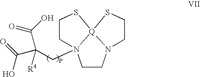
including pharmaceutically acceptable salts hydrates, solvates and metal chelates of the compound represented by the structure as set forth in formula (VII) and solvates and hydrates of said salts, wherein Q is selected from Technetium, oxo-Technetium, Rhenium and oxo-Rhenium, R4 is selected from hydrogen, C1, C2, C3, C4, C5, and C6 linear or branched alkyl, and p stands for an integer, selected from 1, 2, 3, 4 and 5.

including pharmaceutically acceptable salts hydrates, solvates and metal chelates of the compound represented by the structure as set forth in formula (VIII) and solvates and hydrates of said salts, wherein Q is selected from Technetium, oxo-Technetium, Rhenium and oxo-Rhenium.

including pharmaceutically acceptable salts hydrates, solvates and metal chelates of the compound represented by the structure as set forth in formula (IX) and solvates and hydrates of said salts, wherein Q is selected from Technetium, oxo-Technetium, Rhenium and oxo-Rhenium, R4 is selected from hydrogen, C1, C2, C3, C4, C5, and C6 linear or branched alkyl, and p stands for an integer, selected from 1, 2, 3, 4 and 5.

including pharmaceutically acceptable salts hydrates, solvates and metal chelates of the compound represented by the structure as set forth in formula (X) and solvates and hydrates of said salts; wherein R5 is selected from hydrogen, C1, C2, C3, C4, C5, and C6 linear or branched alkyl, C1, C2, C3, C4, C5, and C6 linear or branched fluoro-alkyl, and C1, C2, C3, C4, C5, and C6 linear or branched hydroxy-alkyl; q stands for an integer, selected from 1, 2, 3, 4 and 5; and Y is a marker for fluorescence. In an embodiment of the invention, Y is selected from a dansyl-amide group and fluorescein. In another embodiment of the invention there is provided a compound represented by the structure as set forth in formula (XI):

including pharmaceutically acceptable salts hydrates, solvates and metal chelates of the compound represented by the structure as set forth in formula (XI) and solvates and hydrates of said salts; wherein J is selected from hydrogen, methyl, —F and —OH. In the case that J is methyl, the compound is designated NST203.

including pharmaceutically acceptable salts hydrates, solvates and metal chelates of the compound represented by the structure as set forth in formula (XII) and solvates and hydrates of said salts; wherein J is selected from hydrogen, methyl, —F and —OH.

including pharmaceutically acceptable salts hydrates, solvates and metal chelates of the compound represented by the structure as set forth in formula (XIII) and solvates and hydrates of said salts; wherein Z1 and Z2 are each selected from hydrogen and C1, C2, C3, C4, C5 alkyl, hydroxy-alkyl or fluoro-alkyl; Z groups may be the same or different.

or pharmaceutically acceptable salts, metal chelates, solvates and hydrates of the compound represented by the structure as set forth in formula (I), and solvates and hydrates of said salts; wherein, R represents hydrogen, C1, C2, C3, C4, C5, C6, C7, C8, C9, C10, C11, C12, C13, C14 linear or branched alkyl, linear or branched hydroxy-alkyl, linear or branched fluoro-alkyl, one or two-ring aryl or heteroaryl, or combinations thereof; n and m each stands for an integer of 0, 1, 2, 3 or 4; n and m can be the same or different; M is selected from null, —O—, —S—, —C(O)NH—, and —N(U), wherein U stands for a null, hydrogen, C1, C2, C3, or C4 alkyl, D is a marker for diagnostics selected from a marker for imaging or a labeled metal chelate; said marker for imaging being selected from the group comprising a fluorescent label, a radio-label, a marker for X-ray, a marker for MRI, a marker for PET scan and a label capable of undergoing an enzymatic reaction that produces a detectable color; and where the above alkylene groups in formula (I) bound to M or D may be each substituted at each occurrence by a group selected from amino, F, OH and SH; and (ii) determining the amount of said compound bound to cells in the brain, wherein a significant amount of said compound bound to a cell indicates its being a PNOM-cell.

or pharmaceutically acceptable salts, metal chelates, solvates and hydrates of the compound represented by the structure as set forth in formula (I), and solvates and hydrates of said salts; wherein, R represents hydrogen, C1, C2, C3, C4, C5, C6, C7, C8, C9, C10, C11, C12, C13, C14 linear or branched alkyl, linear or branched hydroxy-alkyl, linear or branched fluoro-alkyl, one or two-ring aryl or heteroaryl, or combinations thereof; n and m each stands for an integer of 0, 1, 2, 3 or 4; n and m can be the same or different; M is selected from null, —O—, —S—, —C(O)NH—, and —N(U), wherein U stands for a null, hydrogen, C1, C2, C3, or C4 alkyl; D is a marker for diagnostics selected from a marker for imaging or a labeled metal chelate; said marker for imaging being selected from the group comprising a fluorescent label, a radio-label, a marker for X-ray, a marker for MRI, a marker for PET scan and a label capable of undergoing an enzymatic reaction that produces a detectable color; and where the above alkylene groups in formula (I) bound to M or D may be each substituted at each occurrence by a group selected from amino, F, OH and SH; and (ii) determining the amount of said compound bound to the examined tumor of said patient, wherein detection of a significant amount of said compound bound to cells in the tumor indicates that these tumor cells are undergoing a death process.

or pharmaceutically acceptable salts, metal chelates, solvates and hydrates of the compound represented by the structure as set forth in formula (I), and solvates and hydrates of said salts; wherein, R represents hydrogen, C1, C2, C3, C4, C5, C6, C7, C8, C9, C10, C11, C12, C13, C14 linear or branched alkyl, linear or branched hydroxy-alkyl, linear or branched fluoro-alkyl, one or two-ring aryl or heteroaryl, or combinations thereof; n and m each stands for an integer of 0, 1, 2, 3 or 4, n and m can be the same or different; M is selected from null, —O—, —S—, —C(O)NH—, and —N(U)—, wherein U stands for a null, hydrogen, C1, C2, C3, or C4 alkyl, or C1, C2, C3, or C4 alkylene; D is selected from hydrogen, a drug to be targeted to the PNOM-cell and a marker for diagnostics selected from a marker for imaging and a metal chelate; the marker for imaging may be detected by color, fluorescence, x-ray, CT scan, magnetic resonance imaging (MRI) or radio-isotope scan such as single photon emission tomography (SPECT) or positron emission tomography (PET); wherein the above alkylene groups bound to M or D, or the aryl or heteroaryl of R may each be substituted at each occurrence by a group selected from amino, F, NO2, OH and SH.


including pharmaceutically acceptable salts hydrates, solvates and metal chelates of the compound represented by the structure as set forth in formula (II) and solvates and hydrates of said salts; wherein R3 is hydroxyl or F and k is an integer selected from 1, 2, 3, 4and 5.


including pharmaceutically acceptable salts hydrates, solvates and metal chelates of the compound represented by the structure as set forth in formula (V) and solvates and hydrates of said salts; wherein J is selected from —F and —OH. In the case that J is —F, said compound is designated NST201.

including pharmaceutically acceptable salts hydrates, solvates and metal chelates of the compound represented by the structure as set forth in formula (VI) and solvates and hydrates of said salts; wherein J is selected from hydrogen, —F and —OH. In the case that J is —F, said compound is designated NST204.

including pharmaceutically acceptable salts hydrates, solvates and metal chelates of the compound represented by the structure as set forth in formula (VII) and solvates and hydrates of said salts, wherein Q is selected from Technetium, oxo-Technetium, Rhenium and oxo-Rhenium, R4 is selected from hydrogen, C1, C2, C3, C4, C5, and C6 linear or branched alkyl, and p stands for an integer, selected from 1, 2, 3, 4 and 5.

including pharmaceutically acceptable salts hydrates, solvates and metal chelates of the compound represented by the structure as set forth in formula (VIII) and solvates and hydrates of said salts, wherein Q is selected from Technetium, oxo-Technetium, Rhenium and oxo-Rhenium.

including pharmaceutically acceptable salts hydrates, solvates and metal chelates of the compound represented by the structure as set forth in formula (IX) and solvates and hydrates of said salts, wherein Q is selected from Technetium, oxo-Technetium, Rhenium and oxo-Rhenium, R4 is selected from hydrogen, C1, C2, C3, C4, C5, and C6 linear or branched alkyl, and p stands for an integer, selected from 1, 2, 3, 4 and 5. In another embodiment of the invention there is provided a compound represented by the structure as set forth in formula (X):

including pharmaceutically acceptable salts hydrates, solvates and metal chelates of the compound represented by the structure as set forth in formula (X) and solvates and hydrates of said salts; wherein R5 is selected from hydrogen, C1, C2, C3, C4, C5, and C6 linear or branched alkyl, C1, C2, C3, C4, C5, and C6 linear or branched fluoro-alkyl, and C1, C2, C3, C4, C5, and C6 linear or branched hydroxy-alkyl; q stands for an integer, selected from 1, 2, 3, 4 and 5; and Y is a marker for fluorescence. In an embodiment of the invention, Y is selected from a dansyl-amide group and fluorescein.
In another embodiment of the invention there is provided a compound represented by the structure as set forth in formula (XI):

including pharmaceutically acceptable salts hydrates, solvates and metal chelates of the compound represented by the structure as set forth in formula (XI) and solvates and hydrates of said salts; wherein J is selected from hydrogen, methyl, —F and —OH. In the case that J is methyl, the compound is designated NST203.

including pharmaceutically acceptable salts hydrates, solvates and metal chelates of the compound represented by the structure as set forth in formula (XII) and solvates and hydrates of said salts; wherein J is selected from hydrogen, methyl, —F and —OH.

including pharmaceutically acceptable salts hydrates, solvates and metal chelates of the compound represented by the structure as set forth in formula (XIII) and solvates and hydrates of said salts; wherein Z1 and Z2 are each selected from hydrogen and C1, C2, C3, C4, C5 alkyl, hydroxy-alkyl or fluoro-alkyl; Z groups may be the same or different.

wherein R stands for a C1, C2, C3, C4, or C5 alkyl. In an embodiment of the invention, R is butyl.
-
- (i). Scrambling of membrane phospholipids, with exposure on the cell surface of phosphatidylethanolamine (PE) and the negatively-charged phosphatidylserine (PS).
- Exposure of PS on the cell surface leads to a negative surface electric potential, and attraction of protons form the bulk to the membrane interface.
- (ii). The increase in the fraction of aminophospholipids (PE and PS) within the outer leaflet of the membrane results in an enhancement of the proton currents in the interface of the outer leaflet of the membrane (interfacial proton currents, IPC). This enhancement is due to the substantial increase in the number of functionalities amenable to participation in proton transfer reactions, as PE and PS replace phosphatidylcholine (PC) and sphingomeylin (SM) in the outer membrane leaflet. PE comprises a primary amine, while PS comprises a primary amine and a carboxyl group. By contrast, PC and SM comprise each a quaternary ammonium, that bears a permanent positive charge, and thus cannot participate in proton transfer reactions.
- (iii). In addition, apoptotic membranes are characterized by a reduced level of packing of the membrane constituents.
- (i). An acid-anion pair is formed, wherein an exceptionally strong hydrogen bond is formed between the protonated and unprotonated carxoyl groups. This hydrogen bond is strong, symmetrical and stabilized by resonance and tautamerization.
- (ii). This leads to distribution of the negative charge over the four carboxyl atoms, i.e., its being partially delocalized.
- (iii). The strong acid-anion hydrogen bond rigidifies the molecule, creating a bulky, rigid, flat, six-membered ring, bearing a partially-delocalized negative charge, and comprising pi-electron clouds over the carboxyl bouble bonds. Such an element undergoes according to a non-limiting hypothesis of the mechanism of action of the compounds of the invention a relatively favorable penetration into the membrane interface. However, its bulky, rigid structure directs its binding selectively to loosely packed membranes, i.e., apoptotic membranes, rather than binding to highly-packed membranes such as the plasma membranes of healthy cells. These steric features therefore promote selectivity in binding to the apoptotic membranes.
-
- A: The compound is in aqueous solution, thus both carboxyl groups are deprotonated, i.e., negatively charged, and the compound is highly soluble.
- B: Upon approaching the negatively charged apoptotic membrane, the compound acquires a proton. An anion-acid dimer is formed, thus creating a stable six-membered, resonance-stabilized ring, which penetrates the membrane interface. The bulky, rigid ring structure assists in selectivity, since its steric features favor binding to the more loosely packed plasma membrane of the apoptotic cell.
- C: Upon compound penetration into the membrane interface, it is subjected to the interfacial network of hydrogen bonds, and to the augmented interfacial proton currents encountered in the interface of the apoptotic membrane. The resultant protonation and hydrogen bonding further acts to stabilize the binding of the compound to the interface (arrows). The alkyl chain further contributes to the free energy gain of binding through formation of hydrophobic interactions with the membrane hydrocarbon core.
-
- (i). According to a first approach, termed hereinafter the “detection approach” said selective binding may be utilized to targeting a marker for imaging to PNOM-cells. This may be used in clinical practice, either in vivo, ex vivo or in vitro, for the diagnosis of diseases in which such cells emerge as will be explained herein below.
- (ii). According to a second approach, termed hereinafter the “therapeutic approach”, said property of selective binding is used for selective targeting of therapeutic agents to organs and tissues in the body wherein PNOM-cells emerge, e.g., regions of cell death, thrombus formation or inflammation.
- (iii). In accordance with a third approach of the invention termed the “clearance approach”, the selective binding of the compounds of the invention to PNOM-cells is utilized, via attachment of said compounds to a solid support, to clear body fluids such as blood from PNOM-cells, which may be potentially hazardous due to their pro-coagulant properties.


| TABLE 1 | ||||
| % of injected dose/g | ||||
| tissue (average of data | Tumor to | |||
| Body Organ | from two animals) | organ ratio | ||
| Tumor | 1.348 | 1.000 | ||
| Kidney | 3.139 | 0.429 | ||
| Heart | 0.569 | 2.367 | ||
| Liver | 0.204 | 6.594 | ||
| Muscle | 0.204 | 6.617 | ||
| Spleen | 0.334 | 4.037 | ||
| Brain | 0.098 | 13.742 | ||
| Fat tissue | 0.493 | 3.07 | ||
| Lung | 0.799 | 1.687 | ||
Claims (10)


Priority Applications (1)
| Application Number | Priority Date | Filing Date | Title |
|---|---|---|---|
| US11/882,490 US7670590B2 (en) | 2004-01-15 | 2007-08-02 | Perturbed membrane-binding compounds and methods of using the same |
Applications Claiming Priority (4)
| Application Number | Priority Date | Filing Date | Title |
|---|---|---|---|
| US53649304P | 2004-01-15 | 2004-01-15 | |
| US53728904P | 2004-01-20 | 2004-01-20 | |
| US10/799,586 US7270799B2 (en) | 2004-01-15 | 2004-03-15 | Perturbed membrane-binding compounds and methods of using the same |
| US11/882,490 US7670590B2 (en) | 2004-01-15 | 2007-08-02 | Perturbed membrane-binding compounds and methods of using the same |
Related Parent Applications (1)
| Application Number | Title | Priority Date | Filing Date |
|---|---|---|---|
| US10/799,586 Division US7270799B2 (en) | 2004-01-15 | 2004-03-15 | Perturbed membrane-binding compounds and methods of using the same |
Publications (2)
| Publication Number | Publication Date |
|---|---|
| US20080014148A1 US20080014148A1 (en) | 2008-01-17 |
| US7670590B2 true US7670590B2 (en) | 2010-03-02 |
Family
ID=34753699
Family Applications (2)
| Application Number | Title | Priority Date | Filing Date |
|---|---|---|---|
| US10/799,586 Active 2025-10-17 US7270799B2 (en) | 2004-01-15 | 2004-03-15 | Perturbed membrane-binding compounds and methods of using the same |
| US11/882,490 Expired - Fee Related US7670590B2 (en) | 2004-01-15 | 2007-08-02 | Perturbed membrane-binding compounds and methods of using the same |
Family Applications Before (1)
| Application Number | Title | Priority Date | Filing Date |
|---|---|---|---|
| US10/799,586 Active 2025-10-17 US7270799B2 (en) | 2004-01-15 | 2004-03-15 | Perturbed membrane-binding compounds and methods of using the same |
Country Status (11)
| Country | Link |
|---|---|
| US (2) | US7270799B2 (en) |
| EP (1) | EP1729820A4 (en) |
| JP (2) | JP5089989B2 (en) |
| KR (1) | KR101203501B1 (en) |
| CN (1) | CN101948380A (en) |
| AU (1) | AU2005204501B2 (en) |
| BR (1) | BRPI0506537A (en) |
| CA (1) | CA2553304A1 (en) |
| MX (1) | MXPA06007987A (en) |
| NZ (1) | NZ548991A (en) |
| WO (1) | WO2005067388A2 (en) |
Cited By (1)
| Publication number | Priority date | Publication date | Assignee | Title |
|---|---|---|---|---|
| US20080279774A1 (en) * | 2004-01-15 | 2008-11-13 | Nst Neuorosurival Technologies Ltd. | Perturbed Membrane-Binding Compounds And Methods Of Using The Same |
Families Citing this family (16)
| Publication number | Priority date | Publication date | Assignee | Title |
|---|---|---|---|---|
| US7365223B2 (en) * | 2003-06-18 | 2008-04-29 | Nst Neurosurvival Technologies Ltd | Method for selective targeting of apoptotic cells and small molecule ligands used thereof |
| NZ576582A (en) * | 2006-11-01 | 2011-12-22 | Bayer Schering Pharma Ag | [f-18]-labeled l-glutamic acid, [f-18]-labeled l-glutamine, derivatives thereof and use thereof and processes for their preparation |
| US20100035296A1 (en) * | 2007-01-04 | 2010-02-11 | Anat Shirvan | Compounds and methods for detection of cells |
| EP2268319A2 (en) * | 2008-03-07 | 2011-01-05 | Universität Ulm | Precursors of lipid metabolism for the diagnosis and treatment of cancer |
| WO2010026388A1 (en) | 2008-09-05 | 2010-03-11 | Imperial Innovations Limited | Isatin derivatives for use as in vivo imaging agents |
| US20120142722A1 (en) | 2009-06-25 | 2012-06-07 | Ilan Ziv | Conjugates of cytotoxic drugs |
| US9192680B2 (en) | 2010-06-01 | 2015-11-24 | Aposense Ltd. | Pharmaceutical compounds |
| US8530444B2 (en) | 2010-06-01 | 2013-09-10 | Aposense Ltd. | Pharmaceutical compounds |
| US20120029223A1 (en) * | 2010-07-27 | 2012-02-02 | Gad Friedman | Method for production of substituted alkyl malonic esters and derivatives thereof |
| JP6155269B2 (en) | 2011-09-26 | 2017-06-28 | プレアナリティクス ゲゼルシャフト ミット ベシュレンクテル ハフツング | Stabilization and isolation of extracellular nucleic acids |
| US11021733B2 (en) | 2011-09-26 | 2021-06-01 | Qiagen Gmbh | Stabilization and isolation of extracellular nucleic acids |
| EP2833877A4 (en) * | 2012-04-03 | 2015-11-25 | Aposense Ltd | Novel targeting agents for diagnostic and therapeutic indications |
| AU2013322643C1 (en) * | 2012-09-25 | 2018-11-08 | Qiagen Gmbh | Stabilisation of biological samples |
| EP2976426A1 (en) | 2013-03-18 | 2016-01-27 | Qiagen GmbH | Stabilization and isolation of extracellular nucleic acids |
| JP6381628B2 (en) | 2013-03-18 | 2018-08-29 | キアゲン ゲーエムベーハー | Biological sample stabilization |
| CN108291250B (en) | 2015-11-20 | 2022-05-27 | 凯杰有限公司 | Method for preparing sterilized composition for stabilizing extracellular nucleic acid |
Citations (3)
| Publication number | Priority date | Publication date | Assignee | Title |
|---|---|---|---|---|
| GB1401579A (en) | 1972-09-06 | 1975-07-16 | Richter Gedeon Vegyeszet | Indolo-quinolizines |
| US4578503A (en) | 1982-08-31 | 1986-03-25 | Daikin Kogyo Co., Ltd. | Alkylated or alkenylated malonic acid or its derivatives having a fluorine |
| WO2002046147A2 (en) * | 2000-12-06 | 2002-06-13 | Nst Neurosurvival Technologies Ltd. | Perturbed membrane-binding compounds |
Family Cites Families (6)
| Publication number | Priority date | Publication date | Assignee | Title |
|---|---|---|---|---|
| US4187381A (en) * | 1973-08-06 | 1980-02-05 | Hoffmann-La Roche Inc. | 16-Substituted prostaglandins |
| CH593903A5 (en) * | 1974-06-12 | 1977-12-30 | Sandoz Ag | |
| JPS6034940B2 (en) * | 1980-11-13 | 1985-08-12 | ダイキン工業株式会社 | Fluoromalonic acid derivatives and their production method |
| JPS5939854A (en) * | 1982-08-31 | 1984-03-05 | Daikin Ind Ltd | Trapping of enolate ion of fluorine-containing group- substituted malonic ester |
| JPS5939849A (en) * | 1982-08-31 | 1984-03-05 | Daikin Ind Ltd | Fluorine-containing aliphatic group-substituted alkylated or alkenylated malonic ester or its derivative |
| GB9512546D0 (en) * | 1995-06-20 | 1995-08-23 | Bnfl Fluorchem Ltd | A process for the preparation of esters |
-
2004
- 2004-03-15 US US10/799,586 patent/US7270799B2/en active Active
-
2005
- 2005-01-16 NZ NZ548991A patent/NZ548991A/en unknown
- 2005-01-16 CA CA002553304A patent/CA2553304A1/en not_active Abandoned
- 2005-01-16 JP JP2006548583A patent/JP5089989B2/en not_active Expired - Fee Related
- 2005-01-16 EP EP05703098A patent/EP1729820A4/en not_active Withdrawn
- 2005-01-16 MX MXPA06007987A patent/MXPA06007987A/en active IP Right Grant
- 2005-01-16 BR BRPI0506537-2A patent/BRPI0506537A/en not_active IP Right Cessation
- 2005-01-16 AU AU2005204501A patent/AU2005204501B2/en not_active Ceased
- 2005-01-16 CN CN2010102229125A patent/CN101948380A/en active Pending
- 2005-01-16 WO PCT/IL2005/000055 patent/WO2005067388A2/en active Application Filing
- 2005-01-16 KR KR1020067016249A patent/KR101203501B1/en not_active IP Right Cessation
-
2007
- 2007-08-02 US US11/882,490 patent/US7670590B2/en not_active Expired - Fee Related
-
2011
- 2011-12-21 JP JP2011279984A patent/JP2012140424A/en not_active Withdrawn
Patent Citations (3)
| Publication number | Priority date | Publication date | Assignee | Title |
|---|---|---|---|---|
| GB1401579A (en) | 1972-09-06 | 1975-07-16 | Richter Gedeon Vegyeszet | Indolo-quinolizines |
| US4578503A (en) | 1982-08-31 | 1986-03-25 | Daikin Kogyo Co., Ltd. | Alkylated or alkenylated malonic acid or its derivatives having a fluorine |
| WO2002046147A2 (en) * | 2000-12-06 | 2002-06-13 | Nst Neurosurvival Technologies Ltd. | Perturbed membrane-binding compounds |
Non-Patent Citations (4)
| Title |
|---|
| Peggy L. Olive et al. Characterization of the Uptake and Toxicity of a Fluorescent Thiol Reagent Cytometry by the Society for Analytical Cytology pp. 349-353 vol. 3, No. 5, Jul. 12, 1982 The John Hopkins Oncology Center, Section of Radiobiology, Baltimore, Maryland USA & Dept. of Radiology, Case Western Reserve University, Cleveland, Ohio, USA. |
| Peggy L. Olive, John E. Biaglow, Marie E. Varnes and Ralph E. Durand, Characterization of the Uptake and Toxicity of a Fluorescent Thiol Reagent, Cytometry, vol. 3(5), 349-353, 1982. * |
| Peggy L.Olive, John E. Biaglow, Marie E. Varnes and Ralph E. Durand, Characterization of the Uptake and Toxicity of a Fluorescent Thiol Reagent, Cytometry, vol. 3(5), 349-353, 1982. * |
| Stubbe et al., "Are Carboxylations Involving Biotin Concerted or Nonconcerted?", The Journal of Biological Chemistry, vol. 255, No. 1, (1980), pp. 236-242. |
Cited By (2)
| Publication number | Priority date | Publication date | Assignee | Title |
|---|---|---|---|---|
| US20080279774A1 (en) * | 2004-01-15 | 2008-11-13 | Nst Neuorosurival Technologies Ltd. | Perturbed Membrane-Binding Compounds And Methods Of Using The Same |
| US7947253B2 (en) * | 2004-01-15 | 2011-05-24 | Aposense Ltd. | Perturbed membrane-binding compounds and methods of using the same |
Also Published As
| Publication number | Publication date |
|---|---|
| JP2012140424A (en) | 2012-07-26 |
| AU2005204501A1 (en) | 2005-07-28 |
| NZ548991A (en) | 2010-05-28 |
| EP1729820A2 (en) | 2006-12-13 |
| CN101948380A (en) | 2011-01-19 |
| WO2005067388A3 (en) | 2005-09-01 |
| US7270799B2 (en) | 2007-09-18 |
| KR20070028312A (en) | 2007-03-12 |
| US20050158239A1 (en) | 2005-07-21 |
| AU2005204501B2 (en) | 2010-02-25 |
| WO2005067388A2 (en) | 2005-07-28 |
| BRPI0506537A (en) | 2007-02-27 |
| EP1729820A4 (en) | 2009-11-11 |
| JP5089989B2 (en) | 2012-12-05 |
| CA2553304A1 (en) | 2005-07-28 |
| KR101203501B1 (en) | 2012-11-21 |
| JP2007523059A (en) | 2007-08-16 |
| US20080014148A1 (en) | 2008-01-17 |
| MXPA06007987A (en) | 2007-01-26 |
Similar Documents
| Publication | Publication Date | Title |
|---|---|---|
| US7670590B2 (en) | Perturbed membrane-binding compounds and methods of using the same | |
| US20220125959A1 (en) | Prostate-specific membrane antigen (psma) inhibitors as diagnostic and radionuclide therapeutic agents | |
| US7605182B2 (en) | Compounds that selectively bind to membranes of apoptotic cells | |
| US7378079B2 (en) | Agents for imaging and diagnostic methods using them | |
| US7396859B2 (en) | Perturbed membrane-binding compounds | |
| US7947253B2 (en) | Perturbed membrane-binding compounds and methods of using the same | |
| US20100035296A1 (en) | Compounds and methods for detection of cells | |
| JP2004512382A (en) | Dual nuclide imaging of cardiac perfusion and cardiac inflammation | |
| JP2001525796A (en) | Radiopharmaceuticals for imaging infections and inflammation | |
| US7365223B2 (en) | Method for selective targeting of apoptotic cells and small molecule ligands used thereof | |
| ZA200606427B (en) | Perturbed membrane-binding compounds and methods of using the same | |
| AU2004246906B2 (en) | Method for selective targeting of apoptotic cells and small molecule ligands used thereof |
Legal Events
| Date | Code | Title | Description |
|---|---|---|---|
| AS | Assignment |
Owner name: NST NEUROSURVIVAL TECHNOLOGIES LTD, ISRAEL Free format text: ASSIGNMENT OF ASSIGNORS INTEREST;ASSIGNORS:ZIV, ILAN;SHIRVAN, ANAT;REEL/FRAME:022756/0801 Effective date: 20040315 Owner name: NST NEUROSURVIVAL TECHNOLOGIES LTD,ISRAEL Free format text: ASSIGNMENT OF ASSIGNORS INTEREST;ASSIGNORS:ZIV, ILAN;SHIRVAN, ANAT;REEL/FRAME:022756/0801 Effective date: 20040315 |
|
| AS | Assignment |
Owner name: APOSENSE LTD., ISRAEL Free format text: CHANGE OF NAME;ASSIGNOR:NST NEUROSURVIVAL TECHNOLOGIES LTD.;REEL/FRAME:022777/0886 Effective date: 20080819 Owner name: APOSENSE LTD.,ISRAEL Free format text: CHANGE OF NAME;ASSIGNOR:NST NEUROSURVIVAL TECHNOLOGIES LTD.;REEL/FRAME:022777/0886 Effective date: 20080819 |
|
| STCF | Information on status: patent grant |
Free format text: PATENTED CASE |
|
| FEPP | Fee payment procedure |
Free format text: PAT HOLDER NO LONGER CLAIMS SMALL ENTITY STATUS, ENTITY STATUS SET TO UNDISCOUNTED (ORIGINAL EVENT CODE: STOL); ENTITY STATUS OF PATENT OWNER: LARGE ENTITY |
|
| SULP | Surcharge for late payment | ||
| FEPP | Fee payment procedure |
Free format text: PAYOR NUMBER ASSIGNED (ORIGINAL EVENT CODE: ASPN); ENTITY STATUS OF PATENT OWNER: LARGE ENTITY |
|
| FPAY | Fee payment |
Year of fee payment: 4 |
|
| MAFP | Maintenance fee payment |
Free format text: PAYMENT OF MAINTENANCE FEE, 8TH YEAR, LARGE ENTITY (ORIGINAL EVENT CODE: M1552) Year of fee payment: 8 |
|
| FEPP | Fee payment procedure |
Free format text: MAINTENANCE FEE REMINDER MAILED (ORIGINAL EVENT CODE: REM.); ENTITY STATUS OF PATENT OWNER: LARGE ENTITY |
|
| LAPS | Lapse for failure to pay maintenance fees |
Free format text: PATENT EXPIRED FOR FAILURE TO PAY MAINTENANCE FEES (ORIGINAL EVENT CODE: EXP.); ENTITY STATUS OF PATENT OWNER: LARGE ENTITY |
|
| STCH | Information on status: patent discontinuation |
Free format text: PATENT EXPIRED DUE TO NONPAYMENT OF MAINTENANCE FEES UNDER 37 CFR 1.362 |
|
| FP | Lapsed due to failure to pay maintenance fee |
Effective date: 20220302 |