US7655393B2 - Adoptive immune cells for tumor vaccines - Google Patents
Adoptive immune cells for tumor vaccines Download PDFInfo
- Publication number
- US7655393B2 US7655393B2 US11/579,280 US57928006A US7655393B2 US 7655393 B2 US7655393 B2 US 7655393B2 US 57928006 A US57928006 A US 57928006A US 7655393 B2 US7655393 B2 US 7655393B2
- Authority
- US
- United States
- Prior art keywords
- cells
- antigen
- peptide
- culturing
- culture vessel
- Prior art date
- Legal status (The legal status is an assumption and is not a legal conclusion. Google has not performed a legal analysis and makes no representation as to the accuracy of the status listed.)
- Expired - Fee Related, expires
Links
- 210000002865 immune cell Anatomy 0.000 title claims abstract description 26
- 206010028980 Neoplasm Diseases 0.000 title claims description 36
- 229960005486 vaccine Drugs 0.000 title claims description 9
- 210000004027 cell Anatomy 0.000 claims abstract description 238
- 238000000034 method Methods 0.000 claims abstract description 81
- 238000012258 culturing Methods 0.000 claims abstract description 67
- 229920000642 polymer Polymers 0.000 claims abstract description 52
- 201000011510 cancer Diseases 0.000 claims abstract description 37
- 235000000346 sugar Nutrition 0.000 claims abstract description 28
- 102000004190 Enzymes Human genes 0.000 claims abstract description 18
- 108090000790 Enzymes Proteins 0.000 claims abstract description 18
- 239000007788 liquid Substances 0.000 claims description 55
- 239000000427 antigen Substances 0.000 claims description 47
- 108091007433 antigens Proteins 0.000 claims description 47
- 102000036639 antigens Human genes 0.000 claims description 47
- KCXVZYZYPLLWCC-UHFFFAOYSA-N EDTA Chemical compound OC(=O)CN(CC(O)=O)CCN(CC(O)=O)CC(O)=O KCXVZYZYPLLWCC-UHFFFAOYSA-N 0.000 claims description 41
- -1 IgM idiotype Proteins 0.000 claims description 28
- 108090000765 processed proteins & peptides Proteins 0.000 claims description 28
- 239000013543 active substance Substances 0.000 claims description 18
- RPOKRGMOEWYIKB-ZFCLCKFASA-N (2r,3r,4r,5r)-n-[(4-ethenylphenyl)methyl]-2,3,6-trihydroxy-5-methyl-4-[(2r,3r,4s,5r,6r)-3,4,5-trihydroxy-6-(hydroxymethyl)oxan-2-yl]oxyhexanamide Chemical group O([C@H]([C@@H](CO)C)[C@H](O)[C@@H](O)C(=O)NCC=1C=CC(C=C)=CC=1)[C@@H]1O[C@H](CO)[C@H](O)[C@H](O)[C@H]1O RPOKRGMOEWYIKB-ZFCLCKFASA-N 0.000 claims description 17
- 210000004443 dendritic cell Anatomy 0.000 claims description 13
- 239000012634 fragment Substances 0.000 claims description 11
- 229920001756 Polyvinyl chloride acetate Polymers 0.000 claims description 10
- 210000001821 langerhans cell Anatomy 0.000 claims description 9
- MZOFCQQQCNRIBI-VMXHOPILSA-N (3s)-4-[[(2s)-1-[[(2s)-1-[[(1s)-1-carboxy-2-hydroxyethyl]amino]-4-methyl-1-oxopentan-2-yl]amino]-5-(diaminomethylideneamino)-1-oxopentan-2-yl]amino]-3-[[2-[[(2s)-2,6-diaminohexanoyl]amino]acetyl]amino]-4-oxobutanoic acid Chemical compound OC[C@@H](C(O)=O)NC(=O)[C@H](CC(C)C)NC(=O)[C@H](CCCN=C(N)N)NC(=O)[C@H](CC(O)=O)NC(=O)CNC(=O)[C@@H](N)CCCCN MZOFCQQQCNRIBI-VMXHOPILSA-N 0.000 claims description 8
- LKKMLIBUAXYLOY-UHFFFAOYSA-N 3-Amino-1-methyl-5H-pyrido[4,3-b]indole Chemical compound N1C2=CC=CC=C2C2=C1C=C(N)N=C2C LKKMLIBUAXYLOY-UHFFFAOYSA-N 0.000 claims description 8
- 108010017213 Granulocyte-Macrophage Colony-Stimulating Factor Proteins 0.000 claims description 8
- 101000578784 Homo sapiens Melanoma antigen recognized by T-cells 1 Proteins 0.000 claims description 8
- 101000767631 Human papillomavirus type 16 Protein E7 Proteins 0.000 claims description 8
- 102100031413 L-dopachrome tautomerase Human genes 0.000 claims description 8
- 101710093778 L-dopachrome tautomerase Proteins 0.000 claims description 8
- 102100028389 Melanoma antigen recognized by T-cells 1 Human genes 0.000 claims description 8
- 102000035195 Peptidases Human genes 0.000 claims description 8
- 108091005804 Peptidases Proteins 0.000 claims description 8
- 108060008682 Tumor Necrosis Factor Proteins 0.000 claims description 8
- 102000000852 Tumor Necrosis Factor-alpha Human genes 0.000 claims description 8
- 239000013592 cell lysate Substances 0.000 claims description 8
- KIUKXJAPPMFGSW-DNGZLQJQSA-N (2S,3S,4S,5R,6R)-6-[(2S,3R,4R,5S,6R)-3-Acetamido-2-[(2S,3S,4R,5R,6R)-6-[(2R,3R,4R,5S,6R)-3-acetamido-2,5-dihydroxy-6-(hydroxymethyl)oxan-4-yl]oxy-2-carboxy-4,5-dihydroxyoxan-3-yl]oxy-5-hydroxy-6-(hydroxymethyl)oxan-4-yl]oxy-3,4,5-trihydroxyoxane-2-carboxylic acid Chemical compound CC(=O)N[C@H]1[C@H](O)O[C@H](CO)[C@@H](O)[C@@H]1O[C@H]1[C@H](O)[C@@H](O)[C@H](O[C@H]2[C@@H]([C@@H](O[C@H]3[C@@H]([C@@H](O)[C@H](O)[C@H](O3)C(O)=O)O)[C@H](O)[C@@H](CO)O2)NC(C)=O)[C@@H](C(O)=O)O1 KIUKXJAPPMFGSW-DNGZLQJQSA-N 0.000 claims description 7
- WEVYNIUIFUYDGI-UHFFFAOYSA-N 3-[6-[4-(trifluoromethoxy)anilino]-4-pyrimidinyl]benzamide Chemical compound NC(=O)C1=CC=CC(C=2N=CN=C(NC=3C=CC(OC(F)(F)F)=CC=3)C=2)=C1 WEVYNIUIFUYDGI-UHFFFAOYSA-N 0.000 claims description 7
- 101710163881 5,6-dihydroxyindole-2-carboxylic acid oxidase Proteins 0.000 claims description 7
- 101100288313 Arabidopsis thaliana KTI4 gene Proteins 0.000 claims description 7
- 102100035526 B melanoma antigen 1 Human genes 0.000 claims description 7
- 108060000903 Beta-catenin Proteins 0.000 claims description 7
- 102000015735 Beta-catenin Human genes 0.000 claims description 7
- 102100025570 Cancer/testis antigen 1 Human genes 0.000 claims description 7
- 102100026548 Caspase-8 Human genes 0.000 claims description 7
- 108090000538 Caspase-8 Proteins 0.000 claims description 7
- 101150029707 ERBB2 gene Proteins 0.000 claims description 7
- 102100039717 G antigen 1 Human genes 0.000 claims description 7
- 102000025850 HLA-A2 Antigen Human genes 0.000 claims description 7
- 108010074032 HLA-A2 Antigen Proteins 0.000 claims description 7
- 101000874316 Homo sapiens B melanoma antigen 1 Proteins 0.000 claims description 7
- 101000856237 Homo sapiens Cancer/testis antigen 1 Proteins 0.000 claims description 7
- 101000886137 Homo sapiens G antigen 1 Proteins 0.000 claims description 7
- 101001094545 Homo sapiens Retrotransposon-like protein 1 Proteins 0.000 claims description 7
- 108010047761 Interferon-alpha Proteins 0.000 claims description 7
- 102000006992 Interferon-alpha Human genes 0.000 claims description 7
- 108010002386 Interleukin-3 Proteins 0.000 claims description 7
- 102000000646 Interleukin-3 Human genes 0.000 claims description 7
- 102000004388 Interleukin-4 Human genes 0.000 claims description 7
- 108090000978 Interleukin-4 Proteins 0.000 claims description 7
- 102100020880 Kit ligand Human genes 0.000 claims description 7
- 101710177504 Kit ligand Proteins 0.000 claims description 7
- 101150028321 Lck gene Proteins 0.000 claims description 7
- 108010080632 MUT 1 peptide Proteins 0.000 claims description 7
- 108010008707 Mucin-1 Proteins 0.000 claims description 7
- 241000187479 Mycobacterium tuberculosis Species 0.000 claims description 7
- 102100034640 PWWP domain-containing DNA repair factor 3A Human genes 0.000 claims description 7
- 108050007154 PWWP domain-containing DNA repair factor 3A Proteins 0.000 claims description 7
- 102100040283 Peptidyl-prolyl cis-trans isomerase B Human genes 0.000 claims description 7
- 102100021768 Phosphoserine aminotransferase Human genes 0.000 claims description 7
- 108091036414 Polyinosinic:polycytidylic acid Proteins 0.000 claims description 7
- 108010072866 Prostate-Specific Antigen Proteins 0.000 claims description 7
- 101800001271 Surface protein Proteins 0.000 claims description 7
- 108700019889 TEL-AML1 fusion Proteins 0.000 claims description 7
- 101150033985 TPI gene Proteins 0.000 claims description 7
- 101150032817 TPI1 gene Proteins 0.000 claims description 7
- 102000003627 TRPC1 Human genes 0.000 claims description 7
- 101800004564 Transforming growth factor alpha Proteins 0.000 claims description 7
- 108700015934 Triose-phosphate isomerases Proteins 0.000 claims description 7
- 102100033598 Triosephosphate isomerase Human genes 0.000 claims description 7
- 102000003425 Tyrosinase Human genes 0.000 claims description 7
- 108060008724 Tyrosinase Proteins 0.000 claims description 7
- 102100027244 U4/U6.U5 tri-snRNP-associated protein 1 Human genes 0.000 claims description 7
- 101710155955 U4/U6.U5 tri-snRNP-associated protein 1 Proteins 0.000 claims description 7
- 210000003850 cellular structure Anatomy 0.000 claims description 7
- 108010048032 cyclophilin B Proteins 0.000 claims description 7
- 229920002674 hyaluronan Polymers 0.000 claims description 7
- 229960003160 hyaluronic acid Drugs 0.000 claims description 7
- OHDXDNUPVVYWOV-UHFFFAOYSA-N n-methyl-1-(2-naphthalen-1-ylsulfanylphenyl)methanamine Chemical compound CNCC1=CC=CC=C1SC1=CC=CC2=CC=CC=C12 OHDXDNUPVVYWOV-UHFFFAOYSA-N 0.000 claims description 7
- 108010044156 peptidyl-prolyl cis-trans isomerase b Proteins 0.000 claims description 7
- 235000019833 protease Nutrition 0.000 claims description 7
- 102100021305 Acyl-CoA:lysophosphatidylglycerol acyltransferase 1 Human genes 0.000 claims description 6
- 101001042227 Homo sapiens Acyl-CoA:lysophosphatidylglycerol acyltransferase 1 Proteins 0.000 claims description 6
- 101000721661 Homo sapiens Cellular tumor antigen p53 Proteins 0.000 claims description 6
- 102100034256 Mucin-1 Human genes 0.000 claims description 6
- 102000046299 Transforming Growth Factor beta1 Human genes 0.000 claims description 6
- 101800002279 Transforming growth factor beta-1 Proteins 0.000 claims description 6
- 229940115272 polyinosinic:polycytidylic acid Drugs 0.000 claims description 6
- 101000914324 Homo sapiens Carcinoembryonic antigen-related cell adhesion molecule 5 Proteins 0.000 claims description 5
- 101000914321 Homo sapiens Carcinoembryonic antigen-related cell adhesion molecule 7 Proteins 0.000 claims description 5
- 101000617725 Homo sapiens Pregnancy-specific beta-1-glycoprotein 2 Proteins 0.000 claims description 5
- 101000873927 Homo sapiens Squamous cell carcinoma antigen recognized by T-cells 3 Proteins 0.000 claims description 5
- 102100022019 Pregnancy-specific beta-1-glycoprotein 2 Human genes 0.000 claims description 5
- 102100035748 Squamous cell carcinoma antigen recognized by T-cells 3 Human genes 0.000 claims description 5
- 102000040856 WT1 Human genes 0.000 claims description 5
- 108700020467 WT1 Proteins 0.000 claims description 5
- 101150084041 WT1 gene Proteins 0.000 claims description 5
- 102000016914 ras Proteins Human genes 0.000 claims description 5
- 108010014186 ras Proteins Proteins 0.000 claims description 5
- 241000124008 Mammalia Species 0.000 claims description 4
- 102000004457 Granulocyte-Macrophage Colony-Stimulating Factor Human genes 0.000 claims 4
- 102000006747 Transforming Growth Factor alpha Human genes 0.000 claims 4
- 238000002360 preparation method Methods 0.000 abstract description 22
- 208000015181 infectious disease Diseases 0.000 abstract description 6
- 206010003645 Atopy Diseases 0.000 abstract description 5
- 206010067584 Type 1 diabetes mellitus Diseases 0.000 abstract description 5
- 208000026935 allergic disease Diseases 0.000 abstract description 5
- 239000003814 drug Substances 0.000 abstract description 5
- 229940030156 cell vaccine Drugs 0.000 abstract description 3
- OKKJLVBELUTLKV-UHFFFAOYSA-N Methanol Chemical compound OC OKKJLVBELUTLKV-UHFFFAOYSA-N 0.000 description 42
- 210000002433 mononuclear leukocyte Anatomy 0.000 description 21
- 239000000243 solution Substances 0.000 description 21
- IAZDPXIOMUYVGZ-UHFFFAOYSA-N Dimethylsulphoxide Chemical compound CS(C)=O IAZDPXIOMUYVGZ-UHFFFAOYSA-N 0.000 description 18
- 238000011084 recovery Methods 0.000 description 18
- 210000001151 cytotoxic T lymphocyte Anatomy 0.000 description 16
- 238000002474 experimental method Methods 0.000 description 15
- 238000006243 chemical reaction Methods 0.000 description 14
- 230000000961 alloantigen Effects 0.000 description 13
- 230000006698 induction Effects 0.000 description 13
- XLYOFNOQVPJJNP-UHFFFAOYSA-N water Substances O XLYOFNOQVPJJNP-UHFFFAOYSA-N 0.000 description 13
- 210000000612 antigen-presenting cell Anatomy 0.000 description 11
- RTZKZFJDLAIYFH-UHFFFAOYSA-N Diethyl ether Chemical compound CCOCC RTZKZFJDLAIYFH-UHFFFAOYSA-N 0.000 description 10
- 101000914484 Homo sapiens T-lymphocyte activation antigen CD80 Proteins 0.000 description 10
- 102100022297 Integrin alpha-X Human genes 0.000 description 10
- NWIBSHFKIJFRCO-WUDYKRTCSA-N Mytomycin Chemical compound C1N2C(C(C(C)=C(N)C3=O)=O)=C3[C@@H](COC(N)=O)[C@@]2(OC)[C@@H]2[C@H]1N2 NWIBSHFKIJFRCO-WUDYKRTCSA-N 0.000 description 10
- 102100027222 T-lymphocyte activation antigen CD80 Human genes 0.000 description 10
- 230000015572 biosynthetic process Effects 0.000 description 10
- UHOVQNZJYSORNB-UHFFFAOYSA-N Benzene Chemical compound C1=CC=CC=C1 UHOVQNZJYSORNB-UHFFFAOYSA-N 0.000 description 9
- HEMHJVSKTPXQMS-UHFFFAOYSA-M Sodium hydroxide Chemical compound [OH-].[Na+] HEMHJVSKTPXQMS-UHFFFAOYSA-M 0.000 description 9
- YXFVVABEGXRONW-UHFFFAOYSA-N Toluene Chemical compound CC1=CC=CC=C1 YXFVVABEGXRONW-UHFFFAOYSA-N 0.000 description 9
- 108090000623 proteins and genes Proteins 0.000 description 9
- 239000000126 substance Substances 0.000 description 9
- 238000003786 synthesis reaction Methods 0.000 description 9
- 229910001868 water Inorganic materials 0.000 description 9
- CSCPPACGZOOCGX-UHFFFAOYSA-N Acetone Chemical compound CC(C)=O CSCPPACGZOOCGX-UHFFFAOYSA-N 0.000 description 8
- LFQSCWFLJHTTHZ-UHFFFAOYSA-N Ethanol Chemical compound CCO LFQSCWFLJHTTHZ-UHFFFAOYSA-N 0.000 description 8
- 102000004142 Trypsin Human genes 0.000 description 8
- 108090000631 Trypsin Proteins 0.000 description 8
- 239000002904 solvent Substances 0.000 description 8
- 238000010186 staining Methods 0.000 description 8
- 239000012588 trypsin Substances 0.000 description 8
- 239000000178 monomer Substances 0.000 description 7
- 210000001519 tissue Anatomy 0.000 description 7
- 239000011534 wash buffer Substances 0.000 description 7
- 238000005406 washing Methods 0.000 description 7
- NHBKXEKEPDILRR-UHFFFAOYSA-N 2,3-bis(butanoylsulfanyl)propyl butanoate Chemical compound CCCC(=O)OCC(SC(=O)CCC)CSC(=O)CCC NHBKXEKEPDILRR-UHFFFAOYSA-N 0.000 description 6
- NALREUIWICQLPS-UHFFFAOYSA-N 7-imino-n,n-dimethylphenothiazin-3-amine;hydrochloride Chemical compound [Cl-].C1=C(N)C=C2SC3=CC(=[N+](C)C)C=CC3=NC2=C1 NALREUIWICQLPS-UHFFFAOYSA-N 0.000 description 6
- GUBGYTABKSRVRQ-QKKXKWKRSA-N Lactose Natural products OC[C@H]1O[C@@H](O[C@H]2[C@H](O)[C@@H](O)C(O)O[C@@H]2CO)[C@H](O)[C@@H](O)[C@H]1O GUBGYTABKSRVRQ-QKKXKWKRSA-N 0.000 description 6
- 210000004748 cultured cell Anatomy 0.000 description 6
- 231100000263 cytotoxicity test Toxicity 0.000 description 6
- 239000011886 peripheral blood Substances 0.000 description 6
- 210000005259 peripheral blood Anatomy 0.000 description 6
- 239000004033 plastic Substances 0.000 description 6
- 230000001954 sterilising effect Effects 0.000 description 6
- 238000012360 testing method Methods 0.000 description 6
- 102100039620 Granulocyte-macrophage colony-stimulating factor Human genes 0.000 description 5
- 238000007664 blowing Methods 0.000 description 5
- 238000000576 coating method Methods 0.000 description 5
- 239000008101 lactose Substances 0.000 description 5
- 229960004857 mitomycin Drugs 0.000 description 5
- 239000008188 pellet Substances 0.000 description 5
- 102000004169 proteins and genes Human genes 0.000 description 5
- 239000006228 supernatant Substances 0.000 description 5
- PHEDXBVPIONUQT-UHFFFAOYSA-N Cocarcinogen A1 Natural products CCCCCCCCCCCCCC(=O)OC1C(C)C2(O)C3C=C(C)C(=O)C3(O)CC(CO)=CC2C2C1(OC(C)=O)C2(C)C PHEDXBVPIONUQT-UHFFFAOYSA-N 0.000 description 4
- 239000004793 Polystyrene Substances 0.000 description 4
- 238000002617 apheresis Methods 0.000 description 4
- 239000007864 aqueous solution Substances 0.000 description 4
- 239000006285 cell suspension Substances 0.000 description 4
- 239000011248 coating agent Substances 0.000 description 4
- 239000002158 endotoxin Substances 0.000 description 4
- 230000001605 fetal effect Effects 0.000 description 4
- 238000001914 filtration Methods 0.000 description 4
- 238000003125 immunofluorescent labeling Methods 0.000 description 4
- 229920006008 lipopolysaccharide Polymers 0.000 description 4
- 238000002156 mixing Methods 0.000 description 4
- 239000000203 mixture Substances 0.000 description 4
- 210000001616 monocyte Anatomy 0.000 description 4
- PHEDXBVPIONUQT-RGYGYFBISA-N phorbol 13-acetate 12-myristate Chemical compound C([C@]1(O)C(=O)C(C)=C[C@H]1[C@@]1(O)[C@H](C)[C@H]2OC(=O)CCCCCCCCCCCCC)C(CO)=C[C@H]1[C@H]1[C@]2(OC(C)=O)C1(C)C PHEDXBVPIONUQT-RGYGYFBISA-N 0.000 description 4
- 229920002223 polystyrene Polymers 0.000 description 4
- 238000000926 separation method Methods 0.000 description 4
- OZAIFHULBGXAKX-UHFFFAOYSA-N 2-(2-cyanopropan-2-yldiazenyl)-2-methylpropanenitrile Chemical compound N#CC(C)(C)N=NC(C)(C)C#N OZAIFHULBGXAKX-UHFFFAOYSA-N 0.000 description 3
- YXHLJMWYDTXDHS-IRFLANFNSA-N 7-aminoactinomycin D Chemical compound C[C@H]1OC(=O)[C@H](C(C)C)N(C)C(=O)CN(C)C(=O)[C@@H]2CCCN2C(=O)[C@@H](C(C)C)NC(=O)[C@H]1NC(=O)C1=C(N)C(=O)C(C)=C2OC(C(C)=C(N)C=C3C(=O)N[C@@H]4C(=O)N[C@@H](C(N5CCC[C@H]5C(=O)N(C)CC(=O)N(C)[C@@H](C(C)C)C(=O)O[C@@H]4C)=O)C(C)C)=C3N=C21 YXHLJMWYDTXDHS-IRFLANFNSA-N 0.000 description 3
- 108700012813 7-aminoactinomycin D Proteins 0.000 description 3
- OZAIFHULBGXAKX-VAWYXSNFSA-N AIBN Substances N#CC(C)(C)\N=N\C(C)(C)C#N OZAIFHULBGXAKX-VAWYXSNFSA-N 0.000 description 3
- 238000011725 BALB/c mouse Methods 0.000 description 3
- 102400001320 Transforming growth factor alpha Human genes 0.000 description 3
- QVGXLLKOCUKJST-UHFFFAOYSA-N atomic oxygen Chemical compound [O] QVGXLLKOCUKJST-UHFFFAOYSA-N 0.000 description 3
- 230000001580 bacterial effect Effects 0.000 description 3
- 210000001185 bone marrow Anatomy 0.000 description 3
- 210000000170 cell membrane Anatomy 0.000 description 3
- 239000003153 chemical reaction reagent Substances 0.000 description 3
- 238000000684 flow cytometry Methods 0.000 description 3
- 230000006870 function Effects 0.000 description 3
- 238000010438 heat treatment Methods 0.000 description 3
- 210000002540 macrophage Anatomy 0.000 description 3
- 239000001301 oxygen Substances 0.000 description 3
- 229910052760 oxygen Inorganic materials 0.000 description 3
- 239000002244 precipitate Substances 0.000 description 3
- 238000007790 scraping Methods 0.000 description 3
- 210000000130 stem cell Anatomy 0.000 description 3
- 238000004659 sterilization and disinfection Methods 0.000 description 3
- 239000000725 suspension Substances 0.000 description 3
- 239000013076 target substance Substances 0.000 description 3
- FYXAVEIRUNEOSG-UHFFFAOYSA-N 4-benzyl-5-ethenylisoindole-1,3-dione Chemical compound C=CC1=CC=C2C(=O)NC(=O)C2=C1CC1=CC=CC=C1 FYXAVEIRUNEOSG-UHFFFAOYSA-N 0.000 description 2
- ZCYVEMRRCGMTRW-UHFFFAOYSA-N 7553-56-2 Chemical compound [I] ZCYVEMRRCGMTRW-UHFFFAOYSA-N 0.000 description 2
- 108010088751 Albumins Proteins 0.000 description 2
- 102000009027 Albumins Human genes 0.000 description 2
- 102000005427 Asialoglycoprotein Receptor Human genes 0.000 description 2
- IJGRMHOSHXDMSA-UHFFFAOYSA-N Atomic nitrogen Chemical compound N#N IJGRMHOSHXDMSA-UHFFFAOYSA-N 0.000 description 2
- 241000894006 Bacteria Species 0.000 description 2
- KHBQMWCZKVMBLN-UHFFFAOYSA-N Benzenesulfonamide Chemical compound NS(=O)(=O)C1=CC=CC=C1 KHBQMWCZKVMBLN-UHFFFAOYSA-N 0.000 description 2
- 108091003079 Bovine Serum Albumin Proteins 0.000 description 2
- 108010008951 Chemokine CXCL12 Proteins 0.000 description 2
- 102000006573 Chemokine CXCL12 Human genes 0.000 description 2
- VEXZGXHMUGYJMC-UHFFFAOYSA-N Hydrochloric acid Chemical compound Cl VEXZGXHMUGYJMC-UHFFFAOYSA-N 0.000 description 2
- 241001465754 Metazoa Species 0.000 description 2
- 239000007832 Na2SO4 Substances 0.000 description 2
- 206010029719 Nonspecific reaction Diseases 0.000 description 2
- PMZURENOXWZQFD-UHFFFAOYSA-L Sodium Sulfate Chemical compound [Na+].[Na+].[O-]S([O-])(=O)=O PMZURENOXWZQFD-UHFFFAOYSA-L 0.000 description 2
- PXIPVTKHYLBLMZ-UHFFFAOYSA-N Sodium azide Chemical compound [Na+].[N-]=[N+]=[N-] PXIPVTKHYLBLMZ-UHFFFAOYSA-N 0.000 description 2
- UIIMBOGNXHQVGW-UHFFFAOYSA-M Sodium bicarbonate Chemical compound [Na+].OC([O-])=O UIIMBOGNXHQVGW-UHFFFAOYSA-M 0.000 description 2
- 101710101607 Toxic shock syndrome toxin-1 Proteins 0.000 description 2
- 241000223997 Toxoplasma gondii Species 0.000 description 2
- 239000002253 acid Substances 0.000 description 2
- 108010006523 asialoglycoprotein receptor Proteins 0.000 description 2
- GHDLZGOOOLEJKI-UHFFFAOYSA-N benzenesulfonylurea Chemical compound NC(=O)NS(=O)(=O)C1=CC=CC=C1 GHDLZGOOOLEJKI-UHFFFAOYSA-N 0.000 description 2
- 210000003969 blast cell Anatomy 0.000 description 2
- 210000004369 blood Anatomy 0.000 description 2
- 239000008280 blood Substances 0.000 description 2
- 210000001124 body fluid Anatomy 0.000 description 2
- 239000010839 body fluid Substances 0.000 description 2
- 230000003915 cell function Effects 0.000 description 2
- 238000005119 centrifugation Methods 0.000 description 2
- 229920001577 copolymer Polymers 0.000 description 2
- 239000013078 crystal Substances 0.000 description 2
- 238000007872 degassing Methods 0.000 description 2
- 230000004069 differentiation Effects 0.000 description 2
- 239000012153 distilled water Substances 0.000 description 2
- 238000001035 drying Methods 0.000 description 2
- 210000003386 epithelial cell of thymus gland Anatomy 0.000 description 2
- 238000001943 fluorescence-activated cell sorting Methods 0.000 description 2
- 238000004108 freeze drying Methods 0.000 description 2
- 238000007710 freezing Methods 0.000 description 2
- 230000008014 freezing Effects 0.000 description 2
- 210000003958 hematopoietic stem cell Anatomy 0.000 description 2
- 230000011132 hemopoiesis Effects 0.000 description 2
- IKDUDTNKRLTJSI-UHFFFAOYSA-N hydrazine hydrate Chemical compound O.NN IKDUDTNKRLTJSI-UHFFFAOYSA-N 0.000 description 2
- 238000009169 immunotherapy Methods 0.000 description 2
- 239000003999 initiator Substances 0.000 description 2
- 229910052740 iodine Inorganic materials 0.000 description 2
- 239000011630 iodine Substances 0.000 description 2
- 210000004185 liver Anatomy 0.000 description 2
- 210000001165 lymph node Anatomy 0.000 description 2
- 239000012046 mixed solvent Substances 0.000 description 2
- BVWUEIUNONATML-UHFFFAOYSA-N n-benzylethenamine Chemical compound C=CNCC1=CC=CC=C1 BVWUEIUNONATML-UHFFFAOYSA-N 0.000 description 2
- 210000003819 peripheral blood mononuclear cell Anatomy 0.000 description 2
- 239000008194 pharmaceutical composition Substances 0.000 description 2
- BWHMMNNQKKPAPP-UHFFFAOYSA-L potassium carbonate Chemical compound [K+].[K+].[O-]C([O-])=O BWHMMNNQKKPAPP-UHFFFAOYSA-L 0.000 description 2
- 230000001376 precipitating effect Effects 0.000 description 2
- 238000010992 reflux Methods 0.000 description 2
- 238000007789 sealing Methods 0.000 description 2
- 229910052938 sodium sulfate Inorganic materials 0.000 description 2
- 239000007787 solid Substances 0.000 description 2
- 210000000952 spleen Anatomy 0.000 description 2
- 238000003756 stirring Methods 0.000 description 2
- 239000003826 tablet Substances 0.000 description 2
- 239000008399 tap water Substances 0.000 description 2
- 235000020679 tap water Nutrition 0.000 description 2
- UEUXEKPTXMALOB-UHFFFAOYSA-J tetrasodium;2-[2-[bis(carboxylatomethyl)amino]ethyl-(carboxylatomethyl)amino]acetate Chemical compound [Na+].[Na+].[Na+].[Na+].[O-]C(=O)CN(CC([O-])=O)CCN(CC([O-])=O)CC([O-])=O UEUXEKPTXMALOB-UHFFFAOYSA-J 0.000 description 2
- 238000010257 thawing Methods 0.000 description 2
- 210000001541 thymus gland Anatomy 0.000 description 2
- DGVVWUTYPXICAM-UHFFFAOYSA-N β‐Mercaptoethanol Chemical compound OCCS DGVVWUTYPXICAM-UHFFFAOYSA-N 0.000 description 2
- HDTRYLNUVZCQOY-UHFFFAOYSA-N α-D-glucopyranosyl-α-D-glucopyranoside Natural products OC1C(O)C(O)C(CO)OC1OC1C(O)C(O)C(O)C(CO)O1 HDTRYLNUVZCQOY-UHFFFAOYSA-N 0.000 description 1
- NWUYHJFMYQTDRP-UHFFFAOYSA-N 1,2-bis(ethenyl)benzene;1-ethenyl-2-ethylbenzene;styrene Chemical compound C=CC1=CC=CC=C1.CCC1=CC=CC=C1C=C.C=CC1=CC=CC=C1C=C NWUYHJFMYQTDRP-UHFFFAOYSA-N 0.000 description 1
- JKMHFZQWWAIEOD-UHFFFAOYSA-N 2-[4-(2-hydroxyethyl)piperazin-1-yl]ethanesulfonic acid Chemical compound OCC[NH+]1CCN(CCS([O-])(=O)=O)CC1 JKMHFZQWWAIEOD-UHFFFAOYSA-N 0.000 description 1
- IWTYTFSSTWXZFU-UHFFFAOYSA-N 3-chloroprop-1-enylbenzene Chemical compound ClCC=CC1=CC=CC=C1 IWTYTFSSTWXZFU-UHFFFAOYSA-N 0.000 description 1
- FXNSVEQMUYPYJS-UHFFFAOYSA-N 4-(2-aminoethyl)benzenesulfonamide Chemical compound NCCC1=CC=C(S(N)(=O)=O)C=C1 FXNSVEQMUYPYJS-UHFFFAOYSA-N 0.000 description 1
- 102100023635 Alpha-fetoprotein Human genes 0.000 description 1
- 102000052587 Anaphase-Promoting Complex-Cyclosome Apc3 Subunit Human genes 0.000 description 1
- 108700004606 Anaphase-Promoting Complex-Cyclosome Apc3 Subunit Proteins 0.000 description 1
- 108020004491 Antisense DNA Proteins 0.000 description 1
- 108020005544 Antisense RNA Proteins 0.000 description 1
- 102100036842 C-C motif chemokine 19 Human genes 0.000 description 1
- 101710112622 C-C motif chemokine 19 Proteins 0.000 description 1
- 102100021943 C-C motif chemokine 2 Human genes 0.000 description 1
- 101710155857 C-C motif chemokine 2 Proteins 0.000 description 1
- 102100036846 C-C motif chemokine 21 Human genes 0.000 description 1
- 102100032367 C-C motif chemokine 5 Human genes 0.000 description 1
- 102100034871 C-C motif chemokine 8 Human genes 0.000 description 1
- 101710155833 C-C motif chemokine 8 Proteins 0.000 description 1
- 102100036170 C-X-C motif chemokine 9 Human genes 0.000 description 1
- 101710085500 C-X-C motif chemokine 9 Proteins 0.000 description 1
- ISCWYOYCXNOXJE-UHFFFAOYSA-N C.C.CCC(C)C1=CC=C(CNC(=O)C(O)C(O)CC(O)CO)C=C1.O.OCC1OCC(O)C(O)C1O Chemical compound C.C.CCC(C)C1=CC=C(CNC(=O)C(O)C(O)CC(O)CO)C=C1.O.OCC1OCC(O)C(O)C1O ISCWYOYCXNOXJE-UHFFFAOYSA-N 0.000 description 1
- ZSASGAYMZHXGAU-UHFFFAOYSA-N CC(=O)NC1COC(CO)C(O)C1O.CCC(C)C1=CC=C(CNC(=O)C(NC(C)=O)C(O)CC(O)CO)C=C1.CCC(CC(C)C(=O)NCCC1=CC=C(S(=O)(=O)NC(=O)NC2CCCCC2)C=C1)C1=CC=C(CNC(=O)C(O)C(O)CC(O)CO)C=C1.O.O.OCC1OCC(O)C(O)C1O Chemical compound CC(=O)NC1COC(CO)C(O)C1O.CCC(C)C1=CC=C(CNC(=O)C(NC(C)=O)C(O)CC(O)CO)C=C1.CCC(CC(C)C(=O)NCCC1=CC=C(S(=O)(=O)NC(=O)NC2CCCCC2)C=C1)C1=CC=C(CNC(=O)C(O)C(O)CC(O)CO)C=C1.O.O.OCC1OCC(O)C(O)C1O ZSASGAYMZHXGAU-UHFFFAOYSA-N 0.000 description 1
- 108700012434 CCL3 Proteins 0.000 description 1
- 101150108242 CDC27 gene Proteins 0.000 description 1
- 102000000013 Chemokine CCL3 Human genes 0.000 description 1
- 102000001326 Chemokine CCL4 Human genes 0.000 description 1
- 108010055165 Chemokine CCL4 Proteins 0.000 description 1
- 108010055166 Chemokine CCL5 Proteins 0.000 description 1
- 108010014414 Chemokine CXCL2 Proteins 0.000 description 1
- 102000016951 Chemokine CXCL2 Human genes 0.000 description 1
- 108010012236 Chemokines Proteins 0.000 description 1
- 102000019034 Chemokines Human genes 0.000 description 1
- 102000009016 Cholera Toxin Human genes 0.000 description 1
- 108010049048 Cholera Toxin Proteins 0.000 description 1
- 108010062580 Concanavalin A Proteins 0.000 description 1
- 229920000742 Cotton Polymers 0.000 description 1
- 108010025464 Cyclin-Dependent Kinase 4 Proteins 0.000 description 1
- 102000013701 Cyclin-Dependent Kinase 4 Human genes 0.000 description 1
- XDTMQSROBMDMFD-UHFFFAOYSA-N Cyclohexane Chemical compound C1CCCCC1 XDTMQSROBMDMFD-UHFFFAOYSA-N 0.000 description 1
- 102000004127 Cytokines Human genes 0.000 description 1
- 108090000695 Cytokines Proteins 0.000 description 1
- 108020004414 DNA Proteins 0.000 description 1
- 101100216227 Dictyostelium discoideum anapc3 gene Proteins 0.000 description 1
- 101000609767 Dromaius novaehollandiae Ovalbumin Proteins 0.000 description 1
- 102100031334 Elongation factor 2 Human genes 0.000 description 1
- 239000004606 Fillers/Extenders Substances 0.000 description 1
- 239000007995 HEPES buffer Substances 0.000 description 1
- 102000002812 Heat-Shock Proteins Human genes 0.000 description 1
- 108010004889 Heat-Shock Proteins Proteins 0.000 description 1
- 102000018713 Histocompatibility Antigens Class II Human genes 0.000 description 1
- 101000713085 Homo sapiens C-C motif chemokine 21 Proteins 0.000 description 1
- 101000866749 Homo sapiens Elongation factor 2 Proteins 0.000 description 1
- 101000746373 Homo sapiens Granulocyte-macrophage colony-stimulating factor Proteins 0.000 description 1
- 101710123134 Ice-binding protein Proteins 0.000 description 1
- 101710082837 Ice-structuring protein Proteins 0.000 description 1
- 102100026720 Interferon beta Human genes 0.000 description 1
- 102100037850 Interferon gamma Human genes 0.000 description 1
- 108090000467 Interferon-beta Proteins 0.000 description 1
- 108010074328 Interferon-gamma Proteins 0.000 description 1
- 108010050904 Interferons Proteins 0.000 description 1
- 102000014150 Interferons Human genes 0.000 description 1
- 102000013462 Interleukin-12 Human genes 0.000 description 1
- 108010065805 Interleukin-12 Proteins 0.000 description 1
- 108010002350 Interleukin-2 Proteins 0.000 description 1
- 102000000588 Interleukin-2 Human genes 0.000 description 1
- 108090001005 Interleukin-6 Proteins 0.000 description 1
- 102000004889 Interleukin-6 Human genes 0.000 description 1
- 108010002586 Interleukin-7 Proteins 0.000 description 1
- 102000000704 Interleukin-7 Human genes 0.000 description 1
- 108090001007 Interleukin-8 Proteins 0.000 description 1
- ZDXPYRJPNDTMRX-VKHMYHEASA-N L-glutamine Chemical compound OC(=O)[C@@H](N)CCC(N)=O ZDXPYRJPNDTMRX-VKHMYHEASA-N 0.000 description 1
- 229930182816 L-glutamine Natural products 0.000 description 1
- 241000222722 Leishmania <genus> Species 0.000 description 1
- 241000712899 Lymphocytic choriomeningitis mammarenavirus Species 0.000 description 1
- 108091054438 MHC class II family Proteins 0.000 description 1
- 108010046938 Macrophage Colony-Stimulating Factor Proteins 0.000 description 1
- 102100028123 Macrophage colony-stimulating factor 1 Human genes 0.000 description 1
- 108010052285 Membrane Proteins Proteins 0.000 description 1
- 102000018697 Membrane Proteins Human genes 0.000 description 1
- 241000699666 Mus <mouse, genus> Species 0.000 description 1
- 241000699670 Mus sp. Species 0.000 description 1
- 108010038807 Oligopeptides Proteins 0.000 description 1
- 102000015636 Oligopeptides Human genes 0.000 description 1
- 229910019142 PO4 Inorganic materials 0.000 description 1
- 108060006580 PRAME Proteins 0.000 description 1
- 102000036673 PRAME Human genes 0.000 description 1
- 201000005702 Pertussis Diseases 0.000 description 1
- 239000004365 Protease Substances 0.000 description 1
- 239000012980 RPMI-1640 medium Substances 0.000 description 1
- 101710173694 Short transient receptor potential channel 2 Proteins 0.000 description 1
- 241000191940 Staphylococcus Species 0.000 description 1
- 210000001744 T-lymphocyte Anatomy 0.000 description 1
- 239000004809 Teflon Substances 0.000 description 1
- 229920006362 Teflon® Polymers 0.000 description 1
- HDTRYLNUVZCQOY-WSWWMNSNSA-N Trehalose Natural products O[C@@H]1[C@@H](O)[C@@H](O)[C@@H](CO)O[C@@H]1O[C@@H]1[C@H](O)[C@@H](O)[C@@H](O)[C@@H](CO)O1 HDTRYLNUVZCQOY-WSWWMNSNSA-N 0.000 description 1
- 101710107540 Type-2 ice-structuring protein Proteins 0.000 description 1
- 241000251539 Vertebrata <Metazoa> Species 0.000 description 1
- 241000700605 Viruses Species 0.000 description 1
- 230000002378 acidificating effect Effects 0.000 description 1
- HFBMWMNUJJDEQZ-UHFFFAOYSA-N acryloyl chloride Chemical compound ClC(=O)C=C HFBMWMNUJJDEQZ-UHFFFAOYSA-N 0.000 description 1
- 239000004480 active ingredient Substances 0.000 description 1
- 239000000654 additive Substances 0.000 description 1
- 239000000853 adhesive Substances 0.000 description 1
- 230000001070 adhesive effect Effects 0.000 description 1
- HDTRYLNUVZCQOY-LIZSDCNHSA-N alpha,alpha-trehalose Chemical compound O[C@@H]1[C@@H](O)[C@H](O)[C@@H](CO)O[C@@H]1O[C@@H]1[C@H](O)[C@@H](O)[C@H](O)[C@@H](CO)O1 HDTRYLNUVZCQOY-LIZSDCNHSA-N 0.000 description 1
- 150000001413 amino acids Chemical class 0.000 description 1
- 239000003816 antisense DNA Substances 0.000 description 1
- WQZGKKKJIJFFOK-FPRJBGLDSA-N beta-D-galactose Chemical compound OC[C@H]1O[C@@H](O)[C@H](O)[C@@H](O)[C@H]1O WQZGKKKJIJFFOK-FPRJBGLDSA-N 0.000 description 1
- 108010005774 beta-Galactosidase Proteins 0.000 description 1
- 239000011230 binding agent Substances 0.000 description 1
- 210000000601 blood cell Anatomy 0.000 description 1
- 230000037396 body weight Effects 0.000 description 1
- 238000009835 boiling Methods 0.000 description 1
- 229940098773 bovine serum albumin Drugs 0.000 description 1
- 239000002775 capsule Substances 0.000 description 1
- 150000001720 carbohydrates Chemical class 0.000 description 1
- 235000014633 carbohydrates Nutrition 0.000 description 1
- 230000021164 cell adhesion Effects 0.000 description 1
- 238000002659 cell therapy Methods 0.000 description 1
- 229920001429 chelating resin Polymers 0.000 description 1
- 238000003776 cleavage reaction Methods 0.000 description 1
- 239000003184 complementary RNA Substances 0.000 description 1
- 238000011109 contamination Methods 0.000 description 1
- KQWGXHWJMSMDJJ-UHFFFAOYSA-N cyclohexyl isocyanate Chemical compound O=C=NC1CCCCC1 KQWGXHWJMSMDJJ-UHFFFAOYSA-N 0.000 description 1
- 231100000433 cytotoxic Toxicity 0.000 description 1
- 230000001472 cytotoxic effect Effects 0.000 description 1
- 238000000432 density-gradient centrifugation Methods 0.000 description 1
- 235000014113 dietary fatty acids Nutrition 0.000 description 1
- 229940079593 drug Drugs 0.000 description 1
- 239000003937 drug carrier Substances 0.000 description 1
- 210000003743 erythrocyte Anatomy 0.000 description 1
- 239000003889 eye drop Substances 0.000 description 1
- 229940012356 eye drops Drugs 0.000 description 1
- 239000000194 fatty acid Substances 0.000 description 1
- 229930195729 fatty acid Natural products 0.000 description 1
- 150000004665 fatty acids Chemical class 0.000 description 1
- 210000004700 fetal blood Anatomy 0.000 description 1
- 239000012894 fetal calf serum Substances 0.000 description 1
- 239000012530 fluid Substances 0.000 description 1
- 239000011521 glass Substances 0.000 description 1
- 239000008187 granular material Substances 0.000 description 1
- 239000003102 growth factor Substances 0.000 description 1
- 210000003494 hepatocyte Anatomy 0.000 description 1
- 230000036039 immunity Effects 0.000 description 1
- 230000001939 inductive effect Effects 0.000 description 1
- 239000007924 injection Substances 0.000 description 1
- 238000002347 injection Methods 0.000 description 1
- 150000002484 inorganic compounds Chemical class 0.000 description 1
- 229910010272 inorganic material Inorganic materials 0.000 description 1
- 229940079322 interferon Drugs 0.000 description 1
- 229940028885 interleukin-4 Drugs 0.000 description 1
- 238000001990 intravenous administration Methods 0.000 description 1
- 239000003456 ion exchange resin Substances 0.000 description 1
- 229920003303 ion-exchange polymer Polymers 0.000 description 1
- 210000000265 leukocyte Anatomy 0.000 description 1
- 150000002632 lipids Chemical class 0.000 description 1
- 239000002502 liposome Substances 0.000 description 1
- 239000000314 lubricant Substances 0.000 description 1
- 210000004698 lymphocyte Anatomy 0.000 description 1
- 239000000463 material Substances 0.000 description 1
- 230000035800 maturation Effects 0.000 description 1
- AEUKDPKXTPNBNY-XEYRWQBLSA-N mcp 2 Chemical compound C([C@@H](C(=O)N[C@@H](CS)C(=O)N[C@@H](CCCNC(N)=N)C(=O)N[C@@H]([C@@H](C)CC)C(=O)N[C@@H](CCCNC(N)=N)C(=O)NCC(=O)N[C@@H](CCCNC(N)=N)C(=O)N[C@@H]([C@@H](C)CC)C(=O)N[C@@H](CC=1NC=NC=1)C(=O)N1[C@@H](CCC1)C(=O)N[C@@H](CC(C)C)C(=O)N[C@@H](CS)C(=O)N[C@@H](CS)C(=O)N[C@@H](CCCNC(N)=N)C(=O)N[C@@H](CCCNC(N)=N)C(O)=O)NC(=O)CNC(=O)[C@H](C)NC(=O)[C@H](CCCNC(N)=N)NC(=O)[C@H](CCCNC(N)=N)NC(=O)[C@H](CCC(O)=O)NC(=O)[C@H](CC(C)C)NC(=O)[C@H]1N(CCC1)C(=O)[C@H](CC(C)C)NC(=O)[C@H](CS)NC(=O)[C@H](CC(C)C)NC(=O)[C@H](C)NC(=O)[C@H](CCCNC(N)=N)NC(=O)[C@H](CCCNC(N)=N)NC(=O)[C@H](CS)NC(=O)[C@H](C)NC(=O)[C@H](CS)NC(=O)[C@@H](NC(=O)[C@@H](N)C(C)C)C(C)C)C1=CC=CC=C1 AEUKDPKXTPNBNY-XEYRWQBLSA-N 0.000 description 1
- 238000005259 measurement Methods 0.000 description 1
- 239000002609 medium Substances 0.000 description 1
- 239000012528 membrane Substances 0.000 description 1
- 210000004379 membrane Anatomy 0.000 description 1
- 210000001167 myeloblast Anatomy 0.000 description 1
- 210000003643 myeloid progenitor cell Anatomy 0.000 description 1
- 229940100662 nasal drops Drugs 0.000 description 1
- 230000003472 neutralizing effect Effects 0.000 description 1
- 229910052757 nitrogen Inorganic materials 0.000 description 1
- 102000039446 nucleic acids Human genes 0.000 description 1
- 108020004707 nucleic acids Proteins 0.000 description 1
- 150000007523 nucleic acids Chemical class 0.000 description 1
- 239000002773 nucleotide Substances 0.000 description 1
- 125000003729 nucleotide group Chemical group 0.000 description 1
- 210000000056 organ Anatomy 0.000 description 1
- 150000002894 organic compounds Chemical class 0.000 description 1
- 230000008520 organization Effects 0.000 description 1
- 210000004738 parenchymal cell Anatomy 0.000 description 1
- NBIIXXVUZAFLBC-UHFFFAOYSA-K phosphate Chemical compound [O-]P([O-])([O-])=O NBIIXXVUZAFLBC-UHFFFAOYSA-K 0.000 description 1
- 239000010452 phosphate Substances 0.000 description 1
- 230000001766 physiological effect Effects 0.000 description 1
- 239000002504 physiological saline solution Substances 0.000 description 1
- 239000006187 pill Substances 0.000 description 1
- 239000002798 polar solvent Substances 0.000 description 1
- 229920001184 polypeptide Polymers 0.000 description 1
- 229910000027 potassium carbonate Inorganic materials 0.000 description 1
- FYRHIOVKTDQVFC-UHFFFAOYSA-M potassium phthalimide Chemical compound [K+].C1=CC=C2C(=O)[N-]C(=O)C2=C1 FYRHIOVKTDQVFC-UHFFFAOYSA-M 0.000 description 1
- CASUWPDYGGAUQV-UHFFFAOYSA-M potassium;methanol;hydroxide Chemical compound [OH-].[K+].OC CASUWPDYGGAUQV-UHFFFAOYSA-M 0.000 description 1
- 239000000843 powder Substances 0.000 description 1
- 102000004196 processed proteins & peptides Human genes 0.000 description 1
- 239000000047 product Substances 0.000 description 1
- 230000035755 proliferation Effects 0.000 description 1
- 235000019419 proteases Nutrition 0.000 description 1
- 239000002510 pyrogen Substances 0.000 description 1
- 230000001105 regulatory effect Effects 0.000 description 1
- 238000011160 research Methods 0.000 description 1
- 230000007017 scission Effects 0.000 description 1
- 229910000030 sodium bicarbonate Inorganic materials 0.000 description 1
- 210000004989 spleen cell Anatomy 0.000 description 1
- 239000008174 sterile solution Substances 0.000 description 1
- 150000008163 sugars Chemical class 0.000 description 1
- 239000000829 suppository Substances 0.000 description 1
- 208000024891 symptom Diseases 0.000 description 1
- 239000006188 syrup Substances 0.000 description 1
- 235000020357 syrup Nutrition 0.000 description 1
- 230000000699 topical effect Effects 0.000 description 1
- 239000003053 toxin Substances 0.000 description 1
- 231100000765 toxin Toxicity 0.000 description 1
- 108700012359 toxins Proteins 0.000 description 1
- 229910021642 ultra pure water Inorganic materials 0.000 description 1
- 239000012498 ultrapure water Substances 0.000 description 1
- 241000712461 unidentified influenza virus Species 0.000 description 1
- 239000003981 vehicle Substances 0.000 description 1
- 210000001325 yolk sac Anatomy 0.000 description 1
Images


Classifications
-
- C—CHEMISTRY; METALLURGY
- C12—BIOCHEMISTRY; BEER; SPIRITS; WINE; VINEGAR; MICROBIOLOGY; ENZYMOLOGY; MUTATION OR GENETIC ENGINEERING
- C12N—MICROORGANISMS OR ENZYMES; COMPOSITIONS THEREOF; PROPAGATING, PRESERVING, OR MAINTAINING MICROORGANISMS; MUTATION OR GENETIC ENGINEERING; CULTURE MEDIA
- C12N5/00—Undifferentiated human, animal or plant cells, e.g. cell lines; Tissues; Cultivation or maintenance thereof; Culture media therefor
- C12N5/0068—General culture methods using substrates
-
- A—HUMAN NECESSITIES
- A61—MEDICAL OR VETERINARY SCIENCE; HYGIENE
- A61K—PREPARATIONS FOR MEDICAL, DENTAL OR TOILETRY PURPOSES
- A61K39/00—Medicinal preparations containing antigens or antibodies
- A61K39/46—Cellular immunotherapy
- A61K39/461—Cellular immunotherapy characterised by the cell type used
- A61K39/4615—Dendritic cells
-
- A—HUMAN NECESSITIES
- A61—MEDICAL OR VETERINARY SCIENCE; HYGIENE
- A61K—PREPARATIONS FOR MEDICAL, DENTAL OR TOILETRY PURPOSES
- A61K39/00—Medicinal preparations containing antigens or antibodies
- A61K39/46—Cellular immunotherapy
- A61K39/462—Cellular immunotherapy characterized by the effect or the function of the cells
- A61K39/4621—Cellular immunotherapy characterized by the effect or the function of the cells immunosuppressive or immunotolerising
-
- A—HUMAN NECESSITIES
- A61—MEDICAL OR VETERINARY SCIENCE; HYGIENE
- A61K—PREPARATIONS FOR MEDICAL, DENTAL OR TOILETRY PURPOSES
- A61K39/00—Medicinal preparations containing antigens or antibodies
- A61K39/46—Cellular immunotherapy
- A61K39/462—Cellular immunotherapy characterized by the effect or the function of the cells
- A61K39/4622—Antigen presenting cells
-
- A—HUMAN NECESSITIES
- A61—MEDICAL OR VETERINARY SCIENCE; HYGIENE
- A61K—PREPARATIONS FOR MEDICAL, DENTAL OR TOILETRY PURPOSES
- A61K39/00—Medicinal preparations containing antigens or antibodies
- A61K39/46—Cellular immunotherapy
- A61K39/464—Cellular immunotherapy characterised by the antigen targeted or presented
- A61K39/4643—Vertebrate antigens
- A61K39/46434—Antigens related to induction of tolerance to non-self
-
- A—HUMAN NECESSITIES
- A61—MEDICAL OR VETERINARY SCIENCE; HYGIENE
- A61P—SPECIFIC THERAPEUTIC ACTIVITY OF CHEMICAL COMPOUNDS OR MEDICINAL PREPARATIONS
- A61P17/00—Drugs for dermatological disorders
-
- A—HUMAN NECESSITIES
- A61—MEDICAL OR VETERINARY SCIENCE; HYGIENE
- A61P—SPECIFIC THERAPEUTIC ACTIVITY OF CHEMICAL COMPOUNDS OR MEDICINAL PREPARATIONS
- A61P3/00—Drugs for disorders of the metabolism
- A61P3/08—Drugs for disorders of the metabolism for glucose homeostasis
- A61P3/10—Drugs for disorders of the metabolism for glucose homeostasis for hyperglycaemia, e.g. antidiabetics
-
- A—HUMAN NECESSITIES
- A61—MEDICAL OR VETERINARY SCIENCE; HYGIENE
- A61P—SPECIFIC THERAPEUTIC ACTIVITY OF CHEMICAL COMPOUNDS OR MEDICINAL PREPARATIONS
- A61P31/00—Antiinfectives, i.e. antibiotics, antiseptics, chemotherapeutics
-
- A—HUMAN NECESSITIES
- A61—MEDICAL OR VETERINARY SCIENCE; HYGIENE
- A61P—SPECIFIC THERAPEUTIC ACTIVITY OF CHEMICAL COMPOUNDS OR MEDICINAL PREPARATIONS
- A61P35/00—Antineoplastic agents
-
- A—HUMAN NECESSITIES
- A61—MEDICAL OR VETERINARY SCIENCE; HYGIENE
- A61P—SPECIFIC THERAPEUTIC ACTIVITY OF CHEMICAL COMPOUNDS OR MEDICINAL PREPARATIONS
- A61P37/00—Drugs for immunological or allergic disorders
- A61P37/08—Antiallergic agents
-
- A—HUMAN NECESSITIES
- A61—MEDICAL OR VETERINARY SCIENCE; HYGIENE
- A61K—PREPARATIONS FOR MEDICAL, DENTAL OR TOILETRY PURPOSES
- A61K39/00—Medicinal preparations containing antigens or antibodies
- A61K2039/51—Medicinal preparations containing antigens or antibodies comprising whole cells, viruses or DNA/RNA
- A61K2039/515—Animal cells
- A61K2039/5154—Antigen presenting cells [APCs], e.g. dendritic cells or macrophages
-
- C—CHEMISTRY; METALLURGY
- C12—BIOCHEMISTRY; BEER; SPIRITS; WINE; VINEGAR; MICROBIOLOGY; ENZYMOLOGY; MUTATION OR GENETIC ENGINEERING
- C12N—MICROORGANISMS OR ENZYMES; COMPOSITIONS THEREOF; PROPAGATING, PRESERVING, OR MAINTAINING MICROORGANISMS; MUTATION OR GENETIC ENGINEERING; CULTURE MEDIA
- C12N2533/00—Supports or coatings for cell culture, characterised by material
- C12N2533/70—Polysaccharides
Definitions
- the present invention relates to adoptive immune cells for tumor vaccines and the like.
- Cells obtained by selectively collecting dendritic cells (DC)/macrophages by apheresis are further cultured in a culture liquid to which has been added interleukin 4, GMCSF and TNF- ⁇ to induce differentiation (Steinman R M et al., Adv. Exp. Med.1997). Subsequently, the cells are detached and recovered from the culture vessel by any method of (i) enzyme treatment, (ii) scraping with a scraper, or (iii) blowing the culture liquid with a pipette (Hodes R J and Singer A., Eur. J. Immunol., 7, 892-897, 1977).
- Mature DC/macrophages obtained in this manner are then co-cultured in medium containing a target antigen to produce cancer-specific antigen-presenting cells having a complex of target antigen epitope and MHC class II molecules.
- the antigen-presenting cells adhered to the bottom of the culture vessel are detached and recovered by any method of (i) enzyme treatment, (ii) scraping with a scraper, or (iii) blowing on the culture liquid with a pipette, followed by administering to the same person in the form of cancer-specific, antigen-presenting cells.
- cultured cells were detached and recovered from a culture vessel by any method of (i) enzyme treatment, (ii) scraping with a scraper, or (iii) blowing on the culture liquid with a pipette (Hodes RJ and Singer A., Eur. J. Immunol., 7, 892-897, 1977).
- a method using trypsin is most commonly used for detaching and recovering adhered cells (Hodes R J and Singer A., Eur J. Immunol., 7, 892-897, 1977).
- PBS containing 0.05% trypsin and 0.024% EDTA is added to a depth of 0.1 to 1 mm from the bottom of the flask and allowed to stand undisturbed at room temperature. 5 to 10 minutes later, culture liquid containing 0.1 to 1% albumin chilled to 4° C. is added followed by stirring gently to gather the suspended cells. The gathered cells are then washed with culture liquid containing albumin for use in subsequent experiments.
- An object of the present invention is to provide adoptive immune cells capable of adequately demonstrating cell functions by using a culture base that solves the aforementioned problems.
- the inventors of the present invention found that the aforementioned problems can be solved by using a culture vessel coated with a sugar chain-containing polymer typically represented by PVLA, thereby leading to completion of the present invention.
- the present invention provides the following.
- a method for preparing adoptive immune cells comprising the following steps:
- FIG. 1 shows a comparison of CD80/86, CD11c and I-A d positive cells detached and recovered from a 1° MLR culture.
- the x axis of a represents the numbers of CD80/86, CD11c and I-A d positive cells which were detached and recovered, while the x axis of b represents the percentages of CD80/86, CD11c and I-A d positive cells which were detached and recovered.
- FIG. 2 shows the results of an alloantigen-specific cytotoxic T cell test.
- the x axis represents the number of dead target cells. The following abbreviations are used for the target cells in the graph.
- n target antigen cells not added
- a alloantigen cells C56B1/6 (H-2b) MNL
- s syngeneic cells BALB/c (H-2 d ) MNL
- X-X-X first parameter indicates 1° MLR antigen
- second parameter indicates antigen used to induce CTL
- third parameter indicates target cells used in the cytotoxicity test.
- a-a-a experiment group in which alloantigen C57B1/6 MNL was used for the 1° MLR antigen, alloantigen C57B1/6 MNL was used for CTL induction, and alloantigen C57B1/6 MNL was used for the target cells of the cytotoxicity test
- s-a-a experiment group in which syngeneic BALB/c MNL was used for the 1° MLR antigen, alloantigen C57B1/6 MNL was used for CTL induction, and alloantigen C57B1/6 MNL was used for the target cells of the cytotoxicity test
- s-s-a experiment group in which syngeneic BALB/c MNL was used for the 1° MLR antigen, syngeneic BALB/c MNL was used for CTL induction, and alloantigen C57B1/6 MNL was used for the target cells of the cytotoxicity test
- n-s-a experiment group in which 1° MLR antigen was not added, syngeneic BALB
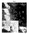
- FIG. 3 shows a comparison of cell densities remaining on the flask culturing surface of each experiment group following separation and recovery of 1° MLR cultured cells.
- Experiment group A comprises detaching and recovering cells from the culturing surface of a culture flask coated with PVGlcNAc by shaking it in EDTA/PBS( ⁇ ), followed by staining the cells remaining on the culturing surface with Giemsa stain and photographing with a microscope.
- Experiment group B comprises detaching and recovering cells from the culturing surface of a culture flask coated with P(VLA-co-SU) by shaking it in EDTA/PBS( ⁇ ), followed by staining the cells remaining on the culturing surface with Giemsa stain and photographing with a microscope.
- Experiment group C comprises detaching and recovering cells from the culturing surface of a commercially available culture flask with a protease (trypsin), followed by staining the cells remaining on the culturing surface with Giemsa stain and photographing with a microscope.
- trypsin a protease
- Experiment group D comprises detaching and recovering cells from the culturing surface of a commercially available culture flask by shaking it in EDTA/PBS( ⁇ ), followed by staining the cells remaining on the culturing surface with Giemsa stain and photographing with a microscope.
- Experiment group E comprises detaching and recovering cells from the culturing surface of a commercially available culture flask by strongly blowing EDTA/PBS( ⁇ ) with a pipette, followed by staining the cells remaining on the culturing surface with Giemsa stain and photographing with a microscope.
- the present invention relates to a method for preparing adoptive immune cells comprising the following steps:
- antigen-presenting in the present invention refers to decomposing an antigen incorporated from outside cells, expressing complex molecules of the resulting fragments and MHC molecules on a cell membrane, and then presenting them to naive immune cells for expressing antigen-specific immunity.
- antigen-presenting associated cells in the present invention refers to cells having the ability to present antigen or cells capable of differentiating into cells having the ability to present antigen.
- Cells having the ability to present antigen refer to all cells which have the ability to present antigen, examples of which include, but are not limited to, thymic epithelial cells, monocytes, monocyte-derived cells, macrophages, dendritic cells and Langerhans cells.
- Cells capable of differentiating into cells having the ability to present antigen refer to all cells capable of acquiring the ability to present antigen, examples of which include, but are not limited to, thymic epithelial cells, stem cells, hematopoietic stem cells, ES cells, ES cultured cells, bone marrow progenitor cells, myeloblasts and myeloid progenitor cells.
- Antigen-presenting associated cells of the present invention are able to be acquired from tissue or body fluid in which they are contained. Specific examples include all biological resources obtainable the antigen-presenting associated cells, but are not limited to, tissue and body fluid containing stem cells or hematopoietic stem cells, early fetal yolk sac, fetal liver from age 2 months to birth, fetal spleen from age 2 months to birth, bone marrow including that starting at fetal age 2 months and after birth, cord blood, peripheral blood, peripheral blood during recovery from a serious infection, and peripheral blood following administration of a hematinic such as GM-CSF.
- Any conventionally known method for acquiring cells can be used to acquire the aforementioned cells, examples of which include collection of whole blood or bone marrow fluid, collection of monocytes from the peripheral blood by apheresis, and proliferation of monocyte-derived primary-cultured cells or cell lines.
- the culture liquid used in the preparation method of the present invention can contain a physiologically active substance and antigen.
- a “physiologically active substance” refers to a substance capable of regulating the physiological activity of cells, tissue or organs, examples of which include proteins in the manner of enzymes, toxins, cytotoxic proteins, cytokines, chemokines, growth factors and interferon; polypeptides; oligopeptides; sugars; nucleotides in the manner of human genes, animal genes, microbe genes, virus genes, antisense DNA and antisense RNA; amino acids; and, fatty acids.
- physiologically active substances maybe PMA (phorbol 12-myristate 13-acetate), LPS, Picibanil, Hasumi vaccine M, PHA, poly I:C, TSST-1 (toxic shock syndrome toxin-1), cholera toxin B subunit, GM-CSF, M-CSF, IFN- ⁇ , IFN- ⁇ , IFN- ⁇ , TNF- ⁇ , IL-1 ⁇ , IL-3, IL-4, IL-6, IL-7, IL-12, SCF, TGF- ⁇ , TGF- ⁇ 1, Mycobacterium tuberculosis cell component, lymphocytic choriomeningitis virus, influenza virus, Staphylococcus bacteria , pertussis bacteria, toxoplasma gondii , cell and bacteria products (such as lipopolysaccharides, lipoteichoic acid, bacterial heat shock protein, CpG motif, mycobacteria trehalose, mycolic acid, toxoplasma gondii , cell
- an “antigen” refers to a substance which induces the formation of a specific antibody in a vertebrate host, or a substance which induces the generation of a specific lymphocyte population which reacts with that substance, and includes organic and inorganic compounds provided they are able to function as antigens.
- antigens include proteins, nucleic acids, carbohydrates, lipids or fragments or derivatives thereof.
- antigens include cancer cell lysate, cancer cell acid-extracted peptide, IgM idiotype, ⁇ -Gal protein, MUT1 peptide, HPV16-E7 peptide, OVA peptide, p53 mutant peptide, MART1, gp100, chitosinase peptide, TRP1, TRP2, tyrosinase, PSA, proteinase 3, MAGE-1, MAGE-3, BAGE, GAGE-1, GAGE-2, NY-ESO-1, ⁇ -catenin, MUM-1, CDK4, TPI, CDC27, LDLR-FUT, CASP-8, KIAA0205, EF2, bcr-abl (0210), TEL-AML1, Her2/new, ras, WT1, CEA, SART-1, KIAA0156, cyclophilin B, Lck, F4.2, MUC-1 mucin, RAGE, HPV16-E7, EBV-EBNA-2
- the step of culturing detached cells in culture liquid containing a physiologically active substance and antigen can also be carried out following the aforementioned cell detaching step.
- a preferable mammal of the present invention is a human.
- PVLA poly(p-N-vinylbenzyl-D-1-actonamide)
- ASGPR asialoglycoprotein receptor
- PVLA is represented by the structural formula shown below.
- Polymer synthesis is carried out by dissolving a monomer in a solvent such as DMSO, toluene or cyclohexane, and after removing the oxygen by deaeration treatment, adding an initiator such as AIBN or BPO. Subsequently, the polymer can be easily synthesized by reacting for a suitable amount of time of 2 hours or more by heating at a temperature equal to or higher than the cleavage temperature of the initiator in a sealed vessel.
- a solvent such as DMSO, toluene or cyclohexane
- a sugar chain monomer such as VLA (N-p-vinylbenzyl-O- ⁇ -D-galactopyranosyl-(1-4)-D-gluconamide)
- VLA N-p-vinylbenzyl-O- ⁇ -D-galactopyranosyl-(1-4)-D-gluconamide
- a comparatively highly polar solvent such as DMSO or DMF
- the solvent is not limited to such solvents.
- the polymer is synthesized either by selecting a solvent in which both dissolve, or mixing solutions in which each one is dissolved.
- VGlcNAc N-p-vinylbenzyl-O- ⁇ -2-acetoamido-2-deoxy-D-glucopyranosyl-(1-4)-2-acetoamido-2-deoxy-D-gluconamide
- VMA N-p-vinylbenzyl-O- ⁇ -D-glucopyranosyl-(1-4)-D-gluconamide
- VCA N-p-vinylbenzyl-O- ⁇ -D-glucopyranosyl-(1-4)-D-gluconamide
- VMan N-p-vinylbenzyl-O- ⁇ -D-mannopyranosyl-(1-4)-D-mannoamide
- VMea N-p-vinylbenzyl-O- ⁇ -D-galactopyranosyl-(1-6)-D-gluconamide
- VLAm N-p-vinylbenzyl-O- ⁇ -D-glucopyranosyl
- a preferable example of a sugar chain-containing polymer of the present invention is P(VLA-co-SU) or PVGlcNAc.
- the cell detaching step of the present invention can contain the following steps:
- the concentration of EDTA is preferably 0.02 to 20 mM.
- the liquid depth of PBS containing EDTA is preferably 2 to 5 mm.
- the present invention relates to adoptive immune cells obtained by the preparation method of the present invention.
- the present invention relates to a culture vessel coated with PVLA, and preferably P(VLA-co-SU) or PVGlcNAc, for use in the preparation method of the present invention.
- the sugar chain-containing polymer can be coated onto a Petri dish in the form of a suitable aqueous solution.
- a suitable aqueous solution is not limited thereto.
- Coating can be carried out by adding a suitable amount of sugar chain-containing polymer solution corresponding to the size of the Petri dish. At this time, preferably a liquid depth should be maintained so that the Petri dish is covered with liquid, and although a liquid depth of 1 to 5 mm is used preferably, the liquid depth is not limited thereto.
- Coating can be carried out by treating it for 1 to 24 hours, preferably coating is carried out by treating it for 2 hours, but not limited thereto.
- the present invention relates to a kit which comprises the aforementioned culture vessel for preparing an adoptive immune cell vaccine.
- the kit of the present invention can also contain a centrifuge tube for cell separation, antigen and/or culture liquid.
- the present invention relates to a method for treating malignant tumors, type I diabetes, atopic allergic diseases or infections using the adoptive immune cells of the present invention.
- treatment method of the present invention include, but are not limited to, those indicated below.
- a cancer-associated antigen solution such as cancer cell lysate, cancer cell acid-extracted peptide, cancer cell-associated synthetic peptide, IgM idiotype, MUT1 peptide, HPV16-E7- peptide, p53 mutant peptide, MART1, gp100, chitosinase peptide, TRP1, TRP2, tyrosinase, PSA, proteinase 3, MAGE-1, MAGE-3, BAGE, GAGE-1, GAGE-2, NY-ESO-1, ⁇ -catenin, MUM-1, TPI, CASP-8, KIAA0205, bcr-abl (0210), TEL-AML1, Her2/new, ras, WT1, CEA, SART-1, KIAA0156, cyclophilin B, Lck, MUC-1, mutant HLA-A2, HA1, HA2, H-Y or LacZ) is added followed
- the cells are detached and recovered from the culture vessel. Detachment and recovery of adhered cells are carried out by shaking. Detachment and recovery by shaking can be carried out according to the method described below. Suspended cells are recovered into a centrifuge tube, and the bottom of the flask is rinsed once with EDTA/PBS( ⁇ ). Next, EDTA/PBS( ⁇ ) is added so that the depth from the bottom of the flask is 2 to 5 mm, followed by allowing to stand undisturbed for 10 minutes at 4° C. Subsequently, the flask is shaken for 10 minutes with a shaker under conditions of an amplitude of 2.0 to 4.0 cm and speed of 130 to 160 times/minute.
- the detached cells are recovered.
- the flask is then washed twice with EDTA/PBS( ⁇ ), and the detached cells are collected in the aforementioned centrifuge tube.
- the resulting cells are referred to as cancer antigen-specific antigen-presenting cells.
- the PBMNL obtained in the previous two steps is cultured for 3 days in the culture liquid in which PHA is added (final concentration: 10 ⁇ g/ml) to fresh culture liquid.
- a plastic culture vessel coated with a sugar chain-containing polymer e.g., P(VLA-co-SU) or PVGlcNAc
- Fresh culture liquid is added to all of the cultured cells every 2 days throughout the culturing period. After 3 days, inomycin was added to each culture (final concentration: 1 ⁇ g/ml) followed by additionally culturing for 3 days.
- the cells in the culture vessel are collected at completion of culturing. Detachment and recovery of adhered cells are carried out by shaking. Detachment and recovering the adhered cells by shaking can be carried out in the manner described below.
- EDTA/PBS( ⁇ ) is added so that the depth from the bottom of the flask is 2 to 5 mm, followed by allowing to stand undisturbed for 10 minutes at 4° C. Subsequently, the flask is shaken for 10 minutes with a shaker under conditions of an amplitude of 2.0 to 4.0 cm and speed of 130 to 160 times/minute. Following shaking, the detached cells are recovered. The flask is then washed twice with EDTA/PBS( ⁇ ), and the detached cells are collected in the aforementioned centrifuge tube. The resulting cells are referred to as AT cells.
- both cells are administered to the same person from which the PBMNL were obtained.
- the present invention relates also to a medicine for treating a malignant tumor, type I diabetes, atopic allergic diseases or infections using the adoptive immune cells.
- the aforementioned medicine can be prepared using the adoptive immune cells of the present invention as it is, or in the form of a pharmaceutical composition by mixing the adoptive immune cells of the present invention with known pharmaceutically acceptable carriers (including a vehicle, extender, binder or lubricant) or commonly used additives.
- the pharmaceutical composition can be administered orally or parenterally corresponding to the prepared drug form (examples of oral preparations: tablets, pills, capsules, powders, granules and syrups; examples of parenteral preparations: injections, intravenous drips, topical preparations, nose drops, eye drops, inhalants and suppositories).
- the dosage varies according to the type of active ingredient, administration route, and age, body weight and symptoms of administered subject or patient.
- mice Male, four-week-old C57B1/6 and BALB/c mice were purchased from Oriental Yeast Co., Ltd.
- mice were sacrificed by severing a cervical vertebrae followed by aseptic removal of the spleen, thymus and celiac lymph nodes. Suspended cells were obtained from the resulting tissues by gently breaking up the tissue in culture liquid with two slide glasses. After removing tissue fragments from the cell suspension by filtering through cotton, the cell suspension was centrifuged for 7 minutes at 300 ⁇ g and 4° C. Following centrifugation, the supernatant was removed and the cells were re-suspended in culture liquid. This series of operations was repeated three times to remove all tissue and cell fragments except for the cells. After washing, the spleen cells, thymus cells and lymph node cells were pooled. In this experiment, these pooled cells are referred to as mononuclear leukocytes (MNL).
- MNL mononuclear leukocytes
- ConA Sigma Chemical Co., Cat. No. C-0412
- 1 ⁇ 5 ⁇ 10 6 cells/ml of the suspended cells was added to the 1 ⁇ 5 ⁇ 10 6 cells/ml of the suspended cells to a final concentration of 2 ⁇ g/ml, and cultured in an incubator under conditions of 37° C. and 5% CO 2 .
- the suspended cells were recovered from the culture vessel into a 50 ml centrifuge tube 48 hours later and washed 5 times with culture liquid.
- MMC (Sigma Chemical Co., Cat No. M-0503) was added to 1 ⁇ 5 ⁇ 10 6 cells/ml of the suspended cells to a final concentration of 50 ⁇ g/ml and allowed to stand undisturbed at 37° C. The cells were washed with culture liquid 60 minutes later and then re-suspended in culture liquid.
- the suspension of immunostained cells was centrifuged in a 15 ml centrifuge tube (300 ⁇ g, 4° C., 7 minutes). The supernatant was discarded to obtain a pellet. The pellet was then broken up gently followed by the addition of Fc BlockTM (Nippon Becton Dickinson Company, Ltd., Cat. No. 01241D) at 2 ⁇ l per 1 ⁇ 10 6 cells. Next, 2 ⁇ l of PE-anti-mouse I-Ad (Nippon Becton Dickinson Company, Ltd., Cat. No. 06035A), 2 ⁇ l of FITC-anti-mouse CD11c (Nippon Becton Dickinson Company, Ltd., Cat No.
- Antigen-specific antigen-presenting cells to alloantigen were obtained by 1° MLR culturing. The procedure was carried out as described below. 12 ml of MNL derived from BALB/c mice (H-2 d ) adjusted to 5 ⁇ 10 6 cells/ml, and 12 ml of MMC-treated C57B1/6 (H-2 b )-derived MNL adjusted to 5 ⁇ 10 6 cells/ml were added to a 75 cm 2 culturing flask followed by the addition of interleukin 2 (Sigma Chemical Co., Cat. No. I-0523) to a final concentration of 20 ng/ml and culturing at 37° C. in 5% CO 2 .
- interleukin 2 Sigma Chemical Co., Cat. No. I-0523
- the suspended cells were recovered in a 50 ml centrifuge tube and the bottoms of the flasks were rinsed once with EDTA/PBS( ⁇ ).
- EDTA/PBS( ⁇ ) was added to a depth of 2 to 5 mm from the bottom of the flasks and allowed to stand undisturbed for 10 minutes at 4° C.
- the flasks were shaken for 10 minutes under conditions of an amplitude of 3.0 cm and speed of 130 to 160 times/minute with a shaker (Nissin Scientific Corporation, Reciprocal Shaker NA-201). Following shaking, the cells that detached from the flasks were recovered.
- the flasks were additionally washed twice with 5 ml of EDTA/PBS( ⁇ ), and the detached cells were collected in the aforementioned centrifuge tube.
- the cells detached and recovered by the enzyme treatment method were prepared in the manner described below.
- the cells that were suspended as a result of this procedure were recovered into a 50 ml centrifuge tube to which 10 ml of culture liquid had added in advance.
- the flask culturing surface was further rinsed twice with 5 ml of EDTA/PBS ( ⁇ ).
- the resulting detached cell groups were pooled for each experiment group.
- CTL cytotoxic T cells
- 7-amino-actinomycin D 7-AAD, Nippon Becton Dickinson Company, Ltd., Cat. No. 555816
- a pellet of 1 ⁇ 10 6 cells was mixed with 20 ⁇ l of FITC-anti-mouse H-2 D b antibody, which specifically reacts with C57B1/6 target MNL, and 20 ⁇ l of Fc BlockTM, followed by allowing to react for 30 minutes at 4° C.
- the cells were washed once with 10 ml of washing buffer followed by the addition of 20 ⁇ l of 7-AAD and allowing to stand undisturbed at room temperature.
- the VLA was dispensed into a vacuum reaction tube. After completely dissolved by adding DMSO or a mixed solvent of DMSO and toluene, freezing, degassing and thawing were repeated to remove the oxygen from the solution. 1/100 mole of AIBN was added thereto to dissolve followed by sealing under reduced pressure. This was then allowed to react for 5 hours at 60° C. to synthesize the polymer.
- the resulting solution was dropped into highly excess amount of methanol to precipitate the polymer.
- a procedure comprising dissolving it in a suitable solvent, dropping into methanol and precipitating the polymer was repeated three times.
- the finally obtained polymer was dissolved in distilled water and dialyzed against highly excess amount of water. Subsequently, the target polymer was obtained by freeze-drying.
- Monomer to be copolymerized with VLA was weighed at a molar ratio of 9:1, 8:2, 7:3 and so forth, and dispensed into a vacuum reaction tube. After completely dissolving by adding DMSO or a mixed solvent of DMSO and toluene, freezing, degassing and thawing were repeated to remove oxygen from the solution. 10 mmol of AIBN was then added thereto to dissolve followed by sealing under reduced pressure. This was then allowed to react for 5 hours at 60° C. to synthesize the polymer.
- the resulting solution was dropped into highly excess amount of methanol to precipitate the polymer.
- a procedure consisting of dissolving it in a suitable solvent, dropping into methanol again and precipitating the polymer was repeated three times.
- the finally obtained polymer was dissolved in distilled water and dialyzed against highly excess amount of water. Subsequently, the target polymer was obtained by freeze-drying.
- the previously described sugar chain-bound monomers and commercially available monomers can be used preferably for the comonomer.
- PVLA, P(VLA-co-SU) and PVGlcNAc were respectively prepared in the form of 0.01% by weight aqueous solutions. 5 ml aliquots of these solutions were dispensed into a flask (Nippon Becton Dickinson Company, Ltd., Cat. No. 353133). Coating treatment was then carried out for 2 hours at room temperature under protection from light. Subsequently, the polymer solution was recovered and each flask was washed three times with 10 ml of pure water prior to use in each test.
- the mixture was sterilized by filtering with a 0.22 ⁇ m sterilization filter (Nippon Millipore Company, Sterivex-GS, Cat. No. SVGSB1010). This is referred to as the culture liquid.
- PBS( ⁇ ) 10 tablets of PBS available from Oxoid Ltd. (phosphate-buffered physiological saline, Cat. No. BR0014G) were dissolved in 1 L of pure water (Nippon Millipore Company, Milli-Q Gradient Milli-Q Ultra-Pure Water System Elix 5 Kit) followed by sterilizing at high temperature and high pressure for 20 minutes at 121° C. in an autoclave. This is referred to as PBS( ⁇ ).
- EDTA/PBS( ⁇ ) 2 g of EDTA (EDTA-4Na, Wako Pure Chemical Industries, Ltd., Cat. No. 343-01883) was added to 1 L of PBS( ⁇ ) followed by sterilizing at high temperature and high pressure for 20 minutes at 121° C. in an autoclave. This is referred to as EDTA/PBS( ⁇ ).
- trypsin/EDTA 0.05 g of trypsin (Sigma Chemical Co., Cat. No. T-7409) and 0.024 g of EDTA (EDTA-4Na, Wako Pure Chemical Industries Ltd., Cat. No. 343-01883) were added to and dissolved in 100 ml of PBS( ⁇ ) followed by sterilizing by filtering with a 0.22 ⁇ m sterilization filter (Nippon Millipore Company, Sterivex-GS, Cat. No. SVGSB1010). This is referred to as trypsin/EDTA.
- the washing buffer used for immunofluorescent staining was prepared in the following manner. Bovine serum albumin (Sigma Chemical Co., Cat. No. A-3059) to a final concentration of 0.1% and NaN 3 (Wako Pure Chemical Industries, Ltd., Cat. No. 195-11092) to a final concentration of 0.001% were added to PBS( ⁇ ), and used after chilling it to 4° C.
- the following flasks were used as flasks for adhered cells used as a control: Treated 75 cm 2 polystyrene flasks (Nippon Becton Dickinson Company, Ltd., Cat. No. 353136), treated 25 cm 2 polystyrene flasks (Nippon Becton Dickinson Company, Ltd., Cat. No. 353108).
- the following tubes were used for the centrifuge tubes: 15 ml centrifuge tubes (Asahi Technoglass Corporation, Cat. No. 2315-015, 50 ml centrifuge tubes (Assist Co., Ltd., Cat. No. 62.548.004S).
- FIG. 1 - a shows the results of comparing the numbers of detached and recovered CD80/86, CD11c and I-A d -positive cells.
- the numbers of CD80/86, CD11c and I-A d -positive cells detached and recovered from the flasks coated with PVGlcNAc and P(VLA-co-SU) by shaking were significantly greater than the numbers of CD80/86, CD11c and I-A d -positive cells detached and recovered by enzyme treatment, pipetting or shaking using conventional commercially available flasks for adhered cells.
- a similar trend was also observed for the ratio (%) of the numbers of CD80/86, CD11c and I-A d -positive cells recovered from 1° MLR culturing flasks to the total number of cells (FIG. 1 - b ).
- Flasks following Detachment and recovery of cells were immediately fixed by addition of 3 ml of undiluted Giemsa stain (Mutoh Pure Chemicals Co., Ltd, Cat. No. 1500-2). After fixing for 30 to 60 minutes at room temperature (15° C.), 20 ml of tap water was added to initiate staining. Staining was carried out for 1 hour at room temperature. After staining, the culturing bottoms of the flasks were washed with tap water and allowed to air dry followed by taking photographs (Nikon Instech Co., Ltd, Coolpix Microsystem VI) with a microscope (Nikon Instech Co., Ltd, Diaphot Model), and comparing the residual cell densities.
- FIG. 3 Colonies in which adhered cells had gathered were observed on the flask culturing surfaces following Detachment and recovery of adhered cells by enzyme treatment, pipetting and shaking from the commercially available flasks for adhered cells, and the residual densities of cells remaining on the flask surfaces were determined to be higher than those flasks from which adhered cells had been detached by shaking from the polymer-coated flasks ( FIG. 3 ).
- the antigen-presenting ability of cells obtained from 1° MLR culturing was further tested by inducing alloantigen-specific cytotoxic T cells according to the method described below.
- BALB/c-derived cells obtained from 1° MLR culturing were prepared to 1 ⁇ 10 5 cells/ml as antigen-presenting cells (H-2 d ), and 1.5 ml thereof were placed in a 25 cm 2 flask.
- C57B1/6-derived ConA blast cells were treated with MMC for use as alloantigen and added thereto.
- 1.5 ml of a cell suspension having a density of 1 ⁇ 10 7 cells/ml was added followed by mixing and culturing for 8 days under conditions of 37° C. and 5% CO 2 .
- antigen-presenting cells prepared using a polymer-coated flask were determined to have carried out significantly higher CTL induction than conventional detachment methods using enzyme treatment.
Abstract
Description
- a. obtaining mammalian antigen-presenting associated cells;
- b. culturing the resulting cells in a culture vessel coated with a sugar chain-containing polymer; and
- c. detaching the cells by shaking the culture vessel without treating the cells with enzyme and without using a cell detaching tool.
2. The preparation method according to 1 above, wherein a culture liquid used in the step b contains a physiologically active substance and an antigen.
3. The preparation method according to 1 or 2 above, which further comprises the following step: - d. culturing the detached cells in a culture liquid containing a physiologically active substance and an antigen.
4. The preparation method according to 2 or 3 above, wherein the physiologically active substance is selected from the group consisting of PMA, PHA, Hasumi vaccine M, GM-CSF, IFN-α, TNF-α, IL-3, IL-4, SCF, TGF-α, TGF-β1, Mycobacterium tuberculosis cell component, LPS, Picibanil, poly I: C and hyaluronic acid complex fragment.
5. The preparation method according to 2 or 3 above, wherein the antigen is selected from the group consisting of cancer cell lysate, cancer cell acid-extracted peptide, cancer cell-associated synthetic peptide, IgM idiotype, MUT1 peptide, HPV16-E7 peptide, p53 mutant peptide, MART1, gp100, chitosinase peptide, TRP1, TRP2, tyrosinase, PSA,proteinase 3, MAGE-1, MAGE-3, BAGE, GAGE-1, GAGE-2, NY-ESO-1, β-catenin, MUM-1, TPI, CASP-8, KIAA0205, bcr-abl (0210), TEL-AML1, Her2/new, ras, WT1, CEA, SART-1, KIAAO156, cyclophilin B, Lck, MUC-1, mutant HLA-A2, HA1, HA2, H-Y and LacZ.
6. The preparation method according to any one of 1 to 5 above, wherein the antigen-presenting associated cells include dendritic cells or Langerhans cells.
7. The preparation method according to any one of 1 to 6 above, wherein the mammal is a human.
8. The preparation method according to any one of 1 to 7 above, wherein the sugar chain-containing polymer is PVLA, P(VLA-co-SU), PVGlcNAc, PVMA, PVCA, PVMea, PVLam or PVMan.
9. The preparation method according to any one of 1 to 8 above, wherein the step c comprises the following steps: - c1. removing the culture liquid and adding PBS containing EDTA;
- c2. allowing the vessel to stand undisturbed for 10 minutes at 4 to 10° C.; and
- c3. detaching the cells by shaking the vessel.
10. The preparation method according to 9 above, wherein a concentration of the EDTA is 0.02 mM to 20 mM.
11. The preparation method according to 9 or 10 above, wherein the liquid depth of the PBS containing EDTA is 2 to 5 mm.
12. Adoptive immune cells obtained by the preparation method according to any one of 1 to 11 above.
13. A culture vessel coated with a sugar chain-containing polymer for use in the preparation-method according to any one of 1 to 11 above.
14. The culture vessel according to 13 above, wherein the sugar chain-containing polymer is PVLA, P(VLA-co-SU), PVGlcNAc, PVMA, PVCA, PVMea, PVLam or PVMan.
15. A kit for preparing an adoptive immune cell vaccine comprising the culture vessel according to 13 or 14 above.
16. The kit according to 15 above, which further comprises a cell separation tube, an antigen and/or a culture liquid.
17. A method for treating a malignant tumor, type I diabetes, atopic allergic diseases or infections using the adoptive immune cells according to 12 above.
18. A medicine for treating a malignant tumor, type I diabetes, atopic allergic diseases or infections comprising the adoptive immune cells according to 12 above.
- A-1: Magnification: 40×. Within the frame, it is shown differences in residual cell densities following detachment at the boundary between a polymer uncoated surface (circular) and a coated surface.
- A-2: Magnification: 100×. Arrows indicate differences in residual cell densities following detachment at the boundary between a polymer uncoated surface (circular) and a coated surface.
- A-3: Magnification: 200×. Arrows indicate differences in residual cell densities following detachment at the boundary between a polymer uncoated surface (circular) and a coated surface.
- B-1: Magnification: 40×
- B-2: Magnification: 100×
- B-3: Magnification: 200×
- C-1: Magnification: 40×
- C-2: Magnification: 100×
- C-3: Magnification: 200×
- D-1: Magnification: 40×
- D-2: Magnification: 100×
- D-3: Magnification: 200×
- E-1: Magnification: 40×. Arrow indicates the portion where the cells were detached along the direction in which EDTA/PBS(−) was blown with the pipette.
- E-2: Magnification: 100×.
- E-3: Magnification: 200×. Arrows indicate the ruffled edges of the adhered cells being curled up along the direction in which EDTA/PBS(−) was blown with the pipette.
- a. obtaining mammalian antigen-presenting associated cells;
- b. culturing the resulting cells in a culture vessel coated with a sugar chain-containing polymer; and
- c. detaching the cells by shaking the culture vessel without treating the cells with enzyme and without using a cell detaching tool.


- c1. removing the culture liquid and adding PBS containing EDTA;
- c2. allowing the vessel to stand undisturbed for 10 minutes at 4 to 10° C.; and
- c3. detaching the cells by shaking the vessel.
- 1. A cell fraction containing mainly of a peripheral blood mononuclear cell component is collected by apheresis after waiting for recovery of hematopoiesis of a patient from which a tumor was excised following tumor excision. Recovery of hematopoiesis is confirmed by a supervising physician according to the number of each component cell in the peripheral blood.
- 2. The peripheral blood mononuclear cell component, which is free of erythrocytes, platelets and so forth, is obtained by fractionating the blood cell fraction obtained by apheresis using density gradient centrifugation (separation liquid: Lymphosepar I, centrifugation conditions: 400×g, 30 minutes). The cells of this component are referred to as PBMNL (peripheral blood mononuclear leukocytes), and will be described later.
- 3. After washing the resulting PBMNL, culturing is started in a culture liquid to which has been added a physiologically active substance (such as PMA, PHA, Hasumi vaccine M, GM-CSF, IFN-α, TNF-α, IL-3, IL-4, SCF, TGF-α, TGF-β1, Mycobacterium tuberculosis cell component, LPS, Picibanil, poly I:C or hyaluronic acid complex fragment) (induction of differentiation and maturation of dendritic cells). A plastic culture vessel coated with a sugar chain-containing polymer (such as P(VLA-co-SU) or PVGlcNAc) is used as the culture vessel. Fresh culture liquid is added to all cultured cells every 3 days throughout the culturing period.
- 4. Induction and culturing of activated T cells are carried out simultaneous to and in parallel with the induction and culturing step of the cancer antigen-specific antigen-presenting cells. The procedure is described below.
- 5. The cancer antigen-specific antigen-presenting cells and the AT cells are washed with fresh culture liquid.
- 1) Mice:
- 1) Preparation of Mouse Mononuclear Leukocytes
- 2) Preparation of Concanavalin A Blast (ConA Blast) Cells
- 3) Preparation of Mitomycin C (MMC) Treated Cells
- 4) Immunofluorescent Staining
- 5) Primary Mixed Leukocyte Reaction (1° MLR)
- 6) Detachment and Recovery of Cells from 1° MLR Culturing Flasks
- 7) Cytotoxicity Test
- 1) PVLA Synthesis
- 2) Polymer Coating Method of Culturing Flask:
- 3) Culture Liquid:
- 4) Ca++, Mg++-Free PBS
- 5) 0.2% EDTA/PBS(−)
- 6) PBS(−) Containing 0.05% Trypsin and 0.024% EDTA
- 7) Washing Buffer
- 8) Sterilization Test of Sterile Solution
- 1) Culturing Flasks for Suspended Cells
- 2) Culturing Flasks for Adhered Cells
- 3) Cell Centrifuge Tubes
- 4) Plastic Culturing Pipettes
Claims (39)
Priority Applications (1)
| Application Number | Priority Date | Filing Date | Title |
|---|---|---|---|
| US12/653,090 US8075882B2 (en) | 2004-05-11 | 2009-12-08 | Adoptive immune cells for tumor vaccines |
Applications Claiming Priority (1)
| Application Number | Priority Date | Filing Date | Title |
|---|---|---|---|
| PCT/JP2004/006599 WO2005108554A1 (en) | 2004-05-11 | 2004-05-11 | Adoptive immunocyte for tumor vaccine and the like |
Related Child Applications (1)
| Application Number | Title | Priority Date | Filing Date |
|---|---|---|---|
| US12/653,090 Division US8075882B2 (en) | 2004-05-11 | 2009-12-08 | Adoptive immune cells for tumor vaccines |
Publications (2)
| Publication Number | Publication Date |
|---|---|
| US20070286847A1 US20070286847A1 (en) | 2007-12-13 |
| US7655393B2 true US7655393B2 (en) | 2010-02-02 |
Family
ID=35320225
Family Applications (2)
| Application Number | Title | Priority Date | Filing Date |
|---|---|---|---|
| US11/579,280 Expired - Fee Related US7655393B2 (en) | 2004-05-11 | 2004-05-11 | Adoptive immune cells for tumor vaccines |
| US12/653,090 Expired - Fee Related US8075882B2 (en) | 2004-05-11 | 2009-12-08 | Adoptive immune cells for tumor vaccines |
Family Applications After (1)
| Application Number | Title | Priority Date | Filing Date |
|---|---|---|---|
| US12/653,090 Expired - Fee Related US8075882B2 (en) | 2004-05-11 | 2009-12-08 | Adoptive immune cells for tumor vaccines |
Country Status (5)
| Country | Link |
|---|---|
| US (2) | US7655393B2 (en) |
| EP (1) | EP1757680B1 (en) |
| JP (1) | JP4485523B2 (en) |
| DE (1) | DE602004023238D1 (en) |
| WO (1) | WO2005108554A1 (en) |
Cited By (7)
| Publication number | Priority date | Publication date | Assignee | Title |
|---|---|---|---|---|
| US20090208478A1 (en) * | 2003-10-24 | 2009-08-20 | Gencia Corporation | Transducible polypeptides for modifying metabolism |
| US8470972B2 (en) | 2003-10-24 | 2013-06-25 | Gencia Corporation | Nonviral vectors for delivering polynucleotides to plants |
| US8507277B2 (en) | 2003-10-24 | 2013-08-13 | Gencia Corporation | Nonviral vectors for delivering polynucleotides |
| US8541550B2 (en) | 2003-10-24 | 2013-09-24 | Gencia Corporation | Methods and compositions for delivering polynucleotides |
| US8927691B2 (en) | 2003-10-24 | 2015-01-06 | Gencia Corporation | Transducible polypeptides for modifying metabolism |
| US8952133B2 (en) | 2003-10-24 | 2015-02-10 | Gencia Corporation | Nonviral vectors for delivering polynucleotides to target tissue |
| US9434924B2 (en) | 2011-04-21 | 2016-09-06 | Nipro Corporation | Cell culture method and cell culture kit |
Families Citing this family (1)
| Publication number | Priority date | Publication date | Assignee | Title |
|---|---|---|---|---|
| EP2539435A2 (en) | 2010-02-23 | 2013-01-02 | Corning Incorporated | Modified substrates for protection of peptide-immobilized surfaces from gamma radiation degradation |
Citations (8)
| Publication number | Priority date | Publication date | Assignee | Title |
|---|---|---|---|---|
| JPH0541983A (en) | 1991-08-09 | 1993-02-23 | W R Grace & Co | Base material for cell cultivation and its preparation |
| JPH08317786A (en) | 1995-03-17 | 1996-12-03 | Kanagawa Kagaku Gijutsu Akad | Cell culture base modified with saccharide chain polymer and cell culture using the same |
| WO1997003186A2 (en) | 1995-07-12 | 1997-01-30 | Activated Cell Therapy, Inc. | Method for in vitro proliferation of dentritic cells, composition containing the cells entrapped in a three-dimensional matrix and use for immunization |
| WO1997005239A1 (en) | 1995-07-25 | 1997-02-13 | Celltherapy, Inc. | Autologous immune cell therapy: cell compositions, methods and applications to treatment of human disease |
| US5851756A (en) * | 1992-04-01 | 1998-12-22 | The Rockefeller University | Method for in vitro proliferation of dendritic cell precursors and their use to produce immunogens |
| JP2002069001A (en) | 2000-08-29 | 2002-03-08 | Asahi Kasei Corp | Cell vaccine consisting mainly of dendritic cell |
| US6642050B1 (en) * | 1999-07-28 | 2003-11-04 | Amcite Research, Ltd. | Three-dimensional cell culture material having sugar polymer containing cell recognition sugar chain |
| WO2004024900A1 (en) | 2002-09-13 | 2004-03-25 | Forschungsinstitut Für Krebskranke Kinder | USE OF DENDRITIC CELLS (DCs) EXPRESSING INTERLEUKIN 12 (IL-12) |
Family Cites Families (2)
| Publication number | Priority date | Publication date | Assignee | Title |
|---|---|---|---|---|
| JP4107410B2 (en) | 2002-03-22 | 2008-06-25 | テルモ株式会社 | Monocyte capture filter |
| CA2487452A1 (en) | 2002-05-28 | 2003-12-04 | Robinson Ramirez-Pineda | A method for generating antigen-presenting cells |
-
2004
- 2004-05-11 US US11/579,280 patent/US7655393B2/en not_active Expired - Fee Related
- 2004-05-11 WO PCT/JP2004/006599 patent/WO2005108554A1/en not_active Application Discontinuation
- 2004-05-11 EP EP04732192A patent/EP1757680B1/en not_active Expired - Fee Related
- 2004-05-11 JP JP2006512905A patent/JP4485523B2/en not_active Expired - Fee Related
- 2004-05-11 DE DE602004023238T patent/DE602004023238D1/en active Active
-
2009
- 2009-12-08 US US12/653,090 patent/US8075882B2/en not_active Expired - Fee Related
Patent Citations (11)
| Publication number | Priority date | Publication date | Assignee | Title |
|---|---|---|---|---|
| JPH0541983A (en) | 1991-08-09 | 1993-02-23 | W R Grace & Co | Base material for cell cultivation and its preparation |
| US5851756A (en) * | 1992-04-01 | 1998-12-22 | The Rockefeller University | Method for in vitro proliferation of dendritic cell precursors and their use to produce immunogens |
| JPH08317786A (en) | 1995-03-17 | 1996-12-03 | Kanagawa Kagaku Gijutsu Akad | Cell culture base modified with saccharide chain polymer and cell culture using the same |
| WO1997003186A2 (en) | 1995-07-12 | 1997-01-30 | Activated Cell Therapy, Inc. | Method for in vitro proliferation of dentritic cells, composition containing the cells entrapped in a three-dimensional matrix and use for immunization |
| US6121044A (en) | 1995-07-12 | 2000-09-19 | Dendreon Corporation | Potent antigen presenting cell composition |
| WO1997005239A1 (en) | 1995-07-25 | 1997-02-13 | Celltherapy, Inc. | Autologous immune cell therapy: cell compositions, methods and applications to treatment of human disease |
| JP2001520509A (en) | 1995-07-25 | 2001-10-30 | セルセラピー・インコーポレイテツド | Autoimmune cell therapy: cell compositions, methods and applications in the treatment of human diseases |
| US6642050B1 (en) * | 1999-07-28 | 2003-11-04 | Amcite Research, Ltd. | Three-dimensional cell culture material having sugar polymer containing cell recognition sugar chain |
| JP2002069001A (en) | 2000-08-29 | 2002-03-08 | Asahi Kasei Corp | Cell vaccine consisting mainly of dendritic cell |
| WO2004024900A1 (en) | 2002-09-13 | 2004-03-25 | Forschungsinstitut Für Krebskranke Kinder | USE OF DENDRITIC CELLS (DCs) EXPRESSING INTERLEUKIN 12 (IL-12) |
| US20060177420A1 (en) | 2002-09-13 | 2006-08-10 | Forschungsinstitut Fur Krebskranke Kinder | Use of dendritic cells (dcs) expressing interleukin 12 (il-12) |
Non-Patent Citations (15)
| Title |
|---|
| D. Choi et al, "Dendritic Cell-based Vaccines in the Setting of Peripheral Blood Stem Cell Transplantation: . . . ", Clin. Cancer Res., vol. 4, pp. 2709-2716, Nov. 1998. |
| English-language International Preliminary Report on Patentability dated Nov. 14, 2006 and Written Opinion of the International Searching Authority, of International Application PCT/JP2004/006599 filed May 11, 2004; Applicants: Shukokai Incorporated et al. |
| F.J. Hsu et al, "Vaccination of patients with B-cell lymphoma . . . ", Nat. Med., vol. 2, No. 1,pp. 52-58, Jan. 1996. |
| Hirose, et al, "Regulation of Asialoglycoprotein Receptor Expression in the Proliferative State of Hepatocytes," Biochemical and Biophysical Research Communications, 287 (2001) 675-681. |
| Ishida, et al, "Cell Adhesion Aside from Integrin System Can Abrogate Anoikis in Rat Liver Cells by Down-regulation of FasL Expression, not by Activation of PI-3K/Akt and ERK Signaling Pathway," Biochemical and Biophysical Research Communications, 300 (2003) 201-208. |
| J. Wang et al, "Eliciting T Cell Immunity Against Poorly Immunogenic Tumors . . . ", J. Immunol., vol. 161, pp. 5516-5524, 1998. |
| Jos W.M Van Der Meer, "Culture of Mononuclear Phagocytes on a Teflon Surface to Prevent Adherence", J. Exp. Med., vol. 147, pp. 271-276, 1978. |
| Jos W.M. Van Der Meer, "Suspension Cultures of Mononuclear Phagocytes in the Teflon Culture Bag", Cell. Immunol., vol. 42, pp. 208-121, 1979. |
| M. Takamizawa et al, "Cellular and Molecular Basis of Human gammadelta T Cell Activation", J. Clin Invest., vol. 95, pp. 296-303, Jan. 1995. |
| M. Takamizawa et al, "Cellular and Molecular Basis of Human γδ T Cell Activation", J. Clin Invest., vol. 95, pp. 296-303, Jan. 1995. |
| M.A. Morse et al, "A Phase I Study of Active Immunotherapy with Carcinoembryonic Antigen Peptide . . . ", Clin. Cancer Res., vol. 5, pp. 1331-1338, Jun. 1999. |
| Na, et al, "Conjugation of Arg-Gly-Asp (RGD) Sequence in Copolymer Bearing Sugar Moiety for Insulinoma Cell Line (MIN6) Culture," Biosci. Biotechnol. Biochem, 65 (6), 1284-1289, (2001). |
| R.J. Hodes et al, "Cellular and genetic control of antibody responses in vitro", Eur. J. Immunol., vol. 7, pp. 892-897, 1977. |
| R.M. Steinman et al, "Dendritic Cell Development and Maturation", Adv. Exp. Med., pp. 1-6, 1997. |
| V.L. Reichardt et al, "Idiotype Vaccination Using Dendritic Cells After Autologous Peripheral Stell Cell Transplantation for Multiple Myeloma . . . ", Blood, Vo. 93, No. 7, pp. 2411-2419, Apr. 1999. |
Cited By (7)
| Publication number | Priority date | Publication date | Assignee | Title |
|---|---|---|---|---|
| US20090208478A1 (en) * | 2003-10-24 | 2009-08-20 | Gencia Corporation | Transducible polypeptides for modifying metabolism |
| US8470972B2 (en) | 2003-10-24 | 2013-06-25 | Gencia Corporation | Nonviral vectors for delivering polynucleotides to plants |
| US8507277B2 (en) | 2003-10-24 | 2013-08-13 | Gencia Corporation | Nonviral vectors for delivering polynucleotides |
| US8541550B2 (en) | 2003-10-24 | 2013-09-24 | Gencia Corporation | Methods and compositions for delivering polynucleotides |
| US8927691B2 (en) | 2003-10-24 | 2015-01-06 | Gencia Corporation | Transducible polypeptides for modifying metabolism |
| US8952133B2 (en) | 2003-10-24 | 2015-02-10 | Gencia Corporation | Nonviral vectors for delivering polynucleotides to target tissue |
| US9434924B2 (en) | 2011-04-21 | 2016-09-06 | Nipro Corporation | Cell culture method and cell culture kit |
Also Published As
| Publication number | Publication date |
|---|---|
| WO2005108554A1 (en) | 2005-11-17 |
| DE602004023238D1 (en) | 2009-10-29 |
| JP4485523B2 (en) | 2010-06-23 |
| US8075882B2 (en) | 2011-12-13 |
| EP1757680A1 (en) | 2007-02-28 |
| US20100092445A1 (en) | 2010-04-15 |
| US20070286847A1 (en) | 2007-12-13 |
| JPWO2005108554A1 (en) | 2008-03-21 |
| EP1757680B1 (en) | 2009-09-16 |
| EP1757680A4 (en) | 2008-07-23 |
Similar Documents
| Publication | Publication Date | Title |
|---|---|---|
| US8075882B2 (en) | Adoptive immune cells for tumor vaccines | |
| CA2632263C (en) | Compositions and methods for inducing the activation of immature monocytic dendritic cells | |
| US9352001B2 (en) | Method for stimulating a host immune system | |
| JP2009518045A5 (en) | ||
| US20080014174A1 (en) | Method for treating tumors, infectious diseases, and autoimmune diseases with an activated HLA matching donor-originating lymphocyte | |
| WO2015014291A1 (en) | Lymph cell amplification and activation method via serum-free cultivation | |
| WO2004055053A1 (en) | Tumor vaccine | |
| CN110819591A (en) | Preparation method of CART cells | |
| AU2013206016B2 (en) | Compositions and methods for inducing the activation of immature monocytic dendritic cells | |
| Tan et al. | Experimental production of clinical-grade dendritic cell vaccine for acute myeloid leukemia. | |
| JP5424538B2 (en) | Maturation-induced dendritic cells for transplantation, production method and use thereof | |
| CHEONG | Experimental production of clinical-grade dendritic cell vaccine for acute myeloid leukemia | |
| MX2008007289A (en) | Compositions and methods for inducing the activation of immature monocytic dendritic cells |
Legal Events
| Date | Code | Title | Description |
|---|---|---|---|
| AS | Assignment |
Owner name: CELAGIX, RES. LTD.,JAPAN Free format text: ASSIGNMENT OF ASSIGNORS INTEREST;ASSIGNORS:OHASUMI, KENICHIRO;IMAIZUMI, AKIRA;IWAMA, MASAMICHI;AND OTHERS;REEL/FRAME:018569/0805 Effective date: 20060919 Owner name: SHUKOKAI INCORPORATED,JAPAN Free format text: ASSIGNMENT OF ASSIGNORS INTEREST;ASSIGNORS:OHASUMI, KENICHIRO;IMAIZUMI, AKIRA;IWAMA, MASAMICHI;AND OTHERS;REEL/FRAME:018569/0805 Effective date: 20060919 Owner name: CELAGIX, RES. LTD., JAPAN Free format text: ASSIGNMENT OF ASSIGNORS INTEREST;ASSIGNORS:OHASUMI, KENICHIRO;IMAIZUMI, AKIRA;IWAMA, MASAMICHI;AND OTHERS;REEL/FRAME:018569/0805 Effective date: 20060919 Owner name: SHUKOKAI INCORPORATED, JAPAN Free format text: ASSIGNMENT OF ASSIGNORS INTEREST;ASSIGNORS:OHASUMI, KENICHIRO;IMAIZUMI, AKIRA;IWAMA, MASAMICHI;AND OTHERS;REEL/FRAME:018569/0805 Effective date: 20060919 |
|
| AS | Assignment |
Owner name: CELAGIX, RES. LTD.,JAPAN Free format text: ASSIGNMENT OF ASSIGNORS INTEREST;ASSIGNORS:HASUMI, KENICHIRO;IMAIZUMI, AKIRA;IWAMA, MASAMICHI;AND OTHERS;REEL/FRAME:019465/0612 Effective date: 20060919 Owner name: SHUKOKAI INCORPORATED,JAPAN Free format text: ASSIGNMENT OF ASSIGNORS INTEREST;ASSIGNORS:HASUMI, KENICHIRO;IMAIZUMI, AKIRA;IWAMA, MASAMICHI;AND OTHERS;REEL/FRAME:019465/0612 Effective date: 20060919 Owner name: CELAGIX, RES. LTD., JAPAN Free format text: ASSIGNMENT OF ASSIGNORS INTEREST;ASSIGNORS:HASUMI, KENICHIRO;IMAIZUMI, AKIRA;IWAMA, MASAMICHI;AND OTHERS;REEL/FRAME:019465/0612 Effective date: 20060919 Owner name: SHUKOKAI INCORPORATED, JAPAN Free format text: ASSIGNMENT OF ASSIGNORS INTEREST;ASSIGNORS:HASUMI, KENICHIRO;IMAIZUMI, AKIRA;IWAMA, MASAMICHI;AND OTHERS;REEL/FRAME:019465/0612 Effective date: 20060919 |
|
| STCF | Information on status: patent grant |
Free format text: PATENTED CASE |
|
| AS | Assignment |
Owner name: JSR CORPORATION, JAPAN Free format text: ASSIGNMENT OF ASSIGNORS INTEREST;ASSIGNOR:CELAGIX, RES. LTD.;REEL/FRAME:024982/0766 Effective date: 20100903 |
|
| FPAY | Fee payment |
Year of fee payment: 4 |
|
| FPAY | Fee payment |
Year of fee payment: 8 |
|
| AS | Assignment |
Owner name: GOTO, MITSUAKI, JAPAN Free format text: ASSIGNMENT OF ASSIGNORS INTEREST;ASSIGNORS:JSR CORPORATION;SHUKOKAI INCORPORATED;REEL/FRAME:052005/0455 Effective date: 20200220 |
|
| FEPP | Fee payment procedure |
Free format text: MAINTENANCE FEE REMINDER MAILED (ORIGINAL EVENT CODE: REM.); ENTITY STATUS OF PATENT OWNER: LARGE ENTITY |
|
| LAPS | Lapse for failure to pay maintenance fees |
Free format text: PATENT EXPIRED FOR FAILURE TO PAY MAINTENANCE FEES (ORIGINAL EVENT CODE: EXP.); ENTITY STATUS OF PATENT OWNER: LARGE ENTITY |
|
| STCH | Information on status: patent discontinuation |
Free format text: PATENT EXPIRED DUE TO NONPAYMENT OF MAINTENANCE FEES UNDER 37 CFR 1.362 |
|
| FP | Lapsed due to failure to pay maintenance fee |
Effective date: 20220202 |