US7442529B2 - Fluorescent probes for biological studies - Google Patents
Fluorescent probes for biological studies Download PDFInfo
- Publication number
- US7442529B2 US7442529B2 US11/106,349 US10634905A US7442529B2 US 7442529 B2 US7442529 B2 US 7442529B2 US 10634905 A US10634905 A US 10634905A US 7442529 B2 US7442529 B2 US 7442529B2
- Authority
- US
- United States
- Prior art keywords
- fluorescence
- gst
- crk
- compound
- abl
- Prior art date
- Legal status (The legal status is an assumption and is not a legal conclusion. Google has not performed a legal analysis and makes no representation as to the accuracy of the status listed.)
- Active, expires
Links
- 0 [4*]C1=C2C(=O)N(C[C@H](C)C(=O)O)C(=O)C2=C([9*])C2=C([8*])C([7*])=C([6*])C([5*])=C12 Chemical compound [4*]C1=C2C(=O)N(C[C@H](C)C(=O)O)C(=O)C2=C([9*])C2=C([8*])C([7*])=C([6*])C([5*])=C12 0.000 description 18
- LNLZDKYLUVIXEF-UHFFFAOYSA-N COC(=O)CN1C(=O)C2=CC3=CC=C(N(C)C)C=C3C=C2C1=O Chemical compound COC(=O)CN1C(=O)C2=CC3=CC=C(N(C)C)C=C3C=C2C1=O LNLZDKYLUVIXEF-UHFFFAOYSA-N 0.000 description 2
- MVUUQAKIQCXRQZ-JTQLQIEISA-N C[C@@H](CN1C(=O)C2=CC3=CC=C(N(C)C)C=C3C=C2C1=O)C(=O)O Chemical compound C[C@@H](CN1C(=O)C2=CC3=CC=C(N(C)C)C=C3C=C2C1=O)C(=O)O MVUUQAKIQCXRQZ-JTQLQIEISA-N 0.000 description 2
- XCRHIFZSKWEBPF-UHFFFAOYSA-N C1CCC1.CC(C)C Chemical compound C1CCC1.CC(C)C XCRHIFZSKWEBPF-UHFFFAOYSA-N 0.000 description 1
- SVXGUXAJMSTRSL-DRLLIAJESA-N C=CCOC(=O)[C@H](CN)NC(=O)OCC1C2=C(C=CC=C2)C2=C1C=CC=C2.C=CCOC(=O)[C@H](CN1C(=O)C2=C/C3=CC=C(N(C)C)C=C3/C=C\2C1=O)NC(=O)OCC1C2=C(C=CC=C2)C2=C1C=CC=C2.C=CCOC(=O)[C@H](CNC(=O)OC(C)(C)C)NC(=O)OCC1C2=C(C=CC=C2)C2=C1C=CC=C2.CC(C)(C)OC(=O)NC[C@H](NC(=O)OCC1C2=C(C=CC=C2)C2=C1C=CC=C2)C(=O)O.CN(C)C1=CC=C2/C=C3/C(=O)N(C[C@H](NC(=O)OCC4C5=C(C=CC=C5)C5=C4C=CC=C5)C(=O)O)C(=O)/C3=C/C2=C1 Chemical compound C=CCOC(=O)[C@H](CN)NC(=O)OCC1C2=C(C=CC=C2)C2=C1C=CC=C2.C=CCOC(=O)[C@H](CN1C(=O)C2=C/C3=CC=C(N(C)C)C=C3/C=C\2C1=O)NC(=O)OCC1C2=C(C=CC=C2)C2=C1C=CC=C2.C=CCOC(=O)[C@H](CNC(=O)OC(C)(C)C)NC(=O)OCC1C2=C(C=CC=C2)C2=C1C=CC=C2.CC(C)(C)OC(=O)NC[C@H](NC(=O)OCC1C2=C(C=CC=C2)C2=C1C=CC=C2)C(=O)O.CN(C)C1=CC=C2/C=C3/C(=O)N(C[C@H](NC(=O)OCC4C5=C(C=CC=C5)C5=C4C=CC=C5)C(=O)O)C(=O)/C3=C/C2=C1 SVXGUXAJMSTRSL-DRLLIAJESA-N 0.000 description 1
- GXQCYIDNIOBARY-UHFFFAOYSA-N CCOC(=O)C1=CC2=CC=C(N(C)C)C=C2C=C1C(=O)OCC.CCOC(=O)C1=CC2=CC=C([N+](=O)[O-])C=C2C=C1C(=O)OCC.CN(C)C1=CC=C2C=C3C(=O)OC(=O)C3=CC2=C1.O=CC1=CC([N+](=O)[O-])=CC=C1.O=[N+]([O-])C1=CC=C(CS(=O)(=O)C2=CC=CC=C2)C(C2OCCO2)=C1.O=[N+]([O-])C1=CC=CC(C2OCCO2)=C1 Chemical compound CCOC(=O)C1=CC2=CC=C(N(C)C)C=C2C=C1C(=O)OCC.CCOC(=O)C1=CC2=CC=C([N+](=O)[O-])C=C2C=C1C(=O)OCC.CN(C)C1=CC=C2C=C3C(=O)OC(=O)C3=CC2=C1.O=CC1=CC([N+](=O)[O-])=CC=C1.O=[N+]([O-])C1=CC=C(CS(=O)(=O)C2=CC=CC=C2)C(C2OCCO2)=C1.O=[N+]([O-])C1=CC=CC(C2OCCO2)=C1 GXQCYIDNIOBARY-UHFFFAOYSA-N 0.000 description 1
Images


Classifications
-
- C—CHEMISTRY; METALLURGY
- C07—ORGANIC CHEMISTRY
- C07D—HETEROCYCLIC COMPOUNDS
- C07D209/00—Heterocyclic compounds containing five-membered rings, condensed with other rings, with one nitrogen atom as the only ring hetero atom
- C07D209/56—Ring systems containing three or more rings
- C07D209/58—[b]- or [c]-condensed
- C07D209/62—Naphtho [c] pyrroles; Hydrogenated naphtho [c] pyrroles
- C07D209/66—Naphtho [c] pyrroles; Hydrogenated naphtho [c] pyrroles with oxygen atoms in positions 1 and 3
-
- C—CHEMISTRY; METALLURGY
- C09—DYES; PAINTS; POLISHES; NATURAL RESINS; ADHESIVES; COMPOSITIONS NOT OTHERWISE PROVIDED FOR; APPLICATIONS OF MATERIALS NOT OTHERWISE PROVIDED FOR
- C09B—ORGANIC DYES OR CLOSELY-RELATED COMPOUNDS FOR PRODUCING DYES, e.g. PIGMENTS; MORDANTS; LAKES
- C09B57/00—Other synthetic dyes of known constitution
- C09B57/04—Isoindoline dyes
-
- G—PHYSICS
- G01—MEASURING; TESTING
- G01N—INVESTIGATING OR ANALYSING MATERIALS BY DETERMINING THEIR CHEMICAL OR PHYSICAL PROPERTIES
- G01N33/00—Investigating or analysing materials by specific methods not covered by groups G01N1/00 - G01N31/00
- G01N33/48—Biological material, e.g. blood, urine; Haemocytometers
- G01N33/50—Chemical analysis of biological material, e.g. blood, urine; Testing involving biospecific ligand binding methods; Immunological testing
- G01N33/53—Immunoassay; Biospecific binding assay; Materials therefor
- G01N33/531—Production of immunochemical test materials
- G01N33/532—Production of labelled immunochemicals
- G01N33/533—Production of labelled immunochemicals with fluorescent label
Definitions
- Fluorescence is the result of a three-stage process that occurs when certain molecules absorb energy.
- the three stages comprise: 1) excitation; 2) excited-state lifetime; and 3) fluorescence emission.
- excitation a photon of a certain energy is absorbed by the fluorophore.
- the fluorophore is initially in its ground state (S 0 ). Absorption of the photon causes that fluorophore to become excited. The energy of the absorbed photon is transferred to an electron. The electron is transferred to a higher energy state.
- S 1′ excited electronic singlet state
- the excited state of the fluorophore exists for a finite time, typically 10 ⁇ 8 to 10 ⁇ 9 seconds.
- the fluorophore changes in its translational, vibrational, and electronic energy states, and is subject to interactions with its molecular environment.
- the excited fluorophore releases energy and returns to the ground state, S 0 , by fluorescence emission.
- Other processes such as fluorescence energy transfer, intersystem crossing, and collisional quenching may also depopulate S 1 .
- the quantum yield is a measure of the efficiency of fluorescence in competition with other processes such as fluorescence energy transfer, intersystem crossing, and collisional quenching.
- a photon of energy hv (where h is Planck's constant and v is the frequency of the photon) is emitted, returning the fluorophore to its ground state S 0 .
- the energy of the emitted photon is lower than the energy of the photon absorbed during the excitation stage.
- the difference in energy can be attributed to dissipation through processes during the excited-state lifetime, such processes include fluorescence energy transfer, intersystem crossing, and collisional quenching.
- the difference in energy of the absorbed photon and the emitted photon is called the Stokes shift.
- the Stokes shift is fundamental to the sensitivity of fluorescence techniques because it allows emission photons to be detected against a low background, and at a different wavelength than the excitation photons.
- Fluorescent molecules can be used in single molecule spectroscopy, liquid crystal displays, light emitting diodes, solar energy collectors, and laser active media. Fluorescent molecules whose spectra or quantum yields are sensitive to their environments are valuable as fluorescent dyes and in the study of heterogeneous media, organized media, and biological media.
- the present invention provides compounds that can be used to monitor biological interactions continuously with a fluorescence readout.
- the compounds of the present invention are environment-sensitive fluorophores which have spectroscopic behavior that is dependent on the physicochemical properties of the surrounding environment.
- the compounds of the present invention can be used in biochemical research to monitor ions, small molecules, and biological processes such as protein folding, protein-protein interactions and phosphorylation events.
- Environment-sensitive fluorophores are a special class of chromophores that have spectroscopic behavior that is dependent on the physicochemical properties of the surrounding environment.
- Solvatochromic fluorophores display sensitivity to the polarity of the local environment. These molecules exhibit a low quantum yield in aqueous solution, but become highly fluorescent in nonpolar solvents or when bound to hydrophobic sites in proteins or membranes. Examples of solvatochromic fluorophores include 2-propionyl-6-dimethylaminonaphthalene (PRODAN) (Weber et al. Biochemistry 1979, 18, 3075-3078; Cohen et al.
- PRODAN 2-propionyl-6-dimethylaminonaphthalene
- the present invention provides novel compounds of the formula (I):
- n, R 3 , R 4 , R 5 , R 6 , R 7 , R 8 and R 9 are as defined below.
- One especially preferred compound (III) referred to as Dap(6DMN) is disclosed.
- the present invention also provides peptides containing the compound (I) of the present invention.
- the present invention also provides a method for probing biological interactions using peptides containing the compound (I).
- FIG. 1 are fluorescence titrations of peptides (Crk-bp and Crk-bp2) binding to GST-Crk SH2 domain.
- FIG. 2 are fluorescence titrations of peptide Abl-bp with various GST SH2 domains.
- FIG. 3 are fluorescence titrations of peptide Abl-bp2 with various GST SH2 domains.
- Abl-bp2 (20 ⁇ M in PBS buffer, pH 7.5) with GST-Abl SH2.
- FIG. 4 are graphs depicting fluorescence titrations of peptides with non-target SH2 domains. a) Crk-bp with GST-Abl SH2, b) Crk-bp2 with GST-Abl SH2, c) Abl-bp with GST-Crk SH2, d) Abl-bp2 with GST-Crk SH2.
- FIG. 5 are plots of integrated fluorescence vs. absorbance used to calculate the quantum yield ( ⁇ ) of Ac-Gly-Dap(6DMN)-Gln-pTyr-Glu-Asn-Val-Gln-Ser-(CONH 2 ) (SEQ ID NO: 6).
- the quantum yield is proportional to the gradient of the plot.
- the absolute value is calculated using a standard sample, quinine sulfate.
- FIG. 6 are UV absorption and fluorescence emission spectra of compound VIII, 6DMN-GlyOMe. a) UV absorption spectra (40 ⁇ M) in methanol. b) Fluorescence emission spectra in different solvents. Labels show maximum emission wavelengths for selected solvents (toluene, chloroform and methanol).
- FIG. 7 are Lippert-Mataga plots for compound VIII, 6DMN-GlyOMe.
- v A ⁇ v F ( ⁇ v ) is the difference in the maximum absorption and emission wavelengths, expressed in wavenumbers.
- v A ⁇ v F ( ⁇ v ) was measured from a series of 1,4-dioxane/acetonitrile mixtures.
- ⁇ f was calculated based upon the composition of the 1,4-dioxane/acetonitrile mixture using the dielectric constant and refraction index of 1,4-dioxane and acetonitrile.
- the slope corresponds to ⁇ e ⁇ g ( ⁇ ) which is the difference between the dipole moments of the excited and ground states for 6DMN-GlyOMe. The greater the change in the dipole moment, the more sensitive the fluorophore.
- ⁇ f was calculated for each solvent using the dielectric constant and refraction index of the corresponding solvent.
- the line corresponds to the best linear fit to the data, excluding the values obtained for protic solvents (methanol, isopropanol and ethanol, closed circles), indicating that the Stokes shift is proportional to the orientation polarizability.
- Protic solvents methanol, isopropanol and ethanol
- the present invention is directed to compounds and salts thereof, compositions and methods useful in monitoring biological interactions continuously with a fluorescence readout.
- Alkyl by itself or as part of another substituent refers to a hydrocarbon group which may be linear, cyclic, or branched or a combination thereof having the number of carbon atoms designated (i.e., C 1-8 means one to eight carbon atoms).
- alkyl groups include methyl, ethyl, n-propyl, isopropyl, n-butyl, t-butyl, isobutyl, sec-butyl, cyclohexyl, cyclopentyl, (cyclohexyl)methyl, cyclopropylmethyl, bicyclo[2.2.1]heptane, bicyclo[2.2.2]octane, etc.
- Alkyl groups can be substituted or unsubstituted, unless otherwise indicated. Examples of substituted alkyl include haloalkyl, thioalkyl, aminoalkyl, and the like.
- Alkoxy refers to —O-alkyl. Examples of an alkoxy group include methoxy, ethoxy, n-propoxy, etc.
- Alkenyl refers to an unsaturated hydrocarbon group which may be linear, cyclic or branched or a combination thereof. Alkenyl groups with 2-8 carbon atoms are preferred. The alkenyl group may contain 1, 2 or 3 carbon-carbon double bonds. Examples of alkenyl groups include ethenyl, n-propenyl, isopropenyl, n-but-2-enyl, n-hex-3-enyl, cyclohexenyl, cyclopentenyl and the like. Alkenyl groups can be substituted or unsubstituted, unless otherwise indicated.
- Alkynyl refers to an unsaturated hydrocarbon group which may be linear, cyclic or branched or a combination thereof. Alkynyl groups with 2-8 carbon atoms are preferred. The alkynyl group may contain 1, 2 or 3 carbon-carbon triple bonds. Examples of alkynyl groups include ethynyl, n-propynyl, n-but-2-ynyl, n-hex-3-ynyl and the like. Alkynyl groups can be substituted or unsubstituted, unless otherwise indicated.
- Aryl refers to a polyunsaturated, aromatic hydrocarbon group having a single ring (monocyclic) or multiple rings (bicyclic), which can be fused together or linked covalently.
- Aryl groups with 6-10 carbon atoms are preferred, where this number of carbon atoms can be designated by C 6-10 , for example.
- Examples of aryl groups include phenyl and naphthalene-1-yl, naphthalene-2-yl, biphenyl and the like.
- Aryl groups can be substituted or unsubstituted, unless otherwise indicated.
- Fluorescence encompasses the release of fluorescent energy. Less broadly, the term “fluorescence” refers to fluorescent emission, the rate of change of fluorescence over time (i.e., fluorescence lifetime), fluorescence polarization, fluorescence anisotropy, and fluorescence resonance energy transfer. See Eftink, M. R., Biophysical J. 66:482-501 (1994).
- Fluorescence probe molecule refers to a compound of the present invention.
- the compound after excitement by light of a defined wavelength or defined range of wavelengths, is capable of emitting fluorescent energy.
- the fluorescent molecule or a compound may be capable of binding to a peptide, protein, membrane or receptor.
- Halo or “halogen”, by itself or as part of a substituent refers to a chlorine, bromine, iodine, or fluorine atom.
- Haloalkyl refers to a monohaloalkyl or polyhaloalkyl group, most typically substituted with from 1-3 halogen atoms. Examples include 1-chloroethyl, 3-bromopropyl, trifluoromethyl and the like.
- Heterocyclyl refers to a saturated or unsaturated non-aromatic ring containing at least one heteroatom (typically 1 to 5 heteroatoms) selected from nitrogen, oxygen or sulfur.
- the heterocyclyl ring may be monocyclic or bicyclic.
- these groups contain 0-5 nitrogen atoms, 0-2 sulfur atoms and 0-2 oxygen atoms. More preferably, these groups contain 0-3 nitrogen atoms, 0-1 sulfur atoms and 0-1 oxygen atoms.
- heterocycle groups include pyrrolidine, piperidine, imidazolidine, pyrazolidine, butyrolactam, valerolactam, imidazolidinone, hydantoin, dioxolane, phthalimide, piperidine, 1,4-dioxane, morpholine, thiomorpholine, thiomorpholine-S-oxide, thiomorpholine-S,S-dioxide, piperazine, pyran, pyridone, 3-pyrroline, thiopyran, pyrone, tetrahydrofuran, tetrahydrothiophene, quinuclidine and the like.
- Preferred heterocyclic groups are monocyclic, though they may be fused or linked covalently to an aryl or heteroaryl ring system.
- heterocyclic groups may be represented by formula (AA) below:
- R a , R b , R c , R d , R e , R f , and R g are independently selected from the group consisting of hydrogen, halogen, unsubstituted or substituted C 1-8 alkyl, unsubstituted or substituted C 2-8 alkenyl, unsubstituted or substituted C 2-8 alkynyl, —COR h , —CO 2 R h , —CONR h R i , —NR h COR i , —SO 2
- R a +R b +R c +R d groups that are other than hydrogen is 0, 1 or 2.
- R a , R b , R c , R d , R e , R f , and R g are independently selected from the group consisting of hydrogen, halogen, unsubstituted or substituted C 1-8 alkyl, —COR h , —CO 2 R h , —CONR h R h , —NR h COR h , —SO 2 R h , —SO 2 NR h R i , —NSO 2 R h R i , —NR h R i , and —OR h , wherein R h and R i are independently selected from the group consisting of hydrogen and unsubstituted C 1-8 alkyl and wherein the aliphatic portions of each of the R a , R b , R
- R a , R b , R c , R d , R e , R f , and R g are independently hydrogen or C 1-4 alkyl. In another preferred embodiment, at least three of R a , R b , R c , R d , R e , R f , and R g are hydrogen.
- Heteroaryl refers to an aromatic group containing at least one heteroatom, where the heteroaryl group may be monocyclic or bicyclic. Examples include pyridyl, pyridazinyl, pyrazinyl, pyrimidinyl, triazinyl, quinolinyl, quinoxalinyl, quinazolinyl, cinnolinyl, phthalazinyl, benzotriazinyl, purinyl, benzimidazolyl, benzopyrazolyl, benzotriazolyl, benzisoxazolyl, isobenzofuryl, isoindolyl, indolizinyl, benzotriazinyl, thienopyridinyl, thienopyrimidinyl, pyrazolopyrimidinyl, imidazopyridines, benzothiazolyl, benzofuranyl, benzothienyl, indolyl, quinolyl, is
- Preferred heteroaryl groups are those having at least one aryl ring nitrogen atom, such as quinolinyl, quinoxalinyl, purinyl, benzimidazolyl, benzopyrazolyl, benzotriazolyl, benzothiazolyl, indolyl, quinolyl, isoquinolyl and the like.
- Preferred 6-ring heteroaryl systems include pyridyl, pyridazinyl, pyrazinyl, pyrimidinyl, triazinyl and the like.
- Preferred 5-ring heteroaryl systems include isothiazolyl, pyrazolyl, imidazolyl, thienyl, furyl, triazolyl, tetrazolyl, oxazolyl, isoxazolyl, oxadiazolyl, thiadiazolyl, pyrrolyl, thiazolyl and the like.
- Heterocyclyl and heteroaryl can be attached at any available ring carbon or heteroatom.
- Each heterocyclyl and heteroaryl may have one or more rings. When multiple rings are present, they can be fused together or linked covalently.
- Each heterocyclyl and heteroaryl must contain at least one heteroatom (typically 1 to 5 heteroatoms) selected from nitrogen, oxygen or sulfur. Preferably, these groups contain 0-5 nitrogen atoms, 0-2 sulfur atoms and 0-2 oxygen atoms. More preferably, these groups contain 0-3 nitrogen atoms, 0-1 sulfur atoms and 0-1 oxygen atoms. Heterocyclyl and heteroaryl groups can be substituted or unsubstituted, unless otherwise indicated.
- the substitution may be on a carbon or heteroatom.
- the substitution is oxo ( ⁇ O or —O ⁇ )
- the resulting group may have either a carbonyl (—C(O)—) or a N-oxide (—N + —O ⁇ ).
- Suitable substituents for substituted alkyl include halogen, —CN, —CO 2 R′, —C(O)R′, —C(O)NR′R′′, oxo ( ⁇ O or —O ⁇ ), —OR′, —OC(O)R′, —OC(O)NR′R′,′ —NO 2 , —NR′C(O)R′′, —NR′′′C(O)NR′R′′, —NR′R′′, —NR′CO 2 R′′, —NR′S(O)R′′, —NR′S(O) 2 R′′′, —NR′′′S(O)NR′R′′, —NR′′′S(O) 2 NR′R′′, —SR′, —S(O)R′, —S(O) 2 R′, —S(O) 2 NR′R′′, —NR′—C(NHR′′) ⁇ NR′′′, —SiR′R′′R′′′,
- Suitable substituents for substituted ring include halogen, —CN, —CO 2 R′, —C(O)R′, —C(O)NR′R′′, oxo ( ⁇ O or —O ⁇ ), —OR′, —OC(O)R′, —OC(O)NR′R′′, —NO 2 , —NR′C(O)R′′, —NR′′′C(O)NR′R′′, —NR′R′′, —NR′CO 2 R′′, —NR′S(O)R′′, —NR′S(O) 2 R′′, —NR′′′S(O)NR′R′′, —NR′′′S(O) 2 NR′R′′, —SR′, —S(O)R′, —S(O) 2 R′, —S(O) 2 NR′R′′, —NR′—C(NHR′′) ⁇ NR′′′, —SiR′R′′R′′′, —N
- R′, R′′ and R′′′ each independently refer to a variety of groups including hydrogen, substituted or unsubstituted C 1-8 alkyl, substituted or unsubstituted C 2-8 alkenyl, substituted or unsubstituted C 2-8 alkynyl, substituted or unsubstituted aryl, substituted or unsubstituted heteroaryl, substituted or unsubstituted heterocyclyl, substituted or unsubstituted arylalkyl, substituted or unsubstituted aryloxyalkyl.
- R′ and R′′ When R′ and R′′ are attached to the same nitrogen atom, they can be combined with the nitrogen atom to form a 3-, 4-, 5-, 6-, or 7-membered ring (for example, —NR′R′′ includes 1-pyrrolidinyl and 4-morpholinyl). Furthermore, R′ and R′′, R′′ and R′′′, or R′ and R′′′ may together with the atom(s) to which they are attached, form a substituted or unsubstituted 5-, 6- or 7-membered ring.
- Heteroatom is meant to include oxygen (O), nitrogen (N), sulfur (S) and silicon (Si).
- N-protecting group refers to an easily removable group which is known in the art to protect an amino group against undesirable reaction during synthetic procedures and to be selectively removable. The use of N-protecting groups is well known in the art for protecting groups against undesirable reactions during a synthetic procedure and many such protecting groups are known. Commonly used N-protecting groups are known to those skilled in the art, examples of which are disclosed in Greene and Wuts, Protective Groups in Organic Synthesis, 2nd ed.; John Wiley & Sons, New York, 1991).
- N-protecting groups include, but are not limited to, acyl groups including formyl, acetyl (Ac), trifluoroacetyl, trichloroacetyl, propionyl, pivaloyl, t-butylacetyl, acylisothiocyanate, aminocaproyl, benzoyl and the like; acyloxy groups, including t-butyloxycarbonyl (Boc), benzyloxycarbonyl (Cbz), 9-fluorenylmethoxycarbonyl (Fmoc), p-methoxybenzyloxycarbonyl, methoxycarbonyl, ethoxycarbonyl, allyloxycarbonyl and the like; sulfonyl groups such as benzenesulfonyl, p-toluenesulfonyl and the like; arylalkyl groups such as benzyl, triphenylmethyl, benzyloxymethyl
- alkyl e.g., “alkyl,” “aryl,” “heteroaryl” etc.
- substituents include alkyl, aryl, heteroaryl, heterocyclyl, halogen, alkoxy, oxygen, and nitrogen.
- Certain compounds of the present invention can exist in unsolvated forms as well as solvated forms, including hydrated forms. In general, both solvated forms and unsolvated forms are intended to be encompassed within the scope of the present invention. Certain compounds of the present invention may exist in multiple crystalline or amorphous forms (i.e., as polymorphs). In general, all physical forms are equivalent for the uses contemplated by the present invention and are intended to be within the scope of the present invention.
- Certain compounds of the present invention possess asymmetric carbon atoms (optical centers) or double bonds; the racemates, diastereomers, geometric isomers and individual isomers (e.g., separate enantiomers) are all intended to be encompassed within the scope of the present invention.
- the compounds of the present invention may also contain unnatural proportions of atomic isotopes at one or more of the atoms that constitute such compounds.
- the compounds may be radiolabeled with radioactive isotopes, such as for example tritium ( 3 H), iodine-125 ( 125 I) or carbon-14 ( 14 C). All isotopic variations of the compounds of the present invention, whether radioactive or not, are intended to be encompassed within the scope of the present invention.
- biological interactions encompasses the interaction of a compound or molecule with a target molecule.
- Protein and “peptide”, as used herein, are synonymous.
- the term “unfolding” encompasses any change in structure due to heating.
- the term “unfolding” refers to the transition of from the liquid crystalline state to the molten globule state. In the molten globule state, tertiary and quaternary structure has been altered, relative to the native state of the protein, and at least some secondary structure remains intact.
- the term “unfolding” also encompasses loss of crystalline ordering of amino acid side-chains, secondary, tertiary or quaternary structure.
- the term “unfolding” also encompasses formation of a random coil.
- “Folding” and “refolding,” and “renaturing” refer to the acquisition of the correct amino acid side-chain ordering, secondary, tertiary, or quaternary structure, of a protein or a nucleic acid, which affords the full chemical and biological function of the biomolecule.
- target molecule encompasses peptides, proteins, nucleic acids, ions, and other receptors.
- the term encompasses both enzymes, and proteins which are not enzymes.
- the term encompasses monomeric and multimeric proteins. Multimeric proteins may be homomeric or heteromeric.
- the term encompasses nucleic acids comprising at least two nucleotides, such as oligonucleotides. Nucleic acids can be single-stranded, double-stranded, or triple-stranded.
- the term encompasses a nucleic acid which is a synthetic oligonucleotide, a portion of a recombinant DNA molecule, or a portion of chromosomal DNA.
- target molecule also encompasses portions of peptides, proteins, and other receptors which are capable of acquiring secondary, tertiary, or quaternary structure through folding, coiling or twisting.
- the target molecule may be substituted with substituents including, but not limited to, cofactors, coenzymes, prosthetic groups, lipids, oligosaccharides, or phosphate groups.
- target molecule and “receptor” are synonymous.
- target molecules examples include those disclosed in Faisst, S. et al., Nucleic Acids Research 20:3-26 (1992); Pimentel, E., Handbook of Growth Factors , Volumes I-III, CRC Press, (1994); Gilman, A. G. et al., The Pharmacological Basis of Therapeutics , Pergamon Press (1990); Lewin, B., Genes V, Oxford University Press (1994); Roitt, I., Essential Immunology , Blackwell Scientific Publ. (1994); Shimizu, Y., Lymphocyte Adhesion Molecules , R G Austin (1993); Hyams, J. S.
- contacting a target molecule refers broadly to placing the target molecule in solution with the molecule to be screened for binding or with the condition(s) to be tested for stabilizing the target molecule. Less broadly, contacting refers to the turning, swirling, shaking or vibrating of a solution of the target molecule and the molecule to be screened for binding. More specifically, contacting refers to the mixing of the target molecule with the molecule to be tested for binding. Mixing can be accomplished, for example, by repeated uptake and discharge through a pipette tip, either manually or using an automated pipetting device. Preferably, contacting refers to the equilibration of binding between the target molecule and the molecule to be tested for binding. Contacting can occur in the container, infra, or before the target molecule and the molecule to be screened are placed in the container.
- the target molecule may be contacted with a nucleic acid prior to being contacted with the molecule to be screened for binding.
- the target molecule may be complexed with a peptide prior to being contacted with the molecule to be screened for binding.
- the target molecule may be phosphorylated or dephosphorylated prior to being contacted with the molecule to be screened for binding.
- a carbohydrate moiety may be added to the target molecule before the target molecule is contacted with the molecule to be screened for binding.
- a carbohydrate moiety may be removed from the target molecule before the target molecule is contacted with the molecule to be screened for binding.
- tainer refers to any vessel or chamber in which the receptor and molecule to be tested for binding can be placed.
- the term “container” encompasses reaction tubes (e.g., test tubes, microtubes, vials, etc.).
- biological sample refers to the contents of a container.
- “Spectral emission,” “thermal change,” and “physical change” encompass the release of energy in the form of light or heat, the absorption of energy in the form or light or heat, changes in turbidity and changes in the polar properties of light.
- the terms refer to fluorescent emission, fluorescent energy transfer, absorption of ultraviolet or visible light, changes in the polarization properties of light, changes in the polarization properties of fluorescent emission, changes in the rate of change of fluorescence over time (i.e., fluorescence lifetime), changes in fluorescence anisotropy, changes in fluorescence resonance energy transfer, changes in turbidity, and changes in enzyme activity.
- the terms refer to fluorescence, and more preferably to fluorescence emission.
- Fluorescence emission can be intrinsic to a protein or can be due to a fluorescence reporter molecule.
- fluorescence techniques to monitor protein unfolding is well known to those of ordinary skill in the art. For example, see Eftink, M. R., Biophysical J. 66:482-501 (1994).
- Biochemical conditions encompass any component of a physical, chemical, or biochemical reaction. Specifically, the term refers to conditions of temperature, pressure, protein concentration, pH, ionic strength, salt concentration, time, electric current, potential difference, concentrations of cofactor, coenzyme, oxidizing agents, reducing agents, detergents, metal ion, ligands, or glycerol.
- the compounds of the present invention undergo enhanced fluorescence in nonpolar environments as compared to polar environments.
- nonpolar environments include nonpolar solvents and hydrophobic proteins or membranes.
- R 4 , R 5 , R 6 , R 7 , R 8 , and R 9 are each independently hydrogen, fluorine, or alkyl, with the proviso that one of R 6 or R 7 is —NR 1 R 2 , —OR 1 , or —SR 1 ;
- R 6 or R 7 is —NR 1 R 2 . More preferably, R 7 is —NR 1 R 2 .
- R 1 or R 2 is alkyl. More preferably, R 1 and R 2 are both alkyl. Even more preferably, R 1 and R 2 are both methyl, ethyl or propyl.
- R 1 and R 2 together with the nitrogen to which they are attached are pyrrolidinyl, piperdinyl, or morpholinyl.
- R 3 is a N-protecting group. More preferably R 3 is Boc, Cbz, or Fmoc. Even more preferably R 3 is Fmoc.
- the compound of the present invention can have the formula (II):
- the compound of the present invention can have the formula (IV):
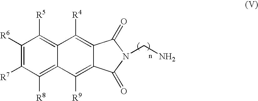
- the compound of the present invention can have the formula (V):
- the compound of the present invention can have the formula (VI):
- the compound of the present invention can have the formula (VII):
- the compound can have the formula (VIII):
- the compound in another embodiment, can be a peptide containing the formula (I):
- the compound can be a peptide containing the formula (II):
- R 1 , R 2 , and R 3 are each described above for formula (I).
- the compound can be a peptide containing the formula (III):
- Compounds of the present invention are useful as fluorescence probe molecules in applications wherein fluorescence probes are known to be useful.
- a compound of the present invention is added to a sample to be probed.
- the sample comprising the compound is then exposed to a light source.
- the light source produces light that is limited to a range of wavelengths.
- the range of wavelengths is between about 360 and about 410 nanometers (nm), preferably between about 370 and about 390 nm, most preferably between about 375 and about 380 nm.
- the compound of the present invention is fluorescent and emits fluorescent energy.
- the emitted fluorescent energy is detected using methods well known in the art.
- the intensity and wavelength of the emitted fluorescent energy provides information about the sample.
- the emitted fluorescent energy preferably has a range of wavelengths between about 480 and about 640 nm, preferably between about 560 and about 590 nm.
- the fluorescence of a molecule is defined by the quantum yield.
- the quantum yield is the ratio of the photons absorbed by the compound to the photons emitted through fluorescence by the compound.
- Compounds of the present invention have quantum yields that are preferably low in aqueous solutions and high in non-polar environments. Quantum yields range from about 0.001 and about 0.1, preferably between about 0.001 and about 0.005 for aqueous solutions, preferably between about 0.2 and about 0.7 for non-polar environment.
- Fluorescence can also be evaluated by determining the dipole moment change between the ground and excited state.
- the change in the dipole moment can be estimated from a plot of the Stokes shift vs. the orientation polarizability, known as a Lippert-Mataga plot.
- a Lippert-Mataga plot (Lippert, V. E. Z. Elektrochem. 1957, 61, 962-975; Mataga, N.; Kaifu, Y.; Koizumi, M. Bull. Chem. Soc. Jpn. 1956, 29, 465-470.)
- the relationship between the Stokes shift ( ⁇ v ) and the orientation polarizability ( ⁇ f) is expressed by the following equation:
- c the velocity of light
- h Plank's constant
- ⁇ the solvent dielectric constant
- n the solvent refraction index
- ⁇ e ⁇ g the difference between the dipole moments of the excited and the ground states respectively ( ⁇ )
- ⁇ 0 is the radius of the Onsager cavity around the compound.
- the magnitude of ⁇ correlates to the sensitivity of the fluorescent probe, where larger
- Fluorescence is sensitive to the pH of the surrounding environment.
- Compounds of the present invention are useful as fluorescence probes in the pH range from about 4 to about 8.
- Bio interactions play important roles in the sequence and mechanisms of action of various cellular processes and signal pathways.
- the time course, nature, and sequence of the different cellular processes can be elucidated by in situ observation using the compounds of the present invention.
- Specific inhibitors and/or activators of the cellular processes and signal pathways being studied may optionally be used in addition to compounds of the present invention.
- Bio interactions comprise the interaction of a compound or molecule with a target molecule.
- target molecules include peptides, proteins, enzymes, nucleic acids, ions, and other receptors; preferably metal ion chelators, proteases, polymerases, hydrolases, phosphatases, and kinases; more preferably protein domains, and protein domains of phosphatases and kinases.
- Proteins and protein-protein interactions play a central role in the various essential biochemical processes. For example, these interactions are evident in the interaction of hormones with their respective receptors, in the intracellular and extracellular signaling events mediated by proteins, in enzyme substrate interactions, in intracellular protein trafficking, in the formation of complex structures like ribosomes, viral coat proteins, and filaments, and in antigen-antibody interactions. These interactions are usually facilitated by the interaction of small regions within the proteins that can fold independently of the rest of the protein. These independent units are called protein domains. Abnormal or disease states can be the direct result of aberrant protein-protein interactions. Protein-protein interactions are also central to the mechanism of a virus recognizing its receptor on the cell surface as a prelude to infection. Identification of domains that interact with each other not only leads to a broader understanding of protein-protein interactions, but also aids in the design of inhibitors of these interactions.
- Phosphorylation-dependent peptide-protein interactions include phosphoserine peptides with 14-3-3, which is a protein involved in cell cycle control (Muslin, A. J., Tanner, J. W., Allen, P. M., Shaw, A. S. Cell 1996, 84, 889-897.), and phosphotyrosine peptides with SH2 domains.
- SH2 domains are binding modules that are involved in tyrosine kinase signaling networks and recognize phosphotyrosine-containing peptide sequences. The phosphotyrosine binding is complemented by simultaneous peptide-protein interactions on the protein surface.
- SH2 domains include Abl SH2, Crk SH2, and C-terminal P13K SH2 which can be expressed in bacteria as GST fusion proteins, which are referred to as GST-Abl SH2, GST-Crk SH2, and GST-PI3K SH2.
- Recognition sequences for SH2 domains comprise a phosphotyrosine residue and other amino acids.
- the recognition sequence is different for different SH2 domains.
- Amino acid recognition sequences for binding members of the SH2 domain family are disclosed in Songyang, Z. et al.; Cell 1993, 72, 767-778.
- the recognition sequence is pTyr-Asp-His-Pro.
- the recognition sequence is pTyr-Glu-Asn-Val.
- Compounds of the formula (I) are useful for studying the peptide-protein interactions on the protein surface of the SH2 domain.
- Compounds of formula (I) can be incorporated into peptides containing the desired SH2 recognition sequence.
- Table 1 shows peptides incorporating the Crk SH2 or Abl SH2 recognition sequences and Dap(6DMN) into the (+2) position relative to the phosphotyrosine residue.
- the peptides of Table 1 were incubated with targeted and nontargeted SH2 domains.
- the binding of peptides Crk-bp, Crk-bp2, Abl-bp, and Abl-bp2 to SH2 target domains can be studied by fluorescence titration as shown in FIGS. 1-4 .
- FIG. 1 ( a ) and ( b ) Fluorescence titrations of Crk-bp and Crk-bp2 with targeted GST-Crk SH2 are shown in FIG. 1 ( a ) and ( b ), respectively. Fluorescence titrations of Crk-bp and Crk-bp2 with non-targeted GST-Abl SH2 are shown in FIG. 4 ( a ) and ( b ), respectively.
- FIG. 1 ( c ) shows relative fluorescence emission intensities for Crk-bp and Crk-bp2 peptides, Crk-bp and Crk-bp2 bound to GST-Crk SH2 domain, and Crk-bp2 bound to non-target domains GST-Abl SH2 and GST-PKI3 SH2.
- the relative fluorescence emission intensities for non-target SH2 domains is notably less than for the targeted GST-Crk SH2 domain.
- Fluorescence titrations of Abl-bp with targeted GST-Abl SH2 is shown in FIG. 2 ( a ). Fluorescence titrations of Abl-bp with non-targeted GST-PI3K SH2 and GST-Abl SH2 are shown in FIG. 2 ( b ) and 4 ( c ), respectively. The relative fluorescence emission intensities for the Abl-bp peptide and aforementioned SH2 domain titrations are shown in FIG. 2 ( c ).
- FIG. 3 ( a ) Fluorescence titrations of Abl-bp2 with targeted GST-Abl SH2 is shown in FIG. 3 ( a ). Fluorescence titrations Abl-bp2 with non-targeted GST-PI3K SH2 and GST-Crk SH2 are shown in FIGS. 3 ( b ) and 4 ( d ), respectively. The relative fluorescence emission intensities for the Abl-bp2 peptide and aforementioned SH2 domain titrations are shown in FIG. 3 ( c ).
- Compounds of the present invention are also useful in a method of monitoring biological interactions, comprising providing a compound of the present invention, contacting a target molecule with the compound to form a biological sample, and monitoring the fluorescence of the biological sample.
- the compound is a peptide. More preferably, the compound is a peptide containing at least one of formula (I), (II), and (III).
- the monitoring step comprise contacting the compound with the one or more target molecules or different biochemical conditions, wherein the measuring step comprises exciting the compound of the present invention with light, and measuring the fluorescence.
- the concentration used will depend on the detection equipment. Typically, the concentration of the compound is from greater than about 0.1 nM.
- the compounds of formula (I) can be prepared according to the reaction as shown in Scheme 1, wherein, n, R 3 , R 4 , R 5 , R 6 , R 7 , R 8 and R 9 are as defined above.
- Anhydride (1) is stirred with an appropriate diamine (2) in a suitable solvent to form compound 3.
- the allyl protecting group of compound 3 can be removed under standard conditions known to those skilled in the art, examples of which are disclosed in Greene and Wuts, Protective Groups in Organic Synthesis” 2nd ed.; John Wiley & Sons, New York, 1991) to afford a compound of Formula I (4).
- the diamine compound 2 can be prepared from the corresponding diamino acid using standard conditions for protecting a carboxylic acid with an allyl protecting group known to those skilled in the art, examples of which are provided in Greene and Wuts.
- Anhydride 1 can be prepared by stirring diester 5 with an appropriate base in a suitable solvent.
- Compound 5 can be assembled by methods known to one skilled in the art such ring annulation methods or Diels-Alder reaction.
- the compounds of formula (IV) can be prepared by stirring anhydride (1) with an appropriate amine in a suitable solvent to form compounds of formula (IV), wherein, n, R 4 , R 5 , R 6 , R 7 , R 8 , R 9 and Y are as defined above.
- Y is NH 2
- the amine is diamino compound (6) as shown in Scheme 3 which is stirred with anhydride (1) to for compound (7).
- Diamines are commercially available from vendors such as Aldrich, for example.
- compound 7 may be prepared by stirring anhydride (1) with a diamine where one of the nitrogens is protected with a suitable N-protecting group. After formation of the imide bond, the N-protecting group can be removed to afford compound (7).
- Methods for introduction and removal of N-protecting groups are known to those skilled in the art, examples of which are disclosed in Greene and Wuts, Protective Groups in Organic Synthesis, 2nd ed.; John Wiley & Sons, New York, 1991
- Compound (10) where X is halogen or alkoxy can be prepared under standard conditions acyl halide ( Org. Syn. Coll. Vol. 1 1941, 12, 147; Org. Syn. Coll. Vol. 3 1955, 169, 490, 547, 555, 712; Org. Syn. Coll. Vol 4 1963, 154, 263, 339, 715, 739) or esterification ( Tetrahedron. 1980, 36, 2409) conditions.
- Peptides containing the formula (I) can be prepared using standard peptide coupling techniques known to those skilled in the art. Examples are provided in Bodansky, M. Peptide Chemistry a Practical Textbook, 2nd ed.; Springer-Verlag, Berlin, 1993. Dap(6DMN), can incorporated into peptides using Fmoc-Dap(6DMN) and standard Fmoc solid-phase peptide synthesis.
- the sensor of the present invention can be used in a method for detecting biological interactions.
- the method of the present invention comprises providing a peptide incorporating an amino acid of the formula (I), contacting a target molecule with the peptide to form a biological sample, and monitoring the fluorescence of the biological sample.
- Electrospray Ionization Mass Spectrometry was performed on a PerSeptive Biosystems MarinerTM Biospectrometry Workstation (Turbo Ion Source). Fluorescence spectroscopy measurements were made using a Fluoromax-P spectrofluorimeter controlled by the DataMax 2.20 software, and coupled to a NesLab RTE-111 water bath for temperature control. All measurements were made at 20° C.
- Peptide synthesis was carried using standard Fmoc-based solid phase peptide synthesis (SPPS) protocols on a 0.05 to 0.1 mmol scale using a 0.21 mmol/g loading PAL-PEG-PS solid support.
- Amino acids were coupled in three-fold excess using a mixture of 0.2 M HBTU/0.2 M HOBt in DMF as activating agents. Each amino acid was activated for two minutes with the HBTU/HOBt mixture (1 eq.) and diisopropylethylamine (DIPEA), 0.195 M in DMF (1.5 eq.) before being added to the resin.
- Peptide coupling was monitored using the 2,4,6-trinitrobenzenesulphonic acid (TNBS) test.
- TNBS 2,4,6-trinitrobenzenesulphonic acid
- Fmoc-amino acids were used as protected Fmoc-amino acids with the standard side chain protecting groups: Fmoc-Ala-OH, Fmoc-Arg(Pbf)-OH, Fmoc-Asp(OtBu)-OH, Fmoc-Glu(OtBu)-OH, Fmoc-lle-OH, Fmoc-Ser(tBu)-OH, Fmoc-Thr(tBu)-OH, Fmoc-Tyr(tBu)-OH, Fmoc-Val-OH and Fmoc-Dap(6DMN).
- Phosphotyrosine was introduced as the monobenzyl ester Fmoc-Tyr(PO(OBzl)OH)-OH.
- High-performance liquid chromatography HPLC was performed using a Waters 600E HPLC fitted with a Waters 600 automated control module and a Waters 2487 dual wavelength absorbance detector recording at 228 and 280 nm.
- analytical HPLC a Beckman Ultrasphere C 18 , 5 ⁇ m, 4.6 ⁇ 150 mm reverse-phase column was used.
- For preparative separations a YMC-pack, C 18 , 250 ⁇ 20 mm reversed phase column was used.
- the standard gradient for analytical and preparative HPLC used was 93:7 to 5:95 over 35 minutes (water:acetonitrile, 0.1% TFA).
- the 6DMN side chain proved resistant to the standard mildly basic amino acid coupling conditions (0.12 M diisopropylethylamine), the Fmoc deprotection conditions (20% piperidine), and the acidic resin cleavage and deprotection cocktail (95% TFA).
- ⁇ x ⁇ std ⁇ ( grad x grad std ) ⁇ ( ⁇ x 2 ⁇ std 2 ) ( 1 )
- ⁇ is the fluorescence quantum yield
- grad is the gradient (grad) from the plot of integrated fluorescence intensity vs. absorbance
- ⁇ the refractive index of the solvent.
- the UV absorbance spectra (40 ⁇ M in methanol) and fluorescence emission spectra of compound (VIII), 6DMN-GlyOMe are shown in FIG. 6 and tabulated in Table 4.
- the UV spectrum of 6DMN-GlyOMe has an intense absorption band at 378 nm which is not found in unsubstituted 2,3-naphthalimides.
- the maximum excitation wavelength of 6DMN-GlyOMe is at 375 nm.
- the data were fitted to a linear equation with gradient (grad), and intercept 0.
- the gradient (grad) is proportional to the quantum yield of the sample. Absolute values were calculated using the standard sample, which has a fixed and known fluorescence quantum yield value, according to the equation above.
- ⁇ v The difference ( ⁇ v ) in the maximum absorption ( v A ) and emission wavelengths ( v F ), known as the Stokes shift and expressed in wavenumbers, is plotted vs. ⁇ f.
- ⁇ f The term ⁇ f is called the orientation polarizability and can be calculated according to the following equation:
- FIG. 7( a ) shows the plot of ( ⁇ v ) for compound (VIII) vs. Af for a series of 1,4-dioxane/acetonitrile mixtures.
- the Onsager radius for compound VIII was calculated from the optimized structure obtained with a DFT minimization using the Gaussian program (Pople, J. A. et al. Gaussian 98 (Gaussian, Inc., Pittsburgh, Pa., 1998), (B3LYP functional using 6-31G(d) orbital base).
- the Onsager radius (4.19 ⁇ ) was taken as half of the average distance between nitrogen of the amine donor and the two carbonyl oxygens, which corresponds to the longest distance across the molecule where charge separation can take place. (Mukherjee, S., Chattopdhyay, A., Samanta, A., Soujanya, T. J. Phys. Chem.
- Peptide stock solutions of known concentration were used to prepare 5 ⁇ M peptide concentration samples in buffered water (150 ⁇ L total volume, 100 mM NaCl, 10 mM sodium phosphate, pH 7.5).
- 2 ⁇ L aliquots of the corresponding stock protein solution (GST-Crk SH2, GST-Abl SH2 or GST-PI3K SH2, concentration determined by Bio-Rad Protein Assay) were consecutively added, and fluorescence emission spectra were recorded 1 minute after each addition (excitation 395 nm, 2 nm excitation and emission slit width). The increase in emission intensity at the maximum emission wavelength was plotted against total protein concentration and fitted by least-square analysis assuming a 1 to 1 binding mode to obtain the apparent Kd.
- the sample was recentrifuged to remove particulate debris (5000 g for 30 min at 4° C.). Then the supernatant was incubated for at least 1 hour with 10 mL of glutathione Sepharose 4 Fast Flow (Amersham Biosciences). The column was equilibrated with 5 volumes of binding buffer at 4° C. The sample was the loaded using a syringe and low flow rate (0.2 to 0.5 mL/min) at 4° C. The column was then washed with 5 volumes of binding buffer (higher rate of 1 mL/min) room temperature. The GST fusion protein was eluted with elution buffer at room temperature.
- Binding buffer PBS pH 7.4 (140 mM NaCl, 2.7 mM KCl, 10 mM Na 2 HPO 4 , 1.8 mM NaHPO 4 ).
- Elution buffer 50 mM Tris-HCl, 10 mM reduced glutathione, pH 8.
- the gel was transferred to a nitrocellulose membrane (1 h, 100 V), and probed using the standard procedure: blocked overnight with TBST+powder milk; washed with TBST (2 ⁇ 10 mL ⁇ 10 min.); incubated with primary antibody (anti-GST, dilution 1:10,000 in TBST) for 30 min; washed with TBST (3 ⁇ 10 mL ⁇ 10 min.); incubated for 30 min with secondary antibody (anti-goat conjugated with alkaline phosphatase, 1:1,000 dilution in TBS); washed with TBST (2 ⁇ 10 mL ⁇ 10 min.); washed with TBS (1 ⁇ 10 mL ⁇ 10 min.); One Step NBT/BCIP added and incubated until the bands appeared; then quenched the reaction with ddH 2 O.
- Bio-Rad Protein Assay was used to quantify the isolated following the standard procedure.
- Dye reagent was prepared by diluting 1 part Dye Reagent Concentrate with 4 parts distilled water.
- Stock BSA dilutions were prepared corresponding to 0.1, 0.2, 0.3, 0.4, 0.5 0.6, 0.7, 0.8 and 0.9 mg/mL as follows. 20 ⁇ L of each stock dilution were added to 980 ⁇ L dye reagent and incubated at room temperature for 5 min. The absorbance was measured at 595 nm using a blank of 20 ⁇ L of distilled water and 980 ⁇ L dye reagent prepared at the same time as the samples. The observed absorbance was plotted against BSA concentration.
- Abl-SH2 was received as a GST fusion in a pGEX-KT vector, amp-resistant, IPTG-inducible.
- the SH2 domain was cloned using BamH1 (5′) EcoR1 (3′) sites.
- Src and PI3K SH2 domains were expressed and purified as GST fusion following reported procedures. (Smith, D. B.; Johnson, K. S. Gene, 1988, 67, 31-40) In short, plasmids including GST-fusion proteins were transformed into bacteria ( E. coli DH5 ⁇ chemically competent cells, Invitrogen). Bacteria were grown to mid log phase, induced at 37° C.
- Proteins were eluted from the beads (50 mM Tris-HCl, 10 mM reduced glutathione, pH 8), and finally concentrated by centrifugation through a cellulose membrane with a 10,000 Da cutoff (Millipore). Proteins were quantified using the Micro BCA protein Assay Kit (Pierce) relative to a BSA standard. All fusion proteins were analyzed by Coomasie staining and Western analysis with anti-GST antibodies.
Abstract

and methods monitoring protein-protein interactions.
Description

where n, R3, R4, R5, R6, R7, R8 and R9 are as defined below. One especially preferred compound (III) referred to as Dap(6DMN) is disclosed.

where formula (AA) is attached via a free valence on either M1 or M2; M1 represents O, NRe, or S(O)l; M2 represents CRfRg, O, S(O)l, or NRe; l is 0, 1 or 2; j is 1, 2 or 3 and k is 1, 2 or 3, with the proviso that j+k is 3, 4, or 5; and Ra, Rb, Rc, Rd, Re, Rf, and Rg are independently selected from the group consisting of hydrogen, halogen, unsubstituted or substituted C1-8 alkyl, unsubstituted or substituted C2-8 alkenyl, unsubstituted or substituted C2-8 alkynyl, —CORh, —CO2Rh, —CONRhRi, —NRhCORi, —SO2Rh, —SO2NRhRi, —NSO2RhRi, —NRhRi, —ORh, -Q1CORh, -Q1CO2Rh, -Q1CONRhRi, -Q1NRhCORi, -Q1SO2R28, -Q1SO2NRhRi, -Q1NSO2RhRi, -Q1NRhRi, -Q1ORh, wherein Q1 is a member selected from the group consisting of C1-4 alkylene, C2-4 alkenylene and C2-4 alkynylene, and Rh and Ri are independently selected from the group consisting of hydrogen and C1-8 alkyl, and wherein the aliphatic portions of each of the Ra, Rb, Rc, Rd, Re, Rf, Rg, Rh and Ri substituents are optionally substituted with from one to three members selected from the group consisting of halogen, —OH, —ORn, —OC(O)NHRn, —OC(O)NRnRo, —SH, —SRn, —S(O)Rn, —S(O)2Rn, —SO2NH2, —S(O)2NHRn, —S(O)2NRnRo, —NHS(O)2Rn, —NRnS(O)2Ro, —C(O)NH2, —C(O)NHRn, —C(O)NRnRo, —C(O)Rn, —NHC(O)Ro, —NRnC(O)Ro, —NHC(O)NH2, —NRnC(O)NH2, —NRnC(O)NHRo, —NHC(O)NHRn, —NRnC(O)NRoRp, —NHC(O)NRnRo, —CO2H, —CO2Rn, —NHCO2Rn, —NRnCO2Ro, —CN, —NO2, —NH2, —NHRn, —NRnRo, —NRnS(O)NH2 and —NRnS(O)2NHRo, wherein Rn, Ro and Rp are independently an unsubstituted C1-8 alkyl. Additionally, any two of Ra, Rb, Rc, Rd, Re, Rf and Rg may be combined to form a bridged or spirocyclic ring system.

where R4, R5, R6, R7, R8, and R9 are each independently hydrogen, fluorine, or alkyl, with the proviso that one of R6 or R7 is —NR1R2, —OR1, or —SR1;
-
- R1 and R2 are each independently hydrogen or alkyl; or
- R1 and R2 together with the nitrogen to which they are attached, may form a substituted or unsubstituted 5- or 6-membered ring; wherein said nitrogen is not conjugated with said 5- or 6-membered ring;
- R3 is hydrogen or a N-protecting group;
- n is 1, 2, 3 or 4.

-
- where R1, R2, and R3 are described above for formula (I).

-
- where n, R4, R5, R6, R7, R8, and R9 are described above for formula (I);
- Y is NH2 or C(O)X; and
- X is halogen, hydroxy or alkoxy.

-
- where n, R4, R5, R6, R7, R8, and R9 are described above for formula (I) and X is described in formula (IV).

-
- where n, R4, R5, R6, R7, R8, and R9 are described above for formula (I).


where n, R1, R2, R3, R4, R5, R6, R7, R8, and R9 are described above for formula (I).

where R1, R2, and R3 are each described above for formula (I).

where R3 is described above for formula (I). When R3 is hydrogen, compound (III) is referred to herein as Dap(6DMN). When R3 is Fmoc, compound (III) is herein referred to as Fmoc-Dap(6DMN).
Uses of Compounds
where c is the velocity of light, h is Plank's constant, ε is the solvent dielectric constant, n is the solvent refraction index, μe−μg is the difference between the dipole moments of the excited and the ground states respectively (Δμ), and α0 is the radius of the Onsager cavity around the compound. The magnitude of Δμ correlates to the sensitivity of the fluorescent probe, where larger changes in the dipole moment correlate to greater sensitivity.
| TABLE 1 |
| Peptide Sequences and Corresponding SH2 Domain Target |
| Target | ||
| Peptide | SH2 | Peptide sequence |
| Crk-bp | Crk | Ac-Glu-Dap(6DMN)-Gln-pTyr-Asp-His-Pro-Asn- |
| Ile-(CONH2) (SEQ ID NO: 1) | ||
| Crk-bp2 | Crk | Ac-Glu-Dap(6DMN)-Gly-pTyr-Asp-His-Pro-Asn- |
| Ile-(CONH2) (SEQ ID NO: 2) | ||
| Abl-bp | Abl | Ac-Glu-Dap(6DMN)-Gly-pTyr-Glu-Asn-Val-Gln- |
| Ser-(CONH2) (SEQ ID NO: 3) | ||
| Abl-bp2 | Abl | Ac-Glu-Dap(6DMN)-pTyr-Glu-Asn-Val-Gln-Ser- |
| (CONH2) (SEQ ID NO: 4) | ||
| TABLE 2 |
| Kinetic and Fluorescence Properties |
| of Peptides containing Dap(6DMN) |
| Target | Fluorescence | |||
| Peptide | SH2 | Peptide Sequence | Kd(μM) | increase |
| Crk-bp | Crk | Ac-Glu-Dap(6DMN)-Gln- | 4.8 ± 2a | 500%b |
| pTyr-Asp-His-Pro-Asn- | ||||
| Ile-(CONH2) (SEQ ID | ||||
| NO: 1) | ||||
| Crk-bp2 | Crk | Ac-Gly-Dap(6DMN)-Gln- | 2.4 ± 2a | 1100%b |
| pTyr-Asp-His-Pro-Asn- | ||||
| Ile-(CONH2) (SEQ ID | ||||
| NO: 5) | ||||
| Abl-bp | Abl | Ac-Gly-Dap(6DMN)-Gln- | 13 ± 2a | 400%c |
| pTyr-Glu-Asn-Val-Gln- | ||||
| Ser-(CONH2) (SEQ ID | ||||
| NO: 6) | ||||
| Abl-bp2 | Abl | Ac-Dap(6DMN)-Gln-pTyr- | 12 ± 2a | — |
| Glu-Asn-Val-Gln-Ser- | ||||
| (CONH2) (SEQ OD NO: 7) | ||||
| aAssay conditions: 20 μM in PBS buffer, pH 7.5. | ||||
| bExcitation wavelength: 375 nm, Emission wavelength, 565 nm. | ||||
| cExcitation wavelength: 375 nm, Emission wavelength, 599 nm. | ||||


| TABLE 4 |
| Photophysical Properties of 6DMN-GlyOMe |
| in Different Solvents: |
| absorbance | emission | quantum yield | |
| solvent | maximum (nm) | maximum (nm) | (Φ)b |
| watera | 388 | 592 | 0.002 |
| methanol | 382 | 589 | 0.012 |
| 2-propanol | 385 | 589 | 0.018 |
| ethanol | 379 | 584 | 0.027 |
| acetonitrile | 380 | 549 | 0.135 |
| DMF | 380 | 545 | 0.155 |
| acetone | 375 | 532 | 0.148 |
| tetrahydrofuran | 373 | 510 | 0.147 |
| 1,4-dioxane | 372 | 498 | 0.220 |
| dichloromethane | 379 | 517 | 0.210 |
| chloroform | 380 | 509 | 0.225 |
| toluene | 373 | 491 | 0.208 |
| aBecause of solubility limitations, the quantum yield in water was calculated using peptide Abl-bp. | |||
| bQuantum yields (Φ) were measured in comparison to quinine sulfate in 0.1 M H2SO4 (Φ = 0.54) as the standard. | |||
where ε is the solvent dielectric constant and n is the solvent refraction index. The plot of (Δ
ε=(χacetonitrile)(38.8)+(χdiox)(2.218) (3)
n=(χacetonitrile)(1.3442)+(χdiox)(1.4224) (4)
where χacetonitrile+χdiox=1.
where c is the velocity of light, h is Plank's constant, μe−μg is the difference between the dipole moments of the excited and the ground states respectively (Δμ), and α0 is the radius of the Onsager cavity around the compound. The Onsager radius for compound VIII was calculated from the optimized structure obtained with a DFT minimization using the Gaussian program (Pople, J. A. et al. Gaussian 98 (Gaussian, Inc., Pittsburgh, Pa., 1998), (B3LYP functional using 6-31G(d) orbital base). The Onsager radius (4.19 Å) was taken as half of the average distance between nitrogen of the amine donor and the two carbonyl oxygens, which corresponds to the longest distance across the molecule where charge separation can take place. (Mukherjee, S., Chattopdhyay, A., Samanta, A., Soujanya, T. J. Phys. Chem. 1994, 98, 2809-2812.) As can be seen in
Claims (4)


Priority Applications (2)
| Application Number | Priority Date | Filing Date | Title |
|---|---|---|---|
| US11/106,349 US7442529B2 (en) | 2005-04-13 | 2005-04-13 | Fluorescent probes for biological studies |
| US12/235,454 US20090082577A1 (en) | 2005-04-13 | 2008-09-22 | Fluorescent probes for biological studies |
Applications Claiming Priority (1)
| Application Number | Priority Date | Filing Date | Title |
|---|---|---|---|
| US11/106,349 US7442529B2 (en) | 2005-04-13 | 2005-04-13 | Fluorescent probes for biological studies |
Related Child Applications (1)
| Application Number | Title | Priority Date | Filing Date |
|---|---|---|---|
| US12/235,454 Division US20090082577A1 (en) | 2005-04-13 | 2008-09-22 | Fluorescent probes for biological studies |
Publications (2)
| Publication Number | Publication Date |
|---|---|
| US20060234206A1 US20060234206A1 (en) | 2006-10-19 |
| US7442529B2 true US7442529B2 (en) | 2008-10-28 |
Family
ID=37108913
Family Applications (2)
| Application Number | Title | Priority Date | Filing Date |
|---|---|---|---|
| US11/106,349 Active 2025-09-02 US7442529B2 (en) | 2005-04-13 | 2005-04-13 | Fluorescent probes for biological studies |
| US12/235,454 Abandoned US20090082577A1 (en) | 2005-04-13 | 2008-09-22 | Fluorescent probes for biological studies |
Family Applications After (1)
| Application Number | Title | Priority Date | Filing Date |
|---|---|---|---|
| US12/235,454 Abandoned US20090082577A1 (en) | 2005-04-13 | 2008-09-22 | Fluorescent probes for biological studies |
Country Status (1)
| Country | Link |
|---|---|
| US (2) | US7442529B2 (en) |
Cited By (4)
| Publication number | Priority date | Publication date | Assignee | Title |
|---|---|---|---|---|
| US20100168428A1 (en) * | 2007-02-26 | 2010-07-01 | Massachusetts Institute Of Technology | Environmentally sensitive fluorophores |
| US20110053180A1 (en) * | 2009-08-31 | 2011-03-03 | Massachusetts Institute Of Technology | Kinase sensors |
| US8586570B2 (en) | 2006-08-28 | 2013-11-19 | Massachusetts Institute Of Technology | Sox-based kinase sensor |
| US10799603B2 (en) | 2018-03-12 | 2020-10-13 | Massachusetts Institute Of Technology | Kinase and/or phosphatase sensing via hydroxyquinoline-sensitized chelates |
Families Citing this family (2)
| Publication number | Priority date | Publication date | Assignee | Title |
|---|---|---|---|---|
| US20100248975A1 (en) * | 2006-12-29 | 2010-09-30 | Gunjan Tiwari | Fluorogenic peptide substrate arrays for highly multiplexed, real-time monitoring of kinase activities |
| US20080206885A1 (en) * | 2007-02-26 | 2008-08-28 | Massachusetts Institute Of Technology | Environmentally sensitive fluorophores |
Citations (1)
| Publication number | Priority date | Publication date | Assignee | Title |
|---|---|---|---|---|
| US5854275A (en) * | 1996-05-16 | 1998-12-29 | Pfizer Inc. | Cyclic imide derivatives |
-
2005
- 2005-04-13 US US11/106,349 patent/US7442529B2/en active Active
-
2008
- 2008-09-22 US US12/235,454 patent/US20090082577A1/en not_active Abandoned
Patent Citations (1)
| Publication number | Priority date | Publication date | Assignee | Title |
|---|---|---|---|---|
| US5854275A (en) * | 1996-05-16 | 1998-12-29 | Pfizer Inc. | Cyclic imide derivatives |
Non-Patent Citations (13)
Cited By (6)
| Publication number | Priority date | Publication date | Assignee | Title |
|---|---|---|---|---|
| US8586570B2 (en) | 2006-08-28 | 2013-11-19 | Massachusetts Institute Of Technology | Sox-based kinase sensor |
| US20100168428A1 (en) * | 2007-02-26 | 2010-07-01 | Massachusetts Institute Of Technology | Environmentally sensitive fluorophores |
| US8440835B2 (en) | 2007-02-26 | 2013-05-14 | Massachusetts Institute Of Technology | Environmentally sensitive fluorophores |
| US20110053180A1 (en) * | 2009-08-31 | 2011-03-03 | Massachusetts Institute Of Technology | Kinase sensors |
| US8409820B2 (en) | 2009-08-31 | 2013-04-02 | Massachusetts Institute Of Technology | Kinase sensors |
| US10799603B2 (en) | 2018-03-12 | 2020-10-13 | Massachusetts Institute Of Technology | Kinase and/or phosphatase sensing via hydroxyquinoline-sensitized chelates |
Also Published As
| Publication number | Publication date |
|---|---|
| US20060234206A1 (en) | 2006-10-19 |
| US20090082577A1 (en) | 2009-03-26 |
Similar Documents
| Publication | Publication Date | Title |
|---|---|---|
| Zheng et al. | Rational design of fluorogenic and spontaneously blinking labels for super-resolution imaging | |
| JP7159233B2 (en) | Quinone Masked Probes as Labeling Reagents for Measuring Uptake by Cells | |
| US9006437B2 (en) | Xanthene dyes comprising a sulfonamide group | |
| Gonçalves | Fluorescent labeling of biomolecules with organic probes | |
| ES2211650T3 (en) | IMIDAZOQUINOLINE COMPOUNDS MARKED WITH COLOR. | |
| US7442529B2 (en) | Fluorescent probes for biological studies | |
| JP6862368B2 (en) | Use as an inhibitor of thienopyrol compounds and their Oplophorus-derived luciferase | |
| US8440835B2 (en) | Environmentally sensitive fluorophores | |
| US20120115128A1 (en) | Selective protein labeling | |
| US8835641B2 (en) | Fluorescent markers and use thereof for labeling specific protein targets | |
| US9701667B2 (en) | Coumarin-based fluorogenic agents and uses thereof for specific protein labelling | |
| US8409820B2 (en) | Kinase sensors | |
| US20080206885A1 (en) | Environmentally sensitive fluorophores | |
| CA3091814A1 (en) | Dyes for analysis of soluble protein aggregates or misfolded protein oligomers | |
| JP2604993B2 (en) | New biotinylation reagent | |
| Zhang et al. | Synthesis and application of the blue fluorescent amino acid l-4-cyanotryptophan to assess peptide–membrane interactions | |
| Dell’Acqua et al. | MediaChrom: discovering a class of pyrimidoindolone-based polarity-sensitive dyes | |
| US20230257587A1 (en) | Fluorenyl cyanine dyes | |
| Clavé et al. | A universal and ready-to-use heterotrifunctional cross-linking reagent for facile synthetic access to sophisticated bioconjugates | |
| EP3649122A1 (en) | Photo-switchable chemical inducers of dimerization for control of protein function in cells by light | |
| US8715529B1 (en) | Synthesis and applications of triazaborolopyridinium compounds and substituted triazaborolopyridinium compounds and methods of use | |
| US10941435B2 (en) | Methods and compositions for detecting or measuring caspases or apoptosis | |
| KR101125058B1 (en) | Compound for labeling material, intermediate therefor and process for producing the same | |
| JP6670502B2 (en) | Development of ligand screening system for neurotransmitter receptor | |
| Wurnig et al. | Development of the first geldanamycin-based HSP90 degraders |
Legal Events
| Date | Code | Title | Description |
|---|---|---|---|
| AS | Assignment |
Owner name: MASSACHUSETTS INSTITUTE OF TECHNOLOGY, MASSACHUSET Free format text: ASSIGNMENT OF ASSIGNORS INTEREST;ASSIGNOR:VAZQUEZ, EUGENIO;REEL/FRAME:016822/0497 Effective date: 20050416 Owner name: MASSACHUSETTS INSTITUTE OF TECHNOLOGY, MASSACHUSET Free format text: ASSIGNMENT OF ASSIGNORS INTEREST;ASSIGNOR:IMPERIALI, BARBARA;REEL/FRAME:016822/0511 Effective date: 20050420 |
|
| STCF | Information on status: patent grant |
Free format text: PATENTED CASE |
|
| FEPP | Fee payment procedure |
Free format text: PAYOR NUMBER ASSIGNED (ORIGINAL EVENT CODE: ASPN); ENTITY STATUS OF PATENT OWNER: SMALL ENTITY |
|
| FPAY | Fee payment |
Year of fee payment: 4 |
|
| FEPP | Fee payment procedure |
Free format text: PATENT HOLDER CLAIMS MICRO ENTITY STATUS, ENTITY STATUS SET TO MICRO (ORIGINAL EVENT CODE: STOM); ENTITY STATUS OF PATENT OWNER: SMALL ENTITY |
|
| FPAY | Fee payment |
Year of fee payment: 8 |
|
| FEPP | Fee payment procedure |
Free format text: ENTITY STATUS SET TO SMALL (ORIGINAL EVENT CODE: SMAL); ENTITY STATUS OF PATENT OWNER: SMALL ENTITY |
|
| MAFP | Maintenance fee payment |
Free format text: PAYMENT OF MAINTENANCE FEE, 12TH YR, SMALL ENTITY (ORIGINAL EVENT CODE: M2553); ENTITY STATUS OF PATENT OWNER: SMALL ENTITY Year of fee payment: 12 |