US7214487B2 - Methods for identifying compounds that modulate enzymatic activities by employing covalently bonded target-extender complexes with ligand candidates - Google Patents
Methods for identifying compounds that modulate enzymatic activities by employing covalently bonded target-extender complexes with ligand candidates Download PDFInfo
- Publication number
- US7214487B2 US7214487B2 US10/374,499 US37449903A US7214487B2 US 7214487 B2 US7214487 B2 US 7214487B2 US 37449903 A US37449903 A US 37449903A US 7214487 B2 US7214487 B2 US 7214487B2
- Authority
- US
- United States
- Prior art keywords
- ptp
- extender
- functionality
- mmol
- covalent bond
- Prior art date
- Legal status (The legal status is an assumption and is not a legal conclusion. Google has not performed a legal analysis and makes no representation as to the accuracy of the status listed.)
- Expired - Fee Related, expires
Links
- 239000004606 Fillers/Extenders Substances 0.000 title claims abstract description 136
- 238000000034 method Methods 0.000 title claims abstract description 103
- 239000003446 ligand Substances 0.000 title claims abstract description 98
- 150000001875 compounds Chemical class 0.000 title abstract description 86
- 230000002255 enzymatic effect Effects 0.000 title abstract description 3
- 150000003573 thiols Chemical class 0.000 claims description 82
- 235000018417 cysteine Nutrition 0.000 claims description 47
- DCWXELXMIBXGTH-UHFFFAOYSA-N phosphotyrosine Chemical compound OC(=O)C(N)CC1=CC=C(OP(O)(O)=O)C=C1 DCWXELXMIBXGTH-UHFFFAOYSA-N 0.000 claims description 46
- XUJNEKJLAYXESH-UHFFFAOYSA-N cysteine Natural products SCC(N)C(O)=O XUJNEKJLAYXESH-UHFFFAOYSA-N 0.000 claims description 44
- 108090000623 proteins and genes Proteins 0.000 claims description 38
- 102000004169 proteins and genes Human genes 0.000 claims description 34
- 235000018102 proteins Nutrition 0.000 claims description 23
- BWGNESOTFCXPMA-UHFFFAOYSA-N Dihydrogen disulfide Chemical compound SS BWGNESOTFCXPMA-UHFFFAOYSA-N 0.000 claims description 22
- 235000001014 amino acid Nutrition 0.000 claims description 21
- 239000002253 acid Substances 0.000 claims description 18
- 150000001413 amino acids Chemical class 0.000 claims description 18
- 239000003638 chemical reducing agent Substances 0.000 claims description 16
- RWSXRVCMGQZWBV-WDSKDSINSA-N glutathione Chemical compound OC(=O)[C@@H](N)CCC(=O)N[C@@H](CS)C(=O)NCC(O)=O RWSXRVCMGQZWBV-WDSKDSINSA-N 0.000 claims description 16
- DGVVWUTYPXICAM-UHFFFAOYSA-N β‐Mercaptoethanol Chemical compound OCCS DGVVWUTYPXICAM-UHFFFAOYSA-N 0.000 claims description 13
- 102000002727 Protein Tyrosine Phosphatase Human genes 0.000 claims description 12
- 108020000494 protein-tyrosine phosphatase Proteins 0.000 claims description 12
- 230000002441 reversible effect Effects 0.000 claims description 9
- 108010024636 Glutathione Proteins 0.000 claims description 8
- XYFCBTPGUUZFHI-UHFFFAOYSA-N Phosphine Chemical compound P XYFCBTPGUUZFHI-UHFFFAOYSA-N 0.000 claims description 8
- 229960003180 glutathione Drugs 0.000 claims description 8
- 239000012279 sodium borohydride Substances 0.000 claims description 8
- 229910000033 sodium borohydride Inorganic materials 0.000 claims description 8
- 229960003151 mercaptamine Drugs 0.000 claims description 7
- 238000002703 mutagenesis Methods 0.000 claims description 7
- 231100000350 mutagenesis Toxicity 0.000 claims description 7
- 230000002427 irreversible effect Effects 0.000 claims description 6
- XUJNEKJLAYXESH-REOHCLBHSA-N L-Cysteine Chemical compound SC[C@H](N)C(O)=O XUJNEKJLAYXESH-REOHCLBHSA-N 0.000 claims description 5
- VHJLVAABSRFDPM-QWWZWVQMSA-N dithiothreitol Chemical compound SC[C@@H](O)[C@H](O)CS VHJLVAABSRFDPM-QWWZWVQMSA-N 0.000 claims description 5
- VHJLVAABSRFDPM-ZXZARUISSA-N dithioerythritol Chemical compound SC[C@H](O)[C@H](O)CS VHJLVAABSRFDPM-ZXZARUISSA-N 0.000 claims description 4
- 150000004820 halides Chemical group 0.000 claims description 4
- 229910000073 phosphorus hydride Inorganic materials 0.000 claims description 4
- 150000001649 bromium compounds Chemical group 0.000 claims description 2
- UFULAYFCSOUIOV-UHFFFAOYSA-N cysteamine Chemical compound NCCS UFULAYFCSOUIOV-UHFFFAOYSA-N 0.000 claims 2
- YMWUJEATGCHHMB-UHFFFAOYSA-N Dichloromethane Chemical compound ClCCl YMWUJEATGCHHMB-UHFFFAOYSA-N 0.000 description 116
- XEKOWRVHYACXOJ-UHFFFAOYSA-N Ethyl acetate Chemical compound CCOC(C)=O XEKOWRVHYACXOJ-UHFFFAOYSA-N 0.000 description 104
- 102100028516 Receptor-type tyrosine-protein phosphatase U Human genes 0.000 description 82
- 229920001469 poly(aryloxy)thionylphosphazene Polymers 0.000 description 79
- 238000006243 chemical reaction Methods 0.000 description 74
- OKKJLVBELUTLKV-UHFFFAOYSA-N Methanol Chemical compound OC OKKJLVBELUTLKV-UHFFFAOYSA-N 0.000 description 60
- 235000019439 ethyl acetate Nutrition 0.000 description 51
- VLKZOEOYAKHREP-UHFFFAOYSA-N n-Hexane Chemical compound CCCCCC VLKZOEOYAKHREP-UHFFFAOYSA-N 0.000 description 45
- 230000015572 biosynthetic process Effects 0.000 description 44
- ZMXDDKWLCZADIW-UHFFFAOYSA-N N,N-Dimethylformamide Chemical compound CN(C)C=O ZMXDDKWLCZADIW-UHFFFAOYSA-N 0.000 description 42
- PMZURENOXWZQFD-UHFFFAOYSA-L Sodium Sulfate Chemical compound [Na+].[Na+].[O-]S([O-])(=O)=O PMZURENOXWZQFD-UHFFFAOYSA-L 0.000 description 38
- DTQVDTLACAAQTR-UHFFFAOYSA-N Trifluoroacetic acid Chemical compound OC(=O)C(F)(F)F DTQVDTLACAAQTR-UHFFFAOYSA-N 0.000 description 38
- 229910052938 sodium sulfate Inorganic materials 0.000 description 38
- 239000002904 solvent Substances 0.000 description 38
- 239000012267 brine Substances 0.000 description 37
- 239000012038 nucleophile Substances 0.000 description 37
- HPALAKNZSZLMCH-UHFFFAOYSA-M sodium;chloride;hydrate Chemical compound O.[Na+].[Cl-] HPALAKNZSZLMCH-UHFFFAOYSA-M 0.000 description 37
- 101710128896 Tyrosine-protein phosphatase non-receptor type 1 Proteins 0.000 description 36
- 102100033001 Tyrosine-protein phosphatase non-receptor type 1 Human genes 0.000 description 36
- WBHQBSYUUJJSRZ-UHFFFAOYSA-M sodium bisulfate Chemical compound [Na+].OS([O-])(=O)=O WBHQBSYUUJJSRZ-UHFFFAOYSA-M 0.000 description 32
- 235000011152 sodium sulphate Nutrition 0.000 description 32
- 229910000342 sodium bisulfate Inorganic materials 0.000 description 31
- 239000011734 sodium Substances 0.000 description 30
- 238000003786 synthesis reaction Methods 0.000 description 27
- 230000002829 reductive effect Effects 0.000 description 26
- XLYOFNOQVPJJNP-UHFFFAOYSA-N water Substances O XLYOFNOQVPJJNP-UHFFFAOYSA-N 0.000 description 25
- WYURNTSHIVDZCO-UHFFFAOYSA-N Tetrahydrofuran Chemical compound C1CCOC1 WYURNTSHIVDZCO-UHFFFAOYSA-N 0.000 description 24
- 239000000243 solution Substances 0.000 description 23
- 239000005457 ice water Substances 0.000 description 22
- ZMANZCXQSJIPKH-UHFFFAOYSA-N Triethylamine Chemical compound CCN(CC)CC ZMANZCXQSJIPKH-UHFFFAOYSA-N 0.000 description 21
- JGFZNNIVVJXRND-UHFFFAOYSA-N N,N-Diisopropylethylamine (DIPEA) Chemical compound CCN(C(C)C)C(C)C JGFZNNIVVJXRND-UHFFFAOYSA-N 0.000 description 20
- IJGRMHOSHXDMSA-UHFFFAOYSA-N Atomic nitrogen Chemical compound N#N IJGRMHOSHXDMSA-UHFFFAOYSA-N 0.000 description 18
- PEDCQBHIVMGVHV-UHFFFAOYSA-N Glycerine Chemical compound OCC(O)CO PEDCQBHIVMGVHV-UHFFFAOYSA-N 0.000 description 18
- UIIMBOGNXHQVGW-DEQYMQKBSA-M Sodium bicarbonate-14C Chemical compound [Na+].O[14C]([O-])=O UIIMBOGNXHQVGW-DEQYMQKBSA-M 0.000 description 18
- 239000007787 solid Substances 0.000 description 18
- 238000003818 flash chromatography Methods 0.000 description 17
- 239000000047 product Substances 0.000 description 17
- 229940024606 amino acid Drugs 0.000 description 16
- 0 *C.*C.CC(=O)C1CO1.CCC1CO1 Chemical compound *C.*C.CC(=O)C1CO1.CCC1CO1 0.000 description 15
- VYPSYNLAJGMNEJ-UHFFFAOYSA-N Silicium dioxide Chemical compound O=[Si]=O VYPSYNLAJGMNEJ-UHFFFAOYSA-N 0.000 description 15
- HEMHJVSKTPXQMS-UHFFFAOYSA-M Sodium hydroxide Chemical compound [OH-].[Na+] HEMHJVSKTPXQMS-UHFFFAOYSA-M 0.000 description 15
- 125000003118 aryl group Chemical group 0.000 description 15
- 108020004414 DNA Proteins 0.000 description 14
- IAZDPXIOMUYVGZ-UHFFFAOYSA-N Dimethylsulphoxide Chemical compound CS(C)=O IAZDPXIOMUYVGZ-UHFFFAOYSA-N 0.000 description 14
- 210000004027 cell Anatomy 0.000 description 14
- 125000005647 linker group Chemical group 0.000 description 14
- 239000000741 silica gel Substances 0.000 description 13
- 229910002027 silica gel Inorganic materials 0.000 description 13
- 150000007970 thio esters Chemical class 0.000 description 13
- YLQBMQCUIZJEEH-UHFFFAOYSA-N tetrahydrofuran Natural products C=1C=COC=1 YLQBMQCUIZJEEH-UHFFFAOYSA-N 0.000 description 12
- 108091034117 Oligonucleotide Proteins 0.000 description 11
- 239000000463 material Substances 0.000 description 11
- 239000000203 mixture Substances 0.000 description 11
- 238000000746 purification Methods 0.000 description 11
- OUYCCCASQSFEME-UHFFFAOYSA-N tyrosine Natural products OC(=O)C(N)CC1=CC=C(O)C=C1 OUYCCCASQSFEME-UHFFFAOYSA-N 0.000 description 11
- 150000001412 amines Chemical class 0.000 description 10
- 125000004429 atom Chemical group 0.000 description 10
- 229910052757 nitrogen Inorganic materials 0.000 description 10
- 235000002374 tyrosine Nutrition 0.000 description 10
- 102000053602 DNA Human genes 0.000 description 9
- OUYCCCASQSFEME-QMMMGPOBSA-N L-tyrosine Chemical compound OC(=O)[C@@H](N)CC1=CC=C(O)C=C1 OUYCCCASQSFEME-QMMMGPOBSA-N 0.000 description 9
- SJRJJKPEHAURKC-UHFFFAOYSA-N N-Methylmorpholine Chemical compound CN1CCOCC1 SJRJJKPEHAURKC-UHFFFAOYSA-N 0.000 description 9
- UIIMBOGNXHQVGW-UHFFFAOYSA-M Sodium bicarbonate Chemical class [Na+].OC([O-])=O UIIMBOGNXHQVGW-UHFFFAOYSA-M 0.000 description 9
- 238000007792 addition Methods 0.000 description 9
- 125000001931 aliphatic group Chemical group 0.000 description 9
- 125000000151 cysteine group Chemical group N[C@@H](CS)C(=O)* 0.000 description 9
- 229960001153 serine Drugs 0.000 description 9
- 238000003756 stirring Methods 0.000 description 9
- QTBSBXVTEAMEQO-UHFFFAOYSA-N Acetic acid Chemical compound CC(O)=O QTBSBXVTEAMEQO-UHFFFAOYSA-N 0.000 description 8
- 102000004160 Phosphoric Monoester Hydrolases Human genes 0.000 description 8
- 108090000608 Phosphoric Monoester Hydrolases Proteins 0.000 description 8
- FAPWRFPIFSIZLT-UHFFFAOYSA-M Sodium chloride Chemical compound [Na+].[Cl-] FAPWRFPIFSIZLT-UHFFFAOYSA-M 0.000 description 8
- 238000003556 assay Methods 0.000 description 8
- 239000000872 buffer Substances 0.000 description 8
- 208000037265 diseases, disorders, signs and symptoms Diseases 0.000 description 8
- 239000006260 foam Substances 0.000 description 8
- 239000007788 liquid Substances 0.000 description 8
- BDAGIHXWWSANSR-UHFFFAOYSA-N methanoic acid Natural products OC=O BDAGIHXWWSANSR-UHFFFAOYSA-N 0.000 description 8
- 108090000765 processed proteins & peptides Proteins 0.000 description 8
- 239000000523 sample Substances 0.000 description 8
- MTCFGRXMJLQNBG-REOHCLBHSA-N (2S)-2-Amino-3-hydroxypropansäure Chemical compound OC[C@H](N)C(O)=O MTCFGRXMJLQNBG-REOHCLBHSA-N 0.000 description 7
- KCXVZYZYPLLWCC-UHFFFAOYSA-N EDTA Chemical compound OC(=O)CN(CC(O)=O)CCN(CC(O)=O)CC(O)=O KCXVZYZYPLLWCC-UHFFFAOYSA-N 0.000 description 7
- 206010028980 Neoplasm Diseases 0.000 description 7
- 239000007983 Tris buffer Substances 0.000 description 7
- 201000011510 cancer Diseases 0.000 description 7
- 239000003153 chemical reaction reagent Substances 0.000 description 7
- 208000035475 disorder Diseases 0.000 description 7
- 238000002474 experimental method Methods 0.000 description 7
- -1 polycyclic hydrocarbon Chemical class 0.000 description 7
- LENZDBCJOHFCAS-UHFFFAOYSA-N tris Chemical compound OCC(N)(CO)CO LENZDBCJOHFCAS-UHFFFAOYSA-N 0.000 description 7
- LMDZBCPBFSXMTL-UHFFFAOYSA-N 1-Ethyl-3-(3-dimethylaminopropyl)carbodiimide Substances CCN=C=NCCCN(C)C LMDZBCPBFSXMTL-UHFFFAOYSA-N 0.000 description 6
- WFDIJRYMOXRFFG-UHFFFAOYSA-N Acetic anhydride Chemical compound CC(=O)OC(C)=O WFDIJRYMOXRFFG-UHFFFAOYSA-N 0.000 description 6
- VEXZGXHMUGYJMC-UHFFFAOYSA-N Hydrochloric acid Chemical compound Cl VEXZGXHMUGYJMC-UHFFFAOYSA-N 0.000 description 6
- WMFOQBRAJBCJND-UHFFFAOYSA-M Lithium hydroxide Chemical compound [Li+].[OH-] WMFOQBRAJBCJND-UHFFFAOYSA-M 0.000 description 6
- 239000007832 Na2SO4 Substances 0.000 description 6
- KWYUFKZDYYNOTN-UHFFFAOYSA-M Potassium hydroxide Chemical compound [OH-].[K+] KWYUFKZDYYNOTN-UHFFFAOYSA-M 0.000 description 6
- MTCFGRXMJLQNBG-UHFFFAOYSA-N Serine Natural products OCC(N)C(O)=O MTCFGRXMJLQNBG-UHFFFAOYSA-N 0.000 description 6
- 230000003197 catalytic effect Effects 0.000 description 6
- 150000002019 disulfides Chemical class 0.000 description 6
- 239000011521 glass Substances 0.000 description 6
- 238000004949 mass spectrometry Methods 0.000 description 6
- 239000013612 plasmid Substances 0.000 description 6
- XZKIHKMTEMTJQX-UHFFFAOYSA-N 4-Nitrophenyl Phosphate Chemical compound OP(O)(=O)OC1=CC=C([N+]([O-])=O)C=C1 XZKIHKMTEMTJQX-UHFFFAOYSA-N 0.000 description 5
- QGZKDVFQNNGYKY-UHFFFAOYSA-N Ammonia Chemical compound N QGZKDVFQNNGYKY-UHFFFAOYSA-N 0.000 description 5
- 102000004190 Enzymes Human genes 0.000 description 5
- 108090000790 Enzymes Proteins 0.000 description 5
- LFQSCWFLJHTTHZ-UHFFFAOYSA-N Ethanol Chemical compound CCO LFQSCWFLJHTTHZ-UHFFFAOYSA-N 0.000 description 5
- 101001087394 Homo sapiens Tyrosine-protein phosphatase non-receptor type 1 Proteins 0.000 description 5
- 108020004682 Single-Stranded DNA Proteins 0.000 description 5
- JLCPHMBAVCMARE-UHFFFAOYSA-N [3-[[3-[[3-[[3-[[3-[[3-[[3-[[3-[[3-[[3-[[3-[[5-(2-amino-6-oxo-1H-purin-9-yl)-3-[[3-[[3-[[3-[[3-[[3-[[5-(2-amino-6-oxo-1H-purin-9-yl)-3-[[5-(2-amino-6-oxo-1H-purin-9-yl)-3-hydroxyoxolan-2-yl]methoxy-hydroxyphosphoryl]oxyoxolan-2-yl]methoxy-hydroxyphosphoryl]oxy-5-(5-methyl-2,4-dioxopyrimidin-1-yl)oxolan-2-yl]methoxy-hydroxyphosphoryl]oxy-5-(6-aminopurin-9-yl)oxolan-2-yl]methoxy-hydroxyphosphoryl]oxy-5-(6-aminopurin-9-yl)oxolan-2-yl]methoxy-hydroxyphosphoryl]oxy-5-(6-aminopurin-9-yl)oxolan-2-yl]methoxy-hydroxyphosphoryl]oxy-5-(6-aminopurin-9-yl)oxolan-2-yl]methoxy-hydroxyphosphoryl]oxyoxolan-2-yl]methoxy-hydroxyphosphoryl]oxy-5-(5-methyl-2,4-dioxopyrimidin-1-yl)oxolan-2-yl]methoxy-hydroxyphosphoryl]oxy-5-(4-amino-2-oxopyrimidin-1-yl)oxolan-2-yl]methoxy-hydroxyphosphoryl]oxy-5-(5-methyl-2,4-dioxopyrimidin-1-yl)oxolan-2-yl]methoxy-hydroxyphosphoryl]oxy-5-(5-methyl-2,4-dioxopyrimidin-1-yl)oxolan-2-yl]methoxy-hydroxyphosphoryl]oxy-5-(6-aminopurin-9-yl)oxolan-2-yl]methoxy-hydroxyphosphoryl]oxy-5-(6-aminopurin-9-yl)oxolan-2-yl]methoxy-hydroxyphosphoryl]oxy-5-(4-amino-2-oxopyrimidin-1-yl)oxolan-2-yl]methoxy-hydroxyphosphoryl]oxy-5-(4-amino-2-oxopyrimidin-1-yl)oxolan-2-yl]methoxy-hydroxyphosphoryl]oxy-5-(4-amino-2-oxopyrimidin-1-yl)oxolan-2-yl]methoxy-hydroxyphosphoryl]oxy-5-(6-aminopurin-9-yl)oxolan-2-yl]methoxy-hydroxyphosphoryl]oxy-5-(4-amino-2-oxopyrimidin-1-yl)oxolan-2-yl]methyl [5-(6-aminopurin-9-yl)-2-(hydroxymethyl)oxolan-3-yl] hydrogen phosphate Polymers Cc1cn(C2CC(OP(O)(=O)OCC3OC(CC3OP(O)(=O)OCC3OC(CC3O)n3cnc4c3nc(N)[nH]c4=O)n3cnc4c3nc(N)[nH]c4=O)C(COP(O)(=O)OC3CC(OC3COP(O)(=O)OC3CC(OC3COP(O)(=O)OC3CC(OC3COP(O)(=O)OC3CC(OC3COP(O)(=O)OC3CC(OC3COP(O)(=O)OC3CC(OC3COP(O)(=O)OC3CC(OC3COP(O)(=O)OC3CC(OC3COP(O)(=O)OC3CC(OC3COP(O)(=O)OC3CC(OC3COP(O)(=O)OC3CC(OC3COP(O)(=O)OC3CC(OC3COP(O)(=O)OC3CC(OC3COP(O)(=O)OC3CC(OC3COP(O)(=O)OC3CC(OC3COP(O)(=O)OC3CC(OC3COP(O)(=O)OC3CC(OC3CO)n3cnc4c(N)ncnc34)n3ccc(N)nc3=O)n3cnc4c(N)ncnc34)n3ccc(N)nc3=O)n3ccc(N)nc3=O)n3ccc(N)nc3=O)n3cnc4c(N)ncnc34)n3cnc4c(N)ncnc34)n3cc(C)c(=O)[nH]c3=O)n3cc(C)c(=O)[nH]c3=O)n3ccc(N)nc3=O)n3cc(C)c(=O)[nH]c3=O)n3cnc4c3nc(N)[nH]c4=O)n3cnc4c(N)ncnc34)n3cnc4c(N)ncnc34)n3cnc4c(N)ncnc34)n3cnc4c(N)ncnc34)O2)c(=O)[nH]c1=O JLCPHMBAVCMARE-UHFFFAOYSA-N 0.000 description 5
- 125000003275 alpha amino acid group Chemical group 0.000 description 5
- UFULAYFCSOUIOV-UHFFFAOYSA-O cysteaminium Chemical compound [NH3+]CCS UFULAYFCSOUIOV-UHFFFAOYSA-O 0.000 description 5
- 230000009977 dual effect Effects 0.000 description 5
- 239000012634 fragment Substances 0.000 description 5
- 238000004128 high performance liquid chromatography Methods 0.000 description 5
- 102200007101 rs34049451 Human genes 0.000 description 5
- SUNMBRGCANLOEG-UHFFFAOYSA-N 1,3-dichloroacetone Chemical class ClCC(=O)CCl SUNMBRGCANLOEG-UHFFFAOYSA-N 0.000 description 4
- JYWKEVKEKOTYEX-UHFFFAOYSA-N 2,6-dibromo-4-chloroiminocyclohexa-2,5-dien-1-one Chemical compound ClN=C1C=C(Br)C(=O)C(Br)=C1 JYWKEVKEKOTYEX-UHFFFAOYSA-N 0.000 description 4
- YEDUAINPPJYDJZ-UHFFFAOYSA-N 2-hydroxybenzothiazole Chemical compound C1=CC=C2SC(O)=NC2=C1 YEDUAINPPJYDJZ-UHFFFAOYSA-N 0.000 description 4
- YOETUEMZNOLGDB-UHFFFAOYSA-N 2-methylpropyl carbonochloridate Chemical compound CC(C)COC(Cl)=O YOETUEMZNOLGDB-UHFFFAOYSA-N 0.000 description 4
- OSWFIVFLDKOXQC-UHFFFAOYSA-N 4-(3-methoxyphenyl)aniline Chemical compound COC1=CC=CC(C=2C=CC(N)=CC=2)=C1 OSWFIVFLDKOXQC-UHFFFAOYSA-N 0.000 description 4
- CQQSQBRPAJSTFB-UHFFFAOYSA-N 4-(bromomethyl)benzoic acid Chemical compound OC(=O)C1=CC=C(CBr)C=C1 CQQSQBRPAJSTFB-UHFFFAOYSA-N 0.000 description 4
- WEVYAHXRMPXWCK-UHFFFAOYSA-N Acetonitrile Chemical compound CC#N WEVYAHXRMPXWCK-UHFFFAOYSA-N 0.000 description 4
- XBZHUUFNPNBLPM-UHFFFAOYSA-N C.C.CC(C)(C)C1=CC(C(=O)OO)=C(OCC(=O)O)C=C1.CC(C)(C)C1=CC=C(C(C(=O)O)C(=O)O)C=C1.CC(C)(C)C1=CC=C(C(F)(F)P(=O)(O)O)C=C1.CC(C)(C)C1=CC=C(C(O)C(=O)O)C=C1.CC(C)(C)C1=CC=C(CC(=O)O)C=C1.CC(C)(C)C1=CC=C(CP(=O)(O)O)C=C1.CC(C)(C)C1=CC=C(NC(=O)C(=O)O)C=C1.CC(C)(C)C1=CC=C(OC(C(=O)O)C(=O)O)C=C1.CC(C)(C)C1=CC=C(OP(=O)(O)O)C=C1 Chemical compound C.C.CC(C)(C)C1=CC(C(=O)OO)=C(OCC(=O)O)C=C1.CC(C)(C)C1=CC=C(C(C(=O)O)C(=O)O)C=C1.CC(C)(C)C1=CC=C(C(F)(F)P(=O)(O)O)C=C1.CC(C)(C)C1=CC=C(C(O)C(=O)O)C=C1.CC(C)(C)C1=CC=C(CC(=O)O)C=C1.CC(C)(C)C1=CC=C(CP(=O)(O)O)C=C1.CC(C)(C)C1=CC=C(NC(=O)C(=O)O)C=C1.CC(C)(C)C1=CC=C(OC(C(=O)O)C(=O)O)C=C1.CC(C)(C)C1=CC=C(OP(=O)(O)O)C=C1 XBZHUUFNPNBLPM-UHFFFAOYSA-N 0.000 description 4
- JDQHKUMTDDDNQF-UHFFFAOYSA-N C.C.CC(C)(C)C1=CC=C(C(=O)O)C=C1.CC(C)(C)C1=CC=C(N(CC(=O)O)C(=O)C(F)(F)F)C=C1.CC(C)(C)C1=CC=C(N2C=C(C(=O)O)N=N2)C=C1.CC(C)(C)C1=CC=C(N2N=NC=C2C(=O)O)C=C1.CC(C)(C)C1=CC=C(NC(=O)C(=O)O)C=C1.[H]N1N=NN=C1C1=CC=C(C(C)(C)C)C=C1.[H]N1N=NN=C1OC1=CC=C(C(C)(C)C)C=C1 Chemical compound C.C.CC(C)(C)C1=CC=C(C(=O)O)C=C1.CC(C)(C)C1=CC=C(N(CC(=O)O)C(=O)C(F)(F)F)C=C1.CC(C)(C)C1=CC=C(N2C=C(C(=O)O)N=N2)C=C1.CC(C)(C)C1=CC=C(N2N=NC=C2C(=O)O)C=C1.CC(C)(C)C1=CC=C(NC(=O)C(=O)O)C=C1.[H]N1N=NN=C1C1=CC=C(C(C)(C)C)C=C1.[H]N1N=NN=C1OC1=CC=C(C(C)(C)C)C=C1 JDQHKUMTDDDNQF-UHFFFAOYSA-N 0.000 description 4
- XIQUQWNTKHPNTL-UHFFFAOYSA-N CC(C)(C)C1=CC(C(=O)O)=C(O)C=C1.CC(C)(C)C1=CC=C(C(F)(F)C(=O)O)C=C1.CC(C)(C)C1=CC=C(C(O)C(=O)O)C=C1.CC(C)(C)C1=CC=C(CC(=O)O)C(O)=C1.CC(C)(C)C1=CC=C(CC(=O)O)C=C1.CC(C)(C)C1=CC=C(OCC(=O)O)C=C1.COOC1=C(O)C=C(C(C)(C)C)C=C1 Chemical compound CC(C)(C)C1=CC(C(=O)O)=C(O)C=C1.CC(C)(C)C1=CC=C(C(F)(F)C(=O)O)C=C1.CC(C)(C)C1=CC=C(C(O)C(=O)O)C=C1.CC(C)(C)C1=CC=C(CC(=O)O)C(O)=C1.CC(C)(C)C1=CC=C(CC(=O)O)C=C1.CC(C)(C)C1=CC=C(OCC(=O)O)C=C1.COOC1=C(O)C=C(C(C)(C)C)C=C1 XIQUQWNTKHPNTL-UHFFFAOYSA-N 0.000 description 4
- JZSNDJYQICPUCL-UHFFFAOYSA-N CC(C)(C)C1=CC(C(=O)OO)=C(OCC(=O)O)C=C1.CC(C)(C)C1=CC(OCC(=O)O)=C(OCC(=O)O)C=C1.CC(C)(C)C1=CC=C(C(C(=O)O)C(=O)O)C=C1.CC(C)(C)C1=CC=C(C(F)(C(=O)O)C(=O)O)C=C1.CC(C)(C)C1=CC=C(CC(=O)O)C(C(=O)O)=C1.CC(C)(C)C1=CC=C(OC(C(=O)O)C(=O)O)C=C1.CC(C)(C)C1=CC=C(OC(F)(C(=O)O)C(=O)O)C=C1 Chemical compound CC(C)(C)C1=CC(C(=O)OO)=C(OCC(=O)O)C=C1.CC(C)(C)C1=CC(OCC(=O)O)=C(OCC(=O)O)C=C1.CC(C)(C)C1=CC=C(C(C(=O)O)C(=O)O)C=C1.CC(C)(C)C1=CC=C(C(F)(C(=O)O)C(=O)O)C=C1.CC(C)(C)C1=CC=C(CC(=O)O)C(C(=O)O)=C1.CC(C)(C)C1=CC=C(OC(C(=O)O)C(=O)O)C=C1.CC(C)(C)C1=CC=C(OC(F)(C(=O)O)C(=O)O)C=C1 JZSNDJYQICPUCL-UHFFFAOYSA-N 0.000 description 4
- HKTOBRQMQNTONS-UHFFFAOYSA-N CC(C)(C)C1=CC=C(C(F)(F)P(=O)(O)O)C=C1.CC(C)(C)C1=CC=C(C(F)P(=O)(O)O)C=C1.CC(C)(C)C1=CC=C(C(O)P(=O)(O)O)C=C1.CC(C)(C)C1=CC=C(CP(=O)(O)O)C=C1.CC(C)(C)C1=CC=C(OP(=O)(O)O)C=C1.CC(C)(C)C1=CC=C(P(=O)(O)O)C=C1 Chemical compound CC(C)(C)C1=CC=C(C(F)(F)P(=O)(O)O)C=C1.CC(C)(C)C1=CC=C(C(F)P(=O)(O)O)C=C1.CC(C)(C)C1=CC=C(C(O)P(=O)(O)O)C=C1.CC(C)(C)C1=CC=C(CP(=O)(O)O)C=C1.CC(C)(C)C1=CC=C(OP(=O)(O)O)C=C1.CC(C)(C)C1=CC=C(P(=O)(O)O)C=C1 HKTOBRQMQNTONS-UHFFFAOYSA-N 0.000 description 4
- 101001087422 Homo sapiens Tyrosine-protein phosphatase non-receptor type 13 Proteins 0.000 description 4
- JUJWROOIHBZHMG-UHFFFAOYSA-N Pyridine Chemical compound C1=CC=NC=C1 JUJWROOIHBZHMG-UHFFFAOYSA-N 0.000 description 4
- 102100033014 Tyrosine-protein phosphatase non-receptor type 13 Human genes 0.000 description 4
- 235000004279 alanine Nutrition 0.000 description 4
- 239000002168 alkylating agent Substances 0.000 description 4
- 229940100198 alkylating agent Drugs 0.000 description 4
- AVKUERGKIZMTKX-NJBDSQKTSA-N ampicillin Chemical compound C1([C@@H](N)C(=O)N[C@H]2[C@H]3SC([C@@H](N3C2=O)C(O)=O)(C)C)=CC=CC=C1 AVKUERGKIZMTKX-NJBDSQKTSA-N 0.000 description 4
- 229960000723 ampicillin Drugs 0.000 description 4
- 229910052799 carbon Inorganic materials 0.000 description 4
- 239000011203 carbon fibre reinforced carbon Substances 0.000 description 4
- 230000001413 cellular effect Effects 0.000 description 4
- 239000002299 complementary DNA Substances 0.000 description 4
- 238000005859 coupling reaction Methods 0.000 description 4
- 238000010511 deprotection reaction Methods 0.000 description 4
- 206010012601 diabetes mellitus Diseases 0.000 description 4
- 239000000706 filtrate Substances 0.000 description 4
- 235000019253 formic acid Nutrition 0.000 description 4
- 229910052739 hydrogen Inorganic materials 0.000 description 4
- 239000001257 hydrogen Substances 0.000 description 4
- NPZTUJOABDZTLV-UHFFFAOYSA-N hydroxybenzotriazole Substances O=C1C=CC=C2NNN=C12 NPZTUJOABDZTLV-UHFFFAOYSA-N 0.000 description 4
- 238000007373 indentation Methods 0.000 description 4
- 230000035772 mutation Effects 0.000 description 4
- 239000012044 organic layer Substances 0.000 description 4
- 238000003752 polymerase chain reaction Methods 0.000 description 4
- 239000002244 precipitate Substances 0.000 description 4
- 239000011541 reaction mixture Substances 0.000 description 4
- 238000004007 reversed phase HPLC Methods 0.000 description 4
- 238000006467 substitution reaction Methods 0.000 description 4
- 239000006188 syrup Substances 0.000 description 4
- 235000020357 syrup Nutrition 0.000 description 4
- DYHSDKLCOJIUFX-UHFFFAOYSA-N tert-butoxycarbonyl anhydride Chemical compound CC(C)(C)OC(=O)OC(=O)OC(C)(C)C DYHSDKLCOJIUFX-UHFFFAOYSA-N 0.000 description 4
- AXKGIPZJYUNAIW-UHFFFAOYSA-N (4-aminophenyl)methanol Chemical compound NC1=CC=C(CO)C=C1 AXKGIPZJYUNAIW-UHFFFAOYSA-N 0.000 description 3
- 125000006527 (C1-C5) alkyl group Chemical group 0.000 description 3
- RYHBNJHYFVUHQT-UHFFFAOYSA-N 1,4-Dioxane Chemical compound C1COCCO1 RYHBNJHYFVUHQT-UHFFFAOYSA-N 0.000 description 3
- 229920001817 Agar Polymers 0.000 description 3
- 101710123299 Alkaline phosphatase L Proteins 0.000 description 3
- UHOVQNZJYSORNB-UHFFFAOYSA-N Benzene Chemical compound C1=CC=CC=C1 UHOVQNZJYSORNB-UHFFFAOYSA-N 0.000 description 3
- WVDDGKGOMKODPV-UHFFFAOYSA-N Benzyl alcohol Chemical compound OCC1=CC=CC=C1 WVDDGKGOMKODPV-UHFFFAOYSA-N 0.000 description 3
- 239000004215 Carbon black (E152) Substances 0.000 description 3
- RTZKZFJDLAIYFH-UHFFFAOYSA-N Diethyl ether Chemical compound CCOCC RTZKZFJDLAIYFH-UHFFFAOYSA-N 0.000 description 3
- QNAYBMKLOCPYGJ-REOHCLBHSA-N L-alanine Chemical compound C[C@H](N)C(O)=O QNAYBMKLOCPYGJ-REOHCLBHSA-N 0.000 description 3
- 208000012902 Nervous system disease Diseases 0.000 description 3
- 208000008589 Obesity Diseases 0.000 description 3
- 108700020796 Oncogene Proteins 0.000 description 3
- YXFVVABEGXRONW-UHFFFAOYSA-N Toluene Chemical compound CC1=CC=CC=C1 YXFVVABEGXRONW-UHFFFAOYSA-N 0.000 description 3
- 239000008272 agar Substances 0.000 description 3
- 125000003342 alkenyl group Chemical group 0.000 description 3
- 125000000217 alkyl group Chemical group 0.000 description 3
- 125000000304 alkynyl group Chemical group 0.000 description 3
- UCMIRNVEIXFBKS-UHFFFAOYSA-N beta-aminopropionic acid Natural products NCCC(O)=O UCMIRNVEIXFBKS-UHFFFAOYSA-N 0.000 description 3
- 238000005119 centrifugation Methods 0.000 description 3
- 230000008878 coupling Effects 0.000 description 3
- 238000010168 coupling process Methods 0.000 description 3
- 150000001945 cysteines Chemical class 0.000 description 3
- HPNMFZURTQLUMO-UHFFFAOYSA-N diethylamine Chemical compound CCNCC HPNMFZURTQLUMO-UHFFFAOYSA-N 0.000 description 3
- 230000000694 effects Effects 0.000 description 3
- 150000002148 esters Chemical class 0.000 description 3
- 125000002485 formyl group Chemical class [H]C(*)=O 0.000 description 3
- 229930195733 hydrocarbon Natural products 0.000 description 3
- 238000004191 hydrophobic interaction chromatography Methods 0.000 description 3
- 150000002576 ketones Chemical class 0.000 description 3
- 210000005036 nerve Anatomy 0.000 description 3
- 125000000449 nitro group Chemical group [O-][N+](*)=O 0.000 description 3
- 239000002773 nucleotide Substances 0.000 description 3
- 125000003729 nucleotide group Chemical group 0.000 description 3
- 235000020824 obesity Nutrition 0.000 description 3
- 230000026731 phosphorylation Effects 0.000 description 3
- 238000006366 phosphorylation reaction Methods 0.000 description 3
- 229920001184 polypeptide Polymers 0.000 description 3
- 239000013615 primer Substances 0.000 description 3
- 102000004196 processed proteins & peptides Human genes 0.000 description 3
- 125000006239 protecting group Chemical group 0.000 description 3
- 102000005962 receptors Human genes 0.000 description 3
- 108020003175 receptors Proteins 0.000 description 3
- 230000008929 regeneration Effects 0.000 description 3
- 238000011069 regeneration method Methods 0.000 description 3
- 238000012216 screening Methods 0.000 description 3
- 238000002741 site-directed mutagenesis Methods 0.000 description 3
- 239000011780 sodium chloride Substances 0.000 description 3
- 238000001228 spectrum Methods 0.000 description 3
- 239000000758 substrate Substances 0.000 description 3
- FHOAKXBXYSJBGX-YFKPBYRVSA-N (2s)-3-hydroxy-2-[(2-methylpropan-2-yl)oxycarbonylamino]propanoic acid Chemical compound CC(C)(C)OC(=O)N[C@@H](CO)C(O)=O FHOAKXBXYSJBGX-YFKPBYRVSA-N 0.000 description 2
- 125000003088 (fluoren-9-ylmethoxy)carbonyl group Chemical group 0.000 description 2
- HTSGKJQDMSTCGS-UHFFFAOYSA-N 1,4-bis(4-chlorophenyl)-2-(4-methylphenyl)sulfonylbutane-1,4-dione Chemical compound C1=CC(C)=CC=C1S(=O)(=O)C(C(=O)C=1C=CC(Cl)=CC=1)CC(=O)C1=CC=C(Cl)C=C1 HTSGKJQDMSTCGS-UHFFFAOYSA-N 0.000 description 2
- ASOKPJOREAFHNY-UHFFFAOYSA-N 1-Hydroxybenzotriazole Chemical compound C1=CC=C2N(O)N=NC2=C1 ASOKPJOREAFHNY-UHFFFAOYSA-N 0.000 description 2
- JUIKUQOUMZUFQT-UHFFFAOYSA-N 2-bromoacetamide Chemical compound NC(=O)CBr JUIKUQOUMZUFQT-UHFFFAOYSA-N 0.000 description 2
- LSTRKXWIZZZYAS-UHFFFAOYSA-N 2-bromoacetyl bromide Chemical compound BrCC(Br)=O LSTRKXWIZZZYAS-UHFFFAOYSA-N 0.000 description 2
- FPQQSJJWHUJYPU-UHFFFAOYSA-N 3-(dimethylamino)propyliminomethylidene-ethylazanium;chloride Chemical compound Cl.CCN=C=NCCCN(C)C FPQQSJJWHUJYPU-UHFFFAOYSA-N 0.000 description 2
- ALYNCZNDIQEVRV-UHFFFAOYSA-N 4-aminobenzoic acid Chemical compound NC1=CC=C(C(O)=O)C=C1 ALYNCZNDIQEVRV-UHFFFAOYSA-N 0.000 description 2
- OCCFRTKCROFJLW-NRFANRHFSA-N 4-o-tert-butyl 1-o-(2,5-dioxopyrrolidin-1-yl) (2s)-2-(9h-fluoren-9-ylmethoxycarbonylamino)butanedioate Chemical compound O=C([C@@H](NC(=O)OCC1C2=CC=CC=C2C2=CC=CC=C21)CC(=O)OC(C)(C)C)ON1C(=O)CCC1=O OCCFRTKCROFJLW-NRFANRHFSA-N 0.000 description 2
- XBQADBXCNQPHHY-LLVKDONJSA-N 61280-75-9 Chemical compound CC(C)(C)OC(=O)N[C@@H](C(O)=O)CC1=CC=C([N+]([O-])=O)C=C1 XBQADBXCNQPHHY-LLVKDONJSA-N 0.000 description 2
- LRAMQBKQEYONOM-UHFFFAOYSA-N 9h-fluoren-9-ylmethyl n-(3-aminopropyl)carbamate Chemical compound C1=CC=C2C(COC(=O)NCCCN)C3=CC=CC=C3C2=C1 LRAMQBKQEYONOM-UHFFFAOYSA-N 0.000 description 2
- NLXLAEXVIDQMFP-UHFFFAOYSA-N Ammonia chloride Chemical compound [NH4+].[Cl-] NLXLAEXVIDQMFP-UHFFFAOYSA-N 0.000 description 2
- 239000004475 Arginine Substances 0.000 description 2
- UZLZYFIGEMNHHN-OLQHLDBMSA-N C=CC(=O)NCCC(=O)N[C@@H](CC(=O)O)C(=O)N[C@@H](CSSCCN)CC1=CC=C(NC(=O)C(=O)O)C=C1.CC(=O)SC[C@@H](CC1=CC=C(NC(=O)C(=O)O)C=C1)NC(=O)[C@H](CC(=O)O)NC(=O)CCNC(=O)CBr.CCC(=O)NCCC(=O)N[C@@H](CO)C(=O)NCCC1=CC=C(NC(=O)C(=O)O)C=C1CSC(C)=O Chemical compound C=CC(=O)NCCC(=O)N[C@@H](CC(=O)O)C(=O)N[C@@H](CSSCCN)CC1=CC=C(NC(=O)C(=O)O)C=C1.CC(=O)SC[C@@H](CC1=CC=C(NC(=O)C(=O)O)C=C1)NC(=O)[C@H](CC(=O)O)NC(=O)CCNC(=O)CBr.CCC(=O)NCCC(=O)N[C@@H](CO)C(=O)NCCC1=CC=C(NC(=O)C(=O)O)C=C1CSC(C)=O UZLZYFIGEMNHHN-OLQHLDBMSA-N 0.000 description 2
- LXZBHFLASHFQSK-QWHCGFSZSA-N CC(=O)N[C@H](CO)C(=O)N[C@H](CO)CC1=CC=C(NC(=O)C(=O)O)C=C1 Chemical compound CC(=O)N[C@H](CO)C(=O)N[C@H](CO)CC1=CC=C(NC(=O)C(=O)O)C=C1 LXZBHFLASHFQSK-QWHCGFSZSA-N 0.000 description 2
- RJEWEIMQPUCBPO-UHFFFAOYSA-N CC(=O)SCCCC(=O)CCl Chemical compound CC(=O)SCCCC(=O)CCl RJEWEIMQPUCBPO-UHFFFAOYSA-N 0.000 description 2
- HEDRZPFGACZZDS-UHFFFAOYSA-N Chloroform Chemical compound ClC(Cl)Cl HEDRZPFGACZZDS-UHFFFAOYSA-N 0.000 description 2
- 108020004705 Codon Proteins 0.000 description 2
- FKLJPTJMIBLJAV-UHFFFAOYSA-N Compound IV Chemical compound O1N=C(C)C=C1CCCCCCCOC1=CC=C(C=2OCCN=2)C=C1 FKLJPTJMIBLJAV-UHFFFAOYSA-N 0.000 description 2
- YXHKONLOYHBTNS-UHFFFAOYSA-N Diazomethane Chemical compound C=[N+]=[N-] YXHKONLOYHBTNS-UHFFFAOYSA-N 0.000 description 2
- 101000606545 Homo sapiens Receptor-type tyrosine-protein phosphatase F Proteins 0.000 description 2
- 101000695838 Homo sapiens Receptor-type tyrosine-protein phosphatase U Proteins 0.000 description 2
- 101001135572 Homo sapiens Tyrosine-protein phosphatase non-receptor type 2 Proteins 0.000 description 2
- AVXURJPOCDRRFD-UHFFFAOYSA-N Hydroxylamine Chemical compound ON AVXURJPOCDRRFD-UHFFFAOYSA-N 0.000 description 2
- WTDHULULXKLSOZ-UHFFFAOYSA-N Hydroxylamine hydrochloride Chemical compound Cl.ON WTDHULULXKLSOZ-UHFFFAOYSA-N 0.000 description 2
- 108010001127 Insulin Receptor Proteins 0.000 description 2
- 102100036721 Insulin receptor Human genes 0.000 description 2
- AYFVYJQAPQTCCC-GBXIJSLDSA-N L-threonine Chemical compound C[C@@H](O)[C@H](N)C(O)=O AYFVYJQAPQTCCC-GBXIJSLDSA-N 0.000 description 2
- 239000012359 Methanesulfonyl chloride Substances 0.000 description 2
- 238000005481 NMR spectroscopy Methods 0.000 description 2
- 108010015832 Non-Receptor Type 2 Protein Tyrosine Phosphatase Proteins 0.000 description 2
- 102000002063 Non-Receptor Type 2 Protein Tyrosine Phosphatase Human genes 0.000 description 2
- 108091028043 Nucleic acid sequence Proteins 0.000 description 2
- KDLHZDBZIXYQEI-UHFFFAOYSA-N Palladium Chemical compound [Pd] KDLHZDBZIXYQEI-UHFFFAOYSA-N 0.000 description 2
- 102100037422 Receptor-type tyrosine-protein phosphatase C Human genes 0.000 description 2
- 101710138741 Receptor-type tyrosine-protein phosphatase F Proteins 0.000 description 2
- 125000000066 S-methyl group Chemical group [H]C([H])([H])S* 0.000 description 2
- 239000002262 Schiff base Substances 0.000 description 2
- 150000004753 Schiff bases Chemical class 0.000 description 2
- PZBFGYYEXUXCOF-UHFFFAOYSA-N TCEP Chemical compound OC(=O)CCP(CCC(O)=O)CCC(O)=O PZBFGYYEXUXCOF-UHFFFAOYSA-N 0.000 description 2
- AYFVYJQAPQTCCC-UHFFFAOYSA-N Threonine Natural products CC(O)C(N)C(O)=O AYFVYJQAPQTCCC-UHFFFAOYSA-N 0.000 description 2
- 239000004473 Threonine Substances 0.000 description 2
- 102100033019 Tyrosine-protein phosphatase non-receptor type 11 Human genes 0.000 description 2
- 101710116241 Tyrosine-protein phosphatase non-receptor type 11 Proteins 0.000 description 2
- 102100021657 Tyrosine-protein phosphatase non-receptor type 6 Human genes 0.000 description 2
- 101710128901 Tyrosine-protein phosphatase non-receptor type 6 Proteins 0.000 description 2
- 102100021648 Tyrosine-protein phosphatase non-receptor type 7 Human genes 0.000 description 2
- 238000002835 absorbance Methods 0.000 description 2
- 150000007513 acids Chemical class 0.000 description 2
- 150000003926 acrylamides Chemical class 0.000 description 2
- 239000008186 active pharmaceutical agent Substances 0.000 description 2
- 150000001298 alcohols Chemical class 0.000 description 2
- 125000002877 alkyl aryl group Chemical group 0.000 description 2
- 150000001408 amides Chemical class 0.000 description 2
- 125000000539 amino acid group Chemical group 0.000 description 2
- 229910021529 ammonia Inorganic materials 0.000 description 2
- BFNBIHQBYMNNAN-UHFFFAOYSA-N ammonium sulfate Chemical compound N.N.OS(O)(=O)=O BFNBIHQBYMNNAN-UHFFFAOYSA-N 0.000 description 2
- 229910052921 ammonium sulfate Inorganic materials 0.000 description 2
- 238000004458 analytical method Methods 0.000 description 2
- 239000000427 antigen Substances 0.000 description 2
- 102000036639 antigens Human genes 0.000 description 2
- 108091007433 antigens Proteins 0.000 description 2
- ODKSFYDXXFIFQN-UHFFFAOYSA-N arginine Natural products OC(=O)C(N)CCCNC(N)=N ODKSFYDXXFIFQN-UHFFFAOYSA-N 0.000 description 2
- 150000001502 aryl halides Chemical class 0.000 description 2
- 150000001541 aziridines Chemical class 0.000 description 2
- 229940000635 beta-alanine Drugs 0.000 description 2
- FPPNZSSZRUTDAP-UWFZAAFLSA-N carbenicillin Chemical compound N([C@H]1[C@H]2SC([C@@H](N2C1=O)C(O)=O)(C)C)C(=O)C(C(O)=O)C1=CC=CC=C1 FPPNZSSZRUTDAP-UWFZAAFLSA-N 0.000 description 2
- 229960003669 carbenicillin Drugs 0.000 description 2
- 230000010261 cell growth Effects 0.000 description 2
- BULLHNJGPPOUOX-UHFFFAOYSA-N chloroacetone Chemical group CC(=O)CCl BULLHNJGPPOUOX-UHFFFAOYSA-N 0.000 description 2
- QQVDYSUDFZZPSU-UHFFFAOYSA-M chloromethylidene(dimethyl)azanium;chloride Chemical compound [Cl-].C[N+](C)=CCl QQVDYSUDFZZPSU-UHFFFAOYSA-M 0.000 description 2
- IJOOHPMOJXWVHK-UHFFFAOYSA-N chlorotrimethylsilane Chemical compound C[Si](C)(C)Cl IJOOHPMOJXWVHK-UHFFFAOYSA-N 0.000 description 2
- 238000004587 chromatography analysis Methods 0.000 description 2
- 238000010367 cloning Methods 0.000 description 2
- 230000000295 complement effect Effects 0.000 description 2
- 239000002131 composite material Substances 0.000 description 2
- 125000004122 cyclic group Chemical group 0.000 description 2
- 238000001514 detection method Methods 0.000 description 2
- 238000010790 dilution Methods 0.000 description 2
- 239000012895 dilution Substances 0.000 description 2
- 210000002472 endoplasmic reticulum Anatomy 0.000 description 2
- 150000002118 epoxides Chemical class 0.000 description 2
- NLFBCYMMUAKCPC-KQQUZDAGSA-N ethyl (e)-3-[3-amino-2-cyano-1-[(e)-3-ethoxy-3-oxoprop-1-enyl]sulfanyl-3-oxoprop-1-enyl]sulfanylprop-2-enoate Chemical compound CCOC(=O)\C=C\SC(=C(C#N)C(N)=O)S\C=C\C(=O)OCC NLFBCYMMUAKCPC-KQQUZDAGSA-N 0.000 description 2
- 230000006870 function Effects 0.000 description 2
- 102000049205 human PTPN2 Human genes 0.000 description 2
- 150000007857 hydrazones Chemical class 0.000 description 2
- 150000002430 hydrocarbons Chemical class 0.000 description 2
- 125000004435 hydrogen atom Chemical group [H]* 0.000 description 2
- NOESYZHRGYRDHS-UHFFFAOYSA-N insulin Chemical compound N1C(=O)C(NC(=O)C(CCC(N)=O)NC(=O)C(CCC(O)=O)NC(=O)C(C(C)C)NC(=O)C(NC(=O)CN)C(C)CC)CSSCC(C(NC(CO)C(=O)NC(CC(C)C)C(=O)NC(CC=2C=CC(O)=CC=2)C(=O)NC(CCC(N)=O)C(=O)NC(CC(C)C)C(=O)NC(CCC(O)=O)C(=O)NC(CC(N)=O)C(=O)NC(CC=2C=CC(O)=CC=2)C(=O)NC(CSSCC(NC(=O)C(C(C)C)NC(=O)C(CC(C)C)NC(=O)C(CC=2C=CC(O)=CC=2)NC(=O)C(CC(C)C)NC(=O)C(C)NC(=O)C(CCC(O)=O)NC(=O)C(C(C)C)NC(=O)C(CC(C)C)NC(=O)C(CC=2NC=NC=2)NC(=O)C(CO)NC(=O)CNC2=O)C(=O)NCC(=O)NC(CCC(O)=O)C(=O)NC(CCCNC(N)=N)C(=O)NCC(=O)NC(CC=3C=CC=CC=3)C(=O)NC(CC=3C=CC=CC=3)C(=O)NC(CC=3C=CC(O)=CC=3)C(=O)NC(C(C)O)C(=O)N3C(CCC3)C(=O)NC(CCCCN)C(=O)NC(C)C(O)=O)C(=O)NC(CC(N)=O)C(O)=O)=O)NC(=O)C(C(C)CC)NC(=O)C(CO)NC(=O)C(C(C)O)NC(=O)C1CSSCC2NC(=O)C(CC(C)C)NC(=O)C(NC(=O)C(CCC(N)=O)NC(=O)C(CC(N)=O)NC(=O)C(NC(=O)C(N)CC=1C=CC=CC=1)C(C)C)CC1=CN=CN1 NOESYZHRGYRDHS-UHFFFAOYSA-N 0.000 description 2
- 238000002372 labelling Methods 0.000 description 2
- 239000010410 layer Substances 0.000 description 2
- 210000000265 leukocyte Anatomy 0.000 description 2
- 108020001756 ligand binding domains Proteins 0.000 description 2
- 238000004895 liquid chromatography mass spectrometry Methods 0.000 description 2
- FDZZZRQASAIRJF-UHFFFAOYSA-M malachite green Chemical compound [Cl-].C1=CC(N(C)C)=CC=C1C(C=1C=CC=CC=1)=C1C=CC(=[N+](C)C)C=C1 FDZZZRQASAIRJF-UHFFFAOYSA-M 0.000 description 2
- 229940107698 malachite green Drugs 0.000 description 2
- QARBMVPHQWIHKH-UHFFFAOYSA-N methanesulfonyl chloride Chemical compound CS(Cl)(=O)=O QARBMVPHQWIHKH-UHFFFAOYSA-N 0.000 description 2
- 230000004048 modification Effects 0.000 description 2
- 238000012986 modification Methods 0.000 description 2
- 125000000896 monocarboxylic acid group Chemical group 0.000 description 2
- OOUSUFKQKJONBC-UHFFFAOYSA-N n-(chloromethyl)acetamide Chemical compound CC(=O)NCCl OOUSUFKQKJONBC-UHFFFAOYSA-N 0.000 description 2
- 150000007523 nucleic acids Chemical class 0.000 description 2
- SOWBFZRMHSNYGE-UHFFFAOYSA-N oxamic acid Chemical compound NC(=O)C(O)=O SOWBFZRMHSNYGE-UHFFFAOYSA-N 0.000 description 2
- 239000008188 pellet Substances 0.000 description 2
- 125000001997 phenyl group Chemical group [H]C1=C([H])C([H])=C(*)C([H])=C1[H] 0.000 description 2
- 125000002467 phosphate group Chemical group [H]OP(=O)(O[H])O[*] 0.000 description 2
- BWHMMNNQKKPAPP-UHFFFAOYSA-L potassium carbonate Chemical compound [K+].[K+].[O-]C([O-])=O BWHMMNNQKKPAPP-UHFFFAOYSA-L 0.000 description 2
- UMJSCPRVCHMLSP-UHFFFAOYSA-N pyridine Natural products COC1=CC=CN=C1 UMJSCPRVCHMLSP-UHFFFAOYSA-N 0.000 description 2
- 230000001105 regulatory effect Effects 0.000 description 2
- 238000004366 reverse phase liquid chromatography Methods 0.000 description 2
- 238000002390 rotary evaporation Methods 0.000 description 2
- 230000035945 sensitivity Effects 0.000 description 2
- 230000011664 signaling Effects 0.000 description 2
- 238000002415 sodium dodecyl sulfate polyacrylamide gel electrophoresis Methods 0.000 description 2
- 239000000126 substance Substances 0.000 description 2
- 125000003107 substituted aryl group Chemical group 0.000 description 2
- 125000001273 sulfonato group Chemical class [O-]S(*)(=O)=O 0.000 description 2
- 239000006228 supernatant Substances 0.000 description 2
- 125000004213 tert-butoxy group Chemical group [H]C([H])([H])C(O*)(C([H])([H])[H])C([H])([H])[H] 0.000 description 2
- 125000000999 tert-butyl group Chemical group [H]C([H])([H])C(*)(C([H])([H])[H])C([H])([H])[H] 0.000 description 2
- GSJJCZSHYJNRPN-UHFFFAOYSA-N tert-butyl n-(2-sulfanylethyl)carbamate Chemical compound CC(C)(C)OC(=O)NCCS GSJJCZSHYJNRPN-UHFFFAOYSA-N 0.000 description 2
- GWIKYPMLNBTJHR-UHFFFAOYSA-M thiosulfonate group Chemical group S(=S)(=O)[O-] GWIKYPMLNBTJHR-UHFFFAOYSA-M 0.000 description 2
- ONDSBJMLAHVLMI-UHFFFAOYSA-N trimethylsilyldiazomethane Chemical compound C[Si](C)(C)[CH-][N+]#N ONDSBJMLAHVLMI-UHFFFAOYSA-N 0.000 description 2
- VCQURUZYYSOUHP-UHFFFAOYSA-N (2,3,4,5,6-pentafluorophenyl) 2,2,2-trifluoroacetate Chemical compound FC1=C(F)C(F)=C(OC(=O)C(F)(F)F)C(F)=C1F VCQURUZYYSOUHP-UHFFFAOYSA-N 0.000 description 1
- PNVPNXKRAUBJGW-UHFFFAOYSA-N (2-chloroacetyl) 2-chloroacetate Chemical compound ClCC(=O)OC(=O)CCl PNVPNXKRAUBJGW-UHFFFAOYSA-N 0.000 description 1
- FMCAFXHLMUOIGG-IWFBPKFRSA-N (2s)-2-[[(2s)-2-[[(2s)-2-[[(2r)-2-formamido-3-sulfanylpropanoyl]amino]-3-methylbutanoyl]amino]-3-(4-hydroxy-2,5-dimethylphenyl)propanoyl]amino]-4-methylsulfanylbutanoic acid Chemical compound O=CN[C@@H](CS)C(=O)N[C@@H](C(C)C)C(=O)N[C@H](C(=O)N[C@@H](CCSC)C(O)=O)CC1=CC(C)=C(O)C=C1C FMCAFXHLMUOIGG-IWFBPKFRSA-N 0.000 description 1
- OJZQOQNSUZLSMV-UHFFFAOYSA-N (3-aminophenyl)methanol Chemical compound NC1=CC=CC(CO)=C1 OJZQOQNSUZLSMV-UHFFFAOYSA-N 0.000 description 1
- 108091032973 (ribonucleotides)n+m Proteins 0.000 description 1
- YBYIRNPNPLQARY-UHFFFAOYSA-N 1H-indene Natural products C1=CC=C2CC=CC2=C1 YBYIRNPNPLQARY-UHFFFAOYSA-N 0.000 description 1
- SMZOUWXMTYCWNB-UHFFFAOYSA-N 2-(2-methoxy-5-methylphenyl)ethanamine Chemical compound COC1=CC=C(C)C=C1CCN SMZOUWXMTYCWNB-UHFFFAOYSA-N 0.000 description 1
- QXHDYMUPPXAMPQ-UHFFFAOYSA-N 2-(4-aminophenyl)ethanol Chemical compound NC1=CC=C(CCO)C=C1 QXHDYMUPPXAMPQ-UHFFFAOYSA-N 0.000 description 1
- NIXOWILDQLNWCW-UHFFFAOYSA-N 2-Propenoic acid Natural products OC(=O)C=C NIXOWILDQLNWCW-UHFFFAOYSA-N 0.000 description 1
- OMMDRDBAUFWKPT-UHFFFAOYSA-N 2-[3-(trifluoromethyl)phenyl]-1,3-thiazole-4-carboxylic acid Chemical compound OC(=O)C1=CSC(C=2C=C(C=CC=2)C(F)(F)F)=N1 OMMDRDBAUFWKPT-UHFFFAOYSA-N 0.000 description 1
- PBVAJRFEEOIAGW-UHFFFAOYSA-N 3-[bis(2-carboxyethyl)phosphanyl]propanoic acid;hydrochloride Chemical compound Cl.OC(=O)CCP(CCC(O)=O)CCC(O)=O PBVAJRFEEOIAGW-UHFFFAOYSA-N 0.000 description 1
- PQIYSSSTRHVOBW-UHFFFAOYSA-N 3-bromopropan-1-amine;hydron;bromide Chemical compound Br.NCCCBr PQIYSSSTRHVOBW-UHFFFAOYSA-N 0.000 description 1
- NHQDETIJWKXCTC-UHFFFAOYSA-N 3-chloroperbenzoic acid Chemical compound OOC(=O)C1=CC=CC(Cl)=C1 NHQDETIJWKXCTC-UHFFFAOYSA-N 0.000 description 1
- JQDXZJYAUSVHDH-UHFFFAOYSA-N 3-chloropropanamide Chemical group NC(=O)CCCl JQDXZJYAUSVHDH-UHFFFAOYSA-N 0.000 description 1
- DKIDEFUBRARXTE-UHFFFAOYSA-N 3-mercaptopropanoic acid Chemical compound OC(=O)CCS DKIDEFUBRARXTE-UHFFFAOYSA-N 0.000 description 1
- RQDFSDFBXBZRCC-UHFFFAOYSA-N 3-methoxy-4-phenylaniline;hydrochloride Chemical compound Cl.COC1=CC(N)=CC=C1C1=CC=CC=C1 RQDFSDFBXBZRCC-UHFFFAOYSA-N 0.000 description 1
- YYSCJLLOWOUSHH-UHFFFAOYSA-N 4,4'-disulfanyldibutanoic acid Chemical compound OC(=O)CCCSSCCCC(O)=O YYSCJLLOWOUSHH-UHFFFAOYSA-N 0.000 description 1
- LNPMZQXEPNWCMG-UHFFFAOYSA-N 4-(2-aminoethyl)aniline Chemical compound NCCC1=CC=C(N)C=C1 LNPMZQXEPNWCMG-UHFFFAOYSA-N 0.000 description 1
- BKMRWJWLBHHGCF-UHFFFAOYSA-N 4-(2-bromoethyl)benzoic acid Chemical compound OC(=O)C1=CC=C(CCBr)C=C1 BKMRWJWLBHHGCF-UHFFFAOYSA-N 0.000 description 1
- FWMNVWWHGCHHJJ-SKKKGAJSSA-N 4-amino-1-[(2r)-6-amino-2-[[(2r)-2-[[(2r)-2-[[(2r)-2-amino-3-phenylpropanoyl]amino]-3-phenylpropanoyl]amino]-4-methylpentanoyl]amino]hexanoyl]piperidine-4-carboxylic acid Chemical compound C([C@H](C(=O)N[C@H](CC(C)C)C(=O)N[C@H](CCCCN)C(=O)N1CCC(N)(CC1)C(O)=O)NC(=O)[C@H](N)CC=1C=CC=CC=1)C1=CC=CC=C1 FWMNVWWHGCHHJJ-SKKKGAJSSA-N 0.000 description 1
- ALBQXDHCMLLQMB-UHFFFAOYSA-N 4-phenylbenzenesulfonyl chloride Chemical compound C1=CC(S(=O)(=O)Cl)=CC=C1C1=CC=CC=C1 ALBQXDHCMLLQMB-UHFFFAOYSA-N 0.000 description 1
- MLPWLRUWQJVULT-UHFFFAOYSA-N 5-amino-2-hydroxy-3-sulfobenzoic acid Chemical compound NC1=CC(C(O)=O)=C(O)C(S(O)(=O)=O)=C1 MLPWLRUWQJVULT-UHFFFAOYSA-N 0.000 description 1
- FCSKOFQQCWLGMV-UHFFFAOYSA-N 5-{5-[2-chloro-4-(4,5-dihydro-1,3-oxazol-2-yl)phenoxy]pentyl}-3-methylisoxazole Chemical compound O1N=C(C)C=C1CCCCCOC1=CC=C(C=2OCCN=2)C=C1Cl FCSKOFQQCWLGMV-UHFFFAOYSA-N 0.000 description 1
- FXDMXJQMOVVRFM-UHFFFAOYSA-N 6-(2-amino-1,3-thiazol-4-yl)-3,4-dihydro-1h-quinolin-2-one Chemical compound S1C(N)=NC(C=2C=C3CCC(=O)NC3=CC=2)=C1 FXDMXJQMOVVRFM-UHFFFAOYSA-N 0.000 description 1
- GUERCAXBAHLNFO-UHFFFAOYSA-N 9h-fluoren-9-ylmethyl n-(4-aminobutyl)carbamate Chemical compound C1=CC=C2C(COC(=O)NCCCCN)C3=CC=CC=C3C2=C1 GUERCAXBAHLNFO-UHFFFAOYSA-N 0.000 description 1
- HRPVXLWXLXDGHG-UHFFFAOYSA-N Acrylamide Chemical compound NC(=O)C=C HRPVXLWXLXDGHG-UHFFFAOYSA-N 0.000 description 1
- 241000024188 Andala Species 0.000 description 1
- LBYOYNJAYZAUTL-UHFFFAOYSA-N B.CCOP(=O)(O)C(F)(F)C1=CC=C(CSCCCC(=O)NC2=CC(S(=O)(=O)O)=C(O)C(C(=O)O)=C2)C=C1.CCOP(=O)(O)C(F)(F)C1=CC=C(CSCCCC(C)=O)C=C1.CCOP(=O)(OCC)C(F)(F)C1=CC=C(CCl)C=C1.CSCCCC(=O)OC(C)(C)C.C[Si](C)(C)Br.Cl.NC1=CC(S(=O)(=O)O)=C(O)C(C(=O)O)=C1.O=C(CCCSCC1=CC=C(C(F)(F)P(=O)(O)O)C=C1)NC1=CC(S(=O)(=O)O)=C(O)C(C(=O)O)=C1.[H]C(Cl)=N(C)C.[NaH] Chemical compound B.CCOP(=O)(O)C(F)(F)C1=CC=C(CSCCCC(=O)NC2=CC(S(=O)(=O)O)=C(O)C(C(=O)O)=C2)C=C1.CCOP(=O)(O)C(F)(F)C1=CC=C(CSCCCC(C)=O)C=C1.CCOP(=O)(OCC)C(F)(F)C1=CC=C(CCl)C=C1.CSCCCC(=O)OC(C)(C)C.C[Si](C)(C)Br.Cl.NC1=CC(S(=O)(=O)O)=C(O)C(C(=O)O)=C1.O=C(CCCSCC1=CC=C(C(F)(F)P(=O)(O)O)C=C1)NC1=CC(S(=O)(=O)O)=C(O)C(C(=O)O)=C1.[H]C(Cl)=N(C)C.[NaH] LBYOYNJAYZAUTL-UHFFFAOYSA-N 0.000 description 1
- UPFPWJOGZXXWJE-UHFFFAOYSA-N BC(=O)NCCCS Chemical compound BC(=O)NCCCS UPFPWJOGZXXWJE-UHFFFAOYSA-N 0.000 description 1
- LYTPZQHOCZIJDC-UHFFFAOYSA-N BC(=O)NCCCS.CB(O)NCCCSC(=O)CCNC(=O)C1=CC=C(NC(=O)C(=O)OC(C)(C)C)C=C1.CC(C)(C)OC(=O)C(=O)N1C=CN=C1.CC(C)(C)OC(=O)C(=O)NC1=CC=C(C(=O)NCCC(=O)O)C=C1.CC(C)(C)OC(=O)C(=O)NC1=CC=C(C(=O)O)C=C1.ClNCCCBr.NC1=CC=C(C(=O)O)C=C1.NCCC(=O)O.O=C(CBr)NCCCSC(=O)CCNC(=O)C1=CC=C(NC(=O)C(=O)O)C=C1 Chemical compound BC(=O)NCCCS.CB(O)NCCCSC(=O)CCNC(=O)C1=CC=C(NC(=O)C(=O)OC(C)(C)C)C=C1.CC(C)(C)OC(=O)C(=O)N1C=CN=C1.CC(C)(C)OC(=O)C(=O)NC1=CC=C(C(=O)NCCC(=O)O)C=C1.CC(C)(C)OC(=O)C(=O)NC1=CC=C(C(=O)O)C=C1.ClNCCCBr.NC1=CC=C(C(=O)O)C=C1.NCCC(=O)O.O=C(CBr)NCCCSC(=O)CCNC(=O)C1=CC=C(NC(=O)C(=O)O)C=C1 LYTPZQHOCZIJDC-UHFFFAOYSA-N 0.000 description 1
- UVCLPYRTCZWDII-FKRMOEPASA-N BC.CC(=O)SCC1=CC(NC(=O)C(=O)O)=CC=C1CCNC(=O)[C@H](CO)NC(=O)CCNC(=O)CBr.CC[C@H](NB(C)O)C(=O)O.CC[C@H](NC(=O)OC(C)(C)C)C(=O)NCCC1=CC=C([N+](=O)[O-])C=C1CO.CC[C@H](NC(=O)OC(C)(C)C)C(=O)NCCC1=CC=C([N+](=O)[O-])C=C1COS(C)(=O)=O.COC(=O)C1=C(CC#N)C=CC=C1.COC(=O)C1=C(CC(N)=O)C=CC([N+](=O)[O-])=C1.NCCC1=CC=C([N+](=O)[O-])C=C1CO Chemical compound BC.CC(=O)SCC1=CC(NC(=O)C(=O)O)=CC=C1CCNC(=O)[C@H](CO)NC(=O)CCNC(=O)CBr.CC[C@H](NB(C)O)C(=O)O.CC[C@H](NC(=O)OC(C)(C)C)C(=O)NCCC1=CC=C([N+](=O)[O-])C=C1CO.CC[C@H](NC(=O)OC(C)(C)C)C(=O)NCCC1=CC=C([N+](=O)[O-])C=C1COS(C)(=O)=O.COC(=O)C1=C(CC#N)C=CC=C1.COC(=O)C1=C(CC(N)=O)C=CC([N+](=O)[O-])=C1.NCCC1=CC=C([N+](=O)[O-])C=C1CO UVCLPYRTCZWDII-FKRMOEPASA-N 0.000 description 1
- CPELXLSAUQHCOX-UHFFFAOYSA-M Bromide Chemical compound [Br-] CPELXLSAUQHCOX-UHFFFAOYSA-M 0.000 description 1
- VNWKTOKETHGBQD-UHFFFAOYSA-N C Chemical compound C VNWKTOKETHGBQD-UHFFFAOYSA-N 0.000 description 1
- LIOUEAPYNUXOOR-LIJBCMMNSA-N C.C.C.C=CC(=O)NCCC.C=CC(=O)NCCC(=O)N[C@@H](CC(=O)O)C(=O)NCCC1=CC=C(NC(=O)C(=O)O)C=C1.C=CC(=O)NCCC(=O)N[C@@H](CC)C(=O)NCCC1=CC=C(NC(=O)C(=O)OC(C)(C)C)C=C1.CC(C)(C)OC(=O)C(=O)N1C=CN=C1.CCN(CC)CC.CCNCC.CC[C@H](N)C(=O)NCCC1=CC=C(NC(=O)C(=O)OC(C)(C)C)C=C1.CC[C@H](NC(=O)OCC1C2=C(C=CC=C2)C2=C1C=CC=C2)C(=O)NCCC1=CC=C(N)C=C1.CC[C@H](NC(=O)OCC1C2=C(C=CC=C2)C2=C1C=CC=C2)C(=O)NCCC1=CC=C(NC(=O)C(=O)OC(C)(C)C)C=C1.CC[C@H](NC(=O)OCC1C2=C(C=CC=C2)C2=C1C=CC=C2)C(C)=O.NCCC1=CC=C(N)C=C1 Chemical compound C.C.C.C=CC(=O)NCCC.C=CC(=O)NCCC(=O)N[C@@H](CC(=O)O)C(=O)NCCC1=CC=C(NC(=O)C(=O)O)C=C1.C=CC(=O)NCCC(=O)N[C@@H](CC)C(=O)NCCC1=CC=C(NC(=O)C(=O)OC(C)(C)C)C=C1.CC(C)(C)OC(=O)C(=O)N1C=CN=C1.CCN(CC)CC.CCNCC.CC[C@H](N)C(=O)NCCC1=CC=C(NC(=O)C(=O)OC(C)(C)C)C=C1.CC[C@H](NC(=O)OCC1C2=C(C=CC=C2)C2=C1C=CC=C2)C(=O)NCCC1=CC=C(N)C=C1.CC[C@H](NC(=O)OCC1C2=C(C=CC=C2)C2=C1C=CC=C2)C(=O)NCCC1=CC=C(NC(=O)C(=O)OC(C)(C)C)C=C1.CC[C@H](NC(=O)OCC1C2=C(C=CC=C2)C2=C1C=CC=C2)C(C)=O.NCCC1=CC=C(N)C=C1 LIOUEAPYNUXOOR-LIJBCMMNSA-N 0.000 description 1
- DONJUGWBWDHABI-UHFFFAOYSA-N C.C.C=P(C)(F)OC.CC(F)(F)P(=O)(O)F.CCC.CCC.CCC.CCC.CCP(=O)(O)F.COP(=O)(O)F Chemical compound C.C.C=P(C)(F)OC.CC(F)(F)P(=O)(O)F.CCC.CCC.CCC.CCC.CCP(=O)(O)F.COP(=O)(O)F DONJUGWBWDHABI-UHFFFAOYSA-N 0.000 description 1
- QTLIWSOKRSAMGF-BRBJCANHSA-K C.C.CC(=O)N[C@@H](CO)C(=O)N[C@@H](CC1=CC=C(NC(=O)C(=O)O)C=C1)C(=O)NCCCNC(=O)C1=CSC(C2=CC(C(F)(F)F)=CC=C2)=N1.CC(C)(C)OC(=O)N[C@@H](CC1=CC=C([N+](=O)[O-])C=C1)C(=O)O.CCNCC.CC[C@H](NC(C)=O)C(=O)N[C@@H](CC1=CC=C(NC(=O)C(=O)OC(C)(C)C)C=C1)C(=O)NCCCN.I[V](I)I.NCCCNC(=O)OCC1C2=C(C=CC=C2)C2=C1C=CC=C2.N[C@@H](CC1=CC=C([N+](=O)[O-])C=C1)C(=O)NCCCNC(=O)OCC1C2=C(C=CC=C2)C2=C1C=CC=C2.O=C(O)C1=CSC(C2=CC(C(F)(F)F)=CC=C2)=N1 Chemical compound C.C.CC(=O)N[C@@H](CO)C(=O)N[C@@H](CC1=CC=C(NC(=O)C(=O)O)C=C1)C(=O)NCCCNC(=O)C1=CSC(C2=CC(C(F)(F)F)=CC=C2)=N1.CC(C)(C)OC(=O)N[C@@H](CC1=CC=C([N+](=O)[O-])C=C1)C(=O)O.CCNCC.CC[C@H](NC(C)=O)C(=O)N[C@@H](CC1=CC=C(NC(=O)C(=O)OC(C)(C)C)C=C1)C(=O)NCCCN.I[V](I)I.NCCCNC(=O)OCC1C2=C(C=CC=C2)C2=C1C=CC=C2.N[C@@H](CC1=CC=C([N+](=O)[O-])C=C1)C(=O)NCCCNC(=O)OCC1C2=C(C=CC=C2)C2=C1C=CC=C2.O=C(O)C1=CSC(C2=CC(C(F)(F)F)=CC=C2)=N1 QTLIWSOKRSAMGF-BRBJCANHSA-K 0.000 description 1
- VCKILZRYERIFAC-AWEZNQCLSA-N C=CC(=O)NCCC(=O)N[C@@H](CC(=O)O)C(=O)NCCC1=CC=C(NC(=O)C(=O)O)C=C1 Chemical compound C=CC(=O)NCCC(=O)N[C@@H](CC(=O)O)C(=O)NCCC1=CC=C(NC(=O)C(=O)O)C=C1 VCKILZRYERIFAC-AWEZNQCLSA-N 0.000 description 1
- QSQUWLVMRQGFLL-SJORKVTESA-N C=CC(=O)NCCC(=O)N[C@@H](CC(=O)O)C(=O)N[C@@H](CSSCCN)CC1=CC=C(NC(=O)C(=O)O)C=C1 Chemical compound C=CC(=O)NCCC(=O)N[C@@H](CC(=O)O)C(=O)N[C@@H](CSSCCN)CC1=CC=C(NC(=O)C(=O)O)C=C1 QSQUWLVMRQGFLL-SJORKVTESA-N 0.000 description 1
- OQNPPPYFHQQFEZ-GKJSJLTJSA-N C=CC(=O)NCCC(=O)N[C@@H](CC(=O)O)C(=O)N[C@@H](CSSCCN)CC1=CC=C(NC(=O)C(=O)O)C=C1.CB(O)NCCS(=O)(=O)SCC/N=C/B=O.CB(O)N[C@@H](CC1=CC=C([N+](=O)[O-])C=C1)C(=O)O.CB(O)N[C@H](CO)CC1=CC=C([N+](=O)[O-])C=C1.CC(=O)SC[C@@H](CC1=CC=C(NC(=O)C(=O)OC(C)(C)C)C=C1)NC(=O)[C@H](CC(=O)OC(C)(C)C)NC(=O)OCC1C2=C(C=CC=C2)C2=C1C=CC=C2.CC(=O)SC[C@@H](CC1=CC=C([N+](=O)[O-])C=C1)NC(=O)[C@H](CC(=O)OC(C)(C)C)NC(=O)OCC1C2=C(C=CC=C2)C2=C1C=CC=C2.CC(C)(C)OC(=O)C(=O)N1C=CN=C1.CC(C)(C)OC(=O)C[C@H](NC(=O)OCC1C2=C(C=CC=C2)C2=C1C=CC=C2)C(=O)N[C@@H](CO)CC1=CC=C([N+](=O)[O-])C=C1.CC[C@H](NC(=O)OCC1C2=C(C=CC=C2)C2=C1C=CC=C2)C(C)=O Chemical compound C=CC(=O)NCCC(=O)N[C@@H](CC(=O)O)C(=O)N[C@@H](CSSCCN)CC1=CC=C(NC(=O)C(=O)O)C=C1.CB(O)NCCS(=O)(=O)SCC/N=C/B=O.CB(O)N[C@@H](CC1=CC=C([N+](=O)[O-])C=C1)C(=O)O.CB(O)N[C@H](CO)CC1=CC=C([N+](=O)[O-])C=C1.CC(=O)SC[C@@H](CC1=CC=C(NC(=O)C(=O)OC(C)(C)C)C=C1)NC(=O)[C@H](CC(=O)OC(C)(C)C)NC(=O)OCC1C2=C(C=CC=C2)C2=C1C=CC=C2.CC(=O)SC[C@@H](CC1=CC=C([N+](=O)[O-])C=C1)NC(=O)[C@H](CC(=O)OC(C)(C)C)NC(=O)OCC1C2=C(C=CC=C2)C2=C1C=CC=C2.CC(C)(C)OC(=O)C(=O)N1C=CN=C1.CC(C)(C)OC(=O)C[C@H](NC(=O)OCC1C2=C(C=CC=C2)C2=C1C=CC=C2)C(=O)N[C@@H](CO)CC1=CC=C([N+](=O)[O-])C=C1.CC[C@H](NC(=O)OCC1C2=C(C=CC=C2)C2=C1C=CC=C2)C(C)=O OQNPPPYFHQQFEZ-GKJSJLTJSA-N 0.000 description 1
- QSQUWLVMRQGFLL-IRXDYDNUSA-N C=CC(=O)NCCC(=O)N[C@@H](CC(=O)O)C(=O)N[C@H](CSSCCN)CC1=CC=C(NC(=O)C(=O)O)C=C1 Chemical compound C=CC(=O)NCCC(=O)N[C@@H](CC(=O)O)C(=O)N[C@H](CSSCCN)CC1=CC=C(NC(=O)C(=O)O)C=C1 QSQUWLVMRQGFLL-IRXDYDNUSA-N 0.000 description 1
- MPPXTDCNDANHDY-UHFFFAOYSA-N C=CC(=O)NCCC.C=CC(=O)NCCC.C=CC(=O)O.CC(=O)C(F)(F)F.CCCN.Cl.[H]C(Cl)=N(C)C Chemical compound C=CC(=O)NCCC.C=CC(=O)NCCC.C=CC(=O)O.CC(=O)C(F)(F)F.CCCN.Cl.[H]C(Cl)=N(C)C MPPXTDCNDANHDY-UHFFFAOYSA-N 0.000 description 1
- QLMWVCPOVPRQTI-FBRBEWJASA-N CB(O)N[C@@H](CC1=CC=C([N+](=O)[O-])C=C1)C(=O)O.CB(O)N[C@H](CO)CC1=CC=C([N+](=O)[O-])C=C1.CC(C)(C)OC(=O)C(=O)N1C=CN=C1.CC(C)(C)OC(=O)CCNCl.CC(Cl)=N(C)C.CCC(=O)NCCC(=O)N[C@@H](CO)C(=O)N[C@@H](CSC(C)=O)CC1=CC=C(NC(=O)C(=O)O)C=C1.CC[C@H](N)C(=O)N[C@@H](CSC(C)=O)CC1=CC=C(NC(=O)C(=O)OC(C)(C)C)C=C1.CC[C@H](NB(C)O)C(=O)N[C@@H](CO)CC1=CC=C([N+](=O)[O-])C=C1.CC[C@H](NB(C)O)C(=O)N[C@@H](CSC(C)=O)CC1=CC=C([N+](=O)[O-])C=C1.CC[C@H](NB(C)O)C(=O)O.Cl.O=C(Br)CBr.O=C(Cl)CCNC(=O)CBr Chemical compound CB(O)N[C@@H](CC1=CC=C([N+](=O)[O-])C=C1)C(=O)O.CB(O)N[C@H](CO)CC1=CC=C([N+](=O)[O-])C=C1.CC(C)(C)OC(=O)C(=O)N1C=CN=C1.CC(C)(C)OC(=O)CCNCl.CC(Cl)=N(C)C.CCC(=O)NCCC(=O)N[C@@H](CO)C(=O)N[C@@H](CSC(C)=O)CC1=CC=C(NC(=O)C(=O)O)C=C1.CC[C@H](N)C(=O)N[C@@H](CSC(C)=O)CC1=CC=C(NC(=O)C(=O)OC(C)(C)C)C=C1.CC[C@H](NB(C)O)C(=O)N[C@@H](CO)CC1=CC=C([N+](=O)[O-])C=C1.CC[C@H](NB(C)O)C(=O)N[C@@H](CSC(C)=O)CC1=CC=C([N+](=O)[O-])C=C1.CC[C@H](NB(C)O)C(=O)O.Cl.O=C(Br)CBr.O=C(Cl)CCNC(=O)CBr QLMWVCPOVPRQTI-FBRBEWJASA-N 0.000 description 1
- BNJGCFKOHGYELR-UHFFFAOYSA-N CC(=O)C1=NC=CN=C1C(=O)NC(C)(C)C Chemical compound CC(=O)C1=NC=CN=C1C(=O)NC(C)(C)C BNJGCFKOHGYELR-UHFFFAOYSA-N 0.000 description 1
- ZJPVDLCBKWFKBZ-BETUJISGSA-N CC(=O)C1=NC=CN=C1C(=O)N[C@H]1CC[C@@H](C(=O)NC(C)(C)C)CC1 Chemical compound CC(=O)C1=NC=CN=C1C(=O)N[C@H]1CC[C@@H](C(=O)NC(C)(C)C)CC1 ZJPVDLCBKWFKBZ-BETUJISGSA-N 0.000 description 1
- HYJIBZUEDFNJTC-DQEYMECFSA-N CC(=O)N[C@@H](CO)C(=O)N[C@@H](CC1=CC=C(NC(=O)C(=O)O)C=C1)C(=O)NCCC(=O)NC1=NC(C2=CC3=C(C=C2)NC(=O)CC3)=CC1 Chemical compound CC(=O)N[C@@H](CO)C(=O)N[C@@H](CC1=CC=C(NC(=O)C(=O)O)C=C1)C(=O)NCCC(=O)NC1=NC(C2=CC3=C(C=C2)NC(=O)CC3)=CC1 HYJIBZUEDFNJTC-DQEYMECFSA-N 0.000 description 1
- UJVVWDKPVXHLFK-NSOVKSMOSA-N CC(=O)N[C@@H](CO)C(=O)N[C@@H](CC1=CC=C(NC(=O)C(=O)O)C=C1)C(=O)NCCCCNS(=O)(=O)C1=CC(C2=CC=CC=C2)=CC=C1 Chemical compound CC(=O)N[C@@H](CO)C(=O)N[C@@H](CC1=CC=C(NC(=O)C(=O)O)C=C1)C(=O)NCCCCNS(=O)(=O)C1=CC(C2=CC=CC=C2)=CC=C1 UJVVWDKPVXHLFK-NSOVKSMOSA-N 0.000 description 1
- MGQRZFMXVWIXGS-WOPXYEHDSA-N CC(=O)N[C@@H](CO)C(=O)N[C@@H](CC1=CC=C(NC(=O)C(=O)O)C=C1)C(=O)NCCCCNS(=O)(=O)C1=CC(C2=CC=CC=C2)=CC=C1.CC(=O)N[C@@H](COC(C)(C)C)C(=O)N[C@@H](CC1=CC=C(NC(=O)C(=O)OC(C)(C)C)C=C1)C(=O)NCCCCN.O=S(=O)(Cl)C1=CC(C2=CC=CC=C2)=CC=C1 Chemical compound CC(=O)N[C@@H](CO)C(=O)N[C@@H](CC1=CC=C(NC(=O)C(=O)O)C=C1)C(=O)NCCCCNS(=O)(=O)C1=CC(C2=CC=CC=C2)=CC=C1.CC(=O)N[C@@H](COC(C)(C)C)C(=O)N[C@@H](CC1=CC=C(NC(=O)C(=O)OC(C)(C)C)C=C1)C(=O)NCCCCN.O=S(=O)(Cl)C1=CC(C2=CC=CC=C2)=CC=C1 MGQRZFMXVWIXGS-WOPXYEHDSA-N 0.000 description 1
- XSDNUWDYJQZJNI-VXKWHMMOSA-N CC(=O)N[C@@H](CO)C(=O)N[C@@H](CC1=CC=C(NC(=O)C(=O)O)C=C1)C(=O)NCCCNC(=O)C1=CSC(C2=CC(C(F)(F)F)=CC=C2)=N1 Chemical compound CC(=O)N[C@@H](CO)C(=O)N[C@@H](CC1=CC=C(NC(=O)C(=O)O)C=C1)C(=O)NCCCNC(=O)C1=CSC(C2=CC(C(F)(F)F)=CC=C2)=N1 XSDNUWDYJQZJNI-VXKWHMMOSA-N 0.000 description 1
- RQLQCIZVMSSKLV-ZDNDMWJNSA-K CC(=O)N[C@H](CO)C(=O)N[C@H](CO)CC1=CC=C(NC(=O)C(=O)O)C=C1.CC[C@H](NC(C)=O)C(=O)N[C@H](CO)CC1=CC=C(NC(=O)C(=O)OC)C=C1.I[V]I.[V]I Chemical compound CC(=O)N[C@H](CO)C(=O)N[C@H](CO)CC1=CC=C(NC(=O)C(=O)O)C=C1.CC[C@H](NC(C)=O)C(=O)N[C@H](CO)CC1=CC=C(NC(=O)C(=O)OC)C=C1.I[V]I.[V]I RQLQCIZVMSSKLV-ZDNDMWJNSA-K 0.000 description 1
- KYUUVFSMOJSSGX-UHFFFAOYSA-L CC(=O)OC(C)=O.CC(=O)SCC(=O)CCl.CC(=O)SCC(=O)O.O=C(O)CS.O=C1CCCS1.O=COO[K].O[K].[KH] Chemical compound CC(=O)OC(C)=O.CC(=O)SCC(=O)CCl.CC(=O)SCC(=O)O.O=C(O)CS.O=C1CCCS1.O=COO[K].O[K].[KH] KYUUVFSMOJSSGX-UHFFFAOYSA-L 0.000 description 1
- WBYNEXLGQJQCJL-UHFFFAOYSA-N CC(=O)SCC(=O)CCl.CC(=O)SCC1=CC=C(C(=O)CCl)C=C1.CC(=O)SCCC1=CC=C(C(=O)CCl)C=C1 Chemical compound CC(=O)SCC(=O)CCl.CC(=O)SCC1=CC=C(C(=O)CCl)C=C1.CC(=O)SCCC1=CC=C(C(=O)CCl)C=C1 WBYNEXLGQJQCJL-UHFFFAOYSA-N 0.000 description 1
- WYBWZTOTYVXNTR-INIZCTEOSA-N CC(=O)SCC1=CC(NC(=O)C(=O)O)=CC=C1CCNC(=O)[C@H](CO)NC(=O)CCNC(=O)CBr Chemical compound CC(=O)SCC1=CC(NC(=O)C(=O)O)=CC=C1CCNC(=O)[C@H](CO)NC(=O)CCNC(=O)CBr WYBWZTOTYVXNTR-INIZCTEOSA-N 0.000 description 1
- FEEFPELJPUUCSB-UHFFFAOYSA-N CC(=O)SCC1=CC=C(C(=O)CCl)C=C1 Chemical compound CC(=O)SCC1=CC=C(C(=O)CCl)C=C1 FEEFPELJPUUCSB-UHFFFAOYSA-N 0.000 description 1
- BKHWGKZMSCYQBG-UHFFFAOYSA-M CC(=O)SCC1=CC=C(C(=O)CCl)C=C1.CC(=O)SCC1=CC=C(C(=O)O)C=C1.CC(=O)S[K].CC(C)COC(=O)Cl.O=C(O)C1=CC=C(CBr)C=C1 Chemical compound CC(=O)SCC1=CC=C(C(=O)CCl)C=C1.CC(=O)SCC1=CC=C(C(=O)O)C=C1.CC(=O)S[K].CC(C)COC(=O)Cl.O=C(O)C1=CC=C(CBr)C=C1 BKHWGKZMSCYQBG-UHFFFAOYSA-M 0.000 description 1
- ZBKRATBGYDJFBH-UHFFFAOYSA-M CC(=O)SCC1=CC=C(N)C=C1.CC(=O)SCC1=CC=C(NC(=O)CCl)C=C1.CC(=O)SCC1=CC=C(NC(=O)OC(C)(C)C)C=C1.CC(=O)S[K].CC(C)(C)OC(=O)NC1=CC=C(CO)C=C1.CC(C)(C)OC(=O)NC1=CC=C(COS(C)(=O)=O)C=C1.NC1=CC=C(CO)C=C1 Chemical compound CC(=O)SCC1=CC=C(N)C=C1.CC(=O)SCC1=CC=C(NC(=O)CCl)C=C1.CC(=O)SCC1=CC=C(NC(=O)OC(C)(C)C)C=C1.CC(=O)S[K].CC(C)(C)OC(=O)NC1=CC=C(CO)C=C1.CC(C)(C)OC(=O)NC1=CC=C(COS(C)(=O)=O)C=C1.NC1=CC=C(CO)C=C1 ZBKRATBGYDJFBH-UHFFFAOYSA-M 0.000 description 1
- JZWSHQKVSNGXQD-UHFFFAOYSA-N CC(=O)SCC1=CC=C(NC(=O)CCl)C=C1 Chemical compound CC(=O)SCC1=CC=C(NC(=O)CCl)C=C1 JZWSHQKVSNGXQD-UHFFFAOYSA-N 0.000 description 1
- KZBRGWHQGZJXTP-UHFFFAOYSA-N CC(=O)SCC1=CC=C(NC(=O)CCl)C=C1.CC(=O)SCCC1=CC=C(NC(=O)CCl)C=C1.CCC(=O)NC1=CC(CSC(C)=O)=CC=C1 Chemical compound CC(=O)SCC1=CC=C(NC(=O)CCl)C=C1.CC(=O)SCCC1=CC=C(NC(=O)CCl)C=C1.CCC(=O)NC1=CC(CSC(C)=O)=CC=C1 KZBRGWHQGZJXTP-UHFFFAOYSA-N 0.000 description 1
- HQMYPWOTYKFBDE-UHFFFAOYSA-N CC(=O)SCCC1=CC=C(C(=O)CCl)C=C1 Chemical compound CC(=O)SCCC1=CC=C(C(=O)CCl)C=C1 HQMYPWOTYKFBDE-UHFFFAOYSA-N 0.000 description 1
- SOSOYUWFQFJRRS-UHFFFAOYSA-N CC(=O)SCCC1=CC=C(NC(=O)CCl)C=C1 Chemical compound CC(=O)SCCC1=CC=C(NC(=O)CCl)C=C1 SOSOYUWFQFJRRS-UHFFFAOYSA-N 0.000 description 1
- KZRJZJYFMDDTTI-UHFFFAOYSA-N CC(C)C.CC(C)C.CC(C)P(=O)(O)O.CCC.CCC.CCC Chemical compound CC(C)C.CC(C)C.CC(C)P(=O)(O)O.CCC.CCC.CCC KZRJZJYFMDDTTI-UHFFFAOYSA-N 0.000 description 1
- YDGSHTGPUUAYEX-ROUUACIJSA-N CC(C)COC[C@@H](C(N[C@@H](Cc(cc1)ccc1NC(C(OC)=O)=O)CO)=O)NC(C)=O Chemical compound CC(C)COC[C@@H](C(N[C@@H](Cc(cc1)ccc1NC(C(OC)=O)=O)CO)=O)NC(C)=O YDGSHTGPUUAYEX-ROUUACIJSA-N 0.000 description 1
- VDPWRWKITMCJIA-KYHPOUJPSA-M CC.CC(=O)C(=O)N1C=CN=C1.CC(=O)OC(C)=O.CC(C)(C)OC(=O)N[C@@H](CC1=CC=C([N+](=O)[O-])C=C1)C(=O)O.CC[C@H](N)C(=O)N[C@H](CO)CC1=CC=C(NC(=O)C(=O)OC)C=C1.CC[C@H](NC(=O)OC(C)(C)C)C(=O)N[C@H](CO)CC1=CC=C(N)C=C1.CC[C@H](NC(=O)OC(C)(C)C)C(=O)N[C@H](CO)CC1=CC=C(NC(=O)C(=O)OC(C)(C)C)C=C1.CC[C@H](NC(=O)OC(C)(C)C)C(=O)N[C@H](CO)CC1=CC=C([N+](=O)[O-])C=C1.CC[C@H](NC(=O)OC(C)(C)C)C(=O)O.I.II.I[IH]I.N[C@H](CO)CC1=CC=C([N+](=O)[O-])C=C1.[V].[V]I Chemical compound CC.CC(=O)C(=O)N1C=CN=C1.CC(=O)OC(C)=O.CC(C)(C)OC(=O)N[C@@H](CC1=CC=C([N+](=O)[O-])C=C1)C(=O)O.CC[C@H](N)C(=O)N[C@H](CO)CC1=CC=C(NC(=O)C(=O)OC)C=C1.CC[C@H](NC(=O)OC(C)(C)C)C(=O)N[C@H](CO)CC1=CC=C(N)C=C1.CC[C@H](NC(=O)OC(C)(C)C)C(=O)N[C@H](CO)CC1=CC=C(NC(=O)C(=O)OC(C)(C)C)C=C1.CC[C@H](NC(=O)OC(C)(C)C)C(=O)N[C@H](CO)CC1=CC=C([N+](=O)[O-])C=C1.CC[C@H](NC(=O)OC(C)(C)C)C(=O)O.I.II.I[IH]I.N[C@H](CO)CC1=CC=C([N+](=O)[O-])C=C1.[V].[V]I VDPWRWKITMCJIA-KYHPOUJPSA-M 0.000 description 1
- XTGWWHCPZPRALH-MEAXNZMISA-N CC.CC(=O)C1=NC=CN=C1C(=O)N[C@H]1CC[C@@H](C)CC1 Chemical compound CC.CC(=O)C1=NC=CN=C1C(=O)N[C@H]1CC[C@@H](C)CC1 XTGWWHCPZPRALH-MEAXNZMISA-N 0.000 description 1
- KUTTWHKWXLAWRY-UHFFFAOYSA-N CC1=CC=C([N+](=O)[O-])C=C1.CC1=CC=CC2=NON=C12.CCC.CCC Chemical compound CC1=CC=C([N+](=O)[O-])C=C1.CC1=CC=CC2=NON=C12.CCC.CCC KUTTWHKWXLAWRY-UHFFFAOYSA-N 0.000 description 1
- BMVQOBZIJKGBSI-UHFFFAOYSA-N CCC(=O)CCSC(C)=O Chemical compound CCC(=O)CCSC(C)=O BMVQOBZIJKGBSI-UHFFFAOYSA-N 0.000 description 1
- QDVSOLSPFAXTCC-UHFFFAOYSA-N CCC(=O)CSC(C)=O Chemical compound CCC(=O)CSC(C)=O QDVSOLSPFAXTCC-UHFFFAOYSA-N 0.000 description 1
- PBXFBOHARXLIAX-UHFFFAOYSA-N CCC(=O)NC1=CC(CSC(C)=O)=CC=C1 Chemical compound CCC(=O)NC1=CC(CSC(C)=O)=CC=C1 PBXFBOHARXLIAX-UHFFFAOYSA-N 0.000 description 1
- LXVWUXIAMPOUQK-AWEZNQCLSA-N CCC(=O)NCCC(=O)N[C@@H](CO)C(=O)NCCC1=CC=C(NC(=O)C(=O)O)C=C1 Chemical compound CCC(=O)NCCC(=O)N[C@@H](CO)C(=O)NCCC1=CC=C(NC(=O)C(=O)O)C=C1 LXVWUXIAMPOUQK-AWEZNQCLSA-N 0.000 description 1
- NDLQYMPLSSBITA-SJORKVTESA-N CCC(=O)NCCC(=O)N[C@@H](CO)C(=O)N[C@@H](CSC(C)=O)CC1=CC=C(NC(=O)C(=O)O)C=C1 Chemical compound CCC(=O)NCCC(=O)N[C@@H](CO)C(=O)N[C@@H](CSC(C)=O)CC1=CC=C(NC(=O)C(=O)O)C=C1 NDLQYMPLSSBITA-SJORKVTESA-N 0.000 description 1
- NDLQYMPLSSBITA-IRXDYDNUSA-N CCC(=O)NCCC(=O)N[C@@H](CO)C(=O)N[C@H](CSC(C)=O)CC1=CC=C(NC(=O)C(=O)O)C=C1 Chemical compound CCC(=O)NCCC(=O)N[C@@H](CO)C(=O)N[C@H](CSC(C)=O)CC1=CC=C(NC(=O)C(=O)O)C=C1 NDLQYMPLSSBITA-IRXDYDNUSA-N 0.000 description 1
- OJHPRWOWLGAAAG-UHFFFAOYSA-N CCC(=O)NCCSC(=O)CCNC(=O)C1=CC=C(NC(=O)C(=O)O)C=C1 Chemical compound CCC(=O)NCCSC(=O)CCNC(=O)C1=CC=C(NC(=O)C(=O)O)C=C1 OJHPRWOWLGAAAG-UHFFFAOYSA-N 0.000 description 1
- NMYNTZBKTICWQL-UHFFFAOYSA-N CCC(=O)NCCSC(=O)CCNC(=O)C1=CC=C(NC(=O)C(=O)O)C=C1.O=C(CBr)NCCCSC(=O)CCNC(=O)C1=CC=C(NC(=O)C(=O)O)C=C1 Chemical compound CCC(=O)NCCSC(=O)CCNC(=O)C1=CC=C(NC(=O)C(=O)O)C=C1.O=C(CBr)NCCCSC(=O)CCNC(=O)C1=CC=C(NC(=O)C(=O)O)C=C1 NMYNTZBKTICWQL-UHFFFAOYSA-N 0.000 description 1
- FMJHCSOHLKXRNX-INIZCTEOSA-N CCCSSCC(=O)NCCC(=O)N[C@@H](CC(=O)O)C(=O)NCCC1=CC=C(NC(=O)C(=O)O)C=C1 Chemical compound CCCSSCC(=O)NCCC(=O)N[C@@H](CC(=O)O)C(=O)NCCC1=CC=C(NC(=O)C(=O)O)C=C1 FMJHCSOHLKXRNX-INIZCTEOSA-N 0.000 description 1
- FBPBUFFPJMGTRL-UIOOFZCWSA-N COC1=CC(NC(=O)CCNC(=O)[C@H](CC2=CC=C(NC(=O)C(=O)O)C=C2)NC(=O)[C@H](CO)NC(C)=O)=CC=C1C1=CC=CC=C1 Chemical compound COC1=CC(NC(=O)CCNC(=O)[C@H](CC2=CC=C(NC(=O)C(=O)O)C=C2)NC(=O)[C@H](CO)NC(C)=O)=CC=C1C1=CC=CC=C1 FBPBUFFPJMGTRL-UIOOFZCWSA-N 0.000 description 1
- OKTJSMMVPCPJKN-UHFFFAOYSA-N Carbon Chemical group [C] OKTJSMMVPCPJKN-UHFFFAOYSA-N 0.000 description 1
- BVKZGUZCCUSVTD-UHFFFAOYSA-L Carbonate Chemical compound [O-]C([O-])=O BVKZGUZCCUSVTD-UHFFFAOYSA-L 0.000 description 1
- 241000157855 Cinchona Species 0.000 description 1
- 235000001258 Cinchona calisaya Nutrition 0.000 description 1
- 108020004635 Complementary DNA Proteins 0.000 description 1
- 102000012410 DNA Ligases Human genes 0.000 description 1
- 108010061982 DNA Ligases Proteins 0.000 description 1
- 239000003155 DNA primer Substances 0.000 description 1
- 102000016928 DNA-directed DNA polymerase Human genes 0.000 description 1
- 108010014303 DNA-directed DNA polymerase Proteins 0.000 description 1
- 101100085213 Dictyostelium discoideum ptpC gene Proteins 0.000 description 1
- KMSNYNIWEORQDJ-UHFFFAOYSA-N Dihydro-2(3H)-thiophenone Chemical compound O=C1CCCS1 KMSNYNIWEORQDJ-UHFFFAOYSA-N 0.000 description 1
- 238000012286 ELISA Assay Methods 0.000 description 1
- 241000588724 Escherichia coli Species 0.000 description 1
- 244000187656 Eucalyptus cornuta Species 0.000 description 1
- 238000007341 Heck reaction Methods 0.000 description 1
- 101000738771 Homo sapiens Receptor-type tyrosine-protein phosphatase C Proteins 0.000 description 1
- 101000591211 Homo sapiens Receptor-type tyrosine-protein phosphatase O Proteins 0.000 description 1
- 101001135565 Homo sapiens Tyrosine-protein phosphatase non-receptor type 3 Proteins 0.000 description 1
- 101000617278 Homo sapiens Tyrosine-protein phosphatase non-receptor type 7 Proteins 0.000 description 1
- UFHFLCQGNIYNRP-UHFFFAOYSA-N Hydrogen Chemical compound [H][H] UFHFLCQGNIYNRP-UHFFFAOYSA-N 0.000 description 1
- 206010020772 Hypertension Diseases 0.000 description 1
- 102000004877 Insulin Human genes 0.000 description 1
- 108090001061 Insulin Proteins 0.000 description 1
- 102000014150 Interferons Human genes 0.000 description 1
- 108010050904 Interferons Proteins 0.000 description 1
- 102000004310 Ion Channels Human genes 0.000 description 1
- ODKSFYDXXFIFQN-BYPYZUCNSA-P L-argininium(2+) Chemical compound NC(=[NH2+])NCCC[C@H]([NH3+])C(O)=O ODKSFYDXXFIFQN-BYPYZUCNSA-P 0.000 description 1
- KZSNJWFQEVHDMF-BYPYZUCNSA-N L-valine Chemical compound CC(C)[C@H](N)C(O)=O KZSNJWFQEVHDMF-BYPYZUCNSA-N 0.000 description 1
- 102000016267 Leptin Human genes 0.000 description 1
- 108010092277 Leptin Proteins 0.000 description 1
- 108010013709 Leukocyte Common Antigens Proteins 0.000 description 1
- 102000043136 MAP kinase family Human genes 0.000 description 1
- 108091054455 MAP kinase family Proteins 0.000 description 1
- 101000694842 Microplitis demolitor bracovirus (isolate Webb) Tyrosine phosphatase-like protein H1 Proteins 0.000 description 1
- 102000008300 Mutant Proteins Human genes 0.000 description 1
- 108010021466 Mutant Proteins Proteins 0.000 description 1
- VZUNGTLZRAYYDE-UHFFFAOYSA-N N-methyl-N'-nitro-N-nitrosoguanidine Chemical compound O=NN(C)C(=N)N[N+]([O-])=O VZUNGTLZRAYYDE-UHFFFAOYSA-N 0.000 description 1
- GRYLNZFGIOXLOG-UHFFFAOYSA-N Nitric acid Chemical compound O[N+]([O-])=O GRYLNZFGIOXLOG-UHFFFAOYSA-N 0.000 description 1
- 108010032109 Non-Receptor Type 12 Protein Tyrosine Phosphatase Proteins 0.000 description 1
- 101710163270 Nuclease Proteins 0.000 description 1
- BZQFBWGGLXLEPQ-UHFFFAOYSA-N O-phosphoryl-L-serine Natural products OC(=O)C(N)COP(O)(O)=O BZQFBWGGLXLEPQ-UHFFFAOYSA-N 0.000 description 1
- DZZFUZBSZJLQGU-UHFFFAOYSA-N O=C(CBr)NCCCSC(=O)CCNC(=O)C1=CC=C(NC(=O)C(=O)O)C=C1 Chemical compound O=C(CBr)NCCCSC(=O)CCNC(=O)C1=CC=C(NC(=O)C(=O)O)C=C1 DZZFUZBSZJLQGU-UHFFFAOYSA-N 0.000 description 1
- UHFIHEREHNESKX-UHFFFAOYSA-N O=C(CCCSCC1=CC=C(C(F)(F)P(=O)(O)O)C=C1)NC1=CC(S(=O)(=O)O)=C(O)C(C(=O)O)=C1 Chemical compound O=C(CCCSCC1=CC=C(C(F)(F)P(=O)(O)O)C=C1)NC1=CC(S(=O)(=O)O)=C(O)C(C(=O)O)=C1 UHFIHEREHNESKX-UHFFFAOYSA-N 0.000 description 1
- 102000000470 PDZ domains Human genes 0.000 description 1
- 108050008994 PDZ domains Proteins 0.000 description 1
- 229910019142 PO4 Inorganic materials 0.000 description 1
- 108010011536 PTEN Phosphohydrolase Proteins 0.000 description 1
- 206010057249 Phagocytosis Diseases 0.000 description 1
- 102100032543 Phosphatidylinositol 3,4,5-trisphosphate 3-phosphatase and dual-specificity protein phosphatase PTEN Human genes 0.000 description 1
- 102000045595 Phosphoprotein Phosphatases Human genes 0.000 description 1
- 229920002594 Polyethylene Glycol 8000 Polymers 0.000 description 1
- 229920001213 Polysorbate 20 Polymers 0.000 description 1
- 102000004022 Protein-Tyrosine Kinases Human genes 0.000 description 1
- 108090000412 Protein-Tyrosine Kinases Proteins 0.000 description 1
- 102100037787 Protein-tyrosine kinase 2-beta Human genes 0.000 description 1
- 101710138774 Receptor-type tyrosine-protein phosphatase U Proteins 0.000 description 1
- 102000014400 SH2 domains Human genes 0.000 description 1
- 108050003452 SH2 domains Proteins 0.000 description 1
- 229920002684 Sepharose Polymers 0.000 description 1
- 238000012300 Sequence Analysis Methods 0.000 description 1
- 102220539060 Signal-induced proliferation-associated 1-like protein 1_C92A_mutation Human genes 0.000 description 1
- 108020005038 Terminator Codon Proteins 0.000 description 1
- 102100033020 Tyrosine-protein phosphatase non-receptor type 12 Human genes 0.000 description 1
- 102100033131 Tyrosine-protein phosphatase non-receptor type 3 Human genes 0.000 description 1
- 101710128883 Tyrosine-protein phosphatase non-receptor type 7 Proteins 0.000 description 1
- KZSNJWFQEVHDMF-UHFFFAOYSA-N Valine Natural products CC(C)C(N)C(O)=O KZSNJWFQEVHDMF-UHFFFAOYSA-N 0.000 description 1
- HCHKCACWOHOZIP-UHFFFAOYSA-N Zinc Chemical compound [Zn] HCHKCACWOHOZIP-UHFFFAOYSA-N 0.000 description 1
- 125000002777 acetyl group Chemical class [H]C([H])([H])C(*)=O 0.000 description 1
- 150000000475 acetylene derivatives Chemical class 0.000 description 1
- 230000004913 activation Effects 0.000 description 1
- 150000001295 alanines Chemical class 0.000 description 1
- 150000001335 aliphatic alkanes Chemical class 0.000 description 1
- 150000001336 alkenes Chemical class 0.000 description 1
- 238000010976 amide bond formation reaction Methods 0.000 description 1
- 125000000320 amidine group Chemical group 0.000 description 1
- 150000001414 amino alcohols Chemical class 0.000 description 1
- 229960004050 aminobenzoic acid Drugs 0.000 description 1
- 235000019270 ammonium chloride Nutrition 0.000 description 1
- 239000011609 ammonium molybdate Substances 0.000 description 1
- APUPEJJSWDHEBO-UHFFFAOYSA-P ammonium molybdate Chemical compound [NH4+].[NH4+].[O-][Mo]([O-])(=O)=O APUPEJJSWDHEBO-UHFFFAOYSA-P 0.000 description 1
- 235000018660 ammonium molybdate Nutrition 0.000 description 1
- 229940010552 ammonium molybdate Drugs 0.000 description 1
- 235000011130 ammonium sulphate Nutrition 0.000 description 1
- 230000003321 amplification Effects 0.000 description 1
- 230000001640 apoptogenic effect Effects 0.000 description 1
- 230000036528 appetite Effects 0.000 description 1
- 235000019789 appetite Nutrition 0.000 description 1
- 238000013459 approach Methods 0.000 description 1
- 235000003704 aspartic acid Nutrition 0.000 description 1
- CKLJMWTZIZZHCS-REOHCLBHSA-N aspartic acid group Chemical group N[C@@H](CC(=O)O)C(=O)O CKLJMWTZIZZHCS-REOHCLBHSA-N 0.000 description 1
- 239000012131 assay buffer Substances 0.000 description 1
- 239000002585 base Substances 0.000 description 1
- 230000008901 benefit Effects 0.000 description 1
- 235000019445 benzyl alcohol Nutrition 0.000 description 1
- KCXMKQUNVWSEMD-UHFFFAOYSA-N benzyl chloride Chemical compound ClCC1=CC=CC=C1 KCXMKQUNVWSEMD-UHFFFAOYSA-N 0.000 description 1
- 229940073608 benzyl chloride Drugs 0.000 description 1
- OQFSQFPPLPISGP-UHFFFAOYSA-N beta-carboxyaspartic acid Natural products OC(=O)C(N)C(C(O)=O)C(O)=O OQFSQFPPLPISGP-UHFFFAOYSA-N 0.000 description 1
- 229920001222 biopolymer Polymers 0.000 description 1
- MCQRPQCQMGVWIQ-UHFFFAOYSA-N boron;methylsulfanylmethane Chemical compound [B].CSC MCQRPQCQMGVWIQ-UHFFFAOYSA-N 0.000 description 1
- ZADPBFCGQRWHPN-UHFFFAOYSA-N boronic acid Chemical class OBO ZADPBFCGQRWHPN-UHFFFAOYSA-N 0.000 description 1
- 125000005620 boronic acid group Chemical group 0.000 description 1
- 229910052794 bromium Inorganic materials 0.000 description 1
- IYYIVELXUANFED-UHFFFAOYSA-N bromo(trimethyl)silane Chemical compound C[Si](C)(C)Br IYYIVELXUANFED-UHFFFAOYSA-N 0.000 description 1
- 210000004899 c-terminal region Anatomy 0.000 description 1
- 238000005251 capillar electrophoresis Methods 0.000 description 1
- 150000004657 carbamic acid derivatives Chemical class 0.000 description 1
- 235000013877 carbamide Nutrition 0.000 description 1
- 150000004649 carbonic acid derivatives Chemical class 0.000 description 1
- 125000002915 carbonyl group Chemical group [*:2]C([*:1])=O 0.000 description 1
- 125000003178 carboxy group Chemical group [H]OC(*)=O 0.000 description 1
- 238000012219 cassette mutagenesis Methods 0.000 description 1
- 230000008568 cell cell communication Effects 0.000 description 1
- 230000022131 cell cycle Effects 0.000 description 1
- 230000012292 cell migration Effects 0.000 description 1
- 230000004663 cell proliferation Effects 0.000 description 1
- 230000036755 cellular response Effects 0.000 description 1
- 229920002301 cellulose acetate Polymers 0.000 description 1
- 125000003636 chemical group Chemical group 0.000 description 1
- 229910052801 chlorine Inorganic materials 0.000 description 1
- 239000012230 colorless oil Substances 0.000 description 1
- 239000012612 commercial material Substances 0.000 description 1
- 230000006957 competitive inhibition Effects 0.000 description 1
- 230000001276 controlling effect Effects 0.000 description 1
- 238000001816 cooling Methods 0.000 description 1
- 239000013078 crystal Substances 0.000 description 1
- 125000000392 cycloalkenyl group Chemical group 0.000 description 1
- 125000000753 cycloalkyl group Chemical group 0.000 description 1
- OOTFVKOQINZBBF-UHFFFAOYSA-N cystamine Chemical compound CCSSCCN OOTFVKOQINZBBF-UHFFFAOYSA-N 0.000 description 1
- 229940099500 cystamine Drugs 0.000 description 1
- 210000000805 cytoplasm Anatomy 0.000 description 1
- 230000003436 cytoskeletal effect Effects 0.000 description 1
- 230000001086 cytosolic effect Effects 0.000 description 1
- 230000003247 decreasing effect Effects 0.000 description 1
- 230000007547 defect Effects 0.000 description 1
- 229950006137 dexfosfoserine Drugs 0.000 description 1
- 230000004069 differentiation Effects 0.000 description 1
- 201000010099 disease Diseases 0.000 description 1
- AFOSIXZFDONLBT-UHFFFAOYSA-N divinyl sulfone Chemical class C=CS(=O)(=O)C=C AFOSIXZFDONLBT-UHFFFAOYSA-N 0.000 description 1
- 238000007876 drug discovery Methods 0.000 description 1
- 238000012912 drug discovery process Methods 0.000 description 1
- 238000007824 enzymatic assay Methods 0.000 description 1
- 230000009088 enzymatic function Effects 0.000 description 1
- DUYAAUVXQSMXQP-UHFFFAOYSA-N ethanethioic S-acid Chemical compound CC(S)=O DUYAAUVXQSMXQP-UHFFFAOYSA-N 0.000 description 1
- 150000002170 ethers Chemical class 0.000 description 1
- 208000021045 exocrine pancreatic carcinoma Diseases 0.000 description 1
- 210000002458 fetal heart Anatomy 0.000 description 1
- 239000012467 final product Substances 0.000 description 1
- 229910052731 fluorine Inorganic materials 0.000 description 1
- 238000002825 functional assay Methods 0.000 description 1
- 125000000524 functional group Chemical group 0.000 description 1
- 125000002541 furyl group Chemical group 0.000 description 1
- 238000007429 general method Methods 0.000 description 1
- 108010025843 glutamine receptor Proteins 0.000 description 1
- 239000005337 ground glass Substances 0.000 description 1
- 150000005171 halobenzenes Chemical class 0.000 description 1
- 229910052736 halogen Inorganic materials 0.000 description 1
- 125000001475 halogen functional group Chemical group 0.000 description 1
- 150000002367 halogens Chemical class 0.000 description 1
- 238000010438 heat treatment Methods 0.000 description 1
- 230000011132 hemopoiesis Effects 0.000 description 1
- 125000001072 heteroaryl group Chemical group 0.000 description 1
- 125000005842 heteroatom Chemical group 0.000 description 1
- 238000012203 high throughput assay Methods 0.000 description 1
- 238000013537 high throughput screening Methods 0.000 description 1
- 150000002463 imidates Chemical class 0.000 description 1
- 125000002883 imidazolyl group Chemical group 0.000 description 1
- 150000002466 imines Chemical class 0.000 description 1
- 230000028993 immune response Effects 0.000 description 1
- 210000000987 immune system Anatomy 0.000 description 1
- 208000026278 immune system disease Diseases 0.000 description 1
- 210000003000 inclusion body Anatomy 0.000 description 1
- 238000010348 incorporation Methods 0.000 description 1
- 125000003392 indanyl group Chemical group C1(CCC2=CC=CC=C12)* 0.000 description 1
- 125000003454 indenyl group Chemical group C1(C=CC2=CC=CC=C12)* 0.000 description 1
- 239000003112 inhibitor Substances 0.000 description 1
- 238000009434 installation Methods 0.000 description 1
- 229940125396 insulin Drugs 0.000 description 1
- 229940079322 interferon Drugs 0.000 description 1
- 229910052740 iodine Inorganic materials 0.000 description 1
- BPHPUYQFMNQIOC-NXRLNHOXSA-N isopropyl beta-D-thiogalactopyranoside Chemical compound CC(C)S[C@@H]1O[C@H](CO)[C@H](O)[C@H](O)[C@H]1O BPHPUYQFMNQIOC-NXRLNHOXSA-N 0.000 description 1
- 125000002183 isoquinolinyl group Chemical group C1(=NC=CC2=CC=CC=C12)* 0.000 description 1
- 210000002510 keratinocyte Anatomy 0.000 description 1
- NRYBAZVQPHGZNS-ZSOCWYAHSA-N leptin Chemical compound O=C([C@H](CO)NC(=O)[C@H](CC(C)C)NC(=O)[C@H](CC(O)=O)NC(=O)[C@H](CC(C)C)NC(=O)[C@H](CCC(N)=O)NC(=O)[C@H](CC=1C2=CC=CC=C2NC=1)NC(=O)[C@H](CC(C)C)NC(=O)[C@@H](NC(=O)[C@H](CC(O)=O)NC(=O)[C@H](CCC(N)=O)NC(=O)[C@H](CC(C)C)NC(=O)[C@H](CO)NC(=O)CNC(=O)[C@H](CCC(N)=O)NC(=O)[C@@H](N)CC(C)C)CCSC)N1CCC[C@H]1C(=O)NCC(=O)N[C@@H](CS)C(O)=O NRYBAZVQPHGZNS-ZSOCWYAHSA-N 0.000 description 1
- 229940039781 leptin Drugs 0.000 description 1
- 208000032839 leukemia Diseases 0.000 description 1
- 230000000670 limiting effect Effects 0.000 description 1
- 238000004811 liquid chromatography Methods 0.000 description 1
- 238000001819 mass spectrum Methods 0.000 description 1
- 230000007246 mechanism Effects 0.000 description 1
- 230000001404 mediated effect Effects 0.000 description 1
- 210000003593 megakaryocyte Anatomy 0.000 description 1
- 230000004060 metabolic process Effects 0.000 description 1
- 229910021645 metal ion Inorganic materials 0.000 description 1
- 238000005649 metathesis reaction Methods 0.000 description 1
- CMADSXVGXNLKHA-UHFFFAOYSA-N methyl 2-(cyanomethyl)benzoate Chemical compound COC(=O)C1=CC=CC=C1CC#N CMADSXVGXNLKHA-UHFFFAOYSA-N 0.000 description 1
- 238000002156 mixing Methods 0.000 description 1
- 238000001823 molecular biology technique Methods 0.000 description 1
- 238000010369 molecular cloning Methods 0.000 description 1
- 125000002950 monocyclic group Chemical group 0.000 description 1
- 125000001624 naphthyl group Chemical group 0.000 description 1
- 229910017604 nitric acid Inorganic materials 0.000 description 1
- LQNUZADURLCDLV-UHFFFAOYSA-N nitrobenzene Substances [O-][N+](=O)C1=CC=CC=C1 LQNUZADURLCDLV-UHFFFAOYSA-N 0.000 description 1
- 239000012299 nitrogen atmosphere Substances 0.000 description 1
- 229910000069 nitrogen hydride Inorganic materials 0.000 description 1
- 238000003199 nucleic acid amplification method Methods 0.000 description 1
- 102000039446 nucleic acids Human genes 0.000 description 1
- 108020004707 nucleic acids Proteins 0.000 description 1
- 230000000269 nucleophilic effect Effects 0.000 description 1
- QNDVLZJODHBUFM-WFXQOWMNSA-N okadaic acid Chemical compound C([C@H](O1)[C@H](C)/C=C/[C@H]2CC[C@@]3(CC[C@H]4O[C@@H](C([C@@H](O)[C@@H]4O3)=C)[C@@H](O)C[C@H](C)[C@@H]3[C@@H](CC[C@@]4(OCCCC4)O3)C)O2)C(C)=C[C@]21O[C@H](C[C@@](C)(O)C(O)=O)CC[C@H]2O QNDVLZJODHBUFM-WFXQOWMNSA-N 0.000 description 1
- VEFJHAYOIAAXEU-UHFFFAOYSA-N okadaic acid Natural products CC(CC(O)C1OC2CCC3(CCC(O3)C=CC(C)C4CC(=CC5(OC(CC(C)(O)C(=O)O)CCC5O)O4)C)OC2C(O)C1C)C6OC7(CCCCO7)CCC6C VEFJHAYOIAAXEU-UHFFFAOYSA-N 0.000 description 1
- 238000005457 optimization Methods 0.000 description 1
- 150000002894 organic compounds Chemical class 0.000 description 1
- 125000002524 organometallic group Chemical group 0.000 description 1
- 125000001715 oxadiazolyl group Chemical group 0.000 description 1
- 150000002917 oxazolidines Chemical class 0.000 description 1
- 125000002971 oxazolyl group Chemical group 0.000 description 1
- 229910052760 oxygen Inorganic materials 0.000 description 1
- 208000008443 pancreatic carcinoma Diseases 0.000 description 1
- 230000037361 pathway Effects 0.000 description 1
- 230000008782 phagocytosis Effects 0.000 description 1
- 235000021317 phosphate Nutrition 0.000 description 1
- DCWXELXMIBXGTH-QMMMGPOBSA-N phosphonotyrosine Chemical compound OC(=O)[C@@H](N)CC1=CC=C(OP(O)(O)=O)C=C1 DCWXELXMIBXGTH-QMMMGPOBSA-N 0.000 description 1
- 150000003013 phosphoric acid derivatives Chemical class 0.000 description 1
- BZQFBWGGLXLEPQ-REOHCLBHSA-N phosphoserine Chemical compound OC(=O)[C@@H](N)COP(O)(O)=O BZQFBWGGLXLEPQ-REOHCLBHSA-N 0.000 description 1
- USRGIUJOYOXOQJ-GBXIJSLDSA-N phosphothreonine Chemical compound OP(=O)(O)O[C@H](C)[C@H](N)C(O)=O USRGIUJOYOXOQJ-GBXIJSLDSA-N 0.000 description 1
- 125000003367 polycyclic group Chemical group 0.000 description 1
- 239000000256 polyoxyethylene sorbitan monolaurate Substances 0.000 description 1
- 235000010486 polyoxyethylene sorbitan monolaurate Nutrition 0.000 description 1
- 229910000027 potassium carbonate Inorganic materials 0.000 description 1
- 238000001556 precipitation Methods 0.000 description 1
- 230000035755 proliferation Effects 0.000 description 1
- 239000012460 protein solution Substances 0.000 description 1
- 230000017854 proteolysis Effects 0.000 description 1
- 101150012125 ptp-3 gene Proteins 0.000 description 1
- 125000003373 pyrazinyl group Chemical group 0.000 description 1
- 125000003226 pyrazolyl group Chemical group 0.000 description 1
- 125000004076 pyridyl group Chemical group 0.000 description 1
- 125000000714 pyrimidinyl group Chemical group 0.000 description 1
- 125000000168 pyrrolyl group Chemical group 0.000 description 1
- 125000003410 quininyl group Chemical group 0.000 description 1
- 125000002943 quinolinyl group Chemical group N1=C(C=CC2=CC=CC=C12)* 0.000 description 1
- 230000008707 rearrangement Effects 0.000 description 1
- 238000006268 reductive amination reaction Methods 0.000 description 1
- 238000011160 research Methods 0.000 description 1
- 239000011347 resin Substances 0.000 description 1
- 229920005989 resin Polymers 0.000 description 1
- 238000012552 review Methods 0.000 description 1
- 206010039073 rheumatoid arthritis Diseases 0.000 description 1
- 102200094422 rs3812883 Human genes 0.000 description 1
- 150000003839 salts Chemical class 0.000 description 1
- 229930195734 saturated hydrocarbon Natural products 0.000 description 1
- 238000005201 scrubbing Methods 0.000 description 1
- 150000007659 semicarbazones Chemical class 0.000 description 1
- 238000000926 separation method Methods 0.000 description 1
- 238000010898 silica gel chromatography Methods 0.000 description 1
- 238000012868 site-directed mutagenesis technique Methods 0.000 description 1
- 210000003491 skin Anatomy 0.000 description 1
- 150000003385 sodium Chemical class 0.000 description 1
- 230000000638 stimulation Effects 0.000 description 1
- 125000002653 sulfanylmethyl group Chemical group [H]SC([H])([H])[*] 0.000 description 1
- 229940124530 sulfonamide Drugs 0.000 description 1
- 150000003456 sulfonamides Chemical class 0.000 description 1
- QAOWNCQODCNURD-UHFFFAOYSA-N sulfuric acid Substances OS(O)(=O)=O QAOWNCQODCNURD-UHFFFAOYSA-N 0.000 description 1
- 238000002198 surface plasmon resonance spectroscopy Methods 0.000 description 1
- 230000004083 survival effect Effects 0.000 description 1
- 238000010189 synthetic method Methods 0.000 description 1
- 230000008685 targeting Effects 0.000 description 1
- OADVCRKWTJARKI-UHFFFAOYSA-N tert-butyl 2-imidazol-1-yl-2-oxoacetate Chemical compound CC(C)(C)OC(=O)C(=O)N1C=CN=C1 OADVCRKWTJARKI-UHFFFAOYSA-N 0.000 description 1
- ZJXHVYSDMUKUCA-UHFFFAOYSA-N tert-butyl 3-aminopropanoate Chemical compound CC(C)(C)OC(=O)CCN ZJXHVYSDMUKUCA-UHFFFAOYSA-N 0.000 description 1
- 125000001712 tetrahydronaphthyl group Chemical group C1(CCCC2=CC=CC=C12)* 0.000 description 1
- WROMPOXWARCANT-UHFFFAOYSA-N tfa trifluoroacetic acid Chemical compound OC(=O)C(F)(F)F.OC(=O)C(F)(F)F WROMPOXWARCANT-UHFFFAOYSA-N 0.000 description 1
- 125000001113 thiadiazolyl group Chemical group 0.000 description 1
- 150000003548 thiazolidines Chemical class 0.000 description 1
- 125000000335 thiazolyl group Chemical group 0.000 description 1
- 125000001544 thienyl group Chemical group 0.000 description 1
- 150000003553 thiiranes Chemical class 0.000 description 1
- 238000004809 thin layer chromatography Methods 0.000 description 1
- 150000003558 thiocarbamic acid derivatives Chemical class 0.000 description 1
- 150000003568 thioethers Chemical class 0.000 description 1
- 231100000027 toxicology Toxicity 0.000 description 1
- 238000013518 transcription Methods 0.000 description 1
- 230000035897 transcription Effects 0.000 description 1
- 230000009466 transformation Effects 0.000 description 1
- 230000007704 transition Effects 0.000 description 1
- KUOGTDMKRNWKQW-UHFFFAOYSA-H trimagnesium hexachloride Chemical compound [Mg+2].[Mg+2].[Mg+2].[Cl-].[Cl-].[Cl-].[Cl-].[Cl-].[Cl-] KUOGTDMKRNWKQW-UHFFFAOYSA-H 0.000 description 1
- 150000003668 tyrosines Chemical class 0.000 description 1
- 150000003672 ureas Chemical class 0.000 description 1
- 239000004474 valine Substances 0.000 description 1
- LSGOVYNHVSXFFJ-UHFFFAOYSA-N vanadate(3-) Chemical compound [O-][V]([O-])([O-])=O LSGOVYNHVSXFFJ-UHFFFAOYSA-N 0.000 description 1
- 238000002424 x-ray crystallography Methods 0.000 description 1
Images


Classifications
-
- G—PHYSICS
- G01—MEASURING; TESTING
- G01N—INVESTIGATING OR ANALYSING MATERIALS BY DETERMINING THEIR CHEMICAL OR PHYSICAL PROPERTIES
- G01N33/00—Investigating or analysing materials by specific methods not covered by groups G01N1/00 - G01N31/00
- G01N33/48—Biological material, e.g. blood, urine; Haemocytometers
- G01N33/50—Chemical analysis of biological material, e.g. blood, urine; Testing involving biospecific ligand binding methods; Immunological testing
- G01N33/53—Immunoassay; Biospecific binding assay; Materials therefor
- G01N33/573—Immunoassay; Biospecific binding assay; Materials therefor for enzymes or isoenzymes
-
- C—CHEMISTRY; METALLURGY
- C07—ORGANIC CHEMISTRY
- C07K—PEPTIDES
- C07K1/00—General methods for the preparation of peptides, i.e. processes for the organic chemical preparation of peptides or proteins of any length
- C07K1/107—General methods for the preparation of peptides, i.e. processes for the organic chemical preparation of peptides or proteins of any length by chemical modification of precursor peptides
- C07K1/1072—General methods for the preparation of peptides, i.e. processes for the organic chemical preparation of peptides or proteins of any length by chemical modification of precursor peptides by covalent attachment of residues or functional groups
-
- G—PHYSICS
- G01—MEASURING; TESTING
- G01N—INVESTIGATING OR ANALYSING MATERIALS BY DETERMINING THEIR CHEMICAL OR PHYSICAL PROPERTIES
- G01N2333/00—Assays involving biological materials from specific organisms or of a specific nature
- G01N2333/90—Enzymes; Proenzymes
- G01N2333/914—Hydrolases (3)
- G01N2333/916—Hydrolases (3) acting on ester bonds (3.1), e.g. phosphatases (3.1.3), phospholipases C or phospholipases D (3.1.4)
-
- G—PHYSICS
- G01—MEASURING; TESTING
- G01N—INVESTIGATING OR ANALYSING MATERIALS BY DETERMINING THEIR CHEMICAL OR PHYSICAL PROPERTIES
- G01N2500/00—Screening for compounds of potential therapeutic value
- G01N2500/02—Screening involving studying the effect of compounds C on the interaction between interacting molecules A and B (e.g. A = enzyme and B = substrate for A, or A = receptor and B = ligand for the receptor)
Definitions
- the drug discovery process usually begins with massive functional screening of compound libraries to identify modest affinity leads for subsequent medicinal chemistry optimization.
- targets of interest are amenable to such screening.
- an assay that is amenable to high throughput screening is not available.
- the target can have multiple binding modes such that any result from such screens is ambiguous and difficult to interpret.
- the assay conditions for high throughput assays are such that they are prone to artifacts.
- alternative methods for ligand discovery are needed that do not necessarily rely on functional screens.
- FIG. 1 is a schematic illustration of one embodiment of the tethering method.
- FIG. 2 is a schematic illustration of another embodiment of the tethering method.
- FIG. 3 is one embodiment of an alignment of the first 298 residues of PTP-1B (SEQ ID NO: 7) and the corresponding residues for TC-PTP (SEQ ID NO: 8) and LAR (SEQ ID NO: 9). All three PTPs are human versions of the enzymes.
- FIG. 4 is the mass spectrum of the R47C mutant of PTP-1B that is modified with an extender comprising a first functionality, a cleaveable linker with a latent second functionality and a phosphotyrosine mimetic.
- FIG. 4A is the spectrum for the R47 mutant of PTP-1B.
- FIG. 4B is the specturm for the R47 mutant of PTP-1B that has been modified with the extender wherein a covalent bond is formed between the thiol (on residue 47 and the first functionality). This complex is referred to as the PTP-1B-extender complex.
- FIG. 4 is the mass spectrum of the R47C mutant of PTP-1B that is modified with an extender comprising a first functionality, a cleaveable linker with a latent second functionality and a phosphotyrosine mimetic.
- FIG. 4A is the spectrum for the R47 mutant of PTP-1B.
- FIG. 4B is the specturm for the R47 mutant of PTP-1
- 4C is the spectrum wherein the cleavable linker is cleaved thereby exposing the second functionality (in this case a thiol) and releasing the phosphotyrosine mimetic.
- This complex is referred to as the modified PTP-1B-extender complex.
- the present invention relates to the use of “tethering” to identify compounds that modulate enzymatic activity.
- compounds are provided. Unless explicitly or implicitly indicated otherwise, these compounds can be in the form of an individual enantiomer, diasteromer, geometric isomer, or mixtures thereof. In the case of compounds containing double bonds, these double bonds can be either Z or E or a mixture thereof, unless otherwise indicated.
- aliphatic or “unsubstituted aliphatic” refers to a straight, branched, cyclic, or polycyclic hydrocarbon and includes alkyl, alkenyl, alkynyl, cycloalkyl, cycloalkenyl, and cycloalkynyl moieties.
- alkyl or “unsubstituted alkyl” refers to a saturated hydrocarbon.
- alkenyl or “unsubstituted alkenyl” refers to a hydrocarbon with at least one carbon-carbon double bond.
- alkynyl or “unsubstituted alkynyl” refers to a hydrocarbon with at least one carbon-carbon triple bond.
- aromatic or “unsubstituted aromatic” refers to moieties having at least one aryl group.
- the term also includes aliphatic modified aryls such as alkylaryls and the like.
- aryl or “unsubstituted aryl” refers to mono or polycyclic unsaturated moieties having at least one aromatic ring.
- the term includes heteroaryls that include one or more heteroatoms within the at least one aromatic ring.
- aromatics include: phenyl, naphthyl, tetrahydronaphthyl, indanyl, indenyl, pyridyl, pyrazinyl, pyrimidinyl, pyrrolyl, pyrazolyl, imidazolyl, thiazolyl, oxazolyl, isooxazoly, thiadiazolyl, oxadiazolyl, thiophenyl, furanyl, quinolinyl, isoquinolinyl, and the like.
- substituted when used to modify a moiety refers to a substituted version of the moiety where at least one hydrogen atom is substituted with another group including but not limited to: aliphatic; aryl, alkylaryl, F, Cl, I, Br, —OH; —NO 2 ; —CN; —CF 3 ; —CH 2 CF 3 ; —CH 2 Cl; —CH 2 OH; —CH 2 CH 2 OH; —CH 2 NH 2 ; —CH 2 SO 2 CH 3 ; —OR x ; —C(O)R x ; —COOR x ; —C(O)N(R x ) 2 ; —OC(O)R x ; —OCOOR x ; —OC(O)N(R x ) 2 ; —N(R x ) 2 ; —S(O) 2 R x ; and —NR x C(O)R x
- ligand candidate or “candidate ligand” refers to a compound that possesses or has been modified to possess a reactive group that is capable of forming a covalent bond with a complimentary or compatible reactive group on a target enzyme.
- the reactive group on either the ligand candidate or the target enzyme can be masked with, for example, a protecting group.
- protected thiol or “masked thiol” as used herein refers to a thiol that has been reacted with a group or molecule to form a covalent bond that renders it less reactive and which may be deprotected to regenerate a free thiol.
- reversible covalent bond refers to a covalent bond that can be broken, preferably under conditions that do not denature the target. Examples include, without limitation, disulfides, Schiff-bases, thioesters, coordination complexes, boronate esters, and the like.
- reactive group is a chemical group or moiety providing a site at which a covalent bond can be made when presented with a compatible or complementary reactive group.
- Illustrative examples are —SH that can react with another —SH or —SS— to form a disulfide; an —NH 2 that can react with an activated —COOH to form an amide; an —NH 2 that can react with an aldehyde or ketone to form a Schiff base and the like.
- reactive nucleophile refers to a nucleophile that is capable of forming a covalent bond with a compatible functional group on another molecule under conditions that do not denature or damage the target enzyme.
- the most relevant nucleophiles are thiols, alcohols, and amines.
- site of interest refers to any site on a target enzyme in which a ligand can bind.
- Tethering is a method of ligand identification that relies upon the formation of a covalent bond between a reactive group on a target and a complimentary reactive group on a potential ligand.
- the tethering method is described in U.S. Pat. No. 6,335,155; PCT Publication Nos. WO 00/00823 and WO 02/42773; U.S. Ser. No. 10/121,216 entitled METHODS FOR LIGAND DISCOVERY by inventors Daniel Erlanson, Andrew Braisted, and James Wells (corresponding PCT Application No. US02/13061); and Erlanson et al., Proc. Nat. Acad. Sci USA 97:9367–9372 (2000), which are all incorporated by reference.
- the resulting covalent complex is termed a target-ligand conjugate. Because the covalent bond is formed at a pre-determined site on the target (e.g., a native or non-native cysteine), the stoichiometry and binding location are known for ligands that are identified by this method.
- a pre-determined site on the target e.g., a native or non-native cysteine
- the ligand portion of the target-ligand conjugate can be identified using a number of methods.
- mass spectrometry detects molecules based on mass-to-charge ratio (m/z) and can resolve molecules based on their sizes (reviewed in Yates, Trends Genet. 16: 5–8 [2000]).
- the target-ligand can be detected directly in the mass spectrometer or fragmented prior to detection.
- the compound can be liberated within the mass spectrophotometer and subsequently identified.
- mass spectrometry can be used alone or in combination with other means for detection or identifying the compounds covalently bound to the target.
- mass spectrometry techniques include Fitzgerald and Siuzdak, Chemistry & Biology 3: 707–715 [1996]; Chu et al., J. Am. Chem. Soc. 118: 7827–7835 [1996]; Siudzak, Proc. Natl. Acad. Sci. USA 91: 11290–11297 [1994]; Burlingame et al., Anal. Chem. 68: 599R-651R [1996]; Wu et al., Chemistry & Biology 4: 653–657 [1997]; and Loo et al., Am. Reports Med. Chem. 31: 319–325 [1996]).
- the target-compound conjugate can be identified using other means.
- various chromatographic techniques such as liquid chromatography, thin layer chromatography and the like for separation of the components of the reaction mixture so as to enhance the ability to identify the covalently bound molecule.
- Such chromatographic techniques can be employed in combination with mass spectrometry or separate from mass spectrometry.
- the formation of the new bonds liberates a labeled probe, which can then be monitored.
- a simple functional assay such as an ELISA or enzymatic assay can also be used to detect binding when binding occurs in an area essential for what the assay measures.
- Other techniques that may find use for identifying the organic compound bound to the target molecule include, for example, nuclear magnetic resonance (NMR), surface plasmon resonance (e.g., BIACORE), capillary electrophoresis, X-ray crystallography, and the like, all of which will be well known to those skilled in the art.
- the present invention relates to a form of tethering that relies on the use of an extender that is attached to a target outside of a target's site of interest. Because the extender is attached to the target outside of the site of interest, the potential for structural or functional perturbation within the site of interest is minimized. In addition, target mutants can be assessed for structural and functional integrity using functional screens.
- a method comprising:
- the target comprises a —OH as the reactive nucleophile and the extender comprises a first functionality that is capable of forming a covalent bond with the reactive nucleophile on the target and a second functionality that is capable of forming a disulfide bond.
- the reactive nucleophile on the target is a —OH from a serine, threonine, or tyrosine that is part of the naturally occurring protein sequence.
- the reactive nucleophile on the target is an engineered —OH group where mutagenesis was used to mutate a naturally occurring amino acid to a serine, threonine, or tyrosine.
- the first functionality of the extender is a boronic acid and the second functionality is a —SH or a masked —SH.
- An illustration of a masked disulfide is a thioester of the formula —SC( ⁇ O)R 1 or a disulfide of the formula —SSR 1 where R 1 is unsubstituted C 1 –C 10 aliphatic, substituted C 1 –C 10 aliphatic, unsubstituted aromatic, or substituted aromatic.
- the masked disulfide is a thioester of the formula —SC( ⁇ O)R 1 where R 1 is C 1 –C 5 alkyl.
- the masked thiol is a disulfide of the formula —SSR 2 R 3 where R 2 is C 1 –C 5 alkyl and R 3 is NH 2 , OH, or COOH.
- the masked thiol is a disulfide of the formula —SSCH 2 CH 2 OH.
- the masked thiol is a disulfide of the formula —SSCH 2 CH 2 NH 2 .
- the target comprises a —SH as the reactive nucleophile and the extender comprises a first functionality that is capable of forming a covalent bond with the reactive nucleophile on the target and a second functionality that is capable of forming a disulfide bond.
- the reactive nucleophile on the target is a naturally occurring —SH from a cysteine that is part of the naturally occurring protein sequence.
- the reactive nucleophile on the target is an engineered —SH group where mutagenesis was used to mutate a naturally occurring amino acid to a cysteine.
- the target protein possesses a masked —SH in the form of a disulfide as the reactive nucleophile.
- the target protein possesses a cysteine where the thiol is masked as a disulfide.
- the target protein possesses a cysteine where the thiol is masked as a disulfide bond with another cysteine.
- the target protein possesses a cysteine where the thiol is masked as a disulfide bond with glutathione.
- the target protein possesses a cysteine where the thiol is masked as a disulfide of the formula —SSR 1 where R 1 is as previously described.
- the first functionality, the second functionality or both are each independently a —SH or a masked —SH.
- An illustrative example of a masked thiol is a thioester of the formula —SC( ⁇ O)R 1 or a disulfide of the formula —SSR 1 where R 1 is as previously described.
- the covalent bond formed between the target and the extender is a disulfide bond and thus is a reversible covalent bond.
- the target is contacted with the extender prior to contacting the target-extender complex with one or more candidate ligands.
- the target is contacted with a pool comprising the extender and one or more candidate ligands.
- the reactive thiol on the target is a naturally occurring —SH from a cysteine that is part of the naturally occurring protein sequence.
- the reactive thiol on the target is an engineered —SH group where mutagenesis was used to mutate a naturally occurring amino acid to a cysteine.
- the target protein possesses a masked —SH in the form of a disulfide as the reactive thiol.
- the target protein possesses a cysteine where the thiol is masked as a disulfide.
- the target protein possesses a cysteine where the thiol is masked as a disulfide bond with another cysteine.
- the target protein possesses a cysteine where the thiol is masked as a disulfide bond with glutathione.
- the target protein possesses a cysteine where the thiol is masked as a disulfide of the formula —SSR 1 where R 1 is as previously described.
- the covalent bond between the reactive thiol and the first functionality is an irreversible covalent bond. In another embodiment, the covalent bond between the reactive thiol and the first functionality is a reversible covalent bond.
- the second functionality is a masked thiol and the method additionally comprises unmasking the second functionality subsequent to forming a target-extender complex.
- the second functionality is a thioester and the method additionally comprises unmasking the thioester by converting the thioester into a thiol.
- the target-extender complex is contacted with a candidate ligand in the presence of a reducing agent.
- suitable reducing agents include but arc not limited to: cysteine, cysteamine, dithiothreitol, dithioerythritol, glutathione, 2-mercaptoethanol, 3-mercaptoproprionic acid, a phosphine such as tris-(2-carboxyethyl-phosphine) (“TCEP”), or sodium borohydride.
- the reducing agent is 2-mercaptoethanol.
- the reducing agent is cysteamine.
- the reducing agent is glutathione.
- the reducing agent is cysteine.
- the first functionality is a group that is capable of forming an irreversible covalent bond with the reactive thiol of the target under conditions that do not denature the target and the second functionality is a —SH or a masked —SH.
- the first functionality is a group capable of undergoing SN2-like addition.
- extenders include: (i) ⁇ -halo acids such as
- R are each independently unsubstituted C 1 –C 20 aliphatic, substituted C 1 –C 20 aliphatic, unsubstituted aryl, and substituted aryl;
- R′ is H, —SR 1 wherein R 1 has been previously defined; and
- X is a leaving group.
- Illustrative examples of include halogen, N 2 , OR, —P( ⁇ O)Ar2, —NO(C ⁇ O)R, —(C ⁇ O)R, —O(C ⁇ O)R, and —SR.
- the first functionality is a group capable of undergoing SN aryl like addition.
- suitable groups include 7-halo-2,1,3-benzoxadiazaoles, and ortho/para nitro substituted halobenzenes such as
- R′ and X are as previously defined.
- the first functionality is a group capable of undergoing Michael-type addition.
- suitable groups include any moiety that includes a double or triple bond adjacent to an electron withdrawing system such as a carbonyl, imines, quinines, CN, NO 2 , —S( ⁇ O)—, and vinyl sulfones.
- extenders include:
- R′ are as previously defined.
- FIG. 1A illustrates one embodiment of tethering using an extender that is attached to a target outside of the site of interest.
- a target is provided with a reactive group (in this case, a thiol) that is located outside of the site of interest (depicted as a circular indentation).
- An extender (depicted by an oval) having a first functionality (X) and a second functionality (in this case, SR′— a thiol or a masked thiol where R′ is H, SR 1 , SC( ⁇ O)R 1 ) is attached to the target via a covalent bond between the target's reactive group and the first functionality thereby forming a target-extender complex.
- the second functionality is used to probe the site of interest by contacting the target-extender complex with a candidate ligand. If the candidate ligand possesses an affinity for the site of interest, a second covalent bond is formed between the target-extender complex and the candidate ligand thereby forming a target-extender-ligand conjugate. Ligands with binding affinity for the site of interest are identified by analyzing the resulting target-extender-ligand conjugates.
- a ligand Once a ligand is identified for the site of interest, it in turn can be used to probe an adjacent site in situations where such sites exist. In such a case, the adjacent site becomes a new site of interest in a subsequent round of tethering.
- FIG. 1B One approach for using ligand-binding information in an extended tether method is illustrated in FIG. 1B .
- the original extender is modified in view of known ligand information. This information can arise from a previous tether experiment or other known methods.
- the modified extender primarily differs from the original extender by the incorporation of the binding determinant (depicted by a circle), the portion of the ligand (or candidate ligand) possessing binding affinity for the original site of interest, and optionally, the location of the second functionality.
- the modified extender possesses a first functionality that forms a covalent bond with a reactive group outside of the site of interest.
- the second functionality is located such that it is in a position to probe a site (depicted by a rectangular indentation) adjacent to the original site of interest (depicted by a circular indentation).
- the modified extender then is used in a similar manner as previously described. Once a second binding determinant (depicted by a rectangle) is identified, the first and second binding determinants are merged into a composite compound. As it can be seen, the original extender (depicted by an oval) does not need to be incorporated into the composite compound.
- a tethering method for use where a target includes a reactive nucleophile within a site of interest and where there is an advantage to preserving this nucleophile.
- this first reactive nucleophile is an active site residue and scrubbing this residue (mutating it to an inert amino acid) would not be compatible with preserving enzymatic function.
- a reactive nucleophile (a second reactive nucleophile) outside of the site of interest is used for tethering.
- An extender is used that attaches to the target via the second reactive nucleophile outside of the site of interest.
- the extender attaches to the second reactive nucleophile instead of the first reactive nucleophile because the extender also includes a binding determinant that is specific for the site of interest. This binding determinant functions as a temporary plug that is subsequently cleaved revealing a latent functionality. Tethering then proceeds as previously described using the latent functionality to explore the now-unplugged site of interest.
- the method comprises:
- the first reactive nucleophile is a —OH and the second reactive nucleohile is a —SH.
- the first reactive nucleophile is a —SH and the second reactive nucleohile is a —OH.
- both the first and second reactive nucleophiles are each —OH.
- both the first and second reactive nucleophiles are each —SH.
- the extender comprises: a) a first functionality that is capable of forming an irreversible covalent bond with the second reactive nucleophile on the target; and b) a latent second functionality that is capable of forming a disulfide bond.
- FIG. 2 illustrates one embodiment of this form of tethering wherein the target comprises a first reactive nucleophile in the site of interest and a second reactive nucleophile outside of the site of interest.
- a target is provided with a site of interest (depicted as a circular indentation), a first reactive nucleophile in the site of interest (not pictured), and a second reactive nucleophile (in this case, a thiol) that is located outside of the site of interest.
- An extender is provided with a site of interest (depicted as a circular indentation), a first reactive nucleophile in the site of interest (not pictured), and a second reactive nucleophile (in this case, a thiol) that is located outside of the site of interest.
- X first functionality
- latent second functionality having a latent second functionality, a cleavable linker and a binding determinant (circle with dots)
- binding determinant circle with dots
- the extender is subsequently cleaved to form a modified target-extender complex revealing the second functionality and releasing the binding determinant from the site of interest.
- Tethering is then performed using the second functionality to probe the site of interest.
- the modified target-extender complex is contacted with one or more candidate ligands.
- a second covalent bond is formed between the modified target-extender complex and the candidate ligand thereby forming a target-extender-ligand conjugate.
- Ligands with binding affinity for the site of interest are identified by analyzing the resulting target-extender-ligand conjugates.
- residues to be mutated to provide a reactive nucleophile are solvent-accessible.
- Solvent accessibility may be calculated from structural models using standard numeric (Lee, B. & Richards, F. M. J. Mol. Biol 55:379–400 (1971); Shrake, A. & Rupley, J. A. J. Mol. Biol. 79:351–371 (1973)) or analytical (Connolly, M. L. Science 221:709–713 (1983); Richmond, T. J. J. Mol. Biol. 178:63–89 (1984)) methods.
- a potential cysteine variant is considered solvent-accessible if the combined surface area of the carbon-beta (C ⁇ ), or sulfur-gamma (S ⁇ ) is greater than about 20 ⁇ 2 when calculated by the method of Lee and Richards (Lee, B. & Richards, F. M. J. Mol. Biol 55:379–400 (1971)). This value represents approximately 33% of the theoretical surface area accessible to a cysteine side-chain as described by Creamer et al. (Creamer, T. P. et al. Biochemistry 34:16245–16250 (1995)).
- the residue to be mutated to provide a reactive nucleophile not participate in hydrogen-bonding with backbone atoms or, that at most, it interacts with the backbone through only one hydrogen bond.
- Wild-type residues where the side-chain participates in multiple (>1) hydrogen bonds with other side-chains are also less preferred.
- Variants for which all standard rotamers ( ⁇ l angle of ⁇ 60°, 60°, or 180°) can introduce unfavorable steric contacts with the N, C ⁇ , C, O, or C ⁇ atoms of any other residue are also less preferred.
- Unfavorable contacts are defined as interatomic distances that are less than 80% of the sum of the van der Waals radii of the participating atoms.
- preferred variants are those which, when mutated to a desired nucleophilic residue would possess a conformation that provides a vector towards the site of interest. For example, if mutating a residue to a cysteine, than the cysteine when tethered as to comprise -Cys-SSR, should possess an allowable conformation that directs the atoms of R towards the site of interest.
- Two general procedures can be used to identify these preferred variants. In the first procedure, a search is made of unique structures (Hobohm, U. et al. Protein Science 1:409–417 (1992)) in the Protein Databank (Berman, H. M. et al.
- the residue at position i is computationally “mutated” to a cysteine, capped with an S-Methyl group via a disulfide bond (such that the side chain is —CH 2 SSCH 3 ), and is placed in the standard rotamer conformations for cysteine.
- a residue is considered to be a suitable candidate for cysteine mutation if it can be substituted with at least one rotamer that places the methyl carbon of the S-methyl group closer to the site of interest than the residue's C ⁇ atom.
- reactive cysteines can be replaced with other amino acids such as alanines for example.
- Various recombinant, chemical, synthetic and/or other techniques can be employed to modify a target for use in tethering.
- Such techniques include, for example, site-directed mutagenesis of the nucleic acid sequence encoding the target polypeptide such that it encodes a polypeptide with a different number of cysteine residues.
- Site-directed mutagenesis using polymerase chain reaction (PCR) amplification is described in, for example, U.S. Pat. No. 4,683,195 issued Jul. 28, 1987 and Current Protocols In Molecular Biology, Chapter 15 (Ausubel et al., ed., 1991).
- Amino acid sequence variants with more than one amino acid substitution may be generated in one of several ways. If the amino acids are located close together in the polypeptide chain, they may be mutated simultaneously, using one oligonucleotide that codes for all of the desired amino acid substitutions. If, however, the amino acids are located some distance from one another (e.g. separated by more than ten amino acids), it is more difficult to generate a single oligonucleotide that encodes all of the desired changes. Instead, one of two alternative methods may be employed. In the first method, a separate oligonucleotide is generated for each amino acid to be substituted.
- the oligonucleotides are then annealed to the single-stranded template DNA simultaneously, and the second strand of DNA that is synthesized from the template will encode all of the desired amino acid substitutions.
- the alternative method involves two or more rounds of mutagenesis to produce the desired mutant.
- Synthetic methods for forming a reversible or irreversible covalent bond between reactive groups on a target and an extender, a target-extender complex and a ligand, or between two ligands are well known in the art, and are described in basic textbooks, such as, e.g. March, Advanced Organic Chemistry , John Wiley & Sons, New York, 4 th edition, 1992. Reductive aminations between aldehydes and ketones and amines are described, for example, in March et al., supra, at pp.
- organometallic reagents such as organoboron, reagents, at p. 662
- organotin, and organozinc reagents formation of oxazolidines (Ede et al., Tetrahedron Letts. 28:7119–7122 [1997]); formation of thiazolidines (Patek et al., Tetrahedron Letts.
- PTPs protein tyrosin phosphatases
- Tyrosine phosphorylation is reversible and dynamic, and the equilibrium between phosphorylated and unphosphorylated protein is governed by the opposing activities of protein tyrosine kinases (“PTKs”) that catalyze the addition of a phosphate group and protein tyrosine phosphatases (“PTPs”) that catalyze the reverse activity or the removal of the added phosphate group.
- PTKs protein tyrosine kinases
- PTPs protein tyrosine phosphatases
- Recent studies indicate that tyrosine phosphorylation is essential in controlling normal cell-to-cell communication, cell cycle, cell growth and proliferation, cell migration, differentiation, gene transcription, immune response, ion channels, metabolism, and survival.
- PTPs have become targets for drug discovery efforts as defects in the pathway or an imbalance in the levels of phosphorylated and unphosphorylated tyrosines in proteins contributes to many human diseases such as cancer, diabetes, rheumatoid arthritis and hypertension.
- PTP PTP signature motif
- the PTP signature motif is found in a critical loop (termed the PTP loop or P-loop) in the active site of the catalytic PTP domain and includes two (cysteine and arginine) of the three essential catalytic residues.
- the third catalytic residue is aspartic acid and is found in the WPD loop (also known as the flexible loop).
- WPD loop also known as the flexible loop.
- PTP's can be categorized into three subfamilies: 1) tyrosine-specific; 2) dual-specific; and 3) low molecular weight phosphatases.
- Tyrosine-specific PTPs can be further divided into two groups: a) receptor-type PTPs and b) non-receptor type PTPs.
- Receptor-type PTPs generally have an extracellular putative ligand-binding domain, a single transmembrane region, and one or two cytoplasmic PTP domains.
- the catalytic domain is termed the PTP domain.
- Table 1 includes illustrative examples of receptor-type PTPs, indications, and references in which the specific receptor PTP is described in greater detail.
- Non-receptor type PTPs contain a single catalytic domain and various amino- or carboxy-terminal extensions. These extensions include SH2 domains, PDZ domains, and extra cellular ligand binding domains. Table 2 includes illustrative examples of non-receptor type PTPs, indications, and references in which the specific PTP is described in greater detail.
- PTPs The other two subfamilies of PTPs, dual-specific PTPs and low molecular weight phosphatases, are not as well characterized in the literature as the tyrosine-specific PTPs.
- dual-specific PTPs can remove phosphates from both phosphotyrosine-containing proteins and phosphoserine or phosphothreonine-containing proteins.
- Illustrative examples of dual-specific PTPs include Cdc25A, PTEN, and MAP kinase phosphatases.
- low molecular weight phosphatases are so termed because they generally include only the PTP domain.
- PTP domains are composed of a highly twisted mixed ⁇ -sheet flanked by ⁇ -helices on both sides.
- the active site of the PTP domain is the most conserved among PTPs and prominently features a pTyr binding pocket. Because the PTP domain includes the active site, it is sometimes referred to as the catalytic domain.
- the diversity of PTPs in function and overall structure is a consequence of the presence of diverse noncatalytic regulatory and targeting domains that are found in the N and C termini that often flank the PTP domain.
- a general method for using extenders for identifying ligands that bind to the active site of PTP.
- the method uses the active site cysteine (e.g, C215 in PTP-1B) as the reactive thiol and comprises:
- the PTP is contacted with a candidate ligand in the presence of a reducing agent.
- suitable reducing agents include but are not limited to: cysteine, cysteamine, dithiothreitol, dithioerythritol, glutathione, 2-mercaptoethanol, 3-mercaptoproprionic acid, a phosphine such as tris-(2-carboxyethylphosphine) (“TCEP”), or sodium borohydride.
- the reducing agent is 2-mercaptoethanol.
- the reducing agent is cysteamine.
- the reducing agent is glutathione.
- the reducing agent is cysteine.
- the candidate ligand possesses a —SH group. In another embodiment, the candidate ligand possesses a masked thiol. In another embodiment, the candidate ligand possesses a masked thiol in the form of a disulfide of the formula —SSR 1 where R 1 is unsubstituted C 1 –C 10 aliphatic, substituted C 1 –C 10 aliphatic, unsubstituted aromatic or substituted aromatic.
- the candidate ligand possesses a thiol masked as a disulfide of the formula —SSR 2 R 3 wherein R 2 is C 1 –C 5 alkyl (preferably —CH 2 —, —CH 2 CH 2 —, or —CH 2 CH 2 CH 2 —) and R 3 is NH 2 , OH, or COOH.
- the candidate ligand possesses a thiol masked as a disulfide of the formula —SSCH 2 CH 2 OH.
- the candidate ligand possesses a thiol masked as a disulfide of the formula —SSCH 2 CH 2 NH 2 .
- Illustrative examples of candidate ligands include:
- the extender comprises a first and second functionalities as described above and includes a binding determinant that possesses an inherent binding affinity for the active site. If the binding determinant does not already include a first and second functionality, then it can be modified to contain them. In one method, tethering is used to identify a binding determinant RC that is then modified to include a first and second functionalities. In another method, the binding determinant is obtained from known substrates of the target or fragments thereof.
- the extender comprises a first and second functionalities as described above and includes a phosphotyrosine or a phosphotyrosine mimetic as the binding determinant.
- Phosphotyrosine mimetics are described for example in Burke et al., Biopolymers, 60: 32–44 (2001) which is incorporated herein by reference.
- the phosphotyrosine or phosphotyrosine mimetic is selected from the group consisting of:
- the phosphotyrosine or phosphotyrosine mimetic is selected from the group consisting of:
- the first and second functionalities of the extender are each independently a —SH or a masked —SH.
- An illustrative example of a masked thiol is a thioester of the formula —SC( ⁇ O)R 1 or a disulfide of the formula —SSR 1 where R 1 is as previously described.
- the covalent bond formed between the target and the extender is a disulfide bond and thus is a reversible covalent bond.
- the target is contacted with the extender prior to contacting the target-extender complex with one or more candidate ligands.
- the target is contacted with a pool comprising the extender and one or more candidate ligands.
- the extender comprises an alkylating agent that additionally comprises a masked thiol that is capable of forming a covalent bond with a candidate ligand.
- the alkylating agent is a halomethyl-ketone or a halomethyl-acetamide.
- chloromethyl-ketones include but are not limited to:
- n 1–5, more preferably 1–3.
- the alkylating agent is a chloromethyl-ketone.
- the alkylating agent is a chloromethyl-acetamide.
- Illustrative examples of chloro-methyl acetamide extenders include but are not limited to:
- chloromethyl ketones and chloromethyl acetamides are examples of extenders that comprise a first and second functionalities. Methods for making these extenders are described in Examples 1–8.
- Scheme 1 illustrates one method for using such extenders to derive ligands that bind PTPs.
- the extender is used to modify the active site thiol and the masked second functionality, a masked thiol, is deprotected.
- the PTP-extender complex then is used in a tethering experiment and is contacted with a library of candidate ligands.
- Tethering identifies a binding determinant R that is specific for a site adjacent to the phosphotyrosine binding site (in which the active site thiol resides).
- conjugate compounds that are active site inhibitors of PTPs can be made combining a phosphotyrosine or phosphotyrosine mimetic with the identified binding determinant R. Because disulfide bonds are generally not stable, these linkages are typically replaced when making conjugate compounds.
- Scheme 2 illustrates a variation of this method for making conjugate compounds using extenders where first functionality is located off a phenyl ring to aid in the superimposition of the phosphotyrosine or phosphotyrosine mimetic.
- the extender strategy is used without forming a covalent bond in the active site to minimize potential structural rearrangements therein.
- the extender comprises a first functionality that is capable of forming a covalent bond with the reactive thiol, and a second functionality that is capable of forming a disulfide.
- the extender additionally comprises a phosphotyrosine or a phosphotyrosine mimetic.
- the method comprises:
- a covalent bond is formed between the reactive thiol (that is located outside of the active site) and the first functionality thereby forming a PTP-extender complex.
- a covalent bond is not formed between the active site cysteine (e.g. C215 in PTP-1B) because the phosphotyrosine or phosphotyrosine mimetic binds to the active site, thus preventing the formation of a covalent bond between the active site thiol and the first functionality. Consequently, tethering experiments using the second functionality identify ligands that bind to a site adjacent to the phosphotyrosine-binding site in the PTP.
- the PTP is a PTP mutant comprising a cysteine instead of the naturally occurring amino acid at the position that corresponds to R47 in human PTP-1B.
- FIG. 3 shows the first 298 residues of human PTP-1B aligned with human TC-PTP and LAR. As it can be seen, the corresponding residue in human TC-PTP is also an arginine and is an alanine in human LAR. The corresponding residue in other PTPs can be identified using the structural alignment disclosed in Andersen et al., Mol Cell Biol 21:7117–7136 (2001).
- cysteines can be mutated to another residue such as alanine, serine, or valine to eliminate the possibility of dual labeling.
- PTP-1B contains two such cysteine, C32 and C92, that were sufficiently reactive in tethering experiments that they were mutated to another amino acid to prevent unwanted labeling.
- C32 was mutated to serine and C92 was mutated to alanine.
- the cloning of human PTP-1B and the cysteine mutants thereof are described further in Examples 11 and 12 respectively.
- the extenders are of the formula:
- A is a moiety comprising phosphotyrosine or a phosphotyrosine mimetic
- L 1 is a linker
- X is a halide or together with the adjacent carbon is a carbon-carbon double bond or a carbon-carbon triple bond.
- the first functionality is X and the second functionality is generally located on L 1 , although it can also be located on A.
- A is selected from the group consisting of:
- A is selected from the group consisting of:
- extenders include:
- Scheme 3 illustrates one embodiment for such a method.
- the phosphotyrosine or phosphotyrosine mimetic is linked with the binding determinant, R, that is identified from tethering.
- R binding determinant
- a method of identifying a novel phosphotyrosine mimetic uses a PTP having an active site cysteine and a reactive thiol that is located outside of the active site, and an extender comprising a first functionality that is capable of forming a covalent bond with the reactive thiol, a cleavable linker having a latent second functionality, and a moiety comprising a phosphotyrosine or a phosphotyrosine mimetic.
- an extender comprising a first functionality that is capable of forming a covalent bond with the reactive thiol, a cleavable linker having a latent second functionality, and a moiety comprising a phosphotyrosine or a phosphotyrosine mimetic.
- a covalent bond is not formed between the active site cysteine (e.g. C215 in PTP-1B) because the phosphotyrosine or phosphotyrosine mimetic binds to the active site and acts as a temporary plug, thus preventing the formation of a covalent bond between the active site thiol and the first functionality.
- the cleavable linker is then cleaved exposing the second functionality and thereby releasing the phosphotyrosine or phosphotyrosine mimetic, and the second functionality is used in tethering experiments to identify novel ligands that bind to the phosphotyrosine-binding site in the PTP.
- the method comprises:
- the extender comprises a compound of the formula
- A is a moiety comprising phosphotyrosine or a phosphotyrosine mimetic
- L is a cleavable linker
- X is a halide.
- the extender comprises a compound of the formula
- A is selected from the group consisting of:
- A is selected from the group consisting of:
- n and m are independently 2 or 3; and X is bromide.
- Examples 26 and 27 describe the synthesis of two illustrative extenders
- a novel phosphotyrosine mimetic is provided.
- the phosphotyrosine mimetic includes the moiety
- the phosphotyrosine mimetic includes the moiety
- the phosphotyrosine mimetic includes the moiety
- This acid was dissolved in 10 ml dry tetrahydrofuran (THF), chilled in an ice-water bath, and treated with 4-methylmorpholine (NMM, 0.26 ml, 2.37 mmol) and isobutylchloroformate (IBCF, 0.3 ml, 2.31 mmol). The reaction was allowed to proceed for 20 minutes before being filtered through a medium glass frit and the resultant solid rinsed with 5 ml THF. The combined pink filtrate was transferred to a round-bottom flask without ground glass joints and cooled in an ice water bath.
- THF dry tetrahydrofuran
- diazomethane was prepared by cooling 11 ml of diethyl ether in an ice-water bath, adding 3.2 ml of 40% potassium hydroxide, followed by 1-methyl-3-nitro-1-nitrosoguanidine (1 g, 6.8 mmol). This solution was allowed to stir for 45 minutes, and then the diazamethane ethereal layer was carefully decanted into the carbonate solution above. The reaction mixture was allowed to slowly warm to room temperature and stirred under nitrogen for two days before being chilled on ice again. A solution of 4 M HCl in dioxane (1 ml, 4 mmol) was added and the reaction allowed to proceed for 80 minutes.
- This compound was made starting with commercially available 4-amino benzyl alcohol (1.9 g, 15.4 mmol) which was reacted with di-tert-butyl-dicarbonate (3.6 g, 16.5 mmol) in 20 ml p-dioxane, 10 ml water, and 10 ml saturated sodium bicarbonate. The reaction was allowed to proceed at room temperature overnight, whereupon 20 ml water was added.
- benzyl alcohol (0.929 g, 4.16 mmol) was dissolved in 10 ml dry DCM, cooled in an ice-water bath, and treated with triethylamine (0.7 ml, 5.0 mmol) and methanesulfonyl chloride (0.35 ml, 4.52 mmol).
- the resulting thioester (0.379 g, 1.35 mmol) was dissolved in 10 ml dry DCM, chilled in an ice-water bath, and treated with 10 ml trifluoroacetic acid. The reaction was allowed to proceed on ice for 30 minutes at which time solvents were removed by rotary evaporation. The residue was then redissolved in 10 ml dry pyridine and treated with chloroacetic anhydride (0.448 g, 2.62 mmol). The reaction was allowed to proceed for one hour at room temperature, then quenched with 10 ml water.
- This compound was made starting with commercially available ⁇ -thiobutyrolactone (1.01 g, 9.89 mmol) which was dissolved in water (10 ml) and THF (15 ml) and sparged with nitrogen. Solid potassium hydroxide (1.73 g, 30.8 mmol) was added and the reaction was allowed to proceed at room temperature for 2.5 hours. The reaction was then cooled in an ice-water bath and acetic anhydride (5 ml, 53 mmol) was added. The reaction was stirred on ice for 1 hour, then flooded with 100 ml 1 N HCl.
- This compound was synthesized by reacting commercially available 1,3-dichloroacetone (1.177 g, 9.27 mmol) with thiolacetic acid (0.7 ml, 9.8 mmol) and triethylamine (1.4 ml, 10 mmol) in THF (20 ml) in an ice-water bath.
- This example describes one embodiment of a tethering experiment using a PTP-1B-extender complex or a modified PTP-1B-extender complex (generically referred to as “the protein”).
- the protein concentration was adjusted to 5 ⁇ M and ⁇ -mercaptoethanol (“ ⁇ -ME” was added such that its concentration was 1 mM.
- ⁇ -ME ⁇ -mercaptoethanol
- the protein/ ⁇ -ME solution was then incubated with a monophore library pool dissolved in DMSO (50 ⁇ M each monophore final) for 1 hour and subjected to LC/MS analysis on either a Finnigan MAT LCQ or an Applied Biosystems Inc. API Qstar Pulsar I. Spectra were deconvoluted using either Xcalibur software package (Finnigan MAT) or Analyst QS software package (Applied Biosystems Inc.). Relative peak heights were used to roughly quantitate the fraction of protein conjugated with bound cysteamine and ligand-candidate hits. Individual ligand candidates that generated hits were identified by mass and incubated individually for 1 hour at 50 uM with 5 uM protein, 1 mM ⁇ -ME and subjected to LC/MS analysis.
- the sample was injected (Gilson 215 autosampler and Agilent 1100 HPLC system) with a flow rate of 0.7 ml/min onto a Phenomenex Jupiter C5, 300 Angstrom, 5 mm, 50 ⁇ 2 mm column in 50% Solvent A (water/0.05% TFA)/50% Solvent B (CH 3 CN/0.05% TFA) and subjected to a linear gradient to 5% Solvent A (water/0.05% TFA)/95% Solvent B (CH 3 CN/0.05% TFA) over 0.75 minutes, followed by a linear gradient to 95% Solvent A/5% Solvent B over 0.25 minutes and then held for 1 min at 95% Solvent A.
- Solvent A water/0.05% TFA
- Solvent B CH 3 CN/0.05% TFA
- Solvent A/10% Solvent B over 0.20 minutes and then held for 0.80 min at 90% Solvent A.
- This material (208 mg) was suspended in 10 ml of DCM, chilled in an ice-water bath, and 10 ml of trifluoroacetic acid (TFA) was added. The reaction was stirred for 75 minutes and then solvent was removed under reduced pressure. The material was then resuspended in 3 ml dry dichloromethane and (chloromethylene)dimethyl ammonium chloride (71 mg, 0.555 mmol) was added, along with another 1 ml of DCM.
- TFA trifluoroacetic acid
- PTP-1B (accession number SWS P18031) is a tyrosine phosphatase that has a C-terminal domain that is associated to the endoplasmic reticulum (ER) and a phosphatase domain that faces the cytoplasm. The proteins that it dephosphorylates are transported to this location by vesicles. The activity of PTP-1B is regulated by phosphorylation on serine and protein degradation. PTP-1B is a negative regulator of insulin signaling, and plays a role in the cellular response to interferon stimulation. This phosphatase may play a role in obesity by decreasing the sensitivity of organisms to leptin, thereby increasing appetite. Additionally, PTP-1B plays a role in the control of cell growth. A crystal structure has been solved for PTP-1B [IPTY, Puius, Y. A., et al., Proc Natl Acad Sci USA 94: 13420–13425 (1997)].
- Full length human PTP-1B is 435 amino acids in length; the phosphotase domain comprises the first 298 amino acids. Because truncated portions of PTP-1B comprising the phosphotase domain is fully active, various truncated versions of PTP-1B are often used.
- a cDNA encoding the first 321 amino acids of human PTP-1B was isolated from human fetal heart total RNA (Clontech). Oligonucleotide primers corresponding to nucleotides 91 to 114 (Forward) and complementary to nucleotides 1030 to 1053 (Rev) of the PTP-1B cDNA [Genbank M31724.1, Chernoff, J., et al., Proc. Natl. Acad. Sci. U.S.A. 87: 2735–2739 (1990)] were synthesized and used to generate DNA using the polymerase chain reaction.
- the primer Forward incorporates an NdeI restriction site at the first ATG codon and the primer Rev inserts a UAA stop codon followed by an EcoRI restriction site after nucleotide 1053.
- cDNAs were digested with restriction nucleases NdeI and EcoRI and cloned into pRSETc (Invitrogen) using standard molecular biology techniques. The identity of the isolated cDNA was verified by DNA sequence analysis (methodology is outlined in a later paragraph).
- a shorter cDNA, PTP-1B 298, encoding amino acid residues 1–298 was generated using oligonuclotide primers Forward and Rev2 and the clone described above as a template in a polymerase chain reaction.
- the R47C mutant was made with the following oligonucleotide:
- Oligonucleotides were designed to contain the desired mutations and 12 bases of flanking sequence on each side of the mutation.
- the single-stranded form of the PTP-1B/pRSET, PTP-1B 298/pRSET and PTP-1B 298-2M/pRSET plasmid was prepared by transformation of double-stranded plasmid into the CJ236 cell line (1 ⁇ l double-stranded plasmid DNA, 2 ⁇ l 5 ⁇ KCM salts, 7 ⁇ l water, 10 ⁇ l PEG-DMSO competent CJ236 cells; incubated on ice for 20 minutes followed by 25° C. for 10 minutes; plated on LB/agar with 100 ⁇ g/ml ampicillin and incubated at 37° C. overnight).
- Single colonies of CJ236 cells were then grown in 100 ml 2YT media to midlog phase; 5 ⁇ l VCS helper phage (Stratagene) were then added and the mixture incubated at 37° C. overnight.
- Single-stranded DNA was isolated from the supernatant by precipitation of phage (1 ⁇ 5 volume 20% PEG 8000/2.5M NaCl; centrifuge at 12K for 15 minutes).
- Single-stranded DNA was then isolated from phage using Qiagen single-stranded DNA kit.
- Site-directed mutagenesis was accomplished as follows. Oligonucleotides were dissolved in TE (10 mM Tris pH 8.0, 1 mM EDTA) to a concentration of 10 OD and phosphorylated on the 5′ end (2 ⁇ l oligonucleotide, 2 ⁇ l 10 mM ATP, 2 ⁇ l 10 ⁇ Tris-magnesium chloride buffer, 1 ⁇ l 100 mM DTT, 12.5 ⁇ l water, 0.5 ⁇ l T4 PNK; incubate at 37° C. for 30 minutes).
- Phosphorylated oligonucleotides were then annealed to single-stranded DNA template (2 ⁇ l single-stranded plasmid, 0.6 ⁇ l oligonucleotide, 6.4 ⁇ l water; heat at 94° C. for 2 minutes, slow cool to room temperature).
- Double-stranded DNA was then prepared from the annealed oligonucleotide/template (add 2 ⁇ l 10 ⁇ TM buffer, 2 ⁇ l 2.5 mM dNTPs, 1 ⁇ l 100 mM DTT, 0.5 ⁇ l 10 mM ATP, 4.6 ⁇ l water, 0.4 ⁇ l T7 DNA polymerase, 0.2 ⁇ l T4 DNA ligase; incubate at room temperature for two hours).
- coli (XL1 blue, Stratagene) were then transformed with the double-stranded DNA (5 ⁇ l double-stranded DNA, 5 ⁇ l 5 ⁇ KCM, 15 ⁇ l water, 25 ⁇ l PEG-DMSO competent cells; incubate 20 minutes on ice, 10 min. at room temperature), plated onto LB/agar containing 100 ⁇ g/ml ampicillin, and incubated at 37° C. overnight. Approximately four colonies from each plate were used to inoculate 5 ml 2YT containing 100 ⁇ g/ml ampicillin; these cultures were grown at 37° C. for 18–24 hours. Plasmids were then isolated from the cultures using Qiagen miniprep kit. These plasmids were sequenced to determine which clones contained the desired mutation.
- Mutant proteins were expressed as follows. PTP-1B clones were transformed into BL21 codon plus cells (Stratagene) (1 ⁇ l double-stranded DNA, 2 ⁇ l 5 ⁇ KCM, 7 ⁇ l water, 10 ⁇ l DMSO competent cells; incubate 20 minutes at 4° C., 10 minutes at room temperature), plated onto LB/agar containing 100 ⁇ g/ml ampicillin, and incubated at 37° C. overnight. 2 single colonies were picked off the plates or from frozen glycerol stocks of these mutants and inoculated in 100 ml 2YT with 50 ⁇ g/ml carbenicillin and grown overnight at 37° C.
- the supernatants from the centrifugation were filtered through 0.45 ⁇ m cellulose acetate (5 ⁇ l of this material was analyzed by SDS-PAGE) and loaded onto an SP Sepharose fast flow column (2.5 cm diameter ⁇ 14 cm long) equilibrated in Buffer A (20 mM MES pH 6.5, 1 mM EDTA, 1 mM DTT, 1% glycerol) at 4 ml/min.
- Buffer A (20 mM MES pH 6.5, 1 mM EDTA, 1 mM DTT, 1% glycerol
- the protein was then eluted using a gradient of 0–50% Buffer B over 60 minutes (Buffer B: 20 mM MES pH 6.5, 1 mM EDTA, 1 mM DTT, 1% glycerol, 1 M NaCl). Yield and purity was examined by SDS-PAGE and, if necessary, PTP-1B was further purified by hydrophobic interaction chromatography (HIC). Protein was supplemented with ammonium sulfate until a final concentration of 1.4 M was reached.
- Buffer B 20 mM MES pH 6.5, 1 mM EDTA, 1 mM DTT, 1% glycerol, 1 M NaCl.
- the protein solution was filtered and loaded onto an HIC column at 4 ml/min in Buffer A2: 25 mM Tris pH 7.5, 1 mM EDTA, 1.4 M (NH 4 ) 2 SO 4 , 1 mM DTT. Protein was eluted with a gradient of 0–100% Buffer B over 30 minutes (Buffer B2: 25 mM Tris pH 7.5, 1 mM EDTA, 1 mM DTT, 1% glycerol). Finally, the purified protein was dialyzed at 4° C. into the appropriate assay buffer (25 mM Tris pH 8, 100 mM NaCl, 5 mM EDTA, 1 mM DTT, 1% glycerol). Yields varied from mutant to mutant but typically were within the range of 3–20 mg/L culture.
- Buffer A2 25 mM Tris pH 7.5, 1 mM EDTA, 1.4 M (NH 4 ) 2 SO 4 , 1 mM DTT.
- the activated acrylamide was synthesized according to Scheme F.
- Acrylic acid (0.5 ml, 7.29 mmol) was reacted with (chloromethylene)dimethyl ammonium chloride (1.12 g, 8.75 mmol) in 20 ml DCM for 25 minutes, followed by addition of ⁇ -alanine-OtBu-hydrochloride (1.6 g, 8.8 mmol) and diisopropylethylamine (3.0 ml, 17.2 mmol).
- reaction mixture was then evaporated to dryness, redissolved in DMF (5 ml), and pyridine (1.2 ml, 14.8 mmol) and pentafluorophenyl trifluoroacetate (1 ml, 5.8 mmol) were added.
- Boc-D-Phe(4-nitro)—OH (2.05 g, 6.61 mmol, Chem-Impex) was dissolved in 30 ml dry THF, chilled in an ice-water bath, and reacted with N-methylmorpholine (0.74 ml, 6.73 mmol) and isobutyl chloroformate (0.86 ml, 6.63 mmol).
- the reaction was stirred on ice for 20 minutes, then filtered through a medium glass frit and the precipitate rinsed with 2 ⁇ 5 ml THF.
- the combined filtrates were then cooled in an ice-water bath again and reduced with sodium borohydride (1.25 g, 33 mmol) along with 7 ml methanol.
- This alcohol was suspended in 40 ml DCM, chilled in an ice-water bath, and treated with 25 ml trifluoroacetic acid. The reaction was warmed to room temperature and allowed to proceed for 25 minutes, and then evaporated to dryness. The material was then resuspended in 20 ml DCM and reacted with triethylamine (2.6 ml, 18.7 mmol) and Fmoc-Asp(OtBu)-OSu (3.18 g, 6.25 mmol) along with another 20 ml DCM.
- reaction was allowed to proceed for 40 minutes and then flooded with 60 ml DCM, rinsed with 50 ml 1 M sodium hydrogen sulfate, 50 ml saturated sodium bicarbonate, and 50 ml brine, dried over sodium sulfate, filtered, and solvent removed under reduced pressure.
- the alcohol was dissolved in 50 ml DCM, chilled in an ice-water bath, and treated with triethylamine (0.56 ml, 4.02 mmol) and methanesulfonyl chloride (0.3 ml, 3.88 mmol).
- the reaction was allowed to proceed on ice for 30 minutes before being flooded with 50 ml DCM, rinsed with 50 ml 1 M sodium hydrogen sulfate, 50 ml saturated sodium bicarbonate, 50 ml brine, dried over sodium sulfate, filtered, and the solvent removed under reduced pressure to yield a yellow solid.
- the nitro group was selectively reduced by dissolving the above compound (0.464 g, 0.716 mmol) in 5 ml methanol, chilling in an ice-water bath, and adding ammonium chloride (0.773 g, 14.5 mmol) and zinc dust (0.244 g, 3.73 mmol) along with another 15 ml methanol. The reaction was slowly allowed to warm to room temperature and allowed to proceed overnight under nitrogen, at which point it was filtered through celite and evaporated.
- the cystamine-based-thiosulfonate was synthesized by dissolving N-Boc-cysteamine (25.7 g, 145 mmol) in dimethylsulfoxide (DMSO, 50 ml, 704 mmol) and heating to 70 degrees C. for 4 days open to the air. The reaction was then flooded with 300 ml EtOAc, rinsed with 3 ⁇ 100 ml water, 100 ml brine, dried over sodium sulfate, filtered, and evaporated to dryness to produce the disulfide as a white solid (25 g, 71 mmol, 98%).
- DMSO dimethylsulfoxide
- the thioester was converted to a disulfide by dissolving it (0.272 g, 0.365 mmol) in 5 ml ethanol and adding the thiosulfonate described above. This was then sparged under nitrogen, and hydroxylamine (0.1 ml, 1.63 mmol, 50% in water) was added.
- This method is similar to the method of Example 13 except for the following.
- the H 2 N-Phe(4-nitro)—CH 2 OH was conjugated to Boc-Ser(OtBu)—OH using an EDC coupling. This was done by dissolving Boc-Ser(OtBu)—OH (1.75 g, 6.7 mmol), HOBT (0.99 g, 7.33 mmol) and EDC (1.4 g, 7.3 mmol) in 20 ml dry DMF.
- the freshly deprotected H 2 N-Phe(4-nitro)—CH 2 OH was then dissolved in 10 ml dry DMF and N,N-diisopropylethylamine (DIEA, 3.7 ml, 21.2 mmol) was added.
- DIEA N,N-diisopropylethylamine
- the bromoacetamide portion of the molecule was constructed as follows. H- ⁇ -Ala-OtBu (1.01 g, 5.56 mmol) was dissolved in 40 ml dry DCM, chilled in an ice-water bath, and then DIEA (2.0 ml, 11.5 mmol) and bromoacetylbromide (0.49 ml, 5.63 mmol) were added and the reaction allowed to proceed on ice for 30 minutes. The reaction was then flooded with 60 ml DCM, rinsed with 2 ⁇ 50 ml saturated sodium bicarbonate and 50 ml brine, dried over sodium sulfate, filtered, and evaporated to dryness.
- the Boc-group of the serine was selectively removed by treating the peptide (97 mg, 0.163 mmol) with neat formic acid (4 ml) at ambient temperature for 20 minutes. The formic acid was then removed by rotary evaporation. Meanwhile, the bromoacetamide-containing acid fragment was activated by dissolving it (89 mg, 0.334 mmol) in 2 ml dry DCM, adding Villsmeier Reagent (46 mg, 0.359 mmol), and 3 ml more DCM and allowing the reaction to proceed for 5 minutes. The freshly deprotected peptide redissolved in DCM was then added along with DIEA (0.2 ml, 1.15 mmol).
- the coupling reaction was allowed to proceed for only 10 minutes, then evaporated to dryness, redissolved in 50 ml EtOAc, rinsed with 2 ⁇ 25 ml 1 M sodium hydrogen sulfate, 2 ⁇ 25 ml saturated sodium bicarbonate, and 25 ml brine, dried over sodium sulfate, filtered, and evaporated to dryness.
- 2-cyanomethyl benzoic acid methyl ester (6.03 g, 34.4 mmol) was cooled in an ice water bath, to which was added concentrated sulfuric acid (25 ml) and 70% nitric acid (3 ml). The reaction was allowed to proceed on ice for 40 minutes, at which point it was poured onto ice (50 grams) and flooded with water (50 ml) and EtOAc (50 ml). This mixture was stirred vigorously on ice and then filtered through a medium glass frit.
- the nitrobenzene compound (1.89 g, 7.93 mmol) was transferred to a teflon-capped glass “bomb,” and a solution of 1 M borane methyl sulfide complex in THF was added (32 ml, 64 mmol).
- the bomb was sealed and heated to 65 deg. C. for 14 hours.
- the bomb was then cooled in an ice-water bath, carefully opened, and the reaction very carefully quenched with methanol (10 ml) and concentrated HCl (10 ml).
- the solution was then heated to 65 deg. C. for 30 minutes, after which point the acid was neutralized with 1 N NaOH (140 ml) and the mixture extracted with 4 ⁇ 30 ml EtOAc.
- This acid (0.440 g, 1.66 mmol) was mixed with 1-ethyl-(3-dimethylaminopropyl)carbodiimide hydrochloride (“EDC”, 0.328 g, 1.71 mmol) and 1-hydroxybenzotriazole (“HOBT”, 0.225 g, 1.71 mmol) and dissolved in dry dimethylformamide (“DMF”, 5 ml) and stirred at room temperature. Meanwhile, ⁇ -alanine was suspended in dry DCM (10 ml) and DIEA (1.4 ml, 8.0 mmol) and trimethylsilyl chloride (“TMSCl”, 1.0 ml, 7.9 mmol) were added and the solution refluxed at 50° C.
- EDC 1-ethyl-(3-dimethylaminopropyl)carbodiimide hydrochloride
- HOBT 1-hydroxybenzotriazole
- ⁇ -alanine was suspended in dry DCM (10 ml) and DIEA (1.4 ml,
- This material (0.958 g, 4.02 mmol) was dissolved in 5 ml dry DMF and treated with potassium thioacetate (0.484 g, 4.24 mmol) and 5 ml more DMF and allowed to react under nitrogen for 2 days.
- the thiol was conjugated to the acid as follows. First, the acid (268 mg, 0.796 mmol) was mixed with EDC (162 mg, 0.845 mmol) and HOBT (108 mg, 799 mmol) and dissolved in 5 ml dry DMF. The thiol (160 mg, 0.836 mmol) was added along with another 3 ml dry DMF. Finally, DIEA (0.3 ml, 1.72 mmol) was added and the reaction allowed to proceed for 45 minutes.
- This material (58 mg, 0.114 mmol) was dissolved in 4 ml dry DCM and TFA (4 ml) was added. The reaction was allowed to stir at room temperature for 30 minutes, then evaporated to dryness. This material was then resuspended in 3 ml dry THF and bromoacetyl bromide (0.03 ml, 0.345 mmol) and DIEA (0.1 ml, 0.574 mmol) was added, followed by 3 ml dry DMF.
- This example describes the procedure for modifying R47C mutant of PTP-1B with the extender whose synthesis was described in Example 27.
- This extender comprises a first functionality, a cleavable linker with a latent second functionality and a phosphotyrosine mimetic.
- DTT dithiothreitol
- the deprotection was allowed to proceed for at least 5 hours, after which another 0.02 ml 1 M DTT was added, the solution was concentrated in Ultrafree-4 ml 5000 MWCO units (Millipore), and then purified into 100 mM Tris 8 buffer via Nap-5. The resulting protein was cleanly and quantitatively modified as shown in FIG. 4 .
- This example describes two illustrative assays to measure the activity of PTP-1B.
- pNPP Assay This assay is performed with 5 mM para-nitrophenyl-phosphate (pNPP) substrate at pH 7 in a 100 ul total reaction volume. Upon addition of 750 ng PTP, the reaction is measured over 15 minutes (OD405–OD655) and IC50s determined using 5 minute and 15 minute rates. Compounds are tested at 7 concentrations using 3-fold dilutions. 1 mM pNPP is used for screening. V max and K m are determined to verify competitive inhibition according to classical Michaelis-Menten kinetics.
- pNPP para-nitrophenyl-phosphate
- Insulin Receptor Kinase (IRK) peptide assay The IRK peptide corresponds to the triphosphorylated segment of the insulin receptor kinase activation loop. This assay is performed using 100 uM peptide, 0.5 pmol of PTP-1B protein at pH 7 for 15 minutes at room temperature. The malachite green reagent, consisting of 3:1 malachite green:ammonium molybdate with 0.5% Tween 20, is added and the mixture incubated at room temperature for 30 minutes. Absorbance at 655 nm is measured. Compounds are tested at 7 concentrations using 3-fold dilutions.
Landscapes
- Chemical & Material Sciences (AREA)
- Health & Medical Sciences (AREA)
- Life Sciences & Earth Sciences (AREA)
- Molecular Biology (AREA)
- Engineering & Computer Science (AREA)
- Immunology (AREA)
- Biomedical Technology (AREA)
- Hematology (AREA)
- General Health & Medical Sciences (AREA)
- Urology & Nephrology (AREA)
- Medicinal Chemistry (AREA)
- Biochemistry (AREA)
- Analytical Chemistry (AREA)
- Organic Chemistry (AREA)
- Food Science & Technology (AREA)
- General Physics & Mathematics (AREA)
- Chemical Kinetics & Catalysis (AREA)
- Biotechnology (AREA)
- Cell Biology (AREA)
- Biophysics (AREA)
- Microbiology (AREA)
- Genetics & Genomics (AREA)
- General Chemical & Material Sciences (AREA)
- Proteomics, Peptides & Aminoacids (AREA)
- Physics & Mathematics (AREA)
- Pathology (AREA)
- Peptides Or Proteins (AREA)
- Measuring Or Testing Involving Enzymes Or Micro-Organisms (AREA)
- Medicines That Contain Protein Lipid Enzymes And Other Medicines (AREA)
- Plural Heterocyclic Compounds (AREA)
- Investigating Or Analysing Biological Materials (AREA)
- Thiazole And Isothizaole Compounds (AREA)
Abstract
Description
-
- a) providing a target having a reactive nucleophile located outside of a site of interest;
- b) contacting the target with an extender thereby forming a target-extender complex wherein the extender comprises a first functionality that forms a first covalent bond with the nucleophile and a second functionality that is capable of forming a second covalent bond;
- c) contacting the target-extender complex with a candidate ligand that comprises a group that is capable of forming a second covalent bond with the second functionality;
- d) forming a second covalent bond between the target-extender complex and the candidate ligand thereby forming a target-extender-ligand conjugate; and,
- e) identifying the candidate ligand present in the target-extender-ligand conjugate.
-
- a) providing a target having a reactive thiol located outside of a site of interest;
- b) contacting the target with an extender thereby forming a target-extender complex wherein the extender comprises a first functionality that forms a covalent bond with the reactive thiol and a second functionality that is capable of forming a disulfide bond;
- c) contacting the target-extender complex with a candidate ligand that comprises a group that is capable of forming a disulfide bond with the second functionality;
- d) forming a disulfide bond between the target-extender complex and the candidate ligand thereby forming a target-extender-ligand conjugate; and,
- e) identifying the candidate ligand present in the target-extender-ligand conjugate.

- (ii) fluorophosphonates such as

- (iii) epoxides such as

- (iv) aziridines such as

- (v) thiiranes such as

- (vi) halomethyl ketones/amides such as

and R are each independently unsubstituted C1–C20 aliphatic, substituted C1–C20 aliphatic, unsubstituted aryl, and substituted aryl; R′ is H, —SR1 wherein R1 has been previously defined; and X is a leaving group. Illustrative examples of include halogen, N2, OR, —P(═O)Ar2, —NO(C═O)R, —(C═O)R, —O(C═O)R, and —SR.

where R′ and X are as previously defined.

and R′ are as previously defined.
-
- a) providing a target having a first reactive nucleophile located inside of the site of interest and a second reactive nucleophile located outside of a site of interest;
- b) contacting the target with an extender thereby forming a target-extender complex, the extender comprising a first functionality and a latent second functionality, a cleavable linker and a binding determinant wherein the first functionality forms a first covalent bond with the second reactive nucleophile and the binding determinant binds to the site of interest;
- c) cleaving the extender at the cleavable linker thereby forming a modified target-extender complex by exposing the second functionality and releasing the binding determinant from the site of interest;
- d) contacting the modified target-extender complex with a candidate ligand that comprises a group that is capable of forming a second covalent bond with the second functionality;
- e) forming a second covalent bond between the modified target-extender complex and the candidate ligand thereby forming a target-extender-ligand conjugate; and,
- f) identifying the candidate ligand present in the target-extender-ligand conjugate.

having a first functionality (X), a latent second functionality, a cleavable linker and a binding determinant (circle with dots), is attached to the target via a covalent bond between the target's reactive group and the first functionality thereby forming a target-extender complex. The extender is subsequently cleaved to form a modified target-extender complex revealing the second functionality and releasing the binding determinant from the site of interest. Tethering is then performed using the second functionality to probe the site of interest. The modified target-extender complex is contacted with one or more candidate ligands. If the candidate ligand possesses an affinity for the site of interest, a second covalent bond is formed between the modified target-extender complex and the candidate ligand thereby forming a target-extender-ligand conjugate. Ligands with binding affinity for the site of interest are identified by analyzing the resulting target-extender-ligand conjugates.
| TABLE 1 | ||
| PHOSPHATASE | INDICATION | REFERENCES |
| PTP α | Diabetes | Oncogene 19 (43), 4979–4987 (2000) |
| (AKA leukocyte common antigen | Cancer | J. Biol. Chem. 273 (48), 31890–31900 |
| related polypeptide PTP; TP | (1998) | |
| alpha; PTPLCA-related | Nature 359 (6393), 336–339 (1992) | |
| phosphatase) | ||
| PTP R type C | immune system disorders | Nature 409 (6818), 349–354 (2001) |
| (AKA leukocyte common | Nature 390 (6660), 629–632 (1997) | |
| antigen; CD45) | ||
| PTP δ | nervous system disorders | PNAS 92 (25), 11686–11690 (1995) |
| where nerve regeneration | ||
| is indicated | ||
| PTP ε | Cancer | J. Biol. Chem. 275 (36), 28216–28221 |
| (2000) | ||
| EMBO J. 19 (15), 4036–4045 (2000) | ||
| Oncogene 18 (36), 5024–5031 (1999) | ||
| LAR, | Diabetes | PNAS 98 (9), 5187–5192 (2001) |
| (AKA PTP R type F, Leukocyte | Obesity | J. Biol. Chem. 274 (15), 10173–10183 |
| antigen related TP; LCA- | (1999) | |
| homolog) | ||
| PTP γ | Cancer | Genomics 32 (2), 225–235 (1996) |
| PNAS 88 (11), 5036–5040 (1991) | ||
| PTP κ | keratinocyte-mediated | Gene 186 (1), 77–82 (1997) |
| skin. | Biochem. Biophys. Res. Commun. 228 (3), | |
| 807–812 (1996) | ||
| J. Biol. Chem. 270 (24), 14247–14250 | ||
| (1995) | ||
| PTP μ | cellular adhesion disorders | J. Biol. Chem. 276 (18), 14896–14901 |
| (2001) | ||
| Cell Biol. 122 (4), 961–972 (1993) | ||
| PTP R type Q | nervous system disorders | Gene 162 (2), 279–284 (1995) |
| (AKA PTP NCPTPCOM1; | where nerve regeneration | J. Biol. Chem. 270 (1), 49–53 (1995) |
| PTPCr1PTPase; Ch-1 PTPase) | is indicated | |
| PTP-J | Cancer and | Biochem. Biophys. Acta 1450 (3), 331–340 |
| (AKA PTPomiron; PTPRO; | nervous system disorders | (1999), Oncogene 12 (12), 2555–2562 |
| PTPpi; pancreatic carcinoma | where nerve regeneration | (1996) |
| phosphatase-2; PCP-2) | is indicated | |
| TABLE 2 | ||
| PHOSPHATASE | INDICATOR | REFERENCES |
| PTP-1B | Diabetes | J. Biol. Chem. 276 (51), 47771–47774 (2001) |
| Obesity | J. Biol. Chem. 276 (13), 10207–10211 (2001) | |
| Science 263 (5152), 1397–1404 (1994) | ||
| TC-PTP | Leukemia | J. Biol. Chem. 274 (39), 27768–27775 (1999) |
| (AKA T-cell-PTP) | Genomics 16 (3), 619–629 (1993) | |
| PTP-H1 | Cancer | J. Biol. Chem. 274 (25), 17806–17812 (1999) |
| (AKA cytoskeletal-associated | J. Biol. Chem. 272 (43), 27281–27287 (1997) | |
| PTP) | J. Gastroenterol 29 (6), 727–732 (1994) | |
| PTP-MEG1 | glutamine receptor | J. Biol. Chem. 275 (21), 16167–16173 (2000) |
| (AKA megakaryocyte PTP) | signaling disorders | Proc. Natl. Acad. Sci. U.S.A. 88 (13), 5867–5871 (1991) |
| SHP-1 | hematopoiesis | EMBO J. 14 (11), 2519–2526 (1995) |
| (AKA SH-PTP1; PTP-1c) | disorders | Molecular and cellular biology, 12 (2), 836–846 (1992) |
| LC-PTP | immune system | J. Immunol. 163 (3), 1282–1288 (1999) |
| (AKA HEPTP; HePTPase; | disorders | J. Biol. Chem. 274 (17), 11693–11700 (1999) |
| hematopoeitic PTP; | ||
| PTP NR type stress induced) | ||
| PTPMEG2 | phagocytosis | J. Biol. Chem. 277 (4), 2620–2628 (2002) |
| (AKA PTPaseMEG2) | disorders | Proc. Natl. Acad. Sci. U.S.A. 89 (7), 2980–2984 (1992) |
| SHP-2 | cellular | J. Biol. Chem. 275 (7), 5208–5213 (2000) |
| (AKA PTP-ID; PTP-2c; SH- | proliferation | J. Biol. Chem. 275 (1), 599–604 (2000) |
| PTP3; SH-PTP2) | disorders | Proc. Natl. Acad. Sci. U.S.A. 96 (17), 9677–9682 (1999) |
| Mol. Cell. Biol. 19 (4), 3125–3135 (1999) | ||
| Proc. Natl. Acad. Sci. U.S.A. 90 (6), 2197–2201 (1993) | ||
| PTP-G1 | Cancer | J. Biol. Chem. 276 (26), 24422–24431 (2001) |
| (AKA PTP-PEST) | Mol. Cell 6 (6); 1413–1423 (2000) | |
| J. Biol. Chem. 274 (6), 3811–3818 (1999) | ||
| FEBS Lett. 339 (3), 222–228 (1994) | ||
| PTP-1E | apoptotic disorders | Eur. J. Biochem. 267 (24), 7170–7175 (2000) |
| (AKA PTPL1; Fas-associated | J. Biol. Chem. 272 (39), 24333–24338 (1997) | |
| phosphatase-1, FAP-1; PTP-BAS; | ||
| Apo1/CD95(Fas)-associated | ||
| phosphatase | ||
-
- a) providing a PTP having active site thiol;
- b) contacting the PTP with an extender thereby forming a PTP-extender complex wherein the extender comprises a first functionality that forms a covalent bond with the active site thiol and a second functionality that is capable of forming a disulfide bond;
- c) contacting the PTP-extender complex with a candidate ligand that comprises a group that is capable of forming a disulfide bond with the second functionality;
- d) forming a disulfide bond between the PTP-extender complex and the candidate ligand thereby forming a PTP-extender-ligand conjugate; and,
- e) identifying the candidate ligand present in the PTP-extender-ligand conjugate.


wherein n is 1–5, more preferably 1–3. In another method, the alkylating agent is a chloromethyl-ketone. In another method, the alkylating agent is a chloromethyl-acetamide. Illustrative examples of chloro-methyl acetamide extenders include but are not limited to:


-
- a) providing a PTP having a reactive thiol located outside of the active site;
- b) contacting the PTP with an extender thereby forming a PTP-extender complex wherein the extender comprises a first functionality that forms a covalent bond with the reactive thiol and a second functionality that is capable of forming a disulfide bond;
- c) contacting the PTP-extender complex with a candidate ligand that comprises a group that is capable of forming a disulfide bond with the second functionality;
- d) forming a disulfide bond between the PTP-extender complex and the candidate ligand thereby forming a PTP-extender-ligand conjugate; and,
- e) identifying the candidate ligand present in the PTP-extender-ligand conjugate.
where A is a moiety comprising phosphotyrosine or a phosphotyrosine mimetic, L1 is a linker, and X is a halide or together with the adjacent carbon is a carbon-carbon double bond or a carbon-carbon triple bond. The first functionality is X and the second functionality is generally located on L1, although it can also be located on A. In another embodiment, A is selected from the group consisting of:


-
- a) providing a PTP having an active site, a cysteine located in the active site and a reactive thiol located outside of the active site;
- b) contacting the PTP with an extender thereby forming a PTP-extender complex, the extender comprising a first functionality and a latent second functionality, a cleavable linker and a binding determinant comprising a phosphotyrosine or a phosphotyrosine mimetic wherein the first functionality forms a first covalent bond with the reactive thiol and the binding determinant binds to the active site;
- c) cleaving the extender at the cleavable linker thereby forming a modified PTP-extender complex by exposing the second functionality and releasing the binding determinant from the active site;
- d) contacting the modified PTP-extender complex with a candidate ligand that comprises a group that is capable of forming a second covalent borid with the second functionality;
- e) forming a second covalent bond between the modified PTP-extender complex and the candidate ligand thereby forming a PTP-extender-ligand conjugate; and,
- f) identifying the candidate ligand present in the PTP-extender-ligand conjugate.
where A is a moiety comprising phosphotyrosine or a phosphotyrosine mimetic, L is a cleavable linker, and X is a halide. In another embodiment, the extender comprises a compound of the formula

where A is selected from the group consisting of:


n is 1–5; m is 2–5; and, X is a halide. In another embodiment, A is selected from the group consisting of:

n and m are independently 2 or 3; and X is bromide. Examples 26 and 27 describe the synthesis of two illustrative extenders

The latter extender was used in tethering experiments to identify a novel phosphotyrosine mimetic


which was made according to Scheme A.


which was made according to Scheme B.


which was made according to Scheme C where n=3.


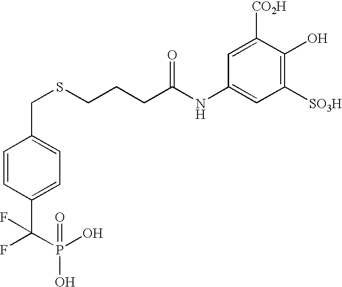
which was made according to Scheme D

| SEQ ID NO: 1 |
| Forward | GCCCATATGGAGATGGAAAAGGAGTTCGAG | |
| SEQ ID NO: 2 |
| Rev | GCGACGCGAATTCTTAATTGTGTGGCTCCAGGATTCGTTT |
| Rev2 | ||
| TGCCGGAATTCCTTAGTCCTCGTGGGAAAGCTCC | SEQ ID NO: 3 |
| C32S | CTTGGCCACTCTAGATGGGAAGTCACT | SEQ ID NO: 4 | |
| C92A | CCAAAAGTGACCGGCTGTGTTAGGCAA | SEQ ID NO: 5 |
| R47C | GGGACTGACGTCACAGTACCTATTTCG | SEQ ID NO: 6 |

which was made according to Scheme E.


which was made according to Scheme G.


which was made according to Scheme H.


which was made according to Scheme I.


which was made according to Scheme J


- a) Boc-4-nitro-D-phenylalanine (5.0 g, 16.11 mmol) was dissolved in 75 mL THF and cooled to 0° C. under a nitrogen atmosphere. N-methylmorpholine (1.81 mL, 16.43 mmol) was added followed by isobutylchloroformate (ICBF) (2.11 mL, 16.27 mmol). The reaction was stirred at 0° C. for 15 minutes and then filtered through a coarse glass frit funnel. Sodium borohydride (3.04 g, 80.39 mmol) was added to the filtrate and the solution stirred for 20 minutes at 0° C. The reaction was flooded with 100 mL 1 M NaHSO4 and extracted with 3×30 mL EtOAc. The combined organic layers were washed with 30 mL 1 M NaHSO4, 30 mL saturated NaHCO3, 30 mL brine, dried over anhydrous Na2SO4, filtered, and the solvent removed under reduced pressure. The resulting residue was dissolved in 10 mL DCM and 10 mL TFA was added. The solution was stirred for 1 hour at which point the solvent was removed under reduced pressure to yield Compound I in quantitative yield, ES (+) MS m/e=197 (M+H) which was used without further purification.
- b) Compound I (2.98 g, 15.19 mmol) was dissolved in 15 mL DMF and DIEA (13.23 mL, 75.95 mmol) was added. This solution was added to a mixture of Boc-O-(t-butyl ether)-L-serine (4.36 g, 16.71 mmol), 1-ethyl-(3-dimethylaminopropyl)carbodiimide hydrochloride (3.49 g, 18.23 mmol) and 1-hydroxybenzotriazole (2.46 g, 18.23 mmol) in 75 mL DMF. This solution was stirred at ambient temperature for 1 hour at which point it was flooded with 200 mL EtOAc, rinsed with 2×50 mL 1 M NaHSO4, 2×50 mL saturated NaHCO3, 50 mL brine, dried over anhydrous Na2SO4, filtered, and the solvent removed under reduced pressure to yield Compound II (5.73 g, 13.04 mmol, 86%), ES (+) MS m/e=462 (M+Na) which was used without further purification.
- c) Compound II (5.73 g, 13.04 mmol) was dissolved in 50 mL MeOH and palladium on carbon (2.77 g, 1.30 mmol) was added. The reaction was stirred at ambient temperature under a hydrogen balloon for 4 hours. The reaction was filtered through celite and the solvent removed under reduced pressure to yield Compound III (4.11 g, 10.0 mmol, 77% yield), ES (+) MS m/e=410 (M+1) which was used without further purification.
- d) Compound III (4.11 g, 10.0 mmol) was dissolved in 30 mL DCM and mixed with imidazol-1-yl-oxo-acetic acid tert-butyl ester (1.97 g, 10.04 mmol) in 20 mL DCM. The reaction was stirred at ambient temperature for 15 hours at which point the DCM was removed under reduced pressure and the residue redissolved in 50 mL EtOAc. The reaction was washed with 2×10 mL 1 M NaHSO4, 2×10 mL saturated NaHCO3, 10 mL brine, dried over anhydrous Na2SO4, filtered, and the solvent removed under reduced pressure to yield Compound IV (4.68, 8.70 mmol, 87%), ES (+) MS m/e=560 (M+Na) which was used without further purification.
- e) Compound IV (1.0 g, 1.86 mmol) was dissolved in 2 mL MeOH and lithium hydroxide (0.045 g, 1.86 mmol) in 2 mL water was added. The solution was stirred for 0.5 hours and then flooded with 30 mL 1 M NaHSO4 and extracted with 3×10 mL EtOAc. The combined organic layers were washed with 15 mL 1 M NaHSO4, 15 mL brine, dried over anhydrous Na2SO4, filtered, and the solvent removed under reduced pressure. The crude residue was then dissolved in 5 mL 4:1 Benzene:MeOH and (trimethylsilyl)diazomethane (0.229 mL, 0.457 mmol) was added drop wise. The solvents were removed and the residue redissolved in 10 mL DCM. 1 mL TFA was added and the solution stirred at ambient temperature for 10 minutes. The solvent was removed under reduced pressure to yield Compound V in quantitative yield, ES (+) MS m/e=396 (M+H) which was used without further purification.
- f) Compound V (0.157 g, 0.398 mmol) was dissolved in 2 mL DCM and triethylamine (TEA) (0.166 mL, 1.19 mmol) was added followed by acetic anhydride (0.038 mL, 0.398 mmol). The reaction was stirred for 1 hour at ambient temperature, flooded with 20 mL EtOAc, washed with 2×5 mL 1 M NaHSO4, 2×5 mL saturated NaHCO3, 5 mL brine, dried over anhydrous Na2SO4, filtered, and the solvent removed under reduced pressure to yield Compound VI (0.127 g, 0.290 mmol, 73%), ES (+) MS m/e=460 (M+Na) which was used without further purification.
- g) Compound VI (0.127 g. 0.290 mmol) was dissolved in 2.5 mL DCM and 2.5 mL TFA was added and the reaction stirred at ambient temperature for 3 hours. The solvent was removed under reduced pressure and the residue was redissolved in 2.5 mL MeOH. Lithium hydroxide (0.014 g, 0.58 mmol) in 2.5 mL water was added and the reaction stirred for 1 hour. The solvent was removed and the crude residue purified by reverse-phase preparatory HPLC to afford Compound VII (0.007 g, 0.019 mmol, 7%), ES (+) MS: m/e=368 (M+1).

which was made according to Scheme K

- a) Compound VIII was prepared according to the method of Example 21b followed by TFA deprotection (Example 21a) except starting from N-1-Fmoc-1,3-diaminopropane HCl instead of Compound I and using Boc-4-nitro-D-phenylalanine instead of Boc-O-(t-butyl ether)-L-serine, ES (+) MS m/e=525 (M+Na)
- b) Compound IX was prepared by treating Compound VIII according to the methods of Example 21 b, e (TFA deprotection only), f, c, and d. The resulting residue was then dissolved in 5 mL DCM and 5 mL diethylamine (DEA) was added. The solution was stirred at ambient temperature for 16 hours, the solvent removed under reduced pressure and the residue purified by silica gel chromatography using CHCl3: 2 M NH3 in MeOH 9:1, yielding Compound IX (0.173 g, 0.306 mmol, 38% yield), ES (+) MS m/e=550 (M+1).
- c) Compound X was prepared by treating Compound IX according to the method of Example 21b followed by the TFA deprotection and HPLC purification portions of 21 h except starting from 2-[3-(trifluoromethyl)phenyl]-1,3-thiazole-4-carboxylic acid instead of Boc-O-(t-butyl ether)-L-serine, ES (+) MS m/e=693 (M+H).

which was made according to Scheme L

- a) Compound XI was prepared according to the methods of Examples 22a–b except starting from N-1-Fmoc-1,4-diaminobutane HCL instead of N-1-Fmoc-1,3-diaminopropane HCL, ES (+) MS m/e 564 (M+H).
- b) Compound XI (0.173 g, 0.307 mmol) was dissolved in 1 mL DCM, DIEA (0.043 mL, 0.307 mmol) was added and this solution was added to 4-biphenylsulfonyl chloride (0.078 g, 0.307 mmol) dissolved in 1 mL DCM. This mixture was stirred at ambient temperature for 0.5 hour at which point it was flooded with 20 mL DCM, washed with 2×5 mL 1 M NaHSO4, 5 mL brine, dried over anhydrous Na2SO4, filtered, and the solvent removed under reduced pressure. The residue was dissolved in 2 mL DCM, 2 mL TFA was added and the reaction stirred for 0.5 hour. The solvent was removed and the crude residue purified by reverse-phase preparatory HPLC to afford Compound XII (0.025 g, 0.037 mmol, 12%), ES (+) MS: m/e=668 (M+1).


which was synthesized according to Scheme M and the procedure below.


(ES (+) MS m/z=460/462 (M+H)).
Claims (19)


Priority Applications (8)
| Application Number | Priority Date | Filing Date | Title |
|---|---|---|---|
| US10/374,499 US7214487B2 (en) | 1998-06-26 | 2003-02-25 | Methods for identifying compounds that modulate enzymatic activities by employing covalently bonded target-extender complexes with ligand candidates |
| PCT/US2003/006217 WO2003087054A2 (en) | 2002-04-10 | 2003-02-26 | Methods for identifying compounds that modulate enzymatic activity |
| JP2003584010A JP2006506947A (en) | 2002-04-10 | 2003-02-26 | An elongation tether approach to identify compounds that modulate enzyme activity |
| AU2003216468A AU2003216468B2 (en) | 2002-04-10 | 2003-02-26 | Methods for identifying compounds that modulate enzymatic activity |
| CA002478398A CA2478398A1 (en) | 2002-04-10 | 2003-02-26 | Methods for identifying compounds that modulate enzymatic activity |
| EP03746539A EP1497450A4 (en) | 2002-04-10 | 2003-02-26 | METHODS OF IDENTIFYING COMPOUNDS THAT MODULATE ENZYMATIC ACTIVITY |
| US11/735,944 US20080220536A1 (en) | 1998-06-26 | 2007-04-16 | Methods for Identifying Compounds that Modulate Enzymatic Activities by Employing Covalently Bonded Target-Extender Complexes with Ligand Candidates |
| AU2008246264A AU2008246264A1 (en) | 2002-04-10 | 2008-11-20 | Methods for identifying compounds that modulate enzymatic activity |
Applications Claiming Priority (7)
| Application Number | Priority Date | Filing Date | Title |
|---|---|---|---|
| US09/105,372 US6335155B1 (en) | 1998-06-26 | 1998-06-26 | Methods for rapidly identifying small organic molecule ligands for binding to biological target molecules |
| US25229400P | 2000-11-21 | 2000-11-21 | |
| US09/981,547 US20020022233A1 (en) | 1998-06-26 | 2001-10-17 | Methods for rapidly identifying small organic molecule ligands for binding to biological target molecules |
| US09/990,421 US6919178B2 (en) | 2000-11-21 | 2001-11-21 | Extended tethering approach for rapid identification of ligands |
| US10/121,216 US6998233B2 (en) | 1998-06-26 | 2002-04-10 | Methods for ligand discovery |
| US37703402P | 2002-05-01 | 2002-05-01 | |
| US10/374,499 US7214487B2 (en) | 1998-06-26 | 2003-02-25 | Methods for identifying compounds that modulate enzymatic activities by employing covalently bonded target-extender complexes with ligand candidates |
Related Parent Applications (3)
| Application Number | Title | Priority Date | Filing Date |
|---|---|---|---|
| US09/105,372 Division US6335155B1 (en) | 1998-06-26 | 1998-06-26 | Methods for rapidly identifying small organic molecule ligands for binding to biological target molecules |
| US09/981,547 Continuation-In-Part US20020022233A1 (en) | 1998-06-26 | 2001-10-17 | Methods for rapidly identifying small organic molecule ligands for binding to biological target molecules |
| US10/121,216 Continuation-In-Part US6998233B2 (en) | 1998-06-26 | 2002-04-10 | Methods for ligand discovery |
Related Child Applications (1)
| Application Number | Title | Priority Date | Filing Date |
|---|---|---|---|
| US11/735,944 Continuation US20080220536A1 (en) | 1998-06-26 | 2007-04-16 | Methods for Identifying Compounds that Modulate Enzymatic Activities by Employing Covalently Bonded Target-Extender Complexes with Ligand Candidates |
Publications (2)
| Publication Number | Publication Date |
|---|---|
| US20040005632A1 US20040005632A1 (en) | 2004-01-08 |
| US7214487B2 true US7214487B2 (en) | 2007-05-08 |
Family
ID=29255201
Family Applications (2)
| Application Number | Title | Priority Date | Filing Date |
|---|---|---|---|
| US10/374,499 Expired - Fee Related US7214487B2 (en) | 1998-06-26 | 2003-02-25 | Methods for identifying compounds that modulate enzymatic activities by employing covalently bonded target-extender complexes with ligand candidates |
| US11/735,944 Abandoned US20080220536A1 (en) | 1998-06-26 | 2007-04-16 | Methods for Identifying Compounds that Modulate Enzymatic Activities by Employing Covalently Bonded Target-Extender Complexes with Ligand Candidates |
Family Applications After (1)
| Application Number | Title | Priority Date | Filing Date |
|---|---|---|---|
| US11/735,944 Abandoned US20080220536A1 (en) | 1998-06-26 | 2007-04-16 | Methods for Identifying Compounds that Modulate Enzymatic Activities by Employing Covalently Bonded Target-Extender Complexes with Ligand Candidates |
Country Status (6)
| Country | Link |
|---|---|
| US (2) | US7214487B2 (en) |
| EP (1) | EP1497450A4 (en) |
| JP (1) | JP2006506947A (en) |
| AU (2) | AU2003216468B2 (en) |
| CA (1) | CA2478398A1 (en) |
| WO (1) | WO2003087054A2 (en) |
Cited By (4)
| Publication number | Priority date | Publication date | Assignee | Title |
|---|---|---|---|---|
| US20070026451A1 (en) * | 1998-06-26 | 2007-02-01 | Sunesis Pharmaceuticals, Inc. | Methods for rapidly identifying small organic molecule ligands for binding to biological target molecules |
| US20090176175A1 (en) * | 2008-01-08 | 2009-07-09 | International Business Machines Corporation | Photoacid generators for extreme ultraviolet lithography |
| WO2011017249A1 (en) | 2009-08-03 | 2011-02-10 | Medarex, Inc. | Antiproliferative compounds, conjugates thereof, methods therefor, and uses thereof |
| US12172992B2 (en) | 2022-03-22 | 2024-12-24 | Abbvie Inc. | Pyrimidines for degrading Bruton's tyrosine kinase |
Families Citing this family (6)
| Publication number | Priority date | Publication date | Assignee | Title |
|---|---|---|---|---|
| PT1565478T (en) | 2002-11-12 | 2017-11-14 | The Brigham And Women`S Hospital Inc | Polysaccharide vaccine for staphylococcal infections |
| WO2005114197A2 (en) * | 2004-04-15 | 2005-12-01 | Albert Einstein College Of Medicine Of Yeshiva University | Activity-based probes for protein tyrosine phosphatases |
| KR20150041178A (en) * | 2008-07-21 | 2015-04-15 | 더 브리검 앤드 우먼즈 하스피털, 인크. | Methods and compositions relating to synthetic beta-1,6 glucosamine oligosaccharides |
| EP2560686A2 (en) | 2010-04-21 | 2013-02-27 | Novo Nordisk A/S | Selective modification of proteins |
| US12397014B2 (en) | 2019-02-05 | 2025-08-26 | The Brigham And Women's Hospital, Inc. | Polysaccharide compositions for use in treating filariasis |
| CN114853629B (en) * | 2021-02-03 | 2023-06-13 | 沈阳农业大学 | A kind of dipeptide compound and its preparation method and application |
Citations (8)
| Publication number | Priority date | Publication date | Assignee | Title |
|---|---|---|---|---|
| US5367058A (en) * | 1993-08-25 | 1994-11-22 | Becton, Dickinson And Company | Modified antibodies with increased affinity |
| US6335155B1 (en) | 1998-06-26 | 2002-01-01 | Sunesis Pharmaceuticals, Inc. | Methods for rapidly identifying small organic molecule ligands for binding to biological target molecules |
| WO2002042773A2 (en) | 2000-11-21 | 2002-05-30 | Sunesis Pharmaceuticals, Inc. | An extended tethering approach for rapid identification of ligands |
| US20020155505A1 (en) * | 1998-06-26 | 2002-10-24 | Jim Wells | Methods for ligand discovery |
| WO2003014072A1 (en) | 2001-08-07 | 2003-02-20 | Sunesis Pharmaceuticals, Inc. | Disulfide and thiosulfonate ligands and libraries comprising these ligands |
| WO2003014308A2 (en) | 2001-08-07 | 2003-02-20 | Sunesis Pharmaceuticals, Inc. | Cysteine mutants and methods for detecting ligand binding to biological molecules |
| WO2003046200A2 (en) | 2001-11-21 | 2003-06-05 | Sunesis Pharmaceuticals, Inc. | Methods for ligand discovery |
| US6919178B2 (en) * | 2000-11-21 | 2005-07-19 | Sunesis Pharmaceuticals, Inc. | Extended tethering approach for rapid identification of ligands |
-
2003
- 2003-02-25 US US10/374,499 patent/US7214487B2/en not_active Expired - Fee Related
- 2003-02-26 CA CA002478398A patent/CA2478398A1/en not_active Abandoned
- 2003-02-26 EP EP03746539A patent/EP1497450A4/en not_active Withdrawn
- 2003-02-26 AU AU2003216468A patent/AU2003216468B2/en not_active Ceased
- 2003-02-26 JP JP2003584010A patent/JP2006506947A/en not_active Ceased
- 2003-02-26 WO PCT/US2003/006217 patent/WO2003087054A2/en not_active Ceased
-
2007
- 2007-04-16 US US11/735,944 patent/US20080220536A1/en not_active Abandoned
-
2008
- 2008-11-20 AU AU2008246264A patent/AU2008246264A1/en not_active Abandoned
Patent Citations (9)
| Publication number | Priority date | Publication date | Assignee | Title |
|---|---|---|---|---|
| US5367058A (en) * | 1993-08-25 | 1994-11-22 | Becton, Dickinson And Company | Modified antibodies with increased affinity |
| US6335155B1 (en) | 1998-06-26 | 2002-01-01 | Sunesis Pharmaceuticals, Inc. | Methods for rapidly identifying small organic molecule ligands for binding to biological target molecules |
| US20020155505A1 (en) * | 1998-06-26 | 2002-10-24 | Jim Wells | Methods for ligand discovery |
| US6811966B2 (en) * | 1998-06-26 | 2004-11-02 | Sunesis Pharmaceuticals, Inc. | Methods for rapidly identifying small organic molecule ligands for binding to biological target molecules |
| WO2002042773A2 (en) | 2000-11-21 | 2002-05-30 | Sunesis Pharmaceuticals, Inc. | An extended tethering approach for rapid identification of ligands |
| US6919178B2 (en) * | 2000-11-21 | 2005-07-19 | Sunesis Pharmaceuticals, Inc. | Extended tethering approach for rapid identification of ligands |
| WO2003014072A1 (en) | 2001-08-07 | 2003-02-20 | Sunesis Pharmaceuticals, Inc. | Disulfide and thiosulfonate ligands and libraries comprising these ligands |
| WO2003014308A2 (en) | 2001-08-07 | 2003-02-20 | Sunesis Pharmaceuticals, Inc. | Cysteine mutants and methods for detecting ligand binding to biological molecules |
| WO2003046200A2 (en) | 2001-11-21 | 2003-06-05 | Sunesis Pharmaceuticals, Inc. | Methods for ligand discovery |
Non-Patent Citations (7)
| Title |
|---|
| Betley et al. Direct screening for Phosphatase Activity by Turnover-Based Capture of Protein Catalysts, Angew. Chem. Int. Ed. 2002, vol. 41, No. 5, pp. 775-777. |
| Burke, T., et al., "Phosphoryltyrosyl Mimetics in the Design of Peptide-Based Signal Transduction Inhibitors", Biopolymers (Peptide Science), vol. 60, 2001, pp. 32-44. |
| Daniel A. Erlanson, et al., "Site-directed Ligand Discovery", PNAS, Aug. 15, 2000, vol. 97, No. 17, pp. 9367-9372. |
| Erlanson et al. In-Situ Assembly of Enzyme Inhibitors Using Extended Tethering. Nature Biotechnology. Mar. 2003, vol. 21, pp. 308-314. |
| Erlanson, D., et al., "Discovery of a New Phosphotyrosine Mimetic for PTP1B Using Breakaway Tethering", Journal of American Chemical Society, Vo. 125, No. 19, 2003, 5602-5603. |
| Szczepankiewicz, B., et al., "Discovery of a Potent, Selective Protein Tyrosine Phosphatase 1B Inhibitor Using a Linked-Fragment Strategy", Journal of American Chemical Society, Vo. 125, No. 14, 2003, 4087-4096. |
| Yasothornsrkul, S.; Aaron, W.; Toneff, T.; Hook, V. Y. H. Evidence for the Proenkephalin Processing Enzyme Prohormone thiol Protease (PTP) as a Multicatalytic Cysteine Protease Complex. Biochemistry 1999, 38, 7421-7430. * |
Cited By (5)
| Publication number | Priority date | Publication date | Assignee | Title |
|---|---|---|---|---|
| US20070026451A1 (en) * | 1998-06-26 | 2007-02-01 | Sunesis Pharmaceuticals, Inc. | Methods for rapidly identifying small organic molecule ligands for binding to biological target molecules |
| US20100022399A1 (en) * | 1998-06-26 | 2010-01-28 | Sunesis Pharmaceuticals, Inc. | Methods for rapidly identifying small organic molecule ligands for binding to biological target molecules |
| US20090176175A1 (en) * | 2008-01-08 | 2009-07-09 | International Business Machines Corporation | Photoacid generators for extreme ultraviolet lithography |
| WO2011017249A1 (en) | 2009-08-03 | 2011-02-10 | Medarex, Inc. | Antiproliferative compounds, conjugates thereof, methods therefor, and uses thereof |
| US12172992B2 (en) | 2022-03-22 | 2024-12-24 | Abbvie Inc. | Pyrimidines for degrading Bruton's tyrosine kinase |
Also Published As
| Publication number | Publication date |
|---|---|
| JP2006506947A (en) | 2006-03-02 |
| EP1497450A4 (en) | 2010-03-10 |
| EP1497450A2 (en) | 2005-01-19 |
| US20040005632A1 (en) | 2004-01-08 |
| US20080220536A1 (en) | 2008-09-11 |
| AU2003216468A1 (en) | 2003-10-27 |
| WO2003087054A2 (en) | 2003-10-23 |
| AU2003216468B2 (en) | 2008-08-21 |
| AU2008246264A1 (en) | 2008-12-11 |
| WO2003087054A3 (en) | 2004-08-05 |
| CA2478398A1 (en) | 2003-10-23 |
Similar Documents
| Publication | Publication Date | Title |
|---|---|---|
| US20080220536A1 (en) | Methods for Identifying Compounds that Modulate Enzymatic Activities by Employing Covalently Bonded Target-Extender Complexes with Ligand Candidates | |
| JP3836791B2 (en) | Extended tethering approach for rapid identification of ligands | |
| US6998233B2 (en) | Methods for ligand discovery | |
| US20050084905A1 (en) | Identification of kinase inhibitors | |
| JP2026041812A (en) | Methods and reagents for analyzing protein-protein interfaces | |
| US20050142614A1 (en) | Methods for ligand discovery | |
| US9586890B2 (en) | Screening methods for the binding affinity of chemical entities to biological molecules and NEDD4-1 inhibitors identified by the screening methods | |
| JP2005534286A (en) | Identification of kinase inhibitors | |
| US7074578B2 (en) | Methods and means of histone methylation | |
| JP2005517184A (en) | Methods and compositions with HSP90 activator AHA1 | |
| AU2003270848A1 (en) | Identification of kinase inhibitors | |
| CA2478395A1 (en) | Methods for identifying allosteric sites | |
| EP1283879A2 (en) | Molecules that modulate ubiquitin-dependent proteolysis and methods for identifying same | |
| US20070026478A1 (en) | Hemiasterlin affinity probes and their uses | |
| US20040072257A1 (en) | Proteomic screening for redox state dependent protein-protein interactions | |
| Dong | Targeting Protein Tyrosine Phosphatases With Small Molecules for Anti-Cancer Therapy | |
| AYOOB et al. | DESIGN, SYNTHESIS, MOLECULAR DOCKING OF NOVEL METHIMAZOLE DERIVATIVES AS PROMISING ANTITHYROID AGENTS. | |
| US20050112709A1 (en) | Histone h3methyltransferase polypeptide | |
| JP2001526890A (en) | Human protein responsible for NEDD8 activation and conjugation |
Legal Events
| Date | Code | Title | Description |
|---|---|---|---|
| AS | Assignment |
Owner name: SUNESIS PHARMACEUTICALS, INC., CALIFORNIA Free format text: ASSIGNMENT OF ASSIGNORS INTEREST;ASSIGNORS:ERLANSON, DANIEL A.;MCDOWELL, ROBERT S.;HANSEN, STIG;REEL/FRAME:013794/0443;SIGNING DATES FROM 20030605 TO 20030618 |
|
| STCF | Information on status: patent grant |
Free format text: PATENTED CASE |
|
| REMI | Maintenance fee reminder mailed | ||
| FPAY | Fee payment |
Year of fee payment: 4 |
|
| SULP | Surcharge for late payment | ||
| FEPP | Fee payment procedure |
Free format text: PAT HOLDER NO LONGER CLAIMS SMALL ENTITY STATUS, ENTITY STATUS SET TO UNDISCOUNTED (ORIGINAL EVENT CODE: STOL); ENTITY STATUS OF PATENT OWNER: LARGE ENTITY |
|
| REFU | Refund |
Free format text: REFUND - PAYMENT OF MAINTENANCE FEE, 8TH YR, SMALL ENTITY (ORIGINAL EVENT CODE: R2552); ENTITY STATUS OF PATENT OWNER: LARGE ENTITY |
|
| FPAY | Fee payment |
Year of fee payment: 8 |
|
| FPAY | Fee payment |
Year of fee payment: 8 |
|
| FEPP | Fee payment procedure |
Free format text: MAINTENANCE FEE REMINDER MAILED (ORIGINAL EVENT CODE: REM.); ENTITY STATUS OF PATENT OWNER: LARGE ENTITY |
|
| LAPS | Lapse for failure to pay maintenance fees |
Free format text: PATENT EXPIRED FOR FAILURE TO PAY MAINTENANCE FEES (ORIGINAL EVENT CODE: EXP.); ENTITY STATUS OF PATENT OWNER: LARGE ENTITY |
|
| STCH | Information on status: patent discontinuation |
Free format text: PATENT EXPIRED DUE TO NONPAYMENT OF MAINTENANCE FEES UNDER 37 CFR 1.362 |
|
| FP | Lapsed due to failure to pay maintenance fee |
Effective date: 20190508 |