US7077808B2 - Ultrasonic imaging catheter - Google Patents
Ultrasonic imaging catheter Download PDFInfo
- Publication number
- US7077808B2 US7077808B2 US10/631,872 US63187203A US7077808B2 US 7077808 B2 US7077808 B2 US 7077808B2 US 63187203 A US63187203 A US 63187203A US 7077808 B2 US7077808 B2 US 7077808B2
- Authority
- US
- United States
- Prior art keywords
- ultrasonic
- reflective member
- catheter apparatus
- actuator
- imaging catheter
- Prior art date
- Legal status (The legal status is an assumption and is not a legal conclusion. Google has not performed a legal analysis and makes no representation as to the accuracy of the status listed.)
- Expired - Lifetime, expires
Links
- 238000003384 imaging method Methods 0.000 title claims abstract description 27
- 229920001746 electroactive polymer Polymers 0.000 claims abstract description 44
- 230000008859 change Effects 0.000 claims abstract description 10
- 238000003780 insertion Methods 0.000 claims abstract description 7
- 230000037431 insertion Effects 0.000 claims abstract description 7
- 239000003792 electrolyte Substances 0.000 claims description 18
- 239000000463 material Substances 0.000 claims description 17
- 239000004020 conductor Substances 0.000 claims description 14
- 229920000128 polypyrrole Polymers 0.000 claims description 10
- HSFWRNGVRCDJHI-UHFFFAOYSA-N alpha-acetylene Natural products C#C HSFWRNGVRCDJHI-UHFFFAOYSA-N 0.000 claims description 5
- 229920002492 poly(sulfone) Polymers 0.000 claims description 5
- 229920001197 polyacetylene Polymers 0.000 claims description 5
- 229920000767 polyaniline Polymers 0.000 claims description 5
- 238000004891 communication Methods 0.000 claims description 3
- 239000003990 capacitor Substances 0.000 claims description 2
- 238000000034 method Methods 0.000 abstract description 10
- 150000002500 ions Chemical class 0.000 description 10
- 229920000642 polymer Polymers 0.000 description 9
- 238000002604 ultrasonography Methods 0.000 description 7
- 239000002322 conducting polymer Substances 0.000 description 6
- 229920001940 conductive polymer Polymers 0.000 description 6
- 239000000499 gel Substances 0.000 description 6
- 230000009286 beneficial effect Effects 0.000 description 5
- 230000008901 benefit Effects 0.000 description 5
- 230000008602 contraction Effects 0.000 description 5
- 239000013078 crystal Substances 0.000 description 5
- 229910052751 metal Inorganic materials 0.000 description 5
- 239000002184 metal Substances 0.000 description 5
- 210000004204 blood vessel Anatomy 0.000 description 4
- 239000007788 liquid Substances 0.000 description 4
- BASFCYQUMIYNBI-UHFFFAOYSA-N platinum Chemical compound [Pt] BASFCYQUMIYNBI-UHFFFAOYSA-N 0.000 description 4
- 239000007787 solid Substances 0.000 description 4
- 210000001519 tissue Anatomy 0.000 description 4
- 238000010276 construction Methods 0.000 description 3
- 238000013461 design Methods 0.000 description 3
- 238000010586 diagram Methods 0.000 description 3
- PCHJSUWPFVWCPO-UHFFFAOYSA-N gold Chemical compound [Au] PCHJSUWPFVWCPO-UHFFFAOYSA-N 0.000 description 3
- 229910052737 gold Inorganic materials 0.000 description 3
- 239000010931 gold Substances 0.000 description 3
- 230000003647 oxidation Effects 0.000 description 3
- 238000007254 oxidation reaction Methods 0.000 description 3
- -1 polyethylene Polymers 0.000 description 3
- 230000001681 protective effect Effects 0.000 description 3
- 230000009467 reduction Effects 0.000 description 3
- RYGMFSIKBFXOCR-UHFFFAOYSA-N Copper Chemical compound [Cu] RYGMFSIKBFXOCR-UHFFFAOYSA-N 0.000 description 2
- XEEYBQQBJWHFJM-UHFFFAOYSA-N Iron Chemical compound [Fe] XEEYBQQBJWHFJM-UHFFFAOYSA-N 0.000 description 2
- FAPWRFPIFSIZLT-UHFFFAOYSA-M Sodium chloride Chemical compound [Na+].[Cl-] FAPWRFPIFSIZLT-UHFFFAOYSA-M 0.000 description 2
- 239000012190 activator Substances 0.000 description 2
- 238000002399 angioplasty Methods 0.000 description 2
- 230000015572 biosynthetic process Effects 0.000 description 2
- 230000001419 dependent effect Effects 0.000 description 2
- 239000002019 doping agent Substances 0.000 description 2
- 229940079593 drug Drugs 0.000 description 2
- 239000003814 drug Substances 0.000 description 2
- 230000000694 effects Effects 0.000 description 2
- 238000013156 embolectomy Methods 0.000 description 2
- 239000000835 fiber Substances 0.000 description 2
- 230000006870 function Effects 0.000 description 2
- 230000001965 increasing effect Effects 0.000 description 2
- 238000002608 intravascular ultrasound Methods 0.000 description 2
- 150000002739 metals Chemical class 0.000 description 2
- 229910052697 platinum Inorganic materials 0.000 description 2
- 229920003229 poly(methyl methacrylate) Polymers 0.000 description 2
- 239000004926 polymethyl methacrylate Substances 0.000 description 2
- 229920002981 polyvinylidene fluoride Polymers 0.000 description 2
- 230000004044 response Effects 0.000 description 2
- 208000037803 restenosis Diseases 0.000 description 2
- 238000002560 therapeutic procedure Methods 0.000 description 2
- 238000012546 transfer Methods 0.000 description 2
- 230000002792 vascular Effects 0.000 description 2
- 210000005166 vasculature Anatomy 0.000 description 2
- 229920001817 Agar Polymers 0.000 description 1
- 241000239290 Araneae Species 0.000 description 1
- 239000004606 Fillers/Extenders Substances 0.000 description 1
- 244000043261 Hevea brasiliensis Species 0.000 description 1
- 229930012538 Paclitaxel Natural products 0.000 description 1
- 208000031481 Pathologic Constriction Diseases 0.000 description 1
- 239000004698 Polyethylene Substances 0.000 description 1
- 108010023197 Streptokinase Proteins 0.000 description 1
- 108090000373 Tissue Plasminogen Activator Proteins 0.000 description 1
- 102000003978 Tissue Plasminogen Activator Human genes 0.000 description 1
- 108090000435 Urokinase-type plasminogen activator Proteins 0.000 description 1
- 102000003990 Urokinase-type plasminogen activator Human genes 0.000 description 1
- XILGXSYEQPUUEW-UHFFFAOYSA-N [H]N1C(C)=CC=C1C1=CC=C(C2=CC=C(C3=CC=C(C4=CC=C(C5=CC=C(C6=CC=C(C7=CC=C(C)N7[H])N6[H])N5[H])N4[H])N3[H])N2[H])N1[H] Chemical compound [H]N1C(C)=CC=C1C1=CC=C(C2=CC=C(C3=CC=C(C4=CC=C(C5=CC=C(C6=CC=C(C7=CC=C(C)N7[H])N6[H])N5[H])N4[H])N3[H])N2[H])N1[H] XILGXSYEQPUUEW-UHFFFAOYSA-N 0.000 description 1
- 238000002679 ablation Methods 0.000 description 1
- 238000005299 abrasion Methods 0.000 description 1
- 239000008272 agar Substances 0.000 description 1
- JRPBQTZRNDNNOP-UHFFFAOYSA-N barium titanate Chemical compound [Ba+2].[Ba+2].[O-][Ti]([O-])([O-])[O-] JRPBQTZRNDNNOP-UHFFFAOYSA-N 0.000 description 1
- 229910002113 barium titanate Inorganic materials 0.000 description 1
- 238000005452 bending Methods 0.000 description 1
- 230000000903 blocking effect Effects 0.000 description 1
- 230000002490 cerebral effect Effects 0.000 description 1
- 238000005234 chemical deposition Methods 0.000 description 1
- 229910052956 cinnabar Inorganic materials 0.000 description 1
- 229920001577 copolymer Polymers 0.000 description 1
- 238000012937 correction Methods 0.000 description 1
- 230000008878 coupling Effects 0.000 description 1
- 238000010168 coupling process Methods 0.000 description 1
- 238000005859 coupling reaction Methods 0.000 description 1
- 230000007423 decrease Effects 0.000 description 1
- 230000032798 delamination Effects 0.000 description 1
- 238000006073 displacement reaction Methods 0.000 description 1
- 238000005553 drilling Methods 0.000 description 1
- 238000004070 electrodeposition Methods 0.000 description 1
- 238000009713 electroplating Methods 0.000 description 1
- 230000005284 excitation Effects 0.000 description 1
- 210000001105 femoral artery Anatomy 0.000 description 1
- 238000002594 fluoroscopy Methods 0.000 description 1
- 230000036039 immunity Effects 0.000 description 1
- 238000001727 in vivo Methods 0.000 description 1
- 230000001939 inductive effect Effects 0.000 description 1
- 229910052742 iron Inorganic materials 0.000 description 1
- 238000000608 laser ablation Methods 0.000 description 1
- 230000007246 mechanism Effects 0.000 description 1
- 238000012986 modification Methods 0.000 description 1
- 230000004048 modification Effects 0.000 description 1
- 229920003052 natural elastomer Polymers 0.000 description 1
- 229920001194 natural rubber Polymers 0.000 description 1
- 231100000252 nontoxic Toxicity 0.000 description 1
- 230000003000 nontoxic effect Effects 0.000 description 1
- 239000011368 organic material Substances 0.000 description 1
- 230000010355 oscillation Effects 0.000 description 1
- 229960001592 paclitaxel Drugs 0.000 description 1
- 230000002093 peripheral effect Effects 0.000 description 1
- 239000008055 phosphate buffer solution Substances 0.000 description 1
- 239000004033 plastic Substances 0.000 description 1
- 229920003023 plastic Polymers 0.000 description 1
- 239000004417 polycarbonate Substances 0.000 description 1
- 229920000515 polycarbonate Polymers 0.000 description 1
- 229920000728 polyester Polymers 0.000 description 1
- 229920000573 polyethylene Polymers 0.000 description 1
- 239000002861 polymer material Substances 0.000 description 1
- 229920002635 polyurethane Polymers 0.000 description 1
- 239000004814 polyurethane Substances 0.000 description 1
- 229920000915 polyvinyl chloride Polymers 0.000 description 1
- 239000004800 polyvinyl chloride Substances 0.000 description 1
- 239000001103 potassium chloride Substances 0.000 description 1
- 238000007639 printing Methods 0.000 description 1
- 238000002310 reflectometry Methods 0.000 description 1
- 238000012552 review Methods 0.000 description 1
- 150000003839 salts Chemical class 0.000 description 1
- 239000000523 sample Substances 0.000 description 1
- 239000004065 semiconductor Substances 0.000 description 1
- 229920002379 silicone rubber Polymers 0.000 description 1
- 239000004945 silicone rubber Substances 0.000 description 1
- 210000000329 smooth muscle myocyte Anatomy 0.000 description 1
- 239000011780 sodium chloride Substances 0.000 description 1
- 229910001220 stainless steel Inorganic materials 0.000 description 1
- 239000010935 stainless steel Substances 0.000 description 1
- 208000037804 stenosis Diseases 0.000 description 1
- 230000036262 stenosis Effects 0.000 description 1
- 230000002966 stenotic effect Effects 0.000 description 1
- 230000000638 stimulation Effects 0.000 description 1
- 229960005202 streptokinase Drugs 0.000 description 1
- 238000010408 sweeping Methods 0.000 description 1
- RCINICONZNJXQF-MZXODVADSA-N taxol Chemical compound O([C@@H]1[C@@]2(C[C@@H](C(C)=C(C2(C)C)[C@H](C([C@]2(C)[C@@H](O)C[C@H]3OC[C@]3([C@H]21)OC(C)=O)=O)OC(=O)C)OC(=O)[C@H](O)[C@@H](NC(=O)C=1C=CC=CC=1)C=1C=CC=CC=1)O)C(=O)C1=CC=CC=C1 RCINICONZNJXQF-MZXODVADSA-N 0.000 description 1
- 230000001225 therapeutic effect Effects 0.000 description 1
- 229960000187 tissue plasminogen activator Drugs 0.000 description 1
- 238000012876 topography Methods 0.000 description 1
- 229960005356 urokinase Drugs 0.000 description 1
Images


Classifications
-
- A—HUMAN NECESSITIES
- A61—MEDICAL OR VETERINARY SCIENCE; HYGIENE
- A61B—DIAGNOSIS; SURGERY; IDENTIFICATION
- A61B8/00—Diagnosis using ultrasonic, sonic or infrasonic waves
- A61B8/12—Diagnosis using ultrasonic, sonic or infrasonic waves in body cavities or body tracts, e.g. by using catheters
-
- A—HUMAN NECESSITIES
- A61—MEDICAL OR VETERINARY SCIENCE; HYGIENE
- A61B—DIAGNOSIS; SURGERY; IDENTIFICATION
- A61B8/00—Diagnosis using ultrasonic, sonic or infrasonic waves
- A61B8/44—Constructional features of the ultrasonic, sonic or infrasonic diagnostic device
- A61B8/4444—Constructional features of the ultrasonic, sonic or infrasonic diagnostic device related to the probe
- A61B8/445—Details of catheter construction
-
- A—HUMAN NECESSITIES
- A61—MEDICAL OR VETERINARY SCIENCE; HYGIENE
- A61B—DIAGNOSIS; SURGERY; IDENTIFICATION
- A61B8/00—Diagnosis using ultrasonic, sonic or infrasonic waves
- A61B8/44—Constructional features of the ultrasonic, sonic or infrasonic diagnostic device
- A61B8/4444—Constructional features of the ultrasonic, sonic or infrasonic diagnostic device related to the probe
- A61B8/4461—Features of the scanning mechanism, e.g. for moving the transducer within the housing of the probe
-
- G—PHYSICS
- G10—MUSICAL INSTRUMENTS; ACOUSTICS
- G10K—SOUND-PRODUCING DEVICES; METHODS OR DEVICES FOR PROTECTING AGAINST, OR FOR DAMPING, NOISE OR OTHER ACOUSTIC WAVES IN GENERAL; ACOUSTICS NOT OTHERWISE PROVIDED FOR
- G10K11/00—Methods or devices for transmitting, conducting or directing sound in general; Methods or devices for protecting against, or for damping, noise or other acoustic waves in general
- G10K11/18—Methods or devices for transmitting, conducting or directing sound
- G10K11/26—Sound-focusing or directing, e.g. scanning
- G10K11/35—Sound-focusing or directing, e.g. scanning using mechanical steering of transducers or their beams
- G10K11/357—Sound-focusing or directing, e.g. scanning using mechanical steering of transducers or their beams by moving a reflector
-
- A—HUMAN NECESSITIES
- A61—MEDICAL OR VETERINARY SCIENCE; HYGIENE
- A61M—DEVICES FOR INTRODUCING MEDIA INTO, OR ONTO, THE BODY; DEVICES FOR TRANSDUCING BODY MEDIA OR FOR TAKING MEDIA FROM THE BODY; DEVICES FOR PRODUCING OR ENDING SLEEP OR STUPOR
- A61M25/00—Catheters; Hollow probes
- A61M25/0043—Catheters; Hollow probes characterised by structural features
- A61M2025/0058—Catheters; Hollow probes characterised by structural features having an electroactive polymer material, e.g. for steering purposes, for control of flexibility, for locking, for opening or closing
Definitions
- This invention relates to catheters appropriate for imaging, and more particularly to catheters appropriate for intravascular ultrasonographic imaging applications.
- Intravascular ultrasound (IVUS) catheters and methods for imaging are known.
- U.S. Pat. No. 5,000,185 to Yock the entire disclosure of which is incorporated by reference, discloses devices and methods for high-resolution intravascular ultrasound imaging to assist with the administration of vascular interventional therapy and to monitor the results of such therapy.
- Yock an ultrasonic transducer is carried by the distal end of a catheter adapted for insertion into a blood vessel, whereupon either the transducer or another element, such as an ultrasound mirror, is rotated and/or translated relative to the catheter to image different portions of the vessel.
- an ultrasonic imaging catheter apparatus which comprises the following: (a) a flexible elongate body adapted for insertion into a body lumen, the elongate body having distal and proximal ends; (b) an ultrasonic transducer generating and detecting ultrasonic energy disposed proximate the distal end of the elongate body; (c) a reflective member disposed proximate the ultrasonic transducer and which is optionally rotatable with respect to an axis of the body lumen, wherein the reflective member is adapted to reflect (i) ultrasonic energy generated by the ultrasonic transducer to a wall of the body lumen and (ii) ultrasonic energy reflected by the wall back to the transducer; and (d) an actuator, such as an electroactive polymer actuator, the electroactive polymer actuator being adapted to electronically control the tilt of the reflector and thus the angle of incidence of
- the electroactive polymer actuators typically comprise an electroactive polymer region, a counter-electrode region, and an electrolyte-containing region disposed between the electroactive polymer region and the counter-electrode region.
- Beneficial electroactive polymers for these embodiments include polyaniline, polysulfone, polyacetylene and polypyrrole.
- control signals for the ultrasonic transducer and for the electroactive polymer actuator are transmitted via a shared single electrical conduction path, for example a coaxial cable.
- a shared single electrical conduction path for example a coaxial cable.
- the catheter apparatus can further comprise a motor and a drive shaft for translating torque from the motor, for example, through a suitable connector or rotary joint, thereby rotating the reflective member, among other elements.
- aspects of the present invention are directed to methods of scanning the inner wall of a body lumen. These methods comprise: (a) providing a catheter apparatus like that above; (b) sweeping the ultrasonic energy from the transducer in a pattern over the interior wall of the body lumen by operating the electroactive polymer actuator to change the angle of incidence of the ultrasonic energy upon the reflective member, and by optionally rotating the reflective member; (c) receiving ultrasonic energy reflected from the interior wall of the body lumen; and (d) producing an image from the reflected ultrasonic energy.
- the ultrasonic energy can be directed at a forward angle between about 10° to about 85° relative to the axis of the body lumen, such that a conical forward sweep is performed.
- One advantage of the present invention is that catheters, systems and methods are provided for intravascular ultrasonography.
- Another advantage of the present invention is that catheters for intravascular ultrasonography are provided, in which the wall of an adjacent body lumen can be axially (longitudinally) scanned, without the need for axial movement of the transducer or other element relative to the body lumen.
- catheters for intravascular ultrasonography are provided, which can provide for forward, lateral and retrograde scanning, without the need for axial movement of the transducer or other element relative to the body lumen.
- FIG. 1A is a schematic partial cross-sectional view of the distal end of a catheter apparatus, in accordance with an embodiment of the present invention.
- FIG. 1B is a schematic partial cross-sectional view of the distal end of a catheter apparatus, in accordance with another embodiment of the present invention.
- FIG. 2 is a schematic top view of the distal portion of FIG. 1A .
- FIG. 3 is schematic diagram illustrating three scanning sections, which can be generated using a catheter apparatus in accordance with the present invention.
- FIG. 4 is a schematic block diagram of the electrical components utilized in a catheter system, in accordance with an embodiment of the present invention.
- FIG. 5 is a schematic cross-sectional view of an electroactive polymer actuator useful in connection with the present invention.
- FIG. 6 is a schematic cross-sectional view of another electroactive polymer actuator configuration useful in connection with the present invention.
- a distal portion of a catheter apparatus 110 is illustrated, which is adapted for insertion into a body lumen, for example, a blood vessel within the coronary vasculature.
- an ultrasonic transducer 132 In the embodiment shown, an ultrasonic transducer 132 , an associated ultrasonic lens 134 , and a reflective member 136 are carried at the distal end of a flexible shaft of catheter apparatus 110 .
- An electrical system (described in more detail below) is connected to the ultrasonic transducer 132 for supplying signals to and receiving signals from the transducer 132 during operation.
- the electrical system also supplies signals to an actuator 140 , which is used to change the angle at which ultrasonic waves are incident upon the reflective member 136 during operation.
- the various elements of the catheter apparatus 110 are typically rotated during operation by means of mechanical torque, which is transmitted along drive shaft 114 .
- the ultrasonic transducer 132 can be formed, for example, using any of a number of materials that are known in the art. For instance, single crystals, which are capable of operating at a frequency range of, for example, 5 to 50 megahertz, are known in the art. Typical materials for forming such crystals include barium titanate or cinnabar. Conductive electrodes, for example, films of gold or other conductive metals, may be provided on opposing surfaces of the crystal. If desired, oscillations from the backside of the crystal can be damped as is known in the art, for example, through the use of a suitable backing material. Of course, other materials are known besides piezoelectric crystal oscillators for the formation of ultrasonic transducers. For example, organic materials such as polyvinylidene difluoride (PVDF) and vinylidene fluoride-trifluoroethylene copolymers are known, which may also be used to form the ultrasonic transducer.
- PVDF polyvinylidene difluoride
- the ultrasonic transducer is also provided with an ultrasonic lens 134 , as is known in the art.
- the ultrasonic transducer 132 is mounted within the end of drive shaft 114 , although many other placement locations are clearly possible.
- the ultrasonic transducer 132 can be mounted to housing 116 , if desired.
- a reflective member 136 is also disposed in the catheter apparatus 110 .
- the reflective member 136 can be, for example, an ultrasonographic mirror made, for example, from metal such as stainless steel or a hard polymer such as polycarbonate with high reflectivity at ultrasound frequencies, as is known in the art.
- Reflective member 136 is disposed within the catheter apparatus 110 such that the energy generated by the transducer 132 is reflected into the tissue of an adjacent body lumen (not shown). Some of this energy will rebound from the lumen tissue, to be again reflected by the reflective member 136 back to the transducer 132 .
- a signal generated by the transducer 132 travels axially until is meets reflective member 136 , at which point the signal is deflected at an angle ⁇ 2 from the device axis ⁇ (see oblique ray r o ).
- the angle at which the signal is deflected relative to the device axis ⁇ is equal to 2 times the angle ⁇ 1 at which the reflective member 136 is tilted from the device axis ⁇ .
- the signal from the transducer 132 will be deflected rearward of the point at which the ultrasound energy is incident upon the reflective member 136 .
- the angle of inclination of the reflective member 136 can vary widely, typically ranging from 10° to 80°, more typically from 10° to 40° relative to the axis a, thereby providing a forward view.
- the reflective member 136 is mounted distal to the transducer 132 .
- alternate embodiments are clearly possible, including those in which the reflective member 136 is provided at a position that is proximal to the ultrasonic transducer 132 .
- catheter apparatus 110 includes a housing 116 attached to the end of drive shaft 114 .
- the drive shaft 114 in this embodiment is of a flexible construction, which allows the catheter apparatus 110 to be guided along tortuous paths, for example, blood vessels of the coronary, peripheral or cerebral vasculature.
- the drive shaft 114 is also engineered with sufficient strength to translate mechanical torque along its length and rotate the housing 116 at a desired rotational rate.
- An example of an appropriate drive shaft material for use in connection with the present invention is a counterwound multifilar structure with good torque fidelity, as disclosed in U.S. Pat. No. 5,372,138, to Crowley et al, the entire disclosure of which is incorporated by reference.
- a motor drive (not shown) is provided in this embodiment for rotating the drive shaft 114 , although manual rotation may also be employed.
- the transducer signal can be swept in a desired pattern, providing, for example, a 360° conical scan of the body lumen.
- the angle of the conical scan of a body lumen l can be swept, for example, between a forward conical scan c f to a lateral disc d to a rearward conical scan c r .
- the housing 116 in the embodiment of FIG. 1A is provided with a cutout 116 a (see FIG. 2 ), which provides an aperture through which ultrasonic energy can be directed without interference from reflective member 136 to a body lumen wall, and back.
- a cutout 116 a (see FIG. 2 )
- Suitable low-attenuation materials include polyethylene, silicone rubber, polyvinyl chloride, polyurethanes, polyesters, natural rubbers, and the like.
- the outer protective sheath can be formed from a variety of materials, for example, materials such as those listed in the prior paragraph. Although every element of the catheter assembly 110 illustrated in the embodiment of FIG. 1 a is adapted to rotate en masse, it is desirable in many embodiments to provide the catheter assembly 110 within an outer protective sheath that does not rotate, as is known in the art.
- a coaxial cable is provided within the drive shaft 114 in the embodiment of FIG. 1A .
- the coaxial cable includes two conductors—a core conductor 122 , commonly a wire such as a copper wire, and an outer annular shield or conductor 124 , commonly a wire braid such as a copper wire braid.
- Coaxial cable is advantageous due to the low attenuation and good electromagnetic shielding associated with the same, particularly at higher frequencies.
- a current path is established form the core conductor 122 to the actuator 140 via conductive line 125
- another current path is established between the annular conductor 124 and the actuator 140 via conductive line 126 .
- the coaxial conductors 122 , 124 carry at least two groups of signals.
- Members of the first group of signals are high frequency signals, which are transmitted to and from the ultrasonic transducer 132 .
- Members of the second group of signals are low frequency or dc signals, which are transmitted to the actuator.
- a high pass filter e.g., a simple capacitor blocking 123
- a low pass filter e.g., a simple inductor (not shown), to isolate the actuator 140 from the high frequency transducer signals.
- the housing 116 is provided with an assembly comprising a reflective member 136 that is rotatable (i.e., tiltable) about an axis established by a mechanical pivot 137 , which axis is orthogonal to the longitudinal axis a of the catheter assembly 110 in the embodiment illustrated.
- a pivot 137 is illustrated, numerous other configurations are possible, including simply mounting the reflective member 136 on a member that can be repeatedly flexed as required.
- the angle of tilt of the reflective member 136 is adjusted in the embodiment of FIG. 1A using a single actuator 140 , although multiple actuators can obviously be employed, if desired.
- the actuators used in connection with the endoscopes of the present invention are typically electrically controlled actuators (as used herein, “electrically controlled actuators” include those actuators that are activated by photons) such as piezoelectric activators, shape memory activators and/or electroactive polymer actuators, with actuators based on electroactive polymers being preferred.
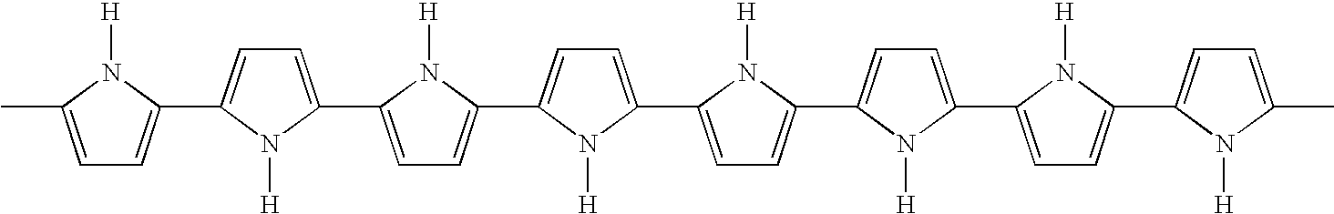
- electroactive polymers are polymers characterized by their ability to change shape in response to electrical stimulation. They commonly feature a conjugated backbone and have the ability to increase electrical conductivity under oxidation or reduction.
- polyaniline polysulfone
- polypyrrole polyacetylene
- These materials are typically semi-conductors in their pure form.
- conductivity is increased.
- the oxidation or reduction leads to a charge imbalance that, in turn, results in a flow of ions into the material in order to balance charge.
- ions, or dopants enter the polymer from an ionically conductive electrolyte medium associated with the electroactive polymer or are redistributed within the polymer.
- the electrolyte may be, for example, in the form of a gel, a solid, or a liquid. If ions are already present in the polymer when it is oxidized or reduced, they may exit the polymer.
- an electroactive polymer actuator 10 is shown schematically in cross-section.
- Active member 12 of actuator 10 has a surface coupled with electrolyte 14 and has an axis 11 .
- Active member 12 includes an electroactive polymer that contracts or expands in response to the flow of ions out of, or into, the active member 12 .
- Ions are provided by electrolyte 14 , which adjoins member 12 over at least a portion, and up to the entirety, of the surface of active member 12 in order to allow for the flow of ions between the two media.
- member 12 may be a film, a group of films, a fiber, a group of fibers, or a combination of the same disposed so as to act collectively to apply a force in a longitudinal direction substantially along axis 11 in this instance.
- Active member 12 includes an electroactive polymer. Many electroactive polymers having desirable properties are known to persons of ordinary skill in the art.
- active member 12 can be a polypyrrole film. Such a polypyrrole film may be synthesized, for example, by electrodeposition according to the method described by M. Yamaura et al., “Enhancement of Electrical Conductivity of Polypyrrole Film by Stretching: Counter-ion Effect,” Synthetic Metals, vol. 36, pp. 209–224 (1988), which is incorporated herein by reference.
- any conducting polymer that exhibits contractile or expansile properties may be used within the scope of the invention. Polyaniline, polysulfone, polyacetylene are examples.
- Electrolyte 14 may be, for example, a liquid, a gel, or a solid, so long as ion movement is allowed. Moreover, where the electrolyte 14 is a solid, it will typically move with the active member 12 and will typically not be subject to delamination. Where the electrolyte 14 is a gel, it may be, for example, an agar or polymethylmethacrylate (PMMA) gel containing a salt dopant. Where the electrolyte is a liquid, it may be, for example, a phosphate buffer solution, KCl, NaCl and so forth. The electrolyte may be non-toxic in the event that a leak inadvertently occurs in vivo.
- Counter electrode 18 is in electrical contact with electrolyte 14 in order to provide a return path for charge to a source 20 of potential difference between member 12 and electrolyte 14 .
- Counter electrode 18 may be any suitable electrical conductor, for example, another conducting polymer, a conducting polymer gel, or a metal such as gold or platinum, which can be, for example, in wire or film form and can be applied, for example, by electroplating, chemical deposition, or printing.
- a current is passed between active member 12 and counter electrode 18 , inducing contraction or expansion of member 12 .
- the actuator may have a flexible skin for separating the electrolyte from an ambient environment.
- the actuator can be provided in an essentially infinite array of configurations as desired, including planar actuator configurations (e.g., with planar active members and counter-electrodes), cylindrical actuator configurations (e.g., see the actuator illustrated in FIG. 5 , which is illustrated as having a cylindrical active member and wire coil counter electrode), and so forth.
- planar actuator configurations e.g., with planar active members and counter-electrodes
- cylindrical actuator configurations e.g., see the actuator illustrated in FIG. 5 , which is illustrated as having a cylindrical active member and wire coil counter electrode
- One or more actuators 140 can be used to change the deflection angle associated with the reflective member 136 .
- these actuators 140 can be associated with the reflective member 136 in a wide range of configurations.
- the angle of inflection of the reflective member 136 increases upon lengthwise expansion of the actuator 140 , and decreases upon lengthwise contraction of the actuator 140 .
- an elongated column of electroactive polymer material can be used in connection with an actuator design like that of FIG. 5 .
- an actuator having substantial tensile strength, but negligible column strength can be placed in tension with a reflective member that is in mechanical communication with a spring element.
- the hinge 137 can be provided with a spring element which urges the mirror in a counterclockwise direction.
- the angle of incidence is controlled based on the degree of contraction or expansion of the actuator 140 , with expansion of the actuator 140 leading to a greater angle of incidence, and contraction leading to a lesser angle of incidence.
- FIG. 6 provides a schematic cross-sectional view of an electroactive polymer layer stack, which can be used in the formation of an expandable actuator 140 .
- a stack of counter-electrode layers 218 , active layers 212 and electrolyte-containing layers 214 are shown.
- the counter-electrode layers 218 may be formed from a suitable electrical conductor, for example, a metal such as gold or platinum.
- the electrolyte within the electrolyte-containing layers 214 can be, for example, a liquid, a gel, or a solid, with appropriate measures being taken, where needed, to prevent short-circuiting between the counter-electrodes 218 and the active layers 212 .
- the active layer 212 comprises an electroactive polymer, for example, polypyrrole, polysulfone, polyacetylene or polyaniline.
- the actively layers 212 can also optionally be provided with conductive electrical contacts (not shown), if desired, to enhance electrical contact with the control system.
- an appropriate potential difference is applied across the active layers 212 and the counter-electrode layers 218 .
- all of the active layers 212 are shorted to one another, as are all of the counter-electrode layers 218 , allowing the active layers 212 to expand and contract simultaneously.
- the electroactive polymer active layers 212 expand and contract upon establishing an appropriate potential difference between the active layers 212 and the counter-electrode layers 218 . This, in turn, expands or contracts the actuator stack.
- electroactive polymer actuators are known in which an electroactive polymer is laminated between conductive layers to produce a bending-type actuation, with the degree of bending being dependent upon on the applied voltage.
- Such an actuator 140 is illustrated in FIG. 1B , wherein the angle at which the reflective member 136 is tilted increases with increasing bend of the actuator 140 .
- Electroactive Polymer Actuators and Devices Yoseph Bar-Cohen, Ed., Proceedings of SPIE Vol. 4329 (5–8 Mar. 20001), pp. 335–349, which is hereby incorporated by reference.
- This reference also describes numerous other known electroactive polymer configurations, including extender, bowtie, diaphragm, spider, tube, and roll configurations, which can be used to change the deflection angle of a reflective member 136 .
- the inclination angle of the reflective member is inferred, for example, from the intrinsic position-dependent electrical properties of the electroactive polymer actuator.
- one or more strain gauges may also be employed to provide electronic feedback regarding the inclination angle of the reflective member. This electronic feedback will also provide a number of additional advantages, including greater stability, error correction, and immunity from drift.
- Strain gauges suitable for use in the present invention include (a) feedback electroactive polymer elements whose impedance or resistance varies as a function of the amount of strain in the device, (b) linear displacement transducers (e.g., an iron slug slidably positioned in the core of a coil) and (c) conventional strain gauges in which the resistance of the device varies as a function of the amount of strain in the device, thus allowing the amount of strain to be readily quantified and monitored.
- Such strain gauges are commercially available from a number of different sources, including National Instruments Co., Austin, Tex., and include piezoresistive strain gauges (for which resistance varies nonlinearly with strain) and bonded metallic strain gauges (for which resistance typically varies linearly with strain).
- Timing and control circuitry is also typically provided in connection with the above described catheter apparatus to control, for example, the operation of the ultrasonic transducer, the actuator, and the motor drive.
- a display is also typically provided, which is operated under the control of the timing and control circuitry for displaying image information.
- FIG. 4 a schematic block diagram is presented in FIG. 4 , which illustrates the electrical components utilized in a catheter system, in accordance with one embodiment of the present invention.
- the entire catheter apparatus including the transducer 132 , actuator 140 , and the coaxial cable 32 , rotates as a single unit in the embodiment described above.
- Electrical connection can nonetheless be established between these components and a non-rotating electrical system using methods known in the art.
- electrical connections can be made as described in U.S. Pat. No. 5,000,185 by using a pair of spaced-apart rotating slip rings 62 , 63 , which are formed of a conducting material, and which are placed in electrical connection with the conductive members of the coaxial cable 32 .
- a pair of spring-urged contacts for example, conductive brushes, can be adapted to slidably engage the slip rings, which contacts are connected to conductors 73 and 74 .
- a rotary transformer familiar in the art (not shown) may provide coupling with no moving parts.
- Motor 99 is driven by and is under the control of electronic circuitry forming a part of electrical system 101 .
- a system 101 includes a timing and control block 102 , which supplies pulses to a transmitter 103 .
- the output of the transmitter 103 is supplied through a transmit/receive switch 104 which supplies the signals through the conductors 73 and 74 , through the slip rings 62 and 63 , through the inner and outer conductors of the coaxial cable 32 , and to the ultrasonic transducer 132 and the actuator 140 as described above.
- System 101 is capable of supplying high frequency energy to the ultrasonic transducer 132 and low frequency/dc energy to the actuator 140 via the transmitter 103 , while at the same time driving the drive shaft 114 using motor 99 , which is also under the control of the timing and control block 102 .
- the motor 99 can be, for example, an open loop stepping motor or a closed drop servo-controlled motor that can be driven by the timing and control block 102 .
- catheters in accordance with the present invention in which motor(s) are provided within the distal end of the catheter, allowing the reflective member to be rotated, for example. Also, as indicated above, the catheter can be manually rotated.
- Voltage pulses for excitation of the transducer 132 commonly range, for example, from 10 to 50 volts.
- the transducer 132 produces ultrasonic waves which emanate therefrom, reflecting from the surface of the reflective member and into the surrounding tissue as described above. Portions of the ultrasonic sonic energy waves rebounding from the tissue are also reflected from the reflective member and back to the transducer 132 , whereupon the transducer acts as a receiver, picking up ultrasonic waves and converting them into electrical signals which are supplied by the coaxial cable 32 , to the slip rings 62 and 63 , through the conductors 73 and 74 , and through the transmit/receive switch 104 to a receiver 106 . These signals are amplified and supplied to a display unit, which includes a display monitor 108 under the control of the timing and control block 102 to supply an image on the display 108 .
- the catheter apparatus of the present invention is introduced into a body lumen of a patient, for example, into the femoral artery.
- the catheter apparatus can be advanced over a guidewire as is known in the art.
- the progress of the catheter into the patient can be observed, for example, under x-ray fluoroscopy.
- the vessel wall itself can be viewed by suitable operation of system 101 . This can be accomplished, for example, by operating the timing control block 102 to cause operation of the motor 99 which in turn causes rotation of the drive shaft.
- the transducer 132 and reflective member are allowed to scan the interior of the vessel in which the catheter is disposed, typically at a rotation rate which achieves a “real-time” scan, for example, 30 frames per second (i.e., 1800 frames, or rotations, per minute). Suitable rotation rates are thus typically in the range of 5 to 60 revolutions per second, i.e., 300 to 3600 rpm.
- An image of what is being scanned will appear on the screen 108 of the display device.
- the drive shaft may be manually rotated (or aimed without rotation) to provide a desired image.
- motorized rotation will provide a higher definition image.
- a portion of the vessel length can also be longitudinally scanned by operating the actuator 140 to tilt the reflective member.
- the scan can constitute a forward scan, a lateral scan, a rearward scan, or a combination of all three.
- the catheters of the present invention may further include interventional capability, for example, for recanalization of occluded regions within the imaged blood vessel, as is known in the art.
- interventional capability for example, for recanalization of occluded regions within the imaged blood vessel, as is known in the art.
- recanalization is meant both the opening of total occlusions as well as the broadening of the vessel lumen in partial occlusions.
- Catheters combining ultrasonic imaging capability with atherectomy devices for severing of the stenotic material are described in detail in U.S. Pat. No. 5,000,185.
- the catheters of the present invention are not limited to use in atherectomy and can be used to perform a wide variety of other interventional techniques that are performed with vascular catheters.
- Suitable interventional techniques include balloon angioplasty, cutting balloons, laser ablation angioplasty, balloon embolectomy, aspiration embolectomy, heat probe ablation, abrasion, drilling, therapeutic ultrasound, and the like.
- the catheters may be adapted for introducing clot-dissolving drugs, such as tissue plasminogen activator, streptokinase, or urokinase, in order to reduce the stenosis, as well as anti-restenosis drug which inhibit restenosis, such as paclitaxel.
Landscapes
- Health & Medical Sciences (AREA)
- Life Sciences & Earth Sciences (AREA)
- Engineering & Computer Science (AREA)
- Physics & Mathematics (AREA)
- Medical Informatics (AREA)
- Molecular Biology (AREA)
- Radiology & Medical Imaging (AREA)
- Nuclear Medicine, Radiotherapy & Molecular Imaging (AREA)
- Biomedical Technology (AREA)
- Heart & Thoracic Surgery (AREA)
- Biophysics (AREA)
- Pathology (AREA)
- Surgery (AREA)
- Animal Behavior & Ethology (AREA)
- General Health & Medical Sciences (AREA)
- Public Health (AREA)
- Veterinary Medicine (AREA)
- Acoustics & Sound (AREA)
- Multimedia (AREA)
- Ultra Sonic Daignosis Equipment (AREA)
Abstract
Description
Claims (17)
Priority Applications (6)
| Application Number | Priority Date | Filing Date | Title |
|---|---|---|---|
| US10/631,872 US7077808B2 (en) | 2003-07-31 | 2003-07-31 | Ultrasonic imaging catheter |
| JP2006522110A JP2007500556A (en) | 2003-07-31 | 2004-07-29 | Ultrasound imaging catheter |
| PCT/US2004/024744 WO2005011504A1 (en) | 2003-07-31 | 2004-07-29 | Ultrasonic imaging catheter |
| CA2534320A CA2534320C (en) | 2003-07-31 | 2004-07-29 | Ultrasonic imaging catheter |
| EP04779714A EP1659951A1 (en) | 2003-07-31 | 2004-07-29 | Ultrasonic imaging catheter |
| US11/487,714 US8092391B2 (en) | 2003-07-31 | 2006-07-17 | Ultrasonic imaging catheter |
Applications Claiming Priority (1)
| Application Number | Priority Date | Filing Date | Title |
|---|---|---|---|
| US10/631,872 US7077808B2 (en) | 2003-07-31 | 2003-07-31 | Ultrasonic imaging catheter |
Related Child Applications (1)
| Application Number | Title | Priority Date | Filing Date |
|---|---|---|---|
| US11/487,714 Continuation US8092391B2 (en) | 2003-07-31 | 2006-07-17 | Ultrasonic imaging catheter |
Publications (2)
| Publication Number | Publication Date |
|---|---|
| US20050027198A1 US20050027198A1 (en) | 2005-02-03 |
| US7077808B2 true US7077808B2 (en) | 2006-07-18 |
Family
ID=34104208
Family Applications (2)
| Application Number | Title | Priority Date | Filing Date |
|---|---|---|---|
| US10/631,872 Expired - Lifetime US7077808B2 (en) | 2003-07-31 | 2003-07-31 | Ultrasonic imaging catheter |
| US11/487,714 Expired - Fee Related US8092391B2 (en) | 2003-07-31 | 2006-07-17 | Ultrasonic imaging catheter |
Family Applications After (1)
| Application Number | Title | Priority Date | Filing Date |
|---|---|---|---|
| US11/487,714 Expired - Fee Related US8092391B2 (en) | 2003-07-31 | 2006-07-17 | Ultrasonic imaging catheter |
Country Status (5)
| Country | Link |
|---|---|
| US (2) | US7077808B2 (en) |
| EP (1) | EP1659951A1 (en) |
| JP (1) | JP2007500556A (en) |
| CA (1) | CA2534320C (en) |
| WO (1) | WO2005011504A1 (en) |
Cited By (17)
| Publication number | Priority date | Publication date | Assignee | Title |
|---|---|---|---|---|
| US20050203416A1 (en) * | 2004-03-10 | 2005-09-15 | Angelsen Bjorn A. | Extended, ultrasound real time 2D imaging probe for insertion into the body |
| US20060264758A1 (en) * | 2005-05-05 | 2006-11-23 | Volcano Corporation | Capacitive microfabricated ultrasound transducer-based intravascular ultrasound probes |
| US20070038110A1 (en) * | 2005-07-07 | 2007-02-15 | Aime Flesch | Motorized ultrasonic scanhead |
| US20070038114A1 (en) * | 2003-07-31 | 2007-02-15 | Couvillon Lucien A Jr | Ultrasonic imaging catheter |
| US20070167827A1 (en) * | 2005-11-12 | 2007-07-19 | Donald Masters | Systems And Methods For Reducing Noise In An Imaging Catheter System |
| US20080027377A1 (en) * | 2006-07-31 | 2008-01-31 | Boston Scientific Scimed, Inc. | Catheters having actuatable lumen assemblies |
| US20080027528A1 (en) * | 2006-07-31 | 2008-01-31 | Boston Scientific Scimed, Inc. | Stent retaining mechanisms |
| US20080086081A1 (en) * | 2006-07-31 | 2008-04-10 | Tracee Eidenschink | Medical balloon incorporating electroactive polymer and methods of making and using the same |
| US20080177139A1 (en) * | 2007-01-19 | 2008-07-24 | Brian Courtney | Medical imaging probe with rotary encoder |
| US20090247879A1 (en) * | 2004-03-09 | 2009-10-01 | Angelsen Bjorn A J | Extended ultrasound imaging probe for insertion into the body |
| US20090264768A1 (en) * | 2007-01-19 | 2009-10-22 | Brian Courtney | Scanning mechanisms for imaging probe |
| WO2010055443A1 (en) * | 2008-11-12 | 2010-05-20 | Koninklijke Philips Electronics N.V. | Acoustical switch and catheter comprising acoustical switch |
| US20100249599A1 (en) * | 2009-03-31 | 2010-09-30 | Boston Scientific Scimed, Inc. | Systems and methods for making and using an imaging core of an intravascular ultrasound imaging system |
| US20120059241A1 (en) * | 2010-09-08 | 2012-03-08 | Boston Scientific Scimed, Inc. | Systems and methods for making and using a steerable imaging system configured and arranged for insertion into a patient |
| US20150359509A1 (en) * | 2014-06-16 | 2015-12-17 | Dongguk University Industry-Academic Cooperation Foundation | Pullback system |
| US9717476B2 (en) | 2013-11-08 | 2017-08-01 | Samsung Electronics Co., Ltd. | Probe and medical imaging apparatus including the same |
| WO2018109052A1 (en) | 2016-12-16 | 2018-06-21 | Koninklijke Philips N.V. | Intravascular doppler ultrasonic device and method for controlling its operation |
Families Citing this family (46)
| Publication number | Priority date | Publication date | Assignee | Title |
|---|---|---|---|---|
| CA2372261A1 (en) * | 2002-02-15 | 2003-08-15 | Paul Ouellette | System for measuring shrinkage behaviour and modulus change during solidification of a polymeric resin, and method therefor |
| CN1910810B (en) * | 2004-02-05 | 2010-04-21 | 松下电器产业株式会社 | Actuator and method for manufacturing planar electrode support for actuator |
| US8211088B2 (en) | 2005-10-14 | 2012-07-03 | Boston Scientific Scimed, Inc. | Catheter with controlled lumen recovery |
| CA2630215A1 (en) * | 2005-11-17 | 2007-05-24 | Micromuscle Ab | Medical devices and methods for their fabrication and use |
| US20070167825A1 (en) * | 2005-11-30 | 2007-07-19 | Warren Lee | Apparatus for catheter tips, including mechanically scanning ultrasound probe catheter tip |
| US20070123750A1 (en) * | 2005-11-30 | 2007-05-31 | General Electric Company | Catheter apparatus and methods of using same |
| US20070167826A1 (en) * | 2005-11-30 | 2007-07-19 | Warren Lee | Apparatuses for thermal management of actuated probes, such as catheter distal ends |
| US20070167821A1 (en) * | 2005-11-30 | 2007-07-19 | Warren Lee | Rotatable transducer array for volumetric ultrasound |
| JP5261730B2 (en) * | 2006-01-05 | 2013-08-14 | イーメックス株式会社 | Conductive polymer actuator element |
| US20080183070A1 (en) * | 2007-01-29 | 2008-07-31 | Wisconsin Alumni Research Foundation | Multi-mode medical device system with thermal ablation capability and methods of using same |
| US8852112B2 (en) | 2007-06-28 | 2014-10-07 | W. L. Gore & Associates, Inc. | Catheter with deflectable imaging device and bendable electrical conductor |
| US8864675B2 (en) * | 2007-06-28 | 2014-10-21 | W. L. Gore & Associates, Inc. | Catheter |
| US8285362B2 (en) | 2007-06-28 | 2012-10-09 | W. L. Gore & Associates, Inc. | Catheter with deflectable imaging device |
| CA2835549C (en) * | 2008-05-30 | 2015-07-28 | Gore Enterprise Holdings, Inc. | Real time ultrasound catheter probe |
| US8465686B2 (en) * | 2008-12-19 | 2013-06-18 | Volcano Corporation | Method of manufacturing a rotational intravascular ultrasound probe |
| US8556813B2 (en) | 2009-07-08 | 2013-10-15 | Sanuwave, Inc. | Extracorporeal pressure shock wave device |
| US8550823B2 (en) * | 2011-01-24 | 2013-10-08 | Single Buoy Moorings, Inc. | Rigid to elastic electrode connection |
| WO2012102415A1 (en) * | 2011-01-24 | 2012-08-02 | 서울대학교 산학협력단 | Electroactive polymer oscillator, method for manufacturing same, and method for dissolving blood clots using same |
| US20120232630A1 (en) * | 2011-03-07 | 2012-09-13 | Eugene Dariush Daneshvar | Articulating interfaces for biological tissues |
| WO2013118628A1 (en) * | 2012-02-08 | 2013-08-15 | アルプス電気株式会社 | Polymer actuator element and method and device for driving polymer actuator element |
| GB2534669A (en) * | 2012-05-03 | 2016-08-03 | Los Alamos Nat Security Llc | Acoustic camera |
| AU2013328995B2 (en) | 2012-10-12 | 2018-02-01 | Muffin Incorporated | Mechanical scanning ultrasound transducer with micromotor |
| EP2906124B1 (en) | 2012-10-12 | 2021-01-20 | Muffin Incorporated | Substantially acoustically transparent and conductive window |
| EP2906134A4 (en) | 2012-10-12 | 2016-06-29 | Muffin Inc | Reciprocating internal ultrasound transducer assembly |
| JP6382201B2 (en) | 2012-10-12 | 2018-08-29 | マフィン・インコーポレイテッドMuffin Incorporated | Equipment for three-dimensional internal ultrasound use |
| EP2908732B1 (en) | 2012-10-16 | 2020-06-24 | Muffin Incorporated | Internal transducer assembly with slip ring |
| CA2895821A1 (en) * | 2012-12-21 | 2014-06-26 | Volcano Corporation | Focused rotational ivus transducer using single crystal composite material |
| WO2014150373A1 (en) | 2013-03-15 | 2014-09-25 | Muffin Incorporated | Internal ultrasound assembly with port for fluid injection |
| WO2014150376A1 (en) | 2013-03-15 | 2014-09-25 | Muffin Incorporated | Internal ultrasound assembly fluid seal |
| CN103892871B (en) | 2014-04-17 | 2015-11-25 | 深圳大学 | A kind of machinery rotating type intravascular ultrasound probes |
| EP3215019B1 (en) * | 2015-01-07 | 2019-05-15 | St. Jude Medical, Cardiology Division, Inc. | Imaging device |
| US11317892B2 (en) * | 2015-08-12 | 2022-05-03 | Muffin Incorporated | Over-the-wire ultrasound system with torque-cable driven rotary transducer |
| US10695026B2 (en) | 2015-08-12 | 2020-06-30 | Muffin Incorporated | Device for three-dimensional, internal ultrasound with rotating transducer and rotating reflector |
| US10327667B2 (en) | 2016-05-13 | 2019-06-25 | Becton, Dickinson And Company | Electro-magnetic needle catheter insertion system |
| US10583269B2 (en) * | 2016-06-01 | 2020-03-10 | Becton, Dickinson And Company | Magnetized catheters, devices, uses and methods of using magnetized catheters |
| US11826522B2 (en) | 2016-06-01 | 2023-11-28 | Becton, Dickinson And Company | Medical devices, systems and methods utilizing permanent magnet and magnetizable feature |
| US20170347914A1 (en) | 2016-06-01 | 2017-12-07 | Becton, Dickinson And Company | Invasive Medical Devices Including Magnetic Region And Systems And Methods |
| US11413429B2 (en) | 2016-06-01 | 2022-08-16 | Becton, Dickinson And Company | Medical devices, systems and methods utilizing permanent magnet and magnetizable feature |
| US10032552B2 (en) | 2016-08-30 | 2018-07-24 | Becton, Dickinson And Company | Cover for tissue penetrating device with integrated magnets and magnetic shielding |
| US10761312B2 (en) * | 2016-09-28 | 2020-09-01 | General Electric Company | Integrated variable view optical adapter for an optical scope |
| EP3518777B1 (en) * | 2016-09-29 | 2021-03-10 | Koninklijke Philips N.V. | Intracardiac echocardiography (ice) catheter tip assembly |
| WO2018077909A1 (en) * | 2016-10-26 | 2018-05-03 | Koninklijke Philips N.V. | Interventional instrument comprising an ultrasound transducer |
| EP3544515B1 (en) * | 2016-11-22 | 2021-01-06 | Koninklijke Philips N.V. | Ultrasound device and acoustic component for use in such a device |
| CN106361295A (en) * | 2016-12-06 | 2017-02-01 | 全景恒升(北京)科学技术有限公司 | Optical and acoustic mixed imaging conduit |
| CN111938694B (en) * | 2020-08-07 | 2022-10-11 | 深圳北芯生命科技股份有限公司 | Transmission device of ultrasonic transducer and manufacturing method thereof |
| WO2023159255A2 (en) * | 2022-02-18 | 2023-08-24 | Simpson Interventions, Inc. | Systems, devices and methods for antegrade dissection and reentry |
Citations (30)
| Publication number | Priority date | Publication date | Assignee | Title |
|---|---|---|---|---|
| EP0139574A2 (en) | 1983-09-29 | 1985-05-02 | Bruno Denis Lucien Fornage | Ultrasonic probe for body tracts |
| US4546771A (en) * | 1982-03-04 | 1985-10-15 | Indianapolis Center For Advanced Research, Inc. (Icfar) | Acoustic microscope |
| US5000185A (en) | 1986-02-28 | 1991-03-19 | Cardiovascular Imaging Systems, Inc. | Method for intravascular two-dimensional ultrasonography and recanalization |
| US5250167A (en) | 1992-06-22 | 1993-10-05 | The United States Of America As Represented By The United States Department Of Energy | Electrically controlled polymeric gel actuators |
| US5268082A (en) | 1991-02-28 | 1993-12-07 | Agency Of Industrial Science And Technology | Actuator element |
| US5372138A (en) | 1988-03-21 | 1994-12-13 | Boston Scientific Corporation | Acousting imaging catheters and the like |
| US5377685A (en) | 1993-12-17 | 1995-01-03 | Baylis Medical Company, Inc. | Ultrasound catheter with mechanically steerable beam |
| US5389222A (en) | 1993-09-21 | 1995-02-14 | The United States Of America As Represented By The United States Department Of Energy | Spring-loaded polymeric gel actuators |
| US5556700A (en) | 1994-03-25 | 1996-09-17 | Trustees Of The University Of Pennsylvania | Conductive polyaniline laminates |
| US5631040A (en) | 1989-07-11 | 1997-05-20 | Ngk Insulators, Ltd. | Method of fabricating a piezoelectric/electrostrictive actuator |
| US5651366A (en) | 1994-09-19 | 1997-07-29 | Board Of Trustees Of The Leland Stanford Junior University | Forward viewing ultrasonic imaging catheter |
| US5682897A (en) | 1990-05-18 | 1997-11-04 | Cardiovascular Imaging Systems, Inc. | Guidewire with imaging capability |
| US5771902A (en) | 1995-09-25 | 1998-06-30 | Regents Of The University Of California | Micromachined actuators/sensors for intratubular positioning/steering |
| US5855565A (en) | 1997-02-21 | 1999-01-05 | Bar-Cohen; Yaniv | Cardiovascular mechanically expanding catheter |
| US5897522A (en) | 1995-12-20 | 1999-04-27 | Power Paper Ltd. | Flexible thin layer open electrochemical cell and applications of same |
| US6060811A (en) | 1997-07-25 | 2000-05-09 | The United States Of America As Represented By The United States National Aeronautics And Space Administration | Advanced layered composite polylaminate electroactive actuator and sensor |
| US6074349A (en) | 1994-11-30 | 2000-06-13 | Boston Scientific Corporation | Acoustic imaging and doppler catheters and guidewires |
| US6109852A (en) | 1996-01-18 | 2000-08-29 | University Of New Mexico | Soft actuators and artificial muscles |
| US6200269B1 (en) | 1998-05-28 | 2001-03-13 | Diasonics, Ultrasound, Inc. | Forward-scanning ultrasound catheter probe |
| US6249076B1 (en) | 1998-04-14 | 2001-06-19 | Massachusetts Institute Of Technology | Conducting polymer actuator |
| US6248074B1 (en) | 1997-09-30 | 2001-06-19 | Olympus Optical Co., Ltd. | Ultrasonic diagnosis system in which periphery of magnetic sensor included in distal part of ultrasonic endoscope is made of non-conductive material |
| WO2001058973A2 (en) | 2000-02-09 | 2001-08-16 | Sri International | Energy efficient electroactive polymers and electroactive polymer devices |
| US6376971B1 (en) | 1997-02-07 | 2002-04-23 | Sri International | Electroactive polymer electrodes |
| US20020120297A1 (en) | 2001-02-26 | 2002-08-29 | Shadduck John H. | Vaso-occlusive implants for interventional neuroradiology |
| US6457365B1 (en) | 2000-02-09 | 2002-10-01 | Endosonics Corporation | Method and apparatus for ultrasonic imaging |
| US6514237B1 (en) | 2000-11-06 | 2003-02-04 | Cordis Corporation | Controllable intralumen medical device |
| US6540677B1 (en) | 2000-11-17 | 2003-04-01 | Bjorn A. J. Angelsen | Ultrasound transceiver system for remote operation through a minimal number of connecting wires |
| US6545384B1 (en) | 1997-02-07 | 2003-04-08 | Sri International | Electroactive polymer devices |
| US6543110B1 (en) | 1997-02-07 | 2003-04-08 | Sri International | Electroactive polymer fabrication |
| US6586859B2 (en) | 2000-04-05 | 2003-07-01 | Sri International | Electroactive polymer animated devices |
Family Cites Families (11)
| Publication number | Priority date | Publication date | Assignee | Title |
|---|---|---|---|---|
| US5368035A (en) * | 1988-03-21 | 1994-11-29 | Boston Scientific Corporation | Ultrasound imaging guidewire |
| US5286897A (en) * | 1990-12-28 | 1994-02-15 | Ajinomoto Co., Inc. | N-t-butyloxycarbonyl-3-cyclohexyl-L-alanine methyl ester in crystalline form |
| US5770902A (en) * | 1995-11-02 | 1998-06-23 | Globe Motors | Motor termination board |
| US6058323A (en) * | 1996-11-05 | 2000-05-02 | Lemelson; Jerome | System and method for treating select tissue in a living being |
| WO1999037211A1 (en) * | 1998-01-26 | 1999-07-29 | Scimed Life Systems, Inc. | Catheter assembly with distal end inductive coupler and embedded transmission line |
| US6379971B1 (en) * | 1998-02-24 | 2002-04-30 | Target Discovery, Inc. | Methods for sequencing proteins |
| US6592526B1 (en) * | 1999-01-25 | 2003-07-15 | Jay Alan Lenker | Resolution ultrasound devices for imaging and treatment of body lumens |
| CA2352653A1 (en) * | 2000-07-06 | 2002-01-06 | Robert I. Macdonald | Acoustically actuated mems devices |
| US6770027B2 (en) * | 2001-10-05 | 2004-08-03 | Scimed Life Systems, Inc. | Robotic endoscope with wireless interface |
| US6749556B2 (en) * | 2002-05-10 | 2004-06-15 | Scimed Life Systems, Inc. | Electroactive polymer based artificial sphincters and artificial muscle patches |
| US7077808B2 (en) * | 2003-07-31 | 2006-07-18 | Boston Scientific Scimed. Inc. | Ultrasonic imaging catheter |
-
2003
- 2003-07-31 US US10/631,872 patent/US7077808B2/en not_active Expired - Lifetime
-
2004
- 2004-07-29 CA CA2534320A patent/CA2534320C/en not_active Expired - Fee Related
- 2004-07-29 JP JP2006522110A patent/JP2007500556A/en not_active Withdrawn
- 2004-07-29 EP EP04779714A patent/EP1659951A1/en not_active Withdrawn
- 2004-07-29 WO PCT/US2004/024744 patent/WO2005011504A1/en active Application Filing
-
2006
- 2006-07-17 US US11/487,714 patent/US8092391B2/en not_active Expired - Fee Related
Patent Citations (33)
| Publication number | Priority date | Publication date | Assignee | Title |
|---|---|---|---|---|
| US4546771A (en) * | 1982-03-04 | 1985-10-15 | Indianapolis Center For Advanced Research, Inc. (Icfar) | Acoustic microscope |
| EP0139574A2 (en) | 1983-09-29 | 1985-05-02 | Bruno Denis Lucien Fornage | Ultrasonic probe for body tracts |
| US5865178A (en) | 1986-02-28 | 1999-02-02 | Cardiovascular Imaging System, Inc. | Method and apparatus for intravascular ultrasonography |
| US5000185A (en) | 1986-02-28 | 1991-03-19 | Cardiovascular Imaging Systems, Inc. | Method for intravascular two-dimensional ultrasonography and recanalization |
| US5372138A (en) | 1988-03-21 | 1994-12-13 | Boston Scientific Corporation | Acousting imaging catheters and the like |
| US5631040A (en) | 1989-07-11 | 1997-05-20 | Ngk Insulators, Ltd. | Method of fabricating a piezoelectric/electrostrictive actuator |
| US5938609A (en) | 1990-05-18 | 1999-08-17 | Cardiovascular Imaging Systems, Inc. | Guidewire with imaging capability |
| US5682897A (en) | 1990-05-18 | 1997-11-04 | Cardiovascular Imaging Systems, Inc. | Guidewire with imaging capability |
| US5268082A (en) | 1991-02-28 | 1993-12-07 | Agency Of Industrial Science And Technology | Actuator element |
| US5250167A (en) | 1992-06-22 | 1993-10-05 | The United States Of America As Represented By The United States Department Of Energy | Electrically controlled polymeric gel actuators |
| US5389222A (en) | 1993-09-21 | 1995-02-14 | The United States Of America As Represented By The United States Department Of Energy | Spring-loaded polymeric gel actuators |
| US5377685A (en) | 1993-12-17 | 1995-01-03 | Baylis Medical Company, Inc. | Ultrasound catheter with mechanically steerable beam |
| US5556700A (en) | 1994-03-25 | 1996-09-17 | Trustees Of The University Of Pennsylvania | Conductive polyaniline laminates |
| US5651366A (en) | 1994-09-19 | 1997-07-29 | Board Of Trustees Of The Leland Stanford Junior University | Forward viewing ultrasonic imaging catheter |
| US6074349A (en) | 1994-11-30 | 2000-06-13 | Boston Scientific Corporation | Acoustic imaging and doppler catheters and guidewires |
| US5771902A (en) | 1995-09-25 | 1998-06-30 | Regents Of The University Of California | Micromachined actuators/sensors for intratubular positioning/steering |
| US5897522A (en) | 1995-12-20 | 1999-04-27 | Power Paper Ltd. | Flexible thin layer open electrochemical cell and applications of same |
| US6109852A (en) | 1996-01-18 | 2000-08-29 | University Of New Mexico | Soft actuators and artificial muscles |
| US6545384B1 (en) | 1997-02-07 | 2003-04-08 | Sri International | Electroactive polymer devices |
| US6376971B1 (en) | 1997-02-07 | 2002-04-23 | Sri International | Electroactive polymer electrodes |
| US6583533B2 (en) | 1997-02-07 | 2003-06-24 | Sri International | Electroactive polymer electrodes |
| US6543110B1 (en) | 1997-02-07 | 2003-04-08 | Sri International | Electroactive polymer fabrication |
| US5855565A (en) | 1997-02-21 | 1999-01-05 | Bar-Cohen; Yaniv | Cardiovascular mechanically expanding catheter |
| US6060811A (en) | 1997-07-25 | 2000-05-09 | The United States Of America As Represented By The United States National Aeronautics And Space Administration | Advanced layered composite polylaminate electroactive actuator and sensor |
| US6248074B1 (en) | 1997-09-30 | 2001-06-19 | Olympus Optical Co., Ltd. | Ultrasonic diagnosis system in which periphery of magnetic sensor included in distal part of ultrasonic endoscope is made of non-conductive material |
| US6249076B1 (en) | 1998-04-14 | 2001-06-19 | Massachusetts Institute Of Technology | Conducting polymer actuator |
| US6200269B1 (en) | 1998-05-28 | 2001-03-13 | Diasonics, Ultrasound, Inc. | Forward-scanning ultrasound catheter probe |
| WO2001058973A2 (en) | 2000-02-09 | 2001-08-16 | Sri International | Energy efficient electroactive polymers and electroactive polymer devices |
| US6457365B1 (en) | 2000-02-09 | 2002-10-01 | Endosonics Corporation | Method and apparatus for ultrasonic imaging |
| US6586859B2 (en) | 2000-04-05 | 2003-07-01 | Sri International | Electroactive polymer animated devices |
| US6514237B1 (en) | 2000-11-06 | 2003-02-04 | Cordis Corporation | Controllable intralumen medical device |
| US6540677B1 (en) | 2000-11-17 | 2003-04-01 | Bjorn A. J. Angelsen | Ultrasound transceiver system for remote operation through a minimal number of connecting wires |
| US20020120297A1 (en) | 2001-02-26 | 2002-08-29 | Shadduck John H. | Vaso-occlusive implants for interventional neuroradiology |
Non-Patent Citations (16)
| Title |
|---|
| American Medical Systems. Important for Paients Considering an Acticon Neosphincter. www.fda.gov/ohrms/dockets/ac/01/briefing/377b1<SUB>-</SUB>02<SUB>-</SUB>patient%20info. |
| David L. Brock, "Review of Artificial Muscle based on Contractile Polymers." www.ai.mit.edu/projects/muscle/papers/memo1330/memo1330html. |
| Edwin H. Jager et al., "Microfabricating Conjugated Polymer Actuators," Science, vol. 290, Nov. 24, 2000, pp. 1540-1545. |
| Eniko T. Enikov et al., "Electrotransport and Deformation Model of Ion Exchange Membrane based Actuators," Smart Structures and Materials 2000: Electroactive Polymer Actuators and Devices, Yoseph Bar-Cohen, ed, SPIE Proceedings, vol. 3987 (2000), pp. 129-139. |
| John D.W. Madden et al., "Conducting Polymer Actuators as Engineer Materials," Smart Structures and Materials 2002: Electroactive Polymer Actuators and Devices, Yoseph Bar-Cohen, ed, SPIE Proceedings, vol. 4695 (2002), pp. 176-190. |
| John D.W. Madden et al., "Polyprrole Actuators: Modeling and Performance," Smart Structures and Materials 2001: Electroactive Polymer Actuators and Devices, Yoseph Bar-Cohen, ed., SPIE Proceedings, vol. 4329 (Mar. 5-8, 2001), pp. 72-83. |
| Jose-Maria Sansinena et al., Electroactive Polymer (EAP) Actuators as Artificial Muscles, chap. 7, Conductive Polymers (SPIE Press, 2001), pp. 193-221. |
| Rainer W. Gulch et al., "Electrochemical Stimulation and Control of Electroactive Polymer Gels," Smart Structures and Materials 2001: Electroactive Polymer Actuators and Devices, Yoseph Bar-Cohen, ed, SPIE Proceedings, vol. 4329 (2001), pp. 328-334. |
| Ron Pelrine, "Applications of Dielectric Elastomer Actuators," Smart Structures and Materials 2001: Electroactive Polymer Actuators and Devices, Yoseph Bar-Cohen, ed., SPIE Proceedings vol. 4329 (Mar. 5-8, 2001), pp. 335-349. |
| T. Hagiwara et al., "Enhancement of the Electrical Conductivity of Polypyrrole Film by Stretching: Influence of the Polymerization Conditions," Synthetic Metals, vol. 36 (1990) pp. 241-252. |
| WorldWide ElectroActive Polymers (Artificial Muscles) Newsletter, vol. 3, No. 1 (Jun. 2001), pp. 1-14. |
| Yamaura, M., et al., "Enhancement of Electrical Conductivity of Polypyrrole Film By Stretching: Counter Ion Effect," Synthetic Metals, vol. 26 (1988) pp. 209-224. |
| Yoseph Bar-Cohen, "Transition of EAP material from novelty to practical applications -are we there yet?" Smart Structures and Materials 2001: Electroactive Polymer Actuators and Devices, Yoseph Bar-Cohen, ed., Proceedings of SPIE, vol. 4329, 1-6. |
| Yoseph Bar-Cohen, Chap. 1, EAP History, Current Status, and Infrastructure, EAP Actuators as Artificial Muscles (SPIE Press, 2001), pp. 3-44. |
| Yoseph Bar-Cohen, ed., Electroactive Polymer (EAP) Actuators as Artificial Muscles: Reality, Potential, and Challenges, chap. 16: Application of Dielectric Elastomer EAP Actuators, SPIE Press (2001), pp. 457-495. |
| Yoseph Bar-Cohen, ed., Electroactive Polymer (EAP) Actuators as Artificial Muscles: Reality, Potential, and Challenges, Chap. 21 EAP Applications, Potential, and Challenges. SPIE Press (2001), pp. 615-659. |
Cited By (42)
| Publication number | Priority date | Publication date | Assignee | Title |
|---|---|---|---|---|
| US20070038114A1 (en) * | 2003-07-31 | 2007-02-15 | Couvillon Lucien A Jr | Ultrasonic imaging catheter |
| US8092391B2 (en) * | 2003-07-31 | 2012-01-10 | Boston Scientific Scimed, Inc. | Ultrasonic imaging catheter |
| US20090247879A1 (en) * | 2004-03-09 | 2009-10-01 | Angelsen Bjorn A J | Extended ultrasound imaging probe for insertion into the body |
| US20050203416A1 (en) * | 2004-03-10 | 2005-09-15 | Angelsen Bjorn A. | Extended, ultrasound real time 2D imaging probe for insertion into the body |
| US20060264758A1 (en) * | 2005-05-05 | 2006-11-23 | Volcano Corporation | Capacitive microfabricated ultrasound transducer-based intravascular ultrasound probes |
| US8231535B2 (en) * | 2005-05-05 | 2012-07-31 | Volcano Corporation | Capacitative microfabricated ultrasound transducer-based intravascular ultrasound probes |
| US20110172543A1 (en) * | 2005-05-05 | 2011-07-14 | Volcano Corporation | Multipurpose Host System for Invasive Cardiovascular Diagnostic Measurement Acquisition and Display |
| US7914458B2 (en) * | 2005-05-05 | 2011-03-29 | Volcano Corporation | Capacitive microfabricated ultrasound transducer-based intravascular ultrasound probes |
| US20070038110A1 (en) * | 2005-07-07 | 2007-02-15 | Aime Flesch | Motorized ultrasonic scanhead |
| US7798971B2 (en) * | 2005-07-07 | 2010-09-21 | Vermon | Motorized ultrasonic scanhead |
| US20070167827A1 (en) * | 2005-11-12 | 2007-07-19 | Donald Masters | Systems And Methods For Reducing Noise In An Imaging Catheter System |
| US7887488B2 (en) * | 2005-11-12 | 2011-02-15 | Scimed Life Systems, Inc. | Systems and methods for reducing noise in an imaging catheter system |
| US8439961B2 (en) | 2006-07-31 | 2013-05-14 | Boston Scientific Scimed, Inc. | Stent retaining mechanisms |
| US7909844B2 (en) | 2006-07-31 | 2011-03-22 | Boston Scientific Scimed, Inc. | Catheters having actuatable lumen assemblies |
| US20080027377A1 (en) * | 2006-07-31 | 2008-01-31 | Boston Scientific Scimed, Inc. | Catheters having actuatable lumen assemblies |
| US7777399B2 (en) | 2006-07-31 | 2010-08-17 | Boston Scientific Scimed, Inc. | Medical balloon incorporating electroactive polymer and methods of making and using the same |
| US20080027528A1 (en) * | 2006-07-31 | 2008-01-31 | Boston Scientific Scimed, Inc. | Stent retaining mechanisms |
| US20080086081A1 (en) * | 2006-07-31 | 2008-04-10 | Tracee Eidenschink | Medical balloon incorporating electroactive polymer and methods of making and using the same |
| US20100312322A1 (en) * | 2006-07-31 | 2010-12-09 | Boston Scientific Scimed, Inc. | Medical Balloon Incorporating Electroactive Polymer and Methods of Making and Using the Same |
| US7919910B2 (en) | 2006-07-31 | 2011-04-05 | Boston Scientific Scimed, Inc. | Medical balloon incorporating electroactive polymer and methods of making and using the same |
| US8784321B2 (en) | 2007-01-19 | 2014-07-22 | Sunnybrook Health Sciences Centre | Imaging probe with combined ultrasound and optical means of imaging |
| EP3120752A1 (en) | 2007-01-19 | 2017-01-25 | Sunnybrook Health Sciences Centre | Scanning mechanisms for imaging probe |
| US20080177138A1 (en) * | 2007-01-19 | 2008-07-24 | Brian Courtney | Scanning mechanisms for imaging probe |
| US7972272B2 (en) | 2007-01-19 | 2011-07-05 | University Health Network | Electrostatically driven image probe |
| US20080177139A1 (en) * | 2007-01-19 | 2008-07-24 | Brian Courtney | Medical imaging probe with rotary encoder |
| US20080177183A1 (en) * | 2007-01-19 | 2008-07-24 | Brian Courtney | Imaging probe with combined ultrasounds and optical means of imaging |
| US20090264768A1 (en) * | 2007-01-19 | 2009-10-22 | Brian Courtney | Scanning mechanisms for imaging probe |
| US8712506B2 (en) | 2007-01-19 | 2014-04-29 | Sunnybrook Health Sciences Centre | Medical imaging probe with rotary encoder |
| US8214010B2 (en) | 2007-01-19 | 2012-07-03 | Sunnybrook Health Sciences Centre | Scanning mechanisms for imaging probe |
| US20080243002A1 (en) * | 2007-01-19 | 2008-10-02 | Nigel Robert Munce | Electrostatically driven image probe |
| US8460195B2 (en) | 2007-01-19 | 2013-06-11 | Sunnybrook Health Sciences Centre | Scanning mechanisms for imaging probe |
| WO2010055443A1 (en) * | 2008-11-12 | 2010-05-20 | Koninklijke Philips Electronics N.V. | Acoustical switch and catheter comprising acoustical switch |
| US20110275962A1 (en) * | 2008-11-12 | 2011-11-10 | Koninklijke Philips Electronics N.V. | Acoustical switch and catheter comprising acoustical switch |
| US8647281B2 (en) | 2009-03-31 | 2014-02-11 | Boston Scientific Scimed, Inc. | Systems and methods for making and using an imaging core of an intravascular ultrasound imaging system |
| US20100249599A1 (en) * | 2009-03-31 | 2010-09-30 | Boston Scientific Scimed, Inc. | Systems and methods for making and using an imaging core of an intravascular ultrasound imaging system |
| US20120059241A1 (en) * | 2010-09-08 | 2012-03-08 | Boston Scientific Scimed, Inc. | Systems and methods for making and using a steerable imaging system configured and arranged for insertion into a patient |
| US9717476B2 (en) | 2013-11-08 | 2017-08-01 | Samsung Electronics Co., Ltd. | Probe and medical imaging apparatus including the same |
| US20150359509A1 (en) * | 2014-06-16 | 2015-12-17 | Dongguk University Industry-Academic Cooperation Foundation | Pullback system |
| US9907537B2 (en) * | 2014-06-16 | 2018-03-06 | Dongguk University Industry-Academic Cooperation Foundation | Pullback system |
| WO2018109052A1 (en) | 2016-12-16 | 2018-06-21 | Koninklijke Philips N.V. | Intravascular doppler ultrasonic device and method for controlling its operation |
| US11246567B2 (en) | 2016-12-16 | 2022-02-15 | Koninklijke Philips N.V. | Intravascular doppler ultrasonic device and method for controlling its operation |
| EP3363369A1 (en) * | 2017-02-21 | 2018-08-22 | Koninklijke Philips N.V. | Intravascular doppler ultrasonic device and method for controlling its operation |
Also Published As
| Publication number | Publication date |
|---|---|
| CA2534320A1 (en) | 2005-02-10 |
| EP1659951A1 (en) | 2006-05-31 |
| WO2005011504A1 (en) | 2005-02-10 |
| US8092391B2 (en) | 2012-01-10 |
| US20050027198A1 (en) | 2005-02-03 |
| US20070038114A1 (en) | 2007-02-15 |
| CA2534320C (en) | 2013-05-14 |
| JP2007500556A (en) | 2007-01-18 |
Similar Documents
| Publication | Publication Date | Title |
|---|---|---|
| US7077808B2 (en) | Ultrasonic imaging catheter | |
| JP5073276B2 (en) | A rotatable transducer array for volumetric ultrasound | |
| US8187193B2 (en) | Miniature actuator mechanism for intravascular imaging | |
| US6592526B1 (en) | Resolution ultrasound devices for imaging and treatment of body lumens | |
| US5115814A (en) | Intravascular ultrasonic imaging probe and methods of using same | |
| EP0591175B1 (en) | Guidewire with imaging capability | |
| CN107126182B (en) | Scanning mechanism for imaging probe | |
| US6190323B1 (en) | Direct contact scanner and related method | |
| US20010021811A1 (en) | Method and apparatus for intravascular two-dimensional ultrasonography | |
| EP0626152A1 (en) | Compact rotationally steerable ultrasound transducer | |
| US9320492B2 (en) | Ultrasound transducer direction control | |
| US20100179434A1 (en) | Systems and methods for making and using intravascular ultrasound systems with photo-acoustic imaging capabilities | |
| JPH11503928A (en) | Intraluminal ecology imaging catheter | |
| JP3524183B2 (en) | Ultrasonic probe | |
| JPH08206114A (en) | Ultrasonic catheter |
Legal Events
| Date | Code | Title | Description |
|---|---|---|---|
| AS | Assignment |
Owner name: SCIMED LIFE SYSTEMS, INC., MINNESOTA Free format text: ASSIGNMENT OF ASSIGNORS INTEREST;ASSIGNOR:COUVILLON, LUCIEN ALFRED JR.;REEL/FRAME:014366/0069 Effective date: 20030610 |
|
| FEPP | Fee payment procedure |
Free format text: PAYOR NUMBER ASSIGNED (ORIGINAL EVENT CODE: ASPN); ENTITY STATUS OF PATENT OWNER: LARGE ENTITY |
|
| AS | Assignment |
Owner name: BOSTON SCIENTIFIC SCIMED, INC., MINNESOTA Free format text: MERGER AND CHANGE OF NAME;ASSIGNOR:SCIMED LIFE SYSTEMS, INC.;REEL/FRAME:017710/0622 Effective date: 20041222 |
|
| STCF | Information on status: patent grant |
Free format text: PATENTED CASE |
|
| AS | Assignment |
Owner name: BOSTON SCIENTIFIC SCIMED, INC., MINNESOTA Free format text: CHANGE OF NAME;ASSIGNOR:SCIMED LIFE SYSTEMS, INC.;REEL/FRAME:018505/0868 Effective date: 20050101 Owner name: BOSTON SCIENTIFIC SCIMED, INC.,MINNESOTA Free format text: CHANGE OF NAME;ASSIGNOR:SCIMED LIFE SYSTEMS, INC.;REEL/FRAME:018505/0868 Effective date: 20050101 |
|
| CC | Certificate of correction | ||
| CC | Certificate of correction | ||
| FEPP | Fee payment procedure |
Free format text: PAYOR NUMBER ASSIGNED (ORIGINAL EVENT CODE: ASPN); ENTITY STATUS OF PATENT OWNER: LARGE ENTITY Free format text: PAYER NUMBER DE-ASSIGNED (ORIGINAL EVENT CODE: RMPN); ENTITY STATUS OF PATENT OWNER: LARGE ENTITY |
|
| FPAY | Fee payment |
Year of fee payment: 4 |
|
| FPAY | Fee payment |
Year of fee payment: 8 |
|
| MAFP | Maintenance fee payment |
Free format text: PAYMENT OF MAINTENANCE FEE, 12TH YEAR, LARGE ENTITY (ORIGINAL EVENT CODE: M1553) Year of fee payment: 12 |