US6960559B1 - Neurite growth regulatory factors - Google Patents
Neurite growth regulatory factors Download PDFInfo
- Publication number
- US6960559B1 US6960559B1 US08/942,013 US94201397A US6960559B1 US 6960559 B1 US6960559 B1 US 6960559B1 US 94201397 A US94201397 A US 94201397A US 6960559 B1 US6960559 B1 US 6960559B1
- Authority
- US
- United States
- Prior art keywords
- cells
- myelin
- cns
- protein
- cell
- Prior art date
- Legal status (The legal status is an assumption and is not a legal conclusion. Google has not performed a legal analysis and makes no representation as to the accuracy of the status listed.)
- Expired - Lifetime
Links
Images


Classifications
-
- C—CHEMISTRY; METALLURGY
- C12—BIOCHEMISTRY; BEER; SPIRITS; WINE; VINEGAR; MICROBIOLOGY; ENZYMOLOGY; MUTATION OR GENETIC ENGINEERING
- C12N—MICROORGANISMS OR ENZYMES; COMPOSITIONS THEREOF; PROPAGATING, PRESERVING, OR MAINTAINING MICROORGANISMS; MUTATION OR GENETIC ENGINEERING; CULTURE MEDIA
- C12N9/00—Enzymes; Proenzymes; Compositions thereof; Processes for preparing, activating, inhibiting, separating or purifying enzymes
- C12N9/14—Hydrolases (3)
- C12N9/48—Hydrolases (3) acting on peptide bonds (3.4)
- C12N9/50—Proteinases, e.g. Endopeptidases (3.4.21-3.4.25)
- C12N9/64—Proteinases, e.g. Endopeptidases (3.4.21-3.4.25) derived from animal tissue
- C12N9/6421—Proteinases, e.g. Endopeptidases (3.4.21-3.4.25) derived from animal tissue from mammals
- C12N9/6489—Metalloendopeptidases (3.4.24)
-
- C—CHEMISTRY; METALLURGY
- C07—ORGANIC CHEMISTRY
- C07K—PEPTIDES
- C07K14/00—Peptides having more than 20 amino acids; Gastrins; Somatostatins; Melanotropins; Derivatives thereof
- C07K14/435—Peptides having more than 20 amino acids; Gastrins; Somatostatins; Melanotropins; Derivatives thereof from animals; from humans
- C07K14/475—Growth factors; Growth regulators
-
- C—CHEMISTRY; METALLURGY
- C07—ORGANIC CHEMISTRY
- C07K—PEPTIDES
- C07K14/00—Peptides having more than 20 amino acids; Gastrins; Somatostatins; Melanotropins; Derivatives thereof
- C07K14/435—Peptides having more than 20 amino acids; Gastrins; Somatostatins; Melanotropins; Derivatives thereof from animals; from humans
- C07K14/705—Receptors; Cell surface antigens; Cell surface determinants
- C07K14/71—Receptors; Cell surface antigens; Cell surface determinants for growth factors; for growth regulators
-
- C—CHEMISTRY; METALLURGY
- C07—ORGANIC CHEMISTRY
- C07K—PEPTIDES
- C07K16/00—Immunoglobulins [IGs], e.g. monoclonal or polyclonal antibodies
- C07K16/18—Immunoglobulins [IGs], e.g. monoclonal or polyclonal antibodies against material from animals or humans
-
- C—CHEMISTRY; METALLURGY
- C07—ORGANIC CHEMISTRY
- C07K—PEPTIDES
- C07K16/00—Immunoglobulins [IGs], e.g. monoclonal or polyclonal antibodies
- C07K16/18—Immunoglobulins [IGs], e.g. monoclonal or polyclonal antibodies against material from animals or humans
- C07K16/22—Immunoglobulins [IGs], e.g. monoclonal or polyclonal antibodies against material from animals or humans against growth factors ; against growth regulators
-
- C—CHEMISTRY; METALLURGY
- C07—ORGANIC CHEMISTRY
- C07K—PEPTIDES
- C07K16/00—Immunoglobulins [IGs], e.g. monoclonal or polyclonal antibodies
- C07K16/40—Immunoglobulins [IGs], e.g. monoclonal or polyclonal antibodies against enzymes
-
- C—CHEMISTRY; METALLURGY
- C07—ORGANIC CHEMISTRY
- C07K—PEPTIDES
- C07K5/00—Peptides containing up to four amino acids in a fully defined sequence; Derivatives thereof
- C07K5/04—Peptides containing up to four amino acids in a fully defined sequence; Derivatives thereof containing only normal peptide links
- C07K5/10—Tetrapeptides
- C07K5/1002—Tetrapeptides with the first amino acid being neutral
- C07K5/1016—Tetrapeptides with the first amino acid being neutral and aromatic or cycloaliphatic
-
- G—PHYSICS
- G01—MEASURING; TESTING
- G01N—INVESTIGATING OR ANALYSING MATERIALS BY DETERMINING THEIR CHEMICAL OR PHYSICAL PROPERTIES
- G01N33/00—Investigating or analysing materials by specific methods not covered by groups G01N1/00 - G01N31/00
- G01N33/48—Biological material, e.g. blood, urine; Haemocytometers
- G01N33/50—Chemical analysis of biological material, e.g. blood, urine; Testing involving biospecific ligand binding methods; Immunological testing
- G01N33/53—Immunoassay; Biospecific binding assay; Materials therefor
- G01N33/574—Immunoassay; Biospecific binding assay; Materials therefor for cancer
- G01N33/57484—Immunoassay; Biospecific binding assay; Materials therefor for cancer involving compounds serving as markers for tumor, cancer, neoplasia, e.g. cellular determinants, receptors, heat shock/stress proteins, A-protein, oligosaccharides, metabolites
-
- G—PHYSICS
- G01—MEASURING; TESTING
- G01N—INVESTIGATING OR ANALYSING MATERIALS BY DETERMINING THEIR CHEMICAL OR PHYSICAL PROPERTIES
- G01N33/00—Investigating or analysing materials by specific methods not covered by groups G01N1/00 - G01N31/00
- G01N33/48—Biological material, e.g. blood, urine; Haemocytometers
- G01N33/50—Chemical analysis of biological material, e.g. blood, urine; Testing involving biospecific ligand binding methods; Immunological testing
- G01N33/74—Chemical analysis of biological material, e.g. blood, urine; Testing involving biospecific ligand binding methods; Immunological testing involving hormones or other non-cytokine intercellular protein regulatory factors such as growth factors, including receptors to hormones and growth factors
-
- A—HUMAN NECESSITIES
- A61—MEDICAL OR VETERINARY SCIENCE; HYGIENE
- A61K—PREPARATIONS FOR MEDICAL, DENTAL OR TOILETRY PURPOSES
- A61K38/00—Medicinal preparations containing peptides
-
- Y—GENERAL TAGGING OF NEW TECHNOLOGICAL DEVELOPMENTS; GENERAL TAGGING OF CROSS-SECTIONAL TECHNOLOGIES SPANNING OVER SEVERAL SECTIONS OF THE IPC; TECHNICAL SUBJECTS COVERED BY FORMER USPC CROSS-REFERENCE ART COLLECTIONS [XRACs] AND DIGESTS
- Y10—TECHNICAL SUBJECTS COVERED BY FORMER USPC
- Y10S—TECHNICAL SUBJECTS COVERED BY FORMER USPC CROSS-REFERENCE ART COLLECTIONS [XRACs] AND DIGESTS
- Y10S436/00—Chemistry: analytical and immunological testing
- Y10S436/811—Test for named disease, body condition or organ function
- Y10S436/813—Cancer
Definitions
- Membrane-Bound Protein Fraction of Rat CNS Myelin is responsible for its Nonpermissive Substrate Properties 7.2.3. Identification of 35 kD and 250 kD Minor Proteins from Myelin as Nonpermissive Substrates For Fibroblast Spreading and Neurite Outgrowth 7.2.4. Nonpermissive Substrate Property is Enriched in CNS White Matter and in Cultured Oligodendrocytes 7.3. Discussion 8. Antibody against Myelin-Associated Inhibitor of Neurite Growth Neutralizes Nonpermissive Substrate Properties of CNS White Matter 8.1. Experimental Procedures 8.1.1. Cell Culture 8.1.2. Substrate Preparation 8.1.3. Immunological Methods 8.1.3.1. Radioimmunoassay 8.1.3.2.
- Glioblastoma Cell Spreading is not Inhibited by CNS Myelin 9.2.3. Specific Blockers of Metallo- proteases Inhibit C6 Cell Spreading on CNS Myelin 9.2.4. A C6 Plasma Membrane-Associated Activity Neutralizes The Inhibitory Substrate Property of CNS Myelin 9.2.5. Inhibitors of Metalloproteases Impair C6 Cell Spreading on CNS White Matter and C6 Infiltration of CNS Explants 9.3. Discussion 10.
- the present invention is directed to genes and their encoded proteins which regulate neurite growth, antibodies thereto, and the therapeutic and diagnostic uses of such proteins and antibodies.
- the proteins of the present invention include central nervous system myelin associated inhibitory proteins, and metalloproteases associated with malignant tumors, in particular, primary brain tumors such as glioblastoma and other tumors capable of metastasizing to and spreading in the brain.
- the central nervous system myelin associated inhibitory proteins inhibit neurite outgrowth and fibroblast spreading and can have important uses in the treatment of malignant tumors.
- Antibodies to such inhibitory proteins can have uses in the diagnosis of malignant tumors and in the treatment of central nervous system damage and degenerative nerve diseases.
- antibody to neurite growth inhibitor may be used to promote the regeneration of neurons over long distances following spinal cord damage.
- the metalloproteases of the invention allow invasive growth of glioblastomas and allow neurite outgrowth in central nervous system tissue. They may have important uses in the treatment of central nervous system damage and degenerative nerve diseases. Inhibition of the metalloprotease can be therapeutically useful in the treatment of malignant tumors.
- NGF nerve growth factor
- the second form is stable at neutral pH and contains three different polypeptide chains, ⁇ , ⁇ and ⁇ (molecular weight ⁇ 140,000).
- the ⁇ chain is the biologically active chain and is identical to the first form of NGF.
- the third form which is isolated primarily from mouse L cells, (see U.S. Pat. No. 4,230,691, by Young, issued Oct. 28, 1980, and references therein) has a molecular weight of about 160,000 but is unstable at neutral pH. NGF has thus far been isolated from the submandibullar glands of mice, mouse L cells, and the prostate gland of the guinea pig and bull (reviewed in Thoenen et al., 1982, supra). No differences between the biological action of mouse, guinea pig and bull NGF have been detected. In addition, NGF isolated from mice have been found to bind to the human NGF receptor (Johnson et al., 1986, Cell 47:545-554).
- the differentiated central nervous system (CNS) of higher vertebrates is capable of only very limited regenerative neurite growth after lesions. Limited regeneration after lesion has been seen in the retina (McConnell and Berry, 1982, Brain Res. 241:362-365) and in aminergic unmyelinated fiber tracts after chemical (Bjorklund and Stenevi, 1979, Physiol. Rev. 59:62-95) but not mechanical lesions (Bregman, 1987, Dev. Brain Res. 34:265-279).
- Neurite growth from implanted embryonic CNS tissues in adult rat CNS has been found in some cases to reach up to 14 mm within some gray matter areas, but has not been found to exceed 1 mm within white matter (Nornes et al., 1983, Cell Tissue Res. 230:15-35; Bjorklund and Stenevi, 1979, Physiol. Rev. 59:62-95; Commission, 1984, Neuroscience 12:839-853).
- extensive regenerative growth has been found in the CNS of lower vertebrates and in the peripheral nervous system of all vertebrates including man.
- the differentiated CNS may lack cellular or substrate constituents that are conducive for neurite growth during development (Liesi, 1985, EMBO J. 4:2505-2511; and Carbonetto et al, 1987, J. Neurosci. 7:610-620), or it may contain components which are nonpermissive or inhibitory for nerve fiber regeneration (Schwab and Thoenen, 1985, J. Neurosci. 5:2415-2423).
- a growth (cell proliferation) inhibitory factor for mouse neuroblastoma cells was partially purified and characterized from the culture medium of fetal rat glioblasts as well as from C6 rat glioma cells (Sakazaki et al., 1983, Brain. Res. 262:125-135).
- the factor was estimated to have a molecular weight of about 75,000 by gel filtration with BioGel P-20 with an isoelectric point of 5.8.
- the factor did not appear to alter the growth rate or morphology of glial cells (C6) or fibroblasts (3T3).
- no significant nerve growth inhibitory factor activity was detected towards neuroblastoma cells (Neuro La, NS-20Y and NIE-115) or cloned fibroblasts (3T3).
- GdNPF glia derived neurite promoting factor
- Neurite outgrowth from normal mouse sensory ganglia can be enhanced by the addition of serine protease inhibitors, ovomucoid trypsin inhibitor, leupeptin, soybean trypsin inhibitor, or thrombin (Hawkins and Seeds, 1986, supra).
- proteases were found to inhibit such neurite outgrowth. Results from preliminary studies indicate that such proteases possess a thrombin or trypsin like activity (Hawkins and Seeds, 1986, supra).
- proteases have also been characterized though their functional role in neurite outgrowth is as yet unknown. These include a urokinase-like plasminogen activator and a calcium dependent metalloprotease released by sympathetic and sensory rat neurons (Pittman, 1985, Dev. Biol. 110:911-101).
- the metalloprotease was found to have a molecular weight of 62 kD, to require 1 mM Ca 2+ for calcium activity, and to degrade native and denatured collagen more readily than casein, albumin, or fibronectin.
- the plasminogen activator was found to have a molecular weight of 51 kD, and was precipitated by a rabbit antiserum produced against human urokinase. It may be converted to its active form of 32 kD.
- Neuroblastoma arises from neuroectoderm and contains anaplastic sympathetic ganglion cells (reviewed in Pinkel and Howarth, 1985, In: Medical Oncology, Calabrese, P., Rosenberg, S. A., and Schein, P. S., eds., MacMillan, N.Y., pp. 1226-1257).
- One interesting aspect of neuroblastoma is that it has one of the highest rates of spontaneous regression among human tumors (Everson, 1964, Ann. NY Acad. Sci. 114:721-735) and a correlation exists between such regression and maturation of benign ganglioneuroma (Bolander, 1977, Am. J. Dis. Child. 122:12-14).
- Neuroblastoma cells have been found to retain the capacity for morphological maturation in culture. The tumors may occur anywhere along the sympathetic chain, with 50% of such tumors originating in the adrenal medulla.
- Neuroblastoma affects predominantly preschool aged children and is the most common extracranial solid tumor in childhood, constituting 6.5% of pediatric neoplasms. One half are less than two years of age upon diagnosis. Metastases are evident in 60% of the patients at presentation usually involving the bones, bone marrow, liver, or skin. The presenting symptoms may be related to the primary tumor (spinal coral compression, abdominal mass), metastatic tumor (bone pain) or metabolic effects of substances such as catecholamines or vasoactive polypeptides secreted by the tumor (e.g. hypertension, diarrhea).
- NGF neuroblastoma
- Glioblastoma is a highly malignant astrocytic tumor usually located in the cerebral hemisphere.
- Astrocytes appear to be a supporting tissue for neurons and comprise the vast majority of the intraparenchymal cells of the brain (reviewed in Cutler, 1987, In: Scientific American Medicine V. 2, Rubenstein and Federman, eds., Scientific American, Inc., NY, pp. 1-7).
- the tumors may also involve multiple lobes and may rupture into the ventricular system or extend across the corpus collosum to the opposite hemisphere. Due to the resulting increase in intracranial pressure, symptoms of tumor growth include headache, nausea and vomiting, mental status changes, and disturbances of consciousness. Due to their highly invasive properties, glioblastomas are associated with a poor prognosis. Chemotherapeutic agents or radiotherapies may be used. However, patients generally do not survive longer than two years even with these therapies.
- the present invention relates to genes and their encoded proteins which regulate neurite growth and the diagnostic and therapeutic uses of such proteins. Such proteins are termed herein neurite growth regulatory factors.
- the neurite growth regulatory factors of the present invention include, in one embodiment, central nervous system myelin associated proteins which inhibit neurite outgrowth, and are termed herein neurite growth inhibitory factors.
- Another embodiment of the invention is directed to neurite growth regulatory factors which are metalloproteases associated with malignant tumors, in particular, those tumors metastatic to the brain. Such metalloproteases enable the malignant cells to overcome the inhibitory CNS environment and invade large areas of brain and spinal cord.
- the CNS myelin associated proteins inhibit neurite outgrowth in nerve cells and neuroblastoma cells and also inhibit the spreading of fibroblasts and melanoma cells.
- Such inhibitory proteins include but are not limited to 35,000 dalton and a 250,000 dalton molecular weight proteins and analogs, derivatives, and fragments thereof.
- the CNS myelin associated inhibitory proteins may be used in the treatment of patients with malignant tumors which include but are not limited to melanoma and nerve tissue tumors (e.g., neuroblastoma).
- the absence of the myelin associated inhibitory proteins can be diagnostic for the presence of a malignant tumor such as those metastatic to the brain (e.g., glioblastoma).
- the present invention also relates to antagonists of the CNS myelin associated inhibitory proteins, including, but not limited to, antibodies, i.e. antibodies IN-1 or IN-2.
- antibodies i.e. antibodies IN-1 or IN-2.
- Such antibodies can be used to neutralize the neurite growth inhibitory factors for regenerative repair after trauma, degeneration, or inflammation.
- monoclonal antibody IN-1 may be used to promote regeneration of nerve fibers over long distances following spinal cord damage.
- the present invention further relates to neurite growth regulatory factor receptors and fragments thereof as well as the nucleic acid sequences coding for such neurite growth regulatory factor receptors and fragments, and their therapeutic and diagnostic uses. Substances which function as either agonists or antagonists to neurite growth regulatory factor receptors are also envisioned and within the scope of the present invention.
- the metalloproteases of the present invention can be found associated with malignant tumors, in particular, those capable of metastasizing to the brain.
- the metalloprotease is associated with membranes of glioblastoma cells.
- the metalloproteases, and analogs, derivatives, and fragments thereof can have value in the treatment of nerve damage resulting from trauma, stroke, degenerative disorders of the central nervous system, etc.
- the metalloprotease may be used in combination with antibodies to the neurite growth inhibitory factors to treat nerve damage.
- the present invention is also directed to inhibitors of and/or antibodies to the metalloproteases of the invention.
- Such inhibitors and/or antibodies can be used in the diagnosis and/or treatment of malignant tumors such as those which can metastasize to the brain, including but not limited to glioblastomas.
- the metalloprotease inhibitors in combination with CNS myelin associated inhibitory protein or analogs, derivatives, or fragments thereof, may be used in the treatment and/or diagnosis of malignant tumors including but not limited to glioblastoma, neuroblastoma, and melanoma.
- AchE acetylcholinesterase BSA: bovine serum albumin cbz-tyr-tyr: carbobenzoxy-tyrosine-tyrosine cbz-gly-phe-NH 2 : carbobenzoxy-glycine-phenylalanine- amide cbz-ala-phe-NH 2 : carbobenzoxy-alanine-phenylalanine- amide cbz-phe-phe-NH 2 : carbobenzoxy-phenylalanine- phenylalanine-amide cbz-gly-phe-phe-NH 2 : carbobenzoxy-glycine-phenylalanine- phenylalanine-amide cbz-phe-ala-phe- carbobenzoxy-phenylalanine-alanine- tyr-NH 2 phenylalanine-tyrosine-amide (SEQ ID NO:1): CNS: central nervous system CST: Cortico
- FIGS. 1 a-h Sympathetic (a-d) or retinal (e) neurons plated into cultures of optic nerve glial cells show nonpermissive substrate effect of highly branched oligodendrocytes and the absence of such effect in immature oligodendrocytes.
- FIGS. 1 a, c, e, g show phase contrast pictures.
- FIGS. 1 b, d, f show immunofluorescence with antibody O 4 .
- FIG. 1 h shows immunofluorescence with antibody O 1 .
- FIGS. 1 a and 1 b show “windows” (areas free of neurites) formed by highly branched oligodendrocytes (10-day-old optic nerves, 18 days in vitro) in the neurite plexus of sympathetic neurons (a: 8 days in vitro; b: 4 days in vitro). Magnification: ⁇ 120.
- FIG. 1 b a neurite changing its direction is seen (arrow-head).
- Schwann cells also avoid the oligodendrocyte.
- FIGS. 1 c and 1 d show antibody O 4 -positive oligodendrocytes (from 10-day-old optic nerves, 7 days in vitro) surrounded by plexus of sympathetic neurites (5 days in vitro). Magnification: ⁇ 220. Neurites characteristically “loop around” the oligodendrocytes. The occasional spanning of neurite bundles over nonpermissive oligodendrocytes occurs as a secondary event.
- FIGS. 1 e and 1 f show that antibody O 4 -positive cells with the typical morphology of immature oligodendrocytes are permissive for sympathetic neurites (arrow-heads) (5 days in vitro). Magnification: ⁇ 380.
- FIGS. 1 g and 1 h show that E 20 rat retinal cells (2 days in vitro) do not adhere or grow neurites onto highly branched, antibody O 1 -positive oligodendrocytes. Magnification: ⁇ 200.
- FIGS. 2 a-d A, B: Histograms showing the frequency of interactions/overlap of sympathetic neurites and Schwann cells with highly branched (A) or immature (B) oligodendrocytes. Glial cells from 8 to 10-day optic nerves (2 days in vitro) were co-cultured for additional 2 days with dissociated neurons from superior cervical ganglia and then stained with antibody O 4 . Oligodendrocytes were classified by morphology on coded fluorescence pictures. On phase contrast pictures, the fractional area of contact with neurites and Schwann cells was determined and classified into 3 categories: ⁇ 20%, 20 to 80%, or >80% of oligodendrocyte territory covered by neurites or Schwann cells. Values represent mean frequencies of cells in 3 categories ⁇ SEM (standard error of the mean) (4 cultures; 70 to 130 systematically sampled cells per culture).
- C, D Histograms showing the interaction of retinal cells with highly branched (C) or immature (D) oligodendrocytes.
- Glial cultures from adult rat optic nerves (6-11 days in vitro) were co-cultured for 1-5 days with embryonic rat retinal cells.
- FIGS. 3 a-d Astrocytes represent an adhesive substrate for neurons and neurites.
- FIGS. 3 a and 3 b show sympathetic neurites (13 days in vitro) growing on reactive protoplasmic astrocyte (arrow in a); GFAP-positive (b): from 10-day-old rat optic nerve, 23 days in vitro. Magnification: ⁇ 220.
- FIGS. 3 c and d show retinal cells (from E17 retina, 2 days in vitro) adhering to astrocytes (GFAP-positive FIG. 3 d ); from 10-day-old optic nerve, 9 days in vitro) with long and with short (arrow) processes. Magnification: ⁇ 400.
- FIGS. 4 a-f . a, b Highly branched oligodendrocytes (O 4 -positive) are non-permissive for attachment and fiber outgrowth of NB-2A neuroblastoma cells.
- NB-2A cells were cultured for 24 hours on optic nerve glial cells (6-day old rat optic nerves, 3 days in culture) and stimulated for neurite outgrowth by GdNPF (Guenther et al., 1985, EMBO J. 4:1963-1966).
- GdNPF Global nerve glial cells adjacent to oligodendrocytes (short arrows) show assymmetric ougrowth; distant cells (long arrows) show random orientation of outgrowth. Magnification: ⁇ 260.
- c-f 3T3 fibroblasts plated at high cell densities into optic nerve glial cultures show nonpermissive substrate effect of highly branched oligodendrocytes (c, e).
- the oligodendrocyte in c/d has large membrane areas connecting its process network.
- An immature oligodendrocyte e, f: arrow; O 4 -positive, irregular morphology
- d, f O 4 -staining.
- FIG. 5 Orientation of neuroblastoma process outgrowth in relation to-highly branched oligodendrocytes.
- Optic nerve glial cells (2 or 6 days in vitro) were co-cultured with NB-2A cells for 24 hours in presence of GdNPF or dibutyryl cAMP.
- Antibody O 4 -positive highly branched oligodendrocytes were systematically sampled and neighbouring neuroblastoma cells were classified as adjacent when the distance between the edge of oligodendrocyte process network and neuroblastoma cell body was less than 2 cell body diameters. Neuroblastoma cells at greater distances were classified as distant.
- Neuroblastoma processes were assigned to four sectors (1-4) according to their direction with regard to the closest oligodendrocyte as illustrated. Values, shown in Table IA (Section 6.2.3.3., infra) represent means ⁇ SEM of 3 experiments (60-100 neurites from 3 cultures per experiment). * p ⁇ 0.05; *** p ⁇ 0.001.
- FIGS. 6 a-b Histograms showing the overlap of 3T3 cells with highly branched (A) or immature (B) oligodendrocytes.
- 3T3 cells were co-cultured for 3-4 hours on optic nerve glial cells at high cell density, and cultures fixed and stained with antibody O 4 .
- Oligodendrocytes were sampled systematically, classified as highly branched or immature oligodendrocytes, and their overlap with 3T3 cells was determined in the 3 categories indicated. Values represent means ⁇ SEM (standard error measurement) of four experiments (70-170 cells).
- FIGS. 7 a-d Inhibition of neurite outgrowth by use of CNS myelin as a substrate.
- Sympathetic neurons from 1-day-old rat superior cervical ganglia cultured in the presence of 100 ng/ml NGF for 26 hours on poly-D-lysine (PLYS)-coated culture dish containing focal spots of CNS or PNS myelin.
- CNS myelin (a, c) strongly. inhibits neurite outgrowth;
- PNS myelin (b, d) is a permissive substrate.
- the border of a myelin islet on the PLYS is shown. Magnification: ⁇ 75.
- FIGS. 8 a-b Nonpermissive substrate effects of CNS myelin but not PNS myelin for neurite outgrowth from neuroblastoma cells (A) and for 3T3 cell spreading (B).
- FIG. 8A shows neuroblastoma cells cultured for 5 hours in the presence of 2 mM dibutyrylcyclic AMP on PLYS (solid bars), CNS myelin coated PLYS (hatched bars), or PNS myelin-coated PLYS (open bars).
- Cells were classified as round cells, filopodia or short process carrying cells, or cells with processes longer than 1 cell diameter. Values represent means ⁇ SEM of 3 cultures (250-450 cells per culture).
- FIG. 8B shows 3T3 cells cultured for 1 hour on PLYS, CNS myelin-coated PLYS, or PNS myelin-coated PLYS.
- Cells were classified as round cells, cells with filopodia or short processes, or large flat cells. Values represent means ⁇ SEM of 3 cultures (300-400 cells per culture).
- FIG. 9 Substrate properties of CNS myelin fractions from rat, chick, trout, and frog.
- Spinal cord myelin fractions from different species were adsorbed to PLYS-coated wells of dishes (Greiner). 3T3 cells were added and experiments were scored after 1 hour. Spreading values are given as mean ⁇ SEM.
- Substrates myelin fractions from: (1) rat CNS; (2) chick CNS; (3) trout CNS; (4) frog CNS; (5) rat PNS.
- FIG. 10 SDS-PAGE fractionation of rat CNS myelin protein.
- Nonpermissive substrate proteins comigrates. with proteins of 250 and of 35 kD on a 3-15% polyacrylamide-reducing gradient gel of rat CNS myelin protein. Protein from the indicated gel regions was extracted and activity-containing regions of ⁇ 35 and 250 kD proteins were determined by assaying the nonpermissiveness of corresponding liposomes. Molecular masses were estimated from commercial standards (Sigma Chemical Co.)
- FIGS. 11A-11C 250 kD- ( FIG. 11A ) and 35 kD- ( FIG. 11B ) protein fractions from rat CNS myelin are nonpermissive substrates for 3T3 spreading and neurite outgrowth. Liposomes formed in the presence of gel-extracted protein fractions as indicated were tested for their substrate properties. Substrate designated as rest: protein from pooled gel regions excluding the 35 kD and 250 kD fractions (FIG. 11 C). Incubation times were one (3T3 cells) and 24 hours (SCG neurons in the presence of NGF), respectively. Bars: (3T3) 100 ⁇ m; (SCG) 50 ⁇ m.
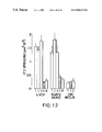
- FIG. 12 35 kD and 250 kD protein fractions from rat CNS myelin convert permissive substrates into non-permissive substrates.
- Total protein (T) liposomes were formed with 100 ⁇ g of protein from each of the three sources (rat) indicated in the figure.
- (1) (250 kD) and (2) (35 kD) liposomes were formed with gel-extracted protein regions from 3-15% gels loaded with 500 ⁇ g protein from the same three sources.
- Columns labeled +1 and +2 indicate that 250 kD (1) or 35 kD (2) protein from 500 ⁇ g of rat CNS myelin, were combined with 100 ⁇ g of total liver or PNS myelin protein before reconstitution.
- FIGS. 13 a-h Nonpermissive substrate properties of CNS myelin and of 35 kD and 250 kD inhibitors were neutralized by monoclonal antibody IN-1.
- SCG neurons were cultivated on test substrates in the absence (a, c, e, and g) or the presence (b, d, f, and h) of monoclonal antibody IN-1. Cultures were photographed after 24 hours. Substrate-adsorbed wells of Greiner dishes were preincubated in the presence of hybridoma medium or IN-1 hybridoma supernatant for 30 minutes, four-fifths of the preincubation medium was then removed, and SCG neurons were added, thus replacing the removed quantity of medium.
- Well-adsorbed substrates were as follows: CNS myelin membranes (a and b), 250 kD inhibitor-containing liposomes (c and d), 35 kd inhibitor-containing liposomes (e and f), and liposomes containing permissive 250 kD protein fraction from rat PNS myelin (g and h). Bar, 50 ⁇ m.
- FIGS. 14 a-f Monoclonal antibody IN-1 binds to the surface of HBOs and neutralizes their nonpermissive substrate properties. Two day old optic nerve cultures were either stained with IN-1 (b) or tested as substrates for 3T3 cell spreading in the presence (c,d) and absence (e,f) of IN-1. IN-1 specifically bound to the surface of HBOs (a, phase contrast; b, immunofluorescence with antibody IN-1).
- FIG. 15 Quantitative determination of IN-1-mediated neutralization of HBO nonpermissive substrate properties.
- 3T3 cells were added to 2 day old optic nerve cultures in the presence of either hybridoma medium (C), monoclonal antibody O 4 or antibody IN-1. Cultures were fixed after 2 hours, and oligodendrocytes were identified by O 1 staining. Preferential adhesion and spreading of 3T3 cells on a polylysine-coated culture dish and on the surface of O 1 + HBOs were determined as described infra in Section 8.1.4.
- a value of 100% inhibition represents no overlap of 3T3 cell surfaces with O 1 30 surfaces; 0% inhibition represents no apparent discrimination by 3T3 cells between the polylysine-coated culture dish and HBO surface; negative inhibition values indicate that the fraction of HBO surfaces covered by 3T3 cells was larger than the fraction of the entire culture surface covered by 3T3 cells, i.e., 3T3 cells preferentially spread on HBOs. Values are means ⁇ SEM. Twenty separate determinations from two independent experiments were analyzed.
- FIG. 16 Immunoblot of rat CNS myelin protein with monoclonal antibody IN-1.
- CNS myelin protein was separated by 3-15% SDS-PAGE under reducing conditions.
- Lane 1 silver-stained gel of rat CNS myelin protein; the positions of the inhibitory protein regions are indicated by arrowheads.
- Lanes 2 and 3 immunoblot with IN-1 (lane 2) or control antibody against horseradish peroxidase (lane 3).
- Lane 1 and lanes 2 and 3 are from two different gels. The approximate migration position of protein bands giving specific IN-1 + signals are indicated by arrowheads (lanes 1 and 2).
- IN-1 binding to protein bands in the 35 kd region was variable, weak, and not detectable on the immunoblot shown.
- FIGS. 17 a-b Laminin immunoreactivity in adult optic nerve in vivo and in cultured optic nerve explants. In the in vivo nerve, only subpial and perivascular basement membranes showed specific laminin immunoreactivity (a). In cultured optic nerve explants (chamber culture, 4 weeks in vitro), strongly laminin-positive cells, presumably astrocytes (arrows), appeared inside the explant (b). Bar, 50 ⁇ m.
- FIGS. 18 a-b Sensory axons in IN-1-injected nerve explants. Electron micrographs from representative experiments as described in Table X. (a) Electron micrograph of IN-1-injected optic nerve 1 mm from the proximal stump; an axon bundle growing in direct contact with the myelin is shown. (b) In the presence of IN-1, numerous axons grew 3 mm into the optic nerve explant. Bar, 0.5 ⁇ m.
- FIGS. 19 a-d C6, but not 3T3 cells, infiltrate optic nerve explants.
- FIGS. 20A-20F C6, but not 3T3 or B16 cells attach and spread on CNS white matter of rat cerebellar frozen sections.
- a clear difference on white matter (wm) emerges for 3T3 and B16 cells compared to C6 cells.
- gl granular layer
- ml molecular layer.
- Gray matter is composed of granular and molecular layer. Bar, 0.3 mm.
- FIGS. 21 a-c C6 cells overcome the inhibitory substrate property of CNS myelin.
- FIG. 22 1,10-phenanthroline inhibits C6 spreading specifically on CNS myelin.
- C6 cells were cultured for 3 hours on the indicated substrates in the presence of increasing doses of 1,10-phenanthroline.
- Spreading was inhibited by low doses exclusively on CNS myelin.
- 1,10-phenanthroline concentrations above 0.5 mM exert a general toxic effect on all substrates.
- Spreading was quantified as indicated in FIG. 21 .
- FIGS. 23A-23D Degradation of CNS inhibitory substrate by C6 plasma membranes is 1,10-phenanthroline sensitive. Spreading of 3T3 cells on CNS myelin was induced by pretreatment of myelin with C6 plasma membranes. 1,10-phenanthroline abolished this effect. Shown are phase contrast micrographs of 3T3 cells on polylysine (PLYS) (FIG. 23 A), on CNS myelin (FIG. 23 B), on CNS myelin pretreated with C6 plasma membranes (FIG. 23 C), and on CNS myelin pretreated with C6 plasma membranes (C6-PM) in the presence of 1,10-phenanthroline (FIG. 23 D).
- PLYS polylysine
- FIGS. 24 a-b C6 cell attachment and spreading on CNS white matter of rat cerebellar frozen section is impaired by metalloprotease blockers.
- FIG. 25 C 6 cell infiltration into CNS explants is impaired by cbz-tyr-tyr.
- Cells were added to one tip of optic nerve explants (chamber cultures) in the presence of the metalloprotease inhibitor cbz-tyr-tyr, or of the control peptide cbz-ala-phe. 14 day old cultures were quantified. Infiltrated cells were counted in the first 1.3 mm of the explants. Each column represents the number of infiltrated cells per 0.1 mm. Only the most central part of the explants was considered (0.25 mm). Values represent means ⁇ SEM of two sets of experiments for a total of 8 explants.
- FIGS. 26 a-b Tumor of antibody-secreting mouse IN-1 hybridoma cells in the cortex of an 8 day old rat. 1 mio. cells (in 2 ⁇ l) were injected at P2; (a) Cresyl violet stained frozen section (15 ⁇ l ) shows well circumscribed, compact tumor (arrow); (b) Antibody production demonstrated by immunofluorescence with anti-mouse-Ig-FITC. Tumor and surrounding tissue up to the lateral ventricle (small arrow) are strongly stained (adjacent section to a). Rat was fixed by perfusion with 4% formaldehyde in phosphate buffer. Magnification: 6.4 ⁇
- FIG. 27 Labelled corticospinal tract (CST) fibers are present far distal to spinal cord lesions in 4 IN-1 treated rats (top), but not in 4 control anti-HRP treated animals (bottom).
- FIG. 29 An extracellular metalloproteolytic activity of C6 cells degrades the substrate cbz-FAF[ 125 I]Y-NH 2 (SEQ ID NO:2) [cbz-phe-ala-phe- 125 I-tyr-NH 2 ].
- 37° C. right panel
- one single product showed clear o-phenanthroline dependency (asterisk).
- Incubation at 4° C. (left panel) or 20° C.
- FIG. 30 Cell fractionation experiments localize a cbz-FAF[ 125 I]Y-NH 2 (SEQ ID NO:2) degradative activity to plasma membranes. Autoradiogram of thin layer chromatography of substrate incubated with plasma membranes (I). For quantification the incubation medium was extracted with chloroform. The organic phase extracted completely unreacted substrate (II), whereas degradation products were retained in the aqueous phase (III). 0.8 mM phenanthroline completely blocked the degradation (IV-VI).
- FIG. 31 The substrate cbz-FAF[ 125 I]Y-NH 2 (SEQ ID NO:2) is degraded at one single site by a metalloendoprotease.
- FIGS. 32 a-d pH-optimum of plasma membrane associated metalloendoprotease.
- Rate of cbz-FAF [ 125 I]Y-NH 2 (SEQ ID NO:2) hydrolysis was measured at different pH values. Activity peaks in the weak acidic range. Pepstatin (b, 10 ⁇ M) sensitive activity was maximal at pH 4.0, whereas o-phenanthroline (c, 0.6 mM) sensitive activity peaked in the range 5.5-7.0.
- plasma membrane activity was measured with pH 0.5 steps, to determine the pH optimum which was 5.5-6.0.
- the incubation media were 100 mM Na-acetate/acetate (circles), 100 mM MES/HCl (squares), 100 mM TRIS/HCl (diamonds), and contained 100 mM NaCl and 10 ⁇ M pepstatin.
- FIGS. 33 a-c Solubilization of metalloproteolytic activity requires detergent.
- Total metalloproteolytic activity associated to C6 plasma membranes was sedimented almost completely as tested by measuring pellet and supernatant separately (a).
- a similar result was obtained with 1 M NaCl treatment (b).
- FIG. 34 Schematic diagram of the hippocampus, showing the caudal (C) or lateral (L) directions of distances measured.
- B extracellular matrix bridge;
- E entry point of regenerating fibers into hippocampus;
- R rostral hippocampus;
- CdH caudal hippocampus.
- FIG. 35 Amino acid composition of the HPLC peak II derived from rat and bovine 35 kD neurite inhibitory factor (NI-35). Values represent the ratio of each amino acid to the amount of aspartic acid.
- FIG. 36 N-terminal amino acid sequence of rat NI-35 derived of HPLC peak II.
- the N-terminal amino acid sequence (SEQ ID NO:4) of NI-35 is shown, as well as a predicted mRNA sequence (SEQ ID NO:5) encoding the protein sequence, and the predicted sequence of a DNA (SEQ ID NO:6) complementary to the RNA which can be used as a hybridization probe for the cloning of NI-35.
- FIG. 37 Internal amino acid sequence of rat NI-35 derived of HPLC peak II.
- the amino acid sequence (SEQ ID NO:7) of an internal peptide of NI-35 derived from Endoproteinase-Lys C cleavage is shown.
- a predicted mRNA sequence (SEQ ID NO:8) encoding the protein sequence is also shown.
- the present invention is directed to genes and their encoded proteins which regulate neurite growth and the diagnostic and therapeutic uses of such proteins.
- the proteins of the present invention include proteins associated with central nervous system myelin with highly nonpermissive substrate properties, termed herein neurite growth inhibitory factors.
- the neurite growth regulatory factors also include metalloproteases which can be found associated with malignant tumors, in particular, those tumors metastatic to the brain.
- the CNS myelin associated proteins of the invention inhibit neurite outgrowth in nerve cells or neuroblastoma cells.
- the protein can also inhibit fibroblast spreading and migration.
- These inhibitory proteins are active cross-species and may be used in the treatment of patients with malignant tumors including but not limited to melanoma and tumors of nerve tissue (e.g. neuroblastoma).
- melanoma e.g. melanoma
- tumors of nerve tissue e.g. neuroblastoma
- a 35 kilodalton and a 250 kilodalton CNS myelin associated protein are described.
- the 250 kD protein is a complex containing the 35 kD protein.
- the present invention is also directed to antibodies to and peptide fragments and derivatives of the neurite growth inhibitory proteins and their therapeutic and diagnostic uses. These antibodies or peptides can be used in the treatment of nerve damage resulting from, e.g., trauma (e.g., spinal cord injuries), stroke, degenerative disorders of the central nervous system, etc.
- antibodies to CNS myelin associated proteins may be used to promote regeneration of nerve fibers.
- monoclonal antibody IN-1 may be used to promote the regeneration of nerve fibers over long distances following spinal cord damage.
- the present invention further relates to neurite growth regulatory factor receptors and peptide fragments thereof as well as the nucleic acid sequences coding for neurite growth regulatory factor receptors and fragments, and their therapeutic and diagnostic uses.
- Antibodies to neurite growth regulatory factor receptors are also envisioned and within the scope of the present invention.
- the present invention is also directed to metalloproteases associated with malignant tumors, in particular, those metastatic to the brain.
- the metalloprotease is associated with glioblastoma cells.
- the metalloproteases of the invention are associated with the CNS infiltration activity of malignant cells, and can neutralize the inhibitory substrate properties of the CNS myelin-associated proteins.
- the metalloproteases can have therapeutic value in the treatment of nerve damage such as that resulting from traumatic injury (e.g. spinal cord injuries), stroke, degenerative disorders of the central nervous system, etc.
- the metalloprotease may be used in combination with antibodies directed against myelin associated inhibitory proteins to treat nerve damage.
- the present invention is also directed to inhibitors of the metalloproteases.
- Such inhibitors can impair malignant cell spreading and infiltration, and can be used in the treatment of malignant tumors (e.g. glioblastoma).
- the metalloprotease inhibitors in combination with CNS myelin associated inhibitory proteins such as the 35,000 dalton and/or the 250,000 dalton molecular weight proteins, may be used in the diagnosis and/or treatment of malignant tumors which include but are not limited to glioblastomas, neuroblastomas, and melanomas.
- the method of the invention can be divided into the following stages, solely for the purpose of description: (1) isolation and purification of neurite growth regulatory factors; (2) characterization of neurite growth regulatory factors; (3) molecular cloning of genes or gene fragment encoding neurite growth regulatory factors; (4) production of antibodies against neurite growth regulatory factors; and (5) generation of neurite growth regulatory factor related derivatives, analogs, and peptides.
- the method further encompasses the diagnostic and therapeutic uses of
- the present invention relates to CNS myelin associated inhibitory proteins of neurite growth, receptors of CNS myelin associated inhibitory proteins of neurite growth, and to metalloproteases such as that associated with membranes of glioblastoma cells.
- the CNS myelin associated inhibitory proteins of the invention may be isolated by first isolating myelin and subsequent purification therefrom.
- the metalloprotease may be obtained by isolation from mammalian glioblastoma cells. Isolation procedures which may be employed are described more fully in the sections which follow.
- the CNS myelin associated inhibitory proteins or the metalloprotease may be obtained from a recombinant expression system (see Section 5.3., infra). Procedures for the isolation and purification of receptors for the CNS myelin associated inhibitory proteins are described in Section 5.1.2., infra.
- CNS myelin associated inhibitory proteins can be isolated from the CNS myelin of higher vertebrates including, but not limited to, birds or mammals (both human and nonhuman such as bovine, rat, porcine, chick, etc.) (see Section 7.2.1., infra).
- Myelin can be obtained from the optic nerve or from central nervous system tissue that includes but is not limited to spinal cords or brain stems. The tissue may be homogenized using procedures described in the art (Colman et al., 1982, J. Cell Biol. 95:598-608). The myelin fraction can be isolated subsequently also using procedures described (Colman et al., 1982, supra).
- the CNS myelin associated inhibitory proteins can be solubilized in detergent (e.g., Nonidet P-40TM, sodium deoxycholate).
- the solubilized proteins can subsequently be purified by various procedures known in the art, including but not limited to chromatography (e.g., ion exchange, affinity, and sizing chromatography), centrifugation, electrophoretic procedures, differential solubility, or by any other standard technique for the purification of proteins (see, e.g., Section 7.2.3., infra).
- the solubilized proteins can be subjected to one dimensional electrophoresis, followed by isoelectric focussing and elution from the focussing gel. Gel-eluted proteins can be acetone-precipitated, redissolved in 10% formic acid and chromatographed on a C 4 reverse phase HPLC column (see Section 12, infra).
- the CNS myelin associated inhibitory proteins may be isolated and purified using immunological procedures.
- the proteins can first be solubilized using detergent (e.g., Nonidet P-40TM, sodium deoxycholate). The proteins may then be isolated by immunoprecipitation with antibodies to the 35 kilodalton and/or the 250 kilodalton proteins.
- the CNS myelin associated inhibitory proteins may be isolated using immunoaffinity chromatography in which the proteins are applied to an antibody column in solubilized form.
- Receptors for the CNS myelin associated inhibitory proteins can be isolated from cells whose attachment, spreading, growth and/or motility is inhibited by the CNS myelin associated inhibitory proteins.
- Such cells include but are not limited to fibroblasts and neurons.
- neurons are used as the source for isolation and purification of the receptors.
- receptors to CNS myelin associated inhibitory proteins may be isolated by affinity chromatography of neuronal plasma membrane fractions, in which a myelin associated inhibitory protein or peptide fragment thereof is immobilized to a solid support.
- receptor cDNA may be isolated by expression cloning using purified 35 kD or 250 kD neurite growth inhibitory factor as a ligand for the selection of receptor-expressing clones.
- the metalloproteases of the present invention may be isolated from cells of malignant tumors, in particular, glioblastomas.
- a metalloprotease can be isolated from mammalian glioblastoma cells.
- the metalloprotease is isolated from the plasma membrane fraction of such cells.
- the cells may be obtained by dissociating and homogenizing tumors using procedures known in the art or from tumor cell lines. Plasma membrane fractions may be obtained using procedures known in the art, e.g., gradient centrifugation (Quigley, 1976, J. Cell Biol. 71:472-486).
- the metalloprotease may be isolated from the membranes by solubilization with mild ionic or nonionic detergent (e.g., deoxycholate, Nonidet P-40TM, TritonTM, BrijTM) using procedures described in the art (reviewed in Cooper, 1977, In Tools of Biochemistry, John Wiley & Sons, NY, pp. 355-406) (see also Section 11, infra).
- mild ionic or nonionic detergent e.g., deoxycholate, Nonidet P-40TM, TritonTM, BrijTM
- Purification of the metalloprotease can be carried out by known procedures, including but not limited to chromatography (e.g., ion exchange, affinity, and sizing column chromatography), centrifugation, electrophoretic procedures, differential solubility, or by any other standard technique for the purification of proteins.
- chromatography e.g., ion exchange, affinity, and sizing column chromatography
- centrifugation e.g., ion exchange, affinity, and sizing column chromatography
- electrophoretic procedures e.g., electrophoretic procedures
- differential solubility e.g., differential solubility
- the neurite growth regulatory factors of the present invention can be analyzed by assays based on their physical, immunological, or functional properties.
- the half life of the neurite growth regulatory factors in cultured cells can be studied, for example, by use of cycloheximide, an inhibitor of protein synthesis (Vasquez, 1974, FEBS Lett. 40:563-584).
- a physiological receptor for a neurite growth regulatory factor could be identified by assays which detect complex formation with a neurite growth regulatory factor, e.g., by use of affinity chromatography with immobilized neurite growth regulatory factor, binding to a labeled neurite growth regulatory factor followed by cross-linking and immunoprecipitation, etc.
- Electrophoretic techniques such as SDS-polyacrylamide gel electrophoresis and two-dimensional electrophoresis can be used to study protein structure.
- Other techniques which can be used include but are not limited to peptide mapping, isoelectric focusing, and chromatographic techniques.
- the amino acid sequences of primary myelin associated inhibitors or of the metalloprotease can be derived by deduction from the DNA sequence if such is available, or alternatively, by direct sequencing of the protein, e.g., with an automated amino acid sequencer.
- the protein sequences can be further characterized by a hydrophilicity analysis (Hopp and Woods, 1981, Proc. Natl. Acad. Sci. U.S.A. 78:3824-3828).
- a hydrophilicity profile can be used to identify the hydrophobic and hydrophilic regions of the protein (and the corresponding regions of the gene sequence, if available, which encode such regions).
- Any mammalian cell can potentially serve as the nucleic acid source for the molecular cloning of the genes encoding the CNS myelin associated inhibitory proteins, including but not limited to the 35 kD and/or 250 kD myelin associated proteins described in Section 7., infra, or the glioblastoma associated metalloprotease, hereinafter referred to as neurite growth regulatory factor genes.
- the DNA may be obtained by standard procedures known in the art from cloned DNA (e.g., a DNA “library”), by chemical synthesis, by cDNA cloning, or by the cloning of genomic DNA, or fragments thereof, purified from the desired mammalian cell.
- cloned DNA e.g., a DNA “library”
- chemical synthesis e.g., chemical synthesis
- cDNA cloning e.g., a DNA “library”
- genomic DNA e.g., genomic DNA, or fragments thereof, purified from the desired mammalian cell.
- Clones derived from genomic DNA may contain regulatory and intron DNA regions, in addition to coding regions; clones derived from cDNA will contain only exon sequences. Whatever the source, a given neurite growth regulatory factor gene should be molecularly cloned into a suitable vector for propagation of the gene.
- DNA fragments are generated, some of which will encode the desired neurite growth regulatory factor gene.
- the DNA may be cleaved at specific sites using various restriction enzymes. Alternatively, one may use DNAse in the presence of manganese to fragment the DNA, or the DNA can be physically sheared, as for example, by sonication.
- the linear DNA fragments can then be separated according to size by standard techniques, including but not limited to, agarose and polyacrylamide gel electrophoresis and column chromatography.
- identification of the specific DNA fragment containing a neurite growth regulatory factor gene may be accomplished in a number of ways. For example, if an amount of a neurite growth regulatory factor gene or its specific RNA, or a fragment thereof, is available and can be purified and labeled, the generated DNA fragments may be screened by nucleic acid hybridization to the labeled probe (Benton and Davis, 1977, Science 196:180; Grunstein and Hogness, 1975, Proc. Natl. Acad. Sci. U.S.A. 72:3961-3965).
- a portion of a neurite growth regulatory factor amino acid sequence can be used to deduce the DNA sequence, which DNA sequence can then be synthesized as an oligonucleotide for use as a hybridization probe.
- nucleic acid fractions enriched in neurite growth regulatory factor may be used as a probe, as an initial selection procedure.
- a neurite growth regulatory factor gene can also be identified by mRNA selection using nucleic acid hybridization followed by in vitro translation or translation in Xenopus oocytes.
- oocytes are injected th total or size fractionated CNS mRNA populations, and the membrane-associated translation products are screened in a functional assay (3T3 cell spreading).
- Preadsorption of the RNA with complementary DNA (cDNA) pools leading to the absence of expressed inhibitory factors indicates the presence of the desired cDNA. Reduction of pool size will finally lead to isolation of a single CDNA clone.
- DNA fragments can be used to isolate complementary mRNAs by hybridization.
- Such DNA fragments may represent available, purified neurite growth regulatory factor DNA, or DNA that has been enriched for neurite growth regulatory factor sequences.
- Immunoprecipitation analysis or functional assays of the in vitro translation products of the isolated mRNAs identifies the mRNA and, therefore, the CDNA fragments that contain neurite growth regulatory factor sequences.
- An example of such a functional assay involves an assay for nonpermissiveness in which the effect of the various translation products on the spreading of 3T3 cells on a polylysine coated tissue culture dish is observed (see Section 7.1.2., infra).
- specific mRNAs may be selected by adsorption of polysomes isolated from cells to immobilized antibodies specifically directed against a neurite growth regulatory factor protein.
- a radiolabeled neurite growth regulatory factor cDNA can be synthesized using the selected mRNA (from the adsorbed polysomes) as a template. The radiolabeled mRNA or cDNA may then be used as a probe to identify the neurite growth regulatory factor DNA fragments from among other genomic DNA fragments.
- isolating the neurite growth regulatory factor genomic DNA include, but are not limited to, chemically synthesizing the gene sequence itself from a known sequence or making cDNA to the mRNA which encodes the neurite growth regulatory factor gene. Other methods are possible and within the scope of the invention.
- the identified and isolated gene or CDNA can then be inserted into an appropriate cloning vector.
- vector-host systems known in the art may be used. Possible vectors include, but are not limited to, cosmids, plasmids or modified viruses, but the vector system must be compatible with the host cell used. Such vectors include, but are not limited to, bacteriophages such as lambda derivatives, or plasmids such as pBR322 or pUC plasmid derivatives. Recombinant molecules can be introduced into host cells via transformation, transfection, infection, electroporation, etc.
- the neurite growth regulatory factor gene may be identified and isolated after insertion into a suitable cloning vector, in a “shot gun” approach. Enrichment for a given neurite growth regulatory factor gene, for example, by size fractionation or subtraction of cDNA specific to low neurite growth regulatory factor producers, can be done before insertion into the cloning vector.
- DNA may be inserted into an expression vector system, and the recombinant expression vector containing a neurite growth regulatory factor gene may then be detected by functional assays for the neurite growth regulatory factor protein.
- the neurite growth regulatory factor gene is inserted into a cloning vector which can be used to transform, transfect, or infect appropriate host cells so that many copies of the gene sequences are generated. This can be accomplished by ligating the DNA fragment into a cloning vector which has complementary cohesive termini. However, if the complementary restriction sites used to fragment the DNA are not present in the cloning vector, the ends of the DNA molecules may be enzymatically modified. Alternatively, any site desired may be produced by ligating nucleotide sequences (linkers) onto the DNA termini; these ligated linkers may comprise specific chemically synthesized oligonucleotides encoding restriction endonuclease recognition sequences. In an alternative method, the cleaved vector and neurite growth regulatory factor gene may be modified by homopolymeric tailing.
- Identification of the cloned neurite growth regulatory factor gene can be accomplished in a number of ways based on the properties of the DNA itself, or alternatively, on the physical, immunological, or functional properties of its encoded protein.
- the DNA itself may be detected by plaque or colony nucleic acid hybridization to labeled probes (Benton, W. and Davis, R., 1977, Science 196:180; Grunstein, M. and Hogness, D., 1975, Proc. Natl. Acad. Sci. U.S.A. 72:3961).
- the presence of a neurite growth regulatory factor gene may be detected by assays based on properties of its expressed product.
- cDNA clones or DNA clones which hybrid-select the proper mRNAs, can be selected which produce a protein that inhibits in vitro neurite outgrowth.
- a neurite growth regulatory factor protein may be identified by binding of labeled antibody to the putatively neurite growth regulatory factor-synthesizing clones, in an ELISA (enzyme-linked immunosorbent assay)-type procedure.
- transformation of host cells with recombinant DNA molecules that incorporate an isolated neurite growth regulatory factor gene, cDNA, or synthesized DNA sequence enables generation of multiple copies of the gene.
- the gene may be obtained in large quantities by growing transformants, isolating the recombinant DNA molecules from the transformants and, when necessary, retrieving the inserted gene from the isolated recombinant DNA.
- the recombinant DNA molecule that incorporates a neurite growth regulatory factor gene can be modified so that the gene is flanked by virus sequences what allow for genetic recombination in cells infected with the virus so that the gene can be inserted into the viral genome.
- neurite growth regulatory factor DNA-containing clone After the neurite growth regulatory factor DNA-containing clone has been identified, grown, and harvested, its DNA insert may be characterized as described in Section 5.3.4, infra.
- the genetic structure of a neurite growth regulatory factor gene is known, it is possible to manipulate the structure for optimal use in the present invention.
- promoter DNA may be ligated 5′ of a neurite growth regulatory factor coding sequence, in addition to or replacement of the native promoter to provide for increased expression of the protein. Many manipulations are possible, and within the scope of the present invention.
- the nucleotide sequence coding for a neurite growth regulatory factor protein or a portion thereof can be inserted into an appropriate expression vector, i.e., a vector which contains the necessary elements for the transcription and translation of the inserted protein-coding sequence.
- the necessary transcriptional and translation signals can also be supplied by the native neurite growth regulatory factor gene and/or its flanking regions.
- a variety of host-vector systems may be utilized to express the protein-coding sequence.
- mammalian cell systems infected with virus e.g., vaccinia virus, adenovirus, etc.
- insect cell systems infected with virus e.g., baculovirus
- microorganisms such as yeast containing yeast vectors, or bacteria transformed with bacteriophage DNA, plasmid DNA, or cosmid DNA.
- the expression elements of these vectors vary in their strengths and specificities. Depending on the host-vector system utilized, any one of a number of suitable transcription and translation elements may be used.
- Any of the methods previously described for the insertion of DNA fragments into a vector may be used to construct expression vectors containing a chimeric gene consisting of appropriate transcriptional/translational control signals and the protein coding sequences. These methods may include in vitro recombinant DNA and synthetic techniques and in vivo recombinations (genetic recombination).
- Expression vectors containing neurite growth regulatory factor gene inserts can be identified by three general approaches: (a) DNA-DNA hybridization, (b) presence or absence of “marker” gene functions, and (c) expression of inserted sequences.
- the presence of a foreign gene inserted in an expression vector can be detected by DNA-DNA hybridization using probes comprising sequences that are homologous to an inserted neurite growth regulatory factor gene.
- the recombinant vector/host system can be identified and selected based upon the presence or absence of certain “marker” gene functions (e.g., thymidine kinase activity, resistance to antibiotics, transformation phenotype, occlusion body formation in baculovirus, etc.) caused by the insertion of foreign genes in the vector.
- certain “marker” gene functions e.g., thymidine kinase activity, resistance to antibiotics, transformation phenotype, occlusion body formation in baculovirus, etc.
- recombinant expression vectors can be identified by assaying the foreign gene product expressed by the recombinant. Such assays can be based on the physical, immunological, or functional properties of a given neurite growth regulatory factor gene product.
- the expression vectors which can be used include, but are not limited to, the following vectors or their derivatives: human or animal viruses such as vaccinia virus or adenovirus; insect viruses such as baculovirus; yeast vectors; bacteriophage vectors (e.g., lambda), and plasmid and cosmid DNA vectors, to name but a few.
- a host cell strain may be chosen which modulates the expression of the inserted sequences, or modifies and processes the gene product in the specific fashion desired. Expression from certain promoters can be elevated in the presence of certain inducers; thus, expression of the genetically engineered neurite growth regulatory factor protein may be controlled.
- different host cells have characteristic and specific mechanisms for the translational and post-translational processing and modification (e.g., glycosylation, cleavage) of proteins. Appropriate cell lines or host systems can be chosen to ensure the desired modification and processing of the foreign protein expressed. For example, expression in a bacterial system can be used to produce an unglycosylated core protein product. Expression in yeast will produce a glycosylated product. Expression in mammalian (e.g. COS) cells can be used to ensure “native” glycosylation of the heterologous neurite growth regulatory factor protein.
- different vector/host expression systems may effect processing reactions sash as proteolytic cleavages to different extents.
- the gene product can be purified as described in Section 5.1, supra, and analyzed as described in Section 5.2, supra.
- amino acid sequence of a given neurite growth regulatory factor protein can be deduced from the nucleotide sequence of the cloned gene, allowing the protein, or a fragment thereof, to be synthesized by standard chemical methods known in the art (e.g., see Hunkapiller, et al., 1984, Nature 310:105-111).
- such neurite growth regulatory factor proteins include but are not limited to those containing altered sequences in which functionally equivalent amino acid residues are substituted for residues within the sequence resulting in a silent change.
- one or more amino acid residues within the sequence can be substituted by another amino acid of a similar polarity which acts as a functional equivalent, resulting in a silent alteration.
- Substitutes for an amino acid within the sequence may be selected from other members of the class to which the amino acid belongs.
- the nonpolar (hydrophobic) amino acids include alanine, leucine, isoleucine, valine, proline, phenylalanine, tryptophan, and methionine.
- the polar neutral amino acids include glycine, serine, threonine, cysteine, tyrosine, asparagine, and glutamine.
- the positively charged (basic) amino acids include arginine, lysine, and histidine.
- the negatively charged (acidic) amino acids include aspartic acid and glutamic acid.
- neurite growth regulatory factor proteins which are differentially modified during or after translation, e.g., by glycosylation, proteolytic cleavage, etc.
- the structure of a given neurite growth regulatory factor gene can be analyzed by various methods known in the art.
- the cloned DNA or cDNA corresponding to a given neurite growth regulatory factor gene can be analyzed by methods including but not limited to Southern hybridization (Southern, 1975, J. Mol. Biol. 98:503-517), Northern hybridization (Alwine, et al., 1977, Proc. Natl. Acad. Sci. U.S.A. 74:5350-5354; Wahl, et al., 1987, Meth. Enzymol. 152:572-581), restriction endonuclease mapping (Maniatis, et al., 1982, Molecular Cloning, A Laboratory Manual, Cold Spring Harbor Laboratory, Cold Spring Harbor, N.Y.), and DNA sequence analysis.
- DNA sequence analysis can be performed by any techniques known in the art including but not limited to the method of Maxam and Gilbert (1980, Meth. Enzymol. 65:499-560), the Sanger dideoxy method (Sanger, et al., 1977, Proc. Natl. Acad. Sci. U.S.A. 74:5463-5467), or use of an automated DNA sequenator (e.g., Applied Biosystems, Foster City, Calif.).
- Antibodies can be produced which recognize neurite growth regulatory factors or related proteins. Such antibodies can be polyclonal or monoclonal.
- a neurite growth regulatory factor protein or a synthetic protein, or fragment thereof, including but not limited to rabbits, mice, rats, etc.
- adjuvants may be used to increase the immunological response, depending on the host species, and including but not limited to Freund's (complete and incomplete), mineral gels such as aluminum hydroxide, surface active substances such as lysolecithin, pluronic polyols, polyanions, peptides, oil emulsions, keyhole limpet hemocyanins, dinitrophenol, and potentially useful human adjuvants such as BCG (bacille Calmette-Guerin) and corynebacterium parvum.
- BCG Bacille Calmette-Guerin
- a monoclonal antibody to an epitope of a neurite growth regulatory factor can be prepared by using any technique which provides for the production of antibody molecules by continuous cell lines in culture. These include but are not limited to the hybridoma technique originally described by Kohler and Milstein (1975, Nature 256:495-497), and the more recent human B cell hybridoma technique (Kozbor et al., 1983, Immunology Today 4:72) and EBV-hybridoma technique (Cole et al., 1985, Monoclonal Antibodies and Cancer Therapy, Alan R. Liss, Inc., pp. 77-96). In a particular embodiment, the procedure described infra in Section 8.1. may be used to obtain mouse monoclonal antibodies which recognize the 35 kD and 250 kD CNS myelin associated inhibitory proteins.
- the monoclonal antibodies for therapeutic use may be human monoclonal antibodies or chimeric human-mouse (or other species) monoclonal antibodies.
- Human monoclonal antibodies may be made by any of numerous techniques known in the art (e.g., Teng et al., 1983, Proc. Natl. Acad. Sci. U.S.A. 80:7308-7312; Kozbor et al., 1983, Immunology Today 4:72-79; Olsson et al., 1982, Meth. Enzymol. 92:3-16).
- Chimeric antibody molecules may be prepared containing a mouse antigen-binding domain with human constant regions (Morrison et al., 1984, Proc. Natl. Acad. Sci. U.S.A. 81:6851, Takeda et al., 1985, Nature 314:452).
- a molecular clone of an antibody to a neurite growth regulatory factor epitope can be prepared by known techniques. Recombinant DNA methodology (see e.g., Maniatis et al., 1982, Molecular Cloning, A Laboratory Manual, Cold Spring Harbor Laboratory, Cold Spring Harbor, N.Y.) may be used to construct nucleic acid sequences which encode a monoclonal antibody molecule, or antigen binding region thereof.
- Antibody molecules may be purified by known techniques, e.g., immunoabsorption or immunoaffinity chromatography, chromatographic methods such as HPLC (high performance liquid chromatography), or a combination thereof, etc.
- Antibody fragments which contain the idiotype of the molecule can be generated by known techniques.
- such fragments include but are not limited to: the F(ab′) 2 fragment which can be produced by pepsin digestion of the antibody molecule; the Fab′ fragments which can be generated by reducing the disulfide bridges of the F(ab′) 2 fragment, and the 2 Fab or Fab fragments which can be generated by treating the antibody molecule with papain and a reducing agent.
- derivatives, analogs, and peptides related to a given neurite growth regulatory factor are also envisioned, and within the scope of the present invention and include molecules antagonistic to neurite growth regulatory factors (for example, and not by way of limitation, anti-idiotype antibodies).
- Such derivatives, analogs, or peptides which have the desired inhibitory activity can be used, for example, in the treatment of neuroblastoma (see Section 5.6, infra).
- Derivatives, analogs, or peptides related to a neurite growth regulatory factor can be tested for the desired activity by assays for nonpermissive substrate effects. For example, procedures such as the assay for nonpermissiveness in which the effect of the various translation products on the spreading of 3T3 cells on a polylysine coated tissue culture dish is observed (see Section 7.1.2., infra).
- the neurite growth regulatory factor-related derivatives, analogs, and peptides of the invention can be produced by various methods known in the art. The manipulations which result in their production can occur at the gene or protein level.
- a cloned neurite growth regulatory factor gene can be modified by any of numerous strategies known in the art (Maniatis, et al., 1982, Molecular Cloning, A Laboratory Manual, Cold Spring Harbor Laboratory, Cold Spring Harbor, N.Y.).
- a given neurite growth regulatory factor sequence can be cleaved at appropriate sites with restriction endonuclease(s), subjected to enzymatic modifications if desired, isolated, and ligated in vitro.
- a given neurite growth regulatory factor gene can be mutated in vitro or in vivo, to create and/or destroy translation, initiation, and/or termination sequences, or to create variations in coding regions and/or form new restriction endonuclease sites or destroy preexisting ones, to facilitate further in vitro modification.
- Any technique for mutagenesis known in the art can be used, including but not limited to, in vitro site-directed mutagenesis (Hutchinson, et al., 1978, J. Biol. Chem. 253:6551), use of TAB® linkers (Pharmacia), etc.
- CNS myelin associated inhibitory proteins, analogs, derivatives, and subsequences thereof, and anti-inhibitory protein antibodies or peptides have uses in diagnostics.
- Such molecules can be used in assays such as immunoassays to detect, prognose, diagnose, or monitor various conditions, diseases, and disorders affecting neurite growth extension, invasiveness, and regeneration. In one embodiment of the invention, these molecules may be used for the diagnosis of malignancies.
- the CNS myelin associated inhibitory proteins, analogs, derivatives, and subsequences thereof and antibodies thereto may be used to monitor therapies for diseases and conditions which ultimately result in nerve damage; such diseases and conditions include but are not limited to CNS trauma, (e.g.
- such molecules may be used to detect an increase in neurite outgrowth as an indicator of CNS fiber regeneration.
- the absence of the CNS myelin associated inhibitory proteins in a patient sample containing CNS myelin can be a diagnostic marker for the presence of a malignancy, including but not limited to glioblastoma, neuroblastoma, and melanoma, or a condition involving nerve growth, invasiveness, or regeneration in a patient.
- the absence of the inhibitory proteins can be detected by means of an immunoassay in which the lack of any binding to anti-inhibitory protein antibodies (e.g., IN-1, IN-2) is observed.
- the immunoassays which can be used include but are not limited to competitive and non-competitive assay systems using techniques such as radioimmunoassays, ELISA (enzyme linked immunosorbent assay), “sandwich” immunoassays, precipitation reactions, gel diffusion precipitation reactions, immunodiffusion assays, agglutination assays, complement-fixation assays, immunoradiometric assays, fluorescent immunoassays, protein A immunoassays, immunoelectrophoresis assays, and immunohistochemistry on tissue sections, to name but a few.
- radioimmunoassays ELISA (enzyme linked immunosorbent assay), “sandwich” immunoassays, precipitation reactions, gel diffusion precipitation reactions, immunodiffusion assays, agglutination assays, complement-fixation assays, immunoradiometric assays, fluorescent immunoassays, protein A immunoassays, immunoelectrophoresis as
- ligands which bind to a CNS myelin associated inhibitory protein can be used in imaging techniques.
- small peptides e.g., inhibitory protein receptor fragments
- imaging techniques such as PET (positron emission tomography) diagnosis or scintigraphy detection, under conditions noninvasive to the patient.
- Neurite growth inhibitory factor genes DNA, cDNA, and RNA, and related nucleic acid sequences and subsequences, including complementary sequences, can also be used in hybridization assays.
- the neurite growth inhibitory factor nucleic acid sequences, or subsequences thereof comprising about at least 15 nucleotides, can be used as hybridization probes.
- Hybridization assays can be used to detect, prognose, diagnose, or monitor conditions, disorders, or disease states associated with changes in neurite growth inhibitory factor expression as described supra.
- total RNA in myelin, e.g., on biopsy tissue sections, from a patient can beassayed for the presence of neurite growth inhibitory factor mRNA, where the amount of neurite growth inhibitory factor mRNA is indicative of the level of inhibition of neurite outgrowth activity in a given patient.
- CNS myelin associated inhibitory protein receptors as well as analogs, derivatives, and subsequences thereof, and anti-receptor antibodies have uses in diagnostics.
- These molecules of the invention can be used in assays such as immunoassays or binding assays to detect, prognose, diagnose, or monitor various conditions, diseases, and disorders affecting neurite growth, extension, invasion, and regeneration. For example, it is possible that a lower level of expression of these receptors may be detected in various disorders associated with enhanced neurite sprouting and plasticity or regeneration such as those involving nerve damage, infarction, degenerative nerve diseases, or malignancies.
- the CNS myelin associated inhibitory protein receptors, analogs, derivatives, and subsequences thereof may also be used to monitor therapies for diseases and disorders which ultimately result in nerve damage, which include but are not limited to CNS trauma (e.g. spinal cord injuries), stroke, degenerative nerve diseases, and for malignancies.
- CNS trauma e.g. spinal cord injuries
- stroke e.g. stroke
- degenerative nerve diseases e.g. stroke, degenerative nerve diseases, and for malignancies.
- the assays which can be used include but are not limited to those described supra in Section 5.6.1.1.
- Neurite growth inhibitory factor receptor genes and related nucleic acid sequences and subsequences, including complementary sequences, can also be used in hybridization assays, to detect, prognose, diagnose, or monitor conditions, disorders, or disease states associated with changes in neurite growth inhibitory factor receptor expression.
- the metalloproteases of the invention may be used for diagnostic purposes.
- These molecules of the invention may be used in assays such as immunoassays or inhibition type assays to detect, prognose, diagnose, or monitor various conditions, diseases, and disorders affecting neurite growth extension, invasiveness, or regeneration.
- the molecules of the present invention can be used to diagnose malignant tumors, in particular, glioblastoma, by detecting the presence of or an increase in metalloprotease levels.
- the molecules of the present invention may be used to monitor therapies for malignant tumors such as glioblastoma by detecting changes in metalloprotease levels.
- metalloprotease levels can be relied upon as an indication of the malignant potential of a cell, e.g., a glial cell.
- Malignant potential shall mean those properties associated with malignant tumors, e.g. invasiveness, lethality, and/or metastatic potential. While lack of metalloprotease activity in vitro does not necessary indicate a lack of malignant potential, the presence of such metalloprotease activity, and the level thereof, can be relied upon as an indication of the malignant potential of the cell expressing the metalloprotease activity.
- metalloprotease activity is measured by a competitive substrate assay, e.g., using the peptide carbobenzoxy-Phe-Ala-Phe-Tyr-amide (SEQ ID NO:1) (cbz-FAFY-NH 2 ) as described in Section 11, infra.
- a competitive substrate assay e.g., using the peptide carbobenzoxy-Phe-Ala-Phe-Tyr-amide (SEQ ID NO:1) (cbz-FAFY-NH 2 ) as described in Section 11, infra.
- the assays which can be used include but are not limited to those described supra in Section 5.6.1.1.
- Metalloprotease genes and related nucleic acid sequences and subsequences, including complementary sequences, can also be used in hybridization assays, to detect, prognose, diagnose, or monitor conditions, disorders, or disease states associated with changes in metalloprotease expression as described supra.
- total RNA in a sample e.g., glial cells
- metalloprotease mRNA e.g., glial cells
- a malignancy that can be metastatic to the brain (e.g., glioblastoma) can be detected.
- CNS myelin associated inhibitory proteins of the present invention can be therapeutically useful in the treatment of patients with malignant tumors including, but not limited to melanoma or tumors of nerve tissue (e.g. neuroblastoma).
- patients with neuroblastoma can be treated with the 35, kD and/or 250 kD proteins or analogs, derivatives, or subsequences thereof, and the human functional equivalents thereof, which are inhibitors of neurite extension.
- a patient can be therapeutically administered both a CNS myelin-associated inhibitory protein and a metalloprotease inhibitor.
- derivatives, analogs, or subsequences of CNS myelin inhibitory proteins which inhibit the native inhibitory protein function can be used in regimens where an increase in neurite extension, growth, or regeneration is desired, e.g., in patients with nervous system damage.
- Patients suffering from traumatic disorders including but not limited to spinal cord injuries, spinal cord lesions, or other CNS pathway lesions), surgical nerve lesions, damage secondary to infarction, infection, exposure to toxic agents, Malignancy, paraneoplastic syndromes, or patients with various types of degenerative disorders of the central nervous system (Cutler, 1987, In: Scientific American Medicines v. 2, Scientific American Inc., NY, pp. 11-1-11-13) can be treated with such inhibitory protein antagonists.
- Such disorders include but are not limited to Alzheimer's Disease, Parkinsons' Disease, Huntington's Chorea, amyotrophic lateral sclerosis, progressive supranuclear palsy and other dementias.
- Such antagonists may be used to promote the regeneration of CNS pathways, fiber systems and tracts.
- Administration of antibodies directed to an epitope of CNS myelin associated inhibitory proteins such as the 35 kD and/or 250 kD proteins, (or the binding portion thereof, or cells secreting such as antibodies) can also be used to inhibit 35 kD and/or 250 kD protein function in patients.
- antibodies directed to the 35 kD and/or 250 kD myelin associated inhibitory protein may be used to promote the regeneration of nerve fibers over long distances following spinal cord damage; in a specific example, monoclonal antibody IN-1 may be used.
- Various delivery systems are known and can be used for delivery of CNS myelin inhibitory proteins, related molecules, or antibodies thereto, e.g., encapsulation in liposomes or semipermeable membranes, expression by bacteria, etc.
- Linkage to ligands such as antibodies can be used to target myelin associated protein-related molecules to therapeutically desirable sites in vivo.
- Methods of introduction include but are not limited to intradermal, intramuscular, intraperitoneal, intravenous, subcutaneous, oral, and intranasal routes, and infusion into ventricles or a site of operation (e.g. for spinal cord lesions) or tumor removal.
- cells secreting CNS myelin inhibitory protein antagonist activity for example, and not by way of limitation, hybridoma cells, encapsulated in a suitable biological membrane may be implanted in a patient so as to provide a continuous source of anti-CNS myelin inhibiting protein antibodies.
- any method which results in decreased synthesis of CNS myelin inhibitory proteins may be used to diminish their biological function.
- agents toxic to the cells which synthesize CNS myelin inhibitory proteins e.g. oligodendrocytes
- CNS myelin associated inhibitory protein receptors or fragments thereof, and antibodies thereto can be therapeutically useful in the treatment of patients with nervous system damage including but not limited to that resulting from CNS trauma (e.g., spinal cord injuries), infarction, or degenerative disorders of the central nervous system which include but are not limited to Alzheimer's disease, Parkinson's disease, Huntington's Chorea, amyotrophic lateral sclerosis, or progressive supranuclear palsy.
- CNS myelin associated inhibitory protein receptors, or subsequences or analogs thereof which contain the inhibitory protein binding site can be administered to a patient to “compete out” binding of the inhibitory proteins to their natural receptor, and to thus promote nerve growth or regeneration in the patient.
- antibodies to the inhibitory protein receptor can be administered to a patient in order to prevent receptor function and thus promote nerve growth or regeneration in the patient.
- Patients in whom such a therapy may be desired include but are not limited to those with nerve damage, stroke, or degenerative disorders of the central nervous system as described supra.
- Various delivery systems are known and can be used for delivery of CNS myelin associated inhibitory protein receptors, related molecules, or antibodies thereto, e.g., encapsulation in liposomes, expression by bacteria, etc.
- Linkage to ligands such as antibodies can be used to target myelin associated protein receptor-related molecules to therapeutically desirable sites in vivo.
- Methods of introduction include but are not limited to intradermal, intramuscular, intraperitoneal, intravenous, subcutaneous, oral, intranasal routes, and infusion into ventricles or a site of tumor removal.
- the metalloproteases of the present invention can be therapeutically useful in the treatment of various types of nervous system damage or degenerative disorders of the central nervous system (such as those described supra, Section 5.6.2.2)
- patients suffering from nervous system damage resulting from trauma, stroke, or neurodegenerative disorders can be treated with the metalloprotease or proteolytically active analogs, derivatives, or subsequences thereof which stimulate neurite extension or regeneration of CNS fiber.
- derivatives, analogs, or subsequences of the metalloproteases which antagonize or inhibit metalloprotease function, or chemical inhibitors of the metalloprotease activity, can be used in regimens where an inhibition of invasive migration and spread in the CNS is desired.
- inhibitors may include but are not limited to o-phenanthroline, EDTA, EGTA, cbz-tyr-tyr, cbz-gly-phe-NH 2 , cbz-phe-phe-NH 2 , and cbz-gly-phe-phe-NH 2 .
- a competitive inhibitor such as carbobenzoxy-Phe-Ala-Phe-Tyr-amide (SEQ ID NO:1) (cbz-FAFY-NH 2 ) can be used.
- o-phenanthroline, EDTA, and EGTA may be obtained from commercial vendors (e.g. Sigma Chemical Co.).
- Cbz-tyr-tyr, cbz-gly-phe-NH 2 , cbz-phe-phe-NH 2 , and cbz-gly-phe-phe-NH 2 may also be obtained from commercial vendors (e.g. Vega Biotechnologies), or may be chemically synthesized.
- patients with various types of malignant tumors in particular, those metastatic to the brain, can be treated with such inhibitors.
- a patient with a glioblastoma can be treated with such inhibitors.
- administration of antibodies directed to an epitope of the metalloprotease can also be used to inhibit metalloprotease function in patients.
- metalloprotease inhibitors and a CNS myelin associated inhibitory protein can both be administered to a patient for the treatment of a malignant tumor, examples of which include but are not limited to glioblastoma, neuroblastoma, or a melanoma.
- Various delivery systems are known and can be used for the delivery of metalloproteases and related molecules, e.g., encapsulation in liposomes or semipermeable membranes, expression by bacteria, etc.
- Linkage to ligands such as antibodies can be used to target molecules to therapeutically desirable sites in vivo.
- Methods of introduction include but are not limited to intradermal, intramuscular, intraperitoneal, intravenous, subcutaneous, oral, and intranasal routes, and infusion into ventricles or a site of tumor removal.
- oligodendrocytes were plated onto cultures of dissociated optic nerve glial cells of young rats. Whereas astrocytes favored neuron adhesion and neurite outgrowth, oligodendrocytes differed markedly in their properties as neuronal substrates. Immature (O 4 + , A 2 B 5 + , GalC ⁇ ), oligodendrocytes were frequently contacted by neurons and neurites.
- differentiated oligodendrocytes (O 4 + , A 2 B 5 ⁇ , GalC + ) represented a non-permissive substrate for neuronal adhesion and neurite growth.
- differentiated oligodendrocytes were non-permissive for cell adhesion, neurite growth, or fibroblast spreading.
- These nonpermissive oligodendrocytes were characterized by a radial, highly branched process network, often contained myelin basic protein (MBP), and may, therefore, correspond to cells actively involved in the production of myelin-like membranes.
- MBP myelin basic protein
- Isolated myelin from adult rat spinal cord was adsorbed to polylysine coated culture dishes and tested as substrate for peripheral neurons, neuroblastoma cells, or 3T3 cells. Again, cell attachment, neurite outgrowth, and fibroblast spreading was strongly impaired. General physico-chemical properties of myelin were not responsible for this effect, since myelin from rat sciatic nerves favored neuron adhesion and neurite growth as well as spreading of 3T3 cells. These results show that differentiated oligodendrocytes express non-permissive substrate properties, which may be of importance in CNS development or regeneration.
- Optic nerves were dissected from 7-12 day old or young adult (180-220 g) Wistar rats and collected in plating medium (air-buffered enriched L 15 with 5% rat serum; Mains and Patterson, 1973, J. Cell Biol. 59:329-345). The meninges and blood vessels were carefully removed under a microscope and the nerves were cut into small pieces. Dissociation of 10 day old nerves was done for 25 minutes twice in 0.25% trypsin (Sigma) and 0.02% collagenase (Worthington) (Raff et al., 1979, Brain Res.
- the culture substrate for most of the experiments was-polyornithine (PORN, Sigma, 0.5 mg/ml in borate buffer, incubated overnight) or polylysine (PLYS, Sigma, 50 ng/ml in water); in some experiments, a dried collagen film (calf skin collagen, incubation overnight with sterile solution), laminin-coated PORN (purified mouse EHS tumor laminin (5 ng/ml, incubated for 3 hours on dishes previously coated with PORN), or plain tissue culture plastic was used.
- the culture medium was an enriched L 15 medium with 5% rat serum, penicillin (100 U/ml) and streptomycin (100 ng/ml) (Mains and Patterson, 1973, J. Cell Biol. 59:329-345). In some experiments, 10% fetal calf serum (FCS) was added instead of the rat serum.
- Optic nerves of E13 or E17 chicken embryos were dissociated by brief trypsin/collagenase treatment and cultured for 2-7 days in L 15 with 5% FCS on PORN-coated culture dishes.
- glial cells were co-cultured with glial cells: sympathetic neurons from the superior cervical ganglion of newborn rats, sensory neurons from dorsal root ganglia of newborn rats, or cells from the retina of E17-E18 embryonic rats. Superior cervical and dorsal root ganglia were dissected and dissociated into single cells as described (Mains and Patterson, 1973, J. Cell Biol. 59:329-345; Schwab and Thoenen, 1985, J. Neurosci. 5:2415-2423).
- Retinas were dissected from the embryos, cleaned from adhering blood vessels and incubated in 0.03% trypsin, 0.03% DNase for 10 minutes at 370° C., washed by centrifugation in serum-containing medium and dissociated by trituration.
- the neurons were added to 2-10 day old glial cultures in the same medium, with the addition of NGF (2.5S NGF, 50 or 100 ng/ml) for sensory and sympathetic neurons or brain-derived neurotrophic factor. for the retinal cells (Johnson, J. E. et al., 1986, J. Neurosci. 3:131-3038).
- NGF 2.5S NGF, 50 or 100 ng/ml
- cytosine arabinoside Ara C, 10 ⁇ 5 M
- the cultures were processed for antibody staining after 1-5 days of co-culture in the case of retina cells, or after 2 days to 3 weeks in the case of peripheral ganglion cells.
- Mouse neuroblastoma cells (line NB-2A) cultured in DMEM/10% FCS were detached from the culture flasks by a brief treatment with 0.1% trypsin in CMF-Hank's solution terminated by addition of DMEM/FCS. After washing, the cells were added to glial cultures (40,000 or 20,000 cells per well) in DMEM/FCS with either 2 mM dibutyryl-cyclic AMP or glia-derived neurite promoting factor (GdDNPF; Guenther et al., 1986, EMBO J. 4:1963-1966).
- GdDNPF 2 mM dibutyryl-cyclic AMP
- GdDNPF glia-derived neurite promoting factor
- Mouse NIH 3T3 cells treated identically to the neuroblastoma cells, were added to 2-3 day old cultures of 7 day old or newborn rat optic nerves at a concentration of 20,000 or 40,000 cells per well in DMEM containing 10% fetal calf serum or in MEM supplied with insulin (20 ng/ml) and transferrin (50 ng/ml). Cultures were returned to the incubator for 2-4 hours and then fixed with warm 4% formalin in phosphate buffer and double stained with the O 1 and O 4 antibodies.
- oligodendrocytes mouse monoclonal antibody (mAB) O 4 (Sommer and Schachner, 1981, Dev. Biol. 83:311-327); mouse mAB O 1 (Sommer and Schachner, 1981, Dev. Biol. 83:311-327); specific for galactocerebroside (GalC; Singh and Pfeiffer, 1985, J. Neurochem. 45:1371-1381); goat antiserum against myelin basic protein of rabbits (Omlin, et al., 1982, J. Cell Biol. 95:242-248).
- mAB mouse monoclonal antibody
- mAB O 1 mouse mAB O 1
- galactocerebroside GalC; Singh and Pfeiffer, 1985, J. Neurochem. 45:1371-1381
- goat antiserum against myelin basic protein of rabbits Omlin, et al., 1982, J. Cell Biol. 95:242-248).
- Precursor cells mouse mAB A 2 B 5 (Sera-Lab, Crawley Down, GB).
- Astrocytes rabbit antiserum agaist glial fibrillary acid protein (GFAP) (Dahl and Bignami, 1976, Brain Res. 116:150-157).
- Neurons mouse mAB against guinea pig or rabbit neurofilaments (Willard and Simon, 1981, J. Cell Biol. 89:198-205).
- Fibroblasts mouse mAB O ⁇ 7 against Thy-1.1 (Sera-Lab); goat antiserum against human fibronectin (LETS protein; Cappel, N.C.).
- the specific antibodies were visualized by the corresponding anti-mouse, anti-rabbit or anti-goat—fluorescein isothiocyanate (FITC) or—rhodamine isothiocyanate (RITC) linked secondary antibodies (Cappel, N.C.).
- FITC fluorescein isothiocyanate
- RITC rhodamine isothiocyanate linked secondary antibodies
- Cappel N.C.
- the antibodies O 1 , O 4 and A 2 B 5 were directed against surface antigens and were therefore incubated on the living cultures at room temperature for 30 minutes at a dilution of 1:20 in PBS/sucrose/BSA.
- Antibodies against Thy-1 were diluted 1:10, anti-fibronectin 1:20.
- the cultures were then rinsed twice, fixed for 10 minutes with 4% formalin in PBS, rinsed again, incubated for 1 hour with the labeled secondary antibodies (dilution 1:30 to 1:100), washed and mounted in PBS:glycerol (1:1).—For visualization of myelin basic protein (MBP) the cultures were briefly fixed in 4% formalin, then treated with ethanol/acetic acid and finally incubated with anti-MBP antiserum (1:500 dilution) for 1 hour at room temperature. Ethanol/acetic acid fixation was also used for visualization of neurofilaments.
- MBP myelin basic protein
- the clutures were briefly fixed in 4% formalin, then treated with ethanol/acetic acid and finally incubated with anti-MBP antiserum (1:500) for one hour at room temperature. Ethanol/acetic acid fixation was also used for visualization of neurofilaments.
- Double-labeled cultures were evaluated by systematically screening in the fluorescence microscope for the presence of one antigen (usually O 4 ), and every labeled cell was examined for the presence of the other antigen, e.g. A 2 B 5 , O 1 or GFAP.
- one antigen usually O 4
- every labeled cell was examined for the presence of the other antigen, e.g. A 2 B 5 , O 1 or GFAP.
- Antibody-labeled cultures were systematically screened in the fluorescence microscope and all O 4 -labeled cells were photographed. The same fields were photographed under phase contrast illumination.
- the oligodendrocyte surface area occupied by or in contact with neurons, neurites, ganglionic Schwann cells, or 3T3 cells was estimated and the oligodendrocytes were grouped into 3 categories: cells with ⁇ 20%, 20% ⁇ 80%, or >80% of the territory covered by neurons, neurites or 3T3 cells.
- Single thin processes, especially of immature cells, were often excluded from the evaluation for reason of comparability with the dense process network of highly branched oligodendrocytes.
- oligodendrocyte territory and areas overlapped by retinal cells were measured with a Hewlett-Packard digitizer.
- the oligodendrocyte subtypes were identified on the corresponding fluorescence micrographs.
- the criteria used for identification were cell morphology and antigenic characteristics (O 4 /O 1 ).
- a 2 B 5 -staining could not be used as a marker for immature cells, since this antigen was rapidly lost (without a concomitant change in cell morphology); after coculture with neurons.
- the distinguishing morphological criteria were: shape and size of the sell body, number of primary processes, branching pattern of processed, and the occurrence of anastomoses and membrane sheets within the process network.
- FIG. 5 Quantification of the direction of neuroblastoma process outgrowth with respect to highly branched oligodendrocytes was done as illustrated in FIG. 5 .
- Highly branched oligodendrocytes were sampled systematically, and neighbouring neuroblastoma cells were classified as “adjacent” if the distance between the edge of the oligodendrocyte process network and the NB-2A cell was less than 2 cell body diameters. Further cells were classified as “distant” ( FIGS. 4 , 5 ).
- a circle with 8 sectors (4 classes) was overlaid over the center of each neuroblastoma cell, oriented towards the nearest oligodendrocyte cell body, and the neuroblastoma processes counted in each sector (FIG. 5 ).
- Membrane fractions in sucrose solutions containing no more than 50 mM ionic species were adsorbed for several hours onto the wells of PLYS-coated tissue culture dishes (about 0.1 mg of protein per cm 2 of tissue culture dish). Unbound membranes were removed by three washes with CMF-Hank's solution. Coated dishes were then immediately used in substrate testing experiments. In experiments with sympathetic or sensory neurons small droplets of central or peripheral myelin were deposited in defined patterns over 35 mm culture dishes.
- Sympathetic or sensory neurons cultured as described above were examined after 12 hours to 4 days, neuroblastoma cells after 5-24 hours, and 3T3 cells after 1-4 hours.
- neuroblastoma cells were. classified as round cells, cells with filopodia or short processes, or cells with processes longer than one cell body diameter.
- 3T3 cells were classified as round cells, cells with filopodia or short processes, or large flat cells. Three to four micrographs per culture were taken at random from 3 cultures for each experimental point.
- GFAP positive astrocytes accounted for about 30% of the cells in dissociated 10 day old rat optic nerves. About 50% of the cells were positive for the O 4 antigen, a marker for differentiated, (GalC-positive) and immature (A 2 B 5 -positive) oligodendrocytes. No overlap was seen in the labeling between O 4 and GFAP or O 4 and Thy-1, confirming the specificity of the O 4 antibody as a marker for the oligodendrocyte family (Sommer and Schachner, 1981, Dev. Biol. 83:311-327). Thy-1-positive fibroblasts with large flat morphologies accounted for about 20% of the cells in young rat optic nerves.
- OLIGODENDROCYTE SUBPOPULATIONS (7 DAY OPTIC NERVES, 2 DAYS IN CULTURE) DIFFER IN THEIR LABELING BY THE ANTIBODY
- a 2 B 5 Percentage of Labeled Cells A 2 B 5 +/O 4 ⁇ A 2 B 5 +/O 4 + A 2 B 5 ⁇ /O 4 + Highly branched 0 9 ⁇ 4 91 ⁇ 4 oligodendrocytes Cells with irregular or polygonal shapes: flat membraneous cells 37 ⁇ 4 a 51 ⁇ 6 12 ⁇ 6 process-bearing cells 18 ⁇ 5 74 ⁇ 5 8 ⁇ 2 cells with filopodia 0 57 ⁇ 8 43 ⁇ 8 B: OLIGODENDROCYTE SUBPOPULATIONS (7-10 DAY OPTIC NERVES, 2 DAYS IN CULTURE) CHARACTERIZED BY THE ANTIBODIES O 1 (GalC) and A 2 B 5 * Percent
- the proportion of double-labeled cells was calculated from the values obtained for A 2 B 5 + /O 4/1 ⁇ and A 2 B 5 + /O 4/1 + cells. Values represent the means ⁇ SEM of 4-6 # cultures (120-200 cells/culture) from 2 separate experiments. a This population of A 2 B 5 + /O 4/1 ⁇ cells contains type II astrocytes and precursor cells not expressing any oligodendrocyte marker. b Variable, weak, granular staining
- O 4 -positive cells About 50% of the O 4 -positive cells were A 2 B 5 -negative and O 1 -positive after 2 days in culture under our culture conditions. Most of these cells showed a typical, highly branched radial process network. Due to this characteristic morphology we called these cells highly branched oligodendrocytes (Table I). After 2 days in culture, most highly branched oligodendrocytes from optice neres of 10 day old rats were stained with an antiserum against myelin basic protein (MBP). We therefore interprete these cells as being myelin forming oligodendrocytes. Their characteristic process network may be the result of an unstable, partially collapsed myelin membrane containing occasional flat membrane areas.
- Dissociated cells from newborn rat superior cervical ganglia or dorsal root ganglia were added to glial cells after 2-10 days in culture.
- Ganglionic Schwann cells and fibroblasts were eliminated by pulses of Ara C in some of the experiments.
- NGF 50 or 100 ng/ml was added to the culture medium, leading to a rapid fiber outgrowth and to the formation of dense neurite networks within a few days.
- NGF alone had no effect on the occurrence and morphology of oligodendrocytes.
- Glial cell types were identified by antibody staining at the end of the experiments (2 days to 2 weeks of co-culture).
- FIG. 1 A quantification of the interaction of oligodendrocytes with sympathetic ganglion cells is shown in FIGS. 2 a and 2 b .
- Astrocytes adjacent to oligodendrocytes were rare in these cultures since the overall glial cell density was low; preferential association with astrocytes could, therefore, not account for this result.
- Highly branched oligodendrocytes excluded neurons from their territory irrespective of the culture substrate used.
- a 2 B 5 could not be used as a marker for immature oligodendrocytes in co-cultures with neurons, as this antigen was rapidly lost after addition of the neurons.
- Recent direct observations of the encounter of growth cones with oligodendrocytes showed that growth cone movement was arrested after filopodial contact is established. Normal growth cone activity was seen during contact and crossing of immature cells. These observations also exclude the possibility that the “windows” were formed secondarily in the neurite plexus. Astrocytes in the same cultures were often overgrown by single neurites or neurite bundles ( FIGS. 3 a , 3 b ). This was true for both morphological types, flat and stellate cells.
- FIGS. 1 g , 1 h , 3 c , 3 d After plating retinal cells at monolayer density on top of 5 day old cultures of optic nerve non-neuronal cells, a typical rearrangement of the retinal cells could be observed; whereas oligodendrocyte precursor cells were often contacted by retina cells, the highly branched oligodendrocytes were mostly free of them ( FIGS. 1 g , 1 h , 3 c , 3 d ). Again, astrocytes were preferred as a substrate over PORN.
- Neuroblastoma cells (line NB-2A) were plated at high cell density into dissociated optic nerve cultures and stimulated for fiber production by 2 mM dibutyryl-cyclic-AMP or by GdNPF. Seven, 24 or 48 hours later, the cultures were fixed and oligodendrocytes were identified by antibodies O 4 and O 1 . Again, the territories of highly branched oligodendrocytes were clearly spared by neuroblastoma cells ( FIGS. 4 a , 4 b ). Processes produced by neuroblastoma cells situated close to oligodendrocytes were pointing away from the oligodendrocytes ( FIGS. 4 a , 4 b ; FIG. 5 and Table IA).
- oligodendrocytes Neither the specific morphology nor the unfavorable substrate property of oligodendrocytes were species specific. Dissociated non-neuronal cells from E13 and E17 chick optic nerve contained besides O 4 -positive/A 2 B 5 -negative/O 1 -positive highly branched oligodendrocytes. 3T3 cells plated on top of chicken non-neuronal cells formed the characteristic “windows” around these chick oligodendrocytes.
- myelin as a substrate for neurons or fibroblasts were also tested, since myelin consists of spirally wrapped oligodendrocyte membranes. Crude myelin fractions from adult rat spinal cord or sciatic nerve were prepared by flotation on a sucrose gradient. The myelin was adsorbed to PLYS-coated tissue culture dishes and tested for its substrate properties for superior cervical ganglion cells, dorsal root ganglion cells, neuroblastoma cells and 3T3 cells. All four cell types attached poorly to CNS myelin and showed marked difficulties in their process outgrowth.
- 3T3 fibroblasts were strongly inhibited by CNS myelin (FIG. 8 B).
- 3T3 cells remained round or produced spindle-shaped or polygonal morphologies with a minimal cell substrate interaction.
- large flat membranes were produced within 20-30 minutes on polylysine and, with a somewhat slower time-course, also on myelin from the peripheral nervous system (FIG. 8 B).
- Nonpermissiveness was associated, at least in large part, with myelin membranes, since sedimentation at 10,000 ⁇ g for 5 minutes under hypotonic conditions (see Section 6.1.5., supra) was sufficient to pellet most nonpermissive membranes. Under these conditions, most surface membrane components floating to densities smaller than the one of 0.85 M sucrose, would not be expected to sediment.
- CNS myelin nonpermissiveness is not due to astrocyte membranes, since a cell membrane preparation from CNS tissue containing minimal amounts of white matter (superficial cortical layers) was a permissive substrate for fibroblast spreading.
- myelin from the CNS is also a strongly nonpermissive substrate for primary culture neurons, neuroblastoma cells, and 3T3 fibroblasts.
- Myelin from the peripheral nervous system does not show a comparable nonpermissive substrate effect.
- myelin forming oligodendrocytes and isolated CNS myelin exert a nonpermissive substrate effect on outgrowing neurites of sympathetic and sensory neurons and neuroblastoma cells, as well as for the attachment of retinal cells and the spreading of fibroblasts.
- oligodendrocytes oligodendrocytes
- GFAP-positive astrocytes
- Thy-1 fibronectin-positive
- precursor cells oligodendrocytes
- GalC O 1 antigen
- MBP MBP
- a 2 B 5 was shown to be a marker for oligodendrocyte/type II astrocyte precursors, type II astrocytes, and neurons (Schnitzer and Schachner, 1982, Cell Tissue Res. 224:625-636; Abney, E. R. et al., 1981, Dev. Biol. 100:166-171; Raff, et al., 1983, Nature 303:390-396). Therefore, we considered this cell class to represent immature oligodendrocytes, probably including precursors such as those described by Dubois-Dalcq (1986, Soc. Neurosci. Abstr. 12:767) and Sommer and Noble (1986, Soc. Neurosci. Abstr. 12:1585).
- O 4 distinguishes these cells from the O2A precursors (Raff, M. C. et al., 1983, Nature 303:390-396). These immature cells showed irregular and variable morphologies with bipolar shapes or polygonal cell bodies and irregular processes, often decorated with filopodia. The cell class is probably heterogenous; cell division could be observed.
- the second main oligodendrocyte subclass consisted of A 2 B 5 -negative, O 1 -positive cells, possessing a radial, highly branched and anastomosing process network. Most of these highly branched oligodendrocytes in 2 day old cultures of 10 day old rat optic nerves were positive for MBP under our culture conditions.
- oligodendrocytes Both immature and myelin forming oligodendrocytes were seen in cultures of 7 to 10 day old or adult rat optic nerves, and also in cultures of 1 day rat optic nerves, newborn rat spinal cord or adult rat corpus callosum, as well as in cultures of spinal cord and optic nerves of E13 or E17 chicken embryos. Immature cells clearly were predominant in dissociates from younger stages, but the large drop in cell yield upon dissociation with increasing age precluded any quantitative population analysis. However, immature oligodendrocytes could also be obtained consistently from adult rat white matter tissues, confirming earlier observations by French-Constant and Raff (1986, Nature 319:499-502).
- oligodendrocytes were adhesive and favorable for neuronal attachment and outgrowth, in agreement with earlier observations (Foucaud et al., 1982, Cell Res. 137:285-294; Hatten, et al., 1984, J. Cell Biol. 98:193-204; Noble, et al., 1984, J. Neurosci. 4:1982-1903; Fallon, 1985, J. Cell Biol. 100:198-207). Immature oligodendrocytes also were frequently contacted by neurites or nerve cell bodies.
- oligodendrocyte precursors migrate into the already formed axonal bundles and extend processes to contact a certain number of axons. These processes then start to enwrap and spiral around the axons, thus forming the structure called myelin (Wood and Bunge, 1984, W. T. Norton, ed., 1-46).
- Oligodendrocyte precursors appear from E17 onward (Raff, et al., 1985, Cell 42:61-69) and express GalC around birth (Miller, et al., 1985, Dev. Biol. 111:35-41). The first myelin detectable by electron microscopy appears at postnatal day 6 (Hildebrand and Waxman, 1984, J. Comp. Neurol. 224:25-37).
- CNS regeneration In contrast to development, during CNS regeneration, axonal growth cones or sprouts do encounter mature oligodendrocytes and myelin.
- Substrate properties of CNS tissue in particular the absence of potent neurite promoting substrates like laminin in the differentiated CNS of higher vertebrates, are important aspects in the context of CNS regeneration (Liesi, 1985, EMBO J. 4:2505-2511; Carbonetto, et al., 1987, J. Neurosci. 7:610-620).
- myelin and oligodendrocytes persist for a long time in denervated CNS tracts (Fulcrand and Privat, 1977, J. Comp. Neur.
- CNS but not PNS, myelin fractions from rat and chick were highly nonpermissive substrates in vitro.
- CNS myelin nonpermissiveness was abolished by treatment with proteases and was not associated with myelin lipid.
- Nonpermissive proteins were found to be membrane bound and yielded highly nonpermissive substrates upon reconstitution into liposomes.
- Mouse NIH 3T3 cells were cultured and assayed for spreading behavior in DMEM containing 10% FCS. In control experiments, use of defined serum-free medium did not alter responses of 3T3 cells to tested substrates.
- Mouse neuroblastoma cells (line NB-2A) were cultured in DMEM with 10% FCS in the presence of either 1 mM dibutyryl-cAMP or of glia-derived neurite promoting factor (GdNPF). Superior cervical and dorsal root ganglia from newborn rats were dissected and dissociated into single cells as described (Mains and Patterson, 1973, J. Cell Biol. 59:329-345; Schwab and Thoenen, 1985, J. Neurosci. 5:2415-2423).
- Neurons were cultured in an enriched L15 medium with 5% rat serum (Mains and Patterson, 1973, J. Cell Biol. 59:329-345) and with 100 ng/ml of 2.5S nerve growth factor. Overgrowth by contaminating dividing cells was prevented by inclusion of cytosine arabinoside (10 ⁇ 5 M) in the culture medium.
- Myelin fractions were all prepared by the same procedure, involving tissue homogenization in isotonic sucrose buffer with a polytron (model PCU-2; Kinematica, Luzern, Switzerland) homogenizer (setting 4, two times 30 s, on ice) and flotation of a low speed supernatant onto a discontinuous sucrose gradient (myelin collected at 0.25 M sucrose top layer) (Colman, et al., 1982, J. Cell Biol. 95:598-608). All isolation media contained trasylol (aprotinin; Sigma Chemical Co., St.
- protease inhibitors this reagent mixture is designated below as protease inhibitors.
- myelin fractions were washed hypotonically in 30 mM Hepes (pH 7.4) (medium A) plus protease inhibitors and frozen in aliquots at ⁇ 80° C.
- CNS myelin fractions were prepared from spinal cords carefully stripped of ventral and dorsal roots, or from rat optic nerves.
- rat spinal cord
- PNS myelin fractions were prepared from rat sciatic nerves (3 month male).
- Rat liver cell membranes were prepared by standard procedures, involving mild isotonic homogenization and collection of membranes at the density of 0.25 M sucrose (discontinuous sucrose gradient).
- Rat CNS tissue enriched in gray matter was obtained from superficial neocortex layers, whereas white matter-enriched tissue consisted of the corpus callosum.
- Substrates to be tested in 40-70 mOsmol solutions, were dried onto polylysine (PLYS)-coated tissue culture dishes. Unbound membranes and solutes were removed by three washes with Ca 2+ Mg 2+ -free Hank's solution. Coated dishes were then immediately used in substrate-testing assays. For most experiments, substrates were dried onto the wells of dishes (35-mm dishes with four internal wells; Greiner, Nurtingen, Federal Republic of Germany). 3T3 cells were detached from ⁇ 30% confluent cultures by brief trypsin (0.2%) treatment in 37° C. PBS plus EDTA.
- Quantitative evaluation of spreading was performed with a surface integration program on at least 30 cells per experimental point. Only spread cells were considered and a zero-spreading value (strongly refractory and round cells) was subtracted. Each experiment was repeated at least five times. Experiments were found to be subject to only small quantitative variations, and values from representative experiments are given. Spreading degrees are given as means ⁇ standard error of the mean. When recoveries of inhibitory activity were estimated, serial dilutions of liposomes (in medium A) were assayed for nonpermissiveness. In some cases, in order to detect differences among strong inhibitory substrates, 3T3-spreading times were extended to up to 5 hours.
- Recovery values are based on internal calibration with a CNS myelin liposome standard, and are to be considered as first approximations.
- neuroblastoma cells or superior cervical ganglia neurons were seeded at ⁇ 25,000 cells/cm 2 and experiments were scored after 24 hours in culture.
- Protease sensitivity of inhibitory fractions was determined by digesting washed, protease inhibitor-depleted membranes with trypsin. Membrane fractions (concentrations of maximally 1 mg of protein per ml) were exposed for 10 minutes at room temperature to 0.1% trypsin. Digestion was interrupted by the addition of 0.2% trypsin inhibitor (Sigma Chemical Co.) and membranes were either washed in medium A or separated from protease by Sephadex G-50 chromatography (liposomes). Under these conditions, trypsin was retarded by the column, whereas liposomes were recovered in the excluded volume. Digested, washed membranes were finally adsorbed to culture dishes, and their substrate properties were analyzed as described supra.
- pronase Sigma Chemical Co.
- elastase Sigma Chemical Co.
- protease was removed by three washes of the myelin in 30 mM Hepes, pH 7.4.
- Ethanol/ether (2:3 vol ratios) extraction of myelin was performed by a standard procedure (see, for example, Everly, J. L. et al., 1973, J. Neurochem. 21:329-334).
- Solvent-insoluble fraction was reconstituted into lipid vesicles (see Section 7.1.5,) infra).
- the lipid-containing soluble fraction was dried and reconstituted by the cholate method (see Section 7.1.5, infra).
- Liposomes were prepared in medium A by the cholate method (Brunner, et al., 1978, J. Biol. Chem. 253:7538-7546). Protein was solubilized in 2% SDS (in medium A plus protease inhibitors); insoluble protein was sedimented and discarded. Solubilized protein was precipitated with a 30-fold excess of acetone. To obtain reproducible yields, acetone precipitation was allowed to proceed for 15 hours at 4° C. Protein extracts from tissues were prepared by homogenization of minced tissue with a glass-teflon potter in 2% SDS-containing, protease inhibitors-supplemented, medium A. Solubilized protein was then precipitated with ice-cold acetone as described above.
- Extracts from cultured cells were prepared by, first, detaching the cells with a rubber policeman in the presence of PBS plus EDTA plus protease inhibitors, and by then homogenizing suspended cells with a glass-teflon potter. Upon low-speed pelleting of nuclear material, 2% SDS was added to supernatants and solubilized protein was precipitated with ice-cold acetone. In all cases, acetone-precipitated protein was sedimented (10,000 ⁇ g, 15 minutes) and resuspended at 1 mg/ml in medium A with 2.5% cholate.
- Phospholipids (phosphatidylcholine/phosphatidylserine, 10:1) dissolved in medium A with 2.5% cholate were then added ( ⁇ 5-10:1 ratio of added phospholipid to protein) and liposomes were formed on a Sephadex G-50 column.
- precipitated protein was resuspended at ⁇ 50 ⁇ g/ml and phospholipid to protein ratios were up to 100:1.
- Protein was run on 3-15% gradient gels under reducing conditions. For this purpose, samples were preincubated for 30 minutes at room temperature in sample buffer containing 2% SDS and ⁇ -mercaptoethanol. Thin lanes were cut and stained with Coomassie Brilliant Blue or with the silver method. Protein bands to be analyzed were carefully aligned with the unstained gel parts to be extracted. Gel regions from the unstained gel part were cut, and minced gel was extracted for 1 hour with 0.5% SDS. In most experiments, 50 ⁇ g/ml of insulin (Sigma Chemical Co.) were included in order to reduce losses due to adsorption of protein present in low concentration. Insulin was selected for its purity and for its small size, resulting in efficient separation from the liposome fraction.
- the amount of protein present was determined by the filter binding method (Schaffner and Weissman, 1973, Anal. Biochem. 56:502-514) with BSA (Sigma Chemical Co.) as a standard.
- Rat CNS myelin was found to be a nonpermissive substrate for neurite outgrowth from rat superior cervical ganglion neurons and for spreading and migration of 3T3 fibroblasts (see also section 6, supra). Analogous results were obtained when myelin was prepared from rat optic nerve or from rat brain. Neuron type apparently did not influence substrate response as similar results were obtained with dorsal root ganglion neurons. Likewise, rat CNS myelin nonpermissiveness was observed for dibutyryl-AMP-induced or GdNPF-induced outgrowth from neuroblastoma cells. Thus, nonpermissiveness of rat CNS myelin is apparently general with regard to neuron type and induction of neurite outgrowth.
- Trout FIG. 9
- frog FIG. 9
- CNS myelin fractions were found to have substrate properties similar to those of rat PNS myelin, whereas CNS myelin from the chick (spinal cord, postnatal 21) was a nonpermissive substrate, although slightly less so than its rat counterpart.
- Rat CNS myelin was processed by standard procedures in order to determine the nature of the component(s) responsible for its nonpermissive substrate properties. Fractions were tested for reduction of 3T3 fibroblast spreading. Data are shown in Table II.
- Protein amounts to be adsorbed to wells of dishes were 20 ⁇ g of CNS myelin protein per cm 2 .
- 100 ⁇ g of CNS myelin protein were extracted and one-fifth of resulting liposome-containing volume was dried onto wells.
- These myelin quantities represent about 10 times saturation levels with respect to observed nonpermissiveness.
- Brief treatment of the myelin with trypsin abolished nonpermissiveness. Similar results were obtained with elastase or with pronase treatment.
- the adsorbed volume contained 20,000 cpm [ 3 H]cholesterol. Dish-adsorbed counts were determined upon SDS solubilization of adsorbed liposomes. For these experiments, liposomes were removed prior to fibroblast addition. Trypsinisation of CNS myelin liposomes and separation of inhibitor-blocked trypsin from vesicles was # performed as described in Section 7.1.4, supra.
- liver protein and sciatic nerve protein could yield nonpermissive substrates for 3T3 cells when supplemented with 250 kD or with 35 kD proteins from rat CNS myelin as shown in FIG. 12 .
- 250 kD and 35 kD proteins were added to amounts of liver (or sciatic nerve) protein equivalent to the ones of total CNS myelin protein from which they were prepared.
- 35 kD and 250 kD proteins from rat CNS myelin act as inhibitors of neurite outgrowth and of fibroblast spreading, as their addition converts a neutral substrate into a nonpermissive one.
- nonpermissiveness is due to the same protein(s) in myelin and in myelin-derived liposomes.
- myelin proteins were fractionated by SDS-PAGE, protein fractions with relative molecular masses of ⁇ 35 kD and 250 kD were found to yield highly nonpermissive liposomes upon reconstitution.
- nonpermissive 35 kD and 250 kD protein fractions could not be found in rat PNS myelin nor in a liver-derived membrane fraction. Therefore, the presence of nonpermissive 35 kD and 250 kD proteins and nonpermissive membrane fractions are correlated. Both protein fractions can function independently.
- CNS white matter from higher vertebrates and cultured differentiated oligodendrocytes are nonpermissive substrates for neurite growth and fibroblast spreading.
- Monoclonal antibodies, termed IN-2 and IN-1 were raised against 35 kD and 250 kD proteins respectively with highly nonpermissive substrate properties extracted from CNS myelin fractions.
- IN-1 and IN-2 bound both to the 35 kD and 250 kD inhibitors and to the surface of differentiated cultured oligodendrocytes.
- Adsorption of nonpermissive CNS myelin or nonpermissive oligodendrocytes with either antibody markedly improved their substrate properties.
- Optic nerve explants injected with IN-1 or IN-2 allowed axon ingrowth of cocultured sensory and sympathetic neurons.
- the nonpermissive substrate properties of CNS white matter are due to these membrane proteins on the surface of differentiated oligodendrocytes and to their in vivo product, myelin.
- Mouse NIH 3T3 cells were cultured and assayed for spreading behavior in DMEM containing 10% FCS. SCGs from newborn rats were dissected and dissociated into single cells as described (Mains and Patterson, 1973, J. Cell. Biol. 59:329-345; Schwab and Thoenen, 1985, J. Neurosci. 5:2415-2423). Neurons were cultured in an enriched L15 medium (Mains and Patterson, supra) with 5% rat serum and 100 ng/ml of 2.5S NGF. Overgrowth by non-neuronal cells was prevented by inclusion of cytosine arabinoside (10 ⁇ 5 M) in the culture medium.
- Inhibitory oligodendrocyte-containing cultures were prepared from the optic nerves of 8-10 day old rats as described (see Section 6.1.1., supra). Optic nerve cultures were maintained in an enriched L15 medium with 5% rat serum. P3U myeloma cells and their hybridomas were cultivated in Iscove medium supplemented with glutamine, antibiotics, 10 ⁇ 4 M ⁇ -mercaptoethanol, and 10% human serum.
- Myelin fractions were isolated as described in Section 6.1.5., supra). Briefly, carefully cleaned adult rat spinal cord tissue (CNS myelin) or rat sciatic nerve (PNS myelin) was homogenized in isotonic sucrose buffer with a polytron homogenizer, and myelin membrane fractions were obtained by flotation of low speed supernatants to densities below that of 0.85 M sucrose (Colman, et al., 1982, J. Cell Biol. 95:598-608). All isolation media contained trasylol (100 U/ml), 5 mM iodoacetamide, and 5 mM EDTA to reduce protease digestion (this reagent mixture is designated herein as protease inhibitors).
- myelin fractions were washed hypotonically in 30 mM Hepes (pH 7.4) (medium A) plus protease inhibitors and frozen in aliquots at ⁇ 80° C.
- To prepare protease-digested myelin membranes the myelin was washed in medium A without protease inhibitors and subsequently incubated at a concentration of 1 mg/ml in the presence of 0.1% trypsin (Sigma). After 10 minutes at room temperature, 0.2% trypsin inhibitor (Sigma) was added and membranes were washed free of protease in medium A.
- Oxidative chemical deglycosylation of myelin membranes was performed by the periodate method as described Beeley, 1985, in Laboratory Techniques in Biochemistry and Molecular Biology, R. H. Burdon and P. H. Van Knippenberger, eds., Elsevier, Amsterdam, pp. 279-288).
- Liposomes containing myelin membrane proteins were prepared by the cholate dialysis method (see Section 7.1.4, supra). Briefly, solubilized protein was precipitated in a 10-fold excess volume of ice-cold acetone. Precipitated protein was collected by centrifugation after a 15 hour incubation at 4° C. and resuspended in 2.5% cholate in medium A. A 5- to 50-fold excess (v/v) of phospholipids (phosphatidylcholine/phosphatidylserine, 10:1) in medium A plus 2.5% cholate was then added to the solubilized protein.
- the lipid-protein mixture was applied to a Sephadex G50 column equilibrated in medium A and liposomes were collected in the void volume.
- precipitated protein was resuspended at approximately 1 mg/ml, while gel-extracted protein was resuspended at approximately 50 ⁇ g/ml.
- antisera and monoclonal antibodies were used in this study; anti-N-CAM (neural cell adhesion molecule) antiserum (gift of M. Schachner, Heidelberg, FRG), anti-tenascin antiserum (gift of R. Chiquet-Ehrismann, Basel, Switzerland), anti-J1 antiserum (gift of M. Schachner), monoclonal antibodies O 4 (anti-sulfatide) and O 1 (anti-galactocerebroside)(gifts of M. Schachner).
- anti-N-CAM neural cell adhesion molecule
- antiserum gift of M. Schachner, Heidelberg, FRG
- anti-tenascin antiserum gift of R. Chiquet-Ehrismann, Basel, Switzerland
- anti-J1 antiserum gift of M. Schachner
- Anti-CNS myelin antiserum was produced in rabbits by the injection of 200 ⁇ g of myelin protein per immunization step. The cold-soluble fraction of the antiserum was heat-inactivated by incubation at 56° C. for 1 hour.
- mice (6 week old females) were injected with approximatly 50 ⁇ g of gel-extracted inhibitory (35 kD or 250 kD) fraction from rat CNS myelin. Gel-extracted protein was precipitated in acetone and resuspended at 1 mg/ml in sterile PBS plus 0.1% cholate. Mice were immunized twice at 3 week intervals, sera were tested for production of nonpermissiveness-neutralizing antibodies (see also below), and mice with strong neutralizing sera were used for hybridoma production.
- gel-extracted inhibitory 35 kD or 250 kD
- Antibody presence was detected by a solid phase radioimmunoassay (Carlson and Kelly, 1983, J. Biol. Chem. 258:11082-11091) using 125 I-labeled goat anti-mouse antibody (Bio-Rad) as a probe.
- Wells of 96 well plates were coated by exposing them to appropriate antigen (2 ⁇ g of protein per ml of 30 mM Tris [pH 7.4], 160 mM NaCl [Tris-saline]; 50 ⁇ l per well) for at least 3 hours at room temperature. Coated wells were then washed in Tris-saline plus 1% BSA, incubated in the presence of the hybridoma supernatants, and finally assayed for the presence of bound antibody with 125 I-labeled goat anti-mouse antibody (approximately 10 5 cpm per well). Values obtained with different antigens cannot be compared quantitatively, as adsorption to the wells varied among different antigens. Background values for goat anti-mouse binding in the absence of mouse antibodies were routinely subtracted. Signals of less than 2 times background values (100-150 cpm) were considered nonsignificant.
- Transfer of CNS myelin protein fractionated by SDS-PAGE onto nitrocellulose was performed in 50 mM sodium phosphate buffer (pH 5.5), 2 mM EDTA, 0.05% SDS (Filbin, M. T. and Poduslo, S. E., 1986, Neurochem. Int. 9:517-520). Transfer time was 3 hours at 1.6 A. Incubation procedures with antibodies and 125 I-labeled second antibody followed standard procedures. Antibody-binding protein bands were visualized by autoradiography using high sensitivity Kodak X-ray films (X-OMAT).
- Preparation of cell cultures for immunofluorescence microscopy was performed as follows. Cultures were rinsed in PBS and pre-fixed at 37° C. with fixation medium containing 4% paraformaldehyde. Upon extensive rinsing with PBS, cultures were incubated with the first antibody for 45 minutes at room temperature (antibody dilutions, 1:50 for antisera and ascites; 1:3 for hybridoma supernatants; dilution buffer consisted of isotonic, sucrose-containing phosphate buffer (pH 7.4) plus 5% BSA). Unbound antibody was removed with 5% BSA-containing medium.
- the cells were incubated with a 1:50 dilution (in 5% BSA-containing medium) of the second antibody, a rabbit anti-mouse antibody (SAKO, Copenhagen, Denmark) in 5% BSA-containing medium.
- the rabbit anti-mouse incubation enhanced signal intensities and was performed for 30 minutes at room temperature.
- Cells were then fixed in isotonic buffer containing 4% paraformaldehyde. Fixation was interrupted after 30 minutes.
- bound antibody was visualized on an Olympus Vanox fluorescence microscope. Control experiments with hybridoma medium were performed to exclude nonspecific fluorescent signals. Highly branched oligodendrocytes or HBOs were identified by their characteristic morphology and by double labeling experiments with the antibody O 1 (see Section 6.1.3 supra).
- Laminin was visualized by indirect immunofluorescence using a rabbit antiserum against EHS-tumor laminin on frozen sections of freshly dissected adult rat optic and sciatic nerves and on sections of nerves after 4 weeks in culture (see below).
- Immunoprecipitation of IN-1-binding (and of IN-2-binding) proteins was performed in immunoprecipitation buffer consisting of 150 mM NaCl, 30 mM Hepes (pH 8.2), 2% NP40TM, 0.5% sodium deoxycholate, plus protease inhibitors.
- Antibody was bound either to solubilized CNS myelin protein (solubilization in immunoprecipitation buffer for 1 hour at 4° C.) or to intact myelin membranes. In both cases, 100 ⁇ g of myelin protein was incubated with 1 ml of hybridoma supernatant for 1 hour at room temperature.
- Myelin membranes incubated in the presence of antibody were washed twice in medium A and solubilized in immunoprecipitation buffer.
- Rabbit anti-mouse was added in both immunoprecipitation protocols to solubilized antigen-antibody complex (20 ⁇ g of rabbit anti-mouse per ml of hybridoma supernatant), and incubation was allowed to proceed for an additional 1 hour period at room temperature.
- Antigen-antibody-rabbit anti-mouse complex was finally sedimented with S. aureus cells (Sigma). Elution was performed by boiling for 5 minutes in 100 mM ammonium chloride (pH 11.5) plus 0.5% SDS and ⁇ -mercaptoethanol. This procedure irreversibly inactivated present antibodies. Eluted, neutralized antigen was precipitated with acetone and assayed for its substrate properties upon reconstitution into liposomes.
- Quantitative evaluation of spreading was performed with a surface integration program on at least 30 cells per experimental point. Photographs of randomly selected fields were used for this purpose, and the outlines of all spread cells present in the field were traced manually with a graphic stylus connected to a computer. Only spread cells were considered, and a zero-spreading value (strongly refractory and round cells) was subtracted. Experiments were found to be subject to only small quantitative variations, and values from representative experiments are given. Spreading degrees are given as means plus or minus standard error of the mean.
- Quantitative evaluation of HBO inhibition of fibroblast spreading was performed by determining areas of overlap between O 1 + oligodendrocytes and 3T3 fibroblasts 1-2 hours after the addition of 3T3 cells to a 2 day old optic nerve culture. The ratio of 3T3-oligodendrocyte overlap to the total area of oligodendrocytes was compared with the proportion of the total examined culture area occupied by 3T3 cells. Zero inhibition was defined as the absence of apparent discrimination by spreading fibroblasts against surface occupied by oligodendrocytes.
- Optic nerves of young adult rats were cultured together with dissociated rat sympathetic or sensory neurons as previously described (Schwab and Thoenen, 1985, J. Neurosci. 5:2415-2423). Briefly, optic nerves were rapidly dissected from 6-8 week old female rats, cleaned from adhering meninges, and injected from both sides using a 10 ⁇ l Hamilton syringe with 2 ⁇ l of either IN-1 or IN-2 hybridoma supernatant or (controls) with O 1 hybridoma supernatant or antibody-free hybridoma medium. A Teflon ring with silicon grease separated the three chambers, and optic nerves were placed through the silicon grease, reaching from the middle chamber into one or the other side chamber.
- the respective antibodies were present in the side chambers at a dilution of 1:10 throughout the culture period.
- Dissociated newborn rat SCG or dorsal root janglion neurons were plated in the central chamber in L15 medium with rat serum and NGF, thus having access to one end of both nerves. After 3 weeks in culture, cultures were fixed with 2.5% glutaraldehyde and disassembled.
- the nerve explants were separately embedded in EPON. Sections for electron microscopy were cut from three regions of each nerve; within the first 1 mm in the central chamber, from the region of the nerve under the Teflon ring, and at a distance of 3 mm from the central chamber end of the nerve (side chamber region). Most of the sections comprised the entire nerve in cross sections; they were sytematically screened for the presence of axons using electron microscopy.
- Antisera were generated against rat CNS myelin and adsorbed to polylysine-bound myelin. The antibody-adsorbed myelin was then assayed for its substrate properties in supporting fibroblast spreading.
- the antiserum contained antibodies that neutralized CNS myelin nonpermissiveness. Nonimmune rabbit serum did not significantly modify myelin substrate properties. In these experiments, care was taken to, first, heat-inactivate rabbit serum fractions in order to prevent highly toxic complement reaction and, second, to deplete the same fractions of cold-insoluble proteins, which included fibronectin as a strong promoter of fibroblast spreading and attachment. The antiserum was also very effective in neutralizing HBO nonpermissiveness.
- mice were immunized with rat CNS myelin 250 kD protein fraction, hybridomas were raised, and supernatants were screened for anti-myelin antibodies. Positives were rescreened for neutralization of CNS myelin non-permissiveness by the 3T3 cell spreading assay. Five myelin-positive antibodies fulfilled the second screening criterion to varying degrees. The antibody with the strongest neutralizing properties was selected and designated IN-1.
- Liposomes were prepared from gel-extracted CNS myelin protein (100 ⁇ g of myelin protein added to the gel). Values are given after subtraction of background binding in the absence of primary antibody. Background values for liposomes were essentially identical to the ones obtained with antibody in the presence of # protein-free liposomes, i.e., approximately 120-150 cpm. **Antibody 1G9 is an anti-myelin monoclonal antibody that binds to the surface of differentiated oligodendrocytes and to protein of 110 kD on Western blots of rat CNS myelin. Neutralization of inhibitory substrate properties of myelin fractions was observed for superior cervical ganglion (SCG) neurons (FIG.
- SCG superior cervical ganglion
- Substrate protein amounts adsorbed to the wells of Greiner dishes were as follows: CNS myelin, 20 ⁇ g per cm 2 ; liposomes, 100 ⁇ g of CNS myelin protein were applied to the gel lane from which proteins of indicated apparent molecular weight were extracted, and the entire extracted and reconstituted protein was applied to the culture well.
- Apparent # molecular weight ranges were estimated with molecular weight standards (Bio-Rad) and were about 35 ⁇ 3 kD and 250 ⁇ 15 kD, respectively. these latter cases, cell attachment and spreading were slightly impaired, but no improvement was obtained with IN-1, demonstrating the specificity of the antibody effect for neutralizing myelin-derived inhibitors. Neutralization was due to the antibody fraction in IN-1-containing supernatants, as an ammonium sulfate-precipitated fraction of IN-1 ascitic fluid was equally effective.
- IN-1 binding is completely abolished by a brief pretreatment of the myelin with trypsin, demonstrating that the antibody does not bind to glycolipids (Table V).
- IN-1 antibody when preadsorbed onto HBO-containing cultures also efficiently reduced HBO nonpermissiveness (see FIG. 14 ).
- 3T3 cells never invaded more than 10% of the surface of O 1 + (galactocerebroside; marker for differentiated oligodendrocytes) HBOs. Fibroblasts seeded directly onto HBOs failed to spread and eventually detached. Often more than 50% of HBO surface was covered by fibroblasts in the presence of IN-1. In addition, spreading of fibroblasts on antibody-adsorbed HBOs was frequently observed (FIG. 14 ).
- FIG. 16 shows that IN-1 bound to the surface of living HBOs. Specific staining of intact cells with the morphology of astrocytes, fibroblasts, or immature, A 2 B 5 + oligodendrocytes was not detected by our method. Also, no specific IN-1 staining could be detected on the surface of living neuronal cells or neuroblastoma cells.
- IN-2 bound to both the 35 kD and the 250 kD protein, it neutralized nonpermissiveness of myelin membranes and HBOs, and it bound to the surface of living HBOs.
- IN-1 and IN-2 epitopes are not identical: IN-2, but not IN-1, strongly bound to cytoskeleton-associated antigens when astrocytes or fibroblasts were permeabilized. As found for IN-1, IN-2 did not bind to protease-treated myelin.
- Solubilized myelin protein was adsorbed with IN-1 antibody, and antigen-antibody complex was sedimented with rabbit anti-mouse bound to Staphylococcus aureus cells. Antigen-antibody complexes were then dissociated under denaturing and reducing conditions to irreversibly inactivate the IN-1 antibody. Liposomes formed in the presence of immunoprecipitated protein were highly inhibitory for fibroblast spreading (Table VIII).
- Protocol 1 immunoprecipitation from solubilized CNS myelin protein.
- Protocol 2 immunoprecipitation upon binding of antibody to intact myelin membranes and subsequent solubilization of antigen-antibody complexes.
- immunoprecipitation was performed using 100 ⁇ g of CNS myelin protein. Total refers to liposomes formed from 1% of the starting material. # Liposome fractions designated depleted were formed from 1% of the immunoprecipitation supernatant. Those designated immunoprecipitated were formed from 5% of the immunoprecipitated and eluted material.
- Immunoprecipatation of inhibitory substrate with IN-1 could also be performed when antibody was first bound to myelin membranes (Table VIII). In the latter case, membranes were subsequently washed free of unbound antibody, myelin protein was solubilized, and antigen-antibody complex was sedimented as described above. This experiment demonstrated that the inhibitory substrate-associated IN-1 epitope(s) was accessible to antibody in its native myelin membrane location.
- inhibitor-containing protein fractions of 35 kD and of 250 kD could be extracted from the gel (Table VIII). Therefore, 35 kD and 250 kD inhibitors of neurite growth and fibroblast spreading expose the IN-1 epitope on the surface of myelin membranes.
- FIG. 16 shows that IN-1 binding to CNS myelin proteins fractionated by SDS-PAGE and adsorbed onto nitrocellulose was restricted to a subset of minor myelin proteins. While binding to 250 kD protein was consistently observed, binding to protein in the 35 kD region was weak and often not detectable. In addition, IN-1 bound to 56 kD protein, which had not previously been recognized as yielding inhibitory liposomes upon reconstitution. When myelin fractions yielding strong IN-1 + 56 kD protein were used as the source of gel-purified 56 kD protein, highly inhibitory liposomes were obtained upon reconstitution.
- Optic nerve explants in contrast to sciatic nerve explants, have been observed not to support growth of neurites in vitro, even when optimal amounts of appropriate neurotrophic factor was present (Schwab and Thoenen, 1985, J. Neurosci. 5:2415-2423).
- Cultured optic nerve explants were assayed for laminin immunoreactivity as neurite growth is known to be supported by laminin and furthermore since laminin is known to be present in sciatic nerve but not in optic nerve in situ.
- Laminin was exclusively present on the pial basement membrane and around blood vessels when freshly dissected optic nerve from adult rat was analyzed. The explant, however, contained substantial amounts of strongly laminin-positive cells, presumably astrocytes, after 3-4 weeks in vitro (FIG.
- IN-1 antibody was injected into the optic nerve explant prior to insertion of the explant in a three-compartment chamber culture system (Schwab and Thoenen, 1985, J. Neurosci. 5:2415-2423).
- IN-1-containing supernatant was also added to the compartment containing the distal end of the nerves for the duration of the experiment.
- supernatants rich in O 1 antibody were injected and included in the culture medium.
- C6 cells infiltrate optic nerve explants, attach and spread on white and grey matter of cerebellar frozen sections or on CNS myelin.
- the metal ions chelator 1,10-phenanthroline and the dipeptide cbz-tyr-tyr, but not inhibitors for three other classes of proteases, blocked up to 67% of C6 cell spreading on CNS myelin.
- a metallodependent activity neutralizing CNS myelin inhibitory substrate properties toward 3T3 cells is associated with a C6 plasma membrane fraction.
- the same inhibitors of metalloprotease also impaired infiltration of CNS nerve explants and spreading on the CNS white matter of cerebellar frozen sections.
- Rat C6, mouse NIH 3T3 and B16 cells were cultured in Dulbecco's modified Eagle's medium (DMEM) supplemented with 10% fetal calf serum (FCS), usually to maximally 70-80% confluency.
- DMEM Dulbecco's modified Eagle's medium
- FCS fetal calf serum
- Cells were harvested with a short trypsin treatment (0.1% in Ca 2+ /Mg 2+ -free Hank's medium for 90 seconds) stopped by addition of FCS in excess, collected by centrifugation. Cells were resuspended in either DMEM/FCS or defined serum-free medium (MEM) and used for experiments.
- MEM defined serum-free medium
- Dissociated rat CNS glial cells were prepared starting from optic nerves of 6-7 days old Lewis rats as described in Section 6.1.1, supra and plated into poly-D-lysine (PLYS) coated wells (100 mm 2 , 100 ⁇ l medium) at a density of 20,000 cells per well.
- the culture medium was an enriched L15 medium with 5% rat serum, penicillin and streptomycin.
- C6, 3T3 and B16 cells were added to 2 day old cultures at a concentration of 30,000 cells per well, incubated for two hours and fixed with warm 4% formalin in phosphate buffer.
- Inhibitory oligodendrocytes were identified by double labelling using the specific antibodies O 1 and O 4 (see Section 6.1.3, supra).
- Optic nerve and sciatic nerve explants were prepared as described (Schwab and Thoenen, 1985, J. Neurosci. 5:2415-2423). Briefly, the nerves were rapidly dissected from about 8 week old male rats, cleaned from the meninges, 3 times frozen and thawed using liquid nitrogen, and placed under a teflon ring (diameter 13 mm, thickness 1 mm) sealed to a culture dish with silicon grease. Two chambers connected only by the explants were in this way obtained. 300,000 C6, 3T3 or B16 cells were plated in the inner chamber in DMEM/FCS and incubated for 5 to 20 days. The medium was changed every other day. Cultures were fixed overnight with 4% formalin.
- the nerve explants were mounted with Tissue-Tek, 10 to 15 ⁇ m sections were cut in a cryostate and collected on gelatine coated cover slips. After drying at room temperature overnight, the sections were stained in 0.75% cresyl violet, and evaluated. The infiltrated cells were counted for each 0.1 mm of the explants, starting from the tip where cells were added. Due to the 15 day incubation, the explants were often different in diameter. Therefore, only the central part of the nerves (0.25 mm) were considered, since only this part of the explants presented a good histological quality. Inhibition experiments were performed with nerve explants previously injected from both sides with 2 ⁇ l of 3 mM cbz-tyr-tyr or cbz-ala-phe solutions.
- rat cerebellum frozen sections were prepared and dried on glass coverslips. 70,000 C6, 3T3, or B16 cells in 100 ⁇ l were added to each well containing slices previously rinsed with cold DMEM/FCS. Cultures were incubated for 2 days at 37° C. Cultures were then fixed and stained with cresyl violet. Three to four cerebellum slices were used per point per experiment, with each experiment being repeated at least 2 times.
- C6 cells grown to 80% confluency were washed twice with Hank's medium, and harvested in 20 ml 8.5% sucrose, 50 mM NaCl, 10 mM Tris buffer, pH 7.4, using a rubber policeman. After mechanical homogenization through a series of needles of decreasing size, a low purity plasma membrane fraction was obtained by centrifugation (5 minutes at 3000 ⁇ g, 10 minutes at 8000 ⁇ g, and then 2 hours at 100,000 ⁇ g). A higher purity fraction was isolated by loading the material on a discontinuous sucrose gradient, containing 50 mM NaCl, 10 mM Tris, pH 7.4 (Quigley, 1976, J. Cell Biol. 71: 472-486). 20-40% sucrose interphase (C6 plasma membranes fraction) and 40-60% sucrose interphase (C6 mitochondrial fraction) were collected, washed in Hank's medium and resuspended in MEM.
- Conditioned media were obtained by cultivating 80% confluent C6 cell cultures for 1 day in MEM. The medium was then collected and centrifuged for 10 minutes at 3000 ⁇ g. In some experiments the conditioned medium was concentrated 10 times using Centricon Tubes.
- CNS myelin coated PLYS wells were prepared as described in the previous section, but instead of being immediately tested as substrate, they were first incubated with 50 ⁇ l of C6 plasma membranes (containing 0.8 mg protein/ml MEM) at 37° for 30 minutes. Dishes were then rinsed twice with Hank's medium and immediately used as substrates for 3T3 cells. In some experiments, protease blockers were added to the membranes using 10 times concentrated solutions.
- Frozen optic nerve and sciatic nerve explants were placed under a teflon ring and sealed with silicon grease (Schwab and Thoenen, 1985, J. Neurosci. 5:2415-2423).
- C6 or 3T3 cells were plated into the ring, in contact with one end of the nerve explants. Culture medium was exchanged every other day, and after 5 to 20 days of incubation the nerves were fixed, and sectioned with a cryotome. Infiltrated cells were recognized by cresyl violet staining.
- PNS explants supported diffuse infiltration of both C6 and 3T3 cells ( FIGS. 19 c, d ). C6 cells were present in the explants at higher density.
- FIGS. 19 a, b In the optic nerve explants, a different picture emerged ( FIGS. 19 a, b ); 3T3 cells did not infiltrate the nerves, with the exception of very few cells which migrated along blood vessels ( FIG. 19 b , arrow). On the other hand, C6 cells infiltrated deep into the optic nerves with a diffuse pattern, reaching a maximum distance of about 3 mm from the entry point in 14 days (migration rate: about 0.2 mm/day).
- C6 cells were confronted with the SDS-PAGE purified inhibitors (35 kD and 250 kD) reconstituted in liposomes, and also with living, cultured oligodendrocytes. Again, 35 kD and 250 kD liposomes strongly inhibited 3T3 cell spreading, but they did not impair C6 cell, spreading; C6 cells adhered and rapidly assumed the well spread characteristic “fried egg” appearance also on these reconstituted CNS myelin fractions.
- SDS-PAGE purified inhibitors 35 kD and 250 kD
- nq not quantified, only qualitative
- the specific metalloprotease blocker 1,10-phenanthroline on the other hand, resulted in a strong inhibition of C6 spreading specifically on CNS myelin: 1,10-phenanthroline inhibited C6 spreading on myelin up to 67% after 2 hours in culture (Table X). None of the blockers tested showed a significant effect on C6 cell spreading on PLYS. 1,10-phenanthroline is a general metalloprotease inhibitor due to its property of metal ion chelation. However, inhibition by this substance is not sufficient to define a proteolytic activity, since other metallodependent enzymes are also inhibited.
- TIMP also does not inhibit a brain membrane associated metalloprotease degrading enkephaline.
- Carboxymethyl-phe-leu (Fournie-Zaluski, M. C. et al., 1983, J. Med. Chem. 26:60-65), a modified peptide with high affinity for enkephalinase (Almenoff, J. and M. Orlowski, 1983, Biochemistry 22:590-599), did not inhibit C6 cell spreading (Table X).
- the dipeptides cbz-gly-phe-NH 2 and cbz-tyr-tyr lead to 55% inhibition of C6 cell spreading on CNS myelin, but not on PLYS, PNS myelin or glass.
- These peptides are substrate peptides with metalloprotease specificity (Almenoff and Orlowski, supra; Baxter, et al., 1983, Proc. Natl. Acad. Sci. U.S.A. 80:4174-4178; Couch and Strittmatter, 1983, Cell 32:257-265; Chen and Chen, 1987, Cell 48:193-203; Lelkes and Pollard, 1987, J. Biol. Chem. 262:15496-14505).
- PNS myelin and glass PNS myelin and glass.
- PNS myelin was chosen as a control for the general properties of a myelin membrane fraction (e.g., high content of lipids), and glass was chosen because of its well known bad substrate qualities.
- Half maximal inhibition of spreading on CNS myelin was obtained with 200 ⁇ M 1,10-phenanthroline.
- 1,10-phenanthroline did not impair C6 cell spreading at concentrations up to 0.5 mM (FIG. 22 ).
- CNS myelin-coated culture wells were incubated with C6 conditioned medium or C6 plasma membranes, and subsequently tested for their inhibitory substrate property with spreading of 3T3 cells.
- C6 plasma membranes contained an activity which strongly reduced CNS myelin inhibitory activity ( FIG. 23 , Table XII).
- the same treatment also decreased the inhibitory effect of CNS myelin protein liposomes or SDS-PAGE-purified, reconstituted 35 kD and 250 kD inhibitory components.
- the decrease in CNS myelin inhibitory activity for 3T3 cell adhesion and spreading was quantitified by measuring spreading values and DNA synthesis (Table XII).
- C6 cell plasma membrane fraction C6 PM
- metalloprotease inhibitors as indicated. 3 H-thymidine was added when 3T3 cells were plated, and incorporation was determined after 20 hours. nd: not determined. 1,10-phenanthroline, EDTA, and the dipeptide cbz-gly-phe-NH 2 completely blocked the C6 plasma membrane effect. Trasylol, leupeptine and pepstatine did not inhibit this effect. C6 conditioned medium used as such, or 10-times concentrated, did not contain any degradative activity able to neturalize CNS myelin inhibitory substrate properties.
- C6 cells were plated on cerebellar frozen sections or added to optic nerve explants in the presence of two metalloprotease inhibitors (1,10-phenanthroline and cbz-tyr-tyr).
- Parallel cultures contained inhibitors for the three other classes of proteases (leupeptine, pepstatine or trasylol), or a control dipeptide (cbz-ala-phe).
- Rat optic nerves were injected with 4 ⁇ l of 3 mM solutions of either cbz-ala-phe or cbz-tyr-tyr. Cells were incubated with medium containing 0.5 mM peptide. In the outer chamber, where no cells were present, the peptide concentration was 1 mM. After 14 days, the immigration of cells into the explants differed greatly (FIG. 24 ). Cbz-ala-phe-injected nerves contained more cells, and C6 cell infiltration was not affected, as compared to explants injected with culture medium only. On the other hand, cbz-tyr-tyr inhibited C6 cell infiltration in all the 8 nerves examined (2 experiments). C6 cells were found mainly at the cut end of these nerve explants, and deep infiltration, which occurred massively in control explants, was strongly reduced by cbz-tyr-tyr.
- C6 glioblastoma cells in contrast to neurons, fibroblasts and B16 melanoma cells, were not impaired in their migration into optic nerve explants or in attachment and spreading on CNS white matter, isolated CNS myelin, or living oligodendrocytes.
- Metalloproteases form an increasingly numerous group, the members of which differ in their sensitivity to various blockers.
- the most general blocker is 1,10-phenanthroline which impaired C6 cell spreading on CNS myelin up to 67%, whereas most inhibitors of the other classes of proteases had no detectable effects.
- an effect of trypsin-like serine-protease inhibitors was also observed.
- the effect of 1,10-phenanthroline was dose-dependent, with an IC 50 of 200 ⁇ M.
- CNS myelin as a substrate, since normal, rapid spreading of C6 cells was observed on other substrates such as CNS grey matter, PNS myelin, glass or PLYS in the presence of 1,10-phenanthroline.
- Other known metalloprotease blockers like bestatine (inhibitor of aminopeptidases; Umezawa, et al., 1976, J. Antibiot. 29:857-859), phosphoramidone (inhibitor of thermolysin-like metalloproteases; Komiyama, et al., 1975, Biochem. Biophys. Res. Commun.
- TIMP inhibitor of ECM degrading metalloproteases
- Cawston, et al., 1981, 195:159-165 did not lead to inhibition of C6 cell spreading on CNS myelin.
- metalloproteases generally hydrolyze peptide bonds followed by large aliphatic or neutral aromatic amino acids, we tested the effect of dipeptide substrate analogues containing such residues.
- Cbz-gly-phe-NH 2 (1 mM) or cbz-tyr-tyr (0.3 mM) inhibited C6 cell spreading specifically on CNS myelin.
- B16 cells were tested in all the assays used with C6 cells. Interestingly, B16 cells did not migrate into optic nerve explants, but responded to the myelin-associated inhibitors in a way very similar to 3T3 cells or neurons. In line with this in vitro behavior, B16 cells, upon intraventricular injection, form mainly meningiomas or intraventricular tumors without significant infiltration of the brain parenchyma. Thus. the mechanisms providing metastatic behavior to B16 cells in the periphery are different from those conferring high mobility to C6 cells in the CNS tissue.
- C6-associated metalloprotease not only inhibited C6 spreading on CNS myelin, but also abolished C6 cell attachment, spreading, and migration on CNS white matter, and the dipeptide, cbz-tyr-tyr strongly impaired the migration of C6 cells into optic nerve explants.
- This metalloprotease activity(ies) may, therefore, be crucially involved in the infiltrative behavior of C6 glioblastoma cells in CNS tissue, also in vivo.
- the monoclonal antibody IN-1 which neutralizes the inhibitory substrate effect of the 35 kD and 250 kD myelin-associated proteins and of CNS tissue explants (Caroni and Schwab, 1988, Neuron 1:85-96), was applied to young rats intracerebrally by implanting antibody producing As tumors into the neocortex. Complete transections of the cortico-spinal component of the pyramidal tract (CST) at 2-4 if weeks of age was followed by massive sprouting around the lesion, and, in IN-1 treated rats, by elongation of fine axons and fascicles up to 8-11 mm distal to the lesion within 2 weeks. In control rats the maximal distance of observed elongation rarely exceeded 1 mm. These results demonstrate the induced regeneration capacity of a major motor CNS tract within differentiated CNS tissue, and point to the clinical importance of CNS neurite growth inhibitors and their antagonists.
- Hybridoma cells IN-1 secreting cells were obtained by fusion of P3U myeloma cells with spleen cells of a BALB/c mouse immunized against the PAGE-purified 250 kD inhibitory protein fraction from rat spinal cord myelin as described by Caroni and Schwab (1988, Neuron 1:85-96); anti-HRP secreting cells were obtained by Dr. P.
- the CST was labeled by anterograde transport of WGA-HRP from the frontal and parietal cortex.
- the histological examination of the lesion site in transverse and longitudinal sections showed a very similar picture in all animals: usually several small caverns were present and communicated with the central canal, a feature which probably greatly enhanced the local access and penetration of the antibodies carried down by the cerebrospinal fluid.
- the tissue was locally altered, but no dense glial scars were present. Labeled CST fibers approached the lesion as a dense and compact bundle from which massive sprouting occurred 0.5-1 mm proximal to the lesion.
- the CST is known to grow down the spinal cord during the first 10 postnatal days, the last axons being added at P9-P10 (Joosten et al., 1987, Dev. Brain Res. 36:121-139; Schreyer and Jones, 1988 Dev. Brain Res. 38:103-119). Lesions of the tract up to P4-P5 lead to a circumvention of the lesion site and to long-distance, often ectopic growth of CST fibers (Schreyer and Jones, 1983, Neurosci. 9:31-40; Bernstein and Stelzner, 1983, J. Comp. Neurol. 221:382-400). No regeneration in the CST has been seen after P6.
- CNS tissue of mammals is a non-permissive substrate for neurite growth beyond a sprouting distance of between 0.2-1 mm (Cajal, 1959, in “Degeneration and Regeneration of the Nervous System,” ed. Hafner, New York, p. 1928; David, 1981, Science 214:931-933; and Vidal-Sanz et al., 1987, J. Neurosci. 7:2894-2909).
- This property is expressed (far more by CNS white matter than CNS gray matter, as shown by culture experiments and by transplantation studies (Schwab and Thoenen, 1985, J. Neurosci. 5:2415-2423; Carbonetto et al., 1987, J. Neurosci.
- NI-35 35 kD
- NI-250 250 kD
- Monoclonal antibody IN-1 which neutralizes the activity of these constituents in various in vitro systems including adult rat optic nerve explants (Caroni and Schwab, 1988, Neuron 1:85-96), is shown here to lead to true regeneration of cortico-spinal axons in young rats over distances of up to 5-11 mm distal to a spinal cord lesion within 2 weeks.
- the continuous supply of high levels of antibodies via the cerebrospinal fluid by an antibody-secreting tumor in the cortex, and the local conditions of the lesion probably helped the penetration of the antibodies into the tissue.
- the absence of axon elongation distal to the lesion in spite of massive sprouting around the lesion site in animals bearing control antibody tumors confirms the specificity of the effect observed.
- C6 glioblastoma cell spreading on CNS myelin as a model, we describe here a tetrapeptide, carbobenzoxy-Phe-Ala-Phe-Tyr-amide (SEQ ID NO:1) (cbz-FAFY-NH 2 ), that acted as a competitive substrate to block C6 cell spreading on CNS myelin.
- SEQ ID NO:1 carbobenzoxy-Phe-Ala-Phe-Tyr-amide
- the specific cleavage of this radio-iodinated substrate could be used as a sensitive, specific enzymatic assay to further characterize the metalloprotease involved in C6 invasive behavior in CNS tissue.
- Rat C6 glioblastoma cells were a kind gift of Prof. D. Monard (Friedrich Miescher Institute, Basel, Switzerland). Culture media were purchased from GIBCO BRL (Basel, Switzerland); cell culture plasticwares were from Falcon (Becton Dickinson, Oxnard, Calif.). All chemicals were purchased from SIGMA (St. Louis, Mo.). Tri-and tetrapeptide protease blockers were synthesized by BACHEM AG (Basel, Switzerland).
- Carbobenzoxy-Gly-Phe-Phe-amide (cbz-GFF-NH 2 ) was synthesized from cbz-G-OH (I) and H 2 N-FF-amide (II) as follows: 1 mmole of I was activated at ⁇ 20° C. with 1 mmole N-methylmorpholine and 1 mmole isobutylchloroformate in 10 ml tetrahydrofuran (THF). To start the reaction, 1 mmole of II in 5 ml THF was added. The reaction was continued by warming the reaction vessel to room temperature. The mixture was then stirred overnight. The product was crystallized by addition of a few drops of water, and was analysed by [ 1 H]NMR-spectrometry. The reaction profile was monitored on thin layer chromatography.
- Rat C6 glioblastoma cells (Benda et al., 1968, Science 161:370-371) were cultured in Dulbecco's modified Eagle's medium (DMEM) supplemented with 10% fetal calf serum (FCS), usually to maximally 70-80% confluency (Paganetti et al., 1988, J. Cell Biol. 107:2281-2291). All cell culture media were routinely supplemented with penicillin and streptomycin. Cells were harvested by a short trypsin treatment (0.1% in Ca 2+ /Mg 2+ -free [CMF] PBS/EDTA solution for 90 seconds) followed by addition of DMEM/10% FCS and centrifugation (600 ⁇ g, 6 min).
- DMEM Dulbecco's modified Eagle's medium
- FCS fetal calf serum
- the cell pellet was rinsed with 5 ml DMEM/10% FCS and centrifuged.
- cells were resuspended at 10 6 /ml in defined serum-free medium (MEM ⁇ ) supplemented with 0.2 ⁇ g/ml insulin, 0.5 ⁇ g/ml transferrin and 0.12 ⁇ g/ml bovine serum albumin (BSA).
- MEM ⁇ defined serum-free medium
- BSA bovine serum albumin
- Myelin-coated wells were used immediately for the spreading assay by adding 9'000 C6 cells per cm 2 . Experiments were scored as described (Paganetti et al., 1988, J. Cell Biol. 107:2281-2291).
- C6 cells were cultured in 30 culture dishes (13.5 cm diameter). At confluency, the culture medium was removed and the cell layers were frozen to ⁇ 20° C. All subsequent manipulations were performed at 4° C.: cell debris were collected using a rubber policeman in 100 ml CMF-Hank's and further homogenized by 10 strokes in a Dounce homogenizer with teflon plunger. A small aliquot of this suspension was stored at ⁇ 20° C. and used later (homogenate-fraction).
- the nuclear pellet was obtained by low speed centrifugation (10 min, 1000 ⁇ g) in a Sorvall S22 rotor and stored at ⁇ 206° C.
- a crude vesicular fraction was obtained by centrifugating the nuclear supernatant in a Sorvall S22 rotor (20,000 ⁇ g, 30 min), and was resuspended in 10 ml CMF-Hank's. After adding 20 ml 2.25 M sucrose in CMF-Hank's, this suspension was loaded on 4 ⁇ 8 ml 2.25 M sucrose in CMF-Hank's and centrifuged at 80,000 ⁇ g for 1 h in a Beckman SW28 motor. Plasma membranes were harvested at the top and the mitochondrial fraction at the interphase of this 2 step gradient.
- Conditioned medium was obtained as described (Paganetti et al., 1988, J. Cell Biol. 107:2281-2291) by collecting MEM ⁇ incubated for 1 day on a C6 cell layer.
- Radio-iodination of cbz-FAFY-NH 2 Radio-iodination was performed using Iodo-Beads (PIERCE, Ill., USA). Briefly, 400 ⁇ l cbz-FAFY-NH 2 (SEQ ID NO:1) (200 ⁇ g/ml) in 50 mM TRIS/HCl pH 7.3 and 1% sodium dodecylsulfate (SDS) were added to 100 ⁇ l 0.5 M TRIS/HCl pH 7.3, 5 ⁇ l 125 INa (NEN, Germany, 100 ⁇ Ci/l, 17.4 Ci/mg), and 3 Iodo-Beads.
- SEQ ID NO:1 400 ⁇ l cbz-FAFY-NH 2 (SEQ ID NO:1) (200 ⁇ g/ml) in 50 mM TRIS/HCl pH 7.3 and 1% sodium dodecylsulfate (SDS) were added to 100 ⁇ l 0.5 M TRIS/HC
- the reaction vessel was gently shaken every 1-2 min at room temperature for a total incubation time of 5 min. Radio-iodination was stopped by removing the Iodo-Beads.
- the probe was then diluted to 10 ml with 10% acetonitrile (ACN)/0.l% trifluor-acetic acid (TFA), loaded on a SEP-PAK cartridge (Waters/Millipore, Mass., USA), rinsed with 10 ml 20% ACN/0.1% TFA and eluted with 1 ml 80% ACN, 0.1% TFA.
- ACN acetonitrile
- TFA trifluor-acetic acid
- This partially purified probe was then lyophilized, resuspended in 2 ml 10% ACN, 0.1% TFA (buffer A) and loaded on a reverse-phase column (300-5 C 18 , Macherey-Nagel, Switzerland; HPLC System Gold, Beckman Calif., USA).
- ACN alpha-1 mM
- TFA buffer A
- One main peak of radio-iodinated cbz-FAF[ 125 I]Y-NH 2 was eluted at 32% buffer B (80% ACN, 0.1% TFA).
- the specific activity of the probe was 10 6 cpm/ ⁇ g peptide (1 ⁇ Ci/nM).
- the incubation was stopped by loading a 10 ⁇ l aliquot of the incubation medium on a thin layer chromatography plate (10 cm height, Kieselgel 60 F 254 , MERCK, Germany) and air dried. Thin layer chromatography was routinely run with CHCl 3 :CH 3 OH:CH 3 COOH 32% (5:3:1). Plates were then air dried and exposed on Kodak X-OMAT films (NY, USA) with an intensifying screen. To identify the degradation products on thin-layer chromatography, we run reference peptides under the same conditions.
- C6 glioblastoma cell spreading on CNS myelin-coated wells is impaired by short oligopeptides.
- C6 glioblastoma cells plated on CNS myelin as a culture substrate rapidly spread and overcome the inhibitory effect of the myelin-associated neurite growth inhibitors of 35 kD (NI-35) and 250 kD (NI-250) (most other cell types remain round and are inhibited from spreading on this substrate).
- C6 cells plated on CNS myelin formed large lamellipchia and were completely spread in less than 2 h. No differences were observed between cells plated on CNS myelin- or on control, PLYS-coated wells.
- o-phenanthroline or cbz-GF-NH 2 impaired C6 cell spreading specifically on CNS myelin: the majority of cells spread poorly, and some did not spread at all (no effect of these compounds is seen on PLYS).
- the tripeptides cbz-GFF-OCH 3 and cbz-AFY-NH 2 had similar effects-as o-phenanthroline, leading to strong inhibition of cell spreading.
- cbz-FAFY-NH 2 The tetrapeptide cbz-FAFY-NH 2 (SEQ ID NO:1) was the longest peptide tested that clearly impaired C6 cell spreading on CNS myelin. Control experiments were performed with peptides containing unrelated amino acid sequences (e.g. cbz-GGG-NH 2 ) or with partially unprotected peptides (e.g. AFY-NH 2 ). cbz-FAFY-NH 2 had no effect on PLYS-coated wells.
- Cbz-FAFY-NH 2 is a competitive substrate.
- the tetrapeptide cbz-FAFY-NH 2 (SEQ ID NO:1) was the most potent blocker of C6 cell spreading on CNS myelin.
- cbz-FAFY-NH could be acting either as an active-site ligand or as a competitive substrate.
- cbz-FAFY-NH 2 we labeled cbz-FAFY-NH 2 by radio-iodination and incubated it on living cultures of C6 cells.
- blockers of the other three protease classes which did not affect C6 cell spreading on CNS myelin.
- Cbz-FAFY-NH 2 is degraded by a plasma membrane associated metalloprotease.
- crude homogenate (0.08 nMol/min) and mitochondrial fraction (0.27 nMol/min) were clearly less active. No activity was found in C6 cell conditioned medium (0.03 nMol/min ( FIG. 30 a ). (All measurements were performed with the same amount (5 ⁇ g) of different cellular fractions).
- bestatin did not affect overall activity ( FIG. 31 b ), but specifically inhibited the formation of Y-NH 2 ( FIG. 31 c ). Since bestatin is a well known aminopeptidase blocker, it could act only upon prior cleavage of cbz-FAFY-NH 2 (SEQ ID NO:1) to cbz-FA+FY-NH 2 . This initial cleavage occured by a metalloendoprotease insensitive to bestatin. From this we conclude that the activity in C6 plasma membranes cleaved the substrate cbz-FAFY-NH 2 (SEQ ID NO:1) at one single site, causing the formation of cbz-FA and FY-NH 2 .
- protease was then reduced stepwise by a factor of 3 for each further point. Activity was measured after 30 min (37° C., pH 7.4). Shown are the amounts of protease necessary to obtain degradation of 50% of the substrate. 0.3 ⁇ g trypsin, 2 ⁇ g papain and 3 ⁇ g collagenase were necessary to cleave 50% of cbz-FAFY-NH 2 (SEQ ID NO:1) in 30 min, 37° C. (pH 7.4). Up to 100 ⁇ g urokinase (uPA) and tissue-type plasminogen activator (tPA) did not cleave cbz -FAFY-NH 2 (SEQ ID NO:1).
- uPA urokinase
- tPA tissue-type plasminogen activator
- the rate of C6 metalloendoprotease activity was measured at different pH values. We found activity in the pH range of 4.0-8.5 ( FIG. 32 a ). This broad range could be due to the presence of a contamination of acidic proteases in the plasma membrane fraction. To test for this, we studied the effect of the general acidic protease blocker pepstatin and of o-phenanthroline at the different pH values. In fact, a pepstatin sensitive activity was found at pH 4.0, but was absent at neutral pH ( FIG. 32 b ). In contrast, the o-phenanthroline sensitive activity was maximal in the pH range of 5.5-7.0 ( FIG. 32 c ), with peak activity between pH 5.5 and 6.0 ( FIG. 32 d ). This result suggests that at low pH values total plasma membrane activity was the sum of an aspartyl- and of a metalloproteolytic activity. At pH 7.0, we were able to measure only metalloproteolytic activity.
- N-terminal sequence data of peak II derived from 33 kD or 35 kD protein (bovine) indicate that both fractions are identical and therefore must be derivatives of the same protein.
- NI-35 or NI-250, see infra
- Endoproteinase Lys-C digestion of the rat 35 kD HPLC-purified protein resulted in several fragments which were separated by HPLC.
- a partial sequence of 17 amino acids (SEQ ID NO:7) was determined and is shown in FIG. 37 , along with its predicted partial mRNA sequence (SEQ ID NO:8), and a DNA (SEQ ID NO:9) which can be used as a hybridization probe in cloning.
- Human CNS myelin was prepared by the same procedure described supra Section 7.1.2. Human material was derived from brain stem and spinal cord. CNS crude myelin was extracted with 2% SDS, pelleted at 45,000 rpm, and the supernatant was precipitated with ice-cold acetone in order to remove the SDS. The precipitated material was redissolved in 20 mM Hepes, pH 7.4 (medium A; see Section 7.1.5) containing 2.5% cholate. To this material, phosphatidylcholine dissolved in medium A with 2.5% cholate (ratio 10:1 of lipid: protein) was added, and liposomes were formed on a Sephadex G50 column by the procedure described by Brunner et al. (1978, J. Biol. Chem. 250:7500-7546). The activity of these liposomes was tested in the 3T3 spreading assay (see Section 7.1.3 for procedure). Results are presented in Table XVI.
- Metalloprotease inhibitors were found to impair human glioblastoma cell spreading on CNS myelin in vitro. Our experiments have shown that there exists a correlation between in vivo invasiveness and lethality of human glioblastoma cells and their infiltrative behavior in vitro as measured by their spreading on CNS myelin or the sensitivity of such spreading to metalloprotease inhibitors.
- Human glioblastoma cell lines were obtained by isolation of tumor cells from a single patient, and subcloning. Eight cell lines were obtained, coded SKI A, B, C, D, E, G, H and I. Each cell line was observed to possess unique properties, rendering it distinct from the other cell lines. The cell lines were tested in vitro for the following:
- the glioblastoma cell lines were also tested for in vivo invasiveness by intracerebral implantation in nude mice, and survival time was measured. A clear correlation was found between malignant potential in vivo (first animals died in 6 weeks with cell line SKI E; longest survival time was about 5-6 months with cell lines SKI H and I), and infiltrative behavior in vitro (spreading on CNS myelin, sensitivity to metalloprotease blockers). In contrast, the production of plasminogen activator was not at all related to high malignancy.
- NI-35 and NI-250 Two oligodendrocyte membrane proteins, NI-35 and NI-250, have been shown to be highly inhibitory for neurite growth. Upon neutralization of these components with the specific monoclonal antibody IN-1, lesioned corticospinal tract fibers were able to regenerate over long distances. In the present study, we have investigated the behavior of regenerating cholinergic septo-hippocampal tract fibers. Large fimbria/fornix aspiration lesions were bridged by human amnion extracellular matrix material, and Nerve Growth Factor and the inhibitor-neutralizing antibody IN-1 or a control antibody were applied.
- acetylcholinesterase (AchE)-positive fibers had crossed the bridge and, upon entering the hippocampus, had developed a profuse fiber plexus.
- AchE acetylcholinesterase
- the fiber growth within the hippocampal tissue remained limited to maximally 1 mm in the caudal and lateral direction.
- AchE-positive fibers were seen to grow for 2-4 mm both in the caudal and lateral direction.
- the regenerated fibers preferably grew to their original terminal areas in the infra- and supra-pyramidal layers of the hippocampus proper and in the hilus and supra-granular layer of the dentate gyrus.
- Epithelial cells were then removed by gentle brushing and extensive rinsing.
- the large amounts of ECM yielded by one human placenta were stored at ⁇ 20° C. until implantation.
- This membraneous material is an excellent neuronal growth-promoting substrate through its laminin rich basement membrane layer at the amnionic side.
- the size of the bridges was adapted for each individual case. Before implantation, the bridge was soaked for 1-2 hours in NGF (2.5 S mouse NGF, 20 ⁇ g/ml) in Hanks solution. In addition, to further support neuronal survival and sprouting, a piece of gelfoam soaked with NGF was placed adjacent to the septum.
- NGF 2.5 S mouse NGF, 20 ⁇ g/ml
- Hybridomas used were the IN-1 antibody secreting cells neutralizing the neurite growth inhibitors NI-35/250 (Caroni and Schwab, 1988, Neuron 1:85-96, or the control anti-HRP antibody producing hybridoma line derived from the same parent myeloma cell line (Schnell and Schwab, 1990, Nature 343: 269-272). Tumors were produced as described previously (Schnell and Schwab, 1990, Nature 343: 269-272).
- 1-2 ⁇ 10 6 antibody-producing hybridoma cells were injected in 1-2 ⁇ l unilaterally into the frontal cortex.
- cyclosporine A (15 mg/kg) was given intraperitoneally 4 days later.
- Local small tumors formed rapidly and were largely resorbed 3-5 weeks later.
- Tumor implantation was therefore combined with the second method: millipore filter ‘ravioli’.
- Millipore filters (pore size 0.22 ⁇ m; Millipore Corp., Bedford, Mass., USA) were folded into 4 ⁇ 4 ⁇ 2 mm ‘ravioli’ and sealed on three sides by heat.
- Hybridoma cells (about 2 mio cells in 10 ⁇ l) were injected and the fourth side was sealed by Histoacryl glue.
- the filters allowed free exchange of molecules, but no direct cell interactions between host and grafted cells, thus protecting the mouse hybridoma cells from the host immune system.
- Cells were seen to survive in vivo for several weeks: ravioli dissected from rat brains after 4 weeks contained cells which proliferated and produced antibodies when reintroduced into culture.
- the ‘ravioli’ were placed in the aspiration cavity, above the bridge.
- AchE-positive fibers cross the bridge from the septum to the hippocampus. Regeneration was assessed at relatively short survival times, 3-5 weeks after the lesion. Almost all AchE-positive fibers entering the bridge were of septal origin, as observed in serial section reconstructions. In some cases, small groups of fibers were seen to grow in from the cortex and corpus callosum, and one case showed a small contribution from the striatum. Under the influence of NGF, AchE-positive fibers grew extensively into the bridge, mainly along the laminin layer. Some GFAP-positive astroglial cells migrated also into the bridge and could possibly further improve its substrate quality. As expected, no difference was seen between controls and IN-1 treated rats for the neurite growth in the bridge.
- the longest regenerated fibers (reaching the ventral, caudal part of the hippocampus) were growing mostly in these specific infra- and supra-pyramidal layers. Regenerating fibers were thus preferentially growing into sites that corresponded to their original anatomical distribution.
- myelin Major myelinated pathways of the hippocampus are the fimbria and alveus, where also the normal septal afferents are found. Regenerating fibers were not seen to grow in these fiber areas, but were entering the hippocampus through the gray matter. Most other regions of the hippocampus do, however, also contain myelin, though not as dense and compact as the fimbria and alveus. These regions include the gyrus dentatus, in particular the hilus, and the perigranular and peripyramidal layers, where the regrowing AchE-positive fibers were seen to be distributed after the first, random invasion. 15.3. Discussion
- the present results show a significant increase in the regeneration distances of septal AchE-positive fibers within the hippocampus by the monoclonal antibody IN-1.
- This antibody was raised against the myelin-associated neurite growth inhibitor NI-250, and has been shown by us to neutralize the inhibitory activity of both, NI-250 and NI-35 as well as that of entire CNS myelin.
- the effects observed are not due to a non-specific effect of antibodies or side effects of the antibody-secreting hybridoma tumors, since in control antibody-treated rats the regeneration distance remained within a 0.1 to 1 mm sprouting zone around the entry point of the fibers.
- tissue architecture many corticospinal axons join the original pathway as they regenerate caudally in the spinal cord, whereas the hippocampus constitutes a large tissue sheath
- local signals inducing arborization and synapse formation and the probably very limited penetration of the large antibody molecules into the dense CNS tissue.
- oligodendrocyte- and myelin associated neurite growth inhibitors which can be neutralized by the monoclonal antibody IN-1, play an important role in restricting regeneration also in the hippocampus and for a fiber system, the cholinergic neurons, which is known to have a high inherent regeneration capacity. Neutralization of these inhibitors results in a rapid growth of fibers over distances greatly exceeding the 1 mm sprouting distance.
Abstract
The present invention relates to genes and their encoded proteins which regulate neurite growth and the diagnostic and therapeutic use of such proteins (termed herein neurite growth regulatory factors). The proteins of the present invention include metalloproteases associated with glioblastoma cells. The metalloproteases of the invention have value in the treatment of nerve damage and of degenerative disorders of the nervous system.
Description
This patent application is a continuation of application Ser. No. 08/078,379, filed Jun. 17, 1993, now abandoned which is a continuation of application Ser. No. 07/719,692, filed Jun. 24, 1991, now U.S. Pat. No. 5,250,414, which is a continuation-in-part of U.S. patent application Ser. No. 07/401,212 filed Aug. 30, 1989, now U.S. Pat. No. 5,684,133, which is a continuation-in-part of U.S. patent application Ser. No. 07/267,941 filed Nov. 4, 1988, now abandoned, each of which is incorporated by reference herein in its entirety.
| TABLE OF |
| 1. | |
| 2. | Background of the Invention |
| 2.1. | Factors Influencing Neurite Growth | |
| 2.2. | Proteases and Their Inhibitors | |
| 2.3. | Neuroblastoma | |
| 2.4. | |
| 3. | Summary of the Invention |
| 3.1. | |
| 4. | Description of the FIGS. |
| 5. | Detailed Description of the Invention |
| 5.1. | Isolation and Purification of Neurite |
| Growth Regulatory Factors |
| 5.1.1. | Isolation and Purification of CNS |
| Myelin Associated Inhibitory | |
| Proteins |
| 5.1.2. | Isolation and Purification of |
| Receptors for the CNS Myelin | |
| Associated Inhibitory Proteins |
| 5.1.3. | Isolation and Purification of |
| Metalloproteases Associated With | |
| Malignant Tumors |
| 5.2. | Protein Characterization | |
| 5.3. | Molecular Cloning of Genes or Gene |
| Fragments Encoding Neurite Growth | |
| Regulatory Factors |
| 5.3.1. | Isolation and Cloning of the |
| Neurite Growth Regulatory Factor | |
| Genes |
| 5.3.2. | Expression of the Cloned Neurite | |
| Growth Regulatory Factor Genes | ||
| 5.3.3. | Identification and Purification | |
| of the Expressed Gene Product | ||
| 5.3.4. | Characterization of the Neurite | |
| Growth Regulatory Factor Genes |
| 5.4. | Production of Antibodies to Neurite Growth | |
| Regulatory Factors | ||
| 5.5. | Neurite Growth Regulatory Factor- | |
| Related Derivatives, Analogs, | ||
| and Peptides | ||
| 5.6. | Uses of Neurite Growth Regulatory | |
| Factors |
| 5.6.1. | Diagnostic Uses |
| 5.6.1.1. | CNS-Myelin Associated | |
| Inhibitory Proteins | ||
| 5.6.1.2. | CNS Myelin Associated | |
| Inhibitory Protein | ||
| Receptors | ||
| 5.6.1.3. | Metalloproteases and their | |
| Inhibitors |
| 5.6.2. | Therapeutic Uses |
| 5.6.2.1. | CNS Myelin Associated | |
| Inhibitory Proteins | ||
| 5.6.2.2. | CNS Myelin Associated | |
| Inhibitory Protein | ||
| Receptors | ||
| 5.6.2.3. | Metalloproteases and their | |
| Inhibitors |
| 6. | Oligodendrocytes and CNS Myelin are Non- |
| permissive Substrates for Neurite Growth | |
| and Fibroblast Spreading in Vitro |
| 6.1. | Materials and Methods |
| 6.1.1. | Glial Cell Cultures | |
| 6.1.2. | Glia-Nerve Cell Co-Cultures | |
| 6.1.3. | Immunofluorescence | |
| 6.1.4. | Evaluation of Co-Cultures With | |
| Nerve Cells, Neuroblastoma Cells, | ||
| or 3T3 Cells | ||
| 6.1.5. | Preparation of Myelin |
| 6.2. | Results |
| 6.2.1. | Cultures of Dissociated Young | |
| or Adult Rat Optic Nerves | ||
| 6.2.2. | Subtypes of Oligodendrocytes | |
| 6.2.3. | Response of Various Cell Types to | |
| Highly Branched Oligodendrocytes |
| 6.2.3.1. | Co-Cultures With | |
| Sympathetic or Sensory | ||
| Neurons | ||
| 6.2.3.2. | Co-Cultures With Fetal | |
| Rat Retinal Cells | ||
| 6.2.3.3. | Response Of Other Cell | |
| Types To Highly Branched | ||
| Oligodendrocytes |
| 6.2.4. | Absence of Species Specificity | |
| 6.2.5. | Myelin as a Substrate |
| 6.3. | |
| 7. | Two Membrane Protein Fractions From Rat Central |
| Nervous System Myelin with Inhibitory Properties | |
| for Neurite Growth and Fibroblast Spreading |
| 7.1. | Materials and Methods |
| 7.1.1. | Cell Culture | |
| 7.1.2. | Sources of Tested Substrates | |
| 7.1.3. | Substrate Assaying Procedure | |
| 7.1.4. | Substrate-Processing | |
| 7.1.5. | Liposomes | |
| 7.1.6. | Gel-Extracted Protein | |
| Fractions As Substrate |
| 7.2. | Results |
| 7.2.1. | Nonpermissive Substrate Effect is | |
| found in CNS Myelin of Higher | ||
| Vertebrates (Chick, Rat), but not | ||
| Of Lower Vertebrates (Trout, | ||
| Frog) | ||
| 7.2.2. | Membrane-Bound Protein Fraction | |
| of Rat CNS Myelin is Responsible | ||
| for its Nonpermissive Substrate | ||
| Properties | ||
| 7.2.3. | Identification of 35 kD and 250 kD | |
| Minor Proteins from Myelin as | ||
| Nonpermissive Substrates For | ||
| Fibroblast Spreading and Neurite | ||
| Outgrowth | ||
| 7.2.4. | Nonpermissive Substrate Property | |
| is Enriched in CNS White Matter | ||
| and in Cultured Oligodendrocytes |
| 7.3. | |
| 8. | Antibody Against Myelin-Associated Inhibitor |
| of Neurite Growth Neutralizes Nonpermissive | |
| Substrate Properties of CNS White Matter |
| 8.1. | Experimental Procedures |
| 8.1.1. | Cell Culture | |
| 8.1.2. | Substrate Preparation | |
| 8.1.3. | Immunological Methods |
| 8.1.3.1. Radioimmunoassay | |
| 8.1.3.2. Immunoblots |
| 8.1.4. | Substrate Testing Procedures | |
| 8.1.5. | Neurite Growth Into Optic | |
| Nerve Explants In Vitro |
| 8.2. | Results |
| 8.2.1. | Antiserum Against Myelin Neutra- | |
| lizes the Nonpermissive Substrate | ||
| Effects of CNS Myelin and of HBOs | ||
| 8.2.2. | IN-1: A Monoclonal Antibody | |
| Against |
||
| Inhibitor from CNS Myelin | ||
| Neutralizes Myelin Nonper- | ||
| missiveness | ||
| 8.2.3. | 250 kD and 35 kD Inhibitors from | |
| CNS Myelin Share Two Neutralizing | ||
| Epitopes | ||
| 8.2.4. | IN-1 Specifically Immunoprecipi- | |
| tates Nonpermissive Substrated | ||
| Activity from Solubilized Myelin | ||
| Protein | ||
| 8.2.5. | Nonpermissiveness of Adult Optic | |
| Nerve is Neutralized by Absorption | ||
| With IN-1 Antibody |
| 8.3. | Discussion |
| 9. | Involvement of a Metalloprotease in Glioblastoma |
| Infiltration Into Central Nervous System Tissue | |
| In Vitro |
| 9.1. | Materials and Methods |
| 9.1.1. | Cell Cultures | |
| 9.1.2. | Preparation of Nerve Explants for | |
| Infiltration Assay | ||
| 9.1.3. | 5 CNS Frozen Sections and Myelin as- | |
| Substrates | ||
| 9.1.4. | C6 Plasma Membranes and Con- | |
| ditioned Medium Preparation | ||
| 9.1.5. | Treatment of CNS Myelin With | |
| C6 Plasma Membranes |
| 9.2. | Results |
| 9.2.1. | C6 Glioblastomas But Not 3T3 | |
| Fibroblasts Or B16 Melanomas | ||
| Infiltrate Optic Nerve and CNS | ||
| White Matter In Vitro | ||
| 9.2._. | Glioblastoma Cell Spreading is not | |
| Inhibited by CNS Myelin | ||
| 9.2.3. | Specific Blockers of Metallo- | |
| proteases Inhibit C6 Cell | ||
| Spreading on CNS Myelin | ||
| 9.2.4. | A C6 Plasma Membrane-Associated | |
| Activity Neutralizes The | ||
| Inhibitory Substrate Property | ||
| of CNS Myelin | ||
| 9.2.5. | Inhibitors of Metalloproteases | |
| Impair C6 Cell Spreading on | ||
| CNS White Matter and C6 | ||
| Infiltration of CNS Explants |
| 9.3. | |
| 10. | Long Distance Tract Regeneration in the |
| Lesioned Spinal Cord of Rats by a Monoclonal | |
| Antibody Against Myelin-Associated Neurite Growth | |
| Inhibitors |
| 10.1. | Materials and Methods |
| 10.1.1. | Pre-Operative Preparation of | |
| Animals, Including Implantation of | ||
| Hybridoma Cells | ||
| 10.1.2. | Procedure for Performing Spinal | |
| Cord Lesion | ||
| 10.1.3. | Post-Lesion Evaluation |
| 10.2. | Results: Regeneration of Corticospinal | |
| Tract (CST) Fibers Over Long Distances In | ||
| Rats Bearing IN-1 Secreting Tumors | ||
| 10.3. | Discussion |
| 11. | A Peptide Substrate Assay for a Metallo- |
| endoprotease Involved in C6 Glioblastoma | |
| Infiltration into Adult Brain White Matter |
| 11.1. | Materials and Methods | |
| 11.2. | Results |
| 12. | Purification and N-Terminal Sequence of |
| Bovine and |
|
| 13. | Isolation and Characterization of Human |
| Neurite Growth Inhibitory Factors | |
| 14. | Malignant Potential In Vivo of Human Glioblastoma |
| Cell Lines Correlates With Their In Vitro | |
| Sensitivity to Metalloprotease Inhibitors | |
| 15. | Regeneration of Lesioned Septo-Hippocampal |
| Acetylcholinesterase-Positive Axons Is | |
| Improved by Antibodies Against The Myelin- | |
| Associated-Neurite Growth Inhibitors | |
| NI-35/250 |
| 15.1. | Material and Methods | |
| 15.2. | Results | |
| 15.3. | Discussion |
| 16. | Deposit of Hybridomas |
The present invention is directed to genes and their encoded proteins which regulate neurite growth, antibodies thereto, and the therapeutic and diagnostic uses of such proteins and antibodies. The proteins of the present invention include central nervous system myelin associated inhibitory proteins, and metalloproteases associated with malignant tumors, in particular, primary brain tumors such as glioblastoma and other tumors capable of metastasizing to and spreading in the brain. The central nervous system myelin associated inhibitory proteins inhibit neurite outgrowth and fibroblast spreading and can have important uses in the treatment of malignant tumors. Antibodies to such inhibitory proteins can have uses in the diagnosis of malignant tumors and in the treatment of central nervous system damage and degenerative nerve diseases. In a specific embodiment of the invention, antibody to neurite growth inhibitor may be used to promote the regeneration of neurons over long distances following spinal cord damage. The metalloproteases of the invention allow invasive growth of glioblastomas and allow neurite outgrowth in central nervous system tissue. They may have important uses in the treatment of central nervous system damage and degenerative nerve diseases. Inhibition of the metalloprotease can be therapeutically useful in the treatment of malignant tumors.
Cell attachment, cell spreading, cell motility, and, in particular, neurite outgrowth are strongly dependent on cell-substrate interactions (Sanes, 1983, Ann. Rev. Physiol. 45:581-600; Carbonetto et al., 1987, J. Neurosci. 7:610-620). An increasing number of substrate molecules favoring neuroblast migration or neurite outgrowth have been found in central and peripheral nervous tissue (Cornbrooks et al., 1983, Proc. Natl. Acad. Sci. USA 80:3850-3854; Edelman, 1984, Exp. Cell Res. 161:1-16; Liesi, 1985, EMBO J. 4:1163-1170; Chiu, A. Y. et al., 1986, J. Cell Biol. 103:1383-1398; Fischer et al., 1986, J. Neurosci. 6:605-612; Lindner et al., 1986, Brain Res. 377:298-304; Mirsky et al., 1986, J. Neurocytol. 15:799-815; Stallcup et al., 1986, J. Neurosci. 5:1090-1101; Carbonetto et al., 1987, J. Neurosci. 7:610-620). The appearance of some of these factors can be correlated with specific developmental stages, and, in the peripheral nervous system (PNS), also with denervation (Edelman, 1984, Exp. Cell Res. 161:1-16; Liesi, 1985, EMBO J. 4:1163-1170; Stallcup et al., 1985, J. Neurosci. 5:1090-1101; Daniloff et al., 1986, J. Cell Biol. 103:929-945; Carbonetto et al., 1987, J. Neurosci. 7:610-620). The extracellular matrix protein tenascin has been shown to possess nonpermissive substrate properties (Chiquet-Ehrismann et al., 1986, Cell 47:131-139).
One of the most characterized of the soluble factors favoring neurite outgrowth is nerve growth factor (NGF). NGF promotes nerve fiber outgrowth from embryonic sensory and sympathetic ganglia in vivo and in vitro as well as neurite outgrowth (reviewed in Thoenen et al., 1982, In: Repair and Regeneration of the Nervous System, J. G. Nicholls, ed., Springer-Verlag, NY, pp. 173-185). NGF may also guide the direction of such neurite outgrowth. Three different molecular forms of NGF have been recognized. One type is a dimer (molecular weight ˜26,000) composed of two noncovalently linked, identical polypeptide chains. The second form is stable at neutral pH and contains three different polypeptide chains, α, β and γ (molecular weight ˜140,000). The β chain is the biologically active chain and is identical to the first form of NGF. The third form, which is isolated primarily from mouse L cells, (see U.S. Pat. No. 4,230,691, by Young, issued Oct. 28, 1980, and references therein) has a molecular weight of about 160,000 but is unstable at neutral pH. NGF has thus far been isolated from the submandibullar glands of mice, mouse L cells, and the prostate gland of the guinea pig and bull (reviewed in Thoenen et al., 1982, supra). No differences between the biological action of mouse, guinea pig and bull NGF have been detected. In addition, NGF isolated from mice have been found to bind to the human NGF receptor (Johnson et al., 1986, Cell 47:545-554).
The differentiated central nervous system (CNS) of higher vertebrates is capable of only very limited regenerative neurite growth after lesions. Limited regeneration after lesion has been seen in the retina (McConnell and Berry, 1982, Brain Res. 241:362-365) and in aminergic unmyelinated fiber tracts after chemical (Bjorklund and Stenevi, 1979, Physiol. Rev. 59:62-95) but not mechanical lesions (Bregman, 1987, Dev. Brain Res. 34:265-279).
Davis et al. (1987, Science 236: 1101-1109) and Gage et al. (1988, Exp. Brain Res. 72: 371-380) observed a limited ingrowth of fibers into the hippocampus at 8-12 weeks. The longest growth of cholinergic fibers reinnervating the hippocampus was reported by Kromer et al. (1981, Brain Res. 210: 153-200) and Tuszynski et al. (1990, Neurosci. 36: 33-44), using embryonic tissue bridges. In these experiments, only short AchE-positive fibers were seen in the hippocampus at 4-6 weeks after operation. However, after very long survival times of 3-14 months, AchE-positive fibers were seen to extend over 2.5-3.5 mm. This very slow regeneration over longer distances was interpretated in the context of “glial reactions” in the denervated hippocampus (Kromer et al., 1981, Brain Res. 210: 153-200; Gage et al., 1988, Exp. Brain Res. 72: 371-380). Other studies have entailed direct grafting of cholinergic neurons into the denervated hippocampus (Bjorklund and Stenevi, 1984, Ann. Rev. Neurosci. 7: 279-308; Gage et al., 1987, in Progr. Brain Res., Seil et al. (eds.), Amsterdam: Elsevier, pp. 335-347, for review). Electrophysiological evidence for the reformation of septo-hippocampal connections across bridges has also been obtained (Segal et al., 1981, Neurosci. Letters 27: 7-12). The finding that grafting of other types of embryonic cholinergic neurons gives rise to an aberrant cholinergic innervation (Nilsson et al., 1988, J. Comp. Neurol. 268: 204-222) shows a high level of specificity in guidance cues in the adult hippocampus.
Neurite growth from implanted embryonic CNS tissues in adult rat CNS has been found in some cases to reach up to 14 mm within some gray matter areas, but has not been found to exceed 1 mm within white matter (Nornes et al., 1983, Cell Tissue Res. 230:15-35; Bjorklund and Stenevi, 1979, Physiol. Rev. 59:62-95; Commission, 1984, Neuroscience 12:839-853). On the other hand, extensive regenerative growth has been found in the CNS of lower vertebrates and in the peripheral nervous system of all vertebrates including man.
Results from transplantation experiments indicate that the lack of regeneration is not an intrinsic property of CNS neurons, as these readily extend processes into implanted peripheral nervous tissue (Benfey and Aguayo, 1982, Nature (London) 296:150-152; Richardson et al., 1984, J. Neurocytol. 13:165-182 and So and Aguayo, 1985, Brain Res. 328:349-354). PNS neurons, however, failed to extend processes into CNS tissue, thus indicating the existence of fundamental differences between the two tissues (Aguayo et al., 1978, Neurosci. Lett. 9:97-104; Weinberg and Spencer, 1979, Brain Res. 162:273-279).
One major difference between PNS and CNS tissue is the differential distribution of the neurite outgrowth promoting extracellular matrix component laminin (Liesi, 1985, EMBO J. 4:2505-2511; Carbonetto et al., 1987, J. Neurosci. 7:610-620). Other factors though may be involved. Drastic differences have been observed in neurite growth supporting properties of sciatic and of optic nerve explants in vitro, in spite of the presence of laminin immunoreactivity in both explants (Schwab and Thoenen, 1985, J. Neurosci. 5:2415-2423). These experiments were carried out in the presence of optimal amounts of neurotrophic factors and differences persisted upon freezing of tested substrates.
It has been suggested that the differentiated CNS may lack cellular or substrate constituents that are conducive for neurite growth during development (Liesi, 1985, EMBO J. 4:2505-2511; and Carbonetto et al, 1987, J. Neurosci. 7:610-620), or it may contain components which are nonpermissive or inhibitory for nerve fiber regeneration (Schwab and Thoenen, 1985, J. Neurosci. 5:2415-2423).
Recently, a growth (cell proliferation) inhibitory factor for mouse neuroblastoma cells was partially purified and characterized from the culture medium of fetal rat glioblasts as well as from C6 rat glioma cells (Sakazaki et al., 1983, Brain. Res. 262:125-135). The factor was estimated to have a molecular weight of about 75,000 by gel filtration with BioGel P-20 with an isoelectric point of 5.8. The factor did not appear to alter the growth rate or morphology of glial cells (C6) or fibroblasts (3T3). In addition, no significant nerve growth inhibitory factor activity was detected towards neuroblastoma cells (Neuro La, NS-20Y and NIE-115) or cloned fibroblasts (3T3).
Different proteolytic activities have in the past been shown to be increased in tumorigenic cell lines (Matrisian et al., 1986, Proc. Natl. Acad. Sci. U.S.A. 83:9413-9417; Mignatti et al., 1986, Cell 47:487-498), in primary tumor explants (Mullins and Rohrlich, 1983, Biochem. Biophys. Acta 695:177-214), or in transformed cells (Quigley, 1976, J. Cell Biol. 71:472-486; Mahdavi and Hynes, 1979, Biochem. Biophys. Acta 583:167-178; Chen et al., 1984, J. Cell Biol. 98:1546-1555; Wilhelm et al., 1987, Proc. Natl. Acad. Sci. U.S.A. 84: 6725-6729). One such group of proteases, metalloproteases has been shown to be involved in a number of membrane events, including myoblast fusion (Couch and Stritmatter, 1983, Cell 32:256-265), and exocytosis in mast cells (Mundy and Stritmatter, 1985, Cell 40:645-656).
The isolation and characterization of a plasma membrane-bound metalloprotease (endopeptidase 24.11, enkephalinase) was reported by Almenoff and Orlowski (1983, Biochemistry 22:590-599). A metalloprotease expressed by Rous sarcoma virus transformed chick embryo fibroblasts which degrades fibronectin and which was localized at adhesion sites and on “invadopodia” was described by Chen and Chen (1987, Cell 48:193-203).
Studies indicate that proteases and their inhibitors can influence neurite extension in neuroblastoma cells (Monard et al., 1983, Prog. Brain Res. 58:359-363) and in cultured neonatal mouse sensory ganglia (Hawkins and Seeds, 1986, Brain Res. 398:63-70). Cultured glial cells and gliomas were found to release a 43 kD protein, a glia derived neurite promoting factor (GdNPF), which induces neurite outgrowth in neuroblastoma cells but inhibits cell migration (Monard, et al., 1983, supra). GdNPF was shown to be a very potent inhibitor of cell surface associated serine protease activity. Neurite outgrowth from normal mouse sensory ganglia can be enhanced by the addition of serine protease inhibitors, ovomucoid trypsin inhibitor, leupeptin, soybean trypsin inhibitor, or thrombin (Hawkins and Seeds, 1986, supra). In contrast, proteases were found to inhibit such neurite outgrowth. Results from preliminary studies indicate that such proteases possess a thrombin or trypsin like activity (Hawkins and Seeds, 1986, supra).
Other proteases have also been characterized though their functional role in neurite outgrowth is as yet unknown. These include a urokinase-like plasminogen activator and a calcium dependent metalloprotease released by sympathetic and sensory rat neurons (Pittman, 1985, Dev. Biol. 110:911-101). The metalloprotease was found to have a molecular weight of 62 kD, to require 1 mM Ca2+ for calcium activity, and to degrade native and denatured collagen more readily than casein, albumin, or fibronectin. The plasminogen activator was found to have a molecular weight of 51 kD, and was precipitated by a rabbit antiserum produced against human urokinase. It may be converted to its active form of 32 kD.
Neuroblastoma arises from neuroectoderm and contains anaplastic sympathetic ganglion cells (reviewed in Pinkel and Howarth, 1985, In: Medical Oncology, Calabrese, P., Rosenberg, S. A., and Schein, P. S., eds., MacMillan, N.Y., pp. 1226-1257). One interesting aspect of neuroblastoma is that it has one of the highest rates of spontaneous regression among human tumors (Everson, 1964, Ann. NY Acad. Sci. 114:721-735) and a correlation exists between such regression and maturation of benign ganglioneuroma (Bolande, 1977, Am. J. Dis. Child. 122:12-14). Neuroblastoma cells have been found to retain the capacity for morphological maturation in culture. The tumors may occur anywhere along the sympathetic chain, with 50% of such tumors originating in the adrenal medulla.
Neuroblastoma affects predominantly preschool aged children and is the most common extracranial solid tumor in childhood, constituting 6.5% of pediatric neoplasms. One half are less than two years of age upon diagnosis. Metastases are evident in 60% of the patients at presentation usually involving the bones, bone marrow, liver, or skin. The presenting symptoms may be related to the primary tumor (spinal coral compression, abdominal mass), metastatic tumor (bone pain) or metabolic effects of substances such as catecholamines or vasoactive polypeptides secreted by the tumor (e.g. hypertension, diarrhea).
Experimental evidence indicates that an altered response to NGF is associated with neuroblastoma (Sonnenfeld and Ishii, 1982, J. Neurosci. Res. 8:375-391). NGF stimulated neurite outgrowth in one-half of the neuroblastoma cell lines tested; the other half was insensitive. However, NGF neither reduced the growth rate nor enhanced survival in any neuroblastoma cell line.
Present therapies for neuroblastoma involve surgery and/or chemotherapy. Radiation therapy is used for incomplete tumor responses to chemotherapy. There is a 70-100% survival rate in individuals with localized tumors, but only a 20% survival rate in those with metastatic disease even with multiagent chemotherapy. It appears that patients less than one year have a better prognosis (70%) than older children.
Glioblastoma is a highly malignant astrocytic tumor usually located in the cerebral hemisphere. Astrocytes appear to be a supporting tissue for neurons and comprise the vast majority of the intraparenchymal cells of the brain (reviewed in Cutler, 1987, In: Scientific American Medicine V. 2, Rubenstein and Federman, eds., Scientific American, Inc., NY, pp. 1-7). Results from a survey conducted by the National Institute of Neurological and communicative Disorders and Stroke indicated that the incidence of primary brain tumors in the United States is approximately eight per 100,000, in which 20% of those tumors are glioblastomas. These tumors are generally found in individuals between 45 and 55 years of age. The tumors may also involve multiple lobes and may rupture into the ventricular system or extend across the corpus collosum to the opposite hemisphere. Due to the resulting increase in intracranial pressure, symptoms of tumor growth include headache, nausea and vomiting, mental status changes, and disturbances of consciousness. Due to their highly invasive properties, glioblastomas are associated with a poor prognosis. Chemotherapeutic agents or radiotherapies may be used. However, patients generally do not survive longer than two years even with these therapies.
The present invention relates to genes and their encoded proteins which regulate neurite growth and the diagnostic and therapeutic uses of such proteins. Such proteins are termed herein neurite growth regulatory factors. The neurite growth regulatory factors of the present invention include, in one embodiment, central nervous system myelin associated proteins which inhibit neurite outgrowth, and are termed herein neurite growth inhibitory factors. Another embodiment of the invention is directed to neurite growth regulatory factors which are metalloproteases associated with malignant tumors, in particular, those tumors metastatic to the brain. Such metalloproteases enable the malignant cells to overcome the inhibitory CNS environment and invade large areas of brain and spinal cord.
The CNS myelin associated proteins inhibit neurite outgrowth in nerve cells and neuroblastoma cells and also inhibit the spreading of fibroblasts and melanoma cells. Such inhibitory proteins include but are not limited to 35,000 dalton and a 250,000 dalton molecular weight proteins and analogs, derivatives, and fragments thereof. The CNS myelin associated inhibitory proteins may be used in the treatment of patients with malignant tumors which include but are not limited to melanoma and nerve tissue tumors (e.g., neuroblastoma). The absence of the myelin associated inhibitory proteins can be diagnostic for the presence of a malignant tumor such as those metastatic to the brain (e.g., glioblastoma). The present invention also relates to antagonists of the CNS myelin associated inhibitory proteins, including, but not limited to, antibodies, i.e. antibodies IN-1 or IN-2. Such antibodies can be used to neutralize the neurite growth inhibitory factors for regenerative repair after trauma, degeneration, or inflammation. In a further specific embodiment, monoclonal antibody IN-1 may be used to promote regeneration of nerve fibers over long distances following spinal cord damage.
The present invention further relates to neurite growth regulatory factor receptors and fragments thereof as well as the nucleic acid sequences coding for such neurite growth regulatory factor receptors and fragments, and their therapeutic and diagnostic uses. Substances which function as either agonists or antagonists to neurite growth regulatory factor receptors are also envisioned and within the scope of the present invention.
The metalloproteases of the present invention can be found associated with malignant tumors, in particular, those capable of metastasizing to the brain. In a specific embodiment, the metalloprotease is associated with membranes of glioblastoma cells. The metalloproteases, and analogs, derivatives, and fragments thereof can have value in the treatment of nerve damage resulting from trauma, stroke, degenerative disorders of the central nervous system, etc. In another embodiment of the invention, the metalloprotease may be used in combination with antibodies to the neurite growth inhibitory factors to treat nerve damage.
The present invention is also directed to inhibitors of and/or antibodies to the metalloproteases of the invention. Such inhibitors and/or antibodies can be used in the diagnosis and/or treatment of malignant tumors such as those which can metastasize to the brain, including but not limited to glioblastomas. Alternatively, the metalloprotease inhibitors, in combination with CNS myelin associated inhibitory protein or analogs, derivatives, or fragments thereof, may be used in the treatment and/or diagnosis of malignant tumors including but not limited to glioblastoma, neuroblastoma, and melanoma.
As used herein, the following terms shall have the meanings indicated:
| AchE: | acetylcholinesterase |
| BSA: | bovine serum albumin |
| cbz-tyr-tyr: | carbobenzoxy-tyrosine-tyrosine |
| cbz-gly-phe-NH2: | carbobenzoxy-glycine-phenylalanine- |
| amide | |
| cbz-ala-phe-NH2: | carbobenzoxy-alanine-phenylalanine- |
| amide | |
| cbz-phe-phe-NH2: | carbobenzoxy-phenylalanine- |
| phenylalanine-amide | |
| cbz-gly-phe-phe-NH2: | carbobenzoxy-glycine-phenylalanine- |
| phenylalanine-amide | |
| cbz-phe-ala-phe- | carbobenzoxy-phenylalanine-alanine- |
| tyr-NH2 | phenylalanine-tyrosine-amide |
| (SEQ ID NO:1): | |
| CNS: | central nervous system |
| CST: | Corticospinal tract |
| DMEM: | Dulbecco's Modified Minimal Essential |
| Media | |
| EDTA: | ethylenediamine tetracetate |
| EGTA: | ethylene glycol-bis-(β-aminoethyl |
| ether)-N,N,N′-N′-tetracetate | |
| FCS: | fetal calf serum |
| FITC: | fluorescein isothiocyanate |
| GdNPF: | glial-derived neurite promoting factor |
| GFAP: | glial fibrillary acid protein |
| HBO: | highly branched oligodendrocyte |
| Hepes: | N-2-hydroxyethylpiperazine-N′-2- |
| ethanesulfonic acid | |
| IN-1: | a monoclonal antibody against gel- |
| purified 250 kD CNS myelin associated | |
| inhibitory protein | |
| IN-2: | a monclonal antibody against gel- |
| purified 35 kD CNS myelin associated | |
| inhibitory protein | |
| J1: | a cell adhesion molecule of molecular |
| weight 160-180 kD | |
| kD: | kilodalton |
| Mab: | monoclonal antibody |
| MW: | molecular weight |
| N-CAM: | neural cell adhesion molecule |
| NGF: | nerve growth factor |
| neurite growth | CNS myelin associated 35 kD and 250 kD |
| regulatory factors: | inhibitory proteins, and a glioblastoma |
| cell membrane associated | |
| metalloprotease | |
| PBS: | phosphate buffered saline |
| PLYS: | poly-D-lysine |
| PMSF: | phenylmethylsulfonyl fluoride |
| PNS: | peripheral nervous system |
| PORN: | polyornithine |
| SCG: | superior cervical ganglion |
| SDS-PAGE: | sodium dodecyl sulfate-polyacrylamide |
| gel electrophoresis | |
| Tris: | Tris (hydroxymethyl) aminomethane |
C, D: Histograms showing the interaction of retinal cells with highly branched (C) or immature (D) oligodendrocytes. Glial cultures from adult rat optic nerves (6-11 days in vitro) were co-cultured for 1-5 days with embryonic rat retinal cells. Antibody O4 stained oligodendrocytes were classified morphologically, and the total area occupied by each oligodendrocyte as well as the fraction occupied by retinal cells was determined by measuring with a graphic tablet. n=109.
FIG. 5. Orientation of neuroblastoma process outgrowth in relation to-highly branched oligodendrocytes. Optic nerve glial cells (2 or 6 days in vitro) were co-cultured with NB-2A cells for 24 hours in presence of GdNPF or dibutyryl cAMP. Antibody O4-positive highly branched oligodendrocytes were systematically sampled and neighbouring neuroblastoma cells were classified as adjacent when the distance between the edge of oligodendrocyte process network and neuroblastoma cell body was less than 2 cell body diameters. Neuroblastoma cells at greater distances were classified as distant. Neuroblastoma processes were assigned to four sectors (1-4) according to their direction with regard to the closest oligodendrocyte as illustrated. Values, shown in Table IA (Section 6.2.3.3., infra) represent means±SEM of 3 experiments (60-100 neurites from 3 cultures per experiment). * p<0.05; *** p<0.001.
FIG. 9. Substrate properties of CNS myelin fractions from rat, chick, trout, and frog. Spinal cord myelin fractions from different species were adsorbed to PLYS-coated wells of dishes (Greiner). 3T3 cells were added and experiments were scored after 1 hour. Spreading values are given as mean±SEM. Substrates: myelin fractions from: (1) rat CNS; (2) chick CNS; (3) trout CNS; (4) frog CNS; (5) rat PNS.
FIG. 10. SDS-PAGE fractionation of rat CNS myelin protein. Nonpermissive substrate proteins comigrates. with proteins of 250 and of 35 kD on a 3-15% polyacrylamide-reducing gradient gel of rat CNS myelin protein. Protein from the indicated gel regions was extracted and activity-containing regions of ˜35 and 250 kD proteins were determined by assaying the nonpermissiveness of corresponding liposomes. Molecular masses were estimated from commercial standards (Sigma Chemical Co.)
FIG. 15. Quantitative determination of IN-1-mediated neutralization of HBO nonpermissive substrate properties. 3T3 cells were added to 2 day old optic nerve cultures in the presence of either hybridoma medium (C), monoclonal antibody O4 or antibody IN-1. Cultures were fixed after 2 hours, and oligodendrocytes were identified by O1 staining. Preferential adhesion and spreading of 3T3 cells on a polylysine-coated culture dish and on the surface of O1 + HBOs were determined as described infra in Section 8.1.4. A value of 100% inhibition represents no overlap of 3T3 cell surfaces with O1 30 surfaces; 0% inhibition represents no apparent discrimination by 3T3 cells between the polylysine-coated culture dish and HBO surface; negative inhibition values indicate that the fraction of HBO surfaces covered by 3T3 cells was larger than the fraction of the entire culture surface covered by 3T3 cells, i.e., 3T3 cells preferentially spread on HBOs. Values are means±SEM. Twenty separate determinations from two independent experiments were analyzed.
FIG. 16. Immunoblot of rat CNS myelin protein with monoclonal antibody IN-1. CNS myelin protein was separated by 3-15% SDS-PAGE under reducing conditions. Lane 1, silver-stained gel of rat CNS myelin protein; the positions of the inhibitory protein regions are indicated by arrowheads. Lanes 2 and 3, immunoblot with IN-1 (lane 2) or control antibody against horseradish peroxidase (lane 3). Lane 1 and lanes 2 and 3 are from two different gels. The approximate migration position of protein bands giving specific IN-1+ signals are indicated by arrowheads (lanes 1 and 2). IN-1 binding to protein bands in the 35 kd region was variable, weak, and not detectable on the immunoblot shown.
FIG. 27. Labelled corticospinal tract (CST) fibers are present far distal to spinal cord lesions in 4 IN-1 treated rats (top), but not in 4 control anti-HRP treated animals (bottom). Camera lucida drawings of longitudinal sections of spinal cords (75μ total thickness, superimposed 25μ sections) showing labelling pattern of CST fibers labelled by anterograde transport of WGA-HRP. long-distance elongation of regeneration CST fibers is present in the IN-1 treated rats. Arrow points to lesion sites, which sometimes contain small caverns communicating with the central canal. r: rostral; c: caudal.
FIG. 29. An extracellular metalloproteolytic activity of C6 cells degrades the substrate cbz-FAF[125I]Y-NH2 (SEQ ID NO:2) [cbz-phe-ala-phe-125I-tyr-NH2]. Autoradiograms of C6 cell culture supernatants analyzed on thin-layer chromatography showing multiple degradation products of the radio-iodinated peptide cbz-FAF[125I]Y-NH2. At 37° C. (right panel), one single product showed clear o-phenanthroline dependency (asterisk). Incubation at 4° C. (left panel) or 20° C. (middle panel) reduced mostly the formation of the o-phenanthroline-insensitive products (arrows). Analysis was performed 45 min upon addition of MEMα containing 0.2 μM cbz-FAF[125I]Y-NH2, 10 μM leupeptin, 1 mM ztmdg, 10 μM pepstatin, 0.1 mM bestatin (solid circles) and, where noted (open circles), 0.8 mM o-phenanthroline to the C6 cell cultures.
FIG. 30. Cell fractionation experiments localize a cbz-FAF[125I]Y-NH2 (SEQ ID NO:2) degradative activity to plasma membranes. Autoradiogram of thin layer chromatography of substrate incubated with plasma membranes (I). For quantification the incubation medium was extracted with chloroform. The organic phase extracted completely unreacted substrate (II), whereas degradation products were retained in the aqueous phase (III). 0.8 mM phenanthroline completely blocked the degradation (IV-VI).
FIG. 31. The substrate cbz-FAF[125I]Y-NH2 (SEQ ID NO:2) is degraded at one single site by a metalloendoprotease.
Autoradiogram of thin layer chromatography showing bestatin effect on Y-NH2 formation. c., plasma membranes; best, bestatin; phen, o-phenanthroline; o, undegraded substrate. Plasma membrane activity was measured as described for FIG. 30.
FIG. 34. Schematic diagram of the hippocampus, showing the caudal (C) or lateral (L) directions of distances measured. B: extracellular matrix bridge; E: entry point of regenerating fibers into hippocampus; R: rostral hippocampus; CdH: caudal hippocampus.
FIG. 35. Amino acid composition of the HPLC peak II derived from rat and bovine 35 kD neurite inhibitory factor (NI-35). Values represent the ratio of each amino acid to the amount of aspartic acid.
FIG. 36. N-terminal amino acid sequence of rat NI-35 derived of HPLC peak II. The N-terminal amino acid sequence (SEQ ID NO:4) of NI-35 is shown, as well as a predicted mRNA sequence (SEQ ID NO:5) encoding the protein sequence, and the predicted sequence of a DNA (SEQ ID NO:6) complementary to the RNA which can be used as a hybridization probe for the cloning of NI-35.
FIG. 37. Internal amino acid sequence of rat NI-35 derived of HPLC peak II. The amino acid sequence (SEQ ID NO:7) of an internal peptide of NI-35 derived from Endoproteinase-Lys C cleavage is shown. Also shown is a predicted mRNA sequence (SEQ ID NO:8) encoding the protein sequence, and the predicted sequence of a DNA (SEQ ID NO:9) complementary to the RNA which can be used as a hybridization probe for the cloning of NI-35.
The present invention is directed to genes and their encoded proteins which regulate neurite growth and the diagnostic and therapeutic uses of such proteins. The proteins of the present invention (termed herein neurite growth regulatory factors) include proteins associated with central nervous system myelin with highly nonpermissive substrate properties, termed herein neurite growth inhibitory factors. The neurite growth regulatory factors also include metalloproteases which can be found associated with malignant tumors, in particular, those tumors metastatic to the brain.
The CNS myelin associated proteins of the invention inhibit neurite outgrowth in nerve cells or neuroblastoma cells. The protein can also inhibit fibroblast spreading and migration. These inhibitory proteins are active cross-species and may be used in the treatment of patients with malignant tumors including but not limited to melanoma and tumors of nerve tissue (e.g. neuroblastoma). In a specific example of the present invention, a 35 kilodalton and a 250 kilodalton CNS myelin associated protein are described. As demonstrated in a specific example of the invention, Section 12, infra, the 250 kD protein is a complex containing the 35 kD protein.
The present invention is also directed to antibodies to and peptide fragments and derivatives of the neurite growth inhibitory proteins and their therapeutic and diagnostic uses. These antibodies or peptides can be used in the treatment of nerve damage resulting from, e.g., trauma (e.g., spinal cord injuries), stroke, degenerative disorders of the central nervous system, etc. In particular, antibodies to CNS myelin associated proteins may be used to promote regeneration of nerve fibers. In a specific embodiment of the invention, monoclonal antibody IN-1 may be used to promote the regeneration of nerve fibers over long distances following spinal cord damage.
The present invention further relates to neurite growth regulatory factor receptors and peptide fragments thereof as well as the nucleic acid sequences coding for neurite growth regulatory factor receptors and fragments, and their therapeutic and diagnostic uses. Antibodies to neurite growth regulatory factor receptors are also envisioned and within the scope of the present invention.
The present invention is also directed to metalloproteases associated with malignant tumors, in particular, those metastatic to the brain. In a specific embodiment, the metalloprotease is associated with glioblastoma cells. The metalloproteases of the invention are associated with the CNS infiltration activity of malignant cells, and can neutralize the inhibitory substrate properties of the CNS myelin-associated proteins. The metalloproteases can have therapeutic value in the treatment of nerve damage such as that resulting from traumatic injury (e.g. spinal cord injuries), stroke, degenerative disorders of the central nervous system, etc. Alternatively, the metalloprotease may be used in combination with antibodies directed against myelin associated inhibitory proteins to treat nerve damage.
The present invention is also directed to inhibitors of the metalloproteases. Such inhibitors can impair malignant cell spreading and infiltration, and can be used in the treatment of malignant tumors (e.g. glioblastoma). In a specific embodiment, the metalloprotease inhibitors in combination with CNS myelin associated inhibitory proteins such as the 35,000 dalton and/or the 250,000 dalton molecular weight proteins, may be used in the diagnosis and/or treatment of malignant tumors which include but are not limited to glioblastomas, neuroblastomas, and melanomas.
The method of the invention can be divided into the following stages, solely for the purpose of description: (1) isolation and purification of neurite growth regulatory factors; (2) characterization of neurite growth regulatory factors; (3) molecular cloning of genes or gene fragment encoding neurite growth regulatory factors; (4) production of antibodies against neurite growth regulatory factors; and (5) generation of neurite growth regulatory factor related derivatives, analogs, and peptides. The method further encompasses the diagnostic and therapeutic uses of meurite growth regulatory factors and their antibodies.
The present invention relates to CNS myelin associated inhibitory proteins of neurite growth, receptors of CNS myelin associated inhibitory proteins of neurite growth, and to metalloproteases such as that associated with membranes of glioblastoma cells. The CNS myelin associated inhibitory proteins of the invention may be isolated by first isolating myelin and subsequent purification therefrom. Similarly, the metalloprotease may be obtained by isolation from mammalian glioblastoma cells. Isolation procedures which may be employed are described more fully in the sections which follow. Alternatively, the CNS myelin associated inhibitory proteins or the metalloprotease may be obtained from a recombinant expression system (see Section 5.3., infra). Procedures for the isolation and purification of receptors for the CNS myelin associated inhibitory proteins are described in Section 5.1.2., infra.
CNS myelin associated inhibitory proteins can be isolated from the CNS myelin of higher vertebrates including, but not limited to, birds or mammals (both human and nonhuman such as bovine, rat, porcine, chick, etc.) (see Section 7.2.1., infra). Myelin can be obtained from the optic nerve or from central nervous system tissue that includes but is not limited to spinal cords or brain stems. The tissue may be homogenized using procedures described in the art (Colman et al., 1982, J. Cell Biol. 95:598-608). The myelin fraction can be isolated subsequently also using procedures described (Colman et al., 1982, supra).
In one embodiment of the invention, the CNS myelin associated inhibitory proteins can be solubilized in detergent (e.g., Nonidet P-40™, sodium deoxycholate). The solubilized proteins can subsequently be purified by various procedures known in the art, including but not limited to chromatography (e.g., ion exchange, affinity, and sizing chromatography), centrifugation, electrophoretic procedures, differential solubility, or by any other standard technique for the purification of proteins (see, e.g., Section 7.2.3., infra). In one aspect, the solubilized proteins can be subjected to one dimensional electrophoresis, followed by isoelectric focussing and elution from the focussing gel. Gel-eluted proteins can be acetone-precipitated, redissolved in 10% formic acid and chromatographed on a C4 reverse phase HPLC column (see Section 12, infra).
Alternatively, the CNS myelin associated inhibitory proteins may be isolated and purified using immunological procedures. For example, in one embodiment of the invention, the proteins can first be solubilized using detergent (e.g., Nonidet P-40™, sodium deoxycholate). The proteins may then be isolated by immunoprecipitation with antibodies to the 35 kilodalton and/or the 250 kilodalton proteins. Alternatively, the CNS myelin associated inhibitory proteins may be isolated using immunoaffinity chromatography in which the proteins are applied to an antibody column in solubilized form.
Receptors for the CNS myelin associated inhibitory proteins can be isolated from cells whose attachment, spreading, growth and/or motility is inhibited by the CNS myelin associated inhibitory proteins. Such cells include but are not limited to fibroblasts and neurons. In a preferred embodiment, neurons are used as the source for isolation and purification of the receptors.
In one embodiment, receptors to CNS myelin associated inhibitory proteins may be isolated by affinity chromatography of neuronal plasma membrane fractions, in which a myelin associated inhibitory protein or peptide fragment thereof is immobilized to a solid support. Alternatively, receptor cDNA may be isolated by expression cloning using purified 35 kD or 250 kD neurite growth inhibitory factor as a ligand for the selection of receptor-expressing clones.
The metalloproteases of the present invention may be isolated from cells of malignant tumors, in particular, glioblastomas. In a preferred embodiment, a metalloprotease can be isolated from mammalian glioblastoma cells. In a preferred method, the metalloprotease is isolated from the plasma membrane fraction of such cells. The cells may be obtained by dissociating and homogenizing tumors using procedures known in the art or from tumor cell lines. Plasma membrane fractions may be obtained using procedures known in the art, e.g., gradient centrifugation (Quigley, 1976, J. Cell Biol. 71:472-486). The metalloprotease may be isolated from the membranes by solubilization with mild ionic or nonionic detergent (e.g., deoxycholate, Nonidet P-40™, Triton™, Brij™) using procedures described in the art (reviewed in Cooper, 1977, In Tools of Biochemistry, John Wiley & Sons, NY, pp. 355-406) (see also Section 11, infra).
Purification of the metalloprotease can be carried out by known procedures, including but not limited to chromatography (e.g., ion exchange, affinity, and sizing column chromatography), centrifugation, electrophoretic procedures, differential solubility, or by any other standard technique for the purification of proteins. Alternatively, an expression cloning system may be used by transfecting glioblastoma cDNA into, e.g., fibroblasts and screening for a membrane associated metalloproteolytic activity.
The neurite growth regulatory factors of the present invention can be analyzed by assays based on their physical, immunological, or functional properties. The half life of the neurite growth regulatory factors in cultured cells can be studied, for example, by use of cycloheximide, an inhibitor of protein synthesis (Vasquez, 1974, FEBS Lett. 40:563-584). In other experiments, a physiological receptor for a neurite growth regulatory factor could be identified by assays which detect complex formation with a neurite growth regulatory factor, e.g., by use of affinity chromatography with immobilized neurite growth regulatory factor, binding to a labeled neurite growth regulatory factor followed by cross-linking and immunoprecipitation, etc.
Electrophoretic techniques such as SDS-polyacrylamide gel electrophoresis and two-dimensional electrophoresis can be used to study protein structure. Other techniques which can be used include but are not limited to peptide mapping, isoelectric focusing, and chromatographic techniques.
The amino acid sequences of primary myelin associated inhibitors or of the metalloprotease can be derived by deduction from the DNA sequence if such is available, or alternatively, by direct sequencing of the protein, e.g., with an automated amino acid sequencer. The protein sequences can be further characterized by a hydrophilicity analysis (Hopp and Woods, 1981, Proc. Natl. Acad. Sci. U.S.A. 78:3824-3828). A hydrophilicity profile can be used to identify the hydrophobic and hydrophilic regions of the protein (and the corresponding regions of the gene sequence, if available, which encode such regions).
Secondary structural analysis (Chou and Fasman, 1974, Biochemistry 13:222) can also be done, to identify regions of the CNS myelin associated inhibitor or gliobastoma metalloprotease sequence that assume specific secondary structures. Other methods of structural analysis can also be employed. These include but are not limited to X-ray crystallography (Engstom, 1974, Biochem. Exp. Biol. 11:7-13) and computer modeling (Fletterick, R. and Zoller, M. (eds.), 1986, Computer Graphics and Molecular Modeling, in Current Communications in Molecular Biology, Cold Spring Harbor Laboratory, Cold Spring Harbor, N.Y.).
Any mammalian cell can potentially serve as the nucleic acid source for the molecular cloning of the genes encoding the CNS myelin associated inhibitory proteins, including but not limited to the 35 kD and/or 250 kD myelin associated proteins described in Section 7., infra, or the glioblastoma associated metalloprotease, hereinafter referred to as neurite growth regulatory factor genes.
The DNA may be obtained by standard procedures known in the art from cloned DNA (e.g., a DNA “library”), by chemical synthesis, by cDNA cloning, or by the cloning of genomic DNA, or fragments thereof, purified from the desired mammalian cell. (See, for example, Maniatis et al., 1982, Molecular Cloning: A Laboratory Manual, Cold Spring Harbor Laboratory, Cold Spring Harbor, N.Y.; Glover, D. M. (ed.), 1985, DNA Cloning: A Practical Approach, MRL Press, Ltd., Oxford, U.K., Vol. I, II.) Clones derived from genomic DNA may contain regulatory and intron DNA regions, in addition to coding regions; clones derived from cDNA will contain only exon sequences. Whatever the source, a given neurite growth regulatory factor gene should be molecularly cloned into a suitable vector for propagation of the gene.
In the molecular cloning of a neurite growth regulatory factor gene from genomic DNA, DNA fragments are generated, some of which will encode the desired neurite growth regulatory factor gene. The DNA may be cleaved at specific sites using various restriction enzymes. Alternatively, one may use DNAse in the presence of manganese to fragment the DNA, or the DNA can be physically sheared, as for example, by sonication. The linear DNA fragments can then be separated according to size by standard techniques, including but not limited to, agarose and polyacrylamide gel electrophoresis and column chromatography.
Once the DNA fragments are generated, identification of the specific DNA fragment containing a neurite growth regulatory factor gene may be accomplished in a number of ways. For example, if an amount of a neurite growth regulatory factor gene or its specific RNA, or a fragment thereof, is available and can be purified and labeled, the generated DNA fragments may be screened by nucleic acid hybridization to the labeled probe (Benton and Davis, 1977, Science 196:180; Grunstein and Hogness, 1975, Proc. Natl. Acad. Sci. U.S.A. 72:3961-3965). For example, in a preferred embodiment, a portion of a neurite growth regulatory factor amino acid sequence can be used to deduce the DNA sequence, which DNA sequence can then be synthesized as an oligonucleotide for use as a hybridization probe. Alternatively, if a purified neurite growth regulatory factor probe is unavailable, nucleic acid fractions enriched in neurite growth regulatory factor may be used as a probe, as an initial selection procedure.
It is also possible to identify an appropriate neurite growth regulatory factor-encoding fragment by restriction enzyme digestion(s) and comparison of fragment sizes with those expected according to a known restriction map if such is available. Further selection on the basis of the properties of the gene, or the physical, chemical, or immunological properties of its expressed product, as described supra, can be employed after the initial selection.
A neurite growth regulatory factor gene can also be identified by mRNA selection using nucleic acid hybridization followed by in vitro translation or translation in Xenopus oocytes. In an example of the latter procedure, oocytes are injected th total or size fractionated CNS mRNA populations, and the membrane-associated translation products are screened in a functional assay (3T3 cell spreading). Preadsorption of the RNA with complementary DNA (cDNA) pools leading to the absence of expressed inhibitory factors indicates the presence of the desired cDNA. Reduction of pool size will finally lead to isolation of a single CDNA clone. In an alternative procedure, DNA fragments can be used to isolate complementary mRNAs by hybridization. Such DNA fragments may represent available, purified neurite growth regulatory factor DNA, or DNA that has been enriched for neurite growth regulatory factor sequences. Immunoprecipitation analysis or functional assays of the in vitro translation products of the isolated mRNAs identifies the mRNA and, therefore, the CDNA fragments that contain neurite growth regulatory factor sequences. An example of such a functional assay involves an assay for nonpermissiveness in which the effect of the various translation products on the spreading of 3T3 cells on a polylysine coated tissue culture dish is observed (see Section 7.1.2., infra). In addition, specific mRNAs may be selected by adsorption of polysomes isolated from cells to immobilized antibodies specifically directed against a neurite growth regulatory factor protein. A radiolabeled neurite growth regulatory factor cDNA can be synthesized using the selected mRNA (from the adsorbed polysomes) as a template. The radiolabeled mRNA or cDNA may then be used as a probe to identify the neurite growth regulatory factor DNA fragments from among other genomic DNA fragments.
Alternatives to isolating the neurite growth regulatory factor genomic DNA include, but are not limited to, chemically synthesizing the gene sequence itself from a known sequence or making cDNA to the mRNA which encodes the neurite growth regulatory factor gene. Other methods are possible and within the scope of the invention.
The identified and isolated gene or CDNA can then be inserted into an appropriate cloning vector. A large number of vector-host systems known in the art may be used. Possible vectors include, but are not limited to, cosmids, plasmids or modified viruses, but the vector system must be compatible with the host cell used. Such vectors include, but are not limited to, bacteriophages such as lambda derivatives, or plasmids such as pBR322 or pUC plasmid derivatives. Recombinant molecules can be introduced into host cells via transformation, transfection, infection, electroporation, etc.
In an alternative embodiment, the neurite growth regulatory factor gene may be identified and isolated after insertion into a suitable cloning vector, in a “shot gun” approach. Enrichment for a given neurite growth regulatory factor gene, for example, by size fractionation or subtraction of cDNA specific to low neurite growth regulatory factor producers, can be done before insertion into the cloning vector. In another embodiment, DNA may be inserted into an expression vector system, and the recombinant expression vector containing a neurite growth regulatory factor gene may then be detected by functional assays for the neurite growth regulatory factor protein.
The neurite growth regulatory factor gene is inserted into a cloning vector which can be used to transform, transfect, or infect appropriate host cells so that many copies of the gene sequences are generated. This can be accomplished by ligating the DNA fragment into a cloning vector which has complementary cohesive termini. However, if the complementary restriction sites used to fragment the DNA are not present in the cloning vector, the ends of the DNA molecules may be enzymatically modified. Alternatively, any site desired may be produced by ligating nucleotide sequences (linkers) onto the DNA termini; these ligated linkers may comprise specific chemically synthesized oligonucleotides encoding restriction endonuclease recognition sequences. In an alternative method, the cleaved vector and neurite growth regulatory factor gene may be modified by homopolymeric tailing.
Identification of the cloned neurite growth regulatory factor gene can be accomplished in a number of ways based on the properties of the DNA itself, or alternatively, on the physical, immunological, or functional properties of its encoded protein. For example, the DNA itself may be detected by plaque or colony nucleic acid hybridization to labeled probes (Benton, W. and Davis, R., 1977, Science 196:180; Grunstein, M. and Hogness, D., 1975, Proc. Natl. Acad. Sci. U.S.A. 72:3961). Alternatively, the presence of a neurite growth regulatory factor gene may be detected by assays based on properties of its expressed product. For example, cDNA clones, or DNA clones which hybrid-select the proper mRNAs, can be selected which produce a protein that inhibits in vitro neurite outgrowth. If an antibody to a neurite growth regulatory factor is available, a neurite growth regulatory factor protein may be identified by binding of labeled antibody to the putatively neurite growth regulatory factor-synthesizing clones, in an ELISA (enzyme-linked immunosorbent assay)-type procedure.
In specific embodiments, transformation of host cells with recombinant DNA molecules that incorporate an isolated neurite growth regulatory factor gene, cDNA, or synthesized DNA sequence enables generation of multiple copies of the gene. Thus, the gene may be obtained in large quantities by growing transformants, isolating the recombinant DNA molecules from the transformants and, when necessary, retrieving the inserted gene from the isolated recombinant DNA.
If the ultimate goal is to insert the gene into virus expression vectors such as vaccinia virus or adenovirus, the recombinant DNA molecule that incorporates a neurite growth regulatory factor gene can be modified so that the gene is flanked by virus sequences what allow for genetic recombination in cells infected with the virus so that the gene can be inserted into the viral genome.
After the neurite growth regulatory factor DNA-containing clone has been identified, grown, and harvested, its DNA insert may be characterized as described in Section 5.3.4, infra. When the genetic structure of a neurite growth regulatory factor gene is known, it is possible to manipulate the structure for optimal use in the present invention. For example, promoter DNA may be ligated 5′ of a neurite growth regulatory factor coding sequence, in addition to or replacement of the native promoter to provide for increased expression of the protein. Many manipulations are possible, and within the scope of the present invention.
The nucleotide sequence coding for a neurite growth regulatory factor protein or a portion thereof, can be inserted into an appropriate expression vector, i.e., a vector which contains the necessary elements for the transcription and translation of the inserted protein-coding sequence. The necessary transcriptional and translation signals can also be supplied by the native neurite growth regulatory factor gene and/or its flanking regions. A variety of host-vector systems may be utilized to express the protein-coding sequence. These include but are not limited to mammalian cell systems infected with virus (e.g., vaccinia virus, adenovirus, etc.); insect cell systems infected with virus (e.g., baculovirus); microorganisms such as yeast containing yeast vectors, or bacteria transformed with bacteriophage DNA, plasmid DNA, or cosmid DNA. The expression elements of these vectors vary in their strengths and specificities. Depending on the host-vector system utilized, any one of a number of suitable transcription and translation elements may be used.
Any of the methods previously described for the insertion of DNA fragments into a vector may be used to construct expression vectors containing a chimeric gene consisting of appropriate transcriptional/translational control signals and the protein coding sequences. These methods may include in vitro recombinant DNA and synthetic techniques and in vivo recombinations (genetic recombination).
Expression vectors containing neurite growth regulatory factor gene inserts can be identified by three general approaches: (a) DNA-DNA hybridization, (b) presence or absence of “marker” gene functions, and (c) expression of inserted sequences. In the first approach, the presence of a foreign gene inserted in an expression vector can be detected by DNA-DNA hybridization using probes comprising sequences that are homologous to an inserted neurite growth regulatory factor gene. In the second approach, the recombinant vector/host system can be identified and selected based upon the presence or absence of certain “marker” gene functions (e.g., thymidine kinase activity, resistance to antibiotics, transformation phenotype, occlusion body formation in baculovirus, etc.) caused by the insertion of foreign genes in the vector. For example, if a given neurite growth regulatory factor gene is inserted within the marker gene sequence of the vector, recombinants containing the neurite growth regulatory factor insert can be identified by the absence of the marker gene function. In the third approach, recombinant expression vectors can be identified by assaying the foreign gene product expressed by the recombinant. Such assays can be based on the physical, immunological, or functional properties of a given neurite growth regulatory factor gene product.
Once a particular recombinant DNA molecule is identified and isolated, several methods known in the art may be used to propagate it. Once a suitable host system and growth conditions are established, recombinant expression vectors can be propagated and prepared in quantity. As previously explained, the expression vectors which can be used include, but are not limited to, the following vectors or their derivatives: human or animal viruses such as vaccinia virus or adenovirus; insect viruses such as baculovirus; yeast vectors; bacteriophage vectors (e.g., lambda), and plasmid and cosmid DNA vectors, to name but a few.
In addition, a host cell strain may be chosen which modulates the expression of the inserted sequences, or modifies and processes the gene product in the specific fashion desired. Expression from certain promoters can be elevated in the presence of certain inducers; thus, expression of the genetically engineered neurite growth regulatory factor protein may be controlled. Furthermore, different host cells have characteristic and specific mechanisms for the translational and post-translational processing and modification (e.g., glycosylation, cleavage) of proteins. Appropriate cell lines or host systems can be chosen to ensure the desired modification and processing of the foreign protein expressed. For example, expression in a bacterial system can be used to produce an unglycosylated core protein product. Expression in yeast will produce a glycosylated product. Expression in mammalian (e.g. COS) cells can be used to ensure “native” glycosylation of the heterologous neurite growth regulatory factor protein. Furthermore, different vector/host expression systems may effect processing reactions sash as proteolytic cleavages to different extents.
Once a recombinant which expresses a given neurite growth regulatory factor gene is identified, the gene product can be purified as described in Section 5.1, supra, and analyzed as described in Section 5.2, supra.
The amino acid sequence of a given neurite growth regulatory factor protein can be deduced from the nucleotide sequence of the cloned gene, allowing the protein, or a fragment thereof, to be synthesized by standard chemical methods known in the art (e.g., see Hunkapiller, et al., 1984, Nature 310:105-111).
In particular embodiments of the present invention, such neurite growth regulatory factor proteins, whether produced by recombinant DNA techniques or by chemical synthetic methods, include but are not limited to those containing altered sequences in which functionally equivalent amino acid residues are substituted for residues within the sequence resulting in a silent change. For example, one or more amino acid residues within the sequence can be substituted by another amino acid of a similar polarity which acts as a functional equivalent, resulting in a silent alteration. Substitutes for an amino acid within the sequence may be selected from other members of the class to which the amino acid belongs. For example, the nonpolar (hydrophobic) amino acids include alanine, leucine, isoleucine, valine, proline, phenylalanine, tryptophan, and methionine. The polar neutral amino acids include glycine, serine, threonine, cysteine, tyrosine, asparagine, and glutamine. The positively charged (basic) amino acids include arginine, lysine, and histidine. The negatively charged (acidic) amino acids include aspartic acid and glutamic acid. Also included within the scope of the invention are neurite growth regulatory factor proteins which are differentially modified during or after translation, e.g., by glycosylation, proteolytic cleavage, etc.
The structure of a given neurite growth regulatory factor gene can be analyzed by various methods known in the art.
The cloned DNA or cDNA corresponding to a given neurite growth regulatory factor gene can be analyzed by methods including but not limited to Southern hybridization (Southern, 1975, J. Mol. Biol. 98:503-517), Northern hybridization (Alwine, et al., 1977, Proc. Natl. Acad. Sci. U.S.A. 74:5350-5354; Wahl, et al., 1987, Meth. Enzymol. 152:572-581), restriction endonuclease mapping (Maniatis, et al., 1982, Molecular Cloning, A Laboratory Manual, Cold Spring Harbor Laboratory, Cold Spring Harbor, N.Y.), and DNA sequence analysis.
DNA sequence analysis can be performed by any techniques known in the art including but not limited to the method of Maxam and Gilbert (1980, Meth. Enzymol. 65:499-560), the Sanger dideoxy method (Sanger, et al., 1977, Proc. Natl. Acad. Sci. U.S.A. 74:5463-5467), or use of an automated DNA sequenator (e.g., Applied Biosystems, Foster City, Calif.).
Antibodies can be produced which recognize neurite growth regulatory factors or related proteins. Such antibodies can be polyclonal or monoclonal.
Various procedures known in the art may be used for the production of polyclonal antibodies to epitopes of a given neurite growth regulatory factor. For the production of antibody, various host animals can be immunized by injection with a neurite growth regulatory factor protein, or a synthetic protein, or fragment thereof, including but not limited to rabbits, mice, rats, etc. Various adjuvants may be used to increase the immunological response, depending on the host species, and including but not limited to Freund's (complete and incomplete), mineral gels such as aluminum hydroxide, surface active substances such as lysolecithin, pluronic polyols, polyanions, peptides, oil emulsions, keyhole limpet hemocyanins, dinitrophenol, and potentially useful human adjuvants such as BCG (bacille Calmette-Guerin) and corynebacterium parvum.
A monoclonal antibody to an epitope of a neurite growth regulatory factor can be prepared by using any technique which provides for the production of antibody molecules by continuous cell lines in culture. These include but are not limited to the hybridoma technique originally described by Kohler and Milstein (1975, Nature 256:495-497), and the more recent human B cell hybridoma technique (Kozbor et al., 1983, Immunology Today 4:72) and EBV-hybridoma technique (Cole et al., 1985, Monoclonal Antibodies and Cancer Therapy, Alan R. Liss, Inc., pp. 77-96). In a particular embodiment, the procedure described infra in Section 8.1. may be used to obtain mouse monoclonal antibodies which recognize the 35 kD and 250 kD CNS myelin associated inhibitory proteins.
The monoclonal antibodies for therapeutic use may be human monoclonal antibodies or chimeric human-mouse (or other species) monoclonal antibodies. Human monoclonal antibodies may be made by any of numerous techniques known in the art (e.g., Teng et al., 1983, Proc. Natl. Acad. Sci. U.S.A. 80:7308-7312; Kozbor et al., 1983, Immunology Today 4:72-79; Olsson et al., 1982, Meth. Enzymol. 92:3-16). Chimeric antibody molecules may be prepared containing a mouse antigen-binding domain with human constant regions (Morrison et al., 1984, Proc. Natl. Acad. Sci. U.S.A. 81:6851, Takeda et al., 1985, Nature 314:452).
A molecular clone of an antibody to a neurite growth regulatory factor epitope can be prepared by known techniques. Recombinant DNA methodology (see e.g., Maniatis et al., 1982, Molecular Cloning, A Laboratory Manual, Cold Spring Harbor Laboratory, Cold Spring Harbor, N.Y.) may be used to construct nucleic acid sequences which encode a monoclonal antibody molecule, or antigen binding region thereof.
Antibody molecules may be purified by known techniques, e.g., immunoabsorption or immunoaffinity chromatography, chromatographic methods such as HPLC (high performance liquid chromatography), or a combination thereof, etc.
Antibody fragments which contain the idiotype of the molecule can be generated by known techniques. For example, such fragments include but are not limited to: the F(ab′)2 fragment which can be produced by pepsin digestion of the antibody molecule; the Fab′ fragments which can be generated by reducing the disulfide bridges of the F(ab′)2 fragment, and the 2 Fab or Fab fragments which can be generated by treating the antibody molecule with papain and a reducing agent.
The production and use of derivatives, analogs, and peptides related to a given neurite growth regulatory factor are also envisioned, and within the scope of the present invention and include molecules antagonistic to neurite growth regulatory factors (for example, and not by way of limitation, anti-idiotype antibodies). Such derivatives, analogs, or peptides which have the desired inhibitory activity can be used, for example, in the treatment of neuroblastoma (see Section 5.6, infra). Derivatives, analogs, or peptides related to a neurite growth regulatory factor can be tested for the desired activity by assays for nonpermissive substrate effects. For example, procedures such as the assay for nonpermissiveness in which the effect of the various translation products on the spreading of 3T3 cells on a polylysine coated tissue culture dish is observed (see Section 7.1.2., infra).
The neurite growth regulatory factor-related derivatives, analogs, and peptides of the invention can be produced by various methods known in the art. The manipulations which result in their production can occur at the gene or protein level. For example, a cloned neurite growth regulatory factor gene can be modified by any of numerous strategies known in the art (Maniatis, et al., 1982, Molecular Cloning, A Laboratory Manual, Cold Spring Harbor Laboratory, Cold Spring Harbor, N.Y.). A given neurite growth regulatory factor sequence can be cleaved at appropriate sites with restriction endonuclease(s), subjected to enzymatic modifications if desired, isolated, and ligated in vitro. In the production of a gene encoding a derivative, analogue, or peptide related to a neurite growth regulatory factor, care should be taken to ensure that the modified gene remains within the same translational reading frame as the neurite growth regulatory factor, uninterrupted by translational stop signals, in the gene region where the desired neurite growth regulatory factor-specific activity is encoded.
Additionally, a given neurite growth regulatory factor gene can be mutated in vitro or in vivo, to create and/or destroy translation, initiation, and/or termination sequences, or to create variations in coding regions and/or form new restriction endonuclease sites or destroy preexisting ones, to facilitate further in vitro modification. Any technique for mutagenesis known in the art can be used, including but not limited to, in vitro site-directed mutagenesis (Hutchinson, et al., 1978, J. Biol. Chem. 253:6551), use of TAB® linkers (Pharmacia), etc.
CNS myelin associated inhibitory proteins, analogs, derivatives, and subsequences thereof, and anti-inhibitory protein antibodies or peptides have uses in diagnostics. Such molecules can be used in assays such as immunoassays to detect, prognose, diagnose, or monitor various conditions, diseases, and disorders affecting neurite growth extension, invasiveness, and regeneration. In one embodiment of the invention, these molecules may be used for the diagnosis of malignancies. Alternatively, the CNS myelin associated inhibitory proteins, analogs, derivatives, and subsequences thereof and antibodies thereto may be used to monitor therapies for diseases and conditions which ultimately result in nerve damage; such diseases and conditions include but are not limited to CNS trauma, (e.g. spinal cord injuries), infarction, infection, malignancy, exposure to toxic agents, nutritional deficiency, paraneoplastic syndromes, and degenerative nerve diseases (including but not limited to Alzheimer's disease, Parkinson's disease, Huntington's Chorea, amyotrophic lateral sclerosis, progressive supra-nuclear palsy, and other dementias). In a specific embodiment, such molecules may be used to detect an increase in neurite outgrowth as an indicator of CNS fiber regeneration.
For example, in specific embodiments, the absence of the CNS myelin associated inhibitory proteins in a patient sample containing CNS myelin can be a diagnostic marker for the presence of a malignancy, including but not limited to glioblastoma, neuroblastoma, and melanoma, or a condition involving nerve growth, invasiveness, or regeneration in a patient. In a particular embodiment, the absence of the inhibitory proteins can be detected by means of an immunoassay in which the lack of any binding to anti-inhibitory protein antibodies (e.g., IN-1, IN-2) is observed.
The immunoassays which can be used include but are not limited to competitive and non-competitive assay systems using techniques such as radioimmunoassays, ELISA (enzyme linked immunosorbent assay), “sandwich” immunoassays, precipitation reactions, gel diffusion precipitation reactions, immunodiffusion assays, agglutination assays, complement-fixation assays, immunoradiometric assays, fluorescent immunoassays, protein A immunoassays, immunoelectrophoresis assays, and immunohistochemistry on tissue sections, to name but a few.
In a specific embodiment, ligands which bind to a CNS myelin associated inhibitory protein can be used in imaging techniques. For example, small peptides (e.g., inhibitory protein receptor fragments) which bind to the inhibitory proteins, and which are able to penetrate through the blood-brain barrier, when labeled appropriately, can be used for imaging techniques such as PET (positron emission tomography) diagnosis or scintigraphy detection, under conditions noninvasive to the patient.
Neurite growth inhibitory factor genes, DNA, cDNA, and RNA, and related nucleic acid sequences and subsequences, including complementary sequences, can also be used in hybridization assays. The neurite growth inhibitory factor nucleic acid sequences, or subsequences thereof comprising about at least 15 nucleotides, can be used as hybridization probes. Hybridization assays can be used to detect, prognose, diagnose, or monitor conditions, disorders, or disease states associated with changes in neurite growth inhibitory factor expression as described supra. For example, total RNA in myelin, e.g., on biopsy tissue sections, from a patient can beassayed for the presence of neurite growth inhibitory factor mRNA, where the amount of neurite growth inhibitory factor mRNA is indicative of the level of inhibition of neurite outgrowth activity in a given patient.
CNS myelin associated inhibitory protein receptors as well as analogs, derivatives, and subsequences thereof, and anti-receptor antibodies have uses in diagnostics. These molecules of the invention can be used in assays such as immunoassays or binding assays to detect, prognose, diagnose, or monitor various conditions, diseases, and disorders affecting neurite growth, extension, invasion, and regeneration. For example, it is possible that a lower level of expression of these receptors may be detected in various disorders associated with enhanced neurite sprouting and plasticity or regeneration such as those involving nerve damage, infarction, degenerative nerve diseases, or malignancies. The CNS myelin associated inhibitory protein receptors, analogs, derivatives, and subsequences thereof may also be used to monitor therapies for diseases and disorders which ultimately result in nerve damage, which include but are not limited to CNS trauma (e.g. spinal cord injuries), stroke, degenerative nerve diseases, and for malignancies.
The assays which can be used include but are not limited to those described supra in Section 5.6.1.1.
Neurite growth inhibitory factor receptor genes and related nucleic acid sequences and subsequences, including complementary sequences, can also be used in hybridization assays, to detect, prognose, diagnose, or monitor conditions, disorders, or disease states associated with changes in neurite growth inhibitory factor receptor expression.
The metalloproteases of the invention, and their analogs, derivatives, and fragments thereof, as well as inhibitors and anti-metalloprotease antibodies, may be used for diagnostic purposes. These molecules of the invention may be used in assays such as immunoassays or inhibition type assays to detect, prognose, diagnose, or monitor various conditions, diseases, and disorders affecting neurite growth extension, invasiveness, or regeneration. In a specific embodiment, the molecules of the present invention can be used to diagnose malignant tumors, in particular, glioblastoma, by detecting the presence of or an increase in metalloprotease levels. Alternatively, the molecules of the present invention may be used to monitor therapies for malignant tumors such as glioblastoma by detecting changes in metalloprotease levels. In this latter embodiment, decreases or the disappearance of metalloprotease levels should can be indicative of treatment efficacy. In another embodiment, metalloprotease levels can be relied upon as an indication of the malignant potential of a cell, e.g., a glial cell. “Malignant potential” as used herein shall mean those properties associated with malignant tumors, e.g. invasiveness, lethality, and/or metastatic potential. While lack of metalloprotease activity in vitro does not necessary indicate a lack of malignant potential, the presence of such metalloprotease activity, and the level thereof, can be relied upon as an indication of the malignant potential of the cell expressing the metalloprotease activity. In a preferred embodiment, metalloprotease activity is measured by a competitive substrate assay, e.g., using the peptide carbobenzoxy-Phe-Ala-Phe-Tyr-amide (SEQ ID NO:1) (cbz-FAFY-NH2) as described in Section 11, infra.
The assays which can be used include but are not limited to those described supra in Section 5.6.1.1.
Metalloprotease genes and related nucleic acid sequences and subsequences, including complementary sequences, can also be used in hybridization assays, to detect, prognose, diagnose, or monitor conditions, disorders, or disease states associated with changes in metalloprotease expression as described supra. For example, total RNA in a sample (e.g., glial cells) from a patient can be assayed for the presence of metalloprotease mRNA, where the presence or amount of metalloprotease mRNA is indicative of a malignancy in the patient. In particular, a malignancy that can be metastatic to the brain (e.g., glioblastoma) can be detected.
CNS myelin associated inhibitory proteins of the present invention can be therapeutically useful in the treatment of patients with malignant tumors including, but not limited to melanoma or tumors of nerve tissue (e.g. neuroblastoma). In one embodiment, patients with neuroblastoma can be treated with the 35, kD and/or 250 kD proteins or analogs, derivatives, or subsequences thereof, and the human functional equivalents thereof, which are inhibitors of neurite extension. In another embodiment, a patient can be therapeutically administered both a CNS myelin-associated inhibitory protein and a metalloprotease inhibitor.
In an alternative embodiment, derivatives, analogs, or subsequences of CNS myelin inhibitory proteins which inhibit the native inhibitory protein function can be used in regimens where an increase in neurite extension, growth, or regeneration is desired, e.g., in patients with nervous system damage. Patients suffering from traumatic disorders (including but not limited to spinal cord injuries, spinal cord lesions, or other CNS pathway lesions), surgical nerve lesions, damage secondary to infarction, infection, exposure to toxic agents, Malignancy, paraneoplastic syndromes, or patients with various types of degenerative disorders of the central nervous system (Cutler, 1987, In: Scientific American Medicines v. 2, Scientific American Inc., NY, pp. 11-1-11-13) can be treated with such inhibitory protein antagonists. Examples of such disorders include but are not limited to Alzheimer's Disease, Parkinsons' Disease, Huntington's Chorea, amyotrophic lateral sclerosis, progressive supranuclear palsy and other dementias. Such antagonists may be used to promote the regeneration of CNS pathways, fiber systems and tracts. Administration of antibodies directed to an epitope of CNS myelin associated inhibitory proteins such as the 35 kD and/or 250 kD proteins, (or the binding portion thereof, or cells secreting such as antibodies) can also be used to inhibit 35 kD and/or 250 kD protein function in patients. In a particular embodiment of the invention, antibodies directed to the 35 kD and/or 250 kD myelin associated inhibitory protein may be used to promote the regeneration of nerve fibers over long distances following spinal cord damage; in a specific example, monoclonal antibody IN-1 may be used.
Various delivery systems are known and can be used for delivery of CNS myelin inhibitory proteins, related molecules, or antibodies thereto, e.g., encapsulation in liposomes or semipermeable membranes, expression by bacteria, etc. Linkage to ligands such as antibodies can be used to target myelin associated protein-related molecules to therapeutically desirable sites in vivo. Methods of introduction include but are not limited to intradermal, intramuscular, intraperitoneal, intravenous, subcutaneous, oral, and intranasal routes, and infusion into ventricles or a site of operation (e.g. for spinal cord lesions) or tumor removal. Likewise, cells secreting CNS myelin inhibitory protein antagonist activity, for example, and not by way of limitation, hybridoma cells, encapsulated in a suitable biological membrane may be implanted in a patient so as to provide a continuous source of anti-CNS myelin inhibiting protein antibodies.
In addition, any method which results in decreased synthesis of CNS myelin inhibitory proteins may be used to diminish their biological function. For example, and not by way of limitation, agents toxic to the cells which synthesize CNS myelin inhibitory proteins (e.g. oligodendrocytes) may be used to decrease the concentration of inhibitory proteins to promote regeneration of neurons.
CNS myelin associated inhibitory protein receptors or fragments thereof, and antibodies thereto, can be therapeutically useful in the treatment of patients with nervous system damage including but not limited to that resulting from CNS trauma (e.g., spinal cord injuries), infarction, or degenerative disorders of the central nervous system which include but are not limited to Alzheimer's disease, Parkinson's disease, Huntington's Chorea, amyotrophic lateral sclerosis, or progressive supranuclear palsy. For example, in one embodiment, CNS myelin associated inhibitory protein receptors, or subsequences or analogs thereof which contain the inhibitory protein binding site, can be administered to a patient to “compete out” binding of the inhibitory proteins to their natural receptor, and to thus promote nerve growth or regeneration in the patient. In an alternative embodiment, antibodies to the inhibitory protein receptor (or the binding portion thereof or cells secreting antibodies binding to the receptor) can be administered to a patient in order to prevent receptor function and thus promote nerve growth or regeneration in the patient. Patients in whom such a therapy may be desired include but are not limited to those with nerve damage, stroke, or degenerative disorders of the central nervous system as described supra.
Various delivery systems are known and can be used for delivery of CNS myelin associated inhibitory protein receptors, related molecules, or antibodies thereto, e.g., encapsulation in liposomes, expression by bacteria, etc. Linkage to ligands such as antibodies can be used to target myelin associated protein receptor-related molecules to therapeutically desirable sites in vivo. Methods of introduction include but are not limited to intradermal, intramuscular, intraperitoneal, intravenous, subcutaneous, oral, intranasal routes, and infusion into ventricles or a site of tumor removal.
The metalloproteases of the present invention can be therapeutically useful in the treatment of various types of nervous system damage or degenerative disorders of the central nervous system (such as those described supra, Section 5.6.2.2) In one embodiment, patients suffering from nervous system damage resulting from trauma, stroke, or neurodegenerative disorders can be treated with the metalloprotease or proteolytically active analogs, derivatives, or subsequences thereof which stimulate neurite extension or regeneration of CNS fiber.
In an alternative embodiment, derivatives, analogs, or subsequences of the metalloproteases which antagonize or inhibit metalloprotease function, or chemical inhibitors of the metalloprotease activity, can be used in regimens where an inhibition of invasive migration and spread in the CNS is desired. Such inhibitors may include but are not limited to o-phenanthroline, EDTA, EGTA, cbz-tyr-tyr, cbz-gly-phe-NH2, cbz-phe-phe-NH2, and cbz-gly-phe-phe-NH2. In a preferred embodiment, a competitive inhibitor such as carbobenzoxy-Phe-Ala-Phe-Tyr-amide (SEQ ID NO:1) (cbz-FAFY-NH2) can be used. o-phenanthroline, EDTA, and EGTA may be obtained from commercial vendors (e.g. Sigma Chemical Co.). Cbz-tyr-tyr, cbz-gly-phe-NH2, cbz-phe-phe-NH2, and cbz-gly-phe-phe-NH2 may also be obtained from commercial vendors (e.g. Vega Biotechnologies), or may be chemically synthesized. In specific embodiments, patients with various types of malignant tumors, in particular, those metastatic to the brain, can be treated with such inhibitors. In a preferred embodiment, a patient with a glioblastoma can be treated with such inhibitors. In another specific embodiment, administration of antibodies directed to an epitope of the metalloprotease can also be used to inhibit metalloprotease function in patients. In yet another specific embodiment of the invention, metalloprotease inhibitors and a CNS myelin associated inhibitory protein can both be administered to a patient for the treatment of a malignant tumor, examples of which include but are not limited to glioblastoma, neuroblastoma, or a melanoma.
Various delivery systems are known and can be used for the delivery of metalloproteases and related molecules, e.g., encapsulation in liposomes or semipermeable membranes, expression by bacteria, etc. Linkage to ligands such as antibodies can be used to target molecules to therapeutically desirable sites in vivo. Methods of introduction include but are not limited to intradermal, intramuscular, intraperitoneal, intravenous, subcutaneous, oral, and intranasal routes, and infusion into ventricles or a site of tumor removal.
To study the interaction of neurons with central nervous system (CNS) glial cells, dissociated sympathetic or sensory ganglion cells or fetal retinal cells were plated onto cultures of dissociated optic nerve glial cells of young rats. Whereas astrocytes favored neuron adhesion and neurite outgrowth, oligodendrocytes differed markedly in their properties as neuronal substrates. Immature (O4 +, A2B5 +, GalC−), oligodendrocytes were frequently contacted by neurons and neurites. In contrast, differentiated oligodendrocytes (O4 +, A2B5 −, GalC+) represented a non-permissive substrate for neuronal adhesion and neurite growth. When neuroblastoma cells or 3T3 fibroblasts were plated into optic nerve glial cultures, the same differences were observed; differentiated oligodendrocytes were non-permissive for cell adhesion, neurite growth, or fibroblast spreading. These nonpermissive oligodendrocytes were characterized by a radial, highly branched process network, often contained myelin basic protein (MBP), and may, therefore, correspond to cells actively involved in the production of myelin-like membranes.
Isolated myelin from adult rat spinal cord was adsorbed to polylysine coated culture dishes and tested as substrate for peripheral neurons, neuroblastoma cells, or 3T3 cells. Again, cell attachment, neurite outgrowth, and fibroblast spreading was strongly impaired. General physico-chemical properties of myelin were not responsible for this effect, since myelin from rat sciatic nerves favored neuron adhesion and neurite growth as well as spreading of 3T3 cells. These results show that differentiated oligodendrocytes express non-permissive substrate properties, which may be of importance in CNS development or regeneration.
Optic nerves were dissected from 7-12 day old or young adult (180-220 g) Wistar rats and collected in plating medium (air-buffered enriched L15 with 5% rat serum; Mains and Patterson, 1973, J. Cell Biol. 59:329-345). The meninges and blood vessels were carefully removed under a microscope and the nerves were cut into small pieces. Dissociation of 10 day old nerves was done for 25 minutes twice in 0.25% trypsin (Sigma) and 0.02% collagenase (Worthington) (Raff et al., 1979, Brain Res. 174:283-318) in CMF-PBS (Ca++/Mg++-free phosphate buffered saline) at 37° C. Adult optic nerves were dissociated in 0.1% trypsin, 0.1% collagenase for 1 hour at 37° C. followed by 0.5% trypsin for 10 minutes. After washing and dissociation by trituration with a Pasteur pipet, the cells were plated into the wells of 35 mm tissue culture dishes containing four internal wells at a density of 20,000 to 30,000 cells per well (surface of well: 95 mm2). For 7-10 day old optic nerves the yield of the dissociation was about 10,000 cells per nerve. The culture substrate for most of the experiments was-polyornithine (PORN, Sigma, 0.5 mg/ml in borate buffer, incubated overnight) or polylysine (PLYS, Sigma, 50 ng/ml in water); in some experiments, a dried collagen film (calf skin collagen, incubation overnight with sterile solution), laminin-coated PORN (purified mouse EHS tumor laminin (5 ng/ml, incubated for 3 hours on dishes previously coated with PORN), or plain tissue culture plastic was used. The culture medium was an enriched L15 medium with 5% rat serum, penicillin (100 U/ml) and streptomycin (100 ng/ml) (Mains and Patterson, 1973, J. Cell Biol. 59:329-345). In some experiments, 10% fetal calf serum (FCS) was added instead of the rat serum.
Optic nerves of E13 or E17 chicken embryos were dissociated by brief trypsin/collagenase treatment and cultured for 2-7 days in L15 with 5% FCS on PORN-coated culture dishes.
Three different types of nerve cells were co-cultured with glial cells: sympathetic neurons from the superior cervical ganglion of newborn rats, sensory neurons from dorsal root ganglia of newborn rats, or cells from the retina of E17-E18 embryonic rats. Superior cervical and dorsal root ganglia were dissected and dissociated into single cells as described (Mains and Patterson, 1973, J. Cell Biol. 59:329-345; Schwab and Thoenen, 1985, J. Neurosci. 5:2415-2423). Retinas were dissected from the embryos, cleaned from adhering blood vessels and incubated in 0.03% trypsin, 0.03% DNase for 10 minutes at 370° C., washed by centrifugation in serum-containing medium and dissociated by trituration.
The neurons were added to 2-10 day old glial cultures in the same medium, with the addition of NGF (2.5S NGF, 50 or 100 ng/ml) for sensory and sympathetic neurons or brain-derived neurotrophic factor. for the retinal cells (Johnson, J. E. et al., 1986, J. Neurosci. 6:3031-3038). In order to suppress the growth of Schwann cells added together with the peripheral neurons, pulses of cytosine arabinoside (Ara C, 10−5 M) were given twice for 24 hours on the 2nd and 5th day of co-culture in some experiments. The cultures were processed for antibody staining after 1-5 days of co-culture in the case of retina cells, or after 2 days to 3 weeks in the case of peripheral ganglion cells.
Mouse neuroblastoma cells (line NB-2A) cultured in DMEM/10% FCS were detached from the culture flasks by a brief treatment with 0.1% trypsin in CMF-Hank's solution terminated by addition of DMEM/FCS. After washing, the cells were added to glial cultures (40,000 or 20,000 cells per well) in DMEM/FCS with either 2 mM dibutyryl-cyclic AMP or glia-derived neurite promoting factor (GdDNPF; Guenther et al., 1986, EMBO J. 4:1963-1966).
Mouse NIH 3T3 cells, treated identically to the neuroblastoma cells, were added to 2-3 day old cultures of 7 day old or newborn rat optic nerves at a concentration of 20,000 or 40,000 cells per well in DMEM containing 10% fetal calf serum or in MEM supplied with insulin (20 ng/ml) and transferrin (50 ng/ml). Cultures were returned to the incubator for 2-4 hours and then fixed with warm 4% formalin in phosphate buffer and double stained with the O1 and O4 antibodies.
The following antibodies as markers for oligodendrocytes, astrocytes, neurons or fibroblasts were used: oligodendrocytes: mouse monoclonal antibody (mAB) O4 (Sommer and Schachner, 1981, Dev. Biol. 83:311-327); mouse mAB O1 (Sommer and Schachner, 1981, Dev. Biol. 83:311-327); specific for galactocerebroside (GalC; Singh and Pfeiffer, 1985, J. Neurochem. 45:1371-1381); goat antiserum against myelin basic protein of rabbits (Omlin, et al., 1982, J. Cell Biol. 95:242-248). Precursor cells: mouse mAB A2B5 (Sera-Lab, Crawley Down, GB). Astrocytes: rabbit antiserum agaist glial fibrillary acid protein (GFAP) (Dahl and Bignami, 1976, Brain Res. 116:150-157). Neurons: mouse mAB against guinea pig or rabbit neurofilaments (Willard and Simon, 1981, J. Cell Biol. 89:198-205). Fibroblasts: mouse mAB O×7 against Thy-1.1 (Sera-Lab); goat antiserum against human fibronectin (LETS protein; Cappel, N.C.).
The specific antibodies were visualized by the corresponding anti-mouse, anti-rabbit or anti-goat—fluorescein isothiocyanate (FITC) or—rhodamine isothiocyanate (RITC) linked secondary antibodies (Cappel, N.C.). Prior to staining, the cultures were washed twice with PBS containing 5% sucrose and 0.1% bovine serum albumin (BSA). The antibodies O1, O4 and A2B5 were directed against surface antigens and were therefore incubated on the living cultures at room temperature for 30 minutes at a dilution of 1:20 in PBS/sucrose/BSA. Antibodies against Thy-1 were diluted 1:10, anti-fibronectin 1:20. The cultures were then rinsed twice, fixed for 10 minutes with 4% formalin in PBS, rinsed again, incubated for 1 hour with the labeled secondary antibodies (dilution 1:30 to 1:100), washed and mounted in PBS:glycerol (1:1).—For visualization of myelin basic protein (MBP) the cultures were briefly fixed in 4% formalin, then treated with ethanol/acetic acid and finally incubated with anti-MBP antiserum (1:500 dilution) for 1 hour at room temperature. Ethanol/acetic acid fixation was also used for visualization of neurofilaments. For double labeling experiments of A2B5 or O1 antibodies with the O4 antibody, living cultures were first incubated with antibodies A2B5 or O1 followed by anti-mouse-FITC, and then with antibody O4 antigen; the sequence was reversed in some experiments. Staining the GFAP was done on cultures previously fixed in 95% ethanol/5% acetic acid for 30 minutes at 4° C. and rehydrated into PBS. In the case of O4/GFAP double-labeling experiments, staining was first done with the O4 antibody on the living cultures followed by 10 minutes fixation in 4% formalin, subsequent ethanol/acetic acid treatment and GFAP-staining. For visualization of MBP, the clutures were briefly fixed in 4% formalin, then treated with ethanol/acetic acid and finally incubated with anti-MBP antiserum (1:500) for one hour at room temperature. Ethanol/acetic acid fixation was also used for visualization of neurofilaments.
Double-labeled cultures were evaluated by systematically screening in the fluorescence microscope for the presence of one antigen (usually O4), and every labeled cell was examined for the presence of the other antigen, e.g. A2B5, O1 or GFAP.
Antibody-labeled cultures were systematically screened in the fluorescence microscope and all O4-labeled cells were photographed. The same fields were photographed under phase contrast illumination. The oligodendrocyte surface area occupied by or in contact with neurons, neurites, ganglionic Schwann cells, or 3T3 cells was estimated and the oligodendrocytes were grouped into 3 categories: cells with <20%, 20% −80%, or >80% of the territory covered by neurons, neurites or 3T3 cells. Single thin processes, especially of immature cells, were often excluded from the evaluation for reason of comparability with the dense process network of highly branched oligodendrocytes. In experiments with retinal cells, total oligodendrocyte territory and areas overlapped by retinal cells were measured with a Hewlett-Packard digitizer. The oligodendrocyte subtypes were identified on the corresponding fluorescence micrographs. The criteria used for identification were cell morphology and antigenic characteristics (O4/O1). A2B5-staining could not be used as a marker for immature cells, since this antigen was rapidly lost (without a concomitant change in cell morphology); after coculture with neurons. The distinguishing morphological criteria were: shape and size of the sell body, number of primary processes, branching pattern of processed, and the occurrence of anastomoses and membrane sheets within the process network. With these criteria, highly branched oligodendrocytes and immature oligodendrocytes could be reproducibly distinguished. Most (but not all) of the highly branched cells were positive for the O1 antigen; immature cells were consistently negative.
Quantification of the direction of neuroblastoma process outgrowth with respect to highly branched oligodendrocytes was done as illustrated in FIG. 5. Highly branched oligodendrocytes were sampled systematically, and neighbouring neuroblastoma cells were classified as “adjacent” if the distance between the edge of the oligodendrocyte process network and the NB-2A cell was less than 2 cell body diameters. Further cells were classified as “distant” (FIGS. 4 , 5). A circle with 8 sectors (4 classes) was overlaid over the center of each neuroblastoma cell, oriented towards the nearest oligodendrocyte cell body, and the neuroblastoma processes counted in each sector (FIG. 5).
Spinal cords were dissected from 200 g rats, carefully cleaned from adhering dorsal and ventral roots, and homogenized (polyton, 30 seconds at half maximal speed). Sciatic nerves were dissected, minced and homogenized. Myelin fractions were isolated by flotation of low speed supernatants on sucrose density gradients (Colman et al., 1982, J. Cell Biol. 95:598-608). In some experiments, to remove possible trapped contaminants, the crude membrane fraction was washed following hypotonic shock. Sedimentation in hypotonic medium was achieved at 10,000×g for 5 minutes. Membrane fractions in sucrose solutions containing no more than 50 mM ionic species were adsorbed for several hours onto the wells of PLYS-coated tissue culture dishes (about 0.1 mg of protein per cm2 of tissue culture dish). Unbound membranes were removed by three washes with CMF-Hank's solution. Coated dishes were then immediately used in substrate testing experiments. In experiments with sympathetic or sensory neurons small droplets of central or peripheral myelin were deposited in defined patterns over 35 mm culture dishes.
Sympathetic or sensory neurons cultured as described above were examined after 12 hours to 4 days, neuroblastoma cells after 5-24 hours, and 3T3 cells after 1-4 hours. For quantification, neuroblastoma cells were. classified as round cells, cells with filopodia or short processes, or cells with processes longer than one cell body diameter. 3T3 cells were classified as round cells, cells with filopodia or short processes, or large flat cells. Three to four micrographs per culture were taken at random from 3 cultures for each experimental point.
GFAP positive astrocytes accounted for about 30% of the cells in dissociated 10 day old rat optic nerves. About 50% of the cells were positive for the O4 antigen, a marker for differentiated, (GalC-positive) and immature (A2B5-positive) oligodendrocytes. No overlap was seen in the labeling between O4 and GFAP or O4 and Thy-1, confirming the specificity of the O4 antibody as a marker for the oligodendrocyte family (Sommer and Schachner, 1981, Dev. Biol. 83:311-327). Thy-1-positive fibroblasts with large flat morphologies accounted for about 20% of the cells in young rat optic nerves.
In cultures from 7-10 day old rats, about 50% of the O4-positive cells were A2B5-labeled cells. Such cells were O1-negative (Table I) and had different morphologies, including cells with irregular processes from polygonal cell bodies, flat cells with peripheral processes, bipolar cells, or cells decorated with filopodia. On the basis of this marker profile (A2B5 +, O4 +, O1 −) and in agreement with Schnitzer and Schachner (1982, Cell Tissue Res. 2245:625-636), we interpret these cells as being precursor and immature oligodendrocytes and collectively called them “immature oligodendrocytes”. This cell group is probably heterogenous, as is also suggested by the different morphologies. Filopodia-carrying cells may be the most advanced (Table I).
| TABLE I |
| A: OLIGODENDROCYTE SUBPOPULATIONS (7 DAY |
| OPTIC NERVES, 2 DAYS IN CULTURE) DIFFER |
| IN THEIR LABELING BY THE ANTIBODY A2B5 |
| Percentage of Labeled Cells |
| A2B5+/O4 − | A2B5+/O4+ | A2B5 −/O4+ | |
| Highly branched | 0 | 9 ± 4 | 91 ± 4 |
| oligodendrocytes | |||
| Cells with irregular | |||
| or polygonal shapes: | |||
| flat membraneous cells | 37 ± 4a | 51 ± 6 | 12 ± 6 |
| process-bearing cells | 18 ± 5 | 74 ± 5 | 8 ± 2 |
| cells with |
0 | 57 ± 8 | 43 ± 8 |
| B: OLIGODENDROCYTE SUBPOPULATIONS (7-10 DAY |
| OPTIC NERVES, 2 DAYS IN CULTURE) CHARACTERIZED |
| BY THE ANTIBODIES O1 (GalC) and A2B5* |
| Percentage of Labeled Cells |
| A2B5+/O1 − | A2B5+/O1+ | A2B5 −/O1+ | |
| Highly branched | 0 | 7 ± 2 | 93 ± 2 |
| oligodendrocytes | |||
| Cells with irregular | |||
| or polygonal shapes: | |||
| flat |
100 | 0 | 0 |
| process-bearing cells | 84 ± 6 | 14 ± 6 | 1.5 ± 1.5 |
| cells with filopodia | 91 | 1 | (8 ± 8)b |
| *Dissociated 7-10 day old rat optic nerve cells were cultured on PORN for 2 days and labeled by either first antibody A2B5 (detected by anti-mouse FITC) followed by O4 or O1 (detected by anti-mouse-RITC) or vice versa. The proportion of double-labeled cells was calculated from the values obtained for A2B5 +/O4/1 − and A2B5 +/O4/1 + cells. Values represent the means ± SEM of 4-6 | |||
| # cultures (120-200 cells/culture) from 2 separate experiments. | |||
| aThis population of A2B5 +/O4/1 − cells contains type II astrocytes and precursor cells not expressing any oligodendrocyte marker. | |||
| bVariable, weak, granular staining | |||
About 50% of the O4-positive cells were A2B5-negative and O1-positive after 2 days in culture under our culture conditions. Most of these cells showed a typical, highly branched radial process network. Due to this characteristic morphology we called these cells highly branched oligodendrocytes (Table I). After 2 days in culture, most highly branched oligodendrocytes from optice neres of 10 day old rats were stained with an antiserum against myelin basic protein (MBP). We therefore interprete these cells as being myelin forming oligodendrocytes. Their characteristic process network may be the result of an unstable, partially collapsed myelin membrane containing occasional flat membrane areas. The total yield of cells from adult nerves was very low. Both, differentiated O1-positive highly branched oligodendrocytes as well as immature A2B5-positive oligodendrocytes were also present in cultures of adult tissue.
Dissociated cells from newborn rat superior cervical ganglia or dorsal root ganglia were added to glial cells after 2-10 days in culture. Ganglionic Schwann cells and fibroblasts were eliminated by pulses of Ara C in some of the experiments. NGF (50 or 100 ng/ml) was added to the culture medium, leading to a rapid fiber outgrowth and to the formation of dense neurite networks within a few days. NGF alone had no effect on the occurrence and morphology of oligodendrocytes. Glial cell types were identified by antibody staining at the end of the experiments (2 days to 2 weeks of co-culture).
In cultures with a dense neurite plexus, the most striking observation was the occurrence of “windows” free of neurites in the center of which cells with radial, highly branched processes could be observed (FIG. 1). Antibody staining identified these cells as highly branched oligodendrocytes. A quantification of the interaction of oligodendrocytes with sympathetic ganglion cells is shown in FIGS. 2 a and 2 b. Astrocytes adjacent to oligodendrocytes were rare in these cultures since the overall glial cell density was low; preferential association with astrocytes could, therefore, not account for this result. Highly branched oligodendrocytes excluded neurons from their territory irrespective of the culture substrate used. The same “windows” were formed on plain plastic, collagen, PORN-or laminin-coated culture dishes. No difference was seen between sympathetic and sensory neurons; both were excluded from the territory of highly branched oligodendrocytes. Likewise, Schwann cells, when present, did not invade or overgrow the oligodendrocyte process networks (FIG. 1 b). In contrast, immature oligodendrocytes, characterized by their irregular shapes and the absence of O1-antigen, did allow neurite growth on their processes and cell bodies (FIGS. 1 b, 1 e, 1 f). A2B5 could not be used as a marker for immature oligodendrocytes in co-cultures with neurons, as this antigen was rapidly lost after addition of the neurons. Recent direct observations of the encounter of growth cones with oligodendrocytes showed that growth cone movement was arrested after filopodial contact is established. Normal growth cone activity was seen during contact and crossing of immature cells. These observations also exclude the possibility that the “windows” were formed secondarily in the neurite plexus. Astrocytes in the same cultures were often overgrown by single neurites or neurite bundles (FIGS. 3 a, 3 b). This was true for both morphological types, flat and stellate cells.
After plating retinal cells at monolayer density on top of 5 day old cultures of optic nerve non-neuronal cells, a typical rearrangement of the retinal cells could be observed; whereas oligodendrocyte precursor cells were often contacted by retina cells, the highly branched oligodendrocytes were mostly free of them (FIGS. 1 g, 1 h, 3 c, 3 d). Again, astrocytes were preferred as a substrate over PORN.
Neuroblastoma cells (line NB-2A) were plated at high cell density into dissociated optic nerve cultures and stimulated for fiber production by 2 mM dibutyryl-cyclic-AMP or by GdNPF. Seven, 24 or 48 hours later, the cultures were fixed and oligodendrocytes were identified by antibodies O4 and O1. Again, the territories of highly branched oligodendrocytes were clearly spared by neuroblastoma cells (FIGS. 4 a, 4 b). Processes produced by neuroblastoma cells situated close to oligodendrocytes were pointing away from the oligodendrocytes (FIGS. 4 a, 4 b; FIG. 5 and Table IA).
| TABLE IA |
| ORIENTATION OF NEUROBLASTOMA PROCESSES |
| WITH REGARD TO HIGHLY BRANCHED OLIGODENDROCYTES |
| % of Processes in Each Sector |
| Adjacent Neuro- | Distant Neuro- | ||
| Sector† | blastoma | blastoma Cells | |
| 1 | 7 ± 1.4 | 25 ± 2.4*** | |
| 2 | 34 ± 1.2 | 26 ± 1.2*** | |
| 3 | 33 ± 2.7 | 25 ± 2.3* | |
| 4 | 26 ± 2.3 | 24 ± 2.7 | |
| †Shown in |
|||
| *p < 0.0.05 | |||
| ***p < 0.001 | |||
Primary culture fibroblasts and astrocytes in the optic nerve preparations as well as mouse 3T3 cells showed a drastic “avoidance behavior” towards highly branched oligodendrocytes. 3T3 cells plated at high cell density into optic nerve glial cultures attached and flattened out between 30 minutes and 3 hours on the PORN substrate. In these forming monolayers, characteristic “windows” appeared corresponding to the territories of highly branched oligodendrocytes (FIGS. 4 c, 4 d). At the sites of contact, 3T3 cells formed a crescent-shaped bulge of cytoplasm. Lamellipodia were absent in this region. Significantly, fibroblasts that landed directly on highly branched oligodendrocytes completely failed to spread. As for neurons, immature oligodendrocytes were not visibly avoided by 3T3 cells (FIG. 6).
Neither the specific morphology nor the unfavorable substrate property of oligodendrocytes were species specific. Dissociated non-neuronal cells from E13 and E17 chick optic nerve contained besides O4-positive/A2B5-negative/O1-positive highly branched oligodendrocytes. 3T3 cells plated on top of chicken non-neuronal cells formed the characteristic “windows” around these chick oligodendrocytes.
The properties of myelin as a substrate for neurons or fibroblasts were also tested, since myelin consists of spirally wrapped oligodendrocyte membranes. Crude myelin fractions from adult rat spinal cord or sciatic nerve were prepared by flotation on a sucrose gradient. The myelin was adsorbed to PLYS-coated tissue culture dishes and tested for its substrate properties for superior cervical ganglion cells, dorsal root ganglion cells, neuroblastoma cells and 3T3 cells. All four cell types attached poorly to CNS myelin and showed marked difficulties in their process outgrowth. Sympathetic and sensory neurons on CNS myelin remained round or produced short, abortive fibers in spite of the presence of NGF (50 ng/ml or 100 ng/ml) (FIGS. 7 a, 17 c). In contrast, long fibers were produced on islets of sciatic nerve myelin in the same culture dishes (FIGS. 7 b, 17 d). Small CNS myelin islets on PLYS appeared as “windows” outlined by excluded neurites, whereas PNS myelin-PLYS boundaries were apparently not detected by growing neurites.
Process outgrowth from neuroblastoma cells (line NB-2A) in the presence of dibutyryl-cyclic AMP was significantly reduced by CNS myelin (FIG. 8A).
Spreading of 3T3 fibroblasts was strongly inhibited by CNS myelin (FIG. 8B). 3T3 cells remained round or produced spindle-shaped or polygonal morphologies with a minimal cell substrate interaction. In contrast, large flat membranes were produced within 20-30 minutes on polylysine and, with a somewhat slower time-course, also on myelin from the peripheral nervous system (FIG. 8B). Nonpermissiveness was associated, at least in large part, with myelin membranes, since sedimentation at 10,000×g for 5 minutes under hypotonic conditions (see Section 6.1.5., supra) was sufficient to pellet most nonpermissive membranes. Under these conditions, most surface membrane components floating to densities smaller than the one of 0.85 M sucrose, would not be expected to sediment.
CNS myelin nonpermissiveness is not due to astrocyte membranes, since a cell membrane preparation from CNS tissue containing minimal amounts of white matter (superficial cortical layers) was a permissive substrate for fibroblast spreading.
These experiments show that, in parallel to the effects of living, highly branched oligodendrocytes, myelin from the CNS is also a strongly nonpermissive substrate for primary culture neurons, neuroblastoma cells, and 3T3 fibroblasts. Myelin from the peripheral nervous system does not show a comparable nonpermissive substrate effect.
In the present study, we observed that myelin forming oligodendrocytes and isolated CNS myelin exert a nonpermissive substrate effect on outgrowing neurites of sympathetic and sensory neurons and neuroblastoma cells, as well as for the attachment of retinal cells and the spreading of fibroblasts.
Several classes of cells were present in short-term cultures of dissociated rat optic nerves: oligodendrocytes, astrocytes (GFAP-positive) fibroblasts (Thy-1, fibronectin-positive) and several types of precursor cells. Within the oligodendrocyte family (O4-positive; Sommer and Schachner, 1981, Dev. Biol. 83:311-327), one main subtype of cells was characterized by the absence of the O1 antigen (GalC) and of MBP, two components highly characteristic of myelin (Mirsky, et al., 1980, J. Cell Biol. 84:483-494), and the presence of binding sites for the antibody A2B5. A2B5 was shown to be a marker for oligodendrocyte/type II astrocyte precursors, type II astrocytes, and neurons (Schnitzer and Schachner, 1982, Cell Tissue Res. 224:625-636; Abney, E. R. et al., 1981, Dev. Biol. 100:166-171; Raff, et al., 1983, Nature 303:390-396). Therefore, we considered this cell class to represent immature oligodendrocytes, probably including precursors such as those described by Dubois-Dalcq (1986, Soc. Neurosci. Abstr. 12:767) and Sommer and Noble (1986, Soc. Neurosci. Abstr. 12:1585). The presence of O4 distinguishes these cells from the O2A precursors (Raff, M. C. et al., 1983, Nature 303:390-396). These immature cells showed irregular and variable morphologies with bipolar shapes or polygonal cell bodies and irregular processes, often decorated with filopodia. The cell class is probably heterogenous; cell division could be observed. The second main oligodendrocyte subclass consisted of A2B5-negative, O1-positive cells, possessing a radial, highly branched and anastomosing process network. Most of these highly branched oligodendrocytes in 2 day old cultures of 10 day old rat optic nerves were positive for MBP under our culture conditions. We thus interpret this frequent cell type as representing oligodendrocytes actively involved in the synthesis of myelin membranes which are deposited flat on the culture substrate in the absence of axons. These membranes are unstable and collapse to form the characteristic, anastomosing process network. This cell type has been described as “hairy eyeball cell” (Sommer and Schachner, 1981, Dev. Biol. 83: 311-327), and formation of whorls of compact myelin by such cells has been observed after prolonged times in culture (Rome et al., 1986, J. Neurosci. Res. 15:49-65; Yim et al., 1986, J. Biol. Chem. 261:11808-11815).
Both immature and myelin forming oligodendrocytes were seen in cultures of 7 to 10 day old or adult rat optic nerves, and also in cultures of 1 day rat optic nerves, newborn rat spinal cord or adult rat corpus callosum, as well as in cultures of spinal cord and optic nerves of E13 or E17 chicken embryos. Immature cells clearly were predominant in dissociates from younger stages, but the large drop in cell yield upon dissociation with increasing age precluded any quantitative population analysis. However, immature oligodendrocytes could also be obtained consistently from adult rat white matter tissues, confirming earlier observations by French-Constant and Raff (1986, Nature 319:499-502).
The addition of neurons to established glial cultures showed dramatic differences in substrate properties for neuronal attachment and fiber outgrowth among the various types of non-neuronal cells. Astrocytes, particularly the flat reactive protoplasmic astrocytes, were adhesive and favorable for neuronal attachment and outgrowth, in agreement with earlier observations (Foucaud et al., 1982, Cell Res. 137:285-294; Hatten, et al., 1984, J. Cell Biol. 98:193-204; Noble, et al., 1984, J. Neurosci. 4:1982-1903; Fallon, 1985, J. Cell Biol. 100:198-207). Immature oligodendrocytes also were frequently contacted by neurites or nerve cell bodies. This behavior could be of high physiological relevance. During development, oligodendrocyte precursors migrate into the already formed axonal bundles and extend processes to contact a certain number of axons. These processes then start to enwrap and spiral around the axons, thus forming the structure called myelin (Wood and Bunge, 1984, W. T. Norton, ed., 1-46).
In sharp contrast to astrocytes and oligodendro-cyte precursors, we found that myelin forming oligodendro-cytes display strongly nonpermissive substrate properties for neuronal attachment and fiber outgrowth as well as for fibroblast attachment and spreading. This effect was strong and pronounced even on laminin-coated culture dishes, which otherwise represent an excellent substrate for neurite growth (Manthorpe, et al., 1983, J. Cell Biol. 97:1882-1980; Rogers, et al., 1983, Dev. Biol. 98:212-220). This effect was not overcome by high doses of NGF in cultures of sympathetic and sensory neurons, or GdNPF or dibutyryl -cyclic-AMP in cultures of neuroblastoma cells. A similar or identical nonpermissive substrate property was associated with rat CNS myelin but not with myelin from peripheral nerves. The effect was strictly contact-dependent, since nerve cells or fibroblasts grew well and were free to move in the immediate surrounding of these oligodendrocytes or of CNS myelin islets. Mouse 3T3 cells were also inhibited by chicken oligodendrocytes, showing that this effect is not species-specific.
In the rat optic nerve, the peak number of axons is reached at embryonic day 20, followed by a dramatic loss of axons (Crespo, et al., 1985, Dev. Brain Res. 19:129-134). Oligodendrocyte precursors appear from E17 onward (Raff, et al., 1985, Cell 42:61-69) and express GalC around birth (Miller, et al., 1985, Dev. Biol. 111:35-41). The first myelin detectable by electron microscopy appears at postnatal day 6 (Hildebrand and Waxman, 1984, J. Comp. Neurol. 224:25-37). This clear-cut temporal dissociation between axonal growth and myelin formation is also present in chicken optic nerves (Rager, 1980, Cell Biol. 63:1-92) and, although less well studied, in many white matter tracts of the CNS (Matthews and Duncan, 1971, J. Comp. Neurol. 142:1-22; Looney and Elberger, 1986, J. Comp. Neurol. 248:336-347). During normal development, growing axons therefore probably never encounter myelin or myelinating oligodendrocytes within their fascicles, but rather interact with precursors and immature oligodendrocytes. The extremely slow time-course observed for in vitro myelination (Wood, et al., 1980, Brain Res. 196:247-252; Wood and Williams, 1984, Dev. Brain Res. 12:225-241) could be consistent with a situation where undifferentiated oligodendrocytes first interact with axons and are then induced to differentiate and to form myelin.
In contrast to development, during CNS regeneration, axonal growth cones or sprouts do encounter mature oligodendrocytes and myelin. Substrate properties of CNS tissue, in particular the absence of potent neurite promoting substrates like laminin in the differentiated CNS of higher vertebrates, are important aspects in the context of CNS regeneration (Liesi, 1985, EMBO J. 4:2505-2511; Carbonetto, et al., 1987, J. Neurosci. 7:610-620). However, since myelin and oligodendrocytes persist for a long time in denervated CNS tracts (Fulcrand and Privat, 1977, J. Comp. Neur. 176:189-224; Bignami, et al., 1981, J. Neuropath, Exp. Neurol. 40:537-550), the absence of any fiber regeneration in white matter areas in contrast to peripheral nerves and PNS/CNS transplants could be related to these nonpermissive substrate factors.
Under normal conditions, blocking certain territories for later growing axonal populations during development, antagonism between favorable and nonpermissive substrate molecules during development of CNS projection patterns, or the spatial limitation of sprouting in the differentiated CNS are possible functions for this oligodendrocyte associated nonpermissive substrate property.
We have searched for surface components in CNS white matter, which would prevent neurite growth. CNS, but not PNS, myelin fractions from rat and chick were highly nonpermissive substrates in vitro. We have used an in vitro spreading assay with 3T3 cells to quantify substrate qualities of membrane fractions and of isolated membrane proteins reconstituted in artificial lipid vesicles. CNS myelin nonpermissiveness was abolished by treatment with proteases and was not associated with myelin lipid. Nonpermissive proteins were found to be membrane bound and yielded highly nonpermissive substrates upon reconstitution into liposomes. Size fractionation of myelin protein by SDS-PAGE revealed two highly nonpermissive minor protein fractions of molecular weight, 35 kD and 250 kD. Removal of 35 kD and of 250 kD protein fractions yielded a CENS myelin protein fraction with permissive substrate properties. Supplementation of permissive membrane protein fractions (PNS, liver) with low amounts of 35 or of 250 kD CNS myelin protein was sufficient to generate highly nonpermissive substrates. Inhibitory 35 and 250 kD proteins were found to be enriched in CNS white matter and were found in optic nerve cell cultures which contained highly nonpermissive, differentiated oligodendrocytes.
The data presented herein (Caroni and Schwab, 1988, J. Cell Biol. 106:1281-1288) demonstrate the existence of 35 kD and 250 kD myelin membrane-bound proteins with potent nonpermissive substrate properties. Their distribution and properties suggest that these proteins might play a crucial inhibitory role during development and regeneration in CNS white matter.
Mouse NIH 3T3 cells were cultured and assayed for spreading behavior in DMEM containing 10% FCS. In control experiments, use of defined serum-free medium did not alter responses of 3T3 cells to tested substrates. Mouse neuroblastoma cells (line NB-2A) were cultured in DMEM with 10% FCS in the presence of either 1 mM dibutyryl-cAMP or of glia-derived neurite promoting factor (GdNPF). Superior cervical and dorsal root ganglia from newborn rats were dissected and dissociated into single cells as described (Mains and Patterson, 1973, J. Cell Biol. 59:329-345; Schwab and Thoenen, 1985, J. Neurosci. 5:2415-2423). Neurons were cultured in an enriched L15 medium with 5% rat serum (Mains and Patterson, 1973, J. Cell Biol. 59:329-345) and with 100 ng/ml of 2.5S nerve growth factor. Overgrowth by contaminating dividing cells was prevented by inclusion of cytosine arabinoside (10−5 M) in the culture medium.
Myelin fractions were all prepared by the same procedure, involving tissue homogenization in isotonic sucrose buffer with a polytron (model PCU-2; Kinematica, Luzern, Switzerland) homogenizer (setting 4, two times 30 s, on ice) and flotation of a low speed supernatant onto a discontinuous sucrose gradient (myelin collected at 0.25 M sucrose top layer) (Colman, et al., 1982, J. Cell Biol. 95:598-608). All isolation media contained trasylol (aprotinin; Sigma Chemical Co., St. Louis, Mo.; 100 U/ml), 5 mM iodoacetamide, and 5 mM EDTA to reduce protease digestion (this reagent mixture is designated below as protease inhibitors). Finally, myelin fractions were washed hypotonically in 30 mM Hepes (pH 7.4) (medium A) plus protease inhibitors and frozen in aliquots at −80° C. CNS myelin fractions were prepared from spinal cords carefully stripped of ventral and dorsal roots, or from rat optic nerves. The following sources were used: rat, spinal cord, 3 month old Lewis rats, male; chick, spinal cord, P21; trout, spinal cord, 2 year; frog, spinal cord, 6 months. PNS myelin fractions were prepared from rat sciatic nerves (3 month male). Rat liver cell membranes were prepared by standard procedures, involving mild isotonic homogenization and collection of membranes at the density of 0.25 M sucrose (discontinuous sucrose gradient). Rat CNS tissue enriched in gray matter was obtained from superficial neocortex layers, whereas white matter-enriched tissue consisted of the corpus callosum.
Substrates to be tested, in 40-70 mOsmol solutions, were dried onto polylysine (PLYS)-coated tissue culture dishes. Unbound membranes and solutes were removed by three washes with Ca2+Mg2+-free Hank's solution. Coated dishes were then immediately used in substrate-testing assays. For most experiments, substrates were dried onto the wells of dishes (35-mm dishes with four internal wells; Greiner, Nurtingen, Federal Republic of Germany). 3T3 cells were detached from ˜30% confluent cultures by brief trypsin (0.2%) treatment in 37° C. PBS plus EDTA. Trypsinization was stopped by 10-fold excess of serum-containing DMEM; cells were collected and resuspended in DMEM-10% FCS at appropriate concentrations. 30,000 cells/cm2 were added to precoated culture dishes, and experiments were scored after 1 hour in culture. In some cases, due to occasional slower spreading behavior of 3T3 cell populations, scoring had to be delayed up to 2 hours in culture. After periods of more than ˜5 hours, inhibitory properties of myelin fractions in the absence of serum were less pronounced than observed in the presence of 10% FCS, possibly due to the presence of substrate digesting proteases. If substantial spreading on PLYS-coated dishes was not obtained within 2 hours in culture, tests were discarded and repeated with a fresh batch of cells. Quantitative evaluation of spreading was performed with a surface integration program on at least 30 cells per experimental point. Only spread cells were considered and a zero-spreading value (strongly refractory and round cells) was subtracted. Each experiment was repeated at least five times. Experiments were found to be subject to only small quantitative variations, and values from representative experiments are given. Spreading degrees are given as means±standard error of the mean. When recoveries of inhibitory activity were estimated, serial dilutions of liposomes (in medium A) were assayed for nonpermissiveness. In some cases, in order to detect differences among strong inhibitory substrates, 3T3-spreading times were extended to up to 5 hours. Recovery values are based on internal calibration with a CNS myelin liposome standard, and are to be considered as first approximations. When neurite extension was evaluated, neuroblastoma cells or superior cervical ganglia neurons were seeded at ˜25,000 cells/cm2 and experiments were scored after 24 hours in culture.
Protease sensitivity of inhibitory fractions was determined by digesting washed, protease inhibitor-depleted membranes with trypsin. Membrane fractions (concentrations of maximally 1 mg of protein per ml) were exposed for 10 minutes at room temperature to 0.1% trypsin. Digestion was interrupted by the addition of 0.2% trypsin inhibitor (Sigma Chemical Co.) and membranes were either washed in medium A or separated from protease by Sephadex G-50 chromatography (liposomes). Under these conditions, trypsin was retarded by the column, whereas liposomes were recovered in the excluded volume. Digested, washed membranes were finally adsorbed to culture dishes, and their substrate properties were analyzed as described supra. In some experiments with myelin fractions, pronase (Sigma Chemical Co.) or elastase (Sigma Chemical Co.) were used. In those instances, protease was removed by three washes of the myelin in 30 mM Hepes, pH 7.4.
Extraction of peripheral membrane proteins from CNS myelin was performed by resuspending membranes in either 4 M guanidinium chloride (Merck & Co., Inc. Rahway, N.J.)/30 mM Hepes or in 500 mM unbuffered Tris base (Sigma Chemical Co.). Protease inhibitors were routinely included in the extraction buffers. After incubation for 30 minutes at room temperature, myelin was sedimented, washed in medium A, and assayed for its substrate properties.
Ethanol/ether (2:3 vol ratios) extraction of myelin was performed by a standard procedure (see, for example, Everly, J. L. et al., 1973, J. Neurochem. 21:329-334). Solvent-insoluble fraction was reconstituted into lipid vesicles (see Section 7.1.5,) infra). The lipid-containing soluble fraction was dried and reconstituted by the cholate method (see Section 7.1.5, infra).
Liposomes were prepared in medium A by the cholate method (Brunner, et al., 1978, J. Biol. Chem. 253:7538-7546). Protein was solubilized in 2% SDS (in medium A plus protease inhibitors); insoluble protein was sedimented and discarded. Solubilized protein was precipitated with a 30-fold excess of acetone. To obtain reproducible yields, acetone precipitation was allowed to proceed for 15 hours at 4° C. Protein extracts from tissues were prepared by homogenization of minced tissue with a glass-teflon potter in 2% SDS-containing, protease inhibitors-supplemented, medium A. Solubilized protein was then precipitated with ice-cold acetone as described above. Extracts from cultured cells were prepared by, first, detaching the cells with a rubber policeman in the presence of PBS plus EDTA plus protease inhibitors, and by then homogenizing suspended cells with a glass-teflon potter. Upon low-speed pelleting of nuclear material, 2% SDS was added to supernatants and solubilized protein was precipitated with ice-cold acetone. In all cases, acetone-precipitated protein was sedimented (10,000×g, 15 minutes) and resuspended at 1 mg/ml in medium A with 2.5% cholate. Phospholipids (phosphatidylcholine/phosphatidylserine, 10:1) dissolved in medium A with 2.5% cholate were then added (˜5-10:1 ratio of added phospholipid to protein) and liposomes were formed on a Sephadex G-50 column. When gel-extracted protein was reconstituted, precipitated protein was resuspended at ˜50 μg/ml and phospholipid to protein ratios were up to 100:1.
A number of control experiments were performed. Thus, acetone precipitated myelin or gel-extracted protein did not prevent 3T3 spreading when resuspended in medium A or in medium A with 2.5% cholate and adsorbed directly onto PLYS-coated dishes. Also, running of cholate-solubilized protein on the Sephadex G-50 column, in the absence of phospholipids did not yield nonpermissive fractions. Experiments with 125I-labeled CNS myelin protein showed that ˜30% of applied label was recovered in the liposome fraction from the G-50 column. In some experiments, lipid vesicles were formed in the presence of trace amounts of [3H] cholesterol and were then dried onto the wells of tissue culture dishes (Greiner). Total culture dish associated membrane amounts ([3H] cholesterol) were found to vary independently of tested protein, indicating that differences in liposome binding to culture dishes cannot be responsible for the observed differences in substrate properties.
Protein was run on 3-15% gradient gels under reducing conditions. For this purpose, samples were preincubated for 30 minutes at room temperature in sample buffer containing 2% SDS and β-mercaptoethanol. Thin lanes were cut and stained with Coomassie Brilliant Blue or with the silver method. Protein bands to be analyzed were carefully aligned with the unstained gel parts to be extracted. Gel regions from the unstained gel part were cut, and minced gel was extracted for 1 hour with 0.5% SDS. In most experiments, 50 μg/ml of insulin (Sigma Chemical Co.) were included in order to reduce losses due to adsorption of protein present in low concentration. Insulin was selected for its purity and for its small size, resulting in efficient separation from the liposome fraction. In control experiments, no substrate differences could be detected when 50 μg/ml of insulin were added to various reconstitution mixtures, including protein-free liposomes. Gel-extracted protein was precipitated with a 10-fold excess of ice-cold acetone (15 hours), and sedimented protein was resuspended in cholate buffer. Protein was stored frozen in cholate buffer and reconstitution mixtures were prepared from these protein stocks. Reconstitution and test of substrate properties were performed as described above.
The amount of protein present was determined by the filter binding method (Schaffner and Weissman, 1973, Anal. Biochem. 56:502-514) with BSA (Sigma Chemical Co.) as a standard.
Rat CNS myelin was found to be a nonpermissive substrate for neurite outgrowth from rat superior cervical ganglion neurons and for spreading and migration of 3T3 fibroblasts (see also section 6, supra). Analogous results were obtained when myelin was prepared from rat optic nerve or from rat brain. Neuron type apparently did not influence substrate response as similar results were obtained with dorsal root ganglion neurons. Likewise, rat CNS myelin nonpermissiveness was observed for dibutyryl-AMP-induced or GdNPF-induced outgrowth from neuroblastoma cells. Thus, nonpermissiveness of rat CNS myelin is apparently general with regard to neuron type and induction of neurite outgrowth.
Lack of regenerative fiber growth is found in the CNS of higher vertebrates but not in those of fishes and to a limited extent in those of amphibia (Bohn, et al., 1982, Am. J. Anat. 165:307-419; Stensas, 1983, In Spinal Cord Reconstruction, C. C. Kao, R. P. Bunge, and P. J. Reier, eds., Raven Press, New York 121-149; Hopkins, et al., 1985, J. Neurosci. 5:3030-3038; Liuzzi and Lasek, 1986, J. Comp. Neurol. 247:111-122). We prepared spinal cord myelin fractions from trout, frog, and chick in order to determine potential differences in substrate properties. Trout (FIG. 9 ) and frog (FIG. 9 ) CNS myelin fractions were found to have substrate properties similar to those of rat PNS myelin, whereas CNS myelin from the chick (spinal cord, postnatal 21) was a nonpermissive substrate, although slightly less so than its rat counterpart.
Rat CNS myelin was processed by standard procedures in order to determine the nature of the component(s) responsible for its nonpermissive substrate properties. Fractions were tested for reduction of 3T3 fibroblast spreading. Data are shown in Table II.
| TABLE II |
| NONPERMISSIVENESS OF CNS MYELIN |
| IS DUE TO MEMBRANE-BOUND PROTEIN* |
| 3T3 spreading | |||
| Substrate | (μm2) | ||
| Tissue culture plastic | 1,646 ± 309 | ||
| CNS myelin | |||
| Untreated | 211 ± 30 | ||
| Trypsin-treated | 1,344 ± 181 | ||
| Liposomes | |||
| Ethanol/ether-soluble myelin fraction | 1,253 ± 159 | ||
| Ethanol/ether-insoluble myelin fraction | 226 ± 45 | ||
| Artificial lipid vesicles, no additions | 1,328 ± 136 | ||
| *Spreading extent of 3T3 cells was estimated after 1 hour in culture. Protein amounts to be adsorbed to wells of dishes (Grenier) were 20 μg of CNS myelin protein per cm2. In the solvent extraction experiments, 100 μg of CNS myelin protein were extracted and one-fifth of resulting liposome-containing volume was dried onto wells. These myelin quantities represent about 10 times saturation levels with respect to observed nonpermissiveness. | |||
Brief treatment of the myelin with trypsin abolished nonpermissiveness. Similar results were obtained with elastase or with pronase treatment.
Extraction of the myelin under conditions that solubilize peripheral membrane proteins (4 M guanidinium chloride or pH 10.5) failed to dissociate nonpermissiveness from low speed myelin membrane pellets. Lipid extraction with ethanol/ether yielded a permissive lipid fraction and a nonpermissive protein fraction (Table II). The latter required detergent to be solubilized and had to be incorporated into lipid vesicles in order to permit detection of nonpermissive substrate property. In control experiments, phosphatidylcholine/phosphatidylserine liposomes were a slightly less favorable substrate than tissue culture plastic (Table II). When CNS myelin protein-containing liposomes were subjected to trypsin treatment, their nonpermissive substrate properties were abolished (Table III). Thus, a membrane-bound protein fraction from rat CNS myelin is a nonpermissive substrate for 3T3 fibroblast spreading. That fraction can apparently be reconstituted in active form into artificial lipid vesicles. In control experiments, protein from membrane fractions with permissive substrate properties yielded, upon reconstitution, liposomes that were permissive for 3T3 spreading (Table III).
| TABLE III |
| NONPERMISSIVE SUBSTRATE PROPERTY OF CNS MYELIN IS |
| PRESERVED UPON RECONSTITUTION INTO ARTIFICIAL LIPID |
| VESICLES |
| Dish-Adsorbed | ||||
| Reconstituted | 3T3 Spreading | Lipids (cpm | ||
| Protein Fraction | (μm2) | [3H]cholesterol) | ||
| No protein | 1,638 ± 91 | 521 ± 65 | ||
| CNS myelin | 136 ± 30 | 650 ± 58 | ||
| CNS myelin; resulting | 1,397 ± 152 | 630 ± 32 | ||
| liposomes trypsinized | ||||
| PNS myelin | 1,570 ± 136 | 620 ± 41 | ||
| Liver membranes | 1,445 ± 121 | 750 ± 47 | ||
| *Tested protein fractions (100 μg) were reconstituted and one-fifth of resulting liposome-containing volume (60 μl) was adsorbed to wells. The adsorbed volume contained 20,000 cpm [3H]cholesterol. Dish-adsorbed counts were determined upon SDS solubilization of adsorbed liposomes. For these experiments, liposomes were removed prior to fibroblast addition. Trypsinisation of CNS myelin liposomes and separation of inhibitor-blocked trypsin from vesicles was | ||||
| # performed as described in Section 7.1.4, supra. | ||||
As myelin nonpermissiveness partially survived denaturing procedures, attempts were made to identify responsible components following separation by SDS-PAGE. In preliminary experiments, it was found that solubilization of myelin proteins in SDS-PAGE sample buffer followed by reconstitution of acetone-precipitated protein yielded a fraction possessing ˜30% of starting nonpermissiveness. Apparent activity recoveries were estimated by assaying serial dilutions of reconstituted protein with the 3T3 fibroblast spreading assay. As a comparison, solubilization in 2% NP-40™, 0.5% Na-deoxycholate yielded apparent activity recoveries of ˜80%. When CNS myelin protein was run on SDS-PAGE and the entire gel was then extracted with 0.5% SDS, recoveries of nonpermissive substrate activity were ˜20%. Activity could be recovered in approximately equal amounts (˜10% of applied activity) from gel regions corresponding to the migration distance of 35 kD and of 250 kD proteins, respectively (FIG. 10). The inhibitory proteins were highly effective, as just 10 ng of 250 kD protein per cm2 of culture dish was required to obtain half-maximal inhibition. Neither the 250 kD nor the 35 kD region contained major myelin protein bands (each region contained <3% of total silver stained myelin protein). These gel regions apparently contained more than one protein species. Reconstitution of pooled gel regions depleted of 35 kD and of 250 kD proteins yielded permissive liposomes (FIG. 11). Thus, 35 kD and 250 kD proteins account for most of the nonpermissive substrate activity of gel-extracted CNS myelin protein. Similarly to unfractionated myelin, 35 kD and 250 kD proteins were nonpermissive substrates for fibroblast spreading and for neurite extension (FIG. 11). In control expriments, sciatic nerve protein or a liver homogenate did not generate 250 kD nor 35 kD nonpermissive protein fractions (FIG. 12). It seems, therefore, reasonable to conclude that the protein fractions identified above are responsible for the marked nonpermissive substrate properties of rat CNS myelin in vitro.
We next asked whether addition of these proteins to fractions with neutral substrate properties is sufficient to generate a nonpermissive substrate. As shown in FIG. 12 , liver protein and sciatic nerve protein could yield nonpermissive substrates for 3T3 cells when supplemented with 250 kD or with 35 kD proteins from rat CNS myelin as shown in FIG. 12. In these experiments, 250 kD and 35 kD proteins were added to amounts of liver (or sciatic nerve) protein equivalent to the ones of total CNS myelin protein from which they were prepared. We conclude that 35 kD and 250 kD proteins from rat CNS myelin act as inhibitors of neurite outgrowth and of fibroblast spreading, as their addition converts a neutral substrate into a nonpermissive one.
Considering the documented poor regenerative fiber growth found in mature CNS white matter (Nornes, H. A., et al., 1983, Cell Tissue Res. 230:15-35; Bjorklund, A. and Stenevi, U., 1984, Annu. Rev. Neurosci. 7:279-308), it was of particular interest to determine whether the 35 kD and 250 kD neurite outgrowth-inhibiting proteins from CNS myelin were enriched in CNS white matter and in myelin forming cells. Protein-containing lipid vesicles from homogenates of different CNS regions were prepared and their substrate properties were determined. Rat CNS white matter material yielded highly nonpermissive liposomes containing inhibitory 250 kD and 35 kD protein fractions (Table IV).
| TABLE IV |
| DISTRIBUTION OF INHIBITORY 250 kD AND 35 kD |
| PROTEIN FRACTIONS |
| 3T3 Spreading on Liposomes From: |
| 250 kD | 35 kD | ||
| Total Protein | Fraction | Fraction | |
| Protein Source | (μm2) | (μm2) | (μm2) |
| CNS white matter | 211 ± 60 | 158 ± 45 | 242 ± 51 |
| CNS gray matter | 845 ± 106 | 362 ± 65 | 460 ± 55 |
| Optic nerve culture | 240 ± 67 | 272 ± 52 | 332 ± 58 |
| Sciatic nerve culture | 1,623 ± 173 | 1,850 ± 250 | 1,261 ± 141 |
| Trout CNS myelin | 1,050 ± 110 | 1,150 ± 135 | 1,585 ± 185 |
| *Protein (source) amounts were 100 μg (total protein liposomes) and 500 μg (gel-applied protein). Sample preparation was as described supra in the Materials and Methods section. Tested protein, if not indicated otherwise, was obtained from rat tissues. | |||
Gray matter-derived liposomes contained markedly less nonpermissive activity. Significantly, high quantities of inhibitory activity were extracted from optic nerve-derived cell cultures. Such cultures contain highly nonpermissive, myelin marker-positive oligodendrocytes (see Section 6.2.1., supra). Analogous protein fractions from a Schwann cell-containing culture yielded no inhibitory proteins. Thus, nonpermissive substrate activity in the nervous system, as detected by our assay, codistributes with CNS white matter and with myelin-forming oligodendrocytes.
In this study, we have determined what makes rat CNS myelin a poor substrate. We first showed that brief treatment of the myelin with protease abolished nonpermissiveness, demonstrating the involvement of protein. These proteins require detergent to be separated from the myelin membranes. Solubilized myelin protein reconstituted with a phosphatidylcholine/phosphatidylserine mixture yielded liposomes with highly nonpermissive substrate properties. Liposomes with such unfavorable substrate properties were obtained from rat CNS myelin protein but not from the protein constituents of membrane fractions possessing permissive substrate properties (PNS myelin, liver). We, therefore, assume that nonpermissiveness is due to the same protein(s) in myelin and in myelin-derived liposomes. When myelin proteins were fractionated by SDS-PAGE, protein fractions with relative molecular masses of ˜35 kD and 250 kD were found to yield highly nonpermissive liposomes upon reconstitution. Furthermore, nonpermissive 35 kD and 250 kD protein fractions could not be found in rat PNS myelin nor in a liver-derived membrane fraction. Therefore, the presence of nonpermissive 35 kD and 250 kD proteins and nonpermissive membrane fractions are correlated. Both protein fractions can function independently.
We have determined that the presence of the 35 kD and 250 kD proteins from CNS myelin can be sufficient to generate a nonpermissive substrate by combining them with otherwise permissive substrate fractions. Thus, not only is nonpermissiveness of depleted rat CNS myelin restored (not shown), but supplemented liver or siatic nerve protein-derived liposomes become nonpermissive (FIG. 12). We therefore conclude that 35 kD and 250 kD proteins of rat CNS myelin are likely to be responsible for its nonpermissive substrate properties and that these proteins can be considered inhibitors of fibroblast spreading and of neurite outgrowth. Proof that the proteins are indeed the cause of CNS white matter nonpermissiveness was obtained by use of specific blocking antibodies; such antibodies neutralized the nonpermissiveness of gel-purified inhibitors-containing liposomes, of CNS myelin membranes, and of living cultured oligodendrocytes (as described in Section 8, infra).
The examples described herein (Caroni and Schwab, March 1988, Neuron 1:85-96) demonstrate that an inhibitory substrate mechanism prevents neurites from growing into optic nerve explants in vitro, over living, cultured oligodendrocytes, and over myelin used as a culture substrate.
CNS white matter from higher vertebrates and cultured differentiated oligodendrocytes are nonpermissive substrates for neurite growth and fibroblast spreading. Monoclonal antibodies, termed IN-2 and IN-1, were raised against 35 kD and 250 kD proteins respectively with highly nonpermissive substrate properties extracted from CNS myelin fractions. IN-1 and IN-2 bound both to the 35 kD and 250 kD inhibitors and to the surface of differentiated cultured oligodendrocytes. Adsorption of nonpermissive CNS myelin or nonpermissive oligodendrocytes with either antibody markedly improved their substrate properties. Optic nerve explants injected with IN-1 or IN-2 allowed axon ingrowth of cocultured sensory and sympathetic neurons. We conclude that the nonpermissive substrate properties of CNS white matter are due to these membrane proteins on the surface of differentiated oligodendrocytes and to their in vivo product, myelin.
Mouse NIH 3T3 cells were cultured and assayed for spreading behavior in DMEM containing 10% FCS. SCGs from newborn rats were dissected and dissociated into single cells as described (Mains and Patterson, 1973, J. Cell. Biol. 59:329-345; Schwab and Thoenen, 1985, J. Neurosci. 5:2415-2423). Neurons were cultured in an enriched L15 medium (Mains and Patterson, supra) with 5% rat serum and 100 ng/ml of 2.5S NGF. Overgrowth by non-neuronal cells was prevented by inclusion of cytosine arabinoside (10−5 M) in the culture medium. Inhibitory oligodendrocyte-containing cultures were prepared from the optic nerves of 8-10 day old rats as described (see Section 6.1.1., supra). Optic nerve cultures were maintained in an enriched L15 medium with 5% rat serum. P3U myeloma cells and their hybridomas were cultivated in Iscove medium supplemented with glutamine, antibiotics, 10−4 M β-mercaptoethanol, and 10% human serum.
Myelin fractions were isolated as described in Section 6.1.5., supra). Briefly, carefully cleaned adult rat spinal cord tissue (CNS myelin) or rat sciatic nerve (PNS myelin) was homogenized in isotonic sucrose buffer with a polytron homogenizer, and myelin membrane fractions were obtained by flotation of low speed supernatants to densities below that of 0.85 M sucrose (Colman, et al., 1982, J. Cell Biol. 95:598-608). All isolation media contained trasylol (100 U/ml), 5 mM iodoacetamide, and 5 mM EDTA to reduce protease digestion (this reagent mixture is designated herein as protease inhibitors). Finally, myelin fractions were washed hypotonically in 30 mM Hepes (pH 7.4) (medium A) plus protease inhibitors and frozen in aliquots at −80° C. To prepare protease-digested myelin membranes, the myelin was washed in medium A without protease inhibitors and subsequently incubated at a concentration of 1 mg/ml in the presence of 0.1% trypsin (Sigma). After 10 minutes at room temperature, 0.2% trypsin inhibitor (Sigma) was added and membranes were washed free of protease in medium A. Oxidative chemical deglycosylation of myelin membranes was performed by the periodate method as described Beeley, 1985, in Laboratory Techniques in Biochemistry and Molecular Biology, R. H. Burdon and P. H. Van Knippenberger, eds., Elsevier, Amsterdam, pp. 279-288).
Liposomes containing myelin membrane proteins were prepared by the cholate dialysis method (see Section 7.1.4, supra). Briefly, solubilized protein was precipitated in a 10-fold excess volume of ice-cold acetone. Precipitated protein was collected by centrifugation after a 15 hour incubation at 4° C. and resuspended in 2.5% cholate in medium A. A 5- to 50-fold excess (v/v) of phospholipids (phosphatidylcholine/phosphatidylserine, 10:1) in medium A plus 2.5% cholate was then added to the solubilized protein. The lipid-protein mixture was applied to a Sephadex G50 column equilibrated in medium A and liposomes were collected in the void volume. When membrane fractions were used as protein source, precipitated protein was resuspended at approximately 1 mg/ml, while gel-extracted protein was resuspended at approximately 50 μg/ml.
Gel extraction of inhibitory proteins from CNS myelin was performed as described (see Section 7.1.5, supra). Myelin protein was fractionated on 3%-15% gradient gels under reducing conditions. For this purpose, samples were preincubated for 30 minutes at room temperature in SDS-PAGE sample buffer containing 2% SDS and β-mercaptoethanol. Protein from gel regions to be analyzed was extracted in the presence of 0.5% SDS and 50 μg/ml insulin (Sigma). The latter was included to reduce losses due to adsorption of protein from low concentration solutions. The added insulin was removed from the protein to be tested by the Sephadex G50 column (liposome formation procedure). When gel-extracted protein was used as immunogen, no insulin was included in the gel extraction medium.
The following antisera and monoclonal antibodies were used in this study; anti-N-CAM (neural cell adhesion molecule) antiserum (gift of M. Schachner, Heidelberg, FRG), anti-tenascin antiserum (gift of R. Chiquet-Ehrismann, Basel, Switzerland), anti-J1 antiserum (gift of M. Schachner), monoclonal antibodies O4 (anti-sulfatide) and O1 (anti-galactocerebroside)(gifts of M. Schachner).
Anti-CNS myelin antiserum was produced in rabbits by the injection of 200 μg of myelin protein per immunization step. The cold-soluble fraction of the antiserum was heat-inactivated by incubation at 56° C. for 1 hour.
To produce anti-inhibitory substrate monoclonal antibodies, BALB/c mice (6 week old females) were injected with approximatly 50 μg of gel-extracted inhibitory (35 kD or 250 kD) fraction from rat CNS myelin. Gel-extracted protein was precipitated in acetone and resuspended at 1 mg/ml in sterile PBS plus 0.1% cholate. Mice were immunized twice at 3 week intervals, sera were tested for production of nonpermissiveness-neutralizing antibodies (see also below), and mice with strong neutralizing sera were used for hybridoma production.
Antibody presence was detected by a solid phase radioimmunoassay (Carlson and Kelly, 1983, J. Biol. Chem. 258:11082-11091) using 125I-labeled goat anti-mouse antibody (Bio-Rad) as a probe.
Wells of 96 well plates were coated by exposing them to appropriate antigen (2 μg of protein per ml of 30 mM Tris [pH 7.4], 160 mM NaCl [Tris-saline]; 50 μl per well) for at least 3 hours at room temperature. Coated wells were then washed in Tris-saline plus 1% BSA, incubated in the presence of the hybridoma supernatants, and finally assayed for the presence of bound antibody with 125I-labeled goat anti-mouse antibody (approximately 105 cpm per well). Values obtained with different antigens cannot be compared quantitatively, as adsorption to the wells varied among different antigens. Background values for goat anti-mouse binding in the absence of mouse antibodies were routinely subtracted. Signals of less than 2 times background values (100-150 cpm) were considered nonsignificant.
Transfer of CNS myelin protein fractionated by SDS-PAGE onto nitrocellulose was performed in 50 mM sodium phosphate buffer (pH 5.5), 2 mM EDTA, 0.05% SDS (Filbin, M. T. and Poduslo, S. E., 1986, Neurochem. Int. 9:517-520). Transfer time was 3 hours at 1.6 A. Incubation procedures with antibodies and 125 I-labeled second antibody followed standard procedures. Antibody-binding protein bands were visualized by autoradiography using high sensitivity Kodak X-ray films (X-OMAT).
Preparation of cell cultures for immunofluorescence microscopy was performed as follows. Cultures were rinsed in PBS and pre-fixed at 37° C. with fixation medium containing 4% paraformaldehyde. Upon extensive rinsing with PBS, cultures were incubated with the first antibody for 45 minutes at room temperature (antibody dilutions, 1:50 for antisera and ascites; 1:3 for hybridoma supernatants; dilution buffer consisted of isotonic, sucrose-containing phosphate buffer (pH 7.4) plus 5% BSA). Unbound antibody was removed with 5% BSA-containing medium. After thus staining the cells with monoclonal antibodies, the cells were incubated with a 1:50 dilution (in 5% BSA-containing medium) of the second antibody, a rabbit anti-mouse antibody (SAKO, Copenhagen, Denmark) in 5% BSA-containing medium. The rabbit anti-mouse incubation enhanced signal intensities and was performed for 30 minutes at room temperature. Cells were then fixed in isotonic buffer containing 4% paraformaldehyde. Fixation was interrupted after 30 minutes. Upon incubation with appropriate fluorescently labeled second antibody and subsequent washes in dilution buffer, bound antibody was visualized on an Olympus Vanox fluorescence microscope. Control experiments with hybridoma medium were performed to exclude nonspecific fluorescent signals. Highly branched oligodendrocytes or HBOs were identified by their characteristic morphology and by double labeling experiments with the antibody O1 (see Section 6.1.3 supra).
Laminin was visualized by indirect immunofluorescence using a rabbit antiserum against EHS-tumor laminin on frozen sections of freshly dissected adult rat optic and sciatic nerves and on sections of nerves after 4 weeks in culture (see below).
Immunoprecipitation of IN-1-binding (and of IN-2-binding) proteins was performed in immunoprecipitation buffer consisting of 150 mM NaCl, 30 mM Hepes (pH 8.2), 2% NP40™, 0.5% sodium deoxycholate, plus protease inhibitors. Antibody was bound either to solubilized CNS myelin protein (solubilization in immunoprecipitation buffer for 1 hour at 4° C.) or to intact myelin membranes. In both cases, 100 μg of myelin protein was incubated with 1 ml of hybridoma supernatant for 1 hour at room temperature. Myelin membranes incubated in the presence of antibody were washed twice in medium A and solubilized in immunoprecipitation buffer. Rabbit anti-mouse was added in both immunoprecipitation protocols to solubilized antigen-antibody complex (20 μg of rabbit anti-mouse per ml of hybridoma supernatant), and incubation was allowed to proceed for an additional 1 hour period at room temperature. Antigen-antibody-rabbit anti-mouse complex was finally sedimented with S. aureus cells (Sigma). Elution was performed by boiling for 5 minutes in 100 mM ammonium chloride (pH 11.5) plus 0.5% SDS and β-mercaptoethanol. This procedure irreversibly inactivated present antibodies. Eluted, neutralized antigen was precipitated with acetone and assayed for its substrate properties upon reconstitution into liposomes.
Substrates were tested as described in Section 7.1.2 supra). Myelin fractions or liposomes in medium A were dried onto the wells of polylysine-coated Greiner dishes (Greiner, Murtingen, FRG). Unbound membranes were removed by 3 washes in Ca2+- and Mg2+-free Hank's medium, and substrate-testing cells, i.e., 3T3 fibroblasts or superior. cervical ganglian SCG neurons, were immediately added to the dishes. When substrates were tested in the presence of antibody, bound substrates were incubated in the presence of undiluted hybridoma supernatants or in the presence of 1:30 dilutions (in Hank's medium) of antisera. After 15 minutes at 37° C., four-fifths of the antibody-containing medium was removed and substituted with cell-containing medium. An analogous preincubation procedure was used in experiments aimed at testing HBO nonpermissiveness. 3T3 experiments were usually scored after a culture period of 1 hour, whereas SCG neurons were allowed to grow processes for 24 hours in culture. When 3T3 cells were preincubated with hybridoma supernatant, incubations were performed in gently agitated suspensions for a period of 15 minutes at room temperature. Cells were then sedimented, resuspended in culture medium, and added to substrate-absorbed Greiner dish wells. 3T3 cells were added at a density of 30,000 cells per cm2, and SCG neurons were added at a density of 20,000 cells per cm2.
Quantitative evaluation of spreading was performed with a surface integration program on at least 30 cells per experimental point. Photographs of randomly selected fields were used for this purpose, and the outlines of all spread cells present in the field were traced manually with a graphic stylus connected to a computer. Only spread cells were considered, and a zero-spreading value (strongly refractory and round cells) was subtracted. Experiments were found to be subject to only small quantitative variations, and values from representative experiments are given. Spreading degrees are given as means plus or minus standard error of the mean.
Quantitative evaluation of HBO inhibition of fibroblast spreading was performed by determining areas of overlap between O1 + oligodendrocytes and 3T3 fibroblasts 1-2 hours after the addition of 3T3 cells to a 2 day old optic nerve culture. The ratio of 3T3-oligodendrocyte overlap to the total area of oligodendrocytes was compared with the proportion of the total examined culture area occupied by 3T3 cells. Zero inhibition was defined as the absence of apparent discrimination by spreading fibroblasts against surface occupied by oligodendrocytes.
Optic nerves of young adult rats were cultured together with dissociated rat sympathetic or sensory neurons as previously described (Schwab and Thoenen, 1985, J. Neurosci. 5:2415-2423). Briefly, optic nerves were rapidly dissected from 6-8 week old female rats, cleaned from adhering meninges, and injected from both sides using a 10 μl Hamilton syringe with 2 μl of either IN-1 or IN-2 hybridoma supernatant or (controls) with O1 hybridoma supernatant or antibody-free hybridoma medium. A Teflon ring with silicon grease separated the three chambers, and optic nerves were placed through the silicon grease, reaching from the middle chamber into one or the other side chamber. The respective antibodies were present in the side chambers at a dilution of 1:10 throughout the culture period. Dissociated newborn rat SCG or dorsal root janglion neurons were plated in the central chamber in L15 medium with rat serum and NGF, thus having access to one end of both nerves. After 3 weeks in culture, cultures were fixed with 2.5% glutaraldehyde and disassembled. The nerve explants were separately embedded in EPON. Sections for electron microscopy were cut from three regions of each nerve; within the first 1 mm in the central chamber, from the region of the nerve under the Teflon ring, and at a distance of 3 mm from the central chamber end of the nerve (side chamber region). Most of the sections comprised the entire nerve in cross sections; they were sytematically screened for the presence of axons using electron microscopy.
Antisera were generated against rat CNS myelin and adsorbed to polylysine-bound myelin. The antibody-adsorbed myelin was then assayed for its substrate properties in supporting fibroblast spreading. The antiserum contained antibodies that neutralized CNS myelin nonpermissiveness. Nonimmune rabbit serum did not significantly modify myelin substrate properties. In these experiments, care was taken to, first, heat-inactivate rabbit serum fractions in order to prevent highly toxic complement reaction and, second, to deplete the same fractions of cold-insoluble proteins, which included fibronectin as a strong promoter of fibroblast spreading and attachment. The antiserum was also very effective in neutralizing HBO nonpermissiveness. Neutralization was specific, as rabbit antiserum against tenascin (Chiquet-Ehrismann et al., 1986, Cell 47:131-139) (FIG. 14), N-CAM (neural cell adhesion molecule), and J1 (Kruse et al., 1985, Nature, 316:146-148) did not influence myelin or HBO substrate properties. The cell adhesion molecules N-CAM and J1 were present in substantial amounts on the surface of HBOs as detected by immunofluorescence. Tenascin antigenicity was absent from the myelin (shown by radioimmunoassay) as well as from the HBO surface (shown by immunofluorescence). This finding is important as it demonstrates that the documented, unfavorable substrate properties of tenascin (Chiquet-Ehrismann et al., 1986, Cell 47:131-139) are not responsible for myelin or HBO substrate properties. The experiments with myelin-antiserum demonstrated that the properties of both tested substrates, myelin and oligodendrocytes, could be improved by antibody binding.
Mice were immunized with rat CNS myelin 250 kD protein fraction, hybridomas were raised, and supernatants were screened for anti-myelin antibodies. Positives were rescreened for neutralization of CNS myelin non-permissiveness by the 3T3 cell spreading assay. Five myelin-positive antibodies fulfilled the second screening criterion to varying degrees. The antibody with the strongest neutralizing properties was selected and designated IN-1.
Adsorption of liposomes containing the 250 kD protein, liposomes containing the 35 kD protein, and rat CNS myelin with IN-1 drastically reduced nonpermissiveness in all three cases (FIG. 13). Neutralization was slightly less efficent for CNS myelin membranes (FIGS. 13 a and 13 b), possibly due to incomplete saturation of inhibitory sites by the antibody. The antibody bound to inhibitor-containing liposomes and to CNS myelin, but not to PNS myelin (Table V).
| TABLE V |
| IN-1 BINDS TO 35 kD AND 250 kD |
| MEMBRANE PROTEINS FROM RAT CNS MYELIN* |
| Amount of Antibody Bound | |
| (cpm of 125I-Labeled Goat Anti-Mouse) |
| Antigen | IN-1 | 1G9** | O1* |
| CNS myelin: | |||
| Control | 550 ± 35 | 1500 ± 110 | 10850 ± 550 |
| Trypsin-treated | 25 ± 20 | 40 ± 30 | 8200 ± 480 |
| PNS myelin: | 80 ± 25 | 350 ± 30 | 9200 ± 500 |
| Liposomes containing: | |||
| 250 kD | 250 ± 30 | 45 ± 20 | 80 ± 50 |
| 35 kD CNS myelin protein | 350 ± 35 | 40 ± 30 | 90 ± 60 |
| *Antibody binding sites were detected by a solid phase radioimmunoassay using 125I-labeled goat anti-mouse antibody as a probe. Liposomes were prepared from gel-extracted CNS myelin protein (100 μg of myelin protein added to the gel). Values are given after subtraction of background binding in the absence of primary antibody. Background values for liposomes were essentially identical to the ones obtained with antibody in the presence of | |||
| # protein-free liposomes, i.e., approximately 120-150 cpm. | |||
| **Antibody 1G9 is an anti-myelin monoclonal antibody that binds to the surface of differentiated oligodendrocytes and to protein of 110 kD on Western blots of rat CNS myelin. | |||
Neutralization of inhibitory substrate properties of myelin fractions was observed for superior cervical ganglion (SCG) neurons (FIG. 13), for 3T3 fibroblasts (see Table VI), and for neuroblastoma cells in the presence of dibutyryl-cAMP. In control experiments, preadsorption of the fibroblasts with antibody did not modify their behavior on antibody-free CNS myelin. Also, IN-1 had no influence on 3T3 spreading when these cells were seeded onto a glass surface, on protein-free lipid vesicle, or on permissive liposomes containing peripheral 250 kD protein (see FIG. 13). In
| TABLE VI |
| IN-1/ NEUTRALIZES NONPERMISSIVENESS OF CNS MYELIN |
| AND ITS SPREADING INHIBITORS OF 35 kD AND 250 kD* |
| 3T3 Spreading (μm2) |
| Substrate | Control | +IN-1 | O1 |
| CNS myelin | 278 ± 31 | 1446 ± 114 | 250 ± 36 |
| 250 kD liposomes | 213 ± 11 | 1335 ± 151 | 245 ± 35 |
| 35 kD liposomes | 185 ± 18 | 1286 ± 113 | 210 ± 21 |
| Protein-free liposomes | 1520 ± 145 | 1410 ± 105 | 1495 ± 145 |
| *Spreading extents were estimated after 1 hour in culture in the presence or the absence of hybridoma supernatant. Substrate protein amounts adsorbed to the wells of Greiner dishes were as follows: CNS myelin, 20 μg per cm2; liposomes, 100 μg of CNS myelin protein were applied to the gel lane from which proteins of indicated apparent molecular weight were extracted, and the entire extracted and reconstituted protein was applied to the culture well. Apparent | |||
| # molecular weight ranges were estimated with molecular weight standards (Bio-Rad) and were about 35 ± 3 kD and 250 ± 15 kD, respectively. | |||
these latter cases, cell attachment and spreading were slightly impaired, but no improvement was obtained with IN-1, demonstrating the specificity of the antibody effect for neutralizing myelin-derived inhibitors. Neutralization was due to the antibody fraction in IN-1-containing supernatants, as an ammonium sulfate-precipitated fraction of IN-1 ascitic fluid was equally effective.
IN-1 binding is completely abolished by a brief pretreatment of the myelin with trypsin, demonstrating that the antibody does not bind to glycolipids (Table V). IN-1 antibody when preadsorbed onto HBO-containing cultures also efficiently reduced HBO nonpermissiveness (see FIG. 14). In control experiments, 3T3 cells never invaded more than 10% of the surface of O1 + (galactocerebroside; marker for differentiated oligodendrocytes) HBOs. Fibroblasts seeded directly onto HBOs failed to spread and eventually detached. Often more than 50% of HBO surface was covered by fibroblasts in the presence of IN-1. In addition, spreading of fibroblasts on antibody-adsorbed HBOs was frequently observed (FIG. 14). Interestingly, quantitative determination of the substrate properties of HBOs in the presence of IN-1 showed that 3T3 cells prefer HBOs over the polylysine-coated culture dish under this condition (FIG. 14). This behavior could be related to the presence of cell adhesion molecules like myelin associated glycoprotein, J1, or N-CAM on these oligodendrocytes. FIG. 16 also shows that IN-1 bound to the surface of living HBOs. Specific staining of intact cells with the morphology of astrocytes, fibroblasts, or immature, A2B5 + oligodendrocytes was not detected by our method. Also, no specific IN-1 staining could be detected on the surface of living neuronal cells or neuroblastoma cells. The observed weak staining of HBOs was probably due to the fact that spreading inhibitors are minor proteins in myelin and optic nerve culture fractions. These experiments demonstrate that nonpermissiveness in HBOs is, as previously shown for myelin, an IN-1-affected process.
As shown in Table VI, IN-1 did bind to liposomes containing the 35 kD inhibitor. Therefore, the epitope defined by IN-1 is shared between the 250 kD and 35 kD i=nhibitors (see also FIG. 16). Such an epitope may be a polypeptide since treatment of the myelin with periodate to remove carbohydrate did not affect IN-1 binding. Table VI demonstrates that the 35 kD inhibitor was neutralized by IN-1. As the antibody neutralized the inhibitory substrate properties of myelin membranes, these experiments are consistent with the interpretation that both the 250 kD and the 35 kD inhibitor contribute to myelin nonpermissiveness.
Control experiments (Table VI) excluded that IN-1 neutralization was due to nonspecific masking of the myelin as a consequence of antibody binding, as monoclonal antibodies O1 and O4, which bind to very abundant antigens on the surface of myelin and HBOs, did not reduce the nonpermissive substrate effects of either myelin (Table VI, for O1) or living HBOs (FIG. 15 , for O4).
Immunization experiments, as previously described for the 250 kD protein, were performed with the gel-purified 35 kD inhibitor fraction from rat CNS myelin, confirming the relatedness of the 250 kD and 35 kD myelin inhibitors. Hybridomas produced from such mice were tested as described for monoclonal antibody IN-1. The strong neutralizing antibody IN-2 was selected. Neutralization and binding properties of IN-2 are summarized in Table VII.
| TABLE VII |
| IN-2 BINDS TO CNS MYELIN INHIBITORY SUBSTRATES |
| AND NEUTRALIZES THEIR NONPERMISSIVE SUBSTRATE |
| PROPERTIES* |
| IN-2 Binding | ||
| (125I-Labeled Goat | 3T3 Spreading (μm2) |
| Antigen/Substrate | Anti-Mouse cpm) | No IN-2 | +IN-2 |
| CNS myelin | 975 | 245 ± 15 | 1300 ± 121 |
| CNS myelin, trypsin- | 100 | 1485 ± 110 | 1430 ± 158 |
| treated | |||
| CNS myelin liposomes | 700 | 180 ± 18 | 1040 ± 110 |
| 35 kD liposomes | 285 | 215 ± 15 | 1250 ± 120 |
| 250 kD liposomes | 410 | 205 ± 11 | 1180 ± 108 |
| Protein- |
100 | 1385 ± 125 | 1420 ± 160 |
| *Experimental details are described supra in Experimental Procedures and Tables V and VI. Data presented in the table were obtained with a 1:100 dilution (in PBS) of ammonium sulfate-precipitated antibody from ascitic fluid (10 μg of protein per ml after dilution). CNS myelin liposomes were formed from 20 μg of CNS myelin protein. | |||
Thus, IN-2 bound to both the 35 kD and the 250 kD protein, it neutralized nonpermissiveness of myelin membranes and HBOs, and it bound to the surface of living HBOs. IN-1 and IN-2 epitopes are not identical: IN-2, but not IN-1, strongly bound to cytoskeleton-associated antigens when astrocytes or fibroblasts were permeabilized. As found for IN-1, IN-2 did not bind to protease-treated myelin.
Since the inhibitory protein fractions used in this study apparently contained more than one protein species, immunoprecipitation experiments were performed to determine whether IN-1 binds directly to neurite growth- and fibroblast spreading-preventing protein(s).
Solubilized myelin protein was adsorbed with IN-1 antibody, and antigen-antibody complex was sedimented with rabbit anti-mouse bound to Staphylococcus aureus cells. Antigen-antibody complexes were then dissociated under denaturing and reducing conditions to irreversibly inactivate the IN-1 antibody. Liposomes formed in the presence of immunoprecipitated protein were highly inhibitory for fibroblast spreading (Table VIII).
| TABLE VIII |
| IN-1 SPECIFICALLY REMOVES INHIBITORY SUBSTRATES |
| FROM CNS MYELIN PROTEIN, INHIBITORY PROTEINS |
| OF 35 kd AND 250 kd ARE IMMUNOPRECIPITATED BY IN-1* |
| 3T3 Spreading (μm2) |
| Myelin | Protocol | 1 | | |
| Total | 322 ± 32 | 255 ± 25 | ||
| IN-1-depleted | 1361 ± 61 | 1280 ± 90 | ||
| O1-depleted | 612 ± 63 | ND | ||
| IN-1-immunoprecipitated | 218 ± 16 | 215 ± 18 | ||
| O1-immunoprecipitated | 1218 ± 108 | 1410 ± 128 | ||
| From IN-1-precipitated: | ||||
| 250 kD liposomes | ND | 245 ± 18 | ||
| 35 kD liposomes | ND | 270 ± 21 | ||
| *Details of immunoprecipitation protocols are given in Section 8.1.3.2., supra. Protocol 1: immunoprecipitation from solubilized CNS myelin protein. Protocol 2: immunoprecipitation upon binding of antibody to intact myelin membranes and subsequent solubilization of antigen-antibody complexes. In both protocols, immunoprecipitation was performed using 100 μg of CNS myelin protein. Total refers to liposomes formed from 1% of the starting material. | ||||
| # Liposome fractions designated depleted were formed from 1% of the immunoprecipitation supernatant. Those designated immunoprecipitated were formed from 5% of the immunoprecipitated and eluted material. Finally, in the experiments presented in the second part of the table, IN-1-immunoprecipitated protein (from 500 μg of starting CNS myelin protein) was separated by SDS-PAGE, and gel regions of indicated apparent molecular weight were extracted from | ||||
| # the gel and reconstituted as described in Section 8.1.3.2., and previous tables. In these cases, liposomes supra corresponding to 5% of the gel-extracted protein were adsorbed to the 1 cm2 wells. | ||||
| ND, not determined. | ||||
In control experiments, both O1 antibody (Table VIII) and a monoclonal antibody against 110 kD myelin protein did not immunoprecipitate inhibitory substrate.
Immunoprecipatation of inhibitory substrate with IN-1 could also be performed when antibody was first bound to myelin membranes (Table VIII). In the latter case, membranes were subsequently washed free of unbound antibody, myelin protein was solubilized, and antigen-antibody complex was sedimented as described above. This experiment demonstrated that the inhibitory substrate-associated IN-1 epitope(s) was accessible to antibody in its native myelin membrane location. When immunoprecipitated proteins were subsequently separated by SDS-PAGE, inhibitor-containing protein fractions of 35 kD and of 250 kD could be extracted from the gel (Table VIII). Therefore, 35 kD and 250 kD inhibitors of neurite growth and fibroblast spreading expose the IN-1 epitope on the surface of myelin membranes.
Optic nerve explants, in contrast to sciatic nerve explants, have been observed not to support growth of neurites in vitro, even when optimal amounts of appropriate neurotrophic factor was present (Schwab and Thoenen, 1985, J. Neurosci. 5:2415-2423). Cultured optic nerve explants were assayed for laminin immunoreactivity as neurite growth is known to be supported by laminin and furthermore since laminin is known to be present in sciatic nerve but not in optic nerve in situ. Laminin was exclusively present on the pial basement membrane and around blood vessels when freshly dissected optic nerve from adult rat was analyzed. The explant, however, contained substantial amounts of strongly laminin-positive cells, presumably astrocytes, after 3-4 weeks in vitro (FIG. 17). Despite the presence of laminin, no neurites were found to grow into optic nerve after periods of up to 5 weeks in vitro. These findings supported the interpretation that a nonpermissive substrate present in the optic nerve explants is responsible for its unfavorable microenvironment.
In subsequent experiments, IN-1 antibody was injected into the optic nerve explant prior to insertion of the explant in a three-compartment chamber culture system (Schwab and Thoenen, 1985, J. Neurosci. 5:2415-2423). In addition, IN-1-containing supernatant was also added to the compartment containing the distal end of the nerves for the duration of the experiment. In control experiments, supernatants rich in O1 antibody were injected and included in the culture medium.
The results from these experiments demonstrated that IN-1, but not O1 antibody, effectively promoted extensive growth of sympathetic and sensory neurites into the optic nerves (Table IX, FIG. 18).
| TABLE IX |
| INJECTION OF OPTIC NERVE EXPLANTS WITH ANTIBODY IN-1 |
| RESULTS IN INGROWTH OF AXONS INTO OPTIC NERVE IN |
| VITRO* |
| Antibody IN-1 | Antibody O1 | |||
| Optic Region | |
| Culture |
| 1 | 3 | 1 | 3 | |
| 1 | +++ | +++ | ++ | − |
| 2 | ++ | +++ | + | + |
| 3 | ++ | +++ | + | + |
| 4 | ++ | ++ | + | − |
| 5 | +++ | +++ | ||
| 6 | ++ | ++ | − | − |
| *Optic nerve explants were injected with antibody IN-1 or antibody O1 and then placed into chamber cultures with sensory neurons in the central chamber. After 3 weeks in culture, nerves were systematically examined by electron microscopy. Presence of axons at 1 and 3 mm in the optic nerves of representative experiment. | ||||
| + indicates 1-20 axons; | ||||
| ++: 20-50 axons; | ||||
| +++: >50 axons per cross section. Large numbers of axons were found deep in the IN-1-injected nerves, but not in the O1-injected nerves. |
Neurites extended for lengths of more than 3 mm into optic nerves in the presence of IN-1 (Table IX). Although preferred as a substrate, growth was not restricted to regions adjacent to basal membrane, and contact of ingrowing neurites with myelin could frequently be observed (FIG. 18). In some experiments, damaged control nerves with large tissue-free spaces did allow limited neurite growth. In those cases, however, neurites were not found in contact with myelin sheets. Neurite growth over a distance of 3 mm into the optic nerves was observed in 5 out of 6 cases when IN-1 was present. Growth in O1-containing nerve was observed in 1 out of 5 cases (Table IX).
These findings strongly suggest that nonpermissiveness of optic nerve explants in vitro is due to IN-1-binding inhibitory substrate. As 35 kD and 250 kD inhibitory substrates from CNS myelin are found in optic nerve tissue, these proteins are likely to be responsible for its nonpermissive microenvironment in vitro and possibly also in vivo.
The experiments described herein demonstrate that monoclonal antibodies raised against each gel-purified inhibitor fraction neutralized or greatly reduced the nonpermissiveness of both inhibitors, of isolated myelin membrane fractions, of cultured HBOs, and of adult rat optic nerve explants. The antibodies bind to the surfaces of myelin membranes and cultured oligodendrocytes. They specifically immunoprecipitate inhibitory substrate proteins of 35 kD and 250 kD from myelin protein fractions. We conclude that nonpermissiveness of adult CNS white matter-derived tissues, cells, and subcellular fractions is due to the same inhibitory substrate mechanism involving IN-1-binding (and IN-2-binding) proteins. Clearly, 35 kD and 250 kD inhibitors share two antigenic sites, IN-1 and IN-2. In both cases, antibody binding abolished nonpermissive substrate properties. Our data are consistent with the interpretation that the proteins reponsible for adult CNS white matter nonpermissiveness are the 35 kD and 250 kD (and 56 kD) inhibitory substrates extracted from rat CNS myelin.
In the examples detailed herein, we describe a membrane-associated metalloprotease which plays a crucial role in the malignant tumor infiltration of CNS tissue in vitro by the rat glioblastoma cell line C6.
We have discovered that malignant tumor infiltration of CNS tissue in vitro by the glioblastoma line C6, requires a plasma membrane bound metallodependent degradative activity. C6 cells infiltrate optic nerve explants, attach and spread on white and grey matter of cerebellar frozen sections or on CNS myelin. The metal ions chelator 1,10-phenanthroline and the dipeptide cbz-tyr-tyr, but not inhibitors for three other classes of proteases, blocked up to 67% of C6 cell spreading on CNS myelin. A metallodependent activity neutralizing CNS myelin inhibitory substrate properties toward 3T3 cells, is associated with a C6 plasma membrane fraction. The same inhibitors of metalloprotease also impaired infiltration of CNS nerve explants and spreading on the CNS white matter of cerebellar frozen sections.
Rat C6, mouse NIH 3T3 and B16 cells were cultured in Dulbecco's modified Eagle's medium (DMEM) supplemented with 10% fetal calf serum (FCS), usually to maximally 70-80% confluency. Cells were harvested with a short trypsin treatment (0.1% in Ca2+/Mg2+-free Hank's medium for 90 seconds) stopped by addition of FCS in excess, collected by centrifugation. Cells were resuspended in either DMEM/FCS or defined serum-free medium (MEM) and used for experiments. Dissociated rat CNS glial cells were prepared starting from optic nerves of 6-7 days old Lewis rats as described in Section 6.1.1, supra and plated into poly-D-lysine (PLYS) coated wells (100 mm2, 100 μl medium) at a density of 20,000 cells per well. The culture medium was an enriched L15 medium with 5% rat serum, penicillin and streptomycin. C6, 3T3 and B16 cells were added to 2 day old cultures at a concentration of 30,000 cells per well, incubated for two hours and fixed with warm 4% formalin in phosphate buffer. Inhibitory oligodendrocytes were identified by double labelling using the specific antibodies O1 and O4 (see Section 6.1.3, supra).
Optic nerve and sciatic nerve explants were prepared as described (Schwab and Thoenen, 1985, J. Neurosci. 5:2415-2423). Briefly, the nerves were rapidly dissected from about 8 week old male rats, cleaned from the meninges, 3 times frozen and thawed using liquid nitrogen, and placed under a teflon ring (diameter 13 mm, thickness 1 mm) sealed to a culture dish with silicon grease. Two chambers connected only by the explants were in this way obtained. 300,000 C6, 3T3 or B16 cells were plated in the inner chamber in DMEM/FCS and incubated for 5 to 20 days. The medium was changed every other day. Cultures were fixed overnight with 4% formalin. The nerve explants were mounted with Tissue-Tek, 10 to 15 μm sections were cut in a cryostate and collected on gelatine coated cover slips. After drying at room temperature overnight, the sections were stained in 0.75% cresyl violet, and evaluated. The infiltrated cells were counted for each 0.1 mm of the explants, starting from the tip where cells were added. Due to the 15 day incubation, the explants were often different in diameter. Therefore, only the central part of the nerves (0.25 mm) were considered, since only this part of the explants presented a good histological quality. Inhibition experiments were performed with nerve explants previously injected from both sides with 2 μl of 3 mM cbz-tyr-tyr or cbz-ala-phe solutions.
Adult rat cerebellum frozen sections were prepared and dried on glass coverslips. 70,000 C6, 3T3, or B16 cells in 100 μl were added to each well containing slices previously rinsed with cold DMEM/FCS. Cultures were incubated for 2 days at 37° C. Cultures were then fixed and stained with cresyl violet. Three to four cerebellum slices were used per point per experiment, with each experiment being repeated at least 2 times.
Myelin from rat spinal cord (CNS) or sciatic nerve (PNS) purified on a discontinuous sucrose gradient as described in Section 6.1.5., was dried overnight onto PLYS coated wells (20 μg protein/well of 100 mm2 surface). Unbound membranes were removed by three washes with Ca2+/Mg2+-free Hank's solution. Myelin coated wells were immediately used in substrate testing assays by the addition of 9,000 cells (C6, 3T3, or B16) per cm2. Alternatively, we used extracted CNS myelin protein, or SDS-PAGE purified 35 and 250 kD inhibitory proteins reconstituted in liposomes (see Section 7.1.5, supra). Experiments were scored at different time points using a phase contrast microscope equipped with a photocamera. Quantifications were done using a surface integration program; three arbitrary fields were photographed for each well at a magnitude of 80×, at least 25 cells per picture were measured. Each point represents the mean of at least 3 wells±SEM. Results are expressed as μ2 of projected cell surface, or as degree, which was calculated by subtracting from the projected surface value of a spreading cell, the surface value of a completely spheric cell.
C6 cells grown to 80% confluency were washed twice with Hank's medium, and harvested in 20 ml 8.5% sucrose, 50 mM NaCl, 10 mM Tris buffer, pH 7.4, using a rubber policeman. After mechanical homogenization through a series of needles of decreasing size, a low purity plasma membrane fraction was obtained by centrifugation (5 minutes at 3000×g, 10 minutes at 8000×g, and then 2 hours at 100,000×g). A higher purity fraction was isolated by loading the material on a discontinuous sucrose gradient, containing 50 mM NaCl, 10 mM Tris, pH 7.4 (Quigley, 1976, J. Cell Biol. 71: 472-486). 20-40% sucrose interphase (C6 plasma membranes fraction) and 40-60% sucrose interphase (C6 mitochondrial fraction) were collected, washed in Hank's medium and resuspended in MEM.
Conditioned media were obtained by cultivating 80% confluent C6 cell cultures for 1 day in MEM. The medium was then collected and centrifuged for 10 minutes at 3000×g. In some experiments the conditioned medium was concentrated 10 times using Centricon Tubes.
CNS myelin coated PLYS wells were prepared as described in the previous section, but instead of being immediately tested as substrate, they were first incubated with 50 μl of C6 plasma membranes (containing 0.8 mg protein/ml MEM) at 37° for 30 minutes. Dishes were then rinsed twice with Hank's medium and immediately used as substrates for 3T3 cells. In some experiments, protease blockers were added to the membranes using 10 times concentrated solutions.
Frozen optic nerve and sciatic nerve explants were placed under a teflon ring and sealed with silicon grease (Schwab and Thoenen, 1985, J. Neurosci. 5:2415-2423). C6 or 3T3 cells were plated into the ring, in contact with one end of the nerve explants. Culture medium was exchanged every other day, and after 5 to 20 days of incubation the nerves were fixed, and sectioned with a cryotome. Infiltrated cells were recognized by cresyl violet staining. PNS explants supported diffuse infiltration of both C6 and 3T3 cells (FIGS. 19 c, d). C6 cells were present in the explants at higher density. In the optic nerve explants, a different picture emerged (FIGS. 19 a, b); 3T3 cells did not infiltrate the nerves, with the exception of very few cells which migrated along blood vessels (FIG. 19 b, arrow). On the other hand, C6 cells infiltrated deep into the optic nerves with a diffuse pattern, reaching a maximum distance of about 3 mm from the entry point in 14 days (migration rate: about 0.2 mm/day).
As an alternative model, adult rat cerebellum frozen sections were used as a culture substrate for C6, B16 or 3T3 cells. The highly metastatic B16 melanoma cells were found to clearly discriminate between the substrate qualities of the grey and white matter with regard to cell attachment, spreading and migration. In fact, B16 cells exclusively attached and spread on grey matter regions and, even if plated at high cell densities, they did not attach on or migrate into white matter areas of the sections (FIG. 20 e). The same picture emerged for 3T3 cells, which formed dense monolayers on grey matter, but not on white matter (FIGS. 20 c, d). In contrast to B16 and 3T3 cells, C6 cells were found frequently on white matter as well as on grey matter (FIGS. 20 a, b). In some cases we found that C6 cells were more dense on the white matter than on the molecular layer of the grey matter, where they often formed little aggregates which spread with difficulty.
The spreading behavior of C6 glioblastomas on CNS myelin adsorbed to PLYS coated wells was compared to that of B16 melanomas and 3T3 fibroblasts. B16 melanoma reaction to a CNS myelin substrate strongly resembled that of 3T3 fibroblasts: 3T3 or B16 cells spreading on CNS myelin was strongly impaired, whereas C6 cell spreading was slightly reduced at the beginning (90 minutes), but no further appreciable differences were detected at later time points (FIG. 21). The differences between cells on CNS myelin or on PLYS also persisted with prolonged incubation times (up to 1 day).
C6 cells were confronted with the SDS-PAGE purified inhibitors (35 kD and 250 kD) reconstituted in liposomes, and also with living, cultured oligodendrocytes. Again, 35 kD and 250 kD liposomes strongly inhibited 3T3 cell spreading, but they did not impair C6 cell, spreading; C6 cells adhered and rapidly assumed the well spread characteristic “fried egg” appearance also on these reconstituted CNS myelin fractions.
The involvement of proteases in C6 behavior was investigated by determining the effect of inhibitors of proteases on C6 cell spreading on either CNS myelin or PLYS. Cys-, Ser- and Asp-protease blockers at the adequate concentrations had no discernible effect on C6 spreading on CNS myelin (Table X).
| TABLE X |
| EFFECT OF DIFFERENT PROTEASE INHIBITORS ON C6 |
| CELL SPREADING ON PLYS OR CNS MYELIN* |
| Spreading on: |
| PLYS | CNS | Inhibition |
| Protease | Protease | (% of control | on | |
| Class | Inhibitor | on PLYS) | CNS (%) | |
| none, | 100 | 95 | 5 | ||||
| control | |||||||
| serine | 6-amino-capronate | 3.0 | mM | 93 | 100 | 0 | |
| hirudine | 1.0 | | nq | nq | 0 | ||
| PMSF | 4.0 | | 100 | 94 | 6 | ||
| trasylol | 200.0 | U/ml | 98 | 93 | 5 | ||
| cysteine | leupeptine | 0.3 | mM | 91 | 83 | 8 | |
| aspartic | pepstatine | 0.3 | mM | 98 | 95 | 3 | |
| | 1,10-phenanthroline | 0.3 | mM | 97 | 30 | 67 | |
| bestatine | 0.1 | mM | nq | 104 | 0 | ||
| phosphoramidon | 0.3 | mM | nq | 91 | 9 | ||
| TIMP | 10.0 | μg/ml | 102 | 93 | 9 | ||
| cm-phe-leu | 0.5 | mM | 95 | 92 | 3 | ||
| cbz-gly-gly-NH2 | 1.0 | mM | nq | 99 | 1 | ||
| cbz-gly-phe-NH2 | 1.0 | | 100 | 45 | 55 | ||
| cbz-ala-phe | 0.3 | mM | 98 | 90 | 8 | ||
| cbz-tyr-tyr | 0.3 | mM | 101 | 56 | 45 | ||
| general | 2-macroglobulin | 3.0 | | 70 | 52 | 18 | |
| cocktail − | nq | | 0 | ||||
| cocktail + | nq | nq | ++ | ||||
| *Cells were plated on PLYS or CNS myelin coated culture dishes. Spreading was determined after 150 minutes as described supra in Materials and Methods. Inhibition values were calculated by subtracting spreading values on CNS myelin from the values on PLYS. | |||||||
| PMSF: Phenyl methyl sulfonyl fluoride. | |||||||
| TIMP: Tissue inhibitor of metalloproteases. | |||||||
| Cocktail −: trasylol, 200 U/ml; leuptine, 0.3 mM; pepstatine, 0.3 mM. | |||||||
| Cocktail +: same as cocktail −, but with 0.3 | |||||||
| nq: not quantified, only qualitative | |||||||
The
TIMP also does not inhibit a brain membrane associated metalloprotease degrading enkephaline. Carboxymethyl-phe-leu (Fournie-Zaluski, M. C. et al., 1983, J. Med. Chem. 26:60-65), a modified peptide with high affinity for enkephalinase (Almenoff, J. and M. Orlowski, 1983, Biochemistry 22:590-599), did not inhibit C6 cell spreading (Table X). On the other hand, we found that the dipeptides cbz-gly-phe-NH2 and cbz-tyr-tyr lead to 55% inhibition of C6 cell spreading on CNS myelin, but not on PLYS, PNS myelin or glass. These peptides are substrate peptides with metalloprotease specificity (Almenoff and Orlowski, supra; Baxter, et al., 1983, Proc. Natl. Acad. Sci. U.S.A. 80:4174-4178; Couch and Strittmatter, 1983, Cell 32:257-265; Chen and Chen, 1987, Cell 48:193-203; Lelkes and Pollard, 1987, J. Biol. Chem. 262:15496-14505).
In order to exclude a possible general enhancement of C6 cell spreading on nonpermissive substrates, we tested metalloprotease-dependent C6 cell spreading on two other substrates in addition to PLYS and CNS myelin (FIG. 22): PNS myelin and glass. PNS myelin was chosen as a control for the general properties of a myelin membrane fraction (e.g., high content of lipids), and glass was chosen because of its well known bad substrate qualities. Half maximal inhibition of spreading on CNS myelin was obtained with 200 μM 1,10-phenanthroline. On PLYS, glass, and PNS myelin (FIG. 22), 1,10-phenanthroline did not impair C6 cell spreading at concentrations up to 0.5 mM (FIG. 22).
Absorption of CNS myelin with a monoclonal antibody (IN-1) raised against CNS myelin inhibitory components (see Section 8.1.3., supra) largely reversed 1,10-phenanthroline dependent inhibition of C6 cell spreading on CNS myelin liposomes (Table XI). IN-1 also almost completely neutralized the inhibitory substrate property of CNS myelin protein liposomes for 3T3 cells (Table XI).
| TABLE XI |
| INHIBITION OF C6 CELL SPREADING BY |
| 1,10-PHENANTHROLINE ON CNS MYELIN |
| IS NEUTRALIZED BY ANTIBODY IN-1* |
| | |||
| 1,10-Phenan- | Spreading value on: | on CNS |
| Cells | Antibody | throline | CNS lipos. | PLYS | lipos. |
| 3T3 | — | 0 | 1.11 | 2.00 | 45 | |
| 3T3 | IN-1 | 0 | 2.03 | 2.26 | 10 | |
| | mouse IgM | 0 | 1.16 | 2.18 | 47 | |
| C6 | — | 0 | 2.48 | 2.52 | 2 | |
| C6 | — | 0.3 mM | 1.35 | 2.49 | 46 | |
| C6 | IN-1 | 0 | 2.46 | 2.48 | 1 | |
| C6 | IN-1 | 0.3 mM | 2.25 | 2.54 | 11 | |
| | mouse IgM | 0 | 2.36 | 2.42 | 2 | |
| C6 | mouse IgM | 0.3 mM | 1.41 | 2.39 | 41 | |
| *CNS myelin protein liposomes were used as substrates, and were preadsorbed with monoclonal antibody IN-1 against the myelin inhibitory substrate constituents (see Section 8.1.4), or with mouse IgM. Spreading was calculated after 150 minutes and is expressed as | ||||||
These results indicate that the metalloprotease(s) plays an important role for overcoming of CNS myelin inhibitory substrates by neutralization of IN-1 inhibitory properties.
CNS myelin-coated culture wells were incubated with C6 conditioned medium or C6 plasma membranes, and subsequently tested for their inhibitory substrate property with spreading of 3T3 cells. We found that C6 plasma membranes contained an activity which strongly reduced CNS myelin inhibitory activity (FIG. 23 , Table XII). The same treatment also decreased the inhibitory effect of CNS myelin protein liposomes or SDS-PAGE-purified, reconstituted 35 kD and 250 kD inhibitory components. The decrease in CNS myelin inhibitory activity for 3T3 cell adhesion and spreading was quantitified by measuring spreading values and DNA synthesis (Table XII).
| TABLE XII |
| C6 PLASMA MEMBRANES REDUCE CNS MYELIN |
| INHIBITORY SUBSTRATE PROPERTY FOR 3T3 CELLS* |
| 3T3 Cell | 3H-Thymidine | |
| Spreading | Incorporation | |
| Substrates | (%) | (%) |
| | 100 | 100 |
| CNS myelin | 15 | 30 |
| CNS myelin, C6 PM | 52 | 83 |
| CNS myelin, C6 PM, phen. treated | 17 | 50 |
| CNS myelin, C6 PM, EDTA treated | 13 | nd |
| *3T3 cells were plated on PLYS or CNS myelin. Spreading was assessed after 150 minutes CNS myelin was preincubated with a C6 cell plasma membrane fraction (C6 PM) in the absence or presence of metalloprotease inhibitors as indicated. 3H-thymidine was added when 3T3 cells were plated, and incorporation was determined after 20 hours. | ||
| nd: not determined. | ||
1,10-phenanthroline, EDTA, and the dipeptide cbz-gly-phe-NH2 completely blocked the C6 plasma membrane effect. Trasylol, leupeptine and pepstatine did not inhibit this effect. C6 conditioned medium used as such, or 10-times concentrated, did not contain any degradative activity able to neturalize CNS myelin inhibitory substrate properties.
In order to investigate the relevance of the C6 plasma membrane metalloprotease activity not only for C6 cell attachment and spreading, but also for C6 cell migration and infiltration, C6 cells were plated on cerebellar frozen sections or added to optic nerve explants in the presence of two metalloprotease inhibitors (1,10-phenanthroline and cbz-tyr-tyr). Parallel cultures contained inhibitors for the three other classes of proteases (leupeptine, pepstatine or trasylol), or a control dipeptide (cbz-ala-phe).
The presence of 1,10-phenanthroline at different concentrations (50, 100, 200 and 300 μM), or the dipeptide cbz-tyr-tyr (100 μM) dramatically changed the distribution and behavior of C6 cells on the white matter areas when cerebellar frozen sections were used as culture substrates (FIG. 23). C6 cells also adhered in large numbers and spread extensively on the grey matter (FIG. 23).
Rat optic nerves were injected with 4 μl of 3 mM solutions of either cbz-ala-phe or cbz-tyr-tyr. Cells were incubated with medium containing 0.5 mM peptide. In the outer chamber, where no cells were present, the peptide concentration was 1 mM. After 14 days, the immigration of cells into the explants differed greatly (FIG. 24). Cbz-ala-phe-injected nerves contained more cells, and C6 cell infiltration was not affected, as compared to explants injected with culture medium only. On the other hand, cbz-tyr-tyr inhibited C6 cell infiltration in all the 8 nerves examined (2 experiments). C6 cells were found mainly at the cut end of these nerve explants, and deep infiltration, which occurred massively in control explants, was strongly reduced by cbz-tyr-tyr.
The present results demonstrate that C6 glioblastoma cells, in contrast to neurons, fibroblasts and B16 melanoma cells, were not impaired in their migration into optic nerve explants or in attachment and spreading on CNS white matter, isolated CNS myelin, or living oligodendrocytes. The fact that the behavior of C6 cells differed characteristically from that of several cell types in all the assay systems studied suggests common underlying cell biological mechanisms, both for C6 spreading on an inhibitory substrate as well as for C6 mobility in an environment (optic nerve) which does not allow fibroblasts, Schwann cell or melanoma cell migration nor does it allow ingrowth of regenerating nerve fibers. This behavior of C6 cells was not due to “insensitivity” to the inhibitory components, since C6 cell motility was drastically inhibited on CNS myelin or white matter in the presence of specific protease blockers, and this effect was reversed by selective neutralization of myelin-associated inhibitory proteins with a monoclonal antibody (IN-1).
Inactivation of myelin-associated inhibitory constituents occurred by living C6 cells as well as by C6 plasma membranes. Our experiments with a number of protease blockers with different known specificities showed that this C6 associated activity belongs to the metalloenzyme family. The close parallelism observed between prevention of C6 cell spreading on CNS myelin and prevention of inactivation of myelin-associated inhibitory proteins strongly suggests that modification of the inhibitory substrate components by a metalloprotease could be the mechanism which enables C6 cells to spread on myelin, on white matter, and to infiltrate optic nerve explants.
Metalloproteases form an increasingly numerous group, the members of which differ in their sensitivity to various blockers. The most general blocker is 1,10-phenanthroline which impaired C6 cell spreading on CNS myelin up to 67%, whereas most inhibitors of the other classes of proteases had no detectable effects. In the early (90 minutes) but not the later (300 minutes) phases of C6 cell spreading on myelin, an effect of trypsin-like serine-protease inhibitors was also observed. The effect of 1,10-phenanthroline was dose-dependent, with an IC50 of 200 μM. This effect was specific for CNS myelin as a substrate, since normal, rapid spreading of C6 cells was observed on other substrates such as CNS grey matter, PNS myelin, glass or PLYS in the presence of 1,10-phenanthroline. Other known metalloprotease blockers like bestatine (inhibitor of aminopeptidases; Umezawa, et al., 1976, J. Antibiot. 29:857-859), phosphoramidone (inhibitor of thermolysin-like metalloproteases; Komiyama, et al., 1975, Biochem. Biophys. Res. Commun. 65:352-357) and TIMP (inhibitor of ECM degrading metalloproteases; Cawston, et al., 1981, 195:159-165) did not lead to inhibition of C6 cell spreading on CNS myelin. Since metalloproteases generally hydrolyze peptide bonds followed by large aliphatic or neutral aromatic amino acids, we tested the effect of dipeptide substrate analogues containing such residues. Cbz-gly-phe-NH2 (1 mM) or cbz-tyr-tyr (0.3 mM) inhibited C6 cell spreading specifically on CNS myelin. Cbz-gly-phe-NH2 was found to inhibit other 1,10-phenanthroline sensitive enzyme activities with relative high specificity (Almenoff and Orlowski, 1983, Biochemistry 22:590-599; Baxter, et al., 1983, Proc. Natl. Acad. Sci. U.S.A. 80:4174-4178; Couch and Strittmatter, 1983, Cell 32:257-265; Chen, J. M. and Chen, W. T., 1987, Cell 48:193-203; Lelkes and Pollard, 1987, J. Biol. Chem. 262:15496-14505).
Inactivity of C6-conditioned medium and cell fractionation experiments demonstrated that the myelin-directed proteolytic actiity is associated with C6 plasma membranes. The isolation and characterization of a plasma membrane-bound metalloprotease (endopeptidase 24.11, enkephalinase), which is also blocked by 1,10-phenanthroline but not by TIMP, was reported by Almenoff and Orlowski (1983, supra). However, the metalloprotease described herein is probably not an enkephalinase, since carboxymethyl-phe-leu, a peptide with high affinity for enkephalinase (Fournie-Zaluski, et al., 1983, J. Med. Chem. 26:60-65), did not affect C6 spreading on myelin. A metalloprotease expressed by Rous sarcoma virus transformed chick embryo fibroblasts and localized at adhesion sites and on “invadopodia” was described by Chen, and Chen, 1987, supra. This enzyme is also inhibited by 1,10-phenanthroline and cbz-gly-phe-NH2, but not by phosphoramidon, as is the metalloprotease described here. However, unlike the enzyme of Chen and Chen, we could not detect any fibronectin degradative activity on C6 cells.
The highly metastatic B16 mouse melanoma cells were tested in all the assays used with C6 cells. Interestingly, B16 cells did not migrate into optic nerve explants, but responded to the myelin-associated inhibitors in a way very similar to 3T3 cells or neurons. In line with this in vitro behavior, B16 cells, upon intraventricular injection, form mainly meningiomas or intraventricular tumors without significant infiltration of the brain parenchyma. Thus. the mechanisms providing metastatic behavior to B16 cells in the periphery are different from those conferring high mobility to C6 cells in the CNS tissue.
Inhibition of C6-associated metalloprotease not only inhibited C6 spreading on CNS myelin, but also abolished C6 cell attachment, spreading, and migration on CNS white matter, and the dipeptide, cbz-tyr-tyr strongly impaired the migration of C6 cells into optic nerve explants. This metalloprotease activity(ies) may, therefore, be crucially involved in the infiltrative behavior of C6 glioblastoma cells in CNS tissue, also in vivo.
The monoclonal antibody IN-1, which neutralizes the inhibitory substrate effect of the 35 kD and 250 kD myelin-associated proteins and of CNS tissue explants (Caroni and Schwab, 1988, Neuron 1:85-96), was applied to young rats intracerebrally by implanting antibody producing As tumors into the neocortex. Complete transections of the cortico-spinal component of the pyramidal tract (CST) at 2-4 if weeks of age was followed by massive sprouting around the lesion, and, in IN-1 treated rats, by elongation of fine axons and fascicles up to 8-11 mm distal to the lesion within 2 weeks. In control rats the maximal distance of observed elongation rarely exceeded 1 mm. These results demonstrate the induced regeneration capacity of a major motor CNS tract within differentiated CNS tissue, and point to the clinical importance of CNS neurite growth inhibitors and their antagonists.
Young Lewis rats (P2-11) were injected unilaterally under ether anesthesia into the dorsal frontal cortex with 1 Mio. hybridoma cells in 1 or 2 μl . Control rats were injected with the same number of cells of a hybridoma line producing antibodies against horseradish peroxidase (HRP). Non-injected controls were also used. Hybridoma cells: IN-1 secreting cells were obtained by fusion of P3U myeloma cells with spleen cells of a BALB/c mouse immunized against the PAGE-purified 250 kD inhibitory protein fraction from rat spinal cord myelin as described by Caroni and Schwab (1988, Neuron 1:85-96); anti-HRP secreting cells were obtained by Dr. P. Streit, Zurich, according to the protocol of Semenenko et al. (1985 Histochem. 83:405-408) using the same myeloma line (P3U) as for IN-1. In all hybridoma-injected rats, tumors formed within a few days as solid, well delineated tumors often spanning the entire thickness of the neocortex and contacting the lateral ventricle (FIG. 26). Cyclosporin A injections (15 μg/g body weight, 2 injections at 3 day intervals) helped to prevent tumor resorption which otherwise occurred after 2-3 weeks. Massive production of antibodies could be detected by staining brain sections with anti-mouse Ig-FITC (FITC-conjugated immunoglobulin) (FIG. 26 b), and by the presence of IN-1 antibodies in the serum (data not shown).
Spinal cord lesions were placed at 2-4 weeks of age (Table III, infra) at the thoracic level T5-7 by slightly separating two vertebrae and transecting the dorsal two thirds of the spinal cord with iridectomy scissors. The lesion completely transsected the CSTs of both sides including the lateral projections into the dorsal gray matter, and also the central canal. Ventral and lateral white matter remained undisturbed, allowing the rats a seemingly normal behavior. Lesions were done at 15-29 days of age, i.e. 5-20 days after termination of axon growth in the CST (Table XIII). A U-shaped stainless steel wire was then implanted into the lesion site in order to assure complete transection of both CSTs and to mark the lesion site. (The wire was removed prior to embedding the fixedspinal cords for sectioning).
| TABLE XIII |
| REGENERATION OF CORTICO-SPINAL TRACT AXONS AFTER |
| MID-THORACIC LESIONS IN CONTROL AND ANTIBODY IN-1 |
| TREATED RATS |
| Tumor- | Day of | Survival | Max. distance of regenerated | ||
| type | lesion | time | CST axons caudal to lesion | ||
| none | P 14 | 19 d. | 0.1 | ||
| 0.2 | |||||
| 0.2 | |||||
| 0.5 | |||||
| none | P 22 | 14 d. | 0.4 | ||
| 0.2 | |||||
| none | P 22 | 11 d. | 0.7 | ||
| 0.6 | |||||
| αHRP | P 15 | 14 d. | 0.4 | ||
| 1.0 | |||||
| 1.8 | |||||
| 2.6 | |||||
| αHRP | P 18 | 16 d. | 0.1 | ||
| 0.2 | |||||
| 0.2 | |||||
| 0.3 | |||||
| 0.4 | |||||
| 0.5 | |||||
| 0.8 | |||||
| IN-1 | P 14 | 16 d. | 2 | ||
| >8* | |||||
| 11 | |||||
| IN-1 | P 15 | 15 d. | 4 | ||
| 4.5 | |||||
| >5* | |||||
| >5 | |||||
| >5 | |||||
| IN-1 | P 18 | 18 d. | 2.5 | ||
| >3 | |||||
| IN-1 | P 19 | 14 d. | 7.7 | ||
| 7.8 | |||||
| IN-1 | P 28 | 14 d. | >4 | ||
| >4 | |||||
| IN-1 | P 29 | 27 d. | >2.5 | ||
| Methods as described in FIG. 27. Only rats with regenerative CST sprouts caudal to the lesion were included in this analysis. Distances of regenerating fibers are measured from the caudal edge of the lesion caverns. | |||||
| *Minimal distance as regenerating fibers reach the caudal end of the tissue block. | |||||
After survival times of 14-28 days (Table XIII), the frontal and parietal cortex contralateral to the tumor as injected with a 5% solution of WGA-HRP (1 μl). Twenty-four hours later, rats were perfused through the heart with 1.25% glutaraldehyde and 1% formaldehyde in 0.1 M phosphate buffer for 10 minutes. The dissected spinal cords (10-15 mm) were postfixed in the same fixative for 1 hour, extensively washed, and embeded for cryostat sectioning. Complete longitudinal section series were mounted on gelatin-coated slides, and reacted for HRP using TMB as a substrate (Mesulam, 1978, J. Histochem. & Cytochem. 26:106-117). Sections were viewed under dark-field illumination in polarized light. Only rats with complete bilateral CST lesions and with sprouts appearing on the caudal side of the lesion were evaluated.
Two weeks after the lesions, at or beyond 1 month of age, the CST was labeled by anterograde transport of WGA-HRP from the frontal and parietal cortex. The histological examination of the lesion site in transverse and longitudinal sections showed a very similar picture in all animals: usually several small caverns were present and communicated with the central canal, a feature which probably greatly enhanced the local access and penetration of the antibodies carried down by the cerebrospinal fluid. The tissue was locally altered, but no dense glial scars were present. Labeled CST fibers approached the lesion as a dense and compact bundle from which massive sprouting occurred 0.5-1 mm proximal to the lesion. In most animals, controls or IN-1-injected, fiber plexus and bundles were seen in and across the lesion area, most often circumventing the lesion caverns ventrally or laterally, but rarely also growing through tissue bridges that had reformed in the wire tract. Fibers leaving the lesion site and travelling in a caudal direction could frequently be observed. In animals without tumors and in rats with anti-HRP-producing tumors, the travelling distances measured on longitudinal sections from the distal edge of the lesion were in most instances below 1 mm (Table XIII, FIGS. 27,28). Even relatively thick fascicles seemed to end abruptly. Very much in contrast, animals bearing IN-1 secreting tumors consistently showed labelled fascicles and fibers at much longer distances caudal to the lesion (FIGS. 27,28). 2.5-5 mm were measured in most animals, 8 and 11 mm were seen in 2 rats (Table XIII). Anatomically, these long distance regenerating CST fibers were most often found close to or in the original CST location, with some fibers also in the gray matter and a few fibers in more dorsal regions corresponding to the sensory tracts.
In the rat, the CST is known to grow down the spinal cord during the first 10 postnatal days, the last axons being added at P9-P10 (Joosten et al., 1987, Dev. Brain Res. 36:121-139; Schreyer and Jones, 1988 Dev. Brain Res. 38:103-119). Lesions of the tract up to P4-P5 lead to a circumvention of the lesion site and to long-distance, often ectopic growth of CST fibers (Schreyer and Jones, 1983, Neurosci. 9:31-40; Bernstein and Stelzner, 1983, J. Comp. Neurol. 221:382-400). No regeneration in the CST has been seen after P6. A very similar lesion response has been observed in hamster and cat (Kalil and Reh, 1982, J. Comp. Neurol. 211:265-275; Tolbert and Der, 1987, J. Comp. Neurol. 260:299-311). For the cat it was demonstrated that these fibers are mostly late-arriving, newly-growing, rather than regenerating axons (Tolbert and Der, 1987, J. Comp. Neurol. 211:265-275). The present results demonstrate that at least a small proportion of CST neurites at 2-3 weeks of age can be induced to regenerate and elongate over long distances inside the spinal cord. The maximal speed of elongation is in the range of 0.5-1 mm/day.
Differentiated CNS tissue of mammals is a non-permissive substrate for neurite growth beyond a sprouting distance of between 0.2-1 mm (Cajal, 1959, in “Degeneration and Regeneration of the Nervous System,” ed. Hafner, New York, p. 1928; David, 1981, Science 214:931-933; and Vidal-Sanz et al., 1987, J. Neurosci. 7:2894-2909). This property is expressed (far more by CNS white matter than CNS gray matter, as shown by culture experiments and by transplantation studies (Schwab and Thoenen, 1985, J. Neurosci. 5:2415-2423; Carbonetto et al., 1987, J. Neurosci. 7:610-620; Savio and Schwab, 1989, in press). Transplantations of fetal adrenergic or serotoninergic neurons of defined fetal ages into adult spinal cords or hippocampus represent up to now the only other experiments where elongation of axons in adult CNS tissue was observed at an anatomical level (Nornes et al., 1983 Cell Tissues Res. 230:15-35; Foster et al., 1985 Exp. Brain Res. 60:427-444 and Bjorklund et al., 1979 Brain Res. 170:409-426). These elongating axons were almost exclusively localized to gray matter areas.
Two oligodendrocyte- and myelin-associated membrane proteins, NI-35 (35 kD) and NI-250 (250 kD), with potent inhibitory effects on neurite growth, were identified by in vitro and biochemical studies (Schwab and Caroni, 1988 J. Neurosci. 8:2381-2393; Caroni and Schwab, 1988, J. Cell. Biol. 106:1281-1288). Monoclonal antibody IN-1, which neutralizes the activity of these constituents in various in vitro systems including adult rat optic nerve explants (Caroni and Schwab, 1988, Neuron 1:85-96), is shown here to lead to true regeneration of cortico-spinal axons in young rats over distances of up to 5-11 mm distal to a spinal cord lesion within 2 weeks. The continuous supply of high levels of antibodies via the cerebrospinal fluid by an antibody-secreting tumor in the cortex, and the local conditions of the lesion probably helped the penetration of the antibodies into the tissue. The absence of axon elongation distal to the lesion in spite of massive sprouting around the lesion site in animals bearing control antibody tumors confirms the specificity of the effect observed. These results clearly demonstrate the ability of antibodies directed toward the myelin-associated neurite growth inhibitor protein to induce neuron fiber regeneration over long distances, as well as the crucial role of the myelin-associated neurite growth inhibitors for the absence of regeneration of lesioned CNS fiber tracts observed under normal conditions.
Using C6 glioblastoma cell spreading on CNS myelin as a model, we describe here a tetrapeptide, carbobenzoxy-Phe-Ala-Phe-Tyr-amide (SEQ ID NO:1) (cbz-FAFY-NH2), that acted as a competitive substrate to block C6 cell spreading on CNS myelin. The specific cleavage of this radio-iodinated substrate could be used as a sensitive, specific enzymatic assay to further characterize the metalloprotease involved in C6 invasive behavior in CNS tissue.
Materials
Rat C6 glioblastoma cells were a kind gift of Prof. D. Monard (Friedrich Miescher Institute, Basel, Switzerland). Culture media were purchased from GIBCO BRL (Basel, Switzerland); cell culture plasticwares were from Falcon (Becton Dickinson, Oxnard, Calif.). All chemicals were purchased from SIGMA (St. Louis, Mo.). Tri-and tetrapeptide protease blockers were synthesized by BACHEM AG (Basel, Switzerland). 102−103×stock solution of peptides, N-ethylmaleimide (NEM), iodacetamide and phenylmethylsulfonyl fluoride (PMSF) were prepared in dimethylsulfoxide (DMSO), while o-phenanthroline stock solution was prepared in ethanol. DMSO (0.5%) or ethanol (1%) were included in control incubations.
Carbobenzoxy-Gly-Phe-Phe-amide (cbz-GFF-NH2) was synthesized from cbz-G-OH (I) and H2N-FF-amide (II) as follows: 1 mmole of I was activated at −20° C. with 1 mmole N-methylmorpholine and 1 mmole isobutylchloroformate in 10 ml tetrahydrofuran (THF). To start the reaction, 1 mmole of II in 5 ml THF was added. The reaction was continued by warming the reaction vessel to room temperature. The mixture was then stirred overnight. The product was crystallized by addition of a few drops of water, and was analysed by [1H]NMR-spectrometry. The reaction profile was monitored on thin layer chromatography.
Cell Spreading Assay
Rat C6 glioblastoma cells (Benda et al., 1968, Science 161:370-371) were cultured in Dulbecco's modified Eagle's medium (DMEM) supplemented with 10% fetal calf serum (FCS), usually to maximally 70-80% confluency (Paganetti et al., 1988, J. Cell Biol. 107:2281-2291). All cell culture media were routinely supplemented with penicillin and streptomycin. Cells were harvested by a short trypsin treatment (0.1% in Ca2+/Mg2+-free [CMF] PBS/EDTA solution for 90 seconds) followed by addition of DMEM/10% FCS and centrifugation (600×g, 6 min). The cell pellet was rinsed with 5 ml DMEM/10% FCS and centrifuged. For cell spreading assays, cells were resuspended at 106/ml in defined serum-free medium (MEMα) supplemented with 0.2 μg/ml insulin, 0.5 μg/ml transferrin and 0.12 μg/ml bovine serum albumin (BSA). Protease blockers or peptides were added directly to the cell suspension from 10×solutions 10 min before plating.
Spinal cord myelin from young adult Lewis rats was purified on a discontinuous sucrose gradient (Colman et al., 1982, J. Cell Biol. 95:598-608; Schwab and Caroni, 1988, J. Neurosci. 8:2381-2393) and stored in 100 mM sucrose, 25 mM TRIS/HCl pH 7.4 at −80° C. up to 6 months. The myelin suspension (20 μg/1001) was incubated for 30 min on poly-D-lysine-coated wells (PLYS-wells). Unbound myelin membranes were removed by rinsing twice with CMF-Hank's and once with MEMα. Myelin-coated wells were used immediately for the spreading assay by adding 9'000 C6 cells per cm2. Experiments were scored as described (Paganetti et al., 1988, J. Cell Biol. 107:2281-2291).
C6 Cell Fractionation and Preparation of Conditioned Medium
Preparation of plasma membrane from C6 cells was performed according to (Raper and Kapfhammer, 1990, Neuron 2:21-29) with some modifications. Briefly, C6 cells were cultured in 30 culture dishes (13.5 cm diameter). At confluency, the culture medium was removed and the cell layers were frozen to −20° C. All subsequent manipulations were performed at 4° C.: cell debris were collected using a rubber policeman in 100 ml CMF-Hank's and further homogenized by 10 strokes in a Dounce homogenizer with teflon plunger. A small aliquot of this suspension was stored at −20° C. and used later (homogenate-fraction). The nuclear pellet was obtained by low speed centrifugation (10 min, 1000×g) in a Sorvall S22 rotor and stored at −206° C. A crude vesicular fraction was obtained by centrifugating the nuclear supernatant in a Sorvall S22 rotor (20,000×g, 30 min), and was resuspended in 10 ml CMF-Hank's. After adding 20 ml 2.25 M sucrose in CMF-Hank's, this suspension was loaded on 4×8 ml 2.25 M sucrose in CMF-Hank's and centrifuged at 80,000×g for 1 h in a Beckman SW28 motor. Plasma membranes were harvested at the top and the mitochondrial fraction at the interphase of this 2 step gradient. Both fractions were diluted 10× with CMF-Hank's and pelleted (Beckman TI80, 22,000×g, 30 min). The two pellets were resuspended in 2 ml MEMα (1.3 mg protein/ml) supplemented with 0.5 mM DTT and stored at −20° C.
Conditioned medium was obtained as described (Paganetti et al., 1988, J. Cell Biol. 107:2281-2291) by collecting MEMα incubated for 1 day on a C6 cell layer.
Radio-iodination of cbz-FAFY-NH2: Radio-iodination was performed using Iodo-Beads (PIERCE, Ill., USA). Briefly, 400 μl cbz-FAFY-NH2 (SEQ ID NO:1) (200 μg/ml) in 50 mM TRIS/HCl pH 7.3 and 1% sodium dodecylsulfate (SDS) were added to 100 μl 0.5 M TRIS/HCl pH 7.3, 5 μl 125INa (NEN, Germany, 100 μCi/l, 17.4 Ci/mg), and 3 Iodo-Beads. The reaction vessel was gently shaken every 1-2 min at room temperature for a total incubation time of 5 min. Radio-iodination was stopped by removing the Iodo-Beads. The probe was then diluted to 10 ml with 10% acetonitrile (ACN)/0.l% trifluor-acetic acid (TFA), loaded on a SEP-PAK cartridge (Waters/Millipore, Mass., USA), rinsed with 10 ml 20% ACN/0.1% TFA and eluted with 1 ml 80% ACN, 0.1% TFA. This partially purified probe was then lyophilized, resuspended in 2 ml 10% ACN, 0.1% TFA (buffer A) and loaded on a reverse-phase column (300-5 C18, Macherey-Nagel, Switzerland; HPLC System Gold, Beckman Calif., USA). One main peak of radio-iodinated cbz-FAF[125I]Y-NH2 was eluted at 32% buffer B (80% ACN, 0.1% TFA). The specific activity of the probe was 106 cpm/μg peptide (1 μCi/nM).
Enzyme Assay
For most experiments, 70-80% confluent cell layers (1 cm2 wells, 100 μl MEMα) or 2-5 μg C6 plasma membrane (100 μl MEMα) were used. Reactions were started by the addition of 10 μl cbz-FAF[125I]Y-NH2 (SEQ ID NO:2) (2 μM in MEMα, 10,000-30,000 cpm). If indicated, cell layers or plasma membrane suspensions were preincubated for 30 min with protease blockers or peptides. NEM or iodacetamide incubations of plasma membranes were performed at pH 8.5. The incubation was stopped by loading a 10 μl aliquot of the incubation medium on a thin layer chromatography plate (10 cm height, Kieselgel 60 F254, MERCK, Germany) and air dried. Thin layer chromatography was routinely run with CHCl3:CH3OH:CH3COOH 32% (5:3:1). Plates were then air dried and exposed on Kodak X-OMAT films (NY, USA) with an intensifying screen. To identify the degradation products on thin-layer chromatography, we run reference peptides under the same conditions.
For the quantification of the activity, the reaction was stopped by addition of an equal volume of chloroform. This simple procedure allowed us to separate an organic phase containing the unreacted substrate cbz-FAF[125I]Y-NH2 and an aqueous phase containing the charged degradation products (see also FIG. 30). Activity is expressed as % radioactivity contained in the aqueous phase. 100% is the sum of the radioactivity of both phases.
C6 glioblastoma cell spreading on CNS myelin-coated wells is impaired by short oligopeptides. C6 glioblastoma cells plated on CNS myelin as a culture substrate rapidly spread and overcome the inhibitory effect of the myelin-associated neurite growth inhibitors of 35 kD (NI-35) and 250 kD (NI-250) (most other cell types remain round and are inhibited from spreading on this substrate). This behavior relies on a defined mechanism: the metalloprotease blockers o-phenanthroline, carbobenzoxy-Gly-Phe-amide (cbz-GF-NH2) and cbz-YY specifically result in impaired cell spreading of C6 cells on NI-35/250 containing substrates (see supra; Paganetti et al., 1988, J. Cell Biol. 107:2281-2291). These blockers, due to their low metalloprotease affinity, were not suitable for the further biochemical characterization of the enzyme. Using C6 cell spreading on CNS myelin as an experimental model, we tested the effect of various other di-, tri- and tetra-peptides. C6 cells plated on CNS myelin formed large lamellipchia and were completely spread in less than 2 h. No differences were observed between cells plated on CNS myelin- or on control, PLYS-coated wells. In contrast, o-phenanthroline or cbz-GF-NH2 impaired C6 cell spreading specifically on CNS myelin: the majority of cells spread poorly, and some did not spread at all (no effect of these compounds is seen on PLYS). The tripeptides cbz-GFF-OCH3 and cbz-AFY-NH2 had similar effects-as o-phenanthroline, leading to strong inhibition of cell spreading. The tetrapeptide cbz-FAFY-NH2 (SEQ ID NO:1) was the longest peptide tested that clearly impaired C6 cell spreading on CNS myelin. Control experiments were performed with peptides containing unrelated amino acid sequences (e.g. cbz-GGG-NH2) or with partially unprotected peptides (e.g. AFY-NH2). cbz-FAFY-NH2 had no effect on PLYS-coated wells.
In Table XIV, half-maximal inhibition values (IC50) for each peptide tested are given.
| TABLE XIV |
| PEPTIDE CONCENTRATIONS RESULTING IN HALF |
| MAXIMAL INHIBITION OF C6 CELL SPREADING |
| (IC50) on PLYS or CNS MYELIN* |
| IC50 PLYS | IC50 on CNS myelin | |
| Compound | [μM] | [μM] |
| 1,10-phenanthroline | 600 | 200 |
| cbz-GF-NH2 | 2000 | 600 |
| cbz-AF-NH2 | 600 | |
| cbz-FA-NH2 | 600 | |
| cbz- | 300 | |
| cbz-GFF- | 100 | 8 |
| cbz- | 300 | |
| cbz-AFY- | 300 | 20 |
| AFY-NH2 | 1000 | |
| cbz-FAFY-NH2 (SEQ ID NO:1) | 3 | |
| FAFY-NH2 (SEQ ID NO:3) | 300 | |
| *Cells were plated on PLYS- or CNS myelin-coated culture dishes in the presence of the indicated compounds. Spreading was determined after 2 h as described in Materials and Methods. Cell spreading inhibition values were calculated on the basis of increasing dilution of the compounds (each point corresponded to a factor-3 dilution). Each value is the mean of at least two experiments. SEM < 10%. | ||
The dipeptides cbz-GF-NH2 and cbz-AF-NH2 required a concentration of 600 μM for half-maximal inhibition of spreading on CNS myelin, whereas on PLYS concentrations up to 2 mM (saturation limit!) had no effect. The tripeptides cbz-GFF-OCH3 and cbz-GLF-NH2 impaired C6 cell spreading on CNS myelin with significantly lower IC50 (5 μM). The most potent blocker of C6 cell spreading on CNS myelin was the tetrapeptide cbz-FAFY-NH2 (SEQ ID NO:1) with an half-maximal inhibition at 3 μM. Protection of both amino- and carboxy-terminals, seems to be a prerequisite for activity, since unprotected, charged peptides had no effect. This property suggests the involvement of an endoprotease rather than of an exopeptidase.
Cbz-FAFY-NH2 is a competitive substrate. As previously described, the tetrapeptide cbz-FAFY-NH2 (SEQ ID NO:1) was the most potent blocker of C6 cell spreading on CNS myelin. As to its mechanism of action, cbz-FAFY-NH could be acting either as an active-site ligand or as a competitive substrate. To obtain insight into the inhibition mechanism, we labeled cbz-FAFY-NH2 by radio-iodination and incubated it on living cultures of C6 cells. In order to eliminate unspecific hydrolysis, we included blockers of the other three protease classes, which did not affect C6 cell spreading on CNS myelin. Cbz-FAF[125I]Y-NH2 (SEQ ID NO:2) (0.2 μM) was readily degraded by living cells to multiple degradation products as shown by thin-layer chromatography (FIG. 29). Although various degradation products were detected under these conditions, only one of the products showed strong o-phenanthroline sensitivity (FIG. 29 a, asterisk). Incubation of cells at 4° C. or at 20° C. strongly reduced endocytosis; we found that at both these temperatures only o-phenanthroline insensitive activities were largely suppressed (FIGS. 29 b, c, arrows), whereas under these conditions the o-phenanthroline sensitive products were still easily detected.
This result suggested that the tetrapeptide cbz-FAFY-NH2 acted as a competitive substrate for the myelin protein degrading C6-associated protease.
Cbz-FAFY-NH2 is degraded by a plasma membrane associated metalloprotease. To define the specific cellular localization of the metalloproteolytic activity of C6 cells, we assayed cbz-FAF[125I]Y-NH2 degradation by different subcellular fractions. Maximal activity was associated with the plasma membrane (2.7 nMol/min). On the other hand, crude homogenate (0.08 nMol/min) and mitochondrial fraction (0.27 nMol/min) were clearly less active. No activity was found in C6 cell conditioned medium (0.03 nMol/min (FIG. 30 a). (All measurements were performed with the same amount (5 μg) of different cellular fractions).
Quantification of activity was achieved by extracting from the incubation medium the unreacted cbz-FAF[125I]Y-NH2 (SEQ ID NO:2) with chloroform; the degradation products remained in the aqueous phase due to their electrical charge (FIG. 30 b). By measuring the radioactivity contained in either phases, it was possible to precisely determine the rate of degradation.
Characterization of a C6 plasma membrane associated metalloprotease. Two degradation products were detected upon incubation of cbz-FAF[125I]Y-NH2 with plasma membranes (see above). To identify these products, and the specific cleavage sites, we ran reference peptides containing the tyrosine-residue (see Materials and Methods). This procedure lead to the identification of two distinct radio-iodinated products, FY-NH2 and Y-NH2, resulting from the hydrolyzation of cbz-FAF[125I]Y-NH2 (SEQ ID NO:2) by C6 plasma membranes. This suggested the involvement of at least two different proteolytic activities. Protease blocker studies were done to identify their specific subclass. Serine-(aprotinin, soybean trypsin inhibitor, PMSF), cysteine-(leupeptin, N-ethylmaleimide, iodacetamide) and aspartyl-protease (pepstatin) blockers had no effect (FIG. 31 a). In contrast to this, o-phenanthroline blocked completely either activities (FIG. 31 b) with an IC50 of 0.2 mM (not shown). Since only metalloproteases can be involved, we have tested various metalloprotease specific blockers. Captopril (for angiotensin converting enzyme), phosphoramidon (for collagenase and enkephalinase) and cm-LF (for enkephalinase) had no effect (FIG. 31 b). On the other hand, bestatin did not affect overall activity (FIG. 31 b), but specifically inhibited the formation of Y-NH2 (FIG. 31 c). Since bestatin is a well known aminopeptidase blocker, it could act only upon prior cleavage of cbz-FAFY-NH2 (SEQ ID NO:1) to cbz-FA+FY-NH2. This initial cleavage occured by a metalloendoprotease insensitive to bestatin. From this we conclude that the activity in C6 plasma membranes cleaved the substrate cbz-FAFY-NH2 (SEQ ID NO:1) at one single site, causing the formation of cbz-FA and FY-NH2.
Subsequently, to obtain information on the specificity of the cleavage of cbz-FAFY-NH2 (SEQ ID NO:1) by the C6 plasma membrane associated metalloendoprotease, we have tested several other known endoproteases.
| TABLE XV |
| DEFINED PROTEASES DEGRADE THE SUBSTRATE |
| cbz-FAF[125I]Y-NH2 WITH DISTINCT AFFINITIES* |
| Amount | |||||
| necessary to | |||||
| degrade 50% of | |||||
| substrate | |||||
| in 30 min | |||||
| Class | Protease | [μg] | |||
| serine | trypsin | 0.3 | |||
| urokinase (uPA) | >100 | ||||
| tPA | >100 | ||||
| cysteine | papain | 2.0 | |||
| acidic | cathepsin D | >100 | (pH 7.4) | ||
| 0.005 | (pH 4.0) | ||||
| metallo | collagenase | 3.0 | |||
| total C6 PM protein | 1.5 | ||||
| *The substrate cbz-FAF[125I]Y-H2 (SEQ ID NO:2) was incubated initially with 100 μg for each protease. The amount of protease was then reduced stepwise by a factor of 3 for each further point. Activity was measured after 30 min (37° C., pH 7.4). Shown are the amounts of protease necessary to obtain degradation of 50% of the substrate. | |||||
0.3 μg trypsin, 2 μg papain and 3 μg collagenase were necessary to cleave 50% of cbz-FAFY-NH2 (SEQ ID NO:1) in 30 min, 37° C. (pH 7.4). Up to 100 μg urokinase (uPA) and tissue-type plasminogen activator (tPA) did not cleave cbz -FAFY-NH2 (SEQ ID NO:1). Half maximal activity for Cathepsin D was obtained with 5 ng at pH 4.0, but no activity was detected at pH 7.4 with up to 100 μg protein. 1.5 μg C6 plasma membranes resulted in 50% substrate cleavage (Table XV). Total degradation of the substrate by plasma membranes was completed after 2 h (not shown).
The rate of C6 metalloendoprotease activity was measured at different pH values. We found activity in the pH range of 4.0-8.5 (FIG. 32 a). This broad range could be due to the presence of a contamination of acidic proteases in the plasma membrane fraction. To test for this, we studied the effect of the general acidic protease blocker pepstatin and of o-phenanthroline at the different pH values. In fact, a pepstatin sensitive activity was found at pH 4.0, but was absent at neutral pH (FIG. 32 b). In contrast, the o-phenanthroline sensitive activity was maximal in the pH range of 5.5-7.0 (FIG. 32 c), with peak activity between pH 5.5 and 6.0 (FIG. 32 d). This result suggests that at low pH values total plasma membrane activity was the sum of an aspartyl- and of a metalloproteolytic activity. At pH 7.0, we were able to measure only metalloproteolytic activity.
Solubilization of the activity. In order to dissociate peripheral membrane proteins, C6 plasma membranes were treated either with pH 11.0 or with high ionic strength (1 M NaCl). Upon centrifugation, pellet and supernatant were tested separately for protease activity using the substrate cbz-FAF[125I]Y-NH2 (SEQ ID NO:2). Activity was routinely measured at its maximum (30 min) and at pH 7.0. Neither treatment extracted activity from the membrane fraction (FIG. 33 a), suggesting the presence of a tightly associated or integral membrane enzyme. The presence of 1 M NaCl in the incubation buffer lead to an 87% increase of total metalloendoproteolytic activity (FIG. 33 b). A similar result was obtained also with 2 M NaCl (65%) or 1.7 M ammonium sulfate (90%).
The detergent CHAPS (0.5% w/v) solubilized 85% of the total activity from plasma membranes (FIG. 33 c). Extraction with this detergent also lead to a net increase in total enzyme activity (FIG. 33 c). Maximal recovery of activity was found at a concentration of 0.5% w/v CHAPS. Under these conditions, a 4.4 fold increase of total activity was measured.
1. Inhibitory activity was tested from bovine and rat crude CNS myelin protein extract, using the fibroblast spreading assay. Both species showed activity.
2. Gel eluted 35 kD and 250 kD proteins from bovine and rat CNS myelin, incorporated into liposomes, showed inhibitory activity that could be neutralized by IN-1 antibody.
3. Gel eluted (after 1-dimensional polyacrylamide gel electrophoresis) 33 kD, 35 kD and 250 kD proteins from bovine or rat CNS myelin sources were acetone precipitated, redissolved in 10% formic acid in H2O and chromatographed on a C4 reverse phase HPLC column. Proteins were chromatographed by gradients from 0.1% trifluoroacetic acid (TFA) in water to 0.1% TFA in 80:20 acetonitrile: water. The eluate absorbance was monitored at both 215 nm and 280 nm. HPLC fractions containing protein peaks were lyophilized, resuspended in sample buffer and subjected to ran on a SDS-PAGE under reducing conditions.
The HPLC chromatographs from bovine 33 kd, 35 kd and 250 kd gel eluted proteins show two common identical peaks, which elute at ˜62% (peak I) and ˜73% (peak II) acetonitrile. Aliquots of these peak fractions migrate at 33 kD on a SDS-PAGE gel. Interestingly, the amino acid compositions (FIG. 35 ) (determined by the orthophtaldialdehyde method) of peaks I and II derived from 33 kD or 35 kD or 250 kD material are very similar, which implies that they represent two subforms of one heterogenous protein. Preliminary N-terminal sequence data of peak II derived from 33 kD or 35 kD protein (bovine) indicate that both fractions are identical and therefore must be derivatives of the same protein. This N-terminal sequence was determined by Edman reaction, using a gasphase sequencer (Applied Biosystems), to be as follows (X=unknown):
X-Lys-Val-Ala-Ala-Arg-Thr-Phe-Arg-Ser-Phe (SEQ ID NO:10)
X-Lys-Val-Ala-Ala-Arg-Thr-Phe-Arg-Ser-Phe (SEQ ID NO:10)
Chromatography of rat 33 kd, 35 kd and 250 kd gel eluted proteins resulted in a very similar peak pattern compared to bovine material, with minor changes in the retention time of the protein peak II of interest. Again, this peak migrated in an SDS-PAGE gel at 33 kD and has a similar, however not identical, N-terminal amino acid sequence compared to the bovine peak. The rat N-terminal sequence (SEQ ID NO:4) was determined, and is shown in FIG. 36. Also shown in FIG. 36 are the partial sequences of its predicted mRNA (SEQ ID NO:5), and a complementary DNA (SEQ ID NO:6) which can be used as a hybridization probe for cloning the NI-35 (or NI-250, see infra) gene.
The N-terminus of peak II derived from rat 35 kD or 250 kD protein fractions was determined to be identical to each other. This result therefore shows that the 250 kD protein is a complex containing the 35 kD protein. It is of interest to note that the same monoclonal antibody neutralizes the inhibitory activity of both NI-35 and NI-250 (see Section 8.2.3)
Endoproteinase Lys-C digestion of the rat 35 kD HPLC-purified protein resulted in several fragments which were separated by HPLC. A partial sequence of 17 amino acids (SEQ ID NO:7) was determined and is shown in FIG. 37 , along with its predicted partial mRNA sequence (SEQ ID NO:8), and a DNA (SEQ ID NO:9) which can be used as a hybridization probe in cloning.
No significant homologies for the amino acid sequences represented by SEQ ID NO:4, NO:7 or NO:10 were found in the EMBL databank.
Of interest is our finding that neither the 35 kd nor the 250 kD neurite growth inhibitory factor binds to peanut agglutinin.
Human CNS myelin was prepared by the same procedure described supra Section 7.1.2. Human material was derived from brain stem and spinal cord. CNS crude myelin was extracted with 2% SDS, pelleted at 45,000 rpm, and the supernatant was precipitated with ice-cold acetone in order to remove the SDS. The precipitated material was redissolved in 20 mM Hepes, pH 7.4 (medium A; see Section 7.1.5) containing 2.5% cholate. To this material, phosphatidylcholine dissolved in medium A with 2.5% cholate (ratio 10:1 of lipid: protein) was added, and liposomes were formed on a Sephadex G50 column by the procedure described by Brunner et al. (1978, J. Biol. Chem. 250:7500-7546). The activity of these liposomes was tested in the 3T3 spreading assay (see Section 7.1.3 for procedure). Results are presented in Table XVI.
| TABLE XVI |
| THE INHIBITORY ACTIVITY IN HUMAN CNS MYELIN |
| Substrate | 3T3 Spreading | ||
| Tissue culture plastic | 1,645 +/− 287 | ||
| Liposomes with myelin protein | 498 +/− 69 | ||
As shown by the results in Table XVI, extracts of human CNS myelin had inhibitory activity when tested in the fibroblast spreading assay.
Gel-eluted human 35 kD and 250 kD protein was subjected to HPLC chromatography, resulting in protein peak patterns that were very similar to each other, including the common peak shared by bovine and rat 35 kD and 250 kD proteins. The amino acid composition of the human 35 kD protein HPLC-eluted peak was similar but not identical to that of bovine and rat NI-35. The approximate amino acid composition of the human NI-35 was determined to be as follows:
-
- Ala: 1.3
- Cys: 1.0
- Asp: 0.9
- Glu: 1.0
- Phe: 0.8
- Gly: 0.5
- His: 1.0
- Ile: 0.7
- Lys: 1.0
- Leu: 0.7
- Met: 0.5
- Asn: 1.2
- Pro: 0.5
- Gln: 1.0
The N-terminus of the human protein was blocked, but Endoproteinase-Lys C digestion resulted in an HPLC peak pattern similar to that of the rat protein. A preliminary internal peptide sequence of NI-35, after Endoproteinase-Lys C digestion, was obtained (Xaa=unknown):
Ala-Val-His-Phe-Asn-Gly-Ala-Phe-Glu-Xaa-Ser-His-Thr-Val-Tyr-Gln-Asp-Xaa-Gly (SEQ ID NO:11).
Ala-Val-His-Phe-Asn-Gly-Ala-Phe-Glu-Xaa-Ser-His-Thr-Val-Tyr-Gln-Asp-Xaa-Gly (SEQ ID NO:11).
Metalloprotease inhibitors were found to impair human glioblastoma cell spreading on CNS myelin in vitro. Our experiments have shown that there exists a correlation between in vivo invasiveness and lethality of human glioblastoma cells and their infiltrative behavior in vitro as measured by their spreading on CNS myelin or the sensitivity of such spreading to metalloprotease inhibitors.
Human glioblastoma cell lines were obtained by isolation of tumor cells from a single patient, and subcloning. Eight cell lines were obtained, coded SKI A, B, C, D, E, G, H and I. Each cell line was observed to possess unique properties, rendering it distinct from the other cell lines. The cell lines were tested in vitro for the following:
-
- (a) spreading on PLYS, or CNS myelin in serum-free medium (MEMα)
- (b) inhibition of cell spreading on CNS myelin by metalloprotease blockers (o-phenanthroline, cbz-Gly-Phe-NH2, cbz-Tyr-Tyr-OH)
- (c) production of plasminogen activator (PA) using a casein-overlay method
The results are shown in Table XVII.
| TABLE XVII |
| IN VITRO ASSAYS OF HUMAN |
| GLIOBLASTOMA CELL LINES* |
| Sensitivity | Production | ||
| Spreading | to metallopro- | of Plasminogen | |
| Cell line | on CNS myelin | tease blockers | Activator |
| A | +(±) | + | +++ |
| B | + | + | − |
| C | +(±) | ++ | ++ |
| D | (±) | + | ++ |
| E | +++ | ++ | − |
| G | (±) | + | (±) |
| H | + | + | +++ |
| I | (±) | + | ++++ |
| rat C6 cells | ++++ | ++ | − |
| *Legend: | |||
| ++++: very pronounced spreading, no inhibition by CNS myelin | |||
| +: very limited spreading, inhibition | |||
The glioblastoma cell lines were also tested for in vivo invasiveness by intracerebral implantation in nude mice, and survival time was measured. A clear correlation was found between malignant potential in vivo (first animals died in 6 weeks with cell line SKI E; longest survival time was about 5-6 months with cell lines SKI H and I), and infiltrative behavior in vitro (spreading on CNS myelin, sensitivity to metalloprotease blockers). In contrast, the production of plasminogen activator was not at all related to high malignancy.
Two oligodendrocyte membrane proteins, NI-35 and NI-250, have been shown to be highly inhibitory for neurite growth. Upon neutralization of these components with the specific monoclonal antibody IN-1, lesioned corticospinal tract fibers were able to regenerate over long distances. In the present study, we have investigated the behavior of regenerating cholinergic septo-hippocampal tract fibers. Large fimbria/fornix aspiration lesions were bridged by human amnion extracellular matrix material, and Nerve Growth Factor and the inhibitor-neutralizing antibody IN-1 or a control antibody were applied. After 3-5 weeks survival time, acetylcholinesterase (AchE)-positive fibers had crossed the bridge and, upon entering the hippocampus, had developed a profuse fiber plexus. In the controls (antibody against peroxidase), the fiber growth within the hippocampal tissue remained limited to maximally 1 mm in the caudal and lateral direction. In the presence of the antibody IN-1, however, AchE-positive fibers were seen to grow for 2-4 mm both in the caudal and lateral direction. Interestingly, the regenerated fibers preferably grew to their original terminal areas in the infra- and supra-pyramidal layers of the hippocampus proper and in the hilus and supra-granular layer of the dentate gyrus. These data show that the neurite growth inhibitors severely restrict regenerative axon growth also for the cholinergic fibers in the hippocampus, and that their neutralization increases axon growth and leads to partial reconstitution of the original anatomical fiber distribution.
Surgical procedures. Three weeks old Lewis rats were anesthetized with Ketalar (50 mg/kg) and Rompun (2.5 mg/kg). At this age, the cholinergic innervation is fully formed (Matthews et al., 1974, Dev. Biol. 36: 130-141; Auburger et al., 1987, Dev. Biol. 120:-322-328) and the myelination is far advanced. A complete unilateral lesion of the septohippocampal pathway was performed by aspirating the dorsal fornix and fimbria up to the rostral tip of the hippocampus including the overlying structures, corpus callosum and parietal and cingulate cortices. The completeness of the lesion was assessed under visual control and if necessary completed by a knife cut. A bridge was then placed between the septum and the rostral part of the hippocampus. Bridges were prepared according to Davis et al. (1987, Science 236: 1101-1109), and Gage et al. (1988, Exp. Brain Res. 72: 371-380), by coating a 2×4 mm nitrocellulose strip with one or two strips of human placental extracellular matrix material (ECM). To prepare the human placental ECM, the amnion membrane was separated from the chorion, rinsed extensively with phosphate-buffered saline containing penicillin and washed with 0.1% ammonium hydroxide for 15 minutes. Epithelial cells were then removed by gentle brushing and extensive rinsing. The large amounts of ECM yielded by one human placenta were stored at −20° C. until implantation. This membraneous material is an excellent neuronal growth-promoting substrate through its laminin rich basement membrane layer at the amnionic side. The size of the bridges was adapted for each individual case. Before implantation, the bridge was soaked for 1-2 hours in NGF (2.5 S mouse NGF, 20 μg/ml) in Hanks solution. In addition, to further support neuronal survival and sprouting, a piece of gelfoam soaked with NGF was placed adjacent to the septum.
Antibody application by hybridoma cell implantation. In order to supply high concentrations of antibodies, hybridoma cells were directly implanted into the brain using two different ways: as local tumors, or included into millipore filter capsules. Hybridomas used were the IN-1 antibody secreting cells neutralizing the neurite growth inhibitors NI-35/250 (Caroni and Schwab, 1988, Neuron 1:85-96, or the control anti-HRP antibody producing hybridoma line derived from the same parent myeloma cell line (Schnell and Schwab, 1990, Nature 343: 269-272). Tumors were produced as described previously (Schnell and Schwab, 1990, Nature 343: 269-272). Briefly, 1-2×10⁶ antibody-producing hybridoma cells were injected in 1-2 μl unilaterally into the frontal cortex. For immunosuppression, cyclosporine A (15 mg/kg) was given intraperitoneally 4 days later. Local small tumors formed rapidly and were largely resorbed 3-5 weeks later. Tumor implantation was therefore combined with the second method: millipore filter ‘ravioli’. Millipore filters (pore size 0.22 μm; Millipore Corp., Bedford, Mass., USA) were folded into 4×4×2 mm ‘ravioli’ and sealed on three sides by heat. Hybridoma cells (about 2 mio cells in 10 μl) were injected and the fourth side was sealed by Histoacryl glue. The filters allowed free exchange of molecules, but no direct cell interactions between host and grafted cells, thus protecting the mouse hybridoma cells from the host immune system. Cells were seen to survive in vivo for several weeks: ravioli dissected from rat brains after 4 weeks contained cells which proliferated and produced antibodies when reintroduced into culture. The ‘ravioli’ were placed in the aspiration cavity, above the bridge.
Tissue fixation and staining. After 3-5 weeks survival time, rats were perfused through the heart with 1.25% (vol/vol) glutaraldehyde/1% paraformaldehyde in 0.1 M phosphate buffer for 10 min. Whole forebrains were embedded for cryostat sectioning and serial sagittal sections (40 μm) were mounted on gelatine-coated slides. They were subsequently reacted for acetylcholinesterase (AchE) (Geneser-Jensen and Blackstad, 1971, Z. Zellforsch.: 460-481). All rats with complete lesions and correctly apposed bridges were evaluated for regeneration of the injured fibers. The complete disappearance of the AchE staining in the dorsal hippocampus was the criterion for a successful lesion. In the caudal hippocampus, residual staining was always observed, due to the preserved ventral pathway that reaches the hippocampus via the piriform lobe (Gage et al., 1983, Brain Res. 268: 27-37; Gage et al., 1984, Neurosci. Let. 44: 211-216). When regenerating fibers grew up to that point, they could not be followed further, as indicated by > in Table XVIII.
| TABLE XVIII |
| REGENERATION DISTANCES OF SEPTAL |
| AchE-POSTIVE AXONS IN THE HIPPOCAMPUS IN CONTROL, |
| ANTI-HRP TREATED, AND IN IN-1 ANTIBODY TREATED RATS |
| Maximal | ||||
| distance | ||||
| covered by the | ||||
| Survival time | regenerating | |||
| Antibody | Day of lesion | (weeks) | axons (mm)* | |
| anti-HRP | 20 | 3 | 1.1 | (0.6 C) |
| anti-HRP | 20 | 3 | 0.3 | (0.2 C) |
| anti-HRP | 22 | 3 | 1 | (0.5 C) |
| anti-HRP | 24 | 3 | 0.5 | |
| anti-HRP | 20 | 5 | 0.8 | (0.6 C) |
| IN-1 | 24 | 3 | 1.8 | (1.1 C) |
| IN-1 | 24 | 3 | 1.9 | (1.4 C) |
| IN-1 | 20 | 3 | >4** | (1.3 L) |
| IN-1 | 20 | 3 | 1.7 | (0.9 L) |
| IN-1 | 20 | 2 | 2 | (1.6 C) |
| IN-1 | 26 | 4 | 1.8 | (1.2 L) |
| IN-1 | 21 | 4 | >3.5 | (1 L) |
| IN-1 | 21 | 4 | 2 | (0.8 C) |
| IN-1 | 21 | 4 | 2 | (1.8 C) |
| IN-1 | 21 | 5 | 2 | (1.2 C) |
| IN-1 | 21 | 5 | >3.5 | (2 L) |
| IN-1 | 21 | 5 | 1.5 | |
| IN-1 | 21 | 5 | >3.5 | (1.5 L) |
| IN-1 | 21 | 5 | 1.6 | (1.4 L) |
| IN-1 | 21 | 5 | 1.9 | (0.7 C) |
Regeneration distances were measured from the entry of the bridge at the rostral tip of the hippocampus in two directions: laterally, i.e. the sagittal elongation of the fibers in a given slide, and caudally, i.e. the extension of the fibers towards the caudal end of the hippocampus measured upon reconstruction of serial sections (FIG. 34).
AchE-positive fibers cross the bridge from the septum to the hippocampus. Regeneration was assessed at relatively short survival times, 3-5 weeks after the lesion. Almost all AchE-positive fibers entering the bridge were of septal origin, as observed in serial section reconstructions. In some cases, small groups of fibers were seen to grow in from the cortex and corpus callosum, and one case showed a small contribution from the striatum. Under the influence of NGF, AchE-positive fibers grew extensively into the bridge, mainly along the laminin layer. Some GFAP-positive astroglial cells migrated also into the bridge and could possibly further improve its substrate quality. As expected, no difference was seen between controls and IN-1 treated rats for the neurite growth in the bridge.
Fibers growing into the hippocampus, effect of the inhibitor neutralizing antibody. All regeneration distances were measured from the rostral tip of the hippocampus at the point of entry of the fibers (FIG. 34). The results are shown in Table XVIII, supra. In all the controls (treated with anti-HRP antibody), regrowth of the fibers in the hippocampus after 3-5 weeks was mostly below 1 mm. In contrast, in the IN-1 treated animals, AchE-positive fibers had grown for up to 2 mm in the caudal or lateral direction. In four rats, a few fascicles could be followed from the lesion site for 3.5-4 mm towards the caudal end of the hippocampus, where they became intermingled with the ventral innervation. (The total rostro-caudal extent of the hippocampus at this age is 4-4.5 mm).
Arborization and distribution of the regenerated fibers in the hippocampus. Upon entering the hippocampus, many fibers arborized profusely and formed a dense local network of variable size, depending on the number of fibers reaching the hippocampus and the kind of antibody applied. Further away from this entry region, AchE-positive fibers were mostly distributed in particular regions: in the gyrus dentatus they were mainly localized in the hilar zone and in a supra-granular band in the molecular layer; in the hippocampus proper they were arranged in a laminar pattern in the supra- and infra-pyramidal position. The longest regenerated fibers (reaching the ventral, caudal part of the hippocampus) were growing mostly in these specific infra- and supra-pyramidal layers. Regenerating fibers were thus preferentially growing into sites that corresponded to their original anatomical distribution.
Distribution with regard to myelin. Major myelinated pathways of the hippocampus are the fimbria and alveus, where also the normal septal afferents are found. Regenerating fibers were not seen to grow in these fiber areas, but were entering the hippocampus through the gray matter. Most other regions of the hippocampus do, however, also contain myelin, though not as dense and compact as the fimbria and alveus. These regions include the gyrus dentatus, in particular the hilus, and the perigranular and peripyramidal layers, where the regrowing AchE-positive fibers were seen to be distributed after the first, random invasion. 15.3. Discussion
The present results show a significant increase in the regeneration distances of septal AchE-positive fibers within the hippocampus by the monoclonal antibody IN-1. This antibody was raised against the myelin-associated neurite growth inhibitor NI-250, and has been shown by us to neutralize the inhibitory activity of both, NI-250 and NI-35 as well as that of entire CNS myelin. The effects observed are not due to a non-specific effect of antibodies or side effects of the antibody-secreting hybridoma tumors, since in control antibody-treated rats the regeneration distance remained within a 0.1 to 1 mm sprouting zone around the entry point of the fibers. In contrast, the fibers regenerating in the presence of IN-1 grew over 2 mm in the lateral and up to a minimum of 3.5 mm in the caudal direction, thus reaching through the whole extent of the hippocampus. In fact, the maximal distance of the longest regenerations could not be determined, as the fibers intermingled with AchE-positive fibers present in the caudal, ventral hippocampus from the unlesioned, ventral cholinergic pathway. (Compensatory collateral sprouting of these ventral cholinergic fibers was not observed in our experiments, probably due to the short survival times; collateral sprouting has been shown to begin 1 month after lesion (Gage et al., 1983, Brain Res. 268-27-37; Gage et al., 1984, Neurosci. Let. 44: 211-216)). The regenerative growth described in the present study occurred much faster than that observed by Kromer, et al. (1981, Brain Res. 210: 153-200), and was reproducibly seen in all the IN-1 treated rats.
The present results in the rat hippocampus closely correspond to findings in the spinal cord: neutralization of neurite growth inhibitors by the antibody IN-1, or total elimination of oligodendrocytes and CNS myelin by neonatal X-irradiation allowed regeneration of corticospinal tract fibers over 4 to at least 10 mm within 2-4 weeks, whereas only abortive sprouting was observed in the control situations (see supra; Schnell and Schwab, 1990, Nature 343: 269-272; Savio and Schwab, 1990, Proc. Natl. Acad. Sci. USA 87: 4130-4133). The, on the average, shorter distances observed here in the hippocampus may be due to a number of reasons such as the tissue architecture (many corticospinal axons join the original pathway as they regenerate caudally in the spinal cord, whereas the hippocampus constitutes a large tissue sheath), local signals inducing arborization and synapse formation, and the probably very limited penetration of the large antibody molecules into the dense CNS tissue.
An interesting observation is the specific distribution of the regenerating AchE-positive fibers to the supra- and infra-pyramidal layers in CA, and in the supra-granular layer and the hilus in the gyrus dentatus. These are the regions normally receiving a dense cholinergic innervation.
The relation of the areas of fiber growth to their myelin content is also of interest. Regenerating AchE-positive fibers did not enter the strongly myelinated fimbria/alveus pathways although these fiber systems represent the normal path of cholinergic septo-hippocampal fibers. (At the time of ingrowth of these fibers in development, these tract systems are completely free of myelin). The incomplete neutralization of the myelin-associated neurite growth inhibitors due to limited antibody penetration could play a role, in addition to possible other guidance cues. However, many regenerating fibers were found e.g. in the hilus, an area which also contains high levels of myelin. The fact that the distribution of regenerating AchE-positive fibers does not correspond to the areas with the lowest myelin content indicates an important role of positive guidance cues which, however, are normally “overridden” by the inhibitory effect of oligodendrocytes.
In conclusion, oligodendrocyte- and myelin associated neurite growth inhibitors, which can be neutralized by the monoclonal antibody IN-1, play an important role in restricting regeneration also in the hippocampus and for a fiber system, the cholinergic neurons, which is known to have a high inherent regeneration capacity. Neutralization of these inhibitors results in a rapid growth of fibers over distances greatly exceeding the 1 mm sprouting distance.
The following hybridomas, producing the indicated monoclonal antibodies, have been deposited with the European Collection of Animal Cell Cultures (ECACC), PHLS Centre for Applied Microbiology and Research, Porton Down, Salisbury, Wiltshire, United Kingdom, and have been assigned the listed accession numbers.
| Hybridoma | Antibody | Accession Number | ||
| Cell line IN-1 | IN-1 | 88102801 | ||
| Cell line IN-2 | IN-2 | 88102802 | ||
The present invention is not be limited in scope by the cell lines deposited or the embodiments disclosed in the examples which are intended as illiustrations of a few aspects of the invention and any embodiment which are functionally equivalent are within the scope of this invention. Indeed, various modifications of the invention in addition to those shown and described herein will become apparent to those skilled in the art and are intended to fall within the scope of the appended claims.
Various publications are cited herein, the disclosures of which are incorporated by reference in their entireties.
Claims (9)
1. An isolated neurite growth regulatory factor from a glioblastoma cell, which is characterized by the following properties:
(a) metalloprotease activity; and
(b) neutralization of the inhibitory substrate property of CNS myelin of a higher vertebrate, wherein said neutralization is characterize by the ability of the CNS myelin in the presence of the factor to support neurite outgrowth or fibroblast spreading in vitro.
2. The factor of claim 1 in which the glioblastoma cells are rat glioblastoma C6 cells.
3. An isolated neurite growth regulatory factor consisting of a molecule characterized by the following properties:
(a) a metalloendoprotease activity capable of cleaving the peptide benzyloxycarbonyl-Phe-Ala-Phe-Tyr, which activity is maximally active at a pH in the range of 5.5 to 7.0 and is substantially lost in 1% (w/v) CHAPS; and
(b) is derived from the plasma membrane of a glioma cell.
4. The factor of claim 3 which is further characterized by the ability to neutralize the inhibitory substrate property of CNS myelin of a higher vertebrate, in which the neutralization is detected by observing the ability of the CNS myelin in the presence of the factor to support neurite outgrowth or fibroblast spreading in vitro.
5. A fragment or derivative of an isolated neurite growth regulatory factor, which fragment or derivative is characterized by the ability to neutralize the inhibitory substrate property of CNS myelin of a higher vertebrate, in which the neutralization is characterized by the ability of the CNS myelin in the presence of the factor to support neurite outgrowth or fibroblast spreading in vitro, and wherein said neurite growth regulatory factor is characterized by the following properties:
(a) a metalloendoprotease activity capable of cleaving the peptide benzyloxycarbonyl-Phe-Ala-Phe-Tyr, which activity is maximally active at a pH range of 5.5 to 7.0 and is substantially lost in 1% (w/v) CHAPS; and
(b) is derived from the plasma membrane of a glioma cell.
6. A method for inducing neurite outgrowth in a patient with damage to the central nervous system comprising administering an effective amount of an isolated neurite growth regulatory factor from a glioblastoma cell, which is characterized by the following properties:
(a) metalloprotease activity; and
(b) neutralization of the inhibitory substrate property of CNS myelin of a higher vertebrate, wherein said neutralization is characterized by the ability of the CNS myelin in the presence of the factor to support neurite outgrowth or fibroblast spreading in vitro;
to the patient, said amount being effective to induce neurite outgrowth in said patient.
7. The method according to claim 6 in which the damage is due to a stroke, traumatic injury, or a degenerative disorder of the central nervous system.
8. A method for inducing neurite outgrowth in a patient with damage to the central nervous system comprising administering an effective amount of an isolated neurite growth regulatory factor from a rat glioblastoma C6 cell, which is characterized by the following properties:
(a) metalloprotease activity; and
(b) neutralization of the inhibitory substrate property of CNS myelin of a higher vertebrate, wherein said neutralization is characterized by the ability of the CNS myelin in the presence of the factor to support neurite outgrowth or fibroblast spreading in vitro;
to the patient, said amount being effective to induce neurite outgrowth in said patient.
9. The method according to claim 8 in which the damage is due to a stroke, traumatic injury, or a degenerative disorder of the central nervous system.
Priority Applications (1)
| Application Number | Priority Date | Filing Date | Title |
|---|---|---|---|
| US08/942,013 US6960559B1 (en) | 1988-11-04 | 1997-10-01 | Neurite growth regulatory factors |
Applications Claiming Priority (4)
| Application Number | Priority Date | Filing Date | Title |
|---|---|---|---|
| US26794188A | 1988-11-04 | 1988-11-04 | |
| US07/401,212 US5684133A (en) | 1988-11-04 | 1989-08-30 | Neurite growth regulatory factors, antibodies thereto, and pharmaceutical compositions |
| US07/719,692 US5250414A (en) | 1988-11-04 | 1991-06-24 | Diagnostic methods using neurite growth regulatory factors |
| US08/942,013 US6960559B1 (en) | 1988-11-04 | 1997-10-01 | Neurite growth regulatory factors |
Related Parent Applications (1)
| Application Number | Title | Priority Date | Filing Date |
|---|---|---|---|
| US08078379 Continuation | 2003-06-17 |
Publications (1)
| Publication Number | Publication Date |
|---|---|
| US6960559B1 true US6960559B1 (en) | 2005-11-01 |
Family
ID=27402023
Family Applications (2)
| Application Number | Title | Priority Date | Filing Date |
|---|---|---|---|
| US07/719,692 Expired - Fee Related US5250414A (en) | 1988-11-04 | 1991-06-24 | Diagnostic methods using neurite growth regulatory factors |
| US08/942,013 Expired - Lifetime US6960559B1 (en) | 1988-11-04 | 1997-10-01 | Neurite growth regulatory factors |
Family Applications Before (1)
| Application Number | Title | Priority Date | Filing Date |
|---|---|---|---|
| US07/719,692 Expired - Fee Related US5250414A (en) | 1988-11-04 | 1991-06-24 | Diagnostic methods using neurite growth regulatory factors |
Country Status (1)
| Country | Link |
|---|---|
| US (2) | US5250414A (en) |
Families Citing this family (34)
| Publication number | Priority date | Publication date | Assignee | Title |
|---|---|---|---|---|
| EP0396719B1 (en) * | 1988-11-04 | 1995-07-05 | Erziehungsdirektion Of The Canton Zurich | Neurite growth regulatory factors |
| EP0610254B1 (en) | 1991-09-20 | 2004-09-01 | Amgen Inc. | Glial derived neurotrophic factor |
| US20040175795A1 (en) * | 1991-09-20 | 2004-09-09 | Amgen Inc. | Glial derived neurotrophic factor |
| US6472585B1 (en) | 1994-04-25 | 2002-10-29 | Genentech, Inc. | Cardiotrophin-1 defective mouse |
| US5534615A (en) * | 1994-04-25 | 1996-07-09 | Genentech, Inc. | Cardiac hypertrophy factor and uses therefor |
| US7258983B2 (en) * | 1994-04-25 | 2007-08-21 | Genentech, Inc. | Cardiotrophin-1 compositions and methods for the treatment of tumor |
| MX9604985A (en) * | 1994-04-25 | 1998-05-31 | Genentech Inc | Cardiotrophin and uses therefor. |
| US5935849A (en) * | 1994-07-20 | 1999-08-10 | Cytotherapeutics, Inc. | Methods and compositions of growth control for cells encapsulated within bioartificial organs |
| US5834029A (en) * | 1994-07-20 | 1998-11-10 | Cytotherapeutics, Inc. | Nerve guidance channel containing bioartificial three-dimensional hydrogel extracellular matrix derivatized with cell adhesive peptide fragment |
| US5795790A (en) * | 1994-07-20 | 1998-08-18 | Cytotherapeutics, Inc. | Method for controlling proliferation and differentiation of cells encapsulated within bioartificial organs |
| US6576607B1 (en) | 1995-04-19 | 2003-06-10 | Acorda Therapeutics | Methods using CNS neurite outgrowth modulators |
| US6495364B2 (en) * | 1995-05-23 | 2002-12-17 | Neurotech, S.A. | Mx-1 conditionally immortalized cells |
| CA2225691A1 (en) | 1995-06-27 | 1997-01-16 | Research Foundation Of Cuny, Hunter College | Compositions and methods using myelin-associated glycoprotein (mag) and inhibitors thereof |
| WO1999066041A1 (en) | 1998-06-16 | 1999-12-23 | Human Genome Sciences, Inc. | 94 human secreted proteins |
| US6440455B1 (en) * | 1997-09-02 | 2002-08-27 | Children's Medical Center Corporation | Methods for modulating the axonal outgrowth of central nervous system neurons |
| IL140888A0 (en) * | 2001-01-14 | 2002-02-10 | Yeda Res & Dev | Pharmaceutical compositions comprising peptides for immune neuroprotection |
| US20020072493A1 (en) * | 1998-05-19 | 2002-06-13 | Yeda Research And Development Co. Ltd. | Activated T cells, nervous system-specific antigens and their uses |
| IL142989A0 (en) * | 1998-11-06 | 2002-04-21 | Nucleotide and protein sequences of nogo genes and methods based thereon | |
| US7119165B2 (en) * | 2000-01-12 | 2006-10-10 | Yale University | Nogo receptor-mediated blockade of axonal growth |
| CZ20022438A3 (en) | 2000-01-12 | 2002-10-16 | Yale University | Nucleic acid encoding Nogo receptor, isolated polypeptide, and pharmaceutical preparation for stimulating growth of axons |
| CN1313312A (en) * | 2000-03-10 | 2001-09-19 | 上海博德基因开发有限公司 | Polypeptide-human proteinase regulatory protein 10 and polynucleotide for coding it |
| US7172871B1 (en) * | 2000-09-07 | 2007-02-06 | Children's Medical Center Corporation | Methods and compositions for modulating axonal outgrowth of central nervous system neurons |
| JP4465148B2 (en) * | 2000-10-06 | 2010-05-19 | イェール ユニバーシティー | Nogo receptor homolog |
| GB0103174D0 (en) * | 2001-02-08 | 2001-03-28 | Smithkline Beecham Plc | Novel method of treatment |
| TWI323265B (en) | 2002-08-06 | 2010-04-11 | Glaxo Group Ltd | Antibodies |
| GB0306309D0 (en) * | 2003-03-19 | 2003-04-23 | Glaxo Group Ltd | Method of treatment |
| US8912144B2 (en) * | 2003-12-16 | 2014-12-16 | Children's Medical Center Corporation | Method for treating stroke via administration of NEP1-40 and inosine |
| JP2008515804A (en) * | 2004-10-01 | 2008-05-15 | イェール ユニバーシティ | Nogo-A polypeptide fragment, variant Nogo receptor 1 polypeptide and uses thereof |
| US20060217324A1 (en) * | 2005-01-24 | 2006-09-28 | Juergen Soutschek | RNAi modulation of the Nogo-L or Nogo-R gene and uses thereof |
| EP1904091A4 (en) * | 2005-07-07 | 2009-12-23 | Univ Yale | Compositions and methods for suppressing axonal growth inhibition |
| CA2607668A1 (en) * | 2005-07-21 | 2007-02-01 | Alnylam Pharmaceuticals, Inc. | Rnai modulation of the rho-a gene and uses thereof |
| CA2619406A1 (en) * | 2005-08-25 | 2007-03-01 | Biogen Idec Ma Inc. | Nogo receptor polypeptides and polypeptide fragments and uses thereof |
| GB0525662D0 (en) | 2005-12-16 | 2006-01-25 | Glaxo Group Ltd | Immunoglobulins |
| US20110123535A1 (en) * | 2006-05-15 | 2011-05-26 | Biogen Idec Ma Inc. | Use of Nogo Receptor-1 (NGR1) for Promoting Oligodendrocyte Survival |
Family Cites Families (11)
| Publication number | Priority date | Publication date | Assignee | Title |
|---|---|---|---|---|
| US4287184A (en) * | 1977-11-23 | 1981-09-01 | The Massachusetts General Hospital | Process for healing wounds |
| US4407744A (en) * | 1977-11-23 | 1983-10-04 | Young David M | Process for obtaining nerve growth factor |
| US4230691A (en) * | 1978-05-23 | 1980-10-28 | The Massachusetts General Hospital | Nerve growth factor antibody and process |
| CA1173823A (en) * | 1980-08-26 | 1984-09-04 | Leonard M. Hjelmeland | Nondenaturing zwitterionic detergent for membrane biochemistry |
| US4444760A (en) * | 1983-06-17 | 1984-04-24 | Merck & Co., Inc. | Purification and characterization of a protein fibroblast growth factor |
| EP0155433A1 (en) * | 1984-03-23 | 1985-09-25 | Adriano Fontana | Immunosuppressant factor |
| CA1341401C (en) * | 1984-03-23 | 2002-11-26 | Adriano Fontana | Immunosuppressant factor derived from human glioblastoma cells |
| US4803163A (en) * | 1984-06-04 | 1989-02-07 | E. I. Du Pont De Nemours And Company | Preparation of a protein fraction exhibiting cell growth-inhibiting activity |
| EP0233838A3 (en) * | 1986-02-04 | 1990-01-31 | Incyte Pharmaceuticals, Inc. | Neurite-promoting factor and process for the manufacture thereof |
| US4923696A (en) * | 1987-05-04 | 1990-05-08 | Baylor College Of Medicine | Method to prepare a neurotrophic composition |
| EP0396719B1 (en) * | 1988-11-04 | 1995-07-05 | Erziehungsdirektion Of The Canton Zurich | Neurite growth regulatory factors |
-
1991
- 1991-06-24 US US07/719,692 patent/US5250414A/en not_active Expired - Fee Related
-
1997
- 1997-10-01 US US08/942,013 patent/US6960559B1/en not_active Expired - Lifetime
Non-Patent Citations (3)
| Title |
|---|
| Benda et al. Science 161: 370-371, 1968. * |
| Jackowski, British J. of Neurosurgery 9:303-317, 1995. * |
| Rudinger, In "Peptide Hormones" (ed. J.A. Parsons) University Park Press, Baltimore, pp. 1-7, Jun. 1976. * |
Also Published As
| Publication number | Publication date |
|---|---|
| US5250414A (en) | 1993-10-05 |
Similar Documents
| Publication | Publication Date | Title |
|---|---|---|
| US6960559B1 (en) | Neurite growth regulatory factors | |
| US6025333A (en) | Treatment of CNS tumors with metalloprotease inhibitors | |
| US5684133A (en) | Neurite growth regulatory factors, antibodies thereto, and pharmaceutical compositions | |
| EP0890105B1 (en) | Methods related to Alzheimer's disease for diagnosis, manufacture of medicaments and screening of substances and beta-amyloid related peptides | |
| AU654938B2 (en) | Inhibiting transforming growth factor beta to prevent accumulation of extracellular matrix | |
| US7332161B2 (en) | Treatment of disease with antibodies against high molecular weight kininogen domain 5 | |
| US4996159A (en) | Neovascularization inhibitors and methods for their production and use | |
| JP2010065045A (en) | Use of rgm (repulsive guidance molecule) and modulator thereof | |
| US20060172943A1 (en) | Restoring vascular function | |
| JPH07504650A (en) | Inhibition of transforming growth factor β to prevent extracellular matrix accumulation | |
| WO1998022499A9 (en) | Arretin, a neurite outgrowth modulator, antibodies thereto and uses thereof | |
| WO1998022499A2 (en) | Arretin, a neurite outgrowth modulator, antibodies thereto and uses thereof | |
| KR20010072825A (en) | Angiocidin: A Cys-Ser-Val-Thr-Cys-Gly specific tumor cell adhesion receptor | |
| WO1993000427A2 (en) | Neurite growth regulatory factors | |
| JP3330953B2 (en) | Use of activated protein C in the manufacture of pharmaceutical formulations | |
| WO1993001300A1 (en) | Methods of treatment of motor neuron diseases using members of the bdnf/nt-3/ngf family of molecules | |
| Kioussi et al. | Endopeptidase‐24.11, a cell‐surface Peptidace of central nervous system neurons, is expressed by Schwann cells in the pig peripheral nervous system | |
| Cantin et al. | Secretory Patterns of Atrial Natriuretic Factor (ANF) By Culture Cardiocytes of Right and Left Atrium From Newborn and Adult RS | |
| AU1091495A (en) | Neuron-glia cell adhesion molecule, ng-cam, in treatment of nerve damage | |
| Kumar | The biology of human brain tumours | |
| JP2001522228A (en) | Activity-dependent neurotrophic factor III (ADNF III) | |
| WO2001034195A1 (en) | Inhibition of angiogenesis by antibodies against high molecular weight kininogen domain 5 | |
| CA2221391A1 (en) | Neuron and neural tumour growth regulatory system, antibodies thereto and uses thereof | |
| JPH01172344A (en) | Enkephalinase-containing drug preparation | |
| AU1217099A (en) | Neuron-glia cell adhesion molecule, NG-CAM, in treatment of nerve damage |
Legal Events
| Date | Code | Title | Description |
|---|---|---|---|
| AS | Assignment |
Owner name: ZURICH UNIVERSITY OF, SWITZERLAND Free format text: ASSIGNMENT OF ASSIGNORS INTEREST;ASSIGNOR:ERZIEHUNGSDIREKTION OF THE CANTON ZURICH;REEL/FRAME:012569/0785 Effective date: 20020107 |
|
| STCF | Information on status: patent grant |
Free format text: PATENTED CASE |
|
| FEPP | Fee payment procedure |
Free format text: PAYOR NUMBER ASSIGNED (ORIGINAL EVENT CODE: ASPN); ENTITY STATUS OF PATENT OWNER: LARGE ENTITY |
|
| FPAY | Fee payment |
Year of fee payment: 4 |
|
| FPAY | Fee payment |
Year of fee payment: 8 |
|
| FPAY | Fee payment |
Year of fee payment: 12 |