US6476196B1 - Estrogen receptor ligands - Google Patents

Estrogen receptor ligands Download PDF

Info

Publication number
US6476196B1
US6476196B1 US09/844,132 US84413201A US6476196B1 US 6476196 B1 US6476196 B1 US 6476196B1 US 84413201 A US84413201 A US 84413201A US 6476196 B1 US6476196 B1 US 6476196B1
Authority
US
United States
Prior art keywords
crystal
binding domain
seq
ligand binding
erβ
Prior art date
Legal status (The legal status is an assumption and is not a legal conclusion. Google has not performed a legal analysis and makes no representation as to the accuracy of the status listed.)
Expired - Fee Related
Application number
US09/844,132
Inventor
Jan Ljunggren
Ann-Gerd Thorsell
Owe Engstrom
Tomas Bonn
Mats Carlquist
Andrzeji Brzozowski
Ashley Pike
Roderick Hubbard
Current Assignee (The listed assignees may be inaccurate. Google has not performed a legal analysis and makes no representation or warranty as to the accuracy of the list.)
Kara Bio AB
Karo Pharma AB
Original Assignee
Kara Bio AB
Priority date (The priority date is an assumption and is not a legal conclusion. Google has not performed a legal analysis and makes no representation as to the accuracy of the date listed.)
Filing date
Publication date
Priority claimed from GBGB9803062.0A external-priority patent/GB9803062D0/en
Application filed by Kara Bio AB filed Critical Kara Bio AB
Priority to US09/844,132 priority Critical patent/US6476196B1/en
Assigned to KARO BIO AB reassignment KARO BIO AB ASSIGNMENT OF ASSIGNORS INTEREST (SEE DOCUMENT FOR DETAILS). Assignors: BRZOZOWSKI, ANDRZEJI, PIKE, ASHLEY, HUBBARD, RODERICK, ENGSTROM, OWE, THORSELL, ANN-GERD, BONN, TOMAS, CARLQUIST, MATS, LJUNGGREN, JAN
Application granted granted Critical
Publication of US6476196B1 publication Critical patent/US6476196B1/en
Anticipated expiration legal-status Critical
Expired - Fee Related legal-status Critical Current

Links

Images

Classifications

    • CCHEMISTRY; METALLURGY
    • C07ORGANIC CHEMISTRY
    • C07KPEPTIDES
    • C07K14/00Peptides having more than 20 amino acids; Gastrins; Somatostatins; Melanotropins; Derivatives thereof
    • C07K14/435Peptides having more than 20 amino acids; Gastrins; Somatostatins; Melanotropins; Derivatives thereof from animals; from humans
    • C07K14/705Receptors; Cell surface antigens; Cell surface determinants
    • C07K14/70567Nuclear receptors, e.g. retinoic acid receptor [RAR], RXR, nuclear orphan receptors
    • CCHEMISTRY; METALLURGY
    • C07ORGANIC CHEMISTRY
    • C07KPEPTIDES
    • C07K2299/00Coordinates from 3D structures of peptides, e.g. proteins or enzymes

Definitions

  • This invention relates to estrogen receptors and ligands for them, and in particular to crystalline estrogen ⁇ receptor (ER ⁇ ) and to methods of identifying ligands utilizing crystalline ER ⁇ .
  • ER ⁇ crystalline estrogen ⁇ receptor
  • TR thyroid hormone receptor
  • Estrogen steroid hormone and thus the estrogen receptor (ER) is a member of the steroid hormone receptor family. Its primary natural ligand is estradiol (E2). However, it is known that a large number of structurally diverse non-steroidal compounds such as raloxifene, centchroman, coumestrol, diethylstilbesterol, esculin, tamoxifen, zearalenone, and zindoxifen also bind to the estrogen receptor (FIG. 4 ). The majority of these non-steroidal estrogen receptor ligands contain 2-4 carboxylic, aromatic, and/or heterocyclic rings connected by a 1-3 atom chain. One or more of the rings may be fused with the central atom chain or with each other.
  • the receptor possesses a multifunctional modular structure potentially having discrete domains for DNA binding, ligand binding, and transactivation.
  • the ligand binding domain (LBD) has been designated domain E and is the largest domain of the estrogen receptor.
  • the ligand binding domain includes a ligand recognition site and regions for receptor dimerization interation with heat shock proteins, nuclear localization and ligand dependent transactivation.
  • estrogen agonists for treatment of disease linked to estrogen deficiency e.g. osteoporosis, cardiovascular and neurodegenerative diseases in post menopausal women
  • estrogen antagonists for treatment of breast and uterine cancer e.g. certain ligands such as tamoxifen display mixed agonist/antagonist action (that is they are either estrogen agonists, estrogen antagonists, or a partial estrogen antagonists when binding to the estrogen receptors of different tissues) and such compounds may simultaneously prevent bone loss and reduce the risk of breast cancer.
  • benzothiophenes are usable as agonists or antagonists to steroid hormones, and that it is possible to modify their binding mechanics, for example the binding affinity, by changing the substituent groups at various positions on the molecule. Therefore, it would be desirable to be able to design ligands which are recognizable by and able to bind to the estrogen receptor. Additionally, it would be desirable to know the three dimensional structure of the estrogen receptor. Such knowledge would be useful for the design of compounds intended to bind to the estrogen receptor. The present inventors have been able to produce an estrogen receptor crystal and to determine from that the three dimensional structure of the estrogen receptor. Unexpectedly, the thus determined ER structure reveals that the TR structure does not provide a good model for binding of ligands to ER.
  • the present invention provides a crystal comprising at least 150 amino acid residues of the ER ⁇ ligand binding domain complexed with a ligand which is an antagonist or a partial agonist.
  • the present invention provides ligands, particularly synthetic ligands, of ER ⁇ identified by use of such a crystal.
  • methods for designing ligands which will bind to ER ⁇ use three dimensional models based on the crystals of the estrogen receptor ligand complex.
  • such methods comprise determining compounds which are likely to bind to the receptor based on their three dimensional shape compared to that of the ER ⁇ and in particular the ligand binding domain of the ER ⁇ .
  • those compounds have a structure which is complementary to that of the ER ⁇ .
  • Such methods comprise the steps of determining which amino acid or amino acids of the ligand binding domain of the ER ⁇ interacts with the binding ligand, and selecting compounds or modifying existing compounds, to improve the interaction.
  • improvements in the interaction are manifested as increases in the binding affinity but may also include increases receptor selectivity and/or modulation of efficacy.
  • the ligands bind to the ER ⁇ with a high binding affinity, for example within the range of 20-2000 pmol.
  • the ligands may bind tightly to the ER ⁇ yet not up-regulate gene expression thereby inhibiting the action of estradiol and estradiol mimetics.
  • the invention also provides a method of inhibiting the activity of estradiol or estradiol mimetics by providing ligands which bind to ER ⁇ with high affinity, blocking the activity of the estrogens.
  • binding of the ligand to the ER ⁇ may cause conformational changes to the ER ⁇ inhibiting further binding thereto.
  • the invention further provides a method of inhibiting estradiol activity in an animal, the method comprising administering to the animal a ligand which binds to at least the LBD, of the ER ⁇ with high affinity and blocks binding of further ligands to at least the LBD of the ER ⁇ .
  • ligands are useful in, for example, the treatment of estrogen receptor mediated diseases in females.
  • FIG. 1 shows orthogonal views (a and b) of the crystallographic structure of the ligand binding domain of the estrogen receptor beta isoform complexed with genistein.
  • the secondary structure of the receptor is represented by ribbons (alpha helices), ribbons with arrows (beta pleated sheets), and tubes (loops and random coils).
  • the ligand genistein is depicted as a space filling CPK model;
  • FIG. 2 shows orthogonal views (a and b) of the crystallographic structure of the ligand binding domain of the estrogen receptor beta isoform complexed with raloxifene.
  • the structure of the receptor and ligand are depicted as in FIG. 1;
  • FIG. 3 shows orthogonal views (a and b) of the crystallographic structure of the ligand binding domain of the estrogen receptor beta isoform complexed with KB-177 ([2-(4-hydroxyphenyl)-6-hydroxybenzo[beta]thienyl-3-yl][4-carboxyphenyl]methanone).
  • the structure of the receptor and ligand are depicted as in FIG. 1;
  • FIG. 4 shows the structure of several representative estrogen receptor ligands
  • FIGS. 5 a , 5 b and 5 c show modifications made to the steroid nucleus of ligands which bind to the estrogen receptor;
  • FIGS. 5 d , 5 e and 5 f show how affinity of the ligand can be enhanced by adding substituents
  • FIGS. 6 a - 6 f show selectivity enhancement by using different substituents on the estrogen receptor ligand
  • FIGS. 7A and 7B show orthogonal views of the crystallographic structure of the ligand binding domain of the estrogen receptor beta complexed with pure antagonist N-(n-butyl)-11-[3,17 ⁇ -dihydroxyestra-1,3,5(10)-trien-7 ⁇ -yl]N-methylundecanamide (ICI 164,384).
  • the secondary structure of the receptor is represented by ribbons (alpha helices), ribbons with arrows (beta pleated sheets), and tubes (loops and random coils).
  • the ligand N-(n-butyl)-11-[3,17 ⁇ -dihydroxyestra-1,3,5(10)-trien-7 ⁇ -yl]N-methylundecanamide is depicted as a space filling CPK model;
  • FIGS. 8A and 8B show orthogonal views of the crystallographic structure of the ligand binding domain of the estrogen receptor beta complexed with (17 ⁇ )-Estra-1,3,5(10)-triene-3,17-diol (estradiol) and LXXLL (SEQ ID NO:4) coactivator peptide.
  • the secondary structure of the receptor is represented by ribbons (alpha helices), ribbons with arrows (beta pleated sheets), and tubes (loops and random coils).
  • the estradiol ligand is depicted as a space filling CPK model; and
  • FIGS. 9A and 9B show orthogonal views of the crystallographic structure of the ligand binding domain of the estrogen receptor beta complexed with (16 ⁇ , 17 ⁇ )-Estra-1,3,5(10)-triene-3,16,17-triol (17-epiestriol) and LXXLL coactivator peptide.
  • the secondary structure of the receptor is represented by ribbons (alpha helices), ribbons with arrows (beta pleated sheets), and tubes (loops and random coils).
  • the epiestriol ligand is depicted as a space filling CPK model.
  • the present work has elucidated the structure of the ligand binding cavity of ER ⁇ .
  • Knowledge of the structure of this cavity has utility in the design of structurally novel ER ⁇ ligands and in the design of non-obvious analogs of known ER ⁇ ligands with improved properties.
  • These enhanced properties include one or more of the following: (1) higher affinity, (2) improved selectivity for either the ⁇ - or ⁇ -isoform of the ER, and/or (3) a designed degree of efficacy (agonism vs. partial agonism vs. antagonism).
  • modifications to produce ligands with enhanced properties and a reasonable likelihood of success would not be available to those skilled in the art.
  • the ER ⁇ structure also has utility in the discovery of new, structurally novel classes of ER ⁇ ligands.
  • Electronic screening of large, structurally diverse compound libraries such as the Available Chemical Directory (ACD) will identify new structural classes of ER ⁇ ligands which will bind to the 3-dimensional structure of the estrogen receptor.
  • ACD Available Chemical Directory
  • the ER ⁇ structure allows for “reverse-engineering” or “de novo design” of compounds to bind to ER ⁇ .
  • the present work has revealed the presence of estrogen receptor beta defined ⁇ - and ⁇ -face cavities centered respectively above and below the B- and C-rings of genistein.
  • the present invention provides new ligands which exploit this discovery by filling the ⁇ - and ⁇ -face cavities.
  • the ligand fills at least one of the ⁇ - and ⁇ -face cavities so as to exclude water from the cavity or cavities.
  • the ligands produced in accordance with the invention bind more effectively to the ER ⁇ than genistein.
  • the ligand may bind with twice the binding affinity of genistein, preferably three times the affinity, and most preferably ten or more times the affinity.
  • Modifications to the steroid nucleus may be made at the positions marked in R in FIG. 8 a and 8 b ( ⁇ -substitution at the 7-, 9-, 12-, 14-, 16-, and 17-positions; ⁇ -substitution at the 8-, 11-, 15-, and 18-positions).
  • those substituents are hydrophobic substituents, e.g., methyl, ethyl, iso-propyl, chlorine, bromine, or iodine.
  • Modifications to 2-aryl benzothiphenes may be made at the 2′-, 3′-, and 6′-positions (FIG. 5C) in order to fill the ⁇ - and ⁇ -face cavities of ER.
  • substituents should be present in at least two of the following three positions; 3, 2′, or 6′ so that a perpendicular conformation between the B- and C-rings of the 2-aryl benzothiophene nucleus is enforced. This perpendicular conformation facilitates the positioning of the 2′-, 3′-, and 6′- substituents in the ⁇ - and ⁇ -face cavities of the ER ⁇ .
  • the affinity of other classes of non-steroidal ER ⁇ ligands may be enhanced by substitution of small hydrophobic substituents at positions marked R 2 ′, R 3 ′, and/or R 6 ′ in FIG. 8 C.
  • the ligand produce in accordance with the invention fills at least one of the ⁇ - and ⁇ - cavities of the ER ⁇ without perturbing the remainder of the ER ⁇ structure.
  • Another aspect of this invention reveals an unfilled hydrophobic cavity in the raloxifene/ER ⁇ complex. Filling this cavity with hydrophobic substituents so as to exclude water will enhance binding affinity.
  • This cavity may be filled by positioning a hydrophobic substituent on the ethoxyphenyl sidechain ⁇ to the piperidinyl nitrogen atom of raloxifene.
  • This hydrophobic substituent may be a linear alkyl or perfluoralkyl group (—CH 3 to —C 10 H 21 , —CF 3 to —C 10 F 21 ), benzyl (—CH 2 Ph), or methylene cyclohexyl (—CH 2 C 6 H 11 ).
  • examination of the ER ⁇ structure reveals that the hydroxyl group at position-3 of estradiol or position-6 of raloxifene form hydrogen bonding interactions with Glu-353 and Arg-394 (FIG. 5 a and 5 b ). It is known that replacement of the hydroxyl group at position-3 of estrodiol or position-6 of raloxifene results in a decrease in affinity for the ER ⁇ .
  • the invention reveals the reason for this reduction in affinity: while one of the hydrogen atoms of the amino group forms a favourable hydrogen bonding interaction with Glu-353, the second hydrogen atom forms an unfavourable electrostatic interaction with Arg-394.
  • this invention reveals a method for enhancing the affinity of 3-amino analogs of estradiol and 6-amino analogs of raloxifene: replacement of one of the two hydrogen atoms of the amino group with an alkyl group to produce a secondary amino group.
  • the group may be replaced with a guanidino group (FIG. 5 e ) which will pick up two additional hydrogen bonding interaction, the first is a salt bridge to Glu-353 and the second is a hydrogen bonding interaction with a backbone carbonyl group in residue Leu-387.
  • Similar enhancement of affinity for the ER ⁇ may be achieved by replacement of the guandino group with a fused 2-aminopyrrole (FIG. 5 ).
  • replacement of the 4-hydroxyl group of raloxifene will enhance affinity by picking up a second hydrogen bonding interaction between the amino group and a backbone carbonyl group in Gly-521 of the ER ⁇ (FIG. 5 d ).
  • the estrogen receptor has been found to have two discrete forms, known as ER ⁇ and ER ⁇ . Furthermore the ratio of the ⁇ - to the ⁇ -forms of the ER may vary considerably in different cell and tissue types. Therefore it may be possible to dissociate desirable therapeutic effects from undesirable side effects of estrogen receptor ligands by designing ligands that selectively bind to one or the other isoforms of the estrogen receptor.
  • the ⁇ - and ⁇ -forms of the estrogen receptor differ significantly in their primary sequence and slightly in their tertiary structure. As a consequence of these receptor differences, ligands may bind with different affinity to the two isoforms.
  • the present inventors have been able to isolate, differentiate and produce crystals for both the ER ⁇ and ER ⁇ .
  • the present invention is confined however to ER ⁇ .
  • the differences between the ER ⁇ and ER ⁇ has been determined and using these differences, the ability of a ligand to bind to either the ER ⁇ and ER ⁇ receptors or to both receptors can be predicted.
  • ligands. may be designed to specifically bind ER ⁇ or ER ⁇ .
  • RAL raloxifen
  • the invention provides estrogen receptor ligand binding domain crystals for ER ⁇ . Specificity of ligands for either the ER ⁇ to and ER ⁇ or even to a specific ratio of ER ⁇ to ER ⁇ is also provided. The advantage of this is that tissue specificity is conferred to the ligand. Thus, the invention also provides ligands, particularly synthetic ligands for ER ⁇ together with methods for their design.
  • the present invention provides new ligands which exploit these differences by positioning ligand substituents in close proximity to one or more amino acid residue that differ between the ⁇ - and ⁇ -isoforms of the ER.
  • the ligands produced in accordance with the invention bind more effectively to either the ⁇ - or ⁇ -isoforms of the ER.
  • the selectivity of the binding between the ⁇ - or ⁇ -isoforms may be ten-fold, more preferably one hundred-fold, and most preferably greater than one thousand-fold.
  • substitutions may be made from either the 2′- or 3′-positions of the 2-arylbenzothiophene nucleus to interact with residue-384 in the ⁇ -face cavity or from the 6′-position to interact with residue-421 in the ⁇ -face cavity (FIG. 6 a and 6 b ).
  • free rotation about the C2—C1′ bond will effectively interchange the substituents at the 2′- and 6′-positions thereby reducing selectivity.
  • Moving the hydroxyl group from position (FIG. 6 a ) to position-5′ (FIG. 6 b ) will bias the binding orientation such that the R 2 ′ substituent will be positioned in the ⁇ -face pocket and the R 6 ′ substituent in the ⁇ -face pocket. This bias results from the fact that only one of the two possible rotamers about the C2—C1′ bond will allow hydrogen bond formation between the 5′-hydroxyl group and the receptor residue His-524.
  • This invention also provides a means of enhancing the selectivity of other classes of non-steroidal ER- ⁇ ligands.
  • substituents larger than methyl may be introduced at either the R 2 ′ or R 3 ′ positions to produced ER- ⁇ selective compounds or at R 6 ′ to produce ER- ⁇ selective compounds (FIG. 5 c ).
  • Substitutions may be made from position-3 of the steroid nucleus or position-6 of the benzothiophene nucleus to exploit the differences between ER- ⁇ and ER- ⁇ at position-326 (Ile in ER- ⁇ and Val in ER- ⁇ ) and at position-445 (Phe in ER- ⁇ and Tyr in ER- ⁇ ).
  • This invention provides an understanding of the differences between estrogen and antiestrogen binding and therefore a means to design ER- ⁇ ligands with the desired degree of efficacy.
  • An examination of the differences between the ER- ⁇ /genistein and ER- ⁇ /raloxifene complexes reveals a large movement in Helix-12 (H12).
  • H12 adopts an “agonistic” conformation defined by the structure of the ER- ⁇ /genistein complex and an “antagonistic” conformation defined by the structure of the ER- ⁇ /raloxifene complex. These two conformation are in thermodynamic equilibrium. When the ER- ⁇ is complexed with a full agonist, such as genistein, the equilibrium lies far in the direction of the “agonistic” conformation.
  • this invention provides a means of developing ligands with the desired degree of efficacy (agonist, partial agonist, or antagonist).
  • H12 has been determined as playing a central role in determining the efficacy (agonism vs. antagonism) of a ligand.
  • ligands which are able to bind to and/or alter the conformation of H12 are of particular importance when designing a ligand or assessing the binding of a ligand, for the estrogen receptor.
  • the present inventors have also found the reason why raloxifene has a different binding conformation to that of estradiol, the distinction lying in its active conformation but being unpredictable by virtue of it antagonistic action.
  • the antagonism has been shown, by the present inventors, to be caused by a protruding portion on the raloxifene molecule which causes a large displacement of H12 relative to its conformation in the ER- ⁇ /estradiol complex.
  • Disruptions of this type can be used to predict antagonism or to produce antagonists. Disruptions may take the form of ligand binding which alters the conformation of the helices that comprise the dimerization interface or direct binding to the dimerization interface which then inhibits dimerization.
  • the orientation of the ligand may be keyed to the receptor, in the dimeric or monomeric form.
  • the influence of ligand binding to the LBD on the receptor conformations can now be shown to have influences on the behavior of the receptor since it may disrupt the binding of co-activator, co-repressor, or heat-shock proteins. Previously, such predictions could not be made.
  • the present inventors have been able to isolate, differentiate and produce crystals for both the ER- ⁇ and ER- ⁇ receptors.
  • the present invention is confined to ER- ⁇ .
  • the differences between the ER- ⁇ and ER- ⁇ receptors has been determined and, using these differences, the ability of a ligand to bind to either the ER- ⁇ and the ER- ⁇ receptor or to both receptors can be predicted.
  • it is known that one tissue possesses predominately one isoform of the estrogen receptor then it is possible to confer a degree of tissue specificity to a ligand by designing the ligand to bind to that predominant isoform of the receptor.
  • RAL raloxifene
  • the crystal is produced from a sequence comprising at least one hundred and fifty amino acids, and preferably at least two hundred amino acids of ER- ⁇ .
  • the sequence comprises at least a portion of the ligand binding domain of ER- ⁇ . More preferably, the sequence comprises the whole ligand binding domain of ER- ⁇ .
  • the crystals used can withstand exposure to X-ray.
  • Beams used to produce the diffraction pattern data necessary to solve the X-ray crystallographic structure For example, crystals grown using estrogen receptor sequence bound to a various of ER- ⁇ ligands have been found to decompose during exposure to X-ray beams at room temperature, whereas crystals grown using estrogen receptor sequence bound to various ER- ⁇ ligands are freezable and are able to withstand exposure to X-ray beams.
  • the crystals have a resolution determined by X-ray crystallography of less than 3.5 ⁇ and most preferably less than 2.8 ⁇ .
  • crystals grown using naturally occurring estradiol have an effective resolution of lower than 3.1 ⁇ and crystals grown using raloxifene have an effective resolution of lower than 2.6 ⁇ .
  • Crystals of the ER- ⁇ binding domain can be used as models in methods for the design of synthetic compounds intended to bind to the receptor. Such models show why very slight difference in chemical moieties of a ligand potentially have widely varying binding affinities. Hence, the three dimensional structure of the ligand binding domain can be used a pharmaceutical model for compounds which bind to estrogen receptors.
  • the rat or human ER-LBD- ⁇ (210-464) was cloned into the pLEX vector (Invitrogen), with an N-terminal FLAG-peptide (IBI), over expressed in Escherichia coli GI 724 under control of the inducible PL promoter. Fermentation was carried out in batch and fed batch (glucose limitation) mode in a defined glucose/salt medium at 30° C. Production of recombinant protein was induced by adding tryptophane to a concentration of 1.2 mM. After 3 hr of induction, cells were harvested by centrifugation, and frozen.
  • IBI N-terminal FLAG-peptide
  • Thawed cells corresponding to 1200 mL fermentation volume were disrupted by a Bead Beater (Biospec, Bartlesville, Okla., USA) homogenizer (6 ⁇ 22 sec., with a 3 min resting time between bursts) in 250 mL 100 mM Tris-HCl (pH 8.0), 300 mM KCl, 10% glycerol, 5 mM EDTA, 4 mM DTT, 0.1 mM PMSF and 210 mL glass beads (212-300 micrones) at 0° C. After centrifugation, the supernatant was applied to a column of estradiol-Sepharose Fast Flow, 25 mL, (Greene G.
  • the ER-LBD- ⁇ was eluted by including 50-75 ⁇ M of the desired ligand and 250 mM NaSCN to the last buffer, The fractions containing ER-LBD- ⁇ was pooled and concentrated (Centriprep 30, Amicon) to 2 mL. Final purification was achieved using a Bio-Rad 491 preparative PAGE instrument according to the user manual. Using one dilution of the Ornstein/Davies buffer system. The stacking (0.5 cm) and resolving (5.5 cm) gels was 5.6% (acrylamide/bis). The elution buffer was 10 mM Tris-HCl pH 8.0 and the electrophoresis was carried out at 12 W. Fractions containing ER-LBD- ⁇ was pooled and concentrated (Centriprep 30) to the desired protein concentration. All buffers contains 0.02% NaN 3 .
  • Crystals were grown by vapor diffusion using hanging drop technique at 19° C. Best crystals were obtained using 0.03 M Na acetate buffer pH 4.6, 9% (w/v) PEG 4000, 0.06 M ammonium acetate with the addition of 4% dimethylformamide on top. The optimum size of the drop was achieved by mixing 3.0 ⁇ L of protein solution (11 ]mg/mL) with 1.5 ⁇ L of the reservoir solution.
  • Crystals were grown by vapor diffusion using hanging drop technique at 19° C. Best crystals were obtained using 0.03 M Na acetate buffer pH 4.6, 9% (w/v) PEG 4000, 0.06 M aimmonium acetate. The optimum size of the drop was achieved by mixing 3.0 ⁇ L of protein solution (9.5 mg/mL) with 1.5 ⁇ L of the reservoir solution.
  • Crystals were grown by vapor diffusion using hanging drop technique at 19° C. Best crystals were obtained using 100 mM Tris-HCl buffer (pH 8.1), 7% (w/v) PEG 6000, 1.9 M NaCl with the addition of 2.7% iso-propanol on top. The optimum size of the drop was achieved by mixing 1.0 ⁇ L of protein solution (7 mg/mL) with 1.0 ⁇ L of the reservoir solution.
  • Rat ER ⁇ -genistein (rER ⁇ -gen) (Crystals)
  • Crystals were grown by vapor diffusion using hanging drop technique at 19° C. Best crystals were obtained using 100 mM Tris-HCl buffer (pH 8.1), 8-12% (w/v) PEG 6000, 1.2-2.1 M NaCI with the addition of 2.5% iso-propanol on top. The optimum size of the drop was achieved by mixing 1.0 ⁇ L of protein solution (9-12 mg/mL) with 1.0 ⁇ L of the reservoir solution.
  • Rat ER ⁇ -LBD was expressed, purified and carboxymethylated as described in Example 1, or by the procedure described in Pike, A.C.W., et al. & Carlquist, M. (1999) EMBO J ., 18, 4608-4618.
  • N-(n-butyl)-11-[3,17 ⁇ -dihydroxyestra- 1,3,5(10)-trien-7 ⁇ -yl]N-methylundecanamide (ICI 164,384; CA Registry number 98007-99-9] was prepared according to the procedure of Bowler et al. (Bowler, J.;technischy, T. J.; Pittam, J. D.; Wakeling, A. E.; Steroids 1989, 54:71-99).
  • Crystals were grown using the hanging-drop vapour diffusion technique at 18° C. Drops, comprising equal volumes of protein (10 mg/ml) and reservoir solution (7% (w/v) polyethylene glycol 2000 monomethyl ether, 3.5 mM nickel chloride, 10% (v/v) dioxane in 35 mM Tris HCl pH8.5), were equilibrated against the same reservoir solution.
  • the crystals were cryoprotected by slow sequential transfer from 2% to 30% ethylene glycol in 5% steps over 4 hrs.
  • X-ray diffraction data were collected to a resolution of less than 2.5 ⁇ , and more preferably about 2.3 ⁇ , on station P7-11 (MAX-LAB, Lund). A total of 804099 observations were recorded on 576 images in two collection sweeps. These were then scaled and reduced to a final unique set of 42156 reflections (95.7% complete) with a R merge (I) of 0.057 for data between 12-2.3 ⁇ .
  • the structure of ER ⁇ -LBD in complex with pure antagonist N-(n-butyl)-11-[3,17 ⁇ -dihydroxyestra- 1,3,5(10)-trien-7 ⁇ -yl]N-methylundecanamide (ICI 164,384) is shown in FIGS. 7A (side view) and 7 B (end-on view).
  • Rat ER ⁇ -LBD was expressed, purified, and carboxymethylated as described in Examples 1 and 2, or by the procedure described in Pike, A.C.W., et al., & Carlquist, N. (1999) EMBO J., 18, 4608-4618. (17 ⁇ )-Estra-1,3,5(10)-triene-3,17-diol (Estradiol; CAS Registry No. 50-28-2) was obtained from Sigma-Aldrich.
  • the LXXLL (SEQ ID NO:4) coactivator peptide was obtained from MWG-Biotech AG.
  • the LXXLL (SEQ ID NO:4) coactivator peptide was derived from the coactivator independent of AF2 function (CIA) described in Suave, F. et al. Mol. Cell. Biol. 21:343-353 (2001), and comprises residues 338-354 of that protein.
  • the LXXLL peptide used in this experiment comprised 17 amino acids in length with the sequence HPPAIQSLINLLADNRY (SEQ ID NO:3).
  • Crystals were grown using the hanging-drop vapour diffusion technique at 18° C. Drops, comprising equal volumes of protein solution and reservoir solution (1 ⁇ L) were equilibrated against the same reservoir solution.
  • the protein solution was prepared from 15 mg/ml ER ⁇ LBD and LXXLL coactivator peptide (5 mM stock) mixed in a 1:5 molar ratio.
  • the reservoir solution was prepared from 0.50-0.605 dilution of 10% (w/v) PEG8K, 10% (w/v) PEG1K, 0.2 M lithium sulphate, 100 mM Tris HCl (pH 8.0). Crystals were stored frozen in reservoir solution containing 25% (v:v) ethanediol.
  • X-ray diffraction data were collected to a resolution of less than 2.5 ⁇ , and preferably about 2.1 ⁇ , on station ID14-4 (ESRF, Grenoble, France).
  • Rat ER ⁇ -LBD was expressed, purified and carboxymethylated as described in Examples 1 and 2, or by the procedure described in Pike, A.C.W., et al. & Carlquist, M. (1999) EMBO J ., 18, 4608-4618. 16 ⁇ ,17 ⁇ )-Estra-1,3,5(10)-triene-3,16,17-triol (17-Epiestriol; CAS Registry No. 1228-72-4) was obtained from Sigma-Aldrich.
  • the LXXLL coactivator peptide was obtained from MWG-Biotech AG.
  • the LXXLL coactivator peptide was derived from the coactivator independent of AF2 function (CIA) described in Sauve, F. et al. Mol. Cell. Biol. 21:343-353 (2001), and comprises residues 338-354 of that protein.
  • the LXXLL peptide used in this experiment comprised 17 amino acids in length with the sequence HPPAIQSLINLLADNRY (SEQ
  • Crystals were grown using the hanging-drop vapour diffusion technique at 18° C. Drops, comprising equal volumes of protein solution and reservoir solution (1 ⁇ L) were equilibrated against the same reservoir solution.
  • the protein solution was prepared from 15 mg/ml ER ⁇ LBD and LXXLL coactivator peptide (5mM stock) mixed in a 1:5 molar ratio.
  • the reservoir solution was prepared from 0.50-0.605 dilution of 10% (w/v) PEG8K, 10% (w/v) PEG1K, 0.2 M lithium sulphate, 100 mM Tris HCl (pH 8.0). Crystals were stored frozen in reservoir solution containing 25% (v:v) ethanediol.
  • X-ray diffraction data were collected to a resolution of less than 2.0 ⁇ , and preferable less than about 1.8 ⁇ , on station ID14-4 (ESRF, Grenoble, France).
  • Affinity for the ER was measured using the scintistrip 1 assay.
  • Human estrogen receptor (hER) alpha was extracted from the nuclei from SF9-cells infected with a recombinant baculovirus transfer vector containing the cloned hER genes. 2 The concentration of hER's in the extract was measured as specific 3 [H]—E2 binding with the G25-assay.

Abstract

The present invention is directed to crystals of ERβ ligand binding domain complexed with (1) N-(n-butyl)-11-[3,17β-dihydroxyestra-1,3,5(10)-trien-7α-yl]N-methylundecanamide; estradiol and a peptide comprising the amino acid sequence LXXLL; or 17-epiestriol and a peptide comprising the amino acid sequence LXXLL.

Description

CROSS-REFERENCE TO RELATED APPLICATIONS
This application is a Continuation-in-Part of U.S. Ser. No. 09/249,645 filed Feb. 12, 1999, now U.S. Pat. No. 6,228,990.
BACKGROUND OF THE INVENTION
1. Field of the Invention
This invention relates to estrogen receptors and ligands for them, and in particular to crystalline estrogen β receptor (ERβ) and to methods of identifying ligands utilizing crystalline ERβ.
2. Brief Description of the Art
The thyroid hormone receptor (TR) is known and its three-dimensional structure, and hence its ligand binding domain, has been determined. Knowledge of the three-dimensional structure has enabled a better understanding of the modes of ligand binding and the determination of the optimum conformation of ligand to bind to the receptor. It is generally believed in the art that the TR structure also provides a guide to the design of ER ligands.
Estrogen steroid hormone and thus the estrogen receptor (ER) is a member of the steroid hormone receptor family. Its primary natural ligand is estradiol (E2). However, it is known that a large number of structurally diverse non-steroidal compounds such as raloxifene, centchroman, coumestrol, diethylstilbesterol, esculin, tamoxifen, zearalenone, and zindoxifen also bind to the estrogen receptor (FIG. 4). The majority of these non-steroidal estrogen receptor ligands contain 2-4 carboxylic, aromatic, and/or heterocyclic rings connected by a 1-3 atom chain. One or more of the rings may be fused with the central atom chain or with each other.
It has been proposed that the receptor possesses a multifunctional modular structure potentially having discrete domains for DNA binding, ligand binding, and transactivation. The ligand binding domain (LBD) has been designated domain E and is the largest domain of the estrogen receptor. The ligand binding domain includes a ligand recognition site and regions for receptor dimerization interation with heat shock proteins, nuclear localization and ligand dependent transactivation.
A review of the structure and functioning of the estrogen receptor is provided in an article by Katzenellenbogen, J. et al., Steroids, (1997) 62(3):268-303.
It is known that compounds which bind to the estrogen receptor are potentially useful in the treatment of a wide range of disease states. These include estrogen agonists for treatment of disease linked to estrogen deficiency (e.g. osteoporosis, cardiovascular and neurodegenerative diseases in post menopausal women) and estrogen antagonists for treatment of breast and uterine cancer. Furthermore, it is known that certain ligands such as tamoxifen display mixed agonist/antagonist action (that is they are either estrogen agonists, estrogen antagonists, or a partial estrogen antagonists when binding to the estrogen receptors of different tissues) and such compounds may simultaneously prevent bone loss and reduce the risk of breast cancer. It is further known that benzothiophenes are usable as agonists or antagonists to steroid hormones, and that it is possible to modify their binding mechanics, for example the binding affinity, by changing the substituent groups at various positions on the molecule. Therefore, it would be desirable to be able to design ligands which are recognizable by and able to bind to the estrogen receptor. Additionally, it would be desirable to know the three dimensional structure of the estrogen receptor. Such knowledge would be useful for the design of compounds intended to bind to the estrogen receptor. The present inventors have been able to produce an estrogen receptor crystal and to determine from that the three dimensional structure of the estrogen receptor. Unexpectedly, the thus determined ER structure reveals that the TR structure does not provide a good model for binding of ligands to ER.
Our copending patent application No. PCT/GB98/01708 discloses inter alia, the crystal co-ordinates of crystalline estrogen receptor alpha (ERα).
SUMMARY OF THE INVENTION
We have now succeeded in crystallizing ERβ bound to a ligand which is an antagonist or partial agonist and determining its crystallographic co-ordinates. Therefore, in a first aspect the present invention provides a crystal comprising at least 150 amino acid residues of the ERβ ligand binding domain complexed with a ligand which is an antagonist or a partial agonist.
In a second aspect, the present invention provides ligands, particularly synthetic ligands, of ERβ identified by use of such a crystal.
In a third aspect of the invention, methods for designing ligands which will bind to ERβ are provided. Such methods use three dimensional models based on the crystals of the estrogen receptor ligand complex. Generally, such methods comprise determining compounds which are likely to bind to the receptor based on their three dimensional shape compared to that of the ERβ and in particular the ligand binding domain of the ERβ. Preferably, those compounds have a structure which is complementary to that of the ERβ. Such methods comprise the steps of determining which amino acid or amino acids of the ligand binding domain of the ERβ interacts with the binding ligand, and selecting compounds or modifying existing compounds, to improve the interaction. Preferably, improvements in the interaction are manifested as increases in the binding affinity but may also include increases receptor selectivity and/or modulation of efficacy.
Preferably the ligands bind to the ERβ with a high binding affinity, for example within the range of 20-2000 pmol.
The ligands may bind tightly to the ERβ yet not up-regulate gene expression thereby inhibiting the action of estradiol and estradiol mimetics. Thus, the invention also provides a method of inhibiting the activity of estradiol or estradiol mimetics by providing ligands which bind to ERβ with high affinity, blocking the activity of the estrogens. Alternatively, binding of the ligand to the ERβ may cause conformational changes to the ERβ inhibiting further binding thereto. The invention further provides a method of inhibiting estradiol activity in an animal, the method comprising administering to the animal a ligand which binds to at least the LBD, of the ERβ with high affinity and blocks binding of further ligands to at least the LBD of the ERβ. Such ligands are useful in, for example, the treatment of estrogen receptor mediated diseases in females.
BRIEF DESCRIPTION OF THE DRAWINGS
Embodiments of the invention will now be described in more detail, by way of example only, with reference to the accompanying drawings in which:
FIG. 1 shows orthogonal views (a and b) of the crystallographic structure of the ligand binding domain of the estrogen receptor beta isoform complexed with genistein. The secondary structure of the receptor is represented by ribbons (alpha helices), ribbons with arrows (beta pleated sheets), and tubes (loops and random coils). The ligand genistein is depicted as a space filling CPK model;
FIG. 2 shows orthogonal views (a and b) of the crystallographic structure of the ligand binding domain of the estrogen receptor beta isoform complexed with raloxifene. The structure of the receptor and ligand are depicted as in FIG. 1;
FIG. 3 shows orthogonal views (a and b) of the crystallographic structure of the ligand binding domain of the estrogen receptor beta isoform complexed with KB-177 ([2-(4-hydroxyphenyl)-6-hydroxybenzo[beta]thienyl-3-yl][4-carboxyphenyl]methanone). The structure of the receptor and ligand are depicted as in FIG. 1;
FIG. 4 shows the structure of several representative estrogen receptor ligands;
FIGS. 5a, 5 b and 5 c show modifications made to the steroid nucleus of ligands which bind to the estrogen receptor;
FIGS. 5d, 5 e and 5 f show how affinity of the ligand can be enhanced by adding substituents;
FIGS. 6a-6 f show selectivity enhancement by using different substituents on the estrogen receptor ligand;
FIGS. 7A and 7B show orthogonal views of the crystallographic structure of the ligand binding domain of the estrogen receptor beta complexed with pure antagonist N-(n-butyl)-11-[3,17β-dihydroxyestra-1,3,5(10)-trien-7α-yl]N-methylundecanamide (ICI 164,384). The secondary structure of the receptor is represented by ribbons (alpha helices), ribbons with arrows (beta pleated sheets), and tubes (loops and random coils). The ligand N-(n-butyl)-11-[3,17β-dihydroxyestra-1,3,5(10)-trien-7α-yl]N-methylundecanamide is depicted as a space filling CPK model;
FIGS. 8A and 8B show orthogonal views of the crystallographic structure of the ligand binding domain of the estrogen receptor beta complexed with (17β)-Estra-1,3,5(10)-triene-3,17-diol (estradiol) and LXXLL (SEQ ID NO:4) coactivator peptide. The secondary structure of the receptor is represented by ribbons (alpha helices), ribbons with arrows (beta pleated sheets), and tubes (loops and random coils). The estradiol ligand is depicted as a space filling CPK model; and
FIGS. 9A and 9B show orthogonal views of the crystallographic structure of the ligand binding domain of the estrogen receptor beta complexed with (16α, 17α)-Estra-1,3,5(10)-triene-3,16,17-triol (17-epiestriol) and LXXLL coactivator peptide. The secondary structure of the receptor is represented by ribbons (alpha helices), ribbons with arrows (beta pleated sheets), and tubes (loops and random coils). The epiestriol ligand is depicted as a space filling CPK model.
DETAILED DESCRIPTION OF THE INVENTION Structure Based Design of ER Ligands
The present work has elucidated the structure of the ligand binding cavity of ERβ. Knowledge of the structure of this cavity has utility in the design of structurally novel ERβ ligands and in the design of non-obvious analogs of known ERβ ligands with improved properties. These enhanced properties include one or more of the following: (1) higher affinity, (2) improved selectivity for either the α- or β-isoform of the ER, and/or (3) a designed degree of efficacy (agonism vs. partial agonism vs. antagonism). Without knowledge of the EROS structure, modifications to produce ligands with enhanced properties and a reasonable likelihood of success would not be available to those skilled in the art. The ERβ structure also has utility in the discovery of new, structurally novel classes of ERβ ligands. Electronic screening of large, structurally diverse compound libraries such as the Available Chemical Directory (ACD) will identify new structural classes of ERβ ligands which will bind to the 3-dimensional structure of the estrogen receptor. Additionally the ERβ structure allows for “reverse-engineering” or “de novo design” of compounds to bind to ERβ.
This work is also described in copending British Application No. GB 9803062.0 which is herein incorported by reference in its entirety.
(1) Enhanced Affinity
The present work has revealed the presence of estrogen receptor beta defined β- and α-face cavities centered respectively above and below the B- and C-rings of genistein.
The present invention provides new ligands which exploit this discovery by filling the α- and β-face cavities.
Preferably, the ligand fills at least one of the α- and β-face cavities so as to exclude water from the cavity or cavities.
The ligands produced in accordance with the invention bind more effectively to the ERβ than genistein. The ligand may bind with twice the binding affinity of genistein, preferably three times the affinity, and most preferably ten or more times the affinity.
Modifications to the steroid nucleus may be made at the positions marked in R in FIG. 8a and 8 b (α-substitution at the 7-, 9-, 12-, 14-, 16-, and 17-positions; β-substitution at the 8-, 11-, 15-, and 18-positions). Preferably, those substituents are hydrophobic substituents, e.g., methyl, ethyl, iso-propyl, chlorine, bromine, or iodine.
Modifications to 2-aryl benzothiphenes may be made at the 2′-, 3′-, and 6′-positions (FIG. 5C) in order to fill the α- and β-face cavities of ER. Preferably substituents should be present in at least two of the following three positions; 3, 2′, or 6′ so that a perpendicular conformation between the B- and C-rings of the 2-aryl benzothiophene nucleus is enforced. This perpendicular conformation facilitates the positioning of the 2′-, 3′-, and 6′- substituents in the α- and β-face cavities of the ERβ.
In a manner analogous to the benzothiophene series, the affinity of other classes of non-steroidal ERβ ligands may be enhanced by substitution of small hydrophobic substituents at positions marked R2′, R3′, and/or R6′ in FIG. 8C.
Preferably, the ligand produce in accordance with the invention fills at least one of the α- and β- cavities of the ERβ without perturbing the remainder of the ERβ structure.
Another aspect of this invention reveals an unfilled hydrophobic cavity in the raloxifene/ERβ complex. Filling this cavity with hydrophobic substituents so as to exclude water will enhance binding affinity. This cavity may be filled by positioning a hydrophobic substituent on the ethoxyphenyl sidechainα to the piperidinyl nitrogen atom of raloxifene. This hydrophobic substituent may be a linear alkyl or perfluoralkyl group (—CH3 to —C10H21, —CF3 to —C10F21), benzyl (—CH2Ph), or methylene cyclohexyl (—CH2C6H11).
In a third aspect of this invention, examination of the ERβ structure reveals that the hydroxyl group at position-3 of estradiol or position-6 of raloxifene form hydrogen bonding interactions with Glu-353 and Arg-394 (FIG. 5a and 5 b). It is known that replacement of the hydroxyl group at position-3 of estrodiol or position-6 of raloxifene results in a decrease in affinity for the ERβ. The invention reveals the reason for this reduction in affinity: while one of the hydrogen atoms of the amino group forms a favourable hydrogen bonding interaction with Glu-353, the second hydrogen atom forms an unfavourable electrostatic interaction with Arg-394. Furthermore this invention reveals a method for enhancing the affinity of 3-amino analogs of estradiol and 6-amino analogs of raloxifene: replacement of one of the two hydrogen atoms of the amino group with an alkyl group to produce a secondary amino group. Alternatively, the group may be replaced with a guanidino group (FIG. 5e) which will pick up two additional hydrogen bonding interaction, the first is a salt bridge to Glu-353 and the second is a hydrogen bonding interaction with a backbone carbonyl group in residue Leu-387. Similar enhancement of affinity for the ERβ may be achieved by replacement of the guandino group with a fused 2-aminopyrrole (FIG. 5).
In a closely related aspect of this invention, an understanding is provided for the reduction in affinity for the ERβ seen in ether derivatives at either position-3 of estradiol or position-6 of raloxifene: electrostatic repulsion between the ether oxygen atom of the ligand and Glu-353 in the ERα. This invention reveals a way of increasing the affinity of estradiol position-3 or raloxifene position-6 ether derivatives: replacement of the ether oxygen atom with an amino (NH) group.
In a fourth aspect of this invention, replacement of the 4-hydroxyl group of raloxifene will enhance affinity by picking up a second hydrogen bonding interaction between the amino group and a backbone carbonyl group in Gly-521 of the ERβ (FIG. 5d).
(2) Improved Selectivity
The estrogen receptor has been found to have two discrete forms, known as ERα and ERβ. Furthermore the ratio of the α- to the β-forms of the ER may vary considerably in different cell and tissue types. Therefore it may be possible to dissociate desirable therapeutic effects from undesirable side effects of estrogen receptor ligands by designing ligands that selectively bind to one or the other isoforms of the estrogen receptor.
The α- and β-forms of the estrogen receptor differ significantly in their primary sequence and slightly in their tertiary structure. As a consequence of these receptor differences, ligands may bind with different affinity to the two isoforms.
The present inventors have been able to isolate, differentiate and produce crystals for both the ERα and ERβ. The present invention is confined however to ERβ. Further, the differences between the ERα and ERβ has been determined and using these differences, the ability of a ligand to bind to either the ERα and ERβ receptors or to both receptors can be predicted. Hence, if it is known that one tissue possesses solely one form of the estrogen receptor, then it is possible to confer a degree of tissue specificity to a ligand by designing the ligand to bind to that predominant form of the receptor. Advantageously, ligands. may be designed to specifically bind ERα or ERβ.
Furthermore, a detailed understanding of the different receptors enables the different behavior of a compound in different tissues to be understood. for example the estrogenic or anti-estrogenic behavior of raloxifen (RAL) dependence on the tissue in which it is active.
Thus, in a further aspect, the invention provides estrogen receptor ligand binding domain crystals for ERβ. Specificity of ligands for either the ERαto and ERβ or even to a specific ratio of ERα to ERβ is also provided. The advantage of this is that tissue specificity is conferred to the ligand. Thus, the invention also provides ligands, particularly synthetic ligands for ERβ together with methods for their design.
The present invention provides new ligands which exploit these differences by positioning ligand substituents in close proximity to one or more amino acid residue that differ between the α- and β-isoforms of the ER.
The ligands produced in accordance with the invention bind more effectively to either the α- or β-isoforms of the ER. The selectivity of the binding between the α- or β-isoforms may be ten-fold, more preferably one hundred-fold, and most preferably greater than one thousand-fold.
For example, in the β-face cavity of ER-α, the amino acid residue at position-384 is Leu (sidechain volume =76.6 Å3) whereas in the corresponding position-293 of ER-β, the amino acid residue is Met (sidechain volume=79.3 Å3). Therefore the β-face cavity of ER-β is smaller. Consequently ER-α selectivity may be enhanced by positioning substituents larger than a methyl group in the β-face cavity in close proximity to residue-384. Interaction between the ligand and residue-384 may be enhanced by introducing substituents at the β 8-, 15-, or 18-positions on the steroid nucleus.
In the α-face cavity of ER-α, the amino acid residue at position-421 is Met (sidechain volume=79.3 Å3) whereas in ER-β, it is Ile-330 (sidechain volume=77.3 Å3). Therefore the α-face cavity of ER-αis smaller. This difference may be exploited to produce β-selective compounds through substitutions larger than a methyl group at the α 14-, 16-, or 17-positions of the steroid nucleus.
Similarly, substitutions may be made from either the 2′- or 3′-positions of the 2-arylbenzothiophene nucleus to interact with residue-384 in the β-face cavity or from the 6′-position to interact with residue-421 in the α-face cavity (FIG. 6a and 6 b). However free rotation about the C2—C1′ bond will effectively interchange the substituents at the 2′- and 6′-positions thereby reducing selectivity. Moving the hydroxyl group from position (FIG. 6a) to position-5′ (FIG. 6b) will bias the binding orientation such that the R2′ substituent will be positioned in the β-face pocket and the R6′ substituent in the α-face pocket. This bias results from the fact that only one of the two possible rotamers about the C2—C1′ bond will allow hydrogen bond formation between the 5′-hydroxyl group and the receptor residue His-524.
This invention also provides a means of enhancing the selectivity of other classes of non-steroidal ER-β ligands. In a manner analogous to the benzothiophene series of ER-β ligands, substituents larger than methyl may be introduced at either the R2′ or R3′ positions to produced ER-α selective compounds or at R6′ to produce ER-β selective compounds (FIG. 5c).
Substitutions may be made from position-3 of the steroid nucleus or position-6 of the benzothiophene nucleus to exploit the differences between ER-α and ER-β at position-326 (Ile in ER-α and Val in ER-β) and at position-445 (Phe in ER-α and Tyr in ER-β).
(3) Modulation of Efficacy
This invention provides an understanding of the differences between estrogen and antiestrogen binding and therefore a means to design ER-β ligands with the desired degree of efficacy. An examination of the differences between the ER-β/genistein and ER-β/raloxifene complexes reveals a large movement in Helix-12 (H12). H12 adopts an “agonistic” conformation defined by the structure of the ER-β/genistein complex and an “antagonistic” conformation defined by the structure of the ER-β/raloxifene complex. These two conformation are in thermodynamic equilibrium. When the ER-β is complexed with a full agonist, such as genistein, the equilibrium lies far in the direction of the “agonistic” conformation. In contrast, while when complexed with an antagonist, the equilibrium is pushed in the direction of the “antagonistic” conformation. In the case of raloxifene, the large sidechain at position-3 sterically collides with H12 in it's agonistic conformation, thereby driving the equilibrium strongly in the antagonistic direction. By introduction of progressively shorter sidechains at position-3 of raloxifene, the equilibrium will be gradually shifted back towards the agonist conformation. Thus, this invention provides a means of developing ligands with the desired degree of efficacy (agonist, partial agonist, or antagonist).
In particular, the importance of H12 has been determined as playing a central role in determining the efficacy (agonism vs. antagonism) of a ligand. Thus, ligands which are able to bind to and/or alter the conformation of H12 are of particular importance when designing a ligand or assessing the binding of a ligand, for the estrogen receptor.
The present inventors have also found the reason why raloxifene has a different binding conformation to that of estradiol, the distinction lying in its active conformation but being unpredictable by virtue of it antagonistic action. The antagonism has been shown, by the present inventors, to be caused by a protruding portion on the raloxifene molecule which causes a large displacement of H12 relative to its conformation in the ER-β/estradiol complex.
Additionally, it has been found that at least the majority of such receptor proteins are in the form a dimer. Such dimerization leads to a potential route for disruption. Disruptions of this type can be used to predict antagonism or to produce antagonists. Disruptions may take the form of ligand binding which alters the conformation of the helices that comprise the dimerization interface or direct binding to the dimerization interface which then inhibits dimerization.
Further, the orientation of the ligand may be keyed to the receptor, in the dimeric or monomeric form. Furthermore, using the crystals of the present invention, the influence of ligand binding to the LBD on the receptor conformations can now be shown to have influences on the behavior of the receptor since it may disrupt the binding of co-activator, co-repressor, or heat-shock proteins. Previously, such predictions could not be made.
Production of Estrogen Receptor Crystals and their Application.
The present inventors have been able to isolate, differentiate and produce crystals for both the ER-α and ER-β receptors. The present invention, however, is confined to ER-β. Further, the differences between the ER-α and ER-β receptors has been determined and, using these differences, the ability of a ligand to bind to either the ER-α and the ER-β receptor or to both receptors can be predicted. Hence. if it is known that one tissue possesses predominately one isoform of the estrogen receptor, then it is possible to confer a degree of tissue specificity to a ligand by designing the ligand to bind to that predominant isoform of the receptor.
Furthermore, a detailed understanding of the different receptors enables the different behavior of a compound in different tissues to be understood, for example the estrogenic or anti-estrogenic behavior of raloxifene (RAL) in dependence on the tissue in which it is active.
Preferably, the crystal is produced from a sequence comprising at least one hundred and fifty amino acids, and preferably at least two hundred amino acids of ER-β. Preferably, the sequence comprises at least a portion of the ligand binding domain of ER-β. More preferably, the sequence comprises the whole ligand binding domain of ER-β.
Preferably, the crystals used can withstand exposure to X-ray. Beams used to produce the diffraction pattern data necessary to solve the X-ray crystallographic structure. For example, crystals grown using estrogen receptor sequence bound to a various of ER-β ligands have been found to decompose during exposure to X-ray beams at room temperature, whereas crystals grown using estrogen receptor sequence bound to various ER-β ligands are freezable and are able to withstand exposure to X-ray beams.
Advantageously, the crystals have a resolution determined by X-ray crystallography of less than 3.5 Å and most preferably less than 2.8 Å. Preferably crystals grown using naturally occurring estradiol have an effective resolution of lower than 3.1 Å and crystals grown using raloxifene have an effective resolution of lower than 2.6 Å.
The production of such crystals has enabled the three dimensional structure of the ligand binding domain of ER-β to be mapped. Use of such crystals in conjunction with the map enables a better understanding of how estradiol and other estrogen bind to ER-β with precision. This technique can also enable the design of isoform selective estrogen agonists and antagonists since now the precise differences in the binding sites between ER-α and ER-β are now known.
For example, it was previously proposed that the amino acid residue Phe-425 in ER-α and the corresponding residue Phe-334 in ER-β which both line the ligand binding cavity adopt similar conformations and therefore ligands that probe this region of the receptor would not expected to be isoform selective. However a comparison of the human ER-α/raloxifene vs. rat ER-β/raloxifene complexes shows that the chi-1 sidechain torsion angle (the N-CA-CB-CG angle) differs markedly between the two structures (-156 vs. -87 resepectively). As a consequence, the ER-α binding cavity is effectively larger compared to the ER-β cavity when bound to antagonists. This suggests that raloxifene analogs which possess 2′- and 3′-substituents should be ER-α isoform selective.
Crystals of the ER-β binding domain can be used as models in methods for the design of synthetic compounds intended to bind to the receptor. Such models show why very slight difference in chemical moieties of a ligand potentially have widely varying binding affinities. Hence, the three dimensional structure of the ligand binding domain can be used a pharmaceutical model for compounds which bind to estrogen receptors.
EXAMPLE 1 Materials Protein Purification and Crystallisation of the Estrogen Receptor (ER-β)
The rat or human ER-LBD-β (210-464) was cloned into the pLEX vector (Invitrogen), with an N-terminal FLAG-peptide (IBI), over expressed in Escherichia coli GI 724 under control of the inducible PL promoter. Fermentation was carried out in batch and fed batch (glucose limitation) mode in a defined glucose/salt medium at 30° C. Production of recombinant protein was induced by adding tryptophane to a concentration of 1.2 mM. After 3 hr of induction, cells were harvested by centrifugation, and frozen. Thawed cells, corresponding to 1200 mL fermentation volume were disrupted by a Bead Beater (Biospec, Bartlesville, Okla., USA) homogenizer (6×22 sec., with a 3 min resting time between bursts) in 250 mL 100 mM Tris-HCl (pH 8.0), 300 mM KCl, 10% glycerol, 5 mM EDTA, 4 mM DTT, 0.1 mM PMSF and 210 mL glass beads (212-300 micrones) at 0° C. After centrifugation, the supernatant was applied to a column of estradiol-Sepharose Fast Flow, 25 mL, (Greene G. et al Proc Natl Acad Sci USA (1980) 77, 5115-5119). (For the human ER-beta the KCl concentration was increased to 600 mM before the extract was loaded to the column.) The column was first washed with 100 mL 100 mM Tris-HCl (pH 8.0), 300 mM KCl (600 mM KCL for human ER-beta), 10% glycerol, 5 mM EDTA, 0.1 mM PMSF, 2 mM DTT, followed by 150 mL 10 mM Tris-HCl (pH 8.0), (300 mM KCl for the human ER-beta), 2 mM EDTA, 0.1 mM PMSF, 2 mM DTT. In the case of human ER-beta the KCl concentration was lowered to 100 mM prior Cys-modification. Reactive Cys residues were modified by washing the column with 100 mL 30 mM Tris-base, 15 mM iodoacetic acid (100 mM KCL for the human ER-beta) and pH 8.1. Excess reagents was washed out by 100 mL 20 mM Tris-HCl pH 8.0 (and 100 mM KCl for human ER-β) followed by 50 mL 20 mM Tris-HCl, pH 8.0, 10% dimethylformamide (and 100 mM KCl for human ER-beta). The ER-LBD-β was eluted by including 50-75 μM of the desired ligand and 250 mM NaSCN to the last buffer, The fractions containing ER-LBD-β was pooled and concentrated (Centriprep 30, Amicon) to 2 mL. Final purification was achieved using a Bio-Rad 491 preparative PAGE instrument according to the user manual. Using one dilution of the Ornstein/Davies buffer system. The stacking (0.5 cm) and resolving (5.5 cm) gels was 5.6% (acrylamide/bis). The elution buffer was 10 mM Tris-HCl pH 8.0 and the electrophoresis was carried out at 12 W. Fractions containing ER-LBD-β was pooled and concentrated (Centriprep 30) to the desired protein concentration. All buffers contains 0.02% NaN3.
Rat ERβ-Raloxifene (rERβ-R) Crystallization (Structure)
Crystals were grown by vapor diffusion using hanging drop technique at 19° C. Best crystals were obtained using 0.03 M Na acetate buffer pH 4.6, 9% (w/v) PEG 4000, 0.06 M ammonium acetate with the addition of 4% dimethylformamide on top. The optimum size of the drop was achieved by mixing 3.0 μL of protein solution (11 ]mg/mL) with 1.5 μL of the reservoir solution.
Upon analysis, the crystals were found to belong to the space group P4122 and have the unit cell dimensions a=67.9 Å, c=148.62 Å, and α=β=γ=90°.
Upon analysis, the crystals were found to belong to the space group P4122 and have the unit cell dimensions a=67.9 Å, b=67.9 Å, c=148.62 Å, and α=β=γ=90°.
Upon analysis, the crystals were found to belong to the space group P6122 and have the unit cell dimensions a=63.2 Å, b=63.2 Å, c=250.36 Å, α=β=90°, and γ=120°.
Rat ERβ-KB-000,177 (rERβ-177) Crystallization (Structure)
Crystals were grown by vapor diffusion using hanging drop technique at 19° C. Best crystals were obtained using 0.03 M Na acetate buffer pH 4.6, 9% (w/v) PEG 4000, 0.06 M aimmonium acetate. The optimum size of the drop was achieved by mixing 3.0 μL of protein solution (9.5 mg/mL) with 1.5 μL of the reservoir solution.
Human ERβ-genistein (hER β-gen) (Structure)
Crystals were grown by vapor diffusion using hanging drop technique at 19° C. Best crystals were obtained using 100 mM Tris-HCl buffer (pH 8.1), 7% (w/v) PEG 6000, 1.9 M NaCl with the addition of 2.7% iso-propanol on top. The optimum size of the drop was achieved by mixing 1.0 μL of protein solution (7 mg/mL) with 1.0 μL of the reservoir solution.
Rat ERβ-genistein (rERβ-gen) (Crystals)
Crystals were grown by vapor diffusion using hanging drop technique at 19° C. Best crystals were obtained using 100 mM Tris-HCl buffer (pH 8.1), 8-12% (w/v) PEG 6000, 1.2-2.1 M NaCI with the addition of 2.5% iso-propanol on top. The optimum size of the drop was achieved by mixing 1.0 μL of protein solution (9-12 mg/mL) with 1.0 μL of the reservoir solution.
EXAMPLE 2 
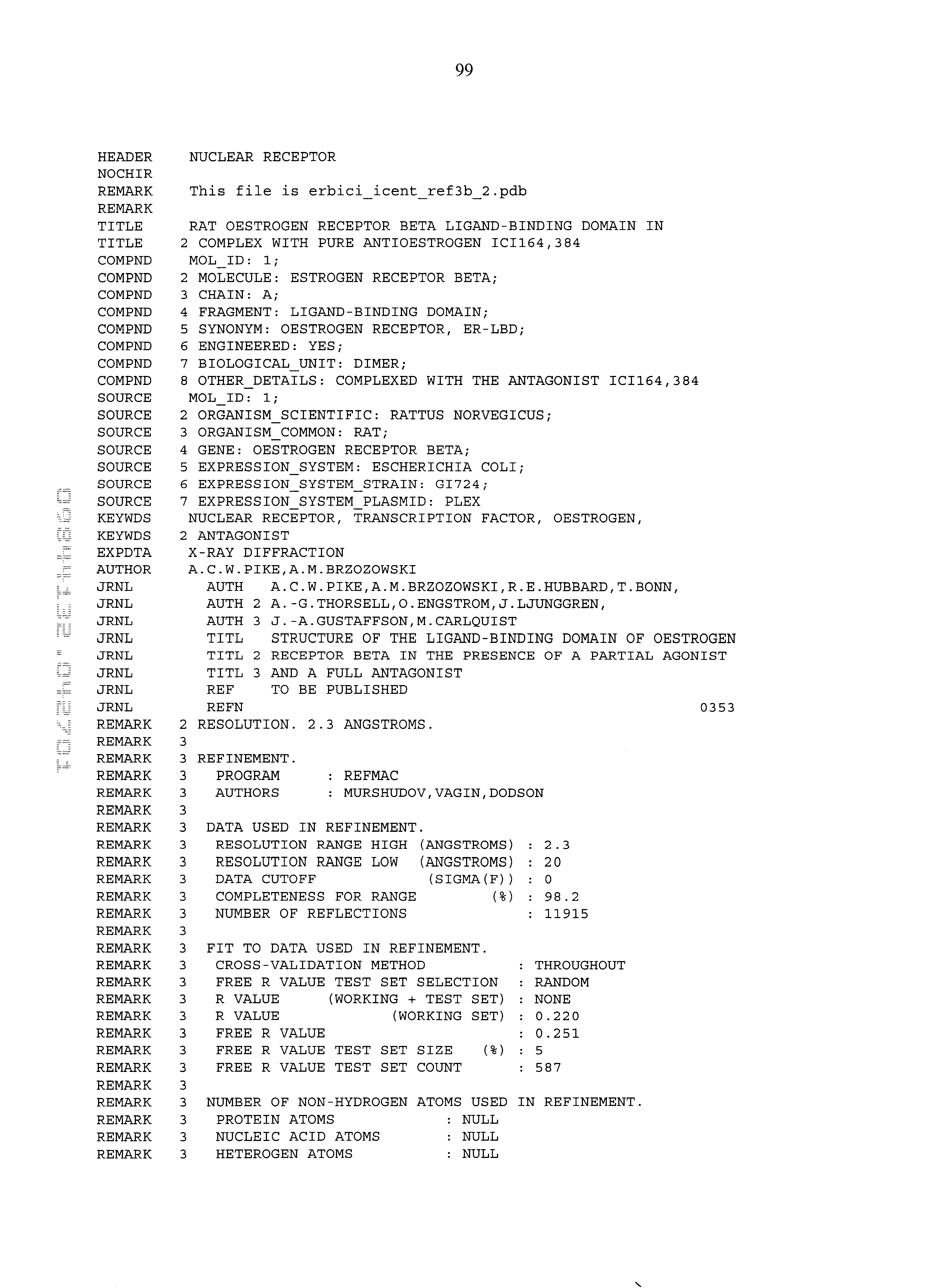
Structure of Rat ER-β Ligand-binding Domain (LBD) Complexed with the Pure Antiestrogen N-(n-Butyl)-11-[3,17β-dihydroxyestra-1,3,5(10)-trien-7α-yl]N-methylundecanamide (ICI 164,384)
Crystallization, data collection, and processing:
Rat ERβ-LBD was expressed, purified and carboxymethylated as described in Example 1, or by the procedure described in Pike, A.C.W., et al. & Carlquist, M. (1999) EMBO J., 18, 4608-4618. N-(n-butyl)-11-[3,17β-dihydroxyestra- 1,3,5(10)-trien-7α-yl]N-methylundecanamide (ICI 164,384; CA Registry number 98007-99-9] was prepared according to the procedure of Bowler et al. (Bowler, J.; Lilley, T. J.; Pittam, J. D.; Wakeling, A. E.; Steroids 1989, 54:71-99). Crystals were grown using the hanging-drop vapour diffusion technique at 18° C. Drops, comprising equal volumes of protein (10 mg/ml) and reservoir solution (7% (w/v) polyethylene glycol 2000 monomethyl ether, 3.5 mM nickel chloride, 10% (v/v) dioxane in 35 mM Tris HCl pH8.5), were equilibrated against the same reservoir solution. The resultant crystals belong to space group I212121 with cell dimensions a=59.37 Å b=81.4 Å c=520.97 Å and contain five LBD monomers per asymmetric unit. The crystals were cryoprotected by slow sequential transfer from 2% to 30% ethylene glycol in 5% steps over 4 hrs. X-ray diffraction data were collected to a resolution of less than 2.5 Å, and more preferably about 2.3 Å, on station P7-11 (MAX-LAB, Lund). A total of 804099 observations were recorded on 576 images in two collection sweeps. These were then scaled and reduced to a final unique set of 42156 reflections (95.7% complete) with a Rmerge(I) of 0.057 for data between 12-2.3 Å. The structure of ERβ-LBD in complex with pure antagonist N-(n-butyl)-11-[3,17β-dihydroxyestra- 1,3,5(10)-trien-7α-yl]N-methylundecanamide (ICI 164,384) is shown in FIGS. 7A (side view) and 7B (end-on view).
EXAMPLE 3 Structure of Rat ER-β Ligand-binding Domain (LBD) Complexed with (17β)-Estra-1,3,5(10)-triene-3,17-diol (Estradiol) and LXXLL Coactivator Peptide
Crystallization, data collection, and processing:
Rat ERβ-LBD was expressed, purified, and carboxymethylated as described in Examples 1 and 2, or by the procedure described in Pike, A.C.W., et al., & Carlquist, N. (1999) EMBO J., 18, 4608-4618. (17β)-Estra-1,3,5(10)-triene-3,17-diol (Estradiol; CAS Registry No. 50-28-2) was obtained from Sigma-Aldrich. The LXXLL (SEQ ID NO:4) coactivator peptide was obtained from MWG-Biotech AG. The LXXLL (SEQ ID NO:4) coactivator peptide was derived from the coactivator independent of AF2 function (CIA) described in Suave, F. et al. Mol. Cell. Biol. 21:343-353 (2001), and comprises residues 338-354 of that protein. The LXXLL peptide used in this experiment comprised 17 amino acids in length with the sequence HPPAIQSLINLLADNRY (SEQ ID NO:3).
Crystals were grown using the hanging-drop vapour diffusion technique at 18° C. Drops, comprising equal volumes of protein solution and reservoir solution (1 μL) were equilibrated against the same reservoir solution. The protein solution was prepared from 15 mg/ml ERβ LBD and LXXLL coactivator peptide (5 mM stock) mixed in a 1:5 molar ratio. The reservoir solution was prepared from 0.50-0.605 dilution of 10% (w/v) PEG8K, 10% (w/v) PEG1K, 0.2 M lithium sulphate, 100 mM Tris HCl (pH 8.0). Crystals were stored frozen in reservoir solution containing 25% (v:v) ethanediol. The resultant crystals belong to space group P4122 with cell dimensions a=62.55 Å, b=62.55 Å, c=171.70 Å. X-ray diffraction data were collected to a resolution of less than 2.5 Å, and preferably about 2.1 Å, on station ID14-4 (ESRF, Grenoble, France).
A total of 499631 observations were recorded on 320 images. These were then scaled and reduced to a final unique set of 20776 reflections (99.9% complete) with a Rmerge(I) of 0.061 for data between 40-2.1 Å. The structure of rat ER-β ligand-binding domain (LBD) complexed with estradiol and LXXLL coactivator peptide is shown in FIGS. 8A (side view) and 8B (end-on view).
EXAMPLE 4 Structure of Rat ER-β Ligand-binding Domain (LBD) Complexed with (16α,17α)-Estra-1,3,5(10)-triene-3,16,17-triol (17-epiestriol) and LXXLL Coactivator Peptide
Crystallization, data collection, and processing:
Rat ERβ-LBD was expressed, purified and carboxymethylated as described in Examples 1 and 2, or by the procedure described in Pike, A.C.W., et al. & Carlquist, M. (1999) EMBO J., 18, 4608-4618. 16α,17α)-Estra-1,3,5(10)-triene-3,16,17-triol (17-Epiestriol; CAS Registry No. 1228-72-4) was obtained from Sigma-Aldrich. The LXXLL coactivator peptide was obtained from MWG-Biotech AG. The LXXLL coactivator peptide was derived from the coactivator independent of AF2 function (CIA) described in Sauve, F. et al. Mol. Cell. Biol. 21:343-353 (2001), and comprises residues 338-354 of that protein. The LXXLL peptide used in this experiment comprised 17 amino acids in length with the sequence HPPAIQSLINLLADNRY (SEQ ID NO:3).
Crystals were grown using the hanging-drop vapour diffusion technique at 18° C. Drops, comprising equal volumes of protein solution and reservoir solution (1 μL) were equilibrated against the same reservoir solution. The protein solution was prepared from 15 mg/ml ERβ LBD and LXXLL coactivator peptide (5mM stock) mixed in a 1:5 molar ratio. The reservoir solution was prepared from 0.50-0.605 dilution of 10% (w/v) PEG8K, 10% (w/v) PEG1K, 0.2 M lithium sulphate, 100 mM Tris HCl (pH 8.0). Crystals were stored frozen in reservoir solution containing 25% (v:v) ethanediol. The resultant crystals belong to space group P4122 with cell dimensions a=62.64 Å, b=62.64 Å, c=171.64 Å. X-ray diffraction data were collected to a resolution of less than 2.0 Å, and preferable less than about 1.8 Å, on station ID14-4 (ESRF, Grenoble, France).
A total of 788988 observations were recorded on 400 images. These were then scaled and reduced to a final unique set of 32284 reflections (98.7% complete) with a Rmerge(I) of 0.039 for data between 30-1.8 Å. The structure of rat ER-b ligand-binding domain (LBD) complexed with epiestriol and LXXLL coactivator peptide is shown in FIGS. 9A (side view) and 9B (end-on view).
Experimental Description of ER Binding Assay
Affinity for the ER (by displacement of 3[H]-estradiol was measured using the scintistrip1 assay. Human estrogen receptor (hER) alpha was extracted from the nuclei from SF9-cells infected with a recombinant baculovirus transfer vector containing the cloned hER genes.2 The concentration of hER's in the extract was measured as specific 3[H]—E2 binding with the G25-assay.
1) Haggblad, J., Carlsson, B., Kivela, P., Siitari, H., (1995) Biotechniques 18, 146-151.
2) Barkhem, T., Carlsson, B., Simons, J., Moller, B., Berkenstam, A., Gustafsson, J. A., Nilsson, D. (1991) J. Steroid Biochem. Molec. Biol. 38,667-75.
3) Salononsson, M., Carlsson, B., Haggblad, J., (1994) J. Steroid Biochem. Molec. Biol. 50, 313-318.
The following pages give the crystal coordinates for:
(1) ERβ-LBD-genistein (SEQ ID NO:1);
(2) ERβ-LBD-Raloxifene(SEQ ID NO:2);
(3) ERβ-LBD-[2-(4-hydroxyphenyl)-6-hydroxybenzo[beta] thienyl-3-yl][4-carboxyphenyl]methanone (KB 177) (SEQ ID NO:2);
(4) ER-β LBD-N-(n-butyl)-11-[3,17β-dihydroxyestra-1,3,5(10)-trien-7α-yl]N-methylundecanamide (ICI 164,384) (SEQ ID NO:2);
(5) ER-β LBD-(17β)-Estradiol/LXXLL coactivator peptide (SEQ ID NO:2); and
(6) ER-β LBD-(17β)-Epiestriol/LXXLL coactivator peptide (SEQ ID NO:2).
Figure US06476196-20021105-P00001
Figure US06476196-20021105-P00002
Figure US06476196-20021105-P00003
Figure US06476196-20021105-P00004
Figure US06476196-20021105-P00005
Figure US06476196-20021105-P00006
Figure US06476196-20021105-P00007
Figure US06476196-20021105-P00008
Figure US06476196-20021105-P00009
Figure US06476196-20021105-P00010
Figure US06476196-20021105-P00011
Figure US06476196-20021105-P00012
Figure US06476196-20021105-P00013
Figure US06476196-20021105-P00014
Figure US06476196-20021105-P00015
Figure US06476196-20021105-P00016
Figure US06476196-20021105-P00017
Figure US06476196-20021105-P00018
Figure US06476196-20021105-P00019
Figure US06476196-20021105-P00020
Figure US06476196-20021105-P00021
Figure US06476196-20021105-P00022
Figure US06476196-20021105-P00023
Figure US06476196-20021105-P00024
Figure US06476196-20021105-P00025
Figure US06476196-20021105-P00026
Figure US06476196-20021105-P00027
Figure US06476196-20021105-P00028
Figure US06476196-20021105-P00029
Figure US06476196-20021105-P00030
Figure US06476196-20021105-P00031
Figure US06476196-20021105-P00032
Figure US06476196-20021105-P00033
Figure US06476196-20021105-P00034
Figure US06476196-20021105-P00035
Figure US06476196-20021105-P00036
Figure US06476196-20021105-P00037
Figure US06476196-20021105-P00038
Figure US06476196-20021105-P00039
Figure US06476196-20021105-P00040
Figure US06476196-20021105-P00041
Figure US06476196-20021105-P00042
Figure US06476196-20021105-P00043
Figure US06476196-20021105-P00044
Figure US06476196-20021105-P00045
Figure US06476196-20021105-P00046
Figure US06476196-20021105-P00047
Figure US06476196-20021105-P00048
Figure US06476196-20021105-P00049
Figure US06476196-20021105-P00050
Figure US06476196-20021105-P00051
Figure US06476196-20021105-P00052
Figure US06476196-20021105-P00053
Figure US06476196-20021105-P00054
Figure US06476196-20021105-P00055
Figure US06476196-20021105-P00056
Figure US06476196-20021105-P00057
Figure US06476196-20021105-P00058
Figure US06476196-20021105-P00059
Figure US06476196-20021105-P00060
Figure US06476196-20021105-P00061
Figure US06476196-20021105-P00062
Figure US06476196-20021105-P00063
Figure US06476196-20021105-P00064
Figure US06476196-20021105-P00065
Figure US06476196-20021105-P00066
Figure US06476196-20021105-P00067
Figure US06476196-20021105-P00068
Figure US06476196-20021105-P00069
Figure US06476196-20021105-P00070
Figure US06476196-20021105-P00071
Figure US06476196-20021105-P00072
Figure US06476196-20021105-P00073
Figure US06476196-20021105-P00074
Figure US06476196-20021105-P00075
Figure US06476196-20021105-P00076
Figure US06476196-20021105-P00077
Figure US06476196-20021105-P00078
Figure US06476196-20021105-P00079
Figure US06476196-20021105-P00080
Figure US06476196-20021105-P00081
Figure US06476196-20021105-P00082
Figure US06476196-20021105-P00083
Figure US06476196-20021105-P00084
Figure US06476196-20021105-P00085
Figure US06476196-20021105-P00086
Figure US06476196-20021105-P00087
Figure US06476196-20021105-P00088
Figure US06476196-20021105-P00089
Figure US06476196-20021105-P00090
Figure US06476196-20021105-P00091
Figure US06476196-20021105-P00092
Figure US06476196-20021105-P00093
Figure US06476196-20021105-P00094
Figure US06476196-20021105-P00095
Figure US06476196-20021105-P00096
Figure US06476196-20021105-P00097
Figure US06476196-20021105-P00098
Figure US06476196-20021105-P00099
Figure US06476196-20021105-P00100
Figure US06476196-20021105-P00101
Figure US06476196-20021105-P00102
Figure US06476196-20021105-P00103
Figure US06476196-20021105-P00104
Figure US06476196-20021105-P00105
Figure US06476196-20021105-P00106
Figure US06476196-20021105-P00107
Figure US06476196-20021105-P00108
Figure US06476196-20021105-P00109
Figure US06476196-20021105-P00110
Figure US06476196-20021105-P00111
Figure US06476196-20021105-P00112
Figure US06476196-20021105-P00113
Figure US06476196-20021105-P00114
Figure US06476196-20021105-P00115
Figure US06476196-20021105-P00116
Figure US06476196-20021105-P00117
Figure US06476196-20021105-P00118
Figure US06476196-20021105-P00119
Figure US06476196-20021105-P00120
Figure US06476196-20021105-P00121
Figure US06476196-20021105-P00122
Figure US06476196-20021105-P00123
Figure US06476196-20021105-P00124
Figure US06476196-20021105-P00125
Figure US06476196-20021105-P00126
Figure US06476196-20021105-P00127
Figure US06476196-20021105-P00128
Figure US06476196-20021105-P00129
Figure US06476196-20021105-P00130
Figure US06476196-20021105-P00131
Figure US06476196-20021105-P00132
Figure US06476196-20021105-P00133
Figure US06476196-20021105-P00134
Figure US06476196-20021105-P00135
Figure US06476196-20021105-P00136
Figure US06476196-20021105-P00137
Figure US06476196-20021105-P00138
Figure US06476196-20021105-P00139
Figure US06476196-20021105-P00140
Figure US06476196-20021105-P00141
Figure US06476196-20021105-P00142
Figure US06476196-20021105-P00143
Figure US06476196-20021105-P00144
Figure US06476196-20021105-P00145
Figure US06476196-20021105-P00146
Figure US06476196-20021105-P00147
Figure US06476196-20021105-P00148
Figure US06476196-20021105-P00149
Figure US06476196-20021105-P00150
Figure US06476196-20021105-P00151
Figure US06476196-20021105-P00152
Figure US06476196-20021105-P00153
Figure US06476196-20021105-P00154
Figure US06476196-20021105-P00155
Figure US06476196-20021105-P00156
Figure US06476196-20021105-P00157
Figure US06476196-20021105-P00158
Figure US06476196-20021105-P00159
Figure US06476196-20021105-P00160
Figure US06476196-20021105-P00161
Figure US06476196-20021105-P00162
Figure US06476196-20021105-P00163
Figure US06476196-20021105-P00164
Figure US06476196-20021105-P00165
Figure US06476196-20021105-P00166
Figure US06476196-20021105-P00167
Figure US06476196-20021105-P00168
Figure US06476196-20021105-P00169
Figure US06476196-20021105-P00170
Figure US06476196-20021105-P00171
Figure US06476196-20021105-P00172
Figure US06476196-20021105-P00173
Figure US06476196-20021105-P00174
Figure US06476196-20021105-P00175
Figure US06476196-20021105-P00176
Figure US06476196-20021105-P00177
Figure US06476196-20021105-P00178
Figure US06476196-20021105-P00179
Figure US06476196-20021105-P00180
Figure US06476196-20021105-P00181
Figure US06476196-20021105-P00182
Figure US06476196-20021105-P00183
Figure US06476196-20021105-P00184
Figure US06476196-20021105-P00185
Figure US06476196-20021105-P00186
Figure US06476196-20021105-P00187
Figure US06476196-20021105-P00188
Figure US06476196-20021105-P00189
Figure US06476196-20021105-P00190
Figure US06476196-20021105-P00191
Figure US06476196-20021105-P00192
Figure US06476196-20021105-P00193
Figure US06476196-20021105-P00194
Figure US06476196-20021105-P00195
Figure US06476196-20021105-P00196
Figure US06476196-20021105-P00197
Figure US06476196-20021105-P00198
Figure US06476196-20021105-P00199
Figure US06476196-20021105-P00200
Figure US06476196-20021105-P00201
Figure US06476196-20021105-P00202
Figure US06476196-20021105-P00203
Figure US06476196-20021105-P00204
4 1 229 PRT Human 1 Asp Ala Leu Ser Glu Glu Gln Leu Val Leu Thr Leu Leu Glu Ala Glu 1 5 10 15 Pro Pro His Val Leu Ile Ser Arg Pro Ser Ala Ser Met Met Met Ser 20 25 30 Leu Thr Lys Leu Ala Asp Leu Glu Leu Val His Met Ile Ser Trp Ala 35 40 45 Lys Lys Ile Pro Gly Phe Val Glu Leu Ser Leu Phe Asp Gln Val Arg 50 55 60 Leu Leu Glu Ser Cys Trp Met Glu Val Leu Met Met Gly Leu Met Trp 65 70 75 80 Arg Ser Ile Asp His Pro Gly Lys Leu Ile Phe Ala Pro Asp Leu Val 85 90 95 Leu Asp Arg Asp Glu Gly Lys Cys Val Glu Gly Ile Leu Glu Ile Phe 100 105 110 Asp Met Leu Leu Ala Thr Thr Ser Arg Phe Arg Glu Leu Lys Leu Gln 115 120 125 His Lys Glu Tyr Leu Cys Val Lys Ala Met Ile Leu Leu Asn Ser Ser 130 135 140 Met Tyr Pro Leu Val Asp Ser Ser Arg Lys Leu Ala His Leu Leu Asn 145 150 155 160 Ala Val Thr Asp Ala Leu Val Trp Val Ile Ala Lys Ser Gly Ile Ser 165 170 175 Ser Gln Gln Gln Ser Met Arg Leu Ala Asn Leu Leu Met Leu Leu Ser 180 185 190 His Val Arg His Ala Ser Asn Lys Gly Met Glu His Leu Leu Met Met 195 200 205 Lys Cys Lys Asn Val Val Pro Val Tyr Asp Leu Leu Leu Glu Met Leu 210 215 220 Asn Ala His Val Leu 225 2 228 PRT RAT 2 Thr Leu Ser Pro Glu Gln Leu Val Leu Thr Leu Leu Glu Ala Glu Pro 1 5 10 15 Pro Asn Val Leu Val Ser Arg Pro Ser Met Pro Phe Thr Glu Ala Ser 20 25 30 Met Met Met Ser Leu Thr Lys Leu Ala Asp Lys Glu Leu Val His Met 35 40 45 Ile Gly Trp Ala Lys Lys Ile Pro Gly Phe Val Glu Leu Ser Leu Leu 50 55 60 Asp Gln Val Arg Leu Leu Glu Ser Cys Trp Met Glu Val Leu Met Val 65 70 75 80 Gly Leu Met Trp Arg Ser Ile Asp His Pro Gly Lys Leu Ile Phe Ala 85 90 95 Pro Asp Leu Val Leu Asp Arg Asp Glu Gly Lys Cys Val Glu Gly Ile 100 105 110 Leu Glu Ile Phe Asp Met Leu Leu Ala Thr Thr Ser Arg Phe Arg Glu 115 120 125 Leu Lys Leu Gln His Lys Glu Tyr Leu Cys Val Lys Ala Met Ile Leu 130 135 140 Leu Asn Ser Ser Met Tyr Pro Leu Ala Ser Ala Asn Gln Glu Ala Glu 145 150 155 160 Ser Ser Arg Lys Leu Thr His Leu Leu Asn Ala Val Thr Asp Ala Leu 165 170 175 Val Trp Val Ile Ala Lys Ser Gly Ile Ser Ser Gln Gln Gln Ser Val 180 185 190 Arg Leu Ala Asn Leu Leu Met Leu Leu Ser His Val Arg His Ile Ser 195 200 205 Asn Lys Gly Met Glu His Leu Leu Ser Met Lys Asp Leu Leu Leu Glu 210 215 220 Met Leu Asn Ala 225 3 17 PRT Artificial Sequence LXXLL coactivator peptide derived from the coactivator independent of AF2 function (CIA) described in Sauve, F. et al. Mol. Cell Biol. 21343-353 (2001). 3 His Pro Pro Ala Ile Gln Ser Leu Ile Asn Leu Leu Ala Asp Asn Arg 1 5 10 15 Tyr 4 5 PRT Artificial Sequence VARIANT (1)...(5) Xaa = Any Amino Acid 4 Leu Xaa Xaa Leu Leu 1 5

Claims (9)

What is claimed is:
1. A crystal of ERβ ligand binding domain of SEQ ID NO:2 complexed with N-(n-butyl)-11-[3,17β-dihydroxyestra-1,3,5(10)-trien-7α-yl]N-methylundecanamide, said crystal belonging to the space group I212121 and having unit cell dimensions a=59.37 Å, b=81.4 Å, c=520.97 Å, and containing five ligand binding domain monomers per asymmetric unit.
2. The crystal of claim 1, wherein said crystal has a resolution determined by X-ray crystallography of less than 2.5 Å.
3. A process of preparing the crystal of claim 1, comprising the steps of:
providing ERβ ligand binding domain of SEQ ID NO:2 complexed with N-(n-butyl)-11-[3,17β-dihydroxyestra-1,3,5(10)-trien-7α-yl]N-methylundecanamide, and
growing one or more of said crystals by hanging drop vapor diffusion at 18° C. from a solution comprising equal volumes of ERβ ligand binding domain complexed with N-(n-butyl)-11-[3,17β-dihydroxyestra-1,3,5(10)-trien-7α-yl]N-methylundecanamide (10 mg/ml) and resevoir solution, said resevoir solution comprising 7% (w/v) polyethylene glycol 2000 monomethyl ether, 3.5 mM nickel chloride, and 10% (v/v) dioxane in 35 mM Tris-HCl, pH 8.5.
4. A crystal of ERβ ligand binding domain of SEQ ID NO:2 complexed with estradiol and a peptide comprising the amino acid sequence of SEQ ID NO:3, said crystal belonging to the space group P4122 and having the unit cell dimensions a=62.55 Å, b=62.55 Å, c=171.70 Å.
5. The crystal of claim 4, wherein said crystal has a resolution determined by X-ray crystallography of less than 2.5 Å.
6. A process of preparing the crystal of claim 4, comprising the steps of:
providing ERβ ligand binding domain of SEQ ID NO:2 complexed with estradiol and a peptide comprising the amino acid sequence of SEQ ID NO:3, and
growing one or more of said crystals by hanging drop vapor diffusion at 18° C. from a solution comprising equal volumes of ERβ ligand binding domain complexed with estradiol and a peptide comprising the amino acid sequence of SEQ ID NO:3, said ERβ ligand binding domain and said peptide present in a 1:5 molar ratio, and resevoir solution, said resevoir solution comprising 0.5-0.605 dilution of 10% (w/v) polyethylene glycol 8000, 10% (w/v) polyethylene glycol 1000, and 0.2 M lithium sulphate, and 100 mM Tris-HCl, pH 8.0.
7. A crystal of ERβ ligand binding domain SEQ ID NO:2 complexed with 17-epiestriol and a peptide comprising the amino acid sequence of SEQ ID NO:3, said crystal belonging to the space group P4122 and having the unit cell dimensions a=62.64 Å, b=62.64 Å, c=171.64 Å.
8. The crystal of claim 7, wherein said crystal has a resolution determined by X-ray crystallography of less than 2.0 Å.
9. A process of preparing the crystal of claim 7, comprising the steps of:
providing ERβ ligand binding domain of SEQ ID NO:2 complexed with 2-(4-hydroxyphenyl)-6-hydroxybenzo-β-thienyl-3-yl (4-carboxyphenyl)methanone and a peptide comprising the amino acid sequence of SEQ ID NO:3, and
growing one or more of said crystals by hanging drop vapor diffusion at 18° C. from a solution comprising equal volumes of ERβ ligand binding domain complexed with 2-(4-hydroxyphenyl)-6-hydroxybenzo-β-thienyl-3-yl (4-carboxyphenyl)methanone and a peptide comprising the amino acid sequence of SEQ ID NO:3, said ERβ ligand binding domain and said peptide present in a 1:5 molar ratio, and resevoir solution, said resevoir solution comprising 0.5-0.605 dilution of 10% (w/v) polyethylene glycol 8000, 10% (w/v) polyethylene glycol 1000, and 0.2 M lithium sulphate, and 100 mM Tris-HCl, pH 8.0.
US09/844,132 1998-02-13 2001-04-27 Estrogen receptor ligands Expired - Fee Related US6476196B1 (en)

Priority Applications (1)

Application Number Priority Date Filing Date Title
US09/844,132 US6476196B1 (en) 1998-02-13 2001-04-27 Estrogen receptor ligands

Applications Claiming Priority (4)

Application Number Priority Date Filing Date Title
GB9803062 1998-02-13
GBGB9803062.0A GB9803062D0 (en) 1998-02-13 1998-02-13 Estrogen receptor ligands
US09/249,645 US6228990B1 (en) 1998-02-13 1999-02-12 Estrogen receptor ligands
US09/844,132 US6476196B1 (en) 1998-02-13 2001-04-27 Estrogen receptor ligands

Related Parent Applications (1)

Application Number Title Priority Date Filing Date
US09/249,645 Continuation-In-Part US6228990B1 (en) 1998-02-13 1999-02-12 Estrogen receptor ligands

Publications (1)

Publication Number Publication Date
US6476196B1 true US6476196B1 (en) 2002-11-05

Family

ID=26313113

Family Applications (1)

Application Number Title Priority Date Filing Date
US09/844,132 Expired - Fee Related US6476196B1 (en) 1998-02-13 2001-04-27 Estrogen receptor ligands

Country Status (1)

Country Link
US (1) US6476196B1 (en)

Cited By (8)

* Cited by examiner, † Cited by third party
Publication number Priority date Publication date Assignee Title
US20020072587A1 (en) * 2000-07-12 2002-06-13 Somers William S. Crystal structure of estrogen receptor-beta complex and uses thereof
US20040102427A1 (en) * 2002-11-22 2004-05-27 Hermsmeyer R. Kent Estrogen beta receptor agonists to prevent or reduce the severity of cardiovascular disease
US20050009137A1 (en) * 2003-05-07 2005-01-13 Cedars-Sinai Medical Center Intracellular estradiol binding protein, a polynucleotide encoding the same and cell lines overexpressing the same
WO2011107735A1 (en) 2010-03-03 2011-09-09 Halliburton Energy Services, Inc. Weak organic acid as gelation retarder for crosslinkable polymer compositions
WO2011107743A1 (en) 2010-03-03 2011-09-09 Halliburton Energy Services Inc Salt of weak bronsted base and bronsted acid as gelation retarder for crosslinkable polymer compositions
WO2011107744A1 (en) 2010-03-03 2011-09-09 Halliburton Energy Services, Inc Ammonium halide as gelation retarder for crosslinkable polymer compositions
WO2011107761A1 (en) 2010-03-03 2011-09-09 Halliburton Energy Services, Inc Lewis acid as gelation retarder for crosslinkable polymer compositions
US10472555B2 (en) 2016-04-08 2019-11-12 Schlumberger Technology Corporation Polymer gel for water control applications

Non-Patent Citations (4)

* Cited by examiner, † Cited by third party
Title
Pike et al., "Structural Insights into the Mode of Action of a Pure Antiestrogen", Structure, vol. 9, Feb. 2001, 145-153.
Pike et al., "Structure of the ligand-binding domain oestrogen receptor beta in the presence of a partial agonist and a full antagonist", EMBO, J. 18 (17), 4608-4618.
Shiau et al., "The structure bases of estrogen receptor/coactivator . . . ", Cell 95, 927-937 (1998).
Tananbaum et al., "Crystallographic comparison of estrogen and progestrone . . . ", Proc. Natl. Acad. Sci. USA, 95, 5998-6003.

Cited By (14)

* Cited by examiner, † Cited by third party
Publication number Priority date Publication date Assignee Title
US20020072587A1 (en) * 2000-07-12 2002-06-13 Somers William S. Crystal structure of estrogen receptor-beta complex and uses thereof
US7235367B2 (en) 2000-07-12 2007-06-26 Genetics Institute, Llc. Method using crystal structure of estrogen receptor-β complex
US20080227960A1 (en) * 2001-07-11 2008-09-18 Genetics Institute, Llc. Crystal structure of estrogen receptor-beta complex and uses thereof
US20040102427A1 (en) * 2002-11-22 2004-05-27 Hermsmeyer R. Kent Estrogen beta receptor agonists to prevent or reduce the severity of cardiovascular disease
US20100152144A1 (en) * 2002-11-22 2010-06-17 Hermsmeyer R Kent Estrogen beta receptor agonists to prevent or reduce the severity of cardiovascular disease
US7674783B2 (en) 2002-11-22 2010-03-09 Dimera Inc. Estrogen beta receptor agonists to prevent or reduce the severity of cardiovascular disease
US7488597B2 (en) 2003-05-07 2009-02-10 Cedars-Sinai Medical Center Polynucleotide encoding an intracellular estradiol binding protein
US7335735B2 (en) 2003-05-07 2008-02-26 Cedars-Sinai Medical Center Intracellular estradiol binding protein
US20050009137A1 (en) * 2003-05-07 2005-01-13 Cedars-Sinai Medical Center Intracellular estradiol binding protein, a polynucleotide encoding the same and cell lines overexpressing the same
WO2011107735A1 (en) 2010-03-03 2011-09-09 Halliburton Energy Services, Inc. Weak organic acid as gelation retarder for crosslinkable polymer compositions
WO2011107743A1 (en) 2010-03-03 2011-09-09 Halliburton Energy Services Inc Salt of weak bronsted base and bronsted acid as gelation retarder for crosslinkable polymer compositions
WO2011107744A1 (en) 2010-03-03 2011-09-09 Halliburton Energy Services, Inc Ammonium halide as gelation retarder for crosslinkable polymer compositions
WO2011107761A1 (en) 2010-03-03 2011-09-09 Halliburton Energy Services, Inc Lewis acid as gelation retarder for crosslinkable polymer compositions
US10472555B2 (en) 2016-04-08 2019-11-12 Schlumberger Technology Corporation Polymer gel for water control applications

Similar Documents

Publication Publication Date Title
Christensen et al. Characterization and functional properties of the A and B forms of human progesterone receptors synthesized in a baculovirus system
JP3385380B2 (en) DNA encoding androgen receptor protein
Fawell et al. Characterization and colocalization of steroid binding and dimerization activities in the mouse estrogen receptor
Mehta et al. The low density lipoprotein receptor in Xenopus laevis. I. Five domains that resemble the human receptor
Danielsen et al. Two amino acids within the knuckle of the first zinc finger specify DNA response element activation by the glucocorticoid receptor
Cadepond et al. Heat shock protein 90 as a critical factor in maintaining glucocorticosteroid receptor in a nonfunctional state.
Chrivia et al. Phosphorylated CREB binds specifically to the nuclear protein CBP
Scherrer et al. Evidence that the hormone binding domain of steroid receptors confers hormonal control on chimeric proteins by determining their hormone-regulated binding to heat-shock protein 90
Weigel et al. Ligands induce conformational changes in the carboxyl-terminus of progesterone receptors which are detected by a site-directed antipeptide monoclonal antibody.
US6266622B1 (en) Nuclear receptor ligands and ligand binding domains
Morgan et al. Purification and characterization of the receptor for insulin-like growth factor I
EP0849361B1 (en) Novel ligands of the neuropeptide receptor HFGAN72
US6476196B1 (en) Estrogen receptor ligands
JP2001525339A (en) Method for designing agonists and antagonists to IGF receptor
Pakdel et al. An assessment of the role of domain F and PEST sequences in estrogen receptor half-life and bioactivity
AU681855B2 (en) Novel malignant cell type markers of the interior nuclear matrix
Reese et al. Identification of two cysteines closely positioned in the ligand-binding pocket of the human estrogen receptor: roles in ligand binding and transcriptional activation.
US6228990B1 (en) Estrogen receptor ligands
AU735703B2 (en) Estrogen receptor ligands
Hirst et al. Discrimination of DNA response elements for thyroid hormone and estrogen is dependent on dimerization of receptor DNA binding domains.
Traish et al. Loss of expression of a 55 kDa nuclear protein (nmt55) in estrogen receptor-negative human breast cancer
Innamorati et al. GIP, a G-protein-coupled receptor interacting protein
US20030032779A1 (en) Estrogen receptor ligands
US6335159B1 (en) Retinoic acid receptor ε(rarε)
JP4124592B2 (en) Polypeptides derived from vitamin D nuclear receptors and their use for screening for vitamin D analogues in particular

Legal Events

Date Code Title Description
AS Assignment

Owner name: KARO BIO AB, SWEDEN

Free format text: ASSIGNMENT OF ASSIGNORS INTEREST;ASSIGNORS:LJUNGGREN, JAN;THORSELL, ANN-GERD;ENGSTROM, OWE;AND OTHERS;REEL/FRAME:012165/0391;SIGNING DATES FROM 20010625 TO 20010821

CC Certificate of correction
FPAY Fee payment

Year of fee payment: 4

SULP Surcharge for late payment
FEPP Fee payment procedure

Free format text: PAYOR NUMBER ASSIGNED (ORIGINAL EVENT CODE: ASPN); ENTITY STATUS OF PATENT OWNER: LARGE ENTITY

FEPP Fee payment procedure

Free format text: PAT HOLDER NO LONGER CLAIMS SMALL ENTITY STATUS, ENTITY STATUS SET TO UNDISCOUNTED (ORIGINAL EVENT CODE: STOL); ENTITY STATUS OF PATENT OWNER: LARGE ENTITY

FPAY Fee payment

Year of fee payment: 8

REMI Maintenance fee reminder mailed
LAPS Lapse for failure to pay maintenance fees
STCH Information on status: patent discontinuation

Free format text: PATENT EXPIRED DUE TO NONPAYMENT OF MAINTENANCE FEES UNDER 37 CFR 1.362

FP Lapsed due to failure to pay maintenance fee

Effective date: 20141105