US6472504B1 - Inhibitors of collagen assembly - Google Patents
Inhibitors of collagen assembly Download PDFInfo
- Publication number
- US6472504B1 US6472504B1 US09/517,866 US51786600A US6472504B1 US 6472504 B1 US6472504 B1 US 6472504B1 US 51786600 A US51786600 A US 51786600A US 6472504 B1 US6472504 B1 US 6472504B1
- Authority
- US
- United States
- Prior art keywords
- collagen
- peptide
- assembly
- peptides
- binding
- Prior art date
- Legal status (The legal status is an assumption and is not a legal conclusion. Google has not performed a legal analysis and makes no representation as to the accuracy of the status listed.)
- Expired - Lifetime
Links
- 102000008186 Collagen Human genes 0.000 title claims description 100
- 108010035532 Collagen Proteins 0.000 title claims description 100
- 229920001436 collagen Polymers 0.000 title claims description 100
- 239000003112 inhibitor Substances 0.000 title description 7
- 108090000765 processed proteins & peptides Proteins 0.000 claims abstract description 270
- 108010022452 Collagen Type I Proteins 0.000 claims abstract description 94
- 102000012422 Collagen Type I Human genes 0.000 claims abstract description 94
- 241000282414 Homo sapiens Species 0.000 claims abstract description 41
- 230000002401 inhibitory effect Effects 0.000 claims description 35
- 125000003275 alpha amino acid group Chemical group 0.000 claims description 30
- 238000003556 assay Methods 0.000 claims description 28
- 238000001338 self-assembly Methods 0.000 claims description 24
- 238000000338 in vitro Methods 0.000 claims description 4
- 102000004196 processed proteins & peptides Human genes 0.000 abstract description 95
- 238000000034 method Methods 0.000 abstract description 40
- 239000000816 peptidomimetic Substances 0.000 abstract description 20
- 230000027455 binding Effects 0.000 description 87
- 108010049937 collagen type I trimeric cross-linked peptide Proteins 0.000 description 41
- 239000000178 monomer Substances 0.000 description 35
- 239000012634 fragment Substances 0.000 description 22
- 239000002245 particle Substances 0.000 description 21
- LOJFGJZQOKTUBR-XAQOOIOESA-N NC(N)=NCCC[C@@H](C(O)=O)NC(=O)CNC(=O)CNC(=O)[C@H](CC(O)=O)NC(=O)[C@@H](NC(=O)[C@@H](NC(=O)[C@H](CCCCN)NC(=O)[C@@H](N)CCC(O)=O)C)CC1=CN=CN1 Chemical compound NC(N)=NCCC[C@@H](C(O)=O)NC(=O)CNC(=O)CNC(=O)[C@H](CC(O)=O)NC(=O)[C@@H](NC(=O)[C@@H](NC(=O)[C@H](CCCCN)NC(=O)[C@@H](N)CCC(O)=O)C)CC1=CN=CN1 LOJFGJZQOKTUBR-XAQOOIOESA-N 0.000 description 19
- 235000001014 amino acid Nutrition 0.000 description 18
- 238000002823 phage display Methods 0.000 description 18
- 229940024606 amino acid Drugs 0.000 description 17
- 150000001413 amino acids Chemical class 0.000 description 17
- 230000015572 biosynthetic process Effects 0.000 description 14
- 238000012360 testing method Methods 0.000 description 14
- 230000000694 effects Effects 0.000 description 13
- 101000945357 Homo sapiens Collagen alpha-1(I) chain Proteins 0.000 description 12
- 241000251539 Vertebrata <Metazoa> Species 0.000 description 12
- 150000001875 compounds Chemical class 0.000 description 12
- 239000000499 gel Substances 0.000 description 12
- 102000029816 Collagenase Human genes 0.000 description 11
- 108060005980 Collagenase Proteins 0.000 description 11
- 229960002424 collagenase Drugs 0.000 description 11
- 241000124008 Mammalia Species 0.000 description 10
- 239000000872 buffer Substances 0.000 description 10
- ATDGTVJJHBUTRL-UHFFFAOYSA-N cyanogen bromide Chemical group BrC#N ATDGTVJJHBUTRL-UHFFFAOYSA-N 0.000 description 10
- 230000005764 inhibitory process Effects 0.000 description 10
- 230000003993 interaction Effects 0.000 description 10
- 239000000243 solution Substances 0.000 description 10
- 241000894007 species Species 0.000 description 10
- 239000000725 suspension Substances 0.000 description 10
- 125000000539 amino acid group Chemical group 0.000 description 9
- 238000002474 experimental method Methods 0.000 description 9
- 230000008569 process Effects 0.000 description 9
- 239000003480 eluent Substances 0.000 description 8
- XKUKSGPZAADMRA-UHFFFAOYSA-N glycyl-glycyl-glycine Chemical compound NCC(=O)NCC(=O)NCC(O)=O XKUKSGPZAADMRA-UHFFFAOYSA-N 0.000 description 8
- 230000002209 hydrophobic effect Effects 0.000 description 8
- 229920001184 polypeptide Polymers 0.000 description 8
- 238000011534 incubation Methods 0.000 description 6
- 210000001724 microfibril Anatomy 0.000 description 6
- 239000000203 mixture Substances 0.000 description 6
- 239000000523 sample Substances 0.000 description 6
- 108020004414 DNA Proteins 0.000 description 5
- 102000010834 Extracellular Matrix Proteins Human genes 0.000 description 5
- 108010037362 Extracellular Matrix Proteins Proteins 0.000 description 5
- 108010067902 Peptide Library Proteins 0.000 description 5
- 102100028437 Versican core protein Human genes 0.000 description 5
- 101710200894 Versican core protein Proteins 0.000 description 5
- 238000006243 chemical reaction Methods 0.000 description 5
- 238000003776 cleavage reaction Methods 0.000 description 5
- LOKCTEFSRHRXRJ-UHFFFAOYSA-I dipotassium trisodium dihydrogen phosphate hydrogen phosphate dichloride Chemical compound P(=O)(O)(O)[O-].[K+].P(=O)(O)([O-])[O-].[Na+].[Na+].[Cl-].[K+].[Cl-].[Na+] LOKCTEFSRHRXRJ-UHFFFAOYSA-I 0.000 description 5
- 210000002744 extracellular matrix Anatomy 0.000 description 5
- 239000002609 medium Substances 0.000 description 5
- 239000002953 phosphate buffered saline Substances 0.000 description 5
- 229920002401 polyacrylamide Polymers 0.000 description 5
- 102000004169 proteins and genes Human genes 0.000 description 5
- 108090000623 proteins and genes Proteins 0.000 description 5
- 230000007017 scission Effects 0.000 description 5
- YBJHBAHKTGYVGT-ZKWXMUAHSA-N (+)-Biotin Chemical compound N1C(=O)N[C@@H]2[C@H](CCCCC(=O)O)SC[C@@H]21 YBJHBAHKTGYVGT-ZKWXMUAHSA-N 0.000 description 4
- 108091003079 Bovine Serum Albumin Proteins 0.000 description 4
- GNNJKUYDWFIBTK-QWRGUYRKSA-N Gly-Tyr-Asp Chemical compound [H]NCC(=O)N[C@@H](CC1=CC=C(O)C=C1)C(=O)N[C@@H](CC(O)=O)C(O)=O GNNJKUYDWFIBTK-QWRGUYRKSA-N 0.000 description 4
- 108010050808 Procollagen Proteins 0.000 description 4
- 229920001222 biopolymer Polymers 0.000 description 4
- 229940098773 bovine serum albumin Drugs 0.000 description 4
- 210000004899 c-terminal region Anatomy 0.000 description 4
- 210000004027 cell Anatomy 0.000 description 4
- 239000012501 chromatography medium Substances 0.000 description 4
- 238000010494 dissociation reaction Methods 0.000 description 4
- 230000005593 dissociations Effects 0.000 description 4
- 238000001502 gel electrophoresis Methods 0.000 description 4
- VPZXBVLAVMBEQI-UHFFFAOYSA-N glycyl-DL-alpha-alanine Natural products OC(=O)C(C)NC(=O)CN VPZXBVLAVMBEQI-UHFFFAOYSA-N 0.000 description 4
- 108010067216 glycyl-glycyl-glycine Proteins 0.000 description 4
- 230000003100 immobilizing effect Effects 0.000 description 4
- 238000012856 packing Methods 0.000 description 4
- 235000018102 proteins Nutrition 0.000 description 4
- 230000009870 specific binding Effects 0.000 description 4
- DGZSVBBLLGZHSF-UHFFFAOYSA-N 4,4-diethylpiperidine Chemical compound CCC1(CC)CCNCC1 DGZSVBBLLGZHSF-UHFFFAOYSA-N 0.000 description 3
- QTBSBXVTEAMEQO-UHFFFAOYSA-N Acetic acid Chemical compound CC(O)=O QTBSBXVTEAMEQO-UHFFFAOYSA-N 0.000 description 3
- 102100036213 Collagen alpha-2(I) chain Human genes 0.000 description 3
- 108010090461 DFG peptide Proteins 0.000 description 3
- 101000875067 Homo sapiens Collagen alpha-2(I) chain Proteins 0.000 description 3
- COLNVLDHVKWLRT-QMMMGPOBSA-N L-phenylalanine Chemical compound OC(=O)[C@@H](N)CC1=CC=CC=C1 COLNVLDHVKWLRT-QMMMGPOBSA-N 0.000 description 3
- 241001465754 Metazoa Species 0.000 description 3
- FAPWRFPIFSIZLT-UHFFFAOYSA-M Sodium chloride Chemical compound [Na+].[Cl-] FAPWRFPIFSIZLT-UHFFFAOYSA-M 0.000 description 3
- HSVPZJLMPLMPOX-BPNCWPANSA-N Tyr-Arg-Ala Chemical compound [H]N[C@@H](CC1=CC=C(O)C=C1)C(=O)N[C@@H](CCCNC(N)=N)C(=O)N[C@@H](C)C(O)=O HSVPZJLMPLMPOX-BPNCWPANSA-N 0.000 description 3
- 108010047857 aspartylglycine Proteins 0.000 description 3
- 238000005094 computer simulation Methods 0.000 description 3
- 239000003814 drug Substances 0.000 description 3
- 238000001962 electrophoresis Methods 0.000 description 3
- 238000010828 elution Methods 0.000 description 3
- 230000013595 glycosylation Effects 0.000 description 3
- 238000006206 glycosylation reaction Methods 0.000 description 3
- 238000001727 in vivo Methods 0.000 description 3
- 108700005457 microfibrillar Proteins 0.000 description 3
- 238000000386 microscopy Methods 0.000 description 3
- 230000004048 modification Effects 0.000 description 3
- 238000012986 modification Methods 0.000 description 3
- 238000000329 molecular dynamics simulation Methods 0.000 description 3
- 229920000136 polysorbate Polymers 0.000 description 3
- QKNYBSVHEMOAJP-UHFFFAOYSA-N 2-amino-2-(hydroxymethyl)propane-1,3-diol;hydron;chloride Chemical compound Cl.OCC(N)(CO)CO QKNYBSVHEMOAJP-UHFFFAOYSA-N 0.000 description 2
- 102000002260 Alkaline Phosphatase Human genes 0.000 description 2
- 108020004774 Alkaline Phosphatase Proteins 0.000 description 2
- 102100021277 Beta-secretase 2 Human genes 0.000 description 2
- 101710150190 Beta-secretase 2 Proteins 0.000 description 2
- 101100208237 Bos taurus THBS2 gene Proteins 0.000 description 2
- 101100315624 Caenorhabditis elegans tyr-1 gene Proteins 0.000 description 2
- 108090000317 Chymotrypsin Proteins 0.000 description 2
- 102000004190 Enzymes Human genes 0.000 description 2
- 108090000790 Enzymes Proteins 0.000 description 2
- RPLLQZBOVIVGMX-QWRGUYRKSA-N Gly-Asp-Phe Chemical compound [H]NCC(=O)N[C@@H](CC(O)=O)C(=O)N[C@@H](CC1=CC=CC=C1)C(O)=O RPLLQZBOVIVGMX-QWRGUYRKSA-N 0.000 description 2
- DHMQDGOQFOQNFH-UHFFFAOYSA-N Glycine Chemical compound NCC(O)=O DHMQDGOQFOQNFH-UHFFFAOYSA-N 0.000 description 2
- PMMYEEVYMWASQN-DMTCNVIQSA-N Hydroxyproline Chemical compound O[C@H]1CN[C@H](C(O)=O)C1 PMMYEEVYMWASQN-DMTCNVIQSA-N 0.000 description 2
- UGTHTQWIQKEDEH-BQBZGAKWSA-N L-alanyl-L-prolylglycine zwitterion Chemical compound C[C@H](N)C(=O)N1CCC[C@H]1C(=O)NCC(O)=O UGTHTQWIQKEDEH-BQBZGAKWSA-N 0.000 description 2
- OUYCCCASQSFEME-QMMMGPOBSA-N L-tyrosine Chemical compound OC(=O)[C@@H](N)CC1=CC=C(O)C=C1 OUYCCCASQSFEME-QMMMGPOBSA-N 0.000 description 2
- UCBPDSYUVAAHCD-UWVGGRQHSA-N Leu-Pro-Gly Chemical compound CC(C)C[C@H](N)C(=O)N1CCC[C@H]1C(=O)NCC(O)=O UCBPDSYUVAAHCD-UWVGGRQHSA-N 0.000 description 2
- TWRXJAOTZQYOKJ-UHFFFAOYSA-L Magnesium chloride Chemical compound [Mg+2].[Cl-].[Cl-] TWRXJAOTZQYOKJ-UHFFFAOYSA-L 0.000 description 2
- JLMZKEQFMVORMA-SRVKXCTJSA-N Pro-Pro-Arg Chemical compound NC(N)=NCCC[C@@H](C(O)=O)NC(=O)[C@@H]1CCCN1C(=O)[C@H]1NCCC1 JLMZKEQFMVORMA-SRVKXCTJSA-N 0.000 description 2
- CDBYLPFSWZWCQE-UHFFFAOYSA-L Sodium Carbonate Chemical compound [Na+].[Na+].[O-]C([O-])=O CDBYLPFSWZWCQE-UHFFFAOYSA-L 0.000 description 2
- 108010090804 Streptavidin Proteins 0.000 description 2
- 108090000631 Trypsin Proteins 0.000 description 2
- 102000004142 Trypsin Human genes 0.000 description 2
- WPVGRKLNHJJCEN-BZSNNMDCSA-N Tyr-Asp-Phe Chemical compound C([C@H](N)C(=O)N[C@@H](CC(O)=O)C(=O)N[C@@H](CC=1C=CC=CC=1)C(O)=O)C1=CC=C(O)C=C1 WPVGRKLNHJJCEN-BZSNNMDCSA-N 0.000 description 2
- XSQUKJJJFZCRTK-UHFFFAOYSA-N Urea Chemical compound NC(N)=O XSQUKJJJFZCRTK-UHFFFAOYSA-N 0.000 description 2
- ASQFIHTXXMFENG-XPUUQOCRSA-N Val-Ala-Gly Chemical compound CC(C)[C@H](N)C(=O)N[C@@H](C)C(=O)NCC(O)=O ASQFIHTXXMFENG-XPUUQOCRSA-N 0.000 description 2
- 238000002441 X-ray diffraction Methods 0.000 description 2
- 230000001594 aberrant effect Effects 0.000 description 2
- 108010069020 alanyl-prolyl-glycine Proteins 0.000 description 2
- 108010029483 alpha 1 Chain Collagen Type I Proteins 0.000 description 2
- CKLJMWTZIZZHCS-REOHCLBHSA-L aspartate group Chemical group N[C@@H](CC(=O)[O-])C(=O)[O-] CKLJMWTZIZZHCS-REOHCLBHSA-L 0.000 description 2
- 229960002685 biotin Drugs 0.000 description 2
- 235000020958 biotin Nutrition 0.000 description 2
- 239000011616 biotin Substances 0.000 description 2
- 239000003153 chemical reaction reagent Substances 0.000 description 2
- 210000003837 chick embryo Anatomy 0.000 description 2
- 102000006533 chordin Human genes 0.000 description 2
- 108010008846 chordin Proteins 0.000 description 2
- 229960002376 chymotrypsin Drugs 0.000 description 2
- 238000012875 competitive assay Methods 0.000 description 2
- 238000013461 design Methods 0.000 description 2
- 238000010790 dilution Methods 0.000 description 2
- 239000012895 dilution Substances 0.000 description 2
- FSXRLASFHBWESK-UHFFFAOYSA-N dipeptide phenylalanyl-tyrosine Natural products C=1C=C(O)C=CC=1CC(C(O)=O)NC(=O)C(N)CC1=CC=CC=C1 FSXRLASFHBWESK-UHFFFAOYSA-N 0.000 description 2
- 208000037265 diseases, disorders, signs and symptoms Diseases 0.000 description 2
- PMMYEEVYMWASQN-UHFFFAOYSA-N dl-hydroxyproline Natural products OC1C[NH2+]C(C([O-])=O)C1 PMMYEEVYMWASQN-UHFFFAOYSA-N 0.000 description 2
- 239000003937 drug carrier Substances 0.000 description 2
- -1 e.g. Chemical group 0.000 description 2
- 229940088598 enzyme Drugs 0.000 description 2
- 102000013373 fibrillar collagen Human genes 0.000 description 2
- 108060002894 fibrillar collagen Proteins 0.000 description 2
- 210000002950 fibroblast Anatomy 0.000 description 2
- 238000009472 formulation Methods 0.000 description 2
- 108010037850 glycylvaline Proteins 0.000 description 2
- 125000001165 hydrophobic group Chemical group 0.000 description 2
- 229960002591 hydroxyproline Drugs 0.000 description 2
- 108010057821 leucylproline Proteins 0.000 description 2
- 239000003446 ligand Substances 0.000 description 2
- BDAGIHXWWSANSR-UHFFFAOYSA-N methanoic acid Natural products OC=O BDAGIHXWWSANSR-UHFFFAOYSA-N 0.000 description 2
- 238000000302 molecular modelling Methods 0.000 description 2
- 239000013642 negative control Substances 0.000 description 2
- 230000009871 nonspecific binding Effects 0.000 description 2
- 108020004707 nucleic acids Proteins 0.000 description 2
- 102000039446 nucleic acids Human genes 0.000 description 2
- 150000007523 nucleic acids Chemical class 0.000 description 2
- 239000008188 pellet Substances 0.000 description 2
- 238000002360 preparation method Methods 0.000 description 2
- 238000012545 processing Methods 0.000 description 2
- 238000000159 protein binding assay Methods 0.000 description 2
- 238000000746 purification Methods 0.000 description 2
- 239000011541 reaction mixture Substances 0.000 description 2
- 238000004007 reversed phase HPLC Methods 0.000 description 2
- 125000003607 serino group Chemical group [H]N([H])[C@]([H])(C(=O)[*])C(O[H])([H])[H] 0.000 description 2
- 210000001626 skin fibroblast Anatomy 0.000 description 2
- 239000011780 sodium chloride Substances 0.000 description 2
- 239000007787 solid Substances 0.000 description 2
- 238000006467 substitution reaction Methods 0.000 description 2
- 238000003786 synthesis reaction Methods 0.000 description 2
- 210000002435 tendon Anatomy 0.000 description 2
- FGMPLJWBKKVCDB-UHFFFAOYSA-N trans-L-hydroxy-proline Natural products ON1CCCC1C(O)=O FGMPLJWBKKVCDB-UHFFFAOYSA-N 0.000 description 2
- 239000012588 trypsin Substances 0.000 description 2
- OUYCCCASQSFEME-UHFFFAOYSA-N tyrosine Natural products OC(=O)C(N)CC1=CC=C(O)C=C1 OUYCCCASQSFEME-UHFFFAOYSA-N 0.000 description 2
- 125000001493 tyrosinyl group Chemical group [H]OC1=C([H])C([H])=C(C([H])=C1[H])C([H])([H])C([H])(N([H])[H])C(*)=O 0.000 description 2
- MTCFGRXMJLQNBG-REOHCLBHSA-N (2S)-2-Amino-3-hydroxypropansäure Chemical compound OC[C@H](N)C(O)=O MTCFGRXMJLQNBG-REOHCLBHSA-N 0.000 description 1
- XVZCXCTYGHPNEM-IHRRRGAJSA-N (2s)-1-[(2s)-2-[[(2s)-2-amino-4-methylpentanoyl]amino]-4-methylpentanoyl]pyrrolidine-2-carboxylic acid Chemical compound CC(C)C[C@H](N)C(=O)N[C@@H](CC(C)C)C(=O)N1CCC[C@H]1C(O)=O XVZCXCTYGHPNEM-IHRRRGAJSA-N 0.000 description 1
- JPOKAKNGULMYHZ-UILVTTEASA-N (2s)-6-amino-2-[[(2s)-2-[[(2s)-2-[[(2s)-2-[[(2s)-6-amino-2-[[(2s)-2-[[(2s)-6-amino-2-[[(2s)-6-amino-2-[[(2s)-2-amino-5-(diaminomethylideneamino)pentanoyl]amino]hexanoyl]amino]hexanoyl]amino]-3-(4-hydroxyphenyl)propanoyl]amino]hexanoyl]amino]-3-(4-hydroxyp Chemical compound C([C@@H](C(=O)N[C@@H](CCCN=C(N)N)C(=O)N[C@@H](CCCN=C(N)N)C(=O)N[C@@H](CCCCN)C(N)=O)NC(=O)[C@H](CCCCN)NC(=O)[C@H](CC=1C=CC(O)=CC=1)NC(=O)[C@H](CCCCN)NC(=O)[C@H](CCCCN)NC(=O)[C@@H](N)CCCN=C(N)N)C1=CC=C(O)C=C1 JPOKAKNGULMYHZ-UILVTTEASA-N 0.000 description 1
- 102000008490 2-Oxoglutarate 5-Dioxygenase Procollagen-Lysine Human genes 0.000 description 1
- 108010020504 2-Oxoglutarate 5-Dioxygenase Procollagen-Lysine Proteins 0.000 description 1
- MUSGYEMSJUFFHT-UWABRSFTSA-N 2-[(4R,7S,10S,13S,19S,22S,25S,28S,31S,34R)-34-[[(2S,3S)-2-[[(2R)-2-amino-3-(4-hydroxyphenyl)propanoyl]amino]-3-methylpentanoyl]amino]-4-[[(2S,3S)-1-amino-3-methyl-1-oxopentan-2-yl]-methylcarbamoyl]-25-(3-amino-3-oxopropyl)-7-(3-carbamimidamidopropyl)-10-(1H-imidazol-5-ylmethyl)-19-(1H-indol-3-ylmethyl)-13,17-dimethyl-28-[(1-methylindol-3-yl)methyl]-6,9,12,15,18,21,24,27,30,33-decaoxo-31-propan-2-yl-1,2-dithia-5,8,11,14,17,20,23,26,29,32-decazacyclopentatriacont-22-yl]acetic acid Chemical compound CC[C@H](C)[C@H](NC(=O)[C@H](N)Cc1ccc(O)cc1)C(=O)N[C@H]1CSSC[C@H](NC(=O)[C@H](CCCNC(N)=N)NC(=O)[C@H](Cc2cnc[nH]2)NC(=O)[C@H](C)NC(=O)CN(C)C(=O)[C@H](Cc2c[nH]c3ccccc23)NC(=O)[C@H](CC(O)=O)NC(=O)[C@H](CCC(N)=O)NC(=O)[C@H](Cc2cn(C)c3ccccc23)NC(=O)[C@@H](NC1=O)C(C)C)C(=O)N(C)[C@@H]([C@@H](C)CC)C(N)=O MUSGYEMSJUFFHT-UWABRSFTSA-N 0.000 description 1
- OSWFIVFLDKOXQC-UHFFFAOYSA-N 4-(3-methoxyphenyl)aniline Chemical compound COC1=CC=CC(C=2C=CC(N)=CC=2)=C1 OSWFIVFLDKOXQC-UHFFFAOYSA-N 0.000 description 1
- XZKIHKMTEMTJQX-UHFFFAOYSA-N 4-Nitrophenyl Phosphate Chemical compound OP(O)(=O)OC1=CC=C([N+]([O-])=O)C=C1 XZKIHKMTEMTJQX-UHFFFAOYSA-N 0.000 description 1
- QFVHZQCOUORWEI-UHFFFAOYSA-N 4-[(4-anilino-5-sulfonaphthalen-1-yl)diazenyl]-5-hydroxynaphthalene-2,7-disulfonic acid Chemical compound C=12C(O)=CC(S(O)(=O)=O)=CC2=CC(S(O)(=O)=O)=CC=1N=NC(C1=CC=CC(=C11)S(O)(=O)=O)=CC=C1NC1=CC=CC=C1 QFVHZQCOUORWEI-UHFFFAOYSA-N 0.000 description 1
- RLMISHABBKUNFO-WHFBIAKZSA-N Ala-Ala-Gly Chemical compound C[C@H](N)C(=O)N[C@@H](C)C(=O)NCC(O)=O RLMISHABBKUNFO-WHFBIAKZSA-N 0.000 description 1
- KIUYPHAMDKDICO-WHFBIAKZSA-N Ala-Asp-Gly Chemical compound C[C@H](N)C(=O)N[C@@H](CC(O)=O)C(=O)NCC(O)=O KIUYPHAMDKDICO-WHFBIAKZSA-N 0.000 description 1
- WMYJZJRILUVVRG-WDSKDSINSA-N Ala-Gly-Gln Chemical compound C[C@H](N)C(=O)NCC(=O)N[C@H](C(O)=O)CCC(N)=O WMYJZJRILUVVRG-WDSKDSINSA-N 0.000 description 1
- FDAZDMAFZYTHGS-XVYDVKMFSA-N Ala-His-Asp Chemical compound [H]N[C@@H](C)C(=O)N[C@@H](CC1=CNC=N1)C(=O)N[C@@H](CC(O)=O)C(O)=O FDAZDMAFZYTHGS-XVYDVKMFSA-N 0.000 description 1
- SUHLZMHFRALVSY-YUMQZZPRSA-N Ala-Lys-Gly Chemical compound NCCCC[C@H](NC(=O)[C@@H](N)C)C(=O)NCC(O)=O SUHLZMHFRALVSY-YUMQZZPRSA-N 0.000 description 1
- NKNILFJYKKHBKE-WPRPVWTQSA-N Arg-Gly-Val Chemical compound [H]N[C@@H](CCCNC(N)=N)C(=O)NCC(=O)N[C@@H](C(C)C)C(O)=O NKNILFJYKKHBKE-WPRPVWTQSA-N 0.000 description 1
- UVTGNSWSRSCPLP-UHFFFAOYSA-N Arg-Tyr Natural products NC(CCNC(=N)N)C(=O)NC(Cc1ccc(O)cc1)C(=O)O UVTGNSWSRSCPLP-UHFFFAOYSA-N 0.000 description 1
- 239000004475 Arginine Substances 0.000 description 1
- UYXXMIZGHYKYAT-NHCYSSNCSA-N Asn-His-Val Chemical compound CC(C)[C@@H](C(=O)O)NC(=O)[C@H](CC1=CN=CN1)NC(=O)[C@H](CC(=O)N)N UYXXMIZGHYKYAT-NHCYSSNCSA-N 0.000 description 1
- DJIMLSXHXKWADV-CIUDSAMLSA-N Asn-Leu-Ser Chemical compound OC[C@@H](C(O)=O)NC(=O)[C@H](CC(C)C)NC(=O)[C@@H](N)CC(N)=O DJIMLSXHXKWADV-CIUDSAMLSA-N 0.000 description 1
- KHBLRHKVXICFMY-GUBZILKMSA-N Asp-Glu-Lys Chemical compound N[C@@H](CC(=O)O)C(=O)N[C@@H](CCC(=O)O)C(=O)N[C@@H](CCCCN)C(=O)O KHBLRHKVXICFMY-GUBZILKMSA-N 0.000 description 1
- WBDWQKRLTVCDSY-WHFBIAKZSA-N Asp-Gly-Asp Chemical compound OC(=O)C[C@H](N)C(=O)NCC(=O)N[C@@H](CC(O)=O)C(O)=O WBDWQKRLTVCDSY-WHFBIAKZSA-N 0.000 description 1
- LIJXJYGRSRWLCJ-IHRRRGAJSA-N Asp-Phe-Arg Chemical compound [H]N[C@@H](CC(O)=O)C(=O)N[C@@H](CC1=CC=CC=C1)C(=O)N[C@@H](CCCNC(N)=N)C(O)=O LIJXJYGRSRWLCJ-IHRRRGAJSA-N 0.000 description 1
- DCXYFEDJOCDNAF-UHFFFAOYSA-N Asparagine Natural products OC(=O)C(N)CC(N)=O DCXYFEDJOCDNAF-UHFFFAOYSA-N 0.000 description 1
- 108090001008 Avidin Proteins 0.000 description 1
- 102100028728 Bone morphogenetic protein 1 Human genes 0.000 description 1
- 108090000654 Bone morphogenetic protein 1 Proteins 0.000 description 1
- 101800001415 Bri23 peptide Proteins 0.000 description 1
- SGHZXLIDFTYFHQ-UHFFFAOYSA-L Brilliant Blue Chemical compound [Na+].[Na+].C=1C=C(C(=C2C=CC(C=C2)=[N+](CC)CC=2C=C(C=CC=2)S([O-])(=O)=O)C=2C(=CC=CC=2)S([O-])(=O)=O)C=CC=1N(CC)CC1=CC=CC(S([O-])(=O)=O)=C1 SGHZXLIDFTYFHQ-UHFFFAOYSA-L 0.000 description 1
- 101800000655 C-terminal peptide Proteins 0.000 description 1
- 102400000107 C-terminal peptide Human genes 0.000 description 1
- 101100512078 Caenorhabditis elegans lys-1 gene Proteins 0.000 description 1
- 101100505161 Caenorhabditis elegans mel-32 gene Proteins 0.000 description 1
- UXVMQQNJUSDDNG-UHFFFAOYSA-L Calcium chloride Chemical compound [Cl-].[Cl-].[Ca+2] UXVMQQNJUSDDNG-UHFFFAOYSA-L 0.000 description 1
- 102000000503 Collagen Type II Human genes 0.000 description 1
- 108010041390 Collagen Type II Proteins 0.000 description 1
- 150000008574 D-amino acids Chemical class 0.000 description 1
- 241000792859 Enema Species 0.000 description 1
- LZRMPXRYLLTAJX-GUBZILKMSA-N Gln-Arg-Glu Chemical compound [H]N[C@@H](CCC(N)=O)C(=O)N[C@@H](CCCNC(N)=N)C(=O)N[C@@H](CCC(O)=O)C(O)=O LZRMPXRYLLTAJX-GUBZILKMSA-N 0.000 description 1
- XFAUJGNLHIGXET-AVGNSLFASA-N Gln-Leu-Leu Chemical compound [H]N[C@@H](CCC(N)=O)C(=O)N[C@@H](CC(C)C)C(=O)N[C@@H](CC(C)C)C(O)=O XFAUJGNLHIGXET-AVGNSLFASA-N 0.000 description 1
- YPMDZWPZFOZYFG-GUBZILKMSA-N Gln-Leu-Ser Chemical compound [H]N[C@@H](CCC(N)=O)C(=O)N[C@@H](CC(C)C)C(=O)N[C@@H](CO)C(O)=O YPMDZWPZFOZYFG-GUBZILKMSA-N 0.000 description 1
- NNQDRRUXFJYCCJ-NHCYSSNCSA-N Glu-Pro-Val Chemical compound [H]N[C@@H](CCC(O)=O)C(=O)N1CCC[C@H]1C(=O)N[C@@H](C(C)C)C(O)=O NNQDRRUXFJYCCJ-NHCYSSNCSA-N 0.000 description 1
- WHUUTDBJXJRKMK-UHFFFAOYSA-N Glutamic acid Natural products OC(=O)C(N)CCC(O)=O WHUUTDBJXJRKMK-UHFFFAOYSA-N 0.000 description 1
- QXPRJQPCFXMCIY-NKWVEPMBSA-N Gly-Ala-Pro Chemical compound C[C@@H](C(=O)N1CCC[C@@H]1C(=O)O)NC(=O)CN QXPRJQPCFXMCIY-NKWVEPMBSA-N 0.000 description 1
- DHDOADIPGZTAHT-YUMQZZPRSA-N Gly-Glu-Arg Chemical compound NCC(=O)N[C@@H](CCC(O)=O)C(=O)N[C@H](C(O)=O)CCCN=C(N)N DHDOADIPGZTAHT-YUMQZZPRSA-N 0.000 description 1
- HQRHFUYMGCHHJS-LURJTMIESA-N Gly-Gly-Arg Chemical compound NCC(=O)NCC(=O)N[C@H](C(O)=O)CCCN=C(N)N HQRHFUYMGCHHJS-LURJTMIESA-N 0.000 description 1
- UESJMAMHDLEHGM-NHCYSSNCSA-N Gly-Ile-Leu Chemical compound NCC(=O)N[C@@H]([C@@H](C)CC)C(=O)N[C@@H](CC(C)C)C(O)=O UESJMAMHDLEHGM-NHCYSSNCSA-N 0.000 description 1
- HAXARWKYFIIHKD-ZKWXMUAHSA-N Gly-Ile-Ser Chemical compound NCC(=O)N[C@@H]([C@@H](C)CC)C(=O)N[C@@H](CO)C(O)=O HAXARWKYFIIHKD-ZKWXMUAHSA-N 0.000 description 1
- UUYBFNKHOCJCHT-VHSXEESVSA-N Gly-Leu-Pro Chemical compound CC(C)C[C@@H](C(=O)N1CCC[C@@H]1C(=O)O)NC(=O)CN UUYBFNKHOCJCHT-VHSXEESVSA-N 0.000 description 1
- YOBGUCWZPXJHTN-BQBZGAKWSA-N Gly-Ser-Arg Chemical compound NCC(=O)N[C@@H](CO)C(=O)N[C@H](C(O)=O)CCCN=C(N)N YOBGUCWZPXJHTN-BQBZGAKWSA-N 0.000 description 1
- OHUKZZYSJBKFRR-WHFBIAKZSA-N Gly-Ser-Asp Chemical compound [H]NCC(=O)N[C@@H](CO)C(=O)N[C@@H](CC(O)=O)C(O)=O OHUKZZYSJBKFRR-WHFBIAKZSA-N 0.000 description 1
- FFJQHWKSGAWSTJ-BFHQHQDPSA-N Gly-Thr-Ala Chemical compound [H]NCC(=O)N[C@@H]([C@@H](C)O)C(=O)N[C@@H](C)C(O)=O FFJQHWKSGAWSTJ-BFHQHQDPSA-N 0.000 description 1
- DNAZKGFYFRGZIH-QWRGUYRKSA-N Gly-Tyr-Ser Chemical compound OC[C@@H](C(O)=O)NC(=O)[C@@H](NC(=O)CN)CC1=CC=C(O)C=C1 DNAZKGFYFRGZIH-QWRGUYRKSA-N 0.000 description 1
- NGBGZCUWFVVJKC-IRXDYDNUSA-N Gly-Tyr-Tyr Chemical compound C([C@H](NC(=O)CN)C(=O)N[C@@H](CC=1C=CC(O)=CC=1)C(O)=O)C1=CC=C(O)C=C1 NGBGZCUWFVVJKC-IRXDYDNUSA-N 0.000 description 1
- 239000004471 Glycine Substances 0.000 description 1
- 244000068988 Glycine max Species 0.000 description 1
- 235000010469 Glycine max Nutrition 0.000 description 1
- JBCLFWXMTIKCCB-UHFFFAOYSA-N H-Gly-Phe-OH Natural products NCC(=O)NC(C(O)=O)CC1=CC=CC=C1 JBCLFWXMTIKCCB-UHFFFAOYSA-N 0.000 description 1
- AWASVTXPTOLPPP-MBLNEYKQSA-N His-Ala-Thr Chemical compound [H]N[C@@H](CC1=CNC=N1)C(=O)N[C@@H](C)C(=O)N[C@@H]([C@@H](C)O)C(O)=O AWASVTXPTOLPPP-MBLNEYKQSA-N 0.000 description 1
- UCDWNBFOZCZSNV-AVGNSLFASA-N His-Arg-Met Chemical compound [H]N[C@@H](CC1=CNC=N1)C(=O)N[C@@H](CCCNC(N)=N)C(=O)N[C@@H](CCSC)C(O)=O UCDWNBFOZCZSNV-AVGNSLFASA-N 0.000 description 1
- LVWIJITYHRZHBO-IXOXFDKPSA-N His-Leu-Thr Chemical compound [H]N[C@@H](CC1=CNC=N1)C(=O)N[C@@H](CC(C)C)C(=O)N[C@@H]([C@@H](C)O)C(O)=O LVWIJITYHRZHBO-IXOXFDKPSA-N 0.000 description 1
- VIJMRAIWYWRXSR-CIUDSAMLSA-N His-Ser-Ser Chemical compound OC[C@@H](C(O)=O)NC(=O)[C@H](CO)NC(=O)[C@@H](N)CC1=CN=CN1 VIJMRAIWYWRXSR-CIUDSAMLSA-N 0.000 description 1
- JGFWUKYIQAEYAH-DCAQKATOSA-N His-Ser-Val Chemical compound [H]N[C@@H](CC1=CNC=N1)C(=O)N[C@@H](CO)C(=O)N[C@@H](C(C)C)C(O)=O JGFWUKYIQAEYAH-DCAQKATOSA-N 0.000 description 1
- 108010001336 Horseradish Peroxidase Proteins 0.000 description 1
- OVPYIUNCVSOVNF-ZPFDUUQYSA-N Ile-Gln-Pro Natural products CC[C@H](C)[C@H](N)C(=O)N[C@@H](CCC(N)=O)C(=O)N1CCC[C@H]1C(O)=O OVPYIUNCVSOVNF-ZPFDUUQYSA-N 0.000 description 1
- 108010058683 Immobilized Proteins Proteins 0.000 description 1
- 235000000177 Indigofera tinctoria Nutrition 0.000 description 1
- QNAYBMKLOCPYGJ-REOHCLBHSA-N L-alanine Chemical compound C[C@H](N)C(O)=O QNAYBMKLOCPYGJ-REOHCLBHSA-N 0.000 description 1
- 150000008575 L-amino acids Chemical class 0.000 description 1
- DCXYFEDJOCDNAF-REOHCLBHSA-N L-asparagine Chemical compound OC(=O)[C@@H](N)CC(N)=O DCXYFEDJOCDNAF-REOHCLBHSA-N 0.000 description 1
- CKLJMWTZIZZHCS-REOHCLBHSA-N L-aspartic acid Chemical compound OC(=O)[C@@H](N)CC(O)=O CKLJMWTZIZZHCS-REOHCLBHSA-N 0.000 description 1
- WHUUTDBJXJRKMK-VKHMYHEASA-N L-glutamic acid Chemical compound OC(=O)[C@@H](N)CCC(O)=O WHUUTDBJXJRKMK-VKHMYHEASA-N 0.000 description 1
- ZDXPYRJPNDTMRX-VKHMYHEASA-N L-glutamine Chemical compound OC(=O)[C@@H](N)CCC(N)=O ZDXPYRJPNDTMRX-VKHMYHEASA-N 0.000 description 1
- AGPKZVBTJJNPAG-WHFBIAKZSA-N L-isoleucine Chemical compound CC[C@H](C)[C@H](N)C(O)=O AGPKZVBTJJNPAG-WHFBIAKZSA-N 0.000 description 1
- ROHFNLRQFUQHCH-YFKPBYRVSA-N L-leucine Chemical compound CC(C)C[C@H](N)C(O)=O ROHFNLRQFUQHCH-YFKPBYRVSA-N 0.000 description 1
- AYFVYJQAPQTCCC-GBXIJSLDSA-N L-threonine Chemical compound C[C@@H](O)[C@H](N)C(O)=O AYFVYJQAPQTCCC-GBXIJSLDSA-N 0.000 description 1
- KZSNJWFQEVHDMF-BYPYZUCNSA-N L-valine Chemical compound CC(C)[C@H](N)C(O)=O KZSNJWFQEVHDMF-BYPYZUCNSA-N 0.000 description 1
- 241000880493 Leptailurus serval Species 0.000 description 1
- AUBMZAMQCOYSIC-MNXVOIDGSA-N Leu-Ile-Gln Chemical compound [H]N[C@@H](CC(C)C)C(=O)N[C@@H]([C@@H](C)CC)C(=O)N[C@@H](CCC(N)=O)C(O)=O AUBMZAMQCOYSIC-MNXVOIDGSA-N 0.000 description 1
- XVZCXCTYGHPNEM-UHFFFAOYSA-N Leu-Leu-Pro Natural products CC(C)CC(N)C(=O)NC(CC(C)C)C(=O)N1CCCC1C(O)=O XVZCXCTYGHPNEM-UHFFFAOYSA-N 0.000 description 1
- BGZCJDGBBUUBHA-KKUMJFAQSA-N Leu-Lys-Leu Chemical compound CC(C)C[C@H](N)C(=O)N[C@@H](CCCCN)C(=O)N[C@@H](CC(C)C)C(O)=O BGZCJDGBBUUBHA-KKUMJFAQSA-N 0.000 description 1
- KWLWZYMNUZJKMZ-IHRRRGAJSA-N Leu-Pro-Leu Chemical compound CC(C)C[C@H](N)C(=O)N1CCC[C@H]1C(=O)N[C@@H](CC(C)C)C(O)=O KWLWZYMNUZJKMZ-IHRRRGAJSA-N 0.000 description 1
- KZZCOWMDDXDKSS-CIUDSAMLSA-N Leu-Ser-Asn Chemical compound [H]N[C@@H](CC(C)C)C(=O)N[C@@H](CO)C(=O)N[C@@H](CC(N)=O)C(O)=O KZZCOWMDDXDKSS-CIUDSAMLSA-N 0.000 description 1
- SVBJIZVVYJYGLA-DCAQKATOSA-N Leu-Ser-Val Chemical compound [H]N[C@@H](CC(C)C)C(=O)N[C@@H](CO)C(=O)N[C@@H](C(C)C)C(O)=O SVBJIZVVYJYGLA-DCAQKATOSA-N 0.000 description 1
- ROHFNLRQFUQHCH-UHFFFAOYSA-N Leucine Natural products CC(C)CC(N)C(O)=O ROHFNLRQFUQHCH-UHFFFAOYSA-N 0.000 description 1
- 241000533950 Leucojum Species 0.000 description 1
- KDXKERNSBIXSRK-UHFFFAOYSA-N Lysine Natural products NCCCCC(N)C(O)=O KDXKERNSBIXSRK-UHFFFAOYSA-N 0.000 description 1
- 239000004472 Lysine Substances 0.000 description 1
- 206010027476 Metastases Diseases 0.000 description 1
- YBAFDPFAUTYYRW-UHFFFAOYSA-N N-L-alpha-glutamyl-L-leucine Natural products CC(C)CC(C(O)=O)NC(=O)C(N)CCC(O)=O YBAFDPFAUTYYRW-UHFFFAOYSA-N 0.000 description 1
- KZNQNBZMBZJQJO-UHFFFAOYSA-N N-glycyl-L-proline Natural products NCC(=O)N1CCCC1C(O)=O KZNQNBZMBZJQJO-UHFFFAOYSA-N 0.000 description 1
- 206010028980 Neoplasm Diseases 0.000 description 1
- 101100342977 Neurospora crassa (strain ATCC 24698 / 74-OR23-1A / CBS 708.71 / DSM 1257 / FGSC 987) leu-1 gene Proteins 0.000 description 1
- 239000000020 Nitrocellulose Substances 0.000 description 1
- BZQFBWGGLXLEPQ-UHFFFAOYSA-N O-phosphoryl-L-serine Natural products OC(=O)C(N)COP(O)(O)=O BZQFBWGGLXLEPQ-UHFFFAOYSA-N 0.000 description 1
- 102000057297 Pepsin A Human genes 0.000 description 1
- 108090000284 Pepsin A Proteins 0.000 description 1
- DJPXNKUDJKGQEE-BZSNNMDCSA-N Phe-Asp-Phe Chemical compound [H]N[C@@H](CC1=CC=CC=C1)C(=O)N[C@@H](CC(O)=O)C(=O)N[C@@H](CC1=CC=CC=C1)C(O)=O DJPXNKUDJKGQEE-BZSNNMDCSA-N 0.000 description 1
- WFHRXJOZEXUKLV-IRXDYDNUSA-N Phe-Gly-Tyr Chemical compound C([C@H](N)C(=O)NCC(=O)N[C@@H](CC=1C=CC(O)=CC=1)C(O)=O)C1=CC=CC=C1 WFHRXJOZEXUKLV-IRXDYDNUSA-N 0.000 description 1
- 102000004160 Phosphoric Monoester Hydrolases Human genes 0.000 description 1
- 108090000608 Phosphoric Monoester Hydrolases Proteins 0.000 description 1
- OAICVXFJPJFONN-UHFFFAOYSA-N Phosphorus Chemical compound [P] OAICVXFJPJFONN-UHFFFAOYSA-N 0.000 description 1
- KIZQGKLMXKGDIV-BQBZGAKWSA-N Pro-Ala-Gly Chemical compound OC(=O)CNC(=O)[C@H](C)NC(=O)[C@@H]1CCCN1 KIZQGKLMXKGDIV-BQBZGAKWSA-N 0.000 description 1
- LCRSGSIRKLXZMZ-BPNCWPANSA-N Pro-Ala-Tyr Chemical compound [H]N1CCC[C@H]1C(=O)N[C@@H](C)C(=O)N[C@@H](CC1=CC=C(O)C=C1)C(O)=O LCRSGSIRKLXZMZ-BPNCWPANSA-N 0.000 description 1
- AMBLXEMWFARNNQ-DCAQKATOSA-N Pro-Asn-Leu Chemical compound CC(C)C[C@@H](C(=O)O)NC(=O)[C@H](CC(=O)N)NC(=O)[C@@H]1CCCN1 AMBLXEMWFARNNQ-DCAQKATOSA-N 0.000 description 1
- UTAUEDINXUMHLG-FXQIFTODSA-N Pro-Asp-Ala Chemical compound C[C@@H](C(=O)O)NC(=O)[C@H](CC(=O)O)NC(=O)[C@@H]1CCCN1 UTAUEDINXUMHLG-FXQIFTODSA-N 0.000 description 1
- OHQFMEIJLZQXHB-GUBZILKMSA-N Pro-Cys-Met Chemical compound CSCC[C@@H](C(O)=O)NC(=O)[C@H](CS)NC(=O)[C@@H]1CCCN1 OHQFMEIJLZQXHB-GUBZILKMSA-N 0.000 description 1
- UPJGUQPLYWTISV-GUBZILKMSA-N Pro-Gln-Glu Chemical compound [H]N1CCC[C@H]1C(=O)N[C@@H](CCC(N)=O)C(=O)N[C@@H](CCC(O)=O)C(O)=O UPJGUQPLYWTISV-GUBZILKMSA-N 0.000 description 1
- ZPPVJIJMIKTERM-YUMQZZPRSA-N Pro-Gln-Gly Chemical compound OC(=O)CNC(=O)[C@H](CCC(=O)N)NC(=O)[C@@H]1CCCN1 ZPPVJIJMIKTERM-YUMQZZPRSA-N 0.000 description 1
- SKICPQLTOXGWGO-GARJFASQSA-N Pro-Gln-Pro Chemical compound C1C[C@H](NC1)C(=O)N[C@@H](CCC(=O)N)C(=O)N2CCC[C@@H]2C(=O)O SKICPQLTOXGWGO-GARJFASQSA-N 0.000 description 1
- CLNJSLSHKJECME-BQBZGAKWSA-N Pro-Gly-Ala Chemical compound OC(=O)[C@H](C)NC(=O)CNC(=O)[C@@H]1CCCN1 CLNJSLSHKJECME-BQBZGAKWSA-N 0.000 description 1
- YTWNSIDWAFSEEI-RWMBFGLXSA-N Pro-His-Pro Chemical compound C1C[C@H](NC1)C(=O)N[C@@H](CC2=CN=CN2)C(=O)N3CCC[C@@H]3C(=O)O YTWNSIDWAFSEEI-RWMBFGLXSA-N 0.000 description 1
- 108010064622 Procollagen N-Endopeptidase Proteins 0.000 description 1
- 102000015339 Procollagen N-endopeptidase Human genes 0.000 description 1
- ONIBWKKTOPOVIA-UHFFFAOYSA-N Proline Natural products OC(=O)C1CCCN1 ONIBWKKTOPOVIA-UHFFFAOYSA-N 0.000 description 1
- 108020004511 Recombinant DNA Proteins 0.000 description 1
- 102000007056 Recombinant Fusion Proteins Human genes 0.000 description 1
- 108010008281 Recombinant Fusion Proteins Proteins 0.000 description 1
- BTKUIVBNGBFTTP-WHFBIAKZSA-N Ser-Ala-Gly Chemical compound [H]N[C@@H](CO)C(=O)N[C@@H](C)C(=O)NCC(O)=O BTKUIVBNGBFTTP-WHFBIAKZSA-N 0.000 description 1
- NRCJWSGXMAPYQX-LPEHRKFASA-N Ser-Arg-Pro Chemical compound C1C[C@@H](N(C1)C(=O)[C@H](CCCN=C(N)N)NC(=O)[C@H](CO)N)C(=O)O NRCJWSGXMAPYQX-LPEHRKFASA-N 0.000 description 1
- MMAPOBOTRUVNKJ-ZLUOBGJFSA-N Ser-Asp-Ser Chemical compound C([C@@H](C(=O)N[C@@H](CO)C(=O)O)NC(=O)[C@H](CO)N)C(=O)O MMAPOBOTRUVNKJ-ZLUOBGJFSA-N 0.000 description 1
- FYUIFUJFNCLUIX-XVYDVKMFSA-N Ser-His-Ala Chemical compound [H]N[C@@H](CO)C(=O)N[C@@H](CC1=CNC=N1)C(=O)N[C@@H](C)C(O)=O FYUIFUJFNCLUIX-XVYDVKMFSA-N 0.000 description 1
- QBUWQRKEHJXTOP-DCAQKATOSA-N Ser-His-Arg Chemical compound [H]N[C@@H](CO)C(=O)N[C@@H](CC1=CNC=N1)C(=O)N[C@@H](CCCNC(N)=N)C(O)=O QBUWQRKEHJXTOP-DCAQKATOSA-N 0.000 description 1
- MLSQXWSRHURDMF-GARJFASQSA-N Ser-His-Pro Chemical compound C1C[C@@H](N(C1)C(=O)[C@H](CC2=CN=CN2)NC(=O)[C@H](CO)N)C(=O)O MLSQXWSRHURDMF-GARJFASQSA-N 0.000 description 1
- MOINZPRHJGTCHZ-MMWGEVLESA-N Ser-Ile-Pro Chemical compound CC[C@H](C)[C@@H](C(=O)N1CCC[C@@H]1C(=O)O)NC(=O)[C@H](CO)N MOINZPRHJGTCHZ-MMWGEVLESA-N 0.000 description 1
- UPLYXVPQLJVWMM-KKUMJFAQSA-N Ser-Phe-Leu Chemical compound [H]N[C@@H](CO)C(=O)N[C@@H](CC1=CC=CC=C1)C(=O)N[C@@H](CC(C)C)C(O)=O UPLYXVPQLJVWMM-KKUMJFAQSA-N 0.000 description 1
- PURRNJBBXDDWLX-ZDLURKLDSA-N Ser-Thr-Gly Chemical compound C[C@H]([C@@H](C(=O)NCC(=O)O)NC(=O)[C@H](CO)N)O PURRNJBBXDDWLX-ZDLURKLDSA-N 0.000 description 1
- MTCFGRXMJLQNBG-UHFFFAOYSA-N Serine Natural products OCC(N)C(O)=O MTCFGRXMJLQNBG-UHFFFAOYSA-N 0.000 description 1
- XUIMIQQOPSSXEZ-UHFFFAOYSA-N Silicon Chemical compound [Si] XUIMIQQOPSSXEZ-UHFFFAOYSA-N 0.000 description 1
- IGROJMCBGRFRGI-YTLHQDLWSA-N Thr-Ala-Ala Chemical compound C[C@@H](O)[C@H](N)C(=O)N[C@@H](C)C(=O)N[C@@H](C)C(O)=O IGROJMCBGRFRGI-YTLHQDLWSA-N 0.000 description 1
- GLQFKOVWXPPFTP-VEVYYDQMSA-N Thr-Arg-Asp Chemical compound [H]N[C@@H]([C@@H](C)O)C(=O)N[C@@H](CCCNC(N)=N)C(=O)N[C@@H](CC(O)=O)C(O)=O GLQFKOVWXPPFTP-VEVYYDQMSA-N 0.000 description 1
- MSIYNSBKKVMGFO-BHNWBGBOSA-N Thr-Gly-Pro Chemical compound C[C@H]([C@@H](C(=O)NCC(=O)N1CCC[C@@H]1C(=O)O)N)O MSIYNSBKKVMGFO-BHNWBGBOSA-N 0.000 description 1
- UJQVSMNQMQHVRY-KZVJFYERSA-N Thr-Met-Ala Chemical compound [H]N[C@@H]([C@@H](C)O)C(=O)N[C@@H](CCSC)C(=O)N[C@@H](C)C(O)=O UJQVSMNQMQHVRY-KZVJFYERSA-N 0.000 description 1
- CGCMNOIQVAXYMA-UNQGMJICSA-N Thr-Met-Phe Chemical compound [H]N[C@@H]([C@@H](C)O)C(=O)N[C@@H](CCSC)C(=O)N[C@@H](CC1=CC=CC=C1)C(O)=O CGCMNOIQVAXYMA-UNQGMJICSA-N 0.000 description 1
- XIHGJKFSIDTDKV-LYARXQMPSA-N Thr-Phe-Trp Chemical compound [H]N[C@@H]([C@@H](C)O)C(=O)N[C@@H](CC1=CC=CC=C1)C(=O)N[C@@H](CC1=CNC2=C1C=CC=C2)C(O)=O XIHGJKFSIDTDKV-LYARXQMPSA-N 0.000 description 1
- MXDOAJQRJBMGMO-FJXKBIBVSA-N Thr-Pro-Gly Chemical compound C[C@@H](O)[C@H](N)C(=O)N1CCC[C@H]1C(=O)NCC(O)=O MXDOAJQRJBMGMO-FJXKBIBVSA-N 0.000 description 1
- IJKNKFJZOJCKRR-GBALPHGKSA-N Thr-Trp-Ser Chemical compound C1=CC=C2C(C[C@H](NC(=O)[C@@H](N)[C@H](O)C)C(=O)N[C@@H](CO)C(O)=O)=CNC2=C1 IJKNKFJZOJCKRR-GBALPHGKSA-N 0.000 description 1
- MNYNCKZAEIAONY-XGEHTFHBSA-N Thr-Val-Ser Chemical compound C[C@@H](O)[C@H](N)C(=O)N[C@@H](C(C)C)C(=O)N[C@@H](CO)C(O)=O MNYNCKZAEIAONY-XGEHTFHBSA-N 0.000 description 1
- AYFVYJQAPQTCCC-UHFFFAOYSA-N Threonine Natural products CC(O)C(N)C(O)=O AYFVYJQAPQTCCC-UHFFFAOYSA-N 0.000 description 1
- 239000004473 Threonine Substances 0.000 description 1
- 102000002938 Thrombospondin Human genes 0.000 description 1
- 108060008245 Thrombospondin Proteins 0.000 description 1
- 241000723873 Tobacco mosaic virus Species 0.000 description 1
- RNFZZCMCRDFNAE-WFBYXXMGSA-N Trp-Asn-Ala Chemical compound [H]N[C@@H](CC1=CNC2=C1C=CC=C2)C(=O)N[C@@H](CC(N)=O)C(=O)N[C@@H](C)C(O)=O RNFZZCMCRDFNAE-WFBYXXMGSA-N 0.000 description 1
- RRXPAFGTFQIEMD-IVJVFBROSA-N Trp-Ile-Pro Chemical compound CC[C@H](C)[C@@H](C(=O)N1CCC[C@@H]1C(=O)O)NC(=O)[C@H](CC2=CNC3=CC=CC=C32)N RRXPAFGTFQIEMD-IVJVFBROSA-N 0.000 description 1
- 101710162629 Trypsin inhibitor Proteins 0.000 description 1
- 229940122618 Trypsin inhibitor Drugs 0.000 description 1
- AZGZDDNKFFUDEH-QWRGUYRKSA-N Tyr-Gly-Ser Chemical compound OC[C@@H](C(O)=O)NC(=O)CNC(=O)[C@@H](N)CC1=CC=C(O)C=C1 AZGZDDNKFFUDEH-QWRGUYRKSA-N 0.000 description 1
- NMKJPMCEKQHRPD-IRXDYDNUSA-N Tyr-Gly-Tyr Chemical compound C([C@H](N)C(=O)NCC(=O)N[C@@H](CC=1C=CC(O)=CC=1)C(O)=O)C1=CC=C(O)C=C1 NMKJPMCEKQHRPD-IRXDYDNUSA-N 0.000 description 1
- HRHYJNLMIJWGLF-BZSNNMDCSA-N Tyr-Ser-Phe Chemical compound C([C@H](N)C(=O)N[C@@H](CO)C(=O)N[C@@H](CC=1C=CC=CC=1)C(O)=O)C1=CC=C(O)C=C1 HRHYJNLMIJWGLF-BZSNNMDCSA-N 0.000 description 1
- LUMQYLVYUIRHHU-YJRXYDGGSA-N Tyr-Ser-Thr Chemical compound [H]N[C@@H](CC1=CC=C(O)C=C1)C(=O)N[C@@H](CO)C(=O)N[C@@H]([C@@H](C)O)C(O)=O LUMQYLVYUIRHHU-YJRXYDGGSA-N 0.000 description 1
- GPLTZEMVOCZVAV-UFYCRDLUSA-N Tyr-Tyr-Arg Chemical compound C([C@H](N)C(=O)N[C@@H](CC=1C=CC(O)=CC=1)C(=O)N[C@@H](CCCN=C(N)N)C(O)=O)C1=CC=C(O)C=C1 GPLTZEMVOCZVAV-UFYCRDLUSA-N 0.000 description 1
- URIRWLJVWHYLET-ONGXEEELSA-N Val-Gly-Leu Chemical compound CC(C)C[C@@H](C(O)=O)NC(=O)CNC(=O)[C@@H](N)C(C)C URIRWLJVWHYLET-ONGXEEELSA-N 0.000 description 1
- MJXNDRCLGDSBBE-FHWLQOOXSA-N Val-His-Trp Chemical compound CC(C)[C@@H](C(=O)N[C@@H](CC1=CN=CN1)C(=O)N[C@@H](CC2=CNC3=CC=CC=C32)C(=O)O)N MJXNDRCLGDSBBE-FHWLQOOXSA-N 0.000 description 1
- UJMCYJKPDFQLHX-XGEHTFHBSA-N Val-Ser-Thr Chemical compound C[C@H]([C@@H](C(=O)O)NC(=O)[C@H](CO)NC(=O)[C@H](C(C)C)N)O UJMCYJKPDFQLHX-XGEHTFHBSA-N 0.000 description 1
- KZSNJWFQEVHDMF-UHFFFAOYSA-N Valine Natural products CC(C)C(N)C(O)=O KZSNJWFQEVHDMF-UHFFFAOYSA-N 0.000 description 1
- 108010006886 Vitrogen Proteins 0.000 description 1
- 230000021736 acetylation Effects 0.000 description 1
- 238000006640 acetylation reaction Methods 0.000 description 1
- 230000002378 acidificating effect Effects 0.000 description 1
- 235000004279 alanine Nutrition 0.000 description 1
- 108010044940 alanylglutamine Proteins 0.000 description 1
- 238000004458 analytical method Methods 0.000 description 1
- 230000003510 anti-fibrotic effect Effects 0.000 description 1
- 238000013459 approach Methods 0.000 description 1
- ODKSFYDXXFIFQN-UHFFFAOYSA-N arginine Natural products OC(=O)C(N)CCCNC(N)=N ODKSFYDXXFIFQN-UHFFFAOYSA-N 0.000 description 1
- 108010009111 arginyl-glycyl-glutamic acid Proteins 0.000 description 1
- 108010029539 arginyl-prolyl-proline Proteins 0.000 description 1
- 125000003118 aryl group Chemical group 0.000 description 1
- 235000009582 asparagine Nutrition 0.000 description 1
- 229960001230 asparagine Drugs 0.000 description 1
- 235000003704 aspartic acid Nutrition 0.000 description 1
- 108010069205 aspartyl-phenylalanine Proteins 0.000 description 1
- 230000008901 benefit Effects 0.000 description 1
- OQFSQFPPLPISGP-UHFFFAOYSA-N beta-carboxyaspartic acid Natural products OC(=O)C(N)C(C(O)=O)C(O)=O OQFSQFPPLPISGP-UHFFFAOYSA-N 0.000 description 1
- 229920000249 biocompatible polymer Polymers 0.000 description 1
- 230000004071 biological effect Effects 0.000 description 1
- 239000012620 biological material Substances 0.000 description 1
- 230000037396 body weight Effects 0.000 description 1
- 239000007853 buffer solution Substances 0.000 description 1
- 239000001110 calcium chloride Substances 0.000 description 1
- 229910001628 calcium chloride Inorganic materials 0.000 description 1
- 235000011148 calcium chloride Nutrition 0.000 description 1
- 238000004364 calculation method Methods 0.000 description 1
- 239000004202 carbamide Substances 0.000 description 1
- 230000021523 carboxylation Effects 0.000 description 1
- 238000006473 carboxylation reaction Methods 0.000 description 1
- 238000005119 centrifugation Methods 0.000 description 1
- 238000011210 chromatographic step Methods 0.000 description 1
- 238000004440 column chromatography Methods 0.000 description 1
- 238000002939 conjugate gradient method Methods 0.000 description 1
- 210000002808 connective tissue Anatomy 0.000 description 1
- 239000000356 contaminant Substances 0.000 description 1
- 239000013068 control sample Substances 0.000 description 1
- NKLPQNGYXWVELD-UHFFFAOYSA-M coomassie brilliant blue Chemical compound [Na+].C1=CC(OCC)=CC=C1NC1=CC=C(C(=C2C=CC(C=C2)=[N+](CC)CC=2C=C(C=CC=2)S([O-])(=O)=O)C=2C=CC(=CC=2)N(CC)CC=2C=C(C=CC=2)S([O-])(=O)=O)C=C1 NKLPQNGYXWVELD-UHFFFAOYSA-M 0.000 description 1
- 238000004132 cross linking Methods 0.000 description 1
- 210000004748 cultured cell Anatomy 0.000 description 1
- 235000018417 cysteine Nutrition 0.000 description 1
- XUJNEKJLAYXESH-UHFFFAOYSA-N cysteine Natural products SCC(N)C(O)=O XUJNEKJLAYXESH-UHFFFAOYSA-N 0.000 description 1
- 230000003247 decreasing effect Effects 0.000 description 1
- 238000001212 derivatisation Methods 0.000 description 1
- 238000001514 detection method Methods 0.000 description 1
- 238000011161 development Methods 0.000 description 1
- 229950006137 dexfosfoserine Drugs 0.000 description 1
- 238000009792 diffusion process Methods 0.000 description 1
- 201000010099 disease Diseases 0.000 description 1
- 238000009826 distribution Methods 0.000 description 1
- 229940079593 drug Drugs 0.000 description 1
- 238000004070 electrodeposition Methods 0.000 description 1
- 238000001493 electron microscopy Methods 0.000 description 1
- 230000009881 electrostatic interaction Effects 0.000 description 1
- 238000005421 electrostatic potential Methods 0.000 description 1
- 239000007920 enema Substances 0.000 description 1
- 229940095399 enema Drugs 0.000 description 1
- 230000002255 enzymatic effect Effects 0.000 description 1
- 230000002349 favourable effect Effects 0.000 description 1
- 239000000835 fiber Substances 0.000 description 1
- 230000003176 fibrotic effect Effects 0.000 description 1
- 235000019253 formic acid Nutrition 0.000 description 1
- 238000002523 gelfiltration Methods 0.000 description 1
- 235000013922 glutamic acid Nutrition 0.000 description 1
- 239000004220 glutamic acid Substances 0.000 description 1
- ZDXPYRJPNDTMRX-UHFFFAOYSA-N glutamine Natural products OC(=O)C(N)CCC(N)=O ZDXPYRJPNDTMRX-UHFFFAOYSA-N 0.000 description 1
- 239000007973 glycine-HCl buffer Substances 0.000 description 1
- 230000001279 glycosylating effect Effects 0.000 description 1
- 108010050848 glycylleucine Proteins 0.000 description 1
- 238000004128 high performance liquid chromatography Methods 0.000 description 1
- 238000012203 high throughput assay Methods 0.000 description 1
- 238000013537 high throughput screening Methods 0.000 description 1
- 230000001900 immune effect Effects 0.000 description 1
- 229940097275 indigo Drugs 0.000 description 1
- COHYTHOBJLSHDF-UHFFFAOYSA-N indigo powder Natural products N1C2=CC=CC=C2C(=O)C1=C1C(=O)C2=CC=CC=C2N1 COHYTHOBJLSHDF-UHFFFAOYSA-N 0.000 description 1
- 229960000310 isoleucine Drugs 0.000 description 1
- AGPKZVBTJJNPAG-UHFFFAOYSA-N isoleucine Natural products CCC(C)C(N)C(O)=O AGPKZVBTJJNPAG-UHFFFAOYSA-N 0.000 description 1
- 108010031424 isoleucyl-prolyl-proline Proteins 0.000 description 1
- 238000002372 labelling Methods 0.000 description 1
- 230000000670 limiting effect Effects 0.000 description 1
- 239000002502 liposome Substances 0.000 description 1
- 239000004973 liquid crystal related substance Substances 0.000 description 1
- 230000004807 localization Effects 0.000 description 1
- 229910001629 magnesium chloride Inorganic materials 0.000 description 1
- 230000014759 maintenance of location Effects 0.000 description 1
- 239000000463 material Substances 0.000 description 1
- 239000011159 matrix material Substances 0.000 description 1
- 230000009401 metastasis Effects 0.000 description 1
- 239000000693 micelle Substances 0.000 description 1
- 239000007922 nasal spray Substances 0.000 description 1
- 229940097496 nasal spray Drugs 0.000 description 1
- 230000010807 negative regulation of binding Effects 0.000 description 1
- 229920001220 nitrocellulos Polymers 0.000 description 1
- 230000003287 optical effect Effects 0.000 description 1
- 210000000056 organ Anatomy 0.000 description 1
- 230000008520 organization Effects 0.000 description 1
- 230000001575 pathological effect Effects 0.000 description 1
- 229940111202 pepsin Drugs 0.000 description 1
- LQRJAEQXMSMEDP-XCHBZYMASA-N peptide a Chemical compound N([C@@H](CC=1C2=CC=CC=C2NC=1)C(=O)N[C@@H](C)C(=O)NCCCC[C@@H](NC(=O)[C@H](C)NC(=O)[C@H](CC=1C2=CC=CC=C2NC=1)NC(=O)C(\NC(=O)[C@@H](CCCCN)NC(=O)CNC(C)=O)=C/C=1C=CC=CC=1)C(N)=O)C(=O)C(\NC(=O)[C@@H](CCCCN)NC(=O)CNC(C)=O)=C\C1=CC=CC=C1 LQRJAEQXMSMEDP-XCHBZYMASA-N 0.000 description 1
- 102000013415 peroxidase activity proteins Human genes 0.000 description 1
- 108040007629 peroxidase activity proteins Proteins 0.000 description 1
- COLNVLDHVKWLRT-UHFFFAOYSA-N phenylalanine Natural products OC(=O)C(N)CC1=CC=CC=C1 COLNVLDHVKWLRT-UHFFFAOYSA-N 0.000 description 1
- BZQFBWGGLXLEPQ-REOHCLBHSA-N phosphoserine Chemical compound OC(=O)[C@@H](N)COP(O)(O)=O BZQFBWGGLXLEPQ-REOHCLBHSA-N 0.000 description 1
- USRGIUJOYOXOQJ-GBXIJSLDSA-N phosphothreonine Chemical compound OP(=O)(O)O[C@H](C)[C@H](N)C(O)=O USRGIUJOYOXOQJ-GBXIJSLDSA-N 0.000 description 1
- DCWXELXMIBXGTH-UHFFFAOYSA-N phosphotyrosine Chemical group OC(=O)C(N)CC1=CC=C(OP(O)(O)=O)C=C1 DCWXELXMIBXGTH-UHFFFAOYSA-N 0.000 description 1
- 230000004962 physiological condition Effects 0.000 description 1
- 229920003023 plastic Polymers 0.000 description 1
- 238000002264 polyacrylamide gel electrophoresis Methods 0.000 description 1
- 229920000642 polymer Polymers 0.000 description 1
- 239000002243 precursor Substances 0.000 description 1
- 230000002265 prevention Effects 0.000 description 1
- 239000000047 product Substances 0.000 description 1
- 108010014614 prolyl-glycyl-proline Proteins 0.000 description 1
- 108010077112 prolyl-proline Proteins 0.000 description 1
- 108010029020 prolylglycine Proteins 0.000 description 1
- 108010053725 prolylvaline Proteins 0.000 description 1
- 238000001742 protein purification Methods 0.000 description 1
- 230000017854 proteolysis Effects 0.000 description 1
- 230000003252 repetitive effect Effects 0.000 description 1
- 150000003839 salts Chemical class 0.000 description 1
- 238000001350 scanning transmission electron microscopy Methods 0.000 description 1
- 238000012163 sequencing technique Methods 0.000 description 1
- 108010026333 seryl-proline Proteins 0.000 description 1
- 229910052710 silicon Inorganic materials 0.000 description 1
- 239000010703 silicon Substances 0.000 description 1
- 238000004088 simulation Methods 0.000 description 1
- 229910000029 sodium carbonate Inorganic materials 0.000 description 1
- 238000002415 sodium dodecyl sulfate polyacrylamide gel electrophoresis Methods 0.000 description 1
- 238000010186 staining Methods 0.000 description 1
- 238000003860 storage Methods 0.000 description 1
- 239000012134 supernatant fraction Substances 0.000 description 1
- 239000000829 suppository Substances 0.000 description 1
- 238000013268 sustained release Methods 0.000 description 1
- 239000012730 sustained-release form Substances 0.000 description 1
- 108010061238 threonyl-glycine Proteins 0.000 description 1
- 210000001519 tissue Anatomy 0.000 description 1
- 238000004448 titration Methods 0.000 description 1
- 239000003656 tris buffered saline Substances 0.000 description 1
- 239000002753 trypsin inhibitor Substances 0.000 description 1
- 108010045269 tryptophyltryptophan Proteins 0.000 description 1
- 239000004474 valine Substances 0.000 description 1
- 238000005406 washing Methods 0.000 description 1
- 230000003442 weekly effect Effects 0.000 description 1
- 108010027345 wheylin-1 peptide Proteins 0.000 description 1
Images


Classifications
-
- G—PHYSICS
- G01—MEASURING; TESTING
- G01N—INVESTIGATING OR ANALYSING MATERIALS BY DETERMINING THEIR CHEMICAL OR PHYSICAL PROPERTIES
- G01N33/00—Investigating or analysing materials by specific methods not covered by groups G01N1/00 - G01N31/00
- G01N33/48—Biological material, e.g. blood, urine; Haemocytometers
- G01N33/50—Chemical analysis of biological material, e.g. blood, urine; Testing involving biospecific ligand binding methods; Immunological testing
- G01N33/68—Chemical analysis of biological material, e.g. blood, urine; Testing involving biospecific ligand binding methods; Immunological testing involving proteins, peptides or amino acids
-
- C—CHEMISTRY; METALLURGY
- C07—ORGANIC CHEMISTRY
- C07K—PEPTIDES
- C07K14/00—Peptides having more than 20 amino acids; Gastrins; Somatostatins; Melanotropins; Derivatives thereof
- C07K14/001—Peptides having more than 20 amino acids; Gastrins; Somatostatins; Melanotropins; Derivatives thereof by chemical synthesis
-
- C—CHEMISTRY; METALLURGY
- C07—ORGANIC CHEMISTRY
- C07K—PEPTIDES
- C07K14/00—Peptides having more than 20 amino acids; Gastrins; Somatostatins; Melanotropins; Derivatives thereof
- C07K14/435—Peptides having more than 20 amino acids; Gastrins; Somatostatins; Melanotropins; Derivatives thereof from animals; from humans
- C07K14/78—Connective tissue peptides, e.g. collagen, elastin, laminin, fibronectin, vitronectin, cold insoluble globulin [CIG]
-
- A—HUMAN NECESSITIES
- A61—MEDICAL OR VETERINARY SCIENCE; HYGIENE
- A61K—PREPARATIONS FOR MEDICAL, DENTAL OR TOILETRY PURPOSES
- A61K38/00—Medicinal preparations containing peptides
Definitions
- Fibrillar collagens form the largest protein structures found in complex organisms (Piez, 1984, In: Extracellular Matrix Biochemistry, Piez et al., Eds., Elsevier, N.Y., pp. 1-40; Prockop et al., 1995, Ann. Rev. Biochem. 64:403-434).
- the most abundant collagen fibrils consist almost entirely of a single monomer of type I collagen.
- the structure of the monomer was established several decades ago, but the precise pattern of packing of the monomer into fibrils has not been defined and remains controversial (Smith, 1968, Nature 219:157-158; Hulmes et al., 1979, Nature 282:878-880; Holmes et al., 1979, Biochem. Biophys. Res. Commun.
- Type I collagen is similar to other fibrillar collagen in that it is first synthesized as a soluble procollagen containing N-propeptides and C-propeptides (Prockop et al., 1995, Ann. Rev. Biochem. 64:403-434). The propeptides are cleaved by specific N- and C-proteinases and the monomers then spontaneously assemble into characteristic fibrils.
- the two ⁇ 1(I) chains and one ⁇ 2(I) chains of a monomer of type I collagen are primarily comprised of about 338 repeating tripeptide sequences of -Gly-Xxx-Yyy- in which -Xxx- is frequently proline and -Yyy- is frequently hydroxyproline.
- the ends of the ⁇ 1(I) and ⁇ 2(I) chains consist of short telopeptides of about 11 to 25 amino acids per chain.
- the distribution of hydroxyproline and charged residues in the -Xxx- and -Yyy- positions in the triple-helical domain define 4.4 repeats or 4.4 D-periods of about 234 amino acids each.
- the monomers are arranged in fibrils in a head-to-head-to-tail orientation with a gap of about 0.6 D-periods and, therefore, repeat of 5 D-periods.
- the continuity of the fibrils is maintained by many of the monomers being staggered by 1, 2, 3, or 4 D-periods relative to the nearest neighbor so as to generate gap and overlap regions.
- Still another view is that the lateral packing of the collagen in many fibrils is either liquid-like or a biological equivalent of a liquid crystal Galloway, 1985, In: Biology of Invertebrate and Lower Vertebrate Collagens, Bairoti et al., Eds., Plenum Press, New York, pp. 73-82; Chapman, 1989, Biopolymers 28:1367-1382).
- the invention relates to type I collagen assembly-inhibiting peptides. These peptides inhibit assembly of human type I collagen, for example, in an in vitro collagen self-assembly assay.
- the peptide is selected from the group consisting of a type I collagen ⁇ 1 N-telopeptide, a type I collagen ⁇ 1 C-telopeptide, a type I collagen ⁇ 2 C-telopeptide, a type I collagen ⁇ 2 C-telopeptide derivative.
- the peptide may have an amino acid sequence selected from the group consisting of SEQ ID NOs: 2, 4-11, and 13-23, preferably from the group consisting of SEQ ID NOs: 2 and 4-11, and more preferably from the group consisting of SEQ ID NOs: 4-8 and 11.
- the peptide is a peptidomimetic of a peptide having an amino acid sequence selected from the group consisting of SEQ ID NOs: 2, 4-11, and 13-23.
- the invention also relates to a method of identifying a type I collagen assembly-inhibiting peptide.
- This method comprises immobilizing a collagen species on a support.
- the collagen species is selected from the group consisting of human type I collagen and a human type I collagen peptide.
- the support is contacted with a labeled peptide in the presence or absence of an unlabeled test peptide.
- the labeled peptide is known to inhibit collagen self-assembly.
- the amount of labeled peptide bound to the support is assessed, and a lower amount of labeled peptide bound to the support in the presence of the test peptide compared with the level of binding of the labeled peptide to the support in the absence of the test peptide is an indication that the test peptide is a type I collagen assembly-inhibiting peptide.
- the invention further relates to a method of identifying a type I collagen assembly-inhibiting peptide.
- This method comprises immobilizing a collagen species on a support. Again, the collagen species is selected from the group consisting of human type I collagen and a human type I collagen peptide.
- the support is contacted with at least one peptide-bearing particle which comprises a test peptide. It is then assessed whether the peptide-bearing particle binds with the support. Binding of the peptide-bearing particle to the support is an indication that the test peptide is a type I collagen assembly-inhibiting peptide.
- the human type I collagen peptide has the amino acid sequence of amino acid residues 776 through 796, inclusive, of the ⁇ 1 chain of human type I collagen.
- the peptide-bearing particle is a phage or a phage display peptide library.
- the support is rinsed prior to assessing whether the peptide-bearing particle binds with the support.
- assessing whether the peptide-bearing particle binds with the support comprises contacting the support with a collagen un-binding eluent and detecting the presence of the peptide-bearing particle in the eluent.
- the collagen un-binding eluent may be selected from the group consisting of Tris-glycine buffer at a pH of about 2.2, a suspension comprising human type I collagen, and a suspension comprising a peptide having the amino acid sequence of amino acid residues 776 through 796, inclusive, of the ⁇ 1 chain of human type I collagen.
- the peptide-bearing particle is a phage, and detecting the presence of the phage comprises detecting the ability of the phage to lyse cultured cells.
- the peptide-bearing particle in the eluent is contacted with a second support, wherein the second support has the same or a different collagen species immobilized thereon.
- the invention also relates to a method of inhibiting type I collagen self-assembly comprising contacting the collagen with a type I collagen assembly-inhibiting peptide.
- the collagen is in a human.
- the invention also relates to use of a type I collagen assembly-inhibiting peptide for preparation of a medicament for inhibiting type I collagen self-assembly in an animal, preferably in a human.
- the invention also relates to a chromatographic medium comprising a support having a type I collagen assembly-inhibiting peptide immobilized thereon.
- the invention further relates to a method of purifying human type I collagen.
- This method comprises contacting a relatively impure suspension comprising human type I collagen with a support having a type I collagen assembly-inhibiting peptide immobilized thereon and separating the support from the suspension to yield relatively purified human type I collagen.
- FIG. 1 is a schematic of the structure of a type I procollagen molecule divided into D periods, N-telopeptides and C-telopeptides. Sequences above and below the model indicate the effect of peptides having the indicated sequence as inhibitors of fibril assembly. The symbols used are as follows: ( ⁇ ), no effect on fibril assembly; (+), about 50% inhibition of fibril assembly at a peptide concentration of 2.5 mM; and (++), complete inhibition of fibril assembly at a peptide concentration of 2.5 mM. “NT” refers to N-terminal telopeptides, and “CT” refers to C-terminal telopeptides.
- FIG. 2 is a pair of images of SDS-PAGE gels which demonstrate fibril formation in the presence of synthetic peptides.
- 14 C-Labeled pCcollagen I (30 mg/ml) was incubated with C-proteinase (15 units/ml) for 24 hours at 37° C.
- F1, F2, F3, F4, F5, and F6 peptides were added to the reaction mixture at concentrations of 2.5 mM.
- Fibrils were separated from collagen monomers by centrifugation and analyzed by gel electrophoresis in SDS.
- “P” indicates to the pellet fraction and “S” indicates the supernatant fraction.
- Sample “C” control sample, and sample “SC/F3” was performed using a peptide having the same amino acid residues as F3, but in a scrambled order (YGAGFDDGDYGYRYG).
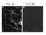
- FIG. 3, comprising FIGS. 3A and 3B is a pair of dark field images of collagen I fibrils.
- pCcollagen was incubated with C-proteinase at 37° C. for 24 hours in a sealed chamber in the presence (+) or absence ( ⁇ ) of 2.5 mM F3 peptide. Fibrils were photographed using microscope with Dark field attachment. In these Figures, the magnification was 300 ⁇ .
- FIG. 4 is a graph which indicates the effects of adding the peptide F3 at varying time points during fibril assembly.
- the open squares denote fibril assembly under control conditions. Arrows indicate times when 2.5 mM of peptide F3 was added to parallel samples, whereafter incubation was continued for 24 hours.
- FIG. 5, comprising FIGS. 5A, 5 B, 5 C, and 5 D, is a quartet of images of electroblot assays. Binding of biotinylated telopeptides to the ⁇ 1(I) chain and the CB7 fragment chain. The ⁇ 1(I) and ⁇ 2(I) chains of type I collagen or CNBr fragments of the chains were separated by polyacrylamide gel electrophoresis and electroblotted onto filters. The filters were then hybridized with a biotinylated telopeptide followed by assay of the washed filter with streptavidin-horseradish peroxidase.
- CBr+ indicates ⁇ 1(I) or ⁇ 2(I) chains which were digested with cyanogen bromide.
- the electroblot assay was performed using biotinylated C-telopeptide of the ⁇ 1 chain of human type I collagen.
- the electroblot assay was performed using biotinylated C-telopeptide of ⁇ 2 chain of human type I collagen.
- the electroblot assay was performed using biotinylated N-telopeptide of the ⁇ 1 chain of human type I collagen.
- FIG. 5D the electroblot assay was performed using biotinylated N-telopeptide of ⁇ 2 chain of human type I collagen.
- FIG. 6, is a trio of images of assays which depict binding of biotinylated telopeptides to collagenase A and B fragments of type I collagen.
- FIG. 6A is a Coomassie Brilliant Blue-stained polyacrylamide gel electrophoretic assay which demonstrates the presence of ⁇ 1(I), ⁇ 2(I) chains fragments with and without vertebrate collagenase treatment.
- FIGS. 6B and 6C are filter electroblot assays performed using biotinylated telopeptides.
- FIG. 7 is a series of assays for inhibition of fibril formation using peptides that are fragments and modified version of the ⁇ 2-C-telopeptide (F3), as described herein.
- FIG. 8 is a schematic of the binding site in the triple helical domain of the ⁇ 1(I) chain.
- FIG. 9 is a pair of graphs which depict competition by the peptide ⁇ 1-776/797 for the binding of the C-telopeptides to type I collagen.
- FIG. 9A competition for binding of the biotinylated C-propeptide of the ⁇ 1(I) chain (peptide F2) is assayed.
- FIG. 9B competition for the binding of the C-telopeptide from the ⁇ 2(I) chain (peptide F3).
- FIG. 10 is a pair of images generated by a computer model of the binding interaction between a 9 amino acid peptide sequence from the C-telopeptide of the ⁇ 2(I) chain and the triple helix of type I collagen in the region of amino acid ⁇ 1-766 to 801.
- FIG. 10A hydrophobic residues are displayed.
- FIG. 10B electrostatic charges are displayed, red indicating positive charge and purple indicating negative charge.
- ⁇ 1-Arg 780, ⁇ 1-Val 783, and ⁇ -Arg 792 are shown to indicate possible regions of interaction.
- the present invention is based on the discovery that collagen telopeptides bind to a specific site in the major triple-helical domain of the collagen monomer. It has been discovered that both the specificity and the affinity of this binding is unexpected in that, as the data presented herein establish, the process of self-assembly of collagen into fibrils is completely inhibited in the presence of these telopeptides. Data are presented herein which comprise the results of a number of experiments on a number of telopeptides, and while several telopeptides function to inhibit collagen self-assembly into fibrils, the C-terminal telopeptide of the ⁇ 1(I) chain had the highest dissociation constant, this being 4 ⁇ 10 ⁇ 6 M.
- peptides other than those described herein may be generated using combinatorial phage display libraries which express proteins on their surface, or by using other recombinant DNA libraries.
- a collagen fragment of about 20 amino acids identified in the collagen triple helix as a major telopeptide binding site is immobilized on a solid surface.
- the telopeptide to which collagen binds is also immobilized.
- Recombinant phage which display the recombinant protein on their surface are added to the immobilized protein fragments under conditions which would ordinarily promote binding of the telopeptide to the collagen fragment.
- Phage which bind the collagen fragment are isolated, DNA is extracted therefrom, and DNA encoding the peptide which binds the collagen fragment is isolated and sequenced. Peptides encoded by the DNA are then isolated by expression of the DNA in any ordinary expression system. Alternatively, once the sequence of a collagen binding peptide is known, the peptides may be generated synthetically in a peptide synthesizer.
- Antibodies which bind the telopeptide binding site in collagen may also be isolated using phage display libraries. Essentially, an antibody-expressing phage display library is added to the immobilized collagen fragment under conditions to promote binding of the antibody to the collagen fragment. Phage which bind to the collagen fragment are subsequently isolated, and the antibody expressed thereby is obtained and sequenced.
- phage display libraries that are commercially available which may be used in the procedure just described for the generation of collagen binding peptides capable of inhibiting assembly of collagen. It is also possible, using analogous methods, to use commercially-available or specially-made cell display libraries, such libraries comprising cells having at least one peptide present on their surface and able to contact a medium in which the cell is suspended.
- the invention also includes peptides which are homologous to the peptides disclosed herein which function to inhibit collagen self-assembly.
- “Homologous” as used herein refers to the subunit sequence similarity between two polymeric molecules, e.g., between two nucleic acid molecules, e.g., two DNA molecules or two RNA molecules, or between two polypeptide molecules. When a subunit position in both of the two molecules is occupied by the same monomeric subunit, e.g., if a position in each of two peptide molecules is occupied by cysteine, then they are homologous at that position.
- the homology between two sequences is a direct function of the number of matching or homologous positions, e.g., if half (e.g., five positions in a polymer ten subunits in length) of the positions in two compound sequences are homologous then the two sequences are 50% homologous, if 90% of the positions, e.g., 9 of 10, are matched or homologous, the two sequences share 90% homology.
- the amino acid sequences Lys-Pro-Cys-Arg and Met-Pro-Cys-Gly share 50% homology.
- the invention includes an isolated human type I collagen assembly-inhibiting peptide capable of inhibiting self-assembly of collagen.
- the amino acid sequence of such an isolated peptide is about 70% homologous, more preferably about 80% homologous, even more preferably about 90% homologous, more preferably, about 95% homologous, and most preferably, at least about 99% homologous to the amino acid sequence of the ⁇ 1(I) C-terminal peptide disclosed herein.
- the type I collagen assembly-inhibiting peptides of the invention may be used to inhibit self-assembly of type I collagen in a variety of animals, particularly mammals.
- Substantially pure type I collagen assembly-inhibiting peptide obtained as described herein may be purified by following known procedures for protein purification, wherein an immunological, enzymatic or other assay is used to monitor purification at each stage in the procedure. Alternatively, the peptide may be generated synthetically.
- substantially pure describes a compound, e.g., a protein or polypeptide which has been separated from components which naturally accompany it.
- a compound is substantially pure when at least 10%, more preferably at least 20%, more preferably at least 50%, more preferably at least 60%, more preferably at least 75%, more preferably at least 90%, and most preferably at least 99% of the total material (by volume, by wet or dry weight, or by mole percent or mole fraction) in a sample is the compound of interest. Purity can be measured by any appropriate method, e.g., in the case of polypeptides by column chromatography, gel electrophoresis or HPLC analysis.
- a compound, e.g., a protein is also substantially purified when it is essentially free of naturally associated components or when it is separated from the native contaminants which accompany it in its natural state.
- the present invention also provides for analogs of peptides which are capable of inhibiting collagen self-assembly.
- Analogs can differ from naturally occurring proteins or peptides by conservative amino acid sequence differences or by modifications which do not affect sequence, or by both.
- conservative amino acid changes may be made, which although they alter the primary sequence of the peptide, do not normally alter its function.
- Conservative amino acid substitutions typically include substitutions within the following groups:
- valine isoleucine, leucine
- Modifications include in vivo, or in vitro chemical derivatization of polypeptides, e.g., acetylation, or carboxylation. Also included are modifications of glycosylation, e.g., those made by modifying the glycosylation patterns of a polypeptide during its synthesis and processing or in further processing steps; e.g., by exposing the polypeptide to enzymes which affect glycosylation, e.g., mammalian glycosylating or deglycosylating enzymes. Also embraced are sequences which have phosphorylated amino acid residues, e.g., phosphotyrosine, phosphoserine, or phosphothreonine.
- peptides which have been modified using ordinary molecular biological techniques so as to improve their resistance to proteolytic degradation or to optimize solubility properties.
- Analogs of such peptides include those containing residues other than naturally occurring L-amino acids, e.g, D-amino acids or non-naturally occurring synthetic amino acids.
- the peptides of the invention are not limited to products of any of the specific exemplary processes listed herein, in that the invention includes any peptides isolated using the procedures described herein which have the biological activity of the ⁇ 1(I) peptide disclosed herein.
- the peptide will ordinarily be at least about four contiguous amino acids, typically at least about four to about eight contiguous amino acids, more typically at least about four to about twelve contiguous amino acids, usually at least about four to about twenty contiguous amino acids, and typically at least about four to about twenty or more contiguous amino acids in length.
- a “collagen assembly inhibitory peptide” is one which when added to a preparation of collagen monomers, inhibits the formation of collagen fibrils.
- a collagen assembly inhibitory peptide is “biologically active” if it is capable of inhibiting collagen self-assembly in any one of the assays described herein.
- biologically active if it is capable of inhibiting collagen self-assembly in any one of the assays described herein.
- a series of wells in a microtiter plate are coated with monomeric collagen.
- An amount of HCl is added to each well and the plates are dried.
- the plates are rinsed in buffer and bovine serum albumin (BSA) is added to block non-specific binding sites on the collagen.
- BSA bovine serum albumin
- samples containing labeled peptide for example, biotinylated peptide
- labeled peptide for example, biotinylated peptide
- Peptides which do not bind collagen serve as negative controls.
- the wells are incubated for a period of time, the plates are washed and streptavidin conjugated to alkaline phosphatase is added. Incubation is continued for a further period of time following which a developing reagent is added to the wells.
- the amount of binding of the labeled peptide to the collagen is assessed and the degree of inhibition of binding by the test peptide is also assessed. Any test peptide which inhibits binding of the labeled peptide is isolated and is tested further for the ability to inhibit collagen self-assembly in the assays described herein in the description of FIGS. 1, 2 and 3 .
- assay of the invention is not limited to the particular components recited herein. Rather, assays which comprise immobilized variants or fragments of collagen, assays which comprise phage which display collagen assembly inhibitory peptides, and assays which include other means of assessing the competition for binding known versus test fragments to collagen i. e., assays which employ alternative labeling/detection systems, are also contemplated as part of the invention.
- the invention includes methods of identifying type I collagen assembly-inhibiting peptides.
- One such method involves immobilizing a collagen species such as human type I collagen or a human type I collagen peptide on a support.
- a labeled peptide which is known to bind to the collagen species. is contacted with the support, as is an unlabeled test peptide. If the test peptide binds to the collagen species, then binding of the labeled peptide to the support will be decreased, resulting in less association of the label with the support and greater retention of label in the medium surrounding the support.
- any label e.g. a radionuclide
- a collagen species is immobilized on a support, as described herein.
- At least one peptide-bearing particle which comprises a test peptide is contacted with the support. If the test peptide is a type I collagen assembly-inhibiting peptide, then the peptide-bearing particle with bind with the support.
- the support may be rinsed, for example, with a solution (e.g. phosphate-buffered saline) which will not cause type I collagen assembly-inhibiting peptides to desorb from the support.
- a solution e.g. phosphate-buffered saline
- a collagen un-binding eluent may be contacted with the support.
- a collagen un-binding eluent is any composition which will cause a type I collagen assembly-inhibiting peptide to desorb from the collagen species.
- collagen un-binding eluents include, but are not limited to, Tris-glycine buffer at a pH of about 2.2, a suspension comprising human type I collagen, and a suspension comprising a peptide having the amino acid sequence of amino acid residues 776 through 796, inclusive, of the ⁇ 1 chain of human type I collagen.
- peptide-bearing particles which may be used according to this method include phage display peptide libraries and cell surface display peptide libraries, both of which are known in the art.
- a “human type I collagen peptide” is a polypeptide having an amino acid sequence homologous with at least a portion of human type I collagen, but being shorter in length than human type I collagen.
- An example of a human type I collagen peptide is the ⁇ 1-776-796 peptide described herein.
- Peptides or peptidomimetics which inhibit the assembly of collagen may be formulated so as to be suitable for administration to a mammal in need of such inhibitors.
- the peptide or peptidomimetic is prepared for administration by being suspended or dissolved in a pharmaceutically acceptable carrier such as saline, salts solution or other formulations apparent to those skilled in the art of administration of peptides and peptidomimetics.
- compositions of the invention may be administered to a mammal in one of the traditional modes (e.g., orally, parenterally, transdermally or transmucosally), in a sustained release formulation using a biodegradable biocompatible polymer, or by on-site delivery using micelles, gels and liposomes, or rectally (e.g., by suppository or enema) or nasally (e.g., by nasal spray).
- the appropriate pharmaceutically acceptable carrier will be evident to those skilled in the art and will depend in large part upon the route of administration and the age and type of mammal being treated.
- Protocols for treatment of mammals involving administration of a peptide or peptidomimetic of the invention will be apparent to those skilled in the art and will vary depending upon the type of disease the type and age of the mammal. Treatment regimes which are contemplated include a single dose or dosage which is administered hourly, daily, weekly or monthly, or yearly. Dosages may vary from 1 ⁇ g to 1000 mg/kg of body weight of the peptide or peptidomimetic and will be in a form suitable for delivery of the compound. The route of administration may also vary depending upon the disorder to be treated.
- the peptide or peptidomimetic may be administered to the mammal in an enclosed yet porous device wherein the peptide is effectively prevented from diffusing away from the site of administration.
- the human type I collagen assembly-inhibiting peptides of the invention may be used to inhibit collagen self-assembly both in vitro and in vivo.
- the peptides of the invention may be administered to a human to inhibit type I collagen assembly in the human.
- the peptides may be administered to the human in the form of a medicament comprising at least one peptide of the invention.
- a problem that has been recognized by those who produce collagen ex vivo is that it is difficult to devise a large-scale method for concentrating and purifying recombinant collagen.
- the type I collagen assembly-inhibiting peptides of the invention bind to human type I collagen, these peptides may be used to make a chromatographic medium suitable for large-scale purification or concentration of human type I collagen.
- This chromatographic medium comprises a support having at least one type I collagen assembly-inhibiting peptide of the invention immobilized thereon.
- any of the numerous art-recognized methods of immobilizing a peptide on a support may be used to make the medium, with the proviso that it is necessary that at least some of the peptide must be available to contact a solution surrounding the medium, so that collagen suspended in the solution may contact, and bind to, the peptide.
- the chromatographic medium may be used, for example to purify recombinant human type I collagen. To do so, a relatively impure suspension comprising human type I collagen is contacted with the support. Collagen in the suspension thereby binds to peptide immobilized on the support. The suspension is then separated from the support, for example by rinsing the support with a solution which does not comprise the suspension.
- Human type I collagen is thereby relatively purified.
- the collagen may be desorbed from the support by contacting the support with a solution in which collagen does not bind. with the peptide.
- solutions include, for example, Tris-glycine buffer at a pH of about 2.2.
- FIG. 1 To design peptides, we have referred to the 1D stagger model for the collagen monomer organization in the fibril (FIG. 1 ).
- Six parent peptides were designed to interact with hydrophobic regions on the collagen I monomer.
- Three of the peptides (F1, F5 and F6) contained sequences found in the triple-helical region of the molecule, and three of the peptides (F2, F3 and F4 had sequences found in non-helical telopeptides.
- the peptides were commercially synthesized and purified. Homogeneity and stability of the peptides in experimental conditions were assayed by reverse-phase HPLC. All the peptides were freely soluble in the fibril formation buffer. pH of peptide solutions was monitored with a solid state micro-pH electrode (Beckman).
- type I procollagen 14 C-labeled type I procollagen was recovered from the medium of cultured human skin fibroblasts and was purified with two chromatographic steps to homogeneity.
- the type I procollagen was processed to pCcollagen I by cleavage with procollagen N-proteinase purified from organ cultures of chick embryo tendons.
- pCcollagen is a partially processed procollagen which is missing the N-terminal telopeptide, but which retains the C-terminal telopeptide.
- the pCcollagen was then isolated on a gel filtration column.
- Fibril assembly was assayed in a 20 ml reaction volume in a 250 ml plastic centrifuge tube sealed with a plunger and containing a physiological bicarbonate buffer, 30 mg/ml pCcollagen I, and 15 units/ml procollagen C-proteinase purified from chick embryo tendons. Potentially inhibitory peptides were added to the reaction mixture to final concentrations of 0.5 to 2.5 mM in 5 ml of buffer. After incubation for 0.5 to 24 hours at 37° C., the sample was centrifuged 13,000 ⁇ g for 10 min.
- the pellet and supernate fractions were separated by electrophoresis on a 7.5% polyacrylamide gel in SDS and the gel assayed either with a phosphor storage imager (Molecular Dynamics) or by staining with colloidal Coomassie blue (Brilliant Blue, Sigma) and analysis with a densitometer (Personal Densitometer, Molecular Dynamics).
- the reaction was carried out in a sealed chamber on a microscopic slide and followed by Dark field light microscopy (Zeiss model 009).
- Type I collagen extracted with 0.5 M acetic acid from mouse skin was digested with pepsin, and the ⁇ chains separated by gel electrophoresis in SDS.
- gel slices containing ⁇ 1 (I) and ⁇ 2(I) chains were placed into tubes and a chains were digested with 10 mg/ml or 200 mg/ml CNBr in 70% formic acid at room temperature overnight. The gel slices were equilibrated with 0.05 mM Tris-HCl buffer (pH 6.8) over 3 hours.
- the filters were blocked with 1% bovine serum albumin (Sigma) and then incubated overnight with 20 ml of 5 mg/ml peptide that was substituted at the N-terminus with biotin (synthesized for us by American Peptide Company, Inc.).
- the filter was washed three times with Tris-buffered saline containing 0.02% Tween (TBST) and incubated 30 min with a 1:30,000 dilution of horseradish peroxidase conjugated with avidin (Sigma).
- the bands were detected by chemiluminescence (ECL; Amersham) after exposure to an X-ray film for 3 to 10 min.
- telopeptide binding site with a competitive assay, 96-well titration plates (Immulon 3, Dynatech Laboratories, Inc.) were used. Seventy microliters of the solution containing 3 mg of monomeric collagen (Vitrogen 100, Collagen Biomaterials) in 0.01 N HCl was added to each well, and the plates were dried at room temperature. The plates were rinsed with sterile phosphate buffered saline and non-specific binding sites were blocked with 1% bovine serum albumin. For the competition assay, pre-mixed samples were prepared that contained 50 mM biotinylated F2 or F3 peptide with 10 to 500 mM peptide ⁇ 1-776/797.
- peptide ⁇ 2-218/233 was used as a negative control at concentrations from 10 to 500 mM.
- the peptide mixtures were added into wells containing immobilized collagen and the samples incubated at 25° C. for 12 h. The plates were washed with PBS containing 0.05% Tween.
- streptavidin conjugated to alkaline phosphatase BioRad was added at 1:20,000 dilution.
- the plates were washed with phosphate buffered saline (PBS) containing 0.05% Tween, and developing reagent 3 mM p-nitrophenyl phosphate in 0.05 M Na 2 CO 3 , 0.05 mM MgCl 2 , pH 9.5, was added. After 2 hours of incubation, the reaction was measured with microtiter plate reader (Dynatech Laboratories) using a 405 nanometer filter.
- PBS phosphate buffered saline
- FIG. 1 Six synthetic peptides (FIG. 1) were prepared on the basis of two general considerations: (1) Extensive previous work (see refs. 1 and 2) had demonstrated that the telopeptides were required to be present on the monomers in order to generate tightly packed fibrils; and (2) the self-assembly of collagen monomers is entropy driven (Kadler et al., 1987, J. Biol. Chem. 262:15696-15701) and, therefore, any specific binding sites in the triple helix are likely to be found in hydrophobic sequences. As indicated in FIG.
- one peptide (F6) contained relatively hydrophobic sequences found near the end of D1 period of the ⁇ 2(I) chain
- second peptide (F5) had hydrophobic sequences found near the middle of the D4 period of the ⁇ 2(I) chain.
- F6 contained sequences that spanned the vertebrate collagenase cleavage site of -Gly-Ile- in the D4 period of the ⁇ 1(I) chain.
- the remaining three peptides had the sequences of the ⁇ 2-N-, ⁇ 1 and ⁇ 2 C-telopeptides.
- Assays by reverse-phase HPLC demonstrated that none of the peptides were degraded under experimental conditions used here (data not shown).
- the peptides were added in concentrations of 2.5 mM to the system for assaying fibril formation by the cleavage of pCcollagen with C-proteinase (Miyahara et al., 1982, J. Biol. Chem. 257:8442-8448; Kadler et al., 1987, J. Biol. Chem. 262:15696-15701; Kadler et al., 1990, Ann. N.Y. Acad. Sci. 580:214-224; Kadler et al., 1990, Biochem. J. 268:339-343; Fertala et al., 1996, J. Biol. Chem. 271:14864-14869). As indicated in FIG.
- the peptides F1 and F6 caused little if any inhibition of fibril assembly.
- Peptides F2, F3 and F4 almost completely inhibited fibril assembly, whereas F5 inhibited assembly by about 50% under the conditions of the experiment.
- Inhibition of fibril assembly with the peptide F3 was directly demonstrated by following the reaction in a sealed chamber with Dark field light microscopy. No fibrils appeared when F3 was present in a concentration of 2.5 mM (FIG. 3, right panel). The specificity of the effects was demonstrated by preparing a peptide in which the same amino acids found in F3 were assembled in a random sequence. As indicated in FIG. 2B, the peptide with the scrambled sequence (SC/F3) had no effect on fibril assembly. Assays in which the concentration of the peptide F3 were varied indicated that fibril assembly was inhibited about 40% with a concentration of about 1.5 mM and almost 100% with 2.5 mM (Table I).
- the peptide F3 inhibits fibril assembly if added to the lag phase and early propagation phase but not later in the assembly process
- the peptide F3 was added in a concentration of 2.5 mM during the lag phase, early propagation phase, mid-propagation phase, or late propagation phase of fibril assembly (FIG. 4 ).
- the peptide inhibited fibril formation if added during the lag period. It had little of any effect if added during the mid-propagation phase of fibril assembly.
- ⁇ 1(I) and ⁇ 2(I) chains of type I collagen were isolated and cyanogen bromide peptides were prepared.
- a filter binding assay was then carried out with a version of the peptides in which the N-terminus was substituted with biotin.
- the biotinylated derivatives bound to the ⁇ 1(I) chain but not the ⁇ 2(I) chain.
- the biotinylated telopeptides bound to CB7 of the ⁇ 1 chain that contains amino acid residues 552 to 819. There was no apparent binding to any of the other cyanogen bromide fragments.
- vertebrate collagenase A and B fragments of type I collagen were prepared and hybridized with the biotinylated telopeptides. As indicated in FIGS. 8 and 9, the peptides bound specifically to the B fragment of the ⁇ 1(I) chain. Since vertebrate collagenase cleaves the two a chains of type I collagen between residues 775 and 776, the binding of the peptides to both CB7 and the B fragments of the ⁇ 1(I) chain indicated that the binding site is between amino acid 776 and 819 of the ⁇ 1(I) chain.
- a peptide was prepared with the sequence of the amino acids in positions 776 to 797 of the ⁇ 1(I).
- the sequence 776 to 797 was selected primarily because it was the most hydrophobic sequence in the region between 776 and 822 that was defined by the experiments with the CNBr peptides and the collagenase fragments (FIG. 9 ).
- the peptide ⁇ 1-776/797 was then used in a competition binding assay in which collagen was bound to the wells of a micro-titer plate, and the plates were incubated with a fixed concentration of one of the biotinylated telopeptides and varying concentrations the peptide ⁇ 1-776/797.
- the binding of the biotinylated peptide was then assayed by incubation with streptavidin-alkaline phosphatase. As indicated in FIG. 9, the peptide ⁇ 1-776/797 effectively competed with binding the C-telopeptides of the ⁇ 1(I) chain (peptide F2) and the ⁇ 2(I) chain (peptide F3). There was no competition for the binding site between biotinylated telopeptides and F1. Similar results were obtained with the binding of the two N-telopeptides.
- the SYBYL program was first used to model the conformation of one of the 9-amino acid fragments (F7) of the ⁇ 2(I) C-telopeptide that inhibited fibril assembly (FIGS. 1 and 8 and Table I). Because the aromatic tyrosine and phenylalanine residues tended to form a hydrophobic stack on one side of the polypeptide chain, a single conformation was favored (FIGS. 10 A and 10 B).
- the binding could initiate assembly of a Smith-type pentameter microfibril (Piez, 1984, In: Extracellular Matrix Biochemistry, Piez et al., Eds., Elsevier, N.Y., pp. 1-40; Silver et al., 1992, Proc. Natl. Acad. Sci. 89:9860-9864).
- the binding of the N-telopeptides to about the same region of the ⁇ 1(I) chain (defined here as residues 776 to 824) places the D-periods out of register by one-third or more of a D-period of 234 residues.
- the binding of the N-telopeptides cannot generate the OD, 1D, 2D, 3D and 4D staggers that are found among many nearest neighbors in fibrils assembled in vivo. Also, the binding of the N-telopeptides does not accurately align the monomers for formation of the major covalent cross-link that forms between the Lys residue at position 9 of the N-telopeptide and the Lys residue at 908 of the ⁇ 1 (I) chain (Piez, 1984, In: Extracellular Matrix Biochemistry, Piez et al., Eds., Elsevier, N.Y., pp. 1-40; Nagan et al., 1994, J. Biol. Chem. 269:22366-22371).
- N-telopeptide binding occurs only with linear N-telopeptides such as those used here but not with N-telopeptides in the unusual hairpin conformation that is present in the native molecule (Holmes et al., 1993, J. Biol. Chem. 268:15758-15765; Mould et al., 1982, J. Molec. Biol. 195:543-553; Helseth et al., 1979, Biopolymers 18:3005-3014; Helseth et al., 1981, J. Biol. Chem.
- a third possibility is that binding through the N-telopeptides did not play an important role in fibril assembly until a core of a microfibril is formed and it is only important for lateral growth of the fibril.
- the last suggestion is consistent with one of the proposed helical models for growth of microfibrils from paraboloidal tips (Silver et al., 1992, Proc. Natl. Acad. Sci. 89:9860-9864).
- the model required one specific binding step governed by one rate constant (k1) for assembly of monomers in a 1D -stagger to form a Smith-type microfibrillar core and to regulate longitudinal growth of the fibril. It required a second binding step governed by a smaller rate constant (k2) to initiate growth of a new layer of helical sheets of monomers on the microfibrillar core and thereby to regulate lateral growth of the core.
- the region contains the C-terminal half of the vertebrate collagenase cleavage site that previously has been designated as a relatively flexible region of the collagen triple-helix (Brown et al., 1977, Biochem. Biophys. Res. Commun. 74:1102-1108). Also, Bhatnagar et al. (1997, J. Biomolec. Struct. Dynamics 14:547-560) recently examined a synthetic peptide with amino acid residues 776 to 780 from the region and found that it had a high potential to form a stable b-bend with the central GIAG sequence that begins in residues 775. Moreover, they demonstrated (Bhatnagar et al., 1997, J. Biomolec. Struct. Dynamics 14:547-560) that the peptide a inhibited the binding of fibroblasts to collagen at a concentration of 7.2 ⁇ 10 ⁇ 6 M. Therefore, the sequence of amino acids in the region may take part in a large number of different binding interactions
- the sites defined here provide interesting targets for peptides, peptidomimetics or related compounds that may inhibit the assembly of collagen fibers in pathologic fibrotic conditions.
- the ability to model binding sites in the triple helix and the conformation of short fragments of the telopeptides provide a rational route for developing inhibitors.
- the competitive assays on microtiter plates provide a means of high throughput screening for large libraries of potential inhibitors.
- a commercially-available phage-display peptide library was used to identify peptides that bind to the telopeptide binding region of type I collagen (i.e. amino acid residues 776-797).
- the library comprised phage particles having about 1.9 ⁇ 10 9 distinct linear peptide sequences.
- glycine HCl buffer having a pH of 2.2.
- Eluted specifically-binding library particles were added to and incubated with microtiter plates having either type I collagen or the 22-mer synthetic peptide bound to the wells thereof, and washing and elution steps were repeated. A total of three cycles of incubation-washing-elution were performed. Following these three cycles, specifically-binding phage particles were isolated, and the amino acid sequence of the peptide encoded by each of these particles was determined by amplifying and sequencing the nucleic acid contained within the particle.
- Amino acid sequences of the random peptide region carried by discrete specifically-binding phage particles are indicated in Table II, together with the identity of the ligand bound to the microtiter wells in each of the three incubation-washing-elution cycles.
- These isolated peptides, or peptides having no more than about fifty, preferably no more than twenty, even more preferably no more than ten, and still more preferably no more than five amino acid residues at one or both ends thereof, may be used to inhibit collagen self-assembly by contacting a collagen molecule such as a collagen triple helix, with one of these peptides.
Abstract
The invention relates to peptides and peptidomimetics which inhibit assembly of human type I collagen. Methods of identifying such peptides and peptidomimetics are also included in the invention.
Description
This application is a continuation of international patent application PCT/US98/18838.
This application is entitled to priority pursuant to 35 U.S.C. §119(e) to U.S. provisional patent application No. 60/058,353, which was filed on Sep. 10, 1997.
Fibrillar collagens form the largest protein structures found in complex organisms (Piez, 1984, In: Extracellular Matrix Biochemistry, Piez et al., Eds., Elsevier, N.Y., pp. 1-40; Prockop et al., 1995, Ann. Rev. Biochem. 64:403-434). The most abundant collagen fibrils consist almost entirely of a single monomer of type I collagen. The structure of the monomer was established several decades ago, but the precise pattern of packing of the monomer into fibrils has not been defined and remains controversial (Smith, 1968, Nature 219:157-158; Hulmes et al., 1979, Nature 282:878-880; Holmes et al., 1979, Biochem. Biophys. Res. Commun. 87:993-999; Piez et al., 1981, Biosci. Rep. 1:801-810; Hulmes et al., 1981, Proc. Natl. Acad. Sci. USA 78:3567-3571; Brodsky et al., 1982, Methods Enzymol. 82:127-173; Woodhead-Galloway, 1984, In: Connective Tissue Matrix, Hukins, Ed., Verlag Chemie, Basel, pp. 133-160; Hulmes et al., 1985, J. Mol. Biol. 184:473-477; Ward et al., 1986, J. Mol. Biol. 190:107-112; Galloway, 1985, In: Biology of Invertebrate and Lower Vertebrate Collagens, Bairoti et al., Eds., Plenum Press, New York, pp. 73-82; Chapman, 1989, Biopolymers 28:1367-1382; Jones et al., 1991, J. Mol. Biol. 218:209-219).
Type I collagen is similar to other fibrillar collagen in that it is first synthesized as a soluble procollagen containing N-propeptides and C-propeptides (Prockop et al., 1995, Ann. Rev. Biochem. 64:403-434). The propeptides are cleaved by specific N- and C-proteinases and the monomers then spontaneously assemble into characteristic fibrils. The two α1(I) chains and one α2(I) chains of a monomer of type I collagen are primarily comprised of about 338 repeating tripeptide sequences of -Gly-Xxx-Yyy- in which -Xxx- is frequently proline and -Yyy- is frequently hydroxyproline. The ends of the α1(I) and α2(I) chains consist of short telopeptides of about 11 to 25 amino acids per chain. The distribution of hydroxyproline and charged residues in the -Xxx- and -Yyy- positions in the triple-helical domain define 4.4 repeats or 4.4 D-periods of about 234 amino acids each. In longitudinal sections, the monomers are arranged in fibrils in a head-to-head-to-tail orientation with a gap of about 0.6 D-periods and, therefore, repeat of 5 D-periods. The continuity of the fibrils is maintained by many of the monomers being staggered by 1, 2, 3, or 4 D-periods relative to the nearest neighbor so as to generate gap and overlap regions. However, there are conflicting data from electron microscopy and X-ray analysis about the lateral packing of the monomers. One view is that the monomers are laterally packed in a tilted quasi-hexagonal lattice (Hulmes et al., 1979, Nature 282:878-880; Jones et al., 1991, J. Mol. Biol. 218:209-219). A related view is that the fibrils consist of “compressed” microfibrils that are comprised of monomers coiled into a rope-like pentameric structure (Smith, 1968, Nature 219:157-158; Piez et al., 1981, Biosci. Rep. 1:801-810). Still another view is that the lateral packing of the collagen in many fibrils is either liquid-like or a biological equivalent of a liquid crystal Galloway, 1985, In: Biology of Invertebrate and Lower Vertebrate Collagens, Bairoti et al., Eds., Plenum Press, New York, pp. 73-82; Chapman, 1989, Biopolymers 28:1367-1382).
One experimental approach to defining the lateral packing of the monomers was to observe the initial assembly of monomers into fibrils. Early experiments (Piez, 1984, In: Extracellular Matrix Biochemistry, Piez et al., Eds., Elsevier, N.Y., pp. 1-40; Ward et al., 1986, J. Mol. Biol. 190:107-112; Veis et al., 1988, In: Collagen: Biochemistry, Vol. 1, Nimni, Ed., CRC Press, Boca Raton, Fla., pp. 113-138) on the re-assembly of fibrils from collagen extracted from tissues with acidic buffers suggested that the first structures formed were linear strands of monomers bound by 0.4 D-period overlaps (4 D staggers). Other observations with extracted collagens suggested the initial stages involved assembly of structures similar -to pentarneric microfibrils (Piez, 1984, In: Extracellular Matrix Biochemistry, Piez et al., Eds., Elsevier, N.Y., pp. 1-40; Veis et al., 1988, In: Collagen: Biochemistry, Vol. 1, Nimni, Ed., CRC Press, Boca Raton, Fla., pp. 113-138; Gelman et al., 1980, J. Biol. Chem. 155:8098-8102). Subsequently, a system was developed for studying assembly of type I collagen fibrils de novo by enzymic cleavage of a purified soluble precursor of procollagen under physiological conditions (Miyahara et al., 1982, J. Biol. Chem. 257:8442-8448; Kadler et al., 1987, J. Biol. Chem. 262:15696-15701; Kadler et al., 1990, Ann. N.Y. Acad. Sci. 580:214-224; Kadler et al., 1990, Biochem. J. 268:339-343). Because thick fibrils were generated in the system, it was possible to use dark-field light microscopy to follow the growth of the fibrils through intermediate stages (Kadler et al., 1990, Biochem. J. 268:339-343). The first fibrils detected had a blunt end and a pointed tip or end. Initial growth of the fibrils were exclusively from the pointed or a-tip. Later, b-tips appeared on the blunt ends of the fibrils and the fibrils grew from both directions. Scanning transmission electron microscopy indicated that both the a-tips and b-tips were near paraboloidal in shape (Holmes et al., 1992, Proc. Natl. Acad. Sci. USA 89:9855-9859 Silver et al., 1992, Proc. Natl. Acad. Sci. 89:9860-9864). Also, it appeared that the monomers are oriented with their N-termini directed toward the tips. Subsequent experiments in the same system with type II collagen suggested that the fibrils also grew from pointed tips. However, the monomers were oriented with a C-termini directed toward the tips (Fertala et al., 1996, J. Biol. Chem. 271:14864-14869). Three different models were proposed to explain the growth of fibrils from near-paraboloidal tips. One model (Silver et al., 1992, Proc. Natl. Acad. Sci. 89:9860-9864) was based on the assumption that the initial core of the fibril was a pentameric microfibril and that the fibril grew by addition of monomers in a helical pattern. Simulations of the model suggested that as little as two specific binding steps were required first for assembly of the microfibrillar core and then a structural nucleus with about the same diameter as the final fibril. After assembly of the structural nucleus, the fibril grew from the paraboloidal tip by addition of monomers through only one of the two binding steps. A second and related model (Hulmes et al., 1995, Biophys. J. 68:1661-1670; Hulmes et al., 1989, J. Mol. Biol. 210:337-345) suggested that assembly began with formation of an undefined inner core and then monomers were added in spiral strands to generate the near-paraboloidal tips. The second model had the advantage that it more readily than the first model accounted for X-ray diffraction data that indicated that some fraction of monomers in fibrils were laterally packed in a tilted quasi-hexagonal lattice (Hulmes et al., 1979, Nature 282:878-880; Galloway, 1985, In: Biology of Invertebrate and Lower Vertebrate Collagens, Bairoti et al., Eds., Plenum Press, New York, pp. 73-82). In contrast to the first two models, a third model (Parkinson et al., 1994, Physical Rev. E. 50:2963-2966) was developed in which monomers were assembled by a process involving only aggregated limited diffusion. The third model, therefore, assumed that the assembly of monomers into fibrils was similar to processes such as electrochemical depositions or perhaps formation of snowflakes, and that the process did not require the presence of specific binding sites on the monomers.
The invention relates to type I collagen assembly-inhibiting peptides. These peptides inhibit assembly of human type I collagen, for example, in an in vitro collagen self-assembly assay. In one embodiment, the peptide is selected from the group consisting of a type I collagen α1 N-telopeptide, a type I collagen α1 C-telopeptide, a type I collagen α2 C-telopeptide, a type I collagen α2 C-telopeptide derivative. By way of example, the peptide may have an amino acid sequence selected from the group consisting of SEQ ID NOs: 2, 4-11, and 13-23, preferably from the group consisting of SEQ ID NOs: 2 and 4-11, and more preferably from the group consisting of SEQ ID NOs: 4-8 and 11. In another embodiment, the peptide is a peptidomimetic of a peptide having an amino acid sequence selected from the group consisting of SEQ ID NOs: 2, 4-11, and 13-23.
The invention also relates to a method of identifying a type I collagen assembly-inhibiting peptide. This method comprises immobilizing a collagen species on a support. The collagen species is selected from the group consisting of human type I collagen and a human type I collagen peptide. The support is contacted with a labeled peptide in the presence or absence of an unlabeled test peptide. The labeled peptide is known to inhibit collagen self-assembly. The amount of labeled peptide bound to the support is assessed, and a lower amount of labeled peptide bound to the support in the presence of the test peptide compared with the level of binding of the labeled peptide to the support in the absence of the test peptide is an indication that the test peptide is a type I collagen assembly-inhibiting peptide.
The invention further relates to a method of identifying a type I collagen assembly-inhibiting peptide. This method comprises immobilizing a collagen species on a support. Again, the collagen species is selected from the group consisting of human type I collagen and a human type I collagen peptide. The support is contacted with at least one peptide-bearing particle which comprises a test peptide. It is then assessed whether the peptide-bearing particle binds with the support. Binding of the peptide-bearing particle to the support is an indication that the test peptide is a type I collagen assembly-inhibiting peptide. In one embodiment of this method, the human type I collagen peptide has the amino acid sequence of amino acid residues 776 through 796, inclusive, of the α1 chain of human type I collagen. In another embodiment, the peptide-bearing particle is a phage or a phage display peptide library. In a variation of this method, the support is rinsed prior to assessing whether the peptide-bearing particle binds with the support. In one embodiment of this method assessing whether the peptide-bearing particle binds with the support comprises contacting the support with a collagen un-binding eluent and detecting the presence of the peptide-bearing particle in the eluent. By way of example, the collagen un-binding eluent may be selected from the group consisting of Tris-glycine buffer at a pH of about 2.2, a suspension comprising human type I collagen, and a suspension comprising a peptide having the amino acid sequence of amino acid residues 776 through 796, inclusive, of the α1 chain of human type I collagen. In one embodiment of this method, the peptide-bearing particle is a phage, and detecting the presence of the phage comprises detecting the ability of the phage to lyse cultured cells. In another variation of this method, the peptide-bearing particle in the eluent is contacted with a second support, wherein the second support has the same or a different collagen species immobilized thereon.
The invention also relates to a method of inhibiting type I collagen self-assembly comprising contacting the collagen with a type I collagen assembly-inhibiting peptide. In one embodiment, the collagen is in a human.
The invention also relates to use of a type I collagen assembly-inhibiting peptide for preparation of a medicament for inhibiting type I collagen self-assembly in an animal, preferably in a human.
The invention also relates to a chromatographic medium comprising a support having a type I collagen assembly-inhibiting peptide immobilized thereon.
The invention further relates to a method of purifying human type I collagen. This method comprises contacting a relatively impure suspension comprising human type I collagen with a support having a type I collagen assembly-inhibiting peptide immobilized thereon and separating the support from the suspension to yield relatively purified human type I collagen.
The file of this patent contains at least one drawing executed in color. Copies of this patent with color drawings(s) will be provided by the Patent and Trademark Office upon request and payment of the necessary fee.
FIG. 1 is a schematic of the structure of a type I procollagen molecule divided into D periods, N-telopeptides and C-telopeptides. Sequences above and below the model indicate the effect of peptides having the indicated sequence as inhibitors of fibril assembly. The symbols used are as follows: (−), no effect on fibril assembly; (+), about 50% inhibition of fibril assembly at a peptide concentration of 2.5 mM; and (++), complete inhibition of fibril assembly at a peptide concentration of 2.5 mM. “NT” refers to N-terminal telopeptides, and “CT” refers to C-terminal telopeptides.
FIG. 2, comprising FIGS. 2A, 2B, and 2C, is a pair of images of SDS-PAGE gels which demonstrate fibril formation in the presence of synthetic peptides. In each of FIGS. 2A, 2B, and 2C, 14C-Labeled pCcollagen I (30 mg/ml) was incubated with C-proteinase (15 units/ml) for 24 hours at 37° C. F1, F2, F3, F4, F5, and F6 peptides were added to the reaction mixture at concentrations of 2.5 mM. Fibrils were separated from collagen monomers by centrifugation and analyzed by gel electrophoresis in SDS. In each instance, “P” indicates to the pellet fraction and “S” indicates the supernatant fraction. Sample “C” control sample, and sample “SC/F3” was performed using a peptide having the same amino acid residues as F3, but in a scrambled order (YGAGFDDGDYGYRYG).
FIG. 3, comprising FIGS. 3A and 3B is a pair of dark field images of collagen I fibrils. pCcollagen was incubated with C-proteinase at 37° C. for 24 hours in a sealed chamber in the presence (+) or absence (−) of 2.5 mM F3 peptide. Fibrils were photographed using microscope with Dark field attachment. In these Figures, the magnification was 300×.
FIG. 4 is a graph which indicates the effects of adding the peptide F3 at varying time points during fibril assembly. The open squares denote fibril assembly under control conditions. Arrows indicate times when 2.5 mM of peptide F3 was added to parallel samples, whereafter incubation was continued for 24 hours.
FIG. 5, comprising FIGS. 5A, 5B, 5C, and 5D, is a quartet of images of electroblot assays. Binding of biotinylated telopeptides to the α1(I) chain and the CB7 fragment chain. The α1(I) and α2(I) chains of type I collagen or CNBr fragments of the chains were separated by polyacrylamide gel electrophoresis and electroblotted onto filters. The filters were then hybridized with a biotinylated telopeptide followed by assay of the washed filter with streptavidin-horseradish peroxidase. “CNBr+” indicates α1(I) or α2(I) chains which were digested with cyanogen bromide. In FIG. 5A, the electroblot assay was performed using biotinylated C-telopeptide of the α1 chain of human type I collagen. In FIG. 5B, the electroblot assay was performed using biotinylated C-telopeptide of α2 chain of human type I collagen. In FIG. 5C, the electroblot assay was performed using biotinylated N-telopeptide of the α1 chain of human type I collagen. In FIG. 5D, the electroblot assay was performed using biotinylated N-telopeptide of α2 chain of human type I collagen.
FIG. 6, comprising FIGS. 6A, 6B, and 6C, is a trio of images of assays which depict binding of biotinylated telopeptides to collagenase A and B fragments of type I collagen. FIG. 6A is a Coomassie Brilliant Blue-stained polyacrylamide gel electrophoretic assay which demonstrates the presence of α1(I), α2(I) chains fragments with and without vertebrate collagenase treatment. FIGS. 6B and 6C are filter electroblot assays performed using biotinylated telopeptides. In these two figures, “+” indicates samples which were treated with vertebrate collagenase prior to electroblotting, and “−” indicates samples which were not treated with vertebrate collagenase. In each of FIGS. 6A, 6B, and 6C, “T” and “CH” refer to bands corresponding to trypsin and chymotrypsin, respectively, which were used to convert the procollagen into collagen.
FIG. 7, comprising FIGS. 7A and 7B, is a series of assays for inhibition of fibril formation using peptides that are fragments and modified version of the α2-C-telopeptide (F3), as described herein.
FIG. 8 is a schematic of the binding site in the triple helical domain of the α1(I) chain.
FIG. 9, comprising FIGS. 9A and 9B, is a pair of graphs which depict competition by the peptide α1-776/797 for the binding of the C-telopeptides to type I collagen. In FIG. 9A, competition for binding of the biotinylated C-propeptide of the α1(I) chain (peptide F2) is assayed. In FIG. 9B, competition for the binding of the C-telopeptide from the α2(I) chain (peptide F3).
FIG. 10, comprising FIGS. 10A and 10B, is a pair of images generated by a computer model of the binding interaction between a 9 amino acid peptide sequence from the C-telopeptide of the α2(I) chain and the triple helix of type I collagen in the region of amino acid α1-766 to 801. In FIG. 10A hydrophobic residues are displayed. In FIG. 10B electrostatic charges are displayed, red indicating positive charge and purple indicating negative charge. α1-Arg 780, α1-Val 783, and α-Arg 792 are shown to indicate possible regions of interaction.
It has been discovered according to the present invention that self-assembly of collagen may be inhibited by specific peptides and peptidomimetics derived from these peptides. These peptides and peptidomimetics are herein referred to as type I collagen assembly-inhibiting peptides. The present discovery thus provides a heretofore unknown class of compounds which are useful as anti-fibrotic compounds, which are useful in the prevention of metastasis of tumors, and are also useful in a variety of other applications.
The present invention is based on the discovery that collagen telopeptides bind to a specific site in the major triple-helical domain of the collagen monomer. It has been discovered that both the specificity and the affinity of this binding is unexpected in that, as the data presented herein establish, the process of self-assembly of collagen into fibrils is completely inhibited in the presence of these telopeptides. Data are presented herein which comprise the results of a number of experiments on a number of telopeptides, and while several telopeptides function to inhibit collagen self-assembly into fibrils, the C-terminal telopeptide of the α1(I) chain had the highest dissociation constant, this being 4×10⁻⁶ M.
As the data presented herein also establish, it has been possible to model both the telopeptide binding site in the collagen triple helix and the collagen binding site in one of the telopeptides. This modeling provides detailed information for use in designing additional peptides and peptidomimetics which inhibit self-assembly of collagen. Once the sequence of a functional peptide is known (in the present situation, the functional peptide is an inhibitor of collagen self-assembly), the designing of other compounds having the same property by computer modeling is now commonplace in the pharmaceutical sciences. Thus, based on the present discovery, a collagen self-assembly inhibitory peptide may be modified to generate an infinite variety of potential compounds that may have improved activity compared with the first discovered compound. The invention thus contemplates the use of the information provided herein for the development of other peptides and peptidomimetics for use as inhibitors of collagen self-assembly.
In addition, peptides other than those described herein may be generated using combinatorial phage display libraries which express proteins on their surface, or by using other recombinant DNA libraries. For example, to isolate additional peptides using a phage display library, a collagen fragment of about 20 amino acids identified in the collagen triple helix as a major telopeptide binding site, is immobilized on a solid surface. In a parallel assay, the telopeptide to which collagen binds is also immobilized. Recombinant phage which display the recombinant protein on their surface are added to the immobilized protein fragments under conditions which would ordinarily promote binding of the telopeptide to the collagen fragment. Phage which bind the collagen fragment are isolated, DNA is extracted therefrom, and DNA encoding the peptide which binds the collagen fragment is isolated and sequenced. Peptides encoded by the DNA are then isolated by expression of the DNA in any ordinary expression system. Alternatively, once the sequence of a collagen binding peptide is known, the peptides may be generated synthetically in a peptide synthesizer.
Antibodies which bind the telopeptide binding site in collagen may also be isolated using phage display libraries. Essentially, an antibody-expressing phage display library is added to the immobilized collagen fragment under conditions to promote binding of the antibody to the collagen fragment. Phage which bind to the collagen fragment are subsequently isolated, and the antibody expressed thereby is obtained and sequenced.
There are a variety of phage display libraries that are commercially available which may be used in the procedure just described for the generation of collagen binding peptides capable of inhibiting assembly of collagen. It is also possible, using analogous methods, to use commercially-available or specially-made cell display libraries, such libraries comprising cells having at least one peptide present on their surface and able to contact a medium in which the cell is suspended.
The invention also includes peptides which are homologous to the peptides disclosed herein which function to inhibit collagen self-assembly.
“Homologous” as used herein, refers to the subunit sequence similarity between two polymeric molecules, e.g., between two nucleic acid molecules, e.g., two DNA molecules or two RNA molecules, or between two polypeptide molecules. When a subunit position in both of the two molecules is occupied by the same monomeric subunit, e.g., if a position in each of two peptide molecules is occupied by cysteine, then they are homologous at that position. The homology between two sequences is a direct function of the number of matching or homologous positions, e.g., if half (e.g., five positions in a polymer ten subunits in length) of the positions in two compound sequences are homologous then the two sequences are 50% homologous, if 90% of the positions, e.g., 9 of 10, are matched or homologous, the two sequences share 90% homology. By way of example, the amino acid sequences Lys-Pro-Cys-Arg and Met-Pro-Cys-Gly share 50% homology.
The invention includes an isolated human type I collagen assembly-inhibiting peptide capable of inhibiting self-assembly of collagen. Preferably, the amino acid sequence of such an isolated peptide is about 70% homologous, more preferably about 80% homologous, even more preferably about 90% homologous, more preferably, about 95% homologous, and most preferably, at least about 99% homologous to the amino acid sequence of the α1(I) C-terminal peptide disclosed herein. Given the similarity among the amino acid sequences of type I collagen proteins of various animals, particularly mammals, it is understood that the type I collagen assembly-inhibiting peptides of the invention may be used to inhibit self-assembly of type I collagen in a variety of animals, particularly mammals.
Substantially pure type I collagen assembly-inhibiting peptide obtained as described herein may be purified by following known procedures for protein purification, wherein an immunological, enzymatic or other assay is used to monitor purification at each stage in the procedure. Alternatively, the peptide may be generated synthetically.
As used herein, the term “substantially pure” describes a compound, e.g., a protein or polypeptide which has been separated from components which naturally accompany it. Typically, a compound is substantially pure when at least 10%, more preferably at least 20%, more preferably at least 50%, more preferably at least 60%, more preferably at least 75%, more preferably at least 90%, and most preferably at least 99% of the total material (by volume, by wet or dry weight, or by mole percent or mole fraction) in a sample is the compound of interest. Purity can be measured by any appropriate method, e.g., in the case of polypeptides by column chromatography, gel electrophoresis or HPLC analysis. A compound, e.g., a protein, is also substantially purified when it is essentially free of naturally associated components or when it is separated from the native contaminants which accompany it in its natural state.
The present invention also provides for analogs of peptides which are capable of inhibiting collagen self-assembly. Analogs can differ from naturally occurring proteins or peptides by conservative amino acid sequence differences or by modifications which do not affect sequence, or by both.
For example, conservative amino acid changes may be made, which although they alter the primary sequence of the peptide, do not normally alter its function. Conservative amino acid substitutions typically include substitutions within the following groups:
glycine, alanine;
valine, isoleucine, leucine;
aspartic acid, glutamic acid;
asparagine, glutamine;
serine, threonine;
lysine, arginine;
phenylalanine, tyrosine.
Modifications (which do not normally alter primary sequence) include in vivo, or in vitro chemical derivatization of polypeptides, e.g., acetylation, or carboxylation. Also included are modifications of glycosylation, e.g., those made by modifying the glycosylation patterns of a polypeptide during its synthesis and processing or in further processing steps; e.g., by exposing the polypeptide to enzymes which affect glycosylation, e.g., mammalian glycosylating or deglycosylating enzymes. Also embraced are sequences which have phosphorylated amino acid residues, e.g., phosphotyrosine, phosphoserine, or phosphothreonine.
Also included are peptides which have been modified using ordinary molecular biological techniques so as to improve their resistance to proteolytic degradation or to optimize solubility properties. Analogs of such peptides include those containing residues other than naturally occurring L-amino acids, e.g, D-amino acids or non-naturally occurring synthetic amino acids. The peptides of the invention are not limited to products of any of the specific exemplary processes listed herein, in that the invention includes any peptides isolated using the procedures described herein which have the biological activity of the α1(I) peptide disclosed herein.
The peptide will ordinarily be at least about four contiguous amino acids, typically at least about four to about eight contiguous amino acids, more typically at least about four to about twelve contiguous amino acids, usually at least about four to about twenty contiguous amino acids, and typically at least about four to about twenty or more contiguous amino acids in length.
A “collagen assembly inhibitory peptide” is one which when added to a preparation of collagen monomers, inhibits the formation of collagen fibrils.
A collagen assembly inhibitory peptide is “biologically active” if it is capable of inhibiting collagen self-assembly in any one of the assays described herein. For example, in a high throughput assay described herein in the description of the data shown in FIGS. 8 and 9, a series of wells in a microtiter plate are coated with monomeric collagen. An amount of HCl is added to each well and the plates are dried. The plates are rinsed in buffer and bovine serum albumin (BSA) is added to block non-specific binding sites on the collagen. Samples containing labeled peptide (for example, biotinylated peptide) which is known to inhibit collagen self-assembly and unlabeled test peptide at various concentrations are premixed and are added to the wells. Peptides which do not bind collagen serve as negative controls. The wells are incubated for a period of time, the plates are washed and streptavidin conjugated to alkaline phosphatase is added. Incubation is continued for a further period of time following which a developing reagent is added to the wells. The amount of binding of the labeled peptide to the collagen is assessed and the degree of inhibition of binding by the test peptide is also assessed. Any test peptide which inhibits binding of the labeled peptide is isolated and is tested further for the ability to inhibit collagen self-assembly in the assays described herein in the description of FIGS. 1, 2 and 3.
It will be appreciated that the assay of the invention is not limited to the particular components recited herein. Rather, assays which comprise immobilized variants or fragments of collagen, assays which comprise phage which display collagen assembly inhibitory peptides, and assays which include other means of assessing the competition for binding known versus test fragments to collagen i. e., assays which employ alternative labeling/detection systems, are also contemplated as part of the invention.
The above described assays and those described in detail herein in the experimental examples section are also applicable to peptidomimetics having the ability to inhibit the self-assembly of collagen. Peptidomimetics may be generated using techniques described in PCT/US93/01201 and in U.S. Pat. No. 5,334,702.
The invention includes methods of identifying type I collagen assembly-inhibiting peptides. One such method involves immobilizing a collagen species such as human type I collagen or a human type I collagen peptide on a support. A labeled peptide which is known to bind to the collagen species. is contacted with the support, as is an unlabeled test peptide. If the test peptide binds to the collagen species, then binding of the labeled peptide to the support will be decreased, resulting in less association of the label with the support and greater retention of label in the medium surrounding the support. Substantially any label (e.g. a radionuclide) which may be attached to a peptide may be used in this method.
An alternate method of identifying a type I collagen assembly-inhibiting peptide is described in greater detail in Example 2 herein. Briefly, a collagen species is immobilized on a support, as described herein. At least one peptide-bearing particle which comprises a test peptide is contacted with the support. If the test peptide is a type I collagen assembly-inhibiting peptide, then the peptide-bearing particle with bind with the support. The support may be rinsed, for example, with a solution (e.g. phosphate-buffered saline) which will not cause type I collagen assembly-inhibiting peptides to desorb from the support. In order to desorb such peptides from the support, a collagen un-binding eluent may be contacted with the support. A collagen un-binding eluent is any composition which will cause a type I collagen assembly-inhibiting peptide to desorb from the collagen species. Examples of collagen un-binding eluents include, but are not limited to, Tris-glycine buffer at a pH of about 2.2, a suspension comprising human type I collagen, and a suspension comprising a peptide having the amino acid sequence of amino acid residues 776 through 796, inclusive, of the α1 chain of human type I collagen. Examples of peptide-bearing particles which may be used according to this method include phage display peptide libraries and cell surface display peptide libraries, both of which are known in the art.
As used herein, a “human type I collagen peptide” is a polypeptide having an amino acid sequence homologous with at least a portion of human type I collagen, but being shorter in length than human type I collagen. An example of a human type I collagen peptide is the α1-776-796 peptide described herein.
Peptides or peptidomimetics which inhibit the assembly of collagen may be formulated so as to be suitable for administration to a mammal in need of such inhibitors. The peptide or peptidomimetic is prepared for administration by being suspended or dissolved in a pharmaceutically acceptable carrier such as saline, salts solution or other formulations apparent to those skilled in the art of administration of peptides and peptidomimetics. The compositions of the invention may be administered to a mammal in one of the traditional modes (e.g., orally, parenterally, transdermally or transmucosally), in a sustained release formulation using a biodegradable biocompatible polymer, or by on-site delivery using micelles, gels and liposomes, or rectally (e.g., by suppository or enema) or nasally (e.g., by nasal spray). The appropriate pharmaceutically acceptable carrier will be evident to those skilled in the art and will depend in large part upon the route of administration and the age and type of mammal being treated.
Protocols for treatment of mammals involving administration of a peptide or peptidomimetic of the invention, will be apparent to those skilled in the art and will vary depending upon the type of disease the type and age of the mammal. Treatment regimes which are contemplated include a single dose or dosage which is administered hourly, daily, weekly or monthly, or yearly. Dosages may vary from 1 μg to 1000 mg/kg of body weight of the peptide or peptidomimetic and will be in a form suitable for delivery of the compound. The route of administration may also vary depending upon the disorder to be treated.
In some instances, it may be necessary to ensure that the peptide or peptidomimetic is administered to a localized site in the mammal and that the peptide or peptidomimetic is prevented from diffusing to other sites in the mammal. In such instances, the peptide or peptidomimetic may be administered to the mammal in an enclosed yet porous device wherein the peptide is effectively prevented from diffusing away from the site of administration.
Additional information describing administration of peptidomimetics is provided in PCT/US93/01201 and U.S. Pat. No. 5,334,702, which are hereby incorporated herein by reference. Any of the techniques described in either of these two references may be employed in the present invention for the administration of peptidomimetics.
One skilled in the art will appreciate, in view of the present disclosure, that the human type I collagen assembly-inhibiting peptides of the invention may be used to inhibit collagen self-assembly both in vitro and in vivo. Thus, for example, the peptides of the invention may be administered to a human to inhibit type I collagen assembly in the human. The peptides may be administered to the human in the form of a medicament comprising at least one peptide of the invention.
A problem that has been recognized by those who produce collagen ex vivo (i.e. recombinant collagen) is that it is difficult to devise a large-scale method for concentrating and purifying recombinant collagen. Because, as described herein, the type I collagen assembly-inhibiting peptides of the invention bind to human type I collagen, these peptides may be used to make a chromatographic medium suitable for large-scale purification or concentration of human type I collagen. This chromatographic medium comprises a support having at least one type I collagen assembly-inhibiting peptide of the invention immobilized thereon. Substantially any of the numerous art-recognized methods of immobilizing a peptide on a support may be used to make the medium, with the proviso that it is necessary that at least some of the peptide must be available to contact a solution surrounding the medium, so that collagen suspended in the solution may contact, and bind to, the peptide. The chromatographic medium may be used, for example to purify recombinant human type I collagen. To do so, a relatively impure suspension comprising human type I collagen is contacted with the support. Collagen in the suspension thereby binds to peptide immobilized on the support. The suspension is then separated from the support, for example by rinsing the support with a solution which does not comprise the suspension. Human type I collagen is thereby relatively purified. If desired, the collagen may be desorbed from the support by contacting the support with a solution in which collagen does not bind. with the peptide. Examples of such solutions include, for example, Tris-glycine buffer at a pH of about 2.2.
The invention is further described in detail by reference to the following experimental examples. These examples are provided for purposes of illustration only, and are not intended to be limiting unless otherwise specified. Thus, the invention should in no way be construed as being limited to the following examples, but rather, should be construed to encompass any and all variations which become evident as a result of the teaching provided herein.
Design and Synthesis of Peptides
To design peptides, we have referred to the 1D stagger model for the collagen monomer organization in the fibril (FIG. 1). Six parent peptides were designed to interact with hydrophobic regions on the collagen I monomer. Three of the peptides (F1, F5 and F6) contained sequences found in the triple-helical region of the molecule, and three of the peptides (F2, F3 and F4 had sequences found in non-helical telopeptides. The peptides were commercially synthesized and purified. Homogeneity and stability of the peptides in experimental conditions were assayed by reverse-phase HPLC. All the peptides were freely soluble in the fibril formation buffer. pH of peptide solutions was monitored with a solid state micro-pH electrode (Beckman).
Assay of Fibril Assembly
Assembly of collagen I into fibrils de novo was assayed under conditions employed previously (Miyahara et al., 1982, J. Biol. Chem. 257:8442-8448; Kadler et al., 1987, J. Biol. Chem. 262:15696-15701; Kadler et al., 1990, Ann. New York Acad. Sci. 580:214-224; Kadler et al., 1990, Biochem. J. 268:339-343; Fertala et al., 1996, J. Biol. Chem. 271:14864-14869). In brief, 14C-labeled type I procollagen was recovered from the medium of cultured human skin fibroblasts and was purified with two chromatographic steps to homogeneity. The type I procollagen was processed to pCcollagen I by cleavage with procollagen N-proteinase purified from organ cultures of chick embryo tendons. “pCcollagen” is a partially processed procollagen which is missing the N-terminal telopeptide, but which retains the C-terminal telopeptide. The pCcollagen was then isolated on a gel filtration column. Fibril assembly was assayed in a 20 ml reaction volume in a 250 ml plastic centrifuge tube sealed with a plunger and containing a physiological bicarbonate buffer, 30 mg/ml pCcollagen I, and 15 units/ml procollagen C-proteinase purified from chick embryo tendons. Potentially inhibitory peptides were added to the reaction mixture to final concentrations of 0.5 to 2.5 mM in 5 ml of buffer. After incubation for 0.5 to 24 hours at 37° C., the sample was centrifuged 13,000×g for 10 min. The pellet and supernate fractions were separated by electrophoresis on a 7.5% polyacrylamide gel in SDS and the gel assayed either with a phosphor storage imager (Molecular Dynamics) or by staining with colloidal Coomassie blue (Brilliant Blue, Sigma) and analysis with a densitometer (Personal Densitometer, Molecular Dynamics). Alternatively, the reaction was carried out in a sealed chamber on a microscopic slide and followed by Dark field light microscopy (Zeiss model 009).
Localization of Binding Sites on Collagen Filters
Type I collagen extracted with 0.5 M acetic acid from mouse skin was digested with pepsin, and the α chains separated by gel electrophoresis in SDS. To generate CNBr peptides, gel slices containing α1 (I) and α2(I) chains were placed into tubes and a chains were digested with 10 mg/ml or 200 mg/ml CNBr in 70% formic acid at room temperature overnight. The gel slices were equilibrated with 0.05 mM Tris-HCl buffer (pH 6.8) over 3 hours. Gel pieces containing CNBr peptides derived from individual collagen a chains were transferred into the wells of the second gel prepared with a 6% polyacrylamide stacking gel and 12% polyacrylamide separating gel with 0.5 M urea. After electrophoresis, the gel was electroblotted overnight at 4° C. onto a nitrocellulose filter (Millipore). To study binding of telopeptides to collagen fragments, the method described by Fujisawa et al. (Fujisawa et al., 1994, Calcif. Tiss. Int. 56:140-144) was used. The filters were blocked with 1% bovine serum albumin (Sigma) and then incubated overnight with 20 ml of 5 mg/ml peptide that was substituted at the N-terminus with biotin (synthesized for us by American Peptide Company, Inc.). The filter was washed three times with Tris-buffered saline containing 0.02% Tween (TBST) and incubated 30 min with a 1:30,000 dilution of horseradish peroxidase conjugated with avidin (Sigma). The bands were detected by chemiluminescence (ECL; Amersham) after exposure to an X-ray film for 3 to 10 min.
To generate vertebrate collagenase fragments, 3 mg of type I pCcollagen from cultured human fibroblasts was cleaved with 10 mg/ml of vertebrate collagenase from cultured rat skin fibroblasts (generous gift from John J. Jeffrey, Department of Medicine, Albany Medical College) for 3 hours at 25° C. in a volume of 40 ml of 50 mM Tris-HCl, 10 mM CaCl2 and 100 mM NaCl, pH 7.4. To remove the C-propeptide, 2 ml of a mixture of trypsin (1 mg/ml) and chymotrypsin (2.5 mg/ml) in the same buffer was added, and the sample was incubated at 20° C. for 2 min. The reaction was stopped with 0.5 mg/ml soybean trypsin inhibitor (Sigma). The sample was then separated by electrophoresis on a 10% polyacrylamide gel in SDS and processed with the same protocol as the CNBr fragments.
To define the telopeptide binding site with a competitive assay, 96-well titration plates (Immulon 3, Dynatech Laboratories, Inc.) were used. Seventy microliters of the solution containing 3 mg of monomeric collagen (Vitrogen 100, Collagen Biomaterials) in 0.01 N HCl was added to each well, and the plates were dried at room temperature. The plates were rinsed with sterile phosphate buffered saline and non-specific binding sites were blocked with 1% bovine serum albumin. For the competition assay, pre-mixed samples were prepared that contained 50 mM biotinylated F2 or F3 peptide with 10 to 500 mM peptide α1-776/797. As a negative control, peptide α2-218/233 was used at concentrations from 10 to 500 mM. The peptide mixtures were added into wells containing immobilized collagen and the samples incubated at 25° C. for 12 h. The plates were washed with PBS containing 0.05% Tween. To detect biotinylated peptides bound to collagen surface, streptavidin conjugated to alkaline phosphatase (BioRad) was added at 1:20,000 dilution. After 2 hours of incubation, the plates were washed with phosphate buffered saline (PBS) containing 0.05% Tween, and developing reagent 3 mM p-nitrophenyl phosphate in 0.05 M Na2CO3, 0.05 mM MgCl2, pH 9.5, was added. After 2 hours of incubation, the reaction was measured with microtiter plate reader (Dynatech Laboratories) using a 405 nanometer filter.
Computer Modeling of Binding Sites
Molecular modeling was performed on a Silicon Graphic (Indigo 2) computer system using the SYBYL software package, version 6.3 (Tripos, Inc.). The model of the collagen I triple helix fragment including sequence from α1 -766/801 and α2-766/801 was carried out as described by Chen et al. (1995, J. Biomol. Struct. & Dyn. 6:1129-1159). The model of F7 peptide was created using SYBYL/Biopolymer module. All the models were energy minimized using a conjugate gradient method and subject to repeating cycles of molecular dynamics using Kollman force field and united atoms (Weiner et al., 1984, J. Am. Chem. Soc. 106:765-784). To analyze interaction of F7 peptide with the collagen I binding region, intermolecular energy of interaction was analyzed to identify possible binding conformations. Surface calculations, lipohelicity potential, and electrostatic potential of the molecules were analyzed using SYBYL/Molcad module.
Assays of inhibition of fibril formation with synthetic peptides
Six synthetic peptides (FIG. 1) were prepared on the basis of two general considerations: (1) Extensive previous work (see refs. 1 and 2) had demonstrated that the telopeptides were required to be present on the monomers in order to generate tightly packed fibrils; and (2) the self-assembly of collagen monomers is entropy driven (Kadler et al., 1987, J. Biol. Chem. 262:15696-15701) and, therefore, any specific binding sites in the triple helix are likely to be found in hydrophobic sequences. As indicated in FIG. 1, one peptide (F6) contained relatively hydrophobic sequences found near the end of D1 period of the α2(I) chain, second peptide (F5) had hydrophobic sequences found near the middle of the D4 period of the α2(I) chain. F6 contained sequences that spanned the vertebrate collagenase cleavage site of -Gly-Ile- in the D4 period of the α1(I) chain. The remaining three peptides had the sequences of the α2-N-, α1 and α2 C-telopeptides. Assays by reverse-phase HPLC demonstrated that none of the peptides were degraded under experimental conditions used here (data not shown). The peptides were added in concentrations of 2.5 mM to the system for assaying fibril formation by the cleavage of pCcollagen with C-proteinase (Miyahara et al., 1982, J. Biol. Chem. 257:8442-8448; Kadler et al., 1987, J. Biol. Chem. 262:15696-15701; Kadler et al., 1990, Ann. N.Y. Acad. Sci. 580:214-224; Kadler et al., 1990, Biochem. J. 268:339-343; Fertala et al., 1996, J. Biol. Chem. 271:14864-14869). As indicated in FIG. 2 and Table I, the peptides F1 and F6 caused little if any inhibition of fibril assembly. Peptides F2, F3 and F4 almost completely inhibited fibril assembly, whereas F5 inhibited assembly by about 50% under the conditions of the experiment. Inhibition of fibril assembly with the peptide F3 was directly demonstrated by following the reaction in a sealed chamber with Dark field light microscopy. No fibrils appeared when F3 was present in a concentration of 2.5 mM (FIG. 3, right panel). The specificity of the effects was demonstrated by preparing a peptide in which the same amino acids found in F3 were assembled in a random sequence. As indicated in FIG. 2B, the peptide with the scrambled sequence (SC/F3) had no effect on fibril assembly. Assays in which the concentration of the peptide F3 were varied indicated that fibril assembly was inhibited about 40% with a concentration of about 1.5 mM and almost 100% with 2.5 mM (Table I).
| TABLE I | |
| Peptide | Fibril Assembly |
| Region of Sequence | Code | Amino Acid Sequence | SEQ ID NO: | Concentration | % Inhibition |
| α2-217/233 | | AKGAAGLPGVAGAPGL | 1 | 2.5 |
5 | |
| α2-778/801 | | GAPGILGLPGSRGERGLPGVAGAV | 2 | 2.5 mM | 50 | |
| α1-761/785 | | ADGPAGAPGTPGPQGIAGQRGVVGL | 3 | 2.5 |
0 | |
| α1 N- | F4 | QLSYGYDEKSTGGISVP | 4 | 2.5 mM | 90 | |
| α1 C- | F2 | SAGFDFSFLPQPPQEKAHDGGRYYRA | 5 | 2.5 mM | 95 | |
| α2 C- | F3 | GGGYDFGYDGDFYRA | 6 | 2.5 mM | 95 | |
| α2 C- | F3 | GGGYDFGYDGDFYRA | 6 | 2.0 mM | 70 | |
| α2 C- | F3 | GGGYDFGYDGDFYRA | 6 | 1.5 mM | 45 | |
| α2 C- | F3 | GGGYDFGYDGDFYRA | 6 | 0.5 mM | 25 | |
| α2 C-telo Derivative | F7 | GGGYDFGYD | 7 | 2.5 mM | 90 | |
| α2 C-telo | F8 | GYDGDFYRA | 8 | 2.5 mM | 90 | |
| α2 C-telo Derivative | F9 | GYDFGYDGDF | 9 | 2.5 mM | 70 | |
| α2 C-telo | F10 | DFRYRA | 10 | 2.5 mM | 70 | |
| α2 C-telo Derivative | F7/A | GGGYSFGYS | 11 | 2.5 mM | 90 | |
| α2 C-telo Derivative | F7/ | GGGSDSGSD | 12 | 2.5 |
0 | |
The peptide F3 inhibits fibril assembly if added to the lag phase and early propagation phase but not later in the assembly process
To define the kinetics for inhibition of fibril assembly by the telopeptides, the peptide F3 was added in a concentration of 2.5 mM during the lag phase, early propagation phase, mid-propagation phase, or late propagation phase of fibril assembly (FIG. 4). The peptide inhibited fibril formation if added during the lag period. It had little of any effect if added during the mid-propagation phase of fibril assembly.
Location of the telopeptide binding site in the triple helix of type I collagen
To define the site in the triple helix of type I collagen to which the telopeptides bound, α1(I) and α2(I) chains of type I collagen were isolated and cyanogen bromide peptides were prepared. A filter binding assay was then carried out with a version of the peptides in which the N-terminus was substituted with biotin. As indicated in FIG. 5, the biotinylated derivatives bound to the α1(I) chain but not the α2(I) chain. As also indicated in FIG. 7, the biotinylated telopeptides bound to CB7 of the α1 chain that contains amino acid residues 552 to 819. There was no apparent binding to any of the other cyanogen bromide fragments.
To define further the binding site in the triple helix, vertebrate collagenase A and B fragments of type I collagen were prepared and hybridized with the biotinylated telopeptides. As indicated in FIGS. 8 and 9, the peptides bound specifically to the B fragment of the α1(I) chain. Since vertebrate collagenase cleaves the two a chains of type I collagen between residues 775 and 776, the binding of the peptides to both CB7 and the B fragments of the α1(I) chain indicated that the binding site is between amino acid 776 and 819 of the α1(I) chain.
Defining the Critical Residues in the Peptide F3 and the Triple Helix
Further experiments were primarily concentrated on the F3 peptide with sequence of the C-telopeptide, because it was relatively short and was somewhat repetitive in sequence. Therefore, it lent itself better than the other inhibitory peptides to defining the minimum sequences required and to modeling of the binding interactions.
To define the critical sequences within the peptide F3, several derivatives were prepared. Two peptides that were 9 mers overlapping the central region of the sequence (F7 and F8) were equally effective as the intact F3 (FIGS. 1 and 8 and Table I). Smaller effects on inhibition of fibril assembly were seen with two other fragments (F9 and F10) (FIGS. 1 and 8 and Table I). Of special interest was that mutating the two aspartate residues in a 9 mer peptide (F7) to serine residues (F7/A) had no effect on inhibition of fibril assembly. However, mutating two tyrosine residues and one phenylalanine residue in the same sequence to serine residues (F7/B) abolished inhibitory effects (FIGS. 1 and 8 and Table I).
To map the binding site in the triple helix still further, a peptide was prepared with the sequence of the amino acids in positions 776 to 797 of the α1(I). The sequence 776 to 797 was selected primarily because it was the most hydrophobic sequence in the region between 776 and 822 that was defined by the experiments with the CNBr peptides and the collagenase fragments (FIG. 9). The peptide α1-776/797 was then used in a competition binding assay in which collagen was bound to the wells of a micro-titer plate, and the plates were incubated with a fixed concentration of one of the biotinylated telopeptides and varying concentrations the peptide α1-776/797. The binding of the biotinylated peptide was then assayed by incubation with streptavidin-alkaline phosphatase. As indicated in FIG. 9, the peptide α1-776/797 effectively competed with binding the C-telopeptides of the α1(I) chain (peptide F2) and the α2(I) chain (peptide F3). There was no competition for the binding site between biotinylated telopeptides and F1. Similar results were obtained with the binding of the two N-telopeptides.
Molecular Modeling of the Two Binding Sites
To model the binding of one of the telopeptides to the triple helix, the SYBYL program was first used to model the conformation of one of the 9-amino acid fragments (F7) of the α2(I) C-telopeptide that inhibited fibril assembly (FIGS. 1 and 8 and Table I). Because the aromatic tyrosine and phenylalanine residues tended to form a hydrophobic stack on one side of the polypeptide chain, a single conformation was favored (FIGS. 10A and 10B).
Binding of F7 to the region between residues 781 to 794 of α1(I) was analyzed. The possible binding conformation was interactively identified using the DOCK command. The refined model demonstrated a favorable interaction of hydrophobic groups (FIG. 10A) and electrostatic groups (FIG. 10B).
The results of the experiments described herein resolve a critical question about the self-assembly of type I collagen into fibrils. If the assembly does not depend on specific interactions of binding sites in the monomers as recently suggested by Parkinson et al. (1994, Physical Rev. E. 50:2963-2966), all the peptides tested here should either have had no effect on fibril assembly or inhibited the process to about the same degree. Instead, the results demonstrated that several peptides specifically inhibited the process. Consistent with previous observations, peptides with sequences found in the telopeptides were the most effective. The telopeptide that was most extensively studied, the C-telopeptide of the α2(I) chain, completely prevented fibril assembly if added at or before the first one-third of the lag phase but had much less effect thereafter. Therefore, the binding of the α2(I) C-telopeptide, probably in concert with α1(I) C-telopeptide, is critical for early steps in the assembly process such as formation of a structural nucleus that is essential for further growth of the fibrils. The binding of the C-telopeptides to the region that encompasses residues 776 to 797 places the monomers in quarter D-period stagger (FIG. 9). Therefore, the binding could initiate assembly of a Smith-type pentameter microfibril (Piez, 1984, In: Extracellular Matrix Biochemistry, Piez et al., Eds., Elsevier, N.Y., pp. 1-40; Silver et al., 1992, Proc. Natl. Acad. Sci. 89:9860-9864). In contrast, the binding of the N-telopeptides to about the same region of the α1(I) chain (defined here as residues 776 to 824) places the D-periods out of register by one-third or more of a D-period of 234 residues. Therefore, the binding of the N-telopeptides cannot generate the OD, 1D, 2D, 3D and 4D staggers that are found among many nearest neighbors in fibrils assembled in vivo. Also, the binding of the N-telopeptides does not accurately align the monomers for formation of the major covalent cross-link that forms between the Lys residue at position 9 of the N-telopeptide and the Lys residue at 908 of the α1 (I) chain (Piez, 1984, In: Extracellular Matrix Biochemistry, Piez et al., Eds., Elsevier, N.Y., pp. 1-40; Nagan et al., 1994, J. Biol. Chem. 269:22366-22371). Accordingly, there are several possible explanations for the observed binding of the N-telopeptides. One is that the binding occurs only with linear N-telopeptides such as those used here but not with N-telopeptides in the unusual hairpin conformation that is present in the native molecule (Holmes et al., 1993, J. Biol. Chem. 268:15758-15765; Mould et al., 1982, J. Molec. Biol. 195:543-553; Helseth et al., 1979, Biopolymers 18:3005-3014; Helseth et al., 1981, J. Biol. Chem. 256:7118-7128; Vitagliano et al., 1995, J. Mol. Biol. 247:69-80) and that is essential both for assembly into well-ordered fibrils and correct cross-linking (Nagan et al., 1994, J. Biol. Chem. 269:22366-22371). A second explanation is that binding of the N-telopeptides generates aberrant structural nuclei that cannot grow into fibrils and that resemble the “overshoot” structures seen in the assembly of tobacco mosaic virus (Potschka et al., 1988, Biochemistry 27:8481-8491). Preliminary assays with an optical biosensor indicated that the dissociation constant for the binding of the α1-C-telopeptide is about 5×10⁻⁶ M for the dissociation constants and the dissociation constants of the two N-telopeptides are about an order of magnitude greater. Therefore, aberrant structures assembled by binding through the N-telopeptides may have a short half life and may rapidly dissociate into monomers that initiate fibril assembly through-binding of C-telopeptides. A third possibility is that binding through the N-telopeptides did not play an important role in fibril assembly until a core of a microfibril is formed and it is only important for lateral growth of the fibril. The last suggestion is consistent with one of the proposed helical models for growth of microfibrils from paraboloidal tips (Silver et al., 1992, Proc. Natl. Acad. Sci. 89:9860-9864). The model required one specific binding step governed by one rate constant (k1) for assembly of monomers in a 1D -stagger to form a Smith-type microfibrillar core and to regulate longitudinal growth of the fibril. It required a second binding step governed by a smaller rate constant (k2) to initiate growth of a new layer of helical sheets of monomers on the microfibrillar core and thereby to regulate lateral growth of the core.
The results presented herein demonstrate that the binding of the C-telopeptide of the α2(I) chain to residues 776 to 797 of the α1(I) chain is directed primarily by hydrophobic interactions, since mutating two tyrosine residues in a 9-amino acid fragment abolished all effects on fibril formation, whereas mutating two aspartate residues had no effect. The modeling experiments indicated that there were conformations of the peptide and the triple helix that allowed good hydrophobic and electrostatic interactions between the 9 amino acid fragment C-telopeptide and the region between 776 and 797. The region contains the C-terminal half of the vertebrate collagenase cleavage site that previously has been designated as a relatively flexible region of the collagen triple-helix (Brown et al., 1977, Biochem. Biophys. Res. Commun. 74:1102-1108). Also, Bhatnagar et al. (1997, J. Biomolec. Struct. Dynamics 14:547-560) recently examined a synthetic peptide with amino acid residues 776 to 780 from the region and found that it had a high potential to form a stable b-bend with the central GIAG sequence that begins in residues 775. Moreover, they demonstrated (Bhatnagar et al., 1997, J. Biomolec. Struct. Dynamics 14:547-560) that the peptide a inhibited the binding of fibroblasts to collagen at a concentration of 7.2×10⁻⁶ M. Therefore, the sequence of amino acids in the region may take part in a large number of different binding interactions.
Finally, it is apparent that given the specificity of the binding interactions, the sites defined here provide interesting targets for peptides, peptidomimetics or related compounds that may inhibit the assembly of collagen fibers in pathologic fibrotic conditions. The ability to model binding sites in the triple helix and the conformation of short fragments of the telopeptides provide a rational route for developing inhibitors. The competitive assays on microtiter plates provide a means of high throughput screening for large libraries of potential inhibitors.
A commercially-available phage-display peptide library was used to identify peptides that bind to the telopeptide binding region of type I collagen (i.e. amino acid residues 776-797). The library comprised phage particles having about 1.9×10⁹ distinct linear peptide sequences.
For the experiments described in this Example, either type I collagen or a 22-mer synthetic peptide having the amino acid sequence of residues 776-797 of type I collagen was bound to the walls of individual wells of microtiter plates. Aliquots of the phage-display peptide library were added to and incubated with the wells. Thereafter, the microtiter wells were rinsed to remove non-specifically-binding library particles, and specifically-binding library particles were eluted using a solution comprising at least one of:
the 22-mer synthetic peptide having the amino acid sequence of residues 776-797 of type I collagen;
type I collagen; and
glycine HCl buffer having a pH of 2.2.
Eluted specifically-binding library particles were added to and incubated with microtiter plates having either type I collagen or the 22-mer synthetic peptide bound to the wells thereof, and washing and elution steps were repeated. A total of three cycles of incubation-washing-elution were performed. Following these three cycles, specifically-binding phage particles were isolated, and the amino acid sequence of the peptide encoded by each of these particles was determined by amplifying and sequencing the nucleic acid contained within the particle.
Amino acid sequences of the random peptide region carried by discrete specifically-binding phage particles are indicated in Table II, together with the identity of the ligand bound to the microtiter wells in each of the three incubation-washing-elution cycles. These isolated peptides, or peptides having no more than about fifty, preferably no more than twenty, even more preferably no more than ten, and still more preferably no more than five amino acid residues at one or both ends thereof, may be used to inhibit collagen self-assembly by contacting a collagen molecule such as a collagen triple helix, with one of these peptides.
| TABLE II | |||||
| Ligand: 1st Cycle | Amino Acid | # of Isolated | |||
| 2nd Cycle | Sequence(s) of Random | SEQ ID | Particles Having | Sequence Similarity | |
| 3rd Cycle | Eluent | Peptide Region(s) | NO: | this Sequence | Detected |
| Type I Collagen | pH 2.2 buffer | LIQPPRYSTTVS | 13 | 10 | Versican Core Protein |
| Type I Collagen | |||||
| Type I Collagen | |||||
| Type I Collagen | pH 2.2 buffer | LIQPPRYSTTVS | 13 | 5 −−−−−−−−−−−−> | Versican Core Protein |
| α1-776/796 | SHPWNAQRELSV | 14 | 1 | ||
| Type I Collagen | TMATMFTRDLSN | 15 | 1 −−−−−−−−−−−−> | Human TSP-1A | |
| Type I Collagen | Type I Collagen | LLPHSVSIPPDA | 16 | 10 | Versican Core Protein |
| Type I Collagen | |||||
| Type I Collagen | |||||
| Type I Collagen | α1-776/796 | SHALPLTWSTAA | 17 | 10 | |
| Type I Collagen | |||||
| Type I Collagen | |||||
| α1-776/796 | pH 2.2 buffer | TFWHLTPPRGYY | 18 | 2 −−−−−−−−−−−−> | LysH, TSP-2 |
| Type I Collagen | NHVHRMHATPAY | 19 | 4 | ||
| α1-776/796 | VHWWIPVSTGTA | 20 | 2 −−−−−−−−−−−−> | Chordin | |
| HSSLKLPNLSHR | 21 | 1 | |||
| α1-776/796 | pH 2.2 buffer | VHWWIPVSTGTA | 20 | 6 −−−−−−−−−−−−> | Chordin |
| α1-776/796 | TFWHLTPPRGYY | 18 | 3 −−−−−−−−−−−−> | LysHB, TSP-2 | |
| α1-776/796 | |||||
| α1-776/796 | Type I Collagen | YGSPHPSRPPGA | 22 | 10 | Versican Core Protein |
| α1-776/796 | |||||
| α1-776/796 | |||||
| α1-776/796 | α1-776/796 | QLLEPVNLSTGP | 23 | 7 | |
| α1-776/796 | SHALPLTWSTAA | 17 | 1 | ||
| α1-776/796 | YGSPHPSRPPGA | 22 | 1 −−−−−−−−−−−−> | Versican Core Protein | |
| Note: | |||||
| ATSP means thrombospondin | |||||
| BLysH means lysine hydroxylase | |||||
The disclosures of each and every patent, patent application and publication cited herein are hereby incorporated herein by reference in their entirety.
While this invention has been disclosed with reference to specific embodiments, it is apparent that other embodiments and variations of this invention may be devised by others skilled in the art without departing from the true spirit and scope of the invention. The appended claims are intended to be construed to include all such embodiments and equivalent variations.
Claims (13)
1. All isolated type I collagen assembly-inhibiting peptide consisting of an amino acid sequence selected from the group consisting of SEQ ID NOs: 2, 4-11, and 13-23.
2. The isolated peptide of claim 1 , wherein said isolated peptide inhibits assembly of human type I collagen in an in vitro collagen self-assembly assay.
3. The isolated peptide of claim 1 , wherein said peptide consists of an amino acid sequence selected from the group consisting of SEQ ID NOs: 2 and 4-11.
4. The isolated peptide of claim 3 , wherein said peptide consists of an amino acid sequence selected from the group consisting of SEQ ID NOs: 4-8 and 11.
5. The isolated peptide of claim 1 , wherein said isolated peptide consists of the amino acid sequence SEQ ID NO:2.
6. The isolated peptide of claim 1 , wherein said isolated peptide consists of the amino acid sequence SEQ ID NO:4.
7. The isolated peptide of claim 1 , wherein said isolated peptide consists of the amino acid sequence SEQ ID NO:5.
8. The isolated peptide of claim 1 , wherein said isolated peptide consists of the amino acid sequence SEQ ID NO:6.
9. The isolated peptide of claim 1 , wherein said isolated peptide consists of the amino acid sequence SEQ ID NO:7.
10. The isolated peptide of claim 1 , wherein said isolated peptide consists of the amino acid sequence SEQ ID NO:8.
11. The isolated peptide of claim 1 , wherein said isolated peptide consists of the amino acid sequence SEQ ID NO:9.
12. The isolated peptide of claim 1 , wherein said isolated peptide consists of the amino acid sequence SEQ ID NO:10.
13. A isolated peptide, wherein said isolated peptide comprises an amino acid sequence selected from the group consisting of SEQ ID NO: 11 and SEQ ID NOS: 13-23.
Priority Applications (2)
| Application Number | Priority Date | Filing Date | Title |
|---|---|---|---|
| US09/517,866 US6472504B1 (en) | 1997-09-10 | 2000-03-03 | Inhibitors of collagen assembly |
| US10/279,991 US7018803B2 (en) | 1997-09-10 | 2002-10-24 | Inhibitors of collagen assembly |
Applications Claiming Priority (3)
| Application Number | Priority Date | Filing Date | Title |
|---|---|---|---|
| US5835397P | 1997-09-10 | 1997-09-10 | |
| PCT/US1998/018838 WO1999012558A1 (en) | 1997-09-10 | 1998-09-10 | Inhibitors of collagen assembly |
| US09/517,866 US6472504B1 (en) | 1997-09-10 | 2000-03-03 | Inhibitors of collagen assembly |
Related Parent Applications (1)
| Application Number | Title | Priority Date | Filing Date |
|---|---|---|---|
| PCT/US1998/018838 Continuation WO1999012558A1 (en) | 1997-09-10 | 1998-09-10 | Inhibitors of collagen assembly |
Related Child Applications (1)
| Application Number | Title | Priority Date | Filing Date |
|---|---|---|---|
| US10/279,991 Division US7018803B2 (en) | 1997-09-10 | 2002-10-24 | Inhibitors of collagen assembly |
Publications (1)
| Publication Number | Publication Date |
|---|---|
| US6472504B1 true US6472504B1 (en) | 2002-10-29 |
Family
ID=22016303
Family Applications (2)
| Application Number | Title | Priority Date | Filing Date |
|---|---|---|---|
| US09/517,866 Expired - Lifetime US6472504B1 (en) | 1997-09-10 | 2000-03-03 | Inhibitors of collagen assembly |
| US10/279,991 Expired - Fee Related US7018803B2 (en) | 1997-09-10 | 2002-10-24 | Inhibitors of collagen assembly |
Family Applications After (1)
| Application Number | Title | Priority Date | Filing Date |
|---|---|---|---|
| US10/279,991 Expired - Fee Related US7018803B2 (en) | 1997-09-10 | 2002-10-24 | Inhibitors of collagen assembly |
Country Status (3)
| Country | Link |
|---|---|
| US (2) | US6472504B1 (en) |
| AU (1) | AU9312598A (en) |
| WO (1) | WO1999012558A1 (en) |
Cited By (2)
| Publication number | Priority date | Publication date | Assignee | Title |
|---|---|---|---|---|
| CN107073065A (en) * | 2014-08-22 | 2017-08-18 | 密歇根大学董事会 | Peptide reagent and method for detection and the targeting of dysplasia, early-stage cancer and cancer |
| CN110361543A (en) * | 2019-07-29 | 2019-10-22 | 清华大学深圳研究生院 | NTx Test paper, NTx detection kit and detection method |
Families Citing this family (12)
| Publication number | Priority date | Publication date | Assignee | Title |
|---|---|---|---|---|
| AU748700B2 (en) * | 1998-06-12 | 2002-06-13 | Aventis Pasteur | HIV virus mimotopes |
| AU2736400A (en) * | 1999-01-27 | 2000-08-18 | University Of South Florida | Inhibition of stat3 signal transduction for human cancer therapy |
| EP1159266B1 (en) | 1999-03-05 | 2004-11-03 | Duke University | C-16 unsaturated fp-selective prostaglandins analogs |
| US20020172693A1 (en) | 2000-03-31 | 2002-11-21 | Delong Michell Anthony | Compositions and methods for treating hair loss using non-naturally occurring prostaglandins |
| US20020013294A1 (en) | 2000-03-31 | 2002-01-31 | Delong Mitchell Anthony | Cosmetic and pharmaceutical compositions and methods using 2-decarboxy-2-phosphinico derivatives |
| US20060246033A1 (en) * | 2005-03-02 | 2006-11-02 | Cook Biotech Incorporated | Injectable bulking agent compositions |
| EP1963530B1 (en) * | 2005-12-22 | 2011-07-27 | Pacific Biosciences of California, Inc. | Active surface coupled polymerases |
| AU2006330947B2 (en) * | 2005-12-22 | 2012-04-12 | Pacific Biosciences Of California, Inc. | Polymerases for nucleotide analogue incorporation |
| CA2633520A1 (en) * | 2005-12-22 | 2007-07-05 | Pacific Biosciences Of California, Inc. | Protein engineering strategies to optimize activity of surface attached proteins |
| US8343746B2 (en) * | 2006-10-23 | 2013-01-01 | Pacific Biosciences Of California, Inc. | Polymerase enzymes and reagents for enhanced nucleic acid sequencing |
| WO2010118521A1 (en) * | 2009-04-17 | 2010-10-21 | National Research Council Of Canada | Peptide ligands for clusterin and uses thereof |
| PT2504363T (en) | 2009-11-24 | 2019-08-02 | Nat Res Council Canada | Anti-clusterin antibodies and antigen binding fragments and their use to reduce tumor volume |
Citations (4)
| Publication number | Priority date | Publication date | Assignee | Title |
|---|---|---|---|---|
| US5334702A (en) | 1991-03-25 | 1994-08-02 | University Of Illinois | Compositions which are immunologically crossreactive with antibodies and preparative methods therefor |
| WO1995008115A1 (en) * | 1993-09-17 | 1995-03-23 | Osteometer Biotech As | A method of assaying collagen fragments in body fluids, a test kit and means for carrying out the method and use of the method to diagnose the presence of disorders associated with the metabolism of collagen |
| CA2151547A1 (en) * | 1994-06-10 | 1995-12-11 | Elliott A. Gruskin | Recombinant chimeric proteins and methods of use thereof |
| EP0699752A2 (en) * | 1994-07-22 | 1996-03-06 | Collagen Corporation | Mutated recombinant collagens |
-
1998
- 1998-09-10 WO PCT/US1998/018838 patent/WO1999012558A1/en active Application Filing
- 1998-09-10 AU AU93125/98A patent/AU9312598A/en not_active Abandoned
-
2000
- 2000-03-03 US US09/517,866 patent/US6472504B1/en not_active Expired - Lifetime
-
2002
- 2002-10-24 US US10/279,991 patent/US7018803B2/en not_active Expired - Fee Related
Patent Citations (4)
| Publication number | Priority date | Publication date | Assignee | Title |
|---|---|---|---|---|
| US5334702A (en) | 1991-03-25 | 1994-08-02 | University Of Illinois | Compositions which are immunologically crossreactive with antibodies and preparative methods therefor |
| WO1995008115A1 (en) * | 1993-09-17 | 1995-03-23 | Osteometer Biotech As | A method of assaying collagen fragments in body fluids, a test kit and means for carrying out the method and use of the method to diagnose the presence of disorders associated with the metabolism of collagen |
| CA2151547A1 (en) * | 1994-06-10 | 1995-12-11 | Elliott A. Gruskin | Recombinant chimeric proteins and methods of use thereof |
| EP0699752A2 (en) * | 1994-07-22 | 1996-03-06 | Collagen Corporation | Mutated recombinant collagens |
Non-Patent Citations (40)
| Title |
|---|
| Bhatnagar et al., 1997, J. Biomolec. Struct. Dynamics 14:547-560. |
| Blattner et al., The Complete Genome Sequence of Eschrichia coli K-12, Science, (1997) 277:1453-1462.* * |
| Brodsky and Eikenberry, 1982, Methods Enzymol. 82:127-173. |
| Brown et al., 1977, Biochem. Biophys. Res. Commun. 74:1102-1108. |
| Chapman 1989, Biopolymers 28:1367-1382. |
| Chen et al., 1995, J. Biomol. Struct. & Dyn. 6:1129-1159. |
| Fertala et al., 1996, J. Biol. Chem. 271:14864-14869. |
| Fujisawa et al., 1994, Calcif. Tissue Int. 56:140-144. |
| Galloway 1985, In: Biology of Invertebrate and Lower Vertebrate Collagens, Bairoti et al., Eds., Plenum Press, New York, pp. 73-82. |
| Gelman et al., 1980, J. Biol. Chem. 155:8098-8102. |
| Helseth et al., 1979, Biopolymers 18:3005-3014. |
| Helseth et al., 1981, J. Biol. Chem. 256:7118-7128. |
| Holmes et al., 1979, Biochem. Biophys. Res. Commun. 87:993-999. |
| Holmes et al., 1992, Proc. Natl. Acad. Sci. USA 89:9855-9859. |
| Holmes et al., 1993, J. Biol. Chem. 268:15758-15765. |
| Hulmes et al., 1979, Nature 282:878-880. |
| Hulmes et al., 1981, Proc. Natl. Acad. Sci. USA 78:3567-3571. |
| Hulmes et al., 1985, J. Mol. Biol. 184:473-477. |
| Hulmes et al., 1989, J. Mol. Biol. 210:337-345. |
| Hulmes et al., 1995, Biophys. J. 68:1661-1670. |
| Jones et al., 1991, J. Mol. Biol. 218:209-219. |
| Kadler et al., 1987, J. Biol. Chem. 262:15696-15701. |
| Kadler et al., 1990, Ann. N.Y. Acad. Sci. 580:214-224. |
| Kadler et al., 1990, Biochem. J. 268:339-343. |
| Miyahara et al., 1982, J. Biol. Chem. 257:8442-8448. |
| Mould and Hulmes, 1982, J. Molec. Biol. 195:543-553. |
| Nagan et al., 1994, J. Biol. Chem. 269:22366-22371. |
| Parkinson et al., 1994, Physical Rev. E. 50:2963-2966. |
| Piez et al., 1981, Biosci. Rep. 1:801-810. |
| Piez, 1984, In: Extracellular Matrix Biochemistry, Piez et al., Eds., Elsevier, New York pp. 1-40. |
| Potschka et al., 1988, Biochemistry 27:8481-8491. |
| Prockop et al. 1993. J. Bone & Mineral Research, 8(Suppl 2):S489-S492.* * |
| Prockop et al., 1995, Ann. Rev. Biochem. 64:403-434. |
| Silver et al., 1992, Proc. Natl. Acad. Sci. 89:9860-9864. |
| Smith 1968, Nature 219:157-158. |
| Veis and Payne, 1988, In:Collagen: Biochemistry, vol. 1, Nimni, Ed., CRC Press, Boca Raton, FL, pp. 113-138. |
| Vitagliano et al., 1995, J. Mol. Biol. 247:69-80. |
| Ward et al., 1986, J. Mol. Biol. 190:107-112. |
| Weiner et al., 1984, J. Am. Chem. Soc. 106:765-784. |
| Woodhead-Galloway, 1984, In: Connective Tissue Matrix, Hukins, Ed., Verlag York, Chemie, Basel, pp. 133-160. |
Cited By (3)
| Publication number | Priority date | Publication date | Assignee | Title |
|---|---|---|---|---|
| CN107073065A (en) * | 2014-08-22 | 2017-08-18 | 密歇根大学董事会 | Peptide reagent and method for detection and the targeting of dysplasia, early-stage cancer and cancer |
| US10500290B2 (en) * | 2014-08-22 | 2019-12-10 | The Regents Of The University Of Michigan | Peptide reagents and methods for detection and targeting of dysplasia, early cancer and cancer |
| CN110361543A (en) * | 2019-07-29 | 2019-10-22 | 清华大学深圳研究生院 | NTx Test paper, NTx detection kit and detection method |
Also Published As
| Publication number | Publication date |
|---|---|
| US20030087315A1 (en) | 2003-05-08 |
| AU9312598A (en) | 1999-03-29 |
| US7018803B2 (en) | 2006-03-28 |
| WO1999012558A1 (en) | 1999-03-18 |
Similar Documents
| Publication | Publication Date | Title |
|---|---|---|
| US6472504B1 (en) | Inhibitors of collagen assembly | |
| Yamada et al. | Recent advances in research on fibronectin and other cell attachment proteins | |
| Bhatnagar et al. | The role in cell binding of a β1-bend within the triple helical region in collagen αl (I) chain: structural and biological evidence for conformational tautomerism on fiber surface | |
| Sabatelli et al. | Collagen VI deficiency affects the organization of fibronectin in the extracellular matrix of cultured fibroblasts | |
| Pierschbacher et al. | Cell attachment activity of fibronectin can be duplicated by small synthetic fragments of the molecule | |
| Akiyama et al. | Synthetic peptides competitively inhibit both direct binding to fibroblasts and functional biological assays for the purified cell-binding domain of fibronectin. | |
| DK2409988T3 (en) | Peptide fragments for inducing synthesis of extracellular matrix proteins | |
| AU766498B2 (en) | Synthetic peptides and methods of use for autoimmune disease therapies | |
| US7060681B2 (en) | Compositions and methods for use of bioactive agents derived from sulfated and sulfonated amino acids | |
| WO2008157483A2 (en) | Polypeptides and methods of use | |
| Carbone et al. | The use of hydrophobic, alpha-helix-defined peptides in delineating the T cell determinant for pigeon cytochrome c. | |
| CN111533800B (en) | Novel somatotropin-releasing hormone analogous peptide modification and dimerization preparation and application thereof | |
| PT87111B (en) | METHOD FOR PREPARING CYCLIC PEPTIDES AND PHARMACEUTICAL COMPOSITIONS CONTAINING THEM | |
| US5082926A (en) | Polypeptide with type iv collagen cell adhesion, spreading and motility activity | |
| Furthmayr | Immunochemistry of the extracellular matrix | |
| JP3442383B2 (en) | Synthetic peptides that enhance cell binding | |
| JP2009539767A (en) | Use of TGF-β superfamily proteins and peptides for purification and therapeutic methods | |
| JPH02111792A (en) | Human chorionic gonatotropic hormone (hcg) peptide useful for antibody purifying method | |
| US20220050116A1 (en) | Dimeric collagen hybridizing peptides and methods of use thereof | |
| RU2163242C2 (en) | Cyclohexapeptides, mixtures thereof, and method of preparing thereof | |
| JPH0137399B2 (en) | ||
| JPS6327360B2 (en) | ||
| WO1992013887A1 (en) | New cell adhesion peptides | |
| WO2011095604A1 (en) | Half-life prolongation of proteins | |
| JPS6149320B2 (en) |
Legal Events
| Date | Code | Title | Description |
|---|---|---|---|
| AS | Assignment |
Owner name: MCP HAHNEMANN UNIVERSITY, PENNSYLVANIA Free format text: ASSIGNMENT OF ASSIGNORS INTEREST;ASSIGNORS:PROCKOP, DARWIN J.;FERTALA, ANDRZEJ;REEL/FRAME:010661/0543 Effective date: 19990129 |
|
| STCF | Information on status: patent grant |
Free format text: PATENTED CASE |
|
| FPAY | Fee payment |
Year of fee payment: 4 |
|
| FPAY | Fee payment |
Year of fee payment: 8 |
|
| REMI | Maintenance fee reminder mailed | ||
| FPAY | Fee payment |
Year of fee payment: 12 |
|
| SULP | Surcharge for late payment |
Year of fee payment: 11 |