US6410825B1 - A-myb null mutant transgenic mice - Google Patents
A-myb null mutant transgenic mice Download PDFInfo
- Publication number
- US6410825B1 US6410825B1 US09/402,929 US40292900A US6410825B1 US 6410825 B1 US6410825 B1 US 6410825B1 US 40292900 A US40292900 A US 40292900A US 6410825 B1 US6410825 B1 US 6410825B1
- Authority
- US
- United States
- Prior art keywords
- myb
- gene
- sequence
- mice
- cells
- Prior art date
- Legal status (The legal status is an assumption and is not a legal conclusion. Google has not performed a legal analysis and makes no representation as to the accuracy of the status listed.)
- Expired - Fee Related
Links
- 0 BC(C(*)C(C(C)C*)N)[C@@](C)*(*)C(*)(C*)*C=C Chemical compound BC(C(*)C(C(C)C*)N)[C@@](C)*(*)C(*)(C*)*C=C 0.000 description 1
Images


Classifications
-
- A—HUMAN NECESSITIES
- A01—AGRICULTURE; FORESTRY; ANIMAL HUSBANDRY; HUNTING; TRAPPING; FISHING
- A01K—ANIMAL HUSBANDRY; CARE OF BIRDS, FISHES, INSECTS; FISHING; REARING OR BREEDING ANIMALS, NOT OTHERWISE PROVIDED FOR; NEW BREEDS OF ANIMALS
- A01K67/00—Rearing or breeding animals, not otherwise provided for; New breeds of animals
- A01K67/027—New breeds of vertebrates
- A01K67/0275—Genetically modified vertebrates, e.g. transgenic
-
- C—CHEMISTRY; METALLURGY
- C07—ORGANIC CHEMISTRY
- C07K—PEPTIDES
- C07K14/00—Peptides having more than 20 amino acids; Gastrins; Somatostatins; Melanotropins; Derivatives thereof
- C07K14/82—Translation products from oncogenes
-
- A—HUMAN NECESSITIES
- A01—AGRICULTURE; FORESTRY; ANIMAL HUSBANDRY; HUNTING; TRAPPING; FISHING
- A01K—ANIMAL HUSBANDRY; CARE OF BIRDS, FISHES, INSECTS; FISHING; REARING OR BREEDING ANIMALS, NOT OTHERWISE PROVIDED FOR; NEW BREEDS OF ANIMALS
- A01K2217/00—Genetically modified animals
- A01K2217/05—Animals comprising random inserted nucleic acids (transgenic)
-
- A—HUMAN NECESSITIES
- A01—AGRICULTURE; FORESTRY; ANIMAL HUSBANDRY; HUNTING; TRAPPING; FISHING
- A01K—ANIMAL HUSBANDRY; CARE OF BIRDS, FISHES, INSECTS; FISHING; REARING OR BREEDING ANIMALS, NOT OTHERWISE PROVIDED FOR; NEW BREEDS OF ANIMALS
- A01K2217/00—Genetically modified animals
- A01K2217/07—Animals genetically altered by homologous recombination
- A01K2217/075—Animals genetically altered by homologous recombination inducing loss of function, i.e. knock out
-
- C—CHEMISTRY; METALLURGY
- C12—BIOCHEMISTRY; BEER; SPIRITS; WINE; VINEGAR; MICROBIOLOGY; ENZYMOLOGY; MUTATION OR GENETIC ENGINEERING
- C12N—MICROORGANISMS OR ENZYMES; COMPOSITIONS THEREOF; PROPAGATING, PRESERVING, OR MAINTAINING MICROORGANISMS; MUTATION OR GENETIC ENGINEERING; CULTURE MEDIA
- C12N2799/00—Uses of viruses
- C12N2799/02—Uses of viruses as vector
- C12N2799/021—Uses of viruses as vector for the expression of a heterologous nucleic acid
- C12N2799/027—Uses of viruses as vector for the expression of a heterologous nucleic acid where the vector is derived from a retrovirus
Definitions
- the myb gene family currently consists of three members, named A, B and c-myb. Of these, c-myb is the most extensively studied member. The B-myb and A-myb genes share extensive sequence homology with c-myb.
- the myb oncogene was first identified as the transforming gene of Avian Myeloblastosis virus (AMV) which causes myeloblastic leukemia in chickens and transforms myelomonocytic cells in culture (Baluda et al., Virology 15: 185-199 (1964); C. Moscovici, Immunol . 71: 79-101 (1975)).
- AMV Avian Myeloblastosis virus
- c-myb The normal cellular counterpart of this oncogene, c-myb, is highly conserved and is present in all vertebrate and some invertebrate species examined (Franchini et al., Proc. Nat. Acad. Sci.
- Proteins encoded by the viral as well as the cellular myb gene appear to be localized in the nucleus, and these proteins exhibit a sequence-specific DNA-binding activity (Klempnauer et al., Cell 37: 537-547 (1984); Boyle et al., Proc. Nat. Acad. Sci. USA 81: 42654269 (1984); Moelling et al., Cell 40: 983-990 (1985); Biedenkapp et al., Nature 335: 835-837 (1988)).
- Stage VII of mouse spermatogenesis is a testosterone dependent stage and includes the following cell types: Type A spermatogonia (stem cells) along with preleptotene spermatocytes, usually situated closest to the basement membrane; pachytene spermatocytes (early meiotic cells) located at the intermediate position between basement membrane and the lumen; and step 7 spermatids and step 16 spermatozoa located closest to the lumen.
- Type A spermatogonia stem cells
- pachytene spermatocytes eyely meiotic cells located at the intermediate position between basement membrane and the lumen
- step 7 spermatids and step 16 spermatozoa located closest to the lumen.
- A-myb is expressed at high levels in mouse testis where it is transcribed as multiple transcripts, some of which are differentially spliced to code for smaller proteins (Mettus et al., Oncogene 9: 3077-3086 (1994)).
- a high level of expression of A-myb was seen in mouse testis and very low levels of expression were detected in mouse spleen, ovary and brain.
- A-myb was maximally expressed in type A spermatogonia which are located proximal to the basement membrane and preleptotene and pachytene spermatocytes located between the basement membrane and the lumen. Less intense hybridization was also seen with spermatids. Thus, A-myb expression was maximal in proliferating stem cells and early meiotic cells but reduced in spermatids and absent in spermatozoa undergoing terminal differentiation.
- A-myb remains to be established, particularly in spermatogenesis. More complete information concerning the function of A-myb requires studying the effect of the encoded protein, or the lack thereof, in vivo.
- Transgenic animals generally harbor at least one copy of a transgene either homologously or nonhomologously integrated into an endogenous chromosomal location so as to encode a foreign or mutant protein.
- Such transgenic animals are usually produced by introducing the transgene or targeting construct into a fertilized egg, or into an embryonic stem (ES) cell which is then injected into an embryo.
- ES embryonic stem
- Introduction of the transgene into the fertilized egg or ES cell is typically performed by microinjection, retroviral infection, electroporation, lipofection, or biolistics.
- the fertilized egg or embryo is then transferred to an appropriate pseudopregnant female for the duration of gestation.
- Knockout mutants may be obtained according to this method where the non-native DNA which is introduced comprises a nucleic acid construct that will be used to suppress expression of a particular gene. Such knockout constructs are typically introduced into ES cells.
- the invention in one aspect is a targeting construct for functionally disrupting an A-myb gene.
- the targeting construct comprises a polynucleotide containing at least one portion having a sequence that is substantially homologous to a sequence present in or flanking an A-myb gene locus and which, when integrated at the corresponding A-myb gene locus, functionally disrupts expression of A-myb protein from the gene locus.
- Such targeting constructs, or portions thereof integrate at the A-myb gene locus by homologous recombination between the endogenous gene locus and the targeting construct.
- the A-myb gene is functionally disrupted by a targeting construct which inserts a sequence, typically into a coding sequence (i.e., exon), wherein the resultant disrupted A-myb gene is substantially incapable of expressing a functional A-myb protein.
- the targeting construct comprises an upstream homology region having a sequence with substantial identity to a first endogenous A-myb gene sequence, a nonhomologous replacement portion, a downstream homology region having a sequence with substantial identity to a second endogenous A-myb gene sequence located downstream from said first endogenous A-myb sequence, wherein the upstream homology region and downstream homology region flank the nonhomologous replacement portion.
- the nonhomologous replacement portion of the targeting construct advantageously comprises a positive selection expression cassette, such as neo.
- the targeting construct further advantageously comprises a negative selection cassette distal to either the upstream homology region or the downstream homology region.
- the negative selection cassette may comprise, for example, a tk gene.
- the invention provides a method for generating stem cells having a functionally disrupted endogenous A-myb gene comprising transferring the aforesaid targeting construct into pluripotent stem cells, and selecting for stem cells having a correctly targeted homologous recombination between the targeting construct and an endogenous A-myb gene sequence.
- the invention provides a method for generating nonhuman animals having a functionally disrupted endogenous A-myb gene, comprising the steps of transferring, into a nonhuman blastocyst, stem cells having a correctly targeted homologous recombination between the aforesaid targeting construct and an endogenous A-myb gene sequence; implanting the resultant blastocyst into a pseudopregnant female; and collecting offspring harboring an endogenous A-myb allele having the correctly targeted homologous recombination.
- the invention provides transgenic nonhuman animals and stem cells having a genome comprising at least one functionally disrupted A-myb gene.
- the animal or stem cell is preferably homozygous for the functionally disrupted A-myb gene.
- Such a homozygous transgenic animal or stem cell is substantially incapable of directing the efficient expression of endogenous A-myb.
- a transgenic mouse is homozygous for an inactivated endogenous (i.e., naturally occurring) A-myb gene.
- the transgenic nonhuman animal or stem cell homozygous for a functionally disrupted A-myb gene comprise an A-myb gene disrupted by an integrated targeting construct, e.g., an integrated targeting construct comprising a neo gene.
- the transgenic animal is a mouse comprising a genome having a functionally disrupted murine A-myb allele.
- the mouse is homozygous for the functionally disrupted A-myb allele.
- Such mice do not produce functional A-myb protein and are infertile.
- a method for generating nonhuman animals producing sperm harboring a desired transgene.
- Spermatogonia are obtained from a nonhuman animal which is homozygous for a functionally disrupted endogenous A-myb gene.
- An A-myb construct comprising a first DNA sequence encoding a functional A-Myb polypeptide and a second DNA sequence encoding the desired transgene of interest, is transferred into the spermatogonia.
- the spermatogonia harboring the A-myb construct are then introduced into the testes of nonhuman animals which are homozygous for a functionally disrupted endogenous A-myb gene.
- Fertile individuals are then selected from the animals having received the transfected spermatogonia. Fertile individuals produce sperm harboring the desired transgene.
- testis of nonhuman animals which are homozygous for a functionally disrupted endogenous A-myb gene are infected with an expression vector directing the incorporation into the DNA of said infected testes a first DNA sequence encoding a functional A-Myb polypeptide and a second DNA sequence encoding the desired transgene of interest, linked to said first DNA sequence.
- Fertile individuals are selected from the infected animals. The fertile individuals produce sperm harboring the desired transgene.
- the present invention also provides for the treatment of male infertility in those occurrences of the disease which arise from a defect in the A-myb locus.
- Treatment comprises the transfer of DNA encoding functional A-Myb polypeptide to the cells of the testes, or by administration of functional A-Myb polypeptide directly to the testes.
- spermatogonia is first obtained from the subject.
- An A-myb construct is transferred into the obtained spermatogonia.
- the A-myb construct comprises a DNA sequence encoding a functional A-Myb polypeptide.
- the spermatogonia harboring the A-myb construct encoding the functional A-Myb polypeptide is introduced into the testes of the individual to obtain production of functional A-Myb polypeptide in the testes.
- an infertility treatment method fertility in a subject who is infertile due to a defect in the A-myb locus is restored by infecting the testis of the individual with a retrovirus vector.
- the vector directs the incorporation of a DNA sequence encoding a functional A-Myb polypeptide into the DNA of the infected testes.
- fertility is restored in a subject who is infertile due to a defect in the A-myb locus by locally administered a functional A-Myb polypeptide to the testes of the subject, such as by injection into the seminiferous tubules.
- the invention is also directed to spermatogonia comprising recombinant DNA encoding a functional A-Myb polypeptide.
- A-myb gene or “A-myb gene locus” refers to a region of a chromosome spanning all of the exons which potentially encode the A-myb polypeptide and extending through flanking sequences (e.g., including promoters, enhancers, etc.) that participate in A-myb protein expression.
- flanking sequences e.g., including promoters, enhancers, etc.
- an A-myb gene locus includes the region spanning from the first exon through the last exon and also includes adjacent flanking sequences (e.g., polyadenylation signals) that may participate in A-myb gene expression.
- a gene locus comprises at least one mutation or structural alteration such that the functionally disrupted gene is substantially incapable of directing the efficient expression of functional gene product.
- an endogenous A-myb gene that has a neo gene cassette integrated into an exon of an A-myb gene is not capable of encoding a functional A-myb protein and is therefore a functionally disrupted A-myb gene locus. Deletion or interruption of essential transcriptional regulatory elements, polyadenylation signals(s), splicing site sequences will also yield a functionally disrupted gene.
- Functional disruption of an endogenous A-myb gene may also be produced by other methods (e.g., antisense polynucleotide gene suppression).
- structural disrupted refers to a targeted gene wherein at least one structural (i.e., exon) sequence has been altered by homologous gene targeting (e.g., by insertion, deletion, point mutation(s), and/or rearrangement).
- homologous gene targeting e.g., by insertion, deletion, point mutation(s), and/or rearrangement
- alleles that are structurally disrupted are consequently functionally disrupted.
- A-myb alleles may also be functionally disrupted without concomitantly being structurally disrupted, i.e., by targeted alteration of a non-exon sequence such as ablation of a promoter.
- An allele comprising a targeted alteration that interferes with the efficient expression of a functional gene product from the allele is referred to as a “null allele”.
- A-Myb polypeptide means a polypeptide which, upon expression in or administration to A-myb ⁇ / ⁇ male individuals, is sufficient to restore spermatogenesis and fertility in such individuals.
- polynucleotide sequence is homologous (i.e., is identical, not strictly evolutionarily related) to all or a portion of a reference polynucleotide sequence, or that a polypeptide sequence is identical to a reference polypeptide sequence.
- TATAC corresponds to a reference sequence “TATAC” and is complementary to a reference sequence “GTATA”.
- nucleic acid sequence has at least about 70 percent sequence identity as compared to a reference sequence, typically at least about 85 percent sequence identity, and preferably at least about 95 percent sequence identity as compared to a reference sequence.
- the percentage of sequence identity is calculated excluding small deletions or additions which total less than 25 percent of the reference sequence.
- the reference sequence may be a subset of a larger sequence, such as a portion of a gene or flanking sequence, or a repetitive portion of a chromosome. However, the reference sequence is at least 18 nucleotides long, typically at least about 30 nucleotides long, and preferably at least about 50 to 100 nucleotides long.
- “Substantially complementary” as used herein refers to a sequence that is complementary to a sequence that substantially corresponds to a reference sequence.
- targeting efficiency increases with the length of the targeting transgene portion (i.e., homology region) that is substantially complementary to a reference sequence present in the target DNA (i.e., crossover target sequence).
- targeting efficiency is optimized with the use of isogeneic DNA homology clamps, although it is recognized that the presence of various recombinases may reduce the degree of sequence identity required for efficient recombination.
- nonhomologous sequence has both a general and a specific meaning, it refers generally to a sequence that is not substantially identical to a specified reference sequence, and where no particular reference sequence is explicitly identified, it refers specifically to a sequence that is not substantially identical to a sequence of at least about 50 contiguous bases at an endogenous A-myb gene.
- naturally-occurring refers to the fact that an object can be found in nature.
- a polypeptide or polynucleotide sequence that is present in an organism (including viruses) that can be isolated from a source in nature and which has not been intentionally modified by man in the laboratory is naturally-occurring.
- laboratory strains of rodents which may have been selectively bred according to classical genetics are considered naturally-occurring animals.
- targeting construct refers to a polynucleotide which comprises: (1) at least one homology region having a sequence that is substantially identical to or substantially complementary to a sequence present in a host cell A-myb gene locus, and (2) a targeting region which becomes integrated into an host cell A-myb gene locus by homologous recombination between a targeting construct homology region and said A-myb gene locus sequence. If the targeting construct is a “hit-and-run” or “in-and-out” type construct (Valancius and Smithies (1991) Mol. Cell. Biol . 11: 1402; Donehower et al. (1992) Nature 356: 215; (1991) J. NIH Res .
- the targeting region is only transiently incorporated into the endogenous A-myb gene locus and is eliminated from the host genome by selection.
- a targeting region may comprise a sequence that is substantially homologous to the endogenous A-myb gene sequence and/or may comprise a nonhomologous sequence, such as a selectable marker (i.e., neo, tk, gkt).
- a selectable marker i.e., neo, tk, gkt.
- targeting construct does not necessarily indicate that the polynucleotide comprises a gene which becomes integrated into the host genome, nor does it necessarily indicate that the polynucleotide comprises a complete structural gene sequence.
- the term “targeting construct” is synonymous with the term “targeting transgene”.
- homology region and “homology clamp” as used herein refer to a segment (i.e., a portion) of a targeting construct having a sequence that substantially corresponds to, or is substantially complementary to, a predetermined A-myb gene sequence, which can include sequences flanking said A-myb.
- a homology region is generally at least about 100 nucleotides long, preferably at least about 250 to 500 nucleotides long, typically at least about 1000 nucleotides long or longer.
- cross target sequences or “endogenous target sequences” as used herein refer to A-myb gene sequences that substantially correspond to, or are substantially complementary to, a transgene homology region.
- targeting region refers to a portion of a targeting construct which becomes integrated into an endogenous chromosomal location following homologous recombination between a homology clamp and an endogenous A-myb gene sequence.
- a targeting region is flanked on each side by a homology clamp, such that a double-crossover recombination between each of the homology clamps and their corresponding endogenous A-myb gene sequences results in replacement of the portion of the endogenous A-myb gene locus by the targeting region; in such double-crossover gene replacement targeting constructs the targeting region can be referred to as a “replacement region”.
- some targeting constructs may employ only a single homology clamp (e.g., some “hit-and-run”-type vectors, see, Bradley et al. (1992) Bio/Technology 10: 534, incorporated herein by reference).
- replacement region refers to a portion of a targeting construct flanked by homology regions. Upon double-crossover homologous recombination between flanking homology regions and their corresponding endogenous A-myb gene crossover target sequences, the replacement region is integrated into the host cell chromosome between the endogenous crossover target sequences.
- Replacement regions can be homologous (e.g., have a sequence similar to the endogenous A-myb gene sequence but having a point mutation or missense mutation), nonhomologous (e.g., a neo gene expression cassette), or a combination of homologous and nonhomologous regions.
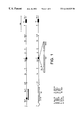
- FIG. 1 is a restriction map of an A-myb genomic clone (top), and the structure of a targeting vector (bottom).
- the coding exons are depicted by black boxes.
- the black boxes in the restriction map represent A-myb exons 3, 4 and 5.
- a genomic fragment comprising the entire exon 3 and a portion of intron 3 was obtained by digestion of the clone with SspI.
- the clone, which was used as a probe, is shown as a black bar.
- the arrows depict the transcriptional orientation of the neo (neomycin transferase gene) and tk (thymidine kinase) genes.
- FIG. 2 is a Southern blot analysis of genomic DNA extracts from ES cell clones electroporated with the targeting vector of FIG. 1 .
- the DNA was digested with HindIII, fractionated on an agarose gel, blotted onto a nitrocellulose paper and hybridized with the 32 P-labeled SspI fragment indicated as a black bar in FIG. 1 .
- Molecular weight markers ( ⁇ DNA digested with HindIII) are shown on the left. Lanes 4 and 5 contain DNA from ES cell clones that contain a disrupted A-myb locus.
- FIG. 3 is a Southern blot analysis of the DNA extracted from tail biopsies of 10-day old pups of A-myb +/ ⁇ intercrosses, digested with HindIII, and blotted onto a nitrocellulose membrane. The blots were hybridized with the same 32 P-labeled probe as in the previous figure.
- the A-myb genotypes of the animals are presented above the lanes. A-myb +/+ (6 kb); A-myb +/ ⁇ (6 kb+8 kb); A-myb ⁇ / ⁇ (8 kb).
- FIG. 4 is a Western blot analysis of extracts prepared from A-myb +/+ , A-myb +/ ⁇ and A-myb ⁇ / ⁇ mouse testes. The blot was probed with a rabbit polyclonal anti-Myb antibody developed by enhanced chemiluminescence. The genotypes of the mouse testes from which the testes extracts were prepared are shown above the lanes.
- FIG. 5A is a comparison of the total body size and testicular size of 4 week-old A-myb +/ ⁇ (left) and A-myb ⁇ / ⁇ (right) mice from the same litter. The testes dissected from the same mice are shown below the mice.
- FIG. 5B is a comparison of the total body size and testicular size of 10 week-old A-myb +/+ (left), A-myb +/ ⁇ (middle) and A-myb ⁇ / ⁇ (right) mice from the same litter. The testes dissected from the same mice are shown below the mice.
- FIG. 6A is a 100 ⁇ view of a hematoxylin and eosin-stained section of the seminiferous tubules of A-myb +/+ mouse testis.
- FIG. 6B is a 100 ⁇ view of a hematoxylin and eosin-stained section of the seminiferous tubules of A-myb ⁇ / ⁇ mouse testis.
- FIG. 7A is a 25 ⁇ magnification cross-section of wild-type mouse ovaries.
- FIG. 7B is a 25 ⁇ magnification cross-section of A-myb ⁇ / ⁇ mouse ovaries.
- FIG. 8A is a 10 ⁇ magnification cross-section of A-myb +/ ⁇ female mouse epithelium, taken from breast tissue of maternal mice two days following the delivery of pups.
- FIG. 8B is a 20 ⁇ view of the same A-myb +/ ⁇ tissue of FIG. 8 A.
- FIG. 8C is a 10 ⁇ magnification cross-section of A-myb ⁇ / ⁇ female mouse epithelium, taken from breast tissue of maternal mice two days following the delivery of pups.
- FIG. 8D is a 20 ⁇ magnification view of the same A-myb ⁇ / ⁇ tissue of FIG. 8 C.
- FIG. 9A shows the structure of the pMV-7 vector of Kirschmeier et al., DNA 7, 219-225 (1988).
- FIG. 9B shows the structure of the modified pMV-7 vector pMV-7 ⁇ ClaI/neo E/H, generated by excision of the neo cassette from pMV-7 of FIG. 9A by ClaI digest and placement of the cassette at the Eco RI/Hind III cloning site of pMV-7.
- FIG. 9C shows the structure of the vector pMV-7 ⁇ ClaI/neo E/H-A-myb, formed by insertion of the 6.7 kbp fragment SEQ ID NO:4 into the ClaI site of the FIG. 9B vector.
- the insert contains the A-myb promoter (cross-hatch), the A-myb coding sequence, and a polyadenylation signal from the bovine growth hormone gene (solid). Arrows in FIGS. 9A-9C indicate transcription start sites.
- Chimeric targeted mice may be derived according to Hogan, et al., Manipulating the Mouse Embryo: A Laboratory Manual , Cold Spring Harbor Laboratory (1988) and Teratocarcinomas and Embryonic Stem Cells: A Practical Approach , E. J. Robertson, ed., IRL Press, Washington, D.C., (1987) which are incorporated herein by reference.
- Embryonic stem cells may be manipulated according to published procedures ( Teratocarcinomas and Embryonic Stem Cells: A Practical Approach , E. J. Robertson, ed., IRL Press, Washington, D.C. (1987); Zjilstra et al., Nature 342:435-438 (1989); and Schwartzberg et al., Science 246:799-803 (1989), each of which is incorporated herein by reference).
- Oligonucleotides can be synthesized on an Applied BioSystems oligonucleotide synthesizer according to specifications provided by the manufacturer.
- the endogenous A-myb alleles of a cell line or nonhuman animal are functionally disrupted so that expression of endogenously encoded A-myb gene is suppressed or eliminated.
- polynucleotide constructs are employed for this purpose. Methods for accomplishing this result are described in detail in WO 95/11968 with respect to transgenic non-human animals and mammalian cells hosting a transgene encoding an amyloid precursor protein (APP) and “knock out” mutants thereof.
- APP amyloid precursor protein
- Gene targeting which is a method of using homologous recombination to modify a mammalian genome, can be used to introduce genetic changes into cultured cells.
- ES embryonic stem
- the gene targeting procedure is accomplished by introducing into tissue culture cells a DNA targeting construct that has a segment homologous to a target locus and which also comprises an intended sequence modification (e.g., insertion, deletion, point mutation). The treated cells are then screened for accurate targeting to identify and isolate those which have been properly targeted.
- a common scheme to disrupt gene function by gene targeting in ES cells is to construct a targeting construct which is designed to undergo a homologous recombination with its chromosomal counterpart in the ES cell genome.
- the targeting constructs are typically arranged so that they insert an additional sequence, such as a positive selection marker, into coding elements of the target gene, thereby functionally disrupting it.
- Targeting constructs usually are insertion-type or replacement-type constructs (Hasty et al. (1991) Mol. Cell. Biol . 11: 4509).
- A-myb genomic DNA useful for this purpose is described herein.
- Appropriate probes may be designed based on known A-myb cDNA nucleotide sequences. For example, the complete nucleotide sequence of the mouse A-myb cDNA (SEQ ID NO:1), deduced amino acid sequence (SEQ ID NO:2), and cDNA restriction map are disclosed in Mettus et al., Oncogene 9: 3077-3086 (1994), the entire disclosure of which is incorporated herein by reference.
- the encoded mouse A-myb protein contains 751 amino acids (SEQ ID NO:2) and has an estimated molecular weight of 83 kDa. It may be appreciated that A-myb genomic DNA may be derived using an appropriate cDNA fragment as a probe to identify and isolate genomic A-myb from an appropriate genomic DNA library.
- Targeting constructs can be transferred into pluripotent stem cells, such as ES cells, wherein the targeting constructs homologously recombine with a portion of the endogenous A-myb gene locus and create mutation(s) (i.e., insertions, deletions, rearrangements, sequence replacements, and/or point mutations) which prevent the functional expression of the endogenous A-myb gene.
- pluripotent stem cells such as ES cells
- One method is to delete, by targeted homologous recombination, essential structural elements of the endogenous A-myb gene.
- a targeting construct can homologously recombine with an endogenous A-myb gene and delete a portion spanning substantially all of one or more exons to create an exon-depleted allele, typically by inserting a replacement region lacking the corresponding exon(s).
- Transgenic animals homozygous for the exon-depleted allele e.g., by breeding of heterozygotes to each other
- homologous gene targeting can be used, if desired, to functionally disrupt the A-myb gene by deleting only a portion of an exon.
- Targeting constructs can also be used to delete essential regulatory elements of the endogenous A-myb gene, such as promoters, enhancers, splice sites, polyadenylation sites, and other regulatory sequences, including cis-acting sequences that may occur upstream or downstream of the A-myb structural gene but which participate in endogenous A-myb gene expression. Deletion of regulatory elements is typically accomplished by inserting, by homologous double-crossover recombination, a replacement region lacking the corresponding regulatory element(s).
- the mouse A-myb gene was isolated by screening a ⁇ DASH mouse genomic library derived from the 129/J mouse strain, using a probe derived from the 5′ end of the A-myb cDNA clone (Mettus et al., Oncogene 9, 3077-3086, 1994) that encodes the DNA binding domain of the protein. Positive clones that contained a 5.9 kbp HindIII fragment were subcloned into pGEM 7Zf(+) plasmid vector. The complete nucleotide sequence of this 5.9 kbp clone was determined using the method of Sanger et al., Proc. Natl. Acad. Sci. USA 74, 5463-5467 (1977).
- A-myb promoter does not contain a putative TATA or CCAT box, it contains a highly GC rich sequence, a feature observed with many testis-specific and housekeeping gene promoters.
- a preferred method is to interrupt essential structural and/or regulatory elements of the endogenous A-myb gene by targeted insertion of a polynucleotide sequence, and thereby functionally disrupt the endogenous A-myb gene.
- a targeting construct can homologously recombine with the endogenous A-myb gene and insert a nonhomologous sequence, such as a neo expression cassettes into a structural element (e.g., an exon) and/or regulatory element (e.g., enhancer, promoter, splice site, polyadenylation site) to yield a targeted A-myb allele having an insertional interruption.
- the inserted sequence can range in size from about 1 nucleotide (e.g., to produce a frameshift in an exon sequence) to several kilobases or more, as limited by efficiency of homologous gene targeting with targeting constructs having a long nonhomologous replacement region.
- One preferred target site is the DNA binding domain of the A-myb gene.
- the DNA binding domain in the A-myb protein spans amino acids tryptophan-32 to valine-188, corresponding to nucleotides 352 to 820 of the murine A-myb cDNA. See SEQ ID NO:1.
- Targeting constructs can also be employed to replace a portion of the endogenous A-myb gene with an exogenous sequence (i.e., a portion of a targeting transgene); for example, a first exon of an A-myb gene may be replaced with a substantially identical portion that contains a nonsense or missense mutation.
- a targeting construct may be transferred by electroporation of microinjection into a totipotent ES cell line.
- the targeting construct homologously recombines with endogenous sequences in or flanking of the A-myb gene locus and functionally disrupts at least one allele of the A-myb gene.
- homologous recombination of the targeting construct with endogenous A-myb locus sequences will result in integration of a nonhomologous sequence encoding and expressing a selectable marker, such as neo, usually in the form of a positive selection cassette.
- ES cells having at least one such A-myb null allele are selected for by propagating the cells in a medium that permits the preferential propagation of cells expressing the selectable marker.
- Selected ES cells are examined by PCR analysis and/or Southern blot analysis to verify the presence of a correctly targeted A-myb allele. Breeding of nonhuman animals which are heterozygous for a null allele may be performed to produce nonhuman animals homozygous for said null allele, so-called “knockout” animals (Donehower et al. (1992) Nature 256: 215 ; Science 256: 1392, incorporated herein by reference). Alternatively ES cells homozygous for a null allele having an integrated selectable marker can be produced in culture by selection in a medium containing high levels of the selection agent (e.g., G418 or hygromycin).
- the selection agent e.g., G418 or hygromycin
- Heterozygosity and/or homozygosity for a correctly targeted null allele can be verified with PCR analysis and/or Southern blot analysis of DNA isolated from an aliquot of a selected ES cell clone and/or from tail biopsies.
- Gene targeting techniques which have been described, include but are not limited to: co-electroporation, “hit-and-run”, single-crossover integration, and double-crossover recombination (Bradley et al. (1992) Bio/Technology 10: 534).
- the preparation of the homozygous A-myb null mutants can be practiced using essentially any applicable homologous gene targeting strategy known in the art.
- the configuration of a targeting construct depends upon the specific targeting technique chosen. For example, a targeting construct for single-crossover integration or “hit-and-run” targeting need only have a single homology clamp linked to the targeting region, whereas a double-crossover replacement-type targeting construct requires two homology clamps, one flanking each side of the replacement region.
- a targeting construct comprises, in order: (1) a first homology clamp having a sequence substantially identical to a sequence within about 3 kilobases upstream (i.e., in the direction opposite to the transnational reading frame of the exons) of an exon of an endogenous A-myb gene, (2) a replacement region comprising a positive selection cassette having a pgk promoter driving transcription of a neo gene, (3) a second homology clamp having a sequence substantially identical to a sequence within about 3 kilobases downstream of said exon of said endogenous A-myb gene, and (4) a negative selection cassette, comprising a PGK promoter driving transcription of an HSV tk gene.
- Such a targeting construct is suitable for double-crossover replacement recombination which deletes a portion of the endogenous A-myb locus spanning said exon and replaces it with the replacement region having the positive selection cassette.
- the deleted exon is one which is essential for expression of a functional A-myb gene product.
- the resultant exon-depleted allele is functionally disrupted and is termed a null allele.
- Targeting constructs comprise at least one homology clamp linked in polynucleotide linkage (i.e., by phosphodiester bonds) to a targeting region.
- a homology clamp has a sequence which substantially corresponds to, or is substantially complementary to, an endogenous A-myb gene sequence of a nonhuman host animal, and may comprise sequences flanking the A-myb gene.
- targeting constructs are generally at least about 50 to 100 nucleotides long, preferably at least about 250 to 500 nucleotides long, more preferably at least abut 1000 to 2000 nucleotides long, or longer.
- Construct homology regions are generally at least about 50 to 100 bases long, preferably at least about 100 to 500 bases long, and more preferably at least about 750 to 2000 bases long.
- homology regions of about 7 to 8 kilobases in length are preferred with one preferred embodiment having a first homology region of about 7 kilobases flanking one side of a replacement region and a second homology region of abut 1 kilobase flanking the other side of said replacement region.
- the length of homology (i.e., substantial identity) for a homology region may be selected at the discretion of the practitioner on the basis of the sequence composition and complexity of the endogenous A-myb gene target sequence(s) and guidance provided in the art.
- Targeting constructs have at least one homology region having a sequence that substantially corresponds to, or is substantially complementary to, an endogenous A-myb gene sequence (e.g., an exon sequence, an enhancer, a promoter, an intronic sequence, or a flanking sequence within about 3-20 kb of the A-myb gene).
- an endogenous A-myb gene sequence e.g., an exon sequence, an enhancer, a promoter, an intronic sequence, or a flanking sequence within about 3-20 kb of the A-myb gene.
- Such a targeting construct homology region serves as a template for homologous pairing and recombination with substantially identical endogenous A-myb gene sequence(s).
- such homology regions typically flank the replacement region, which is a region of the targeting construct that is to undergo replacement with the targeted endogenous A-myb gene sequence.
- a segment of the targeting construct flanked by homology regions can replace a segment of an endogenous A-myb gene sequence by double-crossover homologous recombination.
- Homology regions and targeting regions are linked together in conventional linear polynucleotide linkage (5′ to 3′ phosphodiester backbone).
- Targeting constructs are generally double-stranded DNA molecules, most usually linear.
- Double-crossover replacement recombination thus can be used to delete a portion of the endogenous A-myb and concomitantly transfer a nonhomologous portion (i.e., a neo gene expression cassette) into the corresponding chromosomal location. Double-crossover recombination can also be used to add a nonhomologous portion into the endogenous A-myb gene without deleting endogenous chromosomal portions.
- double-crossover recombination can also be employed simply to delete a portion of an endogenous gene sequence without transferring a nonhomologous portion into the endogenous A-myb gene.
- Upstream and/or downstream from the nonhomologous portion may be a gene which provides for identification of whether a double-crossover homologous recombination has occurred; such a gene is typically the HSV tk gene which may be used for negative selection.
- targeting constructs used for functionally disrupting endogenous A-myb genes will comprise at least two homology regions separated by a nonhomologous sequence which contains an expression cassette encoding a selectable marker, such as neo (Smith and Berg (1984) Cold Spring Harbor Symp. Quant. Biol . 49: 171; Sedivy and Sharp (1989) Proc. Natl. Acad. Sci . ( U.S.A .) 86: 227; Thomas and Capechi (1987), Cell 51: 503).
- a selectable marker such as neo (Smith and Berg (1984) Cold Spring Harbor Symp. Quant. Biol . 49: 171; Sedivy and Sharp (1989) Proc. Natl. Acad. Sci . ( U.S.A .) 86: 227; Thomas and Capechi (1987), Cell 51: 503).
- some targeting transgenes may have the homology region(s) flanking only one side of a nonhomologous sequence.
- Targeting transgenes of the invention may also be of the type referred to in the art as “hit-and-run” or “in-and-out” transgenes (Valancius and Smithies (1991) Mol. Cell. Biol . 11: 1402; Donehower et al. (1992) Nature 356: 215; (1991) J.NIH Res . 3: 59; which are incorporated herein by reference).
- the positive selection expression cassette encodes a selectable marker which affords a means for selecting cells which have integrated targeting transgene sequences spanning the positive selection expression cassette.
- the negative selection expression cassette encodes a selectable marker which affords a means for selecting cells which do not have an integrated copy of the negative selection expression cassette.
- An expression cassette typically comprises a promoter which is operational in the targeted host cell (e.g., ES cell) linked to a structural sequence that encodes a protein or polypeptide that confers a selectable phenotype on the targeted host cell, and a polyadenylation signal.
- a promoter included in an expression cassette may be constitutive, cell type-specific, stage-specific, and/or modulatable (e., by hormones such as glucocorticoids; MMTV promoter), but is expressed prior to and/or during selection.
- An expression cassette can optionally include one or more enhancers, typically linked upstream of the promoter and within about 3-10 kilobases.
- the targeting construct replacement region may comprise only a structural sequence encoding the selectable marker, and rely upon the endogenous promoter to drive transcription (Doetschman et al. (1988) Proc. Natl. Acad. Sci . ( U.S.A .) 85: 8583; incorporated herein by reference).
- an endogenous enhancer located near the targeted endogenous site may be relied on to enhance transcription of transgene sequences in enhancerless transgene constructs.
- Preferred expression cassettes for inclusion in the targeting constructs encode and express a selectable drug resistance marker and/or a HSV thymidine kinase (tk) enzyme.
- Suitable drug resistance genes include, for example: gpt (xanthine-guanine phosphoribosytltransferase), which can be selected for with mycophenolic acid; neo (neomycin phosphotransferase), which can be selected for with G418 or hygromycin; and DFHR (dihydrofolate reductase), which can be selected for with methotrexate (Mulligan and Berg (1981) Proc. Natl. Acad. Sci . ( U.S.A .) 78: 2072; Southern and Berg (1982) J. Mol. Appl. Genet . 1: 327; which are incorporated herein by reference).
- Selection for correctly targeted recombinants will generally employ at least positive selection, wherein a nonhomologous expression cassette encodes and expresses a functional protein (e.g., neo or gpt) that confers a selectable phenotype to targeted cells harboring the endogenously integrated expression cassette, so that, by addition of a selection agent (e.g., G418 or mycophenolic acid) such targeted cells have a growth or survival advantage over cells which do not have an integrated expression cassette.
- a functional protein e.g., neo or gpt
- a selection agent e.g., G418 or mycophenolic acid
- selection for correctly targeted homologous recombinants also employ negative selection, so that cells bearing only nonhomologous integration of the transgene are selected against.
- negative selection employs an expression cassette encoding the herpes simplex virus thymidine kinase gene (HSV tk) positioned in the transgene so that it should integrate only by nonhomologous recombination.
- HSV tk herpes simplex virus thymidine kinase gene
- Such positioning generally is accomplished by linking the HSV tk expression cassette (or other negative selection cassette) distal to the recombinogenic homology regions so that double-crossover replacement recombination of the homology regions transfers the positive selection expression cassette to a chromosomal location but does not transfer the HSV tk gene (or other negative selection cassette) to a chromosomal location.
- a nucleoside analog, ganciclovir which is preferentially toxic to cells expressing HSV tk, can be used as the negative selection agent, as it selects for cells which do not have an integrated HSV tk expression cassette.
- FIAU may also be used as a selective agent to select for cells lacking HSV tk.
- Positive-negative selection involves the use of two active selection cassettes: (1) a positive one (e.g., the neo gene), that can be stably expressed following either random integration or homologous targeting, and (2) a negative one (e.g., the HSV tk gene), that can only be stably expressed following random integration, and cannot be expressed after correctly targeted double-crossover homologous recombination.
- a positive one e.g., the neo gene
- a negative one e.g., the HSV tk gene
- targeting constructs preferably include: (1) a positive selection expression cassette flanked by two homology regions that are substantially identical to host cell endogenous A-myb gene sequences, and (2) a distal negative selection expression cassette.
- targeting constructs which include only a positive selection expression cassette can also be used.
- a targeting construct will contain a positive selection expression cassette which includes a neo gene linked downstream (i.e., towards the carboxy-terminus of the encoded polypeptide in transnational reading frame orientation) of a promoter such as the HSV tk promoter or the pgk promoter.
- the targeting transgene will also contain a negative selection expression cassette which includes an HSV tk gene linked downstream of a PGK promoter.
- FIG. 1 (bottom) is a schematic representation of a typical positive-negative A-myb targeting construct.
- FIG. 1 (bottom) shows the placement of neo and tk (“TK”) genes. Arrows mark the transcriptional orientation of the neo and tk genes.
- FIG. 1 (top) is a schematic representation of the Hind III 5.9 kbp genomic clone used to generate the targeting construct of FIG. 1 (bottom). Black boxes in the restriction maps of FIG. 1 represent exons.
- the targeting construct of FIG. 1 (bottom) was deposited in the Unites States Department of Agriculture Northern Research Laboratories, Peoria, Ill. under accession number B21576 on May 1, 1996.
- the HindIII 5.9 kbp genomic clone used to generate the targeting construct was deposited in the same depository, on the same date, under accession number B21575.
- the nucleotide sequence of the 5.9 kbp clone is SEQ ID NO:3.
- targeting polynucleotides of the invention have at least one homology region that is at least about 50 nucleotides long, and it is preferable that homology regions are at least about 75 to 100 nucleotides long, and more preferably at least about 200-2000 nucleotides long, although the degree of sequence homology between the homology region and the targeted sequence and the base composition of the targeted sequence will determine the optimal and minimal homology region lengths (e., G-C rich sequences are typically more thermodynamically stable and will generally require shorter homology region length).
- homology region length and the degree of sequence homology can only be determined with reference to a particular predetermined sequence, but homology regions generally must be at least about 50 nucleotides long and must also substantially correspond or be substantially complementary to a predetermined endogenous target sequence.
- a homology region is at least about 100 nucleotides long and is identical to or complementary to a predetermined target sequence in or flanking the A-myb gene.
- At least one homology region is isogeneic (i.e., has exact sequence identity with the crossover target sequence(s) of the endogenous A-myb gene), and is more preferred that isogeneic homology regions flank the exogenous targeting construct sequence that is to replace the targeted endogenous A-myb sequence.
- the A-myb sequence may be scanned for possible disruption sites.
- Plasmids are engineered to contain an appropriately sized construct replacement sequence with a deletion or insertion in the A-myb gene and at least one flanking homology region which substantially corresponds or is substantially complementary to an endogenous target DNA sequence.
- flanking homology regions are used, one on each side of the replacement region sequence.
- one homology region may be substantially identical to a sequence upstream (i.e., the direction towards the transcription start site(s) of the murine A-myb exon 4 and a second homology region may be substantially identical to a sequence downstream of the murine A-myb exon 4.
- the A-myb gene is inactivated by homologous recombination in a pluripotent cell line that is capable of differentiating into germ cell tissue.
- a DNA construct, as discussed above, that contains an altered copy of a mouse A-myb gene is introduced into the nuclei of ES cells. In a portion of the cells, the introduced DNA recombines with the endogenous copy of the mouse A-myb gene, replacing it with the altered copy.
- Cells containing the newly engineered genetic lesion are injected into a host mouse embryo, which is reimplanted into a recipient female. Some of these embryos develop into chimeric mice that possess germ cells derived from the mutant cell line. Therefore, by breeding the chimeric mice it is possible to obtain a new line of mice containing the introduced genetic lesion.
- Targeting constructs are typically grown in E. coli and then isolated using standard molecular biology methods, or may be synthesized as oligonucleotides. Direct targeted inactivation which does not require prokaryotic or eukaryotic vectors may also be performed. Targeting constructs can be transferred to host cells by any suitable technique, including microinjection, electroporation, lipofection, biolistics, calcium phosphate precipitation, and viral-based vectors, among others. Other methods used to transform mammalian cells include the use of Polybrene, protoplast fusion, and others (e.g., generally, Sambrook et al. Molecular Cloning: A Laboratory Manual, 2d ed., 1989, Cold Spring Harbor Laboratory Press, Cold Spring Harbor, N.Y., which is incorporated herein by reference).
- ES cells embryonal stem cells
- Murine ES cells such as AB-1 line grown on mitotically inactive SNL76/7 cell feeder layers (McMahon and Bradley (1990) Cell 62: 1073) essentially as described (Robertson, E. J. (1987) in Teratocarcinomas and Embryonic Stem Cells: A Practical Approach . E. J. Robertson, ed. (Oxford: IRL Press), p. 71-112) may be used for homologous gene targeting.
- Other suitable ES lines include but are not limited to, the E14 line (Hooper et al.
- the blastocysts containing the injected ES cells are allowed to develop in the uteri of pseudopregnant nonhuman females and are born as chimeric mice.
- the resultant transgenic mice are chimeric for cells having an inactivated endogenous A-myb locus and are backcrossed and screened for the presence of the correctly targeted transgene(s) by PCR or Southern blot analysis on tail biopsy DNA of offspring so as to identify transgenic mice-heterozygous for the inactivated A-myb.
- By performing the appropriate crosses it is possible to produce a transgenic nonhuman animal homozygous for functionally disrupted A-myb alleles. Such transgenic animals are substantially incapable of making an endogenous A-myb gene product.
- the functionally disrupted A-myb homozygous null mutant transgenic animals will typically comprise rats or mice, but nonmurine species such as dogs, cattle, sheep, goats, pigs and nonhuman primates, for example, may be utilized.
- Homozygous null male A-myb animals are infertile due to a block in spermatogenesis. Histopathological examination of testes from A-myb ⁇ / ⁇ mice of the present invention indicates that the differentiation of spermatogonia is arrested at the pachytene stage of meiosis, indicating an essential role for A-myb in male germ cell differentiation. It appears that A-myb is essential for transition of spermatogonia to the spermatid stage. The results described herein show that the proliferation of primary germ cells is not dependent on the presence of A-myb but that their differentiation is dependent on the synthesis of the A-Myb protein. Loss of A-Myb does not seem to affect the formation of Leydig and Sertoli cells. Earlier studies (Mettus et al., Oncogene 9, 3077-3086, 1994) have shown that A-myb is not expressed in Sertoli cells or Leydig cells. These cells appear normal in A-myb ⁇ / ⁇ mice.
- the histopathology of the testis seen in A-myb ⁇ / ⁇ mice is surprisingly similar to the histopathology seen in a large percentage of men who are infertile (Rosai, J., “Testis” in Ackerman's Surgical Pathology , G. Stamathis ed. (C. V. Mosby Co.), pp. 949-982, 1989; Soderstrom and Suominen, Arch. Pathol. Lab. Med . 104, 476-482, 1980; Wong, et al., Arch. Pathol . 95, 151-159, 1973).
- Biopsy specimens from infertile men with total lack of spermatozoa usually show one of the following conditions: (1) germ cell aplasia (Sertoli cell-only syndrome), in which the tubules are populated by Sertoli cells only and there is a complete absence of germ cells; (2) spermatocystic arrest, characterized by a halt of the maturation sequence, usually at the stage of the primary and secondary spermatocyte development where no spermatids or spermatozoa are present; and (3) generalized fibrosis which appears to result in obstructive azoospermia due to bilateral obstruction or absence of some part of the duct system.
- germ cell aplasia Sesoli cell-only syndrome
- spermatocystic arrest characterized by a halt of the maturation sequence, usually at the stage of the primary and secondary spermatocyte development where no spermatids or spermatozoa are present
- generalized fibrosis which appears to result in obstructive azo
- testicular histopathology of A-myb ⁇ / ⁇ mice is indistinguishable from the histopathology of biopsies described in infertile men suffering from spermatocystic arrest, suggesting that defects in A-myb expression or function may constitute the molecular basis of this form of human infertility.
- the ovaries of the A-myb ⁇ / ⁇ female mice appear normal by histological assessment which is further evidence by the ability of these mice to become pregnant and deliver pups. However, a striking abnormality was observed in the female A-myb ⁇ / ⁇ mice following the birth of the pups. Examination of the mothers revealed complete absence of milk formation following the delivery of pups. Pathological examination of the A-myb ⁇ / ⁇ mothers 48 hours following delivery revealed severe impairment of mammary epithelial proliferation in mutant mice following pregnancy.
- the mammary epithelium occurs in two stages.
- the first stage of development occurs during puberty, when the breast tissue becomes fully developed and is characterized by ductal elongation.
- the ductal cells at this stage express estrogen receptors and the ductal elongation is believed to be stimulated by estrogens.
- the mammary epithelial cells acquire progesterone receptors and at this stage of development, require both estrogen and progesterone for proliferation.
- the combined action of estrogens and progesterone results in ductal side branching and lobuloalveolar development.
- the mammary glands of A-myb ⁇ / ⁇ mice appeared to develop normally during sexual maturation, as evidenced by histological analysis.
- the A-myb ⁇ / ⁇ male animals of the invention may be utilized as a model for male infertility, and for studying spermatogenesis.
- the A-myb ⁇ / ⁇ animals may be used in the screening of potential therapeutic synthetic A-myb peptides. Such peptides could be screened for the ability to induce resumption of spermatogenesis upon local administration to the testes of A-myb ⁇ / ⁇ mice.
- the candidate peptide would be administered locally by injection into the testes of the A-myb ⁇ / ⁇ mice.
- the A-myb ⁇ / ⁇ animals of the invention may also be used as host animals for the transfer of desired transgenes via the sperm of A-myb ⁇ / ⁇ individuals rescued with a transgene construct encoding non-defective A-myb gene and the desired additional transgene.
- the A-myb ⁇ / ⁇ animals described herein contain immature germ cells. Since these are stem cells, they have the potential for self renewal and thus undergo replication. These cells may be induced to replicate and undergo differentiation to mature spermatids upon the introduction of exogenous A-myb.
- spermatogonia are isolated from the A-myb ⁇ / ⁇ animals, cultured in vitro and then transfected with a transgene construct comprising a first DNA sequence encoding a functional A-Myb polypeptide which is linked to a second DNA sequence comprising the transgene of interest, the expression of which is desired in the animal's germline.
- the first and second DNA sequences may be operatively linked in that the transcription of both sequences is under the control of the same promoter or enhancer. Alternatively separate promoter/enhancer elements may be included in the construct for the first and second DNA sequences.
- the transfected cells are then reintroduced into the testes of the A-myb ⁇ / ⁇ animals. This will allow for expression of A-myb and the continuation of spermatogenesis, resulting in the production of viable spermatozoa which include the transgene of interest. While not necessarily required, it is preferred that the spermatogonia donor and transfected cell recipient are of the same species, preferably the same strain.
- transgene The practice of the present invention is exemplified herein using the neo gene as the transgene. It may be appreciated that it is possible to generate nonhuman animals producing sperm which harbor any desired transgene, provided the transgene may be contained in a construct further including a wild type A-myb gene. Spermatogonia which fail to incorporate the transgene construct encoding the wild type A-myb gene and the transgene of interest will not mature into spermatozoa.spermatogonia which successfully incorporate the construct are “rescued” by the A-myb wild type transgene, but will also contain the additional transgene of interest.
- the sperm output of the rescued animal is limited to cells which have successfully incorporated the construct and the transgene of interest.
- the A-myb ⁇ / ⁇ animals “rescued” with the transgene construct thus possess transgenic sperm, which can pass the desired transgene to offspring.
- the transgenic spermatozoa can be harvested and used to artificially inseminate females, or to fertilize eggs in vitro.
- spermatogonia are isolated from A-myb ⁇ / ⁇ animals, cultured in vitro, and transfected with an appropriate “rescue” construct containing wild-type A-myb DNA and the transgene of interest. The transfected cells are then reintroduced into the testis of A-myb ⁇ / ⁇ animals.
- Spermatogonia may be isolated from A-myb ⁇ / ⁇ animals according to known methods. Methods for isolating germ cells from the testis of mammals are known to the art. See, e.g., Ogawa et al., J. Dev. Biol . 41, 111-122 (1997); Brinster and Zimmermann, Proc. Natl. Acad. Sci. USA 91, 11298-302 (1994); Brinster and Avarbock, Proc. Natl. Acad. Sci. USA 91, 11303-7 (1994); Hofmann et al., Exp. Cell Res . 201:417-435 (1992); Hofmann et al., Proc. Natl. Acad. Sci. USA 91, 5333-7 (1994). The entire disclosures of the preceding references are incorporated herein by reference.
- the isolated spermatogonia are transfected with a rescue construct containing A-myb DNA encoding a functional A-Myb polypeptide, e.g. cDNA, under the control of a promoter to obtain A-myb expression, and a transgene of interest.
- the construct includes structural sequences encoding the functional A-Myb polypeptide and the transgene of interest, and linked regulatory elements that drive expression of both structural sequences in the host.
- At least one promoter/enhancer is linked upstream of the first structural sequence in an orientation to drive transcription of the A-myb wild-type DNA and transgene of interest.
- the promoter is selected so as to provide for expression of the transgenes in the testis.
- the PGK2 and CMV promoter/enhancer elements are known to be capable of driving constitutive gene expression in the testis (Robinson et al., Proc. Natl. Acad. Sci. USA 86, 8437-41, 1989; Rosenberg et al., Cell Growth & Differ . 1995; Goto et al., Exp. Cell Res . 186, 273-8, 1990), it is preferred that the promoter drive expression at levels similar to the naturally occurring A-myb gene, and the promoter is selected accordingly. Most advantageously, the promoter comprises the naturally occurring A-myb promoter.
- the rescue construct generally encodes the full-length A-Myb protein, or fragment or analog thereof, having sufficient A-Myb activity to permit spermatogenesis to proceed in the host.
- the A-Myb-encoding DNA encodes the full-length protein.
- the transgene of interest may be designed so as to provide for the expression of any desired gene in the rescued host animal.
- HindIII genomic fragment containing the A-myb promoter region is isolated from a lambda phage mouse genomic library using 32 P-labeled A-myb cDNA as a probe.
- the HindIII genomic fragment is further cleaved with BstEII to generate a HindIII/BstEII fragment containing the A-myb promoter/enhancer region.
- the HindIII/BstEII fragment may be cloned into the BstEII site of an A-myb cDNA clone.
- the isolated A-myb ⁇ / ⁇ spermatogonia may be transfected with the rescue construct by methods known to those skilled in the art such as by the calcium phosphate method (Chen and Okayama, Mol. Cell. Biol . 7, 2745-52, 1987), the electroporation method (Potter, Anal. Biochem 74, 361-373, 1988; Zheng and Chang, Biochim. Biophys. Acta 1088, 104-110, 1991) the liposome-mediated method (Lopata et al., Nucl. Acids Res . 12, 5705, 1984) or the DEAE-dextran method (Felgner et al., Proc. Natl. Acad. Sci. USA 84, 7413-7, 1987).
- the entire disclosures of the aforementioned references are incorporated herein by reference.
- the transfected cells are then introduced into the seminiferous tubules in the testes of A-myb ⁇ / ⁇ mice, such as according to the procedure of Brinster and Zimmermann Brinster, supra.
- the transfected cells may be injected into the rete testis (Ogawa et al., Int. J. Dev. Biol . 41, 111-122 (1997), the entire disclosure of which is incorporated herein by reference).
- a third possibility is to introduce the transfected cells by cannulating one of the five different ducts running from the rete testis to the head of the epididymis, thereby filling the rete and subsequently the tubules (Id.).
- mice are maintained for 2-6 months to allow the transfected stem cells to undergo spermatogenesis. Since A-myb ⁇ / ⁇ lack spermatogenesis, the presence of mature spermatids and spermatozoa in transplanted mice would indicate successful integration of the rescue construct, and the transgene of interest.
- the rescued animals can then be mated to produce offspring containing the germ line transgene, or the transgenic spermatozoa can be harvested and used to artificially inseminate females, or to fertilize eggs in vitro.
- A-myb ⁇ / ⁇ mice may be infected with retroviruses containing A-myb and the desired transgene.
- a retrovirus vector is constructed wherein A-myb cDNA is expressed under the control of the A-myb promoter.
- the structure of one such vector designated pMV-7 ⁇ ClaI/neo E/H-A-myb, wherein the transgene is the neo gene, is shown in FIG. 9 C. Arrows in FIG. 9C indicate transcription start sites. It may be appreciated that the neo gene may be replaced in the vector of FIG. 9C with any transgene which one desires to incorporate into the spermatogonia cell DNA.
- High titer viruses are then generated, such as according to the method described by Kozak and Kabat, J. Virol . 64, 3500-3508, 1990, incorporated herein by reference. It has been demonstrated that recombinant retroviruses of encoding c-myb cDNA may be generated in titers up to 10 7 particles/ml (Patel et al., Mol. Cell. Biol . 13, 2269-2276, 1993, incorporated by reference). Irradiated packaging cells producing virus, or concentrated preparation of virus, are injected into the testes of anesthetized A-myb ⁇ / ⁇ mice. Recipient mice are maintained over a period of several months and analyzed for the production of mature sperm and spermatids. The rescued animals can then be mated to produce offspring containing the germ line transgene, or the transgenic spermatozoa can be harvested and used to artificially inseminate females, or to fertilize eggs in vitro.
- the present invention also provides treatment methods for restoring fertility in male individuals who are infertile due to a genetic defect in the A-myb locus. Such individuals are characterized by a low level of A-Myb protein, or a loss of functional A-Myb protein. Such individuals may be identified by an analysis of the A-Myb protein size, which will reveal mutations that block A-Myb synthesis, or gene rearrangements which result in production of a truncated protein.
- A-myb cDNA from the afflicted individual may be prepared and sequenced according to conventional techniques, and the sequence compared to the wild-type human A-myb cDNA sequence (SEQ ID NO:5).
- the A-myb translation initiation codon in SEQ ID NO:5 comprises nucleotides 105-107.
- the amino acid sequence of the human A-myb polypeptide is set forth as SEQ ID NO:6.
- peripheral blood lymphocytes may be conveniently utilized as a source of A-myb DNA for analysis.
- Methods of sequence analysis aimed at identifying mutations, e.g., the so-called single-strand conformation polymorphism or “SSCP” method (Orita et al., Genomics 5, 874-879, 1989, incorporated herein by reference) may be utilized to screen for A-myb mutations.
- SSCP single-strand conformation polymorphism
- Local gene therapy is carried out to transfer to the cells of the testes of A-myb defective individuals a construct encoding a functional A-Myb polypeptide.
- the transfer may be carried out by removing and engineering spermatogonia of such A-myb ⁇ / ⁇ individuals with a DNA construct designed to express a functional A-Myb polypeptide.
- the construct preferably incorporates the complete coding segment of the human A-myb cDNA.
- the engineered spermatogonia are then returned to the testes of the infertile donor.
- Successful incorporation of the construct results in the production of functional A-Myb polypeptide in the testes of the subject, and production of maturation of competent sperm.
- Fertility may be restored in a subject who is infertile due to a defect in the A-myb locus through the use of a retrovirus vector directing the incorporation of DNA encoding a functional A-Myb protein into appropriate cells of the testes.
- the retrovirus vector is used to infect the testis of the individual in order to obtain the local production of a functional A-Myb polypeptide.
- A-Myb polypeptide which is expressed in the testes according to the aforesaid infertility treatment methods will typically comprise the full-length wild-type A-myb expression product
- also included in the scope of the invention is the expression of polypeptides which comprise fragments of the complete naturally occurring gene product, or analogs thereof which differ from the latter by one or more amino acid insertions, deletions and/or substitutions, provided such fragments and analogs are functional in restoring fertility upon expression in the host.
- Infertility may be also be treated by the local administration to the testes of an infertile A-myb ⁇ / ⁇ individual an exogenous functional A-Myb polypeptide.
- the polypeptide most advantageously comprises the full-length wild-type A-myb expression product, but the functional A-Myb polypeptide may comprise an A-Myb fragment or analog, as described above.
- Such vehicles may comprise, for example, aqueous vehicles such as normal saline.
- the dosage and treatment schedule are selected so as to provide for continuous production of viable sperm at a level which can support successful fertilization.
- “functional A-Myb” polypeptide is a fusion product comprising the naturally occurring A-Myb polypeptide or analog thereof and one or more attached amino acid sequences which enhance the cellular uptake or penetration of the A-Myb polypeptide into the cells of the testes.
- Such fusion products may be prepared with resort to commercially available expression vectors which provide for incorporation of DNA sequences of interest downstream from a DNA segment encoding an amino acid sequence having desirable transport properties.
- the resulting A-Myb fusion protein may be used as the exogenously sourced functional A-Myb protein in treating A-myb ⁇ / ⁇ individuals for infertility.
- a cell-penetrating peptide may be ligated directly to the N-terminal or C-terminal portion of the functional A-Myb polypeptide to enhance uptake.
- One such peptide is the sixteen amino acid peptide from the third helix of the Antennapedia homeodomain, Arg-Gln-Ile-Lys-Ile-Phe-Phe-Gln-Asn-Arg-Arg-Met-Lys-Trp-Lys-Lys (SEQ ID NO:7) (Derossi et al., J. Biol. Chem . 269 (14):10444-10450, 1994).
- This 16 amino acid peptide may be coupled to a functional A-Myb polypeptide according to the manufacturer's protocols (Penetratin 1TM, Appligene, Inc., 1177C Quarry Lane, Pleasanton, Calif. 94566).
- the practice of the invention is illustrated by the following nonlimiting examples. As it relates to the rescue of nonhuman A-myb ⁇ / ⁇ animals from infertility by incorporation of a construct encoding wild-type A-myb DNA and a transgene of interest, the practice of the present invention is exemplified using the neo gene, a bacterial gene, as the transgene. The same neo gene also provides for positive selection of transformants harboring an integrated “rescue” construct encoding. It should be understood that neo DNA may be replaced by any other transgene, or that additional transgenes of interest may be included in addition to neo such that the positive selection function of the neo gene product may be retained in screening transformants.
- the mouse A-myb gene was isolated as follows by screening a ⁇ DASH mouse genomic library derived from the 129/J mouse strain, using a probe derived from the 5′ end of the A-myb cDNA clone that encodes the DNA binding domain of the A-Myb protein.
- DNA obtained from the tissue of a 129/J mouse was digested with the following restriction enzymes: BamHI, EcoRI, HindIII, PstI and XbaI. From a Southern analysis of the digests, the DNA was digested with HindIII to obtain a genomic clone.
- An approximately 800 bp A-myb cDNA probe was prepared consisting of an EcoRI fragment from the 5′ end of the A-myb cDNA spanning cDNA nucleotide positions 1-794 of the A-myb mouse cDNA.
- the mouse A-myb cDNA nucleotide sequence and deduced amino acid sequence of the A-Myb protein are shown in SEQ ID NO:1 and 2, respectively.
- the digested genomic Southern blot was probed with the A-myb cDNA probe.
- a 5.9 kbp Hind III fragment was cloned by size selection in a sucrose gradient followed by cloning into Hind III digested Lambda DASH II phage (Stratagene, LaJolla, Calif.). From the resulting library, the 5.9 kbp A-myb fragment was cloned and subcloned into the pGEM 7Zf(+) plasmid vector (Promega Corp., Madison, Wis.) according to standard procedures ( Molecular Cloning: A Laboratory Manual , T. Maniatis, E. Fritsch and J. Sambrook, eds. (1982), Cold Spring Harbor Laboratory).
- FIG. 1 The structure of the genomic clone and corresponding restriction map of mouse A-myb is shown in FIG. 1 .
- A-myb contains three tandem amino acid direct repeats which make up a DNA binding domain.
- the genomic clone was completely sequenced (SEQ ID NO:3) according to the method of Sanger et al., Proc. Natl. Acad. Sci. USA 74, 5463-5467 (1977) and found to contain A-myb exons 3, 4 and 5 (FIG. 1, black boxes).
- Exon 3 encodes the 5′ end of the first repeat of the DNA binding domain
- exon 4 encodes the 3′ end of the first repeat and the 5′ end of the second repeat.
- Exon 5 encodes the 3′ end of the second repeat and the 5′ end of the third repeat.
- the genomic clone was deposited as NRRL B-21575 on May 1, 1996.
- a gene targeting vector was prepared from the 5.9 kbp genomic clone by following the positive-negative selection method which was originally described by Thomas and Capecchi, Cell 51:503-512 (1987), the entire disclosure of which is incorporated herein by reference.
- the vector was designed to disrupt the DNA binding domain of the A-myb gene by insertion of the neomycin transferase (neo) gene into A-myb exon 4 at the ClaI site (FIG. 1 ).
- the Pgk-Neo gene (Mansour et al., Nature 336, 348-352 (1988), incorporated herein by reference) was digested with EcoRI and HindIII to yield a 2.0 kbp neo cassette. This DNA fragment was filled by using the Klenow fragment of DNA polymerase I. The genomic clone of A-myb was digested with ClaI, blunt ended and then ligated with the DNA fragment containing the neo cassette. Insertion of the neo cassette results in the disruption of the gene which codes for the DNA-binding domain of the A-Myb protein.
- the EcoRV/HindIII fragment of this clone was then released by digestion with HindIII and EcoRV, filled using the Klenow fragment of DNA polymerase I and blunt end ligated to the Pgk-TK vector (Thomas and Capecchi, supra) at the HindIII site.
- the resulting target vector is shown in FIG. 1 (bottom).
- the orientation of the A-myb, neo and tk genes was determined by restriction endonuclease analysis in conjunction with DNA sequence analysis. The orientation of the neo and tk genes is indicated by arrows in FIG. 1 .
- the neo cassette contained the neomycin transferase gene under the control of the phosphoglycerol kinase 1a promoter and polyadenylation signals.
- the thymidine kinase gene was flanked by the same promoter and polyadenylation signals.
- Mouse ES cells (cell line E14a, Handyside et al., Roux's Arch. Dev. Biol . 198, 48-55, 1989) were maintained as previously described by Robertson, “Embryo-derived Stem Cell Lines” in Teratocarcinomas and Embryonic Stem Cells: A Practical Approach , E. J. Robertson, ed. (Oxford, 1987; IRL Press), pp. 71-112, and cultured in Dulbecco's Modified Eagle's Medium supplemented with 15% fetal calf serum (Sigma Chemical Co., St. Louis, Mo.) and 1000 U/ml of leukemia inhibitory factor on ⁇ -irradiated SNL feeder layers.
- the above-prepared A-myb knockout targeting vector was linearized with PvuI and introduced into the mouse ES cells by electroporation (250V, 500 mF) as described by Thomas and Capecchi, supra. Following electroporation, the ES cells were plated into 6 cm plates containing G418 (0.1 mg/ml. active ingredient) and 2 ⁇ M ganciclovir. On days 9 and 10 of selection, individual clones were picked, dispersed into single cell suspension in 0.25% trypsin and seeded into two wells, each in a separate 48 well tissue culture plate. After 5 to 7 days, one plate was used for DNA isolation for Southern blot analysis by standard protocols.
- DNA was isolated from 87 double resistant clones, digested with HindIII, blotted onto nitrocellulose membranes, and probed with a 32 P-labeled 700 bp SspI 5′ A-myb probe.
- the probe hybridizes to a 5.9 kbp fragment derived from the wild-type A-myb allele, while the targeted locus is predicted to yield an 8 kbp fragment.
- FIG. 2 The positions of the molecular weight markers ( ⁇ DNA digested with HindIII) is shown on the left. Lanes 4 and 5 contain the DNA from ES cell clones that contain a disrupted A-myb locus.
- the appearance of an 8 kbp band indicates a homologous recombination event in two of the 87 clones analyzed.
- the mouse A-myb gene was targeted using 4.5 kb of homologous sequence using isogeneic DNA at a frequency of approximately 1 in 40 of the doubly selected clones analyzed.
- the two clones bearing a disrupted A-myb gene were microinjected into C57/B6 blastocysts (10-15 cells/embryo), transferred to pseudopregnant foster CD1 females and male chimeras were produced.
- the chimeras appeared normal and were mated with females from C57/B6 to obtain heterozygous (A-myb +/ ⁇ ) mice.
- the mice appeared normal.
- Eight offspring with agouti color were produced. Southern blot analysis of DNA derived from tail biopsies of these mice showed that three of them were heterozygous for the A-myb disruption.
- Intercrosses were set up between mice heterozygous for the disrupted A-myb allele and progeny were analyzed for A-myb genotype ten days after birth by Southern blot analysis of DNA derived from tail biopsies.
- the DNA was extracted from the tail biopsies of the 10-day old pups, digested with HindIII, and blotted onto a nitrocellulose membrane.
- the blots were hybridized with the 32 P-labeled 700 bp SspI probe. Genotyping was determined according to the sizes of the Hind III fragments hybridized (FIG.
- the progeny generated were in the expected Mendellian ratio of 1:2:1 (103:237:91) for wild type (+/+), heterozygous (+/ ⁇ ), and homozygous ( ⁇ / ⁇ ) A-myb alleles.
- A-myb ⁇ / ⁇ pups showed a similar appearance. At birth, A-myb ⁇ / ⁇ mice appeared to be indistinguishable from their littermates. A difference in size and appearance of A-myb ⁇ / ⁇ mice was seen during the first few weeks of life. The A-myb ⁇ / ⁇ pups were runted, wrinkled, and exhibited hunched posture as compared to their littermates. Such an appearance was not observed with the A-myb +/+ and A-myb +/ ⁇ pups. No difference was observed between the heterozygous and wild type A-myb pups. At four weeks, the A-myb ⁇ / ⁇ pups were approximately 40% the size of their A-myb +/ ⁇ littermates.
- mice As the A-myb ⁇ / ⁇ mice matured (up to three months), their runted and hunched posture appearance became less pronounced and the mice almost attained the body size (as measured by total body weight) that is comparable to the A-myb +/+ and A-myb +/ ⁇ mice (almost 90% for the females and approximately 70% for the males). However, 25% of the mice do not survive after three to four weeks after birth (23 out of 91).
- A-myb ⁇ / ⁇ male mice do not exhibit this particular behavior during matings even after a period of several weeks.
- the A-myb ⁇ / ⁇ female mice have not shown such enhanced aggressive behavior even though they remained hyperactive. All the above abnormal behavioral patterns exhibited by the A-myb ⁇ / ⁇ mice lessened in severity as the mice matured to 3 months of age. Pathological examination of the nervous system did not reveal any obvious abnormalities in the brain, spinal cord or other structures. However, the possibility of minor neuroanatomical defects that could lead to some of these neurological abnormalities can not be ruled out. While the A-myb ⁇ / ⁇ female mice are fertile, the A-myb ⁇ / ⁇ male mice are infertile but are able to copulate as evidenced by the formation of vaginal plugs in the females after mating.
- FIG. 5A is a comparison of the total body size and testicular size of 4 week-old A-myb +/ ⁇ (left) and A-myb ⁇ / ⁇ (right) mice from the same litter. The testes dissected from the same mice are shown below the mice.
- FIG. 5B is a comparison of the total body size and testicular size of 10 week-old A-myb +/+ (left), A-myb +/ ⁇ (middle) and A-myb ⁇ / ⁇ (right) mice from the same litter. The testes dissected from the same mice are shown below the mice. The testis of A-myb ⁇ / ⁇ are atrophic.
- the size and weight of the testis from the A-myb ⁇ / ⁇ mice is approximately 25% of their littermates.
- the small size of the testis is not due to the fact that these A-myb ⁇ / ⁇ mice are, at least in the initial weeks after birth, smaller in body size as compared to their heterozygous and wild type A-myb littermates (FIG. 5 A).
- FIG. 5 A When the ratio of testis weight versus total body weight was compared, it became clear that there was a four-fold reduction in the weights of testes derived from the A-myb ⁇ / ⁇ mice as compared to their heterozygous and wild type litter mates. Even after the male mice attained a more normal weight as a function of age, the testes failed to recover weight unlike other organs and remained in an atrophic state (FIG. 5 B).
- FIG. 6A shows the hematoxylin and eosin-stained sections of the seminiferous tubules of A-myb +/+ mouse testes while FIG. 6B shows similar sections from A-myb ⁇ / ⁇ mouse testis. Magnification is 100 ⁇ .
- A-myb +/+ mice In the A-myb +/+ mice (FIG. 6 A), the differentiation of spermatozoa proceeds in a step-wise manner where the spermatogonia are located close to the basement membrane, while cells at progressively later stages of differentiation are situated closer to the tubular lumen. In the center of the tubular lumen mature spermatozoa with tails can be distinguished from other cell types.
- A-myb +/ ⁇ mice showed the identical pattern as that of A-myb +/+ mice (data not shown).
- A-myb ⁇ / ⁇ mice lack spermatids and mature spermatozoa in the center of the lumen (FIG. 6 B).
- the tubules contain primary and secondary spermatogonia near the basement membrane.
- a small number of cells with small nuclei, with condensed and possible apoptotic nuclear chromatin are seen in some tubules which represent abortive secondary spermatocytes.
- the differentiation of spermatogonia seemed to come to an abrupt halt at the pachytene stage of meiosis. Loss of A-myb did not seem to affect the formation of Leydig and Sertoli cell populations as they appeared to be normal in A-myb ⁇ / ⁇ mice.
- A-myb protein was not synthesized in A-myb ⁇ / ⁇ mice.
- Western blot analyses were performed. Since A-myb is predominantly expressed in the testis, homogenates were prepared using testes dissected from A-myb +/+ , A-myb +/ ⁇ and A-myb ⁇ / ⁇ mice.
- tissue samples were homogenized by Dounce disruption in lysis buffer (10 mM HEPES, pH 7.9; 1 mM EDTA; 60 mM KCl; 0.5% NP40; 1 mM DTT; 1 mM PMSF; 0.5 ⁇ g/ml of Leupeptin; 0.5 ⁇ g/ml of Pepstatin A and 0.5 ⁇ g/ml of Aprotinin), clarified by centrifugation and the protein concentration in the supernatants was determined. Protein samples were separated by SDS-PAGE (10% polyacrylamide) and transferred to nitrocellulose membranes.
- Membranes were blocked by incubation in PBS containing 2% non-fat dry milk, 2% BSA and 0.1% Tween 20 for 1 hour at room temperature and then rinsed several times in T-TBS (0,05% Tween 20, 20 mM Tris-HCl, pH 7.5, and 150 mM NaCl). The membranes were then incubated with a 1:100 dilution of rabbit polyclonal antibody raised against the DNA binding domain of the Myb protein for 1 hour at room temperature and then rinsed several times in T-TBS. The membranes were then incubated with a 1:1000 dilution of the developing antibody (goat anti-rabbit Ig) for 30 minutes at room temperature and then rinsed several times in T-TBS.
- the membranes were then developed according to the supplier by chemiluminescence (ECL).
- ECL chemiluminescence
- a band corresponding to the A-Myb protein in the A-myb +/+ and A-myb +/ ⁇ mouse testis lysates was detected (FIG. 4 ).
- the band was totally absent in the A-myb ⁇ / ⁇ testis lysate (FIG. 4 ).
- FIG. 7A shows a cross-section of wild-type mouse ovaries while FIG. 7B shows the cross-section of A-myb ⁇ / ⁇ mouse ovaries. Magnification is 25 ⁇ . Both cross-sections show normal primary and secondary follicles with normal oocytes.
- the ovaries of the A-myb ⁇ / ⁇ mice are proportionally smaller than their A-myb +/+ or A-myb +/ ⁇ littermates.
- A-myb ⁇ / ⁇ female mice When the A-myb ⁇ / ⁇ female mice were allowed to mate with wild-type or A-myb +/ ⁇ mice, they became pregnant and produced litters of normal number. Two days following delivery of pups, maternal female mice were sacrificed and then breast tissue examined for the development of secondary (pregnancy induced) alveolar and lobular growth and development. A dramatic abnormality in mammary function was apparent. A-myb ⁇ / ⁇ female mice were found to be unable to nurse the pups because of defective mammary tissue proliferation. The mammary epithelium of wild type and A-myb +/ ⁇ mice underwent a massive expansion following pregnancy (FIG. 8A, 8 B), which was considerably diminished in A-myb ⁇ / ⁇ mice (FIG. 8C, 8 D).
- the amount of breast tissue was markedly decreased when compared to a lactating heterozygous mouse. This appeared to be due to diminished secondary (pregnancy induced) alveolar and lobular growth and development (FIG. 8C, 8 D). An intermediate lobular pattern was observed, which was closer to a resting lobular pattern. Mammary epithelial ducts from null mutant mice failed to significantly branch and develop alveolar structures which normally cluster together to form lobules that fill the mammary fat pad (FIG. 8C, 8 D).
- Germ cells are isolated from the testis of A-myb ⁇ / ⁇ mice as follows. Testes from male A-myb ⁇ / ⁇ mice are removed at postnatal day 10. The tunica are removed from the testes and the exposed tubules are treated with collagenase (1 mg/ml 15 min at 37° C.) followed by trypsin digestion (0.25% for 10 min. at 37° C.). The cells are then centrifuged at 600 ⁇ g at 16° C. for 5 min. A cell fraction enriched with preleptotene spermatocytes is then isolated by the STA-PUT procedure and unit gravity sedimentation at 4° C. as described by Brinster and Zimmermann, Proc. Natl. Acad. Sci.
- CMRL-1066 medium supplemented with 80 ⁇ g/ml insulin, 3 ⁇ g/ml transferrin, 80 ⁇ g/ml ascorbic acid and 13% fetal bovine serum.
- a rescue construct encoding a functional A-myb polypeptide and neo as a transgene, is prepared as follows.
- a 10 kbp HindIII genomic murine A-myb fragment is isolated from a lambda phage mouse genomic library using 32 P-labeled A-myb cDNA as a probe.
- the 10 kbp HindIII fragment is further cleaved with BstEII to generate a HindIII/BstEII fragment (SEQ ID NO:4, nucleotides 1-3383) which contains the A-myb promoter/enhancer region.
- a murine cDNA clone containing the A-myb 5′-untranslated region (designated clone 100; Mettus et al., Oncogene 91, 3077-3086, 1994)) is isolated from a murine cDNA library using as a probe a 1273 bp HpaII-NcoI fragment derived from the 5′-region of the human A-myb cDNA (Nomura et al., Nucleic Acids Res . 16, 11075-11089, 1988).
- Clone 100 is digested with BstEII and ClaI to form a 0.3 kbp BstEII/ClaI cDNA fragment containing the A-myb 5′ -untranslated region (SEQ ID NO:4, nucleotides 3384-3730).
- the 3.4 kbp HindIII/BstEII genomic fragment containing the A-myb promoter is ligated to the 0.3 kpb BstEII/ClaI cDNA fragment to form a 3.7 kpb HindIII/ClaI fragment (SEQ ID NO:4, nucleotides 1-3730).
- a clone, designated pcDNA3_PGC2_A-myb, containing the A-myb coding sequence and a polyadenylation signal from the bovine growth hormone gene, is prepared as follows:
- Vector pcDNA3.1v (Invitrogen Corp., 3985 B Sorrento Valley Blvd., San Diego, Calif. 92121) is digested with NruI and HindIII to remove the CMV promoter. The ends of the resulting 4.7 kb fragment are filled by T4 polymerase treatment.
- PGK2 a 1.4 kb HindIII fragment is isolated from a PGK2/chloroamphenicol acetyl transferase (CAT) fusion gene, the preparation of which is described by Robinson et al., Proc. Natl. Acad. Sci. USA 86(21):8437-41, 1989, the entire disclosure of which is incorporated herein by reference.
- the 1.4 kb HindIII fragment is blunt ended by T4 polymerase treatment. The fragment contains the human PGK2 promoter.
- A-myb fragment Preparation of A-myb fragment.
- a construct containing full length A-myb coding sequence in the vector pGEM-37f (Promega Corp., 2800 Woods Hollow Rd., Madison, Wis. 53711-5399) is digested with HindIII and BstEII followed by PvuI and treatment with T4 polymerase (to blunt end the fragment).
- the 2.6 kb A-myb coding sequence (HindIII/BstEII) is purified.
- the PGK2-A-myb-bovine polyadenylation fragment(from the pcDNA3 sequence) is removed by AatII/BsaAI digestion and isolation of the 5.1 kb band.
- the 5.1 kb band is then digested with ClaI and BsaAI to form a 3.0 kbp ClaI/BsaAI fragment comprising SEQ ID NO:4, nucleotides 3371-6775.
- the A-myb coding sequence constitutes SEQ ID NO:4 nucleotides 3506-6134.
- the bovine growth hormone polyA signal from the pcDNA3 vector constitutes SEQ ID NO:4 nucleotides 6135-6775.
- the pMV-7 retroviral vector (Kirschmeier et al., DNA 7:219-225, 1988, incorporated herein by reference) contains the selectable drug resistance gene neo under the regulation of the herpes simplex virus (HSV) thymidine kinase (tk) promoter and unique EcoRI and HindIII cloning sites for the insertion of cDNAs whose transcription is regulated by the 5′ long terminal repeat (LTR). See FIG. 9A for a diagram of pMV-7.
- HSV herpes simplex virus
- tk thymidine kinase
- LTR 5′ long terminal repeat
- This neo cassette is then placed in the EcoRI/HindIII cloning site in the pMV-7 vector to form the vector pMV-7 ⁇ ClaI/neo E/H (FIG. 9 B).
- This vector contains a ClaI site away from the LTR in which the A-myb cDNA may be introduced under the regulation of the A-myb promoter.
- the 3.7 kbp HindIII/ClaI fragment containing the A-myb promoter and 5′-untranslated region is ligated to the 3.0 kbp ClaI/BsaAI fragment of the pcDNA3_PGK2_A-myb clone containing the A-myb coding sequence and a polyadenylation signal form the bovine growth hormone gene.
- the resultant 6.7 kbp fragment (SEQ ID NO:4, nucleotides 1-6775) is blunt ended and cloned into the ClaI site (also blunt ended) of the modified pMV-7 retroviral vector, pMV-7 ⁇ ClaI/neo E/H.
- spermatogonia are obtained from testes of A-myb ⁇ / ⁇ mice from 5 to 60 days after birth by using a two-step enzymatic digestion protocol as described by Ogawa et al., Int. J. Dev. Biol . 41:111-122 (1997).
- the seminiferous tubules are first exposed by peeling the tunica albuginea from the testes. To disperse the tubules the testes are then incubated in 10 volumes of Hanks' balanced salt solution without calcium or magnesium (HBSS) containing 1 mg/ml collagenase at 37° C. with mild agitation for 15 minutes.
- HBSS Hanks' balanced salt solution without calcium or magnesium
- tubules are then washed 4 times in 10 volumes of HBSS, followed by incubation at 37° C. for 5 minutes in HBSS containing 1 mM EDTA, 200 to 700 ⁇ g/ml DNAse and 0.25% trypsin with mild agitation. Trypsin activity is then terminated by the addition of 20% volume of fetal bovine serum. Large pieces of undigested material are removed and the cell suspension is filtered through a nylon mesh with 60 ⁇ m pore size to remove large clumps of cells. The filtrate is then centrifuged at 600 ⁇ g for 5 minutes at 16° C. The supernatant is then carefully removed from the cell pellet.
- the cells are then resuspended in Dulbecco's modified medium containing 10% fetal bovine serum.
- the cells are then transfected with the A-myb rescue retroviral vector pMV-7 ⁇ Cla/neo E/H-A-myb.
- the primary spermatogonia may be transfected by either electroporation (Potter, Anal. Biochem . 74:361-373, 1988; Zheng and Chang, Biochim. Biophys. Acta 1088:104-110, 1991) or by liposome mediated transfection (Felgner et al., Proc. Natl. Acad. Sci U.S.A . 84:7413-7414, 1987), according to the following protocols:
- 5 ⁇ 10 6 primary spermatogonia are washed with ice cold 1 ⁇ phosphate buffered saline (PBS) and then centrifuged for 5 minutes at 600 ⁇ g at 4° C.
- the cell pellet is then resuspended in PBS at 1 ⁇ 10 7 cells/ml.
- 0.5 ml of the cell suspension is then transferred into an electroporation cuvette at 0° C.
- the A-myb rescue retroviral vector DNA (1 to 10 ⁇ g) is added, mixed and incubated on ice for 5 minutes.
- the cuvette is then placed in the electroporation apparatus at room temperature and is shocked at 1.5 kV at 25 ⁇ F.
- the cuvette is then incubated on ice for 10 minutes.
- transfected cells are then diluted 20-fold in Dulbecco's modified medium containing 10% fetal bovine serum and incubated for 48 hours at 37° C. in a CO 2 incubator.
- Stable transfected primary spermatogonia expressing A-myb are then selected for by the addition of G418 (400 ⁇ g/ml) to the medium.
- the A-myb rescue retroviral vector DNA (1 to 10 ⁇ g) is complexed with the liposome in a polystyrene tube.
- the DNA is first diluted into 1 ml Dulbecco's modified medium, vortexed for 1 second, and then the liposome suspension (10 ⁇ l) TransfectACE (GIBCO/BRL) and vortexed again for 1 second.
- the suspension is then incubated at room temperature for 10 minutes.
- 1 ml of the DNA/liposome complex is then added directly to the cells and incubated at 37° C. in a CO 2 incubator for 48 hours.
- Stable transfected primary spermatogonia expressing A-myb are then selected for by the addition of G418 (400 ⁇ g/ml) to the medium.
- the transfected cells are transferred into the recipient A-myb ⁇ / ⁇ mice, according to the protocol as described by Brinster and Zimmermann, supra.
- the mice are first anesthetized, and under a dissecting microscope, the testes are exposed and immobilized to align the seminiferous tubules with an injection glass pipette (1-mm outside diameter and a 40 ⁇ m tip). Small incisions (1-3 mm) are made in the tunica to expose the seminiferous tubules.
- the tubules are entered with the tip of the pipette and the cell suspension is injected into the tubule.
- the mice are maintained for 2 to 6 months to allow the transfected stem cells to undergo spermatogenesis.
- mice lack spermatogenesis, the presence of mature spermatids and spermatozoa indicates that the A-myb transfected into the primary spermatogonial cells is able to reinitiate spermatogenesis in these mice.
- a vector is constructed by removing the CMV promoter from the vector pMV7 by BamHI/HpaI and inserting the A-myb cDNA with the A-myb promoter at the same site (FIG. 9 C).
- the vector contains the neo gene as the transgene.
- High titer viruses using the “Ping-Pong amplification method” described by Kozak and Kabat, J. Virol 64, 3500-3508 (1990) are generated. Briefly, DNA constructs encoding A-myb are first transfected into ⁇ -2 ecotropic packaging cells using calcium phosphate precipitation.
- the medium is used to infect PA317 amphotropic packaging cells Miller and Rosman, Biotechniques 7, 980-90 (1989), incorporated herein by reference. This method is repeated for 3-4 rounds and at the end of last infection, single cell clones of the virus producing cells are selected using G418 resistance as a parameter of virus titer. Culture supernatants with high virus titer are used to infect the testes of A-myb ⁇ / ⁇ mice. Accordingly, recipient A-myb ⁇ / ⁇ mice are anesthetized and the testis are injected with G418 resistant cells previously irradiated and containing A-myb expression vector or concentrated preparations of the virus. The mice are maintained for over a period of several months and analyzed for the production of mature sperm and spermatids by (a) their ability to impregnate female mice or (b) by histochemical analyses.
Abstract
Description
Claims (8)
Priority Applications (1)
| Application Number | Priority Date | Filing Date | Title |
|---|---|---|---|
| US09/402,929 US6410825B1 (en) | 1997-04-15 | 1998-04-07 | A-myb null mutant transgenic mice |
Applications Claiming Priority (3)
| Application Number | Priority Date | Filing Date | Title |
|---|---|---|---|
| US4335397P | 1997-04-15 | 1997-04-15 | |
| PCT/US1998/006896 WO1998046726A1 (en) | 1997-04-15 | 1998-04-07 | A-myb NULL MUTANT TRANSGENIC ANIMALS AND USES THEREOF |
| US09/402,929 US6410825B1 (en) | 1997-04-15 | 1998-04-07 | A-myb null mutant transgenic mice |
Publications (1)
| Publication Number | Publication Date |
|---|---|
| US6410825B1 true US6410825B1 (en) | 2002-06-25 |
Family
ID=21926733
Family Applications (1)
| Application Number | Title | Priority Date | Filing Date |
|---|---|---|---|
| US09/402,929 Expired - Fee Related US6410825B1 (en) | 1997-04-15 | 1998-04-07 | A-myb null mutant transgenic mice |
Country Status (3)
| Country | Link |
|---|---|
| US (1) | US6410825B1 (en) |
| AU (1) | AU6955098A (en) |
| WO (1) | WO1998046726A1 (en) |
Cited By (1)
| Publication number | Priority date | Publication date | Assignee | Title |
|---|---|---|---|---|
| EP1661457A1 (en) * | 2003-08-27 | 2006-05-31 | Kyoto University | Method of constructing offspring originating from frozen sperm stem cell |
Citations (2)
| Publication number | Priority date | Publication date | Assignee | Title |
|---|---|---|---|---|
| WO1994006908A1 (en) * | 1992-09-11 | 1994-03-31 | The Regents Of The University Of California | Transgenic non-human animals having targeted lymphocyte transduction genes |
| WO1995011968A1 (en) * | 1993-10-27 | 1995-05-04 | Athena Neurosciences, Inc. | Transgenic animals harboring app allele having swedish mutation |
-
1998
- 1998-04-07 AU AU69550/98A patent/AU6955098A/en not_active Abandoned
- 1998-04-07 US US09/402,929 patent/US6410825B1/en not_active Expired - Fee Related
- 1998-04-07 WO PCT/US1998/006896 patent/WO1998046726A1/en active Application Filing
Patent Citations (2)
| Publication number | Priority date | Publication date | Assignee | Title |
|---|---|---|---|---|
| WO1994006908A1 (en) * | 1992-09-11 | 1994-03-31 | The Regents Of The University Of California | Transgenic non-human animals having targeted lymphocyte transduction genes |
| WO1995011968A1 (en) * | 1993-10-27 | 1995-05-04 | Athena Neurosciences, Inc. | Transgenic animals harboring app allele having swedish mutation |
Non-Patent Citations (13)
| Title |
|---|
| Bradley, et al., "Modifying The Mouse: Design and Desire", Bio/Technology, 10: 534-539 (1992).* * |
| Golay et al., "The human A-myb protein is a strong sctivator of transcription", Oncogene, 9: 2469-2479 (1994).* * |
| Latham et al., "Temporal patterns of A-myb and B-myb gene expression during testis development", Oncogene, 13:1661-1168 (1996).* * |
| Mansour et al., Disruption of the proto-oncogene int-3 in mouse . . . , Nov. 24, 1988, Nature, vol. 336.* * |
| Mettus et al., "Murine A-myb: evidence of differential splicing and tissue-specific expression", Oncogene, 9L 3077-3086 (1994).* * |
| Mettus et al., Murine A-myb: evidence for differential splicing and tissue-specific expression, Aug. 1994, Oncogene, vol. 9, pp. 3077-3086.* * |
| Moreadith et al., Gene targeting in embryonic stem cells: the new physiology and metabolism, 1997, J. Mol. Med., vol. 75, pp. 208-216.* * |
| Mucenski et al. "A Functional c-myb Gene Is Required for Normal Murine Fetal Hepatic Hematopoiesis", Cell, 65:677-689 (May 1991).* * |
| Mullins et al., Perpectives Series: Molecular Medicine in Genetically Engineered Animals, Apr. 1, 1996, J. Clin. Invest., vol. 98, No. 11, pp. S37-S40.* * |
| Nomura et al., "Isolation of human cDNA clones of myb-related genes, A-myb and B-myb", Nucl. Acids Res., 16: 110750-11089 (1988).* * |
| Sleeman, "Xenopus A-myb is expressed during early spermatogenesis", Oncogene, 8: 1931-1941 (1993).* * |
| Toscani et al., "Arrest of spermatogenesis and defective breast development in mice lacking A-myb", Nature, 386:713-716 (Apr. 1997). * |
| Trauth, et al., "Mouse A-myb encodes a trans-activator and its expressed in mitotically active cells of the developing central nervous system, adult testis and B lymphocytes", EMBO J., 13: 5994-6005 (1994).* * |
Cited By (3)
| Publication number | Priority date | Publication date | Assignee | Title |
|---|---|---|---|---|
| EP1661457A1 (en) * | 2003-08-27 | 2006-05-31 | Kyoto University | Method of constructing offspring originating from frozen sperm stem cell |
| EP1661457A4 (en) * | 2003-08-27 | 2006-12-27 | Univ Kyoto | Method of constructing offspring originating from frozen sperm stem cell |
| US20070011756A1 (en) * | 2003-08-27 | 2007-01-11 | Takashi Shinohara | Method of constructing offspring originating from frozen sperm stem cell |
Also Published As
| Publication number | Publication date |
|---|---|
| AU6955098A (en) | 1998-11-11 |
| WO1998046726A1 (en) | 1998-10-22 |
Similar Documents
| Publication | Publication Date | Title |
|---|---|---|
| US6413769B1 (en) | α(1,3) galactosyltransferase negative porcine cells | |
| EP0613495B1 (en) | Transgenic non-human animals lacking prion proteins | |
| US5416260A (en) | Homologous recombination for universal donor cells and chimeric mammalian hosts | |
| US6139835A (en) | Homologous recombination for allogeneic donor cells | |
| US5413923A (en) | Homologous recombination for universal donor cells and chimeric mammalian hosts | |
| US7989675B2 (en) | Method of identifying compounds using a transgenic pig model of cystic fibrosis | |
| JP4087338B2 (en) | Chimeric non-human animal | |
| US8618352B2 (en) | Transgenic porcine models of cystic fibrosis | |
| JP5075641B2 (en) | Genetically modified animals and uses thereof | |
| JP2001211782A (en) | Tob gene deletion knock out non-human mammal | |
| US6821759B1 (en) | Methods of performing homologous recombination based modification of nucleic acids in recombination deficient cells and use of the modified nucleic acid products thereof | |
| US6909030B2 (en) | PTTG knockout rodent as a model to study mechanisms for various physiological phenomena, including diabetes | |
| US6410825B1 (en) | A-myb null mutant transgenic mice | |
| US5532158A (en) | Interleukin-2 receptor deficient mammals | |
| Simoni | Transgenic animals in male reproduction research | |
| WO1996014401A1 (en) | Transgenic organisms and their uses | |
| Wong et al. | Generation of adenylyl cyclase knockout mice | |
| AU739922B2 (en) | Transgenic organisms and their uses | |
| Andres et al. | Biology of disease | |
| JP2003116407A (en) | Radixin knock-out animal | |
| JP2004041211A (en) | XENOGENIC PPARalpha GENE-TRANSGENIC DISEASE MODEL ANIMAL AND APPLICATION OF THE SAME | |
| JPH1179A (en) | Mouse having modified gas6 gene | |
| JPH11285332A (en) | Animal with insufficiency of betacellulin gene expression | |
| JP2005130748A (en) | Transgenic animal of clca |
Legal Events
| Date | Code | Title | Description |
|---|---|---|---|
| AS | Assignment |
Owner name: TEMPLE UNIVERSITY-OF THE COMMONWEALTH SYSTEM OF HI Free format text: ASSIGNMENT OF ASSIGNORS INTEREST;ASSIGNOR:HATTON, KIMI;REEL/FRAME:010738/0645 Effective date: 19991224 Owner name: TEMPLE UNIVERSITY-OF THE COMMONWEALTH SYSTEM OF HI Free format text: ASSIGNMENT OF ASSIGNORS INTEREST;ASSIGNOR:REDDY, E. PREMKUMAR;REEL/FRAME:010738/0520 Effective date: 20000303 Owner name: TEMPLE UNIVERSITY - OF THE COMMONWEALTH SYSTEM OF Free format text: ASSIGNMENT OF ASSIGNORS INTEREST;ASSIGNOR:DONATO AND AMELIA TOSCANI (LEGAL REPRESENTATIVES OF ANTONIO TOSCANI);REEL/FRAME:010738/0544 Effective date: 20000303 |
|
| FEPP | Fee payment procedure |
Free format text: PAT HOLDER CLAIMS SMALL ENTITY STATUS, ENTITY STATUS SET TO SMALL (ORIGINAL EVENT CODE: LTOS); ENTITY STATUS OF PATENT OWNER: SMALL ENTITY |
|
| FPAY | Fee payment |
Year of fee payment: 4 |
|
| REMI | Maintenance fee reminder mailed | ||
| LAPS | Lapse for failure to pay maintenance fees | ||
| STCH | Information on status: patent discontinuation |
Free format text: PATENT EXPIRED DUE TO NONPAYMENT OF MAINTENANCE FEES UNDER 37 CFR 1.362 |
|
| FP | Lapsed due to failure to pay maintenance fee |
Effective date: 20100625 |