US6376517B1 - Pipecolic acid derivatives for vision and memory disorders - Google Patents

Pipecolic acid derivatives for vision and memory disorders Download PDF

Info

Publication number
US6376517B1
US6376517B1 US09/134,417 US13441798A US6376517B1 US 6376517 B1 US6376517 B1 US 6376517B1 US 13441798 A US13441798 A US 13441798A US 6376517 B1 US6376517 B1 US 6376517B1
Authority
US
United States
Prior art keywords
piperidinecarboxylate
dimethyl
oxopentanoyl
ethyl
oxoacetyl
Prior art date
Legal status (The legal status is an assumption and is not a legal conclusion. Google has not performed a legal analysis and makes no representation as to the accuracy of the status listed.)
Expired - Lifetime
Application number
US09/134,417
Inventor
Douglas T. Ross
Hansjörg Sauer
Gregory S. Hamilton
Joseph P. Steiner
Current Assignee (The listed assignees may be inaccurate. Google has not performed a legal analysis and makes no representation or warranty as to the accuracy of the list.)
Eisai Inc
Original Assignee
GPI Nil Holdings Inc
Priority date (The priority date is an assumption and is not a legal conclusion. Google has not performed a legal analysis and makes no representation as to the accuracy of the date listed.)
Filing date
Publication date
Application filed by GPI Nil Holdings Inc filed Critical GPI Nil Holdings Inc
Assigned to GUILFORD PHARMACEUTICALS INC. reassignment GUILFORD PHARMACEUTICALS INC. ASSIGNMENT OF ASSIGNORS INTEREST (SEE DOCUMENT FOR DETAILS). Assignors: HAMILTON, GREGORY S., ROSS, DOUGLAS T., SAUER, HANSJORG, STEINER, JOSEPH P.
Priority to US09/134,417 priority Critical patent/US6376517B1/en
Priority to EP99942109A priority patent/EP1109554A2/en
Priority to PCT/US1999/018242 priority patent/WO2000009109A2/en
Priority to AU55557/99A priority patent/AU5555799A/en
Priority to CA002344520A priority patent/CA2344520A1/en
Priority to MXPA01001642A priority patent/MXPA01001642A/en
Priority to JP2000564612A priority patent/JP2002522485A/en
Assigned to GPI NIL HOLDINGS, INC. reassignment GPI NIL HOLDINGS, INC. ASSIGNMENT OF ASSIGNORS INTEREST (SEE DOCUMENT FOR DETAILS). Assignors: GUILFORD PHARMACEUTICALS INC.
Publication of US6376517B1 publication Critical patent/US6376517B1/en
Application granted granted Critical
Assigned to GLIAMED, INC. reassignment GLIAMED, INC. ASSIGNMENT OF ASSIGNORS INTEREST (SEE DOCUMENT FOR DETAILS). Assignors: GPI IP, LLC, D/B/A MGL PHARMA
Priority to JP2011018408A priority patent/JP2011088934A/en
Assigned to EISAI, INC. reassignment EISAI, INC. ASSIGNMENT OF ASSIGNORS INTEREST (SEE DOCUMENT FOR DETAILS). Assignors: GLIAMED, INC.
Anticipated expiration legal-status Critical
Expired - Lifetime legal-status Critical Current

Links

Images

Classifications

    • AHUMAN NECESSITIES
    • A61MEDICAL OR VETERINARY SCIENCE; HYGIENE
    • A61KPREPARATIONS FOR MEDICAL, DENTAL OR TOILETRY PURPOSES
    • A61K45/00Medicinal preparations containing active ingredients not provided for in groups A61K31/00 - A61K41/00
    • A61K45/06Mixtures of active ingredients without chemical characterisation, e.g. antiphlogistics and cardiaca
    • AHUMAN NECESSITIES
    • A61MEDICAL OR VETERINARY SCIENCE; HYGIENE
    • A61KPREPARATIONS FOR MEDICAL, DENTAL OR TOILETRY PURPOSES
    • A61K31/00Medicinal preparations containing organic active ingredients
    • AHUMAN NECESSITIES
    • A61MEDICAL OR VETERINARY SCIENCE; HYGIENE
    • A61KPREPARATIONS FOR MEDICAL, DENTAL OR TOILETRY PURPOSES
    • A61K31/00Medicinal preparations containing organic active ingredients
    • A61K31/33Heterocyclic compounds
    • A61K31/395Heterocyclic compounds having nitrogen as a ring hetero atom, e.g. guanethidine or rifamycins
    • A61K31/435Heterocyclic compounds having nitrogen as a ring hetero atom, e.g. guanethidine or rifamycins having six-membered rings with one nitrogen as the only ring hetero atom
    • A61K31/4353Heterocyclic compounds having nitrogen as a ring hetero atom, e.g. guanethidine or rifamycins having six-membered rings with one nitrogen as the only ring hetero atom ortho- or peri-condensed with heterocyclic ring systems
    • A61K31/436Heterocyclic compounds having nitrogen as a ring hetero atom, e.g. guanethidine or rifamycins having six-membered rings with one nitrogen as the only ring hetero atom ortho- or peri-condensed with heterocyclic ring systems the heterocyclic ring system containing a six-membered ring having oxygen as a ring hetero atom, e.g. rapamycin
    • AHUMAN NECESSITIES
    • A61MEDICAL OR VETERINARY SCIENCE; HYGIENE
    • A61KPREPARATIONS FOR MEDICAL, DENTAL OR TOILETRY PURPOSES
    • A61K31/00Medicinal preparations containing organic active ingredients
    • A61K31/33Heterocyclic compounds
    • A61K31/395Heterocyclic compounds having nitrogen as a ring hetero atom, e.g. guanethidine or rifamycins
    • A61K31/435Heterocyclic compounds having nitrogen as a ring hetero atom, e.g. guanethidine or rifamycins having six-membered rings with one nitrogen as the only ring hetero atom
    • A61K31/44Non condensed pyridines; Hydrogenated derivatives thereof
    • A61K31/4427Non condensed pyridines; Hydrogenated derivatives thereof containing further heterocyclic ring systems
    • A61K31/444Non condensed pyridines; Hydrogenated derivatives thereof containing further heterocyclic ring systems containing a six-membered ring with nitrogen as a ring heteroatom, e.g. amrinone
    • AHUMAN NECESSITIES
    • A61MEDICAL OR VETERINARY SCIENCE; HYGIENE
    • A61KPREPARATIONS FOR MEDICAL, DENTAL OR TOILETRY PURPOSES
    • A61K31/00Medicinal preparations containing organic active ingredients
    • A61K31/33Heterocyclic compounds
    • A61K31/395Heterocyclic compounds having nitrogen as a ring hetero atom, e.g. guanethidine or rifamycins
    • A61K31/435Heterocyclic compounds having nitrogen as a ring hetero atom, e.g. guanethidine or rifamycins having six-membered rings with one nitrogen as the only ring hetero atom
    • A61K31/44Non condensed pyridines; Hydrogenated derivatives thereof
    • A61K31/445Non condensed piperidines, e.g. piperocaine
    • A61K31/4523Non condensed piperidines, e.g. piperocaine containing further heterocyclic ring systems
    • A61K31/453Non condensed piperidines, e.g. piperocaine containing further heterocyclic ring systems containing a six-membered ring with oxygen as a ring hetero atom
    • AHUMAN NECESSITIES
    • A61MEDICAL OR VETERINARY SCIENCE; HYGIENE
    • A61KPREPARATIONS FOR MEDICAL, DENTAL OR TOILETRY PURPOSES
    • A61K31/00Medicinal preparations containing organic active ingredients
    • A61K31/33Heterocyclic compounds
    • A61K31/395Heterocyclic compounds having nitrogen as a ring hetero atom, e.g. guanethidine or rifamycins
    • A61K31/435Heterocyclic compounds having nitrogen as a ring hetero atom, e.g. guanethidine or rifamycins having six-membered rings with one nitrogen as the only ring hetero atom
    • A61K31/44Non condensed pyridines; Hydrogenated derivatives thereof
    • A61K31/445Non condensed piperidines, e.g. piperocaine
    • A61K31/4523Non condensed piperidines, e.g. piperocaine containing further heterocyclic ring systems
    • A61K31/4535Non condensed piperidines, e.g. piperocaine containing further heterocyclic ring systems containing a heterocyclic ring having sulfur as a ring hetero atom, e.g. pizotifen
    • AHUMAN NECESSITIES
    • A61MEDICAL OR VETERINARY SCIENCE; HYGIENE
    • A61KPREPARATIONS FOR MEDICAL, DENTAL OR TOILETRY PURPOSES
    • A61K31/00Medicinal preparations containing organic active ingredients
    • A61K31/33Heterocyclic compounds
    • A61K31/395Heterocyclic compounds having nitrogen as a ring hetero atom, e.g. guanethidine or rifamycins
    • A61K31/435Heterocyclic compounds having nitrogen as a ring hetero atom, e.g. guanethidine or rifamycins having six-membered rings with one nitrogen as the only ring hetero atom
    • A61K31/44Non condensed pyridines; Hydrogenated derivatives thereof
    • A61K31/445Non condensed piperidines, e.g. piperocaine
    • A61K31/4523Non condensed piperidines, e.g. piperocaine containing further heterocyclic ring systems
    • A61K31/4545Non condensed piperidines, e.g. piperocaine containing further heterocyclic ring systems containing a six-membered ring with nitrogen as a ring hetero atom, e.g. pipamperone, anabasine
    • AHUMAN NECESSITIES
    • A61MEDICAL OR VETERINARY SCIENCE; HYGIENE
    • A61KPREPARATIONS FOR MEDICAL, DENTAL OR TOILETRY PURPOSES
    • A61K31/00Medicinal preparations containing organic active ingredients
    • A61K31/33Heterocyclic compounds
    • A61K31/395Heterocyclic compounds having nitrogen as a ring hetero atom, e.g. guanethidine or rifamycins
    • A61K31/495Heterocyclic compounds having nitrogen as a ring hetero atom, e.g. guanethidine or rifamycins having six-membered rings with two or more nitrogen atoms as the only ring heteroatoms, e.g. piperazine or tetrazines
    • A61K31/50Pyridazines; Hydrogenated pyridazines
    • A61K31/5025Pyridazines; Hydrogenated pyridazines ortho- or peri-condensed with heterocyclic ring systems
    • AHUMAN NECESSITIES
    • A61MEDICAL OR VETERINARY SCIENCE; HYGIENE
    • A61PSPECIFIC THERAPEUTIC ACTIVITY OF CHEMICAL COMPOUNDS OR MEDICINAL PREPARATIONS
    • A61P17/00Drugs for dermatological disorders
    • A61P17/02Drugs for dermatological disorders for treating wounds, ulcers, burns, scars, keloids, or the like
    • AHUMAN NECESSITIES
    • A61MEDICAL OR VETERINARY SCIENCE; HYGIENE
    • A61PSPECIFIC THERAPEUTIC ACTIVITY OF CHEMICAL COMPOUNDS OR MEDICINAL PREPARATIONS
    • A61P25/00Drugs for disorders of the nervous system
    • AHUMAN NECESSITIES
    • A61MEDICAL OR VETERINARY SCIENCE; HYGIENE
    • A61PSPECIFIC THERAPEUTIC ACTIVITY OF CHEMICAL COMPOUNDS OR MEDICINAL PREPARATIONS
    • A61P25/00Drugs for disorders of the nervous system
    • A61P25/28Drugs for disorders of the nervous system for treating neurodegenerative disorders of the central nervous system, e.g. nootropic agents, cognition enhancers, drugs for treating Alzheimer's disease or other forms of dementia
    • AHUMAN NECESSITIES
    • A61MEDICAL OR VETERINARY SCIENCE; HYGIENE
    • A61PSPECIFIC THERAPEUTIC ACTIVITY OF CHEMICAL COMPOUNDS OR MEDICINAL PREPARATIONS
    • A61P27/00Drugs for disorders of the senses
    • A61P27/02Ophthalmic agents
    • AHUMAN NECESSITIES
    • A61MEDICAL OR VETERINARY SCIENCE; HYGIENE
    • A61PSPECIFIC THERAPEUTIC ACTIVITY OF CHEMICAL COMPOUNDS OR MEDICINAL PREPARATIONS
    • A61P29/00Non-central analgesic, antipyretic or antiinflammatory agents, e.g. antirheumatic agents; Non-steroidal antiinflammatory drugs [NSAID]
    • AHUMAN NECESSITIES
    • A61MEDICAL OR VETERINARY SCIENCE; HYGIENE
    • A61PSPECIFIC THERAPEUTIC ACTIVITY OF CHEMICAL COMPOUNDS OR MEDICINAL PREPARATIONS
    • A61P37/00Drugs for immunological or allergic disorders
    • A61P37/02Immunomodulators
    • A61P37/06Immunosuppressants, e.g. drugs for graft rejection
    • AHUMAN NECESSITIES
    • A61MEDICAL OR VETERINARY SCIENCE; HYGIENE
    • A61PSPECIFIC THERAPEUTIC ACTIVITY OF CHEMICAL COMPOUNDS OR MEDICINAL PREPARATIONS
    • A61P5/00Drugs for disorders of the endocrine system
    • AHUMAN NECESSITIES
    • A61MEDICAL OR VETERINARY SCIENCE; HYGIENE
    • A61PSPECIFIC THERAPEUTIC ACTIVITY OF CHEMICAL COMPOUNDS OR MEDICINAL PREPARATIONS
    • A61P7/00Drugs for disorders of the blood or the extracellular fluid
    • A61P7/04Antihaemorrhagics; Procoagulants; Haemostatic agents; Antifibrinolytic agents
    • AHUMAN NECESSITIES
    • A61MEDICAL OR VETERINARY SCIENCE; HYGIENE
    • A61PSPECIFIC THERAPEUTIC ACTIVITY OF CHEMICAL COMPOUNDS OR MEDICINAL PREPARATIONS
    • A61P9/00Drugs for disorders of the cardiovascular system

Definitions

  • This invention relates to pharmaceutical compositions and methods for treating vision loss, preventing vision degeneration, and promoting vision regeneration (“neopsis”) using low molecular weight, small molecule derivatives.
  • the visual system is composed of the eyes, ocular adnexa and the visual pathways. Dysfunction of the visual system may lead to permanent or temporary visual impairment, i.e. a deviation from normal in one or more functions of the eye. Visual impairment manifests itself in various ways and includes a broad range of visual dysfunctions and disturbances.
  • these dysfunctions and disturbances include partial or total loss of vision, the need for correction of visual acuity for objects near and far, loss of visual field, impaired ocular motility without diplopia (double vision), impaired or skewed color perception, limited adaptation to light and dark, diminished accommodation, metamorphopsic distortion, impaired binocular vision, paresis of accommodation, iridoplegia, entropion, ectropion, epiphora, lagophthalmos, and scarring. See Physicians' Desk Reference ( PDR ) for Ophthalmology , 16th Edition, 6:47 (1988).
  • the visual system may be adversely affected by various ophthalmologic disorders, diseases, injuries, and complications, including, without limitation, genetic disorders; [non-genetic disorders;] disorders associated with aging or degenerative diseases; disorders correlating to physical injury to the eye, head, or other parts of the body resulting from external forces; disorders resulting from environmental factors; disorders resulting from a broad range of diseases; and combinations of any of the above.
  • the visual system is a complex system composed of numerous components. Visual impairment can involve the entire visual system, any one component, or any combination of components, depending upon the precise nature of the circumstances.
  • the eye is composed of a lens, which is suspended in the zonules of Zinn and is focused by the ciliary body.
  • the ciliary body also secretes aqueous humor, which fills the posterior chamber, passes through the pupil into the anterior chamber, then drains primarily via the canal of Schlemm.
  • the iris regulates the quantity of light entering the eye by adjusting the size of its central opening, the pupil.
  • a visual image is focused onto the retina, the fovea centralis being the retinal area of sharpest visual acuity.
  • the conjunctiva is the mucus membrane which lines the eyelids and the eyeball, and ends abruptly at the limbus conjunctivae, the edge of the conjunctiva overlapping the cornea.
  • the cornea is the clear, transparent anterior portion of the fibrous coat of the eye; it is important in light refraction and is covered with an epithelium that differs in many respects from the conjunctival epithelium.
  • the retina is the innermost, light sensitive portion of the eye, containing two types of is photoreceptors, cones, which are responsible for color vision in brighter light, and rods, which are essential for vision in dim light but do not perceive colors.
  • light After light passes through the cornea, lens system, and the vitreous humor, it enters the retina from the inside; that is, it passes through the ganglion cells and nerve fibers, the inner and outer plexiform layers, the inner and outer nuclear layers, and the internal and external limiting membranes before it finally reaches the layer of photoreceptors located near the outside of the retina, just inside the outermost pigment epithelium layer.
  • the cells of the pigment epithelium layer act as an anatomical barrier to liquids and substances located outside of the eye, forming the “blood-retina” barrier, and provide nourishment, oxygen, a source of functionally useful substances like vitamin A, and phagocytosis of decomposition products to photoreceptor cells. There is no anatomical connection between the pigment epithelium and the photoreceptor layer, permitting separation of the layers in some pathological situations.
  • rods or cones When rods or cones are excited by light, signals are transmitted through successive neurons in the retina itself, into the optic nerve fibers, and ultimately to the cerebral cortex. Both rods and cones contain molecules that decompose on exposure to light and, in the process, excite the nerve fibers leading from the eye.
  • the molecule in rods is rhodopsin.
  • the three light-sensitive molecules in cones, collectively called iodopsin have compositions only slightly different from that of rhodopsin and are maximally excited by red, blue, or green light, respectively.
  • rods nor cones generate action potentials. Rather, the light-induced membrane hyperpolarization generated in the outer, photosensitive segment of a rod or cone cell is transmitted from the outer segment through the inner segment to the synaptic body by direct conduction of the electrical voltage itself, a process called electrotonic conduction. At the synaptic body, the membrane potential controls the release of an unknown transmitter molecule. In low light, rod and cone cell membranes are depolarized and the rate of transmitter release is greatest. Light-induced hyperpolarization causes a marked decrease in the release of transmitter molecules.
  • the transmitters released by rod and cone cells induce signals in the bipolar neurons and horizontal cells.
  • the signals in both these cells are also transmitted by electrotonic conduction and not by action potential.
  • the rod bipolar neurons connect with as many as 50 rod cells, while the dwarf and diffuse bipolar cells connect with one or several cone cells.
  • a depolarizing bipolar cell is stimulated when its connecting rods or cones are exposed to light. The release of transmitter molecules inhibits the depolarizing bipolar cell. Therefore, in the dark, when the rods and cones are secreting large quantities of transmitter molecules, the depolarizing bipolar cells are inhibited. In the light, the decrease in release of transmitter molecules from the rods and cones reduces the inhibition of the bipolar cell, allowing it to become excited. In this manner, both positive and negative signals can be transmitted through different bipolar cells from the rods and cones to the amacrine and ganglion cells.
  • horizontal cells project horizontally in the retina, where they may synapse with rods, cones, other horizontal cells, or a combination of cells types.
  • the function of horizontal cells is unclear, although some mechanism in the convergence of photoreceptor signaling has been postulated.
  • bipolar cells connect with ganglion cells, which are of two primary types.
  • A-type ganglion cells predominately connect with rod bipolar cells
  • B-type ganglion cells predominately connect with dwarf and diffuse bipolar cells. It appears that A-type ganglion cells are sensitive to contrast, light intensity, and perception of movement, while B-type ganglion cells appear more concerned with color vision and visual acuity.
  • Amacrine cells horizontally synapse with several to many other cells, in this case bipolar cells, ganglion cells, and other Amacrine cells.
  • the function of Amacrine cells is also unclear.
  • the axons of ganglion cells carry signals into the nerve fiber layer of the eye, where the axons converge into fibers which further converge at the optic disc, where they exit the eye as the optic nerve.
  • the ganglion cells transmit their signals through the optic nerve fibers to the brain in the form of action potentials. These cells, even when unstimulated, transmit continuous nerve impulses at an average, baseline rate of about 5 per second.
  • the visual signal is superimposed onto this baseline level of ganglion cell stimulation. It can be either an excitatory signal, with the number of impulses increasing above the baseline rate, or an inhibitory signal, with the number of nerve impulses decreasing below the baseline rate.
  • the eye As part of the central nervous system, the eye is in some ways an extension of the brain; as such, it has a limited capacity for regeneration. This limited regeneration capacity further complicates the challenging task of improving vision, resolving dysfunction of the visual system, and/or treating or preventing ophthalmologic disorders.
  • Many disorders of the eye such as retinal photic injury, retinal ischemia-induced eye injury, age-related macular degeneration, free radical-induced eye diseases, as well as numerous other disorders, are considered to be entirely untreatable.
  • Other ophthalmologic disorders e.g., disorders causing permanent visual impairment, are corrected only by the use of ophthalmic devices and/or surgery, with varying degrees of success.
  • the immunosuppressant drugs FK506, rapamycin, and cyclosporin are well known as potent T-cell specific immunosuppressants, and are effective against autoimmunity, transplant or graft rejection, inflammation, allergic responses, other autoimmune or immune-mediated diseases, and infectious diseases. It has been disclosed that application of Cyclosporin, FK-506, Rapamycin, Buspirone, Spiperone, and/or their derivatives are effective in treating some ophthalmologic disorders of these types.
  • Several ophthalmologic disorders or vision problems are known to be associated with autoimmune and immunologically-mediated activities; hence, immunomodulatory compounds are expected to demonstrate efficacy for treating those types of ophthalmologic disorders or vision problems.
  • the immunosuppressive compounds disclosed in the prior art suppress the immune system, by definition, and also exhibit other toxic side effects. Accordingly, there is a need for non-immunosuppressant, small molecule compounds, and compositions and methods for use of such compounds, that are useful in improving vision; preventing, treating, and/or repairing visual impairment or dysfunction of the visual system; and preventing, treating, and/or resolving ophthalmologic disorders.
  • non-immunosuppressive compounds disclosing methods of use for permitting or promoting wound healing (whether from injury or surgery); controlling intraocular pressure (often resulting from glaucoma); controlling neurodegenerative eye disorders, including damage or injury to retinal neurons, damage or injury to retinal ganglion cells, and macular degeneration; stimulating neurite outgrowth; preventing or reducing oxidative damage caused by free radicals; and treating impaired oxygen and nutrient supply, as well as impaired waste product removal, resulting from low blood flow.
  • These non-immunosuppressive substances fall into one of two general categories: naturally occurring molecules, such as proteins, glycoproteins, peptides, hormones, and growth factors; and synthetic molecules.
  • GDNF glial cell line derived neurotrophic factor
  • No. 5,667,968 discloses the use of a variety of neurotrophic proteins, including brain-derived neurotrophic factor, ciliary neurotrophic factor, neurotrophin-3 or neurotrophin-4, acidic or basic fibroblast growth factors, interleukin, tumor necrosis factor- ⁇ , insulin-like growth factor-2 and other growth factors.
  • Wong et al., U.S. Pat. No. 5,632,984 discloses the use of interferons, especially interferon ⁇ -2a, for treating the symptoms of macular degeneration by reducing hemorrhage and limiting neovascularization.
  • Wallace et al. U.S. Pat. No. 5,441,937, discloses the use of a lung-derived neurotrophic factor (NTF) to maintain the functionality of ciliary ganglion and parasympathetic neuron cells.
  • NTF lung-derived neurotrophic factor
  • a key characteristic of factors derived from specific cell lines is their localization to specific cell lines or tissues; systemic treatment with these molecules would run a substantial risk of unintended, and potentially dangerous, effects in cell lines where the genes encoding these molecules are inactive. Similarly, hormones and growth factors often activate a large number of genes in many cell lines; again, non-localized application of these molecules would run a substantial risk of provoking an inappropriate, and potentially dangerous, response.
  • Tso et al. U.S. Pat. No. 5,527,533 discloses the use of astaxanthin, a carotenoid antioxidant, for preventing or reducing photoreceptor damage resulting from the presence of free radicals.
  • Babcock et al. U.S. Pat. No. 5,252,319 discloses the use of antioxidant aminosteroids for treating eye disease and injury, by increasing resistance to oxidative damage.
  • Freeman U.S. Pat. No. 5,468,752 discloses the use of the antiviral phosphonylmethoxyalkylcytosines to reduce abnormally increased intraocular pressure.
  • these compounds may be differentiated from the non-immunosuppressive compounds used to treat vision disorders by their novel small molecule structure and their lack of general, systemic effects.
  • Naturally occurring hormones, growth factors, cytokines, and signaling molecules are generally multifunctional and activate many genes in diverse cell lines.
  • the present compounds do not, thus avoiding the unexpected, and potentially dangerous, side effects of systemic use.
  • the present compounds also avoid the potential unexpected side effects of introducing cell line-specific molecules into other cell lines were they do not naturally occur.
  • the present invention relates to a method for treating a vision disorder, improving vision, treating memory impairment, or enhancing memory performance in an animal, which comprises administering to said animal an effective amount of a low molecular weight, small molecule pipecolic acid derivative.
  • the present invention further relates to a pharmaceutical composition which comprises:
  • FIG. 1A, B and C show that GPI 1046 protects retinal ganglion cells against degeneration following retinal ischemia.
  • FIG. 2 shows that GPI 1046 prevents degeneration of optic nerve axons and myelin following retinal ischemia.
  • FIG. 3 shows that GPI 1046 provides moderate protection against retinal ganglion cell death after optic nerve transection.
  • FIG. 4 shows that GPI 1046 treatment duration significantly affects the process of optic nerve axonal degeneration after transection.
  • FIG. 5 shows that GPI 1046 treatment produces a greater effect on optic nerve axons than ganglion cell bodies.
  • FIG. 6 shows that GPI 1046 treatment for 28 days after optic nerve transection prevents myelin degeneration in the proximal stump.
  • FIG. 7 shows that FKBP-12 immunohistochemistry labels oligodendroglia (large dark cells with fibrous processes), the cells which produce myelin, located between the fascicles of optic nerve fibers, and also some optic nerve axons.
  • FIG. 8 shows GPI 1046 treatment for 28 days after optic nerve transection prevents myelin degeneration in the distal stump.
  • FIG. 9 shows that 28 day treatment with GPI 1046 treatment beginning 8 weeks after onset of streptozotocin induced diabetes decreases the extent of neovascularization in the inner and outer retina and protects neurons in the inner nuclear layer (INL) and ganglion cell layer (GCL) from degeneration.
  • INL inner nuclear layer
  • GCL ganglion cell layer
  • FIG. 10 shows the neuroprotective effect of GPI 1046 on retinal ganglion cells following Optic Nerve Transection.
  • FIG. 11 shows the correlation between retinal ganglion cell and optic nerve axon sparing at 90 days following Optic Nerve Transection and 14 or 28 day GPI 1046 treatment.
  • FIG. 12 shows that GPI 1046 preserves optic nerve axons in the proximal stump following Optic Nerve Transection.
  • Eye refers to the anatomical structure responsible for vision in humans and other animals, and encompasses the following anatomical structures, without limitation: lens, vitreous body, ciliary body, posterior chamber, anterior chamber, pupil, cornea, iris, canal of Schlemm, zonules of Zinn, limbus, conjunctiva, choroid, retina, central vessels of the retina, optic nerve, fovea centralis, macula lutea, and sclera.
  • B is 3-Phenylpropyl
  • D is 3-Phenylpropyl
  • L is Phenyl
  • GPI 1102 refers to Compound 98, 4-phenyl-1-(3-phenylpropyl)butyl 1-(3,3-dimethyl-2-oxopentanoyl)-2-piperidinecarboxylate.
  • GPI 1116 refers to Compound 103, 1-phenethyl-3-phenylpropyl 1-(3,3-dimethyl-2-oxopentanoyl)-2-piperidinecarboxylate.
  • “Isomers” refer to different compounds that have the same molecular formula. “Stereoisomers” are isomers that differ only in the way the atoms are arranged in space. “Enantiomers” are a pair of stereoisomers that are non-superimposable mirror images of each other. “Diastereoisomers” are stereoisomers which are not mirror images of each other. “Racemic mixture” means a mixture containing equal parts of individual enantiomers. “Non-racemic mixture” is a mixture containing unequal parts of individual enantiomers or stereoisomers.
  • “Enhancing memory performance” refers to improving or increasing the mental faculty by which to register, retain or recall past experiences, knowledge, ideas, sensations, thoughts or impressions.
  • Memory impairment refers to a diminished mental registration, retention or recall of past experiences, knowledge, ideas, sensations, thoughts or impressions. Memory impairment may affect short and long-term information retention, facility with spatial relationships, memory (rehearsal) strategies, and verbal retrieval and production. Common causes of memory impairment are age, severe head trauma, brain anoxia or ischemia, alcoholic-nutritional diseases, and drug intoxications. Examples of memory impairment include, without limitation, benign forgetfulness, amnesia and any disorder in which memory deficiency is present, such as Korsakoff's amnesic psychosis, dementia and learning disorders.
  • Neopsic factors or “neopsics” refers to compounds useful in treating vision loss, preventing vision degeneration, or promoting vision regeneration.
  • Neopsis refers to the process of treating vision loss, preventing vision degeneration, or promoting vision regeneration.
  • Optological refers to anything about or concerning the eye, without limitation, and is used interchangeably with “ocular,” “ophthalmic,” “ophthalmologic,” and other such terms, without limitation.
  • “Pharmaceutically acceptable salt, ester, or solvate” refers to a salt, ester, or solvate of a subject compound which possesses the desired pharmacological activity and which is neither biologically nor otherwise undesirable.
  • a salt, ester, or solvate can be formed with inorganic acids such as acetate, adipate, alginate, aspartate, benzoate, benzenesulfonate, bisulfate, butyrate, citrate, camphorate, camphorsulfonate, cyclopentanepropionate, digluconate, dodecylsulfate, ethanesulfonate, fumarate, glucoheptanoate, gluconate, glycerophosphate, hemisulfate, heptanoate, hexanoate, hydrochloride, hydrobromide, hydroiodide, 2-hydroxyethanesulfonate, lactate, maleate, methanesulf
  • base salts, esters, or solvates include ammonium salts; alkali metal salts, such as sodium and potassium salts; alkaline earth metal salts, such as calcium and magnesium salts; salts with organic bases, such as dicyclohexylamine salts; N-methyl-D-glucamine; and salts with amino acids, such as arginine, lysine, and so forth.
  • the basic nitrogen-containing groups can be quarternized with such agents as lower alkyl halides, such as methyl, ethyl, propyl, and butyl chlorides, bromides, and iodides; dialkyl sulfates, such as dimethyl, diethyl, dibutyl, and diamyl sulfates; long chain halides, such as decyl, lauryl, myristyl, and stearyl chlorides, bromides, and iodides; aralkyl halides, such as benzyl and phenethyl bromides; and others. Water or oil-soluble or dispersible products are thereby obtained.
  • lower alkyl halides such as methyl, ethyl, propyl, and butyl chlorides, bromides, and iodides
  • dialkyl sulfates such as dimethyl, diethyl, dibutyl, and diamyl sulfates
  • Preventing vision degeneration refers to the ability to prevent degeneration of vision in patients newly diagnosed as having a degenerative disease affecting vision, or at risk of developing a new degenerative disease affecting vision, and for preventing further degeneration of vision in patients who are already suffering from or have symptoms of a degenerative disease affecting vision.
  • “Promoting vision regeneration” refers to maintaining, improving, stimulating or accelerating recovery of, or revitalizing one or more components of the visual system in a manner which improves or enhances vision, either in the presence or absence of any ophthalmologic disorder, disease, or injury.
  • Vision refers to the ability of humans and other animals to process images, and is used interchangeably with “sight”, “seeing”, and other such terms, without limitation.
  • Vision disorder refers to any disorder that affects or involves vision, including without limitation visual impairment, orbital disorders, disorders of the lacrimal apparatus, disorders of the eyelids, disorders of the conjunctiva, disorders of the cornea, cataracts, disorders of the uveal tract, disorders of the retina, disorders of the optic nerve or visual pathways, free radical induced eye disorders and diseases, immunologically-mediated eye disorders and diseases, eye injuries, and symptoms and complications of eye disease, eye disorder, or eye injury.
  • Visual impairment refers to any dysfunction in vision including without limitation disturbances or diminution in vision (e.g., binocular, central, peripheral, scotopic), visual acuity for objects near and far, visual field, ocular motility, color perception, adaptation to light and dark, accommodation, refraction, and lacrimation. See Physician's Desk Reference (PDR) for Ophthalmology, 16 th Edition, 6:47 (1988).
  • PDR Physician's Desk Reference
  • the present invention relates to a method of treating a vision disorder, improving vision, treating memory impairment, or enhancing memory performance in an animal, which comprises administering to said animal an effective amount of a derivative.
  • inventive methods are particularly useful for treating various eye disorders including but not limited to visual disorders, diseases, injuries, and complications, genetic disorders; disorders associated with aging or degenerative vision diseases; vision disorders correlating to physical injury to the eye, head, or other parts of the body resulting from external forces; vision disorders resulting from environmental factors; vision disorders resulting from a broad range of diseases; and combinations of any of the above.
  • compositions and methods of the present invention are useful for improving vision, or correcting, treating, or preventing visual (ocular) impairment or dysfunction of the visual system, including permanent and temporary visual impairment, without limitation.
  • the present invention is also useful in preventing and treating ophthalmologic diseases and disorders, treating damaged and injured eyes, and preventing and treating diseases, disorders, and injuries which result in vision deficiency, vision loss, or reduced capacity to see or process images, and the symptoms and complications resulting from same.
  • the eye diseases and disorders which may be treated or prevented by the compositions and methods of the present invention are not limited with regard to the cause of said diseases or disorders. Accordingly, said compositions and methods are applicable whether the disease or disorder is caused by genetic or environmental factors, as well as any other influences.
  • compositions and methods of the present invention are particularly useful for eye problems or vision loss or deficiency associated with all of the following, without limitation: aging, cellular or physiological degeneration, central nervous system or neurological disorder, vascular defects, muscular defects, and exposure to adverse environmental conditions or substances.
  • compositions and methods of the present invention are particularly useful in correcting, treating, or improving visual impairment, without limitation.
  • Visual impairment in varying degrees occurs in the presence of a deviation from normal in one or more functions of the eye, including (1) visual acuity for objects at distance and near; (2) visual fields; and (3) ocular motility without diplopia.
  • PDR Physicians' Desk Reference
  • Vision is imperfect without the coordinated function of all three. Id.
  • compositions and methods of use are also useful in correcting, treating, or improving other ocular functions including, without limitation, color perception, adaptation to light and dark, accommodation, metamorphopsia, and binocular vision.
  • the compositions and methods of use are particularly useful in treating, correcting, or preventing ocular disturbances including, without limitation, paresis of accommodation, iridoplegia, entropion, ectropion, epiphora, lagophthalmos, scarring, vitreous opacities, non-reactive pupil, light scattering disturbances of the cornea or other media, and permanent deformities of the orbit.
  • compositions and methods of use of the present invention are also highly useful in improving vision and treating vision loss. Vision loss ranging from slight loss to absolute loss may be treated or prevented using said compositions and methods of use. Vision may be improved by the treatment of eye disorders, diseases, and injuries using the compositions and methods of the invention. However, improvements in vision using the compositions and methods of use are not so limited, and may occur in the absence of any such disorder, disease, or injury.
  • compositions and methods of the present invention are also useful in the treatment or prevention of the following non-limiting exemplary diseases and disorders, and symptoms and complications resulting therefrom.
  • Vision disorders include but are not limited to the following:
  • visual impairment such as diminished visual acuity for objects near and far, visual fields, and ocular motility
  • orbital disorders such as orbital cellulitis, periorbital cellulitis, cavernous sinus thrombosis, and exophthalmos (proptosis);
  • disorders of the lacrimal apparatus such as dacryostenosis, congenital dacryostenosis, and dacryocystitis (acute or chronic);
  • disorders of the eyelids such as lid edema, blepharitis, ptosis, Bell's palsy, blepharospasm, hordeolum (stye), external hordeolum, internal hordeolum (meibomian stye), chalazion, entropion (inversion of the eyelid), ectropion (eversion of the eyelid), tumors (benign and malignant), xanthelasma, basil cell carcinoma, squamous cell carcinoma, meibomian gland carcinoma, and melanoma;
  • conjunctiva disorders of the conjunctiva, such as pinguecula, pterygium, and other neoplasms, acute conjunctivitis, chronic conjunctivitis, adult gonococcal conjunctivitis, neonatal conjunctivitis, trachoma (granular conjunctivitis or Egyptian ophthalmia), inclusion conjunctivitis (inclusion blenorrhea or swimming pool conjunctivitis), neonatal inclusion conjunctivitis, adult inclusion conjunctivitis, vernal keratoconjunctivitis, keratoconjunctivitis sicca (keratitis sicca or dry eye syndrome), episcleritis, scleritis, cicatricial pemphigoid (ocular cicatricial pemphigoid or benign mucous membrane pemphigoid), and subconjunctival hemorrhage;
  • corneal disorders of the cornea such as superficial punctate keratitis, corneal ulcer, indolent ulcer, recurrent corneal erosion, corneal epithelial basement membrane dystrophy, corneal endothelial cell dystrophy, herpes simplex keratitis (herpes simplex keratoconjunctivitis), dendritic keratitis, disciform keratitis, ophthalmic herpes zoster, phlyctenular keratoconjunctivitis (phlyctenular or eczematous conjunctivitis), interstitial keratitis (parenchymatous keratitis), peripheral ulcerative keratitis (marginal keratolysis or peripheral rheumatoid ulceration), keratomalacia (xerotic keratitis), xerophthalmia, keratoconus, bullous keratopathy;
  • cataracts including developmental or congenital cataracts, juvenile or adult cataracts, nuclear cataract, posterior subcapsular cataracts;
  • disorders of the uveal tract such as uveitis (inflammation of the uveal tract or retina), anterior uveitis, intermediate uveitis, posterior uveitis, ulceris, cyclitis, choroiditis, ankylosing spondylitis, Reiter's syndrome, pars planitis, toxoplasmosis, cytomegalovirus (CMV), acute retinal necrosis, toxocariasis, birdshot choroidopathy, histoplasmosis (presumed ocular histoplasmosis syndrome), Behcet's syndrome, sympathetic ophthalmia, Vogt-Koyanagi-Harada syndrome, sarcoidosis, reticulum cell sarcoma, large cell lymphoma, syphilis, tuberculosis, juvenile rheumatoid arthritis, endophthalmitis, and malignant melanoma of the choroid;
  • uveitis
  • vascular retinopathies e.g., arteriosclerotic retinopathy and hypertensive retinopathy
  • central and branch retinal artery occlusion central and branch retinal vein occlusion
  • diabetic retinopathy e.g., proliferative retinopathy and non-proliferative retinopathy
  • macular degeneration of the aged e.g., age-related macular degeneration or senile macular degeneration
  • neovascular macular degeneration e.g., retinal detachment, retinitis pigmentosa, retinal photic injury, retinal ischemia-induced eye injury, and glaucoma (e.g., primary glaucoma, chronic open-angle glaucoma, acute or chronic angle-closure, congenital (infantile) glaucoma, secondary glaucoma, and absolute glaucoma);
  • disorders of the optic nerve or visual pathways such as papilledema (choked disk), papillitis (optic neuritis), retrobulbar neuritis, ischemic optic neuropathy, toxic amblyopia, optic atrophy, higher visual pathway lesions, disorders of ocular motility (e.g., third cranial nerve palsies, fourth cranial nerve palsies, sixth cranial nerve palsies, internuclear ophthalmoplegia, and gaze palsies);
  • compositions and methods of the present invention are also useful in the treatment of the following non-limiting eye injuries, and symptoms and complications resulting therefrom: conjunctival and corneal foreign body injuries, corneal abrasion, intraocular foreign body injuries, lacerations, lid lacerations, contusions, lid contusions (black eye), trauma to the globe, laceration of the iris, cataract, dislocated lens, glaucoma, vitreous hemorrhage, orbital-floor fractures, retinal hemorrhage or detachment, and rupture of the eyeball, anterior chamber hemorrhage (traumatic hyphema), burns, eyelid burns, chemical burns, chemical burns of the cornea and conjunctiva, and ultraviolet light burns (sunburn). See The Merck Manual, Sixteenth Edition , 217:2364-2365 (1992).
  • compositions and methods of the present invention are also useful in treating and/or preventing the following non-limiting exemplary symptoms and complications of eye disease, eye disorder or eye injury: subconjunctival hemorrhages, vitreous hemorrhages, retinal hemorrhages, floaters, retinal detachments, photophobia, ocular pain, scotomas (negative and positive), errors of refraction, emmetropia, ametropia, hyperopia (farsightedness), myopia (nearsightedness), astigmatism, anisometropia, aniseikonia, presbyopia, bleeding, recurrent bleeding, sympathetic ophthalmia, inflammation, swelling, redness of the eye, irritation of the eye, corneal ulceration and scarring, iridocyclitis, perforation of the globe, lid deformities, exophthalmos, impaired mobility of the eye, lid swelling, chemosis, loss of vision, including partial or total blindness, optic neuritis, fever, malaise, thromboph
  • the derivative may be administered in combination with an effective amount of one or more factor(s) useful in treating vision disorder, improving vision, treating memory impairment, or enhancing memory performance.
  • the factor(s) to be combined with the derivative is/are selected from the group consisting of immunosuppressants for treating autoimmune, inflammatory, and immunologically-mediated disorders; wound healing agents for treating wounds resulting from injury or surgery; antiglaucomatous medications for treating abnormally elevated intraocular pressure; neurotrophic factors and growth factors for treating neurodegenerative disorders or stimulating neurite outgrowth; compounds effective in limiting or preventing hemorrhage or neovascularization for treating macular degeneration; and antioxidants for treating oxidative damage to eye tissues.
  • immunosuppressants for treating autoimmune, inflammatory, and immunologically-mediated disorders
  • wound healing agents for treating wounds resulting from injury or surgery
  • antiglaucomatous medications for treating abnormally elevated intraocular pressure
  • neurotrophic factors and growth factors for treating neurodegenerative disorders or stimulating neurite outgrowth
  • compounds effective in limiting or preventing hemorrhage or neovascularization for treating macular degeneration and antioxidants for treating oxidative damage to eye tissues.
  • the present invention also relates to a pharmaceutical composition
  • a pharmaceutical composition comprising:
  • the derivative may be administered in combination with an effective amount of one or more factor(s) useful in treating vision disorders, improving vision, treating memory impairment, or enhancing memory performance.
  • the pipecolic acid derivatives used in the methods and pharmaceutical compositions of the present invention have an affinity for FKBP-type immunophilins, such as FKBP12.
  • FKBP-type immunophilins such as FKBP12.
  • a pipecolic acid derivative binds to an FKBP-type immunophilin, it has been found to inhibit the prolyl-peptidyl cis-trans isomerase, or rotamase, activity of the binding protein.
  • the compounds have also been found to treat vision disorders, improve vision, treat memory impairment, and enhance memory performance in an animal.
  • These rotamase inhibiting compounds may be immunosuppressive or non-immunosuppressive. Examples of useful compounds are set forth below.
  • Additional exemplary pipecolic acid derivatives are represented by Formulas XXI-XXV and Tables XXI-XXV.
  • All the compounds of Formulas I-XXV possess asymmetric centers and thus can be produced as mixtures of stereoisomers or as individual R- and S-stereoisomers.
  • the individual stereoisomers may be obtained by using an optically active starting material, by resolving a racemic or non-racemic mixture of an intermediate at some appropriate stage of the synthesis, or by resolving the compounds of Formulas I-XXV. It is understood that the compounds of Formulas I-XXV encompass individual stereoisomers as well as mixtures (racemic and non-racemic) of stereoisomers.
  • S-stereoisomers are used in the pharmaceutical compositions and methods of the present invention.
  • the compounds used in the inventive methods and pharmaceutical compositions have an affinity for the FK506 binding protein, particularly FKBP12.
  • the inhibition of the prolyl peptidyl cis-trans isomerase activity of FKBP may be measured as an indicator of this affinity.
  • the cis-trans isomerization of an alanine-proline bond in a model substrate, N-succinyl-Ala-Ala-Pro-Phe-p-nitroanilide is monitored spectrophotometrically in a chymotrypsin-coupled assay, which releases para-nitroanilide from the trans form of the substrate.
  • the inhibition of this reaction caused by the addition of different concentrations of inhibitor is determined, and the data is analyzed as a change in first-order rate constant as a function of inhibitor concentration to yield the apparent K i values.
  • a plastic cuvette In a plastic cuvette are added 950 mL of ice cold assay buffer (25 mM HEPES, pH 7.8, 100 mM NaCl), 10 mL of FKBP (2.5 mM in 10 mM Tris-Cl pH 7.5, 100 mM NaCl, 1 mM dithiothreitol), 25 mL of chymotrypsin (50 mg/ml in 1 mM HCl) and 10 mL of test compound at various concentrations in dimethyl sulfoxide.
  • the reaction is initiated by the addition of 5 mL of substrate (succinyl-Ala-Phe-Pro-Phe-para-nitroanilide, 5 mg/mL in 2.35 mM LiCl in trifluoroethanol).
  • the absorbance at 390 nm versus time is monitored for 90 seconds using a spectrophotometer and the rate constants are determined from the absorbance versus time data files.
  • the compounds used in the inventive methods and pharmaceutical compositions must readily affect the targeted areas.
  • the compounds are preferably administered [topically to the skin.]
  • Dosage levels on the order of about 0.1 mg to about 10,000 mg of the active ingredient compound are useful in the treatment of the above conditions, with preferred levels of about 0.1 mg to about 1,000 mg.
  • the specific dose level for any particular patient will vary depending upon a variety of factors, including the activity of the specific compound employed; the age, body weight, general health, sex and diet of the patient; the time of administration; the rate of excretion; drug combination; the severity of the particular disease being treated; and the form of administration.
  • in vitro dosage-effect results provide useful guidance on the proper doses for patient administration. Studies in animal models are also helpful. The considerations for determining the proper dose levels are well known in the art.
  • the compounds can be administered with other vision or memory revitalizing agents. Specific dose levels for the other vision or memory revitalizing agents will depend upon the factors previously stated and the effectiveness of the drug combination.
  • Example 1 was obtained 720 mg (80%) of Example 1 as a colorless oil.
  • FIG. 1 GPI 1046 protects retinal ganglion cells against degeneration following retinal ischemia.
  • Retinal ganglion cells were retrogradely labeled in adult rats by bilateral injection of fluorogold in their lateral geniculate nuclei. Labeled ganglion cells in the normal rat retina appear as white profiles against the dark background (FIG. 1 A).
  • Complete retinal ischemia was produced by infusing normal saline solution into the retinal vitreous cavity of each eye until the intraocular pressure exceeded arterial blood pressure. 28 days after the ischemic episode extensive degeneration of retinal ganglion cell was evidenced by massive reduction in the density of fluorogold labeled cells (FIG. 1 B).
  • Administration of GPI 1046 (10 mg/kg, s.c.) 1 hour prior to the ischemic episode and at 10 mg/kg/day for the next four days produced noticeable protection of a large proportion of the vulnerable ganglion cell population (FIG. 1 C).
  • FIG. 2 GPI 1046 prevents degeneration of optic nerve axons and myelin following retinal ischemia.
  • FIG. 3 GPI 1046 provides moderate protection against retinal ganglion cell death after optic nerve transection.
  • FIG. 4 GPI 1046 treatment duration significantly affects the process of optic nerve axonal degeneration after transection.
  • FIG. 5 GPI 1046 treatment produces a greater effect on optic nerve axons than ganglion cell bodies.
  • FIG. 5A&B ganglion cell protection and higher power photomicrographs of optic nerve axon protection
  • 28 day treatment with GPI 1046 produced a significant increase in the density of large, and particularly medium and small caliber optic nerve axons (FIG. 5C&D, lower panels).
  • FIG. 6 GPI 1046 treatment for 28 days after optic nerve transection prevents myelin degeneration in the proximal stump.
  • FIG. 7 FKBP-12 immunohistochemistry labels oligodendroglia (large dark cells with fibrous processes), the cells which produce myelin, located between the fascicles of optic nerve fibers, and also some optic nerve axons.
  • FIG. 8 GPI 1046 treatment for 28 days after optic nerve transection prevents myelin degeneration in the distal stump.
  • GPI 1046 treatment for the first 14 days after transection did not protect against shrinkage of the distal stump but did slightly increase the density of myelin, though the density of degenerating myelin figures remained high (FIG. 8C, Table A).
  • GPI 1046 treatment through the first 28 days produced dramatic protection of the fascicular pattern of myelin labeling, decreased the density of degenerating myelin figures, prevented cross sectional shrinkage of the distal stump of the transected nerve and maintained the myelin levels at ⁇ 99% of normal levels (FIG. 8D, Table A).
  • INL inner nuclear layer
  • GCL ganglion cell layer
  • FIG. 9 A Negative images of cresyl violet stained tangential retinal sections reveals perikarya in the three cellular layers (FIG. 9 A).
  • the retinae of streptozotocin treated animals administered only vehicle (FIG. 9B) exhibited loss of cells from the ONL and INL, decreased thickness of the Outer plexiform layer (the dark area between ONL and INL) and a dramatic increase in the size and density of retinal blood vessels (large black circular outlines) in the INL, OPL, ONL and the photoreceptor layer (PR, the gray fuzzy area above the ONL).
  • GPI 1046 treatment reduced neovascularization (i.e. prevented the proliferation of blood vessels) in the PR, ONL, OPL and INL. Although GPI 1046 did not appear to protect against neuronal loss in the ONL, it appeared to decrease the loss of neurons in both the INL and GCL compared to streptozotocin/vehicle treated controls.
  • the extent of degeneration reduction or prevention in retinal ganglion cells and optic nerve axons was determined in a vision loss model utilizing surgical optic nerve transection to simulate mechanical damage to the optic nerve.
  • the effects of several neuroimmunophilin FKBP ligands on retinal ganglion cells neuroprotection and optic nerve axon density was determined experimentally, comparing 14 day and 28 day neuroimmunophilin FKBP ligand treatments.
  • the effects of treatment with neuroimmunophilin FKBP ligands on retinal ganglion cells and optic nerve axons was correlated.
  • mice were divided into six experimental groups of six rats (12 eyes) per group.
  • One group received a neuroimmunophilin FKBP ligand (10 milligrams per kg per day sc in PEG vehicle (20 percent propylene glycol, 20 percent ethanol, and 60 percent saline)) for 14 days.
  • a second group received the same neuroimmunophilin FKBP ligand dose for 28 days.
  • Each treated group had a corresponding sham/surgery and transection control group which received corresponding 14 or 28 day dosing with the vehicle only.
  • Retinas were removed from eyes and prepared for wholemount analysis. For each group, five eyes with dense and intense FG labeling were selected for quantitative analysis using a 20 power objective. Digital images were obtained from five fields in the central retina (3-4 millimeters radial to optic nerve head). FG labeled Large (>18 ⁇ m), medium (12-16 ⁇ m), and small ( ⁇ 10 ⁇ m) ganglion cells and microglia were counted in five 400 ⁇ m by 400 ⁇ m fields per case, 5 cases per group.
  • Proximal and distal optic nerve stumps were identified, measured, and transferred to 30% sucrose saline.
  • the proximal stumps of five nerves were blocked and affixed to a chuck, and 10 micron cross sections were cut on a cryostat; one in ten sections were saved per set. Sections including the region 1-2 mm behind the orbit were reacted for RT97 neurofilament immunohistochemistry.
  • Analysis of optic nerve axon density was performed using a 63 power oil immersion lens, a Dage 81 camera, and the Simple Image Analysis program.
  • RT97 positive optic nerve axons were counted in three 200 ⁇ m by 200 ⁇ m fields per nerve. The area of the nerve was also determined for each case at 10 power.
  • the number of axons persisting in the proximal stump of the optic nerve represented approximately one half of the number of surviving ganglion cells in groups of animals that received vehicle alone or the 14 day course of treatment with a neuroimmunophilin FKBP ligand.
  • a patient is suffering from macular degeneration.
  • a derivative as identified above, alone or in combination with one or more other neopsic factors, or a pharmaceutical composition comprising the same, may be administered to the patient.
  • a reduction in vision loss, prevention or vision degeneration, and/or promotion of vision regeneration are/is expected to occur following treatment.
  • a patient is suffering from glaucoma, resulting in cupping of the optic nerve disc and damage to nerve fibers.
  • a derivative as identified above, alone or in combination with one or more other neopsic factors, or a pharmaceutical composition comprising the same, may be administered to the patient.
  • a reduction in vision loss, prevention of vision degeneration, and/or promotion of vision regeneration are/is expected to occur following treatment.
  • a patient is suffering from cataracts requiring surgery. Following surgery, a derivative as identified above, alone or in combination with one or more other neopsic factors, or a pharmaceutical composition comprising the same, may be administered to the patient.
  • a reduction in vision loss, prevention of vision degeneration, and/or promotion of vision regeneration are/is expected to occur following treatment.
  • a patient is suffering from an impairment or blockage of retinal blood supply relating to diabetic retinopathy, ischemic optic neuropathy, or retinal artery or vein blockage.
  • a derivative as identified above, alone or in combination with one or more other neopsic factors, or a pharmaceutical composition comprising the same, may be administered to the patient.
  • a reduction in vision loss, prevention of vision degeneration, and/or promotion of vision regeneration are/is expected to occur following treatment.
  • a patient is suffering from a detached retina.
  • a derivative as identified above, alone or in combination with one or more other neopsic factors, or a pharmaceutical compostion comprising the same, may be administered to the patient.
  • a reduction in vision loss, prevention of vision degeneration, and/or promotion of vision regeneration are/is expected to occur following treatment.
  • a patient is suffering from tissue damage caused by inflammation associated with uveitis or conjunctivitis.
  • a derivative as identified above, alone or in combination with one or more other neopsic factors, or a pharmaceutical composition comprising the same, may be administered to the patient.
  • a reduction in vision loss, prevention of vision degeneration, and/or promotion of vision regeneration are/is expected to occur following treatment.
  • a patient is suffering from photoreceptor damage caused by chronic or acute exposure to ultraviolet light.
  • a derivative as identified above, alone or in combination with one or more other neopsic factors, or a pharmaceutical composition comprising the same, may be administered to the patient.
  • a reduction in vision loss, prevention of vision degeneration, and/or promotion of vision regeneration are/is expected to occur following treatment.
  • a patient is suffering from optic neuritis.
  • a derivative as identified above, alone or in combination with one or more other neopsic factors, or a pharmaceutical composition comprising the same, may be administered to the patient.
  • a reduction in vision loss, prevention of vision degeneration, and/or promotion or vision regeneration are/is expected to occur following treatment.
  • a patient is suffering from tissue damage associated with a “dry eye” disorder.
  • a derivative as identified above, alone or in combination with one or more other neopsic factors, or a parmaceutical composition comprising the same, may be administered to the patient.
  • a reduction in vision loss, prevention of vision degeneration, and/or promotion of vision regeneration are/is expected to occur following treatment.
  • Retinal ganglion cells in adult male Sprague Dawley rats were retrogradely labeled by fluorogold injection in the LGNd and four days later the optic nerves were transected 5 mm behind the globe. Groups of animals received either GPI-1046 10 mg/kg/day s.c. or vehicle for 28 days. All experimental animals and controls were sacrificed 90 days after transection.
  • Peripheral neuropathy is a common debilitating complication of Type 2 diabetes in some 30-40% of diabetic patients.
  • Neurotrophic factors such as nerve growth factor (NGF) are known to promote survival of developing and adult neurons of the peripheral nervous system (PNS), and have also been evaluated as treatments for diabetic peripheral neuropathy.
  • NGF nerve growth factor
  • PNS peripheral nervous system
  • Some of the selective ligands of the neuroimmunophilin FKBP-12 such as the small molecule GPI-1046, have also been shown to promote repair and regeneration in the central and peripheral nervous systems (Proc. Nat'l. Acad. Sci. USA 94, 2019-2024, 1997).
  • the Morris watermaze is widely used for assessing spatial memory formation and retention in experimental animals.
  • the test depends on the animal's ability to utilize spatial visual information in order to locate a submerged escape platform in a water tank. It is important that the tank itself be as devoid of specific visual features as possible—thus, it is always circular in shape, the sides are kept smooth and in uniform dull colors, and the water is rendered opaque with nontoxic watercolor pigment or powdered milk. This is to ensure that the animal navigates only by the use of more distant visual cues, or by the use of intra-maze cues specifically provided by the experimenter.
  • the tank is filled to a level which forces the animal to swim actively.
  • Normal mice and rats react aversively to the swimming part of the test and will climb onto, and remain on, an escape platform from which they are removed to a heated resting cage.
  • the platform is visible (i.e. above the surface), animals placed in the tank will quickly learn to home in on the platform and climb out onto it. Testing with a visible platform will also ensure that the experimental animals are not blind and show sufficient motivation and stamina to perform the task, which can be important in experiments involving aged rodents. If the platform is invisible (i.e. submerged just below the surface), normal animals learn to use distant visual cues in the test room for orientation in the test tank, and, when placed in the tank, will quickly home in on the approximate location of the platform and circle in that area until the platform is found.
  • the animals' path, speed, and swim time are tracked with a ceiling camera for later computerized analysis. Over the course of several successive trials, spatial learning can therefore be defined as a drop of distance swum, or time elapsed, from placement in the tank until escape onto the invisible platform.
  • the test can be adapted to assess several aspects of spatial memory: a) acquisition of a cued task, where the animal's ability to link one visual cue directly with the escape platform depends on cortical function (i.e. a ball is suspended over the escape platform and the animal learns to follow this cue to find the platform); b) acquisition of a spatial task, where the animal's ability to learn the location of a submerged escape platform based on a combination of distant visual cues is dependent upon hippocampal function (i.e. the animal learns to triangulate its position in the tank by visually aligning the paper-tower dispenser with the door and ceiling lamp); c) retention of a successfully acquired spatial task, which is predominantly dependant on cortical function (i.e.
  • the animal must remember the spatial location of the platform over several weeks); d) a hippocampus-dependant reversal task where the animals must reacquire a new spatial platform location (i.e. the platform is moved to a new location between swim trials and the animal must abandon its previous search strategy and acquire a new one).
  • This Example shows the effects of chronic treatment with the systemically available FKBP-ligand GPI-1046 on spatial learning and memory in aged rodents.
  • mice were given 4 trials/day (block), for 5 days. Maximum swim time was 90 seconds.
  • Aged mice were allocated to an “aged impaired” group if their performance during blocks 4 or 5 of the acquisition phase was >1 S.D. above the mean of “young” mice, and to an “aged non-impaired” group if their performance was ⁇ 0.5 S.D. above the mean of “young” mice.
  • Aged groups were then split into statistically similar “GPI-1046” and “vehicle” groups.

Abstract

This invention relates to pharmaceutical compositions and methods for treating a vision disorder, improving vision, treating memory impairment, or enhancing memory performance using pipecolic acid derivatives.

Description

BACKGROUND OF THE INVENTION
1. Field of Invention
This invention relates to pharmaceutical compositions and methods for treating vision loss, preventing vision degeneration, and promoting vision regeneration (“neopsis”) using low molecular weight, small molecule derivatives.
2. Description of Related Art
The visual system is composed of the eyes, ocular adnexa and the visual pathways. Dysfunction of the visual system may lead to permanent or temporary visual impairment, i.e. a deviation from normal in one or more functions of the eye. Visual impairment manifests itself in various ways and includes a broad range of visual dysfunctions and disturbances. Without limitation, these dysfunctions and disturbances include partial or total loss of vision, the need for correction of visual acuity for objects near and far, loss of visual field, impaired ocular motility without diplopia (double vision), impaired or skewed color perception, limited adaptation to light and dark, diminished accommodation, metamorphopsic distortion, impaired binocular vision, paresis of accommodation, iridoplegia, entropion, ectropion, epiphora, lagophthalmos, and scarring. See Physicians' Desk Reference (PDR) for Ophthalmology, 16th Edition, 6:47 (1988). The visual system may be adversely affected by various ophthalmologic disorders, diseases, injuries, and complications, including, without limitation, genetic disorders; [non-genetic disorders;] disorders associated with aging or degenerative diseases; disorders correlating to physical injury to the eye, head, or other parts of the body resulting from external forces; disorders resulting from environmental factors; disorders resulting from a broad range of diseases; and combinations of any of the above.
The visual system is a complex system composed of numerous components. Visual impairment can involve the entire visual system, any one component, or any combination of components, depending upon the precise nature of the circumstances. The eye is composed of a lens, which is suspended in the zonules of Zinn and is focused by the ciliary body. The ciliary body also secretes aqueous humor, which fills the posterior chamber, passes through the pupil into the anterior chamber, then drains primarily via the canal of Schlemm. The iris regulates the quantity of light entering the eye by adjusting the size of its central opening, the pupil. A visual image is focused onto the retina, the fovea centralis being the retinal area of sharpest visual acuity. The conjunctiva is the mucus membrane which lines the eyelids and the eyeball, and ends abruptly at the limbus conjunctivae, the edge of the conjunctiva overlapping the cornea. The cornea is the clear, transparent anterior portion of the fibrous coat of the eye; it is important in light refraction and is covered with an epithelium that differs in many respects from the conjunctival epithelium.
The retina is the innermost, light sensitive portion of the eye, containing two types of is photoreceptors, cones, which are responsible for color vision in brighter light, and rods, which are essential for vision in dim light but do not perceive colors. After light passes through the cornea, lens system, and the vitreous humor, it enters the retina from the inside; that is, it passes through the ganglion cells and nerve fibers, the inner and outer plexiform layers, the inner and outer nuclear layers, and the internal and external limiting membranes before it finally reaches the layer of photoreceptors located near the outside of the retina, just inside the outermost pigment epithelium layer. The cells of the pigment epithelium layer act as an anatomical barrier to liquids and substances located outside of the eye, forming the “blood-retina” barrier, and provide nourishment, oxygen, a source of functionally useful substances like vitamin A, and phagocytosis of decomposition products to photoreceptor cells. There is no anatomical connection between the pigment epithelium and the photoreceptor layer, permitting separation of the layers in some pathological situations.
When rods or cones are excited by light, signals are transmitted through successive neurons in the retina itself, into the optic nerve fibers, and ultimately to the cerebral cortex. Both rods and cones contain molecules that decompose on exposure to light and, in the process, excite the nerve fibers leading from the eye. The molecule in rods is rhodopsin. The three light-sensitive molecules in cones, collectively called iodopsin, have compositions only slightly different from that of rhodopsin and are maximally excited by red, blue, or green light, respectively.
Neither rods nor cones generate action potentials. Rather, the light-induced membrane hyperpolarization generated in the outer, photosensitive segment of a rod or cone cell is transmitted from the outer segment through the inner segment to the synaptic body by direct conduction of the electrical voltage itself, a process called electrotonic conduction. At the synaptic body, the membrane potential controls the release of an unknown transmitter molecule. In low light, rod and cone cell membranes are depolarized and the rate of transmitter release is greatest. Light-induced hyperpolarization causes a marked decrease in the release of transmitter molecules.
The transmitters released by rod and cone cells induce signals in the bipolar neurons and horizontal cells. The signals in both these cells are also transmitted by electrotonic conduction and not by action potential.
The rod bipolar neurons connect with as many as 50 rod cells, while the dwarf and diffuse bipolar cells connect with one or several cone cells. A depolarizing bipolar cell is stimulated when its connecting rods or cones are exposed to light. The release of transmitter molecules inhibits the depolarizing bipolar cell. Therefore, in the dark, when the rods and cones are secreting large quantities of transmitter molecules, the depolarizing bipolar cells are inhibited. In the light, the decrease in release of transmitter molecules from the rods and cones reduces the inhibition of the bipolar cell, allowing it to become excited. In this manner, both positive and negative signals can be transmitted through different bipolar cells from the rods and cones to the amacrine and ganglion cells.
As their name suggests, horizontal cells project horizontally in the retina, where they may synapse with rods, cones, other horizontal cells, or a combination of cells types. The function of horizontal cells is unclear, although some mechanism in the convergence of photoreceptor signaling has been postulated.
All types of bipolar cells connect with ganglion cells, which are of two primary types. A-type ganglion cells predominately connect with rod bipolar cells, while B-type ganglion cells predominately connect with dwarf and diffuse bipolar cells. It appears that A-type ganglion cells are sensitive to contrast, light intensity, and perception of movement, while B-type ganglion cells appear more concerned with color vision and visual acuity.
Like horizontal cells, the Amacrine cells horizontally synapse with several to many other cells, in this case bipolar cells, ganglion cells, and other Amacrine cells. The function of Amacrine cells is also unclear.
The axons of ganglion cells carry signals into the nerve fiber layer of the eye, where the axons converge into fibers which further converge at the optic disc, where they exit the eye as the optic nerve. The ganglion cells transmit their signals through the optic nerve fibers to the brain in the form of action potentials. These cells, even when unstimulated, transmit continuous nerve impulses at an average, baseline rate of about 5 per second. The visual signal is superimposed onto this baseline level of ganglion cell stimulation. It can be either an excitatory signal, with the number of impulses increasing above the baseline rate, or an inhibitory signal, with the number of nerve impulses decreasing below the baseline rate.
As part of the central nervous system, the eye is in some ways an extension of the brain; as such, it has a limited capacity for regeneration. This limited regeneration capacity further complicates the challenging task of improving vision, resolving dysfunction of the visual system, and/or treating or preventing ophthalmologic disorders. Many disorders of the eye, such as retinal photic injury, retinal ischemia-induced eye injury, age-related macular degeneration, free radical-induced eye diseases, as well as numerous other disorders, are considered to be entirely untreatable. Other ophthalmologic disorders, e.g., disorders causing permanent visual impairment, are corrected only by the use of ophthalmic devices and/or surgery, with varying degrees of success.
The immunosuppressant drugs FK506, rapamycin, and cyclosporin are well known as potent T-cell specific immunosuppressants, and are effective against autoimmunity, transplant or graft rejection, inflammation, allergic responses, other autoimmune or immune-mediated diseases, and infectious diseases. It has been disclosed that application of Cyclosporin, FK-506, Rapamycin, Buspirone, Spiperone, and/or their derivatives are effective in treating some ophthalmologic disorders of these types. Several ophthalmologic disorders or vision problems are known to be associated with autoimmune and immunologically-mediated activities; hence, immunomodulatory compounds are expected to demonstrate efficacy for treating those types of ophthalmologic disorders or vision problems.
The effects of FK506, Rapamycin, and related agents in the treatment of ophthalmologic diseases are disclosed in several U.S. patents (Goulet et al., U.S. Pat. No. 5,532,248; Mochizuki et al., U.S. Pat. No. 5,514,686; Luly et al., U.S. Pat. No. 5,457,111; Russo et al., U.S. Pat. No. 5,441,937; Kulkarni, U.S. Pat. No. 5,387,589; Asakura et al., U.S. Pat. No. 5,368,865; Goulet et al., U.S. Pat. No. 5,258,389; Armistead et al., U.S. Pat. No. 5,192,773; Goulet et al., U.S. Pat. No. 5,189,042; and Fehr, U.S. Pat. No. 5,011,844). These patents claim FK506 or Rapamycin related compounds and disclose the known use of FK506 or Rapamycin related compounds in the treatment of ophthalmologic disorders in association with the known immunosuppressive effects of FK506 and Rapamycin. The compounds disclosed in these patents are relatively large. Further, the cited patents relate to immunomodulatory compounds limited to treating autoimmunity or related diseases, or immunologically-mediated diseases, for which the efficacy of FK506 and Rapamycin is well known.
Other U.S. patents disclose the use of cyclosporin, Spiperone, Buspirone, their derivatives, and other immunosuppressive compounds for use in the treatment of ophthalmologic diseases (Sharpe et al., U.S. Pat. No. 5,703,088; Sharpe et al., U.S. Pat. No. 5,693,645; Sullivan, U.S. Pat. No. 5,688,765; Sullivan, U.S. Pat. No. 5,620,921; Sharpe et al., U.S. Pat. No. 5,574,041; Eberle, U.S. Pat. No. 5,284,826; Sharpe et al., U.S. Pat. No. 5,244,902; Chiou et al., U.S. Pat. Nos. 5,198,454 and 5,194,434; and Kaswan, U.S. Pat. No. 4,839,342). These patents also relate to compounds useful for treating autoimmune diseases and cite the known use of cyclosporin, Spiperone, Buspirone, their derivatives, and other immunosuppressive compounds in treating ocular inflammation and other immunologically-mediated ophthalmologic diseases.
The immunosuppressive compounds disclosed in the prior art suppress the immune system, by definition, and also exhibit other toxic side effects. Accordingly, there is a need for non-immunosuppressant, small molecule compounds, and compositions and methods for use of such compounds, that are useful in improving vision; preventing, treating, and/or repairing visual impairment or dysfunction of the visual system; and preventing, treating, and/or resolving ophthalmologic disorders.
There are also a number of patents on non-immunosuppressive compounds disclosing methods of use for permitting or promoting wound healing (whether from injury or surgery); controlling intraocular pressure (often resulting from glaucoma); controlling neurodegenerative eye disorders, including damage or injury to retinal neurons, damage or injury to retinal ganglion cells, and macular degeneration; stimulating neurite outgrowth; preventing or reducing oxidative damage caused by free radicals; and treating impaired oxygen and nutrient supply, as well as impaired waste product removal, resulting from low blood flow. These non-immunosuppressive substances fall into one of two general categories: naturally occurring molecules, such as proteins, glycoproteins, peptides, hormones, and growth factors; and synthetic molecules.
Within the group of naturally occurring non-immunosuppressive molecules, several hormones, growth factors, and signaling molecules have been patented for use as supplements to naturally occurring quantities of such molecules, as well as for targeting of specific cells where the particular molecule does not naturally occur in a mature individual. These patents generally claim methods of use for reducing or preventing the symptoms of ocular disease, or arresting or reversing vision loss.
Specifically, Louis et al., U.S. Pat. Nos. 5,736,516 and 5,641,749, disclose the use of a glial cell line derived neurotrophic factor (GDNF) to stop or reverse the degeneration of retinal neurons (i.e. photoreceptors) and retinal ganglion cells caused by glaucoma, or other degenerative or traumatic retinal diseases or injuries. O'Brien, et al., U.S. Pat. Nos. 5,714,459 and 5,700,909, disclose the use of a glycoprotein, Saposin, and its derivatives for stimulating neurite outgrowth and increasing myelination. To stop or reverse degeneration of retinal neurons, LaVail et al., U.S. Pat. No. 5,667,968, discloses the use of a variety of neurotrophic proteins, including brain-derived neurotrophic factor, ciliary neurotrophic factor, neurotrophin-3 or neurotrophin-4, acidic or basic fibroblast growth factors, interleukin, tumor necrosis factor-α, insulin-like growth factor-2 and other growth factors. Wong et al., U.S. Pat. No. 5,632,984, discloses the use of interferons, especially interferon α-2a, for treating the symptoms of macular degeneration by reducing hemorrhage and limiting neovascularization. Finally, Wallace et al., U.S. Pat. No. 5,441,937, discloses the use of a lung-derived neurotrophic factor (NTF) to maintain the functionality of ciliary ganglion and parasympathetic neuron cells.
A key characteristic of factors derived from specific cell lines is their localization to specific cell lines or tissues; systemic treatment with these molecules would run a substantial risk of unintended, and potentially dangerous, effects in cell lines where the genes encoding these molecules are inactive. Similarly, hormones and growth factors often activate a large number of genes in many cell lines; again, non-localized application of these molecules would run a substantial risk of provoking an inappropriate, and potentially dangerous, response.
Within the category of synthetic molecules, most of the patented compounds are immunosuppressive and disclose uses in treating inflammatory, autoimmune, and allergic responses, as discussed above. A few others are non-immunosuppressive and claim the ability to treat cellular degeneration, and in some cases promote cellular regeneration, most often in the context of their antioxidant properties.
Specifically, Tso et al., U.S. Pat. No. 5,527,533, discloses the use of astaxanthin, a carotenoid antioxidant, for preventing or reducing photoreceptor damage resulting from the presence of free radicals. Similarly, Babcock et al., U.S. Pat. No. 5,252,319, discloses the use of antioxidant aminosteroids for treating eye disease and injury, by increasing resistance to oxidative damage. Freeman, U.S. Pat. No. 5,468,752, discloses the use of the antiviral phosphonylmethoxyalkylcytosines to reduce abnormally increased intraocular pressure.
Hamilton and Steiner disclose in U.S. Pat. No. 5,614,547 novel pyrrolidine carboxylate compounds which bind to the immunophilin FKBP12 and stimulate nerve growth, but which lack immunosuppressive effects. Unexpectedly, it has been discovered that these non-immunosuppressant compounds promote improvements in vision and resolve ophthalmologic disorders. Yet their novel small molecule structure and non-immunosuppressive properties differentiate them from FK506 and related immunosuppressive compounds found in the prior art.
Further, these compounds may be differentiated from the non-immunosuppressive compounds used to treat vision disorders by their novel small molecule structure and their lack of general, systemic effects. Naturally occurring hormones, growth factors, cytokines, and signaling molecules are generally multifunctional and activate many genes in diverse cell lines. The present compounds do not, thus avoiding the unexpected, and potentially dangerous, side effects of systemic use. Similarly, the present compounds also avoid the potential unexpected side effects of introducing cell line-specific molecules into other cell lines were they do not naturally occur.
SUMMARY OF THE INVENTION
The present invention relates to a method for treating a vision disorder, improving vision, treating memory impairment, or enhancing memory performance in an animal, which comprises administering to said animal an effective amount of a low molecular weight, small molecule pipecolic acid derivative.
The present invention further relates to a pharmaceutical composition which comprises:
(i) an effective amount of a pipecolic acid derivative for treating a vision disorder, improving vision, treating memory impairment, or enhancing memory performance in an animal; and
(ii) a pharmaceutically acceptable carrier.
BRIEF DESCRIPTION OF THE DRAWINGS
FIG. 1A, B and C show that GPI 1046 protects retinal ganglion cells against degeneration following retinal ischemia.
FIG. 2 shows that GPI 1046 prevents degeneration of optic nerve axons and myelin following retinal ischemia.
FIG. 3 shows that GPI 1046 provides moderate protection against retinal ganglion cell death after optic nerve transection.
FIG. 4 shows that GPI 1046 treatment duration significantly affects the process of optic nerve axonal degeneration after transection.
FIG. 5 shows that GPI 1046 treatment produces a greater effect on optic nerve axons than ganglion cell bodies.
FIG. 6 shows that GPI 1046 treatment for 28 days after optic nerve transection prevents myelin degeneration in the proximal stump.
FIG. 7 shows that FKBP-12 immunohistochemistry labels oligodendroglia (large dark cells with fibrous processes), the cells which produce myelin, located between the fascicles of optic nerve fibers, and also some optic nerve axons.
FIG. 8 shows GPI 1046 treatment for 28 days after optic nerve transection prevents myelin degeneration in the distal stump.
FIG. 9 shows that 28 day treatment with GPI 1046 treatment beginning 8 weeks after onset of streptozotocin induced diabetes decreases the extent of neovascularization in the inner and outer retina and protects neurons in the inner nuclear layer (INL) and ganglion cell layer (GCL) from degeneration.
FIG. 10 shows the neuroprotective effect of GPI 1046 on retinal ganglion cells following Optic Nerve Transection.
FIG. 11 shows the correlation between retinal ganglion cell and optic nerve axon sparing at 90 days following Optic Nerve Transection and 14 or 28 day GPI 1046 treatment.
FIG. 12 shows that GPI 1046 preserves optic nerve axons in the proximal stump following Optic Nerve Transection.
DETAILED DESCRIPTION OF THE INVENTION Definitions
“Eye” refers to the anatomical structure responsible for vision in humans and other animals, and encompasses the following anatomical structures, without limitation: lens, vitreous body, ciliary body, posterior chamber, anterior chamber, pupil, cornea, iris, canal of Schlemm, zonules of Zinn, limbus, conjunctiva, choroid, retina, central vessels of the retina, optic nerve, fovea centralis, macula lutea, and sclera.
“GPI 1044” refers to a compound of formula
Figure US06376517-20020423-C00001
wherein B is 3-Phenylpropyl, D is 3-Phenylpropyl, and L is Phenyl.
“GPI 1102” refers to Compound 98, 4-phenyl-1-(3-phenylpropyl)butyl 1-(3,3-dimethyl-2-oxopentanoyl)-2-piperidinecarboxylate.
“GPI 1116” refers to Compound 103, 1-phenethyl-3-phenylpropyl 1-(3,3-dimethyl-2-oxopentanoyl)-2-piperidinecarboxylate.
“GPI 1206” refers to a compound of formula
Figure US06376517-20020423-C00002
“Isomers” refer to different compounds that have the same molecular formula. “Stereoisomers” are isomers that differ only in the way the atoms are arranged in space. “Enantiomers” are a pair of stereoisomers that are non-superimposable mirror images of each other. “Diastereoisomers” are stereoisomers which are not mirror images of each other. “Racemic mixture” means a mixture containing equal parts of individual enantiomers. “Non-racemic mixture” is a mixture containing unequal parts of individual enantiomers or stereoisomers.
“Enhancing memory performance” refers to improving or increasing the mental faculty by which to register, retain or recall past experiences, knowledge, ideas, sensations, thoughts or impressions.
“Memory impairment” refers to a diminished mental registration, retention or recall of past experiences, knowledge, ideas, sensations, thoughts or impressions. Memory impairment may affect short and long-term information retention, facility with spatial relationships, memory (rehearsal) strategies, and verbal retrieval and production. Common causes of memory impairment are age, severe head trauma, brain anoxia or ischemia, alcoholic-nutritional diseases, and drug intoxications. Examples of memory impairment include, without limitation, benign forgetfulness, amnesia and any disorder in which memory deficiency is present, such as Korsakoff's amnesic psychosis, dementia and learning disorders.
“Neopsic factors” or “neopsics” refers to compounds useful in treating vision loss, preventing vision degeneration, or promoting vision regeneration.
“Neopsis” refers to the process of treating vision loss, preventing vision degeneration, or promoting vision regeneration.
“Ophthalmological” refers to anything about or concerning the eye, without limitation, and is used interchangeably with “ocular,” “ophthalmic,” “ophthalmologic,” and other such terms, without limitation.
“Pharmaceutically acceptable salt, ester, or solvate” refers to a salt, ester, or solvate of a subject compound which possesses the desired pharmacological activity and which is neither biologically nor otherwise undesirable. A salt, ester, or solvate can be formed with inorganic acids such as acetate, adipate, alginate, aspartate, benzoate, benzenesulfonate, bisulfate, butyrate, citrate, camphorate, camphorsulfonate, cyclopentanepropionate, digluconate, dodecylsulfate, ethanesulfonate, fumarate, glucoheptanoate, gluconate, glycerophosphate, hemisulfate, heptanoate, hexanoate, hydrochloride, hydrobromide, hydroiodide, 2-hydroxyethanesulfonate, lactate, maleate, methanesulfonate, naphthylate, 2-naphthalenesulfonate, nicotinate, oxalate, sulfate, thiocyanate, tosylate and undecanoate. Examples of base salts, esters, or solvates include ammonium salts; alkali metal salts, such as sodium and potassium salts; alkaline earth metal salts, such as calcium and magnesium salts; salts with organic bases, such as dicyclohexylamine salts; N-methyl-D-glucamine; and salts with amino acids, such as arginine, lysine, and so forth. Also, the basic nitrogen-containing groups can be quarternized with such agents as lower alkyl halides, such as methyl, ethyl, propyl, and butyl chlorides, bromides, and iodides; dialkyl sulfates, such as dimethyl, diethyl, dibutyl, and diamyl sulfates; long chain halides, such as decyl, lauryl, myristyl, and stearyl chlorides, bromides, and iodides; aralkyl halides, such as benzyl and phenethyl bromides; and others. Water or oil-soluble or dispersible products are thereby obtained.
“Preventing vision degeneration” refers to the ability to prevent degeneration of vision in patients newly diagnosed as having a degenerative disease affecting vision, or at risk of developing a new degenerative disease affecting vision, and for preventing further degeneration of vision in patients who are already suffering from or have symptoms of a degenerative disease affecting vision.
“Promoting vision regeneration” refers to maintaining, improving, stimulating or accelerating recovery of, or revitalizing one or more components of the visual system in a manner which improves or enhances vision, either in the presence or absence of any ophthalmologic disorder, disease, or injury.
“Treating” refers to:
(i) preventing a disease and/or condition from occurring in a subject which may be predisposed to the disease and/or condition but has not yet been diagnosed as having it;
(ii) inhibiting the disease and/or condition, i.e., arresting its development; or
(iii) relieving the disease and/or condition, i.e., causing regression of the disease and/or condition.
“Vision” refers to the ability of humans and other animals to process images, and is used interchangeably with “sight”, “seeing”, and other such terms, without limitation.
“Vision disorder” refers to any disorder that affects or involves vision, including without limitation visual impairment, orbital disorders, disorders of the lacrimal apparatus, disorders of the eyelids, disorders of the conjunctiva, disorders of the cornea, cataracts, disorders of the uveal tract, disorders of the retina, disorders of the optic nerve or visual pathways, free radical induced eye disorders and diseases, immunologically-mediated eye disorders and diseases, eye injuries, and symptoms and complications of eye disease, eye disorder, or eye injury.
“Visual impairment” refers to any dysfunction in vision including without limitation disturbances or diminution in vision (e.g., binocular, central, peripheral, scotopic), visual acuity for objects near and far, visual field, ocular motility, color perception, adaptation to light and dark, accommodation, refraction, and lacrimation. See Physician's Desk Reference (PDR) for Ophthalmology, 16th Edition, 6:47 (1988).
Methods of the Present Invention
The present invention relates to a method of treating a vision disorder, improving vision, treating memory impairment, or enhancing memory performance in an animal, which comprises administering to said animal an effective amount of a derivative.
The inventive methods are particularly useful for treating various eye disorders including but not limited to visual disorders, diseases, injuries, and complications, genetic disorders; disorders associated with aging or degenerative vision diseases; vision disorders correlating to physical injury to the eye, head, or other parts of the body resulting from external forces; vision disorders resulting from environmental factors; vision disorders resulting from a broad range of diseases; and combinations of any of the above.
In particular, the compositions and methods of the present invention are useful for improving vision, or correcting, treating, or preventing visual (ocular) impairment or dysfunction of the visual system, including permanent and temporary visual impairment, without limitation. The present invention is also useful in preventing and treating ophthalmologic diseases and disorders, treating damaged and injured eyes, and preventing and treating diseases, disorders, and injuries which result in vision deficiency, vision loss, or reduced capacity to see or process images, and the symptoms and complications resulting from same. The eye diseases and disorders which may be treated or prevented by the compositions and methods of the present invention are not limited with regard to the cause of said diseases or disorders. Accordingly, said compositions and methods are applicable whether the disease or disorder is caused by genetic or environmental factors, as well as any other influences. The compositions and methods of the present invention are particularly useful for eye problems or vision loss or deficiency associated with all of the following, without limitation: aging, cellular or physiological degeneration, central nervous system or neurological disorder, vascular defects, muscular defects, and exposure to adverse environmental conditions or substances.
The compositions and methods of the present invention are particularly useful in correcting, treating, or improving visual impairment, without limitation. Visual impairment in varying degrees occurs in the presence of a deviation from normal in one or more functions of the eye, including (1) visual acuity for objects at distance and near; (2) visual fields; and (3) ocular motility without diplopia. See Physicians' Desk Reference (PDR) for Ophthalmology, 16th Edition, 6:47 (1988). Vision is imperfect without the coordinated function of all three. Id.
Said compositions and methods of use are also useful in correcting, treating, or improving other ocular functions including, without limitation, color perception, adaptation to light and dark, accommodation, metamorphopsia, and binocular vision. The compositions and methods of use are particularly useful in treating, correcting, or preventing ocular disturbances including, without limitation, paresis of accommodation, iridoplegia, entropion, ectropion, epiphora, lagophthalmos, scarring, vitreous opacities, non-reactive pupil, light scattering disturbances of the cornea or other media, and permanent deformities of the orbit.
The compositions and methods of use of the present invention are also highly useful in improving vision and treating vision loss. Vision loss ranging from slight loss to absolute loss may be treated or prevented using said compositions and methods of use. Vision may be improved by the treatment of eye disorders, diseases, and injuries using the compositions and methods of the invention. However, improvements in vision using the compositions and methods of use are not so limited, and may occur in the absence of any such disorder, disease, or injury.
The compositions and methods of the present invention are also useful in the treatment or prevention of the following non-limiting exemplary diseases and disorders, and symptoms and complications resulting therefrom.
Vision disorders include but are not limited to the following:
visual impairment, such as diminished visual acuity for objects near and far, visual fields, and ocular motility;
orbital disorders, such as orbital cellulitis, periorbital cellulitis, cavernous sinus thrombosis, and exophthalmos (proptosis);
disorders of the lacrimal apparatus, such as dacryostenosis, congenital dacryostenosis, and dacryocystitis (acute or chronic);
disorders of the eyelids, such as lid edema, blepharitis, ptosis, Bell's palsy, blepharospasm, hordeolum (stye), external hordeolum, internal hordeolum (meibomian stye), chalazion, entropion (inversion of the eyelid), ectropion (eversion of the eyelid), tumors (benign and malignant), xanthelasma, basil cell carcinoma, squamous cell carcinoma, meibomian gland carcinoma, and melanoma;
disorders of the conjunctiva, such as pinguecula, pterygium, and other neoplasms, acute conjunctivitis, chronic conjunctivitis, adult gonococcal conjunctivitis, neonatal conjunctivitis, trachoma (granular conjunctivitis or Egyptian ophthalmia), inclusion conjunctivitis (inclusion blenorrhea or swimming pool conjunctivitis), neonatal inclusion conjunctivitis, adult inclusion conjunctivitis, vernal keratoconjunctivitis, keratoconjunctivitis sicca (keratitis sicca or dry eye syndrome), episcleritis, scleritis, cicatricial pemphigoid (ocular cicatricial pemphigoid or benign mucous membrane pemphigoid), and subconjunctival hemorrhage;
disorders of the cornea, such as superficial punctate keratitis, corneal ulcer, indolent ulcer, recurrent corneal erosion, corneal epithelial basement membrane dystrophy, corneal endothelial cell dystrophy, herpes simplex keratitis (herpes simplex keratoconjunctivitis), dendritic keratitis, disciform keratitis, ophthalmic herpes zoster, phlyctenular keratoconjunctivitis (phlyctenular or eczematous conjunctivitis), interstitial keratitis (parenchymatous keratitis), peripheral ulcerative keratitis (marginal keratolysis or peripheral rheumatoid ulceration), keratomalacia (xerotic keratitis), xerophthalmia, keratoconus, bullous keratopathy;
cataracts, including developmental or congenital cataracts, juvenile or adult cataracts, nuclear cataract, posterior subcapsular cataracts;
disorders of the uveal tract, such as uveitis (inflammation of the uveal tract or retina), anterior uveitis, intermediate uveitis, posterior uveitis, iritis, cyclitis, choroiditis, ankylosing spondylitis, Reiter's syndrome, pars planitis, toxoplasmosis, cytomegalovirus (CMV), acute retinal necrosis, toxocariasis, birdshot choroidopathy, histoplasmosis (presumed ocular histoplasmosis syndrome), Behcet's syndrome, sympathetic ophthalmia, Vogt-Koyanagi-Harada syndrome, sarcoidosis, reticulum cell sarcoma, large cell lymphoma, syphilis, tuberculosis, juvenile rheumatoid arthritis, endophthalmitis, and malignant melanoma of the choroid;
disorders of the retina, such as vascular retinopathies (e.g., arteriosclerotic retinopathy and hypertensive retinopathy), central and branch retinal artery occlusion, central and branch retinal vein occlusion, diabetic retinopathy (e.g., proliferative retinopathy and non-proliferative retinopathy), macular degeneration of the aged (age-related macular degeneration or senile macular degeneration), neovascular macular degeneration, retinal detachment, retinitis pigmentosa, retinal photic injury, retinal ischemia-induced eye injury, and glaucoma (e.g., primary glaucoma, chronic open-angle glaucoma, acute or chronic angle-closure, congenital (infantile) glaucoma, secondary glaucoma, and absolute glaucoma);
disorders of the optic nerve or visual pathways, such as papilledema (choked disk), papillitis (optic neuritis), retrobulbar neuritis, ischemic optic neuropathy, toxic amblyopia, optic atrophy, higher visual pathway lesions, disorders of ocular motility (e.g., third cranial nerve palsies, fourth cranial nerve palsies, sixth cranial nerve palsies, internuclear ophthalmoplegia, and gaze palsies);
free radical induced eye disorders and diseases; and
immunologically-mediated eye disorders and diseases, such as Graves' ophthalmopathy, conical cornea, dystrophia epithelialis corneae, corneal leukoma, ocular pemphigus, Mooren's ulcer, scleritis, and sarcoidosis (See The Merck Manual, Sixteenth Edition, 217:2365-2397 (1992) and The Eye Book, Cassel, Billig, and Randall, The Johns Hopkins University Press (1998)).
The compositions and methods of the present invention are also useful in the treatment of the following non-limiting eye injuries, and symptoms and complications resulting therefrom: conjunctival and corneal foreign body injuries, corneal abrasion, intraocular foreign body injuries, lacerations, lid lacerations, contusions, lid contusions (black eye), trauma to the globe, laceration of the iris, cataract, dislocated lens, glaucoma, vitreous hemorrhage, orbital-floor fractures, retinal hemorrhage or detachment, and rupture of the eyeball, anterior chamber hemorrhage (traumatic hyphema), burns, eyelid burns, chemical burns, chemical burns of the cornea and conjunctiva, and ultraviolet light burns (sunburn). See The Merck Manual, Sixteenth Edition, 217:2364-2365 (1992).
The compositions and methods of the present invention are also useful in treating and/or preventing the following non-limiting exemplary symptoms and complications of eye disease, eye disorder or eye injury: subconjunctival hemorrhages, vitreous hemorrhages, retinal hemorrhages, floaters, retinal detachments, photophobia, ocular pain, scotomas (negative and positive), errors of refraction, emmetropia, ametropia, hyperopia (farsightedness), myopia (nearsightedness), astigmatism, anisometropia, aniseikonia, presbyopia, bleeding, recurrent bleeding, sympathetic ophthalmia, inflammation, swelling, redness of the eye, irritation of the eye, corneal ulceration and scarring, iridocyclitis, perforation of the globe, lid deformities, exophthalmos, impaired mobility of the eye, lid swelling, chemosis, loss of vision, including partial or total blindness, optic neuritis, fever, malaise, thrombophlebitis, cavernous sinus thrombosis, panophthalmitis, infection of the meninges and brain, papilledema, severe cerebral symptoms (headache, decreased level of consciousness, and convulsions), cranial nerve palsies, epiphora (chronic or persistent tearing), copious reflux of mucus or pus, follicular subconjunctival hyperplasia, corneal vascularization, cicatrization of the conjunctiva, cornea, and lids, pannus, hypopyon, lagophthalmos, phlyctenules, rubeosis iridis, bitemporal hemianopia, and homonymous hemianopia. See The Merck Manual, Sixteenth Edition, 217:2362-2363 (1992).
The derivative may be administered in combination with an effective amount of one or more factor(s) useful in treating vision disorder, improving vision, treating memory impairment, or enhancing memory performance.
In a preferred embodiment, the factor(s) to be combined with the derivative is/are selected from the group consisting of immunosuppressants for treating autoimmune, inflammatory, and immunologically-mediated disorders; wound healing agents for treating wounds resulting from injury or surgery; antiglaucomatous medications for treating abnormally elevated intraocular pressure; neurotrophic factors and growth factors for treating neurodegenerative disorders or stimulating neurite outgrowth; compounds effective in limiting or preventing hemorrhage or neovascularization for treating macular degeneration; and antioxidants for treating oxidative damage to eye tissues.
Pharmaceutical Compositions of the Present Invention
The present invention also relates to a pharmaceutical composition comprising:
(i) an effective amount of a derivative for treating a vision disorder, improving vision, treating memory impairment, or enhancing memory performance in an animal; and
(ii) a pharmaceutically acceptable carrier.
The derivative may be administered in combination with an effective amount of one or more factor(s) useful in treating vision disorders, improving vision, treating memory impairment, or enhancing memory performance.
PIPECOLIC ACID DERIVATIVES
The pipecolic acid derivatives used in the methods and pharmaceutical compositions of the present invention have an affinity for FKBP-type immunophilins, such as FKBP12. When a pipecolic acid derivative binds to an FKBP-type immunophilin, it has been found to inhibit the prolyl-peptidyl cis-trans isomerase, or rotamase, activity of the binding protein. Unexpectedly, the compounds have also been found to treat vision disorders, improve vision, treat memory impairment, and enhance memory performance in an animal. These rotamase inhibiting compounds may be immunosuppressive or non-immunosuppressive. Examples of useful compounds are set forth below.
COMPOUND 1
Ocain et al., Biochemical and Biophysical Research Communications, Vol. 192, No. 3, 1993, incorporated herein by reference, discloses an exemplary pipecolic acid derivative represented by Formula I. The compound was synthesized at Wyeth-Ayerst by Dr. Phil Hughes by reaction of 4-phenyl-1,2,4-triazoline-3,5-dione with rapamycin.
Figure US06376517-20020423-C00003
COMPOUND 2
Chakraborty et al., Chemistry and Biology, Vol. 2, pp. 157-161, March 1995, incorporated herein by reference, discloses an exemplary pipecolic acid derivative represented by Formula II.
Figure US06376517-20020423-C00004
COMPOUNDS 3-5
Ikeda et al., J. Am. Chem. Soc., Vol. 116, pp. 4143-4144, 1994, incorporated herein by reference, discloses exemplary pipecolic acid derivatives represented by Formula III and Table I.
Figure US06376517-20020423-C00005
TABLE I
Compound Structure
3 n = 1
4 n = 2
5 n = 3
COMPOUNDS 6-9
Wang et al., 
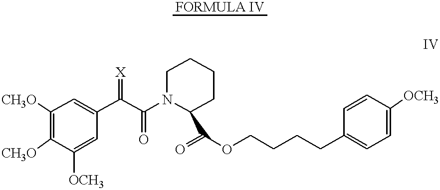
Bioorganic and Medicinal Chemistry Letters, Vol. 4, No. 9, pp. 1161-1166, 1994, incorporated herein by reference, discloses exemplary pipecolic acid derivatives represented by Formula IV and Table II.
Figure US06376517-20020423-C00006
TABLE II
Compound Structure
6 X = H, H
7 X = CH2
8 X = H, CH3
9 X = O
COMPOUND 10
Birkenshaw et al., Bioorganic & Medicinal Chemistry Letters, Vol. 4, No. 21, pp. 2501-2506, 1994, incorporated herein by reference, discloses an exemplary pipecolic acid derivative represented by Formula V.
Figure US06376517-20020423-C00007
COMPOUNDS 11-21
Holt et al., 
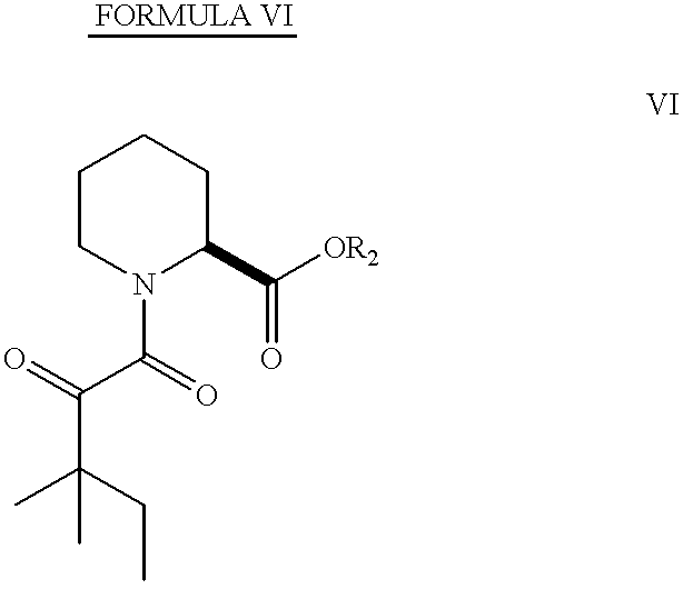
J. Am. Chem. Soc., Vol. 115, pp. 9925-9938, 1993, incorporated herein by reference, discloses exemplary pipecolic acid derivatives represented by Formula VI and Tables III and IV.
Figure US06376517-20020423-C00008
TABLE III
Compound R
2
11
Figure US06376517-20020423-C00009
12
Figure US06376517-20020423-C00010
13
Figure US06376517-20020423-C00011
14
Figure US06376517-20020423-C00012
15
Figure US06376517-20020423-C00013
16
Figure US06376517-20020423-C00014
17
Figure US06376517-20020423-C00015
18
Figure US06376517-20020423-C00016
TABLE IV
Compound Structure
19
Figure US06376517-20020423-C00017
20
Figure US06376517-20020423-C00018
21
Figure US06376517-20020423-C00019
COMPOUNDS 22-30
Caffery et al., Bioorganic & Medicinal Chemistry Letters, Vol. 4, No. 21, pp. 2507-2510, 1994, incorporated herein by reference, discloses exemplary pipecolic acid derivatives represented by Formulas VII-IX and Tables V-VII.
Figure US06376517-20020423-C00020
TABLE V
Compound Structure
22 y = 1
23 y = 2
24 y = 3
Figure US06376517-20020423-C00021
TABLE VI
Compound Structure
25 n = 1
26 n = 2
27 n = 3
Figure US06376517-20020423-C00022
TABLE VII
Compound Structure
28 n = 1
29 n = 2
30 n = 3
COMPOUND 31
Teague et al., Bioorganic & Medicinal Chemistry Letters, Vol. 3, No. 10, pp. 1947-1950, 1993, incorporated herein by reference, discloses an exemplary pipecolic acid derivative represented by Formula X.
Figure US06376517-20020423-C00023
COMPOUNDS 32-34
Yamashita et al., Bioorganic & Medicinal Chemistry Letters, Vol. 4., No. 2, pp. 325-328, 1994, incorporated herein by reference, discloses exemplary pipecolic acid derivatives represented by Formula XI and Table VIII.
Figure US06376517-20020423-C00024
TABLE VIII
Compound Structure
32 R = phenyl
33 R = N(allyl)2
34
Figure US06376517-20020423-C00025
COMPOUND 35-55
Holt et al., Bioorganic & Medicinal Chemistry Letters, Vol. 4, No. 2, pp. 315-320, 1994, incorporated herein by reference, discloses exemplary pipecolic acid derivatives represented by Formula XII and Tables IX-XI.
Figure US06376517-20020423-C00026
TABLE IX
Compound Structure
35
Figure US06376517-20020423-C00027
36
Figure US06376517-20020423-C00028
37
Figure US06376517-20020423-C00029
38
Figure US06376517-20020423-C00030
39
Figure US06376517-20020423-C00031
40
Figure US06376517-20020423-C00032
41
Figure US06376517-20020423-C00033
42
Figure US06376517-20020423-C00034
43
Figure US06376517-20020423-C00035
44
Figure US06376517-20020423-C00036
45
Figure US06376517-20020423-C00037
46
Figure US06376517-20020423-C00038
47
Figure US06376517-20020423-C00039
48
Figure US06376517-20020423-C00040
49
Figure US06376517-20020423-C00041
50
Figure US06376517-20020423-C00042
TABLE X
Compound Structure
51
Figure US06376517-20020423-C00043
52
Figure US06376517-20020423-C00044
53
Figure US06376517-20020423-C00045
TABLE XI
Compound Structure
54
Figure US06376517-20020423-C00046
55
Figure US06376517-20020423-C00047
COMPOUNDS 56-68
Holt et al., Bioorganic & Medicinal Chemistry Letters, Vol. 3, No. 10, pp. 1977-1980, 1993, incorporated herein by reference, discloses exemplary pipecolic acid derivatives represented by Formulas XIII and XIV and Tables XII-XIV.
Figure US06376517-20020423-C00048
TABLE XII
Compound Structure
56 X = OH
57 X = OMe
58 X = Oi Pr
59 X = OBn
60 X = OCH MePh
61 X = OCH2CHCHPh
62 X = OCH2CH2CH2 (3,4-OMe2) Ph
63 X = NHBn
64 X = NHCH2CH2CH2Ph
Figure US06376517-20020423-C00049
TABLE XIII
Compound Structure
65 R = Me
66 R = Bn
TABLE XIV
Compound Structure
67
Figure US06376517-20020423-C00050
68
Figure US06376517-20020423-C00051
COMPOUNDS 69-83
Hauske et al., J. Med. Chem., Vol. 35, pp. 4284-4296, 1992, incorporated herein by reference, discloses exemplary pipecolic acid derivatives represented by Formulas XV-XVIII and Tables XV-XVIII.
Figure US06376517-20020423-C00052
TABLE XV
Compound Sructure
69 n = 2
Figure US06376517-20020423-C00053
R2 = Phe-o-tert-butyl
70 n = 2
Figure US06376517-20020423-C00054
R2 = Phe-o-tert-butyl
Figure US06376517-20020423-C00055
TABLE XVI
Compound Structure
71 R1 = m-OCH3Ph
R3 = Val-O-tert-butyl
72 R1 = m-OCH3Ph
R3 = Leu-O-tert-butyl
73 R1 = m-OCH3Ph
R3 = Ileu-O-tert-butyl
74 R1 = m-OCH3Ph
R3 = hexahydro-Phe-O-tert-
butyl
75 R1 = m-OCH3Ph
R3 = allylalanine-O-tert-
butyl
76 R1 = B-naphthyl
R3 = Val-O-tert-butyl
Figure US06376517-20020423-C00056
TABLE XVII
Compound Structure
77 R1 = CH2(CO)-m-OCH3Ph
R4 = CH2Ph
R5 = OCH3
78 R1 = CH2(CO)-β-naphthyl
R4 = CH2Ph
R5 = OCH3
Figure US06376517-20020423-C00057
TABLE XVIII
Compound Structure
79 R1 = m-OCH3Ph
X = trans-CH═CH
R4 = H
Y = OC(O)Ph
80 R1 = m-OCH3Ph
X = trans-CH═CH
R4 = H
Y = OC(O)CF3
81 R1 = m-OCH3Ph
X = trans-CH═CHI
R4 = −
Y = −
82 R1 = m-OCH3Ph
X = trans-CH═CH
R4 = H
Y = OCH2CH═CH2
83 R1 = m-OCH3Ph
X = C═O
R4 = H
Y = Ph
COMPOUND 84
Teague et al., Bioorganic & Med. Chem. Letters, Vol. 4, No. 13, pp. 1581-1584, 1994, incorporated herein by reference, discloses an exemplary pipecolic acid derivative represented by Formula XIX.
Figure US06376517-20020423-C00058
COMPOUNDS 85-88
Stocks et al., Bioorganic & Med. Chem. Letters, Vol. 4, No. 12, pp. 1457-1460, 1994, incorporated herein by reference, discloses exemplary pipecolic acid derivatives represented by Formula XX and Tables XIX and XX.
TABLE XIX
Compound Structure
85
Figure US06376517-20020423-C00059
Figure US06376517-20020423-C00060
TABLE XX
Compound Structure
86 R1 = H
R2 = OMe
R3 = CH 2OMe
87 R1 = H
R2 = H
R3 = H
88 R1 = Me
R2 = H
R3 = H
COMPOUNDS 89-110
Additional exemplary pipecolic acid derivatives are represented by Formulas XXI-XXV and Tables XXI-XXV.
Figure US06376517-20020423-C00061
TABLE XXI
Compound Structure
89 R = 3,4-dichloro
90 R = 3,4,5-trimethoxy
91 R = H
92 R = 3-(2,5-Dimethoxy)phenylpropyl
93 R = 3-(3,4-Methylenedioxy)phenyl-
propyl
Figure US06376517-20020423-C00062
TABLE XXII
Compound Structure
94 R = 4-(p-Methoxy)butyl
95 R = 3-Phenylpropyl
96 R = 3-(3-Pyridyl)propyl
Figure US06376517-20020423-C00063
TABLE XXIII
Compound Structure
97 R = 3-(3-Pyridyl)propyl
98 R = 1,7-Diphenyl-4-heptyl
99 R = 4-(4-Methoxy)butyl
100 R = 1-Phenyl-6-(4-methoxyphenyl)-4-
hexyl
101 R = 3-(2,5-Dimethoxy)phenylpropyl
102 R = 3-(3,4-Methylenedioxy)phenylpropyl
103 R = 1,5-Diphenylpentyl
Figure US06376517-20020423-C00064
TABLE XXIV
Compound Structure
104 R = 4-(4-Methoxy)butyl
105 R = 3-Cyclohexylpropyl
106 R = 3-Phenylpropyl
Figure US06376517-20020423-C00065
TABLE XXV
Compound Structure
107 R = 3-Cyclohexylpropyl
108 R = 3-Phenylpropyl
109 R = 4-(4-Methoxy)butyl
110 R = 1,7-Diphenyl-4-heptyl
The names of some of the compounds identified above are provided below in Table XXVI.
TABLE XXVI
Compound Name of Species
6 4-(4-methoxyphenyl)butyl (2S)-1-[2-(3,4,5-
trimethoxyphenyl) acetyl]hexahydro-2-
pyridinecarboxylate
7 4-(4-methoxyphenyl)butyl (2S)-1-[2-(3,4,5-
trimethoxyphenyl) acryloyl]hexahydro-2-
pyridinecarboxylate
8 4-(4-methoxyphenyl)butyl (2S)-1-[2-(3,4,5-
trimethoxyphenyl) propanoyl]hexahydro-2-
pyridinecarboxylate
9 4-(4-methoxyphenyl)butyl (2S)-1-[2-oxo-2-
(3-(4,5-trimethoxyphenyl) acetyl]hexahydro-2-
pyridinecarboxylate
11 3-cyclohexylpropyl (2S)-1-(3,3-dimethyl-2-
oxopentanoyl) hexahydro-2-pyridinecarboxylate
12 3-phenylpropyl (2S) -1-(3,3-dimethyl-2-
oxopentanoyl) hexahydro-2-pyridinecarboxylate
13 3-(3,4,5-trimethoxyphenyl)propyl (2S)-1-
(3,3-dimethyl-2-oxopentanoyl) hexahydro-2-
pyridine-carboxylate
14 (1R)-2,2-dimethyl-1-phenethyl-3-butenyl
2S)-1 -(3,3-dimethyl-2-
oxopentanoyl) hexahydro-2-pyridinecarboxylate
15 (1R)-1,3-diphenylpropyl (2S)-1-(3,3-
dimethyl-2-oxopentanoyl) hexahydro-2-
pyridinecarboxylate
(1R)-1-cyclohexyl-3-phenylpropyl (2S)-1-
(3,3-dimethyl-2-oxopentanoyl)hexahydro-2-
pyridine-carboxylate
17 (1S)-1,3-diphenylpropyl (2S)-1-(3,3-
dimethyl-2-oxopentanoyl) hexahydro-2-
pyridinecarboxylate
18 (1S)-1-cyclohexyl-3-phenylpropyl (2S)-1-
(3,3-dimethyl-2-oxopentanoyl) hexahydro-2-
pyridine-carboxylate
19 (22aS) -15,15-dimethyiperhydropyrido[2,1-
c][1,9,4]dioxazacyclononadecine-1,12,16,17-
tetraone
20 (24aS)-17,17-dimethyiperhydropyrido[2,1
c][1,9,4]dioxazacyclohenicosine-1,14,18,19
tetraone
35 ethyl 1-(2-oxo-3-phenylpropanoly)-2-
piperidinecarboxylate
36 ethyl 1-pyruvoyl-2-piperidinecarboxylate
37 ethyl 1-(2-oxobutanoyl)-2-piperidine-
carboxylate
38 ethyl 1-(3-methyl-2-oxobutanoyl)-2-
piperidine-carboxylate
39 ethyl 1-(4-methyl-2-oxopentanoyl)-2-
piperidinecarboxylate
40 ethyl 1-(3,3-dimethyl-2-oxobutanoyl)-2-
piperidinecarboxylate
41 ethyl 1-(3,3-dimethyl-2-oxopentanoyl)-2-
piperidinecarboxylate
42 4-[2-(ethyloxycarbonyl)piperidino]-2,2-
dimethyl-3,4-dioxobutyl acetate
43 ethyl 1-[2-(2-hydroxytetrahydro-2H-2-
pyranyl)-2-oxoacetyl]-2-
piperidinecarboxylate
44 ethyl 1-[2-(2-methoxytetrahydro-2H-2-
pyranyl)-2-oxoacetyl]-2-
piperidinecarboxylate
45 ethyl 1-[2-(1-hydroxycyclohexyl)-2-
oxoacetyl]-2-piperidinecarboxylate
46 ethyl 1-[2-(1-methoxycyclohexyl)-2-
oxoacetyl]-2-piperidinecarboxylate
47 ethyl 1-(2-cyclohexyl-2-oxoacetyl)-2-
piperidinecarboxylate
48 ethyl 1-(2-oxo-2-piperidinoacetyl)-2-
piperidinecarboxylate
49 ethyl 1-[2-(3,4-dihydro-2H-6-pyranyl)-2-
oxoacetyl)-2-piperidinecarboxylate
50 ethyl 1-(2-oxo-2-phenylacetyl)-2-
piperidinecarboxylate
51 ethyl 1-(4-methyl-2-oxo-1-thioxcpentyl)-2-
piperidinecarboxylate
52 3-phenylpropyl 1-(2-hydroxy-3,3-
dimethylpentanoyl)-2-piperidinecarboxylate
53 (1R)-1-phenyl-3-(3,4,5-
trimethoxyphenyl)propyl 1-(3,3-
dimethylbutanoyl)-2-piperidine-carboxylate
54 (1R)-1,3-diphenylpropyl 1-(benzylsulfonyl)-
2 -piperidinecarboxylate
55 3-(3,4,5-trimethoxyphenyl)propyl
(benzylsulfonyl)-2-piperidinecarboxylate
56 1-(2-[(2R,3R,6S)-6-[(2S,3E,5E,7E,9S,11R)-
2,13-dimethoxy-3,9,11-trimethyl-12-oxo-
3,5,7-tridecatrienyl]-2-hydroxy-3-
methyltetrahydro-2H-2-pyranyl)-2-oxoacetyl)-
2-piperidine-carbcxylic acid
57 methyl 1-(2-[(2R,3R,6S)-6-[(25,3E,SE,7E,9S,
11R)-2,13-dimethoxy-3,9,11-trimethyl-12-oxo-
3,5,7-tridecatrienyl]-2-hydroxy-3-methyl-
tetrahydro-2H-2-pyranyl)-2-axoacetyl)-2-
piperidinecarbcxylate
58 isopropyl 1-(2-[(2R,3R,6S)-6-[(25,3E,5E,7E,
9S,11R)-2,13-dimethoxy-3,9,11-trimethyl-12-
oxo-3,5,7-tridecatrienyl]-2-hydroxy-3-
methyl-tetrahydro-2H-2-pyranyl)-2-
oxoacetyl)-2-piperidinecarboxylate
59 benzyl 1-(2-((2R,3R,6S)-6-((25,3E,5E,7E,9S,
11R)-2,13-dimethoxy-3,9,11-trimethyl-12-oxo-
3,5,7-tridecatrienyl]-2-hydroxy-3-methyl-
tetrahydro-2H-2-pyranyl)-2-oxoacetyl)-2-
piperidinecarboxylate
60 1-phenylethyl 1-(2-[(2R,3R,6S)-6-[(2S,3E,SE,
7E,9S,11R)-2,13-dimethoxy-3,9,11-trimethyl-
12-oxo-3,5,7-tridecatrienyl]-2-hydroxy-3-
methyl-tetrahydro-2H-2-pyranyl)-2-
oxoacetyl)-2-piperidinecarboxylate
61 (Z)-3-phenyi-2-propenyl 1-(2-[(2R,3R,6S)-6-
[(2S,3E,5E,7E,9S,11R)-2,13-dimethoxy-3,9,11
trimethyl-12-oxo-3,5,7-tridecatrienyl]-2-
hydroxy-3-methyltetrahydro-2H-2-pyranyl)-2-
oxoacetyl)-2-piperidinecarboxylate
62 3-(3,4-dimethoxyphenyl)propyl 1-(2-[(2R,3R,
6S)-6-[(25,3E,5E,7E,9S,11R)-2,13-dimethoxy
3,9,11-trimethyl-12-oxo-3,5,
tridecatrienyl]-2-hydroxy-3-
methyltetrahydro-2H-2-pyranyl)-2-oxoacetyl)-
2-piperidine-carboxylate
63 N2-benzyl-1-(2-[(2R,3R,6S)-6-
[(25,3E,5E,7E,9S,11R)-2,13-dimethoxy-
3,9,11-trimethyl-12-oxo-3,5,
tridecatrienyl]-2-hydroxy-3-methyl-
tetrahydro-2H-2-pyranyl)-2-oxoacetyl)-2-
piperidinecarboxylate
64 N2-(3-phenylpropyl)-1-(2-[(2R,3R,6S)-6-
[(25,3E,5E,7E,9S,11R)-2,13-dimethoxy-3,9,11-
trimethyl-12-oxo-3,5,7-tridecatrienyl]-2-
hydroxy-3-methyltetrahydro-2H-2-pyranyl)-2-
oxoacetyl)-2-piperidinecarboxylate.
89 (E)-3-(3,4-dichlorophenyl)-2-propenyl
(3,3-dimethyl-2-oxopentanoyl)-2-piperidine-
carboxylate
90 (E)-3-(3,4,5-trimethoxyphenyl)-2-propenyl 1-
(3,3-dimethyl-2-oxopentanoyl)-2-piperidine-
carboxylate
91 (E)-3-phenyl-2-propenyl 1-(3,3-dimethyl-2-
oxo-pentanoyl)-2-piperidinecarboxylate
92 (E)-3-((3-(2,5-dimethoxy)-phenylpropyl)-
phenyl)-2-propenyl 1-(3,3-dimethyl-2-
oxopentanoyl)-2-piperidinecarboxylate
93 (E)-3-(1,3-benzodioxol-5-yl)-2-propenyl 1-
(3,3-dimethyl-2-oxopentanoyl)-2-piperidine-
carboxylate
94 4-(4-methoxyphenyl)butyl 1-(2-oxo-2-
phenylacetyl)-2-piperidinecarboxylate
95 3-phenylpropyl 1-(2-oxo-2-phenylacetyl)-2-
piperidinecarboxylate
96 3-(3-pyridyl)propyl 1-(2-oxo-2-
phenylacetyl)-2-piperidinecarboxylate
97 3-(3-pyridyl)propyl 1-(3,3-dimethyl-2-
oxopentanoyl)-2-piperidinecarboxylate
98 4-phenyl-1-(3-phenylpropyl)butyl 1-(3,3-
dimethyl-2-oxopentanoyl)-2-piperidine-
carboxylate
99 4-(4-methoxyphenyl)butyl 1-(3,3-dimethyl-2-
oxopentanoyl)-2-piperidinecarboxylate
100 1-(4-methoxyphenethyl)-4-phenylbutyl 1-(3,3-
dimethyl-2-oxopentanoyl)-2-piperidine-
carboxylate
101 3-(2,5-dimethoxyphenyl)propyl 1-(3,3-
dimethyl-2-oxopentanoyl)-2-
piperidinecarboxylate
102 3-(1,3-benzodioxol-5-yl)propyl 1-(3,3-
dimethyl-2-oxopentanoyl)-2-piperidine-
carboxylate
103 1-phenethyl-3-phenylpropyl 1-(3,3-dimethyl-
2-oxopentanoyl)-2-piperidinecarboxylate
104 4-(4-methoxyphenyl)butyl 1-(2-cyclohexyl-2-
oxoacetyl)-2-piperidinecarboxylate
105 3-cyclohexylpropyl 1-(2-cyclohexyl-2-
oxoacetyl)-2-piperidinecarboxylate
106 3-phenylpropyl 1-(2-cyclohexyl-2-oxoacetyl)-
2-piperidinecarboxylate
107 3-cyclohexylpropyl 1-(3,3-dimethyl-2-
oxobutanoyl)-2-piperidinecarboxylate
108 3-phenylpropyl 1-(3,3-dimethyl-2-
oxobutanoyl)-2-piperidinecarboxylate
109 4-(4-methoxyphenyl)butyl 1-(3,3-dimethyl-2-
oxobutanoyl)-2-piperidinecarboxylate
110 4-phenyl-1-(3-phenylpropyl)butyl 1-(3,3-
dimethyl-2-oxobutanoyl)-2-piperidine-
carboxylate
All the compounds of Formulas I-XXV possess asymmetric centers and thus can be produced as mixtures of stereoisomers or as individual R- and S-stereoisomers. The individual stereoisomers may be obtained by using an optically active starting material, by resolving a racemic or non-racemic mixture of an intermediate at some appropriate stage of the synthesis, or by resolving the compounds of Formulas I-XXV. It is understood that the compounds of Formulas I-XXV encompass individual stereoisomers as well as mixtures (racemic and non-racemic) of stereoisomers. Preferably, S-stereoisomers are used in the pharmaceutical compositions and methods of the present invention.
Affinity for FKBP12
The compounds used in the inventive methods and pharmaceutical compositions have an affinity for the FK506 binding protein, particularly FKBP12. The inhibition of the prolyl peptidyl cis-trans isomerase activity of FKBP may be measured as an indicator of this affinity.
Ki Test Procedure
Inhibition of the peptidyl-prolyl isomerase (rotamase) activity of the compounds used in the inventive methods and pharmaceutical compositions can be evaluated by known methods described in the literature (Harding et al., Nature, 1989, 341:758-760; Holt et al. J. Am. Chem. Soc., 115:9923-9938). These values are obtained as apparent Ki's and are presented for representative compounds in TABLE XXVII.
The cis-trans isomerization of an alanine-proline bond in a model substrate, N-succinyl-Ala-Ala-Pro-Phe-p-nitroanilide, is monitored spectrophotometrically in a chymotrypsin-coupled assay, which releases para-nitroanilide from the trans form of the substrate. The inhibition of this reaction caused by the addition of different concentrations of inhibitor is determined, and the data is analyzed as a change in first-order rate constant as a function of inhibitor concentration to yield the apparent Ki values.
In a plastic cuvette are added 950 mL of ice cold assay buffer (25 mM HEPES, pH 7.8, 100 mM NaCl), 10 mL of FKBP (2.5 mM in 10 mM Tris-Cl pH 7.5, 100 mM NaCl, 1 mM dithiothreitol), 25 mL of chymotrypsin (50 mg/ml in 1 mM HCl) and 10 mL of test compound at various concentrations in dimethyl sulfoxide. The reaction is initiated by the addition of 5 mL of substrate (succinyl-Ala-Phe-Pro-Phe-para-nitroanilide, 5 mg/mL in 2.35 mM LiCl in trifluoroethanol).
The absorbance at 390 nm versus time is monitored for 90 seconds using a spectrophotometer and the rate constants are determined from the absorbance versus time data files.
TABLE XXVII
In Vitro Test Results - Formulas I-XXV
Compound K1 (μM)
6 140
9 13
11 170
12 250
13 25
15 17
19 12
36 >10,000
41 1300
50 >10,000
89 1800
90 28
91 39
92 75
93 70
94 165
95 740
96 725
97 130
98 30
99 60
100 15
101 12
102 120
103 20
104 103
105 760
106 210
107 32
108 2
109 24
110 5
Route of Administration
To effectively treat vision loss or promote vision regeneration, the compounds used in the inventive methods and pharmaceutical compositions must readily affect the targeted areas. For these purposes, the compounds are preferably administered [topically to the skin.]
Dosage
Dosage levels on the order of about 0.1 mg to about 10,000 mg of the active ingredient compound are useful in the treatment of the above conditions, with preferred levels of about 0.1 mg to about 1,000 mg. The specific dose level for any particular patient will vary depending upon a variety of factors, including the activity of the specific compound employed; the age, body weight, general health, sex and diet of the patient; the time of administration; the rate of excretion; drug combination; the severity of the particular disease being treated; and the form of administration. Typically, in vitro dosage-effect results provide useful guidance on the proper doses for patient administration. Studies in animal models are also helpful. The considerations for determining the proper dose levels are well known in the art.
The compounds can be administered with other vision or memory revitalizing agents. Specific dose levels for the other vision or memory revitalizing agents will depend upon the factors previously stated and the effectiveness of the drug combination.
EXAMPLES
The following examples are illustrative of the present invention and are not intended to be limitations thereon. Unless otherwise indicated, all percentages are based upon 100% by weight of the final composition.
Example 1 Synthesis of 3-phenyl-1-propyl (2S)-1-(3,3-dimethyl-1,2-dioxopentyl)-2-pyrrolidinecarboxylate (1) Methyl (2S)-1-(1,2-dioxo-2-methoxyethyl)-2-Pyrrolidinecarboxylate
A solution of L-proline methyl ester hydrochloride (3.08 g; 18.60 mmol) in dry methylene chloride was cooled to 0° C. and treated with triethylamine (3.92 g; 38.74 mmol; 2.1 eq). After stirring the formed slurry under a nitrogen atmosphere for 15 min, a solution of methyl oxalyl chloride (3.20 g; 26.12 mmol) in methylene chloride (45 ml) was added dropwise. The resulting mixture was stirred at 0° C. for 1.5 hour. After filtering to remove solids, the organic phase was washed with water, dried over MgSO4 and concentrated. The crude residue was purified on a silica gel column, eluting with 50% ethyl acetate in hexane, to obtain 3.52 g (88%) of the product as a reddish oil. Mixture of cis-trans amide rotamers; data for trans rotamer given. 1H NMR (CDCl3): d 1.93 (dm, 2H); 2.17 (m, 2H); 3.62 (m, 2H); 3.71 (s, 3H); 3.79, 3.84 (s, 3H total); 4.86 (dd, 1H, J=8.4, 3.3).
Methyl (2S)-1-(1,2-dioxo-3,3-dimethylpentyl)-2-pyrrolidinecarboxylate
A solution of methyl (2S)-1-(1,2-dioxo-2-methoxyethyl)-2-pyrrolidinecarboxylate (2.35 g; 10.90 mmol) in 30 ml of tetrahydrofuran (THF) was cooled to −78° C. and treated with 14.2 ml of a 1.0 M solution of 1,1-dimethylpropylmagnesium chloride in THF. After stirring the resulting homogeneous mixture at −78° C. for three hours, the mixture was poured into saturated ammonium chloride (100 ml) and extracted into ethyl acetate. The organic phase was washed with water, dried, and concentrated, and the crude material obtained upon removal of the solvent was purified on a silica gel column, eluting with 25% ethyl acetate in hexane, to obtain 2.10 g (75%) of the oxamate as a colorless oil. 1H NMR (CDCl3): d 0.88 (t, 3H); 1.22, 1.26 (s, 3H each); 1.75 (dm, 2H); 1.87-2.10 (m, 3H); 2.23 (m, 1H); 3.54 (m, 2H); 3.76 (s, 3H); 4.52 (dm, 1H, J=8.4, 3.4).
Synthesis of (2S)-1-(1,2-dioxo-3,3-dimethylpentyl)-2-pyrrolidinecarboxylic acid
A mixture of methyl (2S)-1-(1,2-dioxo-3,3-dimethylpentyl)-2-pyrrolidinecarboxylate (2.10 g; 8.23 mmol), 1 N LiOH (15 ml), and methanol (50 ml) was stirred at 0° C. for 30 minutes and at room temperature overnight. The mixture was acidified to pH 1 with 1 N HCl, diluted with water, and extracted into 100 ml of methylene chloride. The organic extract was washed with brine and concentrated to deliver 1.73 g (87%) of snow-white solid which did not require further purification. 1H NMR (CDCl3): d 0.87 (t, 3H); 1.22, 1.25 (s, 3H each); 1.77 (dm, 2H); 2.02 (m, 2H); 2.17 (m, 1H); 2.25 (m, 1H); 3.53 (dd, 2H, J=10.4, 7.3); 4.55 (dd, 1H, J=8.6, 4.1).
3-Phenyl-1-propyl (2S)-1-(3,3-dimethyl-1,2-dioxopentyl)-2-pyrrolidinecarboxylate (1)
A mixture of (2S)-1-(1,2-dioxo-3,3-dimethylpentyl)-2-pyrrolidine-carboxylic acid (600 mg; 2.49 mmol), 3-phenyl-1-propanol (508 mg; 3.73 mmol), dicyclohexylcarbodiimide (822 mg; 3.98 mmol), camphorsulfonic acid (190 mg; 0.8 mmol) and 4-dimethylaminopyridine (100 mg; 0.8 mmol) in methylene chloride (20 ml) was stirred overnight under a nitrogen atmosphere. The reaction mixture was filtered through Celite to remove solids and concentrated in vacuo, and the crude material was purified on a flash column (25% ethyl acetate in hexane) to obtain 720 mg (80%) of Example 1 as a colorless oil. 1H NMR (CDCl3): d 0.84 (t, 3H); 1.19 (s, 3H); 1.23 (s, 3H); 1.70 (dm, 2H); 1.98 (m, 5H); 2.22 (m, 1H); 2.64 (m, 2H); 3.47 (m, 2H); 4.14 (m, 2H); 4.51 (d, 1H); 7.16 (m, 3H); 7.26 (m, 2H).
FIG. 1. GPI 1046 protects retinal ganglion cells against degeneration following retinal ischemia.
Retinal ganglion cells were retrogradely labeled in adult rats by bilateral injection of fluorogold in their lateral geniculate nuclei. Labeled ganglion cells in the normal rat retina appear as white profiles against the dark background (FIG. 1A). Complete retinal ischemia was produced by infusing normal saline solution into the retinal vitreous cavity of each eye until the intraocular pressure exceeded arterial blood pressure. 28 days after the ischemic episode extensive degeneration of retinal ganglion cell was evidenced by massive reduction in the density of fluorogold labeled cells (FIG. 1B). Administration of GPI 1046 (10 mg/kg, s.c.) 1 hour prior to the ischemic episode and at 10 mg/kg/day for the next four days produced noticeable protection of a large proportion of the vulnerable ganglion cell population (FIG. 1C).
FIG. 2. GPI 1046 prevents degeneration of optic nerve axons and myelin following retinal ischemia.
Examination of the optic nerves from the same retinal ischemia cases reveals that GPI 1046 produces dramatic protection of optic nerve element from ischemic degeneration. Toluidine blue staining of epon embedded optic nerve cross sections revealed the detail of myelin sheaths (white circles) and optic nerve axons (black centers) in the normal rat optic nerve. Optic nerves from vehicle treated cases examined 28 days after a 1 hour retinal ischemic episode are characterized by a decreased density of optic nerve axons and the appearance of numerous degenerating myelin figures (bright white filled circles). Treatment with GPI 1046 protected the majority of optic nerve axons from degeneration and also dramatically decreased the density of degenerating myelin figures.
FIG. 3. GPI 1046 provides moderate protection against retinal ganglion cell death after optic nerve transection.
Complete transection of the optic nerve 5 mm from the eyeball produces massive degeneration of retinal ganglion cells, representing loss of >87% of the normal ganglion cell population 90 days after the injury (Table A). Few spared fluorogold pre labeled ganglion cells are present in vehicle treated cases (large white figures) among a population of small microglia that digest the debris of the degenerating cells and take up the fluorogold label (FIG. 3A). Treatment with GPI 1046 for 14 days resulted in a small but not significant increase in the density of retinal ganglion cells that survived 90 days after transection (Table A) but treatment with GPI 1046 for the first 28 days after transection produced moderate but significant protection of 12.6% of the vulnerable ganglion cell population (Table A, FIG. 3B).
FIG. 4. GPI 1046 treatment duration significantly affects the process of optic nerve axonal degeneration after transection.
Examination of optic nerve axon density in the proximal stump of the optic nerve from the same cases revealed a more dramatic protection afforded by GPI 1046 treatment. 90 days after transection few ganglion cell axons remain within the optic nerve (FIG. 4B), representing only 5.6% of the normal population. The loss of axons reflects both the death of retinal ganglion cells and the regression or “dying back” of the axons of ˜70% of the small surviving ganglion cell population into the retina itself (Table A). Treatment with GPI 1046 for the first 14 days after optic nerve transection produced a small but significant 5.3% protection of optic nerve axons (FIG. 4D, Table A). but treatment with the same dose of GPI 1046 for 28 days resulted in the protection of optic nerve axons for the vast majority (81.4%) of spared retinal ganglion cells (FIG. 4C, Table A).
FIG. 5. GPI 1046 treatment produces a greater effect on optic nerve axons than ganglion cell bodies.
This summary figure shows data from FIG. 3 ganglion cell protection and higher power photomicrographs of optic nerve axon protection (FIG. 5A&B, upper panels). 28 day treatment with GPI 1046 produced a significant increase in the density of large, and particularly medium and small caliber optic nerve axons (FIG. 5C&D, lower panels).
FIG. 6. GPI 1046 treatment for 28 days after optic nerve transection prevents myelin degeneration in the proximal stump.
Myelin basic protein immunohistochemistry labels fascicles (darker labeled ‘islands’) of myelinated axons in the normal optic nerve (FIG. 6A, upper left). 90 days after transection extensive degeneration of myelin is evident in vehicle treated cases, characterized by the loss of fascicular organization and the appearance of numerous large dense degenerating myelin figures (FIG. 6B, upper right). Treatment with GPI 1046 for the first 14 days after optic nerve transection did not alter the pattern of myelin degeneration (FIG. 6C, lower left panel), and yielded an insignificant 1.6% quantitative recovery in myelin density (Table A). Extending the GPI 1046 treatment course through the first 28 days after optic nerve transection produced a dramatic preservation of the fascicular staining pattern for myelin basic protein in the proximal stump of the optic nerve and decreased the density of degenerating myelin figures (FIG. 6D, lower right panel), representing a '70% recovery of myelin density (Table A).
FIG. 7. FKBP-12 immunohistochemistry labels oligodendroglia (large dark cells with fibrous processes), the cells which produce myelin, located between the fascicles of optic nerve fibers, and also some optic nerve axons.
FIG. 8. GPI 1046 treatment for 28 days after optic nerve transection prevents myelin degeneration in the distal stump.
Complete transection of the optic nerve leads to degeneration of the distal segments (axon fragments disconnected from the ganglion cell bodies), and the degeneration of their myelin sheaths. 90 days after transection (FIG. 8B) myelin basic protein immunohistochemistry reveals the near total loss of fascicular organization (present in the normal optic nerve, FIG. 8A) and the presence of numerous dense degenerating myelin figures. Quantitation reveals that the cross sectional area of the transected distal stump shrinks by 31% and loses approximately ½ of its myelin (Table A). Treatment with GPI 1046 for the first 14 days after transection did not protect against shrinkage of the distal stump but did slightly increase the density of myelin, though the density of degenerating myelin figures remained high (FIG. 8C, Table A). GPI 1046 treatment through the first 28 days produced dramatic protection of the fascicular pattern of myelin labeling, decreased the density of degenerating myelin figures, prevented cross sectional shrinkage of the distal stump of the transected nerve and maintained the myelin levels at ˜99% of normal levels (FIG. 8D, Table A).
FIG. 9. 28 day treatment with GPI 1046 treatment beginning 8 weeks after onset of streptozotocin induced diabetes decreases the extent of neovascularization in the inner and outer retina and protects neurons in the inner nuclear layer (INL) and ganglion cell layer (GCL) from degeneration.
Negative images of cresyl violet stained tangential retinal sections reveals perikarya in the three cellular layers (FIG. 9A). The retinae of streptozotocin treated animals administered only vehicle (FIG. 9B) exhibited loss of cells from the ONL and INL, decreased thickness of the Outer plexiform layer (the dark area between ONL and INL) and a dramatic increase in the size and density of retinal blood vessels (large black circular outlines) in the INL, OPL, ONL and the photoreceptor layer (PR, the gray fuzzy area above the ONL). GPI 1046 treatment reduced neovascularization (i.e. prevented the proliferation of blood vessels) in the PR, ONL, OPL and INL. Although GPI 1046 did not appear to protect against neuronal loss in the ONL, it appeared to decrease the loss of neurons in both the INL and GCL compared to streptozotocin/vehicle treated controls.
Example 2 In Vivo Retinal Ganglion Cell and Optic Nerve Axon Tests
The extent of degeneration reduction or prevention in retinal ganglion cells and optic nerve axons was determined in a vision loss model utilizing surgical optic nerve transection to simulate mechanical damage to the optic nerve. The effects of several neuroimmunophilin FKBP ligands on retinal ganglion cells neuroprotection and optic nerve axon density was determined experimentally, comparing 14 day and 28 day neuroimmunophilin FKBP ligand treatments. The effects of treatment with neuroimmunophilin FKBP ligands on retinal ganglion cells and optic nerve axons was correlated.
Surgical Procedures
Adult male Sprague Dawley rats (3 months old, 225-250 grams) were anesthetized with a ketamine (87 mg/kg) and xylazine (13 mg/kg) mixture. Retinal ganglion cells were pre-labeled by bilateral stereotaxic injection of the fluorescent retrogradely transported marker fluoro-gold (FG, 0.5 microliters of 2.5% solution in saline) at the coordinates of the LGNd (4.5 millimeters post β, 3.5 millimeters lateral, 4.6 millimeters below dura). Four days later, FG labeled rats underwent a second surgery for microsurgical bilateral intraorbital optic nerve transection 4-5 millimeters behind the orbit.
Experimental animals were divided into six experimental groups of six rats (12 eyes) per group. One group received a neuroimmunophilin FKBP ligand (10 milligrams per kg per day sc in PEG vehicle (20 percent propylene glycol, 20 percent ethanol, and 60 percent saline)) for 14 days. A second group received the same neuroimmunophilin FKBP ligand dose for 28 days. Each treated group had a corresponding sham/surgery and transection control group which received corresponding 14 or 28 day dosing with the vehicle only.
All animals were sacrificed 90 days after optic nerve transection and perfused pericardially with formalin. All eyes and optic nerves stumps were removed. Cases were excluded from the study if the optic nerve vasculature was damaged or if FG labeling was absent in the retina.
Retinal Ganglion Cell Counts
Retinas were removed from eyes and prepared for wholemount analysis. For each group, five eyes with dense and intense FG labeling were selected for quantitative analysis using a 20 power objective. Digital images were obtained from five fields in the central retina (3-4 millimeters radial to optic nerve head). FG labeled Large (>18 μm), medium (12-16 μm), and small (<10 μm) ganglion cells and microglia were counted in five 400 μm by 400 μm fields per case, 5 cases per group.
Examination of Optic Nerves
Proximal and distal optic nerve stumps were identified, measured, and transferred to 30% sucrose saline. The proximal stumps of five nerves were blocked and affixed to a chuck, and 10 micron cross sections were cut on a cryostat; one in ten sections were saved per set. Sections including the region 1-2 mm behind the orbit were reacted for RT97 neurofilament immunohistochemistry. Analysis of optic nerve axon density was performed using a 63 power oil immersion lens, a Dage 81 camera, and the Simple Image Analysis program. RT97 positive optic nerve axons were counted in three 200 μm by 200 μm fields per nerve. The area of the nerve was also determined for each case at 10 power.
As depicted graphically in Tables A&B, the 14 day course of treatment with a neuroimmunophilin FKBP ligand provided moderate neuroprotection of retinal ganglion cells observed 28 days after optic nerve transection. However, by 90 days after transection, only 5% of the ganglion cell population remained viable.
90 days after optic nerve transection the number of axons persisting in the proximal stump of the optic nerve represented approximately one half of the number of surviving ganglion cells in groups of animals that received vehicle alone or the 14 day course of treatment with a neuroimmunophilin FKBP ligand. These results indicate that over half of the transected ganglion cell axons retract beyond the optic nerve head, and that treatment with a neuroimmunophilin FKBP ligand during the first 14 days after optic nerve transection is not sufficient to arrest this retraction.
As depicted graphically in Tables A&B, more prolonged treatment with a neuroimmunophilin. FKBP ligand during the 28 day course of treatment produced a moderate increase in retinal ganglion cell neuroprotection. Approximately 12% of the vulnerable retinal ganglion cell population was protected.
A similar proportion (˜50%) of optic nerve axon density sparing was also observed. These results demonstrate the startling result that extending the duration of treatment with a neuroimmunophilin FKBP ligands to 28 days after transection completely arrests the regression of damaged axons for essentially the entire surviving population of retinal ganglion cells.
TABLE A
Effect of prologned GPI 1046 treatment on retinal ganglion cell survival,
optic nerve axon perservation, and myelination 90 days after optic nerve transection
ON head Increased Spared
RGC ON Axon area % RGCs ON axon RGC ON axon
GROUP Counts1 density2 (% sham) Rescued density3 population Count4
Sham 290 ± 14.8 7600* 100% 120,000* 120,000
ONT/Vehicle 35.9 ± 2.8 428 ± 34  68% (87% loss) 14,855 4593
ONT/14 days 49 ± 5.3 569 ± 23  76%  5.3% 1.5X 20,275 6820
GPI 1046
ONT/28 days 67.9 ± 5.8* 1526 ± 120*  95%* 12.6%* 5.0X 28,096* 22,861*
GPI 1046
% surviving Proximal optic nerve Distal optic nerve
RGCs with myclin basic protein myclin basic protein
ON axons Density5 Density6
Sham 100% normal
ONT/Vehicle 30.9% 52 ± 5.2 SEM 31% shrinkage
% loss 52.3% loss
ONT/14 days 33.6% 1.6 ± 3.0 SEM 33% shrinkage
GPI
1046 % recovery 47% loss
ONT/28 days 81.4% 70 ± 6.3 SEM 56% less shrinkage*
GPI 1046 % recovery 99% myclin
perservation*
*significance p<001
1Mean density + SEM of Fluoro-gold labeled retinal ganglion cells (RGC) in 400 μm × 400 μm sample gridfields.
2mean density + SEM of RT97 neurofilament antibody labeled optic nerve (ON) axons in 200 μm × 200 μm region of interest
*estimate for 200 μm × 200 μm region in normal optic nerve assuming 120,000 RGC axons in normal rat optic nerve, measured to be 0.630 mm2 mean cross sectional area
3adjusted for optic nerve diameter
4calculated by multiplying axonal density by ON area
5determined from 20X analysis of % areal coverage of optic nerve cross section
6shrinkage determined by comparing cross sectional area to sham control, myclin leves determined by multiplying cross sectional area by myclin density
Additional results are set forth in Tables C and D
Example 3
A patient is suffering from macular degeneration. A derivative as identified above, alone or in combination with one or more other neopsic factors, or a pharmaceutical composition comprising the same, may be administered to the patient. A reduction in vision loss, prevention or vision degeneration, and/or promotion of vision regeneration are/is expected to occur following treatment.
Example 4
A patient is suffering from glaucoma, resulting in cupping of the optic nerve disc and damage to nerve fibers. A derivative as identified above, alone or in combination with one or more other neopsic factors, or a pharmaceutical composition comprising the same, may be administered to the patient. A reduction in vision loss, prevention of vision degeneration, and/or promotion of vision regeneration are/is expected to occur following treatment.
Example 5
A patient is suffering from cataracts requiring surgery. Following surgery, a derivative as identified above, alone or in combination with one or more other neopsic factors, or a pharmaceutical composition comprising the same, may be administered to the patient. A reduction in vision loss, prevention of vision degeneration, and/or promotion of vision regeneration are/is expected to occur following treatment.
Example 6
A patient is suffering from an impairment or blockage of retinal blood supply relating to diabetic retinopathy, ischemic optic neuropathy, or retinal artery or vein blockage. A derivative as identified above, alone or in combination with one or more other neopsic factors, or a pharmaceutical composition comprising the same, may be administered to the patient. A reduction in vision loss, prevention of vision degeneration, and/or promotion of vision regeneration are/is expected to occur following treatment.
Example 7
A patient is suffering from a detached retina. A derivative as identified above, alone or in combination with one or more other neopsic factors, or a pharmaceutical compostion comprising the same, may be administered to the patient. A reduction in vision loss, prevention of vision degeneration, and/or promotion of vision regeneration are/is expected to occur following treatment.
Example 8
A patient is suffering from tissue damage caused by inflammation associated with uveitis or conjunctivitis. A derivative as identified above, alone or in combination with one or more other neopsic factors, or a pharmaceutical composition comprising the same, may be administered to the patient. A reduction in vision loss, prevention of vision degeneration, and/or promotion of vision regeneration are/is expected to occur following treatment.
Example 9
A patient is suffering from photoreceptor damage caused by chronic or acute exposure to ultraviolet light. A derivative as identified above, alone or in combination with one or more other neopsic factors, or a pharmaceutical composition comprising the same, may be administered to the patient. A reduction in vision loss, prevention of vision degeneration, and/or promotion of vision regeneration are/is expected to occur following treatment.
Example 10
A patient is suffering from optic neuritis. A derivative as identified above, alone or in combination with one or more other neopsic factors, or a pharmaceutical composition comprising the same, may be administered to the patient. A reduction in vision loss, prevention of vision degeneration, and/or promotion or vision regeneration are/is expected to occur following treatment.
Example 11
A patient is suffering from tissue damage associated with a “dry eye” disorder. A derivative as identified above, alone or in combination with one or more other neopsic factors, or a parmaceutical composition comprising the same, may be administered to the patient. A reduction in vision loss, prevention of vision degeneration, and/or promotion of vision regeneration are/is expected to occur following treatment.
Example 12
Efficacy of representative compounds from different immunophilin ligand series in protecting retinal ganglion cell axons from degeneration following optic nerve transection is set forth in Table E.
TABLE E
Efficacy of representative compounds from different immunophilin ligand series in
protecting retinal ganglion cell axons from degeneration following optic nerve transection
RT97 + RGC axon density 14 days after
Compound Structure Comments On transection (% ON axons rescued)
B
Figure US06376517-20020423-C00066
Adamantyl Thioester of urea Ki Rotamase = 149 nM Clearance = ? μl/min. 100.0% ± 5.2% SEM
A GPI
1046
Figure US06376517-20020423-C00067
Ester Ki rotamase = 7.5 nM Clearance = 63.8 μl/min. 60.5% ± 3.9 SEM
C
Figure US06376517-20020423-C00068
Sulfonamide Ki rotamase = 107 nM Clearance = 31.1 μl/min. 60.4% ± 3.1% SEM
D
Figure US06376517-20020423-C00069
Pipecolic sulfonamide Ki rotamase = nM Clearance = μl/min. 58.4% ± 6.4% SEM
E
Figure US06376517-20020423-C00070
Ester of pipecolic acid Ki rotamase = 20 nM Clearance = 41.8 μl/min. 56.6% ± +9.4% SEM
F
Figure US06376517-20020423-C00071
Proline heterocycle Analog of GPI 1046 Ki rotamase = 272 nM Clearance = ? μl/min 55.1% ± 5.9% SEM
G
Figure US06376517-20020423-C00072
Pipecolic acid dimethyl ketone Ki rotamase > 10,000 nM Clearance = ? μl/min. 34.0% ± 4.8% SEM
H
Figure US06376517-20020423-C00073
Ki rotamase = nM Clearance=? μl/min. 30.3% ± 8.0% SEM
I
Figure US06376517-20020423-C00074
Ester of Thiourea Ki rotamase = 131 nM Clearance = 8.0 μl/min. 23.8% ± 5.3 SEM
J
Figure US06376517-20020423-C00075
Ketone analog of GPI 1046 Ki rotamase = 210 nM Clearance = 1.5 μl/min. 15.8% ± 4.8% SEM
K
Figure US06376517-20020423-C00076
Pipecolic acid Thioester Ki rotamase = 86 nM Clearance = 4.5 μl/min. 13.0% ± 4.2% SEM
L
Figure US06376517-20020423-C00077
Prolyl acid Ki rotamase = >7743 nM Clearance = 5.2 μl/min. 7.8% ± 3.0% SEM
M
Figure US06376517-20020423-C00078
Thioester Ki rotamase = 7 nM Clearance = 12.5 μl/min. −6.3% ± 3.9% SEM
N
Figure US06376517-20020423-C00079
Ki rotamase = 722 nM Clearance = 21.9 μl/min.
Example 13 THE FKBP NEUROIMMUNOPHILIN LIGAND GPI-1046 ENHANCES RETINAL GANGLION CELL SURVIVAL AND ARRESTS AXONAL DYING BACK FOLLOWING OPTIC NERVE TRANSECTION
Transection of the mammalian optic nerve results in a brief period of abortive regeneration, but the majority of axotomized neurons die and the axons from many persisting ganglion cells die back beyond the optic nerve head. The present Example was designed to examine the neuroprotective effects of GPI-1046 following optic nerve transection.
Retinal ganglion cells in adult male Sprague Dawley rats were retrogradely labeled by fluorogold injection in the LGNd and four days later the optic nerves were transected 5 mm behind the globe. Groups of animals received either GPI-1046 10 mg/kg/day s.c. or vehicle for 28 days. All experimental animals and controls were sacrificed 90 days after transection.
By 90 days only—10% of the FG labeled ganglion cell population survived but less than half of these neurons maintained axons that extended past the optic nerve head, as detected with RT97 neurofilament immunohistochemistry. GPI-1046 treatment produced a moderate degree of perikaryal neuroprotection, sparing 25% of the ganglion cell population, and preserved the axons of virtually all protected neurons in the proximal stump of the transected nerve. These results indicate that treatment with the FKBP neuroimmunophilin ligand GPI-1046 produces a fundamental alteration in the pathological process following injury to CNS tracts.
These results also demonstrate that the small molecule FKBP neuroimmunophilin ligand GPI 1046 enhances neurite outgrowth in culture, enhance peripheral nerve regeneration, and stimulate sprouting within the CNS following partial deafferentation.
Example 14 NEUROIMMUNOPHILIN LIGANDS PROMOTE RECOVERY FROM THE PERIPHERAL SENSORY NEUROPATHY ASSOCIATED WITH STREPTOZOTOCIN-INDUCED DIABETES
Peripheral neuropathy is a common debilitating complication of Type 2 diabetes in some 30-40% of diabetic patients. Neurotrophic factors such as nerve growth factor (NGF) are known to promote survival of developing and adult neurons of the peripheral nervous system (PNS), and have also been evaluated as treatments for diabetic peripheral neuropathy. Some of the selective ligands of the neuroimmunophilin FKBP-12 such as the small molecule GPI-1046, have also been shown to promote repair and regeneration in the central and peripheral nervous systems (Proc. Nat'l. Acad. Sci. USA 94, 2019-2024, 1997).
In this Example the potential therapeutic effects of GPI-1046 were evaluated for its ability to improve sensory function in the streptozotocin-induced diabetic rat. The procedure involved using Male Wistar rats which were given a single injection of streptozotocin (65 mg/kg i.v.). Blood glucose levels were determined weekly for the first three weeks and on the last week of the experiment. Animals were evaluated weekly for signs of sensory neuropathy using the conventional hot plate and tail flick apparatus test procedures. After six weeks treatment either with GPI-1046 or vehicle was initiated.
The results demonstrated that behavioral testing using the hot plate and the tail flick apparatus indicated improvement in latency in lesioned animals treated for 6 weeks with GPI-1046 at 10 mg/kg s.c. The results also showed that GPI-1046 ameliorates the behavioral sequelae of diabetic sensory neuropathy and may offer some relief for patients suffering from diabetic peripheral neuropathy.
Morris Watermaze/Aging and Memory Test Procedure
Aged rodents exhibit marked individual differences in performance on a variety of behavioral tasks, including two-choice spatial discrimination in a modified T-maze, spatial discrimination in a circular platform task, passive avoidance, radial maze tasks, and spatial navigation in a water pool.
In all of these tasks, a proportion of aged rats or mice perform as well as the vast majority of young control animals, while other animals display severe impairments in memory function compared to young animals. For example, Fischer and colleagues showed that the proportion of rats displaying significant impairments in spatial navigation increases with age, (Fischer et al. 1991b) with 8% of all 12 month old, 45% of 18 month old, 53% of 24 month old, and 90% of all 30 month old rats displaying impairments in spatial acquisition of the Morris watermaze task relative to young controls.
Specifically, rodent spatial learning and memory decline during aging has been accepted by many investigators as an intriguing correlative animal model of human senile dementia. Cholinergic function in the hippocampus has been extensively studied as a component of spatial learning in rodents, and declining hippocampal cholinergic function has been noted in parallel with the development of learning and memory impairments. In addition, other neurotransmitter systems have been shown to contribute to spatial learning, and to decline with age, such as the dopaminergic and noradrenergic, serotonergic, and glutamatergic systems.
Also, reports on age-related deficits of hippocampal long-term potentiation (LTP)-induction, a reduction in theta rhythm frequency, a loss of experience-dependent plasticity of hippocampal place-units, and reductions in hippocampal protein kinase C are in keeping with the concept that no single underlying pathology can be identified as the cause of age-related behavioral impairment in rodents. However, the various experimental therapeutic approaches that have been undertaken to improve memory function in aged rodents have been somewhat slanted towards the cholinergic hypothesis.
The Morris watermaze is widely used for assessing spatial memory formation and retention in experimental animals. The test depends on the animal's ability to utilize spatial visual information in order to locate a submerged escape platform in a water tank. It is important that the tank itself be as devoid of specific visual features as possible—thus, it is always circular in shape, the sides are kept smooth and in uniform dull colors, and the water is rendered opaque with nontoxic watercolor pigment or powdered milk. This is to ensure that the animal navigates only by the use of more distant visual cues, or by the use of intra-maze cues specifically provided by the experimenter.
The tank is filled to a level which forces the animal to swim actively. Normal mice and rats react aversively to the swimming part of the test and will climb onto, and remain on, an escape platform from which they are removed to a heated resting cage.
If the platform is visible (i.e. above the surface), animals placed in the tank will quickly learn to home in on the platform and climb out onto it. Testing with a visible platform will also ensure that the experimental animals are not blind and show sufficient motivation and stamina to perform the task, which can be important in experiments involving aged rodents. If the platform is invisible (i.e. submerged just below the surface), normal animals learn to use distant visual cues in the test room for orientation in the test tank, and, when placed in the tank, will quickly home in on the approximate location of the platform and circle in that area until the platform is found.
The animals' path, speed, and swim time are tracked with a ceiling camera for later computerized analysis. Over the course of several successive trials, spatial learning can therefore be defined as a drop of distance swum, or time elapsed, from placement in the tank until escape onto the invisible platform.
The test can be adapted to assess several aspects of spatial memory: a) acquisition of a cued task, where the animal's ability to link one visual cue directly with the escape platform depends on cortical function (i.e. a ball is suspended over the escape platform and the animal learns to follow this cue to find the platform); b) acquisition of a spatial task, where the animal's ability to learn the location of a submerged escape platform based on a combination of distant visual cues is dependent upon hippocampal function (i.e. the animal learns to triangulate its position in the tank by visually aligning the paper-tower dispenser with the door and ceiling lamp); c) retention of a successfully acquired spatial task, which is predominantly dependant on cortical function (i.e. the animal must remember the spatial location of the platform over several weeks); d) a hippocampus-dependant reversal task where the animals must reacquire a new spatial platform location (i.e. the platform is moved to a new location between swim trials and the animal must abandon its previous search strategy and acquire a new one).
These different modifications of the Morris watermaze procedure can be applied in sequence to the same set of experimental animals and allow for a thorough characterization of their spatial memory performance and its decline with normal ageing. Moreover, such a series of sequential memory tests sheds some light on the functional integrity of the specific brain systems involved in the acquisition and retention of spatial memory (e.g. rats with cholinergic lesions of the hippocampus may remember a platform location acquired weeks before, but persevere over the old platform location after the platform is moved).
Example 15 EFFECTS OF CHRONIC GPI-1046 ADMINISTRATION ON SPATIAL LEARNING AND MEMORY IN AGED RODENTS
This Example shows the effects of chronic treatment with the systemically available FKBP-ligand GPI-1046 on spatial learning and memory in aged rodents.
The procedure involved using three-month old (young) and 18-19 month old male C57BL/6N-Nia (aged) mice which habituated to the well known and conventional Morris watermaze during a 4 trials/day, 3-4 day visible platform training phase. Subsequent spatial acquisition testing was conducting as follows: All mice were given 4 trials/day (block), for 5 days. Maximum swim time was 90 seconds. Aged mice were allocated to an “aged impaired” group if their performance during blocks 4 or 5 of the acquisition phase was >1 S.D. above the mean of “young” mice, and to an “aged non-impaired” group if their performance was <0.5 S.D. above the mean of “young” mice. Aged groups were then split into statistically similar “GPI-1046” and “vehicle” groups.
Daily treatment with 10 mg/kg GPI-1046 was initiated 3 days after the end of acquisition training, and continued through retention testing. Retention testing began after 3 weeks of dosing using the same methods as the acquisition phase. Swim Distances (cm) were analyzed in a 7×5 ANOVA including Groups and Blocks (1-5) as factors in the analysis, treating Blocks as a repeated measure.
The results showed that planned contrasts revealed that there were significant differences between the “young”, and “aged impaired-vehicle and GPI-1046” treated groups at the end of the acquisition phase, F1.58=26.75, P=0.0001, and F1.58=17.70, P=0.0001 respectively. While there were no significant differences between the two “aged impaired” groups, F1.58=0.67, P=0.42. During retention testing, however, “aged impaired-vehicle” treated animals performed significantly poorer than “aged impaired—GPI-1046”, and “young” animals, F1.69=8.11, P=0.006, and F1.69=25.45, P=0.0001 respectively. There was no longer any statistically significant difference between the “young” and “aged impaired”—GPI-1046” treated groups during the retention phase, F1.69=3.09, P=0.08. In summary, systemic treatment with GPI-1046 significantly enhanced spatial memory performance of mice with age-related spatial memory impairments.
The invention being thus described, it will be obvious that the same may be varied in many ways. Such variations are not to be regarded as a departure from the spirit and scope of the invention and all such modifications are intended to be included within the scope of the following claims.

Claims (26)

We claim:
1. A method for treating a nerve-related vision disorder, improving vision, treating memory impairment, or enhancing memory performance in an animal, which comprises administering to said animal an effective amount of a compound selected from the group consisting of:
Figure US06376517-20020423-C00080
wherein n is 1; 2; or 3;
4-(4-methoxyphenyl)butyl (2S)-1-[2-(3,4,5-trimethoxyphenyl)acetyl]hexahydro-2-pyridinecarboxylate;
4-(4-methoxyphenyl)butyl (2S)-1-[2-(3,4,5-trimethoxyphenyl)acryloyl]hexahydro-2-pyridinecarboxylate;
4-(4-methoxyphenyl)butyl (2S)-1-[2-(3,4,5-trimethoxyphenyl)propanoyl]hexahydro-2-pyridinecarboxylate;
4-(4-methoxyphenyl)butyl (2S)-1-[2-oxo-2-(3,4,5-trimethoxyphenyl)acetyl]hexahydro-2-pyridinecarboxylate;
Figure US06376517-20020423-C00081
 3-cyclohexylpropyl (2S)-1-(3,3-dimethyl-2-oxopentanoyl)hexahydro-2-pyridinecarboxylate;
3-phenylpropyl (2S)-1-(3,3-dimethyl-2-oxopentanoyl)hexahydro-2-pyridinecarboxylate;
3-(3,4,5-trimethoxyphenyl)propyl (2S)-1-(3,3-dimethyl-2-oxopentanoyl)hexahydro-2-pyridinecarboxylate;
(1R)-2,2-dimethyl-1-phenethyl-3-butenyl (2S)-1-(3,3-dimethyl-2-oxopentanoyl)hexahydro-2-pyridinecarboxylate;
(1R)-1,3-diphenylpropyl (2S)-1-(3,3-dimethyl-2-oxopentanoyl)hexahydro-2-pyridinecarboxylate;
(1R)-1-cyclohexyl-3-phenylpropyl (2S)-1-(3,3-dimethyl-2-oxopentanoyl)hexahydro-2-pyridinecarboxylate;
(1S)-1,3-diphenylpropyl (2S)-1-(3,3-dimethyl-2-oxopentanoyl)hexahydro-2-pyridinecarboxylate;
(1S)-1-cyclohexyl-3-phenylpropyl (2S)-1-(3,3-dimethyl-2-oxopentanoyl)hexahydro-2-pyridinecarboxylate;
(22 a S)-15,15-dimethylperhydropyrido[2,1-c][1,9,4]dioxazacyclononadecine-1,12,16,17-tetraone;
(24 a S)-17,17-dimethylperhydropyrido[2,1-c][1,9,4]dioxazacyclohenicosine-1,14,18,19-tetraone;
Figure US06376517-20020423-C00082
(3R,4R,23aS)-8-allyl-4,10-dimethyl-3-[2-(3-pyridyl)ethyl]-1,3,4,5,6,7,8,11,12,15,16,17,18,20,21,22,23,23a-octadecahydro-14H-pyrido[2,1-c][1,10,4]dioxazacycloicosine-1,7,14,17,18-pentaone;
(3R,4R,24aS)-8-allyl-4,10-dimethyl-3-[2-(3-pyridyl)ethyl]-1,3,4,5,6,7,8,11,12,14,15,16,17,18,19,21,22,23,24,24a-icosahydropyrido[2,1-c][1,11,4]dioxazacyclohenicosine-1,7,14,18,19-pentaone;
(3R,4R,25aS)-8-allyl-4,10-dimethyl-3-[2-(3-pyridyl)ethyl]-1,3,4,5,6,7,8,11,12,15,16,17,18,19,20,22,23,24,25,25a-icosahydro-14H-pyrido[2,1-c][1,12,4]dioxazacyclodocosine-1,7,14,19,20-pentaone;
Figure US06376517-20020423-C00083
 wherein n is 1; 2; or 3;
Figure US06376517-20020423-C00084
 wherein n is 1; 2; or 3;
Figure US06376517-20020423-C00085
(1R)-1-(3-benzoylphenyl)-3-phenylpropyl (1R)-2-(3,3-dimethyl-2-oxopentanoyl)cyclohexane-1-carboxylate;
(1R)-1-[3-(diallylcarbamoyl)phenyl]-3-phenylpropyl;
(1R)-2-(3,3-dimethyl-2-oxopentanoyl)cyclohexane-1-carboxylate;
Figure US06376517-20020423-C00086
 ethyl 1-(2-oxo-3-phenylpropanoyl)-2-piperidinecarboxylate;
ethyl 1-pyruvoyl-2-piperidinecarboxylate;
ethyl 1-(2-oxobutanoyl)-2-piperidinecarboxylate;
ethyl 1-(3-methyl-2-oxobutanoyl)-2-piperidinecarboxylate;
ethyl 1-(4-methyl-2-oxopentanoyl)-2-piperidinecarboxylate;
ethyl 1-(3,3-dimethyl-2-oxobutanoyl)-2-piperidinecarboxylate;
ethyl 1-(3,3-dimethyl-2-oxopentanoyl)-2-piperidinecarboxylate;
4-[2-(ethyloxycarbonyl)piperidino]-2,2-dimethyl-3,4-dioxobutyl acetate;
ethyl 1-[2-(2-hydroxytetrahydro-2H-2-pyranyl)-2-oxoacetyl]-2-piperidinecarboxylate;
ethyl 1-[2-(2-methoxytetrahydro-2H-2-pyranyl)-2-oxoacetyl]-2-piperidinecarboxylate;
ethyl 1-[2-(1-hydroxycyclohexyl)-2-oxoacetyl]-2-piperidinecarboxylate;
ethyl 1-[2-(1-methoxycyclohexyl)-2-oxoacetyl]-2-piperidinecarboxylate;
ethyl 1-(2-cyclohexyl-2-oxoacetyl)-2-piperidinecarboxylate;
ethyl 1-(2-oxo-2-piperidinoacetyl)-2-piperidinecarboxylate;
ethyl 1-[2-(3,4-dihydro-2H-6-pyranyl)-2-oxoacetyl)-2-piperidinecarboxylate;
ethyl 1-(2-oxo-2-phenylacetyl)-2-piperidinecarboxylate;
ethyl 1-(4-methyl-2-oxo-1-thioxopentyl)-2-piperidinecarboxylate;
3-phenylpropyl 1-(2-hydroxy-3,3-dimethylpentanoyl)-2-piperidinecarboxylate;
(1R)-1-phenyl-3-(3,4,5-trimethoxyphenyl)propyl 1-(3,3-dimethylbutanoyl)-2-piperidinecarboxylate;
(1R)-1,3-diphenylpropyl 1-(benzylsulfonyl)-2-piperidinecarboxylate; 3-(3,4,5-trimethoxyphenyl)propyl 1-(benzylsulfonyl)-2-piperidinecarboxylate;
1-(2-[(2R,3R,6S)-6-[(2S,3E,5E,7E,9S,11R)-2,13-dimethoxy-3,9,11-trimethyl-12-oxo-3,5,7-tridecatrienyl]-2-hydroxy-3-methyltetrahydro-2H-2-pyranyl)-2-oxoacetyl)-2-piperidinecarboxylic acid;
methyl 1-(2-[(2R,3R,6S)-6-[(2S,3E,5E,7E,9S,11R)-2,13-dimethoxy-3,9,11-trimethyl-12-oxo-3,5,7-tridecatrienyl]-2-hydroxy-3-methyltetrahydro-2H-2-pyranyl)-2-oxoacetyl)-2-piperidinecarboxylate;
isopropyl 1-(2-[(2R,3R,6S)-6-[(2S,3E,5E,7E,9S,11R)-2,13-dimethoxy-3,9,11-trimethyl-12-oxo-3,5,7-tridecatrienyl]-2-hydroxy-3-methyltetrahydro-2H-2-pyranyl)-2-oxoacetyl)-2-piperidinecarboxylate;
benzyl 1-(2-[(2R,3R,6S)-6-[(2S,3E,5E,7E,9S,11R)-2,13-dimethoxy-3,9,11-trimethyl-12-oxo-3,5,7-tridecatrienyl]-2-hydroxy-3-methyltetrahydro-2H-2-pyranyl)-2-oxoacetyl)-2-piperidinecarboxylate;
1-phenylethyl 1-(2-[(2R,3R,6S)-6-[(2S,3E,5E,7E,9S,11R)-2,13-dimethoxy-3,9,11-trimethyl-12-oxo-3,5,7-tridecatrienyl]-2-hydroxy-3-methyltetrahydro-2H-2-pyranyl)-2-oxoacetyl)-2-piperidinecarboxylate;
(Z)-3-phenyl-2-propenyl 1-(2-[(2R,3R,6S)-6-[(2S,3E,5E,7E,9S,11R)-2,13-dimethoxy-3,9,11-trimethyl-12-oxo-3,5,7-tridecatrienyl]-2-hydroxy-3-methyltetrahydro-2H-2-pyranyl)-2-oxoacetyl)-2-piperidinecarboxylate;
3-(3,4-dimethoxyphenyl)propyl 1-(2-[(2R,3R,6S)-6-[(2S,3E,5E,7E,9S,11R)-2,13-dimethoxy-3,9,11-trimethyl-12-oxo-3,5,7-tridecatrienyl]-2-hydroxy-3-methyltetrahydro-2H-2-pyranyl)-2-oxoacetyl)-2-piperidinecarboxylate;
N2-benzyl-1-(2-[(2R,3R,6S)-6-[(2S,3E,5E,7E,9S,11R)-2,13-dimethoxy-3,9,11-trimethyl-12-oxo-3,5,7-tridecatrienyl]-2-hydroxy-3-methyltetrahydro-2H-2-pyranyl)-2-oxoacetyl)-2-piperidinecarboxylate;
N2-(3-phenylpropyl)-1-(2-[(2R,3R,6S)-6-[(2S,3E,5E,7E,9S,11R)-2,13-dimethoxy-3,9,11-trimethyl-12-oxo-3,5,7-tridecatrienyl]-2-hydroxy-3-methyltetrahydro-2H-2-pyranyl)-2-oxoacetyl)-2-piperidinecarboxylate;
Figure US06376517-20020423-C00087
 wherein R is methyl (Me); or benzyl (Bn);
Figure US06376517-20020423-C00088
Figure US06376517-20020423-C00089
 wherein n=2,
R1=
Figure US06376517-20020423-C00090
 and
R2=Phe-o-tert-butyl;
Figure US06376517-20020423-C00091
 wherein
R1 = m-OCH3Ph, R3 = Val-o-tert-butyl; R1 = m-OCH3Ph, R3 = Leu-o-tert-butyl; R1 = m-OCH3Ph, R3 = Ileu-o-tert-butyl; R1 = m-OCH3Ph, R3 = hexahydro-Phe-o-tert-butyl; R1 = m-OCH3Ph, R3 = allylalanine-o-tert-butyl; R1 = B-naphthul, R3 = Val-o-tert-butyl;
Figure US06376517-20020423-C00092
 wherein
R1=CH2(CO)—m-OCH3Ph
R4=CH2Ph
R5=OCH3;
 or
R1=CH2(CO)—B-naphthyl
R4=CH2Ph
R5=OCH3;
Figure US06376517-20020423-C00093
 wherein
R1=m—OCH3Ph
X=trans-CH═CH
R4=H
Y=OC(o)Ph;
R1=OCH3Ph
X=trans-CH═CH
R4=H
Y=OC(o)CF3;
R1=m-OCH3Ph
X=trans-CH═CHI
R4=—
Y=—
R1=m—OCH3Ph
X=trans-CH═CH
R4=H
Y=OCH2CH═CH2;
R1=m—OCH3Ph
X=C═O
R4=H
Y=Ph;
Figure US06376517-20020423-C00094
 wherein
R1 = H, R2 = OMe, and R3 = CH2OMe; R1 = H, R2 = H, and R3 = H; R1 = Me, R2 = H, and R3 = H;
(E)-3-(3,4-dichlorophenyl)-2-propenyl 1-(3,3-dimethyl-2-oxopentanoyl)-2-piperidinecarboxylate;
(E)-3-(3,4,5-trimethoxyphenyl)-2-propenyl 1-(3,3-dimethyl-2-oxopentanoyl)-2-piperidinecarboxylate;
(E)-3-phenyl-2-propenyl 1-(3,3-dimethyl-2-oxopentanoyl)-2-piperidinecarboxylate;
(E)-3-((3-(2,5-dimethoxy)-phenylpropyl)phenyl)-2-propenyl 1-(3,3-dimethyl-2-oxopentanoyl)-2-piperidinecarboxylate;
4-(4-methoxyphenyl)butyl 1-(2-oxo-2-phenylacetyl)-2-piperidinecarboxylate;
3-phenylpropyl 1-(2-oxo-2-phenylacetyl)-2-piperidinecarboxylate;
3-(3-pyridyl)propyl 1-(2-oxo-2-phenylacetyl)-2-piperidinecarboxylate;
3-(3-pyridyl)propyl 1-(3,3-dimethyl-2-oxopentanoyl)-2-piperidinecarboxylate;
4-phenyl-1-(3-phenylpropyl)butyl 1-(3,3-dimethyl-2-oxopentanoyl)-2-piperidinecarboxylate;
4-(4-methoxyphenyl)butyl 1-(3,3-dimethyl-2-oxopentanoyl)-2-piperidinecarboxylate;
1-(4-methoxyphenethyl)-4-phenylbutyl 1-(3,3-dimethyl-2-oxopentanoyl)-2-piperidinecarboxylate;
3-(2,5-dimethoxyphenyl)propyl 1-(3,3-dimethyl-2-oxopentanoyl)-2-piperidinecarboxylate;
3-(1,3-benzodioxol-5-yl)propyl 1-(3,3-dimethyl-2-oxopentanoyl)-2-piperidinecarboxylate;
1-phenethyl-3-phenylpropyl 1-(3,3-dimethyl-2-oxopentanoyl)-2-piperidinecarboxylate;
4-(4-methoxyphenyl)butyl 1-(2-cyclohexyl-2-oxoacetyl)-2-piperidinecarboxylate;
3-cyclohexylpropyl 1-(2-cyclohexyl-2-oxoacetyl)-2-piperidinecarboxylate;
3-phenylpropyl 1-(2-cyclohexyl-2-oxoacetyl)-2-piperidinecarboxylate;
3-cyclohexylpropyl 1-(3,3-dimethyl-2-oxobutanoyl)-2-piperidinecarboxylate;
3-phenylpropyl 1-(3,3-dimethyl-2-oxobutanoyl)-2-piperidinecarboxylate;
4-(4-methoxyphenyl)butyl 1-(3,3-dimethyl-2-oxobutanoyl)-2-piperidinecarboxylate;
4-phenyl-1-(3-phenylpropyl)butyl 1-(3,3-dimethyl-2-oxobutanoyl)-2-piperidinecarboxylate;
Way-124,666;
rapamycin;
Rap-Pa; and SLB-506,
or a pharmaceutically acceptable salt, ester, or solvate thereof,
wherein the nerve-related vision disorder is selected from the group consisting of visual impairments; orbital disorders; disorders of the lacrimal apparatus; disorders of the eyelids; disorders of the conjunctiva; disorders of the cornea; cataract; disorders of the uveal tract; disorders of the retina; disorders of the optic nerve or visual pathways; free radical induced eye disorders and diseases; immunologically-mediated eye disorders and diseases; eye injuries; and symptoms and complications of eye disease, eye disorder, and eye injury.
2. The method of claim 1, wherein the compound is Way-124,666.
3. The method of claim 1, wherein the compound is rapamycin.
4. The method of claim 1, wherein the compound is Rap-Pa.
5. The method of claim 1, wherein the compound is SLB-506.
6. The method of claim 1, which is for improving naturally-occurring vision in an animal, in the absence of any ophthalmologic disorder, disease, or injury.
7. The method of claim 1, wherein the compound is administered to said animal in combination with an effective amount of one or more factor(s) useful in treating vision disorders, improving vision, treating memory impairment, or enhancing memory performance in an animal.
8. The method of claim 7, wherein the one or mor factor(s) is/are selected from the group consisting of immunosuppressants for treating autoimmune, inflammatory, and immunologically-mediated disorders; wound healing agents for treating wounds resulting from injury or surgery; antiglaucomatous medications fro treating abnormally elevated intraocular pressure; neurotrophic factors and growth factors fro treating neurodegenrative disorders or stimulating neurite outgrowth; compounds effective in limiting or preventing hemorrhage or neovascularization for treating macular degeneration; and axtioxidants fro treating oxidative damage to eye tissues.
9. The method of claim 1, wherein the nerve-related vision disorder is retinal ischemia.
10. The method of claim 9, wherein the retinal ischemia is selected from the group consisting of degeneration of retinal ganglion cells, degeneration of optic nerve axons, degeneration of myelin sheaths, ischemic optic neuropathy, and retinal vascular blockage.
11. The method of claim 1, wherein the nerve-related vision disorder is optic nerve transection.
12. The method of claim 11, wherein the optic nerve transection is selected from the group consisting of ganglion cell death after optic nerve transection and myelin degeneration after optic nerve transection.
13. The method of claim 1, wherein the nerve-related vision disorder is diabetes.
14. The method of claim 13, wherein the diabetes is selected from the group consisting of diabetes from degeneration and diabetic retinopathy.
15. The method of claim 1, wherein the nerve-related vision disorder is macular degeneration.
16. The method of claim 1, wherein the nerve-related vision disorder is glaucoma related degeneration.
17. The method of claim 1, wherein the nerve-related vision disorder is cataract related degeneration.
18. The method of claim 1, wherein the nerve-related vision disorder is a detached retina.
19. The method of claim 1, wherein the nerve-related vision disorder is inflammation related degeneration.
20. The method of claim 1, wherein the nerve-related vision disorder is photoreceptor degeneration.
21. The method of claim 1, wherein the nerve-related vision disorder is optic neuritis.
22. The method of claim 1, wherein the nerve-related vision disorder is dry eye degeneration.
23. The method of claim 1, wherein the compound has an affinity for an FKBP-type immunophilin.
24. The method of claim 23, wherein the FKBP-type immunophilin is FKBP-12.
25. The method of claim 1, wherein the compound is immunosuppressive.
26. The method of claim 1, wherein the compound is non-immunosuppressive.
US09/134,417 1998-08-14 1998-08-14 Pipecolic acid derivatives for vision and memory disorders Expired - Lifetime US6376517B1 (en)

Priority Applications (8)

Application Number Priority Date Filing Date Title
US09/134,417 US6376517B1 (en) 1998-08-14 1998-08-14 Pipecolic acid derivatives for vision and memory disorders
JP2000564612A JP2002522485A (en) 1998-08-14 1999-08-12 Pipecolic acid derivatives for visual and memory disorders
PCT/US1999/018242 WO2000009109A2 (en) 1998-08-14 1999-08-12 Pipecolic acid derivatives for vision and memory disorders
AU55557/99A AU5555799A (en) 1998-08-14 1999-08-12 Pipecolic acid derivatives for vision and memory disorders
CA002344520A CA2344520A1 (en) 1998-08-14 1999-08-12 Pipecolic acid derivatives for vision and memory disorders
MXPA01001642A MXPA01001642A (en) 1998-08-14 1999-08-12 Pipecolic acid derivatives for vision and memory disorders.
EP99942109A EP1109554A2 (en) 1998-08-14 1999-08-12 Pipecolic acid derivatives for vision and memory disorders
JP2011018408A JP2011088934A (en) 1998-08-14 2011-01-31 Pipecolic acid derivative for disorder of eyesight and memory

Applications Claiming Priority (1)

Application Number Priority Date Filing Date Title
US09/134,417 US6376517B1 (en) 1998-08-14 1998-08-14 Pipecolic acid derivatives for vision and memory disorders

Publications (1)

Publication Number Publication Date
US6376517B1 true US6376517B1 (en) 2002-04-23

Family

ID=22463300

Family Applications (1)

Application Number Title Priority Date Filing Date
US09/134,417 Expired - Lifetime US6376517B1 (en) 1998-08-14 1998-08-14 Pipecolic acid derivatives for vision and memory disorders

Country Status (7)

Country Link
US (1) US6376517B1 (en)
EP (1) EP1109554A2 (en)
JP (2) JP2002522485A (en)
AU (1) AU5555799A (en)
CA (1) CA2344520A1 (en)
MX (1) MXPA01001642A (en)
WO (1) WO2000009109A2 (en)

Cited By (23)

* Cited by examiner, † Cited by third party
Publication number Priority date Publication date Assignee Title
WO2004026246A2 (en) * 2002-09-17 2004-04-01 New York University Methods of treating age associated memory impairment (aami), mild cognitive impairment (mci), and dementias with cell cycle inhibitors
US20040198763A1 (en) * 2003-01-16 2004-10-07 Sucampo Ag Method of treating dry eye with a macrolide compound
US20050064010A1 (en) * 2003-09-18 2005-03-24 Cooper Eugene R. Transscleral delivery
US6872383B2 (en) 1999-04-30 2005-03-29 Sucampo Ag Use of macrolide compounds for the treatment of dry eye
US20050187241A1 (en) * 2002-09-18 2005-08-25 Rong Wen Method of inhibiting choroidal neovascularization
US20050281861A1 (en) * 2004-04-30 2005-12-22 Allergan, Inc. Macromolecule-containing sustained release intraocular implants and related methods
WO2006086750A1 (en) 2005-02-09 2006-08-17 Macusight, Inc. Liquid formulations for treatment of diseases or conditions
US20060258668A1 (en) * 2003-05-29 2006-11-16 Scolnick Edward M Use of phosphatase inhibitors as adjunct therapy for psychiatric disorders
US20060257450A1 (en) * 2005-03-21 2006-11-16 Sreenivasu Mudumba Drug delivery systems for treatment of diseases or conditions
US20070059336A1 (en) * 2004-04-30 2007-03-15 Allergan, Inc. Anti-angiogenic sustained release intraocular implants and related methods
US20070203173A1 (en) * 2006-02-09 2007-08-30 Sreenivasu Mudumba Stable formulations, and methods of their preparation and use
US20070265294A1 (en) * 2006-03-23 2007-11-15 Kleinman David M Formulations and methods for vascular permeability-related diseases or conditions
US20090074786A1 (en) * 2005-02-09 2009-03-19 Macusight, Inc. Formulations for treating ocular diseases and conditions
US20110034448A1 (en) * 2004-04-30 2011-02-10 Allergan, Inc. Carbonic anhydrase inhibitor sustained release intraocular drug delivery systems
EP2446890A1 (en) 2007-04-30 2012-05-02 Allergan, Inc. High viscosity macromolecular compositions for treating ocular conditions
WO2014201405A1 (en) 2013-06-14 2014-12-18 The Board Of Regents Of The University Of Texas System Novel allosteric inhibitors of proteasome and methods of use thereof
WO2018005777A1 (en) 2016-06-30 2018-01-04 Durect Corporation Depot formulations
WO2018126049A1 (en) 2016-12-30 2018-07-05 Durect Corporation Depot formulations
US10682340B2 (en) 2016-06-30 2020-06-16 Durect Corporation Depot formulations
US11020383B2 (en) 2017-04-20 2021-06-01 The Board Of Regents Of The University Of Texas System Methods for the treatment of cancer metastasis
US11345659B2 (en) 2018-01-30 2022-05-31 Board Of Regents, The University Of Texas System Pipecolic esters for inhibition of the proteasome
US11612590B2 (en) * 2013-03-13 2023-03-28 Santen Pharmaceutical Co., Ltd. Therapeutic agent for meibomian dysfunction
US11644460B2 (en) 2010-12-28 2023-05-09 Revolution Medicines, Inc. Identifying new therapeutic agents

Families Citing this family (11)

* Cited by examiner, † Cited by third party
Publication number Priority date Publication date Assignee Title
CN1224420C (en) * 1998-12-24 2005-10-26 苏坎波公司 Agent for treating visual cell function disorder
US6864232B1 (en) 1998-12-24 2005-03-08 Sucampo Ag Agent for treating visual cell function disorder
TR200103119T2 (en) * 1999-04-30 2002-04-22 Sucampo Ag Substance for the treatment of dry eye.
CN1173960C (en) * 2001-12-06 2004-11-03 中国人民解放军军事医学科学院毒物药物研究所 Substituted hexa azacylo compounds and their use as neuroregulator
AU2005319427B2 (en) 2004-12-20 2011-11-03 Wyeth Rapamycin derivatives and the uses thereof in the treatment of neurological disorders
PE20060765A1 (en) 2004-12-20 2006-10-02 Wyeth Corp RAPAMYCIN ANALOGS
EP2589383A1 (en) 2011-11-06 2013-05-08 Max-Planck-Gesellschaft zur Förderung der Wissenschaften e.V. Berlin FKBP subtype-specific rapamycin analogue for use in treatment of diseases
EP2610245A1 (en) * 2011-12-28 2013-07-03 Max-Planck-Gesellschaft zur Förderung der Wissenschaften e.V. Pipecolate-sulfonamides for treatment of psychiatric disorders
GB201312318D0 (en) * 2013-07-09 2013-08-21 Isomerase Therapeutics Ltd Novel methods and compounds
KR102373289B1 (en) 2016-05-25 2022-03-10 산텐 세이야꾸 가부시키가이샤 Use of sirolimus for the treatment of exudative age-related macular degeneration with persistent edema
EP3773571A1 (en) 2018-03-28 2021-02-17 Georg-August-Universitaet Goettingen Stiftung Oeffentlichen Rechts, Universitaetsmedizin Prevention or treatment of chronic organ injury

Citations (93)

* Cited by examiner, † Cited by third party
Publication number Priority date Publication date Assignee Title
US4070361A (en) 1977-04-21 1978-01-24 E. R. Squibb & Sons, Inc. Mercaptoalkylsulfonyl proline and pipecolic acid and esters thereof
EP0012401A1 (en) 1978-12-11 1980-06-25 Merck & Co. Inc. Carboxyalkyl dipeptide derivatives, process for preparing them and pharmaceutical composition containing them
US4310461A (en) 1980-06-23 1982-01-12 E. R. Squibb & Sons, Inc. Imido, amido and amino derivatives of mercaptoacyl prolines and pipecolic acids
EP0048159A2 (en) 1980-09-17 1982-03-24 University Of Miami Novel carboxyalkyl peptides and thioethers and ethers of peptides as antihypertensive agents
EP0050800A1 (en) 1980-10-23 1982-05-05 Schering Corporation Carboxyalkyl dipeptides, processes for their production and pharmaceutical compositions containing them
EP0073143A2 (en) 1981-08-21 1983-03-02 James Walter Ryan Novel complex amido and imido derivatives of carboxyalkyl peptides and thioethers and ethers of peptides
US4390695A (en) 1980-06-23 1983-06-28 E. R. Squibb & Sons, Inc. Imido, amido and amino derivatives of mercaptoacyl prolines and pipecolic acids
EP0088350A1 (en) 1982-03-08 1983-09-14 Schering Corporation Carboxyalkyl dipeptides, processes for their production and pharmaceutical compositions containing them
US4531964A (en) 1982-09-13 1985-07-30 Nippon Kayaku Kabushiki Kaisha Heterocyclic compound and a herbicidal composition containing said compound
US4574079A (en) 1983-05-27 1986-03-04 Gavras Haralambos P Radiolabeled angiotensin converting enzyme inhibitors for radiolabeling mammalian organ sites
US4578474A (en) 1980-06-23 1986-03-25 E. R. Squibb & Sons, Inc. Imido, amido and amino derivatives of mercaptoacyl prolines and pipecolic acids
US4593102A (en) 1984-04-10 1986-06-03 A. H. Robins Company, Inc. N-[(amino)alkyl]-1-pyrrolidine, 1-piperidine and 1-homopiperidinecarboxamides (and thiocarboxamides) with sulfur linked substitution in the 2, 3 or 4-position
EP0196841A1 (en) 1985-03-22 1986-10-08 Syntex (U.S.A.) Inc. N,N-dialkylguanidino dipeptides
EP0260118A1 (en) 1986-09-10 1988-03-16 Syntex (U.S.A.) Inc. Selective amidination of diamines
WO1988009789A2 (en) 1987-06-03 1988-12-15 Polifarma S.P.A. 3-indolepyruvic acid derivatives and pharmaceutical use thereof
US4808573A (en) 1980-10-23 1989-02-28 Schering Corporation Carboxyalkyl dipeptides and anti-hypertensive use thereof
US4839342A (en) 1987-09-03 1989-06-13 University Of Georgia Research Foundation, Inc. Method of increasing tear production by topical administration of cyclosporin
EP0333174A2 (en) 1988-03-16 1989-09-20 Fujisawa Pharmaceutical Co., Ltd. Peptide compounds, processes for preparation thereof and pharmaceutical composition comprising the same
EP0352000A2 (en) 1988-07-08 1990-01-24 Smithkline Beecham Corporation Retroviral protease binding peptides
EP0378318A1 (en) 1989-01-11 1990-07-18 Merck & Co. Inc. Process for synthesis of FK-506 and tricarbonyl intermediates
WO1990012805A1 (en) 1989-04-15 1990-11-01 Zaidan Hojin Biseibutsu Kagaku Kenkyu Kai Postostatin and related compound thereof, or their salts
EP0405994A2 (en) 1989-06-30 1991-01-02 Merck & Co. Inc. Synthetic process for FK-506 type macrolide intermediates
EP0419049A1 (en) 1989-08-22 1991-03-27 Merck Frosst Canada Inc. (Quinolin-2-ylmethoxy) indoles as inhibitors of the biosynthesis of leukotrienes
WO1991004985A1 (en) 1989-09-29 1991-04-18 Pharma Mar S.A. Dehydrodidemnin b
US5011844A (en) 1988-08-26 1991-04-30 Sandoz Ltd. Substituted 4-azatricyclo(22.3.1.04,9)octacos-18-ene derivatives, their preparation and pharmaceutical compositions containing them
WO1991013088A1 (en) 1990-02-28 1991-09-05 President And Fellows Of Harvard College Terminally blocked antiviral peptides
WO1992000278A1 (en) 1990-07-02 1992-01-09 Vertex Pharmaceuticals Incorporated Novel immunosuppressive compounds
EP0468339A2 (en) 1990-07-27 1992-01-29 Nippon Kayaku Kabushiki Kaisha Alpha-keto-amide derivatives having protease inhibiting activity
GB2247456A (en) 1990-09-03 1992-03-04 Fujisawa Pharmaceutical Co Tetrahydropyrane compounds, a process for their production and a pharmaceutical composition containing the same
WO1992003472A1 (en) 1990-08-24 1992-03-05 The Upjohn Company Peptides containing amino-polyols as transition-state mimics
WO1992004370A1 (en) 1990-08-29 1992-03-19 Vertex Pharmaceuticals Incorporated Modified di- and tripeptidyl immunosuppressive compounds
US5147877A (en) 1991-04-18 1992-09-15 Merck & Co. Inc. Semi-synthetic immunosuppressive macrolides
WO1992016501A1 (en) 1991-03-20 1992-10-01 Vertex Pharmaceuticals Incorporated Tetrahydroxyalkane derivatives as inhibitors of hiv aspartyl protease
WO1992018478A1 (en) 1991-04-09 1992-10-29 Boehringer Mannheim Italia S.P.A. Heterocyclic amines useful in the therapy of asthma and inflammation of the respiratory tract
WO1992019593A1 (en) 1991-05-09 1992-11-12 Vertex Pharmaceuticals Incorporated Novel immunosuppressive compounds
WO1992019745A1 (en) 1991-05-08 1992-11-12 Vertex Pharmaceuticals Incorporated Rfkbp: a novel prolyl isomerase and rapamycin/fk506 binding protein
WO1992021313A2 (en) 1991-05-24 1992-12-10 Vertex Pharmaceuticals Incorporated Novel immunosuppressive compounds
US5189042A (en) 1991-08-22 1993-02-23 Merck & Co. Inc. Fluoromacrolides having immunosuppressive activity
US5194434A (en) 1991-12-03 1993-03-16 Texas A&M University System Use of OB-101 to treat ocular inflammation
US5198454A (en) 1991-12-03 1993-03-30 Texas A&M University System Use of OB-104 to treat ocular inflammation
WO1993007269A1 (en) 1991-10-11 1993-04-15 Vertex Pharmaceuticals Incorporated ISOLATION OF AN Mr 52,000 FK506 BINDING PROTEIN AND MOLECULAR CLONING OF A CORRESPONDING HUMAN cDNA
WO1993013066A1 (en) 1991-12-20 1993-07-08 Syntex (U.S.A.) Inc. Cyclic amides of 3-amino-2-hydroxy-carboxylic acids as hiv-protease inhibitors
JPH05178824A (en) 1991-08-05 1993-07-20 Takeda Chem Ind Ltd Asparagine derivative and its use
US5244902A (en) 1989-08-21 1993-09-14 Beth Israel Hospital Association Topical application of spiperone or derivatives thereof for treatment of pathological conditions associated with immune responses
US5252319A (en) 1990-06-12 1993-10-12 Insite Vision Incorporated Aminosteroids for ophthalmic use
US5252579A (en) 1993-02-16 1993-10-12 American Home Products Corporation Macrocyclic immunomodulators
US5258389A (en) 1992-11-09 1993-11-02 Merck & Co., Inc. O-aryl, O-alkyl, O-alkenyl and O-alkynylrapamycin derivatives
WO1993023548A2 (en) 1992-05-20 1993-11-25 Vertex Pharmaceuticals Incorporated METHOD OF DETECTING TISSUE-SPECIFIC FK506 BINDING PROTEIN MESSENGER RNAs AND USES THEREOF
EP0572365A2 (en) 1992-05-29 1993-12-01 NUOVO CONSORZIO SANITARIO NAZIONALE del Dott. Paolo Malizia &amp; C. - S.A.S. Heteroprostanoids, process for their production and therapeutical use thereof
WO1993025546A1 (en) 1992-06-17 1993-12-23 Merck Frosst Canada Inc. Bicyclic(azaaromatic)indoles as inhibitors of leukotriene biosynthesis
US5284826A (en) 1989-07-24 1994-02-08 Sandoz Ltd. 0-hydroxyethyl and acyloxyethyl derivatives of [ser]8 cyclosporins
US5294603A (en) 1980-09-12 1994-03-15 The Board Of Trustees Of The University Of Illinois Pharmaceutical compositions containing didemnins
WO1994005639A1 (en) 1992-09-08 1994-03-17 Vertex Pharmaceuticals Incorporated Sulfonamide inhibitors of hiv-aspartyl protease
WO1994007858A1 (en) 1992-09-28 1994-04-14 Vertex Pharmaceuticals Incorporated 1-(2-oxo-acetyl)-piperidine-2-carboxylic acid derivatives as multi-drug-resistent cancer cell sensitizers
US5319098A (en) 1993-05-18 1994-06-07 Celgene Corporation Process for the stereoselective preparation of L-alanyl-L-proline
WO1994013629A1 (en) 1992-12-11 1994-06-23 Vertex Pharmaceuticals Incorporated Mannitol derivatives and their use as inhibitors of aspartyl protease
US5359138A (en) 1989-04-15 1994-10-25 Zaidan Hojin Biseibutsu Kagaku Kenkyu Kai Poststatin and related compounds or salts thereof
US5368865A (en) 1990-11-08 1994-11-29 Fujisawa Pharmaceutical Co., Ltd. Suspendible composition and process for preparing the same
US5387589A (en) 1991-07-25 1995-02-07 University Of Louisville Research Foundation, Inc. Method of treating ocular inflammation
US5414083A (en) 1993-01-22 1995-05-09 Chemie Linz Gesellschaft M.B.H. Method for the production of N-cyclic and N,N'-dicyclic ureas and their use as chemical solvents
EP0652229A2 (en) 1993-10-05 1995-05-10 ISAGRO S.p.A. Oligopeptides with fungicidal activity
WO1995012572A1 (en) 1993-11-04 1995-05-11 Abbott Laboratories Cyclobutane derivatives as inhibitors of squalene synthetase and protein farnesyltransferase
US5441937A (en) 1988-10-31 1995-08-15 Houston Biotechnology Incorporated Neurotrophic factor
US5441977A (en) 1992-09-24 1995-08-15 American Home Products Corporation 21-norrapamycin
WO1995024385A1 (en) 1994-03-07 1995-09-14 Vertex Pharmaceuticals Incorporated Sulphonamide derivatives as aspartyl protease inhibitors
WO1995026337A1 (en) 1994-03-25 1995-10-05 Vertex Pharmaceuticals Incorporated Novel carbamates and ureas as modifiers of multi-drug resistance
US5457111A (en) 1991-09-05 1995-10-10 Abbott Laboratories Macrocyclic immunomodulators
US5468752A (en) 1994-04-04 1995-11-21 Freeman; William R. Treatment of conditions of abnormally increased intraocular pressure by administration of HPMPC and related phosphonylmethoxyalkylcytosines
WO1995035367A1 (en) 1994-06-17 1995-12-28 Vertex Pharmaceuticals Incorporated CRYSTAL STRUCTURE AND MUTANTS OF INTERLEUKIN-1β CONVERTING ENZYME
WO1995035308A1 (en) 1994-06-17 1995-12-28 Vertex Pharmaceuticals Incorporated INHIBITORS OF INTERLEUKIN-1β CONVERTING ENZYME
WO1996003318A1 (en) 1994-07-21 1996-02-08 Boehringer Mannheim Corporation Apparatus and continuous process for making coagulation assay device
WO1996006097A1 (en) 1994-08-18 1996-02-29 Ariad Gene Therapeutics, Inc. New multimerizing agents
US5514686A (en) 1991-04-26 1996-05-07 Fujisawa Pharmaceutical Co., Ltd. Use of macrolide compounds for eye diseases
WO1996015101A1 (en) 1994-11-16 1996-05-23 Vertex Pharmaceuticals Incorporated Novel amino acid derivatives with improved multi-drug resistance activity
WO1996017816A1 (en) 1994-12-05 1996-06-13 The Trustees Of The University Of Pennsylvania Processes and intermediates for preparing macrocycles
US5527533A (en) 1994-10-27 1996-06-18 Board Of Trustees Of The University Of Illinois Method of retarding and ameliorating central nervous system and eye damage
US5532248A (en) 1991-05-13 1996-07-02 Merck Co., Inc. O-aryl,O-alkyl, and O-alkenyl-macrolides having immunosuppressive activity
US5543423A (en) 1994-11-16 1996-08-06 Vertex Pharmaceuticals, Incorporated Amino acid derivatives with improved multi-drug resistance activity
WO1996033184A1 (en) 1995-04-19 1996-10-24 Vertex Pharmaceuticals Incorporated Thf-containing sulfonamide inhibitors of aspartyl protease
WO1996033187A1 (en) 1995-04-19 1996-10-24 Vertex Pharmaceuticals Incorporated Oxygenated-heterocycle containing sulfonamide inhibitors of aspartyl protease
US5614547A (en) 1995-06-07 1997-03-25 Guilford Pharmaceuticals Inc. Small molecule inhibitors of rotamase enzyme
US5620921A (en) 1992-04-21 1997-04-15 The Schepens Eye Research Institute, Inc. Ocular androgen therapy in sjogren's syndrome
US5631017A (en) 1993-03-26 1997-05-20 Beth Israel Deaconess Medical Center, Inc. Topical application of buspirone for treatment of pathological conditions associated with immune responses
US5632984A (en) 1993-07-22 1997-05-27 Oculex Pharmaceuticals, Inc. Method of treatment of macular degeneration
US5641750A (en) 1995-11-29 1997-06-24 Amgen Inc. Methods for treating photoreceptors using glial cell line-derived neurotrophic factor (GDNF) protein product
US5641749A (en) 1995-11-29 1997-06-24 Amgen Inc. Method for treating retinal ganglion cell injury using glial cell line-derived neurothrophic factor (GDNF) protein product
US5667968A (en) 1989-08-30 1997-09-16 Regeneron Pharmaceuticals, Inc. Prevention of retinal injury and degeneration by specific factors
US5688765A (en) 1992-04-21 1997-11-18 The Schepens Eye Research Institute, Inc. Ocular therapy in Sjogren's syndrome using topically applied androgensor TGF-β
US5693645A (en) 1992-12-23 1997-12-02 Beth Israel Deaconess Medical Center, Inc. Use of spiperone or spiperone derivatives as immunosuppressant agents
US5696135A (en) * 1995-06-07 1997-12-09 Gpi Nil Holdings, Inc. Inhibitors of rotamase enzyme activity effective at stimulating neuronal growth
US5700909A (en) 1993-07-30 1997-12-23 The Regents Of The University Of California Prosaposin and cytokine-derived peptides
US5703088A (en) 1989-08-21 1997-12-30 Beth Israel Deaconess Medical Center, Inc. Topical application of spiperone or derivatives thereof for treatment of pathological conditions associated with immune responses
US5798355A (en) * 1995-06-07 1998-08-25 Gpi Nil Holdings, Inc. Inhibitors of rotamase enzyme activity

Family Cites Families (18)

* Cited by examiner, † Cited by third party
Publication number Priority date Publication date Assignee Title
WO1990015805A1 (en) * 1989-06-14 1990-12-27 Sandoz Ltd Heteroatoms-containing tricyclic compounds
DE4039587A1 (en) * 1990-12-12 1992-06-17 Sandoz Ag New derivs. of FR 520 and FK 506 - for treating transplant resistance, auto:immune diseases and inflammatory or hyper proliferative skin diseases
EP0509753A1 (en) * 1991-04-18 1992-10-21 Merck & Co. Inc. Semi-synthetic immunosuppressive macrolides
US5541193A (en) * 1991-09-05 1996-07-30 Abbott Laboratories Heterocycle-containing macrocyclic immunomodulators
US5457194A (en) * 1993-03-17 1995-10-10 Abbott Laboratories Substituted aliphatic amine-containing macrocyclic immunomodulators
CA2156065A1 (en) * 1993-03-17 1994-09-29 Rolf Wagner Macrocyclic amide and urea immunomodulators
JPH0892129A (en) * 1993-10-08 1996-04-09 Kanagawa Kagaku Kenkyusho:Kk Therapeutic agent for ophthalmicus attack
CA2175215C (en) * 1993-11-19 2008-06-03 Yat Sun Or Semisynthetic analogs of rapamycin (macrolides) being immunomodulators
US6037370A (en) * 1995-06-08 2000-03-14 Vertex Pharmaceuticals Incorporated Methods and compositions for stimulating neurite growth
US5801197A (en) * 1995-10-31 1998-09-01 Gpi Nil Holdings, Inc. Rotamase enzyme activity inhibitors
GB9601120D0 (en) * 1996-01-19 1996-03-20 Sandoz Ltd Organic compounds
US5786378A (en) * 1996-09-25 1998-07-28 Gpi Nil Holdings, Inc. Heterocyclic thioesters
US5801187A (en) * 1996-09-25 1998-09-01 Gpi-Nil Holdings, Inc. Heterocyclic esters and amides
US5780484A (en) * 1996-11-13 1998-07-14 Vertex Pharmaceuticals Incorporated Methods for stimulating neurite growth with piperidine compounds
US5874449A (en) * 1996-12-31 1999-02-23 Gpi Nil Holdings, Inc. N-linked sulfonamides of heterocyclic thioesters
US5721256A (en) * 1997-02-12 1998-02-24 Gpi Nil Holdings, Inc. Method of using neurotrophic sulfonamide compounds
ZA98825B (en) * 1997-02-27 1998-10-19 Guilford Pharm Inc Method of using neurotrophic carbamates and ureas
ES2192832T3 (en) * 1998-02-23 2003-10-16 Fujisawa Pharmaceutical Co USE OF MACROLID COMPOUNDS TO TREAT GLAUCOMA.

Patent Citations (102)

* Cited by examiner, † Cited by third party
Publication number Priority date Publication date Assignee Title
US4070361A (en) 1977-04-21 1978-01-24 E. R. Squibb & Sons, Inc. Mercaptoalkylsulfonyl proline and pipecolic acid and esters thereof
EP0012401A1 (en) 1978-12-11 1980-06-25 Merck & Co. Inc. Carboxyalkyl dipeptide derivatives, process for preparing them and pharmaceutical composition containing them
US4374829A (en) 1978-12-11 1983-02-22 Merck & Co., Inc. Aminoacid derivatives as antihypertensives
US4310461A (en) 1980-06-23 1982-01-12 E. R. Squibb & Sons, Inc. Imido, amido and amino derivatives of mercaptoacyl prolines and pipecolic acids
US4390695A (en) 1980-06-23 1983-06-28 E. R. Squibb & Sons, Inc. Imido, amido and amino derivatives of mercaptoacyl prolines and pipecolic acids
US4578474A (en) 1980-06-23 1986-03-25 E. R. Squibb & Sons, Inc. Imido, amido and amino derivatives of mercaptoacyl prolines and pipecolic acids
US5294603A (en) 1980-09-12 1994-03-15 The Board Of Trustees Of The University Of Illinois Pharmaceutical compositions containing didemnins
EP0048159A2 (en) 1980-09-17 1982-03-24 University Of Miami Novel carboxyalkyl peptides and thioethers and ethers of peptides as antihypertensive agents
US4808573A (en) 1980-10-23 1989-02-28 Schering Corporation Carboxyalkyl dipeptides and anti-hypertensive use thereof
EP0050800A1 (en) 1980-10-23 1982-05-05 Schering Corporation Carboxyalkyl dipeptides, processes for their production and pharmaceutical compositions containing them
US4818749A (en) 1980-10-23 1989-04-04 Schering Corporation Carboxyalkyl dipeptides and medical use thereof in treating hypertension and congestive heart failure
EP0073143A2 (en) 1981-08-21 1983-03-02 James Walter Ryan Novel complex amido and imido derivatives of carboxyalkyl peptides and thioethers and ethers of peptides
EP0088350A1 (en) 1982-03-08 1983-09-14 Schering Corporation Carboxyalkyl dipeptides, processes for their production and pharmaceutical compositions containing them
US4531964A (en) 1982-09-13 1985-07-30 Nippon Kayaku Kabushiki Kaisha Heterocyclic compound and a herbicidal composition containing said compound
US4574079A (en) 1983-05-27 1986-03-04 Gavras Haralambos P Radiolabeled angiotensin converting enzyme inhibitors for radiolabeling mammalian organ sites
US4593102A (en) 1984-04-10 1986-06-03 A. H. Robins Company, Inc. N-[(amino)alkyl]-1-pyrrolidine, 1-piperidine and 1-homopiperidinecarboxamides (and thiocarboxamides) with sulfur linked substitution in the 2, 3 or 4-position
EP0196841A1 (en) 1985-03-22 1986-10-08 Syntex (U.S.A.) Inc. N,N-dialkylguanidino dipeptides
EP0260118A1 (en) 1986-09-10 1988-03-16 Syntex (U.S.A.) Inc. Selective amidination of diamines
WO1988009789A2 (en) 1987-06-03 1988-12-15 Polifarma S.P.A. 3-indolepyruvic acid derivatives and pharmaceutical use thereof
US4839342A (en) 1987-09-03 1989-06-13 University Of Georgia Research Foundation, Inc. Method of increasing tear production by topical administration of cyclosporin
EP0333174A2 (en) 1988-03-16 1989-09-20 Fujisawa Pharmaceutical Co., Ltd. Peptide compounds, processes for preparation thereof and pharmaceutical composition comprising the same
EP0352000A2 (en) 1988-07-08 1990-01-24 Smithkline Beecham Corporation Retroviral protease binding peptides
US5011844A (en) 1988-08-26 1991-04-30 Sandoz Ltd. Substituted 4-azatricyclo(22.3.1.04,9)octacos-18-ene derivatives, their preparation and pharmaceutical compositions containing them
US5441937A (en) 1988-10-31 1995-08-15 Houston Biotechnology Incorporated Neurotrophic factor
EP0378318A1 (en) 1989-01-11 1990-07-18 Merck & Co. Inc. Process for synthesis of FK-506 and tricarbonyl intermediates
WO1990012805A1 (en) 1989-04-15 1990-11-01 Zaidan Hojin Biseibutsu Kagaku Kenkyu Kai Postostatin and related compound thereof, or their salts
US5359138A (en) 1989-04-15 1994-10-25 Zaidan Hojin Biseibutsu Kagaku Kenkyu Kai Poststatin and related compounds or salts thereof
EP0405994A2 (en) 1989-06-30 1991-01-02 Merck & Co. Inc. Synthetic process for FK-506 type macrolide intermediates
US5284826A (en) 1989-07-24 1994-02-08 Sandoz Ltd. 0-hydroxyethyl and acyloxyethyl derivatives of [ser]8 cyclosporins
US5703088A (en) 1989-08-21 1997-12-30 Beth Israel Deaconess Medical Center, Inc. Topical application of spiperone or derivatives thereof for treatment of pathological conditions associated with immune responses
US5244902A (en) 1989-08-21 1993-09-14 Beth Israel Hospital Association Topical application of spiperone or derivatives thereof for treatment of pathological conditions associated with immune responses
EP0419049A1 (en) 1989-08-22 1991-03-27 Merck Frosst Canada Inc. (Quinolin-2-ylmethoxy) indoles as inhibitors of the biosynthesis of leukotrienes
US5667968A (en) 1989-08-30 1997-09-16 Regeneron Pharmaceuticals, Inc. Prevention of retinal injury and degeneration by specific factors
WO1991004985A1 (en) 1989-09-29 1991-04-18 Pharma Mar S.A. Dehydrodidemnin b
US5447915A (en) 1990-02-28 1995-09-05 President And Fellows Of Harvard College Terminally blocked antiviral peptides
WO1991013088A1 (en) 1990-02-28 1991-09-05 President And Fellows Of Harvard College Terminally blocked antiviral peptides
US5252319A (en) 1990-06-12 1993-10-12 Insite Vision Incorporated Aminosteroids for ophthalmic use
WO1992000278A1 (en) 1990-07-02 1992-01-09 Vertex Pharmaceuticals Incorporated Novel immunosuppressive compounds
US5192773A (en) 1990-07-02 1993-03-09 Vertex Pharmaceuticals, Inc. Immunosuppressive compounds
US5330993A (en) 1990-07-02 1994-07-19 Vertex Pharmaceuticals, Inc. Immunosuppressive compounds
US5516797A (en) 1990-07-02 1996-05-14 Vertex Pharmaceuticals, Incorporated Immunosuppressive compounds
EP0468339A2 (en) 1990-07-27 1992-01-29 Nippon Kayaku Kabushiki Kaisha Alpha-keto-amide derivatives having protease inhibiting activity
WO1992003472A1 (en) 1990-08-24 1992-03-05 The Upjohn Company Peptides containing amino-polyols as transition-state mimics
WO1992004370A1 (en) 1990-08-29 1992-03-19 Vertex Pharmaceuticals Incorporated Modified di- and tripeptidyl immunosuppressive compounds
GB2247456A (en) 1990-09-03 1992-03-04 Fujisawa Pharmaceutical Co Tetrahydropyrane compounds, a process for their production and a pharmaceutical composition containing the same
US5368865A (en) 1990-11-08 1994-11-29 Fujisawa Pharmaceutical Co., Ltd. Suspendible composition and process for preparing the same
WO1992016501A1 (en) 1991-03-20 1992-10-01 Vertex Pharmaceuticals Incorporated Tetrahydroxyalkane derivatives as inhibitors of hiv aspartyl protease
WO1992018478A1 (en) 1991-04-09 1992-10-29 Boehringer Mannheim Italia S.P.A. Heterocyclic amines useful in the therapy of asthma and inflammation of the respiratory tract
US5147877A (en) 1991-04-18 1992-09-15 Merck & Co. Inc. Semi-synthetic immunosuppressive macrolides
US5514686A (en) 1991-04-26 1996-05-07 Fujisawa Pharmaceutical Co., Ltd. Use of macrolide compounds for eye diseases
WO1992019745A1 (en) 1991-05-08 1992-11-12 Vertex Pharmaceuticals Incorporated Rfkbp: a novel prolyl isomerase and rapamycin/fk506 binding protein
WO1992019593A1 (en) 1991-05-09 1992-11-12 Vertex Pharmaceuticals Incorporated Novel immunosuppressive compounds
US5532248A (en) 1991-05-13 1996-07-02 Merck Co., Inc. O-aryl,O-alkyl, and O-alkenyl-macrolides having immunosuppressive activity
WO1992021313A2 (en) 1991-05-24 1992-12-10 Vertex Pharmaceuticals Incorporated Novel immunosuppressive compounds
US5387589A (en) 1991-07-25 1995-02-07 University Of Louisville Research Foundation, Inc. Method of treating ocular inflammation
JPH05178824A (en) 1991-08-05 1993-07-20 Takeda Chem Ind Ltd Asparagine derivative and its use
US5189042A (en) 1991-08-22 1993-02-23 Merck & Co. Inc. Fluoromacrolides having immunosuppressive activity
US5457111A (en) 1991-09-05 1995-10-10 Abbott Laboratories Macrocyclic immunomodulators
WO1993007269A1 (en) 1991-10-11 1993-04-15 Vertex Pharmaceuticals Incorporated ISOLATION OF AN Mr 52,000 FK506 BINDING PROTEIN AND MOLECULAR CLONING OF A CORRESPONDING HUMAN cDNA
US5198454A (en) 1991-12-03 1993-03-30 Texas A&M University System Use of OB-104 to treat ocular inflammation
US5194434A (en) 1991-12-03 1993-03-16 Texas A&M University System Use of OB-101 to treat ocular inflammation
WO1993013066A1 (en) 1991-12-20 1993-07-08 Syntex (U.S.A.) Inc. Cyclic amides of 3-amino-2-hydroxy-carboxylic acids as hiv-protease inhibitors
US5688765A (en) 1992-04-21 1997-11-18 The Schepens Eye Research Institute, Inc. Ocular therapy in Sjogren's syndrome using topically applied androgensor TGF-β
US5620921C1 (en) 1992-04-21 2001-01-09 Schepens Eye Res Inst Ocular androgen therapy in sjogren's syndrome
US5620921A (en) 1992-04-21 1997-04-15 The Schepens Eye Research Institute, Inc. Ocular androgen therapy in sjogren's syndrome
WO1993023548A2 (en) 1992-05-20 1993-11-25 Vertex Pharmaceuticals Incorporated METHOD OF DETECTING TISSUE-SPECIFIC FK506 BINDING PROTEIN MESSENGER RNAs AND USES THEREOF
EP0572365A2 (en) 1992-05-29 1993-12-01 NUOVO CONSORZIO SANITARIO NAZIONALE del Dott. Paolo Malizia &amp; C. - S.A.S. Heteroprostanoids, process for their production and therapeutical use thereof
WO1993025546A1 (en) 1992-06-17 1993-12-23 Merck Frosst Canada Inc. Bicyclic(azaaromatic)indoles as inhibitors of leukotriene biosynthesis
WO1994005639A1 (en) 1992-09-08 1994-03-17 Vertex Pharmaceuticals Incorporated Sulfonamide inhibitors of hiv-aspartyl protease
US5441977A (en) 1992-09-24 1995-08-15 American Home Products Corporation 21-norrapamycin
WO1994007858A1 (en) 1992-09-28 1994-04-14 Vertex Pharmaceuticals Incorporated 1-(2-oxo-acetyl)-piperidine-2-carboxylic acid derivatives as multi-drug-resistent cancer cell sensitizers
US5258389A (en) 1992-11-09 1993-11-02 Merck & Co., Inc. O-aryl, O-alkyl, O-alkenyl and O-alkynylrapamycin derivatives
WO1994013629A1 (en) 1992-12-11 1994-06-23 Vertex Pharmaceuticals Incorporated Mannitol derivatives and their use as inhibitors of aspartyl protease
US5693645A (en) 1992-12-23 1997-12-02 Beth Israel Deaconess Medical Center, Inc. Use of spiperone or spiperone derivatives as immunosuppressant agents
US5414083A (en) 1993-01-22 1995-05-09 Chemie Linz Gesellschaft M.B.H. Method for the production of N-cyclic and N,N'-dicyclic ureas and their use as chemical solvents
US5252579A (en) 1993-02-16 1993-10-12 American Home Products Corporation Macrocyclic immunomodulators
US5631017A (en) 1993-03-26 1997-05-20 Beth Israel Deaconess Medical Center, Inc. Topical application of buspirone for treatment of pathological conditions associated with immune responses
US5424454A (en) 1993-05-18 1995-06-13 Celgene Corporation Process for the stereoselective preparation of l-alanyl-l-proline
US5319098A (en) 1993-05-18 1994-06-07 Celgene Corporation Process for the stereoselective preparation of L-alanyl-L-proline
US5632984A (en) 1993-07-22 1997-05-27 Oculex Pharmaceuticals, Inc. Method of treatment of macular degeneration
US5700909A (en) 1993-07-30 1997-12-23 The Regents Of The University Of California Prosaposin and cytokine-derived peptides
EP0652229A2 (en) 1993-10-05 1995-05-10 ISAGRO S.p.A. Oligopeptides with fungicidal activity
WO1995012572A1 (en) 1993-11-04 1995-05-11 Abbott Laboratories Cyclobutane derivatives as inhibitors of squalene synthetase and protein farnesyltransferase
WO1995024385A1 (en) 1994-03-07 1995-09-14 Vertex Pharmaceuticals Incorporated Sulphonamide derivatives as aspartyl protease inhibitors
WO1995026337A1 (en) 1994-03-25 1995-10-05 Vertex Pharmaceuticals Incorporated Novel carbamates and ureas as modifiers of multi-drug resistance
US5468752A (en) 1994-04-04 1995-11-21 Freeman; William R. Treatment of conditions of abnormally increased intraocular pressure by administration of HPMPC and related phosphonylmethoxyalkylcytosines
WO1995035308A1 (en) 1994-06-17 1995-12-28 Vertex Pharmaceuticals Incorporated INHIBITORS OF INTERLEUKIN-1β CONVERTING ENZYME
WO1995035367A1 (en) 1994-06-17 1995-12-28 Vertex Pharmaceuticals Incorporated CRYSTAL STRUCTURE AND MUTANTS OF INTERLEUKIN-1β CONVERTING ENZYME
WO1996003318A1 (en) 1994-07-21 1996-02-08 Boehringer Mannheim Corporation Apparatus and continuous process for making coagulation assay device
WO1996006097A1 (en) 1994-08-18 1996-02-29 Ariad Gene Therapeutics, Inc. New multimerizing agents
US5527533A (en) 1994-10-27 1996-06-18 Board Of Trustees Of The University Of Illinois Method of retarding and ameliorating central nervous system and eye damage
US5543423A (en) 1994-11-16 1996-08-06 Vertex Pharmaceuticals, Incorporated Amino acid derivatives with improved multi-drug resistance activity
WO1996015101A1 (en) 1994-11-16 1996-05-23 Vertex Pharmaceuticals Incorporated Novel amino acid derivatives with improved multi-drug resistance activity
WO1996017816A1 (en) 1994-12-05 1996-06-13 The Trustees Of The University Of Pennsylvania Processes and intermediates for preparing macrocycles
WO1996033184A1 (en) 1995-04-19 1996-10-24 Vertex Pharmaceuticals Incorporated Thf-containing sulfonamide inhibitors of aspartyl protease
WO1996033187A1 (en) 1995-04-19 1996-10-24 Vertex Pharmaceuticals Incorporated Oxygenated-heterocycle containing sulfonamide inhibitors of aspartyl protease
US5696135A (en) * 1995-06-07 1997-12-09 Gpi Nil Holdings, Inc. Inhibitors of rotamase enzyme activity effective at stimulating neuronal growth
US5614547A (en) 1995-06-07 1997-03-25 Guilford Pharmaceuticals Inc. Small molecule inhibitors of rotamase enzyme
US5798355A (en) * 1995-06-07 1998-08-25 Gpi Nil Holdings, Inc. Inhibitors of rotamase enzyme activity
US5641749A (en) 1995-11-29 1997-06-24 Amgen Inc. Method for treating retinal ganglion cell injury using glial cell line-derived neurothrophic factor (GDNF) protein product
US5641750A (en) 1995-11-29 1997-06-24 Amgen Inc. Methods for treating photoreceptors using glial cell line-derived neurotrophic factor (GDNF) protein product
US5736516A (en) 1995-11-29 1998-04-07 Amgen Inc. Methods for treating photoreceptors using glial cell line-derived neurotrophic factor (GDNF) protein protein

Non-Patent Citations (116)

* Cited by examiner, † Cited by third party
Title
Ando, Takao et al., "Formation of Crossed Phenzine from the Reaction between Tetra-p-anisyl- and Tetra-p-tolyl-hydrazines in Liquid Sulphur Dioxide," Chem. Comm., S. Chem. Comm., 1975, 989.
Andrus, Merrit B., "Structure-based design of an acyclic ligand that bridges FKBP12 and calcineurin," J. Am. Chem. Soc., 1993, 115(2), 10420-1.
Armistead, D.M. et al., "Design, synthesis and structure of non-macrocyclic inhibitors of FKBP12, the major binding protin for the immunosuppressant FK506," Acta Crystallogr. 1995, D51(4), 522-8.
Askin, D. et al., "Chemistry for FK-506: benzilic acid rearrangement of the tricarbonyl system," Tetrahedron Lett., 1989, 30(6), 671-4.
Askin, D. et al., "Effecient Degradation of FK-506 to a versatile synthetic intermediate," J. Org. Chem., 1990, 55(20), 5451-4.
Baader, Ekkehard et al., "Inhibition of prolyl 4-hydroxylase by oxalyl amino acid derivatives in vitro, in isolated microsomes and in embryonic chicken tissues," Biochem. J., 1994, 300(2), 525-30.
Baumann, K. et al., "Synthesis and oxidative cleavage of the major equilibrium products of ascomycin and Fk 506," Tetrahedron Lett., 1995, 26(13), 2231-4.
Behl, C., Amyloid .beta.-protein toxicity . . . , Cell Tissue Res. vol. 290/3, pp. 471-480, 1997.* *
Bender, D., et al., "Periodate oxidation of alpha-keto gamma-lactams. Enol oxidation and beta-lactam formation. Mechanism of periodate hydroxylation reactions," J. Org. Chem., 1978, 43(17), 3354-62.
Bender, D., et al., "Periodate oxidation of α-keto γ-lactams. Enol oxidation and β-lactam formation. Mechanism of periodate hydroxylation reactions," J. Org. Chem., 1978, 43(17), 3354-62.
Birkenshaw, T.N. et al., "Synthetic FKP12 Ligands. Design and Synthesis of Pyranose Replacements," Bioorganic & Medicinal Chemistry Letters, (1994) 4:21, 2501-2506.
Boulmedais, Ali et al., "Stereochemistry of Electrochemical Reduction of Optically Active alpha-ketoamides. II. Electroreduction of benzoylformamides derived from S-(-)-proline," Bull. Soc. Chim. Fr., 1989, (2), 185-91. (French).
Boulmedais, Ali et al., "Stereochemistry of Electrochemical Reduction of Optically Active α-ketoamides. II. Electroreduction of benzoylformamides derived from S-(-)-proline," Bull. Soc. Chim. Fr., 1989, (2), 185-91. (French).
Bourne et al., Epidemic optic neuropathy in primary . . . , British Hournal of Opthalmology, vol. 82/3, pp. 232-234, 1998 Mar.* *
Caffrey, M.V. et al., "Synthesis and Evaluation of Dual Domain Macrocyclic FKBP12 Ligands," Bioorganic & Medicinal Chemistry Letters, (1994) 4:21, 2507-2510.
Cameron, Andrew et al., "Immunophilin FK506 binding protein associated with inositol 1,4,5-triphosphate receptor modulates calcium flux," Proc. Natl. Acad. Sci. USA, 1995, 92, 1784-1788.
Caufield, Craig E. and Musser, John H., "Macrocyclic Immunomodulators," Annual Reports in Medicinal Chemistry, Johns (Ed.), Academic Press, Chapter 21, 195-204, 1989.
Chakaraborty, Tushar K., "Studies towards the development of cyclic peptide-based analogs of macrolide immunosuppressants," Pure Appl. Chem., 1996, 68(3), 565-568.
Chakraborty, TK et al., "Design and Synthesis of a rapamycin-based high affinity binding FKBP12 ligand," Chem. Biol., 1995, 2(3), 157-61.
Coleman, R., and Danishefsky, S., "Degradation and manipulations of the immunosuppressant FK506: preparation of potential synthetic intermediates," Heterocycles, 1989, 28(1), 157-61.
Colombo, L. et al., "Enantioselective synthesis of secondary alcohols in the presence of chiral ligands," Tetrahedron, 1982, 38(17), 2725-7.
Cunliffe, C. Jane et al., "Novel inhibitors of prolyl 4-hydroxylase. 3. Inhibition by the substrate analog N-oxaloglycine and its derivatives," J. Med. Chem., 1992, 35 (14), 2652-8.
Cushman, D.W. et al., "Design of potent competitive inhibitors of angiotensin-converting enzyme. Carboxyalkanoyl and mercaptoalkanoyl amino acids," Biochemistry, 1977, 16(25), 5484-91.
Dawson, T.M. et al., "The immunophilins FK506 binding and cyclophilin, are discretely localized in the brain: relationship to calcineurin," Neuroscience, 1994, 62(2), 569-80.
Dawson, Ted M. et al., "Immunosuppressant FK506 enhances phosphorylation of nitric oxide synthase and protects against glutamate neurotoxicity," Proc. Natl. Acad. Sci. USA, 1993, 90, 9808-12.
Effenberger F. et al., "Diastereoselective addition of benzenesulfenyl chloride to 1-acryloylproline esters," Chemical Abstracts, 1989, 110:154846h.
Egbertson, M. and Danishefsky, S., "A synthetic route to the tricarbonyl region of FK-506," J. Org. Chem., 1989, 54(1), 11-12.
Feutren, Gilles, "The Optimal use of Cyclosporin A in Autoimmune Diseases," J. of Autoimmunity, 1992, 5, 183-95.
Finberg, Robert W. et al., "Prevention of HIV-1 Infection and Preservation of CD4 Function by the Binding of CPFs to gp120," Science, 1990, 249, 287-91.
Fisher, Matthew et al., "On the remarkable propensity for carbon-caron bond cleavage reations in teh C(8)-C(10) region of FK-506," J. Org. Chem., 1991, 56(8), 2900-7.
Fry, Lionel, "Psoriasis: Immunopathology and Long-term treatment with Cyclosporin," J. of Autoimmunity, 1992, 5, 277-83.
Furber, M. et al., "Studies relating to the immunosuppressive activitiy of FK506," Tetrahedron Lett., 1993, 34(8), 1351-4.
Furber, Mark, "FKBP-12-ligand-calcenineurin interactions: analogs of SBL506," J. Am. Chem. Soc., 1995, 117(27), 7267-8.
Goulet, Mark T. and Boger, Joshua, "Degradative studies on the tricarbonyl containing macrolide rapamycin," Tetrahedron Lett., 1991, 32(45), 6454.
Goulet, Mark T., and Boger, Joshua, "Degradative studies on the tricarbonyl containing macrolide rapamycin," Tetrahedron Lett., 1990, 31(34), 4845-8.
Haeusler, Johannes and Schmidt, Ulrich, "Amino acids and peptides. IX. Pyruv oyl amino acids," Chem. Ber., 1974, 107(1), 145-51. (German).
Harding, M.W., et al., "A receptor for the immunosuppressant FK506 is a cis-trans peptidyl-prolyl isomerase," Nature Lett., 1989, 341, 758-60.
Hauske, J.R. et al. "Design and Synthesis of Novel FKBP Inhibitors," J. of Medicinal Chemistry, (1992) 35, 4284-4296.
Hauske, James R. et al., "Investigation of the effects of synthetic, non-cytotoxic immunophilin inhibitors on MDR," Bioorg. Med. Chem.. Lett., 1994, 4(17), 2097-102.
Hayward, C.M. et al., "Total Synthesis of rapamycin via a novel titanium-mediated aldol macrocyclization reaction," J. Am. Chem. Soc., 1993, 115(20), 9345-6.
Hearn, Walter R., and Worthington, Robert E., "L-Proline-N-oxalic anhydride," J. Org. Chem., 1967, 32(12), 4072-4.
Holt, D.A. et al., "Design, Synthesis, and Kinetic Evaluation of High-Affinity FKBP Ligands and the X-ray Crystal Structures of Their Complexes with FKBP12," J. Am. Chem. Soc., (1993) 115, 9925-9938.
Holt, D.A. et al., "Structure-Activity Studies of Nonmacrocyclic Rapamycin Derivatives," Bioorganic & Medicinal Chemistry Letter, (1993) 3:10, 1977-1980.
Holt, D.A. et al., "Structure-Activity Studies of Synthetic FKBP Ligands as Peptidyl-prolyl Isomers Inhibitors," Bioorganic & Medicinal Chemistry Letters, (1994) 4:2, 315-320.
Iwabuchi, T. et al., "Effects of immunosuppressive peptidyl-prolyl cis-trans isomerase (PPIase inhibitors, cyclosporin A, FK506, ascomycin and rapamycin, on hair growth initiation in mouse: immunosuppression is not required for hair growth," J. of Dermatol. Sci., (1995) 9:1, 64-69.
Jiang, H. et al., "Induction of anagen in telogen mouse skin by topical application of FK506, a potent immunosuppressant," J. Invest. Dermatol., (1995) 104:4, 523-525.
Jones, A. et al., "A formal synthesis of FK-506. Exploration of some alternatives to macrolactamization," J. Org. Chem., 1990, 55(9), 2786-97.
Jones, T. et al., "Chemistry of tricarbonyl hemiketals and application of Evans technology to the total synthesis of the immunosuppressant (-)-FK-506," J. Am. Chem. Soc., 1990, 112(8), 2998-3017.
Kaczmar, et al., Makromol. Chem., 1976, 177, 1981-9 (German).
Karle, Isabella L. et al., "Coformation of the oxalamide group in retro-bispeptides. Three crystal Structures," Int. J. Pept. Protein Res., 1994, 43(2), 160-5.
Kino, Toru et al., "FK-506, A Novel immunosuppressnt isolateded from A streptomyces," J. of Antibiotics, 1987, 40(9), 1249-55.
Kocienski, P. et al., "A synthesis of the C(1)-C(15) segment of tsukubaenolide (FK506)," Tetrahedron Lett., 1988, 29(35), 4481-4.
Krit, N.A. et al., "Impact of the nature of alkyl radical on the biological activity of N-carboxyalkyl dipeptides," Khim.-Farm.Zh., 1991, 25(7), 44-6. (Russian).
Linde, Robert G. et al., "Straightforward synthesis of 1,2,3-tricarbonyl systems," J. Org. Chem., 1991, 56(7), 2534-8.
Luengo, J. et al., "Structure-activity studies of rapamycin analogs: evidence that the C-7 methodoxy group is part of the effector domain and positioned at the FKBP:12-FRAP interface," Chem. Biol., 1995, 2(7), 471-81.
Luengo, J. et al., "Studies on the chemistry of rapamycin: novel transformation under Lewis-acid catalysis," Tetrahedron Lett., 1993, 34(6), 991-4.
Luengo, J.I. et al., "Synthesis and Structure-Activity Relationships of Macrocyclic FKBP Ligands" Bioorganic & Medicinal Chemistry Letters, (1994) 4:2, 321-324.
Luengo, Juan I. et al., "Efficient removal of pipecolinate from rapamycin and FK506 by reaction with tetrabutylammonium cyanide," Tetrahedron Lett., 1993, 34(29), 4599-602.
Lyons, W. Ernest et al., "Neronal Regeneration Enhances the Expression of the Immunophilin FKBP-12," The Journal of Neuroscience, 1995, 15, 2985-94.
Marshall, J.A. et al., Convenient synthesis of dioxopiperazines via aminolysis of .alpha.-(pyruvylamino) esters, Synth. Commun., 1975, 5(3), 237-44.
Mashkovskii, M.D. et al., "1-[4-(2-Hydroxy-3-tert-butylaminopropoxy)-indole-3-yl (5-acetamido-1-(S)-carboxypentyl)-DL-alanyl]-L-proline dihydrochloride, a new angiotensin-converting enzyme inhibitor with beta-adrenoblocking properties," Khim.-Farm. Zh., 1993, 27(10), 16-20 (Russian).
Mashkovskii, M.D. et al., "1-[4-(2-Hydroxy-3-tert-butylaminopropoxy)-indole-3-yl (5-acetamido-1-(S)-carboxypentyl)-DL-alanyl]-L-proline dihydrochloride, a new angiotensin-converting enzyme inhibitor with β-adrenoblocking properties," Khim.-Farm. Zh., 1993, 27(10), 16-20 (Russian).
Munegumi, Toratane et al., "Asymmetric Catalytic Hydrogenations of N-pyruvoyl-(S)-proline esters," Bull. Chem. Soc. Jpn., 1987, 60(1), 243-53.
Munoz, Benito et al., "alpha-Ketoamide Phe-Pro isostere as a new core structure for the inhibition of HIV protease," Bioorg. Med. Chem., 1994, 2(10), 1085-90.
Munoz, Benito et al., "α-Ketoamide Phe-Pro isostere as a new core structure for the inhibition of HIV protease," Bioorg. Med. Chem., 1994, 2(10), 1085-90.
Nakatsuka, M et al. "Total Synthesis of FK506 and an FKBP Reagent, (C8, C9- 13C2)-FK-506," J. Am. Chem. Soc., 1990, 112 (194), 5583-90.
Nelson, F. et al., "A novel ring contraction of rapamycin," Tetrahedron Lett., 1994, 35(41), 7557-60.
Nicolaou, K.C. et al., "Total Synthesis of rapamycin," J. Am. Chem. Soc., 1993, 115(10), 4419-20.
Pattenden, Gerald and Tankard, Mark, "Facile Synthesis of the tricarbonyl subunit in the immunosuppressant rapamycin," Tetrahedron Lett., 1993, 34(16), 2677-80.
Ponticelli, Claudio, "Treatment of the Nephrotic Syndrome with Cyclosporin A," J. of Autoimmunity, 1992, 5, 315-24.
Ranganathan, Darshan et al., "Protein Backbone Modification by Novel Calpha-C Side-Chain Scission," 1994, J. Am. Chem. Soc., 116(15), 6545-57.
Ranganathan, Darshan et al., "Protein Backbone Modification by Novel Cα-C Side-Chain Scission," 1994, J. Am. Chem. Soc., 116(15), 6545-57.
Rao, A.V. Rama and Desibhatla, Vidyanand, "Studies directed towards the syntesis of rapamycin: stereoselective synthesis of C-1 to C-15 segment, " Tetrahedron Lett., 1993, 34(44), 7111-14.
Rao, A.V. Rama et al., "Studies directed towards the synthesis of immunosuppressive agent FK-506: synthesis of the entire bottom half," Tetrahedron Lett., 1991, 32(9), 1251-4.
Rao, A.V., et al., "Studies directed towards the synthesis of immunosuppressive agent FK-506: construction of the tricarbonyl moiety," Tetrahedron Lett., 1990, 31(10), 1439-42.
Shu, A. et al., "Synthesis of I-125 labeled photoaffinity rapamycin analogs," J. Labelled Compd. Radiopharm., 1996, 38(3), 277-37.
Skotnicki, Jerauld et al., "Ring expanded rapamycin derivatives," Tetrahedron Lett., 1994, 35(2), 201-2.
Skotnicki, Jerauld et al., "Synthesis of secorapamycin esters and amides," Tetrah Lett., 1994, 35(2), 197-200.
Slee, Deborah H. et al., Selectivity in the Inhibition of HIV and FIV Protease: Inhibitory and Mechanistic Studies of Pyrrolidine-Containing alpha-Keto Amide and Hydroxyethylamine Core Structures, J. Am. Chem. Soc., 1995, 117(48), 1187-78.
Slee, Deborah H. et al., Selectivity in the Inhibition of HIV and FIV Protease: Inhibitory and Mechanistic Studies of Pyrrolidine-Containing α-Keto Amide and Hydroxyethylamine Core Structures, J. Am. Chem. Soc., 1995, 117(48), 1187-78.
Smith, A.B. et al., "Total synthesis of rapamycin and demethoxyrapamycin," J. Am. Chem. Soc., 1995, 117(19), 5407-8.
Soai, Kenso and Hasegawa, Hitoshi, "Diastereoselective reduction of chiral alphaketoamides derived from (S)-proline esters with sodium borohydride. Preparation of optically active alpha-hydroxy acids," J. Chem. Soc., 1985, 1(4), 769-72.
Soai, Kenso and Hasegawa, Hitoshi, "Diastereoselective reduction of chiral αketoamides derived from (S)-proline esters with sodium borohydride. Preparation of optically active α-hydroxy acids," J. Chem. Soc., 1985, 1(4), 769-72.
Soai, Kenso and Ishizaki, Miyuki, "Asymmetric Synthesis of Functionalized tertiary alcohols by diastereoselective allylation of chiral alpha-keto amides derived from (S)-proline esters; control of stereochemistry based on saturated coordination of Lewis acid," J. Org. Chem., 1986, 57(17) 3290-5. (English).
Soai, Kenso and Ishizaki, Miyuki, "Asymmetric Synthesis of Functionalized tertiary alcohols by diastereoselective allylation of chiral α-keto amides derived from (S)-proline esters; control of stereochemistry based on saturated coordination of Lewis acid," J. Org. Chem., 1986, 57(17) 3290-5. (English).
Soai, Kenso and Ishizaki, Miyuki, "Diastereoselective asymmetric allylation of chiral alpha-keto amides with allyltrimethylsilane. Preparation of protected homoallylic alcohols," J. Chem. Soc., 1984, 15, 1016-1017.
Soai, Kenso and Ishizaki, Miyuki, "Diastereoselective asymmetric allylation of chiral α-keto amides with allyltrimethylsilane. Preparation of protected homoallylic alcohols," J. Chem. Soc., 1984, 15, 1016-1017.
Soai, Kenso et al., "Asymmetric Allylation of alpha-keto amides Derived from (S)-proline esters," Pept. Chem., 1986, 24, 327-330.
Soai, Kenso et al., "Asymmetric Allylation of α-keto amides Derived from (S)-proline esters," Pept. Chem., 1986, 24, 327-330.
Soai, Kenso et al., "Asymmetric synthesis of both eaniomers of alpha-hydroxy acids by the diastereoselective reduction of chiral alpha-keto amides with complex metal hydrides in the presence of a metal salt," Chem. Lett., 1986, 11, 1897-900.
Soai, Kenso et al., "Asymmetric synthesis of both eaniomers of α-hydroxy acids by the diastereoselective reduction of chiral α-keto amides with complex metal hydrides in the presence of a metal salt," Chem. Lett., 1986, 11, 1897-900.
Steffan, Robert J. et al., "Base catalyzed degradations of rapamycin," Tetrahedron Lett., 1993, 34(23), 3699-702.
Steglich, Wolfgang and Hinze, Sabine, "A rational synthesis of N-trifluroacetylamino acids," Synthesis, 1976, 8, 399-401. (German).
Steglich, Wolfgang et al., "Activated carboxylic acid derivativfes. II. A simple synthesis of 2-oxycarboxylic acid amides, N-(2-oxoacyl)amino acid esters and 2-oxocarboxylic acid hydrazides," Synthesis, 1978, 8, 622-4. (German).
Steiner, J.P. et al., "Nonimmunosuppressive Ligands for Neuroimmunophilins Promote Nerve Extension In Vitro and In Vivo," Society for Neuroscience Abstracts, 1996, 22, 297.13.
Steiner, Joseph P. et al., "High brain densities of the immunophilin FKBP colocalized with calcineurin," Nature Lett., 1992, 358, 584-7.
Stocks, M. et al., "Macrocyclic ring closures employing the intramolecular Heck reaction," Tetrahedron Lett., 1995, 36(36), 6555-8.
Stocks, M. et al., "The contribution to the binding of the pyranoside sustituents in the excised binding domain of FK-506," Bioorg. Med. Chem. Lett., 1994, 4(12), 1457-60.
Tanaka, H. et al., "Structure of FK506, a novel imunosuppressant isolated from Streptomyces," J. Am. Chem. Soc., 1987, 109(16), 5031-3.
Tatlock, J. et al., "High affinity FKBP-12 ligands from (R)-(-)-carvone. Synthesis and evaluation of FK506 pyranose ring replacements," Bioorg. Med. Chem. Lett., 1995, 5(21), 2489-94.
Teague, S. et al., "Synthesis of FK506-cyclosporin hybrid macrocycles," Bioorg. Med. Chem. Lett., 1995, 5(20), 2341-6.
Teague, S.J. et al., "Synthesis and Study of a Non-Macrocyclic FK506 Derivative," Bioorg. Med. Chem. Lett., (1994) 4:13, 1581-1584.
Tindall, Richard S.A., "Immunointervention with Cyclosporin A in utoimmune Neurological Disorders," J. of Autoimmunity, 1992, 5, 301-13.
Tugwell, Peter, "Clyclosporin in the Treatment of Rheumatoid Arthritis," J. of Autoimmunity, 1992, 5, 231-40.
Waldmann, Herbert, "Amino acid esters as chiral auxiliaries in Barbier-type reactions in aqueous solutions," Liebigs Ann. Chem., 1991, (12), 1317-22. (German).
Waldmann, Herbert, "Proline benzyl ester as chiral auxilary in Barbier-type reactions in aqueous solution," 1990, Synlett, 10, 627-8.
Wang, C.P. et al., "A high performance liquid chromatographic method for the determination of rapamycin {sirolimus} in rat serum, plasma, and blood and in monkey serum," J. Liq. Chromatogr., 1995, 18(9), 1801-8.
Wang, C.P. et al., "High performance liquid chromatographic isolation and spectroscopic characterization of three major metabolites from the plasma of rats receiving rapamycin (sirolimus) orally," J. Liq. Chromatogr., 1995, 18(13), 2559-68.
Wang, G.T. et al., Synthesis and FKBP Binding of Small Molecule Mimics of the Tricarbonyl Region of FK506, Bioorg. Med. Chem. Lett., (1994), 4:9, 1161-1166.
Wasserman, H.H. et al., "Synthesis of the tricarbonyl region of FK-506 through and amidophosphorane [Erratum to document cited in CA111(7):57366p]," J. Org. Chem., 1989, 54(22), 5406.
Whitesell, J.K. et al., "Asymmetric Induction. Reduction, Nucleophilic Addition to, Ene Reactions of Chiral alpha-Ketoesters," J. Chem. Soc., Chem. Commun., 1983, 802.
Whitesell, J.K. et al., "Asymmetric Induction. Reduction, Nucleophilic Addition to, Ene Reactions of Chiral α-Ketoesters," J. Chem. Soc., Chem. Commun., 1983, 802.
Williams, D.R. and Benbow, J.W., "Synthesis of the alphabeta diketo amide segment of the novel immunosuppressive FK506," J. Org. Chem., 1988, 53(191), 4643-4.
Williams, D.R. and Benbow, J.W., "Synthesis of the αβ diketo amide segment of the novel immunosuppressive FK506," J. Org. Chem., 1988, 53(191), 4643-4.
Yamamoto, S. et al., "Stimulation of hair growth by topical application of FK506, a potent immunosuppressive agent," J. Invest. Dermatol, (1994), 102:2, 160-164.
Yohannes, Daniel et al., "Degradation of rapamycin: synthesis of a rapamycin-derived fragment containing the tricarbonyl and triene sectors," Tetrahedron Lett., 1993, 34(13), 2075-8.

Cited By (52)

* Cited by examiner, † Cited by third party
Publication number Priority date Publication date Assignee Title
US7063857B1 (en) 1999-04-30 2006-06-20 Sucampo Ag Use of macrolide compounds for the treatment of dry eye
US6872383B2 (en) 1999-04-30 2005-03-29 Sucampo Ag Use of macrolide compounds for the treatment of dry eye
WO2004026246A3 (en) * 2002-09-17 2004-07-01 Univ New York Methods of treating age associated memory impairment (aami), mild cognitive impairment (mci), and dementias with cell cycle inhibitors
US20080139517A1 (en) * 2002-09-17 2008-06-12 Barry Reisberg Methods of treating age associated memory impairment (AAMI), mild cognitive impairment (MCI), and dementias with cell cycle inhibitors
WO2004026246A2 (en) * 2002-09-17 2004-04-01 New York University Methods of treating age associated memory impairment (aami), mild cognitive impairment (mci), and dementias with cell cycle inhibitors
US8618088B2 (en) 2002-09-18 2013-12-31 University Of Pennsylvania Methods of inhibiting choroidal neovascularization
US20050187241A1 (en) * 2002-09-18 2005-08-25 Rong Wen Method of inhibiting choroidal neovascularization
US20090036479A1 (en) * 2002-09-18 2009-02-05 University Of Pennsylvania Method of inhibiting choroidal neovascuarization
EP2514420A2 (en) 2002-09-18 2012-10-24 Trustees Of The University Of Pennsylvania Use of rapamycin for the treatment or prevention of age-related macular degeneration
US8163726B2 (en) 2002-09-18 2012-04-24 University Of Pennsylvania Method of inhibiting choroidal neovascularization
US20040198763A1 (en) * 2003-01-16 2004-10-07 Sucampo Ag Method of treating dry eye with a macrolide compound
US20060258668A1 (en) * 2003-05-29 2006-11-16 Scolnick Edward M Use of phosphatase inhibitors as adjunct therapy for psychiatric disorders
US20050064010A1 (en) * 2003-09-18 2005-03-24 Cooper Eugene R. Transscleral delivery
US7585517B2 (en) 2003-09-18 2009-09-08 Macusight, Inc. Transscleral delivery
US20050281861A1 (en) * 2004-04-30 2005-12-22 Allergan, Inc. Macromolecule-containing sustained release intraocular implants and related methods
US8591885B2 (en) 2004-04-30 2013-11-26 Allergan, Inc. Carbonic anhydrase inhibitor sustained release intraocular drug delivery systems
US20070059336A1 (en) * 2004-04-30 2007-03-15 Allergan, Inc. Anti-angiogenic sustained release intraocular implants and related methods
US20110034448A1 (en) * 2004-04-30 2011-02-10 Allergan, Inc. Carbonic anhydrase inhibitor sustained release intraocular drug delivery systems
US8367097B2 (en) 2005-02-09 2013-02-05 Santen Pharmaceutical Co., Ltd. Liquid formulations for treatment of diseases or conditions
EP3025713A1 (en) 2005-02-09 2016-06-01 Santen Pharmaceutical Co., Ltd Liquid formulations for treatment of diseases or conditions
US9387165B2 (en) 2005-02-09 2016-07-12 Santen Pharmaceutical Co., Ltd. Rapamycin formulations and methods of their use
US20100227879A1 (en) * 2005-02-09 2010-09-09 Macusight, Inc. Liquid formulations for treatment of diseases or conditions
US9381153B2 (en) 2005-02-09 2016-07-05 Santen Pharmaceutical Co., Ltd. Liquid formulations for treatment of diseases or conditions
US20060264453A1 (en) * 2005-02-09 2006-11-23 Macusight, Inc. Rapamycin formulations and methods of their use
US8927005B2 (en) 2005-02-09 2015-01-06 Santen Pharmaceutical Co., Ltd. Liquid formulations for treatment of diseases or conditions
US20090074786A1 (en) * 2005-02-09 2009-03-19 Macusight, Inc. Formulations for treating ocular diseases and conditions
US8663639B2 (en) 2005-02-09 2014-03-04 Santen Pharmaceutical Co., Ltd. Formulations for treating ocular diseases and conditions
US20060258698A1 (en) * 2005-02-09 2006-11-16 Sreenivasu Mudumba Liquid formulations for treatment of diseases or conditions
US8637070B2 (en) 2005-02-09 2014-01-28 Santen Pharmaceutical Co., Ltd. Rapamycin formulations and methods of their use
WO2006086750A1 (en) 2005-02-09 2006-08-17 Macusight, Inc. Liquid formulations for treatment of diseases or conditions
US20060182771A1 (en) * 2005-02-09 2006-08-17 Dor Philippe J Formulations for ocular treatment
US20060257450A1 (en) * 2005-03-21 2006-11-16 Sreenivasu Mudumba Drug delivery systems for treatment of diseases or conditions
US8492400B2 (en) 2006-02-09 2013-07-23 Santen Pharmaceutical Co., Ltd. Stable formulations, and methods of their preparation and use
US8658667B2 (en) 2006-02-09 2014-02-25 Santen Pharmaceutical Co., Ltd. Stable formulations, and methods of their preparation and use
US20070203173A1 (en) * 2006-02-09 2007-08-30 Sreenivasu Mudumba Stable formulations, and methods of their preparation and use
US8486960B2 (en) 2006-03-23 2013-07-16 Santen Pharmaceutical Co., Ltd. Formulations and methods for vascular permeability-related diseases or conditions
US8222271B2 (en) 2006-03-23 2012-07-17 Santen Pharmaceutical Co., Ltd. Formulations and methods for vascular permeability-related diseases or conditions
US20070265294A1 (en) * 2006-03-23 2007-11-15 Kleinman David M Formulations and methods for vascular permeability-related diseases or conditions
US9452156B2 (en) 2006-03-23 2016-09-27 Santen Pharmaceutical Co., Ltd. Formulations and methods for vascular permeability-related diseases or conditions
EP2446890A1 (en) 2007-04-30 2012-05-02 Allergan, Inc. High viscosity macromolecular compositions for treating ocular conditions
US11078262B2 (en) 2007-04-30 2021-08-03 Allergan, Inc. High viscosity macromolecular compositions for treating ocular conditions
US11644460B2 (en) 2010-12-28 2023-05-09 Revolution Medicines, Inc. Identifying new therapeutic agents
US11612590B2 (en) * 2013-03-13 2023-03-28 Santen Pharmaceutical Co., Ltd. Therapeutic agent for meibomian dysfunction
WO2014201405A1 (en) 2013-06-14 2014-12-18 The Board Of Regents Of The University Of Texas System Novel allosteric inhibitors of proteasome and methods of use thereof
US10167259B2 (en) 2013-06-14 2019-01-01 The Board Of Regents Of The University Of Texas System Allosteric inhibitors of proteasome and methods of use thereof
EP3007696A4 (en) * 2013-06-14 2017-01-25 The Board of Regents of The University of Texas System Novel allosteric inhibitors of proteasome and methods of use thereof
US10668011B2 (en) 2016-06-30 2020-06-02 Durect Corporation Depot formulations
US10682340B2 (en) 2016-06-30 2020-06-16 Durect Corporation Depot formulations
WO2018005777A1 (en) 2016-06-30 2018-01-04 Durect Corporation Depot formulations
WO2018126049A1 (en) 2016-12-30 2018-07-05 Durect Corporation Depot formulations
US11020383B2 (en) 2017-04-20 2021-06-01 The Board Of Regents Of The University Of Texas System Methods for the treatment of cancer metastasis
US11345659B2 (en) 2018-01-30 2022-05-31 Board Of Regents, The University Of Texas System Pipecolic esters for inhibition of the proteasome

Also Published As

Publication number Publication date
EP1109554A2 (en) 2001-06-27
AU5555799A (en) 2000-03-06
WO2000009109A2 (en) 2000-02-24
MXPA01001642A (en) 2002-06-04
WO2000009109A3 (en) 2000-08-17
JP2011088934A (en) 2011-05-06
JP2002522485A (en) 2002-07-23
CA2344520A1 (en) 2000-02-24

Similar Documents

Publication Publication Date Title
US6376517B1 (en) Pipecolic acid derivatives for vision and memory disorders
US7410995B1 (en) N-linked sulfonamide of heterocyclic thioesters for vision and memory disorders
US6339101B1 (en) N-linked sulfonamides of N-heterocyclic carboxylic acids or isosteres for vision and memory disorders
US6218423B1 (en) Pyrrolidine derivatives for vision and memory disorders
US7338976B1 (en) Heterocyclic esters or amides for vision and memory disorders
US7265150B1 (en) Carboxylic acids and carboxylic acid isosteres of N-heterocyclic compounds for vision and memory disorders
US6399648B1 (en) N-oxides of heterocyclic ester, amide, thioester, or ketone for vision and memory disorders
US6335348B1 (en) Nitrogen-containing linear and azepinyl/ compositions and uses for vision and memory disorders
US6384056B1 (en) Heterocyclic thioesters or ketones for vision and memory disorders
US6395758B1 (en) Small molecule carbamates or ureas for vision and memory disorders
US6506788B1 (en) N-linked urea or carbamate of heterocyclic thioesters for vision and memory disorders
US6333340B1 (en) Small molecule sulfonamides for vision and memory disorders
WO2000009125A1 (en) Ureas and carbamates of n-heterocyclic carboxylic acids and isosteres for vision and memory disorders
US6337340B1 (en) Carboxylic acids and isosteres of heterocyclic ring compounds having multiple heteroatoms for vision and memory disorders

Legal Events

Date Code Title Description
AS Assignment

Owner name: GUILFORD PHARMACEUTICALS INC., MARYLAND

Free format text: ASSIGNMENT OF ASSIGNORS INTEREST;ASSIGNORS:ROSS, DOUGLAS T.;SAUER, HANSJORG;HAMILTON, GREGORY S.;AND OTHERS;REEL/FRAME:009389/0621

Effective date: 19980813

AS Assignment

Owner name: GPI NIL HOLDINGS, INC., DELAWARE

Free format text: ASSIGNMENT OF ASSIGNORS INTEREST;ASSIGNOR:GUILFORD PHARMACEUTICALS INC.;REEL/FRAME:011184/0737

Effective date: 20000824

STCF Information on status: patent grant

Free format text: PATENTED CASE

FEPP Fee payment procedure

Free format text: PAYER NUMBER DE-ASSIGNED (ORIGINAL EVENT CODE: RMPN); ENTITY STATUS OF PATENT OWNER: LARGE ENTITY

Free format text: PAYOR NUMBER ASSIGNED (ORIGINAL EVENT CODE: ASPN); ENTITY STATUS OF PATENT OWNER: LARGE ENTITY

FPAY Fee payment

Year of fee payment: 4

FEPP Fee payment procedure

Free format text: PAT HOLDER NO LONGER CLAIMS SMALL ENTITY STATUS, ENTITY STATUS SET TO UNDISCOUNTED (ORIGINAL EVENT CODE: STOL); ENTITY STATUS OF PATENT OWNER: LARGE ENTITY

RR Request for reexamination filed

Effective date: 20061114

AS Assignment

Owner name: GLIAMED, INC., NEW YORK

Free format text: ASSIGNMENT OF ASSIGNORS INTEREST;ASSIGNOR:GPI IP, LLC, D/B/A MGL PHARMA;REEL/FRAME:019477/0568

Effective date: 20070405

B1 Reexamination certificate first reexamination

Free format text: THE PATENTABILITY OF CLAIMS 10 AND 12 IS CONFIRMED. CLAIMS 1-8, 19, 21 AND 23-26 ARE CANCELLED. CLAIMS 9, 11, 13-18, 20 AND 22 ARE DETERMINED TO BE PATENTABLE AS AMENDED. CLAIMS 10 AND 12, DEPENDENT ON AN AMENDED CLAIM, ARE DETERMINED TO BE PATENTABLE. NEW CLAIMS 27-48 ARE ADDED AND DETERMINED TO BE PATENTABLE.

FPAY Fee payment

Year of fee payment: 8

AS Assignment

Owner name: EISAI, INC., NEW JERSEY

Free format text: ASSIGNMENT OF ASSIGNORS INTEREST;ASSIGNOR:GLIAMED, INC.;REEL/FRAME:028116/0845

Effective date: 20120426

FPAY Fee payment

Year of fee payment: 12