US6322981B1 - Rapid method for diagnosing the various forms of alpha-thalassemia - Google Patents
Rapid method for diagnosing the various forms of alpha-thalassemia Download PDFInfo
- Publication number
- US6322981B1 US6322981B1 US09/308,796 US30879699A US6322981B1 US 6322981 B1 US6322981 B1 US 6322981B1 US 30879699 A US30879699 A US 30879699A US 6322981 B1 US6322981 B1 US 6322981B1
- Authority
- US
- United States
- Prior art keywords
- primers
- seq
- gene
- thalassemia
- extension product
- Prior art date
- Legal status (The legal status is an assumption and is not a legal conclusion. Google has not performed a legal analysis and makes no representation as to the accuracy of the status listed.)
- Expired - Fee Related
Links
- 238000000034 method Methods 0.000 title claims abstract description 32
- 201000006288 alpha thalassemia Diseases 0.000 title claims abstract description 25
- 239000008280 blood Substances 0.000 claims abstract description 12
- 210000004369 blood Anatomy 0.000 claims abstract description 12
- 108010054147 Hemoglobins Proteins 0.000 claims description 22
- 108020004707 nucleic acids Proteins 0.000 claims description 19
- 150000007523 nucleic acids Chemical class 0.000 claims description 19
- 102000039446 nucleic acids Human genes 0.000 claims description 19
- 102000001554 Hemoglobins Human genes 0.000 claims description 15
- 238000001514 detection method Methods 0.000 claims description 13
- 239000000523 sample Substances 0.000 claims description 12
- 239000012472 biological sample Substances 0.000 claims description 9
- 239000002773 nucleotide Substances 0.000 claims description 8
- 125000003729 nucleotide group Chemical group 0.000 claims description 8
- 238000001962 electrophoresis Methods 0.000 claims description 7
- 239000000203 mixture Substances 0.000 claims description 7
- FWMNVWWHGCHHJJ-SKKKGAJSSA-N 4-amino-1-[(2r)-6-amino-2-[[(2r)-2-[[(2r)-2-[[(2r)-2-amino-3-phenylpropanoyl]amino]-3-phenylpropanoyl]amino]-4-methylpentanoyl]amino]hexanoyl]piperidine-4-carboxylic acid Chemical compound C([C@H](C(=O)N[C@H](CC(C)C)C(=O)N[C@H](CCCCN)C(=O)N1CCC(N)(CC1)C(O)=O)NC(=O)[C@H](N)CC=1C=CC=CC=1)C1=CC=CC=C1 FWMNVWWHGCHHJJ-SKKKGAJSSA-N 0.000 claims description 5
- 238000000926 separation method Methods 0.000 claims description 5
- 108700026220 vif Genes Proteins 0.000 claims 3
- 238000003556 assay Methods 0.000 abstract description 10
- 238000013459 approach Methods 0.000 abstract description 2
- 230000004069 differentiation Effects 0.000 abstract description 2
- 238000003745 diagnosis Methods 0.000 abstract 1
- 239000013615 primer Substances 0.000 description 91
- 108090000623 proteins and genes Proteins 0.000 description 65
- 108020004414 DNA Proteins 0.000 description 34
- 230000003321 amplification Effects 0.000 description 19
- 238000003199 nucleic acid amplification method Methods 0.000 description 19
- 238000002372 labelling Methods 0.000 description 16
- 108010085238 Actins Proteins 0.000 description 15
- 101150087698 alpha gene Proteins 0.000 description 15
- 208000011580 syndromic disease Diseases 0.000 description 15
- 108091034117 Oligonucleotide Proteins 0.000 description 14
- 102000007469 Actins Human genes 0.000 description 10
- 238000012217 deletion Methods 0.000 description 10
- 230000037430 deletion Effects 0.000 description 10
- 239000000499 gel Substances 0.000 description 9
- 238000006243 chemical reaction Methods 0.000 description 8
- 230000007812 deficiency Effects 0.000 description 7
- 238000012224 gene deletion Methods 0.000 description 7
- YBJHBAHKTGYVGT-ZKWXMUAHSA-N (+)-Biotin Chemical compound N1C(=O)N[C@@H]2[C@H](CCCCC(=O)O)SC[C@@H]21 YBJHBAHKTGYVGT-ZKWXMUAHSA-N 0.000 description 6
- 108091005902 Hemoglobin subunit alpha Proteins 0.000 description 6
- 230000015572 biosynthetic process Effects 0.000 description 6
- 239000000975 dye Substances 0.000 description 6
- 108060003196 globin Proteins 0.000 description 6
- 239000000243 solution Substances 0.000 description 6
- 230000000875 corresponding effect Effects 0.000 description 5
- 201000010099 disease Diseases 0.000 description 5
- 208000037265 diseases, disorders, signs and symptoms Diseases 0.000 description 5
- GNBHRKFJIUUOQI-UHFFFAOYSA-N fluorescein Chemical compound O1C(=O)C2=CC=CC=C2C21C1=CC=C(O)C=C1OC1=CC(O)=CC=C21 GNBHRKFJIUUOQI-UHFFFAOYSA-N 0.000 description 5
- 239000012634 fragment Substances 0.000 description 5
- 108010051149 hemoglobin Bart's Proteins 0.000 description 5
- 238000003786 synthesis reaction Methods 0.000 description 5
- ZMMJGEGLRURXTF-UHFFFAOYSA-N ethidium bromide Chemical compound [Br-].C12=CC(N)=CC=C2C2=CC=C(N)C=C2[N+](CC)=C1C1=CC=CC=C1 ZMMJGEGLRURXTF-UHFFFAOYSA-N 0.000 description 4
- 229960005542 ethidium bromide Drugs 0.000 description 4
- 239000007850 fluorescent dye Substances 0.000 description 4
- 230000001404 mediated effect Effects 0.000 description 4
- 239000011541 reaction mixture Substances 0.000 description 4
- 210000001519 tissue Anatomy 0.000 description 4
- 238000012408 PCR amplification Methods 0.000 description 3
- HEMHJVSKTPXQMS-UHFFFAOYSA-M Sodium hydroxide Chemical compound [OH-].[Na+] HEMHJVSKTPXQMS-UHFFFAOYSA-M 0.000 description 3
- 208000002903 Thalassemia Diseases 0.000 description 3
- 238000004458 analytical method Methods 0.000 description 3
- 230000000692 anti-sense effect Effects 0.000 description 3
- 229960002685 biotin Drugs 0.000 description 3
- 235000020958 biotin Nutrition 0.000 description 3
- 239000011616 biotin Substances 0.000 description 3
- 239000003153 chemical reaction reagent Substances 0.000 description 3
- 239000003795 chemical substances by application Substances 0.000 description 3
- 239000003086 colorant Substances 0.000 description 3
- 230000000295 complement effect Effects 0.000 description 3
- 102000018146 globin Human genes 0.000 description 3
- 238000005286 illumination Methods 0.000 description 3
- 230000001939 inductive effect Effects 0.000 description 3
- 230000005291 magnetic effect Effects 0.000 description 3
- 239000000463 material Substances 0.000 description 3
- PYWVYCXTNDRMGF-UHFFFAOYSA-N rhodamine B Chemical compound [Cl-].C=12C=CC(=[N+](CC)CC)C=C2OC2=CC(N(CC)CC)=CC=C2C=1C1=CC=CC=C1C(O)=O PYWVYCXTNDRMGF-UHFFFAOYSA-N 0.000 description 3
- 238000012216 screening Methods 0.000 description 3
- 108090001008 Avidin Proteins 0.000 description 2
- ZHNUHDYFZUAESO-UHFFFAOYSA-N Formamide Chemical compound NC=O ZHNUHDYFZUAESO-UHFFFAOYSA-N 0.000 description 2
- 208000011526 Hb Bart hydrops fetalis Diseases 0.000 description 2
- 102100027685 Hemoglobin subunit alpha Human genes 0.000 description 2
- 208000026350 Inborn Genetic disease Diseases 0.000 description 2
- TWRXJAOTZQYOKJ-UHFFFAOYSA-L Magnesium chloride Chemical compound [Mg+2].[Cl-].[Cl-] TWRXJAOTZQYOKJ-UHFFFAOYSA-L 0.000 description 2
- 108010006785 Taq Polymerase Proteins 0.000 description 2
- 238000010521 absorption reaction Methods 0.000 description 2
- 239000002253 acid Substances 0.000 description 2
- 239000003513 alkali Substances 0.000 description 2
- 108010053584 alpha-Globins Proteins 0.000 description 2
- 230000008901 benefit Effects 0.000 description 2
- 210000000349 chromosome Anatomy 0.000 description 2
- 230000002596 correlated effect Effects 0.000 description 2
- 230000003292 diminished effect Effects 0.000 description 2
- 210000003754 fetus Anatomy 0.000 description 2
- 238000001914 filtration Methods 0.000 description 2
- 208000016361 genetic disease Diseases 0.000 description 2
- PGLTVOMIXTUURA-UHFFFAOYSA-N iodoacetamide Chemical compound NC(=O)CI PGLTVOMIXTUURA-UHFFFAOYSA-N 0.000 description 2
- 150000002540 isothiocyanates Chemical class 0.000 description 2
- 238000007885 magnetic separation Methods 0.000 description 2
- 239000004005 microsphere Substances 0.000 description 2
- 238000002515 oligonucleotide synthesis Methods 0.000 description 2
- 238000003752 polymerase chain reaction Methods 0.000 description 2
- 125000002924 primary amino group Chemical group [H]N([H])* 0.000 description 2
- 239000011535 reaction buffer Substances 0.000 description 2
- 239000000758 substrate Substances 0.000 description 2
- 238000012360 testing method Methods 0.000 description 2
- MPLHNVLQVRSVEE-UHFFFAOYSA-N texas red Chemical compound [O-]S(=O)(=O)C1=CC(S(Cl)(=O)=O)=CC=C1C(C1=CC=2CCCN3CCCC(C=23)=C1O1)=C2C1=C(CCC1)C3=[N+]1CCCC3=C2 MPLHNVLQVRSVEE-UHFFFAOYSA-N 0.000 description 2
- 238000000108 ultra-filtration Methods 0.000 description 2
- 238000012800 visualization Methods 0.000 description 2
- XLYOFNOQVPJJNP-UHFFFAOYSA-N water Substances O XLYOFNOQVPJJNP-UHFFFAOYSA-N 0.000 description 2
- AYDAHOIUHVUJHQ-UHFFFAOYSA-N 1-(3',6'-dihydroxy-3-oxospiro[2-benzofuran-1,9'-xanthene]-5-yl)pyrrole-2,5-dione Chemical compound C=1C(O)=CC=C2C=1OC1=CC(O)=CC=C1C2(C1=CC=2)OC(=O)C1=CC=2N1C(=O)C=CC1=O AYDAHOIUHVUJHQ-UHFFFAOYSA-N 0.000 description 1
- CQYYGCVORGQMEQ-UHFFFAOYSA-N 2,5-dioxopyrrolidine-1-carboxylic acid Chemical class OC(=O)N1C(=O)CCC1=O CQYYGCVORGQMEQ-UHFFFAOYSA-N 0.000 description 1
- QEQDLKUMPUDNPG-UHFFFAOYSA-N 2-(7-amino-4-methyl-2-oxochromen-3-yl)acetic acid Chemical compound C1=C(N)C=CC2=C1OC(=O)C(CC(O)=O)=C2C QEQDLKUMPUDNPG-UHFFFAOYSA-N 0.000 description 1
- 108700028369 Alleles Proteins 0.000 description 1
- 238000000018 DNA microarray Methods 0.000 description 1
- 239000003155 DNA primer Substances 0.000 description 1
- 108010014303 DNA-directed DNA polymerase Proteins 0.000 description 1
- 102000016928 DNA-directed DNA polymerase Human genes 0.000 description 1
- 238000009007 Diagnostic Kit Methods 0.000 description 1
- SHIBSTMRCDJXLN-UHFFFAOYSA-N Digoxigenin Natural products C1CC(C2C(C3(C)CCC(O)CC3CC2)CC2O)(O)C2(C)C1C1=CC(=O)OC1 SHIBSTMRCDJXLN-UHFFFAOYSA-N 0.000 description 1
- 108010010803 Gelatin Proteins 0.000 description 1
- 206010070538 Gestational hypertension Diseases 0.000 description 1
- 201000005624 HELLP Syndrome Diseases 0.000 description 1
- 101001009007 Homo sapiens Hemoglobin subunit alpha Proteins 0.000 description 1
- 208000006031 Hydrops Fetalis Diseases 0.000 description 1
- 206010020529 Hydrops foetalis Diseases 0.000 description 1
- 206010050789 Hypochromasia Diseases 0.000 description 1
- UEZVMMHDMIWARA-UHFFFAOYSA-N Metaphosphoric acid Chemical compound OP(=O)=O UEZVMMHDMIWARA-UHFFFAOYSA-N 0.000 description 1
- 206010027540 Microcytosis Diseases 0.000 description 1
- 108091028043 Nucleic acid sequence Proteins 0.000 description 1
- 208000005347 Pregnancy-Induced Hypertension Diseases 0.000 description 1
- 108091028664 Ribonucleotide Proteins 0.000 description 1
- 108010090804 Streptavidin Proteins 0.000 description 1
- 239000007983 Tris buffer Substances 0.000 description 1
- 230000005856 abnormality Effects 0.000 description 1
- 238000002835 absorbance Methods 0.000 description 1
- 238000000862 absorption spectrum Methods 0.000 description 1
- 230000006978 adaptation Effects 0.000 description 1
- 239000011543 agarose gel Substances 0.000 description 1
- 210000004381 amniotic fluid Anatomy 0.000 description 1
- -1 and the like Substances 0.000 description 1
- 238000000137 annealing Methods 0.000 description 1
- 230000000903 blocking effect Effects 0.000 description 1
- 229910052799 carbon Inorganic materials 0.000 description 1
- 239000000969 carrier Substances 0.000 description 1
- 238000004587 chromatography analysis Methods 0.000 description 1
- SUYVUBYJARFZHO-RRKCRQDMSA-N dATP Chemical compound C1=NC=2C(N)=NC=NC=2N1[C@H]1C[C@H](O)[C@@H](COP(O)(=O)OP(O)(=O)OP(O)(O)=O)O1 SUYVUBYJARFZHO-RRKCRQDMSA-N 0.000 description 1
- SUYVUBYJARFZHO-UHFFFAOYSA-N dATP Natural products C1=NC=2C(N)=NC=NC=2N1C1CC(O)C(COP(O)(=O)OP(O)(=O)OP(O)(O)=O)O1 SUYVUBYJARFZHO-UHFFFAOYSA-N 0.000 description 1
- RGWHQCVHVJXOKC-SHYZEUOFSA-J dCTP(4-) Chemical compound O=C1N=C(N)C=CN1[C@@H]1O[C@H](COP([O-])(=O)OP([O-])(=O)OP([O-])([O-])=O)[C@@H](O)C1 RGWHQCVHVJXOKC-SHYZEUOFSA-J 0.000 description 1
- HAAZLUGHYHWQIW-KVQBGUIXSA-N dGTP Chemical compound C1=NC=2C(=O)NC(N)=NC=2N1[C@H]1C[C@H](O)[C@@H](COP(O)(=O)OP(O)(=O)OP(O)(O)=O)O1 HAAZLUGHYHWQIW-KVQBGUIXSA-N 0.000 description 1
- NHVNXKFIZYSCEB-XLPZGREQSA-N dTTP Chemical compound O=C1NC(=O)C(C)=CN1[C@@H]1O[C@H](COP(O)(=O)OP(O)(=O)OP(O)(O)=O)[C@@H](O)C1 NHVNXKFIZYSCEB-XLPZGREQSA-N 0.000 description 1
- 238000004925 denaturation Methods 0.000 description 1
- 230000036425 denaturation Effects 0.000 description 1
- 239000005547 deoxyribonucleotide Substances 0.000 description 1
- 125000002637 deoxyribonucleotide group Chemical group 0.000 description 1
- 238000013461 design Methods 0.000 description 1
- 238000002405 diagnostic procedure Methods 0.000 description 1
- QONQRTHLHBTMGP-UHFFFAOYSA-N digitoxigenin Natural products CC12CCC(C3(CCC(O)CC3CC3)C)C3C11OC1CC2C1=CC(=O)OC1 QONQRTHLHBTMGP-UHFFFAOYSA-N 0.000 description 1
- SHIBSTMRCDJXLN-KCZCNTNESA-N digoxigenin Chemical compound C1([C@@H]2[C@@]3([C@@](CC2)(O)[C@H]2[C@@H]([C@@]4(C)CC[C@H](O)C[C@H]4CC2)C[C@H]3O)C)=CC(=O)OC1 SHIBSTMRCDJXLN-KCZCNTNESA-N 0.000 description 1
- 210000001840 diploid cell Anatomy 0.000 description 1
- 239000012153 distilled water Substances 0.000 description 1
- 208000002296 eclampsia Diseases 0.000 description 1
- 238000000295 emission spectrum Methods 0.000 description 1
- 239000003623 enhancer Substances 0.000 description 1
- 238000013401 experimental design Methods 0.000 description 1
- 230000001605 fetal effect Effects 0.000 description 1
- 239000012530 fluid Substances 0.000 description 1
- 230000005021 gait Effects 0.000 description 1
- 238000001502 gel electrophoresis Methods 0.000 description 1
- 239000008273 gelatin Substances 0.000 description 1
- 229920000159 gelatin Polymers 0.000 description 1
- 235000019322 gelatine Nutrition 0.000 description 1
- 235000011852 gelatine desserts Nutrition 0.000 description 1
- 108091008053 gene clusters Proteins 0.000 description 1
- 230000002489 hematologic effect Effects 0.000 description 1
- 238000004128 high performance liquid chromatography Methods 0.000 description 1
- 238000003365 immunocytochemistry Methods 0.000 description 1
- 230000000977 initiatory effect Effects 0.000 description 1
- 229910001629 magnesium chloride Inorganic materials 0.000 description 1
- 238000005259 measurement Methods 0.000 description 1
- 239000002480 mineral oil Substances 0.000 description 1
- 235000010446 mineral oil Nutrition 0.000 description 1
- 238000012986 modification Methods 0.000 description 1
- 230000004048 modification Effects 0.000 description 1
- 238000012544 monitoring process Methods 0.000 description 1
- 229940124276 oligodeoxyribonucleotide Drugs 0.000 description 1
- 239000012188 paraffin wax Substances 0.000 description 1
- 239000013610 patient sample Substances 0.000 description 1
- 208000036335 preeclampsia/eclampsia 1 Diseases 0.000 description 1
- 239000002987 primer (paints) Substances 0.000 description 1
- 238000011160 research Methods 0.000 description 1
- 239000001022 rhodamine dye Substances 0.000 description 1
- 239000002336 ribonucleotide Substances 0.000 description 1
- 125000002652 ribonucleotide group Chemical group 0.000 description 1
- 238000010561 standard procedure Methods 0.000 description 1
- 238000006467 substitution reaction Methods 0.000 description 1
- 230000002194 synthesizing effect Effects 0.000 description 1
- WGTODYJZXSJIAG-UHFFFAOYSA-N tetramethylrhodamine chloride Chemical compound [Cl-].C=12C=CC(N(C)C)=CC2=[O+]C2=CC(N(C)C)=CC=C2C=1C1=CC=CC=C1C(O)=O WGTODYJZXSJIAG-UHFFFAOYSA-N 0.000 description 1
- 230000001225 therapeutic effect Effects 0.000 description 1
- LENZDBCJOHFCAS-UHFFFAOYSA-N tris Chemical compound OCC(N)(CO)CO LENZDBCJOHFCAS-UHFFFAOYSA-N 0.000 description 1
- 210000003954 umbilical cord Anatomy 0.000 description 1
Images


Classifications
-
- C—CHEMISTRY; METALLURGY
- C12—BIOCHEMISTRY; BEER; SPIRITS; WINE; VINEGAR; MICROBIOLOGY; ENZYMOLOGY; MUTATION OR GENETIC ENGINEERING
- C12Q—MEASURING OR TESTING PROCESSES INVOLVING ENZYMES, NUCLEIC ACIDS OR MICROORGANISMS; COMPOSITIONS OR TEST PAPERS THEREFOR; PROCESSES OF PREPARING SUCH COMPOSITIONS; CONDITION-RESPONSIVE CONTROL IN MICROBIOLOGICAL OR ENZYMOLOGICAL PROCESSES
- C12Q1/00—Measuring or testing processes involving enzymes, nucleic acids or microorganisms; Compositions therefor; Processes of preparing such compositions
- C12Q1/68—Measuring or testing processes involving enzymes, nucleic acids or microorganisms; Compositions therefor; Processes of preparing such compositions involving nucleic acids
- C12Q1/6876—Nucleic acid products used in the analysis of nucleic acids, e.g. primers or probes
- C12Q1/6883—Nucleic acid products used in the analysis of nucleic acids, e.g. primers or probes for diseases caused by alterations of genetic material
-
- C—CHEMISTRY; METALLURGY
- C12—BIOCHEMISTRY; BEER; SPIRITS; WINE; VINEGAR; MICROBIOLOGY; ENZYMOLOGY; MUTATION OR GENETIC ENGINEERING
- C12Q—MEASURING OR TESTING PROCESSES INVOLVING ENZYMES, NUCLEIC ACIDS OR MICROORGANISMS; COMPOSITIONS OR TEST PAPERS THEREFOR; PROCESSES OF PREPARING SUCH COMPOSITIONS; CONDITION-RESPONSIVE CONTROL IN MICROBIOLOGICAL OR ENZYMOLOGICAL PROCESSES
- C12Q2600/00—Oligonucleotides characterized by their use
- C12Q2600/156—Polymorphic or mutational markers
Definitions
- the present invention relates to the field of diagnostic methodologies. More particularly, the invention is related to a simple, inexpensive and rapid method for detecting thalassemias and monitoring therapeutic effects on the disease.
- ⁇ -thalassemia (“ ⁇ -thal”) are common genetic disorders that result from reduced synthesis of the a globin chains of fetal ( ⁇ 2 ⁇ 2 , HbF) and adult ( ⁇ 2 ⁇ 2 , HbA) hemoglobin (Weatherall and Clegg, 1981; Higgs et al., 1989, Higgs and Weatherall, 1993).
- the normal human ⁇ globin gene cluster is located on the short arm of chromosome 16 (Breuning et al., 1987; Buckle et al., 1988).
- ⁇ -globin synthesis is either diminished or absent due to either deletional or non-deletional abnormalities involving the ⁇ -globin genes (Higgs et al., 1989; Higgs and Weatherall, 1993). Diploid cells have four ⁇ -chain genes (i.e., ⁇ / ⁇ ). The severity of the hematological and clinical picture is directly proportional to the number of involved ⁇ -globin genes and thus the deletion of one, two, three, or all four of these ⁇ genes attribute to mild to complete a chain deficiencies syndromes.
- Hb Bart's The deletion of ⁇ -chain at birth results in the formation of a ⁇ chain tetramer, Hb Bart's ( ⁇ 4 ).
- the ⁇ -thal-2 genotype has been found to have one of the two genes deleted, thug these heterozygotes ( ⁇ / ⁇ ) possess a mild ⁇ chain deficiency resulting from the presence of only three ⁇ chain genes and have not more than 2% Hb Bart's at birth (Higgs et al., 1989).
- Homozygotes as well as ⁇ -thal-1 heterozygotes ( ⁇ / ⁇ ), possess moderate a chain deficiency resulting from the presence of only two ⁇ chain genes and have approximately 5% Hb Bart's at birth (Higgs and Weatherall, 1993). While Hb H disease is a heterozygosity for ⁇ -thal-2 in association with an ⁇ -thal-1 heterozygosity ( ⁇ / ⁇ ), a severe ⁇ chain deficiency occurs due to the deletion of three ⁇ chain genes (Higgs et al., 1989; Higgs and Weatherall, 1993).
- ⁇ -thal-1 homozygotes which lack any functional ⁇ genes, present with Hb Bart's is known as hydrops fetalis syndrome and results in intra-uterine or early post-delivery death to the fetus (Lie-Injo & Hie, 1960; Higgs et al., 1989).
- Mother bearing fetuses with homozygous ⁇ -thal-1 ( ⁇ / ⁇ ) are at high risk for obstetrical complications, such pregnancy induced hypertension, eclampsia, and/or death.
- Another object of the present invention is to develop a sensitive, non-radioisotopic test capable of differentiating between the various forms of ⁇ -thalassemia, by detecting and quantitating a gene(s) from blood samples.
- the present invention relates to the identification of primers capable of detecting and distinguishing between the various forms of ⁇ -thalassemia.
- the ⁇ -globin primers of the present invention are derived from the ⁇ -globin genes. One such primer comprises a region common to both ⁇ 1 and ⁇ 2 genes. A second such primer is specific for ⁇ 2 gene and a third primer is specific for ⁇ 1 gene. These primers are designed to specifically hybridize to portions of the hemoglobin ⁇ genes, such that, when amplified with an inducing agent, give identifiable amplification products. The presence and amount of the products is correlated to specific forms of ⁇ -thalassemia, based upon the type of gene deletion associated with the disease.
- the present invention relates to a method for identifying and differentiating among the various forms of ⁇ -thalassemia.
- the method of the present invention uses highly specific primers to identify and differentiate among the forms of ⁇ -thalassemia.
- the assay may include a nucleic acid amplification step wherein the hemoglobin a genes are simultaneously amplified and detected.
- a color complementation assay is used to provide a convenient, rapid, non-radioisotopic, assay system for quantitation of the ⁇ gene(s).
- a kit comprising at least three primers, capable of detecting, quantitating and distinguishing ⁇ -thalassemia variants is also encompassed in the present invention.
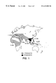
- FIG. 1 Old world distribution and prevalence of ⁇ -thalassemia.
- FIG. 2 Schematic representation of primers, A Primer (SEQ ID NO: 1); B1 Primer (SEQ ID NO: 2); and B2 Primer (SEQ ID NO: 3), for detecting the deletion of hemoglobin ⁇ genes.
- FIG. 3 Coamplification of ⁇ 1- and ⁇ 2-globin genes from blood samples of normal and ⁇ -thal syndrome patients. Three of each category of tested subjects are shown.
- FIG. 4 Gel scanning data from both normal and different genotypes of ⁇ -thal syndrome patients, wherein FIG. 4A represents a normal patient; FIG. 4B represents a patient lacking an ⁇ 2 thalassemia gene; Panels C and D represent patients with a moderate ⁇ chain deficiency, either ⁇ 1 thalassemia heterozygous or homozygous; Panel E represents a patient with Hb H disease; and Panel F represents a patient with the Hb Bart's hydrops fetalis syndrome.
- FIG. 5 PCR-mediated CCA from normal and two ⁇ -thal syndrome patients.
- Panel A shows the amplified a genes with different colors in the reaction tubes as viewed with UV-light.
- lane 1 is an orange color, the result of an equal mixture of red and green fluorescein dye product amplification;
- lane 2 displays green due to the absence of red ( ⁇ 2) amplification;
- lane 3 is colorless due to the total absence of alpha genes ( ⁇ / ⁇ ) for amplification.
- Panel B the amplified ⁇ genes were separated by electrophoresis on a TBE gel and visualized under UV-light by the color and size of each ⁇ gene fragment.
- the present invention relates to the identification of nucleic acid primers capable of detecting and distinguishing between the various forms of ⁇ -thalassemia.
- Samples to be tested using the present invention include blood, either dried or whole blood.
- samples may be derived from any biological tissue containing nucleic acid material, including but not limited to skin tissue, amniotic fluids, umbilical cord, dried tissue and paraffin embedded tissue.
- a particular advantage of the present invention is its adaptability to samples in various forms; that is, the present invention can detect the various forms of ⁇ -thalassemia from fluid or dry biological samples.
- internal standard DNA means a nucleic acid which will be present in a sample whether or not the target DNA is present and which can be labeled and detected independently of the globin target genes.
- the internal standard DNA may correspond to a known sequence of DNA, i.e. a gene, on a chromosome different from that on the globin target genes.
- oligonucleotide as used herein in referring to primers, probes, oligomer fragments to be detected, oligomer controls and unlabeled blocking oligomers is defined as a molecule comprised of two or more deoxyribonucleotides or ribonucleotides, preferably more than three. Its exact size will depend on many factors, which in turn depend on the ultimate function or use of the oligonucleotide.
- primer refers to an oligonucleotide, whether occurring naturally as in a purified restriction digest or produced synthetically, which is capable of acting as a point of initiation of synthesis when placed under conditions in which synthesis of a primer extension product which is complementary to a nucleic acid strand is induced, i.e. in the presence of nucleotides and an inducing agent such as DNA polymerase and at a suitable temperature and pH.
- the primer is preferably single stranded for maximum efficiency in amplification, but may alternatively be double stranded. If double stranded, the primer is first treated to separate its strands before being used to prepare extension products.
- the primer is an oligodeoxyribonucleotide.
- the primer must be sufficiently long to prime the synthesis of extension products in the presence of the inducing agent.
- the exact lengths of the primers will depend on many factors, including temperature, source of primer and use of the method.
- the oligonucleotide primer typically contains 15-25 or more nucleotides, although it may contain fewer nucleotides.
- the oligonucleotide primer is typically shorter, e.g., 7-15 nucleotides.
- the primers of the present invention are selected from regions of the genomic sequence of the hemoglobin ⁇ genes.
- the 5′ primer is preferably located at the 5′ end of each of ⁇ 1 and ⁇ 2 gene and serve as a common sense primer. Most preferred is a 5′ primer comprising the sequence 5′-TGACCCTCTTCTCTGCACAGCTC-3′ (SEQ ID NO: 1) corresponding to a common region in both the ⁇ 1 and the ⁇ 2 genes.
- This sequence referred to herein as “A primer,” is found at position 7242-7264 in the ⁇ 2 gene and at position 11060-11082 in the ⁇ 1 gene of the genomic sequence described by as set forth in Gen Bank, Accession No. J00153, also known as HUMHBA4.
- Two different 3′ primers provide the specificity to detect multiple a gene deletions. These primers are preferably 3′ antisense sequences of the hemoglobin ⁇ genes.
- a preferred ⁇ 2 3′ primer comprises a region within the 3′ untranslated portion of the ⁇ 2 gene.
- One such primer comprises 5′-TTCCGGGACAGAGAGAACCCAGG-3′ (SEQ ID NO:3). This sequence, referred to herein as “B2 primer,” is found at position 7512-7534 in the genomic sequence as set forth in Gen Bank, Accession No. HUMHBA4.
- a second preferred ⁇ 1 3′ primer comprises the sequence 5′-GAGGCCCAAGGGGCAAGAAGC-3′ (SEQ ID NO:2) corresponding to a region within the 3′ untranslated portion of the ⁇ 1 gene.
- This sequence referred to herein as “B1 primer,” is found at position 11229-11249 in the genomic sequence as set forth in Gen Bank, Accession No. HUMHBA4.
- the primers of the present invention can be used in various ratios in detection of the different forms of ⁇ -thalassemia. Ratios selected will depend upon several factors, including the method of detection used, the amount of nucleic acid material and the source of the biological sample. When using amplification techniques, it is preferred that the 5′ common sense primer is present at a ratio range of 1-2 and the 3′ ⁇ 2 and 3′ ⁇ 1 primers are present at a ratio range of 0.1-1.5. A particularly preferred embodiment when using amplification techniques is 5′ common sense primer: 3′ ⁇ 1 primer, 3′ ⁇ 2 primer ratio of about 1:1:1.
- the primers may vary in size but preferably contain a substantial portion of SEQ ID NO:1, SEQ ID NO:2, and SEQ ID NO:3. Specific nucleotide positions of the primers may be varied so long as the primers retain their ability to specifically hybridize to the hemoglobin ⁇ genes.
- the primers of the present invention can be used for amplification using standard polymerase chain reaction techniques. Mullis U.S. Pat. Nos. 4,683,202 and 4,683,195 describe the basic amplification technique and are incorporated herein by reference. Other variant amplification techniques, can also be used. Such techniques are known to the skilled artisan.
- the products can be analyzed by many techniques known in the art. For example, one technique uses physical separation of the amplification products to distinguish the products. Physical separation can be carried out by various methods, including but not limited to filtration, electrophoresis and chromatography techniques.
- Detection and differentiation of the allele-specific amplification products may also be accomplished using immunocytochemistry methods relying on reagents that associate with one another with high specificity and affinity such as biotin-streptavidin, digoxigenin and others (Didenko, et al., 1996).
- a further detection method would allow for high DNA through-put in the context of mass screening employs detection of alpha-globin gene(s) on the surface of a charge coupled device, currently known as a “DNA-chip” (Lamture, et al., 1994).
- a preferred method of detecting the presence and amount of each of the amplified products is one that allows for the detection of more than one target DNA without the need to separate the different amplified products.
- the primers of the present invention may be used in a color complementation assay, as described in U.S. Pat. No. 5,489,507, incorporated herein by reference.
- this embodiment comprises the steps of (i) simultaneously amplifying the target hemoglobin genes and one or more internal standard DNAs in a sample, (ii) providing one or more first labeling means capable of binding to the target hemoglobin genes in the sample, (iii) providing one or more second labeling means capable of binding to one or more internal standard DNAs in the sample, (iv) combining the first labeling means and the second labeling means with the sample so that the first labeling means binds to their target hemoglobin genes to form one or more labeled target hemoglobin genes and the one or more second labeling means bind to their respective internal standard DNAS to form one or more labeled internal standard DNAs, (v) separating the unbound first labeling means from the sample, and (vi) illuminating the sample with an illumination beam having a predetermined wavelength characteristic such that a distinct color signal is produced whose character depends on which of the one or more target DNAs that have been amplified.
- the steps of simultaneously amplifying, providing ea first labeling means, providing one or more second labeling means, and combining are achieved by carrying out a polymerase chain reaction wherein the first labeling means comprises a pair of primers specific for the target globin genes, at least one of the primers being labeled directly or indirectly with a first color-producing or color-absorbing label, and wherein the one or more second labeling means comprise separate pairs of primers, at least one member of each pair being labeled directly or indirectly with a different second color-producing or color-absorbing label.
- Color-producing labels and color-absorbing labels can be attached to primers in a variety of ways either directly or indirectly.
- Mathews et al (1988) provide a comprehensive list of both direct and indirect labeling means for nucleic acids. Accordingly, this reference is incorporated by reference.
- Labels can be directly attached to primers via a 5′ amino or thiol linking group, e.g. Connolly et al (1985) and Fung et al., U.S. Pat. No. 4,757,141. Many commercially available dyes with amino- or thiol-reactive moieties can be used as color-producing and/or color-absorbing labels.
- the following dyes suitable for use with the invention are available: (i) thiol-reactive; 5-iodoacetaminofluorescein, fluorescein-5-maleimide, tetramethylrhodamine-5 (and-6) iodoacetamide, rhodamine X iodoacetamide, and the like, and (ii) amino-reactive; Fluorescein-5 (and-6) isothiocyanate, Texas Red, rhodamine X isothiocyanate, fluorescein-5(and-6) succinimidylcarboxylates, 7-amino-4-methylcoumarin-3-acetic acid, 5- and/or 6-succinimidylcarboxylates of rhodamine dyes, and the like, and dyes disclosed in U.S. pat. appln Ser. no. 07/138,287 filed Dec. 24, 1987.
- Color-producing and/or color-absorbing labels can also be attached to primers indirectly via antibodies or via biotin and avidin or steptavidin.
- Means for synthesizing biotinylated primers is well known in the art (e.g. Chollet et al. 1985; Agrawal et al., 1986). Biotinylating reagents are also commercially available and hence are known and available in the art.
- One or more different internal standard DNAs can be amplified with primers having the same label in order to vary the relative concentrations of the color-producing or color-absorbing labels at the time of analysis.
- primers having the same label For example, if one of the color producing labels is a fluorescent dye with low fluorescent efficiency and a given degree of brightness is required for color complementation, a number of internal standard DNAs can be labeled with the same “weak” fluorescent label to increase the labels relative concentration, and hence, its relative brightness.
- the first and second color-producing or color-absorbing means comprise pairs of amplification primers wherein the 5′ nucleotidle of one primer of the pair is covalently attached to biotin and wherein the 5′ nucleotide of the other primer is directly labeled with a color-producing or color-absorbing label.
- color-producing labels are fluorescent labels, such as fluorescein, Texas Red, tetramethylrhodamine, dichlorodimethoxyfluoroscein, or the like. In some cases, fluorescent labels can also act as color-absorbing labels.
- color-absorbing labels are used with a first labeling means and are organic indicator molecules which can be linked to an amino- or thiol-derivatized primer and whose absorption spectrum substantially overlaps both the emission spectrum of the illumination beam and labeling means. That is, preferably, the color-absorbing label is associated with the target DNA and the fluorescent label used in conjunction with a second labeling means. That is, preferably, the color-absorbing label is associated with the target DNA and the fluorescent label is associated with an internal standard DNA. If a target DNA is present and amplified the measured absorption will be high and fluorescence will be low. The reverse holds if the target DNA is not present. The presence of the target DNA is determined by the ratio of absorption to fluorescence intensity. Most preferably, fluorescent labels such as those disclosed in Khanna et al., U.S. Pat. Nos. 4,318,846; 4,439,356; and 4,481,136.
- the first and second labeling means comprise pairs of primers, one with biotin attached to its 5′ end and the other with a color-producing or color-absorbing label attached to its 5′ end.
- the reaction mixture containing the amplified chains are exposed to an avidin or streptavidin derivatized substrate, e.g. magnetic microspheres (Advanced Magnetics, Inc., Cambridge, Mass.). This allows for rapid separation of the amplified chains from the rest of the reaction mixture which, for example, may contain a preponderance of labeled primers associated with the target DNA if the target DNA is not present in the sample (and consequently not amplified).
- Separation is readily carried out by filtration, or if magnetic microspheres are used, by magnetic separation.
- the labeled strands of the amplified DNA are used, by magnetic separation.
- the labeled strands of the amplified DNA are separated from the biotinylated strands (and primers) by denaturation procedures, e.g. exposure to strong alkali solution (e.g. between 0.1 and 1.0 procedures, e.g. exposure to strong alkali solution (e.g. between 0.1 and 1.0 NaOH), or heat plus exposure to a solution of formamide and water.
- the biotinylated DNA is then removed with the substrate, and the remaining solution containing the labeled strands is illuminated by the illumination beam.
- Automation of the invention is readily carried out by use of a general purpose laboratory robot, such as the disclosed by Wilson et al., BioTechniques, Vol. 6, pgs., 776 (1988).
- Primer B 1 SEQ ID NO:2
- B 2 SEQ ID NO:3
- Primer A located within the 5′ portion of each ⁇ 1 and ⁇ 2 gene, has an identical DNA sequence for both ⁇ 1 and ⁇ 2 gene and serve as a common sense primer.
- the primers for ⁇ -actin gene (sense primer 1854 bp-1874 bp) used as an internal control.
- Primer design for ⁇ genes is illustrated in FIG. 2 .
- ⁇ 2 gene (292 bp), ⁇ 1 gene (190 bp), and ⁇ -actin gene (105 bp) could be observed from 2% NuSieve gel stained with ethidium bromide.
- Genomic DNA (0.3 to 1 ug) is mixed with 5 ⁇ l reaction buffer, 4 ⁇ l of 125 ⁇ M dNTP, and 1 ⁇ l of each 25 ⁇ M primer (primers A, B 1 and B 2 for a genes, as well as sense and antisense primers for ⁇ -actin).
- the reaction is performed in 50 ⁇ l volume.
- the mixture is heated at 95° C. for 5 minutes to denature the DNA before 1.25 units Taq polymerase is added into the solution. Thirty cycles are performed on a Perkin-Elmer Cetus DNA thermal Cycler: denaturing at 94° C. for 1.0 min., annealing at 60° C. for 1.0 min., and extending at 72° C. for 1.0 min.
- Genomic DNA was isolated from either whole blood or dried blood samples embedded on a filter paper from 21 normal subjects and 62 ⁇ -thalassemia patients. This DNA was used as a template to amplify by PCR three DNA fragments ( ⁇ 2 , ⁇ 1 and ⁇ -acting genes) which were easily identified from each sample tested. In normal 4 genes ( ⁇ / ⁇ ) samples, all three bands showed almost equal intensity, which indicated ⁇ 1 or ⁇ 2 genes are present normally without any gene deletion (FIG. 3, lanes 1-3).
- deletion of ⁇ 1 or ⁇ 2 gene could be identified from ethidium bromide-stained NuSieve agarose gel. Such heterozygotes ( ⁇ / ⁇ ) possess a mild ⁇ -chain deletion resulting from the presence of only three ⁇ genes.
- the band for ⁇ 2 gene (292 bp) was found to have less intensity (half intensity of normal ⁇ 2 gene band) as compared to ⁇ 1 gene (190 bp) FIG. 3, lanes 4-6).
- the intensity of each band could be represented by the area under the curve.
- the copy number of a gene then is determined by either the ratio of the area for ⁇ gene band to that of ⁇ -actin gene, or by comparing the percentage of area from each peak. The average values of ratio and the percentage of peak area are summarized in the Table 1. There are significant differences between each category of the ⁇ -thal syndromes.
- the ratios of band intensity among ⁇ 1 over ⁇ -actin or ⁇ 2 over ⁇ -actin in normal subject was 1.10 ⁇ 0.25 and 0.70 ⁇ 0.35, respectively, while in ⁇ -thal patients with different genotypes a significant change in the calculated values were seen (Table 1).
- the two clinical significant types of ⁇ -thal syndrome, ( ⁇ / ⁇ ) and ( ⁇ / ⁇ ) could be identified either comparing the percentage of each band or ratios of ⁇ 1 / ⁇ -actin and ⁇ 2 / ⁇ -actin.
- CCA color complementary assay
- B1 or B2 primer that are differently labeled with either fluorescein or rhodamine were added.
- the primer B1 was labeled with 5′-carbon fluorescein (FAM) appears with a green color, while the B2 primer was labeled with 6-carboxyl-X-rhodamine (ROX) showing red color.
- the common 5′ primer was unlabeled primer A. After coamplification of both ⁇ 1 and ⁇ 2 colored-PCR products were produced in the reaction tubes.
- the oligonucleotide B1 and B2 with a primary amino group attached to the 5′ end were synthesized.
- the dye-labeled oligonucleotide were then purified from the nonconjugated oligonucleotide by HPLC Aquepore 300C-8 column.
- the recovered fluorescent oligonucleotide were then dried down and resuspended in sterile distilled water. Measurement of the absorbance of the oligos at 260 nm was performed and diluted to a final concentration of 25 ⁇ M. Aliquot of the dye-labeled primers are stored at ⁇ 20° C. in the dark until use.
- PCR mediated CCA reaction was processed as follows: 2 ⁇ l of each primer A, FAM-B1 and ROX-B2 was mixed with 1 ⁇ g of genomic DNA in a 50 ⁇ l reaction mixture containing 5 ⁇ l reaction buffer (50 mM KCl, 10 mM Tris (pH8.0), 1.5 mM MgCl 2 ), and 4 ⁇ l each dATP, dCTP, dGTP, dTTP at 125 ⁇ M. 5 units of native Taq DNA polymerase was added, and the reaction mixture was overlaid with 50 ⁇ l of mineral oil. First, amplification reaction is performed at 94° C. for 5 min., 60° C. for 1 min., and 72° C. for 1.0 min.
- PCR template and reaction conditions were the same as described in Example 1 above.
- a gene in solution was used (directly visualization)
- amplified products were separated from free primers by ultrafiltration through centricon C 100 microconcentrators after PCR. Colors were observed in the tubes under UV light.
- FIG. 5A both ⁇ 1 and ⁇ 2 genes from both normal and several ⁇ -thal patients have been successfully amplified.
- ⁇ 1 and ⁇ 2 gene produced a color combination (yellow) from ⁇ 1 gene (green) and ⁇ 2 gene (red) (FIG. 5. A1).
- ⁇ / ⁇ patient only ⁇ 1 gene (green) was observed (FIG.
Landscapes
- Chemical & Material Sciences (AREA)
- Life Sciences & Earth Sciences (AREA)
- Proteomics, Peptides & Aminoacids (AREA)
- Health & Medical Sciences (AREA)
- Organic Chemistry (AREA)
- Wood Science & Technology (AREA)
- Analytical Chemistry (AREA)
- Zoology (AREA)
- Genetics & Genomics (AREA)
- Engineering & Computer Science (AREA)
- Pathology (AREA)
- Immunology (AREA)
- Microbiology (AREA)
- Molecular Biology (AREA)
- Biotechnology (AREA)
- Biophysics (AREA)
- Physics & Mathematics (AREA)
- Biochemistry (AREA)
- Bioinformatics & Cheminformatics (AREA)
- General Engineering & Computer Science (AREA)
- General Health & Medical Sciences (AREA)
- Measuring Or Testing Involving Enzymes Or Micro-Organisms (AREA)
Abstract
The present invention relates to the simultaneous and specific identification of the variant forms of alpha-thalassemia. This invention utilizes simple and readily available equipment to rapidly identify, diagnose and differentiate the different forms of alpha-thalassemia. Specifically, the present invention relates to a simple and rapid non-radioisotopic technique for the diagnosis and differentiation of the common forms of alpha-thalassemia has been developed. This approach works on any biological tissue including blood, wherein the assay works equally well with fresh blood and dried blood samples stored on filter paper.
Description
The instant application is a 371 of PCT/US97/21346, filed Nov. 18, 1997, and claims benefit under 35 U.S.C. 119(e) to provisional application 60/031,880, filed Nov. 27, 1996.
The present invention relates to the field of diagnostic methodologies. More particularly, the invention is related to a simple, inexpensive and rapid method for detecting thalassemias and monitoring therapeutic effects on the disease.
The α-thalassemia (“α-thal”) are common genetic disorders that result from reduced synthesis of the a globin chains of fetal (α2γ2, HbF) and adult (α2β2, HbA) hemoglobin (Weatherall and Clegg, 1981; Higgs et al., 1989, Higgs and Weatherall, 1993). The normal human α globin gene cluster is located on the short arm of chromosome 16 (Breuning et al., 1987; Buckle et al., 1988). In α-thal syndromes, α-globin synthesis is either diminished or absent due to either deletional or non-deletional abnormalities involving the α-globin genes (Higgs et al., 1989; Higgs and Weatherall, 1993). Diploid cells have four α-chain genes (i.e., αα/αα). The severity of the hematological and clinical picture is directly proportional to the number of involved α-globin genes and thus the deletion of one, two, three, or all four of these α genes attribute to mild to complete a chain deficiencies syndromes.
The deletion of α-chain at birth results in the formation of a γ chain tetramer, Hb Bart's (γ4). The percentage of Hb Bart's present corresponds to the degree of α chain deficiency (Cong & Shong, 1982; Liang et al., 1985; Higgs et al., 1989; Higgs and Weatherall, 1993). The α-thal-2 genotype has been found to have one of the two genes deleted, thug these heterozygotes (αα/−α) possess a mild α chain deficiency resulting from the presence of only three α chain genes and have not more than 2% Hb Bart's at birth (Higgs et al., 1989). Homozygotes (−α/−α), as well as α-thal-1 heterozygotes (αα/−−), possess moderate a chain deficiency resulting from the presence of only two α chain genes and have approximately 5% Hb Bart's at birth (Higgs and Weatherall, 1993). While Hb H disease is a heterozygosity for α-thal-2 in association with an α-thal-1 heterozygosity (−−/−α), a severe α chain deficiency occurs due to the deletion of three α chain genes (Higgs et al., 1989; Higgs and Weatherall, 1993). The α-thal-1 homozygotes (−−/−−), which lack any functional α genes, present with Hb Bart's is known as hydrops fetalis syndrome and results in intra-uterine or early post-delivery death to the fetus (Lie-Injo & Hie, 1960; Higgs et al., 1989). Mother bearing fetuses with homozygous α-thal-1 (−−/−−) are at high risk for obstetrical complications, such pregnancy induced hypertension, eclampsia, and/or death.
At present, for a variety of technical, logistical and economical reasons, large-scale carrier screening and appropriate ante-natal detecting programs have not been established for the populations in which this syndrome occurs (Bowden et al., 1992; Higgs and Weatherall, 1993).
It is an object of the invention to develop a diagnostic method and kit for the detection and quantitation of hemoglobin (Hb) α gene(s) in α-thalassemia patients.
It is another object of the present invention to develop a method and kit for screening for carriers of the genetic disorder, α thalassemia.
It is yet another object of the present invention to identify persons who are at risk of having offspring with homozygous α-thalassemia as well as identify α-thalassemia in individuals with unexplained microcytosis and hypochromia.
Another object of the present invention is to develop a sensitive, non-radioisotopic test capable of differentiating between the various forms of α-thalassemia, by detecting and quantitating a gene(s) from blood samples.
The present invention relates to the identification of primers capable of detecting and distinguishing between the various forms of α-thalassemia. The α-globin primers of the present invention are derived from the α-globin genes. One such primer comprises a region common to both α1 and α2 genes. A second such primer is specific for α2 gene and a third primer is specific for α1 gene. These primers are designed to specifically hybridize to portions of the hemoglobin α genes, such that, when amplified with an inducing agent, give identifiable amplification products. The presence and amount of the products is correlated to specific forms of α-thalassemia, based upon the type of gene deletion associated with the disease.
The present invention relates to a method for identifying and differentiating among the various forms of α-thalassemia. The method of the present invention uses highly specific primers to identify and differentiate among the forms of α-thalassemia. The assay may include a nucleic acid amplification step wherein the hemoglobin a genes are simultaneously amplified and detected. In another embodiment of the present invention, a color complementation assay is used to provide a convenient, rapid, non-radioisotopic, assay system for quantitation of the α gene(s).
A kit comprising at least three primers, capable of detecting, quantitating and distinguishing α-thalassemia variants is also encompassed in the present invention.
FIG. 1. Old world distribution and prevalence of α-thalassemia.
FIG. 2. Schematic representation of primers, A Primer (SEQ ID NO: 1); B1 Primer (SEQ ID NO: 2); and B2 Primer (SEQ ID NO: 3), for detecting the deletion of hemoglobin α genes.
FIG. 3. Coamplification of α1- and α2-globin genes from blood samples of normal and α-thal syndrome patients. Three of each category of tested subjects are shown.
FIG. 4. Gel scanning data from both normal and different genotypes of α-thal syndrome patients, wherein FIG. 4A represents a normal patient; FIG. 4B represents a patient lacking an α2 thalassemia gene; Panels C and D represent patients with a moderate α chain deficiency, either α1 thalassemia heterozygous or homozygous; Panel E represents a patient with Hb H disease; and Panel F represents a patient with the Hb Bart's hydrops fetalis syndrome.
FIG. 5. PCR-mediated CCA from normal and two α-thal syndrome patients. Panel A shows the amplified a genes with different colors in the reaction tubes as viewed with UV-light. In lane 1 is an orange color, the result of an equal mixture of red and green fluorescein dye product amplification; lane 2 displays green due to the absence of red (α2) amplification; lane 3 is colorless due to the total absence of alpha genes (−−/−−) for amplification. Panel B: the amplified αgenes were separated by electrophoresis on a TBE gel and visualized under UV-light by the color and size of each α gene fragment.
The present invention relates to the identification of nucleic acid primers capable of detecting and distinguishing between the various forms of α-thalassemia.
The primers of the present invention are used to identify and differentiate between the various forms of α-thalassemia. Samples to be tested using the present invention include blood, either dried or whole blood. In addition, samples may be derived from any biological tissue containing nucleic acid material, including but not limited to skin tissue, amniotic fluids, umbilical cord, dried tissue and paraffin embedded tissue.
A particular advantage of the present invention is its adaptability to samples in various forms; that is, the present invention can detect the various forms of α-thalassemia from fluid or dry biological samples.
The term “internal standard DNA” means a nucleic acid which will be present in a sample whether or not the target DNA is present and which can be labeled and detected independently of the globin target genes. For example, the internal standard DNA may correspond to a known sequence of DNA, i.e. a gene, on a chromosome different from that on the globin target genes.
The term “oligonucleotide” as used herein in referring to primers, probes, oligomer fragments to be detected, oligomer controls and unlabeled blocking oligomers is defined as a molecule comprised of two or more deoxyribonucleotides or ribonucleotides, preferably more than three. Its exact size will depend on many factors, which in turn depend on the ultimate function or use of the oligonucleotide.
The term “primer” as used herein refers to an oligonucleotide, whether occurring naturally as in a purified restriction digest or produced synthetically, which is capable of acting as a point of initiation of synthesis when placed under conditions in which synthesis of a primer extension product which is complementary to a nucleic acid strand is induced, i.e. in the presence of nucleotides and an inducing agent such as DNA polymerase and at a suitable temperature and pH. The primer is preferably single stranded for maximum efficiency in amplification, but may alternatively be double stranded. If double stranded, the primer is first treated to separate its strands before being used to prepare extension products. Preferably, the primer is an oligodeoxyribonucleotide. The primer must be sufficiently long to prime the synthesis of extension products in the presence of the inducing agent. The exact lengths of the primers will depend on many factors, including temperature, source of primer and use of the method. For example, for diagnostics applications, depending on the complexity of the target sequence, the oligonucleotide primer typically contains 15-25 or more nucleotides, although it may contain fewer nucleotides. For other applications, the oligonucleotide primer is typically shorter, e.g., 7-15 nucleotides.
The primers of the present invention are selected from regions of the genomic sequence of the hemoglobin α genes. The 5′ primer is preferably located at the 5′ end of each of α1 and α2 gene and serve as a common sense primer. Most preferred is a 5′ primer comprising the sequence 5′-TGACCCTCTTCTCTGCACAGCTC-3′ (SEQ ID NO: 1) corresponding to a common region in both the α1 and the α2 genes. This sequence, referred to herein as “A primer,” is found at position 7242-7264 in the α2 gene and at position 11060-11082 in the α1 gene of the genomic sequence described by as set forth in Gen Bank, Accession No. J00153, also known as HUMHBA4.
Two different 3′ primers provide the specificity to detect multiple a gene deletions. These primers are preferably 3′ antisense sequences of the hemoglobin α genes. A preferred α 2 3′ primer comprises a region within the 3′ untranslated portion of the α2 gene. One such primer comprises 5′-TTCCGGGACAGAGAGAACCCAGG-3′ (SEQ ID NO:3). This sequence, referred to herein as “B2 primer,” is found at position 7512-7534 in the genomic sequence as set forth in Gen Bank, Accession No. HUMHBA4. A second preferred α 1 3′ primer comprises the sequence 5′-GAGGCCCAAGGGGCAAGAAGC-3′ (SEQ ID NO:2) corresponding to a region within the 3′ untranslated portion of the α1 gene. This sequence, referred to herein as “B1 primer,” is found at position 11229-11249 in the genomic sequence as set forth in Gen Bank, Accession No. HUMHBA4.
The primers of the present invention can be used in various ratios in detection of the different forms of α-thalassemia. Ratios selected will depend upon several factors, including the method of detection used, the amount of nucleic acid material and the source of the biological sample. When using amplification techniques, it is preferred that the 5′ common sense primer is present at a ratio range of 1-2 and the 3′ α2 and 3′ α1 primers are present at a ratio range of 0.1-1.5. A particularly preferred embodiment when using amplification techniques is 5′ common sense primer: 3′ α1 primer, 3′ α2 primer ratio of about 1:1:1.
The primers may vary in size but preferably contain a substantial portion of SEQ ID NO:1, SEQ ID NO:2, and SEQ ID NO:3. Specific nucleotide positions of the primers may be varied so long as the primers retain their ability to specifically hybridize to the hemoglobin α genes.
Primers are readily synthesized by standard techniques. Detailed procedures for the phospho-triester and hydrogen phosphonate methods of oligonucleotide synthesis are described in the following references, which are incorporated by reference: Caruthers et al., U.S. Pat. Nos. 4,458,066 and 4,500,707; Matteucci et al., J. Amer. Chem. Soc. 103:3185-3191 (1981); Caruthers et al., Genet. Engin. 4:1-17 (1987); Jones, Chapter 2 and Atkinson et al., Chapter 3, in Gait, ed., Oligonucleotide Synthesis: A practical Approach (IRL Press, Washington, DC, (1984); Froehler, et al., Tetrahed. Lett. 27:469-472 (1986); Garegg, et al., Tetrahed. Lett. 27:4051-4058 (1986); and Froehler, et al., Nucl. Acid Res. 14:5399-5407 (1986).
The primers of the present invention can be used for amplification using standard polymerase chain reaction techniques. Mullis U.S. Pat. Nos. 4,683,202 and 4,683,195 describe the basic amplification technique and are incorporated herein by reference. Other variant amplification techniques, can also be used. Such techniques are known to the skilled artisan.
Once amplified, the products can be analyzed by many techniques known in the art. For example, one technique uses physical separation of the amplification products to distinguish the products. Physical separation can be carried out by various methods, including but not limited to filtration, electrophoresis and chromatography techniques.
Detection and differentiation of the allele-specific amplification products may also be accomplished using immunocytochemistry methods relying on reagents that associate with one another with high specificity and affinity such as biotin-streptavidin, digoxigenin and others (Didenko, et al., 1996).
A further detection method would allow for high DNA through-put in the context of mass screening employs detection of alpha-globin gene(s) on the surface of a charge coupled device, currently known as a “DNA-chip” (Lamture, et al., 1994).
A preferred method of detecting the presence and amount of each of the amplified products is one that allows for the detection of more than one target DNA without the need to separate the different amplified products. For example, the primers of the present invention may be used in a color complementation assay, as described in U.S. Pat. No. 5,489,507, incorporated herein by reference. In brief, this embodiment comprises the steps of (i) simultaneously amplifying the target hemoglobin genes and one or more internal standard DNAs in a sample, (ii) providing one or more first labeling means capable of binding to the target hemoglobin genes in the sample, (iii) providing one or more second labeling means capable of binding to one or more internal standard DNAs in the sample, (iv) combining the first labeling means and the second labeling means with the sample so that the first labeling means binds to their target hemoglobin genes to form one or more labeled target hemoglobin genes and the one or more second labeling means bind to their respective internal standard DNAS to form one or more labeled internal standard DNAs, (v) separating the unbound first labeling means from the sample, and (vi) illuminating the sample with an illumination beam having a predetermined wavelength characteristic such that a distinct color signal is produced whose character depends on which of the one or more target DNAs that have been amplified. Preferably, the steps of simultaneously amplifying, providing ea first labeling means, providing one or more second labeling means, and combining are achieved by carrying out a polymerase chain reaction wherein the first labeling means comprises a pair of primers specific for the target globin genes, at least one of the primers being labeled directly or indirectly with a first color-producing or color-absorbing label, and wherein the one or more second labeling means comprise separate pairs of primers, at least one member of each pair being labeled directly or indirectly with a different second color-producing or color-absorbing label.
Color-producing labels and color-absorbing labels can be attached to primers in a variety of ways either directly or indirectly. Mathews et al (1988) provide a comprehensive list of both direct and indirect labeling means for nucleic acids. Accordingly, this reference is incorporated by reference.
Labels can be directly attached to primers via a 5′ amino or thiol linking group, e.g. Connolly et al (1985) and Fung et al., U.S. Pat. No. 4,757,141. Many commercially available dyes with amino- or thiol-reactive moieties can be used as color-producing and/or color-absorbing labels. For example, the following dyes suitable for use with the invention are available: (i) thiol-reactive; 5-iodoacetaminofluorescein, fluorescein-5-maleimide, tetramethylrhodamine-5 (and-6) iodoacetamide, rhodamine X iodoacetamide, and the like, and (ii) amino-reactive; Fluorescein-5 (and-6) isothiocyanate, Texas Red, rhodamine X isothiocyanate, fluorescein-5(and-6) succinimidylcarboxylates, 7-amino-4-methylcoumarin-3-acetic acid, 5- and/or 6-succinimidylcarboxylates of rhodamine dyes, and the like, and dyes disclosed in U.S. pat. appln Ser. no. 07/138,287 filed Dec. 24, 1987.
Color-producing and/or color-absorbing labels can also be attached to primers indirectly via antibodies or via biotin and avidin or steptavidin. Means for synthesizing biotinylated primers is well known in the art (e.g. Chollet et al. 1985; Agrawal et al., 1986). Biotinylating reagents are also commercially available and hence are known and available in the art.
One or more different internal standard DNAs can be amplified with primers having the same label in order to vary the relative concentrations of the color-producing or color-absorbing labels at the time of analysis. For example, if one of the color producing labels is a fluorescent dye with low fluorescent efficiency and a given degree of brightness is required for color complementation, a number of internal standard DNAs can be labeled with the same “weak” fluorescent label to increase the labels relative concentration, and hence, its relative brightness.
Preferably, in automated embodiments of the present invention the first and second color-producing or color-absorbing means comprise pairs of amplification primers wherein the 5′ nucleotidle of one primer of the pair is covalently attached to biotin and wherein the 5′ nucleotide of the other primer is directly labeled with a color-producing or color-absorbing label. Preferably, color-producing labels are fluorescent labels, such as fluorescein, Texas Red, tetramethylrhodamine, dichlorodimethoxyfluoroscein, or the like. In some cases, fluorescent labels can also act as color-absorbing labels. Preferably, color-absorbing labels are used with a first labeling means and are organic indicator molecules which can be linked to an amino- or thiol-derivatized primer and whose absorption spectrum substantially overlaps both the emission spectrum of the illumination beam and labeling means. That is, preferably, the color-absorbing label is associated with the target DNA and the fluorescent label used in conjunction with a second labeling means. That is, preferably, the color-absorbing label is associated with the target DNA and the fluorescent label is associated with an internal standard DNA. If a target DNA is present and amplified the measured absorption will be high and fluorescence will be low. The reverse holds if the target DNA is not present. The presence of the target DNA is determined by the ratio of absorption to fluorescence intensity. Most preferably, fluorescent labels such as those disclosed in Khanna et al., U.S. Pat. Nos. 4,318,846; 4,439,356; and 4,481,136.
As mentioned above, for automated embodiments it is preferred that the first and second labeling means comprise pairs of primers, one with biotin attached to its 5′ end and the other with a color-producing or color-absorbing label attached to its 5′ end. In such an embodiment, after amplification, the reaction mixture containing the amplified chains are exposed to an avidin or streptavidin derivatized substrate, e.g. magnetic microspheres (Advanced Magnetics, Inc., Cambridge, Mass.). This allows for rapid separation of the amplified chains from the rest of the reaction mixture which, for example, may contain a preponderance of labeled primers associated with the target DNA if the target DNA is not present in the sample (and consequently not amplified). Separation is readily carried out by filtration, or if magnetic microspheres are used, by magnetic separation. The labeled strands of the amplified DNA are used, by magnetic separation. The labeled strands of the amplified DNA are separated from the biotinylated strands (and primers) by denaturation procedures, e.g. exposure to strong alkali solution (e.g. between 0.1 and 1.0 procedures, e.g. exposure to strong alkali solution (e.g. between 0.1 and 1.0 NaOH), or heat plus exposure to a solution of formamide and water. The biotinylated DNA is then removed with the substrate, and the remaining solution containing the labeled strands is illuminated by the illumination beam. Automation of the invention is readily carried out by use of a general purpose laboratory robot, such as the disclosed by Wilson et al., BioTechniques, Vol. 6, pgs., 776 (1988).
All articles and patents referred to herein are incorporated, in toto, by reference.
While the invention is described above in relation to certain specific embodiments, it will be understood that many variations are possible, and that alternative materials and reagents can be used without departing from the invention. In some cases such variations and substitutions may require some experimentation, but will only involve routine testing.
The foregoing description of the specific embodiments will so fully reveal the general nature of the invention and others can, by applying current knowledge, readily modify and/or adopt for various applications such specific embodiments without departing from the generic concept, and therefore such adaptations and modifications are intended to be comprehended within the meaning and range of equivalents of the disclosed embodiments.
Direct analysis of 5 most frequent α-thal genotypes by PCR amplification of DNA samples from α-thal patients.
1). Primer Designing
To examine the presence of different types of α gene(s) deletion, three primers were designed for specifically coamplifying α1 and α2 genes from single blood sample by PCR. Primer B1 (SEQ ID NO:2) and B2 (SEQ ID NO:3) are respectively 3′ antisense primer of α gene. Primer A(SEQ ID NO:1), located within the 5′ portion of each α1 and α2 gene, has an identical DNA sequence for both α1 and α2 gene and serve as a common sense primer. The primers for β-actin gene (sense primer 1854 bp-1874 bp) used as an internal control. Primer design for α genes is illustrated in FIG. 2. After PCR three amplified DNA fragments: α2 gene (292 bp), α1 gene (190 bp), and β-actin gene (105 bp) could be observed from 2% NuSieve gel stained with ethidium bromide.
2). Optimal Condition for PCR Amplification
Genomic DNA (0.3 to 1 ug) is mixed with 5 μl reaction buffer, 4 μl of 125 μM dNTP, and 1 μl of each 25 μM primer (primers A, B1 and B2 for a genes, as well as sense and antisense primers for β-actin). The reaction is performed in 50 μl volume. The mixture is heated at 95° C. for 5 minutes to denature the DNA before 1.25 units Taq polymerase is added into the solution. Thirty cycles are performed on a Perkin-Elmer Cetus DNA thermal Cycler: denaturing at 94° C. for 1.0 min., annealing at 60° C. for 1.0 min., and extending at 72° C. for 1.0 min. After PCR reaction, 10-15 μl of the amplified product are analyzed on 2% NuSieve gel by electrophoresis. Three bands, corresponding to 190 bp for α1 gene, 292 bp for α2 gene and 105 bp for β-actin gene, were visualized. By comparing the intensity of each α1 or α2 gene bands on ethidium bromide-stained gel between normal and α-thal patient, the deletion status of α genes could be accessed.
3). Detection of α Gene Deletion from α-thal Syndrome Patients
Genomic DNA was isolated from either whole blood or dried blood samples embedded on a filter paper from 21 normal subjects and 62 α-thalassemia patients. This DNA was used as a template to amplify by PCR three DNA fragments (α2, α1 and β-acting genes) which were easily identified from each sample tested. In normal 4 genes (αα/ αα) samples, all three bands showed almost equal intensity, which indicated α1 or α2 genes are present normally without any gene deletion (FIG. 3, lanes 1-3). When the intensity of each amplified α gene was compared with the intensity of the β-actin gene band (a gene product which serves as an internal control), deletion of α1 or α2 gene could be identified from ethidium bromide-stained NuSieve agarose gel. Such heterozygotes (−α/αα) possess a mild α-chain deletion resulting from the presence of only three α genes. The band for α2 gene (292 bp) was found to have less intensity (half intensity of normal α2 gene band) as compared to α1 gene (190 bp) FIG. 3, lanes 4-6). α thal-1 Heterozygotes (−−/αα) as well as homozygotes (−α/−α), possessed moderate α-chain deficiency resulting from the presence of only two α-chain genes, in which less intensity of both 190 bp and 292 bp bands (FIG. 3, lanes 10-12) were observed. However Hb H disease (−−/−α) resulted from deletion of three α-gene showed that complete loss of 292 bp band and less intensity of α1 band compared with β-actin band (FIG. 3, lanes 13-15), while the Hb Bart's hydrops fetalis syndrome (−−/−−) had neither α2 nor α1 gene band (FIG. 3, lanes 16-18). Using this set: of PCR primers, different band intensity from each target gene could reflect the genotype status of a gene detection in α-thal patients. A total of 21 normal samples and 62 α-thal patients representing different α gene deletions were examined by this assay.
In order to obtain quantitative values from this assay, a negative film taken from ethidium bromide stained gel was applied to a densitometer (UltrascanXL enhancer laser densitometer, LKB) to quantitate the area under curve reflecting the intensity of each band. As shown in FIG. 4, different α-gene deletions exhibited different gel scanning patterns of the three peaks (corresponding to β-actin, α1 and α2 bands. The pattern of the peaks correlated very well with the status of α gene deletion, and each genotype has its own characteristic pattern of the peaks. For example, in (−α/αα) an α2 peak is much lower than that of α1 band, while the α1 peak is obviously much higher than that of the β-actin [FIG. 4 (B)]; In (−−/αα), both α1 and α2 peak are lower than that of β-actin [FIG. 4 (C)]; In (−α/−α) the α2 peak diminished, only peaks for α1 and β-actin were observed [FIG. 4 (D)].
The intensity of each band could be represented by the area under the curve. The copy number of a gene then is determined by either the ratio of the area for α gene band to that of β-actin gene, or by comparing the percentage of area from each peak. The average values of ratio and the percentage of peak area are summarized in the Table 1. There are significant differences between each category of the α-thal syndromes.
The ratios of band intensity among α1 over β-actin or α2 over β-actin in normal subject was 1.10±0.25 and 0.70±0.35, respectively, while in α-thal patients with different genotypes a significant change in the calculated values were seen (Table 1). The two clinical significant types of α-thal syndrome, (−α/αα) and (−−/αα) could be identified either comparing the percentage of each band or ratios of α1/β-actin and α2/β-actin.
1) Experimental Design
Another quicker (without gel electrophoresis), PCR-mediated color complementary assay (CCA) for detecting a globin gene(s) deletion was developed. It is based on the simultaneous amplification of two α genes with fluorescent-labeled oligonucleotide primers and the visualization of the e-color gene products by UV light irradiation (Chehab and Kan, 1989). Thus, the generation of a color or combination of colors from the patient samples can be visualized under UV light in the tube without electrophoresis. In this CCA, 2 different fluorophores were coupled to B1 or B2 primer in order to detect and differentiate globin gene α1 or α2. In the same PCR mixture B1 or B2 primer that are differently labeled with either fluorescein or rhodamine were added. The primer B1 was labeled with 5′-carbon fluorescein (FAM) appears with a green color, while the B2 primer was labeled with 6-carboxyl-X-rhodamine (ROX) showing red color. The common 5′ primer was unlabeled primer A. After coamplification of both α1 and α2 colored-PCR products were produced in the reaction tubes.
2) Optimal Condition for PCR-CCA
The oligonucleotide B1 and B2 with a primary amino group attached to the 5′ end were synthesized. The dye-labeled oligonucleotide were then purified from the nonconjugated oligonucleotide by HPLC Aquepore 300C-8 column. The recovered fluorescent oligonucleotide were then dried down and resuspended in sterile distilled water. Measurement of the absorbance of the oligos at 260 nm was performed and diluted to a final concentration of 25 μM. Aliquot of the dye-labeled primers are stored at −20° C. in the dark until use.
PCR mediated CCA reaction was processed as follows: 2 μl of each primer A, FAM-B1 and ROX-B2 was mixed with 1 μg of genomic DNA in a 50 μl reaction mixture containing 5 μl reaction buffer (50 mM KCl, 10 mM Tris (pH8.0), 1.5 mM MgCl2), and 4 μl each dATP, dCTP, dGTP, dTTP at 125 μM. 5 units of native Taq DNA polymerase was added, and the reaction mixture was overlaid with 50 μl of mineral oil. First, amplification reaction is performed at 94° C. for 5 min., 60° C. for 1 min., and 72° C. for 1.0 min. Thirty amplification cycles were followed at 94° C. for 1 min., 60° C. for 1 min., 72° C. for 1 min. Aliquots (10-15 μl) from each amplification mixture were loaded on 6% TBE gel for electrophoresis to confirm the existence and size of amplified product. Also detected were PCR-CCA products by color reactions without electrophoresis. After PCR, amplified samples were separated from free primers by repeated ultrafiltration through Centricon 100 micro concentrators (Amicon). The tubes then were visualized on a long wavelength (300 nm) UV transilluminator and photographed with a Polaroid MP-4 Land Camera. Each film is exposed at f4.5 for 30 sec to 1 min. with a combination of Wratten gelatin filter no.16 and no.21. On the basis of the PCR-CCA data from the large number of samples tested, a range of quantitative values corresponding to normal and the different genotypes of α-thal syndromes can be defined, using a fluorescent spectrophotometer.
3). Detection of Alpha Gene From Normal and α-thal Syndrome Patients by CCA
PCR template and reaction conditions were the same as described in Example 1 above. When detection of PCR-CCA a gene in solution was used (directly visualization), amplified products were separated from free primers by ultrafiltration through centricon C 100 microconcentrators after PCR. Colors were observed in the tubes under UV light. As shown in FIG. 5A, both α1 and α2 genes from both normal and several α-thal patients have been successfully amplified. In normal subjects the existence of both α1 and α2 gene produced a color combination (yellow) from α1 gene (green) and α2 gene (red) (FIG. 5. A1). In (−α/−α) patient only α1 gene (green) was observed (FIG. 5.A2), and in (−−/−−) patient, no color were detected (FIG. 5.A3). We also confirmed the bands as specific amplified DNA fragments by visualizing with color photography from the TBE gels (FIG. 5B). Two of each category subjects has been tested in this assay.
Agrawal et al. (1986) Nucl. Acid Research 14:6227-6245.
Bowden O, Vickers M A, Higgs D R. (1992) A PCR-based strategy to detect the common severe determinants of α-thalassemia. British. Journal. of Hematology 81:104-108.
Bruening M H, Madan K. Verjall M. Wijnen J T, Meera K P, and Peason P L (1987) Human α globin maps to pter-p13.3 in chromosome 16 distal to PGP. Human Genetics. 76,287-289.
Buckel V J, Higgs D R, Wilkie A O M, Super M and Weatheral D J (1988) Localization of human α globin to 16p13.3-pter. J. of Medical Genetics 25:847-849
Chehab F F and Kan Y W (1989) Detection of specific DNA sequences by fluorescence amplification: a color complementary assay. Proc.Natl.Acad.Sci.USA 86 (23):9178-9182
Chollet, et al. (1985) Nucleic Acids Research 13:1529-1541.
Cong V J, Shong H P (1982) Hydrops fetalis and hemaglobinpathy. Chinese J. of Obstetrics and Gynecology. 17:226-228.
Didenko V V; Homsby P J. A quantitative luminescence assay for nonradioactive nucleic acid probes. J. Histochem Cytochem 1996 Jun.44(6):657-60.
Higgs D R, Vickers M A, Mikle A O M, Pretorius I M, Jarman A P and Weatherall D J (1989) A review of the molecular genetics of the human α-globin gene cluster. Blood 73:1081-1104.
Higgs D and Weatherall D J (1993) Bailliere's Clinical Hematology Vol 6 No.1. Bailliere and Tindal press.
Lamture J B; BeattiLe K L; Burke B E; Eggers M D; Ehrlich D J; Fowler R; Hollis M A; Kosicki B B; Reich R K; Smith S R; et al. Direct detection of nucleic acid hybridization on the surface of a chargecoupled device. Nucleic Acids Res 1994 Jun.11;22(11):2121-5.
Liang S T, Wong V C W, So W W K, Ma H K, Chan V. Todd D (1985) Homozygous α-thalassemia: clinical presentation. Diagnosis and management. A review of 46 cases. British Journal of Obstetrics and Gynecology.92:680-684.
Lie-Injo L E, Hie J B (1960) A fast-moving hemoglobin in hydrops feotalis. Nature 185:698.
Mathews et al. (1988) Anal. Biochem. 169:1-25.
Weatherall D J and Clegg J (1981) The thalassemia syndromes 3rd ed. Blackwell Scientific Publications. Oxford.
Claims (9)
1. A nucleic acid composition comprising a nucleic acid consisting of SEQ ID NO: 1, a nucleic acid consisting of SEQ ID NO: 2, and a nucleic acid consisting of SEQ ID NO: 3.
2. A method for detecting a form of α-thalassemia in a biological sample containing nucleic acids encoding hemoglobin comprising:
(a) forming a mixture comprising primers and said biological sample, wherein said primers include a primer consisting of SEQ ID NO: 1 that specifically hybridizes to a region common to α1 and α2 hemoglobin genes, a primer consisting of SEQ ID NO: 2 that specifically hybridizes to the α1 hemoglobin gene, and a primer consisting of SEQ ID NO: 3 that specifically hybridizes to the α2 hemoglobin gene;
(b) hybridizing said primers to nucleic acids in said biological sample;
(c) extending the 3′ ends of the primers to form an α1 extension product and an α2 extension product having different nucleotide lengths;
(d) repeating steps (b) and (c), thereby amplifying said α1 and α2 extension products; and
(e) detecting amplified extension products of (d), wherein absence of an α1 amplified extension product and/or an α2 amplified extension product is indicative of the presence of a form of α-thalassemia.
3. A method for detecting a form of α-thalassemia in a biological sample containing nucleic acids encoding hemoglobin comprising:
(a) forming a mixture comprising primers and said biological sample, wherein said primers include a primer consisting of SEQ ID NO: 1 that specifically hybridizes to a region common to α1 and α2 hemoglobin genes, a primer consisting of SEQ ID NO: 2 that specifically hybridizes to the α1 hemoglobin gene, a primer consisting of SEQ ID NO: 3 that specifically hybridizes to the α2 hemoglobin gene, and primers that specifically hybridize to nucleic acids encoding an internal standard;
(b) hybridizing said primers to nucleic acids in said biological sample;
(c) extending the 3′ ends of the primers to form an α1 extension product and an α2 extension product having different nucleotide lengths, and an internal standard extension product;
(d) repeating steps (b) and (c), thereby amplifying said α1 and α2 extension products and said internal standard extension product; and
(e) detecting amplified extension products of (d), wherein absence of an α1 amplified extension product and/or an α2 amplified extension product, or a lesser amount of α1 amplified extension product and/or an α2 amplified extension product as compared to internal standard amplified extension product, is indicative of the presence of a form of α-thalassemia.
4. The method of claim 2 or claim 3, wherein the biological sample comprises a dried blood sample.
5. The method of claim 2 or claim 3, wherein said amplified extension products are separated prior to detection.
6. The method of claim 5, wherein separation is by electrophoresis.
7. The method of claim 2 or claim 3, wherein the primer consisting of SEQ ID NO: 2 and the primer consisting of SEQ ID NO: 3 are labeled.
8. The method of claim 7, wherein each of said primers of (a) is labeled.
9. The method of claim 8, wherein each of said primers of (a) is labeled with a different label.
Priority Applications (1)
| Application Number | Priority Date | Filing Date | Title |
|---|---|---|---|
| US09/308,796 US6322981B1 (en) | 1996-11-27 | 1997-11-18 | Rapid method for diagnosing the various forms of alpha-thalassemia |
Applications Claiming Priority (3)
| Application Number | Priority Date | Filing Date | Title |
|---|---|---|---|
| US3188096P | 1996-11-27 | 1996-11-27 | |
| PCT/US1997/021346 WO1998023778A1 (en) | 1996-11-27 | 1997-11-18 | A RAPID METHOD FOR DIAGNOSING THE VARIOUS FORMS OF α-THALASSEMIA |
| US09/308,796 US6322981B1 (en) | 1996-11-27 | 1997-11-18 | Rapid method for diagnosing the various forms of alpha-thalassemia |
Publications (1)
| Publication Number | Publication Date |
|---|---|
| US6322981B1 true US6322981B1 (en) | 2001-11-27 |
Family
ID=21861889
Family Applications (1)
| Application Number | Title | Priority Date | Filing Date |
|---|---|---|---|
| US09/308,796 Expired - Fee Related US6322981B1 (en) | 1996-11-27 | 1997-11-18 | Rapid method for diagnosing the various forms of alpha-thalassemia |
Country Status (3)
| Country | Link |
|---|---|
| US (1) | US6322981B1 (en) |
| AU (1) | AU7404498A (en) |
| WO (1) | WO1998023778A1 (en) |
Cited By (4)
| Publication number | Priority date | Publication date | Assignee | Title |
|---|---|---|---|---|
| US20050074792A1 (en) * | 2002-07-04 | 2005-04-07 | De Rooij Esther Regina | Storing and detecting nucleic acid administered to a solid carrier |
| US7569342B2 (en) | 1997-12-10 | 2009-08-04 | Sierra Molecular Corp. | Removal of molecular assay interferences |
| US9347885B2 (en) | 2013-05-07 | 2016-05-24 | King Saud University | Method of detecting thalassemia by optical analysis of blood components |
| US9909184B1 (en) | 2016-08-31 | 2018-03-06 | URIT Medical Electronic Co., Ltd. | Methods, compositions, and diagnostic kits for the detection of alpha and beta thalassemia |
Families Citing this family (6)
| Publication number | Priority date | Publication date | Assignee | Title |
|---|---|---|---|---|
| GB9603249D0 (en) * | 1996-02-16 | 1996-04-17 | Univ Nottingham | Foetal cell analysis |
| SG2014007132A (en) * | 2009-01-30 | 2014-03-28 | Kantonsspital Aarau Ag | Gene dosage analysis |
| CN102344925B (en) * | 2011-10-20 | 2014-04-30 | 昆明金域医学检验所有限公司 | Depletion alpha thalassemia-2 gene and assay kit and PCR sequencing method thereof |
| CN109112200A (en) * | 2017-06-23 | 2019-01-01 | 陈治中 | It is a kind of for detecting the genetic chip, amplifing reagent and kit of alpha Thalassemia |
| CN109112183A (en) * | 2017-06-23 | 2019-01-01 | 陈治中 | A kind of chip and kit detecting non-deletion type alpha Thalassemia |
| CN111455039B (en) * | 2020-04-09 | 2021-08-17 | 广东凯普生物科技股份有限公司 | Nucleic acid composition for detecting alpha-thalassemia, gene chip, kit and application thereof |
Citations (2)
| Publication number | Priority date | Publication date | Assignee | Title |
|---|---|---|---|---|
| US5334499A (en) * | 1989-04-17 | 1994-08-02 | Eastman Kodak Company | Methods of extracting, amplifying and detecting a nucleic acid from whole blood or PBMC fraction |
| WO1997016568A1 (en) | 1995-10-31 | 1997-05-09 | Bowie Lemuel J | DETECTION OF HUMAN α-THALASSEMIA MUTATIONS AND THEIR USE AS PREDICTORS OF BLOOD-RELATED DISORDERS |
-
1997
- 1997-11-18 WO PCT/US1997/021346 patent/WO1998023778A1/en active Application Filing
- 1997-11-18 US US09/308,796 patent/US6322981B1/en not_active Expired - Fee Related
- 1997-11-18 AU AU74044/98A patent/AU7404498A/en not_active Abandoned
Patent Citations (2)
| Publication number | Priority date | Publication date | Assignee | Title |
|---|---|---|---|---|
| US5334499A (en) * | 1989-04-17 | 1994-08-02 | Eastman Kodak Company | Methods of extracting, amplifying and detecting a nucleic acid from whole blood or PBMC fraction |
| WO1997016568A1 (en) | 1995-10-31 | 1997-05-09 | Bowie Lemuel J | DETECTION OF HUMAN α-THALASSEMIA MUTATIONS AND THEIR USE AS PREDICTORS OF BLOOD-RELATED DISORDERS |
Non-Patent Citations (14)
| Title |
|---|
| Bowden, D.K. et al. (1992) "A PCR-based strategy to detect the common severe determinants of alpha thalassaemia" Brit. J. Haematolgy 81: 104-108. |
| Bowden, D.K. et al. (1992) "A PCR-based strategy to detect the common severe determinants of α thalassaemia" Brit. J. Haematolgy 81: 104-108. |
| Bowie, L.J. et al. (1994) "Detection of alpha-Thalassemias by multiplex polymerase chain reaction" Clin. Chem. 40 (12):2260-2266. |
| Bowie, L.J. et al. (1994) "Detection of α-Thalassemias by multiplex polymerase chain reaction" Clin. Chem. 40 (12):2260-2266. |
| Gu, Y. et al. Hum. Genet. 79:68-72, 1988.* |
| Ko, T-M. et al. J. Formos. Med. Assoc. 92(1):88-90, 1993.* |
| Kropp, G.L. et al. Blood 73(7):1987-1992, May 1989.* |
| Kropp, G.L. et al. Blood 78(1):26-29, Jul. 1991.* |
| Lebo, R.V. et al. Hum. Genet. 85:293-299, 1990.* |
| Liu, T-C. et al. (1994) "Molecular basis and hematological characterization of Hb G disease in southeast asia" Am. J. of Hematology 45: 293-297. |
| Molchanova, T.P. et al. Brit. J. Haematol. 88:300-306, 1994.* |
| Newton, C.R., in PCR Essential Data, ed. C.R. Newton, John Wiley & Sons, Chichester, 1995, p. 49-56.* |
| Orkin, S.H. et al. (1981) "Mutation in an intervening sequence splice junction in man" Proc. Natl. Acad. Sci. USA 78 (8): 5041-5405. |
| Wen, X-J. et al., (1992) "The nondeletional types of HB H disease in Guangxi" Hemoglobin 16(1&2): 45-50. |
Cited By (5)
| Publication number | Priority date | Publication date | Assignee | Title |
|---|---|---|---|---|
| US7569342B2 (en) | 1997-12-10 | 2009-08-04 | Sierra Molecular Corp. | Removal of molecular assay interferences |
| US20050074792A1 (en) * | 2002-07-04 | 2005-04-07 | De Rooij Esther Regina | Storing and detecting nucleic acid administered to a solid carrier |
| US7585625B2 (en) | 2002-07-04 | 2009-09-08 | Primagen Holding B.V. | Storing and detecting nucleic acid administered to a solid carrier |
| US9347885B2 (en) | 2013-05-07 | 2016-05-24 | King Saud University | Method of detecting thalassemia by optical analysis of blood components |
| US9909184B1 (en) | 2016-08-31 | 2018-03-06 | URIT Medical Electronic Co., Ltd. | Methods, compositions, and diagnostic kits for the detection of alpha and beta thalassemia |
Also Published As
| Publication number | Publication date |
|---|---|
| AU7404498A (en) | 1998-06-22 |
| WO1998023778A1 (en) | 1998-06-04 |
Similar Documents
| Publication | Publication Date | Title |
|---|---|---|
| Makino et al. | F-SSCP: fluorescence-based polymerase chain reaction-single-strand conformation polymorphism (PCR-SSCP) analysis. | |
| US5489507A (en) | DNA detection by color complementation | |
| US5639611A (en) | Allele specific polymerase chain reaction | |
| US5834181A (en) | High throughput screening method for sequences or genetic alterations in nucleic acids | |
| EP0870059B1 (en) | Method for evaluation of polymorphic genetic sequences, and the use thereof in identification of hla types | |
| US5945283A (en) | Methods and kits for nucleic acid analysis using fluorescence resonance energy transfer | |
| US5849483A (en) | High throughput screening method for sequences or genetic alterations in nucleic acids | |
| DE69131233T2 (en) | METHOD AND REAGENT FOR DETECTING SPECIFIC NUCLEOTIDE VARIATIONS | |
| JP2760553B2 (en) | Mutation detection by priming competitive oligonucleotides | |
| US6180349B1 (en) | Quantitative PCR method to enumerate DNA copy number | |
| JP4216333B2 (en) | Nucleic acid detection and amplification by chemical bonding of oligonucleotides | |
| US5994057A (en) | Method of detecting aneuploidy by amplified short-tandem repeats | |
| US20020001800A1 (en) | Diagnostic methods using serial testing of polymorphic loci | |
| JP2001507929A (en) | Diagnostic method and diagnostic kit for genetic diseases by molecular combing | |
| JP2002508664A (en) | Method for detecting multiple single nucleotide polymorphisms in a single reaction | |
| US6335165B1 (en) | Methods and kits for characterizing GC-rich nucleic acid sequences | |
| DD299319A5 (en) | INTRON SEQUENCE ANALYSIS PROCEDURE FOR DETECTING BENEFICIARIES AND REMOTE GENERAL ALLOYS THAN HAPLOTYPES | |
| US6083698A (en) | Cancer susceptibility mutations of BRCA1 | |
| Adinolfi et al. | Rapid detection of selected aneuploidies by quantitative fluorescent PCR | |
| EP0648222B1 (en) | Methods of single nucleotide primer extension to detect specific alleles and kits therefor | |
| JP2001503281A (en) | Method and kit for diagnosing neuropsychiatric disease using trinucleotide repeats | |
| CA2409752A1 (en) | Single copy genomic hybridization probes and method of generating same | |
| AU2001264610A1 (en) | Single copy genomic hybridization probes and method of generating same | |
| US6322981B1 (en) | Rapid method for diagnosing the various forms of alpha-thalassemia | |
| JP2003534812A (en) | Detection of nucleotide sequence variation by restriction primer extension |
Legal Events
| Date | Code | Title | Description |
|---|---|---|---|
| AS | Assignment |
Owner name: HEALTH AND HUMAN SERVICES, DEPARTMENT OF, MARYLAND Free format text: ASSIGNMENT OF ASSIGNORS INTEREST;ASSIGNORS:RODGERS, GRIFFIN P.;TANG, DELIA C.;REEL/FRAME:011078/0919 Effective date: 20000824 |
|
| FPAY | Fee payment |
Year of fee payment: 4 |
|
| FPAY | Fee payment |
Year of fee payment: 8 |
|
| REMI | Maintenance fee reminder mailed | ||
| LAPS | Lapse for failure to pay maintenance fees | ||
| STCH | Information on status: patent discontinuation |
Free format text: PATENT EXPIRED DUE TO NONPAYMENT OF MAINTENANCE FEES UNDER 37 CFR 1.362 |
|
| FP | Lapsed due to failure to pay maintenance fee |
Effective date: 20131127 |