US6232069B1 - RNA probe for detecting c-fes mRNA - Google Patents
RNA probe for detecting c-fes mRNA Download PDFInfo
- Publication number
- US6232069B1 US6232069B1 US09/262,792 US26279299A US6232069B1 US 6232069 B1 US6232069 B1 US 6232069B1 US 26279299 A US26279299 A US 26279299A US 6232069 B1 US6232069 B1 US 6232069B1
- Authority
- US
- United States
- Prior art keywords
- fes
- cells
- rna
- human
- exon
- Prior art date
- Legal status (The legal status is an assumption and is not a legal conclusion. Google has not performed a legal analysis and makes no representation as to the accuracy of the status listed.)
- Expired - Fee Related
Links
- 108020004999 messenger RNA Proteins 0.000 title abstract description 15
- 108020004518 RNA Probes Proteins 0.000 title description 8
- 239000003391 RNA probe Substances 0.000 title description 8
- 101001026790 Homo sapiens Tyrosine-protein kinase Fes/Fps Proteins 0.000 claims abstract description 19
- 239000013612 plasmid Substances 0.000 claims abstract description 16
- 102000053252 human FES Human genes 0.000 claims abstract description 12
- 108091028043 Nucleic acid sequence Proteins 0.000 claims abstract description 7
- 238000000034 method Methods 0.000 claims description 23
- 108091032973 (ribonucleotides)n+m Proteins 0.000 claims description 18
- 239000002773 nucleotide Substances 0.000 claims description 12
- 125000003729 nucleotide group Chemical group 0.000 claims description 12
- 239000000523 sample Substances 0.000 claims description 11
- 238000001514 detection method Methods 0.000 claims description 8
- 238000009396 hybridization Methods 0.000 claims description 8
- YBJHBAHKTGYVGT-ZKWXMUAHSA-N (+)-Biotin Chemical compound N1C(=O)N[C@@H]2[C@H](CCCCC(=O)O)SC[C@@H]21 YBJHBAHKTGYVGT-ZKWXMUAHSA-N 0.000 claims description 6
- 239000012472 biological sample Substances 0.000 claims description 5
- 241000701832 Enterobacteria phage T3 Species 0.000 claims description 3
- 229960002685 biotin Drugs 0.000 claims description 3
- 235000020958 biotin Nutrition 0.000 claims description 3
- 239000011616 biotin Substances 0.000 claims description 3
- 230000000295 complement effect Effects 0.000 claims description 3
- 108020004707 nucleic acids Proteins 0.000 claims description 3
- 102000039446 nucleic acids Human genes 0.000 claims description 3
- 150000007523 nucleic acids Chemical class 0.000 claims description 3
- 238000013518 transcription Methods 0.000 claims description 3
- 230000035897 transcription Effects 0.000 claims description 3
- 239000003795 chemical substances by application Substances 0.000 claims description 2
- 230000002055 immunohistochemical effect Effects 0.000 claims description 2
- 238000011144 upstream manufacturing Methods 0.000 claims description 2
- 230000000692 anti-sense effect Effects 0.000 claims 9
- 239000007850 fluorescent dye Substances 0.000 claims 1
- 210000004027 cell Anatomy 0.000 description 108
- 230000000694 effects Effects 0.000 description 22
- 108090000623 proteins and genes Proteins 0.000 description 20
- 102000004022 Protein-Tyrosine Kinases Human genes 0.000 description 17
- 108090000412 Protein-Tyrosine Kinases Proteins 0.000 description 17
- 239000012634 fragment Substances 0.000 description 17
- 238000003556 assay Methods 0.000 description 16
- 230000004069 differentiation Effects 0.000 description 14
- 108020004414 DNA Proteins 0.000 description 12
- 239000000284 extract Substances 0.000 description 12
- 239000000499 gel Substances 0.000 description 12
- 230000014509 gene expression Effects 0.000 description 11
- 102000006382 Ribonucleases Human genes 0.000 description 10
- 108010083644 Ribonucleases Proteins 0.000 description 10
- 102000004169 proteins and genes Human genes 0.000 description 10
- BRZYSWJRSDMWLG-CAXSIQPQSA-N geneticin Natural products O1C[C@@](O)(C)[C@H](NC)[C@@H](O)[C@H]1O[C@@H]1[C@@H](O)[C@H](O[C@@H]2[C@@H]([C@@H](O)[C@H](O)[C@@H](C(C)O)O2)N)[C@@H](N)C[C@H]1N BRZYSWJRSDMWLG-CAXSIQPQSA-N 0.000 description 9
- 108010054576 Deoxyribonuclease EcoRI Proteins 0.000 description 8
- 238000002105 Southern blotting Methods 0.000 description 8
- 102000016943 Muramidase Human genes 0.000 description 7
- 108010014251 Muramidase Proteins 0.000 description 7
- 108010062010 N-Acetylmuramoyl-L-alanine Amidase Proteins 0.000 description 7
- 229920004890 Triton X-100 Polymers 0.000 description 7
- 239000013504 Triton X-100 Substances 0.000 description 7
- 238000011534 incubation Methods 0.000 description 7
- 239000004325 lysozyme Substances 0.000 description 7
- 229960000274 lysozyme Drugs 0.000 description 7
- 235000010335 lysozyme Nutrition 0.000 description 7
- 238000002360 preparation method Methods 0.000 description 7
- 239000013598 vector Substances 0.000 description 7
- 229920000936 Agarose Polymers 0.000 description 6
- 101100289995 Caenorhabditis elegans mac-1 gene Proteins 0.000 description 6
- LFQSCWFLJHTTHZ-UHFFFAOYSA-N Ethanol Chemical compound CCO LFQSCWFLJHTTHZ-UHFFFAOYSA-N 0.000 description 6
- 239000012980 RPMI-1640 medium Substances 0.000 description 6
- 230000015572 biosynthetic process Effects 0.000 description 6
- 238000006243 chemical reaction Methods 0.000 description 6
- 239000013604 expression vector Substances 0.000 description 6
- 210000000066 myeloid cell Anatomy 0.000 description 6
- 238000003786 synthesis reaction Methods 0.000 description 6
- 238000000376 autoradiography Methods 0.000 description 5
- 238000001962 electrophoresis Methods 0.000 description 5
- 238000001114 immunoprecipitation Methods 0.000 description 5
- 239000000463 material Substances 0.000 description 5
- 238000001556 precipitation Methods 0.000 description 5
- 238000001890 transfection Methods 0.000 description 5
- 108010053770 Deoxyribonucleases Proteins 0.000 description 4
- 102000016911 Deoxyribonucleases Human genes 0.000 description 4
- 102000009109 Fc receptors Human genes 0.000 description 4
- 108010087819 Fc receptors Proteins 0.000 description 4
- TWRXJAOTZQYOKJ-UHFFFAOYSA-L Magnesium chloride Chemical compound [Mg+2].[Cl-].[Cl-] TWRXJAOTZQYOKJ-UHFFFAOYSA-L 0.000 description 4
- FAPWRFPIFSIZLT-UHFFFAOYSA-M Sodium chloride Chemical compound [Na+].[Cl-] FAPWRFPIFSIZLT-UHFFFAOYSA-M 0.000 description 4
- 210000002540 macrophage Anatomy 0.000 description 4
- 229920002401 polyacrylamide Polymers 0.000 description 4
- 239000000047 product Substances 0.000 description 4
- 210000001938 protoplast Anatomy 0.000 description 4
- 230000009467 reduction Effects 0.000 description 4
- 230000004044 response Effects 0.000 description 4
- ATHGHQPFGPMSJY-UHFFFAOYSA-N spermidine Chemical compound NCCCCNCCCN ATHGHQPFGPMSJY-UHFFFAOYSA-N 0.000 description 4
- QKNYBSVHEMOAJP-UHFFFAOYSA-N 2-amino-2-(hydroxymethyl)propane-1,3-diol;hydron;chloride Chemical compound Cl.OCC(N)(CO)CO QKNYBSVHEMOAJP-UHFFFAOYSA-N 0.000 description 3
- 108020005544 Antisense RNA Proteins 0.000 description 3
- 108091003079 Bovine Serum Albumin Proteins 0.000 description 3
- OUYCCCASQSFEME-QMMMGPOBSA-N L-tyrosine Chemical compound OC(=O)[C@@H](N)CC1=CC=C(O)C=C1 OUYCCCASQSFEME-QMMMGPOBSA-N 0.000 description 3
- 108700020796 Oncogene Proteins 0.000 description 3
- 206010057249 Phagocytosis Diseases 0.000 description 3
- 101710137500 T7 RNA polymerase Proteins 0.000 description 3
- 230000008901 benefit Effects 0.000 description 3
- 239000004202 carbamide Substances 0.000 description 3
- 238000005119 centrifugation Methods 0.000 description 3
- 238000004587 chromatography analysis Methods 0.000 description 3
- 238000010367 cloning Methods 0.000 description 3
- 239000003184 complementary RNA Substances 0.000 description 3
- 238000010276 construction Methods 0.000 description 3
- 239000012894 fetal calf serum Substances 0.000 description 3
- 210000005260 human cell Anatomy 0.000 description 3
- 230000010354 integration Effects 0.000 description 3
- 239000003550 marker Substances 0.000 description 3
- 230000008782 phagocytosis Effects 0.000 description 3
- 230000002285 radioactive effect Effects 0.000 description 3
- 239000011541 reaction mixture Substances 0.000 description 3
- 239000000243 solution Substances 0.000 description 3
- 102000040650 (ribonucleotides)n+m Human genes 0.000 description 2
- JKMHFZQWWAIEOD-UHFFFAOYSA-N 2-[4-(2-hydroxyethyl)piperazin-1-yl]ethanesulfonic acid Chemical compound OCC[NH+]1CCN(CCS([O-])(=O)=O)CC1 JKMHFZQWWAIEOD-UHFFFAOYSA-N 0.000 description 2
- HEDRZPFGACZZDS-UHFFFAOYSA-N Chloroform Chemical compound ClC(Cl)Cl HEDRZPFGACZZDS-UHFFFAOYSA-N 0.000 description 2
- 241000588724 Escherichia coli Species 0.000 description 2
- ZHNUHDYFZUAESO-UHFFFAOYSA-N Formamide Chemical compound NC=O ZHNUHDYFZUAESO-UHFFFAOYSA-N 0.000 description 2
- 101000995252 Gallus gallus Protein NEL Proteins 0.000 description 2
- 108091092195 Intron Proteins 0.000 description 2
- FFEARJCKVFRZRR-BYPYZUCNSA-N L-methionine Chemical compound CSCC[C@H](N)C(O)=O FFEARJCKVFRZRR-BYPYZUCNSA-N 0.000 description 2
- 206010028980 Neoplasm Diseases 0.000 description 2
- 239000000020 Nitrocellulose Substances 0.000 description 2
- 241001494479 Pecora Species 0.000 description 2
- PGAVKCOVUIYSFO-XVFCMESISA-N UTP Chemical compound O[C@@H]1[C@H](O)[C@@H](COP(O)(=O)OP(O)(=O)OP(O)(O)=O)O[C@H]1N1C(=O)NC(=O)C=C1 PGAVKCOVUIYSFO-XVFCMESISA-N 0.000 description 2
- 239000011543 agarose gel Substances 0.000 description 2
- 238000004458 analytical method Methods 0.000 description 2
- 239000000427 antigen Substances 0.000 description 2
- 108091007433 antigens Proteins 0.000 description 2
- 102000036639 antigens Human genes 0.000 description 2
- 201000011510 cancer Diseases 0.000 description 2
- 239000001913 cellulose Substances 0.000 description 2
- 229920002678 cellulose Polymers 0.000 description 2
- 230000029087 digestion Effects 0.000 description 2
- 210000003743 erythrocyte Anatomy 0.000 description 2
- 238000002474 experimental method Methods 0.000 description 2
- 230000004927 fusion Effects 0.000 description 2
- 238000001502 gel electrophoresis Methods 0.000 description 2
- 210000003714 granulocyte Anatomy 0.000 description 2
- BTIJJDXEELBZFS-QDUVMHSLSA-K hemin Chemical compound CC1=C(CCC(O)=O)C(C=C2C(CCC(O)=O)=C(C)\C(N2[Fe](Cl)N23)=C\4)=N\C1=C/C2=C(C)C(C=C)=C3\C=C/1C(C)=C(C=C)C/4=N\1 BTIJJDXEELBZFS-QDUVMHSLSA-K 0.000 description 2
- 229940025294 hemin Drugs 0.000 description 2
- 238000010166 immunofluorescence Methods 0.000 description 2
- 239000012133 immunoprecipitate Substances 0.000 description 2
- 238000000338 in vitro Methods 0.000 description 2
- 238000011065 in-situ storage Methods 0.000 description 2
- 208000032839 leukemia Diseases 0.000 description 2
- 210000000265 leukocyte Anatomy 0.000 description 2
- 229910001629 magnesium chloride Inorganic materials 0.000 description 2
- 230000035800 maturation Effects 0.000 description 2
- 239000012528 membrane Substances 0.000 description 2
- 230000004048 modification Effects 0.000 description 2
- 238000012986 modification Methods 0.000 description 2
- 210000001616 monocyte Anatomy 0.000 description 2
- 229920001220 nitrocellulos Polymers 0.000 description 2
- 239000008188 pellet Substances 0.000 description 2
- YBYRMVIVWMBXKQ-UHFFFAOYSA-N phenylmethanesulfonyl fluoride Chemical compound FS(=O)(=O)CC1=CC=CC=C1 YBYRMVIVWMBXKQ-UHFFFAOYSA-N 0.000 description 2
- 239000013600 plasmid vector Substances 0.000 description 2
- 238000012545 processing Methods 0.000 description 2
- 239000011780 sodium chloride Substances 0.000 description 2
- DAEPDZWVDSPTHF-UHFFFAOYSA-M sodium pyruvate Chemical compound [Na+].CC(=O)C([O-])=O DAEPDZWVDSPTHF-UHFFFAOYSA-M 0.000 description 2
- 229940063673 spermidine Drugs 0.000 description 2
- UCSJYZPVAKXKNQ-HZYVHMACSA-N streptomycin Chemical compound CN[C@H]1[C@H](O)[C@@H](O)[C@H](CO)O[C@H]1O[C@@H]1[C@](C=O)(O)[C@H](C)O[C@H]1O[C@@H]1[C@@H](NC(N)=N)[C@H](O)[C@@H](NC(N)=N)[C@H](O)[C@H]1O UCSJYZPVAKXKNQ-HZYVHMACSA-N 0.000 description 2
- 238000012360 testing method Methods 0.000 description 2
- 230000014616 translation Effects 0.000 description 2
- QORWJWZARLRLPR-UHFFFAOYSA-H tricalcium bis(phosphate) Chemical class [Ca+2].[Ca+2].[Ca+2].[O-]P([O-])([O-])=O.[O-]P([O-])([O-])=O QORWJWZARLRLPR-UHFFFAOYSA-H 0.000 description 2
- QFVHZQCOUORWEI-UHFFFAOYSA-N 4-[(4-anilino-5-sulfonaphthalen-1-yl)diazenyl]-5-hydroxynaphthalene-2,7-disulfonic acid Chemical compound C=12C(O)=CC(S(O)(=O)=O)=CC2=CC(S(O)(=O)=O)=CC=1N=NC(C1=CC=CC(=C11)S(O)(=O)=O)=CC=C1NC1=CC=CC=C1 QFVHZQCOUORWEI-UHFFFAOYSA-N 0.000 description 1
- QRXMUCSWCMTJGU-UHFFFAOYSA-N 5-bromo-4-chloro-3-indolyl phosphate Chemical compound C1=C(Br)C(Cl)=C2C(OP(O)(=O)O)=CNC2=C1 QRXMUCSWCMTJGU-UHFFFAOYSA-N 0.000 description 1
- 208000031261 Acute myeloid leukaemia Diseases 0.000 description 1
- 229920001817 Agar Polymers 0.000 description 1
- 102000002260 Alkaline Phosphatase Human genes 0.000 description 1
- 108020004774 Alkaline Phosphatase Proteins 0.000 description 1
- 108010039627 Aprotinin Proteins 0.000 description 1
- 241000972773 Aulopiformes Species 0.000 description 1
- 208000032791 BCR-ABL1 positive chronic myelogenous leukemia Diseases 0.000 description 1
- 241000894006 Bacteria Species 0.000 description 1
- UXVMQQNJUSDDNG-UHFFFAOYSA-L Calcium chloride Chemical compound [Cl-].[Cl-].[Ca+2] UXVMQQNJUSDDNG-UHFFFAOYSA-L 0.000 description 1
- 108091026890 Coding region Proteins 0.000 description 1
- 102000012410 DNA Ligases Human genes 0.000 description 1
- 108010061982 DNA Ligases Proteins 0.000 description 1
- 102000004594 DNA Polymerase I Human genes 0.000 description 1
- 108010017826 DNA Polymerase I Proteins 0.000 description 1
- 108020003215 DNA Probes Proteins 0.000 description 1
- 239000003298 DNA probe Substances 0.000 description 1
- 102000016928 DNA-directed DNA polymerase Human genes 0.000 description 1
- 108010014303 DNA-directed DNA polymerase Proteins 0.000 description 1
- 239000006144 Dulbecco’s modified Eagle's medium Substances 0.000 description 1
- KCXVZYZYPLLWCC-UHFFFAOYSA-N EDTA Chemical compound OC(=O)CN(CC(O)=O)CCN(CC(O)=O)CC(O)=O KCXVZYZYPLLWCC-UHFFFAOYSA-N 0.000 description 1
- 206010053430 Erythrophagocytosis Diseases 0.000 description 1
- 108700024394 Exon Proteins 0.000 description 1
- 241000714174 Feline sarcoma virus Species 0.000 description 1
- 241000648097 Genetta pardina Species 0.000 description 1
- 244000068988 Glycine max Species 0.000 description 1
- 235000010469 Glycine max Nutrition 0.000 description 1
- 239000012981 Hank's balanced salt solution Substances 0.000 description 1
- 239000007760 Iscove's Modified Dulbecco's Medium Substances 0.000 description 1
- GDBQQVLCIARPGH-UHFFFAOYSA-N Leupeptin Natural products CC(C)CC(NC(C)=O)C(=O)NC(CC(C)C)C(=O)NC(C=O)CCCN=C(N)N GDBQQVLCIARPGH-UHFFFAOYSA-N 0.000 description 1
- 108010052285 Membrane Proteins Proteins 0.000 description 1
- 241000191938 Micrococcus luteus Species 0.000 description 1
- 208000033761 Myelogenous Chronic BCR-ABL Positive Leukemia Diseases 0.000 description 1
- 208000033776 Myeloid Acute Leukemia Diseases 0.000 description 1
- 241000283973 Oryctolagus cuniculus Species 0.000 description 1
- 229910019142 PO4 Inorganic materials 0.000 description 1
- 229930182555 Penicillin Natural products 0.000 description 1
- JGSARLDLIJGVTE-MBNYWOFBSA-N Penicillin G Chemical compound N([C@H]1[C@H]2SC([C@@H](N2C1=O)C(O)=O)(C)C)C(=O)CC1=CC=CC=C1 JGSARLDLIJGVTE-MBNYWOFBSA-N 0.000 description 1
- ISWSIDIOOBJBQZ-UHFFFAOYSA-N Phenol Chemical compound OC1=CC=CC=C1 ISWSIDIOOBJBQZ-UHFFFAOYSA-N 0.000 description 1
- 102000004160 Phosphoric Monoester Hydrolases Human genes 0.000 description 1
- 108090000608 Phosphoric Monoester Hydrolases Proteins 0.000 description 1
- 108091000080 Phosphotransferase Proteins 0.000 description 1
- 239000002202 Polyethylene glycol Substances 0.000 description 1
- 108020005067 RNA Splice Sites Proteins 0.000 description 1
- 238000002123 RNA extraction Methods 0.000 description 1
- 240000004808 Saccharomyces cerevisiae Species 0.000 description 1
- 229930006000 Sucrose Natural products 0.000 description 1
- CZMRCDWAGMRECN-UGDNZRGBSA-N Sucrose Chemical compound O[C@H]1[C@H](O)[C@@H](CO)O[C@@]1(CO)O[C@@H]1[C@H](O)[C@@H](O)[C@H](O)[C@@H](CO)O1 CZMRCDWAGMRECN-UGDNZRGBSA-N 0.000 description 1
- GLNADSQYFUSGOU-GPTZEZBUSA-J Trypan blue Chemical compound [Na+].[Na+].[Na+].[Na+].C1=C(S([O-])(=O)=O)C=C2C=C(S([O-])(=O)=O)C(/N=N/C3=CC=C(C=C3C)C=3C=C(C(=CC=3)\N=N\C=3C(=CC4=CC(=CC(N)=C4C=3O)S([O-])(=O)=O)S([O-])(=O)=O)C)=C(O)C2=C1N GLNADSQYFUSGOU-GPTZEZBUSA-J 0.000 description 1
- 101710162629 Trypsin inhibitor Proteins 0.000 description 1
- 229940122618 Trypsin inhibitor Drugs 0.000 description 1
- XSQUKJJJFZCRTK-UHFFFAOYSA-N Urea Chemical compound NC(N)=O XSQUKJJJFZCRTK-UHFFFAOYSA-N 0.000 description 1
- 241000700605 Viruses Species 0.000 description 1
- 230000001154 acute effect Effects 0.000 description 1
- 239000008272 agar Substances 0.000 description 1
- 230000004075 alteration Effects 0.000 description 1
- 150000001413 amino acids Chemical class 0.000 description 1
- 229960004405 aprotinin Drugs 0.000 description 1
- HFACYLZERDEVSX-UHFFFAOYSA-N benzidine Chemical compound C1=CC(N)=CC=C1C1=CC=C(N)C=C1 HFACYLZERDEVSX-UHFFFAOYSA-N 0.000 description 1
- 239000000872 buffer Substances 0.000 description 1
- 239000007853 buffer solution Substances 0.000 description 1
- AIYUHDOJVYHVIT-UHFFFAOYSA-M caesium chloride Chemical compound [Cl-].[Cs+] AIYUHDOJVYHVIT-UHFFFAOYSA-M 0.000 description 1
- 239000001110 calcium chloride Substances 0.000 description 1
- 229910001628 calcium chloride Inorganic materials 0.000 description 1
- 239000001506 calcium phosphate Substances 0.000 description 1
- 229910000389 calcium phosphate Inorganic materials 0.000 description 1
- 235000011010 calcium phosphates Nutrition 0.000 description 1
- 244000309466 calf Species 0.000 description 1
- 238000004113 cell culture Methods 0.000 description 1
- 230000003833 cell viability Effects 0.000 description 1
- 108091092328 cellular RNA Proteins 0.000 description 1
- 230000001413 cellular effect Effects 0.000 description 1
- 239000003153 chemical reaction reagent Substances 0.000 description 1
- YTRQFSDWAXHJCC-UHFFFAOYSA-N chloroform;phenol Chemical compound ClC(Cl)Cl.OC1=CC=CC=C1 YTRQFSDWAXHJCC-UHFFFAOYSA-N 0.000 description 1
- 238000012761 co-transfection Methods 0.000 description 1
- 230000009089 cytolysis Effects 0.000 description 1
- RGWHQCVHVJXOKC-SHYZEUOFSA-J dCTP(4-) Chemical compound O=C1N=C(N)C=CN1[C@@H]1O[C@H](COP([O-])(=O)OP([O-])(=O)OP([O-])([O-])=O)[C@@H](O)C1 RGWHQCVHVJXOKC-SHYZEUOFSA-J 0.000 description 1
- 238000000326 densiometry Methods 0.000 description 1
- 238000002405 diagnostic procedure Methods 0.000 description 1
- 229940079593 drug Drugs 0.000 description 1
- 239000003814 drug Substances 0.000 description 1
- 230000002255 enzymatic effect Effects 0.000 description 1
- 230000000925 erythroid effect Effects 0.000 description 1
- DEFVIWRASFVYLL-UHFFFAOYSA-N ethylene glycol bis(2-aminoethyl)tetraacetic acid Chemical compound OC(=O)CN(CC(O)=O)CCOCCOCCN(CC(O)=O)CC(O)=O DEFVIWRASFVYLL-UHFFFAOYSA-N 0.000 description 1
- 230000007717 exclusion Effects 0.000 description 1
- 238000000605 extraction Methods 0.000 description 1
- ZJYYHGLJYGJLLN-UHFFFAOYSA-N guanidinium thiocyanate Chemical compound SC#N.NC(N)=N ZJYYHGLJYGJLLN-UHFFFAOYSA-N 0.000 description 1
- 210000003958 hematopoietic stem cell Anatomy 0.000 description 1
- 230000001744 histochemical effect Effects 0.000 description 1
- 230000036039 immunity Effects 0.000 description 1
- 238000003119 immunoblot Methods 0.000 description 1
- 208000015181 infectious disease Diseases 0.000 description 1
- ZPNFWUPYTFPOJU-LPYSRVMUSA-N iniprol Chemical compound C([C@H]1C(=O)NCC(=O)NCC(=O)N[C@H]2CSSC[C@H]3C(=O)N[C@@H](CCCCN)C(=O)N[C@@H](C)C(=O)N[C@@H](CCCNC(N)=N)C(=O)N[C@H](C(N[C@H](C(=O)N[C@@H](CCCNC(N)=N)C(=O)N[C@@H](CC=4C=CC(O)=CC=4)C(=O)N[C@@H](CC=4C=CC=CC=4)C(=O)N[C@@H](CC=4C=CC(O)=CC=4)C(=O)N[C@@H](CC(N)=O)C(=O)N[C@@H](C)C(=O)N[C@@H](CCCCN)C(=O)N[C@@H](C)C(=O)NCC(=O)N[C@@H](CC(C)C)C(=O)N[C@@H](CSSC[C@H](NC(=O)[C@H](CC(O)=O)NC(=O)[C@H](CCC(O)=O)NC(=O)[C@H](C)NC(=O)[C@H](CO)NC(=O)[C@H](CCCCN)NC(=O)[C@H](CC=4C=CC=CC=4)NC(=O)[C@H](CC(N)=O)NC(=O)[C@H](CC(N)=O)NC(=O)[C@H](CCCNC(N)=N)NC(=O)[C@H](CCCCN)NC(=O)[C@H](C)NC(=O)[C@H](CCCNC(N)=N)NC2=O)C(=O)N[C@@H](CCSC)C(=O)N[C@@H](CCCNC(N)=N)C(=O)N[C@@H]([C@@H](C)O)C(=O)N[C@@H](CSSC[C@H](NC(=O)[C@H](CC=2C=CC=CC=2)NC(=O)[C@H](CC(O)=O)NC(=O)[C@H]2N(CCC2)C(=O)[C@@H](N)CCCNC(N)=N)C(=O)N[C@@H](CC(C)C)C(=O)N[C@@H](CCC(O)=O)C(=O)N2[C@@H](CCC2)C(=O)N2[C@@H](CCC2)C(=O)N[C@@H](CC=2C=CC(O)=CC=2)C(=O)N[C@@H]([C@@H](C)O)C(=O)NCC(=O)N2[C@@H](CCC2)C(=O)N3)C(=O)NCC(=O)NCC(=O)N[C@@H](C)C(O)=O)C(=O)N[C@@H](CCC(N)=O)C(=O)N[C@H](C(=O)N[C@@H](CC=2C=CC=CC=2)C(=O)N[C@H](C(=O)N1)C(C)C)[C@@H](C)O)[C@@H](C)CC)=O)[C@@H](C)CC)C1=CC=C(O)C=C1 ZPNFWUPYTFPOJU-LPYSRVMUSA-N 0.000 description 1
- 210000000936 intestine Anatomy 0.000 description 1
- GDBQQVLCIARPGH-ULQDDVLXSA-N leupeptin Chemical compound CC(C)C[C@H](NC(C)=O)C(=O)N[C@@H](CC(C)C)C(=O)N[C@H](C=O)CCCN=C(N)N GDBQQVLCIARPGH-ULQDDVLXSA-N 0.000 description 1
- 108010052968 leupeptin Proteins 0.000 description 1
- 210000004698 lymphocyte Anatomy 0.000 description 1
- 238000005259 measurement Methods 0.000 description 1
- 239000002609 medium Substances 0.000 description 1
- 230000006740 morphological transformation Effects 0.000 description 1
- VMGAPWLDMVPYIA-HIDZBRGKSA-N n'-amino-n-iminomethanimidamide Chemical compound N\N=C\N=N VMGAPWLDMVPYIA-HIDZBRGKSA-N 0.000 description 1
- 230000001537 neural effect Effects 0.000 description 1
- 210000002569 neuron Anatomy 0.000 description 1
- 239000002777 nucleoside Substances 0.000 description 1
- -1 nucleoside triphosphates Chemical class 0.000 description 1
- 229920002113 octoxynol Polymers 0.000 description 1
- 239000002245 particle Substances 0.000 description 1
- 230000037361 pathway Effects 0.000 description 1
- 229940049954 penicillin Drugs 0.000 description 1
- 230000002093 peripheral effect Effects 0.000 description 1
- PHEDXBVPIONUQT-RGYGYFBISA-N phorbol 13-acetate 12-myristate Chemical compound C([C@]1(O)C(=O)C(C)=C[C@H]1[C@@]1(O)[C@H](C)[C@H]2OC(=O)CCCCCCCCCCCCC)C(CO)=C[C@H]1[C@H]1[C@]2(OC(C)=O)C1(C)C PHEDXBVPIONUQT-RGYGYFBISA-N 0.000 description 1
- 239000002644 phorbol ester Substances 0.000 description 1
- NBIIXXVUZAFLBC-UHFFFAOYSA-K phosphate Chemical compound [O-]P([O-])([O-])=O NBIIXXVUZAFLBC-UHFFFAOYSA-K 0.000 description 1
- 239000010452 phosphate Substances 0.000 description 1
- 102000020233 phosphotransferase Human genes 0.000 description 1
- 238000002264 polyacrylamide gel electrophoresis Methods 0.000 description 1
- 229920001223 polyethylene glycol Polymers 0.000 description 1
- 230000008569 process Effects 0.000 description 1
- 108090000765 processed proteins & peptides Proteins 0.000 description 1
- 238000001243 protein synthesis Methods 0.000 description 1
- BOLDJAUMGUJJKM-LSDHHAIUSA-N renifolin D Natural products CC(=C)[C@@H]1Cc2c(O)c(O)ccc2[C@H]1CC(=O)c3ccc(O)cc3O BOLDJAUMGUJJKM-LSDHHAIUSA-N 0.000 description 1
- 238000011160 research Methods 0.000 description 1
- 235000019515 salmon Nutrition 0.000 description 1
- FQENQNTWSFEDLI-UHFFFAOYSA-J sodium diphosphate Chemical compound [Na+].[Na+].[Na+].[Na+].[O-]P([O-])(=O)OP([O-])([O-])=O FQENQNTWSFEDLI-UHFFFAOYSA-J 0.000 description 1
- 238000002415 sodium dodecyl sulfate polyacrylamide gel electrophoresis Methods 0.000 description 1
- 229940048086 sodium pyrophosphate Drugs 0.000 description 1
- 229940054269 sodium pyruvate Drugs 0.000 description 1
- 238000001179 sorption measurement Methods 0.000 description 1
- 108010051423 streptavidin-agarose Proteins 0.000 description 1
- 229960005322 streptomycin Drugs 0.000 description 1
- 239000000758 substrate Substances 0.000 description 1
- 239000005720 sucrose Substances 0.000 description 1
- 239000006228 supernatant Substances 0.000 description 1
- 229920001059 synthetic polymer Polymers 0.000 description 1
- 235000019818 tetrasodium diphosphate Nutrition 0.000 description 1
- 239000001577 tetrasodium phosphonato phosphate Substances 0.000 description 1
- 230000001131 transforming effect Effects 0.000 description 1
- 230000010474 transient expression Effects 0.000 description 1
- 238000013519 translation Methods 0.000 description 1
- YNJBWRMUSHSURL-UHFFFAOYSA-N trichloroacetic acid Chemical compound OC(=O)C(Cl)(Cl)Cl YNJBWRMUSHSURL-UHFFFAOYSA-N 0.000 description 1
- 239000001226 triphosphate Substances 0.000 description 1
- 235000011178 triphosphate Nutrition 0.000 description 1
- 239000002753 trypsin inhibitor Substances 0.000 description 1
- OUYCCCASQSFEME-UHFFFAOYSA-N tyrosine Natural products OC(=O)C(N)CC1=CC=C(O)C=C1 OUYCCCASQSFEME-UHFFFAOYSA-N 0.000 description 1
- 238000005406 washing Methods 0.000 description 1
- XLYOFNOQVPJJNP-UHFFFAOYSA-N water Substances O XLYOFNOQVPJJNP-UHFFFAOYSA-N 0.000 description 1
- 230000003442 weekly effect Effects 0.000 description 1
- 238000001262 western blot Methods 0.000 description 1
Images


Classifications
-
- C—CHEMISTRY; METALLURGY
- C07—ORGANIC CHEMISTRY
- C07K—PEPTIDES
- C07K14/00—Peptides having more than 20 amino acids; Gastrins; Somatostatins; Melanotropins; Derivatives thereof
- C07K14/82—Translation products from oncogenes
-
- C—CHEMISTRY; METALLURGY
- C12—BIOCHEMISTRY; BEER; SPIRITS; WINE; VINEGAR; MICROBIOLOGY; ENZYMOLOGY; MUTATION OR GENETIC ENGINEERING
- C12Q—MEASURING OR TESTING PROCESSES INVOLVING ENZYMES, NUCLEIC ACIDS OR MICROORGANISMS; COMPOSITIONS OR TEST PAPERS THEREFOR; PROCESSES OF PREPARING SUCH COMPOSITIONS; CONDITION-RESPONSIVE CONTROL IN MICROBIOLOGICAL OR ENZYMOLOGICAL PROCESSES
- C12Q1/00—Measuring or testing processes involving enzymes, nucleic acids or microorganisms; Compositions therefor; Processes of preparing such compositions
- C12Q1/68—Measuring or testing processes involving enzymes, nucleic acids or microorganisms; Compositions therefor; Processes of preparing such compositions involving nucleic acids
- C12Q1/6876—Nucleic acid products used in the analysis of nucleic acids, e.g. primers or probes
- C12Q1/6883—Nucleic acid products used in the analysis of nucleic acids, e.g. primers or probes for diseases caused by alterations of genetic material
- C12Q1/6886—Nucleic acid products used in the analysis of nucleic acids, e.g. primers or probes for diseases caused by alterations of genetic material for cancer
-
- C—CHEMISTRY; METALLURGY
- C12—BIOCHEMISTRY; BEER; SPIRITS; WINE; VINEGAR; MICROBIOLOGY; ENZYMOLOGY; MUTATION OR GENETIC ENGINEERING
- C12Q—MEASURING OR TESTING PROCESSES INVOLVING ENZYMES, NUCLEIC ACIDS OR MICROORGANISMS; COMPOSITIONS OR TEST PAPERS THEREFOR; PROCESSES OF PREPARING SUCH COMPOSITIONS; CONDITION-RESPONSIVE CONTROL IN MICROBIOLOGICAL OR ENZYMOLOGICAL PROCESSES
- C12Q2600/00—Oligonucleotides characterized by their use
- C12Q2600/158—Expression markers
Definitions
- the present invention is related generally to diagnostic tests. More particularly, the present invention is related to an RNA probe for detecting the presence of c-fes mRNA in biological samples, such as human cell and tissue RNA preparations.
- an object of the present invention to provide a kit for the detection of c-fes mRNA in biological samples such as human cell and tissue RNA preparations.
- FIG. 1 shows the various elements of the human c-fes genomic clone.
- FIG. 2 shows schematic construction of the recombinant plasmid pcfes4ZKB.
- FIG. 3 schematically shows various steps involved in the RNase protection assay with the c-fes RNA probe in accordance with the present invention.
- FIG. 4 shows non-denaturing gel assay for p93 c-fes tyrosine kinase activity in colonies of K562 cells stably transfected with pECE/fes.
- K562 cells were cotransfected with pECE/fes and pSV2/neo as a selectable marker and G418-resistant colonies were selected and screened for p93 c-fes tyrosine kinase activity.
- Aliquots of membrane proteins (15 ⁇ g) present in 1.0% Triton X-100 cell extracts were assayed for tyrosine kinase activity using the non-denaturing gel assay described in the text.
- FIG. 5 shows the comparison of tyrosine kinase activity in colony WS-1 with differentiated HL-60 cells.
- Triton X-100 extracts were prepared from either wild type K562 cells (“K562”), pSV2/neo-transfected K562 cells (“K562/neo”), colony WS-1 (“K562/fes”), or HL-60 cells treated for 4 days with 1.6% Me 2 SO, and p93 c-fes tyrosine kinase was partially purified by tyrosine-agarose chromatography. Eluates (3 ⁇ g of protein) were assayed for tyrosine kinase activity using the non-denaturing gel assay as described in the text.
- FIG. 6 shows the Southern blot analysis of colonies of K562 cells stably transfected with pECE/fes.
- DNA (10 ⁇ g) was prepared from wild type K562 cells (“K562”) and selected colonies of cells transfected with c-fes (designated as “WS-1, WS-5, WS-6, WD-1, WD-2. WD-3, WD-4, and WD-7”), and digested with Eco RI.
- Plasmid p80 DNA (“p80”) containing the 13.2 kb c-fes gene served as a control. After electrophoresis in 1% agarose gels, Southern blots were prepared and hybridized with a v-fes probe as described herein below.
- FIG. 7 shows the Southern blot analysis of a restriction digest of DNA prepared from colony WS-1.
- DNA (10 ⁇ g) was prepared from wild type K562 cells (“K562”) and colony WS-1 (“K562/fes”) and digested-with Eco RI and Xho I.
- Plasmid p80 DNA (“p80”) containing the 13.2 kb c-fes gene served as a control. Southern blots and hybridization were carried out as described in the text.
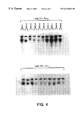
- FIG. 8 shows the RNase protection assay of parental and clonal variants of K562 cells stably transfected with pECE/fes.
- Poly-A+ RNA was selected from 250 ⁇ g of total RNA prepared from wild type HL-60 cells (“HL-60”), wild type K562 cells (“K562”), and colonies WS-1 (“K562/WS-1”), WS-5 (“K562/WS-5”) and WS-6 (“K562/WS-6”).
- Solution hybridization was carried out with 10 6 cpm of a 32 P-labeled c-fes antisense RNA probe containing the 222 bp sequence complementary to exon 2 of the human c-fes gene. Following overnight incubation, the hybridization reaction was digested with RNase and the protected dsRNA fragments were resolved by electrophoresis on 6% polyacrylamide-urea gels, and visualized by autoradiography.
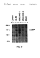
- FIG. 9 shows the immunoprecipitation of parental and clonal variants of K562 cells stably transfected with pECE/fes.
- Cell extracts were prepared from wild-type K562 cells, transfected clones WS-1 and WS-5, and HL-60 cells labeled with [ 35 S]Methionine, and p93 c-fes was immunoprecipitated with an anti-v-fes monoclonal antibody. Immunoprecipitates were analyzed by SDS-polyacrylamide gel electrophoresis and autoradiography as described in the text. The control lane shows precipitation of HL-60 extracts in the absence of the monoclonal antibody.
- FIG. 10 shows the growth curve of K562/fes clone WS-1, K562/neo and parental K562 cells.
- Wild type K562 cells (“K562”), pSV2/neo-transfected cells (“K562/neo”) and colony WS-1 (“K562/fes”) were grown for one week, and cells number was determined at one day intervals with a Coulter particle counter. Cell viability was greater than 95% as determined by trypan blue exclusion.
- FIGS. 11A-F shows the photomicrographs of parental K562 cells and K562/fes clone WS-1.
- Parental K562 cells (A,C,E) and c-fes-transfected clone WS-1 (B,D,F) were tested for their response to 2 day treatment with 10 ⁇ 7 M TPA (A,B), for their ability to reduce NBT (C,D) or for their capacity to phagocytize sheep erythrocytes (E,F).
- RNA sequence which hybridizes only with c-fes mRNA, said specific RNA sequence being obtained from the expression of the recombinant plasmid pcfes4ZKB in a suitable expression vector, such as E. coli , yeast, viruses and other prokaryotic or eukaryotic vectors well known to one of ordinary skill in the art.
- a suitable expression vector such as E. coli , yeast, viruses and other prokaryotic or eukaryotic vectors well known to one of ordinary skill in the art.
- the Mac-1 monoclonal antibody against the macrophage-specific differentiation was obtained from Hybritech, San Diego, Calif.
- the monoclonal antibody (Ab-1) directed against the fes transforming protein common to both the Snyder-Theilen and Gardner strains of feline sarcoma virus was purchased from Oncogene Sciences, Manhasset, N.Y.
- HL-60, K562, and Cos-1 cells were obtained from the American Type Culture Collection.
- HL-60 and K562 cells were grown in RPMI-1640 medium supplemented with 10% heat-inactivated fetal calf serum, 40 mM Hepes, pH 7.4, 1 mM sodium pyruvate, nonessential amino acids, 100 units/ml penicillin, and 100 ⁇ g/ml streptomycin.
- Cos-1 cells were grown in Dulbecco's Modified Eagle's medium supplemented as above. All cells were subcultured twice weekly, and maintained at a density of 10 5 -10 6 cells/ml.
- HL-60 cells were treated with 1.6% Me 2 SO for 4 days to induce granulocytic differentiation.
- Cells (0.5-1.0 ⁇ 10 8 ) were collected by centrifugation and washed twice in Hank's balanced salt solution containing 20 mM EDTA without Mg 2+ or Ca 2+ .
- the cell pellet was sonicated for 5 seconds in 0.5 ml of 50 mM Tris-HCl (pH 7.5) containing 2 mM EGTA, 10 mM DTT, 0.1% Triton-X 100, 1 mM PMSF, 50 ⁇ g/ml aprotinin, 200 ⁇ g/ml leupeptin and 400 ⁇ g/ml soybean trypsin inhibitor, and centrifuged at 15,000 ⁇ g at 4° C. for 10 min.
- Tyrosine kinase activity present in crude cell extracts and column fractions was assayed by non-denaturing polyacrylamide gel electrophoresis as described by Glazer et al (1987) Anal. Biochem. 164, 214-220. Briefly, protein samples were subjected to electrophoresis in 4.5% polyacrylamide mini-gels (Hoefer Scientific) at 4° C. Following electrophoresis, the gels were incubated with Mg 2+ , Mn 2+ and [ ⁇ - 32 P]ATP in the presence and absence of poly(glu,tyr) 4:1 , a synthetic polymer substrate in which tyrosine acts as sole phosphate acceptor. Following incubation at 37° C. for 30 min, the gels were washed extensively in 5% trichloroacetic acid containing 10 mn sodium pyrophosphate, dried and kinase activity was quantitated by autoradiography.
- Triton X-100 extracts were further fractionated by tyrosine-agarose chromatography (Yu et al, 1987, J. Biol. Chem. 262, 17543-17548). Extracts were applied to 1.5 ml tyrosine-agarose columns and aliquots (10 ⁇ g of protein) present in the eluate were resolved on 7.5% SDS-polyacrylamide mini-gels using them Laemmli buffer system (Laemmli, U. K., 1970, Nature 227, 680-685). Proteins were transferred to nitrocellulose membranes using the Genie electrophoretic blotter (Idea Scientific).
- Immunoreactive p93 c-fes was visualized using antiserum to a recombinant human c-fes peptide, and the Protoblot detection system (Promega Biotec) as described by the manufacturer.
- 5 ⁇ 10 7 cells were labeled by incubation at 37° C. for 18 h in 3 ml of methionine-free Iscove's Modified Dulbecco's Medium containing 5% fetal calf serum and 200 ⁇ Ci/ml [ 35 S]-methionine (1,140 Ci/mmol).
- SV40-based mammalian expression vector pECE (Ellis et al, 1986, Cell 45, 721-732) was provided by Dr. William J. Rutter, University of California, San Francisco. pECE was digested with Eco RI and dephosphorylated with calf intestine alkaline phosphatase. The entire human c-fes genomic sequence was isolated as a 13.2 kb Eco RI fragment from the plasmid vector p80 (Trus et al, 1982, J. Biol. Chem. 257, 2730-2733) and cloned into the expression vector pECE.
- the orientation of c-fes was determined by Southern blots using the v-fes probe and it was found to be in the correct orientation such that transcription of the c-fes coding sequence is directed from the SV40 early promoter. This recombinant plasmid is designated pECE/fes.
- Cos-1 cells (5 ⁇ 10 5 cells/100 mm plate) were transfected with 20 ⁇ g of pECE/fes by the modified calcium phosphate precipitation procedure described by Chen and Okayama (1987) Mol. Cell. Biol. 7, 2745-2752.
- analysis was performed 48 h posttransfection.
- K562 cells were transfected by the protoplast fusion technique (Yoakum, G. H., 1984, BioTechnicues 2, 24-31). Briefly, 100 ml of an overnight culture of E. coli transformed with pECE/fes and pSV2/neo were centrifuged at 4000 ⁇ g for 15 min.
- the bacteria were incubated for 2 hr at room temperature (22°-24° C.) with 3 ml of a freshly prepared lysozyme solution (10 mg/ml in 20 mM Hepes, 20% sucrose, pH 7.1). The incubation was stopped by adding 0.8 ml of 1.25 M CaCl 2 and the protoplast preparation was diluted to 10 ml with RPMI-1640. K562 cells (5 ⁇ -10 6 cells) were collected by centrifugation and treated for 1.5 min with 2 ml of the protoplast preparation and 1 ml of fresh 48% polyethylene glycol (mol wt 1000). The cells were then washed five times with RPMI-1640 medium in a CO 2 incubator with the medium changed daily for the first two days.
- High molecular weight DNA prepared by the Gross-Bellard method (Gross-Bellard et al, 1972, Eur. J. Biochem. 36, 32-39) was digested with either Eco RI or Xho I, separated in 0.8% agarose gels and transferred to nitrocellulose (Southern, E. M., 1975, J. Mo. P. Genet. 1, 327-341). Hybridization was carried out with the v-fes probe labeled with [ ⁇ - 32 P]dCTP by the random primer procedure (BRL) at 36° C.
- riboprobe is 498 nucleotides in length, as it contains some sequences transcribed from the vector template.
- Total cellular RNA was prepared by guanidinium isothiocyanate extraction of 10 8 cells followed by centrifugation through cesium chloride (Chirgwin et al, 1979, Biochemistry 18, 5294-5298; Glisin et al, 1973, Biochemistry 13, 2633-2641).
- Poly-A + RNA was selected from 250 ⁇ g total RNA by batch adsorption to oligo-dT cellulose (New England Biolabs). The fraction eluting from oligo-dT cellulose selection was hybridized with 10 6 cpm of the 32 P-labeled c-fes riboprobe (see above). Following overnight (about 12-16 hrs) incubation, the hybridization reaction was digested with RNase, and the protected dsRNA fragments were resolved by polyacrylamide-urea gel electrophoresis and visualized by autoradiography.
- Lysozyme activity was measured spectrophotometrically at 450 nm by the lysis of M. lysodeikticus (Selsted et al, 1978, Infection and Immunity 20, 782-791). The ability of cells to reduce NBT to formazan was assessed by the method described by Breitman et al (1980) Proc. Natl. Acad. Sci. U.S.A. 77, 2936-2940. Fc receptors and immunophagocytosis were determined using sheep erythrocytes coated with anti-erythrocyte antibodies (Breitman et al, 1984, in Methods for Serum - Free Culture of Neuronal and Lymphoid Cells . Alan R.
- Mac-1 macrophage-specific differentiation marker
- the plasmid vector p80 which contains the entire human c-fes genomic sequence (Trus et al, supra), was digested with Kpn I and Xba I. The resulting 1175 bp fragment, which contains c-fes exons 2 and 3, was inserted into the polylinker region of pGEM4Z (Promega Biotec, Madison, Wis.) (FIG. 1 ). This recombinant plasmid, was named pcfes4ZKX.
- pcfes4ZKX was digested with Bgl II and Xba I, which removed c-fes exon 3 and about two-thirds of intron 2.
- the terminal Bgl II and Xba I sites were filled in with the Klenow fragment of DNA polymerase, and the plasmid was re-circularized with T4 DNA ligase.
- the resulting recombinant plasmid, pcfes4ZKB contains c-fes exon 2 flanked by partial sequences of introns 1 and 2 (FIG. 2 ).
- the c-fes insert is upstream from and in opposite orientation to the bacteriophage T7 promoter.
- pcfes4ZKB was digested to completion with Eco Rl, which cuts the plasmid 5′ to the c-fes insert.
- Riboprobe synthesis was conducted in a 20 ⁇ l reaction containing 40 mM Tris-HCl, pH 7.5, 6 mM MgCl 2 , 2 mM spermidine, 10 mM NaCl, 10 mM DTT, 40 units RNasin, 0.5 mM ATP, UTP, and GTP, 12 ⁇ M CTP, 50 ⁇ Ci [ ⁇ - 32 P]CTP (800 Ci/mmol), and 1.0 ⁇ g linearized template DNA (FIG. 3 ). Reactions were initiated by adding 20 units of T7 RNA polymerase, incubated at 37° C. for 1 h, and terminated by the addition of 5 units of RQ1 DNase (Promega).
- the riboprobe was re-dissolved in 100 ⁇ l water, and the amount of labeled CTP incorporated was determined by TCA precipitation (typically 10 6 to 10 9 cpm/pg RNA).
- the c-fes riboprobe synthesized in this manner is 498 nucleotides in length, as it contains some sequences transcribed from the parent vector. Probes were prepared on the same day they were to be used, and the best results were obtained with fresh isotope. This procedure is a modification of the method originally described by Melton et al (1984) Nucleic Acids Res. 12, 7035-7056.
- a deposit of the recombinant plasmid pcfes4ZKB has been made at the ATCC, Rockville, Md., on May 19, 1989, under the accession number 40610.
- the deposit shall be viably maintained, replaced if it becomes non-viable during the life of the patent, for a period of 30 years from the date of the deposit, or for 5 years from the last date of request for a sample of the deposit, whichever is longer, and upon issuance of the patent made available to the public without restriction in accordance with the provisions of the law.
- the Commissioner of Patents and Trademarks, upon request, shall have access to the deposit.
- the 13.2 kb Eco RI fragment identified previously as the human c-fes gene was cloned into the SV40-based mammalian expression vector pECE (Ellis et al, supra) and designated pECE/fes.
- pECE SV40-based mammalian expression vector
- Cos-1 cells were transfected by calcium phosphate precipitation and 48 hr later, Triton X-100 extracts of cellular proteins were analyzed for immunoreactive p93 c-fes and for tyrosine kinase activity.
- Cos-1 cells transfected with pECE/fes expressed a 93 kDa protein which was specifically recognized on Western blots by the c-fes polyclonal antibody (results not shown). Extracts prepared from Cos-1 cells transfected with pECE/fes expressed a single species of tyrosine kinase activity that was present in the 1.0% Triton X-100 cell extract (results not shown). These results indicated that Cos-1 cells are capable of expressing the genomic DNA encoding c-fes and transcribing a functional gene product. However, Cos-1 cells did not acquire characteristics of myeloid cells as a result of c-fes transfection.
- K562 cells do not express p93 c-fes , they are an ideal cell line for transfection experiments with pECE/fes.
- K562 cells were co-transfected with pECE/fes and pSV-2/neo by protoplast fusion and were selected by cloning in soft agar containing 2.5 mg/ml G418. After 14 days in culture, G418-resistant colonies were selected and amplified in RPMI-1640 medium.
- One percent Triton X-100 cell extracts representing the membrane fraction of the cell were prepared from G418-resistant colonies and were screened for tyrosine kinase activity with the nondenaturing gel assay (FIG. 4 ).
- Stably transfected colonies designated WS-1, WS-5, WS-6, and WD-7 had high levels of tyrosine kinase activity.
- Colony WS-1 expressed a level of tyrosine kinase activity comparable to that present in HL-60 cells treated with 1.6% Me 2 SO (FIG. 5 ), a treatment which produces granulocytic differentiation (Zylber-Katz et al, 1985, Cancer Res. 45, 5159-5164).
- p93 c-fes tyrosine kinase activity was not present in either parental or pSV-2/neo-transfected K562 cells (FIG. 5 ).
- a Southern blot of the DNA prepared from several colonies of stably transfected K562 cells indicated varying levels of integration of the c-fes gene (FIG. 6 ).
- the most dramatic example is seen in transfected clone WS-1, in which the level of the c-fes gene is more than 30 times higher than that of the K562 wild-type cells.
- the intensity of the hybridization signal was similar to the level of tyrosine kinase activity expressed by the various clones (FIG. 4 ).
- RNA transcript 498 nucleotides in length containing the 222 nucleotide sequence complimentary to c-fes exon 2. The remainder of the probe is made up of 5′ and 3′ sequences complimentary to c-fes introns 2 and 3, and 37 nucleotides transcribed from the vector template.
- Poly-A + RNA was prepared from K562/fes clones WS-1, WS-5, and WS-6 and hybridized to the c-fes riboprobe overnight.
- polyacrylamide/urea gel electrophoresis revealed a major protected fragment 222 nucleotides in length in each of the transfected clones, which corresponds to c-fes exon 2 (FIG. 8 ).
- the intensity of this band is proportional to the level of c-fes genomic integration (FIG. 6 ), p93 c-fes protein levels (see below).
- an identical protected fragment is present following the RNase protection assay of poly-A + RNA from HL-60 cells, a cell line which normally expresses p93 c-fes .
- no protected fragments were observed following the RNase protection assay of untransfected K562 cells.
- K562/fes clones WS-1 and WS-5 also exhibited a protected fragment of 460 nucleotides (FIG. 8 ), which corresponds to the size of the c-fes genomic fragment contained within the probe (i.e., intron and exon sequences). This indicates that a significant fraction of the c-fes mRNA from transfected cells contains intron sequences, and suggests that c-fes mRNA is less efficiently processed in the transfected clones than in HL-60 cells, which do not exhibit this band.
- transfected K562 clones WS-1 and WS-5 express an immunoreactive 93 kDa protein not seen in the K562 wild type cells. Note that an immunoreactive protein of identical electrophoretic mobility is also seen in immunoprecipitates of HL-60 cells, which are enriched in p93 c-fes .
- Clones WS-1, WS-5 and WS-6 were selected for further study of the changes in maturation which accompanied selection of these cell lines. After 2-3 passages, WS-1 cells grew at a slower rate than wild type K562 cells (FIG. 10 ), a property which may be indicative of differentiation. In addition, all clones adhered loosely to the culture flask, a property which was not seen with parental or pSV-2/neo transfected cells (Table I). Most notable was the response of WS-1 and WS-5 cells to TPA, a treatment which produced approximately 50% macrophage-like cells (FIG. 11A, B).
- TPA-treatment of transfected K562 cells also resulted in expression of the macrophage-specific differentiation antigen Mac-1 (Springer et al, 1979, Eur. J. Immunol. 9, 301-306), whereas TPA-treated wild type cells displayed almost no detectable Mac-1 immunofluorescence (Table I).
- Several functional parameters which are indicative of mature myeloid cells were also examined. Erythrophagocytosis increased dramatically in clones WS-1 and WS-5 and to a lesser extent in WS-6 (Table I and FIG. 11E, F). The percentage of Fc receptor positive cells is high in K562 cells (Koeffler et al, 1981, Cancer Res. 41, 919-926) but doubled in all the clones (Table I).
- K562 cells express mature myeloid characteristics and respond to TPA, they still retain the ability to undergo erythroid differentiation in response to hemin.
- Treatment of K562, K562/WS-1, and K562/WS-5 with 100 mM hemin for 5 days resulted in 64%, 49O, and 63% benzidine-positive cells, respectively.
- the results presented herein clearly indicate that the differentiation-associated 93 kDa tyrosine kinase activity is the product of the human c-fes gene.
- Expression of p93 c-fes is found to be especially high in mature peripheral monocytes and granulocytes, acute and chronic myelogenous leukemias and in leukemia cell lines capable of myeloid differentiation such as K562 and Kg-1a, p93 c-fes expression is either very low or absent.
- the K562 leukemia cell line provided a convenient model to study the function of the human c-fes gene and its role in myeloid differentiation.
- This cell line does not express p93 c-fes and cannot be induced to differentiate along the granulocyte/monocyte pathway by a variety of differentiating agents (Koeffler et al, supra). Therefore, this cell line was utilized herein for transfection with the human c-fes gene in order to identify the role of c-fes in the differentiation process.
- K562 cells transfected with the c-fes gene expressed an active p93 c-fes tyrosine kinase which coincided with the expression of phenotypic markers indicative of a more differentiated cell type such as increased phagocytosis, Fc receptors, NBT reduction and lysozyme activity.
- the latter activity in clone WS-1 was comparable to levels found in mature leukocytes.
- This clonal cell line also responded dramatically to the phorbol ester, TPA, resulting in its morphologic transformation to a macrophage-like cell and expression of the macrophage surface antigen, Mac-1.
- non-radioactive riboprobes are easily prepared as follows.
- the first procedure will utilize a 20 ⁇ l reaction containing 40 mM Tris-HCl, pH 7.5. 6 mM MgCl 2 , 2 mM spermidine, 10 mM NaCl, 10 mM DTT, 40 units RNasin, 0.5 mM ATP, CTP, and GTP, 0.5 mM 5-(N-[N-biotinyl- ⁇ -aminocaproyl]-3-aminoallyl)-uridine 5′-triphosphate, and 1.0 ⁇ g linearized template DNA. Reactions are initiated by adding 20 units of T7 RNA polymerase, incubated at 37° C.
- the second method will employ the same reaction mixture except that 0.5 mM UTP is substituted for 5-(N-[N-biotinyl- ⁇ -aminocaproyl]-3-aminoallyl)-uridine 5′-triphosphate.
- the RNA is reacted with Photoprobe Biotin (Vector Labs, Burlingame, Calif.), a photoactivatable form of biotin which covalently labels the RNA probe.
- the biotinylated RNA probe used in the RNase protection assay is detected with a strepavidin-immunoglobulin-alkaline phosphatase conjugate utilizing NBT and BCIP for color detection (Oncor, Gaithersburg, Md.).
- NBT and BCIP for color detection
- other methods of color detection can, of course, also be employed as will be suggested to one of ordinary skill in the art.
- a kit for the detection of c-fes mRNA comprises a container containing the riboprobe of the present invention, either prepared fresh or cryopreserved.
- a method for the detection of c-fes mRNA in situ or in vitro comprises reacting a cell or tissue preparation with the radioactive or non-radioactive riboprobe of the present invention and determining the degree of hybridization by standard methodologies well known to one of ordinary skill in the art. Such methodologies include radiolabeled, immunohistochemical, fluorescence measurement and the like.
- the present invention now makes it possible to induce myelopoiesis in immature myeloid cells by introducing genomic c-fes gene in immature myeloid cells in which myeloid differentiation is desired.
- Mac-1 88 2 3 70 73 18 a The values for phagocytosis, NBT reduction and lysozyme activity are the average of duplicate determinations. Other values represent a single determination.
- b HL-60 cells were treated for 4 days with 1.25% Me 2 SO.
Landscapes
- Chemical & Material Sciences (AREA)
- Health & Medical Sciences (AREA)
- Organic Chemistry (AREA)
- Life Sciences & Earth Sciences (AREA)
- Proteomics, Peptides & Aminoacids (AREA)
- Genetics & Genomics (AREA)
- Biophysics (AREA)
- Engineering & Computer Science (AREA)
- Biochemistry (AREA)
- Zoology (AREA)
- Molecular Biology (AREA)
- General Health & Medical Sciences (AREA)
- Oncology (AREA)
- Wood Science & Technology (AREA)
- Immunology (AREA)
- Pathology (AREA)
- Analytical Chemistry (AREA)
- Gastroenterology & Hepatology (AREA)
- Physics & Mathematics (AREA)
- Biotechnology (AREA)
- Microbiology (AREA)
- Hospice & Palliative Care (AREA)
- Medicinal Chemistry (AREA)
- Bioinformatics & Cheminformatics (AREA)
- General Engineering & Computer Science (AREA)
- Measuring Or Testing Involving Enzymes Or Micro-Organisms (AREA)
Abstract
A recombinant plasmid and an RNA sequence expressed by said plasmid are described. The RNA sequence hybridize specifically with human c-fes mRNA.
Description
This is a divisional of U.S. application Ser. No. 08/252,136, filed May 31, 1994, issued Mar. 9, 1999 as U.S. Pat. No. 5,879,882, which is a continuation of U.S. application Ser. No. 07/954,427, filed Sep. 30, 1992, now abandoned, which is a continuation of U.S. application Ser. No. 07/355,207, filed May 22, 1989, now abandoned.
The present invention is related generally to diagnostic tests. More particularly, the present invention is related to an RNA probe for detecting the presence of c-fes mRNA in biological samples, such as human cell and tissue RNA preparations.
Expression of the c-fes oncogene is known to play a certain functional role in myelopoiesis in hematopoietic cells (Smithgall et al, 1988, J. Biol. Chem. 263, 15050-15055; Greer et al, 1988, Mol. Cell. Biol., 8, 578-587). However, heretofore direct evidence was lacking to prove that the expression of human c-fes gene induced myeloid differentiation in cells. Furthermore, a specific and sensitive assay to measure the level of c-fes mRNA in human cells and tissues was also heretofore not available.
It is, therefore, an object of the present invention to provide a kit for the detection of c-fes mRNA in biological samples such as human cell and tissue RNA preparations.
It is a further object of the present invention to provide an RNA probe for detecting the presence of c-fes mRNA in vitro or in situ.
It is another object of the present invention to provide a recombinant plasmid comprising exon 2 of the human c-fes genomic sequence for the expression of the transcription product of the c-fes oncogene in a suitable expression vector.
Other objects and advantages will become evident from the following detailed description of the invention.
These and other objects, features and many of the attendant advantages of the invention will be better understood upon a reading of the following detailed description when considered in connection with the accompanying drawings wherein:
FIG. 1 shows the various elements of the human c-fes genomic clone.
FIG. 2 shows schematic construction of the recombinant plasmid pcfes4ZKB.
FIG. 3 schematically shows various steps involved in the RNase protection assay with the c-fes RNA probe in accordance with the present invention.
FIG. 4 shows non-denaturing gel assay for p93c-fes tyrosine kinase activity in colonies of K562 cells stably transfected with pECE/fes. K562 cells were cotransfected with pECE/fes and pSV2/neo as a selectable marker and G418-resistant colonies were selected and screened for p93c-fes tyrosine kinase activity. Aliquots of membrane proteins (15 μg) present in 1.0% Triton X-100 cell extracts were assayed for tyrosine kinase activity using the non-denaturing gel assay described in the text.
FIG. 5 shows the comparison of tyrosine kinase activity in colony WS-1 with differentiated HL-60 cells. One percent Triton X-100 extracts were prepared from either wild type K562 cells (“K562”), pSV2/neo-transfected K562 cells (“K562/neo”), colony WS-1 (“K562/fes”), or HL-60 cells treated for 4 days with 1.6% Me2SO, and p93c-fes tyrosine kinase was partially purified by tyrosine-agarose chromatography. Eluates (3 μg of protein) were assayed for tyrosine kinase activity using the non-denaturing gel assay as described in the text.
FIG. 6 shows the Southern blot analysis of colonies of K562 cells stably transfected with pECE/fes. DNA (10 μg) was prepared from wild type K562 cells (“K562”) and selected colonies of cells transfected with c-fes (designated as “WS-1, WS-5, WS-6, WD-1, WD-2. WD-3, WD-4, and WD-7”), and digested with Eco RI. Plasmid p80 DNA (“p80”) containing the 13.2 kb c-fes gene served as a control. After electrophoresis in 1% agarose gels, Southern blots were prepared and hybridized with a v-fes probe as described herein below. Levels of c-fes integration relative to wild type K562 cells were determined by laser densitometry of the 13.2 kb Eco RI fragment. The endogenous K562 c-fes gene is not visible in the exposure shown (12 h); determination of the c-fes gene in wild type cells required longer autoradiographic exposure (>48 h; data not shown).
FIG. 7 shows the Southern blot analysis of a restriction digest of DNA prepared from colony WS-1. DNA (10 μg) was prepared from wild type K562 cells (“K562”) and colony WS-1 (“K562/fes”) and digested-with Eco RI and Xho I. Plasmid p80 DNA (“p80”) containing the 13.2 kb c-fes gene served as a control. Southern blots and hybridization were carried out as described in the text.
FIG. 8 shows the RNase protection assay of parental and clonal variants of K562 cells stably transfected with pECE/fes. Poly-A+ RNA was selected from 250 μg of total RNA prepared from wild type HL-60 cells (“HL-60”), wild type K562 cells (“K562”), and colonies WS-1 (“K562/WS-1”), WS-5 (“K562/WS-5”) and WS-6 (“K562/WS-6”). Solution hybridization was carried out with 106 cpm of a 32 P-labeled c-fes antisense RNA probe containing the 222 bp sequence complementary to exon 2 of the human c-fes gene. Following overnight incubation, the hybridization reaction was digested with RNase and the protected dsRNA fragments were resolved by electrophoresis on 6% polyacrylamide-urea gels, and visualized by autoradiography.
FIG. 9 shows the immunoprecipitation of parental and clonal variants of K562 cells stably transfected with pECE/fes. Cell extracts were prepared from wild-type K562 cells, transfected clones WS-1 and WS-5, and HL-60 cells labeled with [35S]Methionine, and p93c-fes was immunoprecipitated with an anti-v-fes monoclonal antibody. Immunoprecipitates were analyzed by SDS-polyacrylamide gel electrophoresis and autoradiography as described in the text. The control lane shows precipitation of HL-60 extracts in the absence of the monoclonal antibody.
FIG. 10 shows the growth curve of K562/fes clone WS-1, K562/neo and parental K562 cells. Wild type K562 cells (“K562”), pSV2/neo-transfected cells (“K562/neo”) and colony WS-1 (“K562/fes”) were grown for one week, and cells number was determined at one day intervals with a Coulter particle counter. Cell viability was greater than 95% as determined by trypan blue exclusion.
FIGS. 11A-F shows the photomicrographs of parental K562 cells and K562/fes clone WS-1. Parental K562 cells (A,C,E) and c-fes-transfected clone WS-1 (B,D,F) were tested for their response to 2 day treatment with 10−7 M TPA (A,B), for their ability to reduce NBT (C,D) or for their capacity to phagocytize sheep erythrocytes (E,F).
The above and various other objects and advantages of the present invention are achieved by a specific RNA sequence which hybridizes only with c-fes mRNA, said specific RNA sequence being obtained from the expression of the recombinant plasmid pcfes4ZKB in a suitable expression vector, such as E. coli, yeast, viruses and other prokaryotic or eukaryotic vectors well known to one of ordinary skill in the art.
Unless defined otherwise, all technical and scientific terms used herein have the same meaning as commonly understood by one of ordinary skill in the art to which this invention belongs. Although any methods and materials similar or equivalent to those described herein can be used in the practice or testing of the present invention, the preferred methods and materials are now described. All publications mentioned hereunder are incorporated herein by reference. Unless mentioned otherwise, the techniques employed herein are standard methodologies well known to one of ordinary skill in the art. The materials, methods and examples are illustrative only and not limiting.
Materials
All radioisotopes were obtained from Du Pont-New England Nuclear, Boston, Mass. Tyrosine-agarose, Me2SO, and poly(glu,tyr)4:2 were purchased from Sigma, St. Louis, Mo. The v-fes probe (460 bp Pst I—Pst I fragment) was purchased from Oncor, Gaithersburg, Md. Rabbit antisera to a recombinant c-fes peptide was provided by Dr. Dennis J. Slamon, UCLA School of Medicine, Los Angeles, Calif.. Geneticin (G418) was purchased from Gibco, Grand Island, N.Y. Plasmids p80 and pSV2/neo were obtained from the American Type Culture Collection, Rockville, Md. The Mac-1 monoclonal antibody against the macrophage-specific differentiation was obtained from Hybritech, San Diego, Calif. The monoclonal antibody (Ab-1) directed against the fes transforming protein common to both the Snyder-Theilen and Gardner strains of feline sarcoma virus was purchased from Oncogene Sciences, Manhasset, N.Y.
Cell Culture
HL-60, K562, and Cos-1 cells were obtained from the American Type Culture Collection. HL-60 and K562 cells were grown in RPMI-1640 medium supplemented with 10% heat-inactivated fetal calf serum, 40 mM Hepes, pH 7.4, 1 mM sodium pyruvate, nonessential amino acids, 100 units/ml penicillin, and 100 μg/ml streptomycin. Cos-1 cells were grown in Dulbecco's Modified Eagle's medium supplemented as above. All cells were subcultured twice weekly, and maintained at a density of 105-106 cells/ml. HL-60 cells were treated with 1.6% Me2SO for 4 days to induce granulocytic differentiation.
Preparation of Cell Extracts
Cells (0.5-1.0×10⁸) were collected by centrifugation and washed twice in Hank's balanced salt solution containing 20 mM EDTA without Mg2+ or Ca2+. The cell pellet was sonicated for 5 seconds in 0.5 ml of 50 mM Tris-HCl (pH 7.5) containing 2 mM EGTA, 10 mM DTT, 0.1% Triton- X 100, 1 mM PMSF, 50 μg/ml aprotinin, 200 μg/ml leupeptin and 400 μg/ml soybean trypsin inhibitor, and centrifuged at 15,000×g at 4° C. for 10 min. The supernatant was removed and the pellet was re-extracted with an identical buffer containing 1% Triton X-100. Protein concentrations were determined using a Coomassie blue-based reagent (Pierce Chemical Co.) and BSA as a standard.
Non-denaturing Gel Assay for Tyrosine Kinase Activity
Tyrosine kinase activity present in crude cell extracts and column fractions was assayed by non-denaturing polyacrylamide gel electrophoresis as described by Glazer et al (1987) Anal. Biochem. 164, 214-220. Briefly, protein samples were subjected to electrophoresis in 4.5% polyacrylamide mini-gels (Hoefer Scientific) at 4° C. Following electrophoresis, the gels were incubated with Mg2+, Mn2+ and [τ-32P]ATP in the presence and absence of poly(glu,tyr)4:1, a synthetic polymer substrate in which tyrosine acts as sole phosphate acceptor. Following incubation at 37° C. for 30 min, the gels were washed extensively in 5% trichloroacetic acid containing 10 mn sodium pyrophosphate, dried and kinase activity was quantitated by autoradiography.
Tyrosine-agarose Chromatography, Immunoblotting, and Immunoprecipitation
One percent Triton X-100 extracts were further fractionated by tyrosine-agarose chromatography (Yu et al, 1987, J. Biol. Chem. 262, 17543-17548). Extracts were applied to 1.5 ml tyrosine-agarose columns and aliquots (10 μg of protein) present in the eluate were resolved on 7.5% SDS-polyacrylamide mini-gels using them Laemmli buffer system (Laemmli, U. K., 1970, Nature 227, 680-685). Proteins were transferred to nitrocellulose membranes using the Genie electrophoretic blotter (Idea Scientific). Immunoreactive p93c-fes was visualized using antiserum to a recombinant human c-fes peptide, and the Protoblot detection system (Promega Biotec) as described by the manufacturer. For immunoprecipitation, 5×10⁷ cells were labeled by incubation at 37° C. for 18 h in 3 ml of methionine-free Iscove's Modified Dulbecco's Medium containing 5% fetal calf serum and 200 μCi/ml [35S]-methionine (1,140 Ci/mmol). Cells were then washed, lysed and subjected to immunoprecipitation with biotlnylated anti-v-fes monoclonal antibody (Veronese et al, 1982, J. Virol. 43, 896-904) and streptavidin-agarose according to the manufacturer's protocol. Following extensive washing, immune complexes resolved by electrophoresis through 800 SDS-polyacrylamide gels. Gels were treated with Fluoro-Hance (Research Products International, Mount Prospect, Ill.) prior to autoradiography at −80° C.
Construction of the expression vector pECE/fes
An SV40-based mammalian expression vector pECE (Ellis et al, 1986, Cell 45, 721-732) was provided by Dr. William J. Rutter, University of California, San Francisco. pECE was digested with Eco RI and dephosphorylated with calf intestine alkaline phosphatase. The entire human c-fes genomic sequence was isolated as a 13.2 kb Eco RI fragment from the plasmid vector p80 (Trus et al, 1982, J. Biol. Chem. 257, 2730-2733) and cloned into the expression vector pECE. The orientation of c-fes was determined by Southern blots using the v-fes probe and it was found to be in the correct orientation such that transcription of the c-fes coding sequence is directed from the SV40 early promoter. This recombinant plasmid is designated pECE/fes.
Transfection of Cos-1 and K562 cells
Cos-1 cells (5×10⁵ cells/100 mm plate) were transfected with 20 μg of pECE/fes by the modified calcium phosphate precipitation procedure described by Chen and Okayama (1987) Mol. Cell. Biol. 7, 2745-2752. For transient expression, analysis was performed 48 h posttransfection. K562 cells were transfected by the protoplast fusion technique (Yoakum, G. H., 1984, BioTechnicues 2, 24-31). Briefly, 100 ml of an overnight culture of E. coli transformed with pECE/fes and pSV2/neo were centrifuged at 4000×g for 15 min. The bacteria were incubated for 2 hr at room temperature (22°-24° C.) with 3 ml of a freshly prepared lysozyme solution (10 mg/ml in 20 mM Hepes, 20% sucrose, pH 7.1). The incubation was stopped by adding 0.8 ml of 1.25 M CaCl2 and the protoplast preparation was diluted to 10 ml with RPMI-1640. K562 cells (5×-106 cells) were collected by centrifugation and treated for 1.5 min with 2 ml of the protoplast preparation and 1 ml of fresh 48% polyethylene glycol (mol wt 1000). The cells were then washed five times with RPMI-1640 medium in a CO2 incubator with the medium changed daily for the first two days. After 48 hr, cells were split and plated at 103 cells per 100 mm plate containing RPMI-1640 medium supplemented with 20% heat-inactivated fetal calf serum and 0.4% agarose (SeaPlaque, FMC) and 2.5 mg/ml G418 (Gibco) for selection. After about 14 days of incubation, colonies were selected and cultured in RPMI-1640 medium with 0.2 mg/ml G418.
Southern Blot
High molecular weight DNA, prepared by the Gross-Bellard method (Gross-Bellard et al, 1972, Eur. J. Biochem. 36, 32-39) was digested with either Eco RI or Xho I, separated in 0.8% agarose gels and transferred to nitrocellulose (Southern, E. M., 1975, J. Mo. P. Genet. 1, 327-341). Hybridization was carried out with the v-fes probe labeled with [α-32P]dCTP by the random primer procedure (BRL) at 36° C. for 16 hr in 50% formamide, 5×SSC, 0.50% SDS, 5×Denhardt's solution and 100 μg/ml denatured salmon sperm DNA. Blots were washed with 0.1×SSC, 0.1% SDS at 65° C.
Cloning of genomic c-fes fragments for riboprobe synthesis
A 461 bp Kpn I-Bgl II fragment of the human c-fes locus (Roebroek et al, 1985, EMBO. J. 4, 2897-2903) containing exon 2 and some 3′ and 5′ intron sequences, was cloned into the polylinker region of pGEM-4Z (Promega Biotec). This vector contains the bacteriophage T7 promoter immediately downstream from and in an opposite orientation to the cloning site, allowing for preparation of a c-fes riboprobe (antisense RNA transcript). This was accomplished by linearization of the vector 5′ to the c-fes insert and incubation with T7 RNA polymerase, [α-32P]CTP and unlabeled nucleoside triphosphates according to the manufacturer's protocol. The resulting riboprobe is 498 nucleotides in length, as it contains some sequences transcribed from the vector template.
Poly-A+ RNA Isolation and RNase Protection Assay
Total cellular RNA was prepared by guanidinium isothiocyanate extraction of 108 cells followed by centrifugation through cesium chloride (Chirgwin et al, 1979, Biochemistry 18, 5294-5298; Glisin et al, 1973, Biochemistry 13, 2633-2641). Poly-A+ RNA was selected from 250 μg total RNA by batch adsorption to oligo-dT cellulose (New England Biolabs). The fraction eluting from oligo-dT cellulose selection was hybridized with 106 cpm of the 32 P-labeled c-fes riboprobe (see above). Following overnight (about 12-16 hrs) incubation, the hybridization reaction was digested with RNase, and the protected dsRNA fragments were resolved by polyacrylamide-urea gel electrophoresis and visualized by autoradiography.
Histochemical Assays
Lysozyme activity was measured spectrophotometrically at 450 nm by the lysis of M. lysodeikticus (Selsted et al, 1978, Infection and Immunity 20, 782-791). The ability of cells to reduce NBT to formazan was assessed by the method described by Breitman et al (1980) Proc. Natl. Acad. Sci. U.S.A. 77, 2936-2940. Fc receptors and immunophagocytosis were determined using sheep erythrocytes coated with anti-erythrocyte antibodies (Breitman et al, 1984, in Methods for Serum-Free Culture of Neuronal and Lymphoid Cells. Alan R. Liss, Inc., New York, 215-236). Expression of the macrophage-specific differentiation marker, Mac-1, was examined by immunofluorescence following treatment of cells for 2 days with 100 nM TPA (Ball et al, 1982, Proc. Natl. Acad. Sci. U.S.A. 79, 5374-5378).
Construction of Recombinant Plasmids and Riboprobe Synthesis
The plasmid vector p80, which contains the entire human c-fes genomic sequence (Trus et al, supra), was digested with Kpn I and Xba I. The resulting 1175 bp fragment, which contains c- fes exons 2 and 3, was inserted into the polylinker region of pGEM4Z (Promega Biotec, Madison, Wis.) (FIG. 1). This recombinant plasmid, was named pcfes4ZKX. To prepare a template for riboprobe synthesis, pcfes4ZKX was digested with Bgl II and Xba I, which removed c-fes exon 3 and about two-thirds of intron 2. The terminal Bgl II and Xba I sites were filled in with the Klenow fragment of DNA polymerase, and the plasmid was re-circularized with T4 DNA ligase. The resulting recombinant plasmid, pcfes4ZKB, contains c-fes exon 2 flanked by partial sequences of introns 1 and 2 (FIG. 2). The c-fes insert is upstream from and in opposite orientation to the bacteriophage T7 promoter. Prior to riboprobe synthesis, pcfes4ZKB was digested to completion with Eco Rl, which cuts the plasmid 5′ to the c-fes insert.
Riboprobe synthesis was conducted in a 20 μl reaction containing 40 mM Tris-HCl, pH 7.5, 6 mM MgCl2, 2 mM spermidine, 10 mM NaCl, 10 mM DTT, 40 units RNasin, 0.5 mM ATP, UTP, and GTP, 12 μM CTP, 50 μCi [α-32P]CTP (800 Ci/mmol), and 1.0 μg linearized template DNA (FIG. 3). Reactions were initiated by adding 20 units of T7 RNA polymerase, incubated at 37° C. for 1 h, and terminated by the addition of 5 units of RQ1 DNase (Promega). Following DNase treatment for 15 min at 37° C., 2 μg of carrier tRNA were added, the reaction mixture was extracted with phenol-chloroform, and the labeled RNA was precipitated with ethanol. The riboprobe was re-dissolved in 100 μl water, and the amount of labeled CTP incorporated was determined by TCA precipitation (typically 106 to 109 cpm/pg RNA). The c-fes riboprobe synthesized in this manner is 498 nucleotides in length, as it contains some sequences transcribed from the parent vector. Probes were prepared on the same day they were to be used, and the best results were obtained with fresh isotope. This procedure is a modification of the method originally described by Melton et al (1984) Nucleic Acids Res. 12, 7035-7056.
A deposit of the recombinant plasmid pcfes4ZKB has been made at the ATCC, Rockville, Md., on May 19, 1989, under the accession number 40610. The deposit shall be viably maintained, replaced if it becomes non-viable during the life of the patent, for a period of 30 years from the date of the deposit, or for 5 years from the last date of request for a sample of the deposit, whichever is longer, and upon issuance of the patent made available to the public without restriction in accordance with the provisions of the law. The Commissioner of Patents and Trademarks, upon request, shall have access to the deposit.
Transfection of Cos-1 cells with pECE/fes
The 13.2 kb Eco RI fragment identified previously as the human c-fes gene (Trus et al, supra; Roebroek et al, supra) was cloned into the SV40-based mammalian expression vector pECE (Ellis et al, supra) and designated pECE/fes. To test this construct, Cos-1 cells were transfected by calcium phosphate precipitation and 48 hr later, Triton X-100 extracts of cellular proteins were analyzed for immunoreactive p93c-fes and for tyrosine kinase activity. Cos-1 cells transfected with pECE/fes expressed a 93 kDa protein which was specifically recognized on Western blots by the c-fes polyclonal antibody (results not shown). Extracts prepared from Cos-1 cells transfected with pECE/fes expressed a single species of tyrosine kinase activity that was present in the 1.0% Triton X-100 cell extract (results not shown). These results indicated that Cos-1 cells are capable of expressing the genomic DNA encoding c-fes and transcribing a functional gene product. However, Cos-1 cells did not acquire characteristics of myeloid cells as a result of c-fes transfection.
Co-transfection of K562 Cells with pECE/fes and pSV-2/neo
Since K562 cells do not express p93c-fes , they are an ideal cell line for transfection experiments with pECE/fes. K562 cells were co-transfected with pECE/fes and pSV-2/neo by protoplast fusion and were selected by cloning in soft agar containing 2.5 mg/ml G418. After 14 days in culture, G418-resistant colonies were selected and amplified in RPMI-1640 medium. One percent Triton X-100 cell extracts representing the membrane fraction of the cell were prepared from G418-resistant colonies and were screened for tyrosine kinase activity with the nondenaturing gel assay (FIG. 4). Stably transfected colonies designated WS-1, WS-5, WS-6, and WD-7 had high levels of tyrosine kinase activity. Colony WS-1 expressed a level of tyrosine kinase activity comparable to that present in HL-60 cells treated with 1.6% Me2SO (FIG. 5), a treatment which produces granulocytic differentiation (Zylber-Katz et al, 1985, Cancer Res. 45, 5159-5164). p93c-fes tyrosine kinase activity was not present in either parental or pSV-2/neo-transfected K562 cells (FIG. 5).
A Southern blot of the DNA prepared from several colonies of stably transfected K562 cells indicated varying levels of integration of the c-fes gene (FIG. 6). The most dramatic example is seen in transfected clone WS-1, in which the level of the c-fes gene is more than 30 times higher than that of the K562 wild-type cells. The intensity of the hybridization signal was similar to the level of tyrosine kinase activity expressed by the various clones (FIG. 4). Digestion of WS-1 cell DNA with Eco RI and Xho I generated the expected 13.2 kb and 4.4 kb fragments that were identical to those present in p80 following hybridization with the v-fes DNA probe (FIG. 7).
Analysis of c-fes transcript levels, mRNA processing, and p93c-fes protein synthesis in K562/fes clones
Steady-state levels of c-fes mRNA were determined in transfected K562 clones using the RNase protection assay. The probe used in this assay is an anti-sense RNA transcript 498 nucleotides in length containing the 222 nucleotide sequence complimentary to c-fes exon 2. The remainder of the probe is made up of 5′ and 3′ sequences complimentary to c- fes introns 2 and 3, and 37 nucleotides transcribed from the vector template., Poly-A+ RNA was prepared from K562/fes clones WS-1, WS-5, and WS-6 and hybridized to the c-fes riboprobe overnight. Following RNase digestion, polyacrylamide/urea gel electrophoresis revealed a major protected fragment 222 nucleotides in length in each of the transfected clones, which corresponds to c-fes exon 2 (FIG. 8). The intensity of this band is proportional to the level of c-fes genomic integration (FIG. 6), p93c-fes protein levels (see below). Note that an identical protected fragment is present following the RNase protection assay of poly-A+ RNA from HL-60 cells, a cell line which normally expresses p93c-fes. By contrast, no protected fragments were observed following the RNase protection assay of untransfected K562 cells.
In addition to the major band of 222 nucleotides, K562/fes clones WS-1 and WS-5 also exhibited a protected fragment of 460 nucleotides (FIG. 8), which corresponds to the size of the c-fes genomic fragment contained within the probe (i.e., intron and exon sequences). This indicates that a significant fraction of the c-fes mRNA from transfected cells contains intron sequences, and suggests that c-fes mRNA is less efficiently processed in the transfected clones than in HL-60 cells, which do not exhibit this band. Minor protected fragments approximately 320 and 370 nucleotides in length are also visible in transfected clones WS-1 and WS-5, as well as in HL-60 cells. These fragments may arise from alternate processing of the primary c-fes transcript that occurs 5′ to exon 2, as several alternate splice acceptor sites have been proposed in intron 2 of the c-fes genomic sequence (Roebroek et al, supra).
The results indicate that the translation of c-fes mRNA into p93c-fes protein proceeds normally in transfected K562 cells. This conclusion is based on immunoprecipitation experiments using an anti-v-fes monoclonal antibody. As shown in FIG. 9, transfected K562 clones WS-1 and WS-5 express an immunoreactive 93 kDa protein not seen in the K562 wild type cells. Note that an immunoreactive protein of identical electrophoretic mobility is also seen in immunoprecipitates of HL-60 cells, which are enriched in p93c-fes.
Phenotypic alterations in colonies of K562 cells transfected with c-fes
Clones WS-1, WS-5 and WS-6 were selected for further study of the changes in maturation which accompanied selection of these cell lines. After 2-3 passages, WS-1 cells grew at a slower rate than wild type K562 cells (FIG. 10), a property which may be indicative of differentiation. In addition, all clones adhered loosely to the culture flask, a property which was not seen with parental or pSV-2/neo transfected cells (Table I). Most notable was the response of WS-1 and WS-5 cells to TPA, a treatment which produced approximately 50% macrophage-like cells (FIG. 11A, B). TPA-treatment of transfected K562 cells also resulted in expression of the macrophage-specific differentiation antigen Mac-1 (Springer et al, 1979, Eur. J. Immunol. 9, 301-306), whereas TPA-treated wild type cells displayed almost no detectable Mac-1 immunofluorescence (Table I). Several functional parameters which are indicative of mature myeloid cells were also examined. Erythrophagocytosis increased dramatically in clones WS-1 and WS-5 and to a lesser extent in WS-6 (Table I and FIG. 11E, F). The percentage of Fc receptor positive cells is high in K562 cells (Koeffler et al, 1981, Cancer Res. 41, 919-926) but doubled in all the clones (Table I). Several enzymatic features of mature myeloid cells were also-acquired. In parental K562 cells, lysozyme activity was absent, but it was readily detected in all selected clones, and all transfected cell lines demonstrated high levels of NBT reduction.
Although transfected K562 cells express mature myeloid characteristics and respond to TPA, they still retain the ability to undergo erythroid differentiation in response to hemin. Treatment of K562, K562/WS-1, and K562/WS-5 with 100 mM hemin for 5 days resulted in 64%, 49O, and 63% benzidine-positive cells, respectively.
In summary, the results presented herein clearly indicate that the differentiation-associated 93 kDa tyrosine kinase activity is the product of the human c-fes gene. Expression of p93c-fes is found to be especially high in mature peripheral monocytes and granulocytes, acute and chronic myelogenous leukemias and in leukemia cell lines capable of myeloid differentiation such as K562 and Kg-1a, p93c-fes expression is either very low or absent. These findings suggest that p93e-fes plays a definitive role during the process of maturation of myeloid cells.
The K562 leukemia cell line provided a convenient model to study the function of the human c-fes gene and its role in myeloid differentiation. This cell line does not express p93c-fes and cannot be induced to differentiate along the granulocyte/monocyte pathway by a variety of differentiating agents (Koeffler et al, supra). Therefore, this cell line was utilized herein for transfection with the human c-fes gene in order to identify the role of c-fes in the differentiation process. It was observed that K562 cells transfected with the c-fes gene expressed an active p93c-fes tyrosine kinase which coincided with the expression of phenotypic markers indicative of a more differentiated cell type such as increased phagocytosis, Fc receptors, NBT reduction and lysozyme activity. The latter activity in clone WS-1 was comparable to levels found in mature leukocytes. This clonal cell line also responded dramatically to the phorbol ester, TPA, resulting in its morphologic transformation to a macrophage-like cell and expression of the macrophage surface antigen, Mac-1. Thus, these results demonstrate that an active c-fes gene is imperative for the ultimate expression of the mature myeloid phenotype.
For the purpose of routine assays for the detection of c-fes mRNA, non-radioactive riboprobes are easily prepared as follows.
Nonradioactive RNA probe synthesis
Two procedure can be employed to prepare nonradioactive RNA probes. The first procedure will utilize a 20 μl reaction containing 40 mM Tris-HCl, pH 7.5. 6 mM MgCl2, 2 mM spermidine, 10 mM NaCl, 10 mM DTT, 40 units RNasin, 0.5 mM ATP, CTP, and GTP, 0.5 mM 5-(N-[N-biotinyl-ε-aminocaproyl]-3-aminoallyl)-uridine 5′-triphosphate, and 1.0 μg linearized template DNA. Reactions are initiated by adding 20 units of T7 RNA polymerase, incubated at 37° C. for 1 h, and terminated by the addition of 5 units of RQ1 DNase (Promega). Following DNase treatment for 15 min at 37° C., 2 μg of carrier tRNA is added, the reaction mixture is extracted with phenol/chloroform, and the labeled RNA is precipitated with ethanol.
The second method will employ the same reaction mixture except that 0.5 mM UTP is substituted for 5-(N-[N-biotinyl-ε-aminocaproyl]-3-aminoallyl)-uridine 5′-triphosphate. Following precipitation with ethanol as described above, the RNA is reacted with Photoprobe Biotin (Vector Labs, Burlingame, Calif.), a photoactivatable form of biotin which covalently labels the RNA probe.
In both instances, the biotinylated RNA probe used in the RNase protection assay is detected with a strepavidin-immunoglobulin-alkaline phosphatase conjugate utilizing NBT and BCIP for color detection (Oncor, Gaithersburg, Md.). However, other methods of color detection can, of course, also be employed as will be suggested to one of ordinary skill in the art.
A kit for the detection of c-fes mRNA comprises a container containing the riboprobe of the present invention, either prepared fresh or cryopreserved.
A method for the detection of c-fes mRNA in situ or in vitro comprises reacting a cell or tissue preparation with the radioactive or non-radioactive riboprobe of the present invention and determining the degree of hybridization by standard methodologies well known to one of ordinary skill in the art. Such methodologies include radiolabeled, immunohistochemical, fluorescence measurement and the like.
Of course, the present invention now makes it possible to induce myelopoiesis in immature myeloid cells by introducing genomic c-fes gene in immature myeloid cells in which myeloid differentiation is desired.
It is understood that the examples and embodiments described herein are for illustrative purposes only and that various modifications or changes in light thereof will be suggested to persons skilled in the art and are to be included within the spirit and purview of this application and scope of the appended claims.
| TABLE I |
| Phenotype of parental K562 cells and colonies transfected with c-fes |
| Differentiation | percent positive |
| Markera | HL-60 | K562 | K562/neo | WS-1 | WS-5 | WS-6 |
| Phagocytosis | 42b | 0 | 1 | 65 | 56 | 12 |
| |
68b | 52 | 48 | 94 | 85 | 81 |
| NBT reduction | 77b | 1 | 3 | 64 | 43 | 38 |
| Lysozyme activityc | n.d.d | 0 | 0 | 4.6 | 3.6 | 4.5 |
| Adherence | 0b | 0 | 0 | 80 | 80 | 65 |
| Response to | ||||||
| 10−7 M TPA: | ||||||
| Adherence | 75 | 0 | 0 | 50 | 56 | n.d. |
| Mac-1 | 88 | 2 | 3 | 70 | 73 | 18 |
| aThe values for phagocytosis, NBT reduction and lysozyme activity are the average of duplicate determinations. Other values represent a single determination. | ||||||
| bHL-60 cells were treated for 4 days with 1.25% Me2SO. | ||||||
| cμg of lysozyme/106 cell. Normal leukocytes range from 3.6-8.4 | ||||||
| dn.d., not determined. | ||||||
Claims (7)
1. A single stranded antisense riboprobe comprising at most about 498 nucleotides containing an RNA sequence of 222 nucleotides fully complementary to human c-fes exon-2.
2. A single stranded antisense riboprobe containing an RNA sequence of 222 nucleotides fully complementary to human c-fes exon-2 wherein said riboprobe specifically hybridizes to human nucleic acid comprising c-fes exon-2 sequences but which does not specifically hybridize to human nucleic acid lacking c-fes exon-2 sequences.
3. A kit for the detection of human c-fes RNA comprising a container containing the probe of claim 1 or 2.
4. A method for detecting the presence of human c-fes RNA in a biological sample comprising contacting the RNA present in said biological sample with the antisense riboprobe of claim 1 or 2 wherein the presence of specific hybridization between said antisense riboprobe and human c-fes exon-2 sequence indicates the presence of human c-fes RNA in said biological sample.
5. The antisense riboprobe of claim 1 or 2 wherein said antisense riboprobe is produced by transcription of a plasmid containing a DNA sequence located upstream and in opposite orientation to a bacteriophage T7 promoter.
6. The antisense riboprobe of claim 1 or 2 wherein said antisense riboprobe further comprises a detectable label.
7. The antisense riboprobe of claim 6 wherein said detectable label is selected from the group consisting of a radionuclide, biotin, a fluorescent label and an immunohistochemical agent.
Priority Applications (2)
| Application Number | Priority Date | Filing Date | Title |
|---|---|---|---|
| US09/262,792 US6232069B1 (en) | 1989-05-22 | 1999-03-04 | RNA probe for detecting c-fes mRNA |
| US09/859,000 US20020123049A1 (en) | 1989-05-22 | 2001-05-15 | RNA probe for detecting c-fes mRNA |
Applications Claiming Priority (4)
| Application Number | Priority Date | Filing Date | Title |
|---|---|---|---|
| US35520789A | 1989-05-22 | 1989-05-22 | |
| US95442792A | 1992-09-30 | 1992-09-30 | |
| US08/252,136 US5879882A (en) | 1989-05-22 | 1994-05-31 | RNA probe for detecting c-fes MRNA |
| US09/262,792 US6232069B1 (en) | 1989-05-22 | 1999-03-04 | RNA probe for detecting c-fes mRNA |
Related Parent Applications (1)
| Application Number | Title | Priority Date | Filing Date |
|---|---|---|---|
| US08/252,136 Division US5879882A (en) | 1989-05-22 | 1994-05-31 | RNA probe for detecting c-fes MRNA |
Related Child Applications (1)
| Application Number | Title | Priority Date | Filing Date |
|---|---|---|---|
| US09/859,000 Continuation US20020123049A1 (en) | 1989-05-22 | 2001-05-15 | RNA probe for detecting c-fes mRNA |
Publications (1)
| Publication Number | Publication Date |
|---|---|
| US6232069B1 true US6232069B1 (en) | 2001-05-15 |
Family
ID=23396625
Family Applications (3)
| Application Number | Title | Priority Date | Filing Date |
|---|---|---|---|
| US08/252,136 Expired - Fee Related US5879882A (en) | 1989-05-22 | 1994-05-31 | RNA probe for detecting c-fes MRNA |
| US09/262,792 Expired - Fee Related US6232069B1 (en) | 1989-05-22 | 1999-03-04 | RNA probe for detecting c-fes mRNA |
| US09/859,000 Abandoned US20020123049A1 (en) | 1989-05-22 | 2001-05-15 | RNA probe for detecting c-fes mRNA |
Family Applications Before (1)
| Application Number | Title | Priority Date | Filing Date |
|---|---|---|---|
| US08/252,136 Expired - Fee Related US5879882A (en) | 1989-05-22 | 1994-05-31 | RNA probe for detecting c-fes MRNA |
Family Applications After (1)
| Application Number | Title | Priority Date | Filing Date |
|---|---|---|---|
| US09/859,000 Abandoned US20020123049A1 (en) | 1989-05-22 | 2001-05-15 | RNA probe for detecting c-fes mRNA |
Country Status (3)
| Country | Link |
|---|---|
| US (3) | US5879882A (en) |
| AU (1) | AU5731790A (en) |
| WO (1) | WO1990014440A1 (en) |
Families Citing this family (2)
| Publication number | Priority date | Publication date | Assignee | Title |
|---|---|---|---|---|
| US5348864A (en) * | 1991-01-25 | 1994-09-20 | E. R. Squibb & Sons, Inc. | Mouse vav proto-oncogene DNA and protein sequences |
| IT1282734B1 (en) * | 1996-05-20 | 1998-03-31 | Istituto Biochimico Italiano | ANTISENSE SEQUENCE OLIGONUCLEOTIDES COMPLEMENTARY TO THE PRIMARY TRANSCRIPT OF THE HUMAN PROTOONCOGENE C-FES AND THEIR USE IN COMBINATION |
Citations (2)
| Publication number | Priority date | Publication date | Assignee | Title |
|---|---|---|---|---|
| US4766072A (en) | 1985-07-17 | 1988-08-23 | Promega Corporation | Vectors for in vitro production of RNA copies of either strand of a cloned DNA sequence |
| US5968825A (en) * | 1996-05-20 | 1999-10-19 | Istituto Biochimico Italiano Giovanni Lorenzini S.P.A. | Antisense oligonucleotides complementary to the primary transcript of the human c-fes protooncogene |
Family Cites Families (6)
| Publication number | Priority date | Publication date | Assignee | Title |
|---|---|---|---|---|
| US4699877A (en) * | 1982-11-04 | 1987-10-13 | The Regents Of The University Of California | Methods and compositions for detecting human tumors |
| US4871838A (en) * | 1985-07-23 | 1989-10-03 | The Board Of Rijks Universiteit Leiden | Probes and methods for detecting activated ras oncogenes |
| US4888278A (en) * | 1985-10-22 | 1989-12-19 | University Of Massachusetts Medical Center | In-situ hybridization to detect nucleic acid sequences in morphologically intact cells |
| FI76119C (en) * | 1986-02-27 | 1988-09-09 | Orion Yhtymae Oy | QUANTITATIVE BEARING OF NUCLEIN SYRAMOLEKYLER AND REAGENTS FOERPACKNING SOM ANVAENDS VID FOERFARANDET. |
| EP0304845A3 (en) * | 1987-08-28 | 1991-03-06 | Profile Diagnostic Sciences Inc. | Method and kit for assaying gene expressions |
| DE68909445T2 (en) * | 1988-02-26 | 1994-05-11 | Profile Diagnostic Sciences | Method for determining genetic sequences and test set therefor. |
-
1990
- 1990-05-08 WO PCT/US1990/002567 patent/WO1990014440A1/en not_active Ceased
- 1990-05-08 AU AU57317/90A patent/AU5731790A/en not_active Abandoned
-
1994
- 1994-05-31 US US08/252,136 patent/US5879882A/en not_active Expired - Fee Related
-
1999
- 1999-03-04 US US09/262,792 patent/US6232069B1/en not_active Expired - Fee Related
-
2001
- 2001-05-15 US US09/859,000 patent/US20020123049A1/en not_active Abandoned
Patent Citations (2)
| Publication number | Priority date | Publication date | Assignee | Title |
|---|---|---|---|---|
| US4766072A (en) | 1985-07-17 | 1988-08-23 | Promega Corporation | Vectors for in vitro production of RNA copies of either strand of a cloned DNA sequence |
| US5968825A (en) * | 1996-05-20 | 1999-10-19 | Istituto Biochimico Italiano Giovanni Lorenzini S.P.A. | Antisense oligonucleotides complementary to the primary transcript of the human c-fes protooncogene |
Non-Patent Citations (9)
| Title |
|---|
| Avers, Molecular Cell Biology, Addison-Wesley Publishing Company (1986). |
| DeBold et al., Endocrinology 66(6):2648-2657 (1988). |
| Feldman et al., Oncogene Research 1(4) : 441-458 (Sep.-Oct. 1987).* |
| Matthews et al., Analytical Strategies for the Use of DNA Probes. Analytical Biochemistry 169 : 1-25 (1988).* |
| Melton et al., Nucleic Acids Research, 12(18):7035-7056 (1994). |
| Promega Catalog and Applications Guide, p. 8 and 18 (1988). |
| Roebroek et al., The EMBO Journal 4(11)2897-2903 (1985). |
| The Stragene Catalog p. 39 (1988).* |
| Watson et al., Molecular Biology of the Gene, Benjmain/Cummings Publishing Co., Inc. (1987). |
Also Published As
| Publication number | Publication date |
|---|---|
| WO1990014440A1 (en) | 1990-11-29 |
| US5879882A (en) | 1999-03-09 |
| AU5731790A (en) | 1990-12-18 |
| US20020123049A1 (en) | 2002-09-05 |
Similar Documents
| Publication | Publication Date | Title |
|---|---|---|
| Yu et al. | K562 leukemia cells transfected with the human c-fes gene acquire the ability to undergo myeloid differentiation | |
| Kubo et al. | T‐cell subset‐specific expression of the IL‐4 gene is regulated by a silencer element and STAT6 | |
| Yahraus et al. | The peroxisome biogenesis disorder group 4 gene, PXAAA1, encodes a cytoplasmic ATPase required for stability of the PTS1 receptor. | |
| Sugawara et al. | Induction of cellular senescence in immortalized cells by human chromosome 1 | |
| Haziot et al. | The monocyte differentiation antigen, CD14, is anchored to the cell membrane by a phosphatidylinositol linkage. | |
| US6395510B1 (en) | Vertebrate apoptosis gene: compositions and methods | |
| Chan et al. | A novel abl protein expressed in Philadelphia chromosome positive acute lymphoblastic leukaemia positive acute lymphoblastic leukaemia | |
| Takeda et al. | Deficiency of the GPI anchor caused by a somatic mutation of the PIG-A gene in paroxysmal nocturnal hemoglobinuria | |
| Oren et al. | TAPA-1, the target of an antiproliferative antibody, defines a new family of transmembrane proteins | |
| McCachren et al. | Thrombomodulin expression by human blood monocytes and by human synovial tissue lining macrophages | |
| Molinete et al. | Overproduction of the poly (ADP‐ribose) polymerase DNA‐binding domain blocks alkylation‐induced DNA repair synthesis in mammalian cells. | |
| US5798257A (en) | Nucleic acid encoding human MTS-1 protein | |
| Deuchars et al. | Expression of hamster P-glycoprotein and multidrug resistance in DNA-mediated transformants of mouse LTA cells | |
| WO1997036916A1 (en) | dUTPASE, ITS ISOFORMS, AND DIAGNOSTIC AND OTHER USES | |
| Lefrancois et al. | Acquisition of cytotoxic T lymphocyte-specific carbohydrate differentiation antigens. | |
| Fleischnick et al. | Extended MHC haplotypes in 21-hydroxylase-deficiency congenital adrenal hyperplasia: shared genotypes in unrelated patients | |
| Boggs et al. | Mutations affecting assembly and stability of tubulin: evidence for a nonessential β-tubulin in CHO cells | |
| Modiano et al. | Posttranscriptional regulation of T-cell IL-2 production by human pooled immunoglobin | |
| Hirst et al. | Human GMP synthetase. Protein purification, cloning, and functional expression of cDNA. | |
| WO2000052173A2 (en) | Cloned human sphingosine kinase homologues | |
| Prezant et al. | Biochemical characterization of a pedigree with mitochondrially inherited deafness | |
| US5627039A (en) | Mortalin and methods for determining complementation group assignment of cancer cells | |
| D'Urso et al. | Human glucose-6-phosphate dehydrogenase gene carried on a yeast artificial chromosome encodes active enzyme in monkey cells | |
| Ayusawa et al. | Isolation of functional cDNA clones for human thymidylate synthase. | |
| Arredondo-Vega et al. | Paradoxical expression of adenosine deaminase in T cells cultured from a patient with adenosine deaminase deficiency and combine immunodeficiency. |
Legal Events
| Date | Code | Title | Description |
|---|---|---|---|
| FEPP | Fee payment procedure |
Free format text: PAYOR NUMBER ASSIGNED (ORIGINAL EVENT CODE: ASPN); ENTITY STATUS OF PATENT OWNER: LARGE ENTITY |
|
| REMI | Maintenance fee reminder mailed | ||
| FPAY | Fee payment |
Year of fee payment: 4 |
|
| SULP | Surcharge for late payment | ||
| REMI | Maintenance fee reminder mailed | ||
| LAPS | Lapse for failure to pay maintenance fees | ||
| STCH | Information on status: patent discontinuation |
Free format text: PATENT EXPIRED DUE TO NONPAYMENT OF MAINTENANCE FEES UNDER 37 CFR 1.362 |
|
| FP | Lapsed due to failure to pay maintenance fee |
Effective date: 20090515 |