US6022699A - Myeloperoxidase assay of endotoxin-induced inflammation - Google Patents
Myeloperoxidase assay of endotoxin-induced inflammation Download PDFInfo
- Publication number
- US6022699A US6022699A US09/304,320 US30432099A US6022699A US 6022699 A US6022699 A US 6022699A US 30432099 A US30432099 A US 30432099A US 6022699 A US6022699 A US 6022699A
- Authority
- US
- United States
- Prior art keywords
- composition
- assay
- mpo
- endotoxin
- interfering
- Prior art date
- Legal status (The legal status is an assumption and is not a legal conclusion. Google has not performed a legal analysis and makes no representation as to the accuracy of the status listed.)
- Expired - Lifetime
Links
- 102000003896 Myeloperoxidases Human genes 0.000 title claims abstract description 73
- 108090000235 Myeloperoxidases Proteins 0.000 title claims abstract description 73
- 239000002158 endotoxin Substances 0.000 title claims abstract description 46
- 206010061218 Inflammation Diseases 0.000 title claims abstract description 9
- 230000004054 inflammatory process Effects 0.000 title claims abstract description 9
- 238000003556 assay Methods 0.000 title claims description 27
- 230000002452 interceptive effect Effects 0.000 claims abstract description 16
- 238000011694 lewis rat Methods 0.000 claims abstract description 11
- 230000000694 effects Effects 0.000 claims description 56
- JRBJSXQPQWSCCF-UHFFFAOYSA-N 3,3'-Dimethoxybenzidine Chemical compound C1=C(N)C(OC)=CC(C=2C=C(OC)C(N)=CC=2)=C1 JRBJSXQPQWSCCF-UHFFFAOYSA-N 0.000 claims description 46
- 239000000203 mixture Substances 0.000 claims description 38
- LZZYPRNAOMGNLH-UHFFFAOYSA-M Cetrimonium bromide Chemical compound [Br-].CCCCCCCCCCCCCCCC[N+](C)(C)C LZZYPRNAOMGNLH-UHFFFAOYSA-M 0.000 claims description 29
- HDFGOPSGAURCEO-UHFFFAOYSA-N N-ethylmaleimide Chemical compound CCN1C(=O)C=CC1=O HDFGOPSGAURCEO-UHFFFAOYSA-N 0.000 claims description 27
- GHAZCVNUKKZTLG-UHFFFAOYSA-N N-ethyl-succinimide Natural products CCN1C(=O)CCC1=O GHAZCVNUKKZTLG-UHFFFAOYSA-N 0.000 claims description 25
- 150000003573 thiols Chemical class 0.000 claims description 18
- 150000001875 compounds Chemical class 0.000 claims description 17
- 239000000872 buffer Substances 0.000 claims description 15
- 230000003647 oxidation Effects 0.000 claims description 15
- 238000007254 oxidation reaction Methods 0.000 claims description 15
- 241001465754 Metazoa Species 0.000 claims description 14
- 239000008188 pellet Substances 0.000 claims description 13
- 238000002835 absorbance Methods 0.000 claims description 12
- 230000008859 change Effects 0.000 claims description 8
- 239000006228 supernatant Substances 0.000 claims description 8
- 241000283973 Oryctolagus cuniculus Species 0.000 claims description 7
- 238000010171 animal model Methods 0.000 claims description 5
- 229940079593 drug Drugs 0.000 claims description 5
- 239000003814 drug Substances 0.000 claims description 5
- 239000008187 granular material Substances 0.000 claims description 5
- 238000012544 monitoring process Methods 0.000 claims description 3
- 210000000440 neutrophil Anatomy 0.000 claims description 3
- PEEHTFAAVSWFBL-UHFFFAOYSA-N Maleimide Chemical compound O=C1NC(=O)C=C1 PEEHTFAAVSWFBL-UHFFFAOYSA-N 0.000 claims description 2
- AVOLMBLBETYQHX-UHFFFAOYSA-N etacrynic acid Chemical compound CCC(=C)C(=O)C1=CC=C(OCC(O)=O)C(Cl)=C1Cl AVOLMBLBETYQHX-UHFFFAOYSA-N 0.000 claims description 2
- 229960003199 etacrynic acid Drugs 0.000 claims description 2
- 230000001939 inductive effect Effects 0.000 claims description 2
- FPYJFEHAWHCUMM-UHFFFAOYSA-N maleic anhydride Chemical compound O=C1OC(=O)C=C1 FPYJFEHAWHCUMM-UHFFFAOYSA-N 0.000 claims description 2
- SEEYREPSKCQBBF-UHFFFAOYSA-N n-methylmaleimide Chemical compound CN1C(=O)C=CC1=O SEEYREPSKCQBBF-UHFFFAOYSA-N 0.000 claims description 2
- 230000003381 solubilizing effect Effects 0.000 claims description 2
- 238000000265 homogenisation Methods 0.000 abstract description 8
- 229940121363 anti-inflammatory agent Drugs 0.000 abstract description 6
- 239000002260 anti-inflammatory agent Substances 0.000 abstract description 6
- 125000003396 thiol group Chemical group [H]S* 0.000 abstract description 5
- 239000003638 chemical reducing agent Substances 0.000 abstract description 4
- 239000000470 constituent Substances 0.000 abstract description 4
- 239000000126 substance Substances 0.000 abstract description 4
- 238000010256 biochemical assay Methods 0.000 abstract description 2
- 230000000903 blocking effect Effects 0.000 abstract description 2
- 230000001413 cellular effect Effects 0.000 abstract description 2
- 230000001225 therapeutic effect Effects 0.000 abstract description 2
- 238000007865 diluting Methods 0.000 abstract 1
- 125000000524 functional group Chemical group 0.000 abstract 1
- RWSXRVCMGQZWBV-WDSKDSINSA-N glutathione Chemical compound OC(=O)[C@@H](N)CCC(=O)N[C@@H](CS)C(=O)NCC(O)=O RWSXRVCMGQZWBV-WDSKDSINSA-N 0.000 description 58
- 210000001519 tissue Anatomy 0.000 description 55
- CIWBSHSKHKDKBQ-JLAZNSOCSA-N Ascorbic acid Chemical compound OC[C@H](O)[C@H]1OC(=O)C(O)=C1O CIWBSHSKHKDKBQ-JLAZNSOCSA-N 0.000 description 54
- 238000006243 chemical reaction Methods 0.000 description 40
- 210000003622 mature neutrocyte Anatomy 0.000 description 35
- 229960003180 glutathione Drugs 0.000 description 29
- 235000010323 ascorbic acid Nutrition 0.000 description 27
- 229960005070 ascorbic acid Drugs 0.000 description 27
- 239000011668 ascorbic acid Substances 0.000 description 27
- 229920006008 lipopolysaccharide Polymers 0.000 description 21
- 238000000034 method Methods 0.000 description 20
- 102000004190 Enzymes Human genes 0.000 description 19
- 108090000790 Enzymes Proteins 0.000 description 19
- 238000000605 extraction Methods 0.000 description 12
- 230000000977 initiatory effect Effects 0.000 description 10
- 239000000758 substrate Substances 0.000 description 10
- 241000700159 Rattus Species 0.000 description 9
- 206010046851 Uveitis Diseases 0.000 description 8
- NEFAVCVAHMECFA-UHFFFAOYSA-N [4-(4-diazenyl-3-methoxyphenyl)-2-methoxyphenyl]diazene Chemical compound C1=C(N=N)C(OC)=CC(C=2C=C(OC)C(N=N)=CC=2)=C1 NEFAVCVAHMECFA-UHFFFAOYSA-N 0.000 description 8
- 239000008363 phosphate buffer Substances 0.000 description 8
- 210000004027 cell Anatomy 0.000 description 7
- 238000011156 evaluation Methods 0.000 description 7
- 239000000284 extract Substances 0.000 description 7
- 102000003992 Peroxidases Human genes 0.000 description 6
- PXIPVTKHYLBLMZ-UHFFFAOYSA-N Sodium azide Chemical compound [Na+].[N-]=[N+]=[N-] PXIPVTKHYLBLMZ-UHFFFAOYSA-N 0.000 description 6
- 238000011084 recovery Methods 0.000 description 6
- 238000012360 testing method Methods 0.000 description 6
- 229960003957 dexamethasone Drugs 0.000 description 5
- UREBDLICKHMUKA-CXSFZGCWSA-N dexamethasone Chemical compound C1CC2=CC(=O)C=C[C@]2(C)[C@]2(F)[C@@H]1[C@@H]1C[C@@H](C)[C@@](C(=O)CO)(O)[C@@]1(C)C[C@@H]2O UREBDLICKHMUKA-CXSFZGCWSA-N 0.000 description 5
- 238000002347 injection Methods 0.000 description 5
- 239000007924 injection Substances 0.000 description 5
- 241000894007 species Species 0.000 description 5
- 238000005406 washing Methods 0.000 description 5
- CURLTUGMZLYLDI-UHFFFAOYSA-N Carbon dioxide Chemical compound O=C=O CURLTUGMZLYLDI-UHFFFAOYSA-N 0.000 description 4
- 229910019142 PO4 Inorganic materials 0.000 description 4
- FAPWRFPIFSIZLT-UHFFFAOYSA-M Sodium chloride Chemical compound [Na+].[Cl-] FAPWRFPIFSIZLT-UHFFFAOYSA-M 0.000 description 4
- 238000013459 approach Methods 0.000 description 4
- 238000006555 catalytic reaction Methods 0.000 description 4
- 238000005119 centrifugation Methods 0.000 description 4
- 230000004941 influx Effects 0.000 description 4
- 108040007629 peroxidase activity proteins Proteins 0.000 description 4
- NBIIXXVUZAFLBC-UHFFFAOYSA-K phosphate Chemical compound [O-]P([O-])([O-])=O NBIIXXVUZAFLBC-UHFFFAOYSA-K 0.000 description 4
- 239000010452 phosphate Substances 0.000 description 4
- 238000005063 solubilization Methods 0.000 description 4
- 230000007928 solubilization Effects 0.000 description 4
- 230000000699 topical effect Effects 0.000 description 4
- 208000005623 Carcinogenesis Diseases 0.000 description 3
- 102000008015 Hemeproteins Human genes 0.000 description 3
- 108010089792 Hemeproteins Proteins 0.000 description 3
- 238000009825 accumulation Methods 0.000 description 3
- 210000001742 aqueous humor Anatomy 0.000 description 3
- 230000036952 cancer formation Effects 0.000 description 3
- 235000011089 carbon dioxide Nutrition 0.000 description 3
- 231100000504 carcinogenesis Toxicity 0.000 description 3
- 230000001419 dependent effect Effects 0.000 description 3
- 238000010790 dilution Methods 0.000 description 3
- 239000012895 dilution Substances 0.000 description 3
- 230000002255 enzymatic effect Effects 0.000 description 3
- 210000002683 foot Anatomy 0.000 description 3
- 150000003278 haem Chemical group 0.000 description 3
- 239000011539 homogenization buffer Substances 0.000 description 3
- 239000008057 potassium phosphate buffer Substances 0.000 description 3
- 238000002360 preparation method Methods 0.000 description 3
- 238000011002 quantification Methods 0.000 description 3
- 230000009467 reduction Effects 0.000 description 3
- 239000000523 sample Substances 0.000 description 3
- 239000011780 sodium chloride Substances 0.000 description 3
- WSFSSNUMVMOOMR-UHFFFAOYSA-N Formaldehyde Chemical compound O=C WSFSSNUMVMOOMR-UHFFFAOYSA-N 0.000 description 2
- 108010024636 Glutathione Proteins 0.000 description 2
- MHAJPDPJQMAIIY-UHFFFAOYSA-N Hydrogen peroxide Chemical compound OO MHAJPDPJQMAIIY-UHFFFAOYSA-N 0.000 description 2
- 206010036790 Productive cough Diseases 0.000 description 2
- 230000009471 action Effects 0.000 description 2
- 210000002159 anterior chamber Anatomy 0.000 description 2
- 230000002146 bilateral effect Effects 0.000 description 2
- 239000003795 chemical substances by application Substances 0.000 description 2
- 210000004087 cornea Anatomy 0.000 description 2
- 230000001086 cytosolic effect Effects 0.000 description 2
- 230000007423 decrease Effects 0.000 description 2
- 238000001514 detection method Methods 0.000 description 2
- 238000005194 fractionation Methods 0.000 description 2
- 210000000548 hind-foot Anatomy 0.000 description 2
- 238000001727 in vivo Methods 0.000 description 2
- 238000011065 in-situ storage Methods 0.000 description 2
- 230000028709 inflammatory response Effects 0.000 description 2
- 230000002401 inhibitory effect Effects 0.000 description 2
- 230000005764 inhibitory process Effects 0.000 description 2
- 230000000670 limiting effect Effects 0.000 description 2
- 150000002825 nitriles Chemical class 0.000 description 2
- 239000003113 ocular antiinflammatory agent Substances 0.000 description 2
- 230000003287 optical effect Effects 0.000 description 2
- 230000001590 oxidative effect Effects 0.000 description 2
- 230000008569 process Effects 0.000 description 2
- 239000000047 product Substances 0.000 description 2
- 102000004169 proteins and genes Human genes 0.000 description 2
- 108090000623 proteins and genes Proteins 0.000 description 2
- 210000003802 sputum Anatomy 0.000 description 2
- 208000024794 sputum Diseases 0.000 description 2
- LWIHDJKSTIGBAC-UHFFFAOYSA-K tripotassium phosphate Chemical compound [K+].[K+].[K+].[O-]P([O-])([O-])=O LWIHDJKSTIGBAC-UHFFFAOYSA-K 0.000 description 2
- 230000002792 vascular Effects 0.000 description 2
- GCYDUOSIKYYCLW-WPZUCAASSA-N (2s)-2-amino-5-[[(2r)-1-(carboxymethylamino)-3-(2,5-dioxopyrrolidin-3-yl)sulfanyl-1-oxopropan-2-yl]amino]-5-oxopentanoic acid Chemical compound OC(=O)[C@@H](N)CCC(=O)N[C@H](C(=O)NCC(O)=O)CSC1CC(=O)NC1=O GCYDUOSIKYYCLW-WPZUCAASSA-N 0.000 description 1
- UNZDRHULIOHLKM-IRXDYDNUSA-N (2s)-2-amino-5-[[(2r)-3-[2-amino-5-(4-aminophenyl)phenyl]sulfanyl-1-(carboxymethylamino)-1-oxopropan-2-yl]amino]-5-oxopentanoic acid Chemical compound C1=C(N)C(SC[C@H](NC(=O)CC[C@H](N)C(O)=O)C(=O)NCC(O)=O)=CC(C=2C=CC(N)=CC=2)=C1 UNZDRHULIOHLKM-IRXDYDNUSA-N 0.000 description 1
- UXTIAFYTYOEQHV-UHFFFAOYSA-N 4-(4-amino-3-methoxyphenyl)-2-methoxyaniline;hydron;dichloride Chemical compound [Cl-].[Cl-].C1=C([NH3+])C(OC)=CC(C=2C=C(OC)C([NH3+])=CC=2)=C1 UXTIAFYTYOEQHV-UHFFFAOYSA-N 0.000 description 1
- OAPDFXXWYYMLSW-UHFFFAOYSA-N 4-(4-iminocyclohexa-2,5-dien-1-ylidene)cyclohexa-2,5-dien-1-imine Chemical compound C1=CC(=N)C=CC1=C1C=CC(=N)C=C1 OAPDFXXWYYMLSW-UHFFFAOYSA-N 0.000 description 1
- 102000016938 Catalase Human genes 0.000 description 1
- 108010053835 Catalase Proteins 0.000 description 1
- VEXZGXHMUGYJMC-UHFFFAOYSA-M Chloride anion Chemical compound [Cl-] VEXZGXHMUGYJMC-UHFFFAOYSA-M 0.000 description 1
- 102000016942 Elastin Human genes 0.000 description 1
- 108010014258 Elastin Proteins 0.000 description 1
- 241000588724 Escherichia coli Species 0.000 description 1
- 102000006587 Glutathione peroxidase Human genes 0.000 description 1
- 108700016172 Glutathione peroxidases Proteins 0.000 description 1
- HTTJABKRGRZYRN-UHFFFAOYSA-N Heparin Chemical compound OC1C(NC(=O)C)C(O)OC(COS(O)(=O)=O)C1OC1C(OS(O)(=O)=O)C(O)C(OC2C(C(OS(O)(=O)=O)C(OC3C(C(O)C(O)C(O3)C(O)=O)OS(O)(=O)=O)C(CO)O2)NS(O)(=O)=O)C(C(O)=O)O1 HTTJABKRGRZYRN-UHFFFAOYSA-N 0.000 description 1
- -1 KCN) Chemical class 0.000 description 1
- 206010030113 Oedema Diseases 0.000 description 1
- 108700020962 Peroxidase Proteins 0.000 description 1
- 241000607142 Salmonella Species 0.000 description 1
- 229920002125 Sokalan® Polymers 0.000 description 1
- 208000027418 Wounds and injury Diseases 0.000 description 1
- 230000001154 acute effect Effects 0.000 description 1
- 230000001464 adherent effect Effects 0.000 description 1
- 238000004458 analytical method Methods 0.000 description 1
- 230000003110 anti-inflammatory effect Effects 0.000 description 1
- 230000008901 benefit Effects 0.000 description 1
- 229960002537 betamethasone Drugs 0.000 description 1
- UREBDLICKHMUKA-DVTGEIKXSA-N betamethasone Chemical compound C1CC2=CC(=O)C=C[C@]2(C)[C@]2(F)[C@@H]1[C@@H]1C[C@H](C)[C@@](C(=O)CO)(O)[C@@]1(C)C[C@@H]2O UREBDLICKHMUKA-DVTGEIKXSA-N 0.000 description 1
- 238000002306 biochemical method Methods 0.000 description 1
- 238000004061 bleaching Methods 0.000 description 1
- 210000004369 blood Anatomy 0.000 description 1
- 239000008280 blood Substances 0.000 description 1
- 229920001525 carrageenan Polymers 0.000 description 1
- 239000000679 carrageenan Substances 0.000 description 1
- 229940113118 carrageenan Drugs 0.000 description 1
- 235000010418 carrageenan Nutrition 0.000 description 1
- 239000003153 chemical reaction reagent Substances 0.000 description 1
- 239000007859 condensation product Substances 0.000 description 1
- 230000006378 damage Effects 0.000 description 1
- 230000004665 defense response Effects 0.000 description 1
- 230000007812 deficiency Effects 0.000 description 1
- 238000011161 development Methods 0.000 description 1
- 230000018109 developmental process Effects 0.000 description 1
- 239000000539 dimer Substances 0.000 description 1
- 231100000673 dose–response relationship Toxicity 0.000 description 1
- 229920002549 elastin Polymers 0.000 description 1
- 230000008030 elimination Effects 0.000 description 1
- 238000003379 elimination reaction Methods 0.000 description 1
- 230000003826 endocrine responses Effects 0.000 description 1
- 238000005516 engineering process Methods 0.000 description 1
- 238000006911 enzymatic reaction Methods 0.000 description 1
- YQGOJNYOYNNSMM-UHFFFAOYSA-N eosin Chemical compound [Na+].OC(=O)C1=CC=CC=C1C1=C2C=C(Br)C(=O)C(Br)=C2OC2=C(Br)C(O)=C(Br)C=C21 YQGOJNYOYNNSMM-UHFFFAOYSA-N 0.000 description 1
- 238000002474 experimental method Methods 0.000 description 1
- 239000012530 fluid Substances 0.000 description 1
- 239000003862 glucocorticoid Substances 0.000 description 1
- 150000004820 halides Chemical class 0.000 description 1
- 229960002897 heparin Drugs 0.000 description 1
- 229920000669 heparin Polymers 0.000 description 1
- 230000003118 histopathologic effect Effects 0.000 description 1
- 230000009215 host defense mechanism Effects 0.000 description 1
- 239000000852 hydrogen donor Substances 0.000 description 1
- 238000010952 in-situ formation Methods 0.000 description 1
- 230000006698 induction Effects 0.000 description 1
- 230000002757 inflammatory effect Effects 0.000 description 1
- 230000008798 inflammatory stress Effects 0.000 description 1
- 208000014674 injury Diseases 0.000 description 1
- 230000000968 intestinal effect Effects 0.000 description 1
- 239000002085 irritant Substances 0.000 description 1
- 231100000021 irritant Toxicity 0.000 description 1
- 210000000265 leukocyte Anatomy 0.000 description 1
- 239000003550 marker Substances 0.000 description 1
- 238000000386 microscopy Methods 0.000 description 1
- 238000002156 mixing Methods 0.000 description 1
- 238000012986 modification Methods 0.000 description 1
- 230000004048 modification Effects 0.000 description 1
- 239000000041 non-steroidal anti-inflammatory agent Substances 0.000 description 1
- 229940021182 non-steroidal anti-inflammatory drug Drugs 0.000 description 1
- 229940100654 ophthalmic suspension Drugs 0.000 description 1
- 210000000056 organ Anatomy 0.000 description 1
- 239000012188 paraffin wax Substances 0.000 description 1
- 230000036961 partial effect Effects 0.000 description 1
- 150000002978 peroxides Chemical class 0.000 description 1
- 230000003285 pharmacodynamic effect Effects 0.000 description 1
- 230000000144 pharmacologic effect Effects 0.000 description 1
- 229910000160 potassium phosphate Inorganic materials 0.000 description 1
- 235000011009 potassium phosphates Nutrition 0.000 description 1
- 239000002244 precipitate Substances 0.000 description 1
- 230000000069 prophylactic effect Effects 0.000 description 1
- 238000011555 rabbit model Methods 0.000 description 1
- 239000011535 reaction buffer Substances 0.000 description 1
- 239000011541 reaction mixture Substances 0.000 description 1
- 230000007115 recruitment Effects 0.000 description 1
- 230000002829 reductive effect Effects 0.000 description 1
- 230000008929 regeneration Effects 0.000 description 1
- 238000011069 regeneration method Methods 0.000 description 1
- 230000003252 repetitive effect Effects 0.000 description 1
- 230000004044 response Effects 0.000 description 1
- 238000009738 saturating Methods 0.000 description 1
- 239000007787 solid Substances 0.000 description 1
- 239000000243 solution Substances 0.000 description 1
- 238000000527 sonication Methods 0.000 description 1
- 238000002798 spectrophotometry method Methods 0.000 description 1
- 239000002294 steroidal antiinflammatory agent Substances 0.000 description 1
- 230000003637 steroidlike Effects 0.000 description 1
- 239000003774 sulfhydryl reagent Substances 0.000 description 1
- 239000000725 suspension Substances 0.000 description 1
- 238000012353 t test Methods 0.000 description 1
- 208000037816 tissue injury Diseases 0.000 description 1
- 210000001745 uvea Anatomy 0.000 description 1
- UHVMMEOXYDMDKI-JKYCWFKZSA-L zinc;1-(5-cyanopyridin-2-yl)-3-[(1s,2s)-2-(6-fluoro-2-hydroxy-3-propanoylphenyl)cyclopropyl]urea;diacetate Chemical compound [Zn+2].CC([O-])=O.CC([O-])=O.CCC(=O)C1=CC=C(F)C([C@H]2[C@H](C2)NC(=O)NC=2N=CC(=CC=2)C#N)=C1O UHVMMEOXYDMDKI-JKYCWFKZSA-L 0.000 description 1
Images


Classifications
-
- G—PHYSICS
- G01—MEASURING; TESTING
- G01N—INVESTIGATING OR ANALYSING MATERIALS BY DETERMINING THEIR CHEMICAL OR PHYSICAL PROPERTIES
- G01N33/00—Investigating or analysing materials by specific methods not covered by groups G01N1/00 - G01N31/00
- G01N33/48—Biological material, e.g. blood, urine; Haemocytometers
- G01N33/50—Chemical analysis of biological material, e.g. blood, urine; Testing involving biospecific ligand binding methods; Immunological testing
- G01N33/5005—Chemical analysis of biological material, e.g. blood, urine; Testing involving biospecific ligand binding methods; Immunological testing involving human or animal cells
- G01N33/5008—Chemical analysis of biological material, e.g. blood, urine; Testing involving biospecific ligand binding methods; Immunological testing involving human or animal cells for testing or evaluating the effect of chemical or biological compounds, e.g. drugs, cosmetics
- G01N33/5082—Supracellular entities, e.g. tissue, organisms
- G01N33/5088—Supracellular entities, e.g. tissue, organisms of vertebrates
-
- G—PHYSICS
- G01—MEASURING; TESTING
- G01N—INVESTIGATING OR ANALYSING MATERIALS BY DETERMINING THEIR CHEMICAL OR PHYSICAL PROPERTIES
- G01N33/00—Investigating or analysing materials by specific methods not covered by groups G01N1/00 - G01N31/00
- G01N33/48—Biological material, e.g. blood, urine; Haemocytometers
- G01N33/50—Chemical analysis of biological material, e.g. blood, urine; Testing involving biospecific ligand binding methods; Immunological testing
- G01N33/5005—Chemical analysis of biological material, e.g. blood, urine; Testing involving biospecific ligand binding methods; Immunological testing involving human or animal cells
- G01N33/5008—Chemical analysis of biological material, e.g. blood, urine; Testing involving biospecific ligand binding methods; Immunological testing involving human or animal cells for testing or evaluating the effect of chemical or biological compounds, e.g. drugs, cosmetics
- G01N33/502—Chemical analysis of biological material, e.g. blood, urine; Testing involving biospecific ligand binding methods; Immunological testing involving human or animal cells for testing or evaluating the effect of chemical or biological compounds, e.g. drugs, cosmetics for testing non-proliferative effects
-
- G—PHYSICS
- G01—MEASURING; TESTING
- G01N—INVESTIGATING OR ANALYSING MATERIALS BY DETERMINING THEIR CHEMICAL OR PHYSICAL PROPERTIES
- G01N33/00—Investigating or analysing materials by specific methods not covered by groups G01N1/00 - G01N31/00
- G01N33/48—Biological material, e.g. blood, urine; Haemocytometers
- G01N33/50—Chemical analysis of biological material, e.g. blood, urine; Testing involving biospecific ligand binding methods; Immunological testing
- G01N33/5005—Chemical analysis of biological material, e.g. blood, urine; Testing involving biospecific ligand binding methods; Immunological testing involving human or animal cells
- G01N33/5094—Chemical analysis of biological material, e.g. blood, urine; Testing involving biospecific ligand binding methods; Immunological testing involving human or animal cells for blood cell populations
Definitions
- This invention relates to in vivo models for identifying anti-inflammatory agents.
- this invention relates to a model of endotoxin-induced inflammation for the evaluation of anti-inflammatory agents.
- Carrageenan-induced paw edema in the rat has been a standard in vivo model for the pharmacological evaluation of systemically administered anti-inflammatory agents. See Winter et al., Proc Soc Exp Biol Med 111: 544-547 (1962) and Vinegar et al., J Pharmacol Exp Ther 166: 96-103 (1969). In this model, subplantar administration of irritant into the hind paw of the rat causes a local inflammatory response which can be inhibited by steroidal and non-steroidal anti-inflammatory agents.
- LPS bacterial endotoxin
- MPO myeloperoxidase
- PMN-derived MPO primarily catalyzes hydrogen peroxide (H 2 O 2 )-dependent oxidation of halides (i.e., chloride ions) physiologically
- o-DA o-dianisidine
- Modifications of this method have been introduced to circumvent artifacts contributed by tissue/blood constituents, including cytosolic enzymes such as catalase, glutathione peroxidase, and heme proteins, and reducing substrates such as glutathione (GSH) and ascorbic acid (ASC).
- the present invention relates to a test system for the evaluation of anti-inflammatory agents.
- the test system is an assay of PMN-associated MPO activity in tissues from experimental animals, such as the Lewis rat, challenged with a bacterial endotoxin (LPS).
- the assay provides reliable PMN determinations by eliminating interference that can be caused by endogenous electron donors such as ASC or thiols, including GSH and protein sulfhydryls.
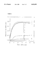
- FIG. 1 shows the effect of ASC on MPO-catalyzed oxidation of o-DA.
- FIG. 2 shows the effect of GSH on MPO-catalyzed oxidation of o-DA.
- Vascular leakage and recruitment of circulating PMNs to the site of injury represents the early phase of the host defense mechanism and response to tissue injury. This response is common to all organs and tissues, including the eye. Clinically, the presence of PMNs in the anterior chamber of the eye can be readily detected by slit-lamp examination or quantified with the use of a Kowa cell/flare meter. However, this technology is not particularly useful when quantifying the presence of PMNs in the eyes of experimental species such as the rat. A more quantitative approach is the use of the biochemical assay of PMN-associated MPO activity.
- This enzyme is highly enriched in the azurophil granules of PMNs (Schultz et al., Arch Biochem Biophys 96: 465-467 (1962) recruited to injured tissue to mediate the acute phase of the inflammatory response (Vinegar et al., J Pharmacol Exp Ther 166: 96-103 (1969). MPO is not found in normal, non-inflamed tissue.
- ASC causes the nonenzymic reduction of 3,3'-dimethoxybenzidinediimine to o-DA (Wise et al., Carcinogenesis 5: 1499-1503 (1984). Both processes lead to an underestimation of MPO activity.
- Suitable non-interfering thiol reactive compounds include maleimide, N-methyl maleimide, N-ethyl maleimide, ethacrynic acid, and maleic anhydride.
- a preferred non-interfering thiol reactive compound is N-ethyl maleimide (NEM). NEM does not affect MPO catalysis in the absence of GSH. For convenience only, NEM will be used as a representative non-interfering thiol reactive compound in the remainder of this Detailed Description section.
- the method of the present invention is applicable to a variety of experimental species, including the Lewis rat, NZA rabbit, DB rabbit and other rabbit strains, the method is preferably conducted with Lewis rats.
- the present method is capable of application to a variety of target tissues within the chosen experimental species, e.g., ocular, dermatological, intestinal, articular and vascular tissues.
- a preferred target tissue for the method of the present invention is the ocular tissue.
- rat ocular tissues contain relatively small amounts of ASC.
- This reductant resides predominantly in the cornea with barely detectable levels in the vitreous (Varma et al., Ophthalmic Res 20: 164-173 (1988).
- the low ASC concentration its selective association with the cornea, combined with its high aqueous solubility and its extensive dilution during tissue homogenization minimize the potential for interference in the MPO assay.
- GSH a ubiquitous mammalian cellular constituent, presents a greater potential for assay interference. It is found at a concentration of about 4 mM in the rat lens (Costagliola et al., Exp Eye Res 43: 905-914 (1986)), a tissue contributing about 40% to the total ocular mass in this species.
- tissue homogenization with HTA-Br buffer alone allowed detection of only 10% of total tissue MPO activity. MPO specific activity was enhanced substantially (40% of total) when tissue homogenization was conducted per published methods (Grisham et al., Methods in Enzymology, Vol. 186, Ed. Packer et al., San Diego: Academic Press, pp. 729-742 (1990)) using a large excess of homogenization buffer in combination with HTA-Br buffer extraction. This finding indicates a partial removal and/or dilution of endogenous reductants/thiols during tissue fractionation. Only upon inclusion of NEM in the homogenization buffer in combination with HTA-Br extraction was maximal recovery of assayable MPO activity achievable following LPS challenge.
- Blocking free sulfhydryl functional groups i.e., residual glutathione and protein sulfhydryls
- NEM blocking free sulfhydryl functional groups during tissue extraction by reaction with NEM prevents their later reaction with 3,3'-dimethoxybenzidinediimine, and thus prevents underestimation of o-DA oxidation in the MPO assay.
- the use of NEM during tissue extraction did not affect the apparent substrate affinities of neutrophil MPO for either H 2 O 2 or o-DA which were comparable to those determined for the human sputum derived enzyme.
- the method of the present invention which in this case has been applied to eye tissue, monitors total ocular MPO activity and has less inherent variability between companion eyes than methods that employ aqueous humor taps combined with cell counting (Bhattacherjee et al., Invest Ophthalmol Vis Sci 24: 196-202 (1983)).
- aqueous humor taps combined with cell counting Bosset of aqueous humor taps combined with cell counting.
- there appears to be considerable variability between animals in total MPO activity This is likely the result of differences among animals in their endocrine response (glucocorticoid release) to the inflammatory stress condition (Udelsman et al., Curr Probl Surg 31: 655-720 (1994)).
- An alternative, but a less likely cause, may be an inadequate delivery of, or variable pharmacodynamics relative to, subplantarly administered LPS.
- an effective endotoxin-induced inflammation assay for comparison of the anti-inflammatory efficacy and potency of a drug comprises the steps of:
- a) administering e.g., topically or systemically
- the drug therapeutically or prophylactically (or both) to a target tissue of an experimental animal
- step (d) centrifuging the composition of step (d) to recover PMN and MPO in a pellet;
- step (e) solubilizing the pellet of step (e) in a buffered composition containing HTA-Br in order to release MPO from PMN azurophil granules, wherein the buffered composition has a pH of about 5-7, preferably about pH 6;
- step (f) centrifuging the composition of step (f) to recover MPO in the supernatant
- step (g) preparing a buffered composition comprising the supernatant of step (g), o-DA, H 2 O 2 and HTA-Br, wherein the buffered composition has a pH of about 5-7, preferably about pH 6;
- the method of the present invention preferably involves bilateral administration of the test article (or control).
- One suitable buffer for use in the compositions of steps (d) and (f) is phosphate buffer a concentration of 50 mM.
- the centrifugation steps (e) and (g) should be conducted at low temperatures, e.g., ice-cold temperatures of about 4° C.
- the non-interfering thiol reactive compound is preferably NEM.
- the concentration of non-interfering thiol reactive compound in the composition of step (d) is should be about 0.1-30 mM, preferably about 10 mM.
- the volume of the composition of step (d) is generally about 10-100 times, preferably about 50 times, the weight of the enucleated eye. For example, assuming that the total weight of the eye tissue is 0.1 g and that 0.1 g is roughly equivalent to 0.1 mL, the volume of the composition of step (d) is preferably 5 mL.
- the method of the present invention comprises two additional steps between step (e) and step (f).
- the two additional steps are an intermediate extraction/washing step and another centrifugation step.
- These additional steps i.e., repetitive extraction/washing
- the pellet is re-dissolved in a fresh buffered, pH about 5-8, preferably pH about 7.4, composition comprising a non-interfering thiol reactive compound, and then centrifuged again before step (f).
- the HTA-Br step (step (f)) preferably involves three freeze/thaw cycles to help maximize the PMN disruption and release of MPO from azurophil granules.
- the solubilization of the pellet of step (e) is preferably aided by mechanical means, such as probe sonication.
- the supernatant (containing MPO) is recovered (step(g)), it is combined with o-DA, H 2 O 2 and HTA-Br in a buffered composition.
- the rate of o-DA oxidation in the buffered composition is then monitored spectrophotometrically (460 nm) as an indication of the amount of MPO activity.
- the amount of o-DA to be present in the buffered composition to be assayed should be about 1-1.5 mM, preferably about 1.4-1.5 mM.
- the amount of H 2 O 2 to be present in the buffered composition to be assayed should be about 150-300 ⁇ M, preferably about 220-300 ⁇ M.
- the amount of HTA-Br present in the buffered composition to be assayed should be about 100-500 ⁇ M, preferably about 250 ⁇ M.
- the supernatant recovered in step (g) is assayed in two separate reactions both supplemented with o-DA and H 2 O 2 as described above.
- One reaction is carried out in the absence and the other in the presence of either a cyanide salt (e.g., KCN), in an amount of about 1-10 mM, preferably about 1 mM, or sodium azide (NaN 3 ), in an amount of about 5-30 mM, preferably about 10 mM.
- a cyanide salt e.g., KCN
- the rate of o-DA oxidation is much greater in the case of the reaction without a cyanide salt or NaN 3 , the o-DA oxidation is attributable to MPO activity. With the method of the present invention, there should be less than about 5% non-enzymatic oxidation of o-DA.
- N-ethylmaleimide N-ethylmaleimide (NEM), o-DA (3,3'-dimethoxybenzidine dihydrochloride), HTA-Br, LPS (lipopolysaccharide, E. coli 0111:B4), heparin, dexamethasone, histopaque 1083 and histopaque 1119 were obtained from Sigma Chemical Company, St. Louis, Mo.
- Dextran-100 was a product of Crescent Chemical Company, Hauppauge, N.Y. Hydrogen peroxide (30%) was obtained from J. T. Baker and stored at 4° C. All other chemicals used were of the highest purity available.
- Human sputum myeloperoxidase HSMPO was obtained from Elastin Products Co., Pacific, Mo.
- the enzyme (2.4 mg solid) was dissolved in 2.4 mL of 50 mM potassium phosphate buffer (pH 6.0), and stored in 0.1 mL aliquots at -20° C., then diluted 10-fold with 50 mM potassium phosphate buffer (pH 6.0) to provide a HSMPO concentration of 0.1 mg/mL.
- Uveitis was induced by subplantar injection of 0.1 mL of a saline solution containing 200 ⁇ g of LPS into the right hind paw of female Lewis rats (4 to 5 animals/group). Twenty-four (24) hours after LPS injection, animals were sacrificed by CO 2 inhalation, and total ocular PMN content was determined by assessing MPO activity, as described below.
- Ocular tissues were prepared for the quantification of PMN content as follows. Freshly enucleated eyes, or eyes rapidly frozen on dry ice and stored at -70° C. were homogenized in 5.0 mL of ice-cold buffer with the aid of a Brinkman Polytron homogenizer. The homogenizer probe was rinsed with 5.0 mL of the same buffer, and washings were combined with the initial tissue homogenate. The homogenate was centrifuged for 30 minutes at 12,000 ⁇ g at 4° C. The proteinaceous pellet was then homogenized once more in ice-cold buffer and the homogenate was centrifuged again (as described above).
- HTA-Br ice-cold 0.5% hexadecyltrimethylammonium bromide
- pH 6.0 mM phosphate buffer
- Solubilization was accomplished using a probe-type sonicator (three 10-second bursts from a Heat Systems, Inc. Ultrasonic Process sonicator, Model XL-2010, at an instrument power setting of 2) followed by three sequential freeze (dry ice)-thaw cycles.
- the HTA-Br buffer-treated pellet homogenate was centrifuged (4° C., 30 minutes 12,000 ⁇ g). The supernatant was collected, frozen and stored at -70° C. for later assay of MPO activity.
- the reaction buffer contained 50 mM phosphate/250 ⁇ M HTA-Br/300 ⁇ M H 2 O 2 and 1.5 mM o-DA (pH 6.0). Reactions were initiated by addition of an appropriate amount of HTA-Br solubilized ocular tissue extract. H 2 O 2 concentration was determined spectrophotometrically at 240 nm. See, Nelson et al., Anal. Biochem., 49: 474-478 (1972).
- Dexamethasone suspensions were prepared in a carbopol ophthalmic suspension vehicle at concentrations of 0.0033%, 0.01%, 0.033%, 0.04% and 0.10%, w/v.
- a 5 ⁇ L aliquot of test drug or vehicle was applied topically to each eye of the experimental animals 24, 20, and 4 hours prior to, at the time of, and 4 and 20 hours after LPS foot pad injection. Twenty-four (24) hours following LPS administration, eyes were enucleated, quickly frozen on dry ice and stored at -70° C. until used for tissue extraction and assessment of MPO activity.
- HSMPO was used as reference to assess the enzyme's substrate dependence on both H 2 O 2 and o-DA. As shown in Table 1, optimal reaction rates were obtained in the presence of 300 ⁇ M H 2 O 2 and 1.5 mM o-DA. However, substrate saturation conditions could not be attained for o-DA since precipitates formed at concentrations exceeding 1.5 mM. H 2 O 2 concentrations greater than 300 ⁇ M resulted in a loss of enzyme activity. This loss of activity was likely the result of peroxide-induced heme bleaching (see, Floris et al., Eur J Biochem 207: 697-702 (1992)) due to a limiting concentration of o-DA.
- MPO activity was assayed at room temperature in 3.0 mL of 50 mM potassium phosphate (pH 6.0), supplemented with 37 mM H 2 O 2 , and 1.5 mM o-DA.
Abstract
Description
              TABLE 1
______________________________________
KineticsΨ of HSMPO
  H.sub.2 O.sub.2
              Reaction    o-DA      Reaction
  Concentration Velocity   Concentration Velocity⊥
  (μM) (μM/min/μg) (mM) (μM/min/μg)
______________________________________
0         0           0           0
  13 2.7 0.097 1.6
  27 6.4 0.25 4.2
  54 9.6 0.49 8.0
  107 14.2 0.97 16.3
  220 18.2 1.44 18.7
  437 13.4
______________________________________
 ΨReactions were conducted at room temperature in 50 mM phosphate
 buffer (pH 6.0) in a final volume of 2.0 mL. MPO reactions were initiated
 by addition of 5 μL (0.5 μg) of HSMPO.
  assayed in the presence of 1.44 mM oDA
 ⊥assayed in the presence of 300 μM H.sub.2 O.sub.2
              TABLE 2
______________________________________
Effect of various tissue extractions on the recovery of ocular MPO
 activity following LPS administration in the Lewis rat
         Homogenization Buffer/Volume
                              MPO
                   Phosphate/       Activity
    NEM  μM/min/
   Phosphate [50 mM/  100 mg
  Animal [50 mM] 10 mM] HTA-Br   (x ± S.D.)
  Treatment pH 7.4 pH 7.4 pH 6.0 (n = 10)
______________________________________
Saline   2 × 5.0 mL
                   --        1.0 mL 0.7 ± 0.1
  Saline -- 2 × 5.0 mL 1.0 mL 1.4 ± 0.4
  200 μg LPSΨ -- -- 0.5 mL 10.9 ± 4.0
  200 μg LPS 2 × 5.0 mL -- 0.5 mL 42.1 ± 10.9
  200 μg LPS -- 2 × 5.0 mL 0.5 mL *
      106.6 ± 35.2
      *
______________________________________
 Ψsubplantar injection
  HTABr Buffer = 0.5% HTABr/50 mM kphosphate (pH 6.0)
 *significantly different from LPS in HTABr alone (p < 0.01, t test)
              TABLE 3
______________________________________
KineticsΨ of HTA-Br solubilized MPO activity from whole eyes
  of Lewis rats 24 hours post-endotoxin challenge in the foot pad
    H.sub.2 O.sub.2
               Reaction   o-DA     Reaction
  Concentration Velocity   Concentration Velocity⊥
  (μM) (μM/min) (mM) (μM/min)
______________________________________
0          0          0          0
  16 11.0 0.15 9.0
  32 21.1 0.30 16.3
  62 35.0 0.59 27.4
  93 42.7 0.89 36.2
  124 46.7 1.18 45.3
  182 51.8 1.46 53.1
  241 52.2
  298 51.8
______________________________________
 ΨReactions were conducted at room temperature in 50 mM phosphate
 buffer (pH 6.0) in a final volume of 2.0 mL. MPO reactions were initiated
 by addition of 100 μL of NEMtreated/HTA-Br solubilized ocular tissue
 extract (1/5 of total extract) prepared from rat eyes 24 hours after
 subplantar bacterial endotoxin challenge (200 μg).
  assayed in the presence of 1.5 mM oDA
 ⊥assayed in the presence of 300 μM H.sub.2 O.sub.2
Claims (12)
Priority Applications (1)
| Application Number | Priority Date | Filing Date | Title |
|---|---|---|---|
| US09/304,320 US6022699A (en) | 1999-05-03 | 1999-05-03 | Myeloperoxidase assay of endotoxin-induced inflammation |
Applications Claiming Priority (2)
| Application Number | Priority Date | Filing Date | Title |
|---|---|---|---|
| US09/304,320 US6022699A (en) | 1999-05-03 | 1999-05-03 | Myeloperoxidase assay of endotoxin-induced inflammation |
| US62209899P | 1999-05-21 | 1999-05-21 |
Publications (1)
| Publication Number | Publication Date |
|---|---|
| US6022699A true US6022699A (en) | 2000-02-08 |
Family
ID=26973958
Family Applications (1)
| Application Number | Title | Priority Date | Filing Date |
|---|---|---|---|
| US09/304,320 Expired - Lifetime US6022699A (en) | 1999-05-03 | 1999-05-03 | Myeloperoxidase assay of endotoxin-induced inflammation |
Country Status (1)
| Country | Link |
|---|---|
| US (1) | US6022699A (en) |
Cited By (4)
| Publication number | Priority date | Publication date | Assignee | Title |
|---|---|---|---|---|
| US20060035308A1 (en) * | 2004-08-11 | 2006-02-16 | Chong-Sheng Yuan | Methods and compositions of enzymatic cycling based assays for myeloperoxidase |
| WO2010062787A1 (en) * | 2008-11-03 | 2010-06-03 | Washington University | Bioluminescence imaging of myeloperoxidase activity in vivo, methods, compositions and apparatuses therefor |
| US20110021640A1 (en) * | 2009-07-27 | 2011-01-27 | Kailash Chandra Agarwal | Mechanism-based biochemical standardization of resveratrol products and their uses thereof |
| WO2011133581A1 (en) | 2010-04-19 | 2011-10-27 | General Atomics | Methods and compositions for assaying enzymatic activity of myeloperoxidase in blood samples |
-
1999
- 1999-05-03 US US09/304,320 patent/US6022699A/en not_active Expired - Lifetime
Non-Patent Citations (69)
| Title |
|---|
| Baatz et al., Invest Ophthalmol Vis Sci 36: 1960 1967 (1995). * |
| Baatz et al., Invest Ophthalmol Vis Sci 36: 1960-1967 (1995). |
| Bhattacherjee et al., Invest Ophthalmol Vis Sci 24: 196 202 (1983). * |
| Bhattacherjee et al., Invest Ophthalmol Vis Sci 24: 196-202 (1983). |
| Bradley et al., J Invest Dermatol 78: 206 209 (1982). * |
| Bradley et al., J Invest Dermatol 78: 206-209 (1982). |
| Claiborne et al., Biochemistry 18: 2324 2329 (1979). * |
| Claiborne et al., Biochemistry 18: 2324-2329 (1979). |
| Costagliola et al., Exp Eye Res 43: 905 914 (1986). * |
| Costagliola et al., Exp Eye Res 43: 905-914 (1986). |
| Cousins et al., Exp Eye Res 39: 665 676 (1984). * |
| Cousins et al., Exp Eye Res 39: 665-676 (1984). |
| Cramer et al., Adv Exp Med Biol 121(A):91 9 (1979). * |
| Cramer et al., Adv Exp Med Biol 121(A):91-9 (1979). |
| De Berardinis et al., Exp Eye Res 4: 179 186 (1965). * |
| De Berardinis et al., Exp Eye Res 4: 179-186 (1965). |
| Egan et al., Agents Actions 29: 266 276 (1980). * |
| Egan et al., Agents Actions 29: 266-276 (1980). |
| Floris et al., Eur J Biochem 207: 697 702 (1992). * |
| Floris et al., Eur J Biochem 207: 697-702 (1992). |
| Forrester et al., Graefes Arch Klin Exp Ophthalmol 213: 221 233 (1980). * |
| Forrester et al., Graefes Arch Klin Exp Ophthalmol 213: 221-233 (1980). |
| Graff et al., J. Pharmacol. Toxicol. Methods 39 (3) 169 178, 1998. * |
| Graff et al., J. Pharmacol. Toxicol. Methods 39 (3) 169-178, 1998. |
| Graff et al., Prostaglandins 38: 473 496 (1989). * |
| Graff et al., Prostaglandins 38: 473-496 (1989). |
| Grisham et al., Methods in Enzymology, vol. 186, Ed. Packer et al., San Diego: Academic Press, pp. 729 742 (1990). * |
| Grisham et al., Methods in Enzymology, vol. 186, Ed. Packer et al., San Diego: Academic Press, pp. 729-742 (1990). |
| Haugaard et al., Anal. Biochem 116(2): 341 343, 1981. * |
| Haugaard et al., Anal. Biochem 116(2): 341-343, 1981. |
| Hockwin et al., Glutathione: Metabolism and physiological functions, Ed., Vina, Boca Raton: CRC Press, pp. 207 215 (1990). * |
| Hockwin et al., Glutathione: Metabolism and physiological functions, Ed., Vina, Boca Raton: CRC Press, pp. 207-215 (1990). |
| Krawisz et al., Gastroenterology 87: 1344 1350 (1984). * |
| Krawisz et al., Gastroenterology 87: 1344-1350 (1984). |
| Liem et al., Anal Biochem 98: 388 393 (1979). * |
| Liem et al., Anal Biochem 98: 388-393 (1979). |
| Marquez et al., J Biol Chem 265: 5666 5670 (1990). * |
| Marquez et al., J Biol Chem 265: 5666-5670 (1990). |
| Nelson et al., Anal Biochem 49: 471 478, 1972. * |
| Nelson et al., Anal Biochem 49: 471-478, 1972. |
| Nelson et al., Anal Biochem 49: 474 478 (1972). * |
| Nelson et al., Anal Biochem 49: 474-478 (1972). |
| Saxena et al., Exp Eye Res 55: 461 468 (1992). * |
| Saxena et al., Exp Eye Res 55: 461-468 (1992). |
| Schultz et al., Arch Biochem Biophys 96: 465 467 (1962). * |
| Schultz et al., Arch Biochem Biophys 96: 465-467 (1962). |
| Tsuji et al., Exp Eye Res 64: 31 36 (1997). * |
| Tsuji et al., Exp Eye Res 64: 31-36 (1997). |
| Udelsman et al., Curr Probl Surg 31: 655 720 (1994). * |
| Udelsman et al., Curr Probl Surg 31: 655-720 (1994). |
| Varma et al., Ophthalmic Res 20: 164 173 (1988). * |
| Varma et al., Ophthalmic Res 20: 164-173 (1988). |
| Vinegar et al., J Pharmacol Exp Ther 166: 96 103 (1969). * |
| Vinegar et al., J Pharmacol Exp Ther 166: 96-103 (1969). |
| Whitcup et al., Invest Ophthalmol Vis Sci 33:2626 2630 (1992). * |
| Whitcup et al., Invest Ophthalmol Vis Sci 33:2626-2630 (1992). |
| Williams et al., Curr Eye Res 2: 465 470 (1982). * |
| Williams et al., Curr Eye Res 2: 465-470 (1982). |
| Williams et al., Exp Eye Res 39: 261 265 (1984). * |
| Williams et al., Exp Eye Res 39: 261-265 (1984). |
| Williams et al., Exp Eye Res 42: 211 218 (1986). * |
| Williams et al., Exp Eye Res 42: 211-218 (1986). |
| Winter et al., Proc Soc Exp Biol Med 111: 544 547 (1962). * |
| Winter et al., Proc Soc Exp Biol Med 111: 544-547 (1962). |
| Wise et al., Carcinogenesis 5: 1499 1503 (1984). * |
| Wise et al., Carcinogenesis 5: 1499-1503 (1984). |
| Wise et al., Carcinogenesis 6: 579 583 (1985). * |
| Wise et al., Carcinogenesis 6: 579-583 (1985). |
| Worthington Biochemical Corporation (1972) Peroxidase. Worthington Enzyme Manual. * |
Cited By (8)
| Publication number | Priority date | Publication date | Assignee | Title |
|---|---|---|---|---|
| US20060035308A1 (en) * | 2004-08-11 | 2006-02-16 | Chong-Sheng Yuan | Methods and compositions of enzymatic cycling based assays for myeloperoxidase |
| WO2006020762A1 (en) * | 2004-08-11 | 2006-02-23 | General Atomics | Methods and kits for assaying myeloperoxidase by means of enzymatic glycolate-glyoxylate cycling reactions |
| US7195891B2 (en) | 2004-08-11 | 2007-03-27 | General Atomics | Methods and compositions of enzymatic cycling based assays for myeloperoxidase |
| US20070154977A1 (en) * | 2004-08-11 | 2007-07-05 | Chong-Sheng Yuan | Methods and compositions of enzymatic cycling based assays for myeloperoxidase |
| WO2010062787A1 (en) * | 2008-11-03 | 2010-06-03 | Washington University | Bioluminescence imaging of myeloperoxidase activity in vivo, methods, compositions and apparatuses therefor |
| US8652442B2 (en) | 2008-11-03 | 2014-02-18 | Washington University | Bioluminescence imaging of myeloperoxidase activity in vivo, methods, compositions and apparatuses therefor |
| US20110021640A1 (en) * | 2009-07-27 | 2011-01-27 | Kailash Chandra Agarwal | Mechanism-based biochemical standardization of resveratrol products and their uses thereof |
| WO2011133581A1 (en) | 2010-04-19 | 2011-10-27 | General Atomics | Methods and compositions for assaying enzymatic activity of myeloperoxidase in blood samples |
Similar Documents
| Publication | Publication Date | Title |
|---|---|---|
| Graff et al. | Improved myeloperoxidase assay for quantitation of neutrophil influx in a rat model of endotoxin-induced uveitis | |
| Girotti et al. | Early measurement of systemic lipid peroxidation products in the plasma of major blunt trauma patients | |
| Kadiiska et al. | Biomarkers of oxidative stress study: are plasma antioxidants markers of CCl4 poisoning? | |
| Millar et al. | Evaluating the antioxidant potential of new treatments for inflammatory bowel disease using a rat model of colitis. | |
| Therond et al. | α-Tocopherol in human spermatozoa and seminal plasma: relationships with motility, antioxidant enzymes and leukocytes | |
| Hoidal et al. | Altered oxidative metabolic responses in vitro of alveolar macrophages from asymptomatic cigarette smokers | |
| Sturniolo et al. | Altered plasma and mucosal concentrations of trace elements and antioxidants in active ulcerative colitis | |
| Glatz et al. | Fatty-acid-binding protein as a plasma marker for the estimation of myocardial infarct size in humans. | |
| Schlorff et al. | Dose-and time-dependent effects of ethanol on plasma antioxidant system in rat | |
| Özmen et al. | Lens superoxide dismutase and catalase activities in diabetic cataract | |
| Terubayashi et al. | Localization of aldose and aldehyde reductase in the kidney | |
| Keng et al. | Peroxynitrite formation and decreased catalase activity in autoimmune MRL-lpr/lpr mice | |
| Bates | Proline and hydroxyproline excretion and vitamin C status in elderly human subjects | |
| Çiftçi et al. | Effects of metamizol and magnesium sulfate on enzyme activity of glucose 6-phosphate dehydrogenase from human erythrocyte in vitro and rat erythrocyte in vivo | |
| Degenhardt et al. | Aminoguanidine inhibits albuminuria, but not the formation of advanced glycation end-products in skin collagen of diabetic rats | |
| US6235495B1 (en) | Methods for the quantitation of oxidized glutathione | |
| US6022699A (en) | Myeloperoxidase assay of endotoxin-induced inflammation | |
| Kluft | Constitutive synthesis of tissue-type plasminogen activator (t-PA) and plasminogen activator inhibitor type 1 (PAT-1): Conditions and therapeutic targets | |
| Bani et al. | Epigallocatechin-3-gallate reduces allergen-induced asthma-like reaction in sensitized guinea pigs | |
| Guglielmotti et al. | Radical scavenger activity of bendazac, an anticataract non-steroidal anti-inflammatory agent | |
| Fevery et al. | Glucuronidation of bilirubin and the occurrence of pigment gallstones in patients with chronic haemolytic diseases | |
| Wilson et al. | Sepsis inhibits recycling and glutamate-stimulated export of ascorbate by astrocytes | |
| Khatami et al. | Ascorbate regeneration in bovine ocular tissues by NADH-dependent semidehydroascorbate reductase | |
| Sokal et al. | Adaptative changes of metabolic zonation during the development of cirrhosis in growing rats | |
| McColl et al. | Effect of rifampicin on haem and bilirubin metabolism in man. |
Legal Events
| Date | Code | Title | Description |
|---|---|---|---|
| AS | Assignment |
Owner name: ALCON LABORATORIES, INC., TEXAS Free format text: ASSIGNMENT OF ASSIGNORS INTEREST;ASSIGNORS:GRAFF, GUSTAV;HELLBERG, MARK R.;REEL/FRAME:009956/0314 Effective date: 19990430 |
|
| STCF | Information on status: patent grant |
Free format text: PATENTED CASE |
|
| AS | Assignment |
Owner name: ALCON MANUFACTURING, LTD., TEXAS Free format text: ASSIGNMENT OF ASSIGNORS INTEREST;ASSIGNOR:ALCON LABORATORIES, INC.;REEL/FRAME:011667/0559 Effective date: 20010322 |
|
| FEPP | Fee payment procedure |
Free format text: PAYER NUMBER DE-ASSIGNED (ORIGINAL EVENT CODE: RMPN); ENTITY STATUS OF PATENT OWNER: LARGE ENTITY Free format text: PAYOR NUMBER ASSIGNED (ORIGINAL EVENT CODE: ASPN); ENTITY STATUS OF PATENT OWNER: LARGE ENTITY |
|
| FPAY | Fee payment |
Year of fee payment: 4 |
|
| FPAY | Fee payment |
Year of fee payment: 8 |
|
| AS | Assignment |
Owner name: ALCON RESEARCH, LTD., TEXAS Free format text: MERGER;ASSIGNOR:ALCON MANUFACTURING, LTD.;REEL/FRAME:021266/0729 Effective date: 20080101 Owner name: ALCON RESEARCH, LTD.,TEXAS Free format text: MERGER;ASSIGNOR:ALCON MANUFACTURING, LTD.;REEL/FRAME:021266/0729 Effective date: 20080101 |
|
| FPAY | Fee payment |
Year of fee payment: 12 |
|
| AS | Assignment |
Owner name: ALCON RESEARCH, LLC, TEXAS Free format text: MERGER;ASSIGNOR:ALCON RESEARCH, LTD.;REEL/FRAME:050735/0465 Effective date: 20190228 |
|
| AS | Assignment |
Owner name: NOVARTIS AG, SWITZERLAND Free format text: CONFIRMATORY DEED OF ASSIGNMENT EFFECTIVE APRIL 8, 2019;ASSIGNOR:ALCON RESEARCH, LLC;REEL/FRAME:050746/0431 Effective date: 20191015 |
|
| AS | Assignment |
Owner name: NOVARTIS AG, SWITZERLAND Free format text: CORRECTIVE ASSIGNMENT TO CORRECT THE ASSIGNEE ADDRESS. PREVIOUSLY RECORDED AT REEL: 050746 FRAME: 0431. ASSIGNOR(S) HEREBY CONFIRMS THE ASSIGNMENT.;ASSIGNOR:ALCON RESEARCH, LLC;REEL/FRAME:050767/0001 Effective date: 20191015 |