US5948678A - Nucleotide sequence encoding the enzyme I-SceI and the uses thereof - Google Patents
Nucleotide sequence encoding the enzyme I-SceI and the uses thereof Download PDFInfo
- Publication number
- US5948678A US5948678A US09/119,024 US11902498A US5948678A US 5948678 A US5948678 A US 5948678A US 11902498 A US11902498 A US 11902498A US 5948678 A US5948678 A US 5948678A
- Authority
- US
- United States
- Prior art keywords
- sce
- dna
- scei
- site
- gene
- Prior art date
- Legal status (The legal status is an assumption and is not a legal conclusion. Google has not performed a legal analysis and makes no representation as to the accuracy of the status listed.)
- Expired - Lifetime
Links
Images


Classifications
-
- C—CHEMISTRY; METALLURGY
- C12—BIOCHEMISTRY; BEER; SPIRITS; WINE; VINEGAR; MICROBIOLOGY; ENZYMOLOGY; MUTATION OR GENETIC ENGINEERING
- C12Q—MEASURING OR TESTING PROCESSES INVOLVING ENZYMES, NUCLEIC ACIDS OR MICROORGANISMS; COMPOSITIONS OR TEST PAPERS THEREFOR; PROCESSES OF PREPARING SUCH COMPOSITIONS; CONDITION-RESPONSIVE CONTROL IN MICROBIOLOGICAL OR ENZYMOLOGICAL PROCESSES
- C12Q1/00—Measuring or testing processes involving enzymes, nucleic acids or microorganisms; Compositions therefor; Processes of preparing such compositions
- C12Q1/68—Measuring or testing processes involving enzymes, nucleic acids or microorganisms; Compositions therefor; Processes of preparing such compositions involving nucleic acids
- C12Q1/6813—Hybridisation assays
- C12Q1/6827—Hybridisation assays for detection of mutation or polymorphism
- C12Q1/683—Hybridisation assays for detection of mutation or polymorphism involving restriction enzymes, e.g. restriction fragment length polymorphism [RFLP]
-
- C—CHEMISTRY; METALLURGY
- C12—BIOCHEMISTRY; BEER; SPIRITS; WINE; VINEGAR; MICROBIOLOGY; ENZYMOLOGY; MUTATION OR GENETIC ENGINEERING
- C12N—MICROORGANISMS OR ENZYMES; COMPOSITIONS THEREOF; PROPAGATING, PRESERVING, OR MAINTAINING MICROORGANISMS; MUTATION OR GENETIC ENGINEERING; CULTURE MEDIA
- C12N15/00—Mutation or genetic engineering; DNA or RNA concerning genetic engineering, vectors, e.g. plasmids, or their isolation, preparation or purification; Use of hosts therefor
- C12N15/09—Recombinant DNA-technology
- C12N15/63—Introduction of foreign genetic material using vectors; Vectors; Use of hosts therefor; Regulation of expression
- C12N15/66—General methods for inserting a gene into a vector to form a recombinant vector using cleavage and ligation; Use of non-functional linkers or adaptors, e.g. linkers containing the sequence for a restriction endonuclease
-
- C—CHEMISTRY; METALLURGY
- C12—BIOCHEMISTRY; BEER; SPIRITS; WINE; VINEGAR; MICROBIOLOGY; ENZYMOLOGY; MUTATION OR GENETIC ENGINEERING
- C12N—MICROORGANISMS OR ENZYMES; COMPOSITIONS THEREOF; PROPAGATING, PRESERVING, OR MAINTAINING MICROORGANISMS; MUTATION OR GENETIC ENGINEERING; CULTURE MEDIA
- C12N9/00—Enzymes; Proenzymes; Compositions thereof; Processes for preparing, activating, inhibiting, separating or purifying enzymes
- C12N9/14—Hydrolases (3)
- C12N9/16—Hydrolases (3) acting on ester bonds (3.1)
- C12N9/22—Ribonucleases RNAses, DNAses
-
- C—CHEMISTRY; METALLURGY
- C12—BIOCHEMISTRY; BEER; SPIRITS; WINE; VINEGAR; MICROBIOLOGY; ENZYMOLOGY; MUTATION OR GENETIC ENGINEERING
- C12Q—MEASURING OR TESTING PROCESSES INVOLVING ENZYMES, NUCLEIC ACIDS OR MICROORGANISMS; COMPOSITIONS OR TEST PAPERS THEREFOR; PROCESSES OF PREPARING SUCH COMPOSITIONS; CONDITION-RESPONSIVE CONTROL IN MICROBIOLOGICAL OR ENZYMOLOGICAL PROCESSES
- C12Q1/00—Measuring or testing processes involving enzymes, nucleic acids or microorganisms; Compositions therefor; Processes of preparing such compositions
- C12Q1/68—Measuring or testing processes involving enzymes, nucleic acids or microorganisms; Compositions therefor; Processes of preparing such compositions involving nucleic acids
- C12Q1/6813—Hybridisation assays
- C12Q1/6841—In situ hybridisation
-
- A—HUMAN NECESSITIES
- A01—AGRICULTURE; FORESTRY; ANIMAL HUSBANDRY; HUNTING; TRAPPING; FISHING
- A01K—ANIMAL HUSBANDRY; CARE OF BIRDS, FISHES, INSECTS; FISHING; REARING OR BREEDING ANIMALS, NOT OTHERWISE PROVIDED FOR; NEW BREEDS OF ANIMALS
- A01K2217/00—Genetically modified animals
- A01K2217/05—Animals comprising random inserted nucleic acids (transgenic)
-
- Y—GENERAL TAGGING OF NEW TECHNOLOGICAL DEVELOPMENTS; GENERAL TAGGING OF CROSS-SECTIONAL TECHNOLOGIES SPANNING OVER SEVERAL SECTIONS OF THE IPC; TECHNICAL SUBJECTS COVERED BY FORMER USPC CROSS-REFERENCE ART COLLECTIONS [XRACs] AND DIGESTS
- Y10—TECHNICAL SUBJECTS COVERED BY FORMER USPC
- Y10S—TECHNICAL SUBJECTS COVERED BY FORMER USPC CROSS-REFERENCE ART COLLECTIONS [XRACs] AND DIGESTS
- Y10S530/00—Chemistry: natural resins or derivatives; peptides or proteins; lignins or reaction products thereof
- Y10S530/82—Proteins from microorganisms
- Y10S530/823—Lower fungi, e.g. mold
- Y10S530/824—Yeasts
Definitions
- This invention relates to a nucleotide sequence that encodes the restriction endonuclease I-SceI.
- This invention also relates to vectors containing the nucleotide sequence, cells transformed with the vectors, transgenic animals based on the vectors, and cell lines derived from cells in the animals.
- This invention also relates to the use of I-SceI for mapping eukaryotic genomes and for in vivo site directed genetic recombination.
- Transgenic mammals have provided a means for studying gene regulation during embryogenesis and in differentiation, for studying the action of genes, and for studying the intricate interaction of cells in the immune system.
- the whole animal is the ultimate assay system for manipulated genes, which direct complex biological processes.
- Transgenic animals can provide a general assay for functionally dissecting DNA sequences responsible for tissue specific or developmental regulation of a variety of genes.
- transgenic animals provide useful vehicles for expressing recombinant proteins and for generating precise animal models of human genetic disorders.
- Transgenic lines which have a predisposition to specific diseases and genetic disorders, are of great value in the investigation of the events leading to these states. It is well known that the efficacy of treatment of a genetic disorder may be dependent on identification of the gene defect that is the primary cause of the disorder. The discovery of effective treatments can be expedited by providing an animal model that will lead to the disease or disorder, which will enable the study of the efficacy, safety, and mode of action of treatment protocols, such as genetic recombination.
- a single-strand nick initiates strand assimilation and branch migration (Meselson and Radding 1975).
- a double-strand break may occur, followed by a repair mechanism that uses an uncleaved homologous sequence as a template (Resnick and Martin 1976).
- HO and intron encoded endonucleases are associated with homologous recombination functions, while others still have unknown genetic functions (Endo-SceI, Endo-SceII) (Shibata et al. 1984; Morishima et al. 1990).
- Endo-SceI, Endo-SceII The HO site-specific endonuclease initiates mating-type interconversion by making a double-strand break near the YZ junction of MAT (Kostriken et al. 1983). The break is subsequently repaired using the intact HML or HMR sequences and resulting in ectopic gene conversion.
- the HO recognition site is a degenerate 24 bp non-symmetrical sequence (Nickoloff, Chen, and Heffron 1986; Nickoloff, Singer and Heffron 1990). This sequence has been used as a "recombinator" in artificial constructs to promote intra- and intermolecular mitotic and meiotic recombination (Nickoloff, Chen and Heffron, 1986; Kolodkin, Klar and Stahl 1986; Ray et al. 1988, Rudin and Haber, 1988; Rudin, Sugarman, and Haber 1989).
- I-SceI Jacquier and Dujon 1985
- I-SceII Delahodde et al. 1989; Wenzlau et al. 1989
- I-SceI which is encoded by the optional intron Sc LSU.1 of the 21S rRNA gene, initiates a double-strand break at the intron insertion site (Macreadie et al. 1985; Dujon et al. 1985; (ref. 7 and ref. A4) Colleaux et al. 1986(ref.8)).
- the recognition site of I-SceI extends over an 18 bp non-symmetrical sequence (Colleaux et al. 1988). Although the two proteins are not obviously related by their structure (HO is 586 amino acids long while I-SceI is 235 amino acids long), they both generate 4 bp staggered cuts with 3'OH overhangs within their respective recognition sites. It has been found that a mitochondrial intron-encoded endonuclease, transcribed in the nucleus and translated in the cytoplasm, generates a double-strand break at a nuclear site. The repair events induced by I-SceI are identical to those initiated by HO.
- the reagents can be based on the restriction enzyme I-SceI and the gene encoding this enzyme.
- I-SceI restriction enzyme
- reagents and methods for replacing a natural gene with another gene that is capable of alleviating the disease or genetic disorder are based on the restriction enzyme I-SceI and the gene encoding this enzyme.
- this invention aids in fulfilling these needs in the art.
- this invention relates to an isolated DNA encoding the enzyme I-SceI.
- the DNA has the following nucleotide sequence:
- This invention also relates to a DNA sequence comprising a promoter operatively linked to the DNA sequence of the invention encoding the enzyme I-SceI.
- This invention further relates to an isolated RNA complementary to the DNA sequence of the invention encoding the enzyme I-SceI and to the other DNA sequences described herein.
- a vector in another embodiment, comprises a plasmid, bacteriophage, or cosmid vector containing the DNA sequence of the invention encoding the enzyme I-SceI.
- this invention relates to E. coli or eukaryotic cells transformed with a vector of the invention.
- this invention relates to transgenic animals containing the DNA sequence encoding the enzyme I-SceI and cell lines cultured from cells of the transgenic animals.
- this invention relates to a transgenic organism in which at least one restriction site for the enzyme I-SceI has been inserted in a chromosome of the organism.
- this invention relates to a method of genetically mapping a eukaryotic genome using the enzyme I-SceI.
- This invention also relates to a method for in vivo site directed recombination in an organism using the enzyme I-SceI.
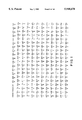
- FIG. 1 depicts the universal code equivalent of the mitochondrial I-SceI gene (SEQ ID NO:3 and 4).
- FIG. 2 depicts the nucleotide sequence of the invention encoding the enzyme I-SceI and the amino acid sequence of the natural I-SceI enzyme (SEQ ID NOS: 5 and 2).
- FIG. 3 depicts the I-SceI recognition sequence and indicates possible base mutations in the recognition site and the effect of such mutations on stringency of recognition (SEQ ID NOS: 6 and 8).
- FIG. 4 is the nucleotide sequence and deduced amino acid sequence of a region of plasmid pSCM525.
- the nucleotide sequence of the invention encoding the enzyme I-SceI is enclosed in the box (SEQ ID NOS: 9 through 16).
- FIG. 5 depicts variations around the amino acid sequence of the enzyme I-SceI (SEQ ID NO: 2).
- FIG. 6 shows Group I intron encoding endonucleases and related endonucleases (SEQ ID NOS: 17 through 44.
- FIG. 7 depicts yeast expression vectors containing the synthetic gene for I-SceI.
- FIG. 8 depicts the mammalian expression vector PRSV I-SceI.
- FIG. 9 is a restriction map of the plasmid pAF100. (See also YEAST, 6:521-534, 1990, which is relied upon and incorporated by reference herein).
- FIGS. 10A and 10B show the nucleotide sequence and restriction sites of regions of the plasmid pAF100 (SEQ ID NOS: 45-50).
- FIG. 11 depicts an insertion vector pTSM ⁇ , pTKM ⁇ , and pTTc ⁇ containing the I-SceI site for E. coli and other bacteria.
- FIG. 12 depicts an insertion vector pTYW6 containing the I-SceI site for yeast.
- FIG. 13 depicts an insertion vector PMLV LTR SAPLZ containing the I-SceI site for mammalian cells.
- FIGS. 14A-14B depict a set of seven transgenic yeast strains cleaved by I-SceI. Chromosomes from FY1679 (control) and from seven transgenic yeast strains with I-SceI sites inserted at various positions along chromosome XI were treated with I-SceI. DNA was electrophoresed on 1% agarose (SeaKem) gel in 0.25 X TBE buffer at 130 V and 12° C. on a Rotaphor apparatus (Biometra) for 70 hrs using 100 sec to 40 sec decreasing pulse times.
- A DNA was stained with ethidium bromide (0.2 ⁇ g/ml) and transferred to a Hybond N (Amersham) membrane for hybridization.
- B 32 P labelled cosmid pUKGO40 which hybridizes with the shortest fragment of the set was used as a probe. Positions of chromosome XI and shorter chromosomes are indicated.
- FIGS. 15A-15D depict the rationale of the nested chromosomal fragmentation strategy for genetic mapping.
- A Positions of I-SceI sites are placed on the map, irrespective of the left/right orientation (shorter fragments are arbitrarily placed on the left). Fragment sizes as measured from PFGE (FIG. 14A) are indicated in kb (note that the sum of the two fragment sizes varies slightly due to the limit of precision of each measurement).
- B Hybridization with the probe that hybridizes the shortest fragment of the set determines the orientation of each fragment (see FIG. 14B). Fragments that hybridize with the probe (full lines) have been placed arbitrarily to the left.
- C Transgenic yeast strains have been ordered with increasing sizes of hybridizing chromosome fragments.
- D Deduced I-SceI map with minimal and maximal size of intervals indicated in kb (variations in some intervals are due to limitations of PFGE measurements).
- E Chromosome subfragments are used as probes to assign each cosmid clone to a given map interval or across a given I-SceI site.
- FIG. 16 depicts mapping of the I-SceI sites of transgenic yeast strains by hybridization with left end and right end probes of chromosome XI. Chromosomes from FY1679 (control) and the seven transgenic yeas: strains were digested with I-SceI. Transgenic strains were placed in order as explained in FIG. 15. Electrophoresis conditions were as in FIG. 14. 32 P labelled cosmids pUKGO40 and pUKGO66 were used as left end and right end probes, respectively.
- FIG. 17A-17H depict mapping of a cosmid collection using the nested chromosomal fragments as probes.
- Cosmid DNAs were digested with EcoRI and electrophoresed on 0.9% agarose (SeaKem) gel at 1.5 V/cm for 14 hrs, stained with ethidium bromide and transferred to a Hybond N membrane. Cosmids were placed in order from previous hybridizations to help visualize the strategy. Hybridizations were carried out serially on three identical membranes using left end nested chromosome fragments purified on PFGE (see FIG. 16) as probes.
- B membrane #1, probe: Left tel to A302 site
- C membrane #1, probe: Left tel to M57 site
- D membrane #2, probe: Left tel to H81 site
- E membrane #2, probe: Left tel to T62 site
- F membrane #3, probe: Left tel to G41 site
- G membrane #3, probe: Left tel to D304 site
- FIG. 18 depicts a map of the yeast chromosome XI as determined from the nested chromosomal fragmentation strategy.
- the chromosome is divided into eight intervals (with sizes indicated in kb, see FIG. 15D) separated by seven I-SceI sites (E40, A302 . . .).
- Cosmid clones falling either within intervals or across a given I-SceI site are listed below intervals or below interval boundaries, respectively.
- Cosmid clones that hybridize with selected genes used as probes are indicated by letters (a-i). They localize the gene with respect to the I-SceI map and allow comparison with the genetic map (top).
- FIG. 19 depicts diagrams of successful site directed homologous recombination experiments performed in yeast.
- FIG.S 20A-20E Experimental design for the detection of HR homologous recombination (HR) induced by I-Sce I.
- HR homologous recombination
- G-MtkPL Maps of the 7.5 kb tk -PhleoLacZ retrovirus (G-MtkPL) and of the 6.0 kb PhleoLacZ retrovirus (G-MPL), SA is splice acceptor site.
- G-MtkPL sequences contains PhleoLacZ fusion gene for positive selection of infected cells (in phleomyzin-containing medium) and tk gene for negative selection (in gancyclovir-containing medium).
- G-MPL sequences (from G-MPL virus) contains only PhleoLacZ sequences.
- I-Sce I PhleoLacZ LTR duplicates, placing I-Sce I PhleoLacZ sequences in the 5'LTR.
- the virus vector (which functions as a promoter trap) is transcribed (arrow) by a flanking cellular promoter, P.
- I-Sce I creates two double strand breaks (DSBs) in host DNA liberating the central segment and leaving broken chromosome ends that can pair with the donor plasmid, pVRneo (d).
- FIGS. 21A-21B A. Scheme of pG-MPL. SD and SA are splice donor and splice acceptor sites. The structure of the unspliced 5.8 kb (genomic) and spliced 4;2 kb transcripts is shown below. Heavy bar is 32 P radiolabelled LacZ probe (P). B. RNA Northern blot analysis of a pG MLP transformed ⁇ -2 producer clone using polyadenylated RNA. Note that the genomic and the spliced mRNA are produced at the same high level.
- FIGS. 22A-22B A. Introduction of duplicated I-Sce I recognition sites into the genome of mammalian cells by retrovirus integration. Scheme of G-MPL and G-MtkPL proviruses which illustrates positions of the two LTRs and pertinent restriction sites. The size of Bcl I fragments and of I-Sce I fragments are indicated. Heavy bar is 32 P radiolabelled LacZ probe (P).
- B Southern blot analysis of cellular DNA from NIH3T3 fibroblasts cells infected by G-MtkPL and PCC7-S multipotent cells infected by G-MPL. Bcl I digests demonstrating LTR mediated PhleoLacZ duplication; I-Sce I digests demonstrating faithful duplication of I-Sce I sites.
- FIGS. 23A-23B Verification of recombination by Southern.
- A. Expected fragment sizes in kilobase pairs (kb) of provirus at the recombinant locus. 1) the parental proviral locus. Heavy bar (P) is 32 P radioactively labelled probe used for hybridization. 2) a recombinant derived after cleavage at the two I-Sce I sites followed by gap repair using pVR neo (double-site homologous recombination, DsHR). 3) a recombination event initiated by the cleavage at the I-Sce I sites in the left LTR (single-site homologous recombination, SsHR).
- B. Southern analysis of DNA from NIH3T3/G-MtkPL clones 1 and 2, PCC7-S/G-MPL clones 3 and 4 and transformants derived from cotransfection with pCMV(I-Sce I+) and pVRneo (1a, 1b, 2a, 3a, 3b and 4a). Kpn I digestion of the parental DNA generates a 4.2 kb fragment containing LacZ fragment.
- Recombinants la and 3a are examples of DsHR
- Recombinants 1b, 2a, 3b and 4a are examples of SsHR.
- FIGS. 24A-24B Verification of recombination by Northern blot analyses.
- A. Expected structure and sizes (in kb) of RNA from PCC7-S/G-MPL clone 3 cells before (top) and after (bottom) I-Sce I induced HR with pVRneo.1 Heavy bars P1 and P2 are 32 P radioactively labelled probes.
- B. Northern blot analysis of the PCC7-S/G-MPL clone 3 recombinant (total RNA). Lane 3 is parental cells, lane 3a recombinant cells. Two first lanes were probed with LacZ P1, two last lanes are probed with neo P2.
- parental PCC7-S/G-MPL clone 3 cells express a 7.0 kb LacZ RNA as expected of trapping of a cellular promoter leading to expression of a cellular-viral fusion RNA.
- the recombinant clone does not express this Lacz RNA but expresses a neo RNA of 5.0 kb, corresponding to the size expected for an accurate replacement of PhleoLacZ by neo gene.
- FIGS. 25A-25C Types of recombination events induced by I-Sce I DSBs, a) Schematic drawing of the structure of the recombination substrate.
- the G-MtkPL has provirus two LTRs, each containing an I-Sce I recognition site and a PhleoLacZ gene. The LTRs are separated by viral sequences containing the tk gene.
- the phenotype of G-MtkPL containing cells is Phleo R , GIs s , ⁇ -Gal ⁇ b) Possible modes of intra-chromosomal recombination. 1)
- the I-Sce I endonuclease cuts the I-Sce I site in the 5' LTR.
- the 5' part of U3 of the 5'LTR can pair and recombine with it homologous sequence in the 3'LTR (by SSA single-strand annealing (55A). 2)
- the I-Sce I endonuclease cuts the I-Sce I site in the 3'LTR.
- the 3' part of U3 of the 3'LTR can pair and recombine with its homologous sequence in the 5'LTR (by SSA).
- the I-Sce I endonuclease cuts I-Sce I sites in the two LTRs.
- the two free ends can relegate (by an end-joining mechanism).
- the resulting recombination product in each of the three models is a solitary LTR (see right side).
- the I-Sce I endonuclease cuts the I-Sce I sites in the two LTRs. The two free ends can be repaired (by a gap repair mechanism) using the homologous chromosome. On the right, the resulting recombination product is the deletion of the proviral integration locus.
- FIGS. 26A-26B Southern blot analysis of DNA from NIH3T3/G-MtkPL 1 and 2, and PhleoLacZ - recombinants derived from transfections with pCMV(I-Sce I+) selected in Gancyclovir containing medium.
- Pst I digestion of the parental DNA NH3T3/G-MtkPL 1 generates two fragments of 10 kbp and of the parental NIH3T3/G-MtkPL 2 two fragments of 7 kbp and 9 kbp.
- FIGS. 27A-27B Southern blot analysis of DNA from NIH3T3/G-MtkPL 1 and 2, and PhleoLacZ + recombinants derived from transfections with pCMV(I-Sce I+) and pCMV(I-Sce I-) and selection in Phleomycin and Gancyclovir containing medium.
- a1 Expected fragment sizes in kbp of parental provirus after digestion with Pst I or Bcl I endonuclease. Pst I digestion of the parental DNA NIH3T3/G-MtkPL 1 generates two fragments of 10 kbp.
- Bcl I digestion of the parental DNA NIH3T3/G-MtkPL 2 generates three fragments of 9.2 kbp, 7.2 kbp and 6.0 kbp. a2) Expected fragment sizes in kbp of recombinants after digestion with Pst I or Bcl I endonuclease.
- Pst I digestion of DNA of the recombinant derived from NIH3T3/G-MtkPL 1 generates one fragment of 13.6 kbp.
- Bcl I digestion of the DNA of the recombinants derived from NIH3T3/G-MtkPL 2 generates two fragments of 9.2 kbp and 6.0 kbp.
- FIG. 28 is a diagram illustrating the loss of heterozygosity by the insertion or presence of an I-Sce I site, expression of the enzyme I-,Sce I, cleavage at the site, and repair of the double strand break at the site with the corresponding chromatid.
- FIG. 29 is a diagram illustrating conditional activation of a gene.
- An I-Sce I site is integrated between tandem repeats, and the enzyme I-Sce I is expressed.
- the enzyme cleaves the double stranded DNA at the I-Sce I site.
- the double strand break is repaired by single stand annealing, yielding an active gene.
- FIG. 30 is a diagram illustrating one step rearrangement of a gene by integration of an I-Sce I site or by use of an I-Sce I site present in the gene.
- a plasmid having either one I-Sce I site within an inactive gene, or two I-Sce I sites at either end of an active gene without a promoter, is introduced into the cell.
- the cell contains an inactive form of the corresponding gene.
- the enzyme I-Sce I cuts the plasmid at the I-Sce I sites, and recombination between the chromosome and the plasmid yields an active gene replacing the inactive gene.
- FIG. 31 is a diagram illustrating the duplication of a locus.
- An I-Sce I site and a distal part of the locus are inserted into the gene by classical gene replacement.
- the I-Sce I site is cleaved by I-Sce I enzyme, and the break is repaired by homologous sequences. This results in duplication of the entire locus.
- FIG. 32 is a diagram illustrating the deletion of a locus. Two I-Sce I sites are added to flank the locus to be deleted. The I-Sce I enzyme is expressed, and the sites are cleaved. The two remaining ends recombine, deleting the locus between the two I-Sce I sites.
- FIG. 33 is a diagram of plasmid pG-Mtk ⁇ PAPL showing the restriction sites.
- the plasmid is constructed by deletion of the polyadenylation region of the tk gene from the pGMtkPL plasmid.
- the genuine mitochondrial gene (ref. 8) cannot be expressed in E. coli, yeast or other organisms due to the peculiarities of the mitochondrial genetic code.
- a "universal code equivalent” has been constructed by in vitro site-directed mutagenesis. Its sequence is given in FIG. 1. Note that all non-universal codons (except two CTN) have been replaced together with some codons extremely rare in E. coli.
- the universal code equivalent has been successfully expressed in E. coli and determines the synthesis of an active enzyme. However, expression levels remained low due to the large number of codons that are extremely rare in E. coli. Expression of the "universal code equivalent" has been detected in yeast.
- a synthetic gene has been designed to encode a protein with the genuine amino acid sequence of I-SceI using, for each codon, that most frequently used in E. coli.
- the sequence of the synthetic gene is given in FIG. 2.
- the synthetic gene was constructed in vitro from eight synthetic oligonucleotides with partial overlaps. oligonucleotides were designed to allow mutual priming for second strand synthesis by Klenow polymerase when annealed by pairs. The elongated pairs were then ligated into plasmids. Appropriately placed restriction sites within the designed sequence allowed final assembly of the synthetic gene by in vitro ligation.
- the synthetic gene has been successfully expressed in both E. coli and yeast.
- This invention relates to an isolated DNA sequence encoding the enzyme I-SceI.
- the enzyme I-SceI is an endonuclease.
- the properties of the enzyme (ref. 14) are as follows:
- I-SceI is a double-stranded endonuclease that cleaves DNA within its recognition site. I-SceI generates a 4 bp staggered cut with 3'OH overhangs.
- Substrate Acts only on double-stranded DNA.
- Substrate DNA can be relaxed or negatively supercoiled.
- Enzymatic activity requires Mg ++ (8 mM is optimum). Mn ++ can replace Mg ++ but this reduces the stringency of recognition.
- Optimum conditions for activity high pH (9 to 10), temperature 20-40° C., no monovalent cations.
- I-SceI is unstable at room temperature.
- the enzyme-substrate complex is more stable than the enzyme alone (presence of recognition sites stabilizes the enzyme.)
- the enzyme I-SceI has a known recognition site. (ref. 14.)
- the recognition site of I-SceI is a non-symmetrical sequence that extends over 18 bp as determined by systematic mutational analysis. The sequence reads: (arrows indicate cuts)
- the recognition site corresponds, in part, to the upstream exon and, in part, to the downstream exon of the intron plus form of the gene.
- the recognition site is partially degenerate: single base substitutions within the 18 bp long sequence result in either complete insensitivity or reduced sensitivity to the enzyme, depending upon position and nature of the substitution.
- the stringency of recognition has been measured on:
- the expected frequency of natural I-SceI sites in a random DNA sequence is, therefore, equal to (0.25) -18 or (1.5 ⁇ 10 -11 ). In other words, one should expect one natural site for the equivalent of ca. 20 human genomes, but the frequency of degenerate sites is more difficult to predict.
- I-SceI belongs to a "degenerate" subfamily of the two-dodecapeptide family. conserveed amino acids of the dodecapeptide motifs are required for activity. In particular, the aspartic residues at positions 9 of the two dodecapeptides cannot be replaced, even with glutamic residues. It is likely that the dodecapeptides form the catalytic site or part of it.
- the endonucleolytic activity of I-SceI requires two successive recognition steps: binding of the enzyme to the downstream half of the site (corresponding to the downstream exon) followed by binding of the enzyme to the upstream half of the site (corresponding to the upstream exon). The first binding is strong, the second is weaker, but the two are necessary for cleavage of DNA.
- the enzyme can bind the downstream exon alone as well as the intron-exon junction sequence, but no cleavage results.
- the nucleotide sequence of the invention which encodes the natural I-SceI enzyme is shown in FIG. 2.
- the nucleotide sequence of the gene of the invention was derived by dideoxynucleotide sequencing.
- the base sequences of the nucleotides are written in the 5' ⁇ 3' direction. Each of the letters shown is a conventional designation for the following nucleotides:
- the DNA sequence encoding the enzyme I-SceI be in a purified form.
- the sequence can be free of human blood-derived proteins, human serum proteins, viral proteins, nucleotide sequences encoding these proteins, human tissue, human tissue components, or combinations of these substances.
- the DNA sequence of the invention is free of extraneous proteins and lipids, and adventitious microorganisms, such as bacteria and viruses.
- the essentially purified and isolated DNA sequence encoding I-SceI is especially useful for preparing expression vectors.
- Plasmid pSCM525 is a pUC12 derivative, containing an artificial sequence encoding the DNA sequence of the invention.
- the nucleotide sequence and deduced amino acid sequence of a region of plasmid pSCM525 is shown in FIG. 4.
- the nucleotide sequence of the invention encoding I-SceI is enclosed in the box.
- the artificial gene is a BamHI--SalI piece of DNA sequence of 723 base pairs, chemically synthesized and assembled. It is placed under tac promoter control.
- the DNA sequence of the artificial gene differs from the natural coding sequence or its universal code equivalent described in Cell (1986), Vol. 44, pages 521-533.
- the translation product of the artificial gene is identical in sequence to the genuine omega-endonuclease except for the addition of a Met-His at the N-terminus. It will be understood that this modified endonuclease is within the scope of this invention.
- Plasmid pSCM525 can be used to transform any suitable E. coli strain and transformed cells become ampicillin-resistant. Synthesis of the omega-endonuclease is obtained by addition of I.P.T.G. or an equivalent inducer of the lactose operon system.
- a plasmid identified as pSCM52S containing the enzyme I-SceI was deposited in E. coli strain TG1 with the Collection Nationale de Cultures de Microorganismes (C.N.C.M.) of Institut Pasteur in Paris, France on Nov. 22, 1990, under culture collection deposit Accession No I-1014.
- the nucleotide sequence of the invention is thus available from this deposit.
- the gene of the invention can also be prepared by the formation of 3' ⁇ 5' phosphate linkages between nucleoside units using conventional chemical synthesis techniques.
- conventional chemical synthesis techniques for example, the well-known phosphodiester, phosphotriester, and phosphite triester techniques, as well as known modifications of these approaches, can be employed.
- Deoxyribonucleotides can be prepared with automatic synthesis machines, such as those based on the phosphoramidite approach. Oligo- and polyribonucleotides can also be obtained with the aid of RNA ligase using conventional techniques.
- This invention of course includes variants of the DNA sequence of the invention exhibiting substantially the same properties as the sequence of the invention. By this it is meant that DNA sequences need not be identical to the sequence disclosed herein. Variations can be attributable to single or multiple base substitutions, deletions, or insertions or local mutations involving one or more nucleotides not substantially detracting from the properties of the DNA sequence as encoding an enzyme having the cleavage properties of the enzyme I-SceI.
- FIG. 5 depicts some of the variations that can be made around the I-SceI amino acid sequence. It has been demonstrated that the following positions can be changed without affecting enzyme activity:
- positions -1 and -2 are not natural.
- the two amino acids are added due to cloning strategies.
- the present invention is intended to encompass fragments of the DNA sequence of the invention in purified form, where the fragments are capable of encoding enzymatically active I-SceI.
- the DNA sequence of the invention coding for the enzyme I-SceI can be amplified in the well known polymerase chain reaction (PCR), which is useful for amplifying all or specific regions of the gene. See e.g., S. Kwok et al., J. Virol., 61:1690-1694 (1987); U.S. Pat. No. 4,683,202; and U.S. Pat. No. 4,683,195. More particularly, DNA primer pairs of known sequence positioned 10-300 base pairs apart that are complementary to the plus and minus strands of the DNA to be amplified can be prepared by well known techniques for the synthesis of oligonucleotides.
- PCR polymerase chain reaction
- each primer can be extended and modified to create restriction endonuclease sites when the primer is annealed to the DNA.
- the PCR reaction mixture can contain the DNA, the DNA primer pairs, four deoxyribonucleoside triphosphates, MgCl 2 , DNA polymerase, and conventional buffers.
- the DNA can be amplified for a number of cycles. It is generally possible to increase the sensitivity of detection by using a multiplicity of cycles, each cycle consisting of a short period of denaturation of the DNA at an elevated temperature, cooling of the reaction mixture, and polymerization with the DNA polymerase. Amplified sequences can be detected by the use of a technique termed oligomer restriction (OR). See, R. K. Saiki et al., Bio/Technology 3:1008-1012 (1985).
- the enzyme I-SceI is one of a number of endonucleases with similar properties. Following is a listing of related enzymes and their sources.
- Putative new enzymes are I-CsmI from cytochrome b intron 1 of Chlamydomonas smithii mitochondria (ref. 15), I-PanI from cytochrome b intron 3 of Podospora anserina mitochondria (Jill Salvo), and probably enzymes encoded by introns Nc nd1•1 and Nc cob•
- the I-endonucleases can be classified as follows:
- Class I Two dodecapeptide motifs, 4 bp staggered cut with 3' OH overhangs, cut internal to recognition site
- Class II GIY-(N 10-11 ) YIG motif, 2 bp staggered cut with 3' OH overhangs, cut external to recognition site:
- Class III no typical structural motifs, 4 bp staggered cut with 3' OH overhangs, cut internal to recognition site:
- Class IV no typical structural motifs, 2 bp staggered cut with 3' OH overhangs, cut external to recognition site:
- Class V no typical structural motifs, 2 bp staggered cut with 5' OH overhangs:
- the DNA sequence of the invention coding for the enzyme I-SceI can also be used as a probe for the detection of a nucleotide sequence in a biological material, such as tissue or body fluids.
- the probe can be labeled with an atom or inorganic radical, most commonly using a radionuclide, but also perhaps with a heavy metal.
- Radicactive labels include 32 P, 3 H, 14 C, or the like. Any radioactive label can be employed, which provides for an adequate signal and has sufficient half-life.
- Other labels include ligands that can serve as a specific binding member to a labeled antibody, fluorescers, chemiluminescers, enzymes, antibodies which can serve as a specific binding pair member for a labeled ligand, and the like. The choice of the label will be governed by the effect of the label on the rate of hybridization and binding of the probe to the DNA or RNA. It will be necessary that the label provide sufficient sensitivity to detect the amount of DNA or RNA available for hybridization
- the nucleotide sequence of the invention When used as a probe for hybridizing to a gene, the nucleotide sequence is preferably affixed to a water insoluble solid, porous support, such as nitrocellulose paper.
- Hybridization can be carried out using labeled polynucleotides of the invention and conventional hybridization reagents. The particular hybridization technique is not essential to the invention.
- the amount of labeled probe present in the hybridization solution will vary widely, depending upon the nature of the label, the amount of the labeled probe which can reasonably bind to the support, and the stringency of the hybridization. Generally, substantial excesses of the probe over stoichiometric will be employed to enhance the rate of binding of the probe to the fixed DNA.
- Various degrees of stringency of hybridization can be employed. The more severe the conditions, the greater the complementarity that is required for hybridization between the probe and the polynucleotide for duplex formation. Severity can be controlled by temperature, probe concentration, probe length, ionic strength, time, and the like. Conveniently, the stringency of hybridization is varied by changing the polarity of the reactant solution. Temperatures to be employed can be empirically determined or determined from well known formulas developed for this purpose.
- This invention also relates to the DNA sequence of the invention encoding the enzyme I-SceI, wherein the nucleotide sequence is linked to other nucleic acids.
- the nucleic acid can be obtained from any source, for example, from plasmids, from cloned DNA or RNA, or from natural DNA or RNA from any source, including prokaryotic and eukaryotic organisms.
- DNA or RNA can be extracted from a biological material, such as biological fluids or tissue, by a variety of techniques including those described by Maniatis et al., Molecular Cloning: A Laboratory Manual, Cold Spring Harbor Laboratory, New York (1982).
- the nucleic acid will generally be obtained from a bacteria, yeast, virus, or a higher organism, such as a plant or animal.
- the nucleic acid can be a fraction of a more complex mixture, such as a portion of a gene contained in whole human DNA or a portion of a nucleic acid sequence of a particular microorganism.
- the nucleic acid can be a fraction of a larger molecule or the nucleic acid can constitute an entire gene or assembly of genes.
- the DNA can be in a single-stranded or double-stranded form. If the fragment is in single-stranded form, it can be converted to double-stranded form using DNA polymerase according to conventional techniques.
- the DNA sequence of the invention can be linked to a structural gene.
- structural gene refers to a DNA sequence that encodes through its template or messenger mRNA a sequence of amino acids characteristic of a specific protein or polypeptide.
- the nucleotide sequence of the invention can function with an expression control sequence, that is, a DNA sequence that controls and regulates expression of the gene when operatively linked to the gene.
- This invention also relates to cloning and expression vectors containing the DNA sequence of the invention coding for the enzyme I-SceI.
- the DNA sequence encoding the enzyme can be ligated to a vehicle for cloning the sequence.
- the major steps involved in gene cloning comprise procedures for separating DNA containing the gene of interest from prokaryotes or eukaryotes, cutting the resulting DNA fragment and the DNA from a cloning vehicle at specific sites, mixing the two DNA fragments together, and ligating the fragments to yield a recombinant DNA molecule.
- the recombinant molecule can then be transferred into a host cell, and the cells allowed to replicate to produce identical cells containing clones of the original DNA sequence.
- the vehicle employed in this invention can be any double-stranded DNA molecule capable of transporting the nucleotide sequence of the invention into a host cell and capable of replicating within the cell. More particularly, the vehicle must contain at least one DNA sequence that can act as the origin of replication in the host cell. In addition, the vehicle must contain two or more sites for insertion of the DNA sequence encoding the gene of the invention. These sites will ordinarily correspond to restriction enzyme sites at which cohesive ends can be formed, and which are complementary to the cohesive ends on the promoter sequence to be ligated to the vehicle. In general, this invention can be carried out with plasmid, bacteriophage, or cosmid vehicles having these characteristics.
- the nucleotide sequence of the invention can have cohesive ends compatible with any combination of sites in the vehicle.
- the sequence can have one or more blunt ends that can be ligated to corresponding blunt ends in the cloning sites of the vehicle.
- the nucleotide sequence to be ligated can be further processed, if desired, by successive exonuclease deletion, such as with the enzyme Bal 31.
- the sequence can be modified by adding a linker, an adaptor, or homopolymer tailing.
- plasmids used for cloning nucleotide sequences of the invention carry one or more genes responsible for a useful characteristic, such as a selectable marker, displayed by the host cell.
- plasmids having genes for resistance to two different drugs are chosen. For example, insertion of the DNA sequence into a gene for an antibiotic inactivates the gene and destroys drug resistance. The second drug resistance gene is not affected when cells are transformed with the recombinants, and colonies containing the gene of interest: can be selected by resistance to the second drug and susceptibility to the first drug.
- Preferred antibiotic markers are genes imparting chloramphenicol, ampicillin, or tetracycline resistance to the host cell.
- restriction enzymes can be used to cut the vehicle.
- the identity of the restriction enzyme will generally depend upon the identity of the ends on the DNA sequence to be ligated and the restriction sites in the vehicle.
- the restriction enzyme is matched to the restriction sites in the vehicle, which in turn is matched to the ends on the nucleic acid fragment being ligated.
- the ligation reaction can be set up using well known techniques and conventional reagents. Ligation is carried out with a DNA ligase that catalyzes the formation of phosphodiester bonds between adjacent 5'-phosphate and the free 3'-hydroxy groups in DNA duplexes.
- the DNA ligase can be derived from a variety of microorganisms.
- the preferred DNA ligases are enzymes from E. coli and bacteriophage T4.
- T4 DNA ligase can ligate DNA fragments with blunt or sticky ends, such as those generated by restriction enzyme digestion.
- E. coli DNA ligase can be used to catalyze the formation of phosphodiester bonds between the termini of duplex DNA molecules containing cohesive ends.
- Cloning can be carried out in prokaryotic or eukaryotic cells.
- the host for replicating the cloning vehicle will of course be one that is compatible with the vehicle and in which the vehicle can replicate.
- the plasmid can be derived from bacteria or some other organism or the plasmid can be synthetically prepared.
- the plasmid can replicate independently of the host cell chromosome or an integrative plasmid (episome) can be employed.
- the plasmid can make use of the DNA replicative enzymes of the host cell in order to replicate or the plasmid can carry genes that code for the enzymes required for plasmid replication.
- a number of different plasmids can be employed in practicing this invention.
- the DNA sequence of the invention encoding the enzyme I-SceI can also be ligated to a vehicle to form an expression vector.
- the vehicle employed in this case is one in which it is possible to express the gene operatively linked to a promoter in an appropriate host cell. It is preferable to employ a vehicle known for use in expressing genes in E. coli, yeast, or mammalian cells. These vehicles include, for example, the following E. coli expression vectors:
- pSCM525 which is an E. coli expression vector derived from pUC12 by insertion of a tac promoter and the synthetic gene for I-SceI. Expression is induced by IPTG.
- pGEX ⁇ 6 which is an E. coli expression vector derived from pGEX in which the synthetic gene from pSCM525 for I-SceI is fused with the glutathione S transferase gene, producing a hybrid protein.
- the hybrid protein possesses the endonuclease activity.
- pDIC73 which is an E. coli expression vector derived from pET-3C by insertion of the synthetic gene for I-SceI (NdeI--BamHI fragment of pSCM525) under T7 promoter control. This vector is used in strain BL21 (DE3) which expresses the T7 RNA polymerase under IPTG induction.
- pSCM351 which is an E. coli expression vector derived from pUR291 in which the synthetic gene for I-SceI is fused with the Lac Z gene, producing a hybrid protein.
- pSCM353 which is an E. coli expression vector derived from pEX1 in which the synthetic gene for I-SceI is fused with the Cro/Lac Z gene, producing a hybrid protein.
- yeast expression vectors are: pPEX7, which is a yeast expression vector derived from pRP51-Bam O (a LEU2d derivative of pLG-SDS) by insertion of the synthetic gene under the control of the galactose promoter. Expression is induced by galactose.
- pPEX408 which is a yeast expression vector derived from pLG-SD5 by insertion of the synthetic gene under the control of the galactose promoter. Expression is induced by galactose.
- FIG. 7 Several yeast expression vectors are depicted in FIG. 7.
- Typical mammalian expression vectors are:
- pRSV I-SceI which is a pRSV derivative in which the synthetic gene (BamHI--PstI fragment from pSCM525) is under the control of the LTR promoter of Rous Sarcoma Virus.
- This expression vector is depicted in FIG. 8.
- Vectors for expression in Chinese Hamster Ovary (CHO) cells can also be employed.
- the vectors of the invention can be inserted into host organisms using conventional techniques.
- the vectors can be inserted by transformation, transfection, electroporation, microinjection, or by means of liposomes (lipofection).
- Cloning can be carried out in prokaryotic or eukaryotic cells.
- the host for replicating the cloning vehicle will of course be one that is compatible with the vehicle and in which the vehicle can replicate.
- Cloning is preferably carried out in bacterial or yeast cells, although cells of fungal, animal, and plant origin can also be employed.
- the preferred host cells for conducting cloning work are bacterial cells, such as E. coli.
- E. coli cells is particularly preferred because most cloning vehicles, such as bacterial plasmids and bacteriophages, replicate in these cells.
- an expression vector containing the DNA sequence encoding the nucleotide sequence of the invention operatively linked to a promoter is inserted into a mammalian cell using conventional techniques.
- yeast all are pAF100 derivatives (Gold et al. (1990) YEAST 6:521-534) containing the following marker genes:
- Neo R inserted in BglII site
- pAF104 HIS3 (inserted in BglII site)
- Kan R inserted in BglII site
- Kan R inserted in BglII site
- pAF107 LYS2 (inserted between HindIII and EcoR V)
- FIG. 9 A restriction map of the plasmid pAF100 is shown in FIG. 9.
- the nucleotide sequence and restriction sites of regions of plasmid pAF100 are shown in FIGS. 10A and 10B.
- Many transgenic yeast strains with the I-SceI site at various and known places along chromosomes are available.
- mini Tn5 derivatives containing the I-SceI site For E. coli and other bacteria: mini Tn5 derivatives containing the I-SceI site and
- pTy ⁇ 6 is a pD123 derivative in which the I-SceI site has been inserted in the LTR of the Ty element.
- pMLV LTR SAPLZ containing the I-SceI site in the LTR of MLV and Phleo-LacZ (FIG. 13). This vector is first grown in ⁇ 2 cells (3T3 derivative, from R. Mulligan). Two transgenic cell lines with the I-SceI site at undetermined locations in the genome are available: 1009 (pluripotent nerve cells, J. F. Nicolas) and D3 (ES cells able to generate transgenic animals).
- the nested chromosomal fragmentation strategy for genetically mapping a eukaryotic genome exploits the unique properties of the restriction endonuclease I-SceI, such as an 18 bp long recognition site.
- I-SceI restriction endonuclease
- one or more I-SceI recognition sites are artificially inserted at various positions in a genome, by homologous recombination using specific cassettes containing selectable markers or by random insertion, as discussed supra.
- the genome of the resulting transgenic strain is then cleaved completely at the artificially inserted I-SceI site(s) upon incubation with the I-SceI restriction enzyme. The cleavage produces nested chromosomal fragments.
- each artificially introduced I-SceI site provides a unique "molecular milestone" in the genome.
- a set of transgenic strains, each carrying a single I-SceI site can be created which defines physical genomic intervals between the milestones. Consequently, an entire genome, a chromosome or any segment of interest can be mapped using artificially introduced I-SceI restriction sites.
- the nested chromosomal fragments may be transferred to a solid membrane and hybridized to a labelled probe containing DNA complementary to the DNA of the fragments. Based on the hybridization banding patterns that are observed, the eukaryotic genome may be mapped.
- the set of transgenic strains with appropriate "milestones" is used as a reference to map any new gene or clone by direct hybridization.
- This strategy has been applied to the mapping of yeast chromosome XI of Saccharamyces cerevisiae.
- the I-SceI site was inserted at 7 different locations along chromosome XI of the diploid strain FY1679, hence defining eight physical intervals in that chromosome.
- Sites were inserted from a URA3-1-I-SceI cassette by homologous recombination. Two sites were inserted within genetically defined genes, TIF1 and FAS1, the others were inserted at unknown positions in the chromosome from five non-overlapping cosmids of our library, taken at random.
- the cosmid clone pUKGO40 which was used to insert the I-SceI site in the transgenic E40, is now used as a probe against all chromosome fragments (FIG. 14B).
- pUKGO40 lights up the two fragments from strain E40 (50 kb and 630 kb, respectively).
- the large fragment is close to the entire chromosome XI and shows a weak hybridization signal due to the fact that the insert of pUKG040, which is 38 kb long, contains less than 4 kb within the large chromosome fragment.
- fragments were hybridized successively with cosmids pUKG040 and pUKG066 which light up, respectively, all fragments from the opposite ends of the chromosome (clone pUKG066 defines the right end of the chromosome as defined from the genetic map because it contains the SIR1 gene. A regular stepwise progression of chromosome fragment sizes is observed. Note some cross hybridization between the probe pUKGO66) and chromosome III, probably due to some repetitive DNA sequences.
- This strategy can be applied to YAC mapping with two possibilities.
- the nested, chromosomal fragments can be purified from preparative PFG and used as probes against clones from a chromosome X1 specific sublibrary.
- This sublibrary is composed of 138 cosmid clones (corresponding to eight times coverage) which have been previously sorted from our complete yeast genomic libraries by colony hybridization with PFG purified chromosome X1.
- This collection of unordered clones has been sequentially hybridized with chromosome fragments taken in order of increasing sizes from the left end of the chromosome. Localization of each cosmic clone on the I-SceI map could be unambiguously determined from such hybridizations.
- cosmid clones in which all EcoRI fragments hybridize with the probe and cosmid clones in which only some of the EcoRI fragments hybridize (i.e., compare pEKG100 to pEKGO98 in FIG. 17b).
- the first category corresponds to clones in which the insert is entirely included in one of the two chromosome fragments, the second to clones in which the insert overlaps an I-SceI site. Note that, for clones of the pEKG series, the EcoRI fragment of 8 kb is entirely composed of vector sequences (pWE15) that do not hybridize with the chromosome fragments. In the case where the chromosome fragment possesses the integration vector, a weak cross hybridization with the cosmid is observed (FIG. 17e).
- FIG. 17 shows that the cosmid clones can unambiguously be ordered with respect to the I-SceI map (FIG. 13E), each clone falling either in a defined interval or across an I-SceI site.
- clones from the second category allow us to place some EcoRI fragments on the I-SceI maps, while others remain unordered.
- the complete set of chromosome XI- specific cosmid clones, covering altogether eight times the equivalent of the chromosome, has been sorted with respect to the I-Scel map, as shown in FIG. 18.
- Partial restriction mapping has been done on yeast DNA and on mammalian cell DNA using the commercial enzyme I-SceI.
- DNA from cells containing an artificially inserted I-SceI site is first cleaved to completion by I-SceI.
- the DNA is then treated under partial cleavage conditions with bacterial restriction endonucleases of interest (e.g., BamHI) and electrophoresed along with size calibration markers.
- the DNA is transferred to a membrane and hybridized successively using the short sequences flanking the I-SceI sites on either side (these sequences are known because they are part of the original insertion vector that was used to introduce the I-SceI site).
- Autoradiography (or other equivalent detection system using non radioactive probes) permit the visualization of ladders, which directly represent the succession of the bacterial restriction endonuclease sites from the I-SceI site.
- the size of each band of the ladder is used to calculate the physical distance between the successive bacterial restriction endonuclease sites.
- the synthetic I-SceI gene has been placed under the control of a galactose inducible promoter on multicopy plasmids pPEX7 and pPEX408. Expression is correct and induces effects on site as indicated below.
- a transgenic yeast with the I-SceI synthetic gene inserted in a chromosome under the control of an inducible promoter can be constructed.
- a single break within a chromosome at an artificial I-SceI site results in cell division arrest followed by death (only a few % of survival). Presence of an intact sequence homologous to the cut site results in repair and 100% cell survival.
- a single break within a chromosome at an artificial I-SceI site results in repair using the chromosome homolog and 100% cell survival. In both cases, repair of the induced double strand break results in loss of heterozygosity with deletion of the non homologous sequences flanking the cut and insertion of the non homologous sequences from the donor DNA molecule.
- Construction of a YAC vector with the I-SceI restriction site next to the cloning site should permit one to induce homologous recombination with another YAC if inserts are partially overlapping. This is useful for the construction of contigs.
- I-SceI restriction site Insertion of an I-SceI restriction site has been done for bacteria (E. coli, Yersinia entorocolitica, Y. pestis, Y. pseudotuberculosis), and mouse cells. Cleavage at the artificial I-SceI site in vitro has been successful with DNA from the transgenic mouse cells. Expression of I-SceI from the synthetic gene in mammalian or plant cells should be successful.
- the I-SceI site has been introduced in mouse cells and bacterial cells as follows:
- -b- Transfected cells were selected in DMEM medium containing phleomycin with 5% fetal calf serum and grown under 12% CO 2 , 100% humidity at 37° C. until they form colonies.
- phleomycin resistant cells were selected in the same medium as above.
- Mini Tn 5 transposons containing the I-SceI recognition site were constructed in E. coli by standard recombinant DNA procedures.
- the mini Tn 5 transposons are carried on a conjugative plasmid.
- Bacterial conjugation between E. coli and Yersinia is used to integrate the mini Tn 5 transposon in Yersinia.
- Yersinia cells resistant to Kanamycin, Streptomycin or tetracycline are selected (vectors pTKM- ⁇ , pTSM- ⁇ and pTTc- ⁇ , respectively).
- Homologous recombination (HR) between chromosomal and exogenous DNA is at the basis of methods for introducing genetic changes into the genome (5E, 20B). Parameters of the recombination mechanism have been determined by studying plasmid sequences introduced into cells (1B, 4B, 10B, 12B) and in in vitro system (8B). HR is inefficient in mammalian cells but is promoted by double-strand breaks in DNA.
- I-Sce I 6B
- I-Sce I protein is an endonuclease responsible for intron homing in mitochondria of yeast, a non-reciprocal mechanism by which a predetermined sequence becomes inserted at a predetermined site.
- endonuclease I-Sce I can catalyze recombination in the nucleus of yeast by initiating a double-strand break (17B).
- the recognition site of endonuclease I-Sce I is 18 bp long, therefore, the I-Sce I protein is a very rare cutting restriction endonuclease in genomes (22B).
- the I-Sce I protein is not a recombinase, its potential for chromosome engineering is larger than that of systems with target sites requirement on both host and donor molecules (9B).
- yeast I-Sce I endonuclease can efficiently induce double-strand breaks in chromosomal target in mammalian cells and that the breaks can be repaired using a donor molecule that shares homology with the regions flanking the break.
- the enzyme catalyzes recombination at a high efficiency. This demonstrates that recombination between chromosomal DNA and exogenous DNA can occur in mammalian cells by the double-strand break repair pathway (21B).
- pG-MPL was obtained in four steps: (I) insertion of the 0.3 kb Bgl II--Sma I fragment (treated with Klenow enzyme) of the Moloney Murine Leukemia Virus (MoMuLV) env gene (25B) containing SA between the Nhe I and Xba I sites (treated with Klenow enzyme), in the U3 sequence of the 3'LTR of MoMuLV, in an intermediate plasmid. (II) insertion in this modified LTR with linkers adaptors of the 3.5 kb Nco I--Xho I fragment containing the PhleoLacZ fusion gene (15B) (from pUT65 from Cayla laboratory) at the Xba I site next to SA.
- MoMuLV Moloney Murine Leukemia Virus
- pG-MtkPl was obtained by the insertion (antisense to the retroviral genome) of the 1.6 kb tk gene with its promoter with linker adaptators at the Pst I site of pG-MPL.
- pVRneo was obtained in two steps (I) insertion into pSP65 (from Promega) linearized by Pst I--EcoR I double digestion of the 4.5 kb Pst I to EcoR I fragment of pG-MPL containing the 3'LTR with the SA and PhleoLacZ, (II) insertion of the 2.0 kb Bgl II--BamH I fragment, (treated with Klenow enzyme) containing neoPolyA from pRSVneo into the Nco I restriction site (treated with Klenow enzyme) of pSP65 containing part of the 3'LTR of G-MPL (between SA and PhleoLacZ).
- pCMV(I-Sce I+) was obtained in two steps: (I) insertion of the 0.73 kb BamH I--Sal I, I-Sce I containing fragment (from pSCM525, A. Thierry, personal gift) into the phCMV1 (F. Meyer, personal gift) plasmid cleaved at the BamH I and the Sal I sites, (II) insertion of a 1.6 kb (nucleotide number 3204 to 1988 in SV40) fragment containing the polyadenylation signal of SV40 into the Pst I site of phCMV1.
- pCMV(I-Sce I-) contains the I-Sce I ORF in reverse orientation in the pCMV(I-Sce I+) plasmid. It has been obtained by inserting the BamH I--Pst I I-Sce I ORF fragment (treated with Klenow enzyme) into the phCMV PolyA vector linearized by Nsi I and Sal I double-digestion and treated with Klenow enzyme.
- Plasmids pG-MPL, pG-MtkPl, pG-Mtk ⁇ PAPL have been described.
- any kind of plasmid vector can be constructed containing various promoters, genes, polyA site, I-Sce I site.
- 3T3, PCC7 S, ⁇ 2 are referenced in (7B) and (13B).
- Cell selection medium gancyclovir (14B, 23B) was added into the tissue culture medium at the concentration of 2 ⁇ M. Gancyclovir selection was maintained on cells during 6 days. G418 was added into the appropriate medium at a concentration of 1 mg/ml for PCC7-S and 400 ⁇ g/ml for 3T3. The selection was maintained during all the cell culture. Phleomycin was used at a concentration of 10 ⁇ g/ml.
- ⁇ cell line was transfected with plasmids containing a proviral recombinant vector that contain I-Sce I recognition site: pG-MPL, pG-MtkPL, pG-Mtk.sub. ⁇ PA PL
- NIH 3T3 Fibroblastic cell line is infected with:
- G-MPL Multiple (more than 30) clones were recovered. The presence of 1 to 14 proviral integrations and the multiplicity of the different points of integration were verified by molecular analysis.
- G-MPL 14 clones were recovered, normal proviral integration.
- --Embryonic stem cell line D3 is infected with:
- Insertion of the retrovirus induces duplication of LTR containing the I-Sce I site.
- the cell is heterozygotic for the site.
- FIG. 20 To detect I-Sce I HR we have designed the experimental system shown in FIG. 20.
- Defective recombinant retroviruses 24B were constructed with the I-Sce I recognition site and a PhleoLacz (15B) fusion gene inserted in their 3'LTR (FIG. 20a).
- Retroviral integration results in two I-Sce I sites distant of 5.8 kb or 7.2 kb from each other into the cell genome (FIG. 20b).
- I-Sce I-induced double-strand breaks (DSB) at these sites could initiate HR with a donor plasmid (pvRneo, FIG. 20d) containing sequences homologous to the flanking regions of the DSBs and that non-homologous sequences, carried by the donor plasmid, could be copied during this recombination (FIG. 20e).
- the G-MtkPL proviral sequences (from G-MtkPL virus) contain the PhleoLacZ fusion gene for positive selection of transduced cells (in phleomycine-containing medium) and the tk gene for negative selection (in gancyclovir-containing medium).
- the G-MPL proviral sequences (from G-MPL virus) contain only the PhleoLacZ sequences.
- G-MtkPL and G-MPL are defective recombinant retroviruses (16B) constructed from an enhancerless Moloney murine leukemia provirus. The virus vector functions as a promoter trap and therefore is activated by flanking cellular promoters.
- Virus-producing cell lines were generated by transfecting pG-MtkPL or G-MPL into the ⁇ -2 package cell line (13B).
- Northern blot analysis of viral transcripts shows (FIG. 21) that the ⁇ -2-G-MPL line expresses 4.2 and 5.8 kb transcripts that hybridized with LacZ probes. These transcripts probably initiate in the 5'LTR and terminate in the 3'LTR.
- the 4.5 kb transcript corresponds to the spliced message and the 5.8 kb transcripts to the unspliced genomic message (FIG. 21.A). This verified the functionality of the 5'LTR and of the splice donor and acceptor in the virus. Similar results have been obtained with ⁇ -2G-MtkPL.
- Virus was prepared from the culture medium of ⁇ -2 cell lines.
- NIH3T3 fibroblasts and PCC7-S multipotent mouse cell lines (7B) were next infected by G-MtkPL and G-MPL respectively, and clones were isolated.
- Southern blot analysis of the DNA prepared from the clones demonstrated LTR-mediated duplication of I-Sce I PhleoLacZ sequences (FIG. 22a).
- Bcl I digestion generated the expected 5.8 kb (G-MPL) or 7.2 kb (G-MtkPL) fragments.
- the presence of two additional fragments corresponding to Bcl I sites in the flanking chromosomal DNA demonstrates a single proviral target in each clone isolated.
- the phenotype conferred to the NIH3T3 cells by G-MtkPL virus is phleo R ⁇ -gal+glss and to PCC7-S by G-MPL is phleo R ⁇ -gal + (FIG. 20b).
- pVRneo donor plasmid To allow for direct selection of recombination events induced by I-Sce I we constructed pVRneo donor plasmid. In pVRneo the neo gene is flanked by 300 bp homologous to sequences 5' to the left chromosomal break and 2.5 kb homologous to sequences 3' to the right break (FIG. 20d).
- a polyadenylation signal was positioned 3' to the neo gene to interrupt the PhleoLacZ message following recombination. If an induced recombination between the provirus and the plasmid occurs, the resulting phenotype will be neo R and due to the presence of a polyadenylation signal in the donor plasmid the PhleoLacZ gene should not be expressed, resulting in a phleo S ⁇ -gal - phenotype.
- G-MtkPL and G-MtkDPQPL it is possible to select simultaneously for the gap by negative selection with the tk gene (with gancyclovir) and for the exchange of the donor plasmid with positive selection with the neo gene (with geneticine).
- G-MPL only the positive selection can be applied in medium containing geneticine. Therefore, we expected to select for both the HR and for an integration event of the donor plasmid near an active endogenous promoter. These two events can be distinguished as an induced HR results in a neo R ⁇ -gal - phenotype and a random integration of the donor plasmid results in a neo R ⁇ -gal + phenotype.
- NIH3T3/G-MtkPL clones were selected either for loss of proviral sequences and acquisition of the neo R phenotype (with gancyclovir and geneticine) or for neo R phenotype only (Table 1).
- neo R gls R colonies were recovered with a frequency of 10 -4 in experimental series, and no colonies were recovered in the control series.
- all neo R gls R colonies were ⁇ -gal - , consistent with their resulting from HR at the proviral site.
- neo R colonies were recovered with a frequency of 10 -3 in experimental series, and with a 10 to 100 fold lower frequency in the control series.
- neo R colonies were found to be ⁇ -gal - (in series with pCMV(I-Sce I+)). This shows that expression of I-Sce I induces HR between pVR neo and the proviral site and that site directed HR is ten times more frequent than random integration of pVR neo near a cellular promoter, and at least 500 times more frequent than spontaneous HR.
- neo R recombinants The molecular structure of neo R recombinants has been examined by Southern blot analysis (FIG. 23 and Table 1). HR at I-Sce I sites predicts that digestion of recombinant DNA generates a 6.4 kb LacZ fragment instead of the 4.2 kb parental fragment. All 15 neo R gls R ⁇ -gal - recombinants from NIH3T3 cells exhibited only the 6.4 kb Kpn I fragment. Therefore, the double selection procedure leads to only the expected recombinants created by gene replacement (Double Site Homologous Recombinants, DsHR).
- the 25 ⁇ -gal - recombinants generated from the single selection fell into four classes: (a) DsHR induced by I-Sce I as above (19 clones); (b) integration of pVRnec) in the left LTR as proven by the presence of a 4.2 Kpn I fragment (corresponding to PhleoLacZ in the remaining LTR), in addition to the 6.4 kb fragment (FIG. 23, Table 1, Single site Homologous Recombinants, SsHR; 3 independent ⁇ -gal - recombinants from clone 3).
- neo RNA corresponds to the exact size expected for an accurate exchange of PhleoLacZ by neo gene and uses of the same cellular and viral splice site (viral PhlecLacZ RNA in the LTR is 3.7 kb and neo RNA in pVRneo is 1.7 kb).
- results presented here demonstrate that double-strand breaks can be induced by the I-Sce I system of Saccharomyces cerevisiaein mammalian cells, and that the breaks in the target chromosomal sequence induce site-specific recombination with input plasmidic donor DNA.
- the system To operate in mammalian cells, the system requires endogenous I-Sce I like activity to be absent from mammalian cells and I-Sce I protein to be neutral for mammalian cells. It is unlikely that endogenous I-Sce I-like actively operates in mammalian cells as the introduction of I-Sce I recognition sites do not appear to lead to rearrangement or mutation in the input DNA sequences. For instance, all NIH3T3 and PCC7-S clones infected with a retroviruses containing the I-Sce I restriction site stably propagated the virus. To test for the toxicity of I-Sce I gene product, an I-Sce I expressing plasmid was introduced into the NIH3T3 cells line (data not shown).
- I-Sce I gene A very high percentage of cotransfer of a functional I-Sce I gene was found, suggesting no selection against this gene. Functionality of I-Sce I gene was demonstrated by analysis of transcription, by immunofluorescence detection of the gene product and biological function (Choulika et al. in preparation).
- the demonstration of the ability of the I-Sce I meganuclease to have biological function on chromosomal sites in mammalian cell paves the route for a number of manipulations of the genome in living organisms.
- site-specific recombinases (9B, 18B)
- the I-Sce I system is non-reversible.
- Site specific recombinases locate not only the sites for cutting the DNA, but also for rejoining by bringing together the two partners.
- the only requirement with the I-Sce I system is homology of the donor molecule with the region flanking the break induced by I-Sce I protein.
- SSA single-strand annealing
- double-strand breaks are substrates in the action of an exonuclease that exposes homologous complementary single-strand DNA on the recipient and donor DNA. Annealing of the complementary strand is then followed by a repair process that generates recombinants.
- the I-Sce I system can be used to evaluate the relative importance of the two pathways.
- I-Sce I meganuclease involved in intron homing of mitochondria of the yeast Saccharomyces cerevisiae (6B, 28B) to induce DSB and mediate recombination in mammalian cells.
- I-Sce I is a very rare-cutting restriction endonuclease, with an 18 bp long recognition site (29B, 22B).
- I-Sce I endonuclease can induce recombination in a modified yeast nucleus by initiating a specific DBS leading to gap repair by the cell (30B, 17B, 21B).
- this approach can potentially be used as a means of introducing specific DSB in chromosomal target DNA with a view to manipulate chromosomes in living cells.
- the I-Sce I-mediated recombination is superior to recombinase system 11B! for chromosome engineering since the latter requires the presence of target sites on both host and donor DNA molecules, leading to reaction that is reversible.
- the I-Sce I endonuclease expression includes recombination events.
- I-Sce I activity can provoke site-directed double strand breaks (DSBs) in a mammalian chromosome. At least two types of events occur in the repair of the DSBs, one leading to intra-chromosomal homologous recombination and the other to the deletion of the transgene. These I-Sce I-mediated recombinations occur at a frequency significantly higher than background.
- pG-MtkPL was obtained in five steps: (I) insertion of the 0.3 kbp Bgl II-Sma I fragment (treated with Klenow enzyme) of the Moloney Murine Leukemia Virus (MoMuLV) env gene (25B) containing a splice acceptor (SA) between the Nhe I and Xba I sites (treated with Klenow enzyme), in the U3 sequence of the 3'LTR of MoMuLV, in an intermediate plasmid. (II) Insertion in this modified LTR of a 3.5 kbp Nco I-Xho I fragment containing the PhleoLacZ fusion gene 13B!
- pCMV(I-Sce I+) was obtained in two steps: (I) insertion of the 0.73 kbp BamH I-Sal I, I-Sce I-containing fragment (from pSCM525, donated by A. Thierry) into the phCMV1 (donated by F. Meyer) plasmid cleaved with BamH I and Sal I, (II) insertion of a 1.6 kbp fragment (nucleotide n° 3204 to 1988 in SV40) containing the polyadenylation signal of SV40 at the Pst I site of phCMV1.
- pCMV(I-Sce I-) contains the I-Sce I ORF in reverse orientation in the pCMV(I-Sce I+) plasmid. It was obtained by inserting the BamH I-Pst I I-Sce I ORF fragment (treated with Klenow enzyme) into the phCMV PolyA vector linearized by Nsi I and Sal I double-digestion and treated with Klenow enzyme.
- T3 and ⁇ 2 are referenced in (7B) and (13B).
- Cell selection medium gancyclovir (14B, 23B) was added into the tissue culture medium at the concentration of 2 ⁇ M. Gancyclovir selection was maintained for 6 days. Phleomycine was used at a concentration of 10 ⁇ g/ml. Double selections were performed in the same conditions.
- the virus-producing cell line is generated by transfecting pG-MtkPL into the ⁇ -2 packaging cell line.
- Virus was prepared from the filtered culture medium of transfected ⁇ -2 cell lines.
- NIH3T3 fibroblasts were infected by G-MtkPL, and clones were isolated in a Phleomycin-containing medium.
- G-MtkPL provirus contains the tk gene (in place of the gag, pol and env viral genes), for negative selection in gancyclovir-containing medium and, in the two LTRs, an I-Sce I recognition site and the PhleoLacZ fusion gene.
- the PhleoLacz gene can be used for positive selection of transduced cells in phleomycine-containing medium.
- I-Sce I endonuclease in these cells would induce double-strand breaks (DSB) at the I-Sce I recognition sites that would be repaired by one of the following mechanisms (illustrated in FIG. 25): a) if the I-Sce I endonuclease induces a cut in only one of the two LTRs (FIG. 1-b 1 and 2), sequences that are homologous between the two LTRs could pair and recombine leading to an intra-chromosomal homologous recombination (i.e.
- the two free ends can religate (end joining mechanism (31B) leading to an intra-chromosomal recombination (FIG. 25-b 3); or alternatively c) the gap created by the two DSBs can be repaired by a gap repair mechanism using sequences either on the homologous chromosome or on other chromosomal segments, leading to the loss of the proviral sequences (32B) (FIG. 25-c).
- the phenotype conferred to the NIH3T3 cells by the G-MtkPL provirus is Phleo R ⁇ -Gal + Gls- s .
- NIH3T3/G-MtkPL 1 and 2 (two independent clones with a different proviral integration site) were transfected with the I-Sce I expression vector pCMV(I-Sce I+) or with the control plasmid pCMV(I-Sce-) which does not express the I-Sce I endonuclease.
- the cells were then propagated in Gancyclovir-containing medium to select for the loss of tk activity.
- the resulting Gls R clones were also assayed for ⁇ -galactosidase activity by histochemical staining (with X-gal) (Table 1).
- Pst I endonuclease cuts twice in the tk gene of the provirus (FIG. 26a).
- the sizes of the two PhleoLacZ containing fragments are determined by the position of the Pst I sites in the flanking cellular DNA. In NIH3T3/G-MtkPL 1, these two PhleoLacZ fragments are 10 kbp long and in NIH3T3/G-MtkPL 2 they are 7 and 9 kbp long.
- DNA from eight clones from NIH3T3/G-MtkPL 2 cells were analyzed by Southern blotting using Bcl I digestion (six from the experimental series and two from the control).
- Bcl I digestion of the parental DNA results in one 7.2 kbp fragment containing the proviral sequences and in two flanking fragments of 6 kbp and 9.2 kbp.
- An intra-chromosomal recombination should result in the loss of the 7.2 kbp fragment leaving the two other bands of 6 kbp and 9.2 kbp unchanged (FIG. 27a).
- the eight clones (2.7 to 2.16) showed the disappearance of the tk containing 7.2 kbp fragment indicative of an intra-chromosomal recombination between the two LTRs (FIG. 27c).
- Double-strand breaks in genomic sequences of various species stimulate recombination 21B, 19B.
- a chromosomal DSB can lead to the use of the homo-allelic locus as a repair matrix. This results in a gene conversion event, the locus then becoming homozygous (30B).
- the chromosomal DSBs can also be repaired by using homologous sequences of an ectopic locus as matrix (32B). This result is observed at a significant level as a consequence of a DSB gap repair mechanism. If the DSB occurs between two direct-repeated chromosomal sequences, the mechanism of recombination uses the single strand annealing (SSA) pathway (11B, 10B).
- SSA single strand annealing
- the SSA pathway involves three steps: 1) an exonucleolysis initiated at the point of the break leaving 3' protruding single-strand DNAs; 2) a pairing of the two single strand DNAs by their homologous sequences, 3) a repair of the DNA by repairs complexes and mutator genes which resolve the non-homologous sequences (33B).
- a special case concerns the haploid yeast for which it has been showed that DSBs induced by HO or I-Sce I endonucleases in a chromosome leads to the repair of the break by end joining (34B). This occurs, but at a low efficiency (30B, 35B).
- tk PhleoLacZ + cells The generation of tk PhleoLacZ + cells is probably the consequence of intra-chromosomal recombination.
- the tk - PhleoLacC + cells can be generated by end joining, allowing intra-chromosomal recombination (see FIG. 1).
- this pathway is not favorable (the break is repaired using homologous chromosomal sequences) (2B), it remains possible that This pathway is used in mammalian cells.
- tk - /PhleoLacZ - cells The generation of tk - /PhleoLacZ - cells is probably a consequence of either a homo-allelic and/or an ectopic gene conversion event (36B). Isolation and detailed molecular analysis of the proviral integration sites will provide information on the relative frequency of each of these events for the resolution of chromosomal DSBs by the cell. This quantitative information is important as, in mammalian cells, the high redundancy of genomic sequences raises the possibility of a repair of DSBs by ectopic homologous sequences. Ectopic recombination for repair of DSBs may be involved in genome shaping and diversity in evolution 29B!.
- the ability to digest specifically a chromosome at a predetermined genomic location has several potential applications for genome manipulation.
- flanking regions of I-Sce--I site in the donor plasmid Done with 300 pb left and 2.5 kb right: Different constructions exist with various size of flanking regions up to a total of 11 kb left and right from I-Sce I site. The sequences depend from the construction (LTR, gene). Any sequence comprising between 3 00 bp to 11 kb can be used.
- Inserts (neo, phleo, phleo-LacZ and Pytk-neo have been constructed).
- Antibiotic resistance neomycin, phleomycin; reporter gene (LacZ); HSV1 thymidine kinase gene: sensitivity to gancyclovir. It is impossible to insert any kind of gene sequence up to 10 kb or to replace it.
- the gene can be expressed under an inducible or constitutive promoter of the retrovirus, or by gene trap and homologous recombination (i.e. Insulin, Hbs, ILs and various proteins).
- I-Sce I transient transfection (plasmid) or direct injection of protein (in embryo nucleus); stable transfection (various promoters like: CMV, RSV and MoMuLV); defective recombinant: retroviruses (integration of ORF in chromosome under MoMuLV promoter); and episomes.
- Recombinant retroviruses carrying I-Sce I site may be produced in various packaging cell lines (amphotropic or xenotropic).
- Stable cell line expressing I-Sce I are protected against infection by a retroviral vector containing I-Sce I site (i.e. NIB 3T3 cell line producing I-Sce I endonuclease under the control of the CMV promoter is resistant to infection by a pG-MPL or pGMtkPL or I-Sce I under MoMuLV promoter in ⁇ 2 cells).
- a retroviral vector containing I-Sce I site i.e. NIB 3T3 cell line producing I-Sce I endonuclease under the control of the CMV promoter is resistant to infection by a pG-MPL or pGMtkPL or I-Sce I under MoMuLV promoter in ⁇ 2 cells.
- Insertion of the I-Sce I site is carried out by a classical gene replacement at the desired locus and at the appropriate position. It is then possible to screen the expression of different genes at the same location in the cell (insertion of the donor gene at the artificially inserted I-Sce I site) or in a transgenic animal. The effect of multiple drugs, ligands, medical protein, etc., can be tested in a tissue specific manner. The gene will consistently be inserted at the same location in the chromosome.
- Vectors (various donor plasmids) with I-Sce I site: one site within the gene (or flanking) or two sites flanking the donor gene.
- Transient expression ORF on the same plasmid or another (cotransfection).
- plasmid containing a defective retrovirus with I-Sce I sites, reporter-selector gene, active LTRs and other essential retroviral sequences a plasmid containing sequences homologous to flanking regions of I-Sce I sites in above engineered retrovirus and containing a multiple cloning site; and a vector allowing expression of I-Sce I endonuclease and adapted to the specific applications.
- Mouse fibroblast ⁇ 2 cell line was used to produce ectopic defective recombinant retroviral vectors containing I-Sce I sites.
- Cell lines producing plasmids as pG-MPL, pG-MtkPL, PG-Mtk.sub. ⁇ PA PL are also available.
- any cells like mouse amphotropic cells lines (such as PA12) or cenotropic cells lines, that produce high titer infectious particles can be used for the production of recombinant retroviruses carrying I-Sce I site (i.e., pG-MPL, pG-MtkPL, pG-Mtk.sub. ⁇ PA PL) in various packaging cell lines (amphotropic, ectropic or xenotropic).
- a variety of plasmids containing I-Sce I can be used in retroviral construction, including pG-MPL, pG-MtkPL, and pG-Mtk.sub. ⁇ PA PL.
- Others kind of plasmid vector can be constructed containing various promoters, genes, polyA site, and I-Sce I site.
- a variety of plasmid containing sequences homologs to flanking regions of I-Sce I can be constructed. The size and sequence of flanking regions of I-Sce I site in the donor plasmid are prepared such that 300 kb are to the left and 2.5 kb are to the right). Other constructions can be used with various sizes of flanking regions of up to about 11 kb to the left and right of the I-Sce I recognition site.
- Inserts containing neomycin, phleomycin and phleo-LacZ have been constructed.
- Other sequences can be inserted such as drug resistance or reporter genes, including Lac2;, HSVl or thymidine kinase gene (sensibility to gancyclovir), insulin, CFTR, IL2 and various proteins. It is normally possible to insert any kind of sequence up to 12 kb, wherein the size depends on the virus capacity of encapsidation).
- the gene can be expressed under inducible or constitutive promoter of the retrovirus, or by gene trap after homologous recombination.
- I-Sce I producing the endonuclease
- Expression vectors such as pCMVI-SceI(+) or similar constructs containing the ORF, can be introduced in cells by transient transfection, electroporation or lipofection.
- the protein can also be introduced directly into the cell by injection of liposomes.
- Plasmid lines with integrated I-Sce I sites can be produced.
- insertion of the retrovirus (proviral integration) induce duplication of LTR containing the I-Sce I site.
- the cell will be hemizygote for the site.
- Appropriate cell lines include:
- Construction of other cell lines and transgenic animals containing the I-Sce I site can be done by insertion of the I-Sce I site by a classical gene replacement at the desired locus and at the appropriate position. Any kind of animal or plant cell lines could a priori be used to integrate I-Sce I sites at a variety of genomic localisation with cell lines adapted.
- the invention can be used as follows:
- the methods allow the production of an unlimited number of cell lines in which various genes or mutants of a given gene can be inserted at the predetermined location defined by the previous integration of the I-Sce I site.
- Such cell lines are thus useful for screening procedures, for phenotypes, ligands, drugs and for reproducible expression at a very high level of recombinant retroviral vectors if the cell line is a transcomplementing cell line for retrovirus production.
- mice cells or equivalents from other vertebrates, including man can be used. Any plant cells that can be maintained in culture can also be used independently of whether they have ability to regenerate or not, or whether or not they have given rise to fertile plants. The methods can also be used with transgenic animals.
- Similar cell lines can also be used to produce proteins, metabolites or other compounds of biological or biotechnological interest using a transgene, a variety of promoters, regulators and/or structural genes.
- the gene will be always inserted at the same localisation in the chromosome.
- transgenic animals it makes possible to test the effect of multiple drugs, ligands, or medical proteins in a tissue-specific manner.
- I-Sce I recognition site in the CFTR locus using homologous sequences flanking the CFTR gene in the genomic DNA.
- the I-Sce I site can be inserted by spontaneous gene replacement by double-crossing over (Le Mouellic et al. PNAS, 1990, Vol. 87, 4712-4716).
- cells from a patient can be infected with a I-Sce I containing retrovirus, screened for integration of the defective retrovirus and then co-transformed with the I-Sce I producing vector and the donor sequence.
- Examples of appropriate cells include hematopoeitic tissue, hepatocytes, skin cells, endothelial cells of blood vessels or any stem cells.
- I-Sce I containing retroviruses include pG-MPL, pG-MtkPL or any kind of retroviral vector containing at least one I-Sce I site.
- I-Sce I producing vectors include pCMVI-Sce I(+) or any plasmid allowing transient expression of I-Sce I endonuclease.
- Donor sequences include (a) Genomic sequences containing the complete IL2 gene; (b) Genomic sequences containing the pre-ProInsulin gene; (c) A large fragment of vertebrate, including human, genomic sequence containing cis-acting elements for gene expression. Modified cells are then reintroduced into the patient according to established protocols for gene therapy.
- a promoter i.e., CMV
- a stem cell i.e., lymphoid
- a gap repair molecule containing a linker (multicloning site) can be inserted between the CMV promoter and the downstream sequence.
- the insertion of a gene i.e., IL-2 gene
- IL-2 gene present in the donor plasmids, can be done efficiently by expression of the I-Sce I meganuclease (i.e., co-transfection with a I-Sce I meganuclease expression vector).
- the direct insertion of IL-2 gene under the CMV promoter lead to the direct selection of a stem cell over-expressing IL-2.
- transgenic cell lines For constructing transgenic cell lines, a retroviral infection is used in presently available systems.
- Other method to introduce I-Sce I sites within genomes can be used, including micro-injection of DNA, Ca-Phosphate induced transfection, electroporation, lipofection, protoplast or cell fusion, and bacterial-cell conjugation.
- Loss of heterozygosity is demonstrated as follows: The I-Sce I site is introduced in a locus (with or without foreign sequences), creating a heterozygous insertion in the cell. In the absence of repair DNA, the induced double-strand break will be extend by non-specific exonucleases, and the gap repaired by the intact sequence of the sister chromatide, thus the cell become homozygotic at this locus.
- gene therapy examples include immunomodulation (i.e. changing range or expression of IL genes); replacement of defective genes; and excretion of proteins (i.e. expression of various secretory protein in organelles).
- I-Sce I induced recombination It is possible to activate a specific gene in vivo by I-Sce I induced recombination.
- the I-Sce I cleavage site is introduced between a duplication of a gene in tandem repeats, creating a loss of function.
- Expression of the endonuclease I-Sce I induces the cleavage between the two copies. The reparation by recombination is stimulated and results in a functional gene.
- I-Sce I cleavage can be induced by I-Sce I cleavage. Locus insertion can be obtained by integration of one at a specific location in the chromosome by "classical gene replacement.” The cleavage of recognition sequence by I-Sce I endonuclease can be repaired by non-lethal translocations or by deletion followed by end-joining. A deletion of a fragment of chromosome could also be obtained by insertion of two or more I-Sce I sites in flanking regions of a locus (see FIG. 32). The cleavage can be repaired by recombination and results in deletion of the complete region between the two sites (see FIG. 32).
- RNA tumor viruses Molecular Biology of tumor viruses. Second Edition. 2) Supplements and appendixes. Cold Spring Harbor Laboratory. 1-1222.
Abstract
An isolated DNA encoding the enzyme I-SceI is provided. The DNA sequence can be incorporated in cloning and expression vectors, transformed cell lines and transgenic animals. The vectors are useful in gene mapping and site-directed insertion of genes.
Description
This is a continuation of application Ser. No. 08/336,241, filed Nov. 7, 1994, U.S. Pat. No. 5,742,632, which is a CIP of application Ser. No. 07/971,160, filed Nov. 5, 1992, U.S. Pat. No. 5,474,896, which is a CIP of application Ser. No. 07/879,689, filed May 5, 1992, all of which are incorporated herein by reference.
This invention relates to a nucleotide sequence that encodes the restriction endonuclease I-SceI. This invention also relates to vectors containing the nucleotide sequence, cells transformed with the vectors, transgenic animals based on the vectors, and cell lines derived from cells in the animals. This invention also relates to the use of I-SceI for mapping eukaryotic genomes and for in vivo site directed genetic recombination.
The ability to introduce genes into the germ line of mammals is of great interest in biology. The propensity of mammalian cells to take up exogenously added DNA and to express genes included in the DNA has been known for many years. The results of gene manipulation are inherited by the offspring of these animals. All cells of these offspring inherit the introduced gene as part of their genetic make-up. Such animals are said to be transgenic.
Transgenic mammals have provided a means for studying gene regulation during embryogenesis and in differentiation, for studying the action of genes, and for studying the intricate interaction of cells in the immune system. The whole animal is the ultimate assay system for manipulated genes, which direct complex biological processes.
Transgenic animals can provide a general assay for functionally dissecting DNA sequences responsible for tissue specific or developmental regulation of a variety of genes. In addition, transgenic animals provide useful vehicles for expressing recombinant proteins and for generating precise animal models of human genetic disorders.
For a general discussion of gene cloning and expression in animals and animal cells, see Old and Primrose, "Principles of Gene Manipulation," Blackwell Scientific Publications, London (1989), page 255 et seq.
Transgenic lines, which have a predisposition to specific diseases and genetic disorders, are of great value in the investigation of the events leading to these states. It is well known that the efficacy of treatment of a genetic disorder may be dependent on identification of the gene defect that is the primary cause of the disorder. The discovery of effective treatments can be expedited by providing an animal model that will lead to the disease or disorder, which will enable the study of the efficacy, safety, and mode of action of treatment protocols, such as genetic recombination.
One of the key issues in understanding genetic recombination is the nature of the initiation step. Studies of homologous recombination in bacteria and fungi have led to the proposal of two types of initiation mechanisms. In the first model, a single-strand nick initiates strand assimilation and branch migration (Meselson and Radding 1975). Alternatively, a double-strand break may occur, followed by a repair mechanism that uses an uncleaved homologous sequence as a template (Resnick and Martin 1976). This latter model has gained support from the fact that integrative transformation in yeast is dramatically increased when the transforming plasmid is linearized in the region of chromosomal homology (Orr-Weaver, Szostak and Rothstein 1981) and from the direct observation of a double-strand break during mating type interconversion of yeast (Strathern et al. 1982). Recently, double-strand breaks have also been characterized during normal yeast meiotic recombination (Sun et al. 1989; Alani, Padmore and Kleckner 1990).
Several double-strand endonuclease activities have been characterized in yeast: HO and intron encoded endonucleases are associated with homologous recombination functions, while others still have unknown genetic functions (Endo-SceI, Endo-SceII) (Shibata et al. 1984; Morishima et al. 1990). The HO site-specific endonuclease initiates mating-type interconversion by making a double-strand break near the YZ junction of MAT (Kostriken et al. 1983). The break is subsequently repaired using the intact HML or HMR sequences and resulting in ectopic gene conversion. The HO recognition site is a degenerate 24 bp non-symmetrical sequence (Nickoloff, Chen, and Heffron 1986; Nickoloff, Singer and Heffron 1990). This sequence has been used as a "recombinator" in artificial constructs to promote intra- and intermolecular mitotic and meiotic recombination (Nickoloff, Chen and Heffron, 1986; Kolodkin, Klar and Stahl 1986; Ray et al. 1988, Rudin and Haber, 1988; Rudin, Sugarman, and Haber 1989).
The two-site specific endonucleases, I-SceI (Jacquier and Dujon 1985) and I-SceII (Delahodde et al. 1989; Wenzlau et al. 1989), that are responsible for intron mobility in mitochondria, initiate a gene conversion that resembles the HO-induced conversion (see Dujon 1989 for review). I-SceI, which is encoded by the optional intron Sc LSU.1 of the 21S rRNA gene, initiates a double-strand break at the intron insertion site (Macreadie et al. 1985; Dujon et al. 1985; (ref. 7 and ref. A4) Colleaux et al. 1986(ref.8)). The recognition site of I-SceI extends over an 18 bp non-symmetrical sequence (Colleaux et al. 1988). Although the two proteins are not obviously related by their structure (HO is 586 amino acids long while I-SceI is 235 amino acids long), they both generate 4 bp staggered cuts with 3'OH overhangs within their respective recognition sites. It has been found that a mitochondrial intron-encoded endonuclease, transcribed in the nucleus and translated in the cytoplasm, generates a double-strand break at a nuclear site. The repair events induced by I-SceI are identical to those initiated by HO.
In summary, there exists a need in the art for reagents and methods for providing transgenic animal models of human diseases and genetic disorders. The reagents can be based on the restriction enzyme I-SceI and the gene encoding this enzyme. In particular, there exists a need for reagents and methods for replacing a natural gene with another gene that is capable of alleviating the disease or genetic disorder.
Accordingly, this invention aids in fulfilling these needs in the art. Specifically, this invention relates to an isolated DNA encoding the enzyme I-SceI. The DNA has the following nucleotide sequence:
                                   ATG CAT ATG AAA AAC ATC AAA AAA AAC
   CAG GTA ATG                                      2670
                                                       (SEQ ID NO:1)
                                   M   H   M   K   N   I   K   K   N   Q
    V   M                                           62 (SEQ ID NO:2)
2671
   AAC CTC GGT CCG AAC TCT AAA CTG CTG AAA GAA TAC AAA TCC CAG CTG ATC
   GAA CTG AAC                                      2730
13 N   L   G   P   N   S   K   L   L   K   E   Y   K   S   Q   L   I   E
    L   N                                           32
2731
   ATC GAA CAG TTC GAA GCA GGT ATC GGT CTG ATC CTG GGT GAT GCT TAC ATC
   CGT TCT CGT                                      2790
33 I   E   Q   F   E   A   G   I   G   L   I   L   G   D   A   Y   I   R
    S   R                                           52
2791
   GAT GAA GGT AAA ACC TAC TGT ATG CAG TTC GAG TGG AAA AAC AAA GCA TAC
   ATG GAC CAC                                      2850
53 D   E   G   K   T   Y   C   M   Q   F   E   W   K   N   K   A   Y   M
    D   H                                           72
2851
   GTA TGT CTG CTG TAC GAT CAG TGG GTA CTG TCC CCG CCG CAC AAA AAA GAA
   CGT GTT AAC                                      2910
73 V   C   L   L   Y   D   Q   W   V   L   S   P   P   H   K   K   E   R
    V   N                                           92
2911
   CAC CTG GGT AAC CTG GTA ATC ACC TGG GGC GCC CAG ACT TTC AAA CAC CAA
   GCT TTC AAC                                      2970
93 H   L   G   N   L   V   I   T   W   G   A   Q   T   F   K   H   Q   A
    F   N                                           112
2971
   AAA CTG GCT AAC CTG TTC ATC GTT AAC AAC AAA AAA ACC ATC CCG AAC AAC
   CTG GTT GAA                                      3030
113
   K   L   A   N   L   F   I   V   N   N   K   K   T   I   P   N   N   L
    V   E                                           132
3031
   AAC TAC CTG ACC CCG ATG TCT CTG GCA TAC TGG TTC ATG GAT GAT GGT AAA
   TGG GAT                                          3090
133
   H   Y   L   T   P   M   S   L   A   Y   W   F   M   D   D   G   G   K
    W   D                                           152
3091
   TAC AAC AAA AAC TCT ACC AAC AAA TCG ATC GTA CTG AAC ACC CAG TCT TTC
   ACT TTC GAA                                      3150
153
   Y   N   K   N   S   T   H   K   S   I   V   L   N   T   Q   S   F   T
    F   E                                           172
3151
   GAA GTA GAA TAC CTG GTT AAG GGT CTG CGT AAC AAA TTC CAA CTG AAC TGT
   TAC GTA AAA                                      3210
173
   E   V   E   Y   L   V   K   G   L   R   N   K   F   Q   L   N   C   Y
    V   K                                           192
3211
   ATC AAC AAA AAC AAA CCG ATC ATC TAC ATC GAT TCT ATG TCT TAC CTG ATC
   TTC TAC AAC                                      3270
193
   I   N   K   N   K   P   I   I   Y   I   D   S   M   S   Y   L   I   F
    Y   N                                           212
3271
   CTG ATC AAA CCG TAC CTG ATC CCG CAG ATG ATG TAC AAA CTG CCG AAC ACT
   ATC TCC TCC                                      3330
213
   L   I   K   P   Y   L   I   P   Q   M   M   Y   K   L   P   N   T   I
    S   S                                           232
3331
   GAA ACT TTC CTG AAA TAA
233
   E   T   F   L   K   *
This invention also relates to a DNA sequence comprising a promoter operatively linked to the DNA sequence of the invention encoding the enzyme I-SceI.
This invention further relates to an isolated RNA complementary to the DNA sequence of the invention encoding the enzyme I-SceI and to the other DNA sequences described herein.
In another embodiment of the invention, a vector is provided. The vector comprises a plasmid, bacteriophage, or cosmid vector containing the DNA sequence of the invention encoding the enzyme I-SceI.
In addition, this invention relates to E. coli or eukaryotic cells transformed with a vector of the invention.
Also, this invention relates to transgenic animals containing the DNA sequence encoding the enzyme I-SceI and cell lines cultured from cells of the transgenic animals.
In addition, this invention relates to a transgenic organism in which at least one restriction site for the enzyme I-SceI has been inserted in a chromosome of the organism.
Further, this invention relates to a method of genetically mapping a eukaryotic genome using the enzyme I-SceI.
This invention also relates to a method for in vivo site directed recombination in an organism using the enzyme I-SceI.
This invention will be more fully described with reference to the drawings in which:
FIG. 1 depicts the universal code equivalent of the mitochondrial I-SceI gene (SEQ ID NO:3 and 4).
FIG. 2 depicts the nucleotide sequence of the invention encoding the enzyme I-SceI and the amino acid sequence of the natural I-SceI enzyme (SEQ ID NOS: 5 and 2).
FIG. 3 depicts the I-SceI recognition sequence and indicates possible base mutations in the recognition site and the effect of such mutations on stringency of recognition (SEQ ID NOS: 6 and 8).
FIG. 4 is the nucleotide sequence and deduced amino acid sequence of a region of plasmid pSCM525. The nucleotide sequence of the invention encoding the enzyme I-SceI is enclosed in the box (SEQ ID NOS: 9 through 16).
FIG. 5 depicts variations around the amino acid sequence of the enzyme I-SceI (SEQ ID NO: 2).
FIG. 6 shows Group I intron encoding endonucleases and related endonucleases (SEQ ID NOS: 17 through 44.
FIG. 7 depicts yeast expression vectors containing the synthetic gene for I-SceI.
FIG. 8 depicts the mammalian expression vector PRSV I-SceI.
FIG. 9 is a restriction map of the plasmid pAF100. (See also YEAST, 6:521-534, 1990, which is relied upon and incorporated by reference herein).
FIGS. 10A and 10B show the nucleotide sequence and restriction sites of regions of the plasmid pAF100 (SEQ ID NOS: 45-50).
FIG. 11 depicts an insertion vector pTSMω, pTKMω, and pTTcω containing the I-SceI site for E. coli and other bacteria.
FIG. 12 depicts an insertion vector pTYW6 containing the I-SceI site for yeast.
FIG. 13 depicts an insertion vector PMLV LTR SAPLZ containing the I-SceI site for mammalian cells.
FIGS. 14A-14B depict a set of seven transgenic yeast strains cleaved by I-SceI. Chromosomes from FY1679 (control) and from seven transgenic yeast strains with I-SceI sites inserted at various positions along chromosome XI were treated with I-SceI. DNA was electrophoresed on 1% agarose (SeaKem) gel in 0.25 X TBE buffer at 130 V and 12° C. on a Rotaphor apparatus (Biometra) for 70 hrs using 100 sec to 40 sec decreasing pulse times. (A) DNA was stained with ethidium bromide (0.2 μg/ml) and transferred to a Hybond N (Amersham) membrane for hybridization. (B) 32 P labelled cosmid pUKGO40 which hybridizes with the shortest fragment of the set was used as a probe. Positions of chromosome XI and shorter chromosomes are indicated.
FIGS. 15A-15D depict the rationale of the nested chromosomal fragmentation strategy for genetic mapping. (A) Positions of I-SceI sites are placed on the map, irrespective of the left/right orientation (shorter fragments are arbitrarily placed on the left). Fragment sizes as measured from PFGE (FIG. 14A) are indicated in kb (note that the sum of the two fragment sizes varies slightly due to the limit of precision of each measurement). (B) Hybridization with the probe that hybridizes the shortest fragment of the set determines the orientation of each fragment (see FIG. 14B). Fragments that hybridize with the probe (full lines) have been placed arbitrarily to the left. (C) Transgenic yeast strains have been ordered with increasing sizes of hybridizing chromosome fragments. (D) Deduced I-SceI map with minimal and maximal size of intervals indicated in kb (variations in some intervals are due to limitations of PFGE measurements). (E) Chromosome subfragments are used as probes to assign each cosmid clone to a given map interval or across a given I-SceI site.
FIG. 16 depicts mapping of the I-SceI sites of transgenic yeast strains by hybridization with left end and right end probes of chromosome XI. Chromosomes from FY1679 (control) and the seven transgenic yeas: strains were digested with I-SceI. Transgenic strains were placed in order as explained in FIG. 15. Electrophoresis conditions were as in FIG. 14. 32 P labelled cosmids pUKGO40 and pUKGO66 were used as left end and right end probes, respectively.
FIG. 17A-17H depict mapping of a cosmid collection using the nested chromosomal fragments as probes. Cosmid DNAs were digested with EcoRI and electrophoresed on 0.9% agarose (SeaKem) gel at 1.5 V/cm for 14 hrs, stained with ethidium bromide and transferred to a Hybond N membrane. Cosmids were placed in order from previous hybridizations to help visualize the strategy. Hybridizations were carried out serially on three identical membranes using left end nested chromosome fragments purified on PFGE (see FIG. 16) as probes. A: ethidium bromide staining (ladder is the BRL "1 kb ladder"), B: membrane # 1, probe: Left tel to A302 site, C: membrane # 1, probe: Left tel to M57 site, D: membrane # 2, probe: Left tel to H81 site, E: membrane # 2, probe: Left tel to T62 site, F: membrane # 3, probe: Left tel to G41 site, G: membrane # 3, probe: Left tel to D304 site, H: membrane # 3, probe: entire chromosome XI.
FIG. 18 depicts a map of the yeast chromosome XI as determined from the nested chromosomal fragmentation strategy. The chromosome is divided into eight intervals (with sizes indicated in kb, see FIG. 15D) separated by seven I-SceI sites (E40, A302 . . .). Cosmid clones falling either within intervals or across a given I-SceI site are listed below intervals or below interval boundaries, respectively. Cosmid clones that hybridize with selected genes used as probes are indicated by letters (a-i). They localize the gene with respect to the I-SceI map and allow comparison with the genetic map (top).
FIG. 19 depicts diagrams of successful site directed homologous recombination experiments performed in yeast.
FIG.S 20A-20E. Experimental design for the detection of HR homologous recombination (HR) induced by I-Sce I. a) Maps of the 7.5 kb tk -PhleoLacZ retrovirus (G-MtkPL) and of the 6.0 kb PhleoLacZ retrovirus (G-MPL), SA is splice acceptor site. G-MtkPL sequences (from G-MtkPL virus) contains PhleoLacZ fusion gene for positive selection of infected cells (in phleomyzin-containing medium) and tk gene for negative selection (in gancyclovir-containing medium). G-MPL sequences (from G-MPL virus) contains only PhleoLacZ sequences. b) Maps of proviral structures following retroviral integration of G-MtkPL and G-MPL. I-Sce I PhleoLacZ LTR duplicates, placing I-Sce I PhleoLacZ sequences in the 5'LTR. The virus vector (which functions as a promoter trap) is transcribed (arrow) by a flanking cellular promoter, P. c) I-Sce I creates two double strand breaks (DSBs) in host DNA liberating the central segment and leaving broken chromosome ends that can pair with the donor plasmid, pVRneo (d). e) Expected recombinant locus following HR.
FIGS. 21A-21B. A. Scheme of pG-MPL. SD and SA are splice donor and splice acceptor sites. The structure of the unspliced 5.8 kb (genomic) and spliced 4;2 kb transcripts is shown below. Heavy bar is 32 P radiolabelled LacZ probe (P). B. RNA Northern blot analysis of a pG MLP transformed ψ-2 producer clone using polyadenylated RNA. Note that the genomic and the spliced mRNA are produced at the same high level.
FIGS. 22A-22B. A. Introduction of duplicated I-Sce I recognition sites into the genome of mammalian cells by retrovirus integration. Scheme of G-MPL and G-MtkPL proviruses which illustrates positions of the two LTRs and pertinent restriction sites. The size of Bcl I fragments and of I-Sce I fragments are indicated. Heavy bar is 32 P radiolabelled LacZ probe (P). B. Southern blot analysis of cellular DNA from NIH3T3 fibroblasts cells infected by G-MtkPL and PCC7-S multipotent cells infected by G-MPL. Bcl I digests demonstrating LTR mediated PhleoLacZ duplication; I-Sce I digests demonstrating faithful duplication of I-Sce I sites.
FIGS. 23A-23B. Verification of recombination by Southern. A.: Expected fragment sizes in kilobase pairs (kb) of provirus at the recombinant locus. 1) the parental proviral locus. Heavy bar (P) is 32 P radioactively labelled probe used for hybridization. 2) a recombinant derived after cleavage at the two I-Sce I sites followed by gap repair using pVR neo (double-site homologous recombination, DsHR). 3) a recombination event initiated by the cleavage at the I-Sce I sites in the left LTR (single-site homologous recombination, SsHR). B.: Southern analysis of DNA from NIH3T3/G- MtkPL clones 1 and 2, PCC7-S/G- MPL clones 3 and 4 and transformants derived from cotransfection with pCMV(I-Sce I+) and pVRneo (1a, 1b, 2a, 3a, 3b and 4a). Kpn I digestion of the parental DNA generates a 4.2 kb fragment containing LacZ fragment. Recombinants la and 3a are examples of DsHR Recombinants 1b, 2a, 3b and 4a are examples of SsHR.
FIGS. 24A-24B. Verification of recombination by Northern blot analyses. A.: Expected structure and sizes (in kb) of RNA from PCC7-S/G-MPL clone 3 cells before (top) and after (bottom) I-Sce I induced HR with pVRneo.1 Heavy bars P1 and P2 are 32 P radioactively labelled probes. B.: Northern blot analysis of the PCC7-S/G-MPL clone 3 recombinant (total RNA). Lane 3 is parental cells, lane 3a recombinant cells. Two first lanes were probed with LacZ P1, two last lanes are probed with neo P2. parental PCC7-S/G-MPL clone 3 cells express a 7.0 kb LacZ RNA as expected of trapping of a cellular promoter leading to expression of a cellular-viral fusion RNA. The recombinant clone does not express this Lacz RNA but expresses a neo RNA of 5.0 kb, corresponding to the size expected for an accurate replacement of PhleoLacZ by neo gene.
FIGS. 25A-25C. Types of recombination events induced by I-Sce I DSBs, a) Schematic drawing of the structure of the recombination substrate. The G-MtkPL has provirus two LTRs, each containing an I-Sce I recognition site and a PhleoLacZ gene. The LTRs are separated by viral sequences containing the tk gene. The phenotype of G-MtkPL containing cells is PhleoR, GIss, β-Gal±b) Possible modes of intra-chromosomal recombination. 1) The I-Sce I endonuclease cuts the I-Sce I site in the 5' LTR. The 5' part of U3 of the 5'LTR can pair and recombine with it homologous sequence in the 3'LTR (by SSA single-strand annealing (55A). 2) The I-Sce I endonuclease cuts the I-Sce I site in the 3'LTR. The 3' part of U3 of the 3'LTR can pair and recombine with its homologous sequence in the 5'LTR (by SSA). 3) The I-Sce I endonuclease cuts I-Sce I sites in the two LTRs. The two free ends can relegate (by an end-joining mechanism). The resulting recombination product in each of the three models is a solitary LTR (see right side). No modification would occur in the cellular sequences flanking the integration site. c) The I-Sce I endonuclease cuts the I-Sce I sites in the two LTRs. The two free ends can be repaired (by a gap repair mechanism) using the homologous chromosome. On the right, the resulting recombination product is the deletion of the proviral integration locus.
FIGS. 26A-26B. Southern blot analysis of DNA from NIH3T3/G- MtkPL 1 and 2, and PhleoLacZ- recombinants derived from transfections with pCMV(I-Sce I+) selected in Gancyclovir containing medium. a) Expected fragment sizes in kilobase pair (kbp) of parental provirus after digestion with Pst I endonuclease. Pst I digestion of the parental DNA NH3T3/G-MtkPL 1 generates two fragments of 10 kbp and of the parental NIH3T3/G-MtkPL 2 two fragments of 7 kbp and 9 kbp. b) Southern blot analysis of DNA digested by Pst I from NIH3T3/G-MtkPL 1, and recombinants derived from transfection with pCMV(I-Sce I+) (1.1 to 1.5). c) Southern blot analysis of DNA digested by Pst I from NIH3T3/G-MtkPL 2, and recombinants derived from transfection with pCMV(I-Sce I+) (2.1 to 2.6). Heavy bar is 32 P radiolabelled LacZ probe (P).
FIGS. 27A-27B. Southern blot analysis of DNA from NIH3T3/G- MtkPL 1 and 2, and PhleoLacZ+ recombinants derived from transfections with pCMV(I-Sce I+) and pCMV(I-Sce I-) and selection in Phleomycin and Gancyclovir containing medium. a1) Expected fragment sizes in kbp of parental provirus after digestion with Pst I or Bcl I endonuclease. Pst I digestion of the parental DNA NIH3T3/G-MtkPL 1 generates two fragments of 10 kbp. Bcl I digestion of the parental DNA NIH3T3/G-MtkPL 2 generates three fragments of 9.2 kbp, 7.2 kbp and 6.0 kbp. a2) Expected fragment sizes in kbp of recombinants after digestion with Pst I or Bcl I endonuclease. Pst I digestion of DNA of the recombinant derived from NIH3T3/G-MtkPL 1 generates one fragment of 13.6 kbp. Bcl I digestion of the DNA of the recombinants derived from NIH3T3/G-MtkPL 2 generates two fragments of 9.2 kbp and 6.0 kbp. b) Southern blot analysis of DNA from NIH3T3/G-MtkPL 1, and recombinants derived from transfection with pCMV(I-Sce I-) and pCMV(I-Sce I+) (1c, 1d). c) Southern analysis of DNA from NIH3T3/G-MtkPL 2, and transformants derived from transfection with pCMV(I-Sce I-) (2a, 2b) and pCMV(I-Sce I+) (2c to 2h). Heavy bar is 32p radiolabelled LacZ probe (P).
FIG. 28. FIG. 28 is a diagram illustrating the loss of heterozygosity by the insertion or presence of an I-Sce I site, expression of the enzyme I-,Sce I, cleavage at the site, and repair of the double strand break at the site with the corresponding chromatid.
FIG. 29. FIG. 29 is a diagram illustrating conditional activation of a gene. An I-Sce I site is integrated between tandem repeats, and the enzyme I-Sce I is expressed. The enzyme cleaves the double stranded DNA at the I-Sce I site. The double strand break is repaired by single stand annealing, yielding an active gene.
FIG. 30. FIG. 30 is a diagram illustrating one step rearrangement of a gene by integration of an I-Sce I site or by use of an I-Sce I site present in the gene. A plasmid having either one I-Sce I site within an inactive gene, or two I-Sce I sites at either end of an active gene without a promoter, is introduced into the cell. The cell contains an inactive form of the corresponding gene. The enzyme I-Sce I cuts the plasmid at the I-Sce I sites, and recombination between the chromosome and the plasmid yields an active gene replacing the inactive gene.
FIG. 31. FIG. 31 is a diagram illustrating the duplication of a locus. An I-Sce I site and a distal part of the locus are inserted into the gene by classical gene replacement. The I-Sce I site is cleaved by I-Sce I enzyme, and the break is repaired by homologous sequences. This results in duplication of the entire locus.
FIG. 32. FIG. 32 is a diagram illustrating the deletion of a locus. Two I-Sce I sites are added to flank the locus to be deleted. The I-Sce I enzyme is expressed, and the sites are cleaved. The two remaining ends recombine, deleting the locus between the two I-Sce I sites.
FIG. 33. FIG. 33 is a diagram of plasmid pG-MtkΔPAPL showing the restriction sites. The plasmid is constructed by deletion of the polyadenylation region of the tk gene from the pGMtkPL plasmid.
The genuine mitochondrial gene (ref. 8) cannot be expressed in E. coli, yeast or other organisms due to the peculiarities of the mitochondrial genetic code. A "universal code equivalent" has been constructed by in vitro site-directed mutagenesis. Its sequence is given in FIG. 1. Note that all non-universal codons (except two CTN) have been replaced together with some codons extremely rare in E. coli.
The universal code equivalent has been successfully expressed in E. coli and determines the synthesis of an active enzyme. However, expression levels remained low due to the large number of codons that are extremely rare in E. coli. Expression of the "universal code equivalent" has been detected in yeast.
To optimize gene expression in heterologous systems, a synthetic gene has been designed to encode a protein with the genuine amino acid sequence of I-SceI using, for each codon, that most frequently used in E. coli. The sequence of the synthetic gene is given in FIG. 2. The synthetic gene was constructed in vitro from eight synthetic oligonucleotides with partial overlaps. oligonucleotides were designed to allow mutual priming for second strand synthesis by Klenow polymerase when annealed by pairs. The elongated pairs were then ligated into plasmids. Appropriately placed restriction sites within the designed sequence allowed final assembly of the synthetic gene by in vitro ligation. The synthetic gene has been successfully expressed in both E. coli and yeast.
1. I-SceI Gene Sequence
This invention relates to an isolated DNA sequence encoding the enzyme I-SceI. The enzyme I-SceI is an endonuclease. The properties of the enzyme (ref. 14) are as follows:
I-SceI is a double-stranded endonuclease that cleaves DNA within its recognition site. I-SceI generates a 4 bp staggered cut with 3'OH overhangs.
Substrate: Acts only on double-stranded DNA. Substrate DNA can be relaxed or negatively supercoiled.
Cations: Enzymatic activity requires Mg++ (8 mM is optimum). Mn++ can replace Mg++ but this reduces the stringency of recognition.
Optimum conditions for activity: high pH (9 to 10), temperature 20-40° C., no monovalent cations.
Enzyme stability: I-SceI is unstable at room temperature. The enzyme-substrate complex is more stable than the enzyme alone (presence of recognition sites stabilizes the enzyme.) The enzyme I-SceI has a known recognition site. (ref. 14.) The recognition site of I-SceI is a non-symmetrical sequence that extends over 18 bp as determined by systematic mutational analysis. The sequence reads: (arrows indicate cuts)
5'TAGGGATM CAGGGTAAT 3'(SEQ ID NO:51)
3'ATCCC T TATTGTCCCATTA 5'(SEQ ID NO:52)
The recognition site corresponds, in part, to the upstream exon and, in part, to the downstream exon of the intron plus form of the gene.
The recognition site is partially degenerate: single base substitutions within the 18 bp long sequence result in either complete insensitivity or reduced sensitivity to the enzyme, depending upon position and nature of the substitution.
The stringency of recognition has been measured on:
-1- mutants of the site.
-2- the total yeast genome (Saccharomyces cerevisiae, genome complexity is 1.4×10⁷ bp). Data are unpublished.
Results are:
-1- Mutants of the site: As shown in FIG. 3, there is a general shifting of stringency, i.e., mutants severely affected in Mg++ become partially affected in Mn++, mutants partially affected in Mg++ become unaffected in Mn++.
-2- Yeast: In magnesium conditions, no cleavage is observed in normal yeast. In the same condition, DNA from transgenic yeasts is cleaved to completion at the artificially inserted I-SceI site and no other cleavage site can be detected. If magnesium is replaced by manganese, five additional cleavage sites are revealed in the entire yeast genome, none of which is cleaved to completion. Therefore, in manganese the enzyme reveals an average of 1 site for ca. 3 millions based pairs (5/1.4×107 bp).
Definition of the recognition site: important bases are indicated in FIG. 3. They correspond to bases for which severely affected mutants exist. Notice however that:
-1- All possible mutations at each position have not been determined; therefore a base that does not correspond to a severely affected mutant may still be important if another mutant was examined at this very same position.
-2- There is no clear-cut limit between a very important base (all mutants are severely affected) and a moderately important base (some of the mutants are severely affected). There is a continuum between excellent substrates and poor substrates for the enzyme.
The expected frequency of natural I-SceI sites in a random DNA sequence is, therefore, equal to (0.25)-18 or (1.5×10⁻¹¹). In other words, one should expect one natural site for the equivalent of ca. 20 human genomes, but the frequency of degenerate sites is more difficult to predict.
I-SceI belongs to a "degenerate" subfamily of the two-dodecapeptide family. Conserved amino acids of the dodecapeptide motifs are required for activity. In particular, the aspartic residues at positions 9 of the two dodecapeptides cannot be replaced, even with glutamic residues. It is likely that the dodecapeptides form the catalytic site or part of it.
Consistent with the recognition site being non-symmetrical, it is likely that the endonucleolytic activity of I-SceI requires two successive recognition steps: binding of the enzyme to the downstream half of the site (corresponding to the downstream exon) followed by binding of the enzyme to the upstream half of the site (corresponding to the upstream exon). The first binding is strong, the second is weaker, but the two are necessary for cleavage of DNA. In vitro, the enzyme can bind the downstream exon alone as well as the intron-exon junction sequence, but no cleavage results.
The evolutionarily conserved dodecapeptide motifs of intron-encoded I-SceI are essential for endonuclease activity. It has been proposed that the role of these motifs is to properly position the acidic amino acids with respect to the DNA sequence recognition domains of the enzyme for the catalysis of phosphodiester bond hydrolysis (ref. P3).
The nucleotide sequence of the invention, which encodes the natural I-SceI enzyme is shown in FIG. 2. The nucleotide sequence of the gene of the invention was derived by dideoxynucleotide sequencing. The base sequences of the nucleotides are written in the 5'→3' direction. Each of the letters shown is a conventional designation for the following nucleotides:
______________________________________
        A           Adenine
        G           Guanine
        T           Thymine
        C           Cytosine.
______________________________________
It is preferred that the DNA sequence encoding the enzyme I-SceI be in a purified form. For instance, the sequence can be free of human blood-derived proteins, human serum proteins, viral proteins, nucleotide sequences encoding these proteins, human tissue, human tissue components, or combinations of these substances. In addition, it is preferred that the DNA sequence of the invention is free of extraneous proteins and lipids, and adventitious microorganisms, such as bacteria and viruses. The essentially purified and isolated DNA sequence encoding I-SceI is especially useful for preparing expression vectors.
Plasmid pSCM525 is a pUC12 derivative, containing an artificial sequence encoding the DNA sequence of the invention. The nucleotide sequence and deduced amino acid sequence of a region of plasmid pSCM525 is shown in FIG. 4. The nucleotide sequence of the invention encoding I-SceI is enclosed in the box. The artificial gene is a BamHI--SalI piece of DNA sequence of 723 base pairs, chemically synthesized and assembled. It is placed under tac promoter control. The DNA sequence of the artificial gene differs from the natural coding sequence or its universal code equivalent described in Cell (1986), Vol. 44, pages 521-533. However, the translation product of the artificial gene is identical in sequence to the genuine omega-endonuclease except for the addition of a Met-His at the N-terminus. It will be understood that this modified endonuclease is within the scope of this invention.
Plasmid pSCM525 can be used to transform any suitable E. coli strain and transformed cells become ampicillin-resistant. Synthesis of the omega-endonuclease is obtained by addition of I.P.T.G. or an equivalent inducer of the lactose operon system.
A plasmid identified as pSCM52S containing the enzyme I-SceI was deposited in E. coli strain TG1 with the Collection Nationale de Cultures de Microorganismes (C.N.C.M.) of Institut Pasteur in Paris, France on Nov. 22, 1990, under culture collection deposit Accession No I-1014. The nucleotide sequence of the invention is thus available from this deposit.
The gene of the invention can also be prepared by the formation of 3'→5' phosphate linkages between nucleoside units using conventional chemical synthesis techniques. For example, the well-known phosphodiester, phosphotriester, and phosphite triester techniques, as well as known modifications of these approaches, can be employed. Deoxyribonucleotides can be prepared with automatic synthesis machines, such as those based on the phosphoramidite approach. Oligo- and polyribonucleotides can also be obtained with the aid of RNA ligase using conventional techniques.
This invention of course includes variants of the DNA sequence of the invention exhibiting substantially the same properties as the sequence of the invention. By this it is meant that DNA sequences need not be identical to the sequence disclosed herein. Variations can be attributable to single or multiple base substitutions, deletions, or insertions or local mutations involving one or more nucleotides not substantially detracting from the properties of the DNA sequence as encoding an enzyme having the cleavage properties of the enzyme I-SceI.
FIG. 5 depicts some of the variations that can be made around the I-SceI amino acid sequence. It has been demonstrated that the following positions can be changed without affecting enzyme activity:
positions -1 and -2 are not natural. The two amino acids are added due to cloning strategies.
position 36: G is tolerated.
position 40: M or V are tolerated.
position 41: S or N are tolerated.
position 43: A is tolerated.
position 46: V or N are tolerated.
position 91: A is tolerated.
position 223: A and S are tolerated.
It will be understood that enzymes containing these modifications are within the scope of this invention.
Changes to the amino acid sequence in FIG. 5 that have been demonstrated to affect enzyme activity are as follows:
position 19: L to S
position 38: I to S or N
position 39: G to D or R
position 40: L to Q
position 42: L to R
position 44: D to E, G or H
position 45: A to E or D
position 46: Y to D
position 47: I to R or N
position 80: L to S
position 144: D to E
position 145: D to E
position 146: G to E
position 147: G to S
It will also be understood that the present invention is intended to encompass fragments of the DNA sequence of the invention in purified form, where the fragments are capable of encoding enzymatically active I-SceI.
The DNA sequence of the invention coding for the enzyme I-SceI can be amplified in the well known polymerase chain reaction (PCR), which is useful for amplifying all or specific regions of the gene. See e.g., S. Kwok et al., J. Virol., 61:1690-1694 (1987); U.S. Pat. No. 4,683,202; and U.S. Pat. No. 4,683,195. More particularly, DNA primer pairs of known sequence positioned 10-300 base pairs apart that are complementary to the plus and minus strands of the DNA to be amplified can be prepared by well known techniques for the synthesis of oligonucleotides. One end of each primer can be extended and modified to create restriction endonuclease sites when the primer is annealed to the DNA. The PCR reaction mixture can contain the DNA, the DNA primer pairs, four deoxyribonucleoside triphosphates, MgCl2, DNA polymerase, and conventional buffers. The DNA can be amplified for a number of cycles. It is generally possible to increase the sensitivity of detection by using a multiplicity of cycles, each cycle consisting of a short period of denaturation of the DNA at an elevated temperature, cooling of the reaction mixture, and polymerization with the DNA polymerase. Amplified sequences can be detected by the use of a technique termed oligomer restriction (OR). See, R. K. Saiki et al., Bio/Technology 3:1008-1012 (1985).
The enzyme I-SceI is one of a number of endonucleases with similar properties. Following is a listing of related enzymes and their sources.
Group I intron encoded endonucleases and related enzymes are listed below with references. Recognition sites are shown in FIG. 6.
______________________________________
Enzyme    Encoded by     Ref
______________________________________
I-SceI    Sc LSU-1 intron
                         this work
I-SceII   Sc cox1-4 intron
                         Sargueil et al., NAR
                         (1990) 18, 5659-5665
I-SceIII  Sc cox1-3 intron
                         Sargueil et al., MGG
                         (1991) 225, 340-341
I-SceIV   Sc cox1-5a intron
                         Seraphin et al. (1992)
                         in press
I-CeuI    Ce LSU-5 intron
                         Marshall, Lemieux Gene
                         (1991) 104, 241-245
I-CreI    Cr LSU-1 intron
                         Rochaix (unpublished)
I-PpoI    Pp LSU-3 intron
                         Muscarella et al., MCB
                         (1990) 10, 3386-3396
I-TevI    T4 td-1 intron Chu et al., PNAS (1990)
                         87, 3574-3578 and Bell-
                         Pedersen et al. NAR
                         (1990) 18, 3763-3770.
I-TevII   T4 sunY intron Bell-Pedersen et al. NAR
                         (1990) 18, 3763-3770.
I-TevIII  RB3 nrdB-1 intron
                         Eddy, Gold, Genes Dev.
                         (1991) 5, 1032-1041
HO        HO yeast gene  Nickoloff et al., MCB
                         (1990) 10, 1174-1179
Endo SceI RF3 yeast mito. gene
                         Kawasaki et al., JBC
                         (1991) 266, 5342-5347
______________________________________
Putative new enzymes (genetic evidence but no activity as yet) are I-CsmI from cytochrome b intron 1 of Chlamydomonas smithii mitochondria (ref. 15), I-PanI from cytochrome b intron 3 of Podospora anserina mitochondria (Jill Salvo), and probably enzymes encoded by introns Nc nd1•1 and Nc cob•| from Neurospora crassa.
The I-endonucleases can be classified as follows:
Class I: Two dodecapeptide motifs, 4 bp staggered cut with 3' OH overhangs, cut internal to recognition site
______________________________________
Subclass "I-SceI"
              Other subclasses
______________________________________
I-SceI        I-SceII
I-SceIV       I-SceIII
I-CsmI        I-CeuI (only one dodecapeptide motif)
I-PanI        I-CreI (only one dodecapeptide motif)
              HO
              TFP1-408 (HO homolog)
              Endo SceI
______________________________________
Class II: GIY-(N10-11) YIG motif, 2 bp staggered cut with 3' OH overhangs, cut external to recognition site:
I-TevI
Class III: no typical structural motifs, 4 bp staggered cut with 3' OH overhangs, cut internal to recognition site:
I-PpoI
Class IV: no typical structural motifs, 2 bp staggered cut with 3' OH overhangs, cut external to recognition site:
I-TevII
Class V: no typical structural motifs, 2 bp staggered cut with 5' OH overhangs:
I-TevIII.
2. Nucleotide Probes Containing the I-SceI Gene of The Invention
The DNA sequence of the invention coding for the enzyme I-SceI can also be used as a probe for the detection of a nucleotide sequence in a biological material, such as tissue or body fluids. The probe can be labeled with an atom or inorganic radical, most commonly using a radionuclide, but also perhaps with a heavy metal. Radicactive labels include 32 P, 3 H, 14 C, or the like. Any radioactive label can be employed, which provides for an adequate signal and has sufficient half-life. Other labels include ligands that can serve as a specific binding member to a labeled antibody, fluorescers, chemiluminescers, enzymes, antibodies which can serve as a specific binding pair member for a labeled ligand, and the like. The choice of the label will be governed by the effect of the label on the rate of hybridization and binding of the probe to the DNA or RNA. It will be necessary that the label provide sufficient sensitivity to detect the amount of DNA or RNA available for hybridization.
When the nucleotide sequence of the invention is used as a probe for hybridizing to a gene, the nucleotide sequence is preferably affixed to a water insoluble solid, porous support, such as nitrocellulose paper. Hybridization can be carried out using labeled polynucleotides of the invention and conventional hybridization reagents. The particular hybridization technique is not essential to the invention.
The amount of labeled probe present in the hybridization solution will vary widely, depending upon the nature of the label, the amount of the labeled probe which can reasonably bind to the support, and the stringency of the hybridization. Generally, substantial excesses of the probe over stoichiometric will be employed to enhance the rate of binding of the probe to the fixed DNA.
Various degrees of stringency of hybridization can be employed. The more severe the conditions, the greater the complementarity that is required for hybridization between the probe and the polynucleotide for duplex formation. Severity can be controlled by temperature, probe concentration, probe length, ionic strength, time, and the like. Conveniently, the stringency of hybridization is varied by changing the polarity of the reactant solution. Temperatures to be employed can be empirically determined or determined from well known formulas developed for this purpose.
3. Nucleotide Sequences Containing the Nucleotide Sequence Encoding I-SceI
This invention also relates to the DNA sequence of the invention encoding the enzyme I-SceI, wherein the nucleotide sequence is linked to other nucleic acids. The nucleic acid can be obtained from any source, for example, from plasmids, from cloned DNA or RNA, or from natural DNA or RNA from any source, including prokaryotic and eukaryotic organisms. DNA or RNA can be extracted from a biological material, such as biological fluids or tissue, by a variety of techniques including those described by Maniatis et al., Molecular Cloning: A Laboratory Manual, Cold Spring Harbor Laboratory, New York (1982). The nucleic acid will generally be obtained from a bacteria, yeast, virus, or a higher organism, such as a plant or animal. The nucleic acid can be a fraction of a more complex mixture, such as a portion of a gene contained in whole human DNA or a portion of a nucleic acid sequence of a particular microorganism. The nucleic acid can be a fraction of a larger molecule or the nucleic acid can constitute an entire gene or assembly of genes. The DNA can be in a single-stranded or double-stranded form. If the fragment is in single-stranded form, it can be converted to double-stranded form using DNA polymerase according to conventional techniques.
The DNA sequence of the invention can be linked to a structural gene. As used herein, the term "structural gene" refers to a DNA sequence that encodes through its template or messenger mRNA a sequence of amino acids characteristic of a specific protein or polypeptide. The nucleotide sequence of the invention can function with an expression control sequence, that is, a DNA sequence that controls and regulates expression of the gene when operatively linked to the gene.
4. Vectors Containing the Nucleotide Sequence of the Invention
This invention also relates to cloning and expression vectors containing the DNA sequence of the invention coding for the enzyme I-SceI.
More particularly, the DNA sequence encoding the enzyme can be ligated to a vehicle for cloning the sequence. The major steps involved in gene cloning comprise procedures for separating DNA containing the gene of interest from prokaryotes or eukaryotes, cutting the resulting DNA fragment and the DNA from a cloning vehicle at specific sites, mixing the two DNA fragments together, and ligating the fragments to yield a recombinant DNA molecule. The recombinant molecule can then be transferred into a host cell, and the cells allowed to replicate to produce identical cells containing clones of the original DNA sequence.
The vehicle employed in this invention can be any double-stranded DNA molecule capable of transporting the nucleotide sequence of the invention into a host cell and capable of replicating within the cell. More particularly, the vehicle must contain at least one DNA sequence that can act as the origin of replication in the host cell. In addition, the vehicle must contain two or more sites for insertion of the DNA sequence encoding the gene of the invention. These sites will ordinarily correspond to restriction enzyme sites at which cohesive ends can be formed, and which are complementary to the cohesive ends on the promoter sequence to be ligated to the vehicle. In general, this invention can be carried out with plasmid, bacteriophage, or cosmid vehicles having these characteristics.
The nucleotide sequence of the invention can have cohesive ends compatible with any combination of sites in the vehicle. Alternatively, the sequence can have one or more blunt ends that can be ligated to corresponding blunt ends in the cloning sites of the vehicle. The nucleotide sequence to be ligated can be further processed, if desired, by successive exonuclease deletion, such as with the enzyme Bal 31. In the event that the nucleotide sequence of the invention does not contain a desired combination of cohesive ends, the sequence can be modified by adding a linker, an adaptor, or homopolymer tailing.
It is preferred that plasmids used for cloning nucleotide sequences of the invention carry one or more genes responsible for a useful characteristic, such as a selectable marker, displayed by the host cell. In a preferred strategy, plasmids having genes for resistance to two different drugs are chosen. For example, insertion of the DNA sequence into a gene for an antibiotic inactivates the gene and destroys drug resistance. The second drug resistance gene is not affected when cells are transformed with the recombinants, and colonies containing the gene of interest: can be selected by resistance to the second drug and susceptibility to the first drug. Preferred antibiotic markers are genes imparting chloramphenicol, ampicillin, or tetracycline resistance to the host cell.
A variety of restriction enzymes can be used to cut the vehicle. The identity of the restriction enzyme will generally depend upon the identity of the ends on the DNA sequence to be ligated and the restriction sites in the vehicle. The restriction enzyme is matched to the restriction sites in the vehicle, which in turn is matched to the ends on the nucleic acid fragment being ligated.
The ligation reaction can be set up using well known techniques and conventional reagents. Ligation is carried out with a DNA ligase that catalyzes the formation of phosphodiester bonds between adjacent 5'-phosphate and the free 3'-hydroxy groups in DNA duplexes. The DNA ligase can be derived from a variety of microorganisms. The preferred DNA ligases are enzymes from E. coli and bacteriophage T4. T4 DNA ligase can ligate DNA fragments with blunt or sticky ends, such as those generated by restriction enzyme digestion. E. coli DNA ligase can be used to catalyze the formation of phosphodiester bonds between the termini of duplex DNA molecules containing cohesive ends.
Cloning can be carried out in prokaryotic or eukaryotic cells. The host for replicating the cloning vehicle will of course be one that is compatible with the vehicle and in which the vehicle can replicate. When a plasmid is employed, the plasmid can be derived from bacteria or some other organism or the plasmid can be synthetically prepared. The plasmid can replicate independently of the host cell chromosome or an integrative plasmid (episome) can be employed. The plasmid can make use of the DNA replicative enzymes of the host cell in order to replicate or the plasmid can carry genes that code for the enzymes required for plasmid replication. A number of different plasmids can be employed in practicing this invention.
The DNA sequence of the invention encoding the enzyme I-SceI can also be ligated to a vehicle to form an expression vector. The vehicle employed in this case is one in which it is possible to express the gene operatively linked to a promoter in an appropriate host cell. It is preferable to employ a vehicle known for use in expressing genes in E. coli, yeast, or mammalian cells. These vehicles include, for example, the following E. coli expression vectors:
pSCM525, which is an E. coli expression vector derived from pUC12 by insertion of a tac promoter and the synthetic gene for I-SceI. Expression is induced by IPTG.
pGEXω6, which is an E. coli expression vector derived from pGEX in which the synthetic gene from pSCM525 for I-SceI is fused with the glutathione S transferase gene, producing a hybrid protein. The hybrid protein possesses the endonuclease activity.
pDIC73, which is an E. coli expression vector derived from pET-3C by insertion of the synthetic gene for I-SceI (NdeI--BamHI fragment of pSCM525) under T7 promoter control. This vector is used in strain BL21 (DE3) which expresses the T7 RNA polymerase under IPTG induction.
pSCM351, which is an E. coli expression vector derived from pUR291 in which the synthetic gene for I-SceI is fused with the Lac Z gene, producing a hybrid protein.
pSCM353, which is an E. coli expression vector derived from pEX1 in which the synthetic gene for I-SceI is fused with the Cro/Lac Z gene, producing a hybrid protein.
Examples of yeast expression vectors are: pPEX7, which is a yeast expression vector derived from pRP51-Bam O (a LEU2d derivative of pLG-SDS) by insertion of the synthetic gene under the control of the galactose promoter. Expression is induced by galactose.
pPEX408, which is a yeast expression vector derived from pLG-SD5 by insertion of the synthetic gene under the control of the galactose promoter. Expression is induced by galactose.
Several yeast expression vectors are depicted in FIG. 7.
Typical mammalian expression vectors are:
pRSV I-SceI, which is a pRSV derivative in which the synthetic gene (BamHI--PstI fragment from pSCM525) is under the control of the LTR promoter of Rous Sarcoma Virus. This expression vector is depicted in FIG. 8. Vectors for expression in Chinese Hamster Ovary (CHO) cells can also be employed.
5. Cells Transformed with Vectors of the Invention
The vectors of the invention can be inserted into host organisms using conventional techniques. For example, the vectors can be inserted by transformation, transfection, electroporation, microinjection, or by means of liposomes (lipofection).
Cloning can be carried out in prokaryotic or eukaryotic cells. The host for replicating the cloning vehicle will of course be one that is compatible with the vehicle and in which the vehicle can replicate. Cloning is preferably carried out in bacterial or yeast cells, although cells of fungal, animal, and plant origin can also be employed. The preferred host cells for conducting cloning work are bacterial cells, such as E. coli. The use of E. coli cells is particularly preferred because most cloning vehicles, such as bacterial plasmids and bacteriophages, replicate in these cells.
In a preferred embodiment of this invention, an expression vector containing the DNA sequence encoding the nucleotide sequence of the invention operatively linked to a promoter is inserted into a mammalian cell using conventional techniques.
1. Occurrence of natural sites in various genomes
Using the purified I-SceI enzyme, the occurrence of natural or degenerate sites has been examined on the complete genomes of several species. No natural site was found in Saccharomyces cerevisiae, Bacillus anthracis, Borrelia burgdorferi, Leptospira biflexa and L. interrogans. One degenerate site was found on T7 phage DNA.
2. Insertion of artificial sites
Given the absence of natural I-SceI sites, artificial sites can be introduced by transformation or transfection. Two cases need to be distinguished: site-directed integration by homologous recombination and random integration by non-homologous recombination, transposon movement or retroviral infection. The first is easy in the case of yeast and a few bacterial species, more difficult for higher eucaryotes. The second is possible in all systems.
3. Insertion vectors
Two types can be distinguished:
-1- Site specific cassettes that introduce the I-SceI site together with a selectable marker..
For yeast: all are pAF100 derivatives (Thierry et al. (1990) YEAST 6:521-534) containing the following marker genes:
pAF101: URA3 (inserted in the HindIII site)
pAF103: NeoR (inserted in BglII site)
pAF104: HIS3 (inserted in BglII site)
pAF105: KanR (inserted in BglII site)
pAF106: KanR (inserted in BglII site)
pAF107: LYS2 (inserted between HindIII and EcoR V)
A restriction map of the plasmid pAF100 is shown in FIG. 9. The nucleotide sequence and restriction sites of regions of plasmid pAF100 are shown in FIGS. 10A and 10B. Many transgenic yeast strains with the I-SceI site at various and known places along chromosomes are available.
-2- Vectors derived from transposable elements or retroviruses.
For E. coli and other bacteria: mini Tn5 derivatives containing the I-SceI site and
pTSm ω StrR
pTKm ω KanR (See FIG. 11) pTTc ω TetR
For yeast: pTyω6 is a pD123 derivative in which the I-SceI site has been inserted in the LTR of the Ty element. (FIG. 12)
For mammalian cells
pMLV LTR SAPLZ: containing the I-SceI site in the LTR of MLV and Phleo-LacZ (FIG. 13). This vector is first grown in ψ2 cells (3T3 derivative, from R. Mulligan). Two transgenic cell lines with the I-SceI site at undetermined locations in the genome are available: 1009 (pluripotent nerve cells, J. F. Nicolas) and D3 (ES cells able to generate transgenic animals).
4. The nested chromosomal fragmentation strategy
The nested chromosomal fragmentation strategy for genetically mapping a eukaryotic genome exploits the unique properties of the restriction endonuclease I-SceI, such as an 18 bp long recognition site. The absence of natural I-SceI recognition sites in most eukaryotic genomes is also exploited in this mapping strategy.
First, one or more I-SceI recognition sites are artificially inserted at various positions in a genome, by homologous recombination using specific cassettes containing selectable markers or by random insertion, as discussed supra. The genome of the resulting transgenic strain is then cleaved completely at the artificially inserted I-SceI site(s) upon incubation with the I-SceI restriction enzyme. The cleavage produces nested chromosomal fragments.
The chromosomal fragments are then purified and separated by pulsed field gel (PFG) electrophoresis, allowing one to "map" the position of the inserted site in the chromosome. If total DNA is cleaved with the restriction enzyme, each artificially introduced I-SceI site provides a unique "molecular milestone" in the genome. Thus, a set of transgenic strains, each carrying a single I-SceI site, can be created which defines physical genomic intervals between the milestones. Consequently, an entire genome, a chromosome or any segment of interest can be mapped using artificially introduced I-SceI restriction sites.
The nested chromosomal fragments may be transferred to a solid membrane and hybridized to a labelled probe containing DNA complementary to the DNA of the fragments. Based on the hybridization banding patterns that are observed, the eukaryotic genome may be mapped. The set of transgenic strains with appropriate "milestones" is used as a reference to map any new gene or clone by direct hybridization.
This strategy has been applied to the mapping of yeast chromosome XI of Saccharamyces cerevisiae. The I-SceI site was inserted at 7 different locations along chromosome XI of the diploid strain FY1679, hence defining eight physical intervals in that chromosome. Sites were inserted from a URA3-1-I-SceI cassette by homologous recombination. Two sites were inserted within genetically defined genes, TIF1 and FAS1, the others were inserted at unknown positions in the chromosome from five non-overlapping cosmids of our library, taken at random. Agarose embedded DNA of each of the seven transgenic strains was then digested with I-SceI and analyzed by pulsed field gel electrophoresis (FIG. 14A). The position of the I-SceI site of each transgenic strain in chromosome XI is first deduced from the fragment sizes without consideration of the left/right orientation of the fragments. Orientation was determined as follows. The most telomere proximal I-SceI site from this set of strains is in the transgenic E40 because the 50 kb fragment is the shortest of all fragments (FIG. 15A). Therefore, the cosmid clone pUKGO40, which was used to insert the I-SceI site in the transgenic E40, is now used as a probe against all chromosome fragments (FIG. 14B). As expected, pUKGO40 lights up the two fragments from strain E40 (50 kb and 630 kb, respectively). The large fragment is close to the entire chromosome XI and shows a weak hybridization signal due to the fact that the insert of pUKG040, which is 38 kb long, contains less than 4 kb within the large chromosome fragment. Note that the entire chromosome XI remains visible after I-SceI digestion, due to the fact that the transgenic strains are diploids in which the I-SceI site is inserted in only one of the two homologs. Now, the pUKG040 probe hybridizes to only one fragment of all other transgenic strains allowing unambiguous left/right orientation of I-SceI sites (See FIG. 15B). No significant cross hybridization between the cosmid vector and the chromosome subfragment containing the I-SceI site insertion vector is visible. Transgenic strains can now be ordered such that I-SceI sites are located at increasing distances from the hybridizing end of the chromosome (FIG. 15C) and the I-SceI map can be deduced (FIG. 15D). Precision of the mapping depends upon PFGE resolution and optimal calibration. Note that actual left/right orientation of the chromosome with respect to the genetic map is not known at this step. To help visualize our strategy and to obtain more precise measurements of the interval sizes between I-SceI sites between I-SceI, a new pulsed field gel electrophoresis with the same transgenic strains now placed in order was made (FIG. 16). After transfer, the fragments were hybridized successively with cosmids pUKG040 and pUKG066 which light up, respectively, all fragments from the opposite ends of the chromosome (clone pUKG066 defines the right end of the chromosome as defined from the genetic map because it contains the SIR1 gene. A regular stepwise progression of chromosome fragment sizes is observed. Note some cross hybridization between the probe pUKGO66) and chromosome III, probably due to some repetitive DNA sequences.
All chromosome fragments, taken together, now define physical intervals as indicated in FIG. 15d. The I-SceI map obtained has an 80 kb average resolution.
This strategy can be applied to YAC mapping with two possibilities.
-1- insertion of the I-SceI site within the gene of interest using homologous recombination in yeast. This permits mapping of that gene in the YAC insert by I-SceI digestion in vitro. This has been done and works.
-2- random integration of I-SceI sites along the YAC insert by homologous recombination in yeast using highly repetitive sequences (e.g., B2 in mouse or Alu in human). Transgenic strains are then used as described in ref. P1 to sort libraries or map genes.
The procedure has now been extended to YAC containing 450 kb of Mouse DNA. To this end, a repeated sequence of mouse DNA (called B2) has been inserted in a plasmid containing the I-SceI site and a selectable yeast marker (LYS2). Transformation of the yeast cells containing the recombinant YAC with the plasmid linearized within the B2 sequence resulted in the integration of the I-SceI site at five different locations distributed along the mouse DNA insert. Cleavage at the inserted I-SceI sites using the enzyme has been successful, producing nested fragments that can be purified after electrophoresis. Subsequent steps of the protocol exactly parallels the procedure described in Example 1.
The nested, chromosomal fragments can be purified from preparative PFG and used as probes against clones from a chromosome X1 specific sublibrary. This sublibrary is composed of 138 cosmid clones (corresponding to eight times coverage) which have been previously sorted from our complete yeast genomic libraries by colony hybridization with PFG purified chromosome X1. This collection of unordered clones has been sequentially hybridized with chromosome fragments taken in order of increasing sizes from the left end of the chromosome. Localization of each cosmic clone on the I-SceI map could be unambiguously determined from such hybridizations. To further verify the results and to provide a more precise map, a subset of all cosmid clones, now placed in order, have been digested with EcoRI, electrophoresed and hybridized with the nested series of chromosome fragments in order of increasing sizes from the left end of the chromosome. Results are given in FIG. 17.
For a given probe, two cases can be distinguished: cosmid clones in which all EcoRI fragments hybridize with the probe and cosmid clones in which only some of the EcoRI fragments hybridize (i.e., compare pEKG100 to pEKGO98 in FIG. 17b). The first category corresponds to clones in which the insert is entirely included in one of the two chromosome fragments, the second to clones in which the insert overlaps an I-SceI site. Note that, for clones of the pEKG series, the EcoRI fragment of 8 kb is entirely composed of vector sequences (pWE15) that do not hybridize with the chromosome fragments. In the case where the chromosome fragment possesses the integration vector, a weak cross hybridization with the cosmid is observed (FIG. 17e).
Examination of FIG. 17 shows that the cosmid clones can unambiguously be ordered with respect to the I-SceI map (FIG. 13E), each clone falling either in a defined interval or across an I-SceI site. In addition, clones from the second category allow us to place some EcoRI fragments on the I-SceI maps, while others remain unordered. The complete set of chromosome XI- specific cosmid clones, covering altogether eight times the equivalent of the chromosome, has been sorted with respect to the I-Scel map, as shown in FIG. 18.
5. Partial restriction mapping using I-SceI
In this embodiment, complete digestion of the DNA at the artificially inserted I-SceI site is followed by partial digestion with bacterial restriction endonucleases of choice. The restriction fragments are then separated by electrophoresis and blotted. Indirect end labelling is accomplished using left or right I-Sce half sites. This technique has been successful with yeast chromosomes and should be applicable without difficulty for YAC.
Partial restriction mapping has been done on yeast DNA and on mammalian cell DNA using the commercial enzyme I-SceI. DNA from cells containing an artificially inserted I-SceI site is first cleaved to completion by I-SceI. The DNA is then treated under partial cleavage conditions with bacterial restriction endonucleases of interest (e.g., BamHI) and electrophoresed along with size calibration markers. The DNA is transferred to a membrane and hybridized successively using the short sequences flanking the I-SceI sites on either side (these sequences are known because they are part of the original insertion vector that was used to introduce the I-SceI site). Autoradiography (or other equivalent detection system using non radioactive probes) permit the visualization of ladders, which directly represent the succession of the bacterial restriction endonuclease sites from the I-SceI site. The size of each band of the ladder is used to calculate the physical distance between the successive bacterial restriction endonuclease sites.
1. Expression of I-SceI in yeast
The synthetic I-SceI gene has been placed under the control of a galactose inducible promoter on multicopy plasmids pPEX7 and pPEX408. Expression is correct and induces effects on site as indicated below. A transgenic yeast with the I-SceI synthetic gene inserted in a chromosome under the control of an inducible promoter can be constructed.
2. Effects of site specific double strand breaks in yeast (refs. 18 and P4)
Effects on plasmid-borne I-SceI sites:
Intramolecular effects are described in detail in Ref. 18. Intermolecular (plasmid to chromosome) recombination can be predicted.
Effects on chromosome integrated I-SceI sites
In a haploid cell, a single break within a chromosome at an artificial I-SceI site results in cell division arrest followed by death (only a few % of survival). Presence of an intact sequence homologous to the cut site results in repair and 100% cell survival. In a diploid cell, a single break within a chromosome at an artificial I-SceI site results in repair using the chromosome homolog and 100% cell survival. In both cases, repair of the induced double strand break results in loss of heterozygosity with deletion of the non homologous sequences flanking the cut and insertion of the non homologous sequences from the donor DNA molecule.
3. Application for in vivo recombination YACs in Yeast
Construction of a YAC vector with the I-SceI restriction site next to the cloning site should permit one to induce homologous recombination with another YAC if inserts are partially overlapping. This is useful for the construction of contigs.
4. Prospects for other organisms
Insertion of an I-SceI restriction site has been done for bacteria (E. coli, Yersinia entorocolitica, Y. pestis, Y. pseudotuberculosis), and mouse cells. Cleavage at the artificial I-SceI site in vitro has been successful with DNA from the transgenic mouse cells. Expression of I-SceI from the synthetic gene in mammalian or plant cells should be successful.
The I-SceI site has been introduced in mouse cells and bacterial cells as follows:
-1- Mouse cells:
-a- Mouse cells (ψ2) were transfected with the DNA of the vector PMLV LTR SAPLZ containing the I-SceI site using standard calcium phosphate transfection technique.
-b- Transfected cells were selected in DMEM medium containing phleomycin with 5% fetal calf serum and grown under 12% CO2, 100% humidity at 37° C. until they form colonies.
-c- Phleomycin resistant colonies were subcloned once in the same medium.
-d- Clone MLOP014, which gave a titer of 105 virus particles per ml, was chosen. This clone was deposited at C.N.C.M. on May 5, 1992 under culture collection accession No. I-1207.
-e- The supernatant of this clone was used to infect other mouse cells (1009) by spreading 105 virus particles on 105 cells in DMEM medium with 10' fetal calf serum and 5 mg/ml of polybrene (hexadimethrine bromide). Medium was replaced 6 hours after infection by the same fresh medium.
-f- 24 hours after infection, phleomycin resistant cells were selected in the same medium as above.
-g- phleomycin resistant colonies were subcloned once in the same medium.
-h- one clone was picked and analyzed. DNA was purified with standard procedures and digested with I-SceI under optimal conditions.
-2- Bacterial cells
Several strategies can be attempted for the site specific insertion of a DNA fragment from a plasmid into a chromosome. This will make it possible to insert transgenes at predetermined sites without laborious screening steps. Strategies are:
-1- Construction of a transgenic cell in which the I-SceI recognition site is inserted at a unique location in a chromosome. Cotransformation of the transgenic cell with the expression vector and a plasmid containing the gene of interest and a segment homologous to the sequence in which the I-SceI site is inserted.
-2- Insertion of the I-SceI recognition site next to or within the gene of interest carried on a plasmid. Cotransformation of a normal cell with the expression vector carrying the synthetic I-SceI gene and the plasmid containing the I-SceI recognition site.
-3- Construction of a stable transgenic cell line in which the I-SceI gene has been integrated in the genome under the control of an inducible or constitutive cellular promoter. Transformation of the cell line by a plasmid containing the I-SceI site next to or within the gene of interest.
Site directed homologous recombination: diagrams of successful experiments performed in yeast are given in FIG. 19.
Induction of homologous recombination in mammalian chromosomes using the I-Sce I system of Saccharomyces cerevisiae
INTRODUCTION
Homologous recombination (HR) between chromosomal and exogenous DNA is at the basis of methods for introducing genetic changes into the genome (5E, 20B). Parameters of the recombination mechanism have been determined by studying plasmid sequences introduced into cells (1B, 4B, 10B, 12B) and in in vitro system (8B). HR is inefficient in mammalian cells but is promoted by double-strand breaks in DNA.
So far, it has not been possible to cleave a specific chromosomal target efficiently, thus limiting our understanding of recombination and its exploitation. Among endonucleases, the Saccharomyces cerevisiae mitochondrial endonuclease I-Sce I (6B) has characteristics which can be exploited as a tool for cleaving a specific chromosomal target and, therefore, manipulating the chromosome in living organisms. I-Sce I protein is an endonuclease responsible for intron homing in mitochondria of yeast, a non-reciprocal mechanism by which a predetermined sequence becomes inserted at a predetermined site. It has been established that endonuclease I-Sce I can catalyze recombination in the nucleus of yeast by initiating a double-strand break (17B). The recognition site of endonuclease I-Sce I is 18 bp long, therefore, the I-Sce I protein is a very rare cutting restriction endonuclease in genomes (22B). In addition, as the I-Sce I protein is not a recombinase, its potential for chromosome engineering is larger than that of systems with target sites requirement on both host and donor molecules (9B).
We demonstrate here that the yeast I-Sce I endonuclease can efficiently induce double-strand breaks in chromosomal target in mammalian cells and that the breaks can be repaired using a donor molecule that shares homology with the regions flanking the break. The enzyme catalyzes recombination at a high efficiency. This demonstrates that recombination between chromosomal DNA and exogenous DNA can occur in mammalian cells by the double-strand break repair pathway (21B).
Plasmid construction
pG-MPL was obtained in four steps: (I) insertion of the 0.3 kb Bgl II--Sma I fragment (treated with Klenow enzyme) of the Moloney Murine Leukemia Virus (MoMuLV) env gene (25B) containing SA between the Nhe I and Xba I sites (treated with Klenow enzyme), in the U3 sequence of the 3'LTR of MoMuLV, in an intermediate plasmid. (II) insertion in this modified LTR with linkers adaptors of the 3.5 kb Nco I--Xho I fragment containing the PhleoLacZ fusion gene (15B) (from pUT65 from Cayla laboratory) at the Xba I site next to SA. (III) insertion of this 3'LTR (containing SA and PhleoLacZ), recovered by Sal I--EcoR I double digestion in p5'LTR plasmid (a plasmid containing the 5'LTR to the nucleotide number 563 of MoMuLV (26B) between the Xho I and the EcoR I sites, and (VI) insertion of a synthetic I-Sce I recognition site into the Nco I site in the 3'LTR (between SA and PhleoLacZ).
pG-MtkPl was obtained by the insertion (antisense to the retroviral genome) of the 1.6 kb tk gene with its promoter with linker adaptators at the Pst I site of pG-MPL. pVRneo was obtained in two steps (I) insertion into pSP65 (from Promega) linearized by Pst I--EcoR I double digestion of the 4.5 kb Pst I to EcoR I fragment of pG-MPL containing the 3'LTR with the SA and PhleoLacZ, (II) insertion of the 2.0 kb Bgl II--BamH I fragment, (treated with Klenow enzyme) containing neoPolyA from pRSVneo into the Nco I restriction site (treated with Klenow enzyme) of pSP65 containing part of the 3'LTR of G-MPL (between SA and PhleoLacZ).
pCMV(I-Sce I+) was obtained in two steps: (I) insertion of the 0.73 kb BamH I--Sal I, I-Sce I containing fragment (from pSCM525, A. Thierry, personal gift) into the phCMV1 (F. Meyer, personal gift) plasmid cleaved at the BamH I and the Sal I sites, (II) insertion of a 1.6 kb (nucleotide number 3204 to 1988 in SV40) fragment containing the polyadenylation signal of SV40 into the Pst I site of phCMV1.
pCMV(I-Sce I-) contains the I-Sce I ORF in reverse orientation in the pCMV(I-Sce I+) plasmid. It has been obtained by inserting the BamH I--Pst I I-Sce I ORF fragment (treated with Klenow enzyme) into the phCMV PolyA vector linearized by Nsi I and Sal I double-digestion and treated with Klenow enzyme.
Plasmids pG-MPL, pG-MtkPl, pG-MtkΔPAPL have been described. In addition to the plasmids described above, any kind of plasmid vector can be constructed containing various promoters, genes, polyA site, I-Sce I site.
Cell culture and selection
3T3, PCC7 S, ψ 2 are referenced in (7B) and (13B). Cell selection medium: gancyclovir (14B, 23B) was added into the tissue culture medium at the concentration of 2 μM. Gancyclovir selection was maintained on cells during 6 days. G418 was added into the appropriate medium at a concentration of 1 mg/ml for PCC7-S and 400 μg/ml for 3T3. The selection was maintained during all the cell culture. Phleomycin was used at a concentration of 10 μg/ml.
Cell lines
ψ cell line was transfected with plasmids containing a proviral recombinant vector that contain I-Sce I recognition site: pG-MPL, pG-MtkPL, pG-Mtk.sub.ΔPA PL
NIH 3T3 Fibroblastic cell line is infected with:
G-MPL. Multiple (more than 30) clones were recovered. The presence of 1 to 14 proviral integrations and the multiplicity of the different points of integration were verified by molecular analysis.
G-MtkPL. 4 clones were recovered (3 of them have one normal proviral integration and 1 of them have a recombination between the two LTR so present only one I-Sce I recognition site). --Embryonal carcinoma PCC7-S cell line is infected with:
G-MPL. 14 clones were recovered, normal proviral integration. --Embryonic stem cell line D3 is infected with:
G-MPL. 4 clones were recovered (3 have normal proviral integration, 1 has 4 proviral integrations).
"Prepared" mouse cells
Insertion of the retrovirus (proviral integration) induces duplication of LTR containing the I-Sce I site. The cell is heterozygotic for the site.
Transfection, infection, cell staining and nucleic acids blot analysis
These procedures were performed as described in (2B, 3B).
RESULTS
To detect I-Sce I HR we have designed the experimental system shown in FIG. 20. Defective recombinant retroviruses (24B) were constructed with the I-Sce I recognition site and a PhleoLacz (15B) fusion gene inserted in their 3'LTR (FIG. 20a). Retroviral integration results in two I-Sce I sites distant of 5.8 kb or 7.2 kb from each other into the cell genome (FIG. 20b). We hypothesized that I-Sce I-induced double-strand breaks (DSB) at these sites (FIG. 20c) could initiate HR with a donor plasmid (pvRneo, FIG. 20d) containing sequences homologous to the flanking regions of the DSBs and that non-homologous sequences, carried by the donor plasmid, could be copied during this recombination (FIG. 20e).
Introduction of duplicated I-Sce I recognition sites into the genome of mammalian cells by retrovirus integration
More specifically, two proviral sequences were used in these studies. The G-MtkPL proviral sequences (from G-MtkPL virus) contain the PhleoLacZ fusion gene for positive selection of transduced cells (in phleomycine-containing medium) and the tk gene for negative selection (in gancyclovir-containing medium). The G-MPL proviral sequences (from G-MPL virus) contain only the PhleoLacZ sequences. G-MtkPL and G-MPL are defective recombinant retroviruses (16B) constructed from an enhancerless Moloney murine leukemia provirus. The virus vector functions as a promoter trap and therefore is activated by flanking cellular promoters.
Virus-producing cell lines were generated by transfecting pG-MtkPL or G-MPL into the ψ-2 package cell line (13B). Northern blot analysis of viral transcripts shows (FIG. 21) that the ψ-2-G-MPL line expresses 4.2 and 5.8 kb transcripts that hybridized with LacZ probes. These transcripts probably initiate in the 5'LTR and terminate in the 3'LTR. The 4.5 kb transcript corresponds to the spliced message and the 5.8 kb transcripts to the unspliced genomic message (FIG. 21.A). This verified the functionality of the 5'LTR and of the splice donor and acceptor in the virus. Similar results have been obtained with ψ-2G-MtkPL. Virus was prepared from the culture medium of ψ-2 cell lines.
NIH3T3 fibroblasts and PCC7-S multipotent mouse cell lines (7B) were next infected by G-MtkPL and G-MPL respectively, and clones were isolated. Southern blot analysis of the DNA prepared from the clones demonstrated LTR-mediated duplication of I-Sce I PhleoLacZ sequences (FIG. 22a). Bcl I digestion generated the expected 5.8 kb (G-MPL) or 7.2 kb (G-MtkPL) fragments. The presence of two additional fragments corresponding to Bcl I sites in the flanking chromosomal DNA demonstrates a single proviral target in each clone isolated. Their variable size from clone to clone indicates integration of retroviruses at distinct loci. That I-Sce I recognition sites have been faithfully duplicated was shown by I-Sce I digests which generated 5.8 kb (G-MPL) fragments or 7.2 kb (G-MtkPL) (FIG. 22.b)
Induction by I-Sce I of recombination leading to DNA exchange
The phenotype conferred to the NIH3T3 cells by G-MtkPL virus is phleoR β-gal+glss and to PCC7-S by G-MPL is phleoR β-gal+ (FIG. 20b). To allow for direct selection of recombination events induced by I-Sce I we constructed pVRneo donor plasmid. In pVRneo the neo gene is flanked by 300 bp homologous to sequences 5' to the left chromosomal break and 2.5 kb homologous to sequences 3' to the right break (FIG. 20d). A polyadenylation signal was positioned 3' to the neo gene to interrupt the PhleoLacZ message following recombination. If an induced recombination between the provirus and the plasmid occurs, the resulting phenotype will be neoR and due to the presence of a polyadenylation signal in the donor plasmid the PhleoLacZ gene should not be expressed, resulting in a phleoS β-gal- phenotype.
With G-MtkPL and G-MtkDPQPL, it is possible to select simultaneously for the gap by negative selection with the tk gene (with gancyclovir) and for the exchange of the donor plasmid with positive selection with the neo gene (with geneticine). With G-MPL only the positive selection can be applied in medium containing geneticine. Therefore, we expected to select for both the HR and for an integration event of the donor plasmid near an active endogenous promoter. These two events can be distinguished as an induced HR results in a neoR β-gal- phenotype and a random integration of the donor plasmid results in a neoR β-gal+ phenotype.
Two different NIH3T3/G-MtkPL and three different PCC7S/G-MPL clones were then co-transfected with an expression vector for I-Sce I, pCMV(I-Sce I+), and the donor plasmid, pVRneo. Transient expression of I-Sce I may result in DSBs at I-Sce I sites, therefore promoting HR with pVRneo. The control is the co-transfection with a plasmid which does not express I-Sce I, pCMV(I-Sce I-), and pVRneo.
NIH3T3/G-MtkPL clones were selected either for loss of proviral sequences and acquisition of the neoR phenotype (with gancyclovir and geneticine) or for neoR phenotype only (Table 1). In the first case, neoR glsR colonies were recovered with a frequency of 10-4 in experimental series, and no colonies were recovered in the control series. In addition, all neoR glsR colonies were β-gal-, consistent with their resulting from HR at the proviral site. In the second case, neoR colonies were recovered with a frequency of 10-3 in experimental series, and with a 10 to 100 fold lower frequency in the control series. In addition, 90% of the neoR colonies were found to be β-gal- (in series with pCMV(I-Sce I+)). This shows that expression of I-Sce I induces HR between pVR neo and the proviral site and that site directed HR is ten times more frequent than random integration of pVR neo near a cellular promoter, and at least 500 times more frequent than spontaneous HR.
              TABLE 1
______________________________________
Induced homologous recombination with I-Sce I
Selection  G418 + Gls    G418
I-Sce I expression
           +         -       +       -
β-gal phenotype
           +      -      +   -   +    -    +    -
______________________________________
(A) Cell line
NIH 3T3/G-MtkPL
Clone 1    0      66     0   0   69   581  93   0
Clone 2    0      120    0   0   15   742  30   0
PCC7-S/G-MPL
Clone 3                          54   777  7    0
Clone 4                          2    91   1    0
Clone 5                          7    338  3    0
(B) Molecular event
RI         0                     8    1    6
DsHR       15                    0    19   0
SsHR       0                     0    4    0
Del        0                     0    1    0
______________________________________
TABLE 1: Effect of I-Sce I mediated double-strand cleavage. A. 106 cells of NIH3T3/G- MtkPL clones 1 and 2 and 5.106 cells of PCC7-S/G-MPL clones 3 to 5 were co-transfected with pVRneo and either pCMV(I-Sce I+) or pCMV(I-Sce I-). Cells were selected in the indicated medium: Geneticin (G418) or geneticin+gancyclovir (G418- Gls). The β-gal expression phenotype was determined by X-gal histochemical staining. If an induced recombination between the provirus and pVRneo occurs, the cells acquire a neoR β-gal- phenotype. B. Molecular analysis of a sample of recombinant clones. RI: random integration of pVRneo, parental proviral structure. DsHR: double site HR. SsHR: single site HR. Del: deletion of the provirus (see also FIG. 20 and 23).
Verification of recombination by Southern and Northern blot analysis
The molecular structure of neoR recombinants has been examined by Southern blot analysis (FIG. 23 and Table 1). HR at I-Sce I sites predicts that digestion of recombinant DNA generates a 6.4 kb LacZ fragment instead of the 4.2 kb parental fragment. All 15 neoR glsR β-gal- recombinants from NIH3T3 cells exhibited only the 6.4 kb Kpn I fragment. Therefore, the double selection procedure leads to only the expected recombinants created by gene replacement (Double Site Homologous Recombinants, DsHR).
The 25 β-gal- recombinants generated from the single selection fell into four classes: (a) DsHR induced by I-Sce I as above (19 clones); (b) integration of pVRnec) in the left LTR as proven by the presence of a 4.2 Kpn I fragment (corresponding to PhleoLacZ in the remaining LTR), in addition to the 6.4 kb fragment (FIG. 23, Table 1, Single site Homologous Recombinants, SsHR; 3 independent β-gal- recombinants from clone 3). These clones correspond to I-Sce I-IHR in left DSB only or (less likely) to double crossing over between LTR and pVRneo; (c) random pVRneo integrations (Table 1, Random Integrations, IR) and simultaneous HR (Table 1, Deletion, Del)(1 β-gal- recombinant); and (d) Random pVRneo integration and simultaneous deletion of provirus (1 β-gal- recombinant). We suggest that this fourth class corresponds to repair of DSBs with the homologous chromosome. As expected, all β-gal+ recombinants from geneticin selection alone, correspond to random pVRneo integrations, whether they originate from the experimental series (eight clones analyzed) or from the control series (six clones analyzed).
We obtained additional evidence that recombination had occurred at the I-Sce I site of PCC7-S/G-MPL 1 by analyzing the RNAs produced in the parental cells and in the recombinant (FIG. 24). Parental PCC7-S/G-MPL 1 cells express a 7.0 kb LacZ RNA indicative of trapping of a cellular promoter leading to expression of a cellular-viral fusion RNA. The recombinant clone does not express this LacZ RNA but expresses a neo RNA of 5.0 kb. The size of the neo RNA corresponds to the exact size expected for an accurate exchange of PhleoLacZ by neo gene and uses of the same cellular and viral splice site (viral PhlecLacZ RNA in the LTR is 3.7 kb and neo RNA in pVRneo is 1.7 kb).
DISCUSSION
The results presented here demonstrate that double-strand breaks can be induced by the I-Sce I system of Saccharomyces cerevisiaein mammalian cells, and that the breaks in the target chromosomal sequence induce site-specific recombination with input plasmidic donor DNA.
To operate in mammalian cells, the system requires endogenous I-Sce I like activity to be absent from mammalian cells and I-Sce I protein to be neutral for mammalian cells. It is unlikely that endogenous I-Sce I-like actively operates in mammalian cells as the introduction of I-Sce I recognition sites do not appear to lead to rearrangement or mutation in the input DNA sequences. For instance, all NIH3T3 and PCC7-S clones infected with a retroviruses containing the I-Sce I restriction site stably propagated the virus. To test for the toxicity of I-Sce I gene product, an I-Sce I expressing plasmid was introduced into the NIH3T3 cells line (data not shown). A very high percentage of cotransfer of a functional I-Sce I gene was found, suggesting no selection against this gene. Functionality of I-Sce I gene was demonstrated by analysis of transcription, by immunofluorescence detection of the gene product and biological function (Choulika et al. in preparation).
We next tested whether the endonuclease would cleave a recognition site placed on a chromosome. This was accomplished by placing two I-Sce I recognition sites separated by 5.8 or 7.2 kb on a chromosome in each LTR of proviral structures and by analyzing the products of a recombination reaction with a targeting vector in the presence of the I-Sce I gene product. Our results indicate that in presence of I-Sce I, the donor vector recombines very efficiently with sequences within the two LTRs to produce a functional neo gene. This suggests that I-Sce I induced very efficiently double strand breaks in both I-Sce I sites. In addition, as double strand breaks were obtained with at least five distinct proviral insertions, the ability of I-Sce I protein to digest an I-Sce I recognition site is not highly dependent on surrounding structures.
The demonstration of the ability of the I-Sce I meganuclease to have biological function on chromosomal sites in mammalian cell paves the route for a number of manipulations of the genome in living organisms. In comparison with site-specific recombinases (9B, 18B), the I-Sce I system is non-reversible. Site specific recombinases locate not only the sites for cutting the DNA, but also for rejoining by bringing together the two partners. In contrast, the only requirement with the I-Sce I system is homology of the donor molecule with the region flanking the break induced by I-Sce I protein.
The results indicate for the first time that double strand DNA breaks in chromosomal targets stimulate HR with introduced DNA in mammalian cells. Because we used a combination of double strand breaks (DSB) in chromosomal recipient DNA and super-coiled donor DNA, we explored the stimulation by I-Sce I endonuclease of recombination by the double strand break repair pathway (21B). Therefore, the induced break is probably repaired by a gene conversion event involving the concerted participation of both broken ends which, after creation of single-stranded region by 5' to 3' exonucleolytic digestion, invade and copy DNA from the donor copy. However, a number of studies of recombination in mammalian cells and in yeast (10B, 11B, 19B) suggest that there is an alternative pathway of recombination termed single-strand annealing (SSA). In the SSA pathway, double-strand breaks are substrates in the action of an exonuclease that exposes homologous complementary single-strand DNA on the recipient and donor DNA. Annealing of the complementary strand is then followed by a repair process that generates recombinants. The I-Sce I system can be used to evaluate the relative importance of the two pathways.
This example describes the use of the I-Sce I meganuclease (involved in intron homing of mitochondria of the yeast Saccharomyces cerevisiae) (6B, 28B) to induce DSB and mediate recombination in mammalian cells. I-Sce I is a very rare-cutting restriction endonuclease, with an 18 bp long recognition site (29B, 22B). In vivo, I-Sce I endonuclease can induce recombination in a modified yeast nucleus by initiating a specific DBS leading to gap repair by the cell (30B, 17B, 21B). Therefore, this approach can potentially be used as a means of introducing specific DSB in chromosomal target DNA with a view to manipulate chromosomes in living cells. The I-Sce I-mediated recombination is superior to recombinase system 11B! for chromosome engineering since the latter requires the presence of target sites on both host and donor DNA molecules, leading to reaction that is reversible.
The I-Sce I endonuclease expression includes recombination events. Thus, I-Sce I activity can provoke site-directed double strand breaks (DSBs) in a mammalian chromosome. At least two types of events occur in the repair of the DSBs, one leading to intra-chromosomal homologous recombination and the other to the deletion of the transgene. These I-Sce I-mediated recombinations occur at a frequency significantly higher than background.
Plasmid construction
pG-MtkPL was obtained in five steps: (I) insertion of the 0.3 kbp Bgl II-Sma I fragment (treated with Klenow enzyme) of the Moloney Murine Leukemia Virus (MoMuLV) env gene (25B) containing a splice acceptor (SA) between the Nhe I and Xba I sites (treated with Klenow enzyme), in the U3 sequence of the 3'LTR of MoMuLV, in an intermediate plasmid. (II) Insertion in this modified LTR of a 3.5 kbp Nco I-Xho I fragment containing the PhleoLacZ fusion gene 13B! (from pUT65; Cayla Laboratory, Zone Commerciale du Gros, Toulouse, France) at the Xba I site next to SA. (III) Insertion of this 3'LTR (containing SA and PhleoLacZ), recovered by Sal I-EcoR I double digestion in the p5'LTR plasmid (a plasmid containing the 5'LTR up to the nucleotide n° 563 of MoMuLV 12B!) between the Xho I and the EcoR I site. (IV) Insertion of a synthetic I-Sce I recognition site into the Nco I site in the 3'LTR (between SA and PhleoLacZ), and (V) insertion (antisense to the retroviral genome) of the 1.6 kbp tk gene with its promoter with linker adaptators at the Pst I site of pG-MPL.
pCMV(I-Sce I+) was obtained in two steps: (I) insertion of the 0.73 kbp BamH I-Sal I, I-Sce I-containing fragment (from pSCM525, donated by A. Thierry) into the phCMV1 (donated by F. Meyer) plasmid cleaved with BamH I and Sal I, (II) insertion of a 1.6 kbp fragment (nucleotide n° 3204 to 1988 in SV40) containing the polyadenylation signal of SV40 at the Pst I site of phCMV1.
pCMV(I-Sce I-) contains the I-Sce I ORF in reverse orientation in the pCMV(I-Sce I+) plasmid. It was obtained by inserting the BamH I-Pst I I-Sce I ORF fragment (treated with Klenow enzyme) into the phCMV PolyA vector linearized by Nsi I and Sal I double-digestion and treated with Klenow enzyme.
Cell culture and selection
T3 and ψ 2 are referenced in (7B) and (13B). Cell selection medium: gancyclovir (14B, 23B) was added into the tissue culture medium at the concentration of 2 μM. Gancyclovir selection was maintained for 6 days. Phleomycine was used at a concentration of 10 μg/ml. Double selections were performed in the same conditions.
Transfection, infection, cell staining and nucleic acids blot analysis
These protocols were performed as described in (2B, 3B).
Virus-producing cell lines
The virus-producing cell line is generated by transfecting pG-MtkPL into the ψ-2 packaging cell line. Virus was prepared from the filtered culture medium of transfected ψ-2 cell lines. NIH3T3 fibroblasts were infected by G-MtkPL, and clones were isolated in a Phleomycin-containing medium.
RESULTS
To assay for I-Sce I endonuclease activity in mammalian cells, NIH3T3 cells containing the G-MtkPL provirus were used. The G-MtkPL provirus (FIG. 25a) contains the tk gene (in place of the gag, pol and env viral genes), for negative selection in gancyclovir-containing medium and, in the two LTRs, an I-Sce I recognition site and the PhleoLacZ fusion gene. The PhleoLacz gene can be used for positive selection of transduced cells in phleomycine-containing medium.
We hypothesized that the expression of I-Sce I endonuclease in these cells would induce double-strand breaks (DSB) at the I-Sce I recognition sites that would be repaired by one of the following mechanisms (illustrated in FIG. 25): a) if the I-Sce I endonuclease induces a cut in only one of the two LTRs (FIG. 1-b 1 and 2), sequences that are homologous between the two LTRs could pair and recombine leading to an intra-chromosomal homologous recombination (i.e. by single strand annealing (SSA) (12B, 10B) or crossing-over); b) If the I-Sce I endonuclease induces a cut in each of the two LTRs, the two free ends can religate (end joining mechanism (31B) leading to an intra-chromosomal recombination (FIG. 25-b 3); or alternatively c) the gap created by the two DSBs can be repaired by a gap repair mechanism using sequences either on the homologous chromosome or on other chromosomal segments, leading to the loss of the proviral sequences (32B) (FIG. 25-c).
The phenotype conferred to the NIH3T3 cells by the G-MtkPL provirus is PhleoR β-Gal+ Gls-s. In a first series of experiments, we searched for recombination by selecting for the loss of the tk gene. NIH3T3/G-MtkPL 1 and 2 (two independent clones with a different proviral integration site) were transfected with the I-Sce I expression vector pCMV(I-Sce I+) or with the control plasmid pCMV(I-Sce-) which does not express the I-Sce I endonuclease. The cells were then propagated in Gancyclovir-containing medium to select for the loss of tk activity. The resulting GlsR clones were also assayed for β-galactosidase activity by histochemical staining (with X-gal) (Table 1).
              TABLE 1
______________________________________
Number and nature of Gls resistant clones
I-Sce I expression
              pCMV(I-SceI+) pCMV(I > SceI-)
β-Gal activity
              +       -         +     -
______________________________________
NIH3T3/G-MtkPL 1
              11      154       0     0
NIH3T3/G-MtkPL 2
              16      196       2     0
______________________________________
TABLE 1: Effect of I-Sce I expression on recombination frequency. 1×10⁶ cells of NIH3T3/G- MtkPL 1 and 2×10⁶ cells of NIH3T3/ G-MtkPL 1 were transfected with either pCMV(I-Sce I+) or pCMV(I-Sce I-). Cells were cultivated in medium containing gancyclovir. β-Galactosidase phenotype of the GlsR clones was determined by X-Gal histochemical staining.
In the control series transfected with pCMV(I-SceI-), GlsR resistant clones were found at a low frequency (2 clones for 3×10⁻⁶ treated cells) and the two were β-Gal+. In the experimental series transfected with pCMV(I-SceI+), expression of the I-Sce I gene increased the frequency of GlsR clones 100 fold. These clones were either β-Gal- (93%) or β-Gal+ (7%). Five β-Gal- clones from the NIH3T3/G-MtkPL 1 and six from the NIH3T3/G-MtkPL 2 were analyzed by Southern blotting using Pst I (FIG. 26). In the parental DNA, Pst I endonuclease cuts twice in the tk gene of the provirus (FIG. 26a). The sizes of the two PhleoLacZ containing fragments are determined by the position of the Pst I sites in the flanking cellular DNA. In NIH3T3/G-MtkPL 1, these two PhleoLacZ fragments are 10 kbp long and in NIH3T3/G-MtkPL 2 they are 7 and 9 kbp long. The five GlsR β-Gal- resistant clones from NIH3T3/G-MtkPL 1 and the six clones from the NIH3T3/G-MtkPL 2 all showed an absence of the tk gene and of the two PhleoLacZ sequences (FIG. 26b and c).
In the experimental series the number of GlsR β-Gal+ clones is increased about 10 fold by I-Sce I expression in comparison to the control series. These were not analyzed further.
In order to increase the number of GlsR β-Gal+ clones recovered, in a second set of experiments, the cells were grown in a medium containing both Gancyclovir and Phleomycin. Gancyclovir selects for cells that have lost tk activity and Phleomycin for cells that maintained the PhleoLacZ gene. We transfected NIH3T3/G- MtkPLs 1 and 2 with pCMV(I-SceI+) or pCMV(I-SceI-) (Table 2).
              TABLE 2
______________________________________
Number of Phleo and Gls resistant clones
I-Sce I expression
              pCMV(I-SceI+)
                          pCMV(I-SceI-)
______________________________________
NIH3T3/G-MtkPL 1
              74          2
NIH3T3/G-MtkPL 2
              207         9
______________________________________
TABLE 2: Effect of I-Sce I expression on the intra-chromosomal recombination frequency. 2×10⁶ cells of NIH3T3/G- MtkPL 1 and 9×10⁶ cells of NIH3T3/G-MtkPL 2 were transfected with either pCMV(I-Sce I+) or pCMV(I-Sce I-). Cells were cultured in Phleomycin and gancyclovir containing medium.
In the control series, the frequency of recovery of PhleoR GlsR resistant clones was 1×10⁻⁶. This result reflects cells that have spontaneously lost tk activity, while still maintaining the PhleoLacZ gene active. In the experimental series, this frequency was raised about 20 to 30 fold, in agreement: with the first set of experiments (Table 1).
The molecular structure of the PhleoR β-Gal+ GlsR clones was analyzed by Southern blotting (FIG. 27). Four clones from NIH3T3 G-MtkPL I were analyzed, two from the experimental series and two from the control. Their DNA was digested with Pst I endonuclease. If an intra-chromosomal event had occurred, we expected a single Pst I fragment of 13.6 kbp (that is the sum of the three Pst I fragments of the parental DNA minus the I-Sce I fragment, see FIG. 27a). All four PhleoR GlsR resistant clones exhibited this 13.6 kbp Pst I fragment, suggesting a faithful intra-molecular recombination (FIG. 27b).
DNA from eight clones from NIH3T3/G-MtkPL 2 cells were analyzed by Southern blotting using Bcl I digestion (six from the experimental series and two from the control). Bcl I digestion of the parental DNA results in one 7.2 kbp fragment containing the proviral sequences and in two flanking fragments of 6 kbp and 9.2 kbp. An intra-chromosomal recombination should result in the loss of the 7.2 kbp fragment leaving the two other bands of 6 kbp and 9.2 kbp unchanged (FIG. 27a). The eight clones (2.7 to 2.16) showed the disappearance of the tk containing 7.2 kbp fragment indicative of an intra-chromosomal recombination between the two LTRs (FIG. 27c).
DISCUSSION
The results presented here demonstrate that the yeast I-Sce I endonuclease induces chromosomal recombination in mammalian cells. This strongly suggests that I-Sce I is able to cut in vivo a chromosome at a predetermined target.
Double-strand breaks in genomic sequences of various species stimulate recombination (21B, 19B). In the diploid yeast, a chromosomal DSB can lead to the use of the homo-allelic locus as a repair matrix. This results in a gene conversion event, the locus then becoming homozygous (30B). The chromosomal DSBs can also be repaired by using homologous sequences of an ectopic locus as matrix (32B). This result is observed at a significant level as a consequence of a DSB gap repair mechanism. If the DSB occurs between two direct-repeated chromosomal sequences, the mechanism of recombination uses the single strand annealing (SSA) pathway (11B, 10B). The SSA pathway involves three steps: 1) an exonucleolysis initiated at the point of the break leaving 3' protruding single-strand DNAs; 2) a pairing of the two single strand DNAs by their homologous sequences, 3) a repair of the DNA by repairs complexes and mutator genes which resolve the non-homologous sequences (33B). A special case concerns the haploid yeast for which it has been showed that DSBs induced by HO or I-Sce I endonucleases in a chromosome leads to the repair of the break by end joining (34B). This occurs, but at a low efficiency (30B, 35B).
Our results show that the presence of two I-Sce I sites in a proviral target and the expression of the I-Sce I endonuclease lead to an increase in the deletion of a thymidine kinase gene at a frequency at least 100 fold greater than that occurring spontaneously. Two types of tk deleted clones arise from I-Sce I mediated recombination: clones that have kept (7%) and clones that have lost (93%) the PhleoLacZ sequences.
The generation of tk PhleoLacZ+ cells is probably the consequence of intra-chromosomal recombination. Studies have shown that in a recombinant provirus with an I-Sce I recognition site in the LTRs, the I-Sce I endonuclease leads in 20% of the cases to the cleavage of only one proviral I-Sce I site and in 80% to the cleavage of the two proviral I-Sce I sites. If only one of the two I-Sce I sites is cut by the endonuclease, an intra-chromosomal recombination can occur by the SSA pathway. If the two I-Sce I sites are cut, the tk- PhleoLacC+ cells can be generated by end joining, allowing intra-chromosomal recombination (see FIG. 1). Although, in the diploid yeast, this pathway is not favorable (the break is repaired using homologous chromosomal sequences) (2B), it remains possible that This pathway is used in mammalian cells.
The generation of tk- /PhleoLacZ- cells is probably a consequence of either a homo-allelic and/or an ectopic gene conversion event (36B). Isolation and detailed molecular analysis of the proviral integration sites will provide information on the relative frequency of each of these events for the resolution of chromosomal DSBs by the cell. This quantitative information is important as, in mammalian cells, the high redundancy of genomic sequences raises the possibility of a repair of DSBs by ectopic homologous sequences. Ectopic recombination for repair of DSBs may be involved in genome shaping and diversity in evolution 29B!.
The ability to digest specifically a chromosome at a predetermined genomic location has several potential applications for genome manipulation.
The protocol of gene replacement described herein can be varied as follows:
Variety of donor vectors
Size and sequence of flanking regions of I-Sce--I site in the donor plasmid (done with 300 pb left and 2.5 kb right): Different constructions exist with various size of flanking regions up to a total of 11 kb left and right from I-Sce I site. The sequences depend from the construction (LTR, gene). Any sequence comprising between 3 00 bp to 11 kb can be used.
Inserts (neo, phleo, phleo-LacZ and Pytk-neo have been constructed). Antibiotic resistance: neomycin, phleomycin; reporter gene (LacZ); HSV1 thymidine kinase gene: sensitivity to gancyclovir. It is impossible to insert any kind of gene sequence up to 10 kb or to replace it. The gene can be expressed under an inducible or constitutive promoter of the retrovirus, or by gene trap and homologous recombination (i.e. Insulin, Hbs, ILs and various proteins).
Various methods can be used to express the enzyme I-Sce I: transient transfection (plasmid) or direct injection of protein (in embryo nucleus); stable transfection (various promoters like: CMV, RSV and MoMuLV); defective recombinant: retroviruses (integration of ORF in chromosome under MoMuLV promoter); and episomes.
Variation of host range to integrate 1-Sce I site:
Recombinant retroviruses carrying I-Sce I site (i.e. pG-MPL, pG-MtkPL, pG-Mtk.sub.Δ PAPL) may be produced in various packaging cell lines (amphotropic or xenotropic).
Construction of stable cell lines expressing I-Sce I and cell protection against retroviral infection
Stable cell line expressing I-Sce I are protected against infection by a retroviral vector containing I-Sce I site (i.e. NIB 3T3 cell line producing I-Sce I endonuclease under the control of the CMV promoter is resistant to infection by a pG-MPL or pGMtkPL or I-Sce I under MoMuLV promoter in ψ 2 cells).
Construction of cell lines and transgenic animals containing the I-Sce I site
Insertion of the I-Sce I site is carried out by a classical gene replacement at the desired locus and at the appropriate position. It is then possible to screen the expression of different genes at the same location in the cell (insertion of the donor gene at the artificially inserted I-Sce I site) or in a transgenic animal. The effect of multiple drugs, ligands, medical protein, etc., can be tested in a tissue specific manner. The gene will consistently be inserted at the same location in the chromosome.
For "Unprepared" mouse cells, and all eucaryotic cells, a one step gene replacement/integration procedure is carried out as follows:
Vectors (various donor plasmids) with I-Sce I site: one site within the gene (or flanking) or two sites flanking the donor gene.
Method to express the enzyme
Transient expression: ORF on the same plasmid or another (cotransfection).
Specific details regarding the methods used are described above. The following additional details allow the construction of the following:
a cell line able to produce high titer of a variety of infective retroviral particles;
plasmid containing a defective retrovirus with I-Sce I sites, reporter-selector gene, active LTRs and other essential retroviral sequences; a plasmid containing sequences homologous to flanking regions of I-Sce I sites in above engineered retrovirus and containing a multiple cloning site; and a vector allowing expression of I-Sce I endonuclease and adapted to the specific applications.
Mouse fibroblast ψ2 cell line was used to produce ectopic defective recombinant retroviral vectors containing I-Sce I sites. Cell lines producing plasmids as pG-MPL, pG-MtkPL, PG-Mtk.sub.ΔPA PL are also available. In addition, any cells, like mouse amphotropic cells lines (such as PA12) or cenotropic cells lines, that produce high titer infectious particles can be used for the production of recombinant retroviruses carrying I-Sce I site (i.e., pG-MPL, pG-MtkPL, pG-Mtk.sub.ΔPA PL) in various packaging cell lines (amphotropic, ectropic or xenotropic).
A variety of plasmids containing I-Sce I can be used in retroviral construction, including pG-MPL, pG-MtkPL, and pG-Mtk.sub.ΔPA PL. Others kind of plasmid vector can be constructed containing various promoters, genes, polyA site, and I-Sce I site. A variety of plasmid containing sequences homologs to flanking regions of I-Sce I can be constructed. The size and sequence of flanking regions of I-Sce I site in the donor plasmid are prepared such that 300 kb are to the left and 2.5 kb are to the right). Other constructions can be used with various sizes of flanking regions of up to about 11 kb to the left and right of the I-Sce I recognition site.
Inserts containing neomycin, phleomycin and phleo-LacZ have been constructed. Other sequences can be inserted such as drug resistance or reporter genes, including Lac2;, HSVl or thymidine kinase gene (sensibility to gancyclovir), insulin, CFTR, IL2 and various proteins. It is normally possible to insert any kind of sequence up to 12 kb, wherein the size depends on the virus capacity of encapsidation). The gene can be expressed under inducible or constitutive promoter of the retrovirus, or by gene trap after homologous recombination.
A variety of plasmids containing I-Sce I producing the endonuclease can be constructed. Expression vectors such as pCMVI-SceI(+) or similar constructs containing the ORF, can be introduced in cells by transient transfection, electroporation or lipofection. The protein can also be introduced directly into the cell by injection of liposomes.
Variety of cells lines with integrated I-Sce I sites can be produced. Preferably, insertion of the retrovirus (proviral integration) induce duplication of LTR containing the I-Sce I site. The cell will be hemizygote for the site. Appropriate cell lines include:
1. Mouse Fibroblastic cell line, NIH 3T3 with 1 to 14 proviral integration of G-MPL. Multiple (more than 30) clones were recovered. The presence of and the multiplicity of the different genomic integrations (uncharacterized) were verified by molecular analysis.
2. Mouse Fibroblastic cell line, NIH 3T3 with 1 copy of G-MtkPL integrated in the genome. 4 clones were covered.
3. Mouse Embryonal Carcinoma cell line, PCC7-S with 1 to 4 copies of G-MPL proviral integration in the genome. 14 clones were covered.
4. Mouse Embryonal Carcinoma cell line, PCC4 with 1 copy of G-MtkPL integrated in the genome.
5. Mouse Embryonic Stem cell line D3 with 1 to 4 copies of G-MPL at a variety of genomic localisation (uncharacterized). 4 clones were recovered.
Construction of other cell lines and transgenic animals containing the I-Sce I site can be done by insertion of the I-Sce I site by a classical gene replacement at the desired locus and at the appropriate position. Any kind of animal or plant cell lines could a priori be used to integrate I-Sce I sites at a variety of genomic localisation with cell lines adapted. The invention can be used as follows:
1. Site specific gene insertion
The methods allow the production of an unlimited number of cell lines in which various genes or mutants of a given gene can be inserted at the predetermined location defined by the previous integration of the I-Sce I site. Such cell lines are thus useful for screening procedures, for phenotypes, ligands, drugs and for reproducible expression at a very high level of recombinant retroviral vectors if the cell line is a transcomplementing cell line for retrovirus production.
Above mouse cells or equivalents from other vertebrates, including man, can be used. Any plant cells that can be maintained in culture can also be used independently of whether they have ability to regenerate or not, or whether or not they have given rise to fertile plants. The methods can also be used with transgenic animals.
2. Site specific gene expression
Similar cell lines can also be used to produce proteins, metabolites or other compounds of biological or biotechnological interest using a transgene, a variety of promoters, regulators and/or structural genes. The gene will be always inserted at the same localisation in the chromosome. In transgenic animals, it makes possible to test the effect of multiple drugs, ligands, or medical proteins in a tissue-specific manner.
3. Insertion of the I-Sce I recognition site in the CFTR locus using homologous sequences flanking the CFTR gene in the genomic DNA. The I-Sce I site can be inserted by spontaneous gene replacement by double-crossing over (Le Mouellic et al. PNAS, 1990, Vol. 87, 4712-4716).
4. Biomedical applications
A. In gene therapy, cells from a patient can be infected with a I-Sce I containing retrovirus, screened for integration of the defective retrovirus and then co-transformed with the I-Sce I producing vector and the donor sequence.
Examples of appropriate cells include hematopoeitic tissue, hepatocytes, skin cells, endothelial cells of blood vessels or any stem cells.
I-Sce I containing retroviruses include pG-MPL, pG-MtkPL or any kind of retroviral vector containing at least one I-Sce I site.
I-Sce I producing vectors include pCMVI-Sce I(+) or any plasmid allowing transient expression of I-Sce I endonuclease.
Donor sequences include (a) Genomic sequences containing the complete IL2 gene; (b) Genomic sequences containing the pre-ProInsulin gene; (c) A large fragment of vertebrate, including human, genomic sequence containing cis-acting elements for gene expression. Modified cells are then reintroduced into the patient according to established protocols for gene therapy.
B. Insertion of a promoter (i.e., CMV) with the I-Sce I site, in a stem cell (i.e., lymphoid). A gap repair molecule containing a linker (multicloning site) can be inserted between the CMV promoter and the downstream sequence. The insertion of a gene (i.e., IL-2 gene), present in the donor plasmids, can be done efficiently by expression of the I-Sce I meganuclease (i.e., co-transfection with a I-Sce I meganuclease expression vector). The direct insertion of IL-2 gene under the CMV promoter lead to the direct selection of a stem cell over-expressing IL-2.
For constructing transgenic cell lines, a retroviral infection is used in presently available systems. Other method to introduce I-Sce I sites within genomes can be used, including micro-injection of DNA, Ca-Phosphate induced transfection, electroporation, lipofection, protoplast or cell fusion, and bacterial-cell conjugation.
Loss of heterozygosity is demonstrated as follows: The I-Sce I site is introduced in a locus (with or without foreign sequences), creating a heterozygous insertion in the cell. In the absence of repair DNA, the induced double-strand break will be extend by non-specific exonucleases, and the gap repaired by the intact sequence of the sister chromatide, thus the cell become homozygotic at this locus.
Specific examples of gene therapy include immunomodulation (i.e. changing range or expression of IL genes); replacement of defective genes; and excretion of proteins (i.e. expression of various secretory protein in organelles).
It is possible to activate a specific gene in vivo by I-Sce I induced recombination. The I-Sce I cleavage site is introduced between a duplication of a gene in tandem repeats, creating a loss of function. Expression of the endonuclease I-Sce I induces the cleavage between the two copies. The reparation by recombination is stimulated and results in a functional gene.
Site-directed genetic macro-rearrangements of chromosomes in cell lines or in organisms.
Specific translocation of chromosomes or deletion can be induced by I-Sce I cleavage. Locus insertion can be obtained by integration of one at a specific location in the chromosome by "classical gene replacement." The cleavage of recognition sequence by I-Sce I endonuclease can be repaired by non-lethal translocations or by deletion followed by end-joining. A deletion of a fragment of chromosome could also be obtained by insertion of two or more I-Sce I sites in flanking regions of a locus (see FIG. 32). The cleavage can be repaired by recombination and results in deletion of the complete region between the two sites (see FIG. 32).
1. B. Dujon, Sequence of the intron and flanking exons of the mitochondrial 21 S rRNA gene of yeast strains having different alleles at the w and RIB 1 loci. Cell (1980) 20, 185-187.
2. F. Michel, A. Jacquier and B. Dujon, Comparison of fun-gal mitochondrial introns reveals extensive homologies in RNA secondary structure. Biochimie, 1982, 64, 867-881.
3. F. Michel and B. Dujon, Conservation of RNA secondary structures in two intron families including mitochondrial-, chloroplast-, and nuclear-encoded members. The EMBO Journal, 1983, 2, 33-38.
4. A. Jacquier and B. Dujon, The intron of the mitochondrial 21S rRNA gene: distribution in different yeast species and sequence comparison between Kluyveromyces thermotolerans and Saccharomyces cerevisiae. Mol. Gen. Gent. (1983) 192, 487-499.
5. B. Dujon and A. Jacquier, organization of the mitochondrial 21S rRNA gene in Saccharomyces cerevisiae: mutants of the peptidyl transferase centre and nature of the omega locus in "Mitochondria 1983", Editors R. J. Schweyen, K. Wolf, F. Kaudewitz, Walter de Gruyter et Co., Berlin, New York (1983), 389-403.
6. A. Jacquier and B. Dujon, An intron encoded protein is active in a gene conversion process that spreads an intron into a mitochondrial gene. Cell (1985) 41, 383-394.
7. B. Dujon, G. Cottarel, L. Colleaux, M. Betermier, A. Jacquier, L. D'Auriol, F. Galibert, Mechanism of integration of an intron within a mitochondrial gene: a double strand break and the transposase function of an intron encoded protein as revealed by in vivo and in vitro assays. "In Achievements and perspectives of Mitochondrial Research". Vol. II, Biogenesis, E. Quagliariello et al. Eds. Elsevier, Amsterdam (1985) pages 215-225.
8. L. Colleaux, L. D'Auriol, M. Betermier, G. Cottarel, A. Jacquier, F. Galibert, and B. Dujon, A universal code equivalent of a yeast mitochondrial intron reading frame is expressed into Escherichia coli as a specific double strand endonuclease. Cell (1986) 44, 521-533.
9. B. Dujon, L. Colleaux, A. Jacquier, F. Michel and C.
Monteilhet, Mitochondrial introns as mobile genetic elements: the role of intron-encoded proteins. In "Extrachromosomal elements in lower eucaryotes", Reed B et al. Eds. (1986) Plenum Pub. Corp. 5-27.
10. F. Michel and B. Dujon, Genetic exchanges between Bacteriophage T4 and Filamentous Fungi? Cell (1986) 46, 323.
11. L. Colleaux, L. D'Auriol, F. Galibert and B. Dujon, Recognition and cleavage site of the intron encoded omega transposase. PNAS (1988), 85, 6022-6026.
12. B. Dujon, Group I introns as mobile genetic elements, facts and mechanistic speculations: A Review. Gene (1989), 82, 91-114.
13. B. Dujon, M. Belfort, R. A. Butow, C. Jacq, C. Lemieux, P. S. Perlman, V. M. Vogt, Mobile introns: definition of terms and recommended nomenclature. Gene (1989), 82, 115-118.
14. C. Monteilhet, A. Perrin, A. Thierry, L. Colleaux, B. Dujon, Purification and Characterization of the in vitro activity of I-SceI, a novel and highly specific endonuclease encoded by a group I intron. Nucleic Acid Research (1990), 18, 1407-1413.
15. L. Colleaux, M-R. Michel-Wolwertz, R. F. Matagne, B. Dujon--The apocytochrome b gene of Chlamydomonas smithii contains a mobile intron related to both Saccharomyces and Neurospora introns. Mol. Gen. Genet. (1990) 223, 288-296.
16. B. Dujon Des introns autonomes et mobiles. Annales de I'Institut Pasteur/ Actualites (1990) 1. 181-194.
17. A. Thierry, A. Perrin, J. Boyer, C. Fairhead, B. Dujon, B. Frey, G. Schmitz. Cleavage of yeast and bacteriophage 17 genomes at a single site using the rare cutter endonuclease I-Sce. I Nuc. Ac. Res. (1991) 19, 189-190.
18. A. Plessis, A. Perrin, J. E. Haber, B. Dujon, Site specific recombination determined by I-SceI, a mitochondrial intron-encoded endonuclease expressed in the yeast nucleus. GENETICS (1992) 130, 451-460.
A1. A. Jacquier, B. Dujon. Intron recombinational insertion at the DNA level: Nature of a specific receptor site and direct role of an intron encoded protein. Cold Spring Harbor Symposium 1984.
A2. I. Colleaux, L. D'Auriol, M. Demariaux, B. Dujon, F. Galibert, and A. Jacquier, Construction of a universal code equivalent from a mitochondrial intron encoded transposase gene using oligonucleotide directed multiple mutagenesis. Colloque International de DNRS "oligonucleotids et Genetique Moleculaire" Aussois (Savoie) 8-12 January 1985.
A3. L. Colleaux, D'Auriol, M. Demariaux, B. Dujon, F. Galibert, and A. Jacquier, Expression in E. coli of a universal code equivalent of a yeast mitochondrial intron reading frame involved in the integration of an intron within a gene. Cold Spring Harbor Meeting on "Molecular Biology of Yeast", Aug. 13-19, 1985.
A4. B. Dujon, G. Cottarel, L. Colleaux, M. Demariaux, A. Jacquier, L. D'Auriol, and F. Galibert, Mechanism of integration of an intron within a mitochondrial gene: a double strand break and the "transposase" function of an intron encoded protein as revealed by in vivo and in vitro assays. International symposium on "Achievements and Perspectives in Mitochondrial Research", Selva de Fasono (Brindisi, Italy) 26 Sep. 1985.
A5. L. Colleaux, G. Cottarel, M. Betermier, A. Jacquier, B. Dujon, L. D'auriol, and F. Galibert, Mise en evidence de l'activite endonuclease double brin d'unc protein codee par un intron mitochondrial de levure. Forum sur la Biologie Moleculaire de la levure, Bonbannes, France 2-4 Oct. 1985.
A6. B. Dujon, L. Colleaux, F. Michel and A. Jacquier, Mitochondrial introns as mobile genetic elements. In "Extrachromosomal elements in lower eucaryotes", Urbana, Ill., 1-5 Jun. 1986.
A7. L. Colleaux and B. Dujon, Activity of a mitochondrial intron encoded transposase. Yeast Genetics and Molecular Biology Meeting, Urbana, Ill. 3-6 Jun. 1986.
A8. L. Colleaux and B. Dujon, The role of a mitochondrial intron encoded protein. XIIIth International Conference on Yeast Genetics and Molecular Biology, Banff, Alberta (Canada) 31 Aug.-5 Sep. 1986.
A9. L. Colleaux, L. D'Aurio, F. Galibert and and B. Dujon, Recognition and cleavage specificity of an intron encoded transposase. 1987 Meeting on Yeast Genetics and Molecular Biology. San Francisco, Calif. 16-21 Jun. 1987.
A10. A. Perrin, C. Monteilhet, L. Colleaux and B. Dujon, Biochemical activity of an introri encoded transposase of Saccharomyces cerevisiae. Cold Spring Harbor Meeting on "Molecular Biology of Mitochondria and chloroplasts" 25-30 Aug. 1987 Cold Spring Harbor, N.Y.
A11. B. Dujon, A. Jacquier, L. Colleaux, C. Monteilhet, A. Perrin, "Les Introns autoepissables et leurs proteins" Colloque "Biologie Moleculaire de la levure: expression genetique chez Saccharomyces" organize par la Societe francaise de Microbiologie 18 Jan. 1988 Institut Pasteur, Paris.
A12. L. Colleaux, L. D'Auriol, C. Monteilhet, F. Galibert and B. Dujon, Characterization of the biochemical activity of an intron encoded transposase. 14th International Conference on Yeast Genetics and Molecular Biology. Espoo, Finland, 7-13 Aug. 1988.
A13. B. Dujon, A goup I intron as a mobile genetic element, Albany Conference sur "RNA: catalysis, splicing, evolution", Albany, N.Y., 22-25 Sep. 1988.
A14. B. Dujon, L. Colleaux, C. Monteilhet, A. Perrin, L. D'Auriol, F. Galibert, Group I introns as mobile genetic elements: the role of intron encoded proteins and the nature of the target site. 14th Annual EMBO Symposium "Organelle genomes and the nucleus" Heidelberg, 26-29 Sep. 1988.
A15. L. Colleaux, R. Matagne, B. Dujon, A new mobile mitochondrial intron provides evidence for genetic exchange between Neurospora and Chlamydomonas species. Cold Spring Harbor, May 1989.
A16. L. Colleaux, M. R. Michel-Wolwertz, R. F. Matagne, B. Dujon, The apoxytochrome b gene of Chlamydomonas smithii contains a mobile intron related to both Saccharomyces and Neurospora introns. Fourth International Conference on Cell and Molecular Biology of Chlamydomonas. Madison, Wis., April 1990.
A17. B. Dujon, L. Colleaux, E. Luzi, C. Monteilhet, A. Perrin, A. Plessis, I. Stroke, A. Thierry, Mobile Introns, EMBO Workshop on "Molecular Mechanisms of transposition and its control", Roscoff (France) June 1990.
A18. A. Perrin, C. Monteilhet, A. Thierry, E. Luzi, I. Stroke, L. Colleaux, B. Dujon. I-SceI, a novel double strand site specific endonuclease, encoded by a mobile group I intron in Yeast. Workshop on "RecA and Related Proteins" Sacly, France 17-21 Sep. 1990.
A19. A. Plessis, A. Perrin, B. Dujon, Site specific recombination induced by double strand endonucleases, HO and I-SceI in yeast. Workshop on "RecA and Related Proteins" Saclay, France 17-21 September 1990.
A20. B. Dujon, The genetic propagation of introns 20th FEBS Meeting, Budapest, Hungary, August 1990.
A21. E. Luzi, B. Dujon, Analysis of the intron encoded site specific endonuclease I-SceI by mutagenesis, Third European Congress on Cell Biology, Florence, Italy, September 1990.
A22. B. Dujon, Self splicing introns as contagious genetic elements. Journees Franco-Beiges de Pont a Mousson. October 1990.
A23. B. Frey, H. Dubler, G. Schmitz, A. Thierry, A. Perrin, J. Boyer, C. Fairhead, B. Dujon, Specific cleavage of the yeast genome at a single site using the rare cutter endonuclease I-SceI Human Genome, Frankfurt, Germany, November 1990.
A24. B. Dujon, A. Perrin, I. Stroke, E. Luzi, L. Colleaux, A. Plessis, A. Thierry, The genetic, mobility of group I introns at the DNA level. Keystone Symposia Meeting on "Molecular Evolution of Introns and Other RNA elements", Taos, N.M., 2-8 Feb. 1991.
A25. B. Dujon, J. Boyer, C. Fairhead, A. Perrin, A Thierry, Cartographie chez la levure. Reunion "Strategies d'etablissement des cartes geniques" Toulouse 30-31 Mai 1991.
A26. B. Dujon, A. Thierry, Nested chromosomal fragmentation using the meganuclease I-SceI: a new method for the rapid mapping of the yeast genome. Elounda, Crete 15-17 Mail 1991.
A27. A. Thierry, L. Gaillon, F. Galibert, B. Dujon. The chromosome XI library: what has been accomplished, what is left. Brugge meeting 22-24 Sep. 1991.
A28. B. Dujon, A. Thierry, Nested chromosomal fragmentation using the meganuclease I-SceI: a new method for the rapid physical mapping of the eulcaryotic genomes. Cold Spring Harbor 6-10 May 1992.
A29. A. Thierry, L. Gaillon, F. Galibert, B. Dujon. Yeast chromosome XI: construction of a cosmid contig. a high resolution map and sequencing progress. Cold Spring Harbor 6-10 May 1992.
P1. A. Thierry and B. Dujon, Nested Chromosomal Fragmentation Using the Meganuclease I-SceI: Application to the physical mapping of a yeast chromosome and the direct sorting of cosmid libraries. Probably Submission to GENOMICS or EMBO J.
P2. A. Thierry, L. Colleaux and B. Dujon: Construction and Expression of a synthetic gene coding for the meganuclease I-SceI. Possible submission: NAR, EMBO J.
P3. I. Stroke, V. Pelicic and B. Dujon: The evolutionarily conserved dodecapeptide motifs of intron-encoded I-SceI are essential for endonuclease function. Submission to EMBO J.
P4. C. Fairhead and B. Dujon: Consequences of a double strand break induced in vivo in yeast at specific artificial sites, using the meganuclease I-SceI. Possible submission to GENETICS, NATURE.
P5 A. Perrin, and B. Dujon: Asymetrical recognition by the I-SceI endonuclease on exon and intron sequences reveals a new step in intron mobility. Possible submission:
The entire disclosure of all publications and abstracts cited herein is incorporated by reference herein.
1B. Bernstein, N., Pennell, N., Ottaway, C. A. and Shulman, M. J. 1992. Gene replacement with one-sided homologous recombination. Mol. Cell Biol. 12: 360-367.
2B. Bonnerot, C., Legouy, E., Choulika, A. and Nicolas, J.-F. 1992. Capture of a cellular transcriptional unit by retrovirus: mode of provirus activation in embryonal carcinoma cells. J. Virol. 66: 4982-4991.
3B. Bonnerot, C., and Nicolas, J.-F. 1993. Application of LacZ gene fusions to post-implantation development. In "Methods in Enzymology: Guide to techniques in mouse development". 451-469. Wassarman, P. M., DePamphilis, M. L.
4B. Brenner, D. A., Smogocki, A. and Camerini-Otero, R. D. 1986. Double-strand gap repair results in homologous recombination in mouse L cells. Proc. Natl. Acad. Sci. USA. 83: 1762-1766.
5B. Capecchi, M. R. 1989. Altering the genome by homologous recombination. Science. 244: 1288-1292.
6B. Jacquier, A. and Dujon, B. 1985. An intron encoded protein is active in a gene conversion process that spreads an intron into a mitochondrial gene. Cell. 41: 383-394
7B. Jakob, H. and Nicolas, J. F. 1987. Mouse Tertocarcinoma Cells. In "Methods in Enzymology: Cell lines for genetic analysis". 66-81. Gottesman, M. H., ed., Academic Press.
8B. Jessberger, R. and Berg, P. 1991. Repair of deletions and double-strand gaps by homologous recombination in a mammalian in vitro system. Mol. Cell Biol. 11: 445-457.
9B. Kilby, N. J., Snaith, M. R. and Murray, J. A. H. 1993. Site-specific recombinases: tools for genome engineering. Reviews. 9: 413-421
10B. Lin, F. L. M., Sperle, K. and Sternberg N. 1990. Repair of double-stranded DNA breaks by homologous DNA fragments during transfer of DNA into mouse L cells. Mol. Cell Biol. 10:113-119.
11B. Lin, F. L. M., Sperle, K. and Sternberg N. 1990. Intermolecular recombination between DNAs introduced into mouse L cells is mediated by a nonconservative pathway that leads to crossover products. Mol. Cell Biol. 10: 103-112.
12B. Lin, F. L. M., Sperle, K. and Sternberg N. 1990. Intermolecular recombination between DNAs introduced into mouse L cells is mediated by a nonconservative pathway that leads to crossover products. Mol. Cell. Biol. 10: 103-112.
13B. Mann, R., Mulligan, R. C. and Baltimore, D. 1983. Construction of a retrovirus packaging mutant and its use to produce helper-free defective retrovirus. Cell. 33: 153-160.
14B. Mansour, S. L., Thomas K. R. and Capecchi, M. R. 1988. Disruption of the proto-oncogene int-2 in mouse embryo-derived stem cells: a general strategy for targeting mutations to non-selectable gene. Nature. 336:348-352.
15B. Mulsant, P., Gatignol, A., Dolens, M. and Tiraby, G. 1988. Somat. Cell. Mol. Genet. 14: 243-252.
16B. Nicolas, J. F. and Rubenstein, J. 1987. Retroviral vectors. Boston London Durban Singapore Sydney Toronto Wellington, Butterworths.
17B. Plessis, A., Perrin, A., Haber, J. E. and Dujon, B. 1992. Site specific recombination determined by I-Sce I, a mitochondrial group I intron-encoded endonuclease expressed in the yeast nucleus. Genetics 130:451-460
18B. Sauer, B. and Henderson, N. 1988. Site-specific DNA recombination in mammalian cells by the Cre recombinase of bacteriophage Pi. Prac. Natl. Acad. Sci. USA. 85: 5166-5170.
19B. Seidman, M. M. 1987. Intermolecular homologous recombination between transfected sequences in mammalian cells is primarily nonconservative. Mol. Cell. Biol. 7: 3561-3565.
20B. Smithies, O., Gregg, R. G., Boggs, S. S., Koralewski, M. A. and Kucherlapati, R. S. 1985. Insertion of DNA sequences into the human chromosomal B-globin locus by homologous recombination. Nature. 317: 230-234.
21B. Szostak, J. W., Orr-Weaver, T. L. and Rothstein, R. J. 1983. The double-strand break repair model for recombination. Cell. 33: 25-35.
22B. Thierry, A., Perrin, A., Boyer, J., Fairhead, C., Dujon, B., Frey, B. and Schmitz, G. 1991. Cleavage of yeast and bacteriophage T7 genomes at a single site using the rare cutter endonuclease I-Sce I. Nucleic Acids Res. 19: 189-90
23B. Tybulewicz, V. L. J., Crawford, C. E., Jackson, P. K., Bronson, R. T. and Mulligan, R. C. 1991. Neonatal Lethality and Lymphopenia in Mice with a Homozygous Disruption of the c-abl Proto-Oncogene. Cell 65: 1153-1163
24B. Varmus, H. and Brown, P., Retroviruses in Mobile DNA, 58-108 (Douglas E. Berg and Martha H. Home eds., 1989).
25B. Weiss, R., Teich, N., Varmus, H. and Coffin, J. 1985. RNA tumor viruses. Molecular Biology of tumor viruses. Second Edition. 2) Supplements and appendixes. Cold Spring Harbor Laboratory. 1-1222.
26B. Weiss, R., Teich, N., Varmus, H. and Coffin, J. 1985. RNA tumor viruses. Molecular Biology of tumor viruses. Second Edition. 2) Supplements and appendixes. Cold Spring Harbor Laboratory. 1-1222.
27B. Phillips J. and Morgan W. 1994. Illegitimate recombination induced by DNA double-strand breaks in mammalian chromosomes. Molecular and Cellular Biology 14:5794-5803.
28B. Dujon B. 1989. Group I introns are mobile genetic elements: facts and mechanistic speculations-a review. Gene 82:91-114.
29B. Colleaux L., D'Aurio L., Galibert F. and Dujon B. 1988. Recognition and cleavage site of the intron-encoded omega transposase. Proc Natl Acad Sci USA 85:6022-6.
30B. Fairchild C. and Dujon B. Consequences of unique double-stranded breaks in yeast chromosomes: death or homozygosis. Molecular general genetics 240:170-180.
31B. Pfeiffer P., Thode S., Hancke J. and Vielmetter W. 1994. Mechanism of overlap information in nonhomologous DNA end joining. Molecular and Cellular Biology 14:888-895.
32B. Mezard C. and Nicholas A. 1994. Homologous, homeologous, and illegitimate repair of double-strand breaks during transformation of a wild-type strain and a rad52 Mutant strain of Saccharomyces cerevisiae. Molecular and Cellular Biology 14:1278-1292.
33B. Feaver W. J., Svejstrup J. Q., Bradwell L., Bradwell A. J., Buratowski S., Gulyas K., Donahue T. F., Friedberg E. C. and Kornberg R. D. 1993. Dual Roles of a Multiprotein Complex from S. cerevisiae in transcription and DNA Repair. Cell 75:1379-1387.
34B. Kramer K., Brock J., Bloom K., Moore K. and Haber J. 1994. Two different types of double-strand breaks in Saccharomyces ceerevisiae are repaired by similar RAD52 independent, nonhomolgous recombination events. Molecular and Cellular Biology 14:1293-1301.
35B. Weiffenbach B. and Haber J. 1981. Homothallelic mating type switching generates lethal chromosomes breaks in rad52 strains of Saccharomyces cerevisiae. Molecular and Cellular Biology 1:522-534.
36B. Nassif N., Penney J., Pal S., Engels W. and Gloor G. 1994. Efficient copying of nonhomologous sequences from ectopic sites via P-element-induced gap repair. Molecular and cellular biology 14:1643-1625.
37B. Charlesworth B., Sniegowski P. and Stephan W. 1994. The evolutionary dynamics of repetitive DNA in eucaryotes. Nature 371:215-220.
__________________________________________________________________________
#             SEQUENCE LISTING
- (1) GENERAL INFORMATION:
-    (iii) NUMBER OF SEQUENCES: 52
- (2) INFORMATION FOR SEQ ID NO:1:
-      (i) SEQUENCE CHARACTERISTICS:
#pairs    (A) LENGTH: 714 base
          (B) TYPE: nucleic acid
          (C) STRANDEDNESS: single
          (D) TOPOLOGY: linear
-     (ii) MOLECULE TYPE: DNA (genomic)
-     (xi) SEQUENCE DESCRIPTION: SEQ ID NO:1:
- ATGCATATGA AAAACATCAA AAAAAACCAG GTAATGAACC TCGGTCCGAA CT - #CTAAACTG
  60
- CTGAAAGAAT ACAAATCCCA GCTGATCGAA CTGAACATCG AACAGTTCGA AG - #CAGGTATC
 120
- GGTCTGATCC TGGGTGATGC TTACATCCGT TCTCGTGATG AAGGTAAAAC CT - #ACTGTATG
 180
- CAGTTCGAGT GGAAAAACAA AGCATACATG GACCACGTAT GTCTGCTGTA CG - #ATCAGTGG
 240
- GTACTGTCCC CGCCGCACAA AAAAGAACGT GTTAACCACC TGGGTAACCT GG - #TAATCACC
 300
- TGGGGCGCCC AGACTTTCAA ACACCAAGCT TTCAACAAAC TGGCTAACCT GT - #TCATCGTT
 360
- AACAACAAAA AAACCATCCC GAACAACCTG GTTGAAAACT ACCTGACCCC GA - #TGTCTCTG
 420
- GCATACTGGT TCATGGATGA TGGTGGTAAA TGGGATTACA ACAAAAACTC TA - #CCAACAAA
 480
- TCGATCGTAC TGAACACCCA GTCTTTCACT TTCGAAGAAG TAGAATACCT GG - #TTAAGGGT
 540
- CTGCGTAACA AATTCCAACT GAACTGTTAC GTAAAAATCA ACAAAAACAA AC - #CGATCATC
 600
- TACATCGATT CTATGTCTTA CCTGATCTTC TACAACCTGA TCAAACCGTA CC - #TGATCCCG
 660
- CAGATGATGT ACAAACTGCC GAACACTATC TCCTCCGAAA CTTTCCTGAA AT - #AA
 714
- (2) INFORMATION FOR SEQ ID NO:2:
-      (i) SEQUENCE CHARACTERISTICS:
#acids    (A) LENGTH: 237 amino
          (B) TYPE: amino acid
          (D) TOPOLOGY: linear
-     (ii) MOLECULE TYPE: peptide
-     (xi) SEQUENCE DESCRIPTION: SEQ ID NO:2:
-      Met His Met Lys Asn Ile Lys Lys - # Asn Gln Val Met Asn Leu Gly
Pro
#   15
-      Asn Ser Lys Leu Leu Lys Glu Tyr - # Lys Ser Gln Leu Ile Glu Leu
Asn
#                 30
-      Ile Glu Gln Phe Glu Ala Gly Ile - # Gly Leu Ile Leu Gly Asp Ala
Tyr
#             45
-      Ile Arg Ser Arg Asp Glu Gly Lys - # Thr Tyr Cys Met Gln Phe Glu
Trp
#         60
-      Lys Asn Lys Ala Tyr Met Asp His - # Val Cys Leu Leu Tyr Asp Gln
Trp
#     80
-      Val Leu Ser Pro Pro His Lys Lys - # Glu Arg Val Asn His Leu Gly
Asn
#   95
-      Leu Val Ile Thr Trp Gly Ala Gln - # Thr Phe Lys His Gln Ala Phe
Asn
#                110
-      Lys Leu Ala Asn Leu Phe Ile Val - # Asn Asn Lys Lys Thr Ile Pro
Asn
#            125
-      Asn Leu Val Glu Asn Tyr Leu Thr - # Pro Met Ser Leu Ala Tyr Trp
Phe
#        140
-      Met Asp Asp Gly Gly Lys Trp Asp - # Tyr Asn Lys Asn Ser Thr Asn
Lys
#    160
-      Ser Ile Val Leu Asn Thr Gln Ser - # Phe Thr Phe Glu Glu Val Glu
Tyr
#   175
-      Leu Val Lys Gly Leu Arg Asn Lys - # Phe Gln Leu Asn Cys Tyr Val
Lys
#                190
-      Ile Asn Lys Asn Lys Pro Ile Ile - # Tyr Ile Asp Ser Met Ser Tyr
Leu
#            205
-      Ile Phe Tyr Asn Leu Ile Lys Pro - # Tyr Leu Ile Pro Gln Met Met
Tyr
#        220
-      Lys Leu Pro Asn Thr Ile Ser Ser - # Glu Thr Phe Leu Lys
#    235
- (2) INFORMATION FOR SEQ ID NO:3:
-      (i) SEQUENCE CHARACTERISTICS:
#pairs    (A) LENGTH: 722 base
          (B) TYPE: nucleic acid
          (C) STRANDEDNESS: single
          (D) TOPOLOGY: linear
-     (ii) MOLECULE TYPE: DNA (genomic)
-     (xi) SEQUENCE DESCRIPTION: SEQ ID NO:3:
- AAAAATAAAA TCATATGAAA AATATTAAAA AAAATCAAGT AATCAATCTC GG - #TCCTATTT
  60
- CTAAATTATT AAAAGAATAT AAATCACAAT TAATTGAATT AAATATTGAA CA - #ATTTGAAG
 120
- CAGGTATTGG TTTAATTTTA GGAGATGCTT ATATTCGTAG TCGTGATGAA GG - #TAAAACTT
 180
- ATTGTATGCA ATTTGAGTGG AAAAATAAGG CATACATGGA TCATGTATGT TT - #ATTATATG
 240
- ATCAATGGGT ATTATCACCT CCTCATAAAA AAGAAAGAGT TAATCATTTA GG - #TAATTTAG
 300
- TAATTACCTG GGGAGCTCAA ACTTTTAAAC ATCAAGCTTT TAATAAATTA GC - #TAACTTAT
 360
- TTATTGTAAA TAATAAAAAA CTTATTCCTA ATAATTTAGT TGAAAATTAT TT - #AACACCTA
 420
- TGAGTCTGGC ATATTGGTTT ATGGATGATG GAGGTAAATG GGATTATAAT AA - #AAATTCTC
 480
- TTAATAAAAG TATTGTATTA AATACACAAA GTTTTACTTT TGAAGAAGTA GA - #ATATTTAC
 540
- TTAAAGGTTT AAGAAATAAA TTTCAATTAA ATTGTTATGT TAAAATTAAT AA - #AAATAAAC
 600
- CAATTATTTA TATTGATTCT ATGAGTTATC TGATTTTTTA TAATTTAATT AA - #ACCTTATT
 660
- TAATTCCTCA AATGATGTAT AAACTGCCTA ATACTATTTC ATCCGAAACT TT - #TTTAAAAT
 720
#             722
- (2) INFORMATION FOR SEQ ID NO:4:
-      (i) SEQUENCE CHARACTERISTICS:
#acids    (A) LENGTH: 235 amino
          (B) TYPE: amino acid
          (D) TOPOLOGY: linear
-     (ii) MOLECULE TYPE: peptide
-     (xi) SEQUENCE DESCRIPTION: SEQ ID NO:4:
-      Met Lys Asn Ile Lys Lys Asn Gln - # Val Met Asn Leu Gly Pro Asn
Ser
#   15
-      Lys Leu Leu Lys Glu Tyr Lys Ser - # Gln Leu Ile Glu Leu Asn Ile
Glu
#                 30
-      Gln Phe Glu Ala Gly Ile Gly Leu - # Ile Leu Gly Asp Ala Tyr Ile
Arg
#             45
-      Ser Arg Asp Glu Gly Lys Thr Tyr - # Cys Met Gln Phe Glu Trp Lys
Asn
#         60
-      Lys Ala Tyr Met Asp His Val Cys - # Leu Leu Tyr Asp Gln Trp Val
Leu
#     80
-      Ser Pro Pro His Lys Lys Glu Arg - # Val Asn His Leu Gly Asn Leu
Val
#   95
-      Ile Thr Trp Gly Ala Gln Thr Phe - # Lys His Gln Ala Phe Asn Lys
Leu
#                110
-      Ala Asn Leu Phe Ile Val Asn Asn - # Lys Lys Leu Ile Pro Asn Asn
Leu
#            125
-      Val Glu Asn Tyr Leu Thr Pro Met - # Ser Leu Ala Tyr Trp Phe Met
Asp
#        140
-      Asp Gly Gly Lys Trp Asp Tyr Asn - # Lys Asn Ser Leu Asn Lys Ser
Ile
#    160
-      Val Leu Asn Thr Gln Ser Phe Thr - # Phe Glu Glu Val Cys Tyr Leu
Val
#   175
-      Lys Gly Leu Arg Asn Lys Phe Gln - # Leu Asn Cys Tyr Val Lys Ile
Asn
#                190
-      Lys Asn Lys Pro Ile Ile Tyr Ile - # Asp Ser Met Ser Tyr Leu Ile
Phe
#            205
-      Tyr Asn Ile Ile Lys Pro Tyr Leu - # Ile Pro Gln Met Met Tyr Lys
Leu
#        220
-      Pro Asn Thr Ile Ser Ser Glu Thr - # Phe Leu Lys
#    235
- (2) INFORMATION FOR SEQ ID NO:5:
-      (i) SEQUENCE CHARACTERISTICS:
#pairs    (A) LENGTH: 754 base
          (B) TYPE: nucleic acid
          (C) STRANDEDNESS: single
          (D) TOPOLOGY: linear
-     (ii) MOLECULE TYPE: DNA (genomic)
-     (xi) SEQUENCE DESCRIPTION: SEQ ID NO:5:
- CCGGATCCAT GCATATGAAA AACATCAAAA AAAACCAGGT AATGAACCTG GG - #TCCGAACT
  60
- CTAAACTGCT GAAAGAATAC AAATCCCAGC TGATCGAACT GAACATCGAA CA - #GTTCGAAG
 120
- CAGGTATCGG TCTGATCCTG GGTGATGCTT ACATCCGTTC TCGTGATGAA GG - #TAAAACCT
 180
- ACTGTATGCA GTTCGAGTGG AAAAACAAAG CATACATGGA CCACGTATGT CT - #GCTGTACG
 240
- ATCAGTGGGT ACTGTCCCCG CCGCACAAAA AACAACGTGT TAACCACCTG GG - #TAACCTGG
 300
- TAATCACCTG GGGCGCCCAG ACTTTCAAAC ACCAAGCTTT CAACAAACTG GC - #TAACCTGT
 360
- TCATCGTTAA CAACAAAAAA ACCATCCCGA ACAACCTGGT TGAAAACTAC CT - #GACCCCGA
 420
- TGTCTCTGGC ATACTGGTTC ATGGATGATG GTGGTAAATG GGATTACAAC AA - #AAACTCTA
 480
- CCAACAAATC GATCGTACTG AACACCCAGT CTTTCACTTT CGAAGAAGTA GA - #ATACCTGG
 540
- TTAAGGGTCT GCGTAACAAA TTCCAACTGA ACTGTTACGT AAAAATCAAC AA - #AAACAAAC
 600
- CGATCATCTA CATCGATTCT ATGTCTTACC TGATCTTCTA CAACCTGATC AA - #ACCGTACC
 660
- TGATCCCGCA GATGATGTAC AAACTGCCGA ACACTATCTC CTCCGAAACT TT - #CCTGAAAT
 720
#       754        GATC CGGTAAGTAA GTAA
- (2) INFORMATION FOR SEQ ID NO:6:
-      (i) SEQUENCE CHARACTERISTICS:
#pairs    (A) LENGTH: 11 base
          (B) TYPE: nucleic acid
          (C) STRANDEDNESS: single
          (D) TOPOLOGY: linear
-     (ii) MOLECULE TYPE: DNA (genomic)
-     (xi) SEQUENCE DESCRIPTION: SEQ ID NO:6:
#       11
- (2) INFORMATION FOR SEQ ID NO:7:
-      (i) SEQUENCE CHARACTERISTICS:
#pairs    (A) LENGTH: 25 base
          (B) TYPE: nucleic acid
          (C) STRANDEDNESS: single
          (D) TOPOLOGY: linear
-     (ii) MOLECULE TYPE: DNA (genomic)
-     (xi) SEQUENCE DESCRIPTION: SEQ ID NO:7:
#               25 CAGG GTAAT
- (2) INFORMATION FOR SEQ ID NO:8:
-      (i) SEQUENCE CHARACTERISTICS:
#pairs    (A) LENGTH: 25 base
          (B) TYPE: nucleic acid
          (C) STRANDEDNESS: single
          (D) TOPOLOGY: linear
-     (ii) MOLECULE TYPE: DNA (genomic)
-     (xi) SEQUENCE DESCRIPTION: SEQ ID NO:8:
#               25 GTCC CATTA
- (2) INFORMATION FOR SEQ ID NO:9:
-      (i) SEQUENCE CHARACTERISTICS:
#pairs    (A) LENGTH: 1738 base
          (B) TYPE: nucleic acid
          (C) STRANDEDNESS: single
          (D) TOPOLOGY: linear
-     (ii) MOLECULE TYPE: DNA (genomic)
-     (xi) SEQUENCE DESCRIPTION: SEQ ID NO:9:
- GCGGACAGGT ATCCGGTAAG CGGCAGGGTC GGAACAGGAG AGCGCACGAG GG - #AGCTTCCA
  60
- GGGGGAAACG CCTGGTATCT TTATAGTCCT GTCGGGTTTC GCCACCTCTG AC - #TTGAGCGT
 120
- CGATTTTTGT GATGCTCGTC AGGGGGGCGG AGCCTATGGA AAAACGCCAG CA - #ACGCGGCC
 180
- TTTTTACGGT TCCTGGCCTT TTGCTGGCCT TTTGCTCACA TGTTCTTTCC TG - #CGTTATCC
 240
- CCTGATTCTG TGGATAACCG TATTACCGCC TTTGAGTGAG CTGATACCGC TC - #GCCGCAGC
 300
- CGAACGACCG AGCGCAGCGA GTCAGTGAGC GAGGAAGCGG AAGAGCGCCC AA - #TACGCAAA
 360
- CCGCCTCTCC CCGCGCGTTG GCCGATTCAT TAATGCAGCT GGCACGACAG GT - #TTCCCGAC
 420
- TGGAAAGCGG GCAGTGAGCG CAACGCAATT AATGTGAGTT AGCTCACTCA TT - #AGGCACCC
 480
- CAGGCTTTAC ACTTTATGCT TCCGGCTCGT ATGTTGTGTG GAATTGTGAG CG - #GATAACAA
 540
- TTTCACACAG GAAACAGCTA TGACCATGAT TACGAATTCT CATGTTTGAC AG - #CTTATCAT
 600
- CGATAAGCTT TAATGCGGTA GTTTATCACA GTTAAATTGC TAACGCAGTC AG - #GCACCGTG
 660
- TATGAAATCT AACAATGCGC TCATCGTCAT CCTCGGCACC GTCACCCTGG AT - #GCTGTAGG
 720
- CATAGGCTTG GTTATGCCGG TACTGCCGGG CCTCTTGCGG GATATCCGCC TG - #ATGCGTGA
 780
- ACGTGACGGA CGTAACCACC GCGACATGTG TGTGCTGTTC CGCTGGGCAT GC - #CAGGACAA
 840
- CTTCTGGTCC GGTAACGTGC TGAGCCCGGC CAAGCTTACT CCCCATCCCC CT - #GTTGACAA
 900
- TTAATCATCG GCTCGTATAA TGTGTGGAAT TGTGAGCGGA TAACAATTTC AC - #ACAGGAAA
 960
- CAGGATCCAT GCATATGAAA AACATCAAAA AAAACCAGGT AATGAACCTG GG - #TCCGAACT
1020
- CTAAACTGCT GAAAGAATAC AAATCCCAGC TGATCGAACT GAACATCGAA CA - #GTTCGAAG
1080
- CAGGTATCGG TCTGATCCTG GGTGATGCTT ACATCCGTTC TCGTGATGAA GG - #TAAAACCT
1140
- ACTGTATGCA GTTCGAGTGG AAAAACAAAG CATACATGGA CCACGTATGT CT - #GCTGTACG
1200
- ATCAGTGGGT ACTGTCCCCG CCGCACAAAA AAGAACGTGT TAACCACCTG GG - #TAACCTGG
1260
- TAATCACCTG GGGCGCCCAG ACTTTCAAAC ACCAAGCTTT CAACAAACTG GC - #TAACCTGT
1320
- TCATCGTTAA CAACAAAAAA ACCATCCCGA ACAACCTGGT TGAAAACTAC CT - #GACCCCGA
1380
- TGTCTCTGGC ATACTGGTTC ATGGATGATG GTGGTAAATG GGATTACAAC AA - #AAACTCTA
1440
- CCAACAAATC GATCGTACTG AACACCCAGT CTTTCACTTT CGAAGAAGTA GA - #ATACCTGG
1500
- TTAAGGGTCT GCGTAACAAA TTCCAACTGA ACTGTTACGT AAAAATCAAC AA - #AAACAAAC
1560
- CGATCATCTA CATCGATTCT ATGTCTTACC TGATCTTCTA CAACCTGATC AA - #ACCGTACC
1620
- TCATCCCCCA GATGATGTAC AAACTGCCGA ACACTATCTC CTCCGAAACT TT - #CCTGAAAT
1680
- AATAAGTCGA CCTGCAGCCC AAGCTTGGCA CTGGCCGTCG TTTTACAACG TC - #GTGACT
1738
- (2) INFORMATION FOR SEQ ID NO:10:
-      (i) SEQUENCE CHARACTERISTICS:
#acids    (A) LENGTH: 37 amino
          (B) TYPE: amino acid
          (D) TOPOLOGY: linear
-     (ii) MOLECULE TYPE: peptide
-     (xi) SEQUENCE DESCRIPTION: SEQ ID NO:10:
-      Met Leu Val Arg Gly Ala Glu Pro - # Met Glu Lys Arg Gln Gln Arg
Gly
#   15
-      Leu Phe Thr Val Pro Gly Leu Leu - # Leu Ala Phe Cys Ser His Val
Leu
#                 30
-      Ser Cys Val Ile Pro
             35
- (2) INFORMATION FOR SEQ ID NO:11:
-      (i) SEQUENCE CHARACTERISTICS:
#acids    (A) LENGTH: 14 amino
          (B) TYPE: amino acid
          (D) TOPOLOGY: linear
-     (ii) MOLECULE TYPE: peptide
-     (xi) SEQUENCE DESCRIPTION: SEQ ID NO:11:
-      Met Gln Leu Ala Arg Gln Val Ser - # Arg Leu Glu Ser Gly Gln
#   10
- (2) INFORMATION FOR SEQ ID NO:12:
-      (i) SEQUENCE CHARACTERISTICS:
#acids    (A) LENGTH: 13 amino
          (B) TYPE: amino acid
          (D) TOPOLOGY: linear
-     (ii) MOLECULE TYPE: peptide
-     (xi) SEQUENCE DESCRIPTION: SEQ ID NO:12:
-      Met Leu Pro Ala Arg Met Leu Cys - # Gly Ile Val Ser Gly
#   10
- (2) INFORMATION FOR SEQ ID NO:13:
-      (i) SEQUENCE CHARACTERISTICS:
#acids    (A) LENGTH: 9 amino
          (B) TYPE: amino acid
          (D) TOPOLOGY: linear
-     (ii) MOLECULE TYPE: peptide
-     (xi) SEQUENCE DESCRIPTION: SEQ ID NO:13:
-      Met Thr Met Ile Thr Asn Ser His - # Val
#  5 1
- (2) INFORMATION FOR SEQ ID NO:14:
-      (i) SEQUENCE CHARACTERISTICS:
#acids    (A) LENGTH: 80 amino
          (B) TYPE: amino acid
          (D) TOPOLOGY: linear
-     (ii) MOLECULE TYPE: peptide
-     (xi) SEQUENCE DESCRIPTION: SEQ ID NO:14:
-      Met Lys Ser Asn Asn Ala Leu Ile - # Val Ile Leu Gly Thr Val Thr
Leu
#   15
-      Asp Ala Val Gly Ile Gly Leu Val - # Met Pro Val Leu Pro Gly Leu
Leu
#                 30
-      Arg Asp Ile Arg Leu Met Arg Glu - # Arg Asp Gly Arg Asn His Arg
Asp
#             45
-      Met Cys Val Leu Phe Arg Trp Ala - # Cys Gln Asp Asn Phe Trp Ser
Gly
#         60
-      Asn Val Leu Ser Pro Ala Lys Leu - # Thr Pro His Pro Pro Val Asp
Asn
#     80
- (2) INFORMATION FOR SEQ ID NO:15:
-      (i) SEQUENCE CHARACTERISTICS:
#acids    (A) LENGTH: 7 amino
          (B) TYPE: amino acid
          (D) TOPOLOGY: linear
-     (ii) MOLECULE TYPE: peptide
-     (xi) SEQUENCE DESCRIPTION: SEQ ID NO:15:
-      Met Cys Gly Ile Val Ser Gly
#  5 1
- (2) INFORMATION FOR SEQ ID NO:16:
-      (i) SEQUENCE CHARACTERISTICS:
#acids    (A) LENGTH: 237 amino
          (B) TYPE: amino acid
          (D) TOPOLOGY: linear
-     (ii) MOLECULE TYPE: peptide
-     (xi) SEQUENCE DESCRIPTION: SEQ ID NO:16:
-      Met His Met Lys Asn Ile Lys Lys - # Asn Gln Val Met Asn Leu Gly
Pro
#   15
-      Asn Ser Lys Leu Leu Lys Glu Tyr - # Lys Ser Gln Leu Ile Glu Leu
Asn
#                 30
-      Ile Glu Gln Phe Glu Ala Gly Ile - # Gly Leu Ile Leu Gly Asp Ala
Tyr
#             45
-      Ile Arg Ser Arg Asp Glu Gly Lys - # Thr Tyr Cys Met Gln Phe Glu
Trp
#         60
-      Lys Asn Lys Ala Tyr Met Asp His - # Val Cys Leu Leu Tyr Asp Gln
Trp
#     80
-      Val Leu Ser Pro Pro His Lys Lys - # Glu Arg Val Asn His Leu Gly
Asn
#   95
-      Leu Val Ile Thr Trp Gly Ala Gln - # Thr Phe Lys His Gln Ala Phe
Asn
#                110
-      Lys Leu Ala Asn Leu Phe Ile Val - # Asn Asn Lys Lys Thr Ile Pro
Asn
#            125
-      Asn Leu Val Glu Asn Tyr Leu Thr - # Pro Met Ser Leu Ala Tyr Trp
Phe
#        140
-      Met Asp Asp Gly Gly Lys Trp Asp - # Tyr Asn Lys Asn Ser Thr Asn
Lys
#    160
-      Ser Ile Val Leu Asn Thr Gln Ser - # Phe Thr Phe Glu Glu Val Glu
Tyr
#   175
-      Leu Val Lys Gly Leu Arg Asn Lys - # Phe Gln Leu Asn Cys Tyr Val
Lys
#                190
-      Ile Asn Lys Asn Lys Pro Ile Ile - # Tyr Ile Asp Ser Met Ser Tyr
Leu
#            205
-      Ile Phe Tyr Asn Leu Ile Lys Pro - # Tyr Leu Ile Pro Gln Met Met
Tyr
#        220
-      Lys Leu Pro Asn Thr Ile Ser Ser - # Glu Thr Phe Leu Lys
#    235
- (2) INFORMATION FOR SEQ ID NO:17:
-      (i) SEQUENCE CHARACTERISTICS:
#pairs    (A) LENGTH: 26 base
          (B) TYPE: nucleic acid
          (C) STRANDEDNESS: single
          (D) TOPOLOGY: linear
-     (ii) MOLECULE TYPE: DNA (genomic)
-     (xi) SEQUENCE DESCRIPTION: SEQ ID NO:17:
#              26  GTAA TATAGC
- (2) INFORMATION FOR SEQ ID NO:18:
-      (i) SEQUENCE CHARACTERISTICS:
#pairs    (A) LENGTH: 26 base
          (B) TYPE: nucleic acid
          (C) STRANDEDNESS: single
          (D) TOPOLOGY: linear
-     (ii) MOLECULE TYPE: DNA (genomic)
-     (xi) SEQUENCE DESCRIPTION: SEQ ID NO:18:
#              26  CATT ATATCG
- (2) INFORMATION FOR SEQ ID NO:19:
-      (i) SEQUENCE CHARACTERISTICS:
#pairs    (A) LENGTH: 26 base
          (B) TYPE: nucleic acid
          (C) STRANDEDNESS: single
          (D) TOPOLOGY: linear
-     (ii) MOLECULE TYPE: DNA (genomic)
-     (xi) SEQUENCE DESCRIPTION: SEQ ID NO:19:
#              26  TAAT CCATGG
- (2) INFORMATION FOR SEQ ID NO:20:
-      (i) SEQUENCE CHARACTERISTICS:
#pairs    (A) LENGTH: 26 base
          (B) TYPE: nucleic acid
          (C) STRANDEDNESS: single
          (D) TOPOLOGY: linear
-     (ii) MOLECULE TYPE: DNA (genomic)
-     (xi) SEQUENCE DESCRIPTION: SEQ ID NO:20:
#              26  ATTA GGTACC
- (2) INFORMATION FOR SEQ ID NO:21:
-      (i) SEQUENCE CHARACTERISTICS:
#pairs    (A) LENGTH: 26 base
          (B) TYPE: nucleic acid
          (C) STRANDEDNESS: single
          (D) TOPOLOGY: linear
-     (ii) MOLECULE TYPE: DNA (genomic)
-     (xi) SEQUENCE DESCRIPTION: SEQ ID NO:21:
#              26  GTAT ATATTT
- (2) INFORMATION FOR SEQ ID NO:22:
-      (i) SEQUENCE CHARACTERISTICS:
#pairs    (A) LENGTH: 26 base
          (B) TYPE: nucleic acid
          (C) STRANDEDNESS: single
          (D) TOPOLOGY: linear
-     (ii) MOLECULE TYPE: DNA (genomic)
-     (xi) SEQUENCE DESCRIPTION: SEQ ID NO:22:
#              26  CATA TATAAA
- (2) INFORMATION FOR SEQ ID NO:23:
-      (i) SEQUENCE CHARACTERISTICS:
#pairs    (A) LENGTH: 26 base
          (B) TYPE: nucleic acid
          (C) STRANDEDNESS: single
          (D) TOPOLOGY: linear
-     (ii) MOLECULE TYPE: DNA (genomic)
-     (xi) SEQUENCE DESCRIPTION: SEQ ID NO:23:
#              26  GCGA AATTCA
- (2) INFORMATION FOR SEQ ID NO:24:
-      (i) SEQUENCE CHARACTERISTICS:
#pairs    (A) LENGTH: 26 base
          (B) TYPE: nucleic acid
          (C) STRANDEDNESS: single
          (D) TOPOLOGY: linear
-     (ii) MOLECULE TYPE: DNA (genomic)
-     (xi) SEQUENCE DESCRIPTION: SEQ ID NO:24:
#              26  CGCT TTAAGT
- (2) INFORMATION FOR SEQ ID NO:25:
-      (i) SEQUENCE CHARACTERISTICS:
#pairs    (A) LENGTH: 26 base
          (B) TYPE: nucleic acid
          (C) STRANDEDNESS: single
          (D) TOPOLOGY: linear
-     (ii) MOLECULE TYPE: DNA (genomic)
-     (xi) SEQUENCE DESCRIPTION: SEQ ID NO:25:
#              26  GCCA AATGCC
- (2) INFORMATION FOR SEQ ID NO:26:
-      (i) SEQUENCE CHARACTERISTICS:
#pairs    (A) LENGTH: 26 base
          (B) TYPE: nucleic acid
          (C) STRANDEDNESS: single
          (D) TOPOLOGY: linear
-     (ii) MOLECULE TYPE: DNA (genomic)
-     (xi) SEQUENCE DESCRIPTION: SEQ ID NO:26:
#              26  CGGT TTACGG
- (2) INFORMATION FOR SEQ ID NO:27:
-      (i) SEQUENCE CHARACTERISTICS:
#pairs    (A) LENGTH: 26 base
          (B) TYPE: nucleic acid
          (C) STRANDEDNESS: single
          (D) TOPOLOGY: linear
-     (ii) MOLECULE TYPE: DNA (genomic)
-     (xi) SEQUENCE DESCRIPTION: SEQ ID NO:27:
#              26  ATTT ATTACC
- (2) INFORMATION FOR SEQ ID NO:28:
-      (i) SEQUENCE CHARACTERISTICS:
#pairs    (A) LENGTH: 26 base
          (B) TYPE: nucleic acid
          (C) STRANDEDNESS: single
          (D) TOPOLOGY: linear
-     (ii) MOLECULE TYPE: DNA (genomic)
-     (xi) SEQUENCE DESCRIPTION: SEQ ID NO:28:
#              26  TAAA TAATGG
- (2) INFORMATION FOR SEQ ID NO:29:
-      (i) SEQUENCE CHARACTERISTICS:
#pairs    (A) LENGTH: 26 base
          (B) TYPE: nucleic acid
          (C) STRANDEDNESS: single
          (D) TOPOLOGY: linear
-     (ii) MOLECULE TYPE: DNA (genomic)
-     (xi) SEQUENCE DESCRIPTION: SEQ ID NO:29:
#              26  GAGA CAGTTT
- (2) INFORMATION FOR SEQ ID NO:30:
-      (i) SEQUENCE CHARACTERISTICS:
#pairs    (A) LENGTH: 26 base
          (B) TYPE: nucleic acid
          (C) STRANDEDNESS: single
          (D) TOPOLOGY: linear
-     (ii) MOLECULE TYPE: DNA (genomic)
-     (xi) SEQUENCE DESCRIPTION: SEQ ID NO:30:
#              26  CTCT GTCAAA
- (2) INFORMATION FOR SEQ ID NO:31:
-      (i) SEQUENCE CHARACTERISTICS:
#pairs    (A) LENGTH: 26 base
          (B) TYPE: nucleic acid
          (C) STRANDEDNESS: single
          (D) TOPOLOGY: linear
-     (ii) MOLECULE TYPE: DNA (genomic)
-     (xi) SEQUENCE DESCRIPTION: SEQ ID NO:31:
#              26  GCTT GGTTAT
- (2) INFORMATION FOR SEQ ID NO:32:
-      (i) SEQUENCE CHARACTERISTICS:
#pairs    (A) LENGTH: 26 base
          (B) TYPE: nucleic acid
          (C) STRANDEDNESS: single
          (D) TOPOLOGY: linear
-     (ii) MOLECULE TYPE: DNA (genomic)
-     (xi) SEQUENCE DESCRIPTION: SEQ ID NO:32:
#              26  CGAA CCAATA
- (2) INFORMATION FOR SEQ ID NO:33:
-      (i) SEQUENCE CHARACTERISTICS:
#pairs    (A) LENGTH: 26 base
          (B) TYPE: nucleic acid
          (C) STRANDEDNESS: single
          (D) TOPOLOGY: linear
-     (ii) MOLECULE TYPE: DNA (genomic)
-     (xi) SEQUENCE DESCRIPTION: SEQ ID NO:33:
#              26  AATT TTATAA
- (2) INFORMATION FOR SEQ ID NO:34:
-      (i) SEQUENCE CHARACTERISTICS:
#pairs    (A) LENGTH: 26 base
          (B) TYPE: nucleic acid
          (C) STRANDEDNESS: single
          (D) TOPOLOGY: linear
-     (ii) MOLECULE TYPE: DNA (genomic)
-     (xi) SEQUENCE DESCRIPTION: SEQ ID NO:34:
#              26  TTAA AATATT
- (2) INFORMATION FOR SEQ ID NO:35:
-      (i) SEQUENCE CHARACTERISTICS:
#pairs    (A) LENGTH: 26 base
          (B) TYPE: nucleic acid
          (C) STRANDEDNESS: single
          (D) TOPOLOGY: linear
-     (ii) MOLECULE TYPE: DNA (genomic)
-     (xi) SEQUENCE DESCRIPTION: SEQ ID NO:35:
#              26  TCTT TCTGGG
- (2) INFORMATION FOR SEQ ID NO:36:
-      (i) SEQUENCE CHARACTERISTICS:
#pairs    (A) LENGTH: 26 base
          (B) TYPE: nucleic acid
          (C) STRANDEDNESS: single
          (D) TOPOLOGY: linear
-     (ii) MOLECULE TYPE: DNA (genomic)
-     (xi) SEQUENCE DESCRIPTION: SEQ ID NO:36:
#              26  AGAA AGACCC
- (2) INFORMATION FOR SEQ ID NO:37:
-      (i) SEQUENCE CHARACTERISTICS:
#pairs    (A) LENGTH: 26 base
          (B) TYPE: nucleic acid
          (C) STRANDEDNESS: single
          (D) TOPOLOGY: linear
-     (ii) MOLECULE TYPE: DNA (genomic)
-     (xi) SEQUENCE DESCRIPTION: SEQ ID NO:37:
#              26  TATT ACCTTT
- (2) INFORMATION FOR SEQ ID NO:38:
-      (i) SEQUENCE CHARACTERISTICS:
#pairs    (A) LENGTH: 26 base
          (B) TYPE: nucleic acid
          (C) STRANDEDNESS: single
          (D) TOPOLOGY: linear
-     (ii) MOLECULE TYPE: DNA (genomic)
-     (xi) SEQUENCE DESCRIPTION: SEQ ID NO:38:
#              26  TATT ACCTTT
- (2) INFORMATION FOR SEQ ID NO:39:
-      (i) SEQUENCE CHARACTERISTICS:
#pairs    (A) LENGTH: 39 base
          (B) TYPE: nucleic acid
          (C) STRANDEDNESS: single
          (D) TOPOLOGY: linear
-     (ii) MOLECULE TYPE: DNA (genomic)
-     (xi) SEQUENCE DESCRIPTION: SEQ ID NO:39:
#    39            TTTT CTTGGGTCTA CCGTTTAAT
- (2) INFORMATION FOR SEQ ID NO:40:
-      (i) SEQUENCE CHARACTERISTICS:
#pairs    (A) LENGTH: 39 base
          (B) TYPE: nucleic acid
          (C) STRANDEDNESS: single
          (D) TOPOLOGY: linear
-     (ii) MOLECULE TYPE: DNA (genomic)
-     (xi) SEQUENCE DESCRIPTION: SEQ ID NO:40:
#    39            AAAA GAACCCAGAT GGCAAATTA
- (2) INFORMATION FOR SEQ ID NO:41:
-      (i) SEQUENCE CHARACTERISTICS:
#pairs    (A) LENGTH: 32 base
          (B) TYPE: nucleic acid
          (C) STRANDEDNESS: single
          (D) TOPOLOGY: linear
-     (ii) MOLECULE TYPE: DNA (genomic)
-     (xi) SEQUENCE DESCRIPTION: SEQ ID NO:41:
#          32      AAGT GAACACGTTA TT
- (2) INFORMATION FOR SEQ ID NO:42:
-      (i) SEQUENCE CHARACTERISTICS:
#pairs    (A) LENGTH: 32 base
          (B) TYPE: nucleic acid
          (C) STRANDEDNESS: single
          (D) TOPOLOGY: linear
-     (ii) MOLECULE TYPE: DNA (genomic)
-     (xi) SEQUENCE DESCRIPTION: SEQ ID NO:42:
#          32      TTCA CTTGTGCAAT AA
- (2) INFORMATION FOR SEQ ID NO:43:
-      (i) SEQUENCE CHARACTERISTICS:
#pairs    (A) LENGTH: 38 base
          (B) TYPE: nucleic acid
          (C) STRANDEDNESS: single
          (D) TOPOLOGY: linear
-     (ii) MOLECULE TYPE: DNA (genomic)
-     (xi) SEQUENCE DESCRIPTION: SEQ ID NO:43:
#     38           TATC TTTTGCGTGT AGCTTTAA
- (2) INFORMATION FOR SEQ ID NO:44:
-      (i) SEQUENCE CHARACTERISTICS:
#pairs    (A) LENGTH: 38 base
          (B) TYPE: nucleic acid
          (C) STRANDEDNESS: single
          (D) TOPOLOGY: linear
-     (ii) MOLECULE TYPE: DNA (genomic)
-     (xi) SEQUENCE DESCRIPTION: SEQ ID NO:44:
#     38           ATAG AAAACGCACA TGGAAATT
- (2) INFORMATION FOR SEQ ID NO:45:
-      (i) SEQUENCE CHARACTERISTICS:
#pairs    (A) LENGTH: 80 base
          (B) TYPE: nucleic acid
          (C) STRANDEDNESS: single
          (D) TOPOLOGY: linear
-     (ii) MOLECULE TYPE: DNA (genomic)
-     (xi) SEQUENCE DESCRIPTION: SEQ ID NO:45:
- CCAAGCTCGA ATTCGCATGC TCTAGAGCTC GGTACCCGGG ATCCTGCAGT CG - #ACGCTAGG
  60
# 80               AGAT
- (2) INFORMATION FOR SEQ ID NO:46:
-      (i) SEQUENCE CHARACTERISTICS:
#pairs    (A) LENGTH: 80 base
          (B) TYPE: nucleic acid
          (C) STRANDEDNESS: single
          (D) TOPOLOGY: linear
-     (ii) MOLECULE TYPE: DNA (genomic)
-     (xi) SEQUENCE DESCRIPTION: SEQ ID NO:46:
- GGTTCGAGCT TAAGCGTACG AGATCTCGAG CCATGGGCCC TAGGACGTCA GC - #TGCGATCC
  60
# 80               TCTA
- (2) INFORMATION FOR SEQ ID NO:47:
-      (i) SEQUENCE CHARACTERISTICS:
#pairs    (A) LENGTH: 80 base
          (B) TYPE: nucleic acid
          (C) STRANDEDNESS: single
          (D) TOPOLOGY: linear
-     (ii) MOLECULE TYPE: DNA (genomic)
-     (xi) SEQUENCE DESCRIPTION: SEQ ID NO:47:
- ATCAGATCTA AGCTTGCATG CCTGCAGGTC GACTCTAGAG GATCCCCGGG TA - #CCGAGCTC
  60
# 80               GTTT
- (2) INFORMATION FOR SEQ ID NO:48:
-      (i) SEQUENCE CHARACTERISTICS:
#pairs    (A) LENGTH: 80 base
          (B) TYPE: nucleic acid
          (C) STRANDEDNESS: single
          (D) TOPOLOGY: linear
-     (ii) MOLECULE TYPE: DNA (genomic)
-     (xi) SEQUENCE DESCRIPTION: SEQ ID NO:48:
- TAGTCTAGAT TCGAACGTAC GGACGTCCAG CTGAGATCTC CTAGGGGCCC AT - #GGCTCGAG
  60
# 80               CAAA
- (2) INFORMATION FOR SEQ ID NO:49:
-      (i) SEQUENCE CHARACTERISTICS:
#pairs    (A) LENGTH: 80 base
          (B) TYPE: nucleic acid
          (C) STRANDEDNESS: single
          (D) TOPOLOGY: linear
-     (ii) MOLECULE TYPE: DNA (genomic)
-     (xi) SEQUENCE DESCRIPTION: SEQ ID NO:49:
- TACAACGTCG TGACTGGGAA AACCCTGGCG TTACCCAACT TAATCGCCTT GC - #AGCACATC
  60
# 80               GCGT
- (2) INFORMATION FOR SEQ ID NO:50:
-      (i) SEQUENCE CHARACTERISTICS:
#pairs    (A) LENGTH: 80 base
          (B) TYPE: nucleic acid
          (C) STRANDEDNESS: single
          (D) TOPOLOGY: linear
-     (ii) MOLECULE TYPE: DNA (genomic)
-     (xi) SEQUENCE DESCRIPTION: SEQ ID NO:50:
- ATGTTGCAGC ACTGACCCTT TTGGGACCGC AATGGGTTGA ATTAGCGGAA CG - #TCGTGTAG
  60
# 80               CGCA
- (2) INFORMATION FOR SEQ ID NO:51:
-      (i) SEQUENCE CHARACTERISTICS:
#pairs    (A) LENGTH: 18 base
          (B) TYPE: nucleic acid
          (C) STRANDEDNESS: single
          (D) TOPOLOGY: linear
-     (ii) MOLECULE TYPE: DNA (genomic)
-     (xi) SEQUENCE DESCRIPTION: SEQ ID NO:51:
#  18              AT
- (2) INFORMATION FOR SEQ ID NO:52:
-      (i) SEQUENCE CHARACTERISTICS:
#pairs    (A) LENGTH: 18 base
          (B) TYPE: nucleic acid
          (C) STRANDEDNESS: single
          (D) TOPOLOGY: linear
-     (ii) MOLECULE TYPE: DNA (genomic)
-     (xi) SEQUENCE DESCRIPTION: SEQ ID NO:52:
#  18              TA
__________________________________________________________________________
Claims (14)
1. A recombinant animal or plant chromosome comprising a nucleotide sequence encoding I-Scel.
2. The recombinant chromosome of claim 1, wherein said nucleotide sequence is under the control of an inducible promoter.
3. The recombinant chromosome of claim 1, wherein said nucleotide sequence is under the control of a constitutive promoter.
4. The recombinant chromosome of claim 1, wherein said chromosome is a mouse chromosome.
5. A recombinant animal or plant cell comprising a nucleotide sequence encoding I-Scel.
6. The recombinant cell of claim 5, wherein said nucleotide sequence is under the control of an inducible promoter.
7. The recombinant cell of claim 5, wherein said nucleotide sequence is under the control of a constitutive promoter.
8. The recombinant cell of claim 5, wherein said cell is a mouse cell.
9. The recombinant cell of claim 5, wherein said cell is a stem cell.
10. A recombinant animal or plant chromosome comprising an I-Scel site.
11. The recombinant chromosome of claim 10, wherein said cell is a mouse chromosome.
12. A recombinant animal or plant cell comprising a nucleotide sequence comprising an I-Scel site.
13. The recombinant cell of claim 12, wherein said cell is a mouse cell.
14. The recombinant cell of claim 12, wherein said cell is a stem cell.
Priority Applications (7)
| Application Number | Priority Date | Filing Date | Title |
|---|---|---|---|
| US09/119,024 US5948678A (en) | 1992-05-05 | 1998-07-20 | Nucleotide sequence encoding the enzyme I-SceI and the uses thereof |
| US09/244,130 US6822137B1 (en) | 1992-05-05 | 1999-02-04 | Nucleotide sequence encoding the enzyme I-SCEI and the uses thereof |
| US09/492,697 US6833252B1 (en) | 1992-05-05 | 2000-01-27 | Nucleotide sequence encoding the enzyme I-SecI and the uses thereof |
| US10/820,843 US7309605B1 (en) | 1992-05-05 | 2004-04-09 | Nucleotide sequence encoding the enzyme I-SceI and the uses thereof |
| US10/931,246 US7214536B2 (en) | 1992-05-05 | 2004-09-01 | Nucleotide sequence encoding the enzyme I-SceI and the uses thereof |
| US11/797,219 US20080160616A1 (en) | 1992-05-05 | 2007-05-01 | Nucleotide sequence encoding the enzyme I-SceI and the uses thereof |
| US12/000,888 US20090081788A1 (en) | 1992-05-05 | 2007-12-18 | Nucleotide sequence encoding the enzyme I-SceI and the uses thereof |
Applications Claiming Priority (4)
| Application Number | Priority Date | Filing Date | Title |
|---|---|---|---|
| US87968992A | 1992-05-05 | 1992-05-05 | |
| US07/971,160 US5474896A (en) | 1992-05-05 | 1992-11-05 | Nucleotide sequence encoding the enzyme I-SceI and the uses thereof |
| US08/336,241 US5792632A (en) | 1992-05-05 | 1994-11-07 | Nucleotide sequence encoding the enzyme I-SceI and the uses thereof |
| US09/119,024 US5948678A (en) | 1992-05-05 | 1998-07-20 | Nucleotide sequence encoding the enzyme I-SceI and the uses thereof |
Related Parent Applications (1)
| Application Number | Title | Priority Date | Filing Date |
|---|---|---|---|
| US08/336,241 Continuation US5792632A (en) | 1992-05-05 | 1994-11-07 | Nucleotide sequence encoding the enzyme I-SceI and the uses thereof |
Related Child Applications (1)
| Application Number | Title | Priority Date | Filing Date |
|---|---|---|---|
| US09/244,130 Division US6822137B1 (en) | 1992-05-05 | 1999-02-04 | Nucleotide sequence encoding the enzyme I-SCEI and the uses thereof |
Publications (1)
| Publication Number | Publication Date |
|---|---|
| US5948678A true US5948678A (en) | 1999-09-07 |
Family
ID=23315188
Family Applications (9)
| Application Number | Title | Priority Date | Filing Date |
|---|---|---|---|
| US08/336,241 Expired - Lifetime US5792632A (en) | 1992-05-05 | 1994-11-07 | Nucleotide sequence encoding the enzyme I-SceI and the uses thereof |
| US08/465,273 Expired - Lifetime US5866361A (en) | 1992-05-05 | 1995-06-05 | Nucleotide sequence encoding the enzyme I-SceI and the uses thereof |
| US09/119,024 Expired - Lifetime US5948678A (en) | 1992-05-05 | 1998-07-20 | Nucleotide sequence encoding the enzyme I-SceI and the uses thereof |
| US09/244,130 Expired - Fee Related US6822137B1 (en) | 1992-05-05 | 1999-02-04 | Nucleotide sequence encoding the enzyme I-SCEI and the uses thereof |
| US09/492,697 Expired - Fee Related US6833252B1 (en) | 1992-05-05 | 2000-01-27 | Nucleotide sequence encoding the enzyme I-SecI and the uses thereof |
| US10/820,843 Expired - Fee Related US7309605B1 (en) | 1992-05-05 | 2004-04-09 | Nucleotide sequence encoding the enzyme I-SceI and the uses thereof |
| US10/931,246 Expired - Fee Related US7214536B2 (en) | 1992-05-05 | 2004-09-01 | Nucleotide sequence encoding the enzyme I-SceI and the uses thereof |
| US11/797,219 Abandoned US20080160616A1 (en) | 1992-05-05 | 2007-05-01 | Nucleotide sequence encoding the enzyme I-SceI and the uses thereof |
| US12/000,888 Abandoned US20090081788A1 (en) | 1992-05-05 | 2007-12-18 | Nucleotide sequence encoding the enzyme I-SceI and the uses thereof |
Family Applications Before (2)
| Application Number | Title | Priority Date | Filing Date |
|---|---|---|---|
| US08/336,241 Expired - Lifetime US5792632A (en) | 1992-05-05 | 1994-11-07 | Nucleotide sequence encoding the enzyme I-SceI and the uses thereof |
| US08/465,273 Expired - Lifetime US5866361A (en) | 1992-05-05 | 1995-06-05 | Nucleotide sequence encoding the enzyme I-SceI and the uses thereof |
Family Applications After (6)
| Application Number | Title | Priority Date | Filing Date |
|---|---|---|---|
| US09/244,130 Expired - Fee Related US6822137B1 (en) | 1992-05-05 | 1999-02-04 | Nucleotide sequence encoding the enzyme I-SCEI and the uses thereof |
| US09/492,697 Expired - Fee Related US6833252B1 (en) | 1992-05-05 | 2000-01-27 | Nucleotide sequence encoding the enzyme I-SecI and the uses thereof |
| US10/820,843 Expired - Fee Related US7309605B1 (en) | 1992-05-05 | 2004-04-09 | Nucleotide sequence encoding the enzyme I-SceI and the uses thereof |
| US10/931,246 Expired - Fee Related US7214536B2 (en) | 1992-05-05 | 2004-09-01 | Nucleotide sequence encoding the enzyme I-SceI and the uses thereof |
| US11/797,219 Abandoned US20080160616A1 (en) | 1992-05-05 | 2007-05-01 | Nucleotide sequence encoding the enzyme I-SceI and the uses thereof |
| US12/000,888 Abandoned US20090081788A1 (en) | 1992-05-05 | 2007-12-18 | Nucleotide sequence encoding the enzyme I-SceI and the uses thereof |
Country Status (9)
| Country | Link |
|---|---|
| US (9) | US5792632A (en) |
| EP (3) | EP2233567B1 (en) |
| JP (3) | JPH10508478A (en) |
| AT (1) | ATE453711T1 (en) |
| CA (1) | CA2203569C (en) |
| DE (1) | DE69536038D1 (en) |
| DK (1) | DK0791058T3 (en) |
| ES (1) | ES2338952T3 (en) |
| WO (1) | WO1996014408A2 (en) |
Cited By (17)
| Publication number | Priority date | Publication date | Assignee | Title |
|---|---|---|---|---|
| US6395959B1 (en) * | 1992-05-05 | 2002-05-28 | Institut Pasteur | Nucleotide sequence encoding the enzyme I SceI and the use thereof |
| US20040002092A1 (en) * | 2002-03-15 | 2004-01-01 | Sylvain Arnould | Hybrid and single chain meganucleases and use thereof |
| WO2004067736A2 (en) | 2003-01-28 | 2004-08-12 | Cellectis | Custom-made meganuclease and use thereof |
| US6833252B1 (en) * | 1992-05-05 | 2004-12-21 | Institut Pasteur | Nucleotide sequence encoding the enzyme I-SecI and the uses thereof |
| US20050091328A1 (en) * | 2001-04-04 | 2005-04-28 | Chatguard.Com, Llc | System and method for identifying information |
| US20060188987A1 (en) * | 2003-08-08 | 2006-08-24 | Dmitry Guschin | Targeted deletion of cellular DNA sequences |
| US20100071083A1 (en) * | 2008-03-12 | 2010-03-18 | Smith James J | Temperature-dependent meganuclease activity |
| US20100151556A1 (en) * | 2002-03-15 | 2010-06-17 | Cellectis | Hybrid and single chain meganucleases and use thereof |
| US20110033935A1 (en) * | 2007-12-07 | 2011-02-10 | Precision Biosciences, Inc. | Rationally-designed meganucleases with recognition sequences found in dnase hypersensitive regions of the human genome |
| US20110113509A1 (en) * | 2008-03-11 | 2011-05-12 | Precision Biosciences, Inc. | Rationally-designed meganucleases for maize genome engineering |
| US20110123509A1 (en) * | 2008-04-28 | 2011-05-26 | Derek Jantz | Fusion molecules of rationally-designed dna-binding proteins and effector domains |
| US20110179506A1 (en) * | 2008-01-31 | 2011-07-21 | Sylvestre Grizot | New i-crei derived single-chain meganuclease and uses thereof |
| US20110191870A1 (en) * | 2005-10-25 | 2011-08-04 | Cellectis | I-crei homing endonuclease variants having novel cleavage specificity and use therof |
| US20110202479A1 (en) * | 2008-07-14 | 2011-08-18 | Derek Jantz | Recognition sequences for i-crei-derived meganucleases and uses thereof |
| US8021867B2 (en) | 2005-10-18 | 2011-09-20 | Duke University | Rationally-designed meganucleases with altered sequence specificity and DNA-binding affinity |
| US9249428B2 (en) | 2003-08-08 | 2016-02-02 | Sangamo Biosciences, Inc. | Methods and compositions for targeted genomic deletion |
| WO2023056430A1 (en) | 2021-10-01 | 2023-04-06 | Abcellera Biologics Inc. | Transgenic rodents for cell line identification and enrichment |
Families Citing this family (213)
| Publication number | Priority date | Publication date | Assignee | Title |
|---|---|---|---|---|
| US20020136708A1 (en) | 1993-06-24 | 2002-09-26 | Graham Frank L. | System for production of helper dependent adenovirus vectors based on use of endonucleases |
| EP1151124A1 (en) * | 1999-02-03 | 2001-11-07 | The Children's Medical Center Corporation | Gene repair involving in vivo excision of targeting dna |
| JP2002535995A (en) | 1999-02-03 | 2002-10-29 | ザ チルドレンズ メディカル センター コーポレイション | Gene repair involving induction of double-stranded DNA breaks at chromosomal target sites |
| AU2004202860B2 (en) * | 1999-02-03 | 2008-01-24 | Institut Pasteur | Gene repair involving in vivo excision of targeting DNA |
| WO2000049166A2 (en) * | 1999-02-18 | 2000-08-24 | Merck & Co., Inc. | Production of helper dependent adenovirus vectors based on use of endonucleases |
| US6890726B1 (en) | 1999-04-06 | 2005-05-10 | Oklahoma Medical Research Foundation | Method for selecting recombinase variants with altered specificity |
| WO2000061809A2 (en) * | 1999-04-09 | 2000-10-19 | Iconix Pharmaceuticals Inc. | Methods and nucleic acid vectors for rapid and parallel assay development, for characterization of biological response modifiers |
| EP1183380A1 (en) * | 1999-06-02 | 2002-03-06 | The Trustees Of The University Of Pennsylvania | Compositions and methods useful for production of recombinant viruses which require helper viruses |
| DE10131786A1 (en) * | 2001-07-04 | 2003-01-16 | Sungene Gmbh & Co Kgaa | Recombination systems and methods for removing nucleic acid sequences from the genome of eukaryotic organisms |
| ATE375388T1 (en) * | 2001-07-27 | 2007-10-15 | Us Gov Health & Human Serv | SYSTEMS FOR SITE-DIRECTED IN VIVO MUTAGENesis USING OLIGONUCLEOTIDES |
| DE60236182D1 (en) | 2001-09-14 | 2010-06-10 | Cellectis | ACCIDENTAL INTEGRATION OF POLYNUCLEOTIDE AFTER IN VIVO LINEARIZATION |
| CA2470033A1 (en) * | 2001-12-14 | 2003-06-26 | Yale University | Intracellular generation of single-stranded dna |
| WO2003054189A2 (en) * | 2001-12-20 | 2003-07-03 | Sungene Gmbh & Co. Kgaa | Methods for the transformation of vegetal plastids |
| US6989265B2 (en) | 2002-01-23 | 2006-01-24 | Wisconsin Alumni Research Foundation | Bacteria with reduced genome |
| US8765408B2 (en) | 2002-01-23 | 2014-07-01 | Wisconsin Alumni Research Foundation | Prophage element-free bacteria |
| US8039243B2 (en) | 2002-01-23 | 2011-10-18 | Wisconsin Alumni Research Foundation | Insertion sequence-free bacteria |
| US8119365B2 (en) | 2002-01-23 | 2012-02-21 | Wisconsin Alumni Research Foundation | Insertion sequence-free bacteria |
| EP1359217B1 (en) | 2002-04-29 | 2006-12-13 | The Trustees of The University of Pennsylvania | Method for direct rescue and amplification of integrated viruses from cellular DNA of tissues |
| US7786344B2 (en) | 2002-07-26 | 2010-08-31 | Basf Plant Science Gmbh | Selection method |
| US7303906B2 (en) | 2002-09-06 | 2007-12-04 | Wisconsin Alumni Research Foundation | Competent bacteria |
| DK2025756T3 (en) * | 2003-11-18 | 2011-10-03 | Bayer Bioscience Nv | Improved targeted DNA insertion in plants |
| US8133558B2 (en) | 2004-08-30 | 2012-03-13 | Plastics Suppliers, Inc. | Polylactic acid blown film and method of manufacturing same |
| EP1669456A3 (en) | 2004-12-11 | 2006-07-12 | SunGene GmbH | Expression cassettes for meristem-preferential expression in plants |
| WO2006074956A1 (en) * | 2005-01-14 | 2006-07-20 | Bayer Bioscience N.V. | Improved plant transformation methods |
| DK1863909T3 (en) | 2005-03-15 | 2010-08-30 | Cellectis | I-CreI mechanuclease variants with modified specificity, method of preparation and uses thereof |
| ATE484594T1 (en) | 2005-04-04 | 2010-10-15 | Bayer Bioscience Nv | METHOD AND MEANS FOR REMOVING A SELECTED DNA SEQUENCE |
| ATE552343T1 (en) | 2005-04-19 | 2012-04-15 | Basf Plant Science Gmbh | ENDOSPERM-SPECIFIC EXPRESSION AND/OR EXPRESSION IN GERMINATING EMBRYOS OF MONOCOTYLEDONE PLANTS |
| EP1890862A4 (en) * | 2005-04-19 | 2008-09-03 | Plastic Suppliers Inc | Polylactic acid shrink films and methods of manufacturing same |
| CA2630563C (en) * | 2005-11-21 | 2015-07-21 | Plastic Suppliers, Inc. | Polylactic acid shrink films and methods of casting same |
| US8273956B2 (en) | 2006-05-18 | 2012-09-25 | Biogemma S.A.S. | Method for performing homologous recombination in plants |
| WO2007136685A2 (en) * | 2006-05-19 | 2007-11-29 | Sangamo Biosciences, Inc. | Methods and compositions for inactivation of dihydrofolate reductase |
| AU2007267874B2 (en) | 2006-05-25 | 2012-03-15 | Sangamo Therapeutics, Inc. | Methods and compositions for gene inactivation |
| KR101439568B1 (en) | 2006-08-11 | 2014-09-12 | 다우 아그로사이언시즈 엘엘씨 | Zinc finger nuclease-mediated homologous recombination |
| WO2008037436A1 (en) | 2006-09-28 | 2008-04-03 | Bayer Bioscience N.V. | Methods and means for removal of a selected dna sequence |
| CA2675926A1 (en) | 2007-02-16 | 2008-08-21 | Basf Plant Science Gmbh | Nucleic acid sequences for regulation of embryo-specific expression in monocotyledonous plants |
| WO2008148559A1 (en) | 2007-06-05 | 2008-12-11 | Bayer Bioscience N.V. | Methods and means for exact replacement of target dna in eukaryotic organisms |
| US8912392B2 (en) * | 2007-06-29 | 2014-12-16 | Pioneer Hi-Bred International, Inc. | Methods for altering the genome of a monocot plant cell |
| CA2692453C (en) | 2007-07-12 | 2018-01-09 | Trevor Collingwood | Methods and compositions for inactivating alpha 1,6 fucosyltransferase (fut8) gene expression |
| CN101878307B (en) | 2007-09-27 | 2017-07-28 | 陶氏益农公司 | Using sour 3 phosphate synthase genes of 5 enolpyrul-shikimates as the engineered zinc finger of target |
| US8563314B2 (en) | 2007-09-27 | 2013-10-22 | Sangamo Biosciences, Inc. | Methods and compositions for modulating PD1 |
| US8936936B2 (en) | 2007-10-25 | 2015-01-20 | Sangamo Biosciences, Inc. | Methods and compositions for targeted integration |
| EP2105505A1 (en) * | 2008-03-28 | 2009-09-30 | Celonic AG | Methods and materials for the reproducible generation of high producer cell lines for recombinant proteins |
| EP2281050B1 (en) | 2008-04-14 | 2014-04-02 | Sangamo BioSciences, Inc. | Linear donor constructs for targeted integration |
| AU2009239333A1 (en) | 2008-04-21 | 2009-10-29 | Danziger Innovations Ltd. | Plant viral expression vectors and use of same for generating genotypic variations in plant genomes |
| WO2009155084A1 (en) | 2008-05-30 | 2009-12-23 | Stryker Corporation | Method of assembling an electrode array that includes a plastically deformable carrier |
| CA2726768C (en) | 2008-06-10 | 2016-09-06 | Sangamo Biosciences, Inc. | Methods and compositions for generation of bax- and bak-deficient cell lines |
| JP5908725B2 (en) | 2008-08-22 | 2016-04-26 | サンガモ バイオサイエンシーズ, インコーポレイテッド | Methods and compositions for targeted single-strand cleavage and targeted integration |
| JP5756016B2 (en) | 2008-10-29 | 2015-07-29 | サンガモ バイオサイエンシーズ, インコーポレイテッド | Method and composition for inactivating expression of glutamine synthetase gene |
| EP2352369B1 (en) | 2008-12-04 | 2017-04-26 | Sangamo BioSciences, Inc. | Genome editing in rats using zinc-finger nucleases |
| DK2370575T3 (en) * | 2008-12-17 | 2018-02-05 | Dow Agrosciences Llc | TARGETED INTEGRATION IN THE ZP15 LOCUS |
| US8551945B2 (en) | 2009-02-04 | 2013-10-08 | Sangamo Biosciences, Inc. | Methods and compositions for treating neuropathies |
| CA2755192C (en) | 2009-03-20 | 2018-09-11 | Sangamo Biosciences, Inc. | Modification of cxcr4 using engineered zinc finger proteins |
| WO2010117464A1 (en) * | 2009-04-09 | 2010-10-14 | Sangamo Biosciences, Inc. | Targeted integration into stem cells |
| US8772008B2 (en) | 2009-05-18 | 2014-07-08 | Sangamo Biosciences, Inc. | Methods and compositions for increasing nuclease activity |
| CA2765488C (en) | 2009-06-30 | 2018-01-02 | Sangamo Biosciences, Inc. | Rapid screening of biologically active nucleases and isolation of nuclease-modified cells |
| US20120171771A1 (en) | 2009-07-08 | 2012-07-05 | Cellular Dynamics International, Inc. | Modified ips cells having a mutant form of a human immunodeficiency virus (hiv) cellular entry gene |
| EP2727600B1 (en) | 2009-07-28 | 2019-03-27 | Sangamo Therapeutics, Inc. | Zinc finger fusion proteins for repressing a huntington gene |
| JP5940977B2 (en) | 2009-08-11 | 2016-06-29 | サンガモ バイオサイエンシーズ, インコーポレイテッド | Homozygous organisms by targeted modification |
| US9476060B2 (en) | 2009-10-21 | 2016-10-25 | Danziger Innovations Ltd. | Generating genotypic variations in plant genomes by gamete infection |
| BR122019025207B1 (en) | 2009-10-22 | 2022-10-04 | Sangamo Biosciences, Inc. | NON-NATURALLY OCCURRING ZINC FINGER PROTEIN AND FUSION PROTEIN |
| EP2504439B1 (en) | 2009-11-27 | 2016-03-02 | BASF Plant Science Company GmbH | Optimized endonucleases and uses thereof |
| US8704041B2 (en) * | 2009-12-30 | 2014-04-22 | Pioneer Hi Bred International Inc | Methods and compositions for targeted polynucleotide modification |
| WO2011100058A1 (en) | 2010-02-09 | 2011-08-18 | Sangamo Biosciences, Inc. | Targeted genomic modification with partially single-stranded donor molecules |
| EP2550358A1 (en) | 2010-03-22 | 2013-01-30 | Philip Morris Products S.A. | Modifying enzyme activity in plants |
| EP3636766A1 (en) | 2010-05-03 | 2020-04-15 | Sangamo Therapeutics, Inc. | Compositions for linking zinc finger modules |
| WO2011146121A1 (en) | 2010-05-17 | 2011-11-24 | Sangamo Biosciences, Inc. | Novel dna-binding proteins and uses thereof |
| US9593317B2 (en) | 2010-06-09 | 2017-03-14 | Bayer Cropscience Nv | Methods and means to modify a plant genome at a nucleotide sequence commonly used in plant genome engineering |
| KR101995698B1 (en) | 2010-06-09 | 2019-07-03 | 바이엘 크롭사이언스 엔.브이. | Methods and means to modify a plant genome at a nucleotide sequence commonly used in plant genome engineering |
| SG186932A1 (en) | 2010-07-07 | 2013-02-28 | Cellectis | Meganucleases variants cleaving a dna target sequence in the nanog gene and uses thereof |
| WO2012010976A2 (en) | 2010-07-15 | 2012-01-26 | Cellectis | Meganuclease variants cleaving a dna target sequence in the tert gene and uses thereof |
| US8945868B2 (en) | 2010-07-21 | 2015-02-03 | Sangamo Biosciences, Inc. | Methods and compositions for modification of a HLA locus |
| EP3489359A1 (en) | 2010-07-23 | 2019-05-29 | Sigma Aldrich Co. LLC | Genome editing using targeting endonucleases and single-stranded nucleic acids |
| CA2805803A1 (en) | 2010-08-13 | 2012-02-16 | Pioneer Hi-Bred International, Inc. | Compositions and methods comprising sequences having hydroxyphenylpyruvate dioxygenase (hppd) activity |
| AU2011312562B2 (en) | 2010-09-27 | 2014-10-09 | Sangamo Therapeutics, Inc. | Methods and compositions for inhibiting viral entry into cells |
| AR085302A1 (en) | 2011-02-24 | 2013-09-18 | Sanofi Sa | METHOD OF PRODUCTION OF STIRATED ANTIBODIES |
| EP2689019A2 (en) | 2011-03-23 | 2014-01-29 | Pioneer Hi-Bred International Inc. | Methods for producing a complex transgenic trait locus |
| ES2657825T3 (en) | 2011-06-06 | 2018-03-07 | Bayer Cropscience Nv | Methods and means to modify the genome of a plant in a preselected site |
| US10538774B2 (en) | 2011-08-22 | 2020-01-21 | Basf Agricultural Solutions Seed, Us Llc | Methods and means to modify a plant genome |
| US9394545B2 (en) | 2011-09-21 | 2016-07-19 | Sangamo Biosciences, Inc. | Methods and compositions for regulation of transgene expression |
| CA3099582A1 (en) | 2011-10-27 | 2013-05-02 | Sangamo Biosciences, Inc. | Methods and compositions for modification of the hprt locus |
| WO2013103366A1 (en) | 2012-01-06 | 2013-07-11 | Pioneer Hi-Bred International, Inc. | A method to screen plants for genetic elements inducing parthenogenesis in plants |
| US8841260B2 (en) | 2012-02-29 | 2014-09-23 | Sangamo Biosciences, Inc. | Methods and compositions for treating Huntington's Disease |
| CA2872124C (en) | 2012-05-02 | 2022-05-03 | Dow Agrosciences Llc | Plant with targeted modification of the endogenous malate dehydrogenase gene |
| CA2871524C (en) | 2012-05-07 | 2021-07-27 | Sangamo Biosciences, Inc. | Methods and compositions for nuclease-mediated targeted integration of transgenes |
| JP6329537B2 (en) | 2012-07-11 | 2018-05-23 | サンガモ セラピューティクス, インコーポレイテッド | Methods and compositions for delivery of biological agents |
| AU2013289206B2 (en) | 2012-07-11 | 2018-08-09 | Sangamo Therapeutics, Inc. | Methods and compositions for the treatment of lysosomal storage diseases |
| EP2687605A1 (en) | 2012-07-19 | 2014-01-22 | Biogemma | Method for performing homologous recombination |
| RS60838B1 (en) | 2012-08-29 | 2020-10-30 | Sangamo Therapeutics Inc | Methods and compositions for treatment of a genetic condition |
| DK2906684T3 (en) | 2012-10-10 | 2020-09-28 | Sangamo Therapeutics Inc | T-CELL MODIFIING COMPOUNDS AND USES THEREOF |
| US20150291967A1 (en) | 2012-10-31 | 2015-10-15 | Luc Mathis | Coupling herbicide resistance with targeted insertion of transgenes in plants |
| US20150322152A1 (en) | 2012-11-30 | 2015-11-12 | Institut Pasteur | Use of anti-fcyri and/or anti-fcyriia antibodies for treating arthritis, inflammation, thrombocytopenia and allergic shock |
| CN105378088A (en) | 2012-12-13 | 2016-03-02 | 陶氏益农公司 | Precision gene targeting to a particular locus in maize |
| US10047366B2 (en) | 2013-03-06 | 2018-08-14 | The Johns Hopkins University | Telomerator-a tool for chromosome engineering |
| PT2971039T (en) | 2013-03-14 | 2020-05-06 | Immusoft Corp | Methods for in vitro memory b cell differentiation and transduction with vsv-g pseudotyped viral vectors |
| EP3679785A3 (en) | 2013-04-05 | 2020-09-16 | Dow AgroSciences LLC | Methods and compositions for integration of an exogenous sequence within the genome of plants |
| CN105916989A (en) | 2013-08-22 | 2016-08-31 | 纳幕尔杜邦公司 | A soybean U6 polymerase III promoter and methods of use |
| CA2920899C (en) | 2013-08-28 | 2023-02-28 | Sangamo Biosciences, Inc. | Compositions for linking dna-binding domains and cleavage domains |
| WO2015057976A1 (en) | 2013-10-17 | 2015-04-23 | Sangamo Biosciences, Inc. | Delivery methods and compositions for nuclease-mediated genome engineering in hematopoietic stem cells |
| DK3441468T3 (en) | 2013-10-17 | 2021-07-26 | Sangamo Therapeutics Inc | Delivery methods and compositions for nuclease-mediated genome manipulation |
| UA120502C2 (en) | 2013-11-04 | 2019-12-26 | Дау Агросайєнсиз Елелсі | Optimal maize loci |
| EP3066110B1 (en) | 2013-11-04 | 2021-12-29 | Corteva Agriscience LLC | Optimal maize loci |
| WO2015066643A1 (en) | 2013-11-04 | 2015-05-07 | Dow Agrosciences Llc | Optimal soybean loci |
| MX2016006106A (en) | 2013-11-11 | 2016-12-09 | Sangamo Biosciences Inc | Methods and compositions for treating huntington's disease. |
| ES2895069T3 (en) | 2013-11-13 | 2022-02-17 | Childrens Medical Center | Nuclease-mediated regulation of gene expression |
| CA2931848A1 (en) | 2013-12-09 | 2015-06-18 | Sangamo Biosciences, Inc. | Methods and compositions for genome engineering |
| RS60514B1 (en) | 2014-02-03 | 2020-08-31 | Sangamo Therapeutics Inc | Methods and compositions for treatment of a beta thalessemia |
| ES2730962T3 (en) | 2014-02-05 | 2019-11-13 | Fraunhofer Ges Forschung | DNA error free sequencing |
| TW201538518A (en) | 2014-02-28 | 2015-10-16 | Dow Agrosciences Llc | Root specific expression conferred by chimeric gene regulatory elements |
| ES2879373T3 (en) | 2014-03-18 | 2021-11-22 | Sangamo Therapeutics Inc | Methods and compositions for the regulation of zinc finger protein expression |
| AU2015255877B2 (en) | 2014-05-08 | 2020-03-26 | Chdi Foundation, Inc. | Methods and compositions for treating huntington's disease |
| US9944933B2 (en) * | 2014-07-17 | 2018-04-17 | Georgia Tech Research Corporation | Aptamer-guided gene targeting |
| WO2016019144A2 (en) | 2014-07-30 | 2016-02-04 | Sangamo Biosciences, Inc. | Gene correction of scid-related genes in hematopoietic stem and progenitor cells |
| EP3194570B1 (en) | 2014-09-16 | 2021-06-30 | Sangamo Therapeutics, Inc. | Methods and compositions for nuclease-mediated genome engineering and correction in hematopoietic stem cells |
| CA2968808A1 (en) | 2014-11-27 | 2016-06-02 | Danziger Innovations Ltd. | Nucleic acid constructs for genome editing |
| ES2870983T3 (en) | 2014-12-19 | 2021-10-28 | Univ Nantes | Anti-IL-34 antibodies |
| WO2016118726A2 (en) | 2015-01-21 | 2016-07-28 | Sangamo Biosciences, Inc. | Methods and compositions for identification of highly specific nucleases |
| US20180094243A1 (en) | 2015-04-03 | 2018-04-05 | Dana-Farber Cancer Institute, Inc. | Composition and methods of genome editing of b-cells |
| KR20170136573A (en) | 2015-04-15 | 2017-12-11 | 다우 아그로사이언시즈 엘엘씨 | Plant promoters for transgen expression |
| RU2017137599A (en) | 2015-04-15 | 2019-05-15 | ДАУ АГРОСАЙЕНСИЗ ЭлЭлСи | VEGETABLE PROMOTER FOR THE TRANSGEN EXPRESSION |
| WO2016176652A2 (en) | 2015-04-29 | 2016-11-03 | Fred Hutchinson Cancer Research Center | Modified stem cells and uses thereof |
| EP3091076A1 (en) | 2015-05-07 | 2016-11-09 | Limagrain Europe | Polynucleotide responsible of haploid induction in maize plants and related processes |
| CA2991301A1 (en) | 2015-07-13 | 2017-01-19 | Sangamo Therapeutics, Inc. | Delivery methods and compositions for nuclease-mediated genome engineering |
| TW201718861A (en) | 2015-09-22 | 2017-06-01 | 道禮責任有限公司 | Plant promoter and 3'UTR for transgene expression |
| TW201718862A (en) | 2015-09-22 | 2017-06-01 | Dow Agrosciences Llc | Plant promoter and 3' UTR for transgene expression |
| CN108473997B (en) | 2015-10-22 | 2022-04-12 | 美国陶氏益农公司 | Plant promoters for transgene expression |
| US20170121726A1 (en) | 2015-11-04 | 2017-05-04 | Dow Agrosciences Llc | Plant promoter for transgene expression |
| CA3008413A1 (en) | 2015-12-18 | 2017-06-22 | Sangamo Therapeutics, Inc. | Targeted disruption of the t cell receptor |
| AU2016374253B2 (en) | 2015-12-18 | 2021-10-21 | Sangamo Therapeutics, Inc. | Targeted disruption of the MHC cell receptor |
| CA3011049A1 (en) | 2016-02-02 | 2017-08-10 | Sangamo Therapeutics, Inc. | Compositions for linking dna-binding domains and cleavage domains |
| JP2019508037A (en) | 2016-02-16 | 2019-03-28 | イェール ユニバーシティーYale Universit | Compositions for enhancing targeted gene editing and methods of use thereof |
| WO2017147536A1 (en) | 2016-02-24 | 2017-08-31 | The Rockefeller University | Embryonic cell-based therapeutic candidate screening systems, models for huntington's disease and uses thereof |
| EP3427055A1 (en) | 2016-03-07 | 2019-01-16 | Pierre Fabre Medicament | A new universal method to capture and analyze adcs for characterization of drug distribution and the drug-to-antibody ratio in biological samples |
| WO2017205665A1 (en) | 2016-05-25 | 2017-11-30 | Cargill, Incorporated | Engineered nucleases to generate deletion mutants in plants |
| WO2018015936A2 (en) | 2016-07-21 | 2018-01-25 | Maxcyte, Inc. | Methods and compositions for modifying genomic dna |
| IL264792B2 (en) | 2016-08-24 | 2023-10-01 | Sangamo Therapeutics Inc | Engineered target-specific zinc finger nucleases |
| CA3035075A1 (en) | 2016-09-23 | 2018-03-29 | Fred Hutchinson Cancer Research Center | Tcrs specific for minor histocompatibility (h) antigen ha-1 and uses thereof |
| CA3038520A1 (en) | 2016-10-03 | 2018-04-12 | Dow Agrosciences Llc | Plant promoter for transgene expression |
| CA3038508A1 (en) | 2016-10-03 | 2018-04-12 | Dow Agrosciences Llc | Plant promoter for transgene expression |
| AU2017345430A1 (en) | 2016-10-20 | 2019-05-02 | Sangamo Therapeutics, Inc. | Methods and compositions for the treatment of Fabry disease |
| CA3041668A1 (en) | 2016-10-31 | 2018-05-03 | Sangamo Therapeutics, Inc. | Gene correction of scid-related genes in hematopoietic stem and progenitor cells |
| CA3046199A1 (en) | 2016-12-08 | 2018-06-14 | Case Western Reserve University | Methods and compositions for enhancing functional myelin production |
| CN106834332B (en) * | 2017-03-09 | 2021-05-04 | 中国科学院过程工程研究所 | Recombinant plasmid and preparation method and application thereof |
| SG11201908527SA (en) | 2017-03-15 | 2019-10-30 | Hutchinson Fred Cancer Res | High affinity mage-a1-specific tcrs and uses thereof |
| CN110799492B (en) | 2017-04-28 | 2023-06-27 | 爱康泰生治疗公司 | Novel carbonyl lipid and lipid nanoparticle formulations for delivery of nucleic acids |
| US11655275B2 (en) | 2017-05-03 | 2023-05-23 | Sangamo Therapeutics, Inc. | Methods and compositions for modification of a cystic fibrosis transmembrane conductance regulator (CFTR) gene |
| US10780119B2 (en) | 2017-05-24 | 2020-09-22 | Effector Therapeutics Inc. | Methods and compositions for cellular immunotherapy |
| CA3071661A1 (en) | 2017-09-06 | 2019-03-14 | Fred Hutchinson Cancer Research Center | Strep-tag specific chimeric receptors and uses thereof |
| US20210106618A1 (en) | 2017-09-06 | 2021-04-15 | Fred Hutchinson Cancer Research Center | Methods for improving adoptive cell therapy |
| JP2021502330A (en) | 2017-10-20 | 2021-01-28 | フレッド ハッチンソン キャンサー リサーチ センター | Compositions and immunotherapeutic methods targeting TIGIT and / or CD112R or comprising CD226 overexpression |
| WO2019094725A2 (en) | 2017-11-09 | 2019-05-16 | Sangamo Therapeutics, Inc. | Genetic modification of cytokine inducible sh2-containing protein (cish) gene |
| WO2019109047A1 (en) | 2017-12-01 | 2019-06-06 | Fred Hutchinson Cancer Research Center | Binding proteins specific for 5t4 and uses thereof |
| EP3720879B1 (en) | 2017-12-05 | 2022-05-11 | Progastrine et Cancers S.à r.l. | Combination therapy between anti-progastrin antibody and immunotherapy to treat cancer |
| WO2019140278A1 (en) | 2018-01-11 | 2019-07-18 | Fred Hutchinson Cancer Research Center | Immunotherapy targeting core binding factor antigens |
| WO2019165116A1 (en) | 2018-02-26 | 2019-08-29 | Fred Hutchinson Cancer Research Center | Compositions and methods for cellular immunotherapy |
| CN112368579A (en) | 2018-02-27 | 2021-02-12 | Ecs前胃泌素股份有限公司 | Progastrin as biomarker for immunotherapy |
| JP2021515576A (en) | 2018-03-16 | 2021-06-24 | イミュソフト コーポレーション | B cells genetically engineered to secrete follistatin and methods of using it to treat follistatin-related diseases, conditions, disorders, and to enhance muscle growth and strength. |
| US11690921B2 (en) | 2018-05-18 | 2023-07-04 | Sangamo Therapeutics, Inc. | Delivery of target specific nucleases |
| MX2021000786A (en) | 2018-07-20 | 2021-06-15 | Pf Medicament | Receptor for vista. |
| WO2020018964A1 (en) | 2018-07-20 | 2020-01-23 | Fred Hutchinson Cancer Research Center | Compositions and methods for controlled expression of antigen-specific receptors |
| CA3109496A1 (en) | 2018-08-22 | 2020-02-27 | Fred Hutchinson Cancer Research Center | Immunotherapy targeting kras or her2 antigens |
| WO2020047099A1 (en) | 2018-08-28 | 2020-03-05 | Fred Hutchinson Cancer Research Center | Methods and compositions for adoptive t cell therapy incorporating induced notch signaling |
| EP4234570A3 (en) | 2018-09-18 | 2023-12-13 | Sangamo Therapeutics, Inc. | Programmed cell death 1 (pd1) specific nucleases |
| WO2020068702A1 (en) | 2018-09-24 | 2020-04-02 | Fred Hutchinson Cancer Research Center | Chimeric receptor proteins and uses thereof |
| CA3114179A1 (en) | 2018-09-28 | 2020-04-02 | Pierre Fabre Medicament | New immunocytokines for the treatment of cancer |
| MX2021005308A (en) | 2018-11-09 | 2021-09-08 | Fred Hutchinson Cancer Center Star | T cell receptors specific for mesothelin and their use in immunotherapy. |
| WO2020142598A2 (en) | 2019-01-04 | 2020-07-09 | Cargill, Incorporated | Engineered nucleases to generate mutations in plants |
| WO2020146805A1 (en) | 2019-01-11 | 2020-07-16 | Acuitas Therapeutics, Inc. | Lipids for lipid nanoparticle delivery of active agents |
| EP3826664A4 (en) | 2019-02-06 | 2022-10-26 | Sangamo Therapeutics, Inc. | Method for the treatment of mucopolysaccharidosis type i |
| GB2596461B (en) | 2019-02-20 | 2023-09-27 | Fred Hutchinson Cancer Center | Binding proteins specific for RAS neoantigens and uses thereof |
| SG11202109745PA (en) | 2019-03-11 | 2021-10-28 | Hutchinson Fred Cancer Res | High avidity wt1 t cell receptors and uses thereof |
| EP3946384A1 (en) | 2019-04-02 | 2022-02-09 | Sangamo Therapeutics, Inc. | Methods for the treatment of beta-thalassemia |
| SG11202111443SA (en) | 2019-04-23 | 2021-11-29 | Sangamo Therapeutics Inc | Modulators of chromosome 9 open reading frame 72 gene expression and uses thereof |
| JP2022543112A (en) | 2019-08-01 | 2022-10-07 | サナ バイオテクノロジー,インコーポレイテッド | DUX4-expressing cells and their uses |
| US20220409661A1 (en) | 2019-08-20 | 2022-12-29 | Fred Hutchinson Cancer Research Center | T-cell immunotherapy specific for wt-1 |
| AU2020336302A1 (en) | 2019-08-23 | 2022-03-03 | Sana Biotechnology, Inc. | CD24 expressing cells and uses thereof |
| US20220306736A1 (en) | 2019-09-04 | 2022-09-29 | Y-Biologics Inc. | Anti-vsig4 antibody or antigen binding fragment and uses thereof |
| CN114728037A (en) | 2019-10-02 | 2022-07-08 | 桑格摩生物治疗股份有限公司 | Zinc finger protein transcription factor for prion disease treatment |
| CA3156258A1 (en) | 2019-10-02 | 2021-04-08 | Sangamo Therapeutics, Inc. | Zinc finger protein transcription factors for repressing alpha-synuclein expression |
| WO2021116277A1 (en) | 2019-12-10 | 2021-06-17 | Institut Pasteur | New antibody blocking human fcgriiia and fcgriiib |
| JP2023520997A (en) | 2020-03-25 | 2023-05-23 | サナ バイオテクノロジー,インコーポレイテッド | Hypoimmunogenic nerve cells for the treatment of neurological disorders and conditions |
| EP3889183A1 (en) | 2020-04-01 | 2021-10-06 | Pierre Fabre Medicament | A protein complex comprising an immunocytokine |
| EP4146284A1 (en) | 2020-05-06 | 2023-03-15 | Cellectis S.A. | Methods to genetically modify cells for delivery of therapeutic proteins |
| JP2023525513A (en) | 2020-05-06 | 2023-06-16 | セレクティス ソシエテ アノニム | Methods for targeted insertion of exogenous sequences in the cellular genome |
| WO2021236852A1 (en) | 2020-05-20 | 2021-11-25 | Sana Biotechnology, Inc. | Methods and compositions for treatment of viral infections |
| MX2023001831A (en) | 2020-08-13 | 2023-06-29 | Sana Biotechnology Inc | Methods of treating sensitized patients with hypoimmunogenic cells, and associated methods and compositions. |
| WO2022046760A2 (en) | 2020-08-25 | 2022-03-03 | Kite Pharma, Inc. | T cells with improved functionality |
| JP2023542528A (en) | 2020-09-24 | 2023-10-10 | フレッド ハッチンソン キャンサー センター | Immunotherapy targeting SOX2 antigen |
| WO2022066973A1 (en) | 2020-09-24 | 2022-03-31 | Fred Hutchinson Cancer Research Center | Immunotherapy targeting pbk or oip5 antigens |
| CN116261594A (en) | 2020-09-25 | 2023-06-13 | 桑格摩生物治疗股份有限公司 | Zinc finger fusion proteins for nucleobase editing |
| WO2022076353A1 (en) | 2020-10-06 | 2022-04-14 | Fred Hutchinson Cancer Research Center | Compositions and methods for treating mage-a1-expressing disease |
| IL302728A (en) | 2020-11-13 | 2023-07-01 | Catamaran Bio Inc | Genetically modified natural killer cells and methods of use thereof |
| US20220162288A1 (en) | 2020-11-25 | 2022-05-26 | Catamaran Bio, Inc. | Cellular therapeutics engineered with signal modulators and methods of use thereof |
| CA3201767A1 (en) | 2020-12-14 | 2022-06-23 | Thomas M. Schmitt | Compositions and methods for cellular immunotherapy |
| KR20230137900A (en) | 2020-12-31 | 2023-10-05 | 사나 바이오테크놀로지, 인크. | Methods and compositions for modulating CAR-T activity |
| EP4301786A1 (en) | 2021-03-03 | 2024-01-10 | Pierre Fabre Medicament | Anti-vsig4 antibody or antigen binding fragment and uses thereof |
| IL308106A (en) | 2021-04-30 | 2023-12-01 | Pf Medicament | New stable anti-vista antibody |
| CN115867645A (en) | 2021-05-18 | 2023-03-28 | 赛斯尔擎生物技术(上海)有限公司 | Method for modifying cells |
| IL308097A (en) | 2021-05-27 | 2023-12-01 | Sana Biotechnology Inc | Hypoimmunogenic cells comprising engineered hla-e or hla-g |
| AU2022309875A1 (en) | 2021-07-14 | 2024-01-25 | Sana Biotechnology, Inc. | Altered expression of y chromosome-linked antigens in hypoimmunogenic cells |
| TW202317602A (en) | 2021-07-15 | 2023-05-01 | 福瑞德哈金森腫瘤中心 | Chimeric polypeptides |
| WO2023014922A1 (en) | 2021-08-04 | 2023-02-09 | The Regents Of The University Of Colorado, A Body Corporate | Lat activating chimeric antigen receptor t cells and methods of use thereof |
| WO2023019227A1 (en) | 2021-08-11 | 2023-02-16 | Sana Biotechnology, Inc. | Genetically modified cells for allogeneic cell therapy to reduce complement-mediated inflammatory reactions |
| IL310691A (en) | 2021-08-11 | 2024-04-01 | Sana Biotechnology Inc | Genetically modified primary cells for allogeneic cell therapy |
| AU2022327174A1 (en) | 2021-08-11 | 2024-02-15 | Sana Biotechnology, Inc. | Inducible systems for altering gene expression in hypoimmunogenic cells |
| AU2022325955A1 (en) | 2021-08-11 | 2024-02-08 | Sana Biotechnology, Inc. | Genetically modified cells for allogeneic cell therapy to reduce instant blood mediated inflammatory reactions |
| WO2023019226A1 (en) | 2021-08-11 | 2023-02-16 | Sana Biotechnology, Inc. | Genetically modified cells for allogeneic cell therapy |
| WO2023069790A1 (en) | 2021-10-22 | 2023-04-27 | Sana Biotechnology, Inc. | Methods of engineering allogeneic t cells with a transgene in a tcr locus and associated compositions and methods |
| WO2023122337A1 (en) | 2021-12-23 | 2023-06-29 | Sana Biotechnology, Inc. | Chimeric antigen receptor (car) t cells for treating autoimmune disease and associated methods |
| WO2023154578A1 (en) | 2022-02-14 | 2023-08-17 | Sana Biotechnology, Inc. | Methods of treating patients exhibiting a prior failed therapy with hypoimmunogenic cells |
| WO2023158836A1 (en) | 2022-02-17 | 2023-08-24 | Sana Biotechnology, Inc. | Engineered cd47 proteins and uses thereof |
| WO2023173123A1 (en) | 2022-03-11 | 2023-09-14 | Sana Biotechnology, Inc. | Genetically modified cells and compositions and uses thereof |
| WO2023215725A1 (en) | 2022-05-02 | 2023-11-09 | Fred Hutchinson Cancer Center | Compositions and methods for cellular immunotherapy |
| WO2023213814A1 (en) | 2022-05-02 | 2023-11-09 | Pierre Fabre Medicament | New formulation of anti vista antibody |
Citations (1)
| Publication number | Priority date | Publication date | Assignee | Title |
|---|---|---|---|---|
| WO1996004397A1 (en) * | 1994-08-05 | 1996-02-15 | Maria Anvret | Gene therapy using homologous recombination |
Family Cites Families (11)
| Publication number | Priority date | Publication date | Assignee | Title |
|---|---|---|---|---|
| US4683195A (en) | 1986-01-30 | 1987-07-28 | Cetus Corporation | Process for amplifying, detecting, and/or-cloning nucleic acid sequences |
| US4683202A (en) | 1985-03-28 | 1987-07-28 | Cetus Corporation | Process for amplifying nucleic acid sequences |
| US5658775A (en) * | 1988-05-17 | 1997-08-19 | Sloan-Kettering Institute For Cancer Research | Double copy retroviral vector |
| AU7145991A (en) * | 1989-12-22 | 1991-07-24 | Board Of Regents, The University Of Texas System | Site-specific double stranded dna endonuclease |
| KR930701625A (en) * | 1990-07-13 | 1993-06-12 | 리처드 에프. 셀든 | Library Screening Method |
| US5278060A (en) * | 1990-08-30 | 1994-01-11 | New England Biolabs, Inc. | Method for producing the Nla III restriction endonuclease and methylase |
| US5916794A (en) * | 1992-04-03 | 1999-06-29 | Johns Hopkins University | Methods for inactivating target DNA and for detecting conformational change in a nucleic acid |
| US5474896A (en) * | 1992-05-05 | 1995-12-12 | Institut Pasteur | Nucleotide sequence encoding the enzyme I-SceI and the uses thereof |
| US6395959B1 (en) * | 1992-05-05 | 2002-05-28 | Institut Pasteur | Nucleotide sequence encoding the enzyme I SceI and the use thereof |
| US5792632A (en) * | 1992-05-05 | 1998-08-11 | Institut Pasteur | Nucleotide sequence encoding the enzyme I-SceI and the uses thereof |
| US5830729A (en) * | 1996-04-18 | 1998-11-03 | Institut Pasteur | I Sce I-induced gene replacement and gene conversion in embryonic stem cells |
-
1994
- 1994-11-07 US US08/336,241 patent/US5792632A/en not_active Expired - Lifetime
-
1995
- 1995-06-05 US US08/465,273 patent/US5866361A/en not_active Expired - Lifetime
- 1995-11-06 AT AT95938418T patent/ATE453711T1/en active
- 1995-11-06 EP EP09175319.4A patent/EP2233567B1/en not_active Expired - Lifetime
- 1995-11-06 ES ES95938418T patent/ES2338952T3/en not_active Expired - Lifetime
- 1995-11-06 DK DK95938418.1T patent/DK0791058T3/en active
- 1995-11-06 WO PCT/EP1995/004351 patent/WO1996014408A2/en active Application Filing
- 1995-11-06 JP JP8515058A patent/JPH10508478A/en not_active Withdrawn
- 1995-11-06 DE DE69536038T patent/DE69536038D1/en not_active Expired - Lifetime
- 1995-11-06 CA CA2203569A patent/CA2203569C/en not_active Expired - Lifetime
- 1995-11-06 EP EP10183787A patent/EP2336302A1/en not_active Withdrawn
- 1995-11-06 EP EP95938418A patent/EP0791058B8/en not_active Expired - Lifetime
-
1998
- 1998-07-20 US US09/119,024 patent/US5948678A/en not_active Expired - Lifetime
-
1999
- 1999-02-04 US US09/244,130 patent/US6822137B1/en not_active Expired - Fee Related
-
2000
- 2000-01-27 US US09/492,697 patent/US6833252B1/en not_active Expired - Fee Related
-
2004
- 2004-04-09 US US10/820,843 patent/US7309605B1/en not_active Expired - Fee Related
- 2004-09-01 US US10/931,246 patent/US7214536B2/en not_active Expired - Fee Related
-
2005
- 2005-08-10 JP JP2005232121A patent/JP2006020640A/en not_active Withdrawn
-
2006
- 2006-07-26 JP JP2006246100A patent/JP2007014347A/en not_active Withdrawn
-
2007
- 2007-05-01 US US11/797,219 patent/US20080160616A1/en not_active Abandoned
- 2007-12-18 US US12/000,888 patent/US20090081788A1/en not_active Abandoned
Patent Citations (1)
| Publication number | Priority date | Publication date | Assignee | Title |
|---|---|---|---|---|
| WO1996004397A1 (en) * | 1994-08-05 | 1996-02-15 | Maria Anvret | Gene therapy using homologous recombination |
Non-Patent Citations (58)
| Title |
|---|
| Colleaux et al., "Recognition and Cleavage Site of the Intron-Encoded Omega Transposase", Proc. Natl. Acad. Sci. USA, vol. 85, 1988, pp. 6022-6026. |
| Colleaux et al., Rapid Mapping of YAC Inserts..I, Human Mol. Genet., vol. 2(3), 1993, pp. 265 271. * |
| Colleaux et al., Rapid Mapping of YAC Inserts..I, Human Mol. Genet., vol. 2(3), 1993, pp. 265-271. |
| Colleaux et al., Recognition and Cleavage Site of the Intron Encoded Omega Transposase , Proc. Natl. Acad. Sci. USA, vol. 85, 1988, pp. 6022 6026. * |
| Colleaux et al., The Apocytochrome b Gene of Chlamydomonas smithii Contains a Mobile Intron Related to Both Saccharomyces and Neurospora Introns, Mol. Gen. Genet., vol. 223, 1990, pp. 288 296. * |
| Colleaux et al., The Apocytochrome b Gene of Chlamydomonas smithii Contains a Mobile Intron Related to Both Saccharomyces and Neurospora Introns, Mol. Gen. Genet., vol. 223, 1990, pp. 288-296. |
| Colleaux et al., Universal Code Equivalent of a Yeast Mitochrondrial Intron Reading Frame is Expressed into E. Coli as a Specific Double Strand Endonuclease, Cell, vol. 44, 1986, pp. 521 533. * |
| Colleaux et al., Universal Code Equivalent of a Yeast Mitochrondrial Intron Reading Frame is Expressed into E. Coli as a Specific Double Strand Endonuclease, Cell, vol. 44, 1986, pp. 521-533. |
| Dujon B. et al., "Group I Introns as Mobile Genetic Elements: Facts and Mechanistic Speculations--A Review", Gene, vol. 82, 1989, pp. 91-114. |
| Dujon B. et al., Group I Introns as Mobile Genetic Elements: Facts and Mechanistic Speculations A Review , Gene, vol. 82, 1989, pp. 91 114. * |
| Dujon et al., "In Achievements and Perspective of Mitochrondrial Research", Biogenesis, vol. II, Elsevier Science Publishers, 1985, pp. 215-225. |
| Dujon et al., "In Extrachromosomal Elements in Lower Eukaryotes", Plenum Publishing Corporation, 1986, pp. 5-27. |
| Dujon et al., "Mobile Introns," Abstract presented at EMBO Workshop, Roscoff, France, Jun. 24-28, 1990. |
| Dujon et al., "Mobile Introns: Definition of Terms and Recommended Nomenclature," Gene, vol. 82, 1989, pp. 115-118. |
| Dujon et al., In Achievements and Perspective of Mitochrondrial Research , Biogenesis, vol. II, Elsevier Science Publishers, 1985, pp. 215 225. * |
| Dujon et al., In Extrachromosomal Elements in Lower Eukaryotes , Plenum Publishing Corporation, 1986, pp. 5 27. * |
| Dujon et al., Mitochondria, 1983, Walter de Gruyter & Co., pp. 389 403. * |
| Dujon et al., Mitochondria, 1983, Walter de Gruyter & Co., pp. 389-403. |
| Dujon et al., Mobile Introns, Abstract presented at EMBO Workshop, Roscoff, France, Jun. 24 28, 1990. * |
| Dujon et al., Mobile Introns: Definition of Terms and Recommended Nomenclature, Gene, vol. 82, 1989, pp. 115 118. * |
| Dujon et al., Sequence of the Intron and Flanking Exons of the Mitochondrial 21s rRNA Gene of Yeast Strains Having Different Alleles at the and rib 1 Loci, Cell, vol. 20, 1980, pp. 185 187. * |
| Dujon et al., Sequence of the Intron and Flanking Exons of the Mitochondrial 21s rRNA Gene of Yeast Strains Having Different Alleles at the ω and rib-1 Loci, Cell, vol. 20, 1980, pp. 185-187. |
| Jacquier et al., An Intron Encoded Protein is Active in a Gene Conversion Process that Spreads an Intron into a Mitochrondrial Gene, Cell, vol. 41, 1985, pp. 383 394. * |
| Jacquier et al., An Intron-Encoded Protein is Active in a Gene Conversion Process that Spreads an Intron into a Mitochrondrial Gene, Cell, vol. 41, 1985, pp. 383-394. |
| Jacquier et al., The Intron of the Mitochrondrial 21s rRNA Gene: Distribution in Different Yeast Species and Sequence Comparison Between Kluyveromyces thermotolerans and Saccharamyces cerevisiae, Mol. Gen. Genet., vol. 192, 1983, pp. 487 499. * |
| Jacquier et al., The Intron of the Mitochrondrial 21s rRNA Gene: Distribution in Different Yeast Species and Sequence Comparison Between Kluyveromyces thermotolerans and Saccharamyces cerevisiae, Mol. Gen. Genet., vol. 192, 1983, pp. 487-499. |
| Jasin, "Genetic Manipulation of Genomes with Rare-Cutting Endonucleases," Trends in Genetics, 12:6, pp. 224-228, Jun. 1996. |
| Jasin, et al., "Targeted Transgenesis," Pro. Natl. Acad. Sci., USA, 93:17, pp. 8804-8804, Aug. 20, 1996. |
| Jasin, et al., Targeted Transgenesis, Pro. Natl. Acad. Sci., USA, 93:17, pp. 8804 8804, Aug. 20, 1996. * |
| Jasin, Genetic Manipulation of Genomes with Rare Cutting Endonucleases, Trends in Genetics, 12:6, pp. 224 228, Jun. 1996. * |
| Lukasovich, et al. "Repair of a Specific Double-Strand Break Generated within a Mammalian Chromosome by Yeast Endonuclease 1-Sce 1," Nucleic Acids Research, 22:25, pp. 5649-5657, 1994. |
| Lukasovich, et al. Repair of a Specific Double Strand Break Generated within a Mammalian Chromosome by Yeast Endonuclease 1 Sce 1, Nucleic Acids Research, 22:25, pp. 5649 5657, 1994. * |
| Michel et al., "Genetic Exchanges Between Bacteriophage T4 and Filamentous Fungi?", Cell, vol. 46, 1986, p. 323. |
| Michel et al., Comparision of Fungal Mitochrondiral Introns Reveals Extensive Homologies in RNA Secondary Structure, Biochemie, vol. 64, 1982, pp. 867 881. * |
| Michel et al., Comparision of Fungal Mitochrondiral Introns Reveals Extensive Homologies in RNA Secondary Structure, Biochemie, vol. 64, 1982, pp. 867-881. |
| Michel et al., Conservation of RNA Secondary Structure in Two Intron Families Including Mitochrondrial , Chloroplast and Nuclear Encoded Members, EMBO Journal, vol. 2(1), 1983, pp. 33 38. * |
| Michel et al., Conservation of RNA Secondary Structure in Two Intron Families Including Mitochrondrial-, Chloroplast-and Nuclear-Encoded Members, EMBO Journal, vol. 2(1), 1983, pp. 33-38. |
| Michel et al., Genetic Exchanges Between Bacteriophage T4 and Filamentous Fungi , Cell, vol. 46, 1986, p. 323. * |
| Monteilhet et al., Purification and Characterization of the in vitro Activity of I Sce I, a Novel and Highly Specific Endonuclease Encoded by a Group 1 Intron, Nucleic Acids Research, vol. 18(6), 1990, pp. 1407 1413. * |
| Monteilhet et al., Purification and Characterization of the in vitro Activity of I-Sce I, a Novel and Highly Specific Endonuclease Encoded by a Group 1 Intron, Nucleic Acids Research, vol. 18(6), 1990, pp. 1407-1413. |
| Moynahan, et al., "A Model for Testing Recombinogenic Sequences in the Mouse Germline," Human Molecular Genetics, 5:7, pp. 875-886, 1996. |
| Moynahan, et al., A Model for Testing Recombinogenic Sequences in the Mouse Germline, Human Molecular Genetics, 5:7, pp. 875 886, 1996. * |
| Muscarella et al., "A Mobile Group I Intron in the Nuclear rDNA of Physarum Polycephalum," Cell, vol. 56, pp. 443-454. |
| Muscarella et al., A Mobile Group I Intron in the Nuclear rDNA of Physarum Polycephalum, Cell, vol. 56, pp. 443 454. * |
| Plessis et al., Site Specific Recombination by I Sce I: A Mitochrondrial Group I Intron Encoded Endonuclease Expressed in the Yeast Nucleus, Genetics, vol. 130(3), 1992, pp. 451 460. * |
| Plessis et al., Site-Specific Recombination by I-Sce I: A Mitochrondrial Group I Intron-Encoded Endonuclease Expressed in the Yeast Nucleus, Genetics, vol. 130(3), 1992, pp. 451-460. |
| Rouet et al., "Expression of a Site-Specific Endonuclease Stimulates Homologus Recombination in Mammalian Cells," Proc. Natl. Acad. Sci, USA, 99:6064-6068, Jun. 1994. |
| Rouet et al., Expression of a Site Specific Endonuclease Stimulates Homologus Recombination in Mammalian Cells, Proc. Natl. Acad. Sci, USA, 99:6064 6068, Jun. 1994. * |
| Rouet, et al., "Introduction of Double-Strand Breaks into the Genome of Mouse Cells by Expression of a Rare-Cutting Endonuclease," Molecular and Cellular Biology, pp. 8096-8106, Dec. 1994. |
| Rouet, et al., Introduction of Double Strand Breaks into the Genome of Mouse Cells by Expression of a Rare Cutting Endonuclease, Molecular and Cellular Biology, pp. 8096 8106, Dec. 1994. * |
| Rudin et al., Efficient Repart of HO Induced Chromosomal Breaks in Saccharomyces cervisiae by Recombination Between Flanking Homologous Sequences, Mol. Cell Biol., vol. 8, pp. 3918 3928. * |
| Rudin et al.,"Efficient Repart of HO-Induced Chromosomal Breaks in Saccharomyces cervisiae by Recombination Between Flanking Homologous Sequences," Mol. Cell Biol., vol. 8, pp. 3918-3928. |
| Smith, et al., "Double-Strand Breaks at the Target Locus Stimulate Gene Targeting in Embryonic Stem Cells," Nucleic Acids Research, 23:24, pp. 5012-5019, 1995. |
| Smith, et al., Double Strand Breaks at the Target Locus Stimulate Gene Targeting in Embryonic Stem Cells, Nucleic Acids Research, 23:24, pp. 5012 5019, 1995. * |
| Tartot et al., "Gene: New Cloning Vectors and Techniques . . .," Gene, vol. 67, 1988, pp. 169-182. |
| Tartot et al., Gene: New Cloning Vectors and Techniques . . ., Gene, vol. 67, 1988, pp. 169 182. * |
| Thierry et al., Cleavage of Yeast and Bacteriophage T7 Genomes at a Single Site Using the Rare Cutter Endonuclease I Sce I, Nucleic Acid Research, vol. 19(1), 1991, pp. 189 190. * |
| Thierry et al., Cleavage of Yeast and Bacteriophage T7 Genomes at a Single Site Using the Rare Cutter Endonuclease I-Sce I, Nucleic Acid Research, vol. 19(1), 1991, pp. 189-190. |
Cited By (55)
| Publication number | Priority date | Publication date | Assignee | Title |
|---|---|---|---|---|
| US7214536B2 (en) | 1992-05-05 | 2007-05-08 | Institut Pasteur | Nucleotide sequence encoding the enzyme I-SceI and the uses thereof |
| US6395959B1 (en) * | 1992-05-05 | 2002-05-28 | Institut Pasteur | Nucleotide sequence encoding the enzyme I SceI and the use thereof |
| US20090081788A1 (en) * | 1992-05-05 | 2009-03-26 | Institut Pasteur | Nucleotide sequence encoding the enzyme I-SceI and the uses thereof |
| US6833252B1 (en) * | 1992-05-05 | 2004-12-21 | Institut Pasteur | Nucleotide sequence encoding the enzyme I-SecI and the uses thereof |
| US20050032223A1 (en) * | 1992-05-05 | 2005-02-10 | Institut Pasteur | Nucleotide sequence encoding the enzyme I-SceI and the uses thereof |
| US20080160616A1 (en) * | 1992-05-05 | 2008-07-03 | Institut Pasteur | Nucleotide sequence encoding the enzyme I-SceI and the uses thereof |
| US7309605B1 (en) | 1992-05-05 | 2007-12-18 | Institut Pasteur | Nucleotide sequence encoding the enzyme I-SceI and the uses thereof |
| US7271000B2 (en) | 1992-05-05 | 2007-09-18 | Institut Pasteur | Nucleotide sequence encoding the enzyme I-Scel and the uses thereof |
| US20050091328A1 (en) * | 2001-04-04 | 2005-04-28 | Chatguard.Com, Llc | System and method for identifying information |
| US20100151556A1 (en) * | 2002-03-15 | 2010-06-17 | Cellectis | Hybrid and single chain meganucleases and use thereof |
| US20040002092A1 (en) * | 2002-03-15 | 2004-01-01 | Sylvain Arnould | Hybrid and single chain meganucleases and use thereof |
| US20060078552A1 (en) * | 2002-03-15 | 2006-04-13 | Sylvain Arnould | Hybrid and single chain meganucleases and use thereof |
| US8206965B2 (en) | 2002-03-15 | 2012-06-26 | Cellectis S.A. | Hybrid and single chain meganucleases and use thereof |
| US20100144012A1 (en) * | 2003-01-28 | 2010-06-10 | Cellectis | Use of meganucleases for inducing homologous recombination ex vivo and in toto in vertebrate somatic tissues and application thereof |
| US8697395B2 (en) | 2003-01-28 | 2014-04-15 | Cellectis S.A. | Use of meganucleases for inducing homologous recombination ex vivo and in toto in vertebrate somatic tissues and application thereof |
| EP3202899A1 (en) | 2003-01-28 | 2017-08-09 | Cellectis | Custom-made meganuclease and use thereof |
| WO2004067736A2 (en) | 2003-01-28 | 2004-08-12 | Cellectis | Custom-made meganuclease and use thereof |
| US7842489B2 (en) | 2003-01-28 | 2010-11-30 | Cellectis | Use of meganucleases for inducing homologous recombination ex vivo and in toto in vertebrate somatic tissues and application thereof |
| US8624000B2 (en) | 2003-01-28 | 2014-01-07 | Cellectis S.A. | Use of meganucleases for inducing homologous recombination ex vivo and in toto in vertebrate somatic tissues and application thereof |
| US8530214B2 (en) | 2003-01-28 | 2013-09-10 | Cellectis S.A. | Use of meganucleases for inducing homologous recombination ex vivo and in toto in vertebrate somatic tissues and application thereof |
| US20060153826A1 (en) * | 2003-01-28 | 2006-07-13 | Sylvain Arnould | Use of meganucleases for inducing homologous recombination ex vivo and in toto in vertebrate somatic tissues and application thereof |
| EP2559759A1 (en) | 2003-01-28 | 2013-02-20 | Cellectis | Custom-made meganuclease and use thereof |
| EP2522723A1 (en) | 2003-01-28 | 2012-11-14 | Cellectis | Custom-made meganuclease and use thereof |
| US8409861B2 (en) * | 2003-08-08 | 2013-04-02 | Sangamo Biosciences, Inc. | Targeted deletion of cellular DNA sequences |
| US20140065667A1 (en) * | 2003-08-08 | 2014-03-06 | Sangamo Biosciences, Inc. | Targeted deletion of cellular dna sequences |
| US10669557B2 (en) | 2003-08-08 | 2020-06-02 | Sangamo Therapeutics, Inc. | Targeted deletion of cellular DNA sequences |
| US9752140B2 (en) | 2003-08-08 | 2017-09-05 | Sangamo Therapeutics, Inc. | Methods and compostions for targeted genomic deletion |
| US20060188987A1 (en) * | 2003-08-08 | 2006-08-24 | Dmitry Guschin | Targeted deletion of cellular DNA sequences |
| US9695442B2 (en) * | 2003-08-08 | 2017-07-04 | Sangamo Therapeutics, Inc. | Targeted deletion of cellular DNA sequences |
| US9249428B2 (en) | 2003-08-08 | 2016-02-02 | Sangamo Biosciences, Inc. | Methods and compositions for targeted genomic deletion |
| US8377674B2 (en) | 2005-10-18 | 2013-02-19 | Duke University | Method for producing genetically-modified cells with rationally-designed meganucleases with altered sequence specificity |
| US8124369B2 (en) | 2005-10-18 | 2012-02-28 | Duke University | Method of cleaving DNA with rationally-designed meganucleases |
| US8148098B2 (en) | 2005-10-18 | 2012-04-03 | Duke University | Methods of cleaving DNA with rationally-designed meganucleases |
| US8163514B2 (en) | 2005-10-18 | 2012-04-24 | Duke University | Methods of cleaving DNA with rationally-designed meganucleases |
| US8119361B2 (en) | 2005-10-18 | 2012-02-21 | Duke University | Methods of cleaving DNA with rationally-designed meganucleases |
| US8304222B1 (en) | 2005-10-18 | 2012-11-06 | Duke University | Rationally-designed meganucleases with altered sequence specificity and heterodimer formation |
| US8119381B2 (en) | 2005-10-18 | 2012-02-21 | Duke University | Rationally-designed meganucleases with altered sequence specificity and DNA-binding affinity |
| US8143015B2 (en) | 2005-10-18 | 2012-03-27 | Duke University | Methods of cleaving DNA with rationally-designed meganucleases |
| US8021867B2 (en) | 2005-10-18 | 2011-09-20 | Duke University | Rationally-designed meganucleases with altered sequence specificity and DNA-binding affinity |
| US8129134B2 (en) | 2005-10-18 | 2012-03-06 | Duke University | Methods of cleaving DNA with rationally-designed meganucleases |
| US8133697B2 (en) | 2005-10-18 | 2012-03-13 | Duke University | Methods of cleaving DNA with rationally-designed meganucleases |
| US8143016B2 (en) | 2005-10-18 | 2012-03-27 | Duke University | Methods of cleaving DNA with rationally-designed meganucleases |
| US20110191870A1 (en) * | 2005-10-25 | 2011-08-04 | Cellectis | I-crei homing endonuclease variants having novel cleavage specificity and use therof |
| US20110033935A1 (en) * | 2007-12-07 | 2011-02-10 | Precision Biosciences, Inc. | Rationally-designed meganucleases with recognition sequences found in dnase hypersensitive regions of the human genome |
| US20110179506A1 (en) * | 2008-01-31 | 2011-07-21 | Sylvestre Grizot | New i-crei derived single-chain meganuclease and uses thereof |
| US8927247B2 (en) | 2008-01-31 | 2015-01-06 | Cellectis, S.A. | I-CreI derived single-chain meganuclease and uses thereof |
| US8338157B2 (en) | 2008-03-11 | 2012-12-25 | Precision Biosciences, Inc. | Rationally-designed meganuclease variants of lig-34 and I-crei for maize genome engineering |
| US20110113509A1 (en) * | 2008-03-11 | 2011-05-12 | Precision Biosciences, Inc. | Rationally-designed meganucleases for maize genome engineering |
| US20100071083A1 (en) * | 2008-03-12 | 2010-03-18 | Smith James J | Temperature-dependent meganuclease activity |
| US20110123509A1 (en) * | 2008-04-28 | 2011-05-26 | Derek Jantz | Fusion molecules of rationally-designed dna-binding proteins and effector domains |
| US9683257B2 (en) | 2008-07-14 | 2017-06-20 | Precision Biosciences, Inc. | Recognition sequences for I-CreI-derived meganucleases and uses thereof |
| US10273524B2 (en) | 2008-07-14 | 2019-04-30 | Precision Biosciences, Inc. | Recognition sequences for I-CreI-derived meganucleases and uses thereof |
| US10287626B2 (en) | 2008-07-14 | 2019-05-14 | Precision Biosciences, Inc. | Recognition sequences for I-CreI-derived meganucleases and uses thereof |
| US20110202479A1 (en) * | 2008-07-14 | 2011-08-18 | Derek Jantz | Recognition sequences for i-crei-derived meganucleases and uses thereof |
| WO2023056430A1 (en) | 2021-10-01 | 2023-04-06 | Abcellera Biologics Inc. | Transgenic rodents for cell line identification and enrichment |
Also Published As
| Publication number | Publication date |
|---|---|
| EP0791058B1 (en) | 2009-12-30 |
| EP0791058B8 (en) | 2010-02-17 |
| US20050032223A1 (en) | 2005-02-10 |
| US6833252B1 (en) | 2004-12-21 |
| US7214536B2 (en) | 2007-05-08 |
| ES2338952T3 (en) | 2010-05-13 |
| EP2336302A1 (en) | 2011-06-22 |
| EP0791058A1 (en) | 1997-08-27 |
| WO1996014408A3 (en) | 1996-08-29 |
| JPH10508478A (en) | 1998-08-25 |
| CA2203569A1 (en) | 1996-05-17 |
| DE69536038D1 (en) | 2010-02-11 |
| US20080160616A1 (en) | 2008-07-03 |
| JP2007014347A (en) | 2007-01-25 |
| US7309605B1 (en) | 2007-12-18 |
| JP2006020640A (en) | 2006-01-26 |
| EP2233567A1 (en) | 2010-09-29 |
| EP2233567B1 (en) | 2015-03-18 |
| US5866361A (en) | 1999-02-02 |
| WO1996014408A2 (en) | 1996-05-17 |
| US5792632A (en) | 1998-08-11 |
| CA2203569C (en) | 2012-07-10 |
| DK0791058T3 (en) | 2010-05-10 |
| ATE453711T1 (en) | 2010-01-15 |
| US6822137B1 (en) | 2004-11-23 |
| US20090081788A1 (en) | 2009-03-26 |
Similar Documents
| Publication | Publication Date | Title |
|---|---|---|
| US5948678A (en) | Nucleotide sequence encoding the enzyme I-SceI and the uses thereof | |
| US5962327A (en) | Nucleotide sequence encoding the enzyme I-SceI and the uses thereof | |
| WO1996014408A9 (en) | Nucleotide sequence encoding the enzyme i-scei and the uses thereof | |
| US7271000B2 (en) | Nucleotide sequence encoding the enzyme I-Scel and the uses thereof | |
| Rouet et al. | Introduction of double-strand breaks into the genome of mouse cells by expression of a rare-cutting endonuclease | |
| Rouet et al. | Expression of a site-specific endonuclease stimulates homologous recombination in mammalian cells. | |
| US5830711A (en) | Thermostable ligase mediated DNA amplification system for the detection of genetic diseases | |
| AU716362B2 (en) | Compositions and methods to promote homologous recombination in eukaryotic cells and organisms | |
| Masson et al. | Essential large transcripts of the maize Spm transposable element are generated by alternative splicing | |
| NZ230053A (en) | Amplification of a nucleic acid fragment of unknown sequence | |
| WO1991017239A1 (en) | A thermostable ligase mediated dna amplification system for the detection of genetic diseases | |
| FIELD et al. | Structure and organization of amplicons containing the E4 esterase genes responsible for insecticide resistance in the aphid Myzus persicae (Sulzer) | |
| WO1989001526A1 (en) | Coincidence cloning method and library | |
| Goedecke et al. | Nonhomologous DNA end joining in Schizosaccharomyces pombe efficiently eliminates DNA double-strand-breaks from haploid sequences | |
| Allen et al. | Tandemly repeated transgenes of the human minisatellite MS32 (D1S8), with novel mouse gamma satellite integration | |
| Pawlik et al. | End joining of genomic DNA and transgene DNA in fertilized mouse eggs | |
| Herman | Physical mapping of the mouse genome | |
| Vaiman et al. | Isolation of subtelomeric DNA sequences labelling sheep and goat chromosome ends | |
| CA2440966A1 (en) | Characterization of the i-spomi endonuclease from fission yeast |
Legal Events
| Date | Code | Title | Description |
|---|---|---|---|
| STCF | Information on status: patent grant |
Free format text: PATENTED CASE |
|
| FEPP | Fee payment procedure |
Free format text: PAYOR NUMBER ASSIGNED (ORIGINAL EVENT CODE: ASPN); ENTITY STATUS OF PATENT OWNER: LARGE ENTITY |
|
| FPAY | Fee payment |
Year of fee payment: 4 |
|
| FPAY | Fee payment |
Year of fee payment: 8 |
|
| FPAY | Fee payment |
Year of fee payment: 12 |