US5883228A - Functionally active regions of signal transducer and activator of transcription - Google Patents
Functionally active regions of signal transducer and activator of transcription Download PDFInfo
- Publication number
- US5883228A US5883228A US08/852,091 US85209197A US5883228A US 5883228 A US5883228 A US 5883228A US 85209197 A US85209197 A US 85209197A US 5883228 A US5883228 A US 5883228A
- Authority
- US
- United States
- Prior art keywords
- dna
- protein
- stat
- binding
- stat1
- Prior art date
- Legal status (The legal status is an assumption and is not a legal conclusion. Google has not performed a legal analysis and makes no representation as to the accuracy of the status listed.)
- Expired - Fee Related
Links
Images


Classifications
-
- C—CHEMISTRY; METALLURGY
- C07—ORGANIC CHEMISTRY
- C07K—PEPTIDES
- C07K14/00—Peptides having more than 20 amino acids; Gastrins; Somatostatins; Melanotropins; Derivatives thereof
- C07K14/435—Peptides having more than 20 amino acids; Gastrins; Somatostatins; Melanotropins; Derivatives thereof from animals; from humans
- C07K14/46—Peptides having more than 20 amino acids; Gastrins; Somatostatins; Melanotropins; Derivatives thereof from animals; from humans from vertebrates
- C07K14/47—Peptides having more than 20 amino acids; Gastrins; Somatostatins; Melanotropins; Derivatives thereof from animals; from humans from vertebrates from mammals
- C07K14/4701—Peptides having more than 20 amino acids; Gastrins; Somatostatins; Melanotropins; Derivatives thereof from animals; from humans from vertebrates from mammals not used
- C07K14/4718—Cytokine-induced proteins
-
- C—CHEMISTRY; METALLURGY
- C07—ORGANIC CHEMISTRY
- C07K—PEPTIDES
- C07K14/00—Peptides having more than 20 amino acids; Gastrins; Somatostatins; Melanotropins; Derivatives thereof
- C07K14/435—Peptides having more than 20 amino acids; Gastrins; Somatostatins; Melanotropins; Derivatives thereof from animals; from humans
- C07K14/46—Peptides having more than 20 amino acids; Gastrins; Somatostatins; Melanotropins; Derivatives thereof from animals; from humans from vertebrates
- C07K14/47—Peptides having more than 20 amino acids; Gastrins; Somatostatins; Melanotropins; Derivatives thereof from animals; from humans from vertebrates from mammals
- C07K14/4701—Peptides having more than 20 amino acids; Gastrins; Somatostatins; Melanotropins; Derivatives thereof from animals; from humans from vertebrates from mammals not used
- C07K14/4702—Regulators; Modulating activity
- C07K14/4705—Regulators; Modulating activity stimulating, promoting or activating activity
-
- A—HUMAN NECESSITIES
- A61—MEDICAL OR VETERINARY SCIENCE; HYGIENE
- A61K—PREPARATIONS FOR MEDICAL, DENTAL OR TOILETRY PURPOSES
- A61K38/00—Medicinal preparations containing peptides
-
- A—HUMAN NECESSITIES
- A61—MEDICAL OR VETERINARY SCIENCE; HYGIENE
- A61K—PREPARATIONS FOR MEDICAL, DENTAL OR TOILETRY PURPOSES
- A61K48/00—Medicinal preparations containing genetic material which is inserted into cells of the living body to treat genetic diseases; Gene therapy
-
- Y—GENERAL TAGGING OF NEW TECHNOLOGICAL DEVELOPMENTS; GENERAL TAGGING OF CROSS-SECTIONAL TECHNOLOGIES SPANNING OVER SEVERAL SECTIONS OF THE IPC; TECHNICAL SUBJECTS COVERED BY FORMER USPC CROSS-REFERENCE ART COLLECTIONS [XRACs] AND DIGESTS
- Y10—TECHNICAL SUBJECTS COVERED BY FORMER USPC
- Y10S—TECHNICAL SUBJECTS COVERED BY FORMER USPC CROSS-REFERENCE ART COLLECTIONS [XRACs] AND DIGESTS
- Y10S530/00—Chemistry: natural resins or derivatives; peptides or proteins; lignins or reaction products thereof
- Y10S530/81—Carrier - bound or immobilized peptides or proteins and the preparation thereof, e.g. biological cell or cell fragment as carrier
-
- Y—GENERAL TAGGING OF NEW TECHNOLOGICAL DEVELOPMENTS; GENERAL TAGGING OF CROSS-SECTIONAL TECHNOLOGIES SPANNING OVER SEVERAL SECTIONS OF THE IPC; TECHNICAL SUBJECTS COVERED BY FORMER USPC CROSS-REFERENCE ART COLLECTIONS [XRACs] AND DIGESTS
- Y10—TECHNICAL SUBJECTS COVERED BY FORMER USPC
- Y10S—TECHNICAL SUBJECTS COVERED BY FORMER USPC CROSS-REFERENCE ART COLLECTIONS [XRACs] AND DIGESTS
- Y10S530/00—Chemistry: natural resins or derivatives; peptides or proteins; lignins or reaction products thereof
- Y10S530/827—Proteins from mammals or birds
Definitions
- the present invention relates generally to intracellular receptor recognition proteins or factors, termed Signal Transducers and Activators of Transcription (STAT), to methods and compositions utilizing such factors, and to the antibodies reactive toward them, in assays and for diagnosing, preventing and/or treating cellular debilitation, derangement or dysfunction. More particularly, the present invention relates to particular functional domains of molecules that exhibit both receptor recognition and message delivery via DNA binding in receptor-ligand specific manner, i.e., that directly participate both in the interaction with the ligand-bound receptor at the cell surface and in the activity of transcription in the nucleus as a DNA binding protein. The invention likewise relates to the antibodies and other entities that are specific to the functional domain of a STAT protein and that would thereby selectively modulate its activity.
- STAT Signal Transducers and Activators of Transcription
- the STAT proteins have the dual purpose of, first, signal transduction from ligand-activated receptor kinase complexes followed by nuclear translocation and DNA binding to activate transcription (Darnell et al., 1994, Science 264:1415-1421). To function as specific transcriptional activators, STAT proteins by themselves or in combination with other proteins must have the ability to recognize specific DNA sequence elements in the promoters of their target genes.
- the nucleotide sequences of cDNA encoding receptor recognition factors having molecular weights of 113 kD (i.e., 113 kD protein, Stat113, or Stat2), 91 kD (i.e., 91 kD protein, Stat91, or Stat1 ⁇ ) and 84 kD (i.e., 84 kD protein, Stat84, or Stat1 ⁇ ) are reiterated herein in SEQ ID NOS:1, 3, and 5, respectively: the corresponding deduced amino acid sequences of the STAT proteins are shown in SEQ ID NOS:2, 4, and 6, respectively.
- Stat84 was found to be a truncated form of Stat91.
- the receptor recognition proteins thus possess multiple properties, among them: 1) recognizing and being activated during such recognition by receptors; 2) being translocated to the nucleus by an inhibitable process (e.g., NaF inhibits translocation); and 3) combining with transcription activating proteins or acting themselves as transcription activation proteins, and that all of these properties are possessed by the proteins described herein.
- the proteins are activated by binding of interferons to receptors on cells, in particular interferon- ⁇ (all three Stat proteins) and interferon- ⁇ (Stat91).
- the present invention is related to the identification of a specific region on a STAT protein associated with activation of transcription.
- the present invention relates to the DNA-binding domain of a STAT protein, and to a serine phosphorylation site of a STAT protein.
- STAT proteins Stat1 ⁇ (SEQ ID NOS:4 and 8), Stat1 ⁇ (SEQ ID NO:6), Stat2 (SEQ ID NO:2), Stat3 (SEQ ID NO:10), and Stat4 (SEQ ID NO:12).
- the invention is directed to a peptide, which peptide consists of no more than about 110 amino acid residues and has an amino acid sequence corresponding to the sequence of the same number of amino acid residues from a DNA-binding domain of a STAT protein.
- the DNA-binding domain is in the region from amino acid residue 400 to amino acid residue 510 of the STAT protein.
- the region from amino acid residue 400 to amino acid residue 510 of the STAT protein has an amino acid sequence selected from the group consisting of:
- the invention relates to a chimeric protein containing a STAT DNA-binding domain.
- the chimeric protein is a second STAT protein in which the wild-type DNA-binding domain is substituted with the DNA-binding domain from the STAT protein.
- the invention further provides antibodies specific for the DNA binding domain of a Stat protein, and methods for generating such antibodies. Accordingly, the invention is further directed to an immunogenic composition comprising the peptide described above in an admixture with an adjuvant. In a specific aspect, the peptide is conjugated to a carrier molecule.
- a method for generating an antibody to a DNA-binding domain of a STAT protein comprises immunizing an animal with the immunogenic composition.
- the invention is directed to an antagonist of a STAT protein for binding to DNA, which antagonist is a compound capable of binding to a DNA-binding domain on a STAT protein.
- the DNA-binding domain is in the region from amino acid residue 400 to amino acid residue 510 of the STAT protein.
- the region from amino acid residue 400 to amino acid residue 510 of the STAT protein has an amino acid sequence selected from the group consisting of:
- the antagonist is selected from the group consisting of a peptide and an antibody.
- the antibody may be selected from the group consisting of a polyclonal antibody, a monoclonal antibody, a single chain antibody, an F(ab') 2 fragment of an immunoglobulin, an F(ab') fragment of an immunoglobulin, an Fv fragment of an immunoglobulin, and an Fab fragment of an immunoglobulin.
- the invention further provides a method for identifying any chemical compound that is an antagonist of a STAT protein for binding to DNA.
- the method comprises contacting a biological sample containing the STAT protein and an oligonucleotide probe to which the STAT protein binds with a candidate compound, e.g., by mixing the putative inhibitor with the STAT protein and the oligonucleotide, and detecting whether the level of binding of the STAT protein to the probe is decreased relative to the level of binding of the STAT protein to the probe in a control biological sample.
- a decrease in the level of binding of the level of binding of the STAT protein to the probe indicates that the candidate is an antagonist of binding of the STAT protein to DNA.
- the compound under test would be capable of binding to or directly interacting with a DNA-binding domain on the STAT protein.
- Binding to a DNA-binding domain on the STAT protein can be tested, for example, by detecting binding of the compound to the peptide corresponding to the DNA-binding domain, as described above, or by detecting specific binding to a chimeric protein, such as (and preferably) a STAT protein in which the wild-type DNA-binding domain is substituted with a DNA-binding domain from a different STAT protein.
- the DNA-binding domain is in the region from amino acid residue 400 to amino acid residue 510 of the STAT protein.
- the region from amino acid residue 400 to amino acid residue 510 of the STAT protein has an amino acid sequence as set forth above.
- the candidate antagonist compound is a compound from a combinatorial library.
- the candidate compound is selected from the group consisting of a peptide and an antibody.
- the invention further extends to a method for inhibiting signal transduction and activation of transcription mediated by a STAT protein comprising introducing a STAT protein having a mutation in the DNA-binding domain into a cell, whereby binding of a ligand to a receptor associated with the STAT protein leads to activation of the mutant form of the STAT protein which binds DNA with reduced affinity compared to the wild-type protein.
- the DNA-binding domain is in the region from amino acid residue 400 to amino acid residue 510 of the STAT protein.
- the region from amino acid residue 400 to amino acid residue 510 of the STAT protein has an amino acid sequence set forth above.
- the mutation in the STAT protein may be selected from the group consisting of mutation of at least one glutamic acid residue corresponding to glutamic acid-434 or glutamic acid residue-435 of Stat1 or Stat3, and mutation of at least one valine residue corresponding to valine-461, valine-462, or valine-463 of Stat1 or Stat3.
- the mutation is of amino acids corresponding to glutamic acid-434 and glutamic acid-435 of Stat1 or Stat3, in particular substitution of alanine for glutamic acid in each residue.
- the present invention relates to transgenic treatment for inhibiting signal transduction and activation of transcription mediated by a STAT protein.
- the mutant STAT protein may be introduced into the cell by introducing a gene encoding the mutant STAT protein operatively associated with an expression control sequence for expression in the cell, whereby the mutant STAT protein is expressed by the cell.
- the gene may be introduced to cells in vivo or ex vivo.
- the invention provides a method for inhibiting signal transduction and activation of transcription mediated by a STAT protein comprising introducing an antagonist of binding of a STAT protein to DNA, whereby binding of a ligand to a receptor associated with the STAT protein leads to activation of the STAT protein, which binds DNA with reduced affinity compared to the wild-type protein.
- the antagonist may be selected from the group consisting of a peptide and an antibody.
- the antagonist may be an antibody selected from the group consisting of a polyclonal antibody, a monoclonal antibody, a single chain antibody, an F(ab') 2 fragment of an immunoglobulin, an F(ab') fragment of an immunoglobulin, an Fv fragment of an immunoglobulin, and an Fab fragment of an immunoglobulin.
- the invention further relates to the amplification of transcription activation that results from phosphorylation of a C-terminal serine residue of a STAT protein, which serine phosphorylation is not specific for receptor-binding, but relates to the state of cellular activation, i.e., the activity of serine kinases in the cell.
- the invention provides a method for inhibiting signal transduction and activation of transcription mediated by a STAT protein in response to binding of a ligand to a specific receptor for the ligand comprising introducing a STAT protein having a mutation in the serine phosphorylation site into a cell, whereby binding of the ligand to a receptor associated with the STAT protein leads to partial activation of the mutant form of the STAT protein which has reduced transcriptional activation capacity compared to the wild-type STAT protein.
- the transcription activation capacity is reduced to 20% of the activity of the wild-type STAT protein.
- the mutant STAT protein is introduced into the cell by introducing a gene encoding the mutant STAT protein operatively associated with an expression control sequence for expression in the cell, whereby the mutant STAT protein is expressed by the cell.
- the gene may be introduced to cells in vivo or ex vivo.
- the STAT protein is Stat1 ⁇ and the ligand is interferon- ⁇ .
- the STAT protein is Stat3 and the ligand is interleukin-6 (IL-6) or epidermal growth factor (EGF).
- the invention provides a method for detecting the level of activation of a STAT protein in a biological sample as a result of binding of ligand to a specific receptor for ligand comprising detecting the presence of a phosphorylated tyrosine residue and the presence of a phosphorylated serine residue on the STAT protein.
- Phosphorylation of tyrosine only is indicative of low level specific activation of the STAT protein
- phosphorylation of serine only is indicative of general activation of the cell, but not of activation of the STAT protein
- phosphorylation of both tyrosine and serine is indicative of maximal activation of the STAT protein.
- the STAT protein is Stat1 ⁇ and the ligand is interferon- ⁇ .
- the STAT protein is Stat3 and the ligand is interleukin-6 (IL-6) or epidermal growth factor (EGF).
- the activation is associated with a disease or disorder selected from the group consisting of oncogenesis, inflammation, autoimmunity, infection, and the presence of a parasite.
- compositions for use in therapeutic methods which comprise or are based upon the recognition factor, its subunits, their binding partner(s), or upon agents or drugs that control the production, or that mimic or antagonize the activities of the recognition factors.
- FIGS. 1A-1C show the Binding Site Selection for Stat1 and Stat3.
- FIG. 1C depicts the Electrophoretic Mobility Shift Assay (EMSA) with Labeled Stat1 and Stat3 Consensus Site Oligonucleotides.
- ESA Electrophoretic Mobility Shift Assay
- a radio labelled probe that corresponds either to the Stat1 (S1) or Stat3 (S3) consensus sites was incubated with HepG2 nuclear extracts of cells that were untreated (-) or treated (+) with IL6. Positions of SIF A SIF B and SIF C complexes are marked.
- FIG. 2 Binding of Stat1 and Stat3 to known GAS Elements Reveals Differential Binding Patterns.
- Nuclear extracts from untreated (-), IFN- ⁇ treated ( ⁇ ), and IL-6 treated HepG2 cells were incubated with the indicated probes and DNA protein complexes detected by EMSA. Positions of SIF A, SIF B, and SIF C are marked.
- S1 Stat1 selected consensus sequence.
- SIE cfos promoter sis-inducible element.
- M67 hyperactive mutated form of SIE.
- Ly6E GAS element from the Ly6E gene promoter.
- GRR Fc ⁇ R1 promoter IFN- ⁇ response element.
- FIG. 3 Diagrammatic Representation of the Stat1/Stat3 Chimeras used in this Study. Open box depicts the Stat1 molecule and the black box depicts Stat3. The numbers above the boxes refer to the amino acid residues of Stat1 or Stat3 before and after the chimeric junction. Positions of the src homology domains (SH3, SH2) and activating tyrosine (Y) are indicated for Stat 1. Binding properties for the M67 and GRR oligodeoxynucleotides as determined in this study (see FIG. 4) are indicated to the right. The bottom box depicts the positions of the two mutations made in Stat3 (see FIG. 5). Drawn to approximate scale.
- FIGS. 4A-4B Differential Binding of the Chimeric STAT Proteins. Nuclear extracts from untreated (-) and interferon treated (+) U3A cells expressing the chimeric STAT proteins were incubated with M67 probe to reveal all DNA binding complexes (FIG. 4). Positions of SIF A, SIF B, and SIF C are marked as determined from IL6-treated HepG2 cell nuclear extracts. The same extracts incubated with GRR probe (FIG. 4B). The position of SIF C from IL6-treated HepG2 cell nuclear extracts is marked, and the position where SIF A and SIF B would migrate are marked in parentheses.
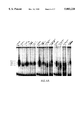
- FIGS. 5A-5C Mutations in Stat3 influence DNA Binding Affinity.
- 5A EMSA analysis of DNA:protein complexes. Nuclear extracts from EGF-treated COS cells transfected with Stat3, mutant EE>AA or mutant VVV>AAA (see Methods) were incubated with labeled M67 probe to reveal DNA binding complexes. Position of SIF A is marked.
- 5B Phosphotyrosine immunoblotting. Extracts from the cells in panel A were immunoprecipitated with Stat3-specific antiserum, separated by SDS PAGE, transferred to nitrocellulose and probed with monoclonal antibody PY20.
- 5C Co-immunoprecipitation of Stat1 and Stat3 mutants.
- COS cells were transfected with FLAG-tagged Stat3 or mutants along with untagged Stat1 and treated (+) or not treated (-) with EGR.
- FLAG immunoprecipitates were separated by SDA PAGE, transferred to nitrocellulose, and probed with Stat1 specific antiserum (top panel).
- STAT1 refers to transfection with Stat1 alone.
- Bottom panel is an immunoblot with FLAG specific monoclonal antibody to demonstrate similar expression levels in the transfected cells.
- FIGS. 6A-6B Alignment of STAT Family Members in the Putative DNA Binding Region. Lines below indicate boundaries of putative helices (H,h) and beta sheets (B,b) predicted by the algorithms of Chou and Fasman for each of the family members. Numbering above the alignment refers to the Stat1 sequence. The conserved amino acids mutated in this study are overlined. Sequences were aligned using the GCG pileup program and secondary structure was predicted using the GCG peptide structure program (Genetics Computer Group, 1991).
- FIG. 7 Comparison of the partial carboxyl terminal sequence in a series of STAT proteins.
- FIGS. 8A-8B Phosphorylation of wild type and mutant proteins on tyrosine as tested by anti-phosphotyrosine antibody reaction with Stat1 immunoprecipitates separated on polyacrylamide gel (FIG. 8A). Electrophoretic gel shift assay (EMSA) with nuclear extracts of cells treated for 20 minutes with INF- ⁇ 32 P-labeled IRF-1 GAS as probe (FIG. 8B).
- ESA Electrophoretic gel shift assay
- FIG. 9 Wild type and mutant Stat1 ⁇ binding to IRF-1 GAS.
- the gel shift bands were specific because anti-Stat1C serum produced a supershift while the pre-immune serum had no affect.
- FIGS. 10A-10L Protein extracts were prepared, exposed to anti-Stat1C serum and the 91 kDa 32 P-labeled band was detected by PAGE analysis.
- FIG. 10A Autoradiongraphs of two dimensional thin layer chromatograms of trypsin digested wild type and mutant Stat1 ⁇ from U3-cellular extracts treated or not treated with IFN- ⁇ (FIGS. 10B-10L).
- FIG. 11 Level of expression of a luciferase protein under control of three GAS sites from the promoter of the Ly6E gene in cells transfected with wild type Stat1 ⁇ , mutant Stat1 ⁇ , and Stat1 ⁇ .
- FIGS. 12A-12B depict the Northern blot analysis for IRF1 mRNA, an INF- ⁇ -induced gene, in U3A-derived cell lines containing wild type Stat1 ⁇ or mutant Stat1 ⁇ s treated with INF- ⁇ .
- FIG. 12B shows the comparison of the run-on transcriptional signal from the IRF1 gene in the two U3A cell derivatives.
- receptor recognition factor means receptor recognition factor
- receptor recognition-tyrosine kinase factor means receptor recognition factor/tyrosine kinase substrate
- receptor recognition/transcription factor means receptor recognition factor
- recognition factor means recognition factor protein(s)
- signal transducers and activators of transcription means receptor activators of transcription
- modifications may be deliberate, for example, such as modifications obtained through site-directed mutagenesis, or may be accidental, such as those obtained through mutations in hosts that are producers of the complex or its named subunits.
- the terms "receptor recognition factor”, “recognition factor”, “recognition factor protein(s)”, “signal transducers and activators of transcription”, and “STAT” are intended to include within their scope proteins specifically recited herein as well as all substantially homologous analogs and allelic variations.
- amino acid residues described herein are preferred to be in the "L" isomeric form.
- residues in the "D" isomeric form can be substituted for any L-amino acid residue, as long as the desired functional property of immunoglobulin-binding is retained by the polypeptide.
- NH2 refers to the free amino group present at the amino terminus of a polypeptide.
- COOH refers to the free carboxyl group present at the carboxyl terminus of a polypeptide.
- amino-acid residue sequences are represented herein by formulae whose left and right orientation is in the conventional direction of amino-terminus to carboxy-terminus. Furthermore, it should be noted that a dash at the beginning or end of an amino acid residue sequence indicates a peptide bond to a further sequence of one or more amino-acid residues.
- the above Table is presented to correlate the three-letter and one-letter notations which may appear alternately herein.
- a “replicon” is any genetic element (e.g., plasmid, chromosome, virus) that functions as an autonomous unit of DNA replication in vivo, i.e., capable of replication under its own control.
- a "vector” is a replicon, such as plasmid, phage or cosmid, to which another DNA segment may be attached so as to bring about the replication of the attached segment.
- a "cassette” refers to a segment of DNA that can be inserted into a vector at specific restriction sites.
- the segment of DNA encodes a polypeptide of interest, and the cassette and restriction sites are designed to ensure insertion of the cassette in the proper reading frame for transcription and translation.
- a cell has been "transfected” by exogenous or heterologous DNA when such DNA has been introduced inside the cell.
- a cell has been "transformed” by exogenous or heterologous DNA when the transfected DNA effects a phenotypic change.
- the transforming DNA should be integrated (covalently linked) into chromosomal DNA making up the genome of the cell.
- Heterologous DNA refers to DNA not naturally located in the cell, or in a chromosomal site of the cell.
- the heterologous DNA includes a gene foreign to the cell.
- a "clone” is a population of cells derived from a single cell or common ancestor by mitosis.
- nucleic acid molecule refers to the phosphate ester polymeric form of ribonucleosides (adenosine, guanosine, uridine or cytidine: "RNA molecules”) or deoxyribonucleosides (deoxyadenosine, deoxyguanosine, deoxythymidine, or deoxycytidine; "DNA molecules”) in either single stranded form, or a double-stranded helix. Double stranded DNA--DNA, DNA-RNA and RNA--RNA helices are possible.
- nucleic acid molecule refers only to the primary and secondary structure of the molecule, and does not limit it to any particular tertiary forms.
- this term includes double-stranded DNA found, inter alia, in linear or circular DNA molecules (e.g., restriction fragments), plasmids, and chromosomes.
- sequences may be described herein according to the normal convention of giving only the sequence in the 5' to 3' direction along the nontranscribed strand of DNA (i.e., the strand having a sequence homologous to the mRNA).
- a "recombinant DNA molecule” is a DNA molecule that has undergone a molecular biological manipulation.
- a nucleic acid molecule is "hybridizable" to another nucleic acid molecule, such as a cDNA, genomic DNA, or RNA, when a single stranded form of the nucleic acid molecule can anneal to the other nucleic acid molecule under the appropriate conditions of temperature and solution ionic strength (see Sambrook et al., supra).
- the conditions of temperature and ionic strength determine the "stringency" of the hybridization.
- low stringency hybridization conditions corresponding to a T m of 55°, can be used, e.g., 5 ⁇ SSC, 0.1% SDS, 0.25% milk, and no formamide; or 30% formamide, 5 ⁇ SSC, 0.5% SDS).
- Moderate stringency hybridization conditions correspond to a higher T m , e.g., 40% formamide, with 5 ⁇ or 6 ⁇ SCC.
- High stringency hybridization conditions correspond to the highest T m , e.g., 50% formamide, 5 ⁇ or 6 ⁇ SCC.
- Hybridization requires that the two nucleic acids contain complementary sequences, although depending on the stringency of the hybridization, mismatches between bases are possible.
- the appropriate stringency for hybridizing nucleic acids depends on the length of the nucleic acids and the degree of complementation, variables well known in the art. The greater the degree of similarity or homology between two nucleotide sequences, the greater the value of T m for hybrids of nucleic acids having those sequences.
- RNA:RNA, DNA:RNA, DNA:DNA The relative stability (corresponding to higher T m ) of nucleic acid hybridizations decreases in the following order: RNA:RNA, DNA:RNA, DNA:DNA.
- equations for calculating T m have been derived (see Sambrook et al., supra, 9.50-0.51).
- the position of mismatches becomes more important, and the length of the oligonucleotide determines its specificity (see Sambrook et al., supra. 11.7-11.8).
- a minimum length for a hybridizable nucleic acid is at least about 10 nucleotides; more preferably at least about 15 nucleotides; most preferably the length is at least about 20 nucleotides.
- Homologous recombination refers to the insertion of a foreign DNA sequence of a vector in a chromosome.
- the vector targets a specific chromosomal site for homologous recombination.
- the vector will contain sufficiently long regions of homology to sequences of the chromosome to allow complementary binding and incorporation of the vector into the chromosome. Longer regions of homology, and greater degrees of sequence similarity, may increase the efficiency of homologous recombination.
- a DNA "coding sequence” is a double-stranded DNA sequence which is transcribed and translated into a polypeptide in a cell in vitro or in vivo when placed under the control of appropriate regulatory sequences. The boundaries of the coding sequence are determined by a start codon at the 5' (amino) terminus and a translation stop codon at the 3' (carboxyl) terminus.
- a coding sequence can include, but is not limited to, prokaryotic sequences, cDNA from eukaryotic mRNA, genomic DNA sequences from eukaryotic (e.g., mammalian) DNA, and even synthetic DNA sequences. If the coding sequence is intended for expression in a eukaryotic cell, a polyadenylation signal and transcription termination sequence will usually be located 3' to the coding sequence.
- Transcriptional and translational control sequences are DNA regulatory sequences, such as promoters, enhancers, terminators, and the like, that provide for the expression of a coding sequence in a host cell.
- polyadenylation signals are control sequences.
- a “promoter sequence” is a DNA regulatory region capable of binding RNA polymerase in a cell and initiating transcription of a downstream (3' direction) coding sequence.
- the promoter sequence is bounded at its 3' terminus by the transcription initiation site and extends upstream (5' direction) to include the minimum number of bases or elements necessary to initiate transcription at levels detectable above background.
- a transcription initiation site (conveniently defined for example, by mapping with nuclease S1), as well as protein binding domains (consensus sequences) responsible for the binding of RNA polymerase.
- a coding sequence is "under the control" of transcriptional and translational control sequences in a cell when RNA polymerase transcribes the coding sequence into mRNA, which is then trans-RNA spliced and translated into the protein encoded by the coding sequence.
- a “signal sequence” is included at the beginning of the coding sequence of a protein to be expressed on the surface of a cell. This sequence encodes a signal peptide, N-terminal to the mature polypeptide, that directs the host cell to translocate the polypeptide.
- the term "translocation signal sequence” is used herein to refer to this sort of signal sequence. Translocation signal sequences can be found associated with a variety of proteins native to eukaryotes and prokaryotes, and are often functional in both types of organisms.
- oligonucleotide as used herein in referring to the probe of the present invention, is defined as a molecule comprised of two or more ribonucleotides or deoxyribonucleotides, preferably more than three. Its exact size will depend upon many factors which, in turn, depend upon the ultimate function and use of the oligonucleotide.
- nucleotide probe refers to an oligonucleotide of at least about 9 bases, which has a sequence corresponding to a portion of the DNA to which a STAT protein binds, and thus is capable of binding to a STAT protein.
- a nucleotide probe binds to the STAT protein with high specificity and affinity.
- Such a nucleotide probe corresponds to a specific STAT binding site.
- nucleotide probes of the invention may correspond to a general STAT binding site on DNA as well.
- sequence homology in all its grammatical forms refers to the relationship between proteins that possess a "common evolutionary origin,” including proteins from superfamilies (e.g., the immunoglobulin superfamily) and homologous proteins from different species (e.g., myosin light chain, etc.) (Reeck et al., 1987, Cell 50:667).
- sequence similarity in all its grammatical forms refers to the degree of identity or correspondence between nucleic acid or amino acid sequences of proteins that do not share a common evolutionary origin (see Reeck et al., supra).
- Two DNA sequences are "substantially homologous” or “substantially similar” when at least about 75% (preferably at least about 80%, and most preferably at least about 90 or 95%) of the nucleotides match over the defined length of the DNA sequences. Sequences that are substantially homologous can be identified by comparing the sequences using standard software available in sequence data banks, or in a Southern hybridization experiment under, for example, stringent conditions as defined for that particular system. Defining appropriate hybridization conditions is within the skill of the art. See, e.g., Maniatis et al., supra; DNA Cloning, Vols. I & II, supra; Nucleic Acid Hybridization, supra.
- two amino acid sequences are “substantially homologous” or “substantially similar” when greater than 70% of the amino acids are identical, or functionally identical.
- the similar or homologous sequences are identified by alignment using, for example, the GCG (Genetics Computer Group, Program Manual for the GCG Package, Version 7, Madison, Wis.) pileup program.
- corresponding to is used herein to refer similar or homologous sequences, whether the exact position is identical or different from the molecule to which the similarity or homology is measured.
- sequences of the DNA-binding domains of the STAT proteins can be aligned, and the corresponding amino acid residues determined, despite the deletion of amino acid residues at some positions in one STAT protein compared to another.
- corresponding to refers to the sequence similarity, and not the numbering of the amino acid residues or nucleotide bases.
- an “antibody” is any immunoglobulin, including antibodies and fragments thereof, that binds a specific epitope.
- the term encompasses polyclonal, monoclonal, and chimeric antibodies, the last mentioned described in further detail in U.S. Pat. Nos. 4,816,397 and 4,816,567.
- An “antibody combining site” or “antigen recognition site” is that structural portion of an antibody molecule comprised of heavy and light chain variable and hypervariable regions that specifically binds antigen.
- the phrase "antibody molecule” in its various grammatical forms as used herein contemplates both an intact immunoglobulin molecule and an immunologically active portion of an immunoglobulin molecule.
- Exemplary antibody molecules are intact immunoglobulin molecules, substantially intact immunoglobulin molecules and those portions of an immunoglobulin molecule that contains the paratope, including those portions known in the art as Fab, Fab', F(ab') 2 and F(v), which portions are preferred for use in the therapeutic methods described herein.
- the phrase "monoclonal antibody” in its various grammatical forms refers to an antibody having only one species of antibody combining site capable of immunoreacting with a particular antigen. A monoclonal antibody thus typically displays a single binding affinity for any antigen with which it immunoreacts.
- a monoclonal antibody may therefore contain an antibody molecule having a plurality of antibody combining sites, each immunospecific for a different antigen: e.g., a bispecific (chimeric) monoclonal antibody.
- a molecule is "antigenic" when it is capable of specifically interacting with an antigen recognition molecule of the immune system, such as an immunoglobulin (antibody) or T cell antigen receptor.
- An antigenic polypeptide contains at least about 5, and preferably at least about 10, amino acids.
- An antigenic portion of a molecule can be that portion that is immunodominant for antibody or T cell receptor recognition, or it can be a portion used to generate an antibody to the molecule by conjugating the antigenic portion to a carrier molecule for immunization.
- a molecule that is antigenic need not be itself immunogenic, i.e., capable of eliciting an immune response without a carrier.
- adjuvant refers to a compound or mixture that enhances the immune response to an antigen.
- An adjuvant can serve as a tissue depot that slowly releases the antigen and also as a lymphoid system activator that non-specifically enhances the immune response (Hood et al., Immunology, Second Ed., 1984, Benjamin/Cummings: Menlo Park, Calif. p. 384).
- a primary challenge with an antigen alone, in the absence of an adjuvant will fail to elicit a humoral or cellular immune response.
- Adjuvants include, but are not limited to, complete Freund's adjuvant, incomplete Freund's adjuvant, saponin, mineral gels such as aluminum hydroxide, surface active substances such as lysolecithin, pluronic polyols, polyanions, peptides, oil or hydrocarbon emulsions, keyhole limpet hemocyanins, dinitrophenol, and potentially useful human adjuvants such as BCG (bacille Calmette-Guerin) and Corynebacterium parvum.
- the adjuvant is pharmaceutically acceptable.
- a composition comprising "A” (where "A” is a single protein, DNA molecule. vector, recombinant host cell, etc.) is substantially free of "B” (where “B” comprises one or more contaminating proteins, DNA molecules, vectors, etc.) when at least about 75% by weight of the proteins, DNA, vectors (depending on the category of species to which A and B belong) in the composition is "A".
- "A” comprises at least about 90% by weight of the A+B species in the composition, most preferably at least about 99% by weight. It is also preferred that a composition. which is substantially free of contamination, contain only a single molecular weight species having the activity or characteristic of the species of interest.
- pharmaceutically acceptable refers to molecular entities and compositions that are physiologically tolerable and do not typically produce an allergic or similar untoward reaction, such as gastric upset, dizziness and the like, when administered to a human.
- pharmaceutically acceptable means approved by a regulatory agency of the Federal or a state government or listed in the U.S. Pharmacopeia or other generally recognized pharmacopeia for use in animals, and more particularly in humans.
- carrier refers to a diluent, adjuvant, excipient, or vehicle with which the compound is administered.
- Such pharmaceutical carriers can be sterile liquids, such as water and oils, including those of petroleum, animal, vegetable or synthetic origin, such as peanut oil, soybean oil, mineral oil, sesame oil and the like.
- Water or aqueous solution saline solutions and aqueous dextrose and glycerol solutions are preferably employed as carriers, particularly for injectable solutions. Suitable pharmaceutical carriers are described in "Remington's Pharmaceutical Sciences” by E. W. Martin.
- terapéuticaally effective amount is used herein to mean an amount sufficient to reduce by at least about 15 percent, preferably by at least 50 percent, more preferably by at least 90 percent, and most preferably prevent, a clinically significant deficit in the activity, function and response of the host. Alternatively, a therapeutically effective amount is sufficient to cause an improvement in a clinically significant condition in the host.
- biological sample is used herein to refer to a sample containing cells that express or may express a STAT protein. Such cells may be obtained from a subject, or from in vitro culture.
- biological sample further extends to an extract of cells from either source.
- the present invention relates to the discovery that Stat1 and Stat3, which are two members of the ligand-activated transcription factor family that serve the dual functions of signal transducers and activators of transcription, select similar, but not identical, optimum binding sites from random oligonucleotides. Differences in their binding affinity were readily apparent with natural STAT binding sites. However, unlike other DNA binding proteins, fragments of the STAT proteins could not be shown to bind to DNA.
- chimeric Stat1:Stat3 molecules were used to locate the amino acids that could discriminate a general binding site from a specific binding site.
- the amino acids between residues ⁇ 400 and ⁇ 500 of these ⁇ 750 amino acid long proteins were discovered to determine the DNA binding site specificity. Mutations within this region result in Stat proteins which are activated normally by tyrosine phosphorylation and which dimerize, but have greatly reduced DNA binding affinities.
- the invention further relates to the discovery that phosphorylation of a serine residue at position 727, in the carboxyl-terminus, of Stat1 ⁇ is required for maximal interferon- ⁇ (IFN- ⁇ ) dependent transcriptional response.
- IFN- ⁇ interferon- ⁇
- the present invention particularly relates to functionally active regions of the STAT proteins, e.g., as exemplified herein with portions of Stat1 ⁇ , particularly such fragments that contain a DNA binding domain, and a C-terminal serine residue that is phosphorylated non-specifically as a consequence of cellular activation, but which is critical for maximum transcriptional activation.
- the invention contemplates antagonists of STAT proteins targeted to the DNA-binding domain.
- the invention is directed to mutant forms of STAT proteins that can compete as substrates for tyrosine phosphorylation and dimerization, but which are poor DNA-binding proteins, or have reduced transcriptional activation activity.
- each member of the family of receptor recognition factors is designated as a signal transducer and activator of transcription (STAT) protein.
- STAT protein is designated by the apparent molecular weight (e.g., Stat113, Stat91, Stat84, etc.), or by the order in which it has been identified (e.g., Stat1 ⁇ Stat91!, Stat1 ⁇ Stat84!, Stat2 Stat113!, Stat3 a murine protein also termed 19sf6!, and Stat4 a murine STAT protein also termed 13sf1!).
- the present invention also relates to a recombinant DNA molecule or cloned gene, or a degenerate variant thereof, which encodes a receptor recognition factor, or a fragment thereof, that encodes a DNA binding domain, or a chimeric protein containing a functionally active DNA binding domain of a STAT protein.
- an appropriate antagonist of the DNA-binding domain of a STAT protein could be introduced to block the interaction of the STAT protein with its DNA binding site.
- mutation of the C-terminal phosphorylation site, or introduction of a mutant STAT protein lacking such a C-terminal phosphorylation site would be expected to lead to a decrease in the level of transcriptional activation mediated by a STAT protein containing such a serine phosphorylation site.
- the antagonists of the STAT binding to DNA may be prepared in pharmaceutical compositions, with a suitable carrier and at a strength effective for administration by various means to a patient experiencing an adverse medical condition associated specific transcriptional stimulation for the treatment thereof.
- the pharmaceutical formulation will provide for transmembrane migration of the antagonists, which will be active in the cytoplasm.
- parenteral techniques such as subcutaneous, intravenous and intraperitoneal injections, catheterizations and the like. Average quantities of the recognition factors or their subunits may vary and in particular should be based upon the recommendations and prescription of a qualified physician or veterinarian.
- antibodies including both polyclonal and monoclonal antibodies may possess certain diagnostic or therapeutic (inhibitory) applications and may for example, be utilized for the purpose of detecting and/or measuring conditions such as cellular activation as a result of viral infection, inflammation, or the like.
- the STAT protein DNA-binding domain, or a peptide corresponding to a STAT protein epitope containing the phosphorylated serine residue may be used to produce both polyclonal and monoclonal antibodies to themselves in a variety of cellular media, by such well known techniques as immunization of rabbit using Complete and Incomplete Freund's Adjuvant and the hybridoma technique utilizing, for example, fused mouse spleen lymphocytes and myeloma cells, respectively.
- such proteins are conjugated to a carrier molecule, as described above.
- small molecules that mimic or antagonize the activity(ies) of the receptor recognition factors of the invention may be discovered or synthesized, and may be used in diagnostic and/or therapeutic protocols.
- Identification of important regions of the STAT proteins for function provides a basis for screening for drugs capable of specific interaction with the functionally relevant domains. According, in addition to rational design of compounds that bind to, and preferably competitively inhibit the functional activity of the STAT protein. i.e., antagonists, based on the structure of relevant domain, the present invention contemplates an alternative method for identifying specific binding compounds of the DNA-binding domain or the region containing phosphoserine using various screening assays known in the art.
- Any screening technique known in the art can be used to screen for STAT DNA-binding antagonists.
- the present invention contemplates screens for small molecule ligands or ligand analogs and mimics, as well as screens for natural ligands that bind to and antagonize STAT activates in vivo.
- synthetic libraries (Needels et al., 1993, "Generation and screening of an oligonucleotide encoded synthetic peptide library," Proc. Natl. Acad. Sci. USA 90:10700-4; Lam et al., International Patent Publication No. WO 92/00252, each of which is incorporated herein by reference in its entirety), and the like can be used to screen for STAT DNA-binding domain or phosphoserine region ligands according to the present invention.
- the screening can be performed directly using peptides corresponding to the DNA binding domain or the region containing the phosphoserine residue.
- chimeric proteins which contain the DNA binding domain (or the serine residue) may be used, as such proteins will contain the element specifically under investigation. Specific examples of such chimeric proteins are disclosed in the Examples, infra.
- the reagents that contain the STAT DNA-binding domain can be labeled for use in the screening assays.
- the compound may be directly labeled.
- a labeled secondary reagent may be used to detect binding of the compound to a solid phase support containing a binding molecule of interest. Binding may be detected by in situ formation of a chromophore by an enzyme label. Suitable enzymes include, but are not limited to, alkaline phosphatase and horseradish peroxidase.
- labels for use in the invention include colored latex beads, magnetic beads, fluorescent labels (e.g., fluorescene isothiocyanate (FITC), phycoerythrin (PE), Texas red (TR), rhodamine, free or chelated lanthanide series salts, especially Eu 3+ , to name a few fluorophores), chemiluminescent molecules, radio-isotopes, or magnetic resonance imaging labels.
- fluorescent labels e.g., fluorescene isothiocyanate (FITC), phycoerythrin (PE), Texas red (TR), rhodamine, free or chelated lanthanide series salts, especially Eu 3+ , to name a few fluorophores
- chemiluminescent molecules chemiluminescent molecules
- radio-isotopes or magnetic resonance imaging labels.
- the diagnostic method of the present invention comprises examining a cellular sample or medium by means of an assay including an effective amount of a reagent that specifically binds to a serine-phosphorylated STAT protein.
- a reagent is an antibody, preferably an affinity-purified polyclonal antibody, and more preferably a mAb.
- the anti-recognition factor antibody molecules used herein be in the form of Fab, Fab', F(ab') 2 or F(v) portions or whole antibody molecules.
- patients capable of benefiting from this method include those suffering from cancer, a pre-cancerous lesion, a viral infection or other like pathological derangement.
- the present invention relates to detection of both phosphotyrosine and phosphoserine on a STAT protein, which is indicative of maximum activity of the STAT protein, and thus an indicator of the degree of cellular activation. Since cellular activation is associated with certain pathological states, as discussed above, the present invention provides an advantageous method for evaluating cellular activation. Moreover, the present invention is the first instance known to the inventors in which the specific tyrosine phosphorylation activation pathway and the general serine phosphorylation activation pathway cross in the same trascription activation factor. Accordingly, this discovery has important implications for detection of diseases or disorders, i.e., pathological conditions, associated with cellular activation.
- Detection of phosphorylation of tyrosine and serine can be accomplished by any techniques known in the art, including measuring the level of phosphorylation per unit mass of protein; using specific phosphatases and an appropriate detection system to detect specific phosphorylation; using antibodies generated against the phosphorylated forms of the proteins; or using well known biochemical techniques, as described in the Examples, infra.
- a subject therapeutic composition includes, in admixture, a pharmaceutically acceptable excipient (carrier) and one or more of an antagonist of STAT binding to DNA, e.g., a molecule that specifically interacts with the DNA-binding domain of a STAT protein, as described herein as an active ingredient.
- a pharmaceutically acceptable excipient carrier
- an antagonist of STAT binding to DNA e.g., a molecule that specifically interacts with the DNA-binding domain of a STAT protein, as described herein as an active ingredient.
- a mutant STAT which has been mutated in the DNA-binding domain or in the serine phosphorylation site can be introduced into the cells of a subject.
- the presence of such mutant forms of the STAT proteins, which are capable of interacting with the receptor, being phosphorylated on tyrosine, and translocating to the nucleus can be used as "decoys.”
- Such proteins when dimerized with other STAT proteins (either with a mutant or wild-type form of the protein, or with another STAT protein), are expected to bind to the DNA with lower affinity, and thus be less effective at transcription activation.
- such a "decoy" mutant STAT protein is introduced into a cell via transgenic therapy.
- the present invention contemplates preparation of a gene encoding a mutant form of a STAT protein, wherein the mutation is found in the DNA binding domain, or is a mutation of the C-terminal serine residues that is phosphorylated in the highly functional forms of the protein.
- the term "gene” refers to an assembly of nucleotides that encode a polypeptide, and includes cDNA and genomic DNA nucleic acids.
- a gene encoding a mutant STAT protein can be isolated from any source, particularly from a human cDNA or genomic library, and mutated according to standard methods.
- Specific cDNA sequences encoding STAT proteins are disclosed in SEQ ID NOS:1, 3, 5, 7, 9, and 11.
- Methods for obtaining the STAT gene are well known in the art, as described above (see, e.g., Sambrook et al., 1989, supra). Any technique for mutagenesis known in the art can be used, including but not limited to, in vitro site-directed mutagenesis (Hutchinson, C., et al., 1978, J. Biol. Chem.
- any animal cell potentially can serve as the nucleic acid source for the molecular cloning of a STAT gene.
- the DNA may be obtained by standard procedures known in the art from cloned DNA (e.g., a DNA "library”), by chemical synthesis, by cDNA cloning, or by the cloning of genomic DNA, or fragments thereof, purified from the desired cell (See, for example, Sambrook et al., 1989, supra, Glover, D. M. (ed.), 1985. DNA Cloning: A Practical Approach, MRL Press, Ltd., Oxford. U.K. Vol. I, II).
- Clones derived from genomic DNA may contain regulatory and intron DNA regions in addition to coding regions; clones derived from cDNA will not contain intron sequences. Whatever the source, the gene should be molecularly cloned into a suitable vector for propagation of the gene.
- the nucleotide sequence coding for a mutant STAT protein can be inserted into an appropriate expression vector, i.e., a vector which contains the necessary elements for the transcription and translation of the inserted protein-coding sequence. Such elements are termed herein a "promoter.”
- the nucleic acid encoding the mutant STAT protein of the invention is operatively associated with a promoter in an expression vector of the invention. Both cDNA and genomic sequences can be cloned and expressed under control of such regulatory sequences.
- An expression vector also preferably includes a replication origin.
- the necessary transcriptional and translational signals can be provided on a recombinant expression vector, or they may be supplied by the native gene encoding a STAT and/or its flanking regions.
- a chimeric STAT protein or mutant STAT protein can be prepared, e.g., a glutathione-S-transferase (GST) fusion protein, a maltose-binding (MBP) protein fusion protein, or a poly-histidine-tagged fusion protein, for expression in bacteria.
- GST glutathione-S-transferase
- MBP maltose-binding
- poly-histidine-tagged fusion protein for expression in bacteria.
- GST binds glutathione conjugated to a solid support matrix
- MBP binds to a maltose matrix
- poly-histidine chelates to a Ni-chelation support matrix.
- the fusion protein can be eluted from the specific matrix with appropriate buffers, or by treating with a protease specific for a cleavage site usually engineered between the STAT polypeptide and the fusion partner (e.g., GST, MBP, or poly-His).
- a protease specific for a cleavage site usually engineered between the STAT polypeptide and the fusion partner (e.g., GST, MBP, or poly-His).
- the present invention contemplates fusions between a domain from one STAT protein in the site of the corresponding domain of a second STAT protein.
- Such chimeric constructs are specifically exemplified in the Examples, infra.
- Potential host-vector systems include but are not limited to mammalian cell systems infected with virus (e.g., vaccinia virus, adenovirus, etc.); insect cell systems infected with virus (e.g., baculovirus); microorganisms such as yeast containing yeast vectors; or bacteria transformed with bacteriophage, DNA, plasmid DNA, or cosmid DNA.
- virus e.g., vaccinia virus, adenovirus, etc.
- insect cell systems infected with virus e.g., baculovirus
- microorganisms such as yeast containing yeast vectors
- bacteria transformed with bacteriophage, DNA, plasmid DNA, or cosmid DNA e.g., bacteriophage, DNA, plasmid DNA, or cosmid DNA.
- the expression elements of vectors vary in their strengths and specificities. Depending on the host-vector system utilized, any one of a number of suitable transcription and translation elements may be used.
- a recombinant mutant or chimeric STAT of the invention, or functional fragment, derivative or analog thereof, may be expressed chromosomally, after integration of the protein coding sequence by recombination.
- any of a number of amplification systems may be used to achieve high levels of stable gene expression (See Sambrook et al., 1989, supra).
- the cell into which the recombinant vector comprising the nucleic acid encoding the mutant or chimeric STAT is cultured in an appropriate cell culture medium under conditions that provide for expression of the protein by the cell.
- Any of the methods previously described for the insertion of DNA fragments into a cloning vector may be used to construct expression vectors containing a gene consisting of appropriate transcriptional/translational control signals and the protein coding sequences. These methods may include in vitro recombinant DNA and synthetic techniques and in vivo recombination (genetic recombination).
- Promoters which may be used to control gene expression include, but are not limited to, the SV40 early promoter region (Benoist and Chambon, 1981, Nature 290:304-310), the promoter contained in the 3' long terminal repeat of Rous sarcoma virus (Yamamoto, et al., 1980, Cell 22:787-797). the herpes thymidine kinase promoter (Wagner et al., 1981, Proc. Natl. Acad. Sci. U.S.A.
- prokaryotic expression vectors such as the ⁇ -lactamase promoter (Villa-Kamaroff, et al., 1978, Proc. Natl. Acad. Sci. U.S.A. 75:3727-3731), or the tac promoter (DeBoer, et al., 1983, Proc. Natl. Acad. Sci. U.S.A.
- Vectors are introduced into the desired host cells by methods known in the art, e.g., transfection, electroporation, microinjection, transduction, cell fusion, DEAE dextran. calcium phosphate precipitation, lipofection (lysosome fusion), use of a gene gun, or a DNA vector transporter (see, e.g., Wu et al., 1992, J. Biol. Chem. 267:963-967; Wu and Wu, 1988, J. Biol. Chem. 263:14621-14624; Hartmut et al., Canadian Patent Application No. 2,012,311, filed Mar. 15, 1990).
- a gene encoding a mutant STAT protein is introduced in vivo in a viral vector.
- viral vectors include an attenuated or defective DNA virus, such as but not limited to herpes simplex virus (HSV), papillomavirus, Epstein Barr virus (EBV), adenovirus, adeno-associated virus (AAV), and the like.
- HSV herpes simplex virus
- EBV Epstein Barr virus
- AAV adeno-associated virus
- Defective viruses which entirely or almost entirely lack viral genes, are preferred. Defective virus is not infective after introduction into a cell.
- Use of defective viral vectors allows for administration to cells in a specific, localized area, without concern that the vector can infect other cells. Thus, a particular locus, e.g., the organ implicated in the rejection episode, can be specifically targeted with the vector.
- vectors include, but are not limited to, a defective herpes virus 1 (HSV1) vector (Kaplitt et al., 1991, Molec. Cell. Neurosci. 2:320-330), an attenuated adenovirus vector, such as the vector described by Stratford Perricaudet et al. (1992, J. Clin. Invest. 90:626-630), and a defective adeno-associated virus vector (Samulski et al., 1987, J. Virol. 61:3096-3101; Samulski et al., 1989, J. Virol. 63:3822-3828).
- HSV1 herpes virus 1
- the vector can be introduced in vivo by lipofection.
- liposomes for encapsulation and transfection of nucleic acids in vitro.
- Synthetic cationic lipids designed to limit the difficulties and dangers encountered with liposome mediated transfection can be used to prepare liposomes for in vivo transfection of a gene encoding a protein (Felgner, et. al., 1987, Proc. Natl. Acad. Sci. U.S.A. 84:7413-7417; see Mackey, et al., 1988, Proc. Natl. Acad. Sci. U.S.A. 85:8027-8031)).
- cationic lipids may promote encapsulation of negatively charged nucleic acids, and also promote fusion with negatively charged cell membranes (Felgner and Ringold, 1989, Science 337:387-388).
- lipofection to introduce exogenous genes into the specific organs in vivo has certain practical advantages.
- Molecular targeting of liposomes to specific cells represents one area of benefit. It is clear that directing transfection to particular cell types would be particularly advantageous in a tissue with cellular heterogeneity, such as pancrease, liver, kidney, and the brain.
- Lipids may be chemically coupled to other molecules for the purpose of targeting (see Mackey, et. al., 1988, supra).
- Targeted peptides e.g., hormones or neurotransmitters, and proteins such as antibodies or non-peptide molecules could be coupled to liposomes chemically.
- naked DNA vectors for gene therapy can be introduced into the desired host cells by methods known in the art, e.g., transfection, electroporation, microinjection, transduction, cell fusion, DEAE dextran, calcium phosphate precipitation, use of a gene gun, or use of a DNA vector transporter (see, e.g., Wu et al., 1992, J. Biol. Chem. 267:963-967; Wu and Wu, 1988, J. Biol. Chem. 263:14621-14624; Hartmut et al., Canadian Patent Application No. 2,012,311, filed Mar. 15, 1990).
- compositions which contain polypeptides, analogs or active fragments as active ingredients are well understood in the art.
- such compositions are prepared as injectables, either as liquid solutions or suspensions, however, solid forms suitable for solution in, or suspension in, liquid prior to injection can also be prepared.
- the preparation can also be emulsified.
- the active therapeutic ingredient is often mixed with excipients which are pharmaceutically acceptable and compatible with the active ingredient. Suitable excipients are, for example, water, saline, dextrose, glycerol, ethanol, or the like and combinations thereof.
- the composition can contain minor amounts of auxiliary substances such as wetting or emulsifying agents, pH buffering agents which enhance the effectiveness of the active ingredient.
- a polypeptide, analog or active fragment can be formulated into the therapeutic composition as neutralized pharmaceutically acceptable salt forms.
- Pharmaceutically acceptable salts include the acid addition salts (formed with the free amino groups of the polypeptide or antibody molecule) and which are formed with inorganic acids such as, for example, hydrochloric or phosphoric acids, or such organic acids as acetic, oxalic, tartaric, mandelic, and the like. Salts formed from the free carboxyl groups can also be derived from inorganic bases such as, for example, sodium, potassium, ammonium, calcium, or ferric hydroxides, and such organic bases as isopropylamine, trimethylamine, 2-ethylamino ethanol, histidine, procaine, and the like.
- the therapeutic polypeptide-, analog- or active fragment-containing compositions are conventionally administered intravenously, as by injection of a unit dose, for example.
- unit dose when used in reference to a therapeutic composition of the present invention refers to physically discrete units suitable as unitary dosage for humans, each unit containing a predetermined quantity of active material calculated to produce the desired therapeutic effect in association with the required diluent; i.e., carrier, or vehicle.
- compositions are administered in a manner compatible with the dosage formulation, and in a therapeutically effective amount.
- the quantity to be administered depends on the subject to be treated, capacity of the subject's immune system to utilize the active ingredient, and degree of inhibition or neutralization of recognition factor binding capacity desired. Precise amounts of active ingredient required to be administered depend on the judgment of the practitioner and are peculiar to each individual. However, suitable dosages may range from about 0.1 to 20, preferably about 0.5 to about 10, and more preferably one to several, milligrams of active ingredient per kilogram body weight of individual per day and depend on the route of administration. Suitable regimes for initial administration and booster shots are also variable, but are typified by an initial administration followed by repeated doses at one or more hour intervals by a subsequent injection or other administration. Alternatively, continuous intravenous infusion sufficient to maintain concentrations of ten nanomolar to ten micromolar in the blood are contemplated.
- the therapeutic compositions may further include an effective amount of the factor/factor synthesis promoter antagonist or analog thereof, and one or more of the following active ingredients: an antibiotic, a steroid.
- active ingredients an antibiotic, a steroid.
- Exemplary formulations are well known in the art, e.g., as disclosed in International Patent Publication WO 93/19179.
- an assay useful and contemplated in accordance with the present invention is known as a "cis/trans” assay. Briefly, this assay employs two genetic constructs, one of which is typically a plasmid that continually expresses a particular receptor of interest when transfected into an appropriate cell line, and the second of which is a plasmid that expresses a reporter such as luciferase, under the control of a receptor/ligand complex.
- one of the plasmids would be a construct that results in expression of the receptor in the chosen cell line, while the second plasmid would possess a promoter linked to the luciferase gene in which the response element to the particular receptor is inserted.
- the compound under test is an agonist for the receptor
- the ligand will complex with the receptor, and the resulting complex will bind the response element and initiate transcription of the luciferase gene.
- the resulting chemiluminescence is then measured photometrically, and dose response curves are obtained and compared to those of known ligands.
- the foregoing protocol is described in detail in U.S. Pat. No. 4,981,784 and PCT International Publication No. WO 88/03168, for which purpose the artisan is referred.
- kits suitable for use by a medical specialist may be prepared to determine the presence or absence of predetermined transcriptional activity or predetermined transcriptional activity capability in suspected target cells, as set forth above.
- one class of such kits will contain at least a reagent capable of specifically binding to the receptor STAT protein, and means for detecting binding of the reagent to a STAT protein.
- a specific binding reagent specific for phosphotyrosine, and a second specific binding reagent specific for phosphoserine are used.
- a reagent is an antibody.
- Means for detecting binding may be a label on the antibody (labels have been described above), or a label on a STAT protein or fragment thereof.
- the kits may also contain peripheral reagents such as buffers, stabilizers, etc.
- Stat1 and Stat3 are two members of the ligand-activated transcription factor family that serve the dual functions of signal transducers and activators of transcription. While the two proteins select similar (but not identical) optimum binding sites from random oligonucleotides, differences in their binding affinity were readily apparent with natural STAT binding sites. To take advantage of these different affinities, chimeric Stat1:Stat3 molecules were used to locate the amino acids that could discriminate a general binding site from a specific binding site. The amino acids between residues ⁇ 400 and ⁇ 500 of these ⁇ 750 amino acid long proteins determine the DNA binding site specificity. Mutations within this region result in Stat proteins which are activated normally by tyrosine phosphorylation and which dimerize, but have greatly reduced DNA binding affinities.
- Cytoplasmic and nuclear extracts were prepared as described (Sadowski and Gilman, 1993, Nature 362:79).
- Stat1 or Stat3 carboxyl terminal antiserum was used at a 1:200 dilution.
- Immobilized FLAG-specific monoclonal antibody was used for precipitation according to the manufacturer's instructions (Kodak).
- Phosphotyrosine-specific monoclonal antibody PY20 was used at 1:2000 dilution according to the manufacturer's instructions (Transduction Laboratories).
- Expression plasmid pRcCMV Invitrogen carrying Stat1 or Stat3 cDNA (Improta et al., 1994, Proc. Natl. Acad. Sci. USA 91:4776; Zhong et al., 1994, Science 264:95) was used for all cell lines. All of the recombinant STAT proteins were constructed by PCR amplification using Vent Polymerase (NEB) and verified by DNA sequencing. The chimeric Stat1 and Stat3 cDNAs included the FLAG epitope Kodak IBI; (Hopp et al., 1988, Bio/Technology 6:1204)! to easily identify the recombinant proteins.
- Ly6E 5'-ATATTCCTGTAAGTGAT-3' (SEQ ID NO:21)
- binding site selection for Stat1 was carried out essentially according to the method of Pollock and Triesman. IFN- ⁇ treated BUD 8 fibroblast nuclear extracts were mixed with a double stranded random 176 base oligomer and immunoprecipitated with antiserum specific for Stat1 and protein A agarose. The co-purifying DNA was isolated, amplified by polymerase chain reaction, and analyzed for binding by EMSa. Following five rounds of selection, Stat-specific complex was observed, eluted from the gel, and subcloned. To obtain the Stat3 optimum site, nuclear extracts from EGF-treated COS 1 cells transfected with Stat3 expression vector were bound to the random oligomer and applied to an EMSA gel.
- the region corresponding to the mobility of the Stat3 gel shift on one of the 76 bp Stat1-selected sites was excised and the DNA amplified by PCR. Following 5 rounds of selection from the gel, the resulting complex was supershifted by Stat3 specific antiserum and the DNA isolated from the supershifted complex eluted from the gel, amplified and subcloned.
- Stat1 and Stat3 homodimers In vitro binding site selection for Stat1 and Stat3. To determine whether Stat1 and Stat3 homodimers preferred different high affinity oligonucleotide binding sites, we carried out synthesis of a set of deoxyoligonucleotides 76 bases long: a random stretch of 26 bases was sandwiched between two constant 25 oligonucleotide regions that could be used as PCR primers. Stat1 optimum binding sites were determined first. Stat1 activation was carried out by IFN- ⁇ treatment of Bud-8 fibroblast cells and total cell extracts were exposed to the random deoxyoligonucleotide mixture.
- Stat1 COOH-terminal antiserum (Schindler et al., Science 257:809-815) was used to immunoprecipitate the protein/DNA complexes followed by PCR amplification of the DNA in the precipitate (Pollock and Triesman, 1990, Nucl. Acids Res. 18:6197-6204). Five such cycles were carried out and individual DNA segments were cloned after the final amplification. Sequencing of 55 individual clones demonstrated a clear consensus binding site with strong similarity to the earlier identified GAS elements (Decker et al., 1991, EMBO J. 10:927-932; Lew et al., 1991, Mol. Cell. Biol.
- ESA electrophoretic mobility shift assay
- Extracts were used from both IFN- ⁇ treated HepG2 cells and HepG2 cells treated with a high dose of IL-6 which induces three well recognized bands (Sadowski et al., 1993, Nature 362:79-83) described as SIF A, SIF B, and SIF C because there are three DNA binding complexes inducible by medium from cells expressing the sis oncogene (SIE, sis-inducible element; SIF, sis-inducible factor (Wagner et al., EMBO, 1990, EMBO J. 9:4477-4484).
- SIE sis-inducible element
- SIF sis-inducible factor
- the SIF C complex is identical in mobility and protein content to the IFN- ⁇ induced complex (Sadowski et al., 1993, Science 261:1739-1744) and is therefore a Stat1 homodimer. This complex reacts with Stat1 specific antiserum.
- the SIF A complex which migrates more slowly (most likely due to a greater number of positively charged amino acids in addition to a slightly longer polypeptide chain) reacts with the Stat3 antiserum (Zhong et al., 1994, Science 264:95-98) and is considered to contain a Stat3 homodimer.
- the SIF B complex which migrates between complex A and C reacts with both Stat1 and Stat3 antisera is considered a Stat1:3 heterodimer.
- the gel electrophoretic band was excised, DNA amplified and five cycles of gel shifts and amplification were carried out before cloning of individual examples of DNA from the SIF A complex. Sequencing of 55 individual clones with Stat3-selected sequences also revealed a clear consensus sequence which was identical in the core sequence TTCC C or G!GGAA to that selected by the Stat1 (FIG. 1A). Just as did the Stat1 site, the Stat3 selected site contained an A or T at positions +6 or -6, respectively, but in addition the Stat3 site also showed a strong preference of A and T at positions +5 and -5 making a 13 nucleotide palindrome the favored Stat3 site. As with Stat1, a preference for G at position +7 was not matched by a C at position -7. Also, position -9 was G in about 60% of cases. As with Stat1, these flanking sequence preferences may contribute to the optimum site.
- oligonucleotide probe was synthesized to represent the Stat3 optimal site (position -9 to +9) and used in a gel shift experiment (FIG. 1B, lanes 9-13). Since the Stat1 optimum site core is contained within the Stat3 probe, it was not surprising that, like the selected Stat1 probe, the Stat3 probe bound well to all of the SIF complexes. Unfortunately, the Stat3 consensus probe used also bound even more strongly to a constitutively active protein (marked by the asterisk in FIG. 1B) that comigrates closely with SIF B, obscuring the center section of the gel shift pattern.
- Stat3 consensus probe bound somewhat better in the SIF A complex from which it had been selected than did the Stat1 optimum probe, but this was estimated by competition experiments to be only a 3-5 fold difference. While it is clear that such relatively minor differences might be important at individual sites in genomic DNA, we could not use these "consensus" probes to easily distinguish the binding affinities of Stat1 from Stat3.
- Stat protein binding to natural sites Previously identified Stat protein binding elements were next examined to determine if any sites gave sufficient specificity to distinguish easily Stat1 from Stat3 binding. Oligonucleotide probes representing GAS IFN- ⁇ activates sites (Decker et al., 1991, EMBO J. 10:927-932; Lew et al., 1991, Mol. Cell. Biol. 11:182-191) from the murine surface antigen Ly6e (Kahn et al., 1993. Proc. Natl. Acad. Sci. USA 90:6806-6810), IFN- ⁇ response region (the GRR) of the FcgR1 gene (Pearse et al., 1993, Proc. Natl. Acad. Sci.
- the Stat1 optimum core sequence was also bound by all of the SIF species, but with overall lower affinity as judged by the intensity of the binding signal.
- the M67 probe binds well to both Stat1 or Stat3 but cannot distinguish between them.
- the GRR and Ly6e probes were both bound by the SIF C protein (Stat1 homodimer), with the GRR probe giving 2-3 fold more binding than the Ly6e probe. Both probes were bound poorly by the SIF B complex, the heterodimer of Stat3 and Stat1. Most significantly, the SIF A complex that represents Stat3 homodimer binding was not observed with the GRR or Ly6e probes unless the autoradiograms were overexposed.
- the two closely related proteins Stat3 and Stat1 differ in their ability to recognize these two natural GAS elements.
- Other GAS elements tested displayed intermediate binding properties with respect to Stat1 and Stat3 binding and were not useful for this analysis (data not shown).
- the Stat1 -SH2 group lies between amino acids 573 and 700 (resides ⁇ 6600-700) (Fu, 1992, Cell 70:323-335; Schindler et al., 1992, Proc. Natl. Acad. Sci. USA 89:7836-7839, Schindler et al., 1992, Science 257:809-815) and the Y that becomes phosphorylated is at residue 701.
- Stat3 also contains an SH2 region from ⁇ 60-700 and a Y in a position comparable to Stat1 at residue 705 but Stat3 is not activated by IFN- ⁇ (Zhong et al., 1994, Proc. Natl. Acad. Sci. USA 91:4806-4810). Mutations of the Stat3 Y residue at 705 to phenylalanine likewise blocks phosphorylation of Stat3, Z. Wen and J. E. Darnell, unpublished observations).
- U3A cells lack expression of Stat1 protein, but contain active receptors for IFN- ⁇ or IFN- ⁇ (Pellegrini et al., Mol. Cell. Biol. 9:4605-4612; Muller et al., 1993, EMBO J. 12:4221-4228).
- Stat1 (and chimeric proteins containing the Stat1 carboxyl terminal activation regions) introduced into this cell line can be activated by IFN- ⁇ or IFN- ⁇ (Muller et al., 1993, EMBO J. 12:4221-4228; Improta et al., 1994, Proc. Natl. Acad. Sci. USA 91:4776-4780; FIG. 4).
- Stat3 can be activated by IFN- ⁇ in the U3A precursor cell line, 2FTGH (I. Kerr, personal comm.; C. M. Horvath, Z. Zhong and J. E.
- Stat3 homodimers As mentioned earlier, a greater number of glutamine and aspartic acid residues plus a slightly greater length in Stat3 compared to Stat1 is the cause for the slower migration of Stat3 homodimers compared to Stat1 homodimers. In chimeric proteins, these differences were reflected in protein:DNA complexes that migrated at intermediate rates.
- a chimeric Stat protein containing the first 508 amino acids of Stat1 and the carboxyl terminus of Stat3 exhibited the general binding property of Stat1 in that the chimeric protein, designated 1 508 3 , bound well to both test probes and migrated just slightly slower than Stat1 (FIGS. 4A and B, lane 6).
- the complementary chimera, 3 514 1 with the amino terminal 514 amino acids of Stat3 fused to the carboxyl terminus of Stat1 had the recognition property of Stat3, that is, it bound well to M67 probe, but not to GRR (FIGS. 4A and B, lane 8).
- the STAT DNA recognition capacity was localized to the amino terminal 508 amino acids of Stat1 or 514 amino acids of Stat3, and was not influenced by the putative SH3 domain ( ⁇ 500-600), the SH2 domain ( ⁇ 600-700) or other sequences in the carboxyl terminal third of the molecule which itself can utilize different ligand-receptor complexes for activation (IFN- ⁇ for Stat1 and IFN- ⁇ for Stat3).
- Point mutations alter DNA binding affinity.
- the proposed DNA recognition domain ( ⁇ 400-500) encompasses one of the most highly conserved regions of the STAT protein family, although no function had been previously assigned to this region either from experiment or from sequence comparison with other proteins in the data banks.
- mutations were made in two of the highly conserved regions of Stat3 in the ⁇ 400-500 region.
- the sequence VTEEL (residues 432 to 436) was changed to VTAAL (mutant EE>AA) or the conserved sequence SLPVVVISN (residues 458 to 466) was changed to SLPAAAISN (mutant VVV>AAA).
- Mutant EE>AA had a more severe effect on DNA binding (nearly undetectable) than mutant VV>AA, which exhibited a distinctly reduced but still detectable binding (FIG. 5A).
- Stat3 antiserum was used to precipitate proteins from the same COS cell extracts and the precipitates were tested by immunoblotting with antiphosphotyrosine antibody. Both mutant proteins were phosphorylated as well as the wild type protein (FIG. 5B).
- the mutant STAT proteins were capable of dimerization, the mutant EE>AA or mutant VVV>AAA were tagged with a FLAG epitope (Hopp et al., 1988, Bio/Technology 6:1204-1210) so that they could be distinguished from endogenous STAT 3 and transfected into COS cells along with non-tagged Stat1 cDNA.
- Extracts of the COS cells treated with EGF were then precipitated with monoclonal antibody to the FLAG epitope (M2). If dimerization occurred the FLAG tagged protein should carry along both endogenous and transfected activated Stat1 protein in heterodimers into the precipitate.
- FIG. 5C shows clearly that this was the case; Stat1 was detected in all FLAG-containing extracts, but not in control cells transfected with Stat1 alone.
- a small amount of Stat1 coprecipitated with FLAG-Stat3 from untreated COS cells, reflecting a low basal level of Stat3 activation. The amount of Stat1 from the treated cells was from about 5-fold greater than from the untreated cells, indicating a ligand-induced heterodimerization.
- this domain lies between the SH3 homology which binds proline rich sequences (Cicchetti et al., 1992, Science 257:803-806) and the conserved STAT sequence PCMPXXPXXP. If these two sequences interacted within a STAT molecule prior to phosphorylation of the protein, the DNA binding domain might be shielded in the non-phosphorylated protein or conversely such an interaction after phosphorylation might present the putative helical domain.
- the primary dimerization of the STAT proteins is mediated by intermolecular SH2/phosphotyrosyl interactions ( ⁇ 600-710) which predicts an antiparallel interaction of the two chains in this dimeric region (Shuai et al., 1994, Cell 76:821-828). Perhaps this orientation requires compensation as the chains emerge from the dimer in order to present the residues of the 400-500 region to DNA.
- ZIP and HLH dimerization domains are parallel with a short hinge region that allows the short DNA contact helices of those proteins to rotate correctly to form "induced sites" on the DNA (Burley, 1994, Current Opin. in Structural Biol. 4:3-11). Since the potential STAT DNA contact region has only a limited helical content, it could be that the domain must make a protein fold that has not yet been described in other DNA binding proteins.
- the STAT proteins are latent transcription factors that becomes activated by phosphorylation on tyrosine in response to polypeptide receptor interaction at the cell surface.
- the activated STATs dimerize, translocate to the cell nucleus and bind DNA.
- the STAT proteins were originally recognized in studies of interferon alpha (INF- ⁇ ) and interferon gamma (INF- ⁇ transcriptional activation: Stat1 and Stat2 are phosphorylated in response to INF- ⁇ , heterodimerize and together with a 48 kD protein that is not phosphorylated bind to the INF- ⁇ - specific DNA element, the ISRE.
- Stat1 ⁇ is activated by INF- ⁇ , homodimerizes, translocates to the nucleus and binds to a different DNA element, the GAS site (INF- ⁇ -activated site).
- Cell lines that lack Stat2 were deficient for the INF- ⁇ response only. In U3 cells, Stat1 ⁇ or Stat1 ⁇ suffice to restore the INF- ⁇ pathway.
- Stat1 ⁇ can restore the INF- ⁇ pathway but Stat1 ⁇ cannot despite the fact that Stat1 ⁇ is phosphorylated on tyrosine, dimerizes, enters the nucleus and can bind DNA. Since the only difference in Stat1 ⁇ and 1 ⁇ the lack of the COOH terminal 38 amino acids in Stat1 ⁇ compared to Stat1 ⁇ , this focused our attention on these residues in IFN- ⁇ -dependent transcriptional activation.
- the present Example demonstrates that serine 727, which is lacking in Stat1 ⁇ , is in fact phosphorylated, probably constitutively in serum-grown cells. Furthermore, Stat1 protein that is mutant in serine 727 Ser 727 ⁇ Ala 727) is phosphorylated normally on tyrosine, dimerizes and binds DNA, but in cells bearing the mutant protein only about 20 percent as much INF- ⁇ -dependent transcription occurs. Thus, the Stat1 protein requires both phosphorylation on tyrosine and serine to be fully competent in inducing transcription.
- FIG. 7 compares the partial carboxyl terminal sequence in a series of STAT proteins. Despite the overall diversity within this region, there is a highly conserved sequence PMSP in Stats1 ⁇ , 3, 4, and 5(PLSP). The conserved sequence is lacking in the Stat1 ⁇ spliced variant from the Stat1 gene, Stat2 and 6. This PMSP sequence is known to be at least part of MAP kinase recognition consensus sites.
- Serine727 is phosphorylated in vivo.
- Cells expressing either wild type Stat1 ⁇ of Stat1 ⁇ s were exposed to 32 -orthophosphate for 2.5 hours and treated with INF- ⁇ for 20 minutes. (As a control, the wild type cells were also labeled without INF- ⁇ treatment.)
- Protein extracts were prepared, exposed to anti-Stat1C serum and the 91 kDa 32 P-labeled band (FIG. 10A) was selected after SDS polyacrylamide gel electrophoresis.
- the labeled Stat1 samples were digested with trypsin, applied to thin-layer cellulose plates and separated by a two-dimensional procedure involving first electrophoresis at pH 3.5, rotating the plate 90°, followed by chromatography in 1-butanol/acidic acid/pyridine solution. Autoradiograms of the samples revealed an INF- ⁇ -induced peptide in both wild type and mutant samples that migrated similarly to the earlier described phosphotyrosine containing peptide, GIYTEK (FIGS. 10B-G) (SEQ ID NO:39). This phosphopeptide was not present in the sample from cells expressing wild type protein that were not treated with INF- ⁇ .
- a second peptide (actually a double spot possibly due to incomplete trypsin digestion) contained phosphoserine.
- This phosphoserine containing peptide was present in either INF- ⁇ -treated or untreated cells containing the wild type protein but was completely absent from cells containing the mutant protein Stat1 ⁇ s.
- a single serine to alanine mutation at residue 727 apparently removed the major target site in these cells for serine phosphorylation in Stat1.
- the site of serine phosphorylation was confirmed as residue 727 by synthesizing a 29 residue long peptide matching the human Stat1 ⁇ sequence from residue 712 to 740.
- This peptide was treated with MAP kinase in the presence of 32 P- ⁇ ATP.
- the resulting labeled peptide was subjected to two-dimensional separation and eluted from the TLC plate.
- the purified 32 P-labeled peptide was then digested with trypsin and the synthetic and authentic 32 P phosphoserine labeled tryptic peptides compared by two-dimensional analysis (FIGS. 10J-K).
- the two labeled peptides migrated very similarly (each sample was analyzed in a different chromatography tank leading to the slight differences in migration) and when mixed yield a single spot, the conventional method of demonstrating phosphopeptide identity.
- the experiment also established that the Stat1 peptide was a substrate for the MAP kinase which was suspected to be possible because the sequence of the potential phosphorylation site PMSP matches the known MAP kinase recognition site. Of course, this does not prove the nature of the responsible kinase inside cells.
- Stat1 ⁇ lacks the terminal 38 amino acids of Stat1 ⁇ including the serine 727 residue and is known not to drive INF- ⁇ -induced transcription.
- the results of this experiment are shown in FIG. 11.
- the wild type Stat1 ⁇ produced a 30-fold higher luciferase signal after IFN- ⁇ induction whereas the Stat1 ⁇ gave almost no increased signal.
- Stat1 ⁇ s gave about a 5-fold increase consistent with the conclusion that a large fraction but not all of the INF- ⁇ transcriptional response requires not only the phosphotyrosine as demonstrated earlier but requires phosphoserine on residue 727.
- IFN- ⁇ pathway connect specific gene activation through the JAK-STAT pathway with one or more of the possible pathways that can result in the activation of serine kinases.
- serum grown cells that may, of course, be responding to polypeptides in the serum, apparently carry out a phosphorylation-dephosphorylation cycle of the latent Stat1 ⁇ cytoplasmic proteins. This is detected as 32 P labeling of Stat1 ⁇ in serum grown cells in the absence of INF- ⁇ . Only after INF- ⁇ stimulation however is Stat1 ⁇ tyrosine phosphorylated and activated to participate in transcription.
Abstract
The present invention relates generally to intracellular receptor recognition proteins or factors, termed Signal Transducers and Activators of Transcription (STAT), to methods and compositions utilizing such factors, and to the antibodies reactive toward them, in assays and for diagnosing, preventing and/or treating cellular debilitation, derangement or dysfunction. More particularly, the present invention relates to particular functional domains of molecules that exhibit both receptor recognition and message delivery via DNA binding in receptor-ligand specific manner, i.e., that directly participate both in the interaction with the ligand-bound receptor at the cell surface and in the activity of transcription in the nucleus as a DNA binding protein. The invention likewise relates to the antibodies and other entities that are specific to the functional domain of a STAT protein and that would thereby selectively modulate its activity.
Description
The research leading to the present invention was supported by National Institute of Health Grant Nos. AI34420 and AI32489. Accordingly, the Government may have certain rights in the invention.
This Application is a Continuation, of application Ser. No. 08/369,796 filed Jan. 6, 1995, U.S. Pat. No. 5,716,622.
The present invention relates generally to intracellular receptor recognition proteins or factors, termed Signal Transducers and Activators of Transcription (STAT), to methods and compositions utilizing such factors, and to the antibodies reactive toward them, in assays and for diagnosing, preventing and/or treating cellular debilitation, derangement or dysfunction. More particularly, the present invention relates to particular functional domains of molecules that exhibit both receptor recognition and message delivery via DNA binding in receptor-ligand specific manner, i.e., that directly participate both in the interaction with the ligand-bound receptor at the cell surface and in the activity of transcription in the nucleus as a DNA binding protein. The invention likewise relates to the antibodies and other entities that are specific to the functional domain of a STAT protein and that would thereby selectively modulate its activity.
The STAT proteins have the dual purpose of, first, signal transduction from ligand-activated receptor kinase complexes followed by nuclear translocation and DNA binding to activate transcription (Darnell et al., 1994, Science 264:1415-1421). To function as specific transcriptional activators, STAT proteins by themselves or in combination with other proteins must have the ability to recognize specific DNA sequence elements in the promoters of their target genes. The binding of the STATs to DNA occurs only after tyrosine phosphorylation when the proteins form either homodimers (Shuai et al., 1994, Cell 76:821-828) or heterodimers (Schindler et al., 1992, Science 257:809-815: Zhong et al., 1994, Proc. Natl. Acad. Sci. USA 91:4806-4810; Zhong et al., 1994. Science 264:95-98) that bind DNA either alone or in combination with other proteins (Fu et al., 1990, Proc. Natl. Acad. Sci. USA 87:8555-8559; Schindler et al., 1992, Science 257:809-815). Since a number of mutations in the STAT proteins block phosphorylation and thus dimerization (Shuai et al., Science 261:1744-1746; Improta et al., 1994, Proc. Natl. Acad. Sci. USA 91:4776-4780), and none of the STAT sequences resembles previously well-defined DNA binding domains in other proteins, it has not been possible to quickly and easily define the DNA binding domains of the STATs.
U.S. Ser. No. 07/980,498, filed Nov. 23, 1992 now abandoned, which is a Continuation-In-Part of copending U.S. Ser. No. 07/854,296, filed Mar. 19, 1992 now abandoned, and International Patent Publication No. WO 93/19179 (published 30 Sep. 1993, by James E. Darnell, Jr. et al.) (each of which is hereby incorporated by reference in its entirety) disclosed the existence of receptor recognition factors, now termed signal transducers and activators of transcription (STAT). The nucleotide sequences of cDNA encoding receptor recognition factors having molecular weights of 113 kD (i.e., 113 kD protein, Stat113, or Stat2), 91 kD (i.e., 91 kD protein, Stat91, or Stat1α) and 84 kD (i.e., 84 kD protein, Stat84, or Stat1β) are reiterated herein in SEQ ID NOS:1, 3, and 5, respectively: the corresponding deduced amino acid sequences of the STAT proteins are shown in SEQ ID NOS:2, 4, and 6, respectively. Stat84 was found to be a truncated form of Stat91. There is 42% amino acid sequence similarity between Stat113 and Stat91/84 in an overlapping 715 amino acid sequence, including four leucine and one valine heptad repeats in the middle helix region, and several tyrosine residues were conserved near the ends of both proteins. The receptor recognition proteins thus possess multiple properties, among them: 1) recognizing and being activated during such recognition by receptors; 2) being translocated to the nucleus by an inhibitable process (e.g., NaF inhibits translocation); and 3) combining with transcription activating proteins or acting themselves as transcription activation proteins, and that all of these properties are possessed by the proteins described herein. In particular, the proteins are activated by binding of interferons to receptors on cells, in particular interferon-α (all three Stat proteins) and interferon-γ (Stat91).
U.S. application Ser. No. 08/126,595, filed Sep. 24, 1993 now abandoned, which is incorporated herein by reference in its entirety, relates to identification of functional sites of Stat1α, particularly identification of tyrosine-701 as the phosphorylation site, and the presence of a functional SH2 domain in the protein. This application further disclosed a murine Stat1 homolog (the nucleotide sequence is shown in SEQ ID NO:7; the amino acid sequence is shown in SEQ ID NO:8). Stat1 was further found to be active as a homodimer (Stat1α--Stat1α, Stat1α-Statβ, and Statβ--Statβ) (U.S. application Ser. No. 08/212,184, filed Mar. 11, 1994, which is incorporated herein by reference in its entirety). Additional Stat proteins, Stat3 (nucleotide sequence in SEQ ID NO:9 and amino acid sequence in SEQ ID NO:10) and Stat4 (nucleotide sequence in SEQ ID NO:11 and amino acid sequence in SEQ ID NO:12), were disclosed and characterized in U.S. applications Ser. No. 08/126,588, filed Sep. 24, 1993 now abandoned, and Ser. No. 08/212,185, filed Mar. 11, 1994 co-pending, each of which is incorporated herein by reference in its entirety.
In its broadest aspect, the present invention is related to the identification of a specific region on a STAT protein associated with activation of transcription. In particular, the present invention relates to the DNA-binding domain of a STAT protein, and to a serine phosphorylation site of a STAT protein. Of particular interest are the STAT proteins Stat1α (SEQ ID NOS:4 and 8), Stat1β (SEQ ID NO:6), Stat2 (SEQ ID NO:2), Stat3 (SEQ ID NO:10), and Stat4 (SEQ ID NO:12).
Accordingly, in a first aspect, the invention is directed to a peptide, which peptide consists of no more than about 110 amino acid residues and has an amino acid sequence corresponding to the sequence of the same number of amino acid residues from a DNA-binding domain of a STAT protein. In particular, the DNA-binding domain is in the region from amino acid residue 400 to amino acid residue 510 of the STAT protein. In a specific embodiment, the region from amino acid residue 400 to amino acid residue 510 of the STAT protein has an amino acid sequence selected from the group consisting of:
SLAAEFRHLQLKEQKNAGTRTNEGPLIVTEELHSLSFETQLCQPGLVIDLETT SLPVVVISNVSQLPSGWASILWYNMLVAEPRNLSFFLTPPCARWAQLSEVLSWQFSS (SEQ ID NO:13)
SLSAEFKHLTLREQRCGNGGRANCDASLIVTEELHLITFETEVYHQGLKIDLE THSLPVVVISNICQMPNAWASILWYNMLTNNPKNVNFFTKPPIGTWDQVAEVLSWQFSS (SEQ ID NO:14)
SLSVEFRHLQPKEMKCSTGSKGNEGCHMVTEELHSITFETQICLYGLTINLET SSLPVVMISNVSQLPNAWASIIWYNVSTNDSQNLVFFNNPPSVTLGQLLEVMSWQFSS (SEQ ID NO:15)
TLSAHFRNMSLKRIKRADRRGAESVTEEKFTVLFESQFSVGSNELVFQVKTLS LPVVVIVHGSQDHNATATVLWDNAFAEPGRVPFAVPDKVLWPQLCEALNMKFKA (SEQ ID NO:16)
CCSALFKNLLLKKIKRCERKGTESVTEEKCAVLFSASFTLGPGKLPIQLQALS LPLVVIVHGNQDNNAKATILWDNAFSEMDRVPFVVAERVPWEKMCETLNLKFMA (SEQ ID NO:17)
LIWDFGYLTLVEQRSGGSGKGSNKGPLGVTEELHIISFTVKYTYQGLKQELKT DTLPVVIISNMNQLSIAWASVLWFNLLSPNLQNQQFFSNPPKAPWSLLGPALSWQFSS (SEQ ID NO:18)
In a further embodiment, the invention relates to a chimeric protein containing a STAT DNA-binding domain. In a specific embodiment, the chimeric protein is a second STAT protein in which the wild-type DNA-binding domain is substituted with the DNA-binding domain from the STAT protein.
The invention further provides antibodies specific for the DNA binding domain of a Stat protein, and methods for generating such antibodies. Accordingly, the invention is further directed to an immunogenic composition comprising the peptide described above in an admixture with an adjuvant. In a specific aspect, the peptide is conjugated to a carrier molecule. A method for generating an antibody to a DNA-binding domain of a STAT protein comprises immunizing an animal with the immunogenic composition.
In a related aspect the invention is directed to an antagonist of a STAT protein for binding to DNA, which antagonist is a compound capable of binding to a DNA-binding domain on a STAT protein. More particularly, the DNA-binding domain is in the region from amino acid residue 400 to amino acid residue 510 of the STAT protein. In a specific embodiment, the region from amino acid residue 400 to amino acid residue 510 of the STAT protein has an amino acid sequence selected from the group consisting of:
SLAAEFRHLQLKEQKNAGTRTNEGPLIVTEELHSLSFETQLCQPGLVIDLETT SLPVVVISNVSQLPSGWASILWYNMLVAEPRNLSFFLTPPCARWAQLSEVLSWQFSS (SEQ ID NO:13)
SLSAEFKH LTLREQRCGNGGRANCDASLIVTEELHLITFETEVYHQGLKIDLE THSLPVVVISNICQMPNAWASILWYNMLTNNPKNVNFFTKPPIGTWDQVAEVLSWQFSS (SEQ ID NO:14)
SLSVEFRHLQPKEMKCSTGSKGNEGCHMVTEELHSITFETQICLYGLTINLET SSLPVVMISNVSQLPNAWASIIWYNVSTNDSQNLVFFNNPPSVTLGQLLEVMSWQFSS (SEQ ID NO:15)
TLSAHFRNMSLKRIKRADRRGAESVTEEKFTVLFESQFSVGSNELVFQVKTLS LPVVVIVHGSQDHNATATVLWDNAFAEPGRVPFAVPDKVLWPQLCEALNMKFKA (SEQ ID NO:16)
CCSALFKNLLLKKIKRCERKGTESVTEEKCAVLFSASFTLGPGKLPIQLQALS LPLVVIVHGNQDNNAKATILWDNAFSEMDRVPFVVAERVPWEKMCETLNLKFMA (SEQ ID NO:17)
LIWDFGYLTLVEQRSGGSGKGSNKGPLGVTEELHIISFTVKYTYQGLKQELKT DTLPVVIISNMNQLSIAWASVLWFNLLSPNLQNQQFFSNPPKAPWSLLGPALSWQFSS (SEQ ID NO:18)
In specific aspects, the antagonist is selected from the group consisting of a peptide and an antibody. In particular, the antibody may be selected from the group consisting of a polyclonal antibody, a monoclonal antibody, a single chain antibody, an F(ab')2 fragment of an immunoglobulin, an F(ab') fragment of an immunoglobulin, an Fv fragment of an immunoglobulin, and an Fab fragment of an immunoglobulin.
The invention further provides a method for identifying any chemical compound that is an antagonist of a STAT protein for binding to DNA. The method comprises contacting a biological sample containing the STAT protein and an oligonucleotide probe to which the STAT protein binds with a candidate compound, e.g., by mixing the putative inhibitor with the STAT protein and the oligonucleotide, and detecting whether the level of binding of the STAT protein to the probe is decreased relative to the level of binding of the STAT protein to the probe in a control biological sample. According to the invention, a decrease in the level of binding of the level of binding of the STAT protein to the probe indicates that the candidate is an antagonist of binding of the STAT protein to DNA.
Preferably, the compound under test would be capable of binding to or directly interacting with a DNA-binding domain on the STAT protein. Binding to a DNA-binding domain on the STAT protein can be tested, for example, by detecting binding of the compound to the peptide corresponding to the DNA-binding domain, as described above, or by detecting specific binding to a chimeric protein, such as (and preferably) a STAT protein in which the wild-type DNA-binding domain is substituted with a DNA-binding domain from a different STAT protein. More particularly, the DNA-binding domain is in the region from amino acid residue 400 to amino acid residue 510 of the STAT protein. In a specific embodiment, the region from amino acid residue 400 to amino acid residue 510 of the STAT protein has an amino acid sequence as set forth above.
In a specific embodiment, the candidate antagonist compound is a compound from a combinatorial library. In a further specific embodiment, the candidate compound is selected from the group consisting of a peptide and an antibody.
The invention further extends to a method for inhibiting signal transduction and activation of transcription mediated by a STAT protein comprising introducing a STAT protein having a mutation in the DNA-binding domain into a cell, whereby binding of a ligand to a receptor associated with the STAT protein leads to activation of the mutant form of the STAT protein which binds DNA with reduced affinity compared to the wild-type protein. As noted above, more particularly the DNA-binding domain is in the region from amino acid residue 400 to amino acid residue 510 of the STAT protein. In a specific embodiment, the region from amino acid residue 400 to amino acid residue 510 of the STAT protein has an amino acid sequence set forth above.
The mutation in the STAT protein may be selected from the group consisting of mutation of at least one glutamic acid residue corresponding to glutamic acid-434 or glutamic acid residue-435 of Stat1 or Stat3, and mutation of at least one valine residue corresponding to valine-461, valine-462, or valine-463 of Stat1 or Stat3. In a specific embodiment, exemplified infra, the mutation is of amino acids corresponding to glutamic acid-434 and glutamic acid-435 of Stat1 or Stat3, in particular substitution of alanine for glutamic acid in each residue.
The present invention relates to transgenic treatment for inhibiting signal transduction and activation of transcription mediated by a STAT protein. For example, the mutant STAT protein may be introduced into the cell by introducing a gene encoding the mutant STAT protein operatively associated with an expression control sequence for expression in the cell, whereby the mutant STAT protein is expressed by the cell. The gene may be introduced to cells in vivo or ex vivo.
In another aspect, the invention provides a method for inhibiting signal transduction and activation of transcription mediated by a STAT protein comprising introducing an antagonist of binding of a STAT protein to DNA, whereby binding of a ligand to a receptor associated with the STAT protein leads to activation of the STAT protein, which binds DNA with reduced affinity compared to the wild-type protein. The antagonist may be selected from the group consisting of a peptide and an antibody. For example, the antagonist may be an antibody selected from the group consisting of a polyclonal antibody, a monoclonal antibody, a single chain antibody, an F(ab')2 fragment of an immunoglobulin, an F(ab') fragment of an immunoglobulin, an Fv fragment of an immunoglobulin, and an Fab fragment of an immunoglobulin.
In a further aspect, the invention further relates to the amplification of transcription activation that results from phosphorylation of a C-terminal serine residue of a STAT protein, which serine phosphorylation is not specific for receptor-binding, but relates to the state of cellular activation, i.e., the activity of serine kinases in the cell. Accordingly, the invention provides a method for inhibiting signal transduction and activation of transcription mediated by a STAT protein in response to binding of a ligand to a specific receptor for the ligand comprising introducing a STAT protein having a mutation in the serine phosphorylation site into a cell, whereby binding of the ligand to a receptor associated with the STAT protein leads to partial activation of the mutant form of the STAT protein which has reduced transcriptional activation capacity compared to the wild-type STAT protein. Preferably, the transcription activation capacity is reduced to 20% of the activity of the wild-type STAT protein. In a specific embodiment, relating to transgenic treatment, the mutant STAT protein is introduced into the cell by introducing a gene encoding the mutant STAT protein operatively associated with an expression control sequence for expression in the cell, whereby the mutant STAT protein is expressed by the cell. For example, the gene may be introduced to cells in vivo or ex vivo. In a specific embodiment, the STAT protein is Stat1α and the ligand is interferon-γ. In another specific embodiment, the STAT protein is Stat3 and the ligand is interleukin-6 (IL-6) or epidermal growth factor (EGF).
In a related aspect, the invention provides a method for detecting the level of activation of a STAT protein in a biological sample as a result of binding of ligand to a specific receptor for ligand comprising detecting the presence of a phosphorylated tyrosine residue and the presence of a phosphorylated serine residue on the STAT protein. Phosphorylation of tyrosine only is indicative of low level specific activation of the STAT protein; phosphorylation of serine only is indicative of general activation of the cell, but not of activation of the STAT protein; and phosphorylation of both tyrosine and serine is indicative of maximal activation of the STAT protein. In a specific embodiment, the STAT protein is Stat1α and the ligand is interferon-γ. In another specific embodiment, the STAT protein is Stat3 and the ligand is interleukin-6 (IL-6) or epidermal growth factor (EGF). In a specific aspect, the activation is associated with a disease or disorder selected from the group consisting of oncogenesis, inflammation, autoimmunity, infection, and the presence of a parasite.
Accordingly, it is a principal object of the present invention to provide a novel domain or region associated with activation of transcription activity of the family of STAT proteins.
Is a particular object of the invention to provide compound that inhibit DNA-binding binding and transcription activation activities of the factors.
It is a further object of the present invention to provide antibodies to the STAT protein domains, particularly the DNA-binding domain and the serine phosphorylation site, and methods for their preparation, including recombinant means.
It is a further object of the present invention to provide a method for detecting the presence of the STAT protein phosphorylated on tyrosine and on serine, in mammals in which invasive, spontaneous, or idiopathic pathological states are suspected to be present.
It is a further object of the present invention to provide a method and associated assay system for screening substances such as drugs, agents and the like, potentially effective in combating the adverse effects of the recognition factor and/or its subunits in mammals.
It is a still further object of the present invention to provide a method for the treatment of mammals to control the amount or activity of the recognition factor or subunits thereof, so as to alter the adverse consequences of such presence or activity, or where beneficial, to enhance such activity, of the STAT protein.
It is a still further object of the present invention to provide a method for the treatment of mammals to control the amount or activity of the recognition factor or its subunits, so as to treat or avert the adverse consequences of invasive, spontaneous or idiopathic pathological states.
It is a still further object of the present invention to provide pharmaceutical compositions for use in therapeutic methods which comprise or are based upon the recognition factor, its subunits, their binding partner(s), or upon agents or drugs that control the production, or that mimic or antagonize the activities of the recognition factors.
Other objects and advantages will become apparent to those skilled in the art from a review of the ensuing description which proceeds with reference to the following illustrative drawings.
FIGS. 1A-1C FIGS. 1A-1B show the Binding Site Selection for Stat1 and Stat3. Graphical representation of the nucleotide frequency in 55 independent binding sites selected by Stat1 (FIG. 1A) and Stat3 (FIG. 1B) in vitro from a pool of random oligodeoxynucleotides. Sequences were aligned to fit the TTNNNNNAA consensus previously recognized to be present in natural GAS elements (Table 1). The common core consensus is underlined with the central nucleotide assigned position zero. The optimum consensus sequence and base preference in the flanking region is written beneath the graphs in I.U.B. code. N=G,C, AT,T; D=G,A,T; H=A,C,T; S=G,C; K=G,T; B=G,C,T; V=G,A,C; R=G,A. FIG. 1C depicts the Electrophoretic Mobility Shift Assay (EMSA) with Labeled Stat1 and Stat3 Consensus Site Oligonucleotides. A radio labelled probe that corresponds either to the Stat1 (S1) or Stat3 (S3) consensus sites was incubated with HepG2 nuclear extracts of cells that were untreated (-) or treated (+) with IL6. Positions of SIF A SIF B and SIF C complexes are marked. Supershifting of the IL6-induced complexes with Stat1 (1C) or Stat3 (3C) specific antisera is indicated above the lanes. Probes are identified above the lanes. (*) Indicates the position of the constitutive comigrating band described in the text.
FIG. 2 Binding of Stat1 and Stat3 to known GAS Elements Reveals Differential Binding Patterns. Nuclear extracts from untreated (-), IFN-γ treated (γ), and IL-6 treated HepG2 cells were incubated with the indicated probes and DNA protein complexes detected by EMSA. Positions of SIF A, SIF B, and SIF C are marked. S1=Stat1 selected consensus sequence. SIE=cfos promoter sis-inducible element. M67=hyperactive mutated form of SIE. Ly6E=GAS element from the Ly6E gene promoter. GRR=FcγR1 promoter IFN-γ response element.
FIG. 3 Diagrammatic Representation of the Stat1/Stat3 Chimeras used in this Study. Open box depicts the Stat1 molecule and the black box depicts Stat3. The numbers above the boxes refer to the amino acid residues of Stat1 or Stat3 before and after the chimeric junction. Positions of the src homology domains (SH3, SH2) and activating tyrosine (Y) are indicated for Stat 1. Binding properties for the M67 and GRR oligodeoxynucleotides as determined in this study (see FIG. 4) are indicated to the right. The bottom box depicts the positions of the two mutations made in Stat3 (see FIG. 5). Drawn to approximate scale.
FIGS. 4A-4B Differential Binding of the Chimeric STAT Proteins. Nuclear extracts from untreated (-) and interferon treated (+) U3A cells expressing the chimeric STAT proteins were incubated with M67 probe to reveal all DNA binding complexes (FIG. 4). Positions of SIF A, SIF B, and SIF C are marked as determined from IL6-treated HepG2 cell nuclear extracts. The same extracts incubated with GRR probe (FIG. 4B). The position of SIF C from IL6-treated HepG2 cell nuclear extracts is marked, and the position where SIF A and SIF B would migrate are marked in parentheses.
FIGS. 5A-5C Mutations in Stat3 influence DNA Binding Affinity. 5A. EMSA analysis of DNA:protein complexes. Nuclear extracts from EGF-treated COS cells transfected with Stat3, mutant EE>AA or mutant VVV>AAA (see Methods) were incubated with labeled M67 probe to reveal DNA binding complexes. Position of SIF A is marked. 5B. Phosphotyrosine immunoblotting. Extracts from the cells in panel A were immunoprecipitated with Stat3-specific antiserum, separated by SDS PAGE, transferred to nitrocellulose and probed with monoclonal antibody PY20. 5C. Co-immunoprecipitation of Stat1 and Stat3 mutants. COS cells were transfected with FLAG-tagged Stat3 or mutants along with untagged Stat1 and treated (+) or not treated (-) with EGR. FLAG immunoprecipitates were separated by SDA PAGE, transferred to nitrocellulose, and probed with Stat1 specific antiserum (top panel). STAT1 refers to transfection with Stat1 alone. Bottom panel is an immunoblot with FLAG specific monoclonal antibody to demonstrate similar expression levels in the transfected cells.
FIGS. 6A-6B Alignment of STAT Family Members in the Putative DNA Binding Region. Lines below indicate boundaries of putative helices (H,h) and beta sheets (B,b) predicted by the algorithms of Chou and Fasman for each of the family members. Numbering above the alignment refers to the Stat1 sequence. The conserved amino acids mutated in this study are overlined. Sequences were aligned using the GCG pileup program and secondary structure was predicted using the GCG peptide structure program (Genetics Computer Group, 1991).
FIG. 7 Comparison of the partial carboxyl terminal sequence in a series of STAT proteins.
FIGS. 8A-8B. Phosphorylation of wild type and mutant proteins on tyrosine as tested by anti-phosphotyrosine antibody reaction with Stat1 immunoprecipitates separated on polyacrylamide gel (FIG. 8A). Electrophoretic gel shift assay (EMSA) with nuclear extracts of cells treated for 20 minutes with INF-γ32 P-labeled IRF-1 GAS as probe (FIG. 8B).
FIG. 9 Wild type and mutant Stat1α binding to IRF-1 GAS. The gel shift bands were specific because anti-Stat1C serum produced a supershift while the pre-immune serum had no affect.
FIGS. 10A-10L. Protein extracts were prepared, exposed to anti-Stat1C serum and the 91 kDa 32 P-labeled band was detected by PAGE analysis. (FIG. 10A). Autoradiongraphs of two dimensional thin layer chromatograms of trypsin digested wild type and mutant Stat1α from U3-cellular extracts treated or not treated with IFN-γ (FIGS. 10B-10L).
FIG. 11 Level of expression of a luciferase protein under control of three GAS sites from the promoter of the Ly6E gene in cells transfected with wild type Stat1α, mutant Stat1α, and Stat1β.
FIGS. 12A-12B. FIG. 12A depicts the Northern blot analysis for IRF1 mRNA, an INF-γ-induced gene, in U3A-derived cell lines containing wild type Stat1α or mutant Stat1αs treated with INF-γ. FIG. 12B shows the comparison of the run-on transcriptional signal from the IRF1 gene in the two U3A cell derivatives.
In accordance with the present invention there may be employed conventional molecular biology, microbiology, and recombinant DNA techniques within the skill of the art. Such techniques are explained fully in the literature. See, e.g., Sambrook, Fritsch & Maniatis, Molecular Cloning: A Laboratory Manual, Second Edition (1989) Cold Spring Harbor Laboratory Press, Cold Spring Harbor, N.Y. (herein "Sambrook et al., 1989"); DNA Cloning; A Practical Approach, Volumes I and II (D. N. Glover ed. 1985); Oligonucleotide Synthesis (M. J. Gait ed. 1984); Nucleic Acid Hybridization B. D. Hames & S. J. Higgins eds. (1985)!; Transcription And Translation B. D. Hames & S. J. Higgins, eds. (1984)!; Animal Cell Culture R. I. Freshney, ed. (1986)!; Immobilized Cells And Enzymes IRL Press, (1986)!; B. Perbal, A Practical Guide To Molecular Cloning (1984): F. M. Ausubel et al. (eds.), Current Protocols in Molecular Biology, John Wiley & Sons, Inc. (1994).
Therefore if appearing herein, the following terms shall have the definitions set out below.
The terms "receptor recognition factor", "receptor recognition-tyrosine kinase factor", "receptor recognition factor/tyrosine kinase substrate", "receptor recognition/transcription factor", "recognition factor", "recognition factor protein(s)", "signal transducers and activators of transcription", "STAT", and any variants not specifically listed, may be used herein interchangeably, and as used throughout the present application and claims refer to proteinaceous material including single or multiple proteins, and extends to those proteins having the amino acid sequence data described herein and presented in SEQ ID NOS:2, 4, 6, 8, 10, and 12. Accordingly, proteins displaying substantially equivalent or altered activity are likewise contemplated. These modifications may be deliberate, for example, such as modifications obtained through site-directed mutagenesis, or may be accidental, such as those obtained through mutations in hosts that are producers of the complex or its named subunits. Also, the terms "receptor recognition factor", "recognition factor", "recognition factor protein(s)", "signal transducers and activators of transcription", and "STAT" are intended to include within their scope proteins specifically recited herein as well as all substantially homologous analogs and allelic variations.
The amino acid residues described herein are preferred to be in the "L" isomeric form. However, residues in the "D" isomeric form can be substituted for any L-amino acid residue, as long as the desired functional property of immunoglobulin-binding is retained by the polypeptide. NH2 refers to the free amino group present at the amino terminus of a polypeptide. COOH refers to the free carboxyl group present at the carboxyl terminus of a polypeptide. In keeping with standard polypeptide nomenclature, J. Biol. Chem., 243:3552-59 (1969), abbreviations for amino acid residues are shown in the following Table of Correspondence:
______________________________________
TABLE OF CORRESPONDENCE
SYMBOL
1-Letter  3-Letter        AMINO ACID
______________________________________
Y         Tyr             tyrosine
G         Gly             glycine
F         Phe             phenylalanine
M         Met             methionine
A         Ala             alanine
S         Ser             serine
I         Ile             isoleucine
L         Leu             leucine
T         Thr             threonine
V         Val             valine
P         Pro             proline
K         Lys             lysine
H         His             histidine
Q         Gln             glutamine
E         Glu             glutamic acid
W         Trp             tryptophan
R         Arg             arginine
D         Asp             aspartic acid
N         Asn             asparagine
C         Cys             cysteine
______________________________________
It should be noted that all amino-acid residue sequences are represented herein by formulae whose left and right orientation is in the conventional direction of amino-terminus to carboxy-terminus. Furthermore, it should be noted that a dash at the beginning or end of an amino acid residue sequence indicates a peptide bond to a further sequence of one or more amino-acid residues. The above Table is presented to correlate the three-letter and one-letter notations which may appear alternately herein.
A "replicon" is any genetic element (e.g., plasmid, chromosome, virus) that functions as an autonomous unit of DNA replication in vivo, i.e., capable of replication under its own control.
A "vector" is a replicon, such as plasmid, phage or cosmid, to which another DNA segment may be attached so as to bring about the replication of the attached segment.
A "cassette" refers to a segment of DNA that can be inserted into a vector at specific restriction sites. The segment of DNA encodes a polypeptide of interest, and the cassette and restriction sites are designed to ensure insertion of the cassette in the proper reading frame for transcription and translation.
A cell has been "transfected" by exogenous or heterologous DNA when such DNA has been introduced inside the cell. A cell has been "transformed" by exogenous or heterologous DNA when the transfected DNA effects a phenotypic change. Preferably, the transforming DNA should be integrated (covalently linked) into chromosomal DNA making up the genome of the cell.
"Heterologous" DNA refers to DNA not naturally located in the cell, or in a chromosomal site of the cell. Preferably, the heterologous DNA includes a gene foreign to the cell.
A "clone" is a population of cells derived from a single cell or common ancestor by mitosis.
A "nucleic acid molecule" refers to the phosphate ester polymeric form of ribonucleosides (adenosine, guanosine, uridine or cytidine: "RNA molecules") or deoxyribonucleosides (deoxyadenosine, deoxyguanosine, deoxythymidine, or deoxycytidine; "DNA molecules") in either single stranded form, or a double-stranded helix. Double stranded DNA--DNA, DNA-RNA and RNA--RNA helices are possible. The term nucleic acid molecule, and in particular DNA or RNA molecule, refers only to the primary and secondary structure of the molecule, and does not limit it to any particular tertiary forms. Thus, this term includes double-stranded DNA found, inter alia, in linear or circular DNA molecules (e.g., restriction fragments), plasmids, and chromosomes. In discussing the structure of particular double-stranded DNA molecules, sequences may be described herein according to the normal convention of giving only the sequence in the 5' to 3' direction along the nontranscribed strand of DNA (i.e., the strand having a sequence homologous to the mRNA). A "recombinant DNA molecule" is a DNA molecule that has undergone a molecular biological manipulation.
A nucleic acid molecule is "hybridizable" to another nucleic acid molecule, such as a cDNA, genomic DNA, or RNA, when a single stranded form of the nucleic acid molecule can anneal to the other nucleic acid molecule under the appropriate conditions of temperature and solution ionic strength (see Sambrook et al., supra). The conditions of temperature and ionic strength determine the "stringency" of the hybridization. For preliminary screening for homologous nucleic acids, low stringency hybridization conditions, corresponding to a Tm of 55°, can be used, e.g., 5×SSC, 0.1% SDS, 0.25% milk, and no formamide; or 30% formamide, 5×SSC, 0.5% SDS). Moderate stringency hybridization conditions correspond to a higher Tm, e.g., 40% formamide, with 5× or 6×SCC. High stringency hybridization conditions correspond to the highest Tm, e.g., 50% formamide, 5× or 6×SCC. Hybridization requires that the two nucleic acids contain complementary sequences, although depending on the stringency of the hybridization, mismatches between bases are possible. The appropriate stringency for hybridizing nucleic acids depends on the length of the nucleic acids and the degree of complementation, variables well known in the art. The greater the degree of similarity or homology between two nucleotide sequences, the greater the value of Tm for hybrids of nucleic acids having those sequences. The relative stability (corresponding to higher Tm) of nucleic acid hybridizations decreases in the following order: RNA:RNA, DNA:RNA, DNA:DNA. For hybrids of greater than 100 nucleotides in length, equations for calculating Tm have been derived (see Sambrook et al., supra, 9.50-0.51). For hybridization with shorter nucleic acids, i.e., oligonucleotides, the position of mismatches becomes more important, and the length of the oligonucleotide determines its specificity (see Sambrook et al., supra. 11.7-11.8). Preferably a minimum length for a hybridizable nucleic acid is at least about 10 nucleotides; more preferably at least about 15 nucleotides; most preferably the length is at least about 20 nucleotides.
"Homologous recombination" refers to the insertion of a foreign DNA sequence of a vector in a chromosome. Preferably, the vector targets a specific chromosomal site for homologous recombination. For specific homologous recombination, the vector will contain sufficiently long regions of homology to sequences of the chromosome to allow complementary binding and incorporation of the vector into the chromosome. Longer regions of homology, and greater degrees of sequence similarity, may increase the efficiency of homologous recombination.
A DNA "coding sequence" is a double-stranded DNA sequence which is transcribed and translated into a polypeptide in a cell in vitro or in vivo when placed under the control of appropriate regulatory sequences. The boundaries of the coding sequence are determined by a start codon at the 5' (amino) terminus and a translation stop codon at the 3' (carboxyl) terminus. A coding sequence can include, but is not limited to, prokaryotic sequences, cDNA from eukaryotic mRNA, genomic DNA sequences from eukaryotic (e.g., mammalian) DNA, and even synthetic DNA sequences. If the coding sequence is intended for expression in a eukaryotic cell, a polyadenylation signal and transcription termination sequence will usually be located 3' to the coding sequence.
Transcriptional and translational control sequences are DNA regulatory sequences, such as promoters, enhancers, terminators, and the like, that provide for the expression of a coding sequence in a host cell. In eukaryotic cells, polyadenylation signals are control sequences.
A "promoter sequence" is a DNA regulatory region capable of binding RNA polymerase in a cell and initiating transcription of a downstream (3' direction) coding sequence. For purposes of defining the present invention, the promoter sequence is bounded at its 3' terminus by the transcription initiation site and extends upstream (5' direction) to include the minimum number of bases or elements necessary to initiate transcription at levels detectable above background. Within the promoter sequence will be found a transcription initiation site (conveniently defined for example, by mapping with nuclease S1), as well as protein binding domains (consensus sequences) responsible for the binding of RNA polymerase.
A coding sequence is "under the control" of transcriptional and translational control sequences in a cell when RNA polymerase transcribes the coding sequence into mRNA, which is then trans-RNA spliced and translated into the protein encoded by the coding sequence.
A "signal sequence" is included at the beginning of the coding sequence of a protein to be expressed on the surface of a cell. This sequence encodes a signal peptide, N-terminal to the mature polypeptide, that directs the host cell to translocate the polypeptide. The term "translocation signal sequence" is used herein to refer to this sort of signal sequence. Translocation signal sequences can be found associated with a variety of proteins native to eukaryotes and prokaryotes, and are often functional in both types of organisms.
The term "oligonucleotide", as used herein in referring to the probe of the present invention, is defined as a molecule comprised of two or more ribonucleotides or deoxyribonucleotides, preferably more than three. Its exact size will depend upon many factors which, in turn, depend upon the ultimate function and use of the oligonucleotide.
A "nucleotide probe" as used herein refers to an oligonucleotide of at least about 9 bases, which has a sequence corresponding to a portion of the DNA to which a STAT protein binds, and thus is capable of binding to a STAT protein. Preferably, a nucleotide probe binds to the STAT protein with high specificity and affinity. Such a nucleotide probe corresponds to a specific STAT binding site. However, nucleotide probes of the invention may correspond to a general STAT binding site on DNA as well.
As used herein, the term "sequence homology" in all its grammatical forms refers to the relationship between proteins that possess a "common evolutionary origin," including proteins from superfamilies (e.g., the immunoglobulin superfamily) and homologous proteins from different species (e.g., myosin light chain, etc.) (Reeck et al., 1987, Cell 50:667).
Accordingly, the term "sequence similarity" in all its grammatical forms refers to the degree of identity or correspondence between nucleic acid or amino acid sequences of proteins that do not share a common evolutionary origin (see Reeck et al., supra).
Two DNA sequences are "substantially homologous" or "substantially similar" when at least about 75% (preferably at least about 80%, and most preferably at least about 90 or 95%) of the nucleotides match over the defined length of the DNA sequences. Sequences that are substantially homologous can be identified by comparing the sequences using standard software available in sequence data banks, or in a Southern hybridization experiment under, for example, stringent conditions as defined for that particular system. Defining appropriate hybridization conditions is within the skill of the art. See, e.g., Maniatis et al., supra; DNA Cloning, Vols. I & II, supra; Nucleic Acid Hybridization, supra.
Similarly, two amino acid sequences are "substantially homologous" or "substantially similar" when greater than 70% of the amino acids are identical, or functionally identical. Preferably, the similar or homologous sequences are identified by alignment using, for example, the GCG (Genetics Computer Group, Program Manual for the GCG Package, Version 7, Madison, Wis.) pileup program.
The term "corresponding to" is used herein to refer similar or homologous sequences, whether the exact position is identical or different from the molecule to which the similarity or homology is measured. For example, as demonstrated in FIG. 6A-6B, infra, the sequences of the DNA-binding domains of the STAT proteins can be aligned, and the corresponding amino acid residues determined, despite the deletion of amino acid residues at some positions in one STAT protein compared to another. Thus, the term "corresponding to" refers to the sequence similarity, and not the numbering of the amino acid residues or nucleotide bases.
An "antibody" is any immunoglobulin, including antibodies and fragments thereof, that binds a specific epitope. The term encompasses polyclonal, monoclonal, and chimeric antibodies, the last mentioned described in further detail in U.S. Pat. Nos. 4,816,397 and 4,816,567. An "antibody combining site" or "antigen recognition site" is that structural portion of an antibody molecule comprised of heavy and light chain variable and hypervariable regions that specifically binds antigen. The phrase "antibody molecule" in its various grammatical forms as used herein contemplates both an intact immunoglobulin molecule and an immunologically active portion of an immunoglobulin molecule. Exemplary antibody molecules are intact immunoglobulin molecules, substantially intact immunoglobulin molecules and those portions of an immunoglobulin molecule that contains the paratope, including those portions known in the art as Fab, Fab', F(ab')2 and F(v), which portions are preferred for use in the therapeutic methods described herein. The phrase "monoclonal antibody" in its various grammatical forms refers to an antibody having only one species of antibody combining site capable of immunoreacting with a particular antigen. A monoclonal antibody thus typically displays a single binding affinity for any antigen with which it immunoreacts. A monoclonal antibody may therefore contain an antibody molecule having a plurality of antibody combining sites, each immunospecific for a different antigen: e.g., a bispecific (chimeric) monoclonal antibody.
A molecule is "antigenic" when it is capable of specifically interacting with an antigen recognition molecule of the immune system, such as an immunoglobulin (antibody) or T cell antigen receptor. An antigenic polypeptide contains at least about 5, and preferably at least about 10, amino acids. An antigenic portion of a molecule can be that portion that is immunodominant for antibody or T cell receptor recognition, or it can be a portion used to generate an antibody to the molecule by conjugating the antigenic portion to a carrier molecule for immunization. A molecule that is antigenic need not be itself immunogenic, i.e., capable of eliciting an immune response without a carrier.
The term "adjuvant" refers to a compound or mixture that enhances the immune response to an antigen. An adjuvant can serve as a tissue depot that slowly releases the antigen and also as a lymphoid system activator that non-specifically enhances the immune response (Hood et al., Immunology, Second Ed., 1984, Benjamin/Cummings: Menlo Park, Calif. p. 384). Often, a primary challenge with an antigen alone, in the absence of an adjuvant, will fail to elicit a humoral or cellular immune response. Adjuvants include, but are not limited to, complete Freund's adjuvant, incomplete Freund's adjuvant, saponin, mineral gels such as aluminum hydroxide, surface active substances such as lysolecithin, pluronic polyols, polyanions, peptides, oil or hydrocarbon emulsions, keyhole limpet hemocyanins, dinitrophenol, and potentially useful human adjuvants such as BCG (bacille Calmette-Guerin) and Corynebacterium parvum. Preferably, the adjuvant is pharmaceutically acceptable.
A composition comprising "A" (where "A" is a single protein, DNA molecule. vector, recombinant host cell, etc.) is substantially free of "B" (where "B" comprises one or more contaminating proteins, DNA molecules, vectors, etc.) when at least about 75% by weight of the proteins, DNA, vectors (depending on the category of species to which A and B belong) in the composition is "A". Preferably, "A" comprises at least about 90% by weight of the A+B species in the composition, most preferably at least about 99% by weight. It is also preferred that a composition. which is substantially free of contamination, contain only a single molecular weight species having the activity or characteristic of the species of interest.
The phrase "pharmaceutically acceptable" refers to molecular entities and compositions that are physiologically tolerable and do not typically produce an allergic or similar untoward reaction, such as gastric upset, dizziness and the like, when administered to a human. Preferably, as used herein, the term "pharmaceutically acceptable" means approved by a regulatory agency of the Federal or a state government or listed in the U.S. Pharmacopeia or other generally recognized pharmacopeia for use in animals, and more particularly in humans. The term "carrier" refers to a diluent, adjuvant, excipient, or vehicle with which the compound is administered. Such pharmaceutical carriers can be sterile liquids, such as water and oils, including those of petroleum, animal, vegetable or synthetic origin, such as peanut oil, soybean oil, mineral oil, sesame oil and the like. Water or aqueous solution saline solutions and aqueous dextrose and glycerol solutions are preferably employed as carriers, particularly for injectable solutions. Suitable pharmaceutical carriers are described in "Remington's Pharmaceutical Sciences" by E. W. Martin.
The phrase "therapeutically effective amount" is used herein to mean an amount sufficient to reduce by at least about 15 percent, preferably by at least 50 percent, more preferably by at least 90 percent, and most preferably prevent, a clinically significant deficit in the activity, function and response of the host. Alternatively, a therapeutically effective amount is sufficient to cause an improvement in a clinically significant condition in the host.
The term "biological sample" is used herein to refer to a sample containing cells that express or may express a STAT protein. Such cells may be obtained from a subject, or from in vitro culture. The term "biological sample" further extends to an extract of cells from either source.
The term "about" is used herein to mean within a 10% variance from the figure given, preferably within a 5% variance, and more preferably within a 1% variance.
As noted above, the present invention relates to the discovery that Stat1 and Stat3, which are two members of the ligand-activated transcription factor family that serve the dual functions of signal transducers and activators of transcription, select similar, but not identical, optimum binding sites from random oligonucleotides. Differences in their binding affinity were readily apparent with natural STAT binding sites. However, unlike other DNA binding proteins, fragments of the STAT proteins could not be shown to bind to DNA.
To take advantage of the different affinities for specific DNA sequences, chimeric Stat1:Stat3 molecules were used to locate the amino acids that could discriminate a general binding site from a specific binding site. The amino acids between residues ˜400 and ˜500 of these ˜750 amino acid long proteins were discovered to determine the DNA binding site specificity. Mutations within this region result in Stat proteins which are activated normally by tyrosine phosphorylation and which dimerize, but have greatly reduced DNA binding affinities.
The invention further relates to the discovery that phosphorylation of a serine residue at position 727, in the carboxyl-terminus, of Stat1α is required for maximal interferon-γ (IFN-γ) dependent transcriptional response. This observation has important implications for the detection of the level of activation of a cell, based on activation of a STAT protein. Moreover, this observation provides the first link between ligand activated STATs and serine kinases in transcriptional control.
The present invention particularly relates to functionally active regions of the STAT proteins, e.g., as exemplified herein with portions of Stat1α, particularly such fragments that contain a DNA binding domain, and a C-terminal serine residue that is phosphorylated non-specifically as a consequence of cellular activation, but which is critical for maximum transcriptional activation.
The invention contemplates antagonists of STAT proteins targeted to the DNA-binding domain. In another aspect, the invention is directed to mutant forms of STAT proteins that can compete as substrates for tyrosine phosphorylation and dimerization, but which are poor DNA-binding proteins, or have reduced transcriptional activation activity.
Subsequent to the filing of the initial patent applications directed to the present invention, the inventors have termed each member of the family of receptor recognition factors as a signal transducer and activator of transcription (STAT) protein. Each STAT protein is designated by the apparent molecular weight (e.g., Stat113, Stat91, Stat84, etc.), or by the order in which it has been identified (e.g., Stat1α Stat91!, Stat1β Stat84!, Stat2 Stat113!, Stat3 a murine protein also termed 19sf6!, and Stat4 a murine STAT protein also termed 13sf1!). As will be readily appreciated by one of ordinary skill in the art, the choice of name has no effect on the intrinsic characteristics of the factors described herein, which were first disclosed in International Patent Publication No. WO 93/19179, published 30 Sep. 1993. The present inventors have chosen to adopt this newly derived terminology herein as a convenience to the skilled artisan who is familiar with the subsequently published papers relating to the same, and in accordance with the proposal to harmonize the terminology for the novel class of proteins, and nucleic acids encoding the proteins, disclosed by the instant inventors. The terms molecular weight! kd receptor recognition factor, Stat molecular weight!, and Stat number! are used herein interchangeably, and have the meanings given above. For example, the terms 91 kd protein, Stat91, and Stat1α refer to the same protein, and in the appropriate context refer to the nucleic acid molecule encoding such protein.
As stated above, the present invention also relates to a recombinant DNA molecule or cloned gene, or a degenerate variant thereof, which encodes a receptor recognition factor, or a fragment thereof, that encodes a DNA binding domain, or a chimeric protein containing a functionally active DNA binding domain of a STAT protein.
Diagnostic and therapeutic applications are raised by the identification of the DNA-binding domain of STAT proteins, and that C-terminal serine phosphorylation of a STAT protein appears to be required for maximum signal transduction activity. As suggested earlier and elaborated further on herein, the present invention contemplates pharmaceutical intervention in the cascade of reactions in which the STAT protein is implicated, to modulate the activity initiated by the stimulus bound to the cellular receptor.
Thus, in instances where it is desired to reduce or inhibit the gene activity resulting from a particular stimulus or factor, an appropriate antagonist of the DNA-binding domain of a STAT protein could be introduced to block the interaction of the STAT protein with its DNA binding site. Similarly, mutation of the C-terminal phosphorylation site, or introduction of a mutant STAT protein lacking such a C-terminal phosphorylation site, would be expected to lead to a decrease in the level of transcriptional activation mediated by a STAT protein containing such a serine phosphorylation site.
As discussed earlier, the antagonists of the STAT binding to DNA, or that are specific for the phosphoserine STAT proteins, may be prepared in pharmaceutical compositions, with a suitable carrier and at a strength effective for administration by various means to a patient experiencing an adverse medical condition associated specific transcriptional stimulation for the treatment thereof. Preferably, the pharmaceutical formulation will provide for transmembrane migration of the antagonists, which will be active in the cytoplasm. A variety of administrative techniques may be utilized, among them parenteral techniques such as subcutaneous, intravenous and intraperitoneal injections, catheterizations and the like. Average quantities of the recognition factors or their subunits may vary and in particular should be based upon the recommendations and prescription of a qualified physician or veterinarian.
Also, antibodies including both polyclonal and monoclonal antibodies, may possess certain diagnostic or therapeutic (inhibitory) applications and may for example, be utilized for the purpose of detecting and/or measuring conditions such as cellular activation as a result of viral infection, inflammation, or the like. For example, the STAT protein DNA-binding domain, or a peptide corresponding to a STAT protein epitope containing the phosphorylated serine residue, may be used to produce both polyclonal and monoclonal antibodies to themselves in a variety of cellular media, by such well known techniques as immunization of rabbit using Complete and Incomplete Freund's Adjuvant and the hybridoma technique utilizing, for example, fused mouse spleen lymphocytes and myeloma cells, respectively. Preferably, such proteins are conjugated to a carrier molecule, as described above. These techniques have been described in numerous publications in great detail, e.g., International Patent Publication WO 93/19179, and do not bear repeating here.
Likewise, small molecules that mimic or antagonize the activity(ies) of the receptor recognition factors of the invention may be discovered or synthesized, and may be used in diagnostic and/or therapeutic protocols.
Identification of important regions of the STAT proteins for function provides a basis for screening for drugs capable of specific interaction with the functionally relevant domains. According, in addition to rational design of compounds that bind to, and preferably competitively inhibit the functional activity of the STAT protein. i.e., antagonists, based on the structure of relevant domain, the present invention contemplates an alternative method for identifying specific binding compounds of the DNA-binding domain or the region containing phosphoserine using various screening assays known in the art.
Any screening technique known in the art can be used to screen for STAT DNA-binding antagonists. The present invention contemplates screens for small molecule ligands or ligand analogs and mimics, as well as screens for natural ligands that bind to and antagonize STAT activates in vivo.
Knowledge of the primary sequence of the STAT DNA-binding domain, and the similarity of that sequence with proteins of known function, can provide an initial clue as the inhibitors or antagonists of the protein. Identification and screening of antagonists is further facilitated by determining structural features of the protein, e.g., using X-ray crystallography, neutron diffraction, nuclear magnetic resonance spectrometry, and other techniques for structure determination. These techniques provide for the rational design or identification of agonists and antagonists.
Another approach uses recombinant bacteriophage to produce large libraries. Using the "phage method" (Scott and Smith, 1990, Science 249:386-390: Cwirla, et al., 1990, Proc. Natl. Acad. Sci., 87:6378-6382: Devlin et al., 1990, Science, 249:404-406), very large libraries can be constructed (106 -108 chemical entities). A second approach uses primarily chemical methods, of which the Geysen method (Geysen et al., 1986, Molecular Immunology 23:709-715; Geysen et al. 1987, J. Immunologic Method 102:259-274) and the recent method of Fodor et al. (1991, Science 251, 767-773) are examples. Furka et al. (1988, 14th International Congress of Biochemistry, Volume 5, Abstract FR:013: Furka, 1991, Int. J. Peptide Protein Res. 37:487-493), Houghton (U.S. Pat. No. 4,631,211, issued December 1986) and Rutter et al. (U.S. Pat. No. 5,010,175, issued Apr. 23, 1991) describe methods to produce a mixture of peptides that can be tested as agonists or antagonists.
In another aspect, synthetic libraries (Needels et al., 1993, "Generation and screening of an oligonucleotide encoded synthetic peptide library," Proc. Natl. Acad. Sci. USA 90:10700-4; Lam et al., International Patent Publication No. WO 92/00252, each of which is incorporated herein by reference in its entirety), and the like can be used to screen for STAT DNA-binding domain or phosphoserine region ligands according to the present invention.
The screening can be performed directly using peptides corresponding to the DNA binding domain or the region containing the phosphoserine residue. Alternatively, chimeric proteins, which contain the DNA binding domain (or the serine residue) may be used, as such proteins will contain the element specifically under investigation. Specific examples of such chimeric proteins are disclosed in the Examples, infra.
The reagents that contain the STAT DNA-binding domain (e.g., the approximately 100 amino acid residue polypeptide, or a chimeric protein), or the serine residue, can be labeled for use in the screening assays. In one embodiment, the compound may be directly labeled. In another embodiment, a labeled secondary reagent may be used to detect binding of the compound to a solid phase support containing a binding molecule of interest. Binding may be detected by in situ formation of a chromophore by an enzyme label. Suitable enzymes include, but are not limited to, alkaline phosphatase and horseradish peroxidase. Other labels for use in the invention include colored latex beads, magnetic beads, fluorescent labels (e.g., fluorescene isothiocyanate (FITC), phycoerythrin (PE), Texas red (TR), rhodamine, free or chelated lanthanide series salts, especially Eu3+, to name a few fluorophores), chemiluminescent molecules, radio-isotopes, or magnetic resonance imaging labels.
As suggested earlier, the diagnostic method of the present invention comprises examining a cellular sample or medium by means of an assay including an effective amount of a reagent that specifically binds to a serine-phosphorylated STAT protein. Preferably, such a reagent is an antibody, preferably an affinity-purified polyclonal antibody, and more preferably a mAb. In addition, it is preferable for the anti-recognition factor antibody molecules used herein be in the form of Fab, Fab', F(ab')2 or F(v) portions or whole antibody molecules. As previously discussed, patients capable of benefiting from this method include those suffering from cancer, a pre-cancerous lesion, a viral infection or other like pathological derangement. Methods for determining and optimizing the ability of anti-recognition factor antibodies to assist in the examination of the target cells are all well-known in the art.
In a specific aspect, the present invention relates to detection of both phosphotyrosine and phosphoserine on a STAT protein, which is indicative of maximum activity of the STAT protein, and thus an indicator of the degree of cellular activation. Since cellular activation is associated with certain pathological states, as discussed above, the present invention provides an advantageous method for evaluating cellular activation. Moreover, the present invention is the first instance known to the inventors in which the specific tyrosine phosphorylation activation pathway and the general serine phosphorylation activation pathway cross in the same trascription activation factor. Accordingly, this discovery has important implications for detection of diseases or disorders, i.e., pathological conditions, associated with cellular activation.
Detection of phosphorylation of tyrosine and serine can be accomplished by any techniques known in the art, including measuring the level of phosphorylation per unit mass of protein; using specific phosphatases and an appropriate detection system to detect specific phosphorylation; using antibodies generated against the phosphorylated forms of the proteins; or using well known biochemical techniques, as described in the Examples, infra.
The present invention further contemplates therapeutic compositions useful in practicing the therapeutic methods of this invention. A subject therapeutic composition includes, in admixture, a pharmaceutically acceptable excipient (carrier) and one or more of an antagonist of STAT binding to DNA, e.g., a molecule that specifically interacts with the DNA-binding domain of a STAT protein, as described herein as an active ingredient.
Alternatively, a mutant STAT, which has been mutated in the DNA-binding domain or in the serine phosphorylation site can be introduced into the cells of a subject. According to the present invention, the presence of such mutant forms of the STAT proteins, which are capable of interacting with the receptor, being phosphorylated on tyrosine, and translocating to the nucleus, can be used as "decoys." Such proteins, when dimerized with other STAT proteins (either with a mutant or wild-type form of the protein, or with another STAT protein), are expected to bind to the DNA with lower affinity, and thus be less effective at transcription activation. Similarly, such proteins that are mutated at the serine residue which is phosphorylated in the most active state would be expected to be less efficient at transcription activation. Specific mutations that lead to reduction of transcription activation activity, but have no effect on tyrosine phosphorylation or dimerization, are shown in the Example, infra.
In a preferred aspect, such a "decoy" mutant STAT protein is introduced into a cell via transgenic therapy.
The present invention contemplates preparation of a gene encoding a mutant form of a STAT protein, wherein the mutation is found in the DNA binding domain, or is a mutation of the C-terminal serine residues that is phosphorylated in the highly functional forms of the protein. As used herein, the term "gene" refers to an assembly of nucleotides that encode a polypeptide, and includes cDNA and genomic DNA nucleic acids.
A gene encoding a mutant STAT protein, whether genomic DNA or cDNA, can be isolated from any source, particularly from a human cDNA or genomic library, and mutated according to standard methods. Specific cDNA sequences encoding STAT proteins are disclosed in SEQ ID NOS:1, 3, 5, 7, 9, and 11. Methods for obtaining the STAT gene are well known in the art, as described above (see, e.g., Sambrook et al., 1989, supra). Any technique for mutagenesis known in the art can be used, including but not limited to, in vitro site-directed mutagenesis (Hutchinson, C., et al., 1978, J. Biol. Chem. 253:6551; Zoller and Smith, 1984, DNA 3:479-488; Oliphant et al., 1986, Gene 44:177; Hutchinson et al., 1986, Proc. Natl. Acad. Sci. U.S.A. 83:710). use of TAB® linkers (Pharmacia), etc. PCR techniques are preferred for site directed mutagenesis (see Higuchi, 1989, "Using PCR to Engineer DNA", in PCR Technology: Principles and Applications for DNA Amplification, H. Erlich, ed., Stockton Press, Chapter 6, pp. 61-70).
Accordingly, any animal cell potentially can serve as the nucleic acid source for the molecular cloning of a STAT gene. The DNA may be obtained by standard procedures known in the art from cloned DNA (e.g., a DNA "library"), by chemical synthesis, by cDNA cloning, or by the cloning of genomic DNA, or fragments thereof, purified from the desired cell (See, for example, Sambrook et al., 1989, supra, Glover, D. M. (ed.), 1985. DNA Cloning: A Practical Approach, MRL Press, Ltd., Oxford. U.K. Vol. I, II). Clones derived from genomic DNA may contain regulatory and intron DNA regions in addition to coding regions; clones derived from cDNA will not contain intron sequences. Whatever the source, the gene should be molecularly cloned into a suitable vector for propagation of the gene.
The nucleotide sequence coding for a mutant STAT protein, can be inserted into an appropriate expression vector, i.e., a vector which contains the necessary elements for the transcription and translation of the inserted protein-coding sequence. Such elements are termed herein a "promoter." Thus, the nucleic acid encoding the mutant STAT protein of the invention is operatively associated with a promoter in an expression vector of the invention. Both cDNA and genomic sequences can be cloned and expressed under control of such regulatory sequences. An expression vector also preferably includes a replication origin.
The necessary transcriptional and translational signals can be provided on a recombinant expression vector, or they may be supplied by the native gene encoding a STAT and/or its flanking regions.
In another embodiment, a chimeric STAT protein or mutant STAT protein can be prepared, e.g., a glutathione-S-transferase (GST) fusion protein, a maltose-binding (MBP) protein fusion protein, or a poly-histidine-tagged fusion protein, for expression in bacteria. Expression of a STAT protein as a fusion protein can facilitate stable expression, or allow for purification based on the properties of the fusion partner. For example, GST binds glutathione conjugated to a solid support matrix, MBP binds to a maltose matrix, and poly-histidine chelates to a Ni-chelation support matrix. The fusion protein can be eluted from the specific matrix with appropriate buffers, or by treating with a protease specific for a cleavage site usually engineered between the STAT polypeptide and the fusion partner (e.g., GST, MBP, or poly-His). Furthermore, the present invention contemplates fusions between a domain from one STAT protein in the site of the corresponding domain of a second STAT protein. Such chimeric constructs are specifically exemplified in the Examples, infra.
Potential host-vector systems include but are not limited to mammalian cell systems infected with virus (e.g., vaccinia virus, adenovirus, etc.); insect cell systems infected with virus (e.g., baculovirus); microorganisms such as yeast containing yeast vectors; or bacteria transformed with bacteriophage, DNA, plasmid DNA, or cosmid DNA. The expression elements of vectors vary in their strengths and specificities. Depending on the host-vector system utilized, any one of a number of suitable transcription and translation elements may be used.
A recombinant mutant or chimeric STAT of the invention, or functional fragment, derivative or analog thereof, may be expressed chromosomally, after integration of the protein coding sequence by recombination. In this regard, any of a number of amplification systems may be used to achieve high levels of stable gene expression (See Sambrook et al., 1989, supra).
The cell into which the recombinant vector comprising the nucleic acid encoding the mutant or chimeric STAT is cultured in an appropriate cell culture medium under conditions that provide for expression of the protein by the cell.
Any of the methods previously described for the insertion of DNA fragments into a cloning vector may be used to construct expression vectors containing a gene consisting of appropriate transcriptional/translational control signals and the protein coding sequences. These methods may include in vitro recombinant DNA and synthetic techniques and in vivo recombination (genetic recombination).
Expression of a protein may be controlled by any promoter/enhancer element known in the art, but these regulatory elements must be functional in the host selected for expression. Promoters which may be used to control gene expression include, but are not limited to, the SV40 early promoter region (Benoist and Chambon, 1981, Nature 290:304-310), the promoter contained in the 3' long terminal repeat of Rous sarcoma virus (Yamamoto, et al., 1980, Cell 22:787-797). the herpes thymidine kinase promoter (Wagner et al., 1981, Proc. Natl. Acad. Sci. U.S.A. 78:1441-1445), the regulatory sequences of the metallothionein gene (Brinster et al., 1982, Nature 296:39-42); prokaryotic expression vectors such as the β-lactamase promoter (Villa-Kamaroff, et al., 1978, Proc. Natl. Acad. Sci. U.S.A. 75:3727-3731), or the tac promoter (DeBoer, et al., 1983, Proc. Natl. Acad. Sci. U.S.A. 80:21-25); see also "Useful proteins from recombinant bacteria" in Scientific American, 1980, 242:74-94; promoter elements from yeast or other fungi such as the Gal 4 promoter, the ADC (alcohol dehydrogenase) promoter, PGK (phosphoglycerol kinase) promoter, alkaline phosphatase promoter; and the animal transcriptional control regions, which exhibit tissue specificity and have been utilized in transgenic animals.
Vectors are introduced into the desired host cells by methods known in the art, e.g., transfection, electroporation, microinjection, transduction, cell fusion, DEAE dextran. calcium phosphate precipitation, lipofection (lysosome fusion), use of a gene gun, or a DNA vector transporter (see, e.g., Wu et al., 1992, J. Biol. Chem. 267:963-967; Wu and Wu, 1988, J. Biol. Chem. 263:14621-14624; Hartmut et al., Canadian Patent Application No. 2,012,311, filed Mar. 15, 1990).
In one embodiment, a gene encoding a mutant STAT protein is introduced in vivo in a viral vector. Such vectors include an attenuated or defective DNA virus, such as but not limited to herpes simplex virus (HSV), papillomavirus, Epstein Barr virus (EBV), adenovirus, adeno-associated virus (AAV), and the like. Defective viruses, which entirely or almost entirely lack viral genes, are preferred. Defective virus is not infective after introduction into a cell. Use of defective viral vectors allows for administration to cells in a specific, localized area, without concern that the vector can infect other cells. Thus, a particular locus, e.g., the organ implicated in the rejection episode, can be specifically targeted with the vector. Examples of particular vectors include, but are not limited to, a defective herpes virus 1 (HSV1) vector (Kaplitt et al., 1991, Molec. Cell. Neurosci. 2:320-330), an attenuated adenovirus vector, such as the vector described by Stratford Perricaudet et al. (1992, J. Clin. Invest. 90:626-630), and a defective adeno-associated virus vector (Samulski et al., 1987, J. Virol. 61:3096-3101; Samulski et al., 1989, J. Virol. 63:3822-3828).
Alternatively, the vector can be introduced in vivo by lipofection. For the past decade, there has been increasing use of liposomes for encapsulation and transfection of nucleic acids in vitro. Synthetic cationic lipids designed to limit the difficulties and dangers encountered with liposome mediated transfection can be used to prepare liposomes for in vivo transfection of a gene encoding a protein (Felgner, et. al., 1987, Proc. Natl. Acad. Sci. U.S.A. 84:7413-7417; see Mackey, et al., 1988, Proc. Natl. Acad. Sci. U.S.A. 85:8027-8031)). The use of cationic lipids may promote encapsulation of negatively charged nucleic acids, and also promote fusion with negatively charged cell membranes (Felgner and Ringold, 1989, Science 337:387-388). The use of lipofection to introduce exogenous genes into the specific organs in vivo has certain practical advantages. Molecular targeting of liposomes to specific cells represents one area of benefit. It is clear that directing transfection to particular cell types would be particularly advantageous in a tissue with cellular heterogeneity, such as pancrease, liver, kidney, and the brain. Lipids may be chemically coupled to other molecules for the purpose of targeting (see Mackey, et. al., 1988, supra). Targeted peptides, e.g., hormones or neurotransmitters, and proteins such as antibodies or non-peptide molecules could be coupled to liposomes chemically.
It is also possible to introduce the vector in vivo as a naked DNA plasmid. Naked DNA vectors for gene therapy can be introduced into the desired host cells by methods known in the art, e.g., transfection, electroporation, microinjection, transduction, cell fusion, DEAE dextran, calcium phosphate precipitation, use of a gene gun, or use of a DNA vector transporter (see, e.g., Wu et al., 1992, J. Biol. Chem. 267:963-967; Wu and Wu, 1988, J. Biol. Chem. 263:14621-14624; Hartmut et al., Canadian Patent Application No. 2,012,311, filed Mar. 15, 1990).
The preparation of therapeutic compositions which contain polypeptides, analogs or active fragments as active ingredients is well understood in the art. Typically, such compositions are prepared as injectables, either as liquid solutions or suspensions, however, solid forms suitable for solution in, or suspension in, liquid prior to injection can also be prepared. The preparation can also be emulsified. The active therapeutic ingredient is often mixed with excipients which are pharmaceutically acceptable and compatible with the active ingredient. Suitable excipients are, for example, water, saline, dextrose, glycerol, ethanol, or the like and combinations thereof. In addition, if desired, the composition can contain minor amounts of auxiliary substances such as wetting or emulsifying agents, pH buffering agents which enhance the effectiveness of the active ingredient.
A polypeptide, analog or active fragment can be formulated into the therapeutic composition as neutralized pharmaceutically acceptable salt forms. Pharmaceutically acceptable salts include the acid addition salts (formed with the free amino groups of the polypeptide or antibody molecule) and which are formed with inorganic acids such as, for example, hydrochloric or phosphoric acids, or such organic acids as acetic, oxalic, tartaric, mandelic, and the like. Salts formed from the free carboxyl groups can also be derived from inorganic bases such as, for example, sodium, potassium, ammonium, calcium, or ferric hydroxides, and such organic bases as isopropylamine, trimethylamine, 2-ethylamino ethanol, histidine, procaine, and the like.
The therapeutic polypeptide-, analog- or active fragment-containing compositions are conventionally administered intravenously, as by injection of a unit dose, for example. The term "unit dose" when used in reference to a therapeutic composition of the present invention refers to physically discrete units suitable as unitary dosage for humans, each unit containing a predetermined quantity of active material calculated to produce the desired therapeutic effect in association with the required diluent; i.e., carrier, or vehicle.
The compositions are administered in a manner compatible with the dosage formulation, and in a therapeutically effective amount. The quantity to be administered depends on the subject to be treated, capacity of the subject's immune system to utilize the active ingredient, and degree of inhibition or neutralization of recognition factor binding capacity desired. Precise amounts of active ingredient required to be administered depend on the judgment of the practitioner and are peculiar to each individual. However, suitable dosages may range from about 0.1 to 20, preferably about 0.5 to about 10, and more preferably one to several, milligrams of active ingredient per kilogram body weight of individual per day and depend on the route of administration. Suitable regimes for initial administration and booster shots are also variable, but are typified by an initial administration followed by repeated doses at one or more hour intervals by a subsequent injection or other administration. Alternatively, continuous intravenous infusion sufficient to maintain concentrations of ten nanomolar to ten micromolar in the blood are contemplated.
The therapeutic compositions may further include an effective amount of the factor/factor synthesis promoter antagonist or analog thereof, and one or more of the following active ingredients: an antibiotic, a steroid. Exemplary formulations are well known in the art, e.g., as disclosed in International Patent Publication WO 93/19179.
An assay useful and contemplated in accordance with the present invention is known as a "cis/trans" assay. Briefly, this assay employs two genetic constructs, one of which is typically a plasmid that continually expresses a particular receptor of interest when transfected into an appropriate cell line, and the second of which is a plasmid that expresses a reporter such as luciferase, under the control of a receptor/ligand complex. Thus, for example, if it is desired to evaluate a compound as a ligand for a particular receptor, one of the plasmids would be a construct that results in expression of the receptor in the chosen cell line, while the second plasmid would possess a promoter linked to the luciferase gene in which the response element to the particular receptor is inserted. If the compound under test is an agonist for the receptor, the ligand will complex with the receptor, and the resulting complex will bind the response element and initiate transcription of the luciferase gene. The resulting chemiluminescence is then measured photometrically, and dose response curves are obtained and compared to those of known ligands. The foregoing protocol is described in detail in U.S. Pat. No. 4,981,784 and PCT International Publication No. WO 88/03168, for which purpose the artisan is referred.
In a further embodiment of this invention, commercial test kits suitable for use by a medical specialist may be prepared to determine the presence or absence of predetermined transcriptional activity or predetermined transcriptional activity capability in suspected target cells, as set forth above. In accordance with the testing techniques discussed above, one class of such kits will contain at least a reagent capable of specifically binding to the receptor STAT protein, and means for detecting binding of the reagent to a STAT protein. Preferably, a specific binding reagent specific for phosphotyrosine, and a second specific binding reagent specific for phosphoserine, are used. In a specific aspect, such a reagent is an antibody. Means for detecting binding may be a label on the antibody (labels have been described above), or a label on a STAT protein or fragment thereof. The kits may also contain peripheral reagents such as buffers, stabilizers, etc.
The present invention may be better understood by reference to the following Examples, which are provided by way of exemplification and not limitation.
Stat1 and Stat3 are two members of the ligand-activated transcription factor family that serve the dual functions of signal transducers and activators of transcription. While the two proteins select similar (but not identical) optimum binding sites from random oligonucleotides, differences in their binding affinity were readily apparent with natural STAT binding sites. To take advantage of these different affinities, chimeric Stat1:Stat3 molecules were used to locate the amino acids that could discriminate a general binding site from a specific binding site. The amino acids between residues ˜400 and ˜500 of these ˜750 amino acid long proteins determine the DNA binding site specificity. Mutations within this region result in Stat proteins which are activated normally by tyrosine phosphorylation and which dimerize, but have greatly reduced DNA binding affinities.
Cell Culture, Cytokines, and Antisera. Human U3A cells, HepG2 cells, and COS-1 cells were maintained in DMEM supplemented with 10% bovine calf serum. Transfection of cells and selection of stable cell lines were carried out by standard procedures (Shuai et al., 1993, Science 261:1744). Treatment of cells with cytokines was for 15 minutes unless otherwise noted. IFN-γ (a gift from Amgen) was used at a concentration of 5 ng/ml, IFN-α was used at a concentration of 500 I.U./ml. IL-6 (UBI) was used at a concentration of 30 ng/ml. EGF was used at 50 ng/ml. Cytoplasmic and nuclear extracts were prepared as described (Sadowski and Gilman, 1993, Nature 362:79). For immunoprecipitation of cell extracts, Stat1 or Stat3 carboxyl terminal antiserum was used at a 1:200 dilution. Immobilized FLAG-specific monoclonal antibody was used for precipitation according to the manufacturer's instructions (Kodak). Phosphotyrosine-specific monoclonal antibody PY20 was used at 1:2000 dilution according to the manufacturer's instructions (Transduction Laboratories).
Plasmid Construction. Expression plasmid pRcCMV (Invitrogen) carrying Stat1 or Stat3 cDNA (Improta et al., 1994, Proc. Natl. Acad. Sci. USA 91:4776; Zhong et al., 1994, Science 264:95) was used for all cell lines. All of the recombinant STAT proteins were constructed by PCR amplification using Vent Polymerase (NEB) and verified by DNA sequencing. The chimeric Stat1 and Stat3 cDNAs included the FLAG epitope Kodak IBI; (Hopp et al., 1988, Bio/Technology 6:1204)! to easily identify the recombinant proteins.
Electrophoretic Mobility Shift Assay. Gel mobility shift assays were carried out as described (Levy et al, 1989, Genes & Devel. 3:1362). Double stranded oligonucleotide probes were synthesized for use as the probe with 5'-GATC protruding ends. Probe sequences used in this study are:
SIE: 5'-CAGTTCCCGTCAATCAT-3' (SEQ ID NO:19)
M67: 5'-CATTTCCCGTAAATCAT-3' (SEQ ID NO:20)
Ly6E: 5'-ATATTCCTGTAAGTGAT-3' (SEQ ID NO:21)
GRR: 5'-GTATTTCCCAGAAAAGG-3' (SEQ ID NO:22)
S1: 5'-GTTGTTCCGGGAAAATT-3' (SEQ ID NO:23)
S3: 5'-TATTTCCGGGAAATCCC-3' (SEQ ID NO:24)
Binding Site Selection. In vitro, binding site selection for Stat1 was carried out essentially according to the method of Pollock and Triesman. IFN-γ treated BUD 8 fibroblast nuclear extracts were mixed with a double stranded random 176 base oligomer and immunoprecipitated with antiserum specific for Stat1 and protein A agarose. The co-purifying DNA was isolated, amplified by polymerase chain reaction, and analyzed for binding by EMSa. Following five rounds of selection, Stat-specific complex was observed, eluted from the gel, and subcloned. To obtain the Stat3 optimum site, nuclear extracts from EGF-treated COS 1 cells transfected with Stat3 expression vector were bound to the random oligomer and applied to an EMSA gel. The region corresponding to the mobility of the Stat3 gel shift on one of the 76 bp Stat1-selected sites was excised and the DNA amplified by PCR. Following 5 rounds of selection from the gel, the resulting complex was supershifted by Stat3 specific antiserum and the DNA isolated from the supershifted complex eluted from the gel, amplified and subcloned.
In vitro binding site selection for Stat1 and Stat3. To determine whether Stat1 and Stat3 homodimers preferred different high affinity oligonucleotide binding sites, we carried out synthesis of a set of deoxyoligonucleotides 76 bases long: a random stretch of 26 bases was sandwiched between two constant 25 oligonucleotide regions that could be used as PCR primers. Stat1 optimum binding sites were determined first. Stat1 activation was carried out by IFN-τ treatment of Bud-8 fibroblast cells and total cell extracts were exposed to the random deoxyoligonucleotide mixture. Stat1 COOH-terminal antiserum (Schindler et al., Science 257:809-815) was used to immunoprecipitate the protein/DNA complexes followed by PCR amplification of the DNA in the precipitate (Pollock and Triesman, 1990, Nucl. Acids Res. 18:6197-6204). Five such cycles were carried out and individual DNA segments were cloned after the final amplification. Sequencing of 55 individual clones demonstrated a clear consensus binding site with strong similarity to the earlier identified GAS elements (Decker et al., 1991, EMBO J. 10:927-932; Lew et al., 1991, Mol. Cell. Biol. 11:182-191; Darnell et al., 1994, Science 264:1415-1421; FIG. 1A). The most prominent feature of the selected sequence was a 9 base pair inverted repeat with TTCCC/G as the half site consensus, a feature consistent with the fact that Stat1 binds DNA as a dimer (Shuai et al., 1994, Cell 76:821-828). The symmetry around the central C or G designated position zero! is also reflected in the flanking sequence by a strong preference for A at position -6 and T at +6. There was also a preference at position +7 for a G but position -7 did not show a preference suggesting that the flanking sequences surrounding the core sequence may contribute to optimum binding.
A double-stranded deoxyoligonucleotide of 22 base pairs containing in its center the consensus core sequence (TTCCCGGAA) (SEQ ID NO:25) was synthesized and used as probe in the electrophoretic mobility shift assay (EMSA) (Fried and Crothers, 1981, Nucl. Acids Res. 9:6505-6525); Levy et al., Genes & Devel. 3:1362-1372; FIG. 1B). Extracts were used from both IFN-τ treated HepG2 cells and HepG2 cells treated with a high dose of IL-6 which induces three well recognized bands (Sadowski et al., 1993, Nature 362:79-83) described as SIF A, SIF B, and SIF C because there are three DNA binding complexes inducible by medium from cells expressing the sis oncogene (SIE, sis-inducible element; SIF, sis-inducible factor (Wagner et al., EMBO, 1990, EMBO J. 9:4477-4484). The SIF C complex is identical in mobility and protein content to the IFN-τ induced complex (Sadowski et al., 1993, Science 261:1739-1744) and is therefore a Stat1 homodimer. This complex reacts with Stat1 specific antiserum. The SIF A complex which migrates more slowly (most likely due to a greater number of positively charged amino acids in addition to a slightly longer polypeptide chain) reacts with the Stat3 antiserum (Zhong et al., 1994, Science 264:95-98) and is considered to contain a Stat3 homodimer. The SIF B complex which migrates between complex A and C reacts with both Stat1 and Stat3 antisera is considered a Stat1:3 heterodimer. These earlier conclusions are supported by results in FIG. 1b, lanes 1-4 with synthetic oligonucleotide M67 (Wagner et al., 1990, EMBO J. 9:4477-4484) as the labeled DNA probe.! The Stat1 selected consensus oligonucleotide bound weakly to some protein in untreated cells (lane 5, FIG. 1b) but also bound strongly to the induced STAT proteins that form SIF A, B and C. Thus, it seemed possible there would be overlap of the Stat1 optimum binding site and any Stat3 response element.
To determine the optimum binding site for Stat3, extracts were used that contained high levels of activated Stat3 with much less Stat1. This was achieved by preparing extracts of EGF-treated, Stat3 transfected COS cells as the source of binding activity (Zhong et al., 1994. Science 264:95-98); the activated Stat3 homodimer bound to the random 76 base pair probe (corresponding to the SIF A band) was identified by electrophoretic separation. The position of SIF A was marked using one of the Stat1-selected 76 nucleotide high affinity sites which binds to Stat3 as shown in FIG. 1B. The gel electrophoretic band was excised, DNA amplified and five cycles of gel shifts and amplification were carried out before cloning of individual examples of DNA from the SIF A complex. Sequencing of 55 individual clones with Stat3-selected sequences also revealed a clear consensus sequence which was identical in the core sequence TTCC C or G!GGAA to that selected by the Stat1 (FIG. 1A). Just as did the Stat1 site, the Stat3 selected site contained an A or T at positions +6 or -6, respectively, but in addition the Stat3 site also showed a strong preference of A and T at positions +5 and -5 making a 13 nucleotide palindrome the favored Stat3 site. As with Stat1, a preference for G at position +7 was not matched by a C at position -7. Also, position -9 was G in about 60% of cases. As with Stat1, these flanking sequence preferences may contribute to the optimum site.
An oligonucleotide probe was synthesized to represent the Stat3 optimal site (position -9 to +9) and used in a gel shift experiment (FIG. 1B, lanes 9-13). Since the Stat1 optimum site core is contained within the Stat3 probe, it was not surprising that, like the selected Stat1 probe, the Stat3 probe bound well to all of the SIF complexes. Unfortunately, the Stat3 consensus probe used also bound even more strongly to a constitutively active protein (marked by the asterisk in FIG. 1B) that comigrates closely with SIF B, obscuring the center section of the gel shift pattern. It was noted that the Stat3 consensus probe bound somewhat better in the SIF A complex from which it had been selected than did the Stat1 optimum probe, but this was estimated by competition experiments to be only a 3-5 fold difference. While it is clear that such relatively minor differences might be important at individual sites in genomic DNA, we could not use these "consensus" probes to easily distinguish the binding affinities of Stat1 from Stat3.
Stat protein binding to natural sites. Previously identified Stat protein binding elements were next examined to determine if any sites gave sufficient specificity to distinguish easily Stat1 from Stat3 binding. Oligonucleotide probes representing GAS IFN-τ activates sites (Decker et al., 1991, EMBO J. 10:927-932; Lew et al., 1991, Mol. Cell. Biol. 11:182-191) from the murine surface antigen Ly6e (Kahn et al., 1993. Proc. Natl. Acad. Sci. USA 90:6806-6810), IFN-τ response region (the GRR) of the FcgR1 gene (Pearse et al., 1993, Proc. Natl. Acad. Sci. USA 90:4314-4318), the c-fos SIE and its high affinity mutated form, M67 (Wagner et al., 1990, EMBO J. 9:4477-4484), and the optimum Stat1 or Stat3 binding sites (FIG. 2). Using extracts from HepG2 cells treated with IL-6 that contain SIF A, SIF B and SIF C binding activity, differences were clearly observed among these probes. The M67 SIE bound probes to form in near equimolar amounts the SIF A, SIF B and SIF C complexes while the natural c-fos site gave a very weak signal with STAT proteins. The Stat1 optimum core sequence was also bound by all of the SIF species, but with overall lower affinity as judged by the intensity of the binding signal. Thus, the M67 probe binds well to both Stat1 or Stat3 but cannot distinguish between them. In contrast, the GRR and Ly6e probes were both bound by the SIF C protein (Stat1 homodimer), with the GRR probe giving 2-3 fold more binding than the Ly6e probe. Both probes were bound poorly by the SIF B complex, the heterodimer of Stat3 and Stat1. Most significantly, the SIF A complex that represents Stat3 homodimer binding was not observed with the GRR or Ly6e probes unless the autoradiograms were overexposed. Thus, the two closely related proteins Stat3 and Stat1 differ in their ability to recognize these two natural GAS elements. Other GAS elements tested (from the IRF1 gene, the alpha-2 macroglobulin gene, the guanylate binding protein gene, and the B-casein gene) displayed intermediate binding properties with respect to Stat1 and Stat3 binding and were not useful for this analysis (data not shown).
Localization of specific DNA binding region of Stat proteins. We proceeded to use the differential binding affinities of Stat1 and Stat3 to the GRR compared to uniform binding to the M67 SIE probe in determining the STAT protein region that discriminates between the probes. The Stat1 -SH2 group lies between amino acids 573 and 700 (resides ˜6600-700) (Fu, 1992, Cell 70:323-335; Schindler et al., 1992, Proc. Natl. Acad. Sci. USA 89:7836-7839, Schindler et al., 1992, Science 257:809-815) and the Y that becomes phosphorylated is at residue 701. Mutations at the Y701 and in R602 in the pocket of Stat1 -SH2 have proved the necessity of these regions in STAT tyrosine phosphorylation and subsequent activation as a DNA binding protein (Shuai et al., 1993, Science 261:1744-1746; Shuai et al., 1993, Nature 366:580-583; Shuai et al., 1994, Cell 76:821-828). Moreover, the -SH2 region of Stat1 has been shown to confer IFN-τ inducibility on Stat2 (Heim et al., 1994, Science, in press). Thus, a chimeric protein with the Stat1 -COOH terminus can be activated by IFN-τ. Stat3 also contains an SH2 region from ˜60-700 and a Y in a position comparable to Stat1 at residue 705 but Stat3 is not activated by IFN-τ (Zhong et al., 1994, Proc. Natl. Acad. Sci. USA 91:4806-4810). Mutations of the Stat3 Y residue at 705 to phenylalanine likewise blocks phosphorylation of Stat3, Z. Wen and J. E. Darnell, unpublished observations).
As the segment of STAT proteins from ˜600 to ˜750 appear to function in activation and dimerization, we focused on the NJ2 terminal regions as a possible source of DNA binding specificity. Gene fusions were constructed which code for chimeric Stat proteins containing regions of Stat1 fused to Stat3 or vice versa (FIG. 3). The chimeras are named to specify the source of the fused Stat protein from NH2 to COOH terminus with the amino acid number of the joint in subscript. For example, 1 5003 means Stat1 amino acids 1-500 joined to Stat3 at amino acid 500. The cDNAs were transfected into U3A cells and permanent cell lines expressing the recombinant proteins were selected. U3A cells lack expression of Stat1 protein, but contain active receptors for IFN-τ or IFN-α (Pellegrini et al., Mol. Cell. Biol. 9:4605-4612; Muller et al., 1993, EMBO J. 12:4221-4228).
Stat1 (and chimeric proteins containing the Stat1 carboxyl terminal activation regions) introduced into this cell line can be activated by IFN-τ or IFN-α (Muller et al., 1993, EMBO J. 12:4221-4228; Improta et al., 1994, Proc. Natl. Acad. Sci. USA 91:4776-4780; FIG. 4). Stat3 can be activated by IFN-α in the U3A precursor cell line, 2FTGH (I. Kerr, personal comm.; C. M. Horvath, Z. Zhong and J. E. Darnell, Jr., unpublished observations), but we found that the U3A cells derived from 2FTGH by extensive mutagenesis (Pellegrini et al., 1989, Mol. Cell. Biol. 9:4605-4612) did not respond by activating the endogenous Stat3. However, the wild type Stat3 permanently introduced into U3A cells was activated by IFN-α (FIG. 3, last lane) (C. M. Horvath and J. E. Darnell, Jr., unpublished observations). Therefore, we used IFN-α to activate in U3A derived cell lines the chimeric proteins containing the Stat3 carboxyl terminal activation regions.
Consistent with the results using IL-6 treated HepG2 extracts (FIG. 1B), extracts of U3A cells permanently transfected with either Stat1 and treated with IFN-τ or transfected with Stat3 and treated with IFN-α, displayed the same differential DNA binding properties as did the same proteins activated in HepG2 cells (FIG. 4). Activated Stat1 binds well to both M67 and GRR p robes, while activated Stat3 binds to M67 but not (or very poorly) to the GRR (FIGS. 4A and B, lanes 4 and 26). Chimeric junctions in the first ˜500 amino acids were chosen based on regions of amino acid sequence identity between Stat1 and Stat3 so as not to disrupt potentially important domains of the resulting hybrid proteins. As mentioned earlier, a greater number of glutamine and aspartic acid residues plus a slightly greater length in Stat3 compared to Stat1 is the cause for the slower migration of Stat3 homodimers compared to Stat1 homodimers. In chimeric proteins, these differences were reflected in protein:DNA complexes that migrated at intermediate rates. A chimeric Stat protein containing the first 508 amino acids of Stat1 and the carboxyl terminus of Stat3 exhibited the general binding property of Stat1 in that the chimeric protein, designated 1 5083, bound well to both test probes and migrated just slightly slower than Stat1 (FIGS. 4A and B, lane 6). The complementary chimera, 3 5141 with the amino terminal 514 amino acids of Stat3 fused to the carboxyl terminus of Stat1 had the recognition property of Stat3, that is, it bound well to M67 probe, but not to GRR (FIGS. 4A and B, lane 8).
Thus, the STAT DNA recognition capacity was localized to the amino terminal 508 amino acids of Stat1 or 514 amino acids of Stat3, and was not influenced by the putative SH3 domain (˜500-600), the SH2 domain (˜600-700) or other sequences in the carboxyl terminal third of the molecule which itself can utilize different ligand-receptor complexes for activation (IFN-τ for Stat1 and IFN-α for Stat3).
To further dissect the STAT DNA recognition region, additional chimeras were constructed containing the amino terminal 111 or 296 amino acids of Stat3 substituted into Stat1. Both recombinant molecules, 3 1111 or 3 2961, retained the binding characteristic of Stat1 (FIGS. 4A and B, lanes 10 and 14), recognizing both M67 and GRR probes. These results suggest that the amino terminal 296 amino acids do not determine the specificity of DNA sequence recognition. It seemed reasonable to infer from this set of chimeras that the region from amino acid 297 to 514 of Stat3 (or 508 of Stat1) imparted the ability to discriminate between DNA elements. To test this suggestion directly, the region of Stat1 between 292 and 509 was replaced with the Stat3 amino acids 297 to 514 (chimera 1,3 297,514,1) and a corresponding Stat3 with a Stat1 insertion, chimera 1,3 297-514,1 molecule showed that while the amino acid sequence was primarily Stat1, the recombinant molecule now bound M67 but failed to bind the GRR showing that recognition capacity of Stat3 was transferred to Stat1. Reciprocally, when chimera 3,1 293-508,3 was tested, the recombinant, largely Stat3 sequence could now bind well to both the M67 and GRR probes, transferring the DNA binding property of Stat1 (FIGS. 4A and B, lanes 16 and 18). We concluded that the portion of the STAT protein which recognizes the DNA response element lies between amino acids 297 and 514 of Stat3 and between amino acids 293 and 508 of Stat1. A final set of chimeric molecules that more accurately positioned the Stat3 recognition capacity was then constructed. The 200 amino acid region was divided into two approximately 100 amino acid insertions of Stat3 into Stat1. These chimeras showed that amino acids 297 to 406 left Stat1 recognition intact while insertions of amino acids 406 to 514 resulted in the transfer of Stat3 recognition (FIGS. 4A and 4B, lanes 22 and 24). We conclude that the amino acids that determine DNA binding specificity lie in this approximately 108 amino acid segment between residues 406 and 514.
Point mutations alter DNA binding affinity. The proposed DNA recognition domain (˜400-500) encompasses one of the most highly conserved regions of the STAT protein family, although no function had been previously assigned to this region either from experiment or from sequence comparison with other proteins in the data banks. To ascertain if specific amino acids within the conserved amino acid stretches were important for binding to DNA, mutations were made in two of the highly conserved regions of Stat3 in the ˜400-500 region. The sequence VTEEL (residues 432 to 436) was changed to VTAAL (mutant EE>AA) or the conserved sequence SLPVVVISN (residues 458 to 466) was changed to SLPAAAISN (mutant VVV>AAA). Each mutant protein was expressed transiently in COS-1 cells which have low endogenous Stat3 protein level (Zhong et al., 1994, Science 264:95-98) and nuclear extracts prepared following activation with EGF. Neither of the two mutants produced STAT proteins capable of binding the M67 element to the same extent as wild type STAT 3, suggesting that both mutations influenced DNA recognition. Mutant EE>AA had a more severe effect on DNA binding (nearly undetectable) than mutant VV>AA, which exhibited a distinctly reduced but still detectable binding (FIG. 5A). To determine whether these mutations blocked activation of the protein, Stat3 antiserum was used to precipitate proteins from the same COS cell extracts and the precipitates were tested by immunoblotting with antiphosphotyrosine antibody. Both mutant proteins were phosphorylated as well as the wild type protein (FIG. 5B). To determine if the mutant STAT proteins were capable of dimerization, the mutant EE>AA or mutant VVV>AAA were tagged with a FLAG epitope (Hopp et al., 1988, Bio/Technology 6:1204-1210) so that they could be distinguished from endogenous STAT 3 and transfected into COS cells along with non-tagged Stat1 cDNA. Extracts of the COS cells treated with EGF were then precipitated with monoclonal antibody to the FLAG epitope (M2). If dimerization occurred the FLAG tagged protein should carry along both endogenous and transfected activated Stat1 protein in heterodimers into the precipitate. FIG. 5C shows clearly that this was the case; Stat1 was detected in all FLAG-containing extracts, but not in control cells transfected with Stat1 alone. A small amount of Stat1 coprecipitated with FLAG-Stat3 from untreated COS cells, reflecting a low basal level of Stat3 activation. The amount of Stat1 from the treated cells was from about 5-fold greater than from the untreated cells, indicating a ligand-induced heterodimerization. These data support the conclusion that the mutant EE>AA and VVV>AAA proteins become phosphorylated in response to ligand and dimerize but cannot bind DNA as well as wild type Stat3. These results greatly strengthen the conclusion that this highly conserved region of the STAT proteins between 406 and 514 participate in recognition of and binding to GAS-like DNA response elements.
In the past two years a large number of reports have indicated that sequences of the general motif TTNCNNNAA, the originally defined GAS consensus, can be used to detect activated STAT DNA binding (Lew et al., 1989, Mol. Cell. Biol. 9, 5404-5411; Kahn et al., 1993, Proc. Natl. Acad. Sci. USA 90:6806-6810; Pearse et al., Proc. Natl. Acad. Sci. USA 90:4314-4318; Wegenka et al., 1993, Mol. Cell. Biol. 13:276-288). We sought to determine first whether two specific STAT members that are activated by different ligands would select individual binding sites. However, optimum site selection experiments showed that both Stat1 and Stat3 preferred very similar nine base pair core elements and only minor differences in flanking sequences. The selection of highly similar optimum sites is characteristic of other DNA binding protein families such as homeobox protein (Wilson et al., 1993, Genes & Devel. 7:2120-2134), yet it is clear that specific biologic events are controlled by different family members. It is generally believed therefore that optimum binding sites may be used less commonly in evolution but that chromosomal binding sites evolved that are differentially distinguished by particular members of protein families. In line with this conjecture we found that two sites from genes known to be activated by IFN-τ, the GRR of the FcτR1 gene and the GAS site in the promoter of the Ly6e gene are in fact bound by Stat1 homodimers but not by Stat3 homodimers. The high affinity synthetic derivative of the cfos promoter, M67, in contrast is bound by both proteins and served to monitor the binding of either protein. It is interesting to note that the GRR sequence differs from the selected core sequence only at position +1 where A replaces G. Similarly, the Ly6e sequence differs from the M67 probe at only one position within the core (T replaces C at the zero position). Thus, these central nucleotides within the nine base pair are important for Stat3 binding while Stat1 binding is less demanding at these sites.
In fact, most of the genomic DNA sites (Table 1) that presumably function to bind STAT proteins do not contain the perfect nine base palindrome selected by the optimum site selection techniques. Considerable additional work will be required to determine the in vivo binding specificity of chromosomal GAS sites for particular STAT proteins especially since few experiments have yet been reported on the influence of adjacent binding sites for additional transcription factors that may bind coordinately with STAT proteins.
              TABLE 1
______________________________________
Comparison of GAS-like Promoter Elements
Source         Core Element
                          SEQ ID NO:
______________________________________
S3             TTCCGGGAA  26
S1             TTCCGGGAA  27
M67 SIE        TTCCCGTAA  28
cFOS-SIE       TTCCCGTCA  29
Ly6E/A         TTCCTGTAA  30
FcγR1    TTCCCAGAA  31
GBP            TTACTCTAA  32
MIG            TTACTATAA  33
IFP53          TTCTCAGAA  34
ICAM-1         TTCCCGGAA  25
IRF1           TTCCCCGAA  35
ICSBP          TTCTCGGAA  36
α2 Macroglobulin
               TTCCCGTAA  37
Acid Glycoprotein
               TTCCCAGAA  38
______________________________________
The high amino acid sequence identity between Stat1 and Stat3, coupled with the inherent ability of Stat3 to distinguish between M67 and GRR elements, made it possible to define the DNA binding domain of the STAT proteins by exchanging regions between two proteins and assaying the substituted proteins for DNA site binding preference. This technique resulted in identifying residues 406 to 514 as capable of the transfer of binding specificity, since an activated Stat1 molecule containing residues 406 to 514 of Stat3 could bind only to the M67 probe and not the GRR probe while activated Stat1 itself binds to both probes. Within these 108 amino acids, Stat1 and Stat3 have only 43 amino acid differences. Counting conservative amino acid changes the sequences are even more similar. Mutations targeted to the most conserved sequences in this domain have no effect on phosphorylation or dimerization of the STAT proteins, but reduce DNA binding. We conclude that this region of the Stat1 and Stat3 proteins between 406 and 514 controls DNA binding specificity and is likely to be the DNA binding domain. Since the region between 400 and 500 is highly conserved in all the other reported STATs, it seems likely that this region will function for all family members.
In order to suggest any possible folding motifs in the putative DNA binding regions, amino acids in the 293-467 region of all the presently cloned STATs (1-6) were analyzed by computer comparison that predict secondary structure motifs by the algorithm of Chou and Fasman (FIGS. 6A-6B; Genetics Computer Group, 1991). The consensus prediction suggests a helical domain surrounding the VTEEL sequence which extends until the SLPVVV sequence which is at the beginning of a predicted beta sheet. Comparison of the possible DNA binding region we define here to known DNA binding domains does not reveal any similarity. Perhaps the STAT protein DNA binding domain will represent an unusual class of DNA binding domain. It is interesting also that this domain lies between the SH3 homology which binds proline rich sequences (Cicchetti et al., 1992, Science 257:803-806) and the conserved STAT sequence PCMPXXPXXP. If these two sequences interacted within a STAT molecule prior to phosphorylation of the protein, the DNA binding domain might be shielded in the non-phosphorylated protein or conversely such an interaction after phosphorylation might present the putative helical domain.
The exchange of this 108 amino acid domain can substitute the DNA recognition properties of these two STAT proteins. A more direct demonstration that this region is the DNA contact domain would be to transfer this domain to another class of dimeric transcription factors. We have attempted to reconstitute specific DNA recognition by grafting these sequences onto an unrelated dimerization domain from the heterologous bZIP or HLH families. STAT amino acids ˜300 to ˜500 were joined to the c/EBP leucine zipper and the E47 HLH domains, but demonstration of specific DNA binding by these fusion proteins has been unsuccessful so far. One reason might be that specific structural properties inherent in the STAT family of transcription factors are not provided simply by the dimerization motifs of these other factors. For example, the primary dimerization of the STAT proteins is mediated by intermolecular SH2/phosphotyrosyl interactions (˜600-710) which predicts an antiparallel interaction of the two chains in this dimeric region (Shuai et al., 1994, Cell 76:821-828). Perhaps this orientation requires compensation as the chains emerge from the dimer in order to present the residues of the 400-500 region to DNA. ZIP and HLH dimerization domains are parallel with a short hinge region that allows the short DNA contact helices of those proteins to rotate correctly to form "induced sites" on the DNA (Burley, 1994, Current Opin. in Structural Biol. 4:3-11). Since the potential STAT DNA contact region has only a limited helical content, it could be that the domain must make a protein fold that has not yet been described in other DNA binding proteins.
The STAT proteins are latent transcription factors that becomes activated by phosphorylation on tyrosine in response to polypeptide receptor interaction at the cell surface. The activated STATs dimerize, translocate to the cell nucleus and bind DNA. The STAT proteins were originally recognized in studies of interferon alpha (INF-α) and interferon gamma (INF-γ transcriptional activation: Stat1 and Stat2 are phosphorylated in response to INF-α, heterodimerize and together with a 48 kD protein that is not phosphorylated bind to the INF-α- specific DNA element, the ISRE. Stat1, but not Stat2, is activated by INF-γ, homodimerizes, translocates to the nucleus and binds to a different DNA element, the GAS site (INF-γ-activated site). Cell lines (U3 cell) that lack Stat1α and Stat1β, which lacks of the COOH-terminal 38 amino acids of Statα, were defective in response to either INF-α or INF-γ. Cell lines that lack Stat2 were deficient for the INF-α response only. In U3 cells, Stat1α or Stat1β suffice to restore the INF-α pathway. Stat1α can restore the INF-γ pathway but Stat1β cannot despite the fact that Stat1β is phosphorylated on tyrosine, dimerizes, enters the nucleus and can bind DNA. Since the only difference in Stat1α and 1β the lack of the COOH terminal 38 amino acids in Stat1β compared to Stat1α, this focused our attention on these residues in IFN-γ-dependent transcriptional activation.
We had earlier encountered some parallels and some differences in drug sensitivity in the INF-α and INF-γ transcriptional pathways. Both pathways are inhibited by genistein or staurosporine which are primarily inhibitors of tyrosine phosphorylation in line with the obligatory requirement for tyrosine phosphorylation for STAT dimer formation and DNA binding. However, both 6-aminopurine and H7 which are serine/threonine kinase inhibitors blocked INF-γ-induced transcription but had very much less effect on INF-α induced transcription. In addition 32 p is incorporated into phosphoserine in Stat1α to a greater extent than in Stat1β. Based on all of these results, we reasoned that perhaps Stat1α contained a critical serine in the 38 terminal amino acids that served in gene activation.
The present Example demonstrates that serine 727, which is lacking in Stat1β, is in fact phosphorylated, probably constitutively in serum-grown cells. Furthermore, Stat1 protein that is mutant in serine 727 Ser 727→Ala 727) is phosphorylated normally on tyrosine, dimerizes and binds DNA, but in cells bearing the mutant protein only about 20 percent as much INF-γ-dependent transcription occurs. Thus, the Stat1 protein requires both phosphorylation on tyrosine and serine to be fully competent in inducing transcription.
Sequence alignment of STATs reveals conserved PMSP box. Amino acids sequence comparison of Stats have revealed that the conserved regions are scattered throughout nearly the entire length of the proteins. However, the COOH-terminal (from about 710 to the end) of the Stats is quite diverse. FIG. 7 compares the partial carboxyl terminal sequence in a series of STAT proteins. Despite the overall diversity within this region, there is a highly conserved sequence PMSP in Stats1α, 3, 4, and 5(PLSP). The conserved sequence is lacking in the Stat1β spliced variant from the Stat1 gene, Stat2 and 6. This PMSP sequence is known to be at least part of MAP kinase recognition consensus sites.
Tyrosine phosphorylation and DNA binding of Stat1αs. To test the possible functional importance of serine 727 a recombinant mutant construct was prepared in which alanine was substituted for serine at residue 727. We first tested whether the serine727 to alanine mutant(Stat1αs) had any affect on IFN-γ-induced phosphorylation on tyrosine and the subsequent development of DNA binding capacity. U3A cells that lack Stat1 protein were permanently transfected with expression vectors for wild type Stat1α or mutant Stat1αs. Individual clones of cells expressing Stat1α or Stat1αs to comparable levels (also comparable to Stat1α expression of parental 2fTGH cells) were chosen for the remainder of this work (except that described in FIG. 11). After treatment with INF-γ for 20 minutes, both wild type and mutant proteins were phosphorylated on tyrosine as tested by anti-phosphotyrosine antibody reaction with Stat1 immunoprecipitates separated on polyacrylamide gel (FIG. 8A). Electrophoretic gel shift assay (EMSA) with nuclear extracts of cells treated for 20 minutes with INF-γ showed induced DNA binding activity using the 32 P-labeled IRF-1 GAS as probe (FIG. 8B). In fact both wild type and mutant bound IRF-1 GAS (FIG. 9), Ly6E GAS and M67 deoxynucleotide probes equally (data not shown). The gel shift bands were specific because anti-Stat1C serum produced a supershift while the pre-immune serum had no affect (FIG. 9).
Serine727 is phosphorylated in vivo. We next determined directly whether the serine 727 residue participated in phosphorylation. Cells expressing either wild type Stat1α of Stat1αs were exposed to 32 -orthophosphate for 2.5 hours and treated with INF-γ for 20 minutes. (As a control, the wild type cells were also labeled without INF-γ treatment.) Protein extracts were prepared, exposed to anti-Stat1C serum and the 91 kDa 32 P-labeled band (FIG. 10A) was selected after SDS polyacrylamide gel electrophoresis. The labeled Stat1 samples were digested with trypsin, applied to thin-layer cellulose plates and separated by a two-dimensional procedure involving first electrophoresis at pH 3.5, rotating the plate 90°, followed by chromatography in 1-butanol/acidic acid/pyridine solution. Autoradiograms of the samples revealed an INF-γ-induced peptide in both wild type and mutant samples that migrated similarly to the earlier described phosphotyrosine containing peptide, GIYTEK (FIGS. 10B-G) (SEQ ID NO:39). This phosphopeptide was not present in the sample from cells expressing wild type protein that were not treated with INF-γ. A second peptide (actually a double spot possibly due to incomplete trypsin digestion) contained phosphoserine. This phosphoserine containing peptide was present in either INF-γ-treated or untreated cells containing the wild type protein but was completely absent from cells containing the mutant protein Stat1αs. Thus, a single serine to alanine mutation at residue 727 apparently removed the major target site in these cells for serine phosphorylation in Stat1.
Note that the serine phosphorylation occurred whether or not the cells were treated with INF-γ in the presence of serum and that there was more phosphoserine than phosphotyrosine (FIGS. 10H-I). This indicated that more Stat1α molecules were phosphorylated on serine than on phosphotyrosine since there is apparently a single serine of each residue that was phosphorylated, at least in U3-Stat1α complemented cells.
The site of serine phosphorylation was confirmed as residue 727 by synthesizing a 29 residue long peptide matching the human Stat1α sequence from residue 712 to 740. This peptide was treated with MAP kinase in the presence of 32 P-γATP. The resulting labeled peptide was subjected to two-dimensional separation and eluted from the TLC plate. The purified 32 P-labeled peptide was then digested with trypsin and the synthetic and authentic 32 P phosphoserine labeled tryptic peptides compared by two-dimensional analysis (FIGS. 10J-K). The two labeled peptides migrated very similarly (each sample was analyzed in a different chromatography tank leading to the slight differences in migration) and when mixed yield a single spot, the conventional method of demonstrating phosphopeptide identity. The experiment also established that the Stat1 peptide was a substrate for the MAP kinase which was suspected to be possible because the sequence of the potential phosphorylation site PMSP matches the known MAP kinase recognition site. Of course, this does not prove the nature of the responsible kinase inside cells.
Requirement for serine 727 in Stat1α transcriptional induction. Having demonstrated that serine phosphorylation of residue 727 in Stat1 occurs in vivo, we tested for any effects on INF-γ dependent transcription. Three experiments indicated that the serine at position 727 was required for maximal IFN-γ-dependent transcriptional stimulation. First, U3 cells were transfected either with wild type Stat1α or the mutant Stat1αs plus a reporter gene construct with three GAS sites from the promoter of the Ly6E gene. After 16 hours, the cells were either treated with INF-γ or left untreated and extracts were assayed for luciferase activity six hours later. As a control Stat1β was also used. Stat1β lacks the terminal 38 amino acids of Stat1α including the serine 727 residue and is known not to drive INF-γ-induced transcription. The results of this experiment are shown in FIG. 11. The wild type Stat1α produced a 30-fold higher luciferase signal after IFN-γ induction whereas the Stat1β gave almost no increased signal. Stat1αs gave about a 5-fold increase consistent with the conclusion that a large fraction but not all of the INF-γ transcriptional response requires not only the phosphotyrosine as demonstrated earlier but requires phosphoserine on residue 727.
A second experiment tested that response of endogenous genes that are transcriptionally induced by INF-γ treatment. Permanent U3A-derived cell lines containing wild type Stat1α or mutant Stat1αs were treated with INF-γ for 3 hours, poly(A)+RNA extracted, and subjected to Northern blot analysis for IRF1 mRNA, an INF-γ-induced gene (FIG. 12A). There was an about 12-fold increase in IRF1 mRNA in cells containing wild type Stat1α whereas the cells with Stat1αs were induced about 3-fold, consistent with the transfectional analysis in FIG. 11.
A final experiment compared the run-on transcriptional signal from the IRF1 gene in the two U3A cell derivatives. Again the INF-γ-induced transcriptional signal from the endogenous gene was significantly stronger with wild type than with mutant protein incorporated into the cells (FIG. 12B).
This example demonstrates that a number of the STAT proteins contain a highly conserved potential serine kinase site in the carboxyl terminal residues. At least in Stat1 this residue must be phosphorylated for maximal IFN-induced transcription. Other data suggests that this serine is likely phosphorylated in the Stat3 molecule after IL-6 or EGF treatment as well. Stat1 protein containing an alanine residue 727 can be phosphorylated on tyrosine, dimerize and bind DNA but has only about 20% the transcriptional activation capacity of the wild type protein.
While this serine phosphorylation is required for maximal INF-γ transcriptional induction, it may not function at least for most genes in the INF-α pathway. Here Stat1β which lacks the serine site is equally active in forming functional ISGF-3, the transcription factor that activates INF-α sensitive genes and in IFN-α-induced mRNA accumulation.
These results in the IFN-γ pathway connect specific gene activation through the JAK-STAT pathway with one or more of the possible pathways that can result in the activation of serine kinases. In the present experiments serum grown cells that may, of course, be responding to polypeptides in the serum, apparently carry out a phosphorylation-dephosphorylation cycle of the latent Stat1α cytoplasmic proteins. This is detected as 32 P labeling of Stat1α in serum grown cells in the absence of INF-γ. Only after INF-γ stimulation however is Stat1α tyrosine phosphorylated and activated to participate in transcription. A possible conclusion from these experiments is that transcriptional activation of a STAT protein by a polypeptide ligand depends specifically on tyrosine phosphorylation to initiate the formation of transcriptively active complexes but the level of stimulation achieved depends in addition on serine phosphorylation which might come from any different serine kinases. Analysis of the importance of serine phosphorylation of the STAT proteins in general and of Stat1 in different cell types under different conditions is surely in order.
This invention may be embodied in other forms or carried out in other ways without departing from the spirit or essential characteristics thereof. The present disclosure is therefore to be considered as in all respects illustrative and not restrictive, the scope of the invention being indicated by the appended Claims, and all changes which come within the meaning and range of equivalency are intended to be embraced therein.
It is further to be understood that all base-pair sizes given for nucleotides, and molecular weight or amino acid number given for protein, polypeptides, and peptides, are approximate, and are provided by way of comparison.
Various references are cited throughout this specification, each of which is incorporated herein by reference in its entirety.
__________________________________________________________________________
SEQUENCE LISTING
(1) GENERAL INFORMATION:
(iii) NUMBER OF SEQUENCES: 39
(2) INFORMATION FOR SEQ ID NO:1:
(i) SEQUENCE CHARACTERISTICS:
(A) LENGTH: 3268 base pairs
(B) TYPE: nucleic acid
(C) STRANDEDNESS: both
(D) TOPOLOGY: unknown
(ii) MOLECULE TYPE: cDNA
(iii) HYPOTHETICAL: NO
(iv) ANTI-SENSE: NO
(vi) ORIGINAL SOURCE:
(A) ORGANISM: Homo sapiens
(vii) IMMEDIATE SOURCE:
(B) CLONE: HeLa
(ix) FEATURE:
(A) NAME/KEY: CDS
(B) LOCATION: 25..2577
(xi) SEQUENCE DESCRIPTION: SEQ ID NO:1:
ACTGCAACCCTAATCAGAGCCCAAATGGCGCAGTGGGAAATGCTGCAGAAT51
MetAlaGlnTrpGluMetLeuGlnAsn
15
CTTGACAGCCCCTTTCAGGATCAGCTGCACCAGCTTTACTCGCACAGC99
LeuAspSerProPheGlnAspGlnLeuHisGlnLeuTyrSerHisSer
10152025
CTCCTGCCTGTGGACATTCGACAGTACTTGGCTGTCTGGATTGAAGAC147
LeuLeuProValAspIleArgGlnTyrLeuAlaValTrpIleGluAsp
303540
CAGAACTGGCAGGAAGCTGCACTTGGGAGTGATGATTCCAAGGCTACC195
GlnAsnTrpGlnGluAlaAlaLeuGlySerAspAspSerLysAlaThr
455055
ATGCTATTCTTCCACTTCTTGGATCAGCTGAACTATGAGTGTGGCCGT243
MetLeuPhePheHisPheLeuAspGlnLeuAsnTyrGluCysGlyArg
606570
TGCAGCCAGGACCCAGAGTCCTTGTTGCTGCAGCACAATTTGCGGAAA291
CysSerGlnAspProGluSerLeuLeuLeuGlnHisAsnLeuArgLys
758085
TTCTGCCGGGACATTCAGCCCTTTTCCCAGGATCCTACCCAGTTGGCT339
PheCysArgAspIleGlnProPheSerGlnAspProThrGlnLeuAla
9095100105
GAGATGATCTTTAACCTCCTTCTGGAAGAAAAAAGAATTTTGATCCAG387
GluMetIlePheAsnLeuLeuLeuGluGluLysArgIleLeuIleGln
110115120
GCTCAGAGGGCCCAATTGGAACAAGGAGAGCCAGTTCTCGAAACACCT435
AlaGlnArgAlaGlnLeuGluGlnGlyGluProValLeuGluThrPro
125130135
GTGGAGAGCCAGCAACATGAGATTGAATCCCGGATCCTGGATTTAAGG483
ValGluSerGlnGlnHisGluIleGluSerArgIleLeuAspLeuArg
140145150
GCTATGATGGAGAAGCTGGTAAAATCCATCAGCCAACTGAAAGACCAG531
AlaMetMetGluLysLeuValLysSerIleSerGlnLeuLysAspGln
155160165
CAGGATGTCTTCTGCTTCCGATATAAGATCCAGGCCAAAGGGAAGACA579
GlnAspValPheCysPheArgTyrLysIleGlnAlaLysGlyLysThr
170175180185
CCCTCTCTGGACCCCCATCAGACCAAAGAGCAGAAGATTCTGCAGGAA627
ProSerLeuAspProHisGlnThrLysGluGlnLysIleLeuGlnGlu
190195200
ACTCTCAATGAACTGGACAAAAGGAGAAAGGAGGTGCTGGATGCCTCC675
ThrLeuAsnGluLeuAspLysArgArgLysGluValLeuAspAlaSer
205210215
AAAGCACTGCTAGGCCGATTAACTACCCTAATCGAGCTACTGCTGCCA723
LysAlaLeuLeuGlyArgLeuThrThrLeuIleGluLeuLeuLeuPro
220225230
AAGTTGGAGGAGTGGAAGGCCCAGCAGCAAAAAGCCTGCATCAGAGCT771
LysLeuGluGluTrpLysAlaGlnGlnGlnLysAlaCysIleArgAla
235240245
CCCATTGACCACGGGTTGGAACAGCTGGAGACATGGTTCACAGCTGGA819
ProIleAspHisGlyLeuGluGlnLeuGluThrTrpPheThrAlaGly
250255260265
GCAAAGCTGTTGTTTCACCTGAGGCAGCTGCTGAAGGAGCTGAAGGGA867
AlaLysLeuLeuPheHisLeuArgGlnLeuLeuLysGluLeuLysGly
270275280
CTGAGTTGCCTGGTTAGCTATCAGGATGACCCTCTGACCAAAGGGGTG915
LeuSerCysLeuValSerTyrGlnAspAspProLeuThrLysGlyVal
285290295
GACCTACGCAACGCCCAGGTCACAGAGTTGCTACAGCGTCTGCTCCAC963
AspLeuArgAsnAlaGlnValThrGluLeuLeuGlnArgLeuLeuHis
300305310
AGAGCCTTTGTGGTAGAAACCCAGCCCTGCATGCCCCAAACTCCCCAT1011
ArgAlaPheValValGluThrGlnProCysMetProGlnThrProHis
315320325
CGACCCCTCATCCTCAAGACTGGCAGCAAGTTCACCGTCCGAACAAGG1059
ArgProLeuIleLeuLysThrGlySerLysPheThrValArgThrArg
330335340345
CTGCTGGTGAGACTCCAGGAAGGCAATGAGTCACTGACTGTGGAAGTC1107
LeuLeuValArgLeuGlnGluGlyAsnGluSerLeuThrValGluVal
350355360
TCCATTGACAGGAATCCTCCTCAATTACAAGGCTTCCGGAAGTTCAAC1155
SerIleAspArgAsnProProGlnLeuGlnGlyPheArgLysPheAsn
365370375
ATTCTGACTTCAAACCAGAAAACTTTGACCCCCGAGAAGGGGCAGAGT1203
IleLeuThrSerAsnGlnLysThrLeuThrProGluLysGlyGlnSer
380385390
CAGGGTTTGATTTGGGACTTTGGTTACCTGACTCTGGTGGAGCAACGT1251
GlnGlyLeuIleTrpAspPheGlyTyrLeuThrLeuValGluGlnArg
395400405
TCAGGTGGTTCAGGAAAGGGCAGCAATAAGGGGCCACTAGGTGTGACA1299
SerGlyGlySerGlyLysGlySerAsnLysGlyProLeuGlyValThr
410415420425
GAGGAACTGCACATCATCAGCTTCACGGTCAAATATACCTACCAGGGT1347
GluGluLeuHisIleIleSerPheThrValLysTyrThrTyrGlnGly
430435440
CTGAAGCAGGAGCTGAAAACGGACACCCTCCCTGTGGTGATTATTTCC1395
LeuLysGlnGluLeuLysThrAspThrLeuProValValIleIleSer
445450455
AACATGAACCAGCTCTCAATTGCCTGGGCTTCAGTTCTCTGGTTCAAT1443
AsnMetAsnGlnLeuSerIleAlaTrpAlaSerValLeuTrpPheAsn
460465470
TTGCTCAGCCCAAACCTTCAGAACCAGCAGTTCTTCTCCAACCCCCCC1491
LeuLeuSerProAsnLeuGlnAsnGlnGlnPhePheSerAsnProPro
475480485
AAGGCCCCCTGGAGCTTGCTGGGCCCTGCTCTCAGTTGGCAGTTCTCC1539
LysAlaProTrpSerLeuLeuGlyProAlaLeuSerTrpGlnPheSer
490495500505
TCCTATGTTGGCCGAGGCCTCAACTCAGACCAGCTGAGCATGCTGAGA1587
SerTyrValGlyArgGlyLeuAsnSerAspGlnLeuSerMetLeuArg
510515520
AACAAGCTGTTCGGGCAGAACTGTAGGACTGAGGATCCATTATTGTCC1635
AsnLysLeuPheGlyGlnAsnCysArgThrGluAspProLeuLeuSer
525530535
TGGGCTGACTTCACTAAGCGAGAGAGCCCTCCTGGCAAGTTACCATTC1683
TrpAlaAspPheThrLysArgGluSerProProGlyLysLeuProPhe
540545550
TGGACATGGCTGGACAAAATTCTGGAGTTGGTACATGACCACCTGAAG1731
TrpThrTrpLeuAspLysIleLeuGluLeuValHisAspHisLeuLys
555560565
GATCTCTGGAATGATGGACGCATCATGGGCTTTGTGAGTCGGAGCCAG1779
AspLeuTrpAsnAspGlyArgIleMetGlyPheValSerArgSerGln
570575580585
GAGCGCCGGCTGCTGAAGAAGACCATGTCTGGCACCTTTCTACTGCGC1827
GluArgArgLeuLeuLysLysThrMetSerGlyThrPheLeuLeuArg
590595600
TTCAGTGAATCGTCAGAAGGGGGCATTACCTGCTCCTGGGTGGAGCAC1875
PheSerGluSerSerGluGlyGlyIleThrCysSerTrpValGluHis
605610615
CAGGATGATGACAAGGTGCTCATCTACTCTGTGCAACCGTACACGAAG1923
GlnAspAspAspLysValLeuIleTyrSerValGlnProTyrThrLys
620625630
GAGGTGCTGCAGTCACTCCCGCTGACTGAAATCATCCGCCATTACCAG1971
GluValLeuGlnSerLeuProLeuThrGluIleIleArgHisTyrGln
635640645
TTGCTCACTGAGGAGAATATACCTGAAAACCCACTGCGCTTCCTCTAT2019
LeuLeuThrGluGluAsnIleProGluAsnProLeuArgPheLeuTyr
650655660665
CCCCGAATCCCCCGGGATGAAGCTTTTGGGTGCTACTACCAGGAGAAA2067
ProArgIleProArgAspGluAlaPheGlyCysTyrTyrGlnGluLys
670675680
GTTAATCTCCAGGAACGGAGGAAATACCTGAAACACAGGCTCATTGTG2115
ValAsnLeuGlnGluArgArgLysTyrLeuLysHisArgLeuIleVal
685690695
GTCTCTAATAGACAGGTGGATGAACTGCAACAACCGCTGGAGCTTAAG2163
ValSerAsnArgGlnValAspGluLeuGlnGlnProLeuGluLeuLys
700705710
CCAGAGCCAGAGCTGGAGTCATTAGAGCTGGAACTAGGGCTGGTGCCA2211
ProGluProGluLeuGluSerLeuGluLeuGluLeuGlyLeuValPro
715720725
GAGCCAGAGCTCAGCCTGGACTTAGAGCCACTGCTGAAGGCAGGGCTG2259
GluProGluLeuSerLeuAspLeuGluProLeuLeuLysAlaGlyLeu
730735740745
GATCTGGGGCCAGAGCTAGAGTCTGTGCTGGAGTCCACTCTGGAGCCT2307
AspLeuGlyProGluLeuGluSerValLeuGluSerThrLeuGluPro
750755760
GTGATAGAGCCCACACTATGCATGGTATCACAAACAGTGCCAGAGCCA2355
ValIleGluProThrLeuCysMetValSerGlnThrValProGluPro
765770775
GACCAAGGACCTGTATCACAGCCAGTGCCAGAGCCAGATTTGCCCTGT2403
AspGlnGlyProValSerGlnProValProGluProAspLeuProCys
780785790
GATCTGAGACATTTGAACACTGAGCCAATGGAAATCTTCAGAAACTGT2451
AspLeuArgHisLeuAsnThrGluProMetGluIlePheArgAsnCys
795800805
GTAAAGATTGAAGAAATCATGCCGAATGGTGACCCACTGTTGGCTGGC2499
ValLysIleGluGluIleMetProAsnGlyAspProLeuLeuAlaGly
810815820825
CAGAACACCGTGGATGAGGTTTACGTCTCCCGCCCCAGCCACTTCTAC2547
GlnAsnThrValAspGluValTyrValSerArgProSerHisPheTyr
830835840
ACTGATGGACCCTTGATGCCTTCTGACTTCTAGGAACCACATTTCCTCTG2597
ThrAspGlyProLeuMetProSerAspPhe
845850
TTCTTTTCATATCTCTTTGCCCTTCCTACTCCTCATAGCATGATATTGTTCTCCAAGG2657
GGGAATCAGGCATGTGTCCCTTCCAAGCTGTGTTAACTGTTCAAACTCAGGCCTGTGT2717
CTCCATTGGGGTGAGAGGTGAAAGCATAACATGGGTACAGAGGGGACAACAATGAATC2777
AACAGATGCTGAGCCATAGGTCTAAATAGGATCCTGGAGGCTGCCTGCTGTGCTGGGA2837
TATAGGGGTCCTGGGGGCAGGCCAGGGCAGTTGACAGGTACTTGGAGGGCTCAGGGCA2897
GGCTTCTTTCCAGTATGGAAGGATTTCAACATTTTAATAGTTGGTTAGGCTAAACTGG2957
CATACTGGCATTGGCCTTGGTGGGGAGCACAGACACAGGATAGGACTCCATTTCTTTC3017
CCATTCCTTCATGTCTAGGATAACTTGCTTTCTTCTTTCCTTTACTCCTGGCTCAAGC3077
TGAATTTCTTCTTTTCCTGCAGGGGTTGAGAGCTTTCTGCCTTAGCCTACCATGTGAA3137
TCTACCCTGAAGAAAGGGATGGATAGGAAGTAGACCTCTTTTTCTTACCAGTCTCCTC3197
CTACTCTGCCCCCTAAGCTGGCTGTACCTGTTCCTCCCCCATAAAATGATCCTGCCAA3257
TAAAAAAAAAA3268
(2) INFORMATION FOR SEQ ID NO:2:
(i) SEQUENCE CHARACTERISTICS:
(A) LENGTH: 851 amino acids
(B) TYPE: amino acid
(D) TOPOLOGY: linear
(ii) MOLECULE TYPE: protein
(xi) SEQUENCE DESCRIPTION: SEQ ID NO:2:
MetAlaGlnTrpGluMetLeuGlnAsnLeuAspSerProPheGlnAsp
151015
GlnLeuHisGlnLeuTyrSerHisSerLeuLeuProValAspIleArg
202530
GlnTyrLeuAlaValTrpIleGluAspGlnAsnTrpGlnGluAlaAla
354045
LeuGlySerAspAspSerLysAlaThrMetLeuPhePheHisPheLeu
505560
AspGlnLeuAsnTyrGluCysGlyArgCysSerGlnAspProGluSer
65707580
LeuLeuLeuGlnHisAsnLeuArgLysPheCysArgAspIleGlnPro
859095
PheSerGlnAspProThrGlnLeuAlaGluMetIlePheAsnLeuLeu
100105110
LeuGluGluLysArgIleLeuIleGlnAlaGlnArgAlaGlnLeuGlu
115120125
GlnGlyGluProValLeuGluThrProValGluSerGlnGlnHisGlu
130135140
IleGluSerArgIleLeuAspLeuArgAlaMetMetGluLysLeuVal
145150155160
LysSerIleSerGlnLeuLysAspGlnGlnAspValPheCysPheArg
165170175
TyrLysIleGlnAlaLysGlyLysThrProSerLeuAspProHisGln
180185190
ThrLysGluGlnLysIleLeuGlnGluThrLeuAsnGluLeuAspLys
195200205
ArgArgLysGluValLeuAspAlaSerLysAlaLeuLeuGlyArgLeu
210215220
ThrThrLeuIleGluLeuLeuLeuProLysLeuGluGluTrpLysAla
225230235240
GlnGlnGlnLysAlaCysIleArgAlaProIleAspHisGlyLeuGlu
245250255
GlnLeuGluThrTrpPheThrAlaGlyAlaLysLeuLeuPheHisLeu
260265270
ArgGlnLeuLeuLysGluLeuLysGlyLeuSerCysLeuValSerTyr
275280285
GlnAspAspProLeuThrLysGlyValAspLeuArgAsnAlaGlnVal
290295300
ThrGluLeuLeuGlnArgLeuLeuHisArgAlaPheValValGluThr
305310315320
GlnProCysMetProGlnThrProHisArgProLeuIleLeuLysThr
325330335
GlySerLysPheThrValArgThrArgLeuLeuValArgLeuGlnGlu
340345350
GlyAsnGluSerLeuThrValGluValSerIleAspArgAsnProPro
355360365
GlnLeuGlnGlyPheArgLysPheAsnIleLeuThrSerAsnGlnLys
370375380
ThrLeuThrProGluLysGlyGlnSerGlnGlyLeuIleTrpAspPhe
385390395400
GlyTyrLeuThrLeuValGluGlnArgSerGlyGlySerGlyLysGly
405410415
SerAsnLysGlyProLeuGlyValThrGluGluLeuHisIleIleSer
420425430
PheThrValLysTyrThrTyrGlnGlyLeuLysGlnGluLeuLysThr
435440445
AspThrLeuProValValIleIleSerAsnMetAsnGlnLeuSerIle
450455460
AlaTrpAlaSerValLeuTrpPheAsnLeuLeuSerProAsnLeuGln
465470475480
AsnGlnGlnPhePheSerAsnProProLysAlaProTrpSerLeuLeu
485490495
GlyProAlaLeuSerTrpGlnPheSerSerTyrValGlyArgGlyLeu
500505510
AsnSerAspGlnLeuSerMetLeuArgAsnLysLeuPheGlyGlnAsn
515520525
CysArgThrGluAspProLeuLeuSerTrpAlaAspPheThrLysArg
530535540
GluSerProProGlyLysLeuProPheTrpThrTrpLeuAspLysIle
545550555560
LeuGluLeuValHisAspHisLeuLysAspLeuTrpAsnAspGlyArg
565570575
IleMetGlyPheValSerArgSerGlnGluArgArgLeuLeuLysLys
580585590
ThrMetSerGlyThrPheLeuLeuArgPheSerGluSerSerGluGly
595600605
GlyIleThrCysSerTrpValGluHisGlnAspAspAspLysValLeu
610615620
IleTyrSerValGlnProTyrThrLysGluValLeuGlnSerLeuPro
625630635640
LeuThrGluIleIleArgHisTyrGlnLeuLeuThrGluGluAsnIle
645650655
ProGluAsnProLeuArgPheLeuTyrProArgIleProArgAspGlu
660665670
AlaPheGlyCysTyrTyrGlnGluLysValAsnLeuGlnGluArgArg
675680685
LysTyrLeuLysHisArgLeuIleValValSerAsnArgGlnValAsp
690695700
GluLeuGlnGlnProLeuGluLeuLysProGluProGluLeuGluSer
705710715720
LeuGluLeuGluLeuGlyLeuValProGluProGluLeuSerLeuAsp
725730735
LeuGluProLeuLeuLysAlaGlyLeuAspLeuGlyProGluLeuGlu
740745750
SerValLeuGluSerThrLeuGluProValIleGluProThrLeuCys
755760765
MetValSerGlnThrValProGluProAspGlnGlyProValSerGln
770775780
ProValProGluProAspLeuProCysAspLeuArgHisLeuAsnThr
785790795800
GluProMetGluIlePheArgAsnCysValLysIleGluGluIleMet
805810815
ProAsnGlyAspProLeuLeuAlaGlyGlnAsnThrValAspGluVal
820825830
TyrValSerArgProSerHisPheTyrThrAspGlyProLeuMetPro
835840845
SerAspPhe
850
(2) INFORMATION FOR SEQ ID NO:3:
(i) SEQUENCE CHARACTERISTICS:
(A) LENGTH: 3943 base pairs
(B) TYPE: nucleic acid
(C) STRANDEDNESS: both
(D) TOPOLOGY: unknown
(ii) MOLECULE TYPE: cDNA
(iii) HYPOTHETICAL: NO
(iv) ANTI-SENSE: NO
(vi) ORIGINAL SOURCE:
(A) ORGANISM: Homo sapiens
(vii) IMMEDIATE SOURCE:
(B) CLONE: Human Stat91
(ix) FEATURE:
(A) NAME/KEY: CDS
(B) LOCATION: 197..2449
(xi) SEQUENCE DESCRIPTION: SEQ ID NO:3:
ATTAAACCTCTCGCCGAGCCCCTCCGCAGACTCTGCGCCGGAAAGTTTCATTTGCTGTAT60
GCCATCCTCGAGAGCTGTCTAGGTTAACGTTCGCACTCTGTGTATATAACCTCGACAGT120
TTGGCACCTAACGTGCTGTGCGTAGCTGCTCCTTTGGTTGAATCCCCAGGCCCTTGTTG180
GGCACAAGGTGGCAGGATGTCTCAGTGGTACGAACTTCAGCAGCTTGAC229
MetSerGlnTrpTyrGluLeuGlnGlnLeuAsp
1510
TCAAAATTCCTGGAGCAGGTTCACCAGCTTTATGATGACAGTTTTCCC277
SerLysPheLeuGluGlnValHisGlnLeuTyrAspAspSerPhePro
152025
ATGGAAATCAGACAGTACCTGGCACAGTGGTTAGAAAAGCAAGACTGG325
MetGluIleArgGlnTyrLeuAlaGlnTrpLeuGluLysGlnAspTrp
303540
GAGCACGCTGCCAATGATGTTTCATTTGCCACCATCCGTTTTCATGAC373
GluHisAlaAlaAsnAspValSerPheAlaThrIleArgPheHisAsp
455055
CTCCTGTCACAGCTGGATGATCAATATAGTCGCTTTTCTTTGGAGAAT421
LeuLeuSerGlnLeuAspAspGlnTyrSerArgPheSerLeuGluAsn
60657075
AACTTCTTGCTACAGCATAACATAAGGAAAAGCAAGCGTAATCTTCAG469
AsnPheLeuLeuGlnHisAsnIleArgLysSerLysArgAsnLeuGln
808590
GATAATTTTCAGGAAGACCCAATCCAGATGTCTATGATCATTTACAGC517
AspAsnPheGlnGluAspProIleGlnMetSerMetIleIleTyrSer
95100105
TGTCTGAAGGAAGAAAGGAAAATTCTGGAAAACGCCCAGAGATTTAAT565
CysLeuLysGluGluArgLysIleLeuGluAsnAlaGlnArgPheAsn
110115120
CAGGCTCAGTCGGGGAATATTCAGAGCACAGTGATGTTAGACAAACAG613
GlnAlaGlnSerGlyAsnIleGlnSerThrValMetLeuAspLysGln
125130135
AAAGAGCTTGACAGTAAAGTCAGAAATGTGAAGGACAAGGTTATGTGT661
LysGluLeuAspSerLysValArgAsnValLysAspLysValMetCys
140145150155
ATAGAGCATGAAATCAAGAGCCTGGAAGATTTACAAGATGAATATGAC709
IleGluHisGluIleLysSerLeuGluAspLeuGlnAspGluTyrAsp
160165170
TTCAAATGCAAAACCTTGCAGAACAGAGAACACGAGACCAATGGTGTG757
PheLysCysLysThrLeuGlnAsnArgGluHisGluThrAsnGlyVal
175180185
GCAAAGAGTGATCAGAAACAAGAACAGCTGTTACTCAAGAAGATGTAT805
AlaLysSerAspGlnLysGlnGluGlnLeuLeuLeuLysLysMetTyr
190195200
TTAATGCTTGACAATAAGAGAAAGGAAGTAGTTCACAAAATAATAGAG853
LeuMetLeuAspAsnLysArgLysGluValValHisLysIleIleGlu
205210215
TTGCTGAATGTCACTGAACTTACCCAGAATGCCCTGATTAATGATGAA901
LeuLeuAsnValThrGluLeuThrGlnAsnAlaLeuIleAsnAspGlu
220225230235
CTAGTGGAGTGGAAGCGGAGACAGCAGAGCGCCTGTATTGGGGGGCCG949
LeuValGluTrpLysArgArgGlnGlnSerAlaCysIleGlyGlyPro
240245250
CCCAATGCTTGCTTGGATCAGCTGCAGAACTGGTTCACTATAGTTGCG997
ProAsnAlaCysLeuAspGlnLeuGlnAsnTrpPheThrIleValAla
255260265
GAGAGTCTGCAGCAAGTTCGGCAGCAGCTTAAAAAGTTGGAGGAATTG1045
GluSerLeuGlnGlnValArgGlnGlnLeuLysLysLeuGluGluLeu
270275280
GAACAGAAATACACCTACGAACATGACCCTATCACAAAAAACAAACAA1093
GluGlnLysTyrThrTyrGluHisAspProIleThrLysAsnLysGln
285290295
GTGTTATGGGACCGCACCTTCAGTCTTTTCCAGCAGCTCATTCAGAGC1141
ValLeuTrpAspArgThrPheSerLeuPheGlnGlnLeuIleGlnSer
300305310315
TCGTTTGTGGTGGAAAGACAGCCCTGCATGCCAACGCACCCTCAGAGG1189
SerPheValValGluArgGlnProCysMetProThrHisProGlnArg
320325330
CCGCTGGTCTTGAAGACAGGGGTCCAGTTCACTGTGAAGTTGAGACTG1237
ProLeuValLeuLysThrGlyValGlnPheThrValLysLeuArgLeu
335340345
TTGGTGAAATTGCAAGAGCTGAATTATAATTTGAAAGTCAAAGTCTTA1285
LeuValLysLeuGlnGluLeuAsnTyrAsnLeuLysValLysValLeu
350355360
TTTGATAAAGATGTGAATGAGAGAAATACAGTAAAAGGATTTAGGAAG1333
PheAspLysAspValAsnGluArgAsnThrValLysGlyPheArgLys
365370375
TTCAACATTTTGGGCACGCACACAAAAGTGATGAACATGGAGGAGTCC1381
PheAsnIleLeuGlyThrHisThrLysValMetAsnMetGluGluSer
380385390395
ACCAATGGCAGTCTGGCGGCTGAATTTCGGCACCTGCAATTGAAAGAA1429
ThrAsnGlySerLeuAlaAlaGluPheArgHisLeuGlnLeuLysGlu
400405410
CAGAAAAATGCTGGCACCAGAACGAATGAGGGTCCTCTCATCGTTACT1477
GlnLysAsnAlaGlyThrArgThrAsnGluGlyProLeuIleValThr
415420425
GAAGAGCTTCACTCCCTTAGTTTTGAAACCCAATTGTGCCAGCCTGGT1525
GluGluLeuHisSerLeuSerPheGluThrGlnLeuCysGlnProGly
430435440
TTGGTAATTGACCTCGAGACGACCTCTCTGCCCGTTGTGGTGATCTCC1573
LeuValIleAspLeuGluThrThrSerLeuProValValValIleSer
445450455
AACGTCAGCCAGCTCCCGAGCGGTTGGGCCTCCATCCTTTGGTACAAC1621
AsnValSerGlnLeuProSerGlyTrpAlaSerIleLeuTrpTyrAsn
460465470475
ATGCTGGTGGCGGAACCCAGGAATCTGTCCTTCTTCCTGACTCCACCA1669
MetLeuValAlaGluProArgAsnLeuSerPhePheLeuThrProPro
480485490
TGTGCACGATGGGCTCAGCTTTCAGAAGTGCTGAGTTGGCAGTTTTCT1717
CysAlaArgTrpAlaGlnLeuSerGluValLeuSerTrpGlnPheSer
495500505
TCTGTCACCAAAAGAGGTCTCAATGTGGACCAGCTGAACATGTTGGGA1765
SerValThrLysArgGlyLeuAsnValAspGlnLeuAsnMetLeuGly
510515520
GAGAAGCTTCTTGGTCCTAACGCCAGCCCCGATGGTCTCATTCCGTGG1813
GluLysLeuLeuGlyProAsnAlaSerProAspGlyLeuIleProTrp
525530535
ACGAGGTTTTGTAAGGAAAATATAAATGATAAAAATTTTCCCTTCTGG1861
ThrArgPheCysLysGluAsnIleAsnAspLysAsnPheProPheTrp
540545550555
CTTTGGATTGAAAGCATCCTAGAACTCATTAAAAAACACCTGCTCCCT1909
LeuTrpIleGluSerIleLeuGluLeuIleLysLysHisLeuLeuPro
560565570
CTCTGGAATGATGGGTGCATCATGGGCTTCATCAGCAAGGAGCGAGAG1957
LeuTrpAsnAspGlyCysIleMetGlyPheIleSerLysGluArgGlu
575580585
CGTGCCCTGTTGAAGGACCAGCAGCCGGGGACCTTCCTGCTGCGGTTC2005
ArgAlaLeuLeuLysAspGlnGlnProGlyThrPheLeuLeuArgPhe
590595600
AGTGAGAGCTCCCGGGAAGGGGCCATCACATTCACATGGGTGGAGCGG2053
SerGluSerSerArgGluGlyAlaIleThrPheThrTrpValGluArg
605610615
TCCCAGAACGGAGGCGAACCTGACTTCCATGCGGTTGAACCCTACACG2101
SerGlnAsnGlyGlyGluProAspPheHisAlaValGluProTyrThr
620625630635
AAGAAAGAACTTTCTGCTGTTACTTTCCCTGACATCATTCGCAATTAC2149
LysLysGluLeuSerAlaValThrPheProAspIleIleArgAsnTyr
640645650
AAAGTCATGGCTGCTGAGAATATTCCTGAGAATCCCCTGAAGTATCTG2197
LysValMetAlaAlaGluAsnIleProGluAsnProLeuLysTyrLeu
655660665
TATCCAAATATTGACAAAGACCATGCCTTTGGAAAGTATTACTCCAGG2245
TyrProAsnIleAspLysAspHisAlaPheGlyLysTyrTyrSerArg
670675680
CCAAAGGAAGCACCAGAGCCAATGGAACTTGATGGCCCTAAAGGAACT2293
ProLysGluAlaProGluProMetGluLeuAspGlyProLysGlyThr
685690695
GGATATATCAAGACTGAGTTGATTTCTGTGTCTGAAGTTCACCCTTCT2341
GlyTyrIleLysThrGluLeuIleSerValSerGluValHisProSer
700705710715
AGACTTCAGACCACAGACAACCTGCTCCCCATGTCTCCTGAGGAGTTT2389
ArgLeuGlnThrThrAspAsnLeuLeuProMetSerProGluGluPhe
720725730
GACGAGGTGTCTCGGATAGTGGGCTCTGTAGAATTCGACAGTATGATG2437
AspGluValSerArgIleValGlySerValGluPheAspSerMetMet
735740745
AACACAGTATAGAGCATGAATTTTTTTCATCTTCTCTGGCGACAGTTTT2486
AsnThrVal
750
CCTTCTCATCTGTGATTCCCTCCTGCTACTCTGTTCCTTCACATCCTGTGTTTCTAGG2546
AATGAAAGAAAGGCCAGCAAATTCGCTGCAACCTGTTGATAGCAAGTGAATTTTTCTC2606
ACTCAGAAACATCAGTTACTCTGAAGGGCATCATGCATCTTACTGAAGGTAAAATTGA2666
GGCATTCTCTGAAGAGTGGGTTTCACAAGTGAAAAACATCCAGATACACCCAAAGTAT2726
GGACGAGAATGAGGGTCCTTTGGGAAAGGAGAAGTTAAGCAACATCTAGCAAATGTTA2786
CATAAAGTCAGTGCCCAACTGTTATAGGTTGTTGGATAAATCAGTGGTTATTTAGGGA2846
TGCTTGACGTAGGAACGGTAAATTTCTGTGGGAGAATTCTTACATGTTTTCTTTGCTT2906
AGTGTAACTGGCAGTTTTCCATTGGTTTACCTGTGAAATAGTTCAAAGCCAAGTTTAT2966
ACAATTATATCAGTCCTCTTTCAAAGGTAGCCATCATGGATCTGGTAGGGGGAAAATG3026
TATTTTATTACATCTTTCACATTGGCTATTTAAAGACAAAGACAAATTCTGTTTCTTG3086
AAGAGAACATTTCCAAATTCACAAGTTGTGTTTGATATCCAAAGCTGAATACATTCTG3146
TTCATCTTGGTCACATACAATTATTTTTACAGTTCTCCCAAGGGAGTTAGGCTATTCA3206
ACCACTCATTCAAAAGTTGAAATTAACCATAGATGTAGATAAACTCAGAAATTTAATT3266
TGTTTCTTAAATGGGCTACTTTGTCCTTTTTGTTATTAGGGTGGTATTTAGTCTATTA3326
CACAAAATTGGGAAAGGAGTAGAAAAAGCAGTAACTGACAACTTGAATAATACACCAG3386
ATAATATGAGAATCAGATCATTTCAAAACTCATTTCCTATGTAACTGCATTGAGAACT3446
ATATGTTTCGCTGATATATGTGTTTTTCACATTTGCGAATGGTTCCATTCTCTCTCCT3506
ACTTTTTCCAGACACTTTTTTGAGTGGATGATGTTTCGTGAAGTATACTGTATTTTTA3566
TTTTTCCTTCCTTATCACTGACACAAAAAGTAGATTAAGAGATGGGTTTGACAAGGTT3626
TCCCTTTTACATACTGCTGTCTATGTGGCTGTATCTTGTTTTTCCACTACTGCTACCA3686
ACTATATTATCATGCAAATGCTGTATTCTTCTTTGGTGGAGATAAAGATTTCTTGAGT3746
TGTTTTAAAATTAAAGCTAAAGTATCTGTATTGCATTAAATATAATATCGACACAGTG3806
TTCCGTGGCACTGCATACAATCTGAGGCCTCCTCTCTCAGTTTTTATATAGATGGCGA3866
ACCTAAGTTTCAGTTGATTTTACAATTGAAATGACTAAAAAACAAAGAAGACAACATT3926
AAACAATATTGTTTCTA3943
NFORMATIONFORSEQIDNO:4:
(i) SEQUENCE CHARACTERISTICS:
(A) LENGTH: 750 amino acids
(B) TYPE: amino acid
(D) TOPOLOGY: linear
(ii) MOLECULE TYPE: protein
(xi) SEQUENCE DESCRIPTION: SEQ ID NO:4:
MetSerGlnTrpTyrGluLeuGlnGlnLeuAspSerLysPheLeuGlu
151015
GlnValHisGlnLeuTyrAspAspSerPheProMetGluIleArgGln
202530
TyrLeuAlaGlnTrpLeuGluLysGlnAspTrpGluHisAlaAlaAsn
354045
AspValSerPheAlaThrIleArgPheHisAspLeuLeuSerGlnLeu
505560
AspAspGlnTyrSerArgPheSerLeuGluAsnAsnPheLeuLeuGln
65707580
HisAsnIleArgLysSerLysArgAsnLeuGlnAspAsnPheGlnGlu
859095
AspProIleGlnMetSerMetIleIleTyrSerCysLeuLysGluGlu
100105110
ArgLysIleLeuGluAsnAlaGlnArgPheAsnGlnAlaGlnSerGly
115120125
AsnIleGlnSerThrValMetLeuAspLysGlnLysGluLeuAspSer
130135140
LysValArgAsnValLysAspLysValMetCysIleGluHisGluIle
145150155160
LysSerLeuGluAspLeuGlnAspGluTyrAspPheLysCysLysThr
165170175
LeuGlnAsnArgGluHisGluThrAsnGlyValAlaLysSerAspGln
180185190
LysGlnGluGlnLeuLeuLeuLysLysMetTyrLeuMetLeuAspAsn
195200205
LysArgLysGluValValHisLysIleIleGluLeuLeuAsnValThr
210215220
GluLeuThrGlnAsnAlaLeuIleAsnAspGluLeuValGluTrpLys
225230235240
ArgArgGlnGlnSerAlaCysIleGlyGlyProProAsnAlaCysLeu
245250255
AspGlnLeuGlnAsnTrpPheThrIleValAlaGluSerLeuGlnGln
260265270
ValArgGlnGlnLeuLysLysLeuGluGluLeuGluGlnLysTyrThr
275280285
TyrGluHisAspProIleThrLysAsnLysGlnValLeuTrpAspArg
290295300
ThrPheSerLeuPheGlnGlnLeuIleGlnSerSerPheValValGlu
305310315320
ArgGlnProCysMetProThrHisProGlnArgProLeuValLeuLys
325330335
ThrGlyValGlnPheThrValLysLeuArgLeuLeuValLysLeuGln
340345350
GluLeuAsnTyrAsnLeuLysValLysValLeuPheAspLysAspVal
355360365
AsnGluArgAsnThrValLysGlyPheArgLysPheAsnIleLeuGly
370375380
ThrHisThrLysValMetAsnMetGluGluSerThrAsnGlySerLeu
385390395400
AlaAlaGluPheArgHisLeuGlnLeuLysGluGlnLysAsnAlaGly
405410415
ThrArgThrAsnGluGlyProLeuIleValThrGluGluLeuHisSer
420425430
LeuSerPheGluThrGlnLeuCysGlnProGlyLeuValIleAspLeu
435440445
GluThrThrSerLeuProValValValIleSerAsnValSerGlnLeu
450455460
ProSerGlyTrpAlaSerIleLeuTrpTyrAsnMetLeuValAlaGlu
465470475480
ProArgAsnLeuSerPhePheLeuThrProProCysAlaArgTrpAla
485490495
GlnLeuSerGluValLeuSerTrpGlnPheSerSerValThrLysArg
500505510
GlyLeuAsnValAspGlnLeuAsnMetLeuGlyGluLysLeuLeuGly
515520525
ProAsnAlaSerProAspGlyLeuIleProTrpThrArgPheCysLys
530535540
GluAsnIleAsnAspLysAsnPheProPheTrpLeuTrpIleGluSer
545550555560
IleLeuGluLeuIleLysLysHisLeuLeuProLeuTrpAsnAspGly
565570575
CysIleMetGlyPheIleSerLysGluArgGluArgAlaLeuLeuLys
580585590
AspGlnGlnProGlyThrPheLeuLeuArgPheSerGluSerSerArg
595600605
GluGlyAlaIleThrPheThrTrpValGluArgSerGlnAsnGlyGly
610615620
GluProAspPheHisAlaValGluProTyrThrLysLysGluLeuSer
625630635640
AlaValThrPheProAspIleIleArgAsnTyrLysValMetAlaAla
645650655
GluAsnIleProGluAsnProLeuLysTyrLeuTyrProAsnIleAsp
660665670
LysAspHisAlaPheGlyLysTyrTyrSerArgProLysGluAlaPro
675680685
GluProMetGluLeuAspGlyProLysGlyThrGlyTyrIleLysThr
690695700
GluLeuIleSerValSerGluValHisProSerArgLeuGlnThrThr
705710715720
AspAsnLeuLeuProMetSerProGluGluPheAspGluValSerArg
725730735
IleValGlySerValGluPheAspSerMetMetAsnThrVal
740745750
(2) INFORMATION FOR SEQ ID NO:5:
(i) SEQUENCE CHARACTERISTICS:
(A) LENGTH: 2607 base pairs
(B) TYPE: nucleic acid
(C) STRANDEDNESS: both
(D) TOPOLOGY: unknown
(ii) MOLECULE TYPE: cDNA
(iii) HYPOTHETICAL: NO
(iv) ANTI-SENSE: NO
(vi) ORIGINAL SOURCE:
(A) ORGANISM: Homo sapiens
(ix) FEATURE:
(A) NAME/KEY: CDS
(B) LOCATION: 197..2335
(xi) SEQUENCE DESCRIPTION: SEQ ID NO:5:
ATTAAACCTCTCGCCGAGCCCCTCCGCAGACTCTGCGCCGGAAAGTTTCATTTGCTGTAT60
GCCATCCTCGAGAGCTGTCTAGGTTAACGTTCGCACTCTGTGTATATAACCTCGACAGT120
TTGGCACCTAACGTGCTGTGCGTAGCTGCTCCTTTGGTTGAATCCCCAGGCCCTTGTTG180
GGCACAAGGTGGCAGGATGTCTCAGTGGTACGAACTTCAGCAGCTTGAC229
MetSerGlnTrpTyrGluLeuGlnGlnLeuAsp
1510
TCAAAATTCCTGGAGCAGGTTCACCAGCTTTATGATGACAGTTTTCCC277
SerLysPheLeuGluGlnValHisGlnLeuTyrAspAspSerPhePro
152025
ATGGAAATCAGACAGTACCTGGCACAGTGGTTAGAAAAGCAAGACTGG325
MetGluIleArgGlnTyrLeuAlaGlnTrpLeuGluLysGlnAspTrp
303540
GAGCACGCTGCCAATGATGTTTCATTTGCCACCATCCGTTTTCATGAC373
GluHisAlaAlaAsnAspValSerPheAlaThrIleArgPheHisAsp
455055
CTCCTGTCACAGCTGGATGATCAATATAGTCGCTTTTCTTTGGAGAAT421
LeuLeuSerGlnLeuAspAspGlnTyrSerArgPheSerLeuGluAsn
60657075
AACTTCTTGCTACAGCATAACATAAGGAAAAGCAAGCGTAATCTTCAG469
AsnPheLeuLeuGlnHisAsnIleArgLysSerLysArgAsnLeuGln
808590
GATAATTTTCAGGAAGACCCAATCCAGATGTCTATGATCATTTACAGC517
AspAsnPheGlnGluAspProIleGlnMetSerMetIleIleTyrSer
95100105
TGTCTGAAGGAAGAAAGGAAAATTCTGGAAAACGCCCAGAGATTTAAT565
CysLeuLysGluGluArgLysIleLeuGluAsnAlaGlnArgPheAsn
110115120
CAGGCTCAGTCGGGGAATATTCAGAGCACAGTGATGTTAGACAAACAG613
GlnAlaGlnSerGlyAsnIleGlnSerThrValMetLeuAspLysGln
125130135
AAAGAGCTTGACAGTAAAGTCAGAAATGTGAAGGACAAGGTTATGTGT661
LysGluLeuAspSerLysValArgAsnValLysAspLysValMetCys
140145150155
ATAGAGCATGAAATCAAGAGCCTGGAAGATTTACAAGATGAATATGAC709
IleGluHisGluIleLysSerLeuGluAspLeuGlnAspGluTyrAsp
160165170
TTCAAATGCAAAACCTTGCAGAACAGAGAACACGAGACCAATGGTGTG757
PheLysCysLysThrLeuGlnAsnArgGluHisGluThrAsnGlyVal
175180185
GCAAAGAGTGATCAGAAACAAGAACAGCTGTTACTCAAGAAGATGTAT805
AlaLysSerAspGlnLysGlnGluGlnLeuLeuLeuLysLysMetTyr
190195200
TTAATGCTTGACAATAAGAGAAAGGAAGTAGTTCACAAAATAATAGAG853
LeuMetLeuAspAsnLysArgLysGluValValHisLysIleIleGlu
205210215
TTGCTGAATGTCACTGAACTTACCCAGAATGCCCTGATTAATGATGAA901
LeuLeuAsnValThrGluLeuThrGlnAsnAlaLeuIleAsnAspGlu
220225230235
CTAGTGGAGTGGAAGCGGAGACAGCAGAGCGCCTGTATTGGGGGGCCG949
LeuValGluTrpLysArgArgGlnGlnSerAlaCysIleGlyGlyPro
240245250
CCCAATGCTTGCTTGGATCAGCTGCAGAACTGGTTCACTATAGTTGCG997
ProAsnAlaCysLeuAspGlnLeuGlnAsnTrpPheThrIleValAla
255260265
GAGAGTCTGCAGCAAGTTCGGCAGCAGCTTAAAAAGTTGGAGGAATTG1045
GluSerLeuGlnGlnValArgGlnGlnLeuLysLysLeuGluGluLeu
270275280
GAACAGAAATACACCTACGAACATGACCCTATCACAAAAAACAAACAA1093
GluGlnLysTyrThrTyrGluHisAspProIleThrLysAsnLysGln
285290295
GTGTTATGGGACCGCACCTTCAGTCTTTTCCAGCAGCTCATTCAGAGC1141
ValLeuTrpAspArgThrPheSerLeuPheGlnGlnLeuIleGlnSer
300305310315
TCGTTTGTGGTGGAAAGACAGCCCTGCATGCCAACGCACCCTCAGAGG1189
SerPheValValGluArgGlnProCysMetProThrHisProGlnArg
320325330
CCGCTGGTCTTGAAGACAGGGGTCCAGTTCACTGTGAAGTTGAGACTG1237
ProLeuValLeuLysThrGlyValGlnPheThrValLysLeuArgLeu
335340345
TTGGTGAAATTGCAAGAGCTGAATTATAATTTGAAAGTCAAAGTCTTA1285
LeuValLysLeuGlnGluLeuAsnTyrAsnLeuLysValLysValLeu
350355360
TTTGATAAAGATGTGAATGAGAGAAATACAGTAAAAGGATTTAGGAAG1333
PheAspLysAspValAsnGluArgAsnThrValLysGlyPheArgLys
365370375
TTCAACATTTTGGGCACGCACACAAAAGTGATGAACATGGAGGAGTCC1381
PheAsnIleLeuGlyThrHisThrLysValMetAsnMetGluGluSer
380385390395
ACCAATGGCAGTCTGGCGGCTGAATTTCGGCACCTGCAATTGAAAGAA1429
ThrAsnGlySerLeuAlaAlaGluPheArgHisLeuGlnLeuLysGlu
400405410
CAGAAAAATGCTGGCACCAGAACGAATGAGGGTCCTCTCATCGTTACT1477
GlnLysAsnAlaGlyThrArgThrAsnGluGlyProLeuIleValThr
415420425
GAAGAGCTTCACTCCCTTAGTTTTGAAACCCAATTGTGCCAGCCTGGT1525
GluGluLeuHisSerLeuSerPheGluThrGlnLeuCysGlnProGly
430435440
TTGGTAATTGACCTCGAGACGACCTCTCTGCCCGTTGTGGTGATCTCC1573
LeuValIleAspLeuGluThrThrSerLeuProValValValIleSer
445450455
AACGTCAGCCAGCTCCCGAGCGGTTGGGCCTCCATCCTTTGGTACAAC1621
AsnValSerGlnLeuProSerGlyTrpAlaSerIleLeuTrpTyrAsn
460465470475
ATGCTGGTGGCGGAACCCAGGAATCTGTCCTTCTTCCTGACTCCACCA1669
MetLeuValAlaGluProArgAsnLeuSerPhePheLeuThrProPro
480485490
TGTGCACGATGGGCTCAGCTTTCAGAAGTGCTGAGTTGGCAGTTTTCT1717
CysAlaArgTrpAlaGlnLeuSerGluValLeuSerTrpGlnPheSer
495500505
TCTGTCACCAAAAGAGGTCTCAATGTGGACCAGCTGAACATGTTGGGA1765
SerValThrLysArgGlyLeuAsnValAspGlnLeuAsnMetLeuGly
510515520
GAGAAGCTTCTTGGTCCTAACGCCAGCCCCGATGGTCTCATTCCGTGG1813
GluLysLeuLeuGlyProAsnAlaSerProAspGlyLeuIleProTrp
525530535
ACGAGGTTTTGTAAGGAAAATATAAATGATAAAAATTTTCCCTTCTGG1861
ThrArgPheCysLysGluAsnIleAsnAspLysAsnPheProPheTrp
540545550555
CTTTGGATTGAAAGCATCCTAGAACTCATTAAAAAACACCTGCTCCCT1909
LeuTrpIleGluSerIleLeuGluLeuIleLysLysHisLeuLeuPro
560565570
CTCTGGAATGATGGGTGCATCATGGGCTTCATCAGCAAGGAGCGAGAG1957
LeuTrpAsnAspGlyCysIleMetGlyPheIleSerLysGluArgGlu
575580585
CGTGCCCTGTTGAAGGACCAGCAGCCGGGGACCTTCCTGCTGCGGTTC2005
ArgAlaLeuLeuLysAspGlnGlnProGlyThrPheLeuLeuArgPhe
590595600
AGTGAGAGCTCCCGGGAAGGGGCCATCACATTCACATGGGTGGAGCGG2053
SerGluSerSerArgGluGlyAlaIleThrPheThrTrpValGluArg
605610615
TCCCAGAACGGAGGCGAACCTGACTTCCATGCGGTTGAACCCTACACG2101
SerGlnAsnGlyGlyGluProAspPheHisAlaValGluProTyrThr
620625630635
AAGAAAGAACTTTCTGCTGTTACTTTCCCTGACATCATTCGCAATTAC2149
LysLysGluLeuSerAlaValThrPheProAspIleIleArgAsnTyr
640645650
AAAGTCATGGCTGCTGAGAATATTCCTGAGAATCCCCTGAAGTATCTG2197
LysValMetAlaAlaGluAsnIleProGluAsnProLeuLysTyrLeu
655660665
TATCCAAATATTGACAAAGACCATGCCTTTGGAAAGTATTACTCCAGG2245
TyrProAsnIleAspLysAspHisAlaPheGlyLysTyrTyrSerArg
670675680
CCAAAGGAAGCACCAGAGCCAATGGAACTTGATGGCCCTAAAGGAACT2293
ProLysGluAlaProGluProMetGluLeuAspGlyProLysGlyThr
685690695
GGATATATCAAGACTGAGTTGATTTCTGTGTCTGAAGTGTAAGTGAACA2342
GlyTyrIleLysThrGluLeuIleSerValSerGluVal
700705710
CAGAAGAGTGACATGTTTACAAACCTCAAGCCAGCCTTGCTCCTGGCTGGGGCCTGTT2402
AGATGCTTGTATTTTACTTTTCCATTGTAATTGCTATCGCCATCACAGCTGAACTTGT2462
AGATCCCCGTGTTACTGCCTATCAGCATTTTACTACTTTAAAAAAAAAAAAAAAAGCC2522
AAACCAAATTTGTATTTAAGGTATATAAATTTTCCCAAAACTGATACCCTTTGAAAAA2582
ATAAATAAAATGAGCAAAAGTTGAA2607
(2) INFORMATION FOR SEQ ID NO:6:
(i) SEQUENCE CHARACTERISTICS:
(A) LENGTH: 712 amino acids
(B) TYPE: amino acid
(D) TOPOLOGY: linear
(ii) MOLECULE TYPE: protein
(xi) SEQUENCE DESCRIPTION: SEQ ID NO:6:
MetSerGlnTrpTyrGluLeuGlnGlnLeuAspSerLysPheLeuGlu
151015
GlnValHisGlnLeuTyrAspAspSerPheProMetGluIleArgGln
202530
TyrLeuAlaGlnTrpLeuGluLysGlnAspTrpGluHisAlaAlaAsn
354045
AspValSerPheAlaThrIleArgPheHisAspLeuLeuSerGlnLeu
505560
AspAspGlnTyrSerArgPheSerLeuGluAsnAsnPheLeuLeuGln
65707580
HisAsnIleArgLysSerLysArgAsnLeuGlnAspAsnPheGlnGlu
859095
AspProIleGlnMetSerMetIleIleTyrSerCysLeuLysGluGlu
100105110
ArgLysIleLeuGluAsnAlaGlnArgPheAsnGlnAlaGlnSerGly
115120125
AsnIleGlnSerThrValMetLeuAspLysGlnLysGluLeuAspSer
130135140
LysValArgAsnValLysAspLysValMetCysIleGluHisGluIle
145150155160
LysSerLeuGluAspLeuGlnAspGluTyrAspPheLysCysLysThr
165170175
LeuGlnAsnArgGluHisGluThrAsnGlyValAlaLysSerAspGln
180185190
LysGlnGluGlnLeuLeuLeuLysLysMetTyrLeuMetLeuAspAsn
195200205
LysArgLysGluValValHisLysIleIleGluLeuLeuAsnValThr
210215220
GluLeuThrGlnAsnAlaLeuIleAsnAspGluLeuValGluTrpLys
225230235240
ArgArgGlnGlnSerAlaCysIleGlyGlyProProAsnAlaCysLeu
245250255
AspGlnLeuGlnAsnTrpPheThrIleValAlaGluSerLeuGlnGln
260265270
ValArgGlnGlnLeuLysLysLeuGluGluLeuGluGlnLysTyrThr
275280285
TyrGluHisAspProIleThrLysAsnLysGlnValLeuTrpAspArg
290295300
ThrPheSerLeuPheGlnGlnLeuIleGlnSerSerPheValValGlu
305310315320
ArgGlnProCysMetProThrHisProGlnArgProLeuValLeuLys
325330335
ThrGlyValGlnPheThrValLysLeuArgLeuLeuValLysLeuGln
340345350
GluLeuAsnTyrAsnLeuLysValLysValLeuPheAspLysAspVal
355360365
AsnGluArgAsnThrValLysGlyPheArgLysPheAsnIleLeuGly
370375380
ThrHisThrLysValMetAsnMetGluGluSerThrAsnGlySerLeu
385390395400
AlaAlaGluPheArgHisLeuGlnLeuLysGluGlnLysAsnAlaGly
405410415
ThrArgThrAsnGluGlyProLeuIleValThrGluGluLeuHisSer
420425430
LeuSerPheGluThrGlnLeuCysGlnProGlyLeuValIleAspLeu
435440445
GluThrThrSerLeuProValValValIleSerAsnValSerGlnLeu
450455460
ProSerGlyTrpAlaSerIleLeuTrpTyrAsnMetLeuValAlaGlu
465470475480
ProArgAsnLeuSerPhePheLeuThrProProCysAlaArgTrpAla
485490495
GlnLeuSerGluValLeuSerTrpGlnPheSerSerValThrLysArg
500505510
GlyLeuAsnValAspGlnLeuAsnMetLeuGlyGluLysLeuLeuGly
515520525
ProAsnAlaSerProAspGlyLeuIleProTrpThrArgPheCysLys
530535540
GluAsnIleAsnAspLysAsnPheProPheTrpLeuTrpIleGluSer
545550555560
IleLeuGluLeuIleLysLysHisLeuLeuProLeuTrpAsnAspGly
565570575
CysIleMetGlyPheIleSerLysGluArgGluArgAlaLeuLeuLys
580585590
AspGlnGlnProGlyThrPheLeuLeuArgPheSerGluSerSerArg
595600605
GluGlyAlaIleThrPheThrTrpValGluArgSerGlnAsnGlyGly
610615620
GluProAspPheHisAlaValGluProTyrThrLysLysGluLeuSer
625630635640
AlaValThrPheProAspIleIleArgAsnTyrLysValMetAlaAla
645650655
GluAsnIleProGluAsnProLeuLysTyrLeuTyrProAsnIleAsp
660665670
LysAspHisAlaPheGlyLysTyrTyrSerArgProLysGluAlaPro
675680685
GluProMetGluLeuAspGlyProLysGlyThrGlyTyrIleLysThr
690695700
GluLeuIleSerValSerGluVal
705710
(2) INFORMATION FOR SEQ ID NO:7:
(i) SEQUENCE CHARACTERISTICS:
(A) LENGTH: 2277 base pairs
(B) TYPE: nucleic acid
(C) STRANDEDNESS: both
(D) TOPOLOGY: unknown
(ii) MOLECULE TYPE: cDNA
(iii) HYPOTHETICAL: NO
(iv) ANTI-SENSE: NO
(vi) ORIGINAL SOURCE:
(A) ORGANISM: Mouse
(vii) IMMEDIATE SOURCE:
(B) CLONE: Murine Stat91
(ix) FEATURE:
(A) NAME/KEY: CDS
(B) LOCATION: 5..2251
(xi) SEQUENCE DESCRIPTION: SEQ ID NO:7:
CAGGATGTCACAGTGGTTCGAGCTTCAGCAGCTGGACTCCAAGTTCCTG49
MetSerGlnTrpPheGluLeuGlnGlnLeuAspSerLysPheLeu
151015
GAGCAGGTCCACCAGCTGTACGATGACAGTTTCCCCATGGAAATCAGA97
GluGlnValHisGlnLeuTyrAspAspSerPheProMetGluIleArg
202530
CAGTACCTGGCCCAGTGGCTGGAAAAGCAAGACTGGGAGCACGCTGCC145
GlnTyrLeuAlaGlnTrpLeuGluLysGlnAspTrpGluHisAlaAla
354045
TATGATGTCTCGTTTGCGACCATCCGCTTCCATGACCTCCTCTCACAG193
TyrAspValSerPheAlaThrIleArgPheHisAspLeuLeuSerGln
505560
CTGGACGACCAGTACAGCCGCTTTTCTCTGGAGAATAATTTCTTGTTG241
LeuAspAspGlnTyrSerArgPheSerLeuGluAsnAsnPheLeuLeu
657075
CAGCACAACATACGGAAAAGCAAGCGTAATCTCCAGGATAACTTCCAA289
GlnHisAsnIleArgLysSerLysArgAsnLeuGlnAspAsnPheGln
80859095
GAAGATCCCGTACAGATGTCCATGATCATCTACAACTGTCTGAAGGAA337
GluAspProValGlnMetSerMetIleIleTyrAsnCysLeuLysGlu
100105110
GAAAGGAAGATTTTGGAAAATGCCCAAAGATTTAATCAGGCCCAGGAG385
GluArgLysIleLeuGluAsnAlaGlnArgPheAsnGlnAlaGlnGlu
115120125
GGAAATATTCAGAACACTGTGATGTTAGATAAACAGAAGGAGCTGGAC433
GlyAsnIleGlnAsnThrValMetLeuAspLysGlnLysGluLeuAsp
130135140
AGTAAAGTCAGAAATGTGAAGGATCAAGTCATGTGCATAGAGCAGGAA481
SerLysValArgAsnValLysAspGlnValMetCysIleGluGlnGlu
145150155
ATCAAGACCCTAGAAGAATTACAAGATGAATATGACTTTAAATGCAAA529
IleLysThrLeuGluGluLeuGlnAspGluTyrAspPheLysCysLys
160165170175
ACCTCTCAGAACAGAGAAGGTGAAGCCAATGGTGTGGCGAAGAGCGAC577
ThrSerGlnAsnArgGluGlyGluAlaAsnGlyValAlaLysSerAsp
180185190
CAAAAACAGGAACAGCTGCTGCTCCACAAGATGTTTTTAATGCTTGAC625
GlnLysGlnGluGlnLeuLeuLeuHisLysMetPheLeuMetLeuAsp
195200205
AATAAGAGAAAGGAGATAATTCACAAAATCAGAGAGTTGCTGAATTCC673
AsnLysArgLysGluIleIleHisLysIleArgGluLeuLeuAsnSer
210215220
ATCGAGCTCACTCAGAACACTCTGATTAATGACGAGCTCGTGGAGTGG721
IleGluLeuThrGlnAsnThrLeuIleAsnAspGluLeuValGluTrp
225230235
AAGCGAAGGCAGCAGAGCGCCTGCATCGGGGGACCGCCCAACGCCTGC769
LysArgArgGlnGlnSerAlaCysIleGlyGlyProProAsnAlaCys
240245250255
CTGGATCAGCTGCAAACGTGGTTCACCATTGTTGCAGAGACCCTGCAG817
LeuAspGlnLeuGlnThrTrpPheThrIleValAlaGluThrLeuGln
260265270
CAGATCCGTCAGCAGCTTAAAAAGCTGGAGGAGTTGGAACAGAAATTC865
GlnIleArgGlnGlnLeuLysLysLeuGluGluLeuGluGlnLysPhe
275280285
ACCTATGAGCCCGACCCTATTACAAAAAACAAGCAGGTGTTGTCAGAT913
ThrTyrGluProAspProIleThrLysAsnLysGlnValLeuSerAsp
290295300
CGAACCTTCCTCCTCTTCCAGCAGCTCATTCAGAGCTCCTTCGTGGTA961
ArgThrPheLeuLeuPheGlnGlnLeuIleGlnSerSerPheValVal
305310315
GAACGACAGCCGTGCATGCCCACTCACCCGCAGAGGCCCCTGGTCTTG1009
GluArgGlnProCysMetProThrHisProGlnArgProLeuValLeu
320325330335
AAGACTGGGGTACAGTTCACTGTCAAGTCGAGACTGTTGGTGAAATTG1057
LysThrGlyValGlnPheThrValLysSerArgLeuLeuValLysLeu
340345350
CAAGAGTCGAATCTATTAACGAAAGTGAAATGTCACTTTGACAAAGAT1105
GlnGluSerAsnLeuLeuThrLysValLysCysHisPheAspLysAsp
355360365
GTGAACGAGAAAAACACAGTTAAAGGATTTCGGAAGTTCAACATCTTG1153
ValAsnGluLysAsnThrValLysGlyPheArgLysPheAsnIleLeu
370375380
GGTACGCACACAAAAGTGATGAACATGGAAGAATCCACCAACGGAAGT1201
GlyThrHisThrLysValMetAsnMetGluGluSerThrAsnGlySer
385390395
CTGGCAGCTGAGCTCCGACACCTGCAACTGAAGGAACAGAAAAACGCT1249
LeuAlaAlaGluLeuArgHisLeuGlnLeuLysGluGlnLysAsnAla
400405410415
GGGAACAGAACTAATGAGGGGCCTCTCATTGTCACCGAAGAACTTCAC1297
GlyAsnArgThrAsnGluGlyProLeuIleValThrGluGluLeuHis
420425430
TCTCTTAGCTTTGAAACCCAGTTGTGCCAGCCAGGCTTGGTGATTGAC1345
SerLeuSerPheGluThrGlnLeuCysGlnProGlyLeuValIleAsp
435440445
CTGGAGACCACCTCTCTTCCTGTCGTGGTGATCTCCAACGTCAGCCAG1393
LeuGluThrThrSerLeuProValValValIleSerAsnValSerGln
450455460
CTCCCCAGTGGCTGGGCGTCTATCCTGTGGTACAACATGCTGGTGACA1441
LeuProSerGlyTrpAlaSerIleLeuTrpTyrAsnMetLeuValThr
465470475
GAGCCCAGGAATCTCTCCTTCTTCCTGAACCCCCCGTGCGCGTGGTGG1489
GluProArgAsnLeuSerPhePheLeuAsnProProCysAlaTrpTrp
480485490495
TCCCAGCTCTCAGAGGTGTTGAGTTGGCAGTTTTCATCAGTCACCAAG1537
SerGlnLeuSerGluValLeuSerTrpGlnPheSerSerValThrLys
500505510
AGAGGTCTGAACGCAGACCAGCTGAGCATGCTGGGAGAGAAGCTGCTG1585
ArgGlyLeuAsnAlaAspGlnLeuSerMetLeuGlyGluLysLeuLeu
515520525
GGCCCTAATGCTGGCCCTGATGGTCTTATTCCATGGACAAGGTTTTGT1633
GlyProAsnAlaGlyProAspGlyLeuIleProTrpThrArgPheCys
530535540
AAGGAAAATATTAATGATAAAAATTTCTCCTTCTGGCCTTGGATTGAC1681
LysGluAsnIleAsnAspLysAsnPheSerPheTrpProTrpIleAsp
545550555
ACCATCCTAGAGCTCATTAAGAACGACCTGCTGTGCCTCTGGAATGAT1729
ThrIleLeuGluLeuIleLysAsnAspLeuLeuCysLeuTrpAsnAsp
560565570575
GGGTGCATTATGGGCTTCATCAGCAAGGAGCGAGAACGCGCTCTGCTC1777
GlyCysIleMetGlyPheIleSerLysGluArgGluArgAlaLeuLeu
580585590
AAGGACCAGCAGCCAGGGACGTTCCTGCTTAGATTCAGTGAGAGCTCC1825
LysAspGlnGlnProGlyThrPheLeuLeuArgPheSerGluSerSer
595600605
CGGGAAGGGGCCATCACATTCACATGGGTGGAACGGTCCCAGAACGGA1873
ArgGluGlyAlaIleThrPheThrTrpValGluArgSerGlnAsnGly
610615620
GGTGAACCTGACTTCCATGCCGTGGAGCCCTACACGAAAAAAGAACTT1921
GlyGluProAspPheHisAlaValGluProTyrThrLysLysGluLeu
625630635
TCAGCTGTTACTTTCCCAGATATTATTCGCAACTACAAAGTCATGGCT1969
SerAlaValThrPheProAspIleIleArgAsnTyrLysValMetAla
640645650655
GCCGAGAACATACCAGAGAATCCCCTGAAGTATCTGTACCCCAATATT2017
AlaGluAsnIleProGluAsnProLeuLysTyrLeuTyrProAsnIle
660665670
GACAAAGACCACGCCTTTGGGAAGTATTATTCCAGACCAAAGGAAGCA2065
AspLysAspHisAlaPheGlyLysTyrTyrSerArgProLysGluAla
675680685
CCAGAACCGATGGAGCTTGACGACCCTAAGCGAACTGGATACATCAAG2113
ProGluProMetGluLeuAspAspProLysArgThrGlyTyrIleLys
690695700
ACTGAGTTGATTTCTGTGTCTGAAGTCCACCCTTCTAGACTTCAGACC2161
ThrGluLeuIleSerValSerGluValHisProSerArgLeuGlnThr
705710715
ACAGACAACCTGCTTCCCATGTCTCCAGAGGAGTTTGATGAGATGTCC2209
ThrAspAsnLeuLeuProMetSerProGluGluPheAspGluMetSer
720725730735
CGGATAGTGGGCCCCGAATTTGACAGTATGATGAGCACAGTA2251
ArgIleValGlyProGluPheAspSerMetMetSerThrVal
740745
TAAACACGAATTTCTCTCTGGCGACA2277
(2) INFORMATION FOR SEQ ID NO:8:
(i) SEQUENCE CHARACTERISTICS:
(A) LENGTH: 749 amino acids
(B) TYPE: amino acid
(D) TOPOLOGY: linear
(ii) MOLECULE TYPE: protein
(xi) SEQUENCE DESCRIPTION: SEQ ID NO:8:
MetSerGlnTrpPheGluLeuGlnGlnLeuAspSerLysPheLeuGlu
151015
GlnValHisGlnLeuTyrAspAspSerPheProMetGluIleArgGln
202530
TyrLeuAlaGlnTrpLeuGluLysGlnAspTrpGluHisAlaAlaTyr
354045
AspValSerPheAlaThrIleArgPheHisAspLeuLeuSerGlnLeu
505560
AspAspGlnTyrSerArgPheSerLeuGluAsnAsnPheLeuLeuGln
65707580
HisAsnIleArgLysSerLysArgAsnLeuGlnAspAsnPheGlnGlu
859095
AspProValGlnMetSerMetIleIleTyrAsnCysLeuLysGluGlu
100105110
ArgLysIleLeuGluAsnAlaGlnArgPheAsnGlnAlaGlnGluGly
115120125
AsnIleGlnAsnThrValMetLeuAspLysGlnLysGluLeuAspSer
130135140
LysValArgAsnValLysAspGlnValMetCysIleGluGlnGluIle
145150155160
LysThrLeuGluGluLeuGlnAspGluTyrAspPheLysCysLysThr
165170175
SerGlnAsnArgGluGlyGluAlaAsnGlyValAlaLysSerAspGln
180185190
LysGlnGluGlnLeuLeuLeuHisLysMetPheLeuMetLeuAspAsn
195200205
LysArgLysGluIleIleHisLysIleArgGluLeuLeuAsnSerIle
210215220
GluLeuThrGlnAsnThrLeuIleAsnAspGluLeuValGluTrpLys
225230235240
ArgArgGlnGlnSerAlaCysIleGlyGlyProProAsnAlaCysLeu
245250255
AspGlnLeuGlnThrTrpPheThrIleValAlaGluThrLeuGlnGln
260265270
IleArgGlnGlnLeuLysLysLeuGluGluLeuGluGlnLysPheThr
275280285
TyrGluProAspProIleThrLysAsnLysGlnValLeuSerAspArg
290295300
ThrPheLeuLeuPheGlnGlnLeuIleGlnSerSerPheValValGlu
305310315320
ArgGlnProCysMetProThrHisProGlnArgProLeuValLeuLys
325330335
ThrGlyValGlnPheThrValLysSerArgLeuLeuValLysLeuGln
340345350
GluSerAsnLeuLeuThrLysValLysCysHisPheAspLysAspVal
355360365
AsnGluLysAsnThrValLysGlyPheArgLysPheAsnIleLeuGly
370375380
ThrHisThrLysValMetAsnMetGluGluSerThrAsnGlySerLeu
385390395400
AlaAlaGluLeuArgHisLeuGlnLeuLysGluGlnLysAsnAlaGly
405410415
AsnArgThrAsnGluGlyProLeuIleValThrGluGluLeuHisSer
420425430
LeuSerPheGluThrGlnLeuCysGlnProGlyLeuValIleAspLeu
435440445
GluThrThrSerLeuProValValValIleSerAsnValSerGlnLeu
450455460
ProSerGlyTrpAlaSerIleLeuTrpTyrAsnMetLeuValThrGlu
465470475480
ProArgAsnLeuSerPhePheLeuAsnProProCysAlaTrpTrpSer
485490495
GlnLeuSerGluValLeuSerTrpGlnPheSerSerValThrLysArg
500505510
GlyLeuAsnAlaAspGlnLeuSerMetLeuGlyGluLysLeuLeuGly
515520525
ProAsnAlaGlyProAspGlyLeuIleProTrpThrArgPheCysLys
530535540
GluAsnIleAsnAspLysAsnPheSerPheTrpProTrpIleAspThr
545550555560
IleLeuGluLeuIleLysAsnAspLeuLeuCysLeuTrpAsnAspGly
565570575
CysIleMetGlyPheIleSerLysGluArgGluArgAlaLeuLeuLys
580585590
AspGlnGlnProGlyThrPheLeuLeuArgPheSerGluSerSerArg
595600605
GluGlyAlaIleThrPheThrTrpValGluArgSerGlnAsnGlyGly
610615620
GluProAspPheHisAlaValGluProTyrThrLysLysGluLeuSer
625630635640
AlaValThrPheProAspIleIleArgAsnTyrLysValMetAlaAla
645650655
GluAsnIleProGluAsnProLeuLysTyrLeuTyrProAsnIleAsp
660665670
LysAspHisAlaPheGlyLysTyrTyrSerArgProLysGluAlaPro
675680685
GluProMetGluLeuAspAspProLysArgThrGlyTyrIleLysThr
690695700
GluLeuIleSerValSerGluValHisProSerArgLeuGlnThrThr
705710715720
AspAsnLeuLeuProMetSerProGluGluPheAspGluMetSerArg
725730735
IleValGlyProGluPheAspSerMetMetSerThrVal
740745
(2) INFORMATION FOR SEQ ID NO:9:
(i) SEQUENCE CHARACTERISTICS:
(A) LENGTH: 2375 base pairs
(B) TYPE: nucleic acid
(C) STRANDEDNESS: both
(D) TOPOLOGY: unknown
(ii) MOLECULE TYPE: cDNA
(iii) HYPOTHETICAL: NO
(iv) ANTI-SENSE: NO
(vi) ORIGINAL SOURCE:
(A) ORGANISM: Mouse
(vii) IMMEDIATE SOURCE:
(A) LIBRARY: splenic/thymic
(B) CLONE: Murine 13sf1
(ix) FEATURE:
(A) NAME/KEY: CDS
(B) LOCATION: 34..2277
(xi) SEQUENCE DESCRIPTION: SEQ ID NO:9:
TGCCACTACCTGGACGGAGAGAGAGAGAGCAGCATGTCTCAGTGGAATCAAGTC54
MetSerGlnTrpAsnGlnVal
15
CAACAATTAGAAATCAAGTTTTTGGAGCAAGTAGATCAGTTCTATGAT102
GlnGlnLeuGluIleLysPheLeuGluGlnValAspGlnPheTyrAsp
101520
GACAACTTTCCTATGGAAATCCGGCATCTGCTAGCTCAGTGGATTGAG150
AspAsnPheProMetGluIleArgHisLeuLeuAlaGlnTrpIleGlu
253035
ACTCAAGACTGGGAAGTAGCTTCTAACAATGAAACTATGGCAACAATT198
ThrGlnAspTrpGluValAlaSerAsnAsnGluThrMetAlaThrIle
40455055
CTGCTTCAAAACTTACTAATACAATTGGATGAACAGTTGGGGCGGGTT246
LeuLeuGlnAsnLeuLeuIleGlnLeuAspGluGlnLeuGlyArgVal
606570
TCCAAAGAAAAAAATCTGCTATTGATTCACAATCTAAAGAGAATTAGA294
SerLysGluLysAsnLeuLeuLeuIleHisAsnLeuLysArgIleArg
758085
AAAGTTCTTCAGGGCAAGTTTCATGGAAATCCAATGCATGTAGCTGTG342
LysValLeuGlnGlyLysPheHisGlyAsnProMetHisValAlaVal
9095100
GTAATTTCAAATTGCTTAAGGGAAGAGAGGAGAATATTGGCTGCAGCC390
ValIleSerAsnCysLeuArgGluGluArgArgIleLeuAlaAlaAla
105110115
AACATGCCTATCCAGGGACCTCTGGAGAAATCCTTACAGAGTTCTTCA438
AsnMetProIleGlnGlyProLeuGluLysSerLeuGlnSerSerSer
120125130135
GTTTCTGAAAGACAAAGGAATGTGGAACACAAAGTGTCTGCCATTAAA486
ValSerGluArgGlnArgAsnValGluHisLysValSerAlaIleLys
140145150
AACAGTGTGCAGATGACAGAACAAGATACCAAATACTTAGAAGACCTG534
AsnSerValGlnMetThrGluGlnAspThrLysTyrLeuGluAspLeu
155160165
CAAGATGAGTTTGACTACAGGTATAAAACAATTCAGACAATGGATCAG582
GlnAspGluPheAspTyrArgTyrLysThrIleGlnThrMetAspGln
170175180
GGTGACAAAAACAGTATCCTGGTGAACCAGGAAGTTTTGACACTGCTG630
GlyAspLysAsnSerIleLeuValAsnGlnGluValLeuThrLeuLeu
185190195
CAAGAAATGCTTAATAGTCTGGACTTCAAGAGAAAGGAAGCACTCAGT678
GlnGluMetLeuAsnSerLeuAspPheLysArgLysGluAlaLeuSer
200205210215
AAGATGACGCAGATAGTGAACGAGACAGACCTGCTCATGAACAGCATG726
LysMetThrGlnIleValAsnGluThrAspLeuLeuMetAsnSerMet
220225230
CTTCTAGAAGAGCTGCAGGACTGGAAAAAGCGGCACAGGATTGCCTGC774
LeuLeuGluGluLeuGlnAspTrpLysLysArgHisArgIleAlaCys
235240245
ATTGGTGGCCCGCTCCACAATGGGCTGGACCAGCTTCAGAACTGCTTT822
IleGlyGlyProLeuHisAsnGlyLeuAspGlnLeuGlnAsnCysPhe
250255260
ACCCTACTGGCAGAGAGTCTTTTCCAACTCAGACAGCAACTGGAGAAA870
ThrLeuLeuAlaGluSerLeuPheGlnLeuArgGlnGlnLeuGluLys
265270275
CTACAGGAGCAATCTACTAAAATGACCTATGAAGGGGATCCCATCCCT918
LeuGlnGluGlnSerThrLysMetThrTyrGluGlyAspProIlePro
280285290295
GCTCAAAGAGCACACCTCCTGGAAAGAGCTACCTTCCTGATCTACAAC966
AlaGlnArgAlaHisLeuLeuGluArgAlaThrPheLeuIleTyrAsn
300305310
CTTTTCAAGAACTCATTTGTGGTCGAGCGACACGCATGCATGCCAACG1014
LeuPheLysAsnSerPheValValGluArgHisAlaCysMetProThr
315320325
CACCCTCAGAGGCCGATGGTACTTAAAACCCTCATTCAGTTCACTGTA1062
HisProGlnArgProMetValLeuLysThrLeuIleGlnPheThrVal
330335340
AAACTGAGATTACTAATAAAATTGCCGGAACTAAACTATCAGGTGAAA1110
LysLeuArgLeuLeuIleLysLeuProGluLeuAsnTyrGlnValLys
345350355
GTAAAGGCGTCCATTGACAAGAATGTTTCAACTCTAAGCAATAGAAGA1158
ValLysAlaSerIleAspLysAsnValSerThrLeuSerAsnArgArg
360365370375
TTTGTGCTTTGTGGAACTCACGTCAAAGCTATGTCCAGTGAGGAATCT1206
PheValLeuCysGlyThrHisValLysAlaMetSerSerGluGluSer
380385390
TCCAATGGGAGCCTCTCAGTGGAGTTAGACATTGCAACCCAAGGAGAT1254
SerAsnGlySerLeuSerValGluLeuAspIleAlaThrGlnGlyAsp
395400405
GAAGTGCAGTACTGGAGTAAAGGAAACGAGGGCTGCCACATGGTGACA1302
GluValGlnTyrTrpSerLysGlyAsnGluGlyCysHisMetValThr
410415420
GAGGAGTTGCATTCCATAACCTTTGAGACCCAGATCTGCCTCTATGGC1350
GluGluLeuHisSerIleThrPheGluThrGlnIleCysLeuTyrGly
425430435
CTCACCATTAACCTAGAGACCAGCTCATTACCTGTCGTGATGATTTCT1398
LeuThrIleAsnLeuGluThrSerSerLeuProValValMetIleSer
440445450455
AATGTCAGCCAACTACCTAATGCATGGGCATCCATCATTTGGTACAAT1446
AsnValSerGlnLeuProAsnAlaTrpAlaSerIleIleTrpTyrAsn
460465470
GTATCAACTAACGACTCCCAGAACTTGGTTTTCTTTAATAACCCTCCA1494
ValSerThrAsnAspSerGlnAsnLeuValPhePheAsnAsnProPro
475480485
TCTGTCACTTTGGGCCAACTCCTGGAAGTGATGAGCTGGCAATTTTCA1542
SerValThrLeuGlyGlnLeuLeuGluValMetSerTrpGlnPheSer
490495500
TCCTATGTCGGTCGTGGCCTTAATTCAGAGCAGCTCAACATGCTGGCA1590
SerTyrValGlyArgGlyLeuAsnSerGluGlnLeuAsnMetLeuAla
505510515
GAGAAGCTCACAGTTCAGTCTAACTACAATGATGGTCACCTCACCTGG1638
GluLysLeuThrValGlnSerAsnTyrAsnAspGlyHisLeuThrTrp
520525530535
GCCAAGTTCTGCAAGGAACATTTGCCTGGCAAAACATTTACCTTCTGG1686
AlaLysPheCysLysGluHisLeuProGlyLysThrPheThrPheTrp
540545550
ACTTGGCTTGAAGCAATATTGGACCTAATTAAAAAACATATTCTTCCC1734
ThrTrpLeuGluAlaIleLeuAspLeuIleLysLysHisIleLeuPro
555560565
CTCTGGATTGATGGGTACATCATGGGATTTGTTAGTAAAGAGAAGGAA1782
LeuTrpIleAspGlyTyrIleMetGlyPheValSerLysGluLysGlu
570575580
CGGCTTCTGCTCAAAGATAAAATGCCTGGGACATTTTTGTTAAGATTC1830
ArgLeuLeuLeuLysAspLysMetProGlyThrPheLeuLeuArgPhe
585590595
AGTGAGAGCCATCTTGGAGGGATAACCTTCACCTGGGTGGACCAATCT1878
SerGluSerHisLeuGlyGlyIleThrPheThrTrpValAspGlnSer
600605610615
GAAAATGGAGAAGTGAGATTCCACTCTGTAGAACCCTACAACAAAGGG1926
GluAsnGlyGluValArgPheHisSerValGluProTyrAsnLysGly
620625630
AGACTGTCGGCTCTGGCCTTCGCTGACATCCTGCGAGACTACAAGGTT1974
ArgLeuSerAlaLeuAlaPheAlaAspIleLeuArgAspTyrLysVal
635640645
ATCATGGCTGAAAACATCCCTGAAAACCCTCTGAAGTACCTCTACCCT2022
IleMetAlaGluAsnIleProGluAsnProLeuLysTyrLeuTyrPro
650655660
GACATTCCCAAAGACAAAGCCTTTGGCAAACACTACAGCTCCCAGCCG2070
AspIleProLysAspLysAlaPheGlyLysHisTyrSerSerGlnPro
665670675
TGCGAAGTCTCAAGACCAACCGAACGGGGAGACAAGGGTTACGTCCCC2118
CysGluValSerArgProThrGluArgGlyAspLysGlyTyrValPro
680685690695
TCTGTTTTTATCCCCATTTCAACAATCCGAAGCGATTCCACGGAGCCA2166
SerValPheIleProIleSerThrIleArgSerAspSerThrGluPro
700705710
CAATCTCCTTCAGACCTTCTCCCCATGTCTCCAAGTGCATATGCTGTG2214
GlnSerProSerAspLeuLeuProMetSerProSerAlaTyrAlaVal
715720725
CTGAGAGAAAACCTGAGCCCAACGACAATTGAAACTGCAATGAATTCC2262
LeuArgGluAsnLeuSerProThrThrIleGluThrAlaMetAsnSer
730735740
CCATATTCTGCTGAATGACGGTGCAAACGGACACTTTAAAGAAGGAAGCAGATGA2317
ProTyrSerAlaGlu
745
AACTGGAGAGTGTTCTTTACCATAGATCACAATTTATTTCTTCGGCTTTGTAAATACC2375
(2) INFORMATION FOR SEQ ID NO:10:
(i) SEQUENCE CHARACTERISTICS:
(A) LENGTH: 748 amino acids
(B) TYPE: amino acid
(D) TOPOLOGY: linear
(ii) MOLECULE TYPE: protein
(xi) SEQUENCE DESCRIPTION: SEQ ID NO:10:
MetSerGlnTrpAsnGlnValGlnGlnLeuGluIleLysPheLeuGlu
151015
GlnValAspGlnPheTyrAspAspAsnPheProMetGluIleArgHis
202530
LeuLeuAlaGlnTrpIleGluThrGlnAspTrpGluValAlaSerAsn
354045
AsnGluThrMetAlaThrIleLeuLeuGlnAsnLeuLeuIleGlnLeu
505560
AspGluGlnLeuGlyArgValSerLysGluLysAsnLeuLeuLeuIle
65707580
HisAsnLeuLysArgIleArgLysValLeuGlnGlyLysPheHisGly
859095
AsnProMetHisValAlaValValIleSerAsnCysLeuArgGluGlu
100105110
ArgArgIleLeuAlaAlaAlaAsnMetProIleGlnGlyProLeuGlu
115120125
LysSerLeuGlnSerSerSerValSerGluArgGlnArgAsnValGlu
130135140
HisLysValSerAlaIleLysAsnSerValGlnMetThrGluGlnAsp
145150155160
ThrLysTyrLeuGluAspLeuGlnAspGluPheAspTyrArgTyrLys
165170175
ThrIleGlnThrMetAspGlnGlyAspLysAsnSerIleLeuValAsn
180185190
GlnGluValLeuThrLeuLeuGlnGluMetLeuAsnSerLeuAspPhe
195200205
LysArgLysGluAlaLeuSerLysMetThrGlnIleValAsnGluThr
210215220
AspLeuLeuMetAsnSerMetLeuLeuGluGluLeuGlnAspTrpLys
225230235240
LysArgHisArgIleAlaCysIleGlyGlyProLeuHisAsnGlyLeu
245250255
AspGlnLeuGlnAsnCysPheThrLeuLeuAlaGluSerLeuPheGln
260265270
LeuArgGlnGlnLeuGluLysLeuGlnGluGlnSerThrLysMetThr
275280285
TyrGluGlyAspProIleProAlaGlnArgAlaHisLeuLeuGluArg
290295300
AlaThrPheLeuIleTyrAsnLeuPheLysAsnSerPheValValGlu
305310315320
ArgHisAlaCysMetProThrHisProGlnArgProMetValLeuLys
325330335
ThrLeuIleGlnPheThrValLysLeuArgLeuLeuIleLysLeuPro
340345350
GluLeuAsnTyrGlnValLysValLysAlaSerIleAspLysAsnVal
355360365
SerThrLeuSerAsnArgArgPheValLeuCysGlyThrHisValLys
370375380
AlaMetSerSerGluGluSerSerAsnGlySerLeuSerValGluLeu
385390395400
AspIleAlaThrGlnGlyAspGluValGlnTyrTrpSerLysGlyAsn
405410415
GluGlyCysHisMetValThrGluGluLeuHisSerIleThrPheGlu
420425430
ThrGlnIleCysLeuTyrGlyLeuThrIleAsnLeuGluThrSerSer
435440445
LeuProValValMetIleSerAsnValSerGlnLeuProAsnAlaTrp
450455460
AlaSerIleIleTrpTyrAsnValSerThrAsnAspSerGlnAsnLeu
465470475480
ValPhePheAsnAsnProProSerValThrLeuGlyGlnLeuLeuGlu
485490495
ValMetSerTrpGlnPheSerSerTyrValGlyArgGlyLeuAsnSer
500505510
GluGlnLeuAsnMetLeuAlaGluLysLeuThrValGlnSerAsnTyr
515520525
AsnAspGlyHisLeuThrTrpAlaLysPheCysLysGluHisLeuPro
530535540
GlyLysThrPheThrPheTrpThrTrpLeuGluAlaIleLeuAspLeu
545550555560
IleLysLysHisIleLeuProLeuTrpIleAspGlyTyrIleMetGly
565570575
PheValSerLysGluLysGluArgLeuLeuLeuLysAspLysMetPro
580585590
GlyThrPheLeuLeuArgPheSerGluSerHisLeuGlyGlyIleThr
595600605
PheThrTrpValAspGlnSerGluAsnGlyGluValArgPheHisSer
610615620
ValGluProTyrAsnLysGlyArgLeuSerAlaLeuAlaPheAlaAsp
625630635640
IleLeuArgAspTyrLysValIleMetAlaGluAsnIleProGluAsn
645650655
ProLeuLysTyrLeuTyrProAspIleProLysAspLysAlaPheGly
660665670
LysHisTyrSerSerGlnProCysGluValSerArgProThrGluArg
675680685
GlyAspLysGlyTyrValProSerValPheIleProIleSerThrIle
690695700
ArgSerAspSerThrGluProGlnSerProSerAspLeuLeuProMet
705710715720
SerProSerAlaTyrAlaValLeuArgGluAsnLeuSerProThrThr
725730735
IleGluThrAlaMetAsnSerProTyrSerAlaGlu
740745
(2) INFORMATION FOR SEQ ID NO:11:
(i) SEQUENCE CHARACTERISTICS:
(A) LENGTH: 2869 base pairs
(B) TYPE: nucleic acid
(C) STRANDEDNESS: both
(D) TOPOLOGY: unknown
(ii) MOLECULE TYPE: cDNA
(iii) HYPOTHETICAL: NO
(iv) ANTI-SENSE: NO
(vi) ORIGINAL SOURCE:
(A) ORGANISM: Mouse
(vii) IMMEDIATE SOURCE:
(A) LIBRARY: splenic/thymic
(B) CLONE: Murine 19sf6
(ix) FEATURE:
(A) NAME/KEY: CDS
(B) LOCATION: 69..2378
(xi) SEQUENCE DESCRIPTION: SEQ ID NO:11:
GCCGCGACCAGCCAGGCCGGCCAGTCGGGCTCAGCCCGGAGACAGTCGAGACCCCTGACT60
GCAGCAGGATGGCTCAGTGGAACCAGCTGCAGCAGCTGGACACACGCTAC110
MetAlaGlnTrpAsnGlnLeuGlnGlnLeuAspThrArgTyr
1510
CTGAAGCAGCTGCACCAGCTGTACAGCGACACGTTCCCCATGGAGCTG158
LeuLysGlnLeuHisGlnLeuTyrSerAspThrPheProMetGluLeu
15202530
CGGCAGTTCCTGGCACCTTGGATTGAGAGTCAAGACTGGGCATATGCA206
ArgGlnPheLeuAlaProTrpIleGluSerGlnAspTrpAlaTyrAla
354045
GCCAGCAAAGAGTCACATGCCACGTTGGTGTTTCATAATCTCTTGGGT254
AlaSerLysGluSerHisAlaThrLeuValPheHisAsnLeuLeuGly
505560
GAAATTGACCAGCAATATAGCCGATTCCTGCAAGAGTCCAATGTCCTC302
GluIleAspGlnGlnTyrSerArgPheLeuGlnGluSerAsnValLeu
657075
TATCAGCACAACCTTCGAAGAATCAAGCAGTTTCTGCAGAGCAGGTAT350
TyrGlnHisAsnLeuArgArgIleLysGlnPheLeuGlnSerArgTyr
808590
CTTGAGAAGCCAATGGAAATTGCCCGGATCGTGGCCCGATGCCTGTGG398
LeuGluLysProMetGluIleAlaArgIleValAlaArgCysLeuTrp
95100105110
GAAGAGTCTCGCCTCCTCCAGACGGCAGCCACGGCAGCCCAGCAAGGG446
GluGluSerArgLeuLeuGlnThrAlaAlaThrAlaAlaGlnGlnGly
115120125
GGCCAGGCCAACCACCCAACAGCCGCCGTAGTGACAGAGAAGCAGCAG494
GlyGlnAlaAsnHisProThrAlaAlaValValThrGluLysGlnGln
130135140
ATGTTGGAGCAGCATCTTCAGGATGTCCGGAAGCGAGTGCAGGATCTA542
MetLeuGluGlnHisLeuGlnAspValArgLysArgValGlnAspLeu
145150155
GAACAGAAAATGAAGGTGGTGGAGAACCTCCAGGACGACTTTGATTTC590
GluGlnLysMetLysValValGluAsnLeuGlnAspAspPheAspPhe
160165170
AACTACAAAACCCTCAAGAGCCAAGGAGACATGCAGGATCTGAATGGA638
AsnTyrLysThrLeuLysSerGlnGlyAspMetGlnAspLeuAsnGly
175180185190
AACAACCAGTCTGTGACCAGACAGAAGATGCAGCAGCTGGAACAGATG686
AsnAsnGlnSerValThrArgGlnLysMetGlnGlnLeuGluGlnMet
195200205
CTCACAGCCCTGGACCAGATGCGGAGAAGCATTGTGAGTGAGCTGGCG734
LeuThrAlaLeuAspGlnMetArgArgSerIleValSerGluLeuAla
210215220
GGGCTCTTGTCAGCAATGGAGTACGTGCAGAAGACACTGACTGATGAA782
GlyLeuLeuSerAlaMetGluTyrValGlnLysThrLeuThrAspGlu
225230235
GAGCTGGCTGACTGGAAGAGGCGGCCAGAGATCGCGTGCATCGGAGGC830
GluLeuAlaAspTrpLysArgArgProGluIleAlaCysIleGlyGly
240245250
CCTCCCAACATCTGCCTGGACCGTCTGGAAAACTGGATAACTTCATTA878
ProProAsnIleCysLeuAspArgLeuGluAsnTrpIleThrSerLeu
255260265270
GCAGAATCTCAACTTCAGACCCGCCAACAAATTAAGAAACTGGAGGAG926
AlaGluSerGlnLeuGlnThrArgGlnGlnIleLysLysLeuGluGlu
275280285
CTGCAGCAGAAAGTGTCCTACAAGGGCGACCCTATCGTGCAGCACCGG974
LeuGlnGlnLysValSerTyrLysGlyAspProIleValGlnHisArg
290295300
CCCATGCTGGAGGAGAGGATCGTGGAGCTGTTCAGAAACTTAATGAAG1022
ProMetLeuGluGluArgIleValGluLeuPheArgAsnLeuMetLys
305310315
AGTGCCTTCGTGGTGGAGCGGCAGCCCTGCATGCCCATGCACCCGGAC1070
SerAlaPheValValGluArgGlnProCysMetProMetHisProAsp
320325330
CGGCCCTTAGTCATCAAGACTGGTGTCCAGTTTACCACGAAAGTCAGG1118
ArgProLeuValIleLysThrGlyValGlnPheThrThrLysValArg
335340345350
TTGCTGGTCAAATTTCCTGAGTTGAATTATCAGCTTAAAATTAAAGTG1166
LeuLeuValLysPheProGluLeuAsnTyrGlnLeuLysIleLysVal
355360365
TGCATTGATAAAGACTCTGGGGATGTTGCTGCCCTCAGAGGGTCTCGG1214
CysIleAspLysAspSerGlyAspValAlaAlaLeuArgGlySerArg
370375380
AAATTTAACATTCTGGGCACGAACACAAAAGTGATGAACATGGAGGAG1262
LysPheAsnIleLeuGlyThrAsnThrLysValMetAsnMetGluGlu
385390395
TCTAACAACGGCAGCCTGTCTGCAGAGTTCAAGCACCTGACCCTTAGG1310
SerAsnAsnGlySerLeuSerAlaGluPheLysHisLeuThrLeuArg
400405410
GAGCAGAGATGTGGGAATGGAGGCCGTGCCAATTGTGATGCCTCCTTG1358
GluGlnArgCysGlyAsnGlyGlyArgAlaAsnCysAspAlaSerLeu
415420425430
ATCGTGACTGAGGAGCTGCACCTGATCACCTTCGAGACTGAGGTGTAC1406
IleValThrGluGluLeuHisLeuIleThrPheGluThrGluValTyr
435440445
CACCAAGGCCTCAAGATTGACCTAGAGACCCACTCCTTGCCAGTTGTG1454
HisGlnGlyLeuLysIleAspLeuGluThrHisSerLeuProValVal
450455460
GTGATCTCCAACATCTGTCAGATGCCAAATGCTTGGGCATCAATCCTG1502
ValIleSerAsnIleCysGlnMetProAsnAlaTrpAlaSerIleLeu
465470475
TGGTATAACATGCTGACCAATAACCCCAAGAACGTGAACTTCTTCACT1550
TrpTyrAsnMetLeuThrAsnAsnProLysAsnValAsnPhePheThr
480485490
AAGCCGCCAATTGGAACCTGGGACCAAGTGGCCGAGGTGCTCAGCTGG1598
LysProProIleGlyThrTrpAspGlnValAlaGluValLeuSerTrp
495500505510
CAGTTCTCGTCCACCACCAAGCGAGGGCTGAGCATCGAGCAGCTGACA1646
GlnPheSerSerThrThrLysArgGlyLeuSerIleGluGlnLeuThr
515520525
ACGCTGGCTGAGAAGCTCCTAGGGCCTGGTGTGAACTACTCAGGGTGT1694
ThrLeuAlaGluLysLeuLeuGlyProGlyValAsnTyrSerGlyCys
530535540
CAGATCACATGGGCTAAATTCTGCAAAGAAAACATGGCTGGCAAGGGC1742
GlnIleThrTrpAlaLysPheCysLysGluAsnMetAlaGlyLysGly
545550555
TTCTCCTTCTGGGTCTGGCTAGACAATATCATCGACCTTGTGAAAAAG1790
PheSerPheTrpValTrpLeuAspAsnIleIleAspLeuValLysLys
560565570
TATATCTTGGCCCTTTGGAATGAAGGGTACATCATGGGTTTCATCAGC1838
TyrIleLeuAlaLeuTrpAsnGluGlyTyrIleMetGlyPheIleSer
575580585590
AAGGAGCGGGAGCGGGCCATCCTAAGCACAAAGCCCCCGGGCACCTTC1886
LysGluArgGluArgAlaIleLeuSerThrLysProProGlyThrPhe
595600605
CTACTGCGCTTCAGCGAGAGCAGCAAAGAAGGAGGGGTCACTTTCACT1934
LeuLeuArgPheSerGluSerSerLysGluGlyGlyValThrPheThr
610615620
TGGGTGGAAAAGGACATCAGTGGCAAGACCCAGATCCAGTCTGTAGAG1982
TrpValGluLysAspIleSerGlyLysThrGlnIleGlnSerValGlu
625630635
CCATACACCAAGCAGCAGCTGAACAACATGTCATTTGCTGAAATCATC2030
ProTyrThrLysGlnGlnLeuAsnAsnMetSerPheAlaGluIleIle
640645650
ATGGGCTATAAGATCATGGATGCGACCAACATCCTGGTGTCTCCACTT2078
MetGlyTyrLysIleMetAspAlaThrAsnIleLeuValSerProLeu
655660665670
GTCTACCTCTACCCCGACATTCCCAAGGAGGAGGCATTTGGAAAGTAC2126
ValTyrLeuTyrProAspIleProLysGluGluAlaPheGlyLysTyr
675680685
TGTAGGCCCGAGAGCCAGGAGCACCCCGAAGCCGACCCAGGTAGTGCT2174
CysArgProGluSerGlnGluHisProGluAlaAspProGlySerAla
690695700
GCCCCGTACCTGAAGACCAAGTTCATCTGTGTGACACCAACGACCTGC2222
AlaProTyrLeuLysThrLysPheIleCysValThrProThrThrCys
705710715
AGCAATACCATTGACCTGCCGATGTCCCCCCGCACTTTAGATTCATTG2270
SerAsnThrIleAspLeuProMetSerProArgThrLeuAspSerLeu
720725730
ATGCAGTTTGGAAATAACGGTGAAGGTGCTGAGCCCTCAGCAGGAGGG2318
MetGlnPheGlyAsnAsnGlyGluGlyAlaGluProSerAlaGlyGly
735740745750
CAGTTTGAGTCGCTCACGTTTGACATGGATCTGACCTCGGAGTGTGCT2366
GlnPheGluSerLeuThrPheAspMetAspLeuThrSerGluCysAla
755760765
ACCTCCCCCATGTGAGGAGCTGAAACCAGAAGCTGCAGAGACGTGACTTGAG2418
ThrSerProMet
770
ACACCTGCCCCGTGCTCCACCCCTAAGCAGCCGAACCCCATATCGTCTGAAACTCCTA2478
TTTGTGGTTCCAGATTTTTTTTTTTAATTTCCTACTTCTGCTATCTTTGGGCAATCTG2538
CACTTTTTAAAAGAGAGAAATGAGTGAGTGTGGGTGATAAACTGTTATGTAAAGAGGA2598
GACCTCTGAGTCTGGGGATGGGGCTGAGAGCAGAAGGGAGGCAAAGGGGAACACCTCC2658
TCCTGCCCGCCTGCCCTCCTTTTTCAGCAGCTCGGGGGTTGGTTGTTAGACAAGTGCC2718
CTGGTGCCCATGGCTACCTGTTGCCCCACTCTGTGAGCTGATACCCCATTCTGGGAAC2778
CTGGCTCTGCACTTTCAACCTTGCTAATATCCACATAGAAGCTAGGACTAAGCCCAGG2838
GTTCCTCTTTAAATTAAAAAAAAAAAAAAAA2869
(2) INFORMATION FOR SEQ ID NO:12:
(i) SEQUENCE CHARACTERISTICS:
(A) LENGTH: 770 amino acids
(B) TYPE: amino acid
(D) TOPOLOGY: linear
(ii) MOLECULE TYPE: protein
(xi) SEQUENCE DESCRIPTION: SEQ ID NO:12:
MetAlaGlnTrpAsnGlnLeuGlnGlnLeuAspThrArgTyrLeuLys
151015
GlnLeuHisGlnLeuTyrSerAspThrPheProMetGluLeuArgGln
202530
PheLeuAlaProTrpIleGluSerGlnAspTrpAlaTyrAlaAlaSer
354045
LysGluSerHisAlaThrLeuValPheHisAsnLeuLeuGlyGluIle
505560
AspGlnGlnTyrSerArgPheLeuGlnGluSerAsnValLeuTyrGln
65707580
HisAsnLeuArgArgIleLysGlnPheLeuGlnSerArgTyrLeuGlu
859095
LysProMetGluIleAlaArgIleValAlaArgCysLeuTrpGluGlu
100105110
SerArgLeuLeuGlnThrAlaAlaThrAlaAlaGlnGlnGlyGlyGln
115120125
AlaAsnHisProThrAlaAlaValValThrGluLysGlnGlnMetLeu
130135140
GluGlnHisLeuGlnAspValArgLysArgValGlnAspLeuGluGln
145150155160
LysMetLysValValGluAsnLeuGlnAspAspPheAspPheAsnTyr
165170175
LysThrLeuLysSerGlnGlyAspMetGlnAspLeuAsnGlyAsnAsn
180185190
GlnSerValThrArgGlnLysMetGlnGlnLeuGluGlnMetLeuThr
195200205
AlaLeuAspGlnMetArgArgSerIleValSerGluLeuAlaGlyLeu
210215220
LeuSerAlaMetGluTyrValGlnLysThrLeuThrAspGluGluLeu
225230235240
AlaAspTrpLysArgArgProGluIleAlaCysIleGlyGlyProPro
245250255
AsnIleCysLeuAspArgLeuGluAsnTrpIleThrSerLeuAlaGlu
260265270
SerGlnLeuGlnThrArgGlnGlnIleLysLysLeuGluGluLeuGln
275280285
GlnLysValSerTyrLysGlyAspProIleValGlnHisArgProMet
290295300
LeuGluGluArgIleValGluLeuPheArgAsnLeuMetLysSerAla
305310315320
PheValValGluArgGlnProCysMetProMetHisProAspArgPro
325330335
LeuValIleLysThrGlyValGlnPheThrThrLysValArgLeuLeu
340345350
ValLysPheProGluLeuAsnTyrGlnLeuLysIleLysValCysIle
355360365
AspLysAspSerGlyAspValAlaAlaLeuArgGlySerArgLysPhe
370375380
AsnIleLeuGlyThrAsnThrLysValMetAsnMetGluGluSerAsn
385390395400
AsnGlySerLeuSerAlaGluPheLysHisLeuThrLeuArgGluGln
405410415
ArgCysGlyAsnGlyGlyArgAlaAsnCysAspAlaSerLeuIleVal
420425430
ThrGluGluLeuHisLeuIleThrPheGluThrGluValTyrHisGln
435440445
GlyLeuLysIleAspLeuGluThrHisSerLeuProValValValIle
450455460
SerAsnIleCysGlnMetProAsnAlaTrpAlaSerIleLeuTrpTyr
465470475480
AsnMetLeuThrAsnAsnProLysAsnValAsnPhePheThrLysPro
485490495
ProIleGlyThrTrpAspGlnValAlaGluValLeuSerTrpGlnPhe
500505510
SerSerThrThrLysArgGlyLeuSerIleGluGlnLeuThrThrLeu
515520525
AlaGluLysLeuLeuGlyProGlyValAsnTyrSerGlyCysGlnIle
530535540
ThrTrpAlaLysPheCysLysGluAsnMetAlaGlyLysGlyPheSer
545550555560
PheTrpValTrpLeuAspAsnIleIleAspLeuValLysLysTyrIle
565570575
LeuAlaLeuTrpAsnGluGlyTyrIleMetGlyPheIleSerLysGlu
580585590
ArgGluArgAlaIleLeuSerThrLysProProGlyThrPheLeuLeu
595600605
ArgPheSerGluSerSerLysGluGlyGlyValThrPheThrTrpVal
610615620
GluLysAspIleSerGlyLysThrGlnIleGlnSerValGluProTyr
625630635640
ThrLysGlnGlnLeuAsnAsnMetSerPheAlaGluIleIleMetGly
645650655
TyrLysIleMetAspAlaThrAsnIleLeuValSerProLeuValTyr
660665670
LeuTyrProAspIleProLysGluGluAlaPheGlyLysTyrCysArg
675680685
ProGluSerGlnGluHisProGluAlaAspProGlySerAlaAlaPro
690695700
TyrLeuLysThrLysPheIleCysValThrProThrThrCysSerAsn
705710715720
ThrIleAspLeuProMetSerProArgThrLeuAspSerLeuMetGln
725730735
PheGlyAsnAsnGlyGluGlyAlaGluProSerAlaGlyGlyGlnPhe
740745750
GluSerLeuThrPheAspMetAspLeuThrSerGluCysAlaThrSer
755760765
ProMet
770
(2) INFORMATION FOR SEQ ID NO:13:
(i) SEQUENCE CHARACTERISTICS:
(A) LENGTH: 110 amino acids
(B) TYPE: amino acid
(C) STRANDEDNESS: single
(D) TOPOLOGY: linear
(ii) MOLECULE TYPE: peptide
(iii) HYPOTHETICAL: NO
(v) FRAGMENT TYPE: internal
(xi) SEQUENCE DESCRIPTION: SEQ ID NO:13:
SerLeuAlaAlaGluPheArgHisLeuGlnLeuLysGluGlnLysAs
151015
AlaGlyThrArgThrAsnGluGlyProLeuIleValThrGluGluLe
202530
HisSerLeuSerPheGluThrGlnLeuCysGlnProGlyLeuValIl
354045
AspLeuGluThrThrSerLeuProValValValIleSerAsnValSe
505560
GlnLeuProSerGlyTrpAlaSerIleLeuTrpTyrAsnMetLeuVa
65707580
AlaGluProArgAsnLeuSerPhePheLeuThrProProCysAlaAr
859095
TrpAlaGlnLeuSerGluValLeuSerTrpGlnPheSerSer
100105110
(2) INFORMATION FOR SEQ ID NO:14:
(i) SEQUENCE CHARACTERISTICS:
(A) LENGTH: 112 amino acids
(B) TYPE: amino acid
(C) STRANDEDNESS: single
(D) TOPOLOGY: linear
(ii) MOLECULE TYPE: peptide
(iii) HYPOTHETICAL: NO
(v) FRAGMENT TYPE: internal
(xi) SEQUENCE DESCRIPTION: SEQ ID NO:14:
SerLeuSerAlaGluPheLysHisLeuThrLeuArgGluGlnArgCy
151015
GlyAsnGlyGlyArgAlaAsnCysAspAlaSerLeuIleValThrGl
202530
GluLeuHisLeuIleThrPheGluThrGluValTyrHisGlnGlyLe
354045
LysIleAspLeuGluThrHisSerLeuProValValValIleSerAs
505560
IleCysGlnMetProAsnAlaTrpAlaSerIleLeuTrpTyrAsnMe
65707580
LeuThrAsnAsnProLysAsnValAsnPhePheThrLysProProIl
859095
GlyThrTrpAspGlnValAlaGluValLeuSerTrpGlnPheSerSe
100105110
(2) INFORMATION FOR SEQ ID NO:15:
(i) SEQUENCE CHARACTERISTICS:
(A) LENGTH: 111 amino acids
(B) TYPE: amino acid
(C) STRANDEDNESS: single
(D) TOPOLOGY: linear
(ii) MOLECULE TYPE: peptide
(iii) HYPOTHETICAL: NO
(v) FRAGMENT TYPE: internal
(xi) SEQUENCE DESCRIPTION: SEQ ID NO:15:
SerLeuSerValGluPheArgHisLeuGlnProLysGluMetLysCy
151015
SerThrGlySerLysGlyAsnGluGlyCysHisMetValThrGluGl
202530
LeuHisSerIleThrPheGluThrGlnIleCysLeuTyrGlyLeuTh
354045
IleAsnLeuGluThrSerSerLeuProValValMetIleSerAsnVa
505560
SerGlnLeuProAsnAlaTrpAlaSerIleIleTrpTyrAsnValSe
65707580
ThrAsnAspSerGlnAsnLeuValPhePheAsnAsnProProSerVa
859095
ThrLeuGlyGlnLeuLeuGluValMetSerTrpGlnPheSerSer
100105110
(2) INFORMATION FOR SEQ ID NO:16:
(i) SEQUENCE CHARACTERISTICS:
(A) LENGTH: 107 amino acids
(B) TYPE: amino acid
(C) STRANDEDNESS: single
(D) TOPOLOGY: linear
(ii) MOLECULE TYPE: peptide
(iii) HYPOTHETICAL: NO
(v) FRAGMENT TYPE: internal
(xi) SEQUENCE DESCRIPTION: SEQ ID NO:16:
ThrLeuSerAlaHisPheArgAsnMetSerLeuLysArgIleLysAr
151015
AlaAspArgArgGlyAlaGluSerValThrGluGluLysPheThrVa
202530
LeuPheGluSerGlnPheSerValGlySerAsnGluLeuValPheGl
354045
ValLysThrLeuSerLeuProValValValIleValHisGlySerGl
505560
AspHisAsnAlaThrAlaThrValLeuTrpAspAsnAlaPheAlaGl
65707580
ProGlyArgValProPheAlaValProAspLysValLeuTrpProGl
859095
LeuCysGluAlaLeuAsnMetLysPheLysAla
100105
(2) INFORMATION FOR SEQ ID NO:17:
(i) SEQUENCE CHARACTERISTICS:
(A) LENGTH: 107 amino acids
(B) TYPE: amino acid
(C) STRANDEDNESS: single
(D) TOPOLOGY: linear
(ii) MOLECULE TYPE: peptide
(iii) HYPOTHETICAL: NO
(v) FRAGMENT TYPE: internal
(xi) SEQUENCE DESCRIPTION: SEQ ID NO:17:
CysCysSerAlaLeuPheLysAsnLeuLeuLeuLysLysIleLysAr
151015
CysGluArgLysGlyThrGluSerValThrGluGluLysCysAlaVa
202530
LeuPheSerAlaSerPheThrLeuGlyProGlyLysLeuProIleGl
354045
LeuGlnAlaLeuSerLeuProLeuValValIleValHisGlyAsnGl
505560
AspAsnAsnAlaLysAlaThrIleLeuTrpAspAsnAlaPheSerGl
65707580
MetAspArgValProPheValValAlaGluArgValProTrpGluLy
859095
MetCysGluThrLeuAsnLeuLysPheMetAla
100105
(2) INFORMATION FOR SEQ ID NO:18:
(i) SEQUENCE CHARACTERISTICS:
(A) LENGTH: 111 amino acids
(B) TYPE: amino acid
(C) STRANDEDNESS: single
(D) TOPOLOGY: linear
(ii) MOLECULE TYPE: peptide
(iii) HYPOTHETICAL: NO
(v) FRAGMENT TYPE: internal
(xi) SEQUENCE DESCRIPTION: SEQ ID NO:18:
LeuIleTrpAspPheGlyTyrLeuThrLeuValGluGlnArgSerGl
151015
GlySerGlyLysGlySerAsnLysGlyProLeuGlyValThrGluGl
202530
LeuHisIleIleSerPheThrValLysTyrThrTyrGlnGlyLeuLy
354045
GlnGluLeuLysThrAspThrLeuProValValIleIleSerAsnMe
505560
AsnGlnLeuSerIleAlaTrpAlaSerValLeuTrpPheAsnLeuLe
65707580
SerProAsnLeuGlnAsnGlnGlnPhePheSerAsnProProLysAl
859095
ProTrpSerLeuLeuGlyProAlaLeuSerTrpGlnPheSerSer
100105110
(2) INFORMATION FOR SEQ ID NO:19:
(i) SEQUENCE CHARACTERISTICS:
(A) LENGTH: 17 base pairs
(B) TYPE: nucleic acid
(C) STRANDEDNESS: double
(D) TOPOLOGY: linear
(ii) MOLECULE TYPE: DNA synthetic probe
(iii) HYPOTHETICAL: NO
(iv) ANTI-SENSE: NO
(xi) SEQUENCE DESCRIPTION: SEQ ID NO:19:
CAGTTCCCGTCAATCAT17
(2) INFORMATION FOR SEQ ID NO:20:
(i) SEQUENCE CHARACTERISTICS:
(A) LENGTH: 17 base pairs
(B) TYPE: nucleic acid
(C) STRANDEDNESS: double
(D) TOPOLOGY: linear
(ii) MOLECULE TYPE: DNA synthetic probe
(iii) HYPOTHETICAL: NO
(iv) ANTI-SENSE: NO
(xi) SEQUENCE DESCRIPTION: SEQ ID NO:20:
CATTTCCCGTAAATCAT17
(2) INFORMATION FOR SEQ ID NO:21:
(i) SEQUENCE CHARACTERISTICS:
(A) LENGTH: 17 base pairs
(B) TYPE: nucleic acid
(C) STRANDEDNESS: double
(D) TOPOLOGY: linear
(ii) MOLECULE TYPE: DNA synthetic probe
(iii) HYPOTHETICAL: NO
(iv) ANTI-SENSE: NO
(xi) SEQUENCE DESCRIPTION: SEQ ID NO:21:
ATATTCCTGTAAGTGAT17
(2) INFORMATION FOR SEQ ID NO:22:
(i) SEQUENCE CHARACTERISTICS:
(A) LENGTH: 17 base pairs
(B) TYPE: nucleic acid
(C) STRANDEDNESS: double
(D) TOPOLOGY: linear
(ii) MOLECULE TYPE: DNA synthetic probe
(iii) HYPOTHETICAL: NO
(iv) ANTI-SENSE: NO
(xi) SEQUENCE DESCRIPTION: SEQ ID NO:22:
GTATTTCCCAGAAAAGG17
(2) INFORMATION FOR SEQ ID NO:23:
(i) SEQUENCE CHARACTERISTICS:
(A) LENGTH: 17 base pairs
(B) TYPE: nucleic acid
(C) STRANDEDNESS: double
(D) TOPOLOGY: linear
(ii) MOLECULE TYPE: DNA synthetic probe
(iii) HYPOTHETICAL: NO
(iv) ANTI-SENSE: NO
(xi) SEQUENCE DESCRIPTION: SEQ ID NO:23:
GTTGTTCCGGGAAAATT17
(2) INFORMATION FOR SEQ ID NO:24:
(i) SEQUENCE CHARACTERISTICS:
(A) LENGTH: 17 base pairs
(B) TYPE: nucleic acid
(C) STRANDEDNESS: double
(D) TOPOLOGY: linear
(ii) MOLECULE TYPE: DNA synthetic probe
(iii) HYPOTHETICAL: NO
(iv) ANTI-SENSE: NO
(xi) SEQUENCE DESCRIPTION: SEQ ID NO:24:
TATTTCCGGGAAATCCC17
(2) INFORMATION FOR SEQ ID NO:25:
(i) SEQUENCE CHARACTERISTICS:
(A) LENGTH: 9 base pairs
(B) TYPE: nucleic acid
(C) STRANDEDNESS: double
(D) TOPOLOGY: linear
(ii) MOLECULE TYPE: DNA synthetic probe
(iii) HYPOTHETICAL: NO
(iv) ANTI-SENSE: NO
(xi) SEQUENCE DESCRIPTION: SEQ ID NO:25:
TTCCCGGAA9
(2) INFORMATION FOR SEQ ID NO:26:
(i) SEQUENCE CHARACTERISTICS:
(A) LENGTH: 9 base pairs
(B) TYPE: nucleic acid
(C) STRANDEDNESS: double
(D) TOPOLOGY: linear
(ii) MOLECULE TYPE: DNA synthetic probe
(iii) HYPOTHETICAL: NO
(iv) ANTI-SENSE: NO
(xi) SEQUENCE DESCRIPTION: SEQ ID NO:26:
TTCCGGGAA9
(2) INFORMATION FOR SEQ ID NO:27:
(i) SEQUENCE CHARACTERISTICS:
(A) LENGTH: 9 base pairs
(B) TYPE: nucleic acid
(C) STRANDEDNESS: double
(D) TOPOLOGY: linear
(ii) MOLECULE TYPE: DNA synthetic probe
(iii) HYPOTHETICAL: NO
(iv) ANTI-SENSE: NO
(xi) SEQUENCE DESCRIPTION: SEQ ID NO:27:
TTCCGGGAA9
(2) INFORMATION FOR SEQ ID NO:28:
(i) SEQUENCE CHARACTERISTICS:
(A) LENGTH: 9 base pairs
(B) TYPE: nucleic acid
(C) STRANDEDNESS: double
(D) TOPOLOGY: linear
(ii) MOLECULE TYPE: DNA synthetic probe
(iii) HYPOTHETICAL: NO
(iv) ANTI-SENSE: NO
(xi) SEQUENCE DESCRIPTION: SEQ ID NO:28:
TTCCCGTAA9
(2) INFORMATION FOR SEQ ID NO:29:
(i) SEQUENCE CHARACTERISTICS:
(A) LENGTH: 9 base pairs
(B) TYPE: nucleic acid
(C) STRANDEDNESS: double
(D) TOPOLOGY: linear
(ii) MOLECULE TYPE: DNA synthetic probe
(iii) HYPOTHETICAL: NO
(iv) ANTI-SENSE: NO
(xi) SEQUENCE DESCRIPTION: SEQ ID NO:29:
TTCCCGTCA9
(2) INFORMATION FOR SEQ ID NO:30:
(i) SEQUENCE CHARACTERISTICS:
(A) LENGTH: 9 base pairs
(B) TYPE: nucleic acid
(C) STRANDEDNESS: double
(D) TOPOLOGY: linear
(ii) MOLECULE TYPE: DNA synthetic probe
(iii) HYPOTHETICAL: NO
(iv) ANTI-SENSE: NO
(xi) SEQUENCE DESCRIPTION: SEQ ID NO:30:
TTCCTGTAA9
(2) INFORMATION FOR SEQ ID NO:31:
(i) SEQUENCE CHARACTERISTICS:
(A) LENGTH: 9 base pairs
(B) TYPE: nucleic acid
(C) STRANDEDNESS: double
(D) TOPOLOGY: linear
(ii) MOLECULE TYPE: DNA synthetic probe
(iii) HYPOTHETICAL: NO
(iv) ANTI-SENSE: NO
(xi) SEQUENCE DESCRIPTION: SEQ ID NO:31:
TTCCCAGAA9
(2) INFORMATION FOR SEQ ID NO:32:
(i) SEQUENCE CHARACTERISTICS:
(A) LENGTH: 9 base pairs
(B) TYPE: nucleic acid
(C) STRANDEDNESS: double
(D) TOPOLOGY: linear
(ii) MOLECULE TYPE: DNA synthetic probe
(iii) HYPOTHETICAL: NO
(iv) ANTI-SENSE: NO
(xi) SEQUENCE DESCRIPTION: SEQ ID NO:32:
TTACTCTAA9
(2) INFORMATION FOR SEQ ID NO:33:
(i) SEQUENCE CHARACTERISTICS:
(A) LENGTH: 9 base pairs
(B) TYPE: nucleic acid
(C) STRANDEDNESS: double
(D) TOPOLOGY: linear
(ii) MOLECULE TYPE: DNA synthetic probe
(iii) HYPOTHETICAL: NO
(iv) ANTI-SENSE: NO
(xi) SEQUENCE DESCRIPTION: SEQ ID NO:33:
TTACTATAA9
(2) INFORMATION FOR SEQ ID NO:34:
(i) SEQUENCE CHARACTERISTICS:
(A) LENGTH: 9 base pairs
(B) TYPE: nucleic acid
(C) STRANDEDNESS: double
(D) TOPOLOGY: linear
(ii) MOLECULE TYPE: DNA synthetic probe
(iii) HYPOTHETICAL: NO
(iv) ANTI-SENSE: NO
(xi) SEQUENCE DESCRIPTION: SEQ ID NO:34:
TTCTCAGAA9
(2) INFORMATION FOR SEQ ID NO:35:
(i) SEQUENCE CHARACTERISTICS:
(A) LENGTH: 9 base pairs
(B) TYPE: nucleic acid
(C) STRANDEDNESS: double
(D) TOPOLOGY: linear
(ii) MOLECULE TYPE: DNA synthetic probe
(iii) HYPOTHETICAL: NO
(iv) ANTI-SENSE: NO
(xi) SEQUENCE DESCRIPTION: SEQ ID NO:35:
TTCCCCGAA9
(2) INFORMATION FOR SEQ ID NO:36:
(i) SEQUENCE CHARACTERISTICS:
(A) LENGTH: 9 base pairs
(B) TYPE: nucleic acid
(C) STRANDEDNESS: double
(D) TOPOLOGY: linear
(ii) MOLECULE TYPE: DNA synthetic probe
(iii) HYPOTHETICAL: NO
(iv) ANTI-SENSE: NO
(xi) SEQUENCE DESCRIPTION: SEQ ID NO:36:
TTCTCGGAA9
(2) INFORMATION FOR SEQ ID NO:37:
(i) SEQUENCE CHARACTERISTICS:
(A) LENGTH: 9 base pairs
(B) TYPE: nucleic acid
(C) STRANDEDNESS: double
(D) TOPOLOGY: linear
(ii) MOLECULE TYPE: DNA synthetic probe
(iii) HYPOTHETICAL: NO
(iv) ANTI-SENSE: NO
(xi) SEQUENCE DESCRIPTION: SEQ ID NO:37:
TTCCCGTAA9
(2) INFORMATION FOR SEQ ID NO:38:
(i) SEQUENCE CHARACTERISTICS:
(A) LENGTH: 9 base pairs
(B) TYPE: nucleic acid
(C) STRANDEDNESS: double
(D) TOPOLOGY: linear
(ii) MOLECULE TYPE: DNA synthetic probe
(iii) HYPOTHETICAL: NO
(iv) ANTI-SENSE: NO
(xi) SEQUENCE DESCRIPTION: SEQ ID NO:38:
TTCCCAGAA9
(2) INFORMATION FOR SEQ ID NO:39:
(i) SEQUENCE CHARACTERISTICS:
(A) LENGTH: 6 amino acids
(B) TYPE: amino acid
(C) STRANDEDNESS: single
(D) TOPOLOGY: linear
(ii) MOLECULE TYPE: peptide
(iii) HYPOTHETICAL: NO
(v) FRAGMENT TYPE: internal
(xi) SEQUENCE DESCRIPTION: SEQ ID NO:39:
GlyIleTyrThrGluLys
__________________________________________________________________________
Claims (13)
1. A peptide having a DNA binding domain of a Stat protein.
2. A peptide having no more than about 110 amino acid residues and has an amino acid sequence corresponding to the sequence of the same number of amino acid residues from a DNA-binding domain of STAT protein.
3. A chimeric protein which is a fusion protein having a DNA binding domain of a Stat protein and a fusion partner.
4. The chimeric protein of claim 3 wherein the fusion partner is glutathione-S-transferase.
5. The chimeric protein of claim 3 wherein the fusion partner is maltose-binding protein.
6. The chimeric protein of claim 3 wherein the fusion partner is poly A-histidine.
7. The chimeric protein of claim 3 wherein the fusion protein facilitates stable expression of the Stat DNA binding domain.
8. The chimeric protein of claim 3 wherein the fusion protein facilitates the purification of the Stat DNA binding domain based on the properties of the fusion partner.
9. The chimeric protein of claim 3 that is labeled.
10. The chimeric protein of claim 3 wherein the Stat DNA binding domain has no more than about 110 amino acid residues and has an amino acid sequence corresponding to the sequence of the same number of amino acid residues from a DNA-binding domain of STAT protein.
11. The chimeric protein of claim 4 wherein the Stat DNA binding domain has no more than about 110 amino acid residues and has an amino acid sequence corresponding to the sequence of the same number of amino acid residues from a DNA-binding domain of STAT protein.
12. The chimeric protein of claim 5 wherein the Stat DNA binding domain has essentially of no more than about 110 amino acid residues and has an amino acid sequence corresponding to the sequence of the same number of amino acid residues from a DNA-binding domain of STAT protein.
13. The chimeric protein of claim 6 wherein the Stat DNA binding domain has no more than about 110 amino acid residues and has an amino acid sequence corresponding to the sequence of the same number of amino acid residues from a DNA-binding domain of STAT protein.
Priority Applications (1)
| Application Number | Priority Date | Filing Date | Title |
|---|---|---|---|
| US08/852,091 US5883228A (en) | 1995-01-06 | 1997-05-06 | Functionally active regions of signal transducer and activator of transcription |
Applications Claiming Priority (2)
| Application Number | Priority Date | Filing Date | Title |
|---|---|---|---|
| US08/369,796 US5716622A (en) | 1995-01-06 | 1995-01-06 | Functionally active regions of signal transducer and activators of transcription |
| US08/852,091 US5883228A (en) | 1995-01-06 | 1997-05-06 | Functionally active regions of signal transducer and activator of transcription |
Related Parent Applications (1)
| Application Number | Title | Priority Date | Filing Date |
|---|---|---|---|
| US08/369,796 Continuation US5716622A (en) | 1995-01-06 | 1995-01-06 | Functionally active regions of signal transducer and activators of transcription |
Publications (1)
| Publication Number | Publication Date |
|---|---|
| US5883228A true US5883228A (en) | 1999-03-16 |
Family
ID=23456957
Family Applications (2)
| Application Number | Title | Priority Date | Filing Date |
|---|---|---|---|
| US08/369,796 Expired - Fee Related US5716622A (en) | 1995-01-06 | 1995-01-06 | Functionally active regions of signal transducer and activators of transcription |
| US08/852,091 Expired - Fee Related US5883228A (en) | 1995-01-06 | 1997-05-06 | Functionally active regions of signal transducer and activator of transcription |
Family Applications Before (1)
| Application Number | Title | Priority Date | Filing Date |
|---|---|---|---|
| US08/369,796 Expired - Fee Related US5716622A (en) | 1995-01-06 | 1995-01-06 | Functionally active regions of signal transducer and activators of transcription |
Country Status (4)
| Country | Link |
|---|---|
| US (2) | US5716622A (en) |
| AU (1) | AU4530596A (en) |
| CA (1) | CA2209604A1 (en) |
| WO (1) | WO1996020954A2 (en) |
Cited By (20)
| Publication number | Priority date | Publication date | Assignee | Title |
|---|---|---|---|---|
| US6210931B1 (en) * | 1998-11-30 | 2001-04-03 | The United States Of America As Represented By The Secretary Of Agriculture | Ribozyme-mediated synthesis of circular RNA |
| US6379894B1 (en) * | 1998-02-19 | 2002-04-30 | Novimaune S.A. | Method for screening compounds capable of inhibiting binding between the transcription factor of STAT1 and the transcription factor of USF1 |
| DE10148886A1 (en) * | 2001-10-04 | 2003-04-30 | Avontec Gmbh | Inhibition of STAT-1 |
| DE10148828A1 (en) * | 2001-10-04 | 2003-06-12 | Avontec Gmbh | Modulation of the expression of STAT-1-dependent genes |
| US20040175369A1 (en) * | 2003-03-07 | 2004-09-09 | University Of South Florida | Stat3 antagonists and their use as vaccines against cancer |
| US20060148715A1 (en) * | 2004-12-20 | 2006-07-06 | Baylor College Of Medicine | Structural requirements for STAT3 binding and recruitment to phosphotyrosine ligands |
| US20070060521A1 (en) * | 1999-01-27 | 2007-03-15 | The University Of South Florida, A Public Corporation Of The State Of Florida Corporation | Inhibition of STAT3 signal transduction for human cancer therapy |
| US20090227489A1 (en) * | 2000-09-08 | 2009-09-10 | Johns Hopkins University | STAT3 Agonists and Antagonists and Therapeutic Uses Thereof |
| WO2014085154A1 (en) * | 2012-11-27 | 2014-06-05 | Beth Israel Deaconess Medical Center, Inc. | Methods for treating renal disease |
| US10179116B2 (en) | 2006-11-20 | 2019-01-15 | President And Fellows Of Harvard College | Methods, compositions, and kits for treating pain and pruritis |
| US10729664B2 (en) | 2009-07-10 | 2020-08-04 | President And Fellows Of Harvard College | Permanently charged sodium and calcium channel blockers as anti-inflammatory agents |
| US10780083B1 (en) | 2019-03-11 | 2020-09-22 | Nocion Therapeutics, Inc. | Charged ion channel blockers and methods for use |
| US10786485B1 (en) | 2019-03-11 | 2020-09-29 | Nocion Therapeutics, Inc. | Charged ion channel blockers and methods for use |
| US10842798B1 (en) | 2019-11-06 | 2020-11-24 | Nocion Therapeutics, Inc. | Charged ion channel blockers and methods for use |
| US10927096B2 (en) | 2019-03-11 | 2021-02-23 | Nocion Therapeutics, Inc. | Ester substituted ion channel blockers and methods for use |
| US10934263B2 (en) | 2019-03-11 | 2021-03-02 | Nocion Therapeutics, Inc. | Charged ion channel blockers and methods for use |
| US10933055B1 (en) | 2019-11-06 | 2021-03-02 | Nocion Therapeutics, Inc. | Charged ion channel blockers and methods for use |
| US10968179B2 (en) | 2019-03-11 | 2021-04-06 | Nocion Therapeutics, Inc. | Charged ion channel blockers and methods for use |
| US11021443B2 (en) | 2015-08-03 | 2021-06-01 | President And Fellows Of Harvard College | Charged ion channel blockers and methods for use |
| US11332446B2 (en) | 2020-03-11 | 2022-05-17 | Nocion Therapeutics, Inc. | Charged ion channel blockers and methods for use |
Families Citing this family (22)
| Publication number | Priority date | Publication date | Assignee | Title |
|---|---|---|---|---|
| US5821053A (en) * | 1995-02-10 | 1998-10-13 | Center For Blood Research, Inc. | LIL-Stat DNA binding sites and methods for identifying inhibitory binding agents |
| AU6407696A (en) * | 1995-07-07 | 1997-02-10 | Government Of The United States Of America, As Represented By The Secretary Of The Department Of Health And Human Services, The | Method of identifying inhibitors of the jak-stat signal transduction pathway |
| DE19637947A1 (en) * | 1996-09-17 | 1998-03-19 | Biomec Technologie Transfer Gm | Nucleic acid construct coding for a protein complex of stat protein and nuclear receptor and its use |
| CA2218456A1 (en) * | 1996-10-15 | 1998-04-15 | The Rockefeller University | Purified stat proteins and methods of purifying thereof |
| US6720154B1 (en) * | 1997-10-15 | 2004-04-13 | The Rockefeller University | Purified stat proteins and methods of purifying thereof |
| EP0906953A1 (en) * | 1997-09-16 | 1999-04-07 | Applied Research Systems Ars Holding N.V. | Allelic variant of human stat3 |
| EP0905234A3 (en) | 1997-09-16 | 1999-06-23 | Applied Research Systems Ars Holding N.V. | Allelic variant of human STAT3 |
| WO1999028465A2 (en) * | 1997-11-28 | 1999-06-10 | The Regents Of The University Of California | Pias molecules that recognize and bind stat proteins and uses thereof |
| US6884870B2 (en) | 1998-03-20 | 2005-04-26 | California Institute Of Technology | Fusion proteins for identifying proteases, protease target sites and regulators of protease activity in living cells |
| AU3115599A (en) * | 1998-03-27 | 1999-10-18 | Temple University - Of The Commonwealth System Of Higher Education | Methods for cancer prognosis and screening antiproliferative agents |
| US6324479B1 (en) | 1998-05-08 | 2001-11-27 | Rosetta Impharmatics, Inc. | Methods of determining protein activity levels using gene expression profiles |
| CA2335107C (en) * | 1998-07-28 | 2010-01-19 | Vlaams Interuniversitair Instituut Voor Biotechnologie Vzw | Leptin-mediated gene-induction |
| AU760197B2 (en) * | 1999-02-24 | 2003-05-08 | Fibrostatin, Sociedad Limitada | Goodpasture antigen binding protein |
| US7098192B2 (en) | 1999-04-08 | 2006-08-29 | Isis Pharmaceuticals, Inc. | Antisense oligonucleotide modulation of STAT3 expression |
| US6391572B1 (en) | 1999-08-31 | 2002-05-21 | The Rockefeller University | Methods for identifying modulators of transcriptional activator protein interactions |
| WO2001090188A2 (en) * | 2000-05-22 | 2001-11-29 | Vlaams Interuniversitair Instituut Voor Biotechnologie Vzw | Receptor-based interaction trap |
| US6884782B2 (en) * | 2000-11-08 | 2005-04-26 | Amgen Inc. | STAT modulators |
| US20020150895A1 (en) * | 2000-12-22 | 2002-10-17 | Raymond Wheeler | Method and apparatus for filtering and extending RNA alignment coverage |
| EP1382603B1 (en) | 2001-04-26 | 2008-07-23 | Eisai R&D Management Co., Ltd. | Nitrogenous fused-ring compound having pyrazolyl group as substituent and medicinal composition thereof |
| US20080279222A1 (en) * | 2001-10-18 | 2008-11-13 | Level 3 Communications Llc | Distribution of traffic across a computer network |
| DE10201791A1 (en) * | 2002-01-17 | 2003-08-07 | Forschungsverbund Berlin Ev | Use of dimer-specific nuclear localization signal peptides (dsNLS) derived from the STAT DNA binding domain |
| WO2006053883A1 (en) * | 2004-11-18 | 2006-05-26 | Vib Vzw | Fibronectin iii domain as leptin receptor antagonists |
Citations (3)
| Publication number | Priority date | Publication date | Assignee | Title |
|---|---|---|---|---|
| WO1992000252A1 (en) * | 1990-06-22 | 1992-01-09 | Tas, Inc. | Fluidized calcining process |
| WO1993019179A1 (en) * | 1992-03-19 | 1993-09-30 | The Rockefeller University | Ifn receptors recognition factors, protein sequences and methods of use thereof |
| US5480972A (en) * | 1992-10-30 | 1996-01-02 | The University Of Melbourne | Allergenic proteins from Johnson grass pollen |
-
1995
- 1995-01-06 US US08/369,796 patent/US5716622A/en not_active Expired - Fee Related
- 1995-12-28 WO PCT/US1995/017025 patent/WO1996020954A2/en active Application Filing
- 1995-12-28 AU AU45305/96A patent/AU4530596A/en not_active Abandoned
- 1995-12-28 CA CA002209604A patent/CA2209604A1/en not_active Abandoned
-
1997
- 1997-05-06 US US08/852,091 patent/US5883228A/en not_active Expired - Fee Related
Patent Citations (3)
| Publication number | Priority date | Publication date | Assignee | Title |
|---|---|---|---|---|
| WO1992000252A1 (en) * | 1990-06-22 | 1992-01-09 | Tas, Inc. | Fluidized calcining process |
| WO1993019179A1 (en) * | 1992-03-19 | 1993-09-30 | The Rockefeller University | Ifn receptors recognition factors, protein sequences and methods of use thereof |
| US5480972A (en) * | 1992-10-30 | 1996-01-02 | The University Of Melbourne | Allergenic proteins from Johnson grass pollen |
Non-Patent Citations (62)
| Title |
|---|
| Burly(1994), Curr. Opin. Struct. Biol., 4:3 11. * |
| Burly(1994), Curr. Opin. Struct. Biol., 4:3-11. |
| Cicchetti(1992), Science, 257:803 6. * |
| Cicchetti(1992), Science, 257:803-6. |
| Darnell et al., 1994, Science 264:1415 1421. * |
| Darnell et al., 1994, Science 264:1415-1421. |
| Decker et al., 1991, EMBO J. 10:927 32. * |
| Decker et al., 1991, EMBO J. 10:927-32. |
| Eck et al., 1993, Nature 362:87 91. * |
| Eck et al., 1993, Nature 362:87-91. |
| Felder et al., 1993, Mol. Cell. Biol. 13:1449 55. * |
| Felder et al., 1993, Mol. Cell. Biol. 13:1449-55. |
| Fu et al., 1990, Proc. Natl. Acad. Sci. USA 87:8555 59. * |
| Fu et al., 1990, Proc. Natl. Acad. Sci. USA 87:8555-59. |
| Horvath(1995), Genes and Development, 9:984 94. * |
| Horvath(1995), Genes and Development, 9:984-94. |
| Improta et al., 1994, Proc. Natl. Acad. Sci. USA 91:4776 80. * |
| Improta et al., 1994, Proc. Natl. Acad. Sci. USA 91:4776-80. |
| Khan et al., 1993, Proc. Natl. Acad. Sci. USA 90:6806 10. * |
| Khan et al., 1993, Proc. Natl. Acad. Sci. USA 90:6806-10. |
| Lew et al., 1991, Mol. Cell. Biol. 11:182 91. * |
| Lew et al., 1991, Mol. Cell. Biol. 11:182-91. |
| Lew(1989), Mol. Cell Biol., 9:5404 11. * |
| Lew(1989), Mol. Cell Biol., 9:5404-11. |
| M u ller et al., 1993, EMBO J. 12:4221 28. * |
| M u ller et al., 1993, Nature 366:129 35. * |
| Muller et al., 1993, EMBO J. 12:4221-28. |
| Muller et al., 1993, Nature 366:129-35. |
| Nakajima et al. (1996) EMBO J. 15:3651 8. * |
| Nakajima et al. (1996) EMBO J. 15:3651-8. |
| Pearse et al., 1993, Proc. Natl. Acad. Sci. 90:4314 18. * |
| Pearse et al., 1993, Proc. Natl. Acad. Sci. 90:4314-18. |
| Pellegrini(1989), Mol. Cell Biol., 9:4605 12. * |
| Pellegrini(1989), Mol. Cell Biol., 9:4605-12. |
| Sadowski et al., 1993, Nature 362:79 83. * |
| Sadowski et al., 1993, Nature 362:79-83. |
| Sadowski et al., 1993, Science 261:1739 44. * |
| Sadowski et al., 1993, Science 261:1739-44. |
| Schindler et al., 1992, Proc. Natl. Acad. Sci. USA 89:7836 39. * |
| Schindler et al., 1992, Proc. Natl. Acad. Sci. USA 89:7836-39. |
| Schindler et al., 1992, Science 257:809 13. * |
| Schindler et al., 1992, Science 257:809-13. |
| Shuai et al., 1993, Nature 366:580 83. * |
| Shuai et al., 1993, Nature 366:580-83. |
| Shuai et al., 1993, Science 261:1744 46. * |
| Shuai et al., 1993, Science 261:1744-46. |
| Shuai et al., 1994, Cell 76:821 28. * |
| Shuai et al., 1994, Cell 76:821-28. |
| Songyang et al., 1993, Cell 72:767 78. * |
| Songyang et al., 1993, Cell 72:767-78. |
| Wagner et al., 1990, EMBO J. 9:4477 84. * |
| Wagner et al., 1990, EMBO J. 9:4477-84. |
| Watling et al., 1993, Nature 366:166 70. * |
| Watling et al., 1993, Nature 366:166-70. |
| Wegenka et al., 1993, Mol. Cell. Biol. 13:276 88. * |
| Wegenka et al., 1993, Mol. Cell. Biol. 13:276-88. |
| Wen et al. (1995) Cell 82:241 50. * |
| Wen et al. (1995) Cell 82:241-50. |
| Zhong et al., 1994, Proc. Natl. Acad. Sci. USA 91:4806 4810. * |
| Zhong et al., 1994, Proc. Natl. Acad. Sci. USA 91:4806-4810. |
| Zhong et al., 1994, Science 264:95 98. * |
| Zhong et al., 1994, Science 264:95-98. |
Cited By (35)
| Publication number | Priority date | Publication date | Assignee | Title |
|---|---|---|---|---|
| US6379894B1 (en) * | 1998-02-19 | 2002-04-30 | Novimaune S.A. | Method for screening compounds capable of inhibiting binding between the transcription factor of STAT1 and the transcription factor of USF1 |
| US6210931B1 (en) * | 1998-11-30 | 2001-04-03 | The United States Of America As Represented By The Secretary Of Agriculture | Ribozyme-mediated synthesis of circular RNA |
| US9345682B2 (en) | 1999-01-27 | 2016-05-24 | University Of South Florida | Inhibition of STAT3 signal transduction for human cancer therapy |
| US20070060521A1 (en) * | 1999-01-27 | 2007-03-15 | The University Of South Florida, A Public Corporation Of The State Of Florida Corporation | Inhibition of STAT3 signal transduction for human cancer therapy |
| US20090227489A1 (en) * | 2000-09-08 | 2009-09-10 | Johns Hopkins University | STAT3 Agonists and Antagonists and Therapeutic Uses Thereof |
| DE10148886A1 (en) * | 2001-10-04 | 2003-04-30 | Avontec Gmbh | Inhibition of STAT-1 |
| DE10148828A1 (en) * | 2001-10-04 | 2003-06-12 | Avontec Gmbh | Modulation of the expression of STAT-1-dependent genes |
| US20050037494A1 (en) * | 2001-10-04 | 2005-02-17 | Markus Hecker | Inhibition of stat-1 |
| DE10148828B4 (en) * | 2001-10-04 | 2005-05-19 | Avontec Gmbh | Modulation of the expression of STAT-1-dependent genes |
| US7485628B2 (en) | 2001-10-04 | 2009-02-03 | Avontec Gmbh | Inhibition of STAT-1 |
| US20090203767A1 (en) * | 2001-10-04 | 2009-08-13 | Markus Hecker | Inhibition stat-1 |
| US20040175369A1 (en) * | 2003-03-07 | 2004-09-09 | University Of South Florida | Stat3 antagonists and their use as vaccines against cancer |
| US7638122B2 (en) | 2003-03-07 | 2009-12-29 | University Of South Florida | Stat3 antagonists and their use as vaccines against cancer |
| US20100297061A1 (en) * | 2003-03-07 | 2010-11-25 | University Of South Florida | Stat3 antagonists and their uses as vaccines against cancer |
| US20060148715A1 (en) * | 2004-12-20 | 2006-07-06 | Baylor College Of Medicine | Structural requirements for STAT3 binding and recruitment to phosphotyrosine ligands |
| US10179116B2 (en) | 2006-11-20 | 2019-01-15 | President And Fellows Of Harvard College | Methods, compositions, and kits for treating pain and pruritis |
| US10729664B2 (en) | 2009-07-10 | 2020-08-04 | President And Fellows Of Harvard College | Permanently charged sodium and calcium channel blockers as anti-inflammatory agents |
| US10130632B2 (en) | 2012-11-27 | 2018-11-20 | Beth Israel Deaconess Medical Center, Inc. | Methods for treating renal disease |
| US10940151B2 (en) | 2012-11-27 | 2021-03-09 | Beth Israel Deaconess Medical Center, Inc. | Methods for treating renal disease |
| WO2014085154A1 (en) * | 2012-11-27 | 2014-06-05 | Beth Israel Deaconess Medical Center, Inc. | Methods for treating renal disease |
| US11021443B2 (en) | 2015-08-03 | 2021-06-01 | President And Fellows Of Harvard College | Charged ion channel blockers and methods for use |
| US11377422B2 (en) | 2019-03-11 | 2022-07-05 | Nocion Therapeutics, Inc. | Charged ion channel blockers and methods for use |
| US11512058B2 (en) | 2019-03-11 | 2022-11-29 | Nocion Therapeutics, Inc. | Charged ion channel blockers and methods for use |
| US10927096B2 (en) | 2019-03-11 | 2021-02-23 | Nocion Therapeutics, Inc. | Ester substituted ion channel blockers and methods for use |
| US10934263B2 (en) | 2019-03-11 | 2021-03-02 | Nocion Therapeutics, Inc. | Charged ion channel blockers and methods for use |
| US11643404B2 (en) | 2019-03-11 | 2023-05-09 | Nocion Therapeutics, Inc. | Ester substituted ion channel blockers and methods for use |
| US10828287B2 (en) | 2019-03-11 | 2020-11-10 | Nocion Therapeutics, Inc. | Charged ion channel blockers and methods for use |
| US10968179B2 (en) | 2019-03-11 | 2021-04-06 | Nocion Therapeutics, Inc. | Charged ion channel blockers and methods for use |
| US10786485B1 (en) | 2019-03-11 | 2020-09-29 | Nocion Therapeutics, Inc. | Charged ion channel blockers and methods for use |
| US11603355B2 (en) | 2019-03-11 | 2023-03-14 | Nocion Therapeutics, Inc. | Charged ion channel blockers and methods for use |
| US10780083B1 (en) | 2019-03-11 | 2020-09-22 | Nocion Therapeutics, Inc. | Charged ion channel blockers and methods for use |
| US10842798B1 (en) | 2019-11-06 | 2020-11-24 | Nocion Therapeutics, Inc. | Charged ion channel blockers and methods for use |
| US10933055B1 (en) | 2019-11-06 | 2021-03-02 | Nocion Therapeutics, Inc. | Charged ion channel blockers and methods for use |
| US11696912B2 (en) | 2019-11-06 | 2023-07-11 | Nocion Therapeutics, Inc. | Charged ion channel blockers and methods for use |
| US11332446B2 (en) | 2020-03-11 | 2022-05-17 | Nocion Therapeutics, Inc. | Charged ion channel blockers and methods for use |
Also Published As
| Publication number | Publication date |
|---|---|
| CA2209604A1 (en) | 1996-07-11 |
| US5716622A (en) | 1998-02-10 |
| WO1996020954A3 (en) | 1996-09-06 |
| WO1996020954A2 (en) | 1996-07-11 |
| AU4530596A (en) | 1996-07-24 |
Similar Documents
| Publication | Publication Date | Title |
|---|---|---|
| US5883228A (en) | Functionally active regions of signal transducer and activator of transcription | |
| US5756669A (en) | P53-binding polypeptides and polynucleotides encoding same | |
| AU7608898A (en) | Ntn-2 member of tnf ligand family | |
| US5744313A (en) | Assay employing novel protein domain which binds tyrosine phosphorylated proteins | |
| AU688355B2 (en) | Regulation of transcription factor, NF-IL6/LAP | |
| US6475775B1 (en) | PARG, a GTPase activating protein which interacts with PTPL1 | |
| AU733874B2 (en) | Novel CREBa isoform | |
| US7375203B2 (en) | Canine cold- and menthol-sensitive receptor 1 | |
| US6677117B1 (en) | Human polyhomeotic 2 (hph2) acts as a tumor suppressor | |
| JP2002511735A (en) | Adapter protein FRS2 and related materials and methods | |
| US20040229274A1 (en) | Synaptic activation protein compositions and method | |
| JPH10201491A (en) | Protein r5 binding protein phosphatase 1 | |
| US20020102240A1 (en) | Methods for use for integrin b1c cell growth inhibitor | |
| US6673911B1 (en) | Human polyhomeotic 2 (hph2) acts as an oncogene | |
| US6545141B1 (en) | Brain-specific adapter molecule, gene thereof, and antibody thereto | |
| US20050228169A1 (en) | Polypeptide binding to human syntaxin 1a | |
| US7309783B2 (en) | Mammalian early developmental regulator gene | |
| JP2002516394A (en) | Assays, methods and means for modulating E2F activity | |
| US20020197626A1 (en) | Protein-protein interactions | |
| Gonzalez | Cyclic AMP mediated gene expression: the cloning and characterization of the transcription factor CREB | |
| WO1999058566A1 (en) | INHIBITION OF p53 FUNCTION BY A p53-DERIVED SUBSTANCE | |
| WO1997036918A1 (en) | Protein mot12 and diagnosis of insulin-dependent diabetes |
Legal Events
| Date | Code | Title | Description |
|---|---|---|---|
| FPAY | Fee payment |
Year of fee payment: 4 |
|
| FPAY | Fee payment |
Year of fee payment: 8 |
|
| REMI | Maintenance fee reminder mailed | ||
| LAPS | Lapse for failure to pay maintenance fees | ||
| STCH | Information on status: patent discontinuation |
Free format text: PATENT EXPIRED DUE TO NONPAYMENT OF MAINTENANCE FEES UNDER 37 CFR 1.362 |
|
| FP | Lapsed due to failure to pay maintenance fee |
Effective date: 20110316 |