BACKGROUND OF THE INVENTION
Cross Reference to Related Application
This application is a continuation in part of U.S. patent application Ser. No. 08/328,399, filed Oct. 24, 1994 which is a continuation of U.S. patent application Ser. No. 08/023,271, filed Feb. 26, 1993, now abandoned.
FIELD OF THE INVENTION
The present invention relates generally to the fields of medical oncology and the pharmacotherapy of human cancers. More specifically, the present invention relates to novel methods of treating cisplatin resistant or cisplatin non-resistant cancers using a combination of an anti-estrogen, such as tamoxifen and a platininum containing drug, such as cisplatin.
DESCRIPTION OF THE RELATED ART
The treatment of cancer patients with platinum coordination complex antineoplastic agents, such as cis-diamminedichloroplatinum (II) (cisplatin or cDDP) has increase substantially in the last decade. Cisplatin is one of the drugs successfully used in non-melanoma cancers, such as ovarian carcinoma, small cell lung carcinoma and head and neck carcinoma. The current treatment of these malignancies utilizes three major modalities of therapy: chemotherapy, radiation therapy, and surgery.
Cancer patients eventually become resistant to treament with platinum coordination complexes, such as cisplatin. It is known that in vitro and in vivo resistance to cDDP develops rapidly, and clinically significant degrees of resistance are present after as few as three exposures to the drug. The mechanism of resistance to cisplatin is unclear but may be related to decreased accumulation, elevation of intracellular cisplatin, or to decreased cisplatin-DNA adduct formation or repair.
Tamoxifen is exemplary of a group of agents known as antiestrogens and has been used extensively in the treatment of breast cancer. The accepted mechanism of action is via antagonism of the estrogen receptor leading to interference with estrogen induced cell growth.
The prior art remains deficient in the absence of an efficient and efficacious method of preventing resistance or overcoming established resistance to platinum coordination complex anti-neoplastic agents. In addition, the prior art is deficient in the lack of an effective method of potentiating the cytotoxic effects of platinum coorination complex anti-neoplastic agents.
SUMMARY OF THE INVENTION
The present invention provides a novel composition of matter for the treatment of human cancers. In addition, the present invention provides novel methods of reducing and/or overcoming resistance to platinum chemotherapeutic agents.
The present invention demonstrates a novel pharmacodynamic effect of tamoxifen (TAM), namely the ability to delay the emergence of resistance to cisplatin or cis-diaminedichloroplatinum (DDP) in vitro for cell lines representative of several important types of human malignancy, i.e., melanoma, head and neck cancer and ovarian carcinoma. It has not previously been demonstrated that TAM could alter the processes that underlie the development of resistance.
In one embodiment of the present invention, there is provided a composition of matter for the treatment of non-melanoma cancers, wherein the composition comprises a platinum anti-neoplastic compound and tamoxifen, wherein the platinum anti-neoplastic compound and tamoxifen exert a synergistic anti-tumor effect on the non-melanoma cancer.
In another embodiment of the present invention, there is provided a method of treating a non-melanoma cancer comprising administering to an individual having a non-melanoma cancer, a pharmacological dose of a synergistic, cytotoxic composition, the composition comprising a platinum anti-neoplastic compound and tamoxifen, wherein the platinum anti-neoplastic compound and tamoxifen exert a synergistic anti-tumor effect on the non-melanoma cancer.
In yet another embodiment of the present invention, there is provided method of reducing resistance to platinum anti-neoplastic compounds in an individual having a neoplastic disease comprising administering tamoxifen to individual, wherein the individual is susceptible to developing resistance to platinum anti-neoplastic compounds.
In still yet another embodiment of the present invention, there is provided a method of overcoming resistance to platinum anti-neoplastic agents in individual having a neoplastic disease, comprising administering tamoxifen to the individual.
In still yet another embodiment of the present invention, there is provided a method of killing non-melanoma neoplastic cells in bone marrow, comprising the steps of removing bone marrow from an individual having a neoplastic disease; contacting said bone marrow with a cytocidally effective dose of a platinum containing anti-neoplastic compound and tamoxifen; and returning said contacted bone marrow to said individual.
Other and further objects, features and advantages will be apparent from the following descriptions of the presently preferred embodiments in the invention which are given for the purpose of disclosure and when taken in conjunction with the accompanying drawings.
BRIEF DESCRIPTION OF THE DRAWINGS
The drawings are not necessarily to scale. Certain features of the invention may be exaggerated in scale or shown in schematic form in the interest of clarity and conciseness.
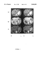
FIG. 1 shows photographs of computerized tomograms for patient 4. The scans show 2 consecutive levels through the liver; 1A) prior to therapy; 1B) after 1 cycle of DDP alone; and 1C) after 1 cycle of TAM/DDP.
FIG. 2 shows photographs of computerized tomograms for patient 6. The scans show 2 consecutive levels through the chest. The arrow points to a soft tissue mass adjacent to the rib. 2) Prior to therapy; 2B) after 2 cycles of DDP alone; and 2C) after 1 cycle of TAM/DDP.
FIG. 3 shows the effect of TAM of the development of resistance to DDP in T-289 human melanoma cells (, DDP alone; •, DDP plus TAM). Each point represents DDP sensitivity relative to unselected T-289 cells determined from triplicate cultures.
FIG. 4 shows the effect of TAM on the development of resistance to DDP in 2008 human ovarian carcinoma cells (•, DDP alone; , DDP plus TAM). Each point represents the mean DDP sensitivity relative to unselected 2008 cells determine from 3 separate repeats of the experiment, each performed with triplicate cultures.
FIG. 5 shows the plot of the CI as a function of cell kill for the interaction between DDP and TAM against human melanoma T-289 cells. Each data point represents the mean of a minimum of 3 experiments performed with triplicate cultures. Vertical bars are standard deviations (SD); where the bars are absent, the SD was less than the size of the symbol
FIG. 6 shows the plot of the CI as a function of cell kill for the interaction between DDP and TAM against human ovarian 2008 cells. Each data point represents the mean of a minimum of 3 experiments performed with triplicate cultures. Vertical bars are SD.
FIG. 7 shows the plot of the CI as a function of cell kill for the interaction between DDP and TAM against human small cell lung cancer UMC 5 cells. Each data point represents the mean of a minimum of 3 experiments performed with triplicate cultures. Vertical bars are SD.
FIG. 8 shows the combination index plots for the interaction between cDDP and TAM for UM-SCC-10B (top) and UM-SCC-5 cells (bottom) and two head and neck carcinoma cell lines. Each point represents the mean±SD of three independent experiments performed in triplicate.
FIG. 9 shows the IC50 values for cDDP during selection for resistance in UM-SCC-10B (left) and UM-SCC-5 (right) cells in the absence (solid bar) or presence (open bar) of TAM. Vertical bars±SD of three to five independent experiments performed in triplicate.
FIG. 10 shows the emergence of cDDP resistance as a function of time in the UM-SCC-10B (lines with square symbols) and UM-SCC-5 (dotted lines with circles) cell lines treated with cDDP alone (solid symbols) or cDDP plus TAM (open symbols). Each point represents the mean±SD of two independent cDDP selections performed in triplicate. The resistance in the selected lines is compared to the unselected cell line and expressed as fold resistance.
FIG. 11 shows the competitive binding assay for AEBS (3 independant experiments). A 12,000 g subcellular fraction, containing AEBS, of the UM-SCC-10B (upper left) and UM-SCC-5 cell lines (upper right) were incubated with 3 H!-TAM and increasing concentrations of estradiol. 3 H!-TAM values were normalized for protein concentration of the cell homogenates. The Kd as well as the number of sites were determined by Scatchard plot analysis, lower left for UM-SCC-10B and lower right for UM-SCC-5.
DETAILED DESCRIPTION OF THE INVENTION
The present invention provides a composition of matter for the treatment of non-melanoma cancers, wherein said composition comprises a platinum anti-neoplastic compound and tamoxifen, wherein said platinum anti-neoplastic compound and tamoxifen exert a synergistic anti-tumor effect on said non-melanoma cancer.
The novel composition of matter of the present invention may be used to potentiate anti-neoplastic activity in a wide variety of non-melanoma cancers. Preferably, the novel composition of matter is used to treat ovarian carcinoma, small lung cell carcinoma, bladder cancer, testicular cancer and squamous cell cancer of the head and neck.
Generally, the platinum containing anti-neoplastic agent of the novel composition of matter may be any platinum coordination complex that has an anti-neoplastic effect. Most preferably, the platinum containing anti-neoplastic agent of the composition of the present invention is cisplatin or carboplatin (CBDCA) but could include tetraplatin and topotecan.
Generally, the concentration of the platinum containing anti-neoplastic agent and of tamoxifen in the novel composition of the present invention may be that which allow for a synergistic cytotoxic effect of the combination. Preferably, the amount of the platinum containing anti-neoplastic agent is from about 1 μM to about 10 μM. Similarly, the concentration of tamoxifen administered as a component of the novel composition or in the methods of the present invention is from about 0.1 μM to about 2 μM.
Tamoxifen is the preferred antiestrogen useful in the composition and methods of the present invention. However, a person having ordinary skill in this art would readily recognize that other antiestrogens and tamoxifen-like compounds may be useful as part of the composition or in the methods of the present invention. For example, DDPE (N,N-diethyl-2- 4-(phenylmethyl)phenoxy) ethanamine is an antagonist of antiestrogen binding sites and microsomal and intranuclear binding sites. DDPE synergizes with cisplatin to produce a synergistic cytotoxic effect and can also overcome cisplatin resistance. Concentration of DDPE between and 1 μM and 10 μM is desirable.
Administration of the novel composition may be by topical, parenteral, oral, intranasal, intravenous, intramuscular, subcutaneous or any other suitable means. The dosage administered is dependent upon the age, weight, kind of concurrent treatment, if any, and nature of the cancer. The effective composition useful in the methods of the present invention may be employed in such forms as capsules, tablets, liquid solutions, suspensions or elixirs for oral administration or sterile liquid forms such as solutions, suspensions or emulsions. Any inert carrier is preferably sued, such as saline or phosphate buffered saline or any such carrier in which the compounds used in the methods of the present invention have suitable solubility properties.
The novel composition of matter of the present invention may be administered in a biologically effective or pharmaceutically acceptable carrier. The carriers may include any solvent with which the composition of the present invention is compatible and which is non-toxic to the individuals treated at the amounts administered. A pharmaceutically acceptable carrier is any solvent with which the composition of the present invention is compatible and which is non-toxic to the individuals treated at the amounts administered. A pharmacological dose of the novel composition of the present invention useful in the methods of the present invention is that amount of the platinum containing antineoplastic agent and tamoxifen which achieves a synergistic cytotoxic effect on non-melanoma tumors. Similarly, when used alone to treat resistance to platinum containing anti-neoplastic agents, a pharmacological dose of tamoxifen is that which reduces or overcomes resistance to platinum containing antineoplastic agents in individuals having a neoplastic disease.
The present invention also provides a method of treating a non-melanoma cancer in vivo or in vitro comprising administering to an individual having said non-melanoma cancer, a pharmacological dose of a platinum anti-neoplastic compound and tamoxifen, wherein said platinum containing anti-neoplastic compound and tamoxifen exert a synergistic anti-tumor effect on said non-melanoma cancer. The platinum containing antineoplastic compound is preferably cisplatin or carboplatin.
Generally, the platinum containing antineoplastic compound and tamoxifen may be administed in any order. Preferably, tamoxifen is administered prior to the administration of the platinum containing antineoplastic compound. Most preferably, tamoxifen is administered 24 hours prior to the administration of the platinum containing antineoplastic compound.
The present invention also provides a method of reducing resistance to platinum anti-neoplastic compounds in an individual having a neoplastic disease comprising administering tamoxifen to said individual, wherein said individual is susceptible to developing resistance to platinum anti-neoplastic compounds. Similarly, the present invention also provides a method of overcoming resistance to platinum anti-neoplastic agents in individual having a neoplastic disease, comprising administering tamoxifen to the individual.
Generally, the methods of reducing or overcoming platinum containing anti-neoplastic agent resistance of the present invention may be useful in the treatment of any neoplastic disease. Preferably, these methods may be used to treat melanoma, ovarian carcinoma, small cell lung carcinoma, bladder cancer, testicular cancer and squamous cell cancer of the head and neck.
The methods of reducing or overcoming resistance to platinum containing anti-neoplastic agents may be generally useful in treating resistance that occurs to any platinum coordination complex anti-neoplastic chemotherapeutic agent. Preferably, the platinum anti-neoplastic compounds are selected from the group consisting of cisplatin and carboplatin.
Another embodiment of the present invention provides a method of killing non-melanoma neoplastic cells in bone marrow, comprising the steps of removing bone marrow from an individual having a neoplastic disease; contacting said bone marrow with a cytocidally effective dose of a platinum containing anti-neoplastic compound and tamoxifen; and returning said contacted bone marrow to said individual.
EXAMPLE 1
In vivo Utility of DDP and Tamoxifen Combination
A clinical trial was conducted in which patients were initially treated with DDP alone. Once patients were found to be resistant to single agent DDP, they were treated with the combination of TAM and DDP to determine whether the addition of TAM could overcome established DDP resistance.
From June 1990 to March 1992, 24 patients who had not had prior DDP or TAM treatment were treated. Entry requirements included histologically documented melanoma, measurable disease, signed informed consent, and no anti-tumor therapy during the previous 4 weeks. Patients were required to have normal hematologic and renal function, an ECOG performance status of 0-2 and an estimated survival of at least 3 months. Patients with a previous history of deep venous thrombosis or pulmonary embolism were excluded.
A complete response was defined as the complete disappearance of all evidence of disease for at least 4 weeks. A partial response was a decrease in the mean diameter of the target lesion by 50% or more lasting at least 4 weeks. A mixed response was a decrease in the diameter of some target lesions of 50% or more while others remained stable or increased in size. A stable disease was defined as a <50% decrease or <25% increase in the size of target lesions for at least 2 months without the growth or appearance of other lesions. A progressive disease was defined as an increase in the size of target lesions by 25% or more. Patients were considered to be resistant to DDP if they had progressive disease after receiving one cycle of DDP or stable disease after receiving two cycles of treatment with DDP alone.
DDP, 100 mg/ml, was mixed in sufficient 0.9% sodium chloride solution to produce a final concentration of at most 1 mg/ml and was administered over 2 hours. Pre-treatment hydration consisted of 1 liter of 5% dextrose/0.45% saline containing 20 mEq of potassium chloride and 2 g magnesium sulfate administered over 4 hours. Post-DDP hydration consisted of 2 liters of 5% dextrose/0.45% saline containing 20 mEq of potassium chloride and 2 g magnesium sulfate administered at 150 ml/hr. Patients were retreated every 3 weeks. On cycles where TAM was administered, 40 mg p.o. four doses per day were given on day 1 followed by 20 mg p.o. daily thereafter. The initial 7 patients were treated with a combination of lorazepam, metaclopramide, benadryl and dexamethasone to control nausea and vomiting. Subsequent patients received an ondansetron-based regimen.
Patients were initially treated with DDP as a single agent and were evaluated for response every 3 weeks. Patients whose disease progressed one cycle of DDP alone were candidates for treatment with DDP plus TAM, as were patients whose disease was stable after 2 cycles of DDP alone. Patients responding to DDP alone continued to receive single agent DDP treatment until either a complete response was achieved or progressive disease supervened.
Pre-treatment characteristics are listed in Table 1. Only 2 patients had previously received chemotherapy (1 of whom also received immunotherapy) and only 1 patient had received prior radiation therapy. The majority of patients had an excellent performance status and the metastatic pattern was as expected for melanoma.
              TABLE 1
______________________________________
Patient characteristics
______________________________________
Male/Female        16/8
Median age (years) 58
Range
Performance status 30-77
(ECOG)
0                  12
1                  8
2                  4
3                  0
4                  0
Sites of Metastatic Disease
Lymph node         11
Skin               10
Lung               7
Liver              7
Adrenal gland      2
Gastrointestinal   1
Bone               1
Brain              1
Previous Treatment
None               21
Chemotherapy       1
Immunotherapy      1
Radiation          1
______________________________________
Response data is listed in Table 2. All 24 patients were evaluated for response to DDP alone. One patient had a complete response while 2 patients demonstrated a partial response. The complete response lasted more than 15 months. One of the patients with a partial response has remained stable off treatment for 6+ months, while the other patient progressed after 4 months and went on to TAM/DDP treatment. The patient who had a complete response with DDP alone had multiple subcutaneous nodules as the only site of disease. The partial responses were seen in patients with lung and retroperitoneal lymph node disease.
              TABLE 2
______________________________________
Responses to treatment with DDP or a DDP/TAM
combination
              Cisplatin
                       Tamoxifen/Cisplatin
______________________________________
Total Patients  24         20.sup.2
Complete Response (%)
                1 (4%)     0
Partial Response (%)
                2 (8%)     3 (15.5%)
Mixed Response (%)
                0          3 (15.5%)
Progressive Disease
                19.sup.1   13
Stable Disease  2          0
Not Evaluable   0          3.sup.3
______________________________________
 .sup.1 Two patients refused DDP/TAM
 .sup.2 Includes 1 patient with a PR to DDP alone
 .sup.3 One suicide, 2 patients too early.
Twenty of the 24 patients initially treated with DDP alone were treated with the combination of TAM and DDP. Two patients refused the combination therapy due to unacceptable toxicity while receiving DDP alone. The patient attaining a complete response was not offered further therapy and one of the 2 patients attaining a partial response remained stable off treatment for 6+ months and did not receive combination treatment. Nineteen of these 20 patients were evaluated for response. One patient committed suicide after receiving the combination of TAM and DDP but prior to evaluation for response.
Of the 19 evaluated patients, 17 had progressive disease and 2 had stable disease while receiving single agent DDP therapy. Three patients achieved a partial response while 3 others had a mixed response for an overall response rate of 35%. This permitted rejection of the null hypothesis that no responses would be observed (p<0.001). One patient attained a partial response with 4 cycles of TAM and DDP, and then was rendered surgically free of disease. Response to the combination of TAM and DDP was seen in patients with live (FIG. 1), bone, and soft tissue disease (FIG. 2). The partial responses ranged from 6 to 8 weeks. Patients who did not respond or who progressed after responding, as well as those who demonstrated a mixed response, were treated with the full four drug regimen of TAM/DDP/carmustine/dacarbazine. There were no responses among the 12 patients treated with this regimen.
The toxicities encountered with the DDP/TAM combination treatment are listed in Table 3. Hematologic toxicity with either DDP alone or DDP in combination with TAM was modest with no grade 3 or 4 neutropenia or thrombocytopenia. No increased toxicity was noted with the addition of TAM to single agent DDP. Similarly, nephrotoxicity was uncommon and unaffected by the administration of TAM. This regimen did produce a difficulty for some patients with nausea and vomiting. While TAM administration did not increase nausea or vomiting, this problem proved to be the most difficult to deal with from the patient's perspective. The addition of ondonsetron markedly reduced nausea and vomiting during hospitalization. Ototoxicity and peripheral neuropathy were uncommon. This is most likely due to the fact that few patients received more than 2 or 3 doses of DDP. One patient, who was a musician, refused further DDP based therapy after one dose due to unacceptable abnormalities in tonal perception. A second patient required a hearing aid after a total dose of 545 mg of DDP. No patients developed deep venous thrombosis or pulmonary embolus.
              TABLE 3
______________________________________
Toxicities with DDP or DDP/TAM treatment
                   DDP  TAM/DDP
______________________________________
No. of cycles        39     36
Neutropenia (<1000/mm.sup.3)
                     0      0
Thrombocytopenia (<50,000/mm.sup.3)
                     0      0
Renal (creatinine > 2 mg/dl)
                     1      1
Nausea/Vomiting (Grade 3/4)
                     13/3   13/3
Ototoxicity (Grade 3/4)
                     1/0    1/0
Peripheral neuropathy (Grade 3/4)
                     0/0    0/0
______________________________________
The present invention establishes that the addition of TAM to single agent DDP therapy can overcome established resistance to DDP in a significant fraction of patients with malignant melanoma. All patients who were treated with the combination of TAM plus DDP were documented to be resistant to single agent DDP therapy in that they had either progressive disease after 1 cycle of DDP or stable disease after 2 cycles. The 31% overall response rate under circumstances where no responses were expected is statistically significant (p<0.001).
The responses resulting from the addition of TAM were not due to an independent antitumor effect of TAM. The response rate reported for single agent TAM therapy in 203 treated melanoma patients was only 6%.
As might be expected in patients with resistant tumors, the clinical responses to the combination of TAM and DDP following failure of single agent DDP therapy were interesting from the standpoint of quality and durability. In one patient, the partial response permitted surgical removal of all remaining disease and this patient has remained in complete remission for 12+ months. The other 2 partial responses were maintained for 6 and 8 weeks, respectively. Patients with a mixed response had clearly demonstrated progression of the target lesions with single agent DDP that responded to the combination of TAM/DDP, however, other lesions either progressed or remained stable. Thus, despite the fact that TAM can sensitize resistant tumors to DDP in vivo, alternate dose schedules will probably be required to maximize the fraction of responding patients and the magnitude of the response.
The design of the study of Example 1 itself was biased against response and the response rate in other settings would be higher. All patients were treated with DDP alone until they were clearly resistant. DDP resistance emerges rapidly. While the concentration of DDP required to achieve one log of cell kill in the resistant cells in vitro can be achieved in patients, the concentration of TAM required to overcome resistance in vitro cannot be achieved in patients with standard doses. Chronic administration of TAM at 10 mg p.o. twice a day produces steady-state plasma concentrations in the range of 0.29 μM. However, the in vitro data predicts that plasma concentrations of more than 0.5 μM will be necessary in DDP-resistant patients. Thus, there is reason to be optimistic that the response rate to the DDP/TAM combination may be higher in patients treated initially with this combination up front and that DDP resistance can be overcome by increasing the dose of both DDP and TAM.
EXAMPLE 2
In vitro Effects of Tamoxifen and Cisplatin
The synergy between TAM and DDP was observed with other human malignancies, the human small cell lung cancer line UMC-5 and the human ovarian carcinoma cell line 2008. In addition, synergy was clearly observed between DDP and TAM in the UM-SCC-10B head and neck carcinoma cell line as illustrated by the CI50 of 0.62. As was observed within the T-289 melanoma cells, there was strong synergy between TAM and DDP with both the UMC-5 cells (CI30 --0.38) and the 2008 cells (CI30 --0.63).
If TAM can synergize with DDP to overcome DDP resistance, then TAM may also delay the development of DDP resistance by a similar mechanism. TAM can delay the development of DDP resistance in T-289 cells, 2008 cells and UM-SCC-10B cells when given concurrently in cell culture.
The T-289 melanoma cell line was derived from a tumor explant and was in passage for over 7 years. The 2008 cell line is an ovarian carcinoma line derived from a patient with an ovarian serous cystadenocarcinoma (type cite). Cells were cultured in 75 cm2 flasks in RPMI 1640 supplemented with 10% fetal bovine serum, 50 μg/ml gentamicin, 2 mM L-glutamine, 10 nM hydrocortisone, 5 μg/ml insulin, 5 μg/ml human transferrin, 10 nM estradiol and 5 ng/ml selenium.
EXAMPLE 3
Induction of DDP Resistance
Resistance to DDP was induced by growing both the T-289 and the 2008 cells in culture in the continuous presence of DDP with or without TAM. Initial selections were performed at concentrations equivalent to the IC90. Thus, the initial concentrations were 0.1, 0.25 and 0.5 μM and 1.0, 2.5 and 5.0 μM for DDP and TAM, respectively. The cells were allowed to grow to confluence (approximately 7 days) at which time successful cultures were split 1:4 and re-exposed to the same concentration of DDP±TAM. After 3 selections, the cells were exposed to incremental higher concentrations of DDP±TAM at the same ratio. Following each selection series, an aliquot of cells was removed, grown in drug-free media for 3 weeks and used in a colony forming assay to determine DDP sensitivity.
EXAMPLE 4
Colony Formation Assay
Cells were seeded into tissue culture dishes containing complete media (2008) or 0.2% agarose/media layered over a 1% agarose layer (T-289). Dishes received DDP at increasing concentrations and were incubated for 10 days at 37° C. with 5% CO2. After 10 days, the colonies were counted, with each DDP concentration being expressed as a percentage of the drug-free control dishes.
FIG. 3 shows the time course for the development of resistance to DDP±TAM in the human melanoma T-289 cells. This cell line was relatively slow growing (doubling time 48-72 hours) and required a substantial amount of time to recover normal growth after each exposure to drug. DDP resistance became apparent after 200 days of selection, at which point, cells treated with DDP alone were 2.3-fold resistant to DDP. Cells treated with DDP plus TAM were only 1.5-fold resistant. Thereafter, DDP resistance emerged progressively at a rate that was 2.75 fold higher in the cells treated with DDP alone compared with those treated with DDP and TAM. The difference in the rate of development of resistance was statistically significant (p<0.01).
EXAMPLE 5
Effects of Tamoxifen on Cisplatin-resistant Cells
The 2008 cell line grew at a faster rate (doubling time 23 hours), which facilitated the repetition of the selection experiment. FIG. 4 shows that DDP resistance emerged as a linear function of time in this cell line. A difference in DDP sensitivity was apparent after selection in the lowest DDP concentration (38 days), and became statistically significant after 3 selections (103 days). The rate of development of resistance to DDP was reduced by TAM in each of the 3 repeats of this experiment. The mean ratio of the rate of development of resistance in the presence of DDP alone versus DDP plus TAM selections was 3.46±1.42 (p<0.05). Thus, in both the T-289 melanoma and 2008 ovarian carcinoma cell lines, concurrent exposure of cells to both DDP and TAM reduced the rate of development of resistance as well as the magnitude of the resistance.
The fact that DDP resistance is genetically stable over many cell generations and is expressed in a dominant fashion in hybrids suggests that TAM can influences a genetic process fundamental to the development of resistance. The absence of an effect on the biochemical pharmacology of either DDP or TAM, coupled with the observation that TAM can produce synergy with DDP even when added up to 48 hours after the end of a 1 hour exposure to DDP, is also consistent with an effect of TAM on a genetic process. The fact that TAM can delay the development of resistance in at least two types of human cancers indicates that the biochemical or molecular genetic mechanisms are common to multiple cell types, and that the synergistic interaction is not dependent on a specific biochemical pathway or genetic process found only a particular type of malignancy.
The TAM-induced delay in the emergence of DDP resistance was not a result of those cultures receiving both agents having sustained greater cell kill, leaving fewer cells at risk for a somatic resistance-generating mutation. Cell cultures were allowed to recover normal growth rates and reach confluence before subsequent treatment. Thus, the same number of cells were exposed to selective pressure at each step. Therefore, greater cell kill per selection, by the drug combination was not responsible for TAM induced delay of DDP resistance development.
The present invention gives further insight into the complex nature of the interaction between TAM and DDP. TAM is synergistic with DDP not only in DDP sensitive tumors such as ovarian carcinoma and small cell lung cancer but also in melanoma, a tumor that is classically considered a DDP resistant tumor. Likewise, TAM can delay the development of DDP resistance in both a DDP-sensitive and DDP-resistant tumor type. Both the synergy and the delay in resistance development was observed at concentrations of TAM and DDP achievable in patients.
The present invention examined the nature of the interaction between the cytotoxic effects of DDP and TAM using the technique of median effect analysis. There is a highly synergistic interaction between these two drugs, not only with respect to the killing of a human melanoma cell line, but also in the case of human ovarian and small cell lung cancer cell lines in vitro.
EXAMPLE 6
Colony Forming Assay
The T-289 melanoma cell line and the 2008 cell line are the same as used in Example 3. The UMC 5 small cell lung cancer cell line, obtained from Stephen L. Graziano M.D., is also of human origin. Cells were cultured as in Example 2.
Colony forming assays using a 1 hour drug exposure were performed by seeding cells onto 60 mm tissue culture dishes at 20,000 cells per dish and allowing 2 hours for them to attach. Drug was added to the dishes and incubated for 1 hour, then the dishes were washed and the cells were harvested by trypsinization, washed once to remove drug and resuspended in 5 ml of complete media containing 0.2% low melting-temperature agarose at 37° C. The cell suspension was mixed well, then aliquoted at 1 ml per dish in triplicate on to pre-prepared 35 mm dishes containing a basement layer of solidified 1% agarose. The cell containing layer was allowed to solidify at room temperature and the dishes were incubated at 37° C. in humidified 5% CO2. Colonies greater than 124 μm were counted after 5 days. Colony forming assays using continuous exposure were performed by suspending cells in 0.2% agarose at 4000 cells/ml, aliquoting them into drug-containing tubes, then seeding these onto 35 mm dishes.
EXAMPLE 7
Median Effect Analysis
Median effect analysis was used to determine the nature of the interaction between TAM and DDP. The combination index (CI) was determined from colony forming assays at increasing levels of cell kill. Drugs were combined at a ratio equal to the ratio of the IC50 values for each drug determined by clonogenic assay. The combination was compared to the cytotoxicity of each drug alone in every experiment. Each data point represents the mean of a minimum of 3 experiments, each preformed with triplicate cultures.
EXAMPLE 8
Effects of TAM/cisplatin on Melanoma Cells
FIG. 5 shows the plot of the CI for the interaction between DDP and TAM for the T-289 melanoma cell line. The CI at 50% cell kill (CI50) of 0.26±0.02 (SD) (p<0.01) demonstrates a marked synergism between TAM and DDP.
EXAMPLE 9
Effects of TAM/cisplatin on Ovarian Carcinoma Cells
FIG. 6 shows the CI plot for the combination of DDP and TAM for the human ovarian carcinoma cell line 2008. The combination was synergistic over the lower range of cell kill and yielded a CI50 of 0.63±0.07 (SD) (p<0.01). Because of the counting error associated with high levels of cell kill (small numbers of surviving colonies) and because the interaction can only be analysed over the portion of the dose-response where the curve for each agent alone and the combination curve is well fit. The CI plot is most reliable for the middle portion of the curve and relatively less reliable for cell kill >80%.
EXAMPLE 10
Effects of TAM/cisplatin on Small Cell Lung Cancer Cells
The CI plot for the combination of DDP and TAM against the UMC small cell lung cancer line is shown in FIG. 7. The CI plot again illustrated a highly synergistic interaction and yielded a CI50 value of 0.38±0.13 (SD) (p<0.01).
The present invention demonstrates a highly synergistic interaction between TAM and DDP with respect to cytotoxicity towards human melanoma, ovarian carcinoma and small cell lung cancer cell lines.
Median effect analysis provides mathematically rigorous methodology for both identifying the nature of the interaction between two cytotoxic drugs and quantitating the magnitude of the interaction at different levels of cell kill. Median effect analysis identified the interaction as synergistic for these three human cell lines. The magnitude of the synergy was greater for both the melanoma line (CI50 0.26) and the small cell lung cancer line (CI50 0.38) than for the ovarian carcinoma line (0.63).
Several other features of the synergistic interaction are of potential clinical significance. For all three cell lines, synergy was clearly present even at the lowest levels of cell kill. Thus, tamoxifen may sensitize tumor cells to DDP in vivo even under circumstances where DDP delivery to the tumor may be mariginally adequate. Second, synergy was observed at concentrations of both DDP and TAM that are readily attainable in the plasma of patients treated with standard doses of these drugs.
Effects of TAM/cisplatin on Human Squamous Cell Carcinoma
EXAMPLE 11
Cell Lines and Clonogenic Assay
The UM-SCC-10B and UM-SCC-5 cell lines were derived from human squamous cell carcinomas of the in head and neck region. Cells were cultured at 37° C. in humidified 5% CO2 in 150 cm2 flasks (Corning, Corning, N.Y.) with RPMI 1640 medium (Mediatech Inc., Herndon, Va.) containing 10% fetal bovine serum (Gemini Bioproducts Inc., Calabasas, Calif.), 2 mM L-glutamine, 100 units/ml of penicillin and 100 μg/ml of streptomycin sulfate. The sensitivity of the UM-SCC-10B and UM-SCC-5 cell lines and their cDDP-resistant variants to cDDP, TAM and the combination of both agents was tested by colony forming assay. Single-cell suspension were plated into 60 mm tissue culture dishes (Corning) at 300 cells per dish. Following drug exposure, the cultures were washed twice with PBS to remove cDDP, 5 ml of fresh medium was added, and they were incubated for 13-15 days to allow formation of colonies. Colonies were fixed with methanol, stained with Giemsa stain for 60 minutes, and counted manually.
EXAMPLE 12
Statistical Analysis
Median effect analysis was used to assess the interaction between cDDP and TAM using a schedule of one hour exposure to cDDP and continuous exposure to TAM. After seeding the cells into 60 mm tissue culture dishes at 300 cells per dish in fresh medium and allowing 5 hours for them to attach, TAM was added. For the combined treatment, cDDP was added to the dishes 24 hours after seeding the cells. Drugs were combined in a fixed ratio equivalent to the ratio of the drug concentration inhibiting colony formation by 50% (IC50) values for each drug as determined by colony forming assay (CFA). After incubation the dishes were washed twice with phosphate-buffered saline and fresh medium containing the appropriate concentration of TAM was added. In synergy experiments, dose-response curves for the single agents were initially generated. The extend of the combined treatment was then analyzed by the isobole method for a combination of cDDP and TAM from the equation
cDDP.sub.c /cDDP.sub.i +TAM.sub.c /TAM.sub.i =CI
where cDDPc and TAMc correspond to doses of drugs used in the combination treatment, and cDDPi and TAMi correspond to the concentrations of drugs able to give the same magnitude of effect if used individually. If CI (combination Index)<1 the effect of the combination was synergistic, while if CI=1 or CI>1 the effect was additive or antagonistic, respectively.
EXAMPLE 13
Selection of Cisplatin Resistant Variants
Cisplatin-resistant cells were selected by chronic exposure to cisplatin in the absence or presence of tamoxifen. The cisplatin levels during the first three selections for the UM-SCC-10B and UM-SCC-5 cell lines were 0.25 μM and 0.5 μM, respectively, which allowed a quadrupling cell number in a week (doubling time of both cell lines was approximately 48 hours). For each subsequent set of three selections the cisplatin dose was increased by approximately 20%. When selections were done in the presence of tamoxifen, the concentration of tamoxifen was always 4-fold higher than the concentration of cisplatin. Cells were cultured in drug-free medium for two weeks prior to being tested for drug sensitivity.
EXAMPLE 14
Estrogen and Progesterone Receptor Assays
The estrogen receptor (ER) and progesterone receptor (PR) content of UM-SCC-10B and UM-SCC-5 cells were determined by quantitative dextran-coated charcoal method of McGuire, et al., J. Clin. Endocrinol. Metab., 36: 548-552 (1973).
EXAMPLE 15
Assay for Antiestrogen Binding Sites
Quantitation of antiestrogen binding sites content was based on the method of Katzenellenbogenet al. Briefly, UM-SCC-10B and UM-SCC-5 cells were cultured in 225 cm2 flasks and allowed to grow to confluence. For each assay, three flasks were used and cells were harvested by scrapping. The cells were centrifuged for 10 minutes at 800 g, resuspended in 4 ml of 10 mM Tris-Cl (pH 7.4) containing 1.5 mM EDTA (pH8) (TE buffer), and homogenized with a Dounce homogenizer (100 strokes with pestle A). The cell homogenates were centrifuged for 30 minutes at 12,000 g, the supernatant was collected and diluted to a total volume of 6 ml with TE buffer. The supernatants were mixed well and 250 μl was aliquoted into 20 tubes. To each tube was added 5 μl of 150 nM 3 H!-tamoxifen (84 Ci/mmol) in dimethyl sulfoxide and 5 μl of ethanol or 100 μM estradiol (Sigma) in 5 μl of ethanol. After an incubation period of 30 minutes at 4° C., 235 μl of TE buffer was added together with the appropriate concentration of tamoxifen (1 nM--500 μM). The tubes were incubated for 18 hours at 4° C. To remove non-binding 3 H!-tamoxifen, 88 μl of dextran-coated charcoal was added, followed by an incubation of 10 minutes at 4° C. and centrifugation for 10 minutes at 12,000 g. The radioactivity of 400 μl of the supernatant mixed in 10 ml of scintillation fluid EcoLite (ICN, Costa Mesa, Calif.) was counted by liquid scintillation. Cell homogenates were assayed for protein content by the method of Bradford. The data were subjected to Scatchard analysis.
EXAMPLE 16
Effects of Tamoxifen and Cisplatin in Squamous Cell Carcinoma
Median effect analysis was used to examine the nature of the interaction between cisplatin and tamoxifen using the head and neck carcinoma cell lines UM-SCC-10B and UM-SCC-5. The IC50 values for continuous tamoxifen exposure for the parental UM-SCC-10B and UM-SCC-5 cells were 3.56±0.41 μM and 3.22±0.54 μM, respectively. The IC50 values for a one hour exposure to cisplatin were 6.5±0.6 and 7.2±1.5 μM, respectively. When UM-SCC-10B cells were exposed to cisplatin for one hour and continuously to tamoxifen, the CI plot indicated a high degree of synergy with a CI50 value of 0.62±0.02 (FIG. 8).
EXAMPLE 17
Effect of Tamoxifen on the Development of Cisplatin Resistance
The UM-SCC-10B and UM-SCC-5 cells were selected for resistance to cisplatin by continuous exposure to cisplatin in the presence or absence of tamoxifen. The rate of development of resistance to cisplatin was monitored by periodically determining the sensitivity of the cells to one hour cisplatin exposure using a clonogenic assay. Repeated exposures to cisplatin alone led to the development of detectable levels of resistance as early as the third selection, and the level of resistance continued to increase with subsequent cisplatin selections as shown in FIG. 9. The rate of development of cisplatin was more rapid in the UM-SCC-10B cells than the UM-SCC-5 cells. After the fifteenth selection, the IC50 values for the UM-SCC-10B and UM-SCC-5 cells were 40.1±1.2 μM and 20.1±1.2 μM, respectively. The presence of tamoxifen during the selection delayed the emergence of cisplatin resistance significantly in UM-SCC-10B cells. The IC50 values in the cisplatin plus tamoxifen selected cells were significantly lower than those for the cells selected in cisplatin alone at each point during the selection process.
The rate of development of cisplatin resistance was determined by linear regression analysis of the change in IC50 values over time, and the results are presented in FIG. 10. It is clear that the emergence of resistance in the UM-SCC-10B cells occurred more slowly in the presence of tamoxifen. The slopes of the regression line was 0.36 in the absence of tamoxifen, and this was reduced by more than 40% to 0.21 in the presence of tamoxifen. In contrast, the presence of tamoxifen had no effect on the slope of the regression line for the emergence of resistance to cisplatin in the UM-SCC-5 where the slopes were 0.14 and 0.13, respectively. Thus, in the cell line where there was synergy between cisplatin and tamoxifen with respect to cytotoxicity, tamoxifen delayed the emergence of resistance. However, under circumstances where they was no synergy between the drugs tamoxifen had no effect on the rate of resistance development.
EXAMPLE 18
Estrogen Receptor and Progesterone Receptor Assay
It has been reported that the estrogen receptor can mediate the antitumor effect of tamoxifen; therefore, the estrogen receptor and progesterone receptor content of both cell lines was determined. The dextran-coated charcoal method indicated a estrogen receptor content <3 fmol/mg and a progesterone receptor <5 fmol/mg in both the UM-SCC-10B and UM-SCC-5 cells. Such levels are characteristic of estrogen non-responsive tumors, suggesting that it was unlikely that tamoxifen as acting via the estrogen receptor or progesterone receptor.
EXAMPLE 19
Antiestrogen Binding Site Analysis
In addition to binding to estrogen receptor, tamoxifen binds to a partially characterized group of receptors referred to as anti-estrogen binding sites or antiestrogen binding sites. The role of these binding sites in mediating the action of tamoxifen is unknown, but they do mediate the effects of several other anti-estrogen compounds. Both the number and affinity of the antiestrogen binding sites in each cell line was determined using the MCF-7 breast carcinoma cell line as a positive control. MCF-7 cells contained 404.8±21.5 fmol/mg protein antiestrogen binding sites, which was similar to values previously reported for these cells. The number of antiestrogen binding sites in the microsomal fraction of the UM-SCC-10B and UM-SCC-5 cells were similar to that in the MCF-7 cell line, 404.2±85.4 and 352.5±24.5 fmol/mg, respectively, and these levels were not significantly different from each other. The affinity of tamoxifen for the antiestrogen binding sites was determined in each of the lines by measuring the equilibrium dissociation constant determined from Scatchard plots (FIG. 11). The values for UM-SCC-10B and UM-SCC-5 cells were 1.9±0.3 nM and 1.7±0.5 nM, respectively. Thus, neither antiestrogen binding sites numbers nor affinity were sufficiently different.
Development of resistance to cisplatin and its analogues occurs commonly during treatment and is a major obstacle to the cure of head and neck cancers. Modulation of cellular sensitivity to the platinum-containing resistance, therefore, has become an important aspect of clinical strategies directed at improving cisplatin-based chemotherapy. As taught by the present invention, tamoxifen, a nonsteroidal triphenylethylene compound, is synergistic with cisplatin in at least three different cell lines of human origin, including a small cell carcinoma of the lung, a melanoma and an ovarian carcinoma cell line. The observed synergy was not due to an effect of tamoxifen on the known mechanisms of cisplatin resistance, including drug uptake, intracellular glutathione or metallothionein levels, or DNA intrastrand adduct formation or repair. In addition to the cytotoxic synergy between cisplatin and tamoxifen, tamoxifen enhances the efficacy of cisplatin treatment by delaying the development of cisplatin resistance in an ovarian carcinoma and in melanoma cell lines.
In the present invention, synergy between cisplatin and tamoxifen was demonstrated in the UM-SCC-10B head and neck cancer cell line. Concordant with the synergistic effect between cisplatin and tamoxifen in the UM-SCC-10B cells, the rate of development of resistance to cisplatin was delayed when selections were performed in the presence of tamoxifen. It is important to emphasize that the delay in resistance development could not be attributed simply to greater degrees of cell kill by the cisplatin/tamoxifen combination in the UM-SCC-10B cells since cultures were allowed to grow to equal cell numbers following each round of selection.
In the case of the head and neck carcinoma cell line, the difference with respect to synergistic interaction with tamoxifen and the effect of tamoxifen on resistance development were not related to any difference in sensitivity to the cytotoxic effect of either drug alone, indicating that the key determinants of synergy were not ones that impacted on overall drug sensitivity. This appears to be different from the T289 melanoma cell line where it was shown that while selection for resistance to cisplatin did not alter the degree of synergy between cisplatin and tamoxifen, selection for resistance to tamoxifen completely eliminated the synergistic interaction. It is noteworthy that in the case of the UM-SCC-10B cell line, synergy was present over a full two logs of tumor cell kill. This reflects a broader range of synergy than has been observed between cisplatin and some other drugs.
Although the mechanism of action of tamoxifen is not fully understood, there is evidence that tamoxifen competes with estrogen at the estrogen receptor. A role for the estrogen receptor in the synergistic interaction between cisplatin and tamoxifen was not identified. In general, it is accepted that the presence of estrogen receptor in a tumor enhance the likelihood that it will respond to tamoxifen. However, tamoxifen can also produce responses in a small fraction of patients with estrogen receptor negative tumors, suggesting that tamoxifen has at least one other mechanism of action.
The ability of tamoxifen to delay the emergence of resistance to cisplatin, and to do so in a cell line which also demonstrated a synergistic interaction between cisplatin and tamoxifen was particularly intriguing. This suggests that the determinants of synergy are also determinants of the ability of tamoxifen to suppress the emergence of resistance.
One skilled in the art will readily appreciate that the present invention is well adapted to carry out the objects and obtain the ends and advantages mentioned, as well as those inherent therein. The present examples along with the methods, procedures, treatments, molecules, and specific compounds described herein are presently representative of preferred embodiments, are exemplary, and are not intended as limitations on the scope of the invention. Changes therein and other uses will occur to those skilled in the art which are encompassed within the spirit of the invention as defined by the scope of the claims.