US5841835A - Apparatus and method for automatic monitoring and assessment of image quality in x-ray systems - Google Patents
Apparatus and method for automatic monitoring and assessment of image quality in x-ray systems Download PDFInfo
- Publication number
- US5841835A US5841835A US08/823,037 US82303797A US5841835A US 5841835 A US5841835 A US 5841835A US 82303797 A US82303797 A US 82303797A US 5841835 A US5841835 A US 5841835A
- Authority
- US
- United States
- Prior art keywords
- image
- digital
- phantom
- ray
- image quality
- Prior art date
- Legal status (The legal status is an assumption and is not a legal conclusion. Google has not performed a legal analysis and makes no representation as to the accuracy of the status listed.)
- Expired - Fee Related
Links
- 238000000034 method Methods 0.000 title claims description 21
- 238000012544 monitoring process Methods 0.000 title claims description 8
- 239000002131 composite material Substances 0.000 claims abstract description 15
- 238000005259 measurement Methods 0.000 claims abstract description 8
- 238000012545 processing Methods 0.000 claims description 11
- RYGMFSIKBFXOCR-UHFFFAOYSA-N Copper Chemical compound [Cu] RYGMFSIKBFXOCR-UHFFFAOYSA-N 0.000 claims description 8
- 229910052802 copper Inorganic materials 0.000 claims description 8
- 239000010949 copper Substances 0.000 claims description 8
- WABPQHHGFIMREM-UHFFFAOYSA-N lead(0) Chemical compound [Pb] WABPQHHGFIMREM-UHFFFAOYSA-N 0.000 claims description 6
- 230000004807 localization Effects 0.000 claims description 4
- 238000004364 calculation method Methods 0.000 claims description 3
- 238000001914 filtration Methods 0.000 claims description 2
- 238000012360 testing method Methods 0.000 abstract description 15
- 238000002594 fluoroscopy Methods 0.000 abstract description 6
- 238000002601 radiography Methods 0.000 abstract description 4
- 238000003384 imaging method Methods 0.000 description 5
- 238000011156 evaluation Methods 0.000 description 4
- 230000003993 interaction Effects 0.000 description 4
- 229910000831 Steel Inorganic materials 0.000 description 3
- 238000004458 analytical method Methods 0.000 description 3
- 238000005457 optimization Methods 0.000 description 3
- 230000005855 radiation Effects 0.000 description 3
- 239000010959 steel Substances 0.000 description 3
- XAGFODPZIPBFFR-UHFFFAOYSA-N aluminium Chemical compound [Al] XAGFODPZIPBFFR-UHFFFAOYSA-N 0.000 description 2
- 229910052782 aluminium Inorganic materials 0.000 description 2
- 238000013459 approach Methods 0.000 description 2
- 239000000463 material Substances 0.000 description 2
- 238000010606 normalization Methods 0.000 description 2
- 230000001105 regulatory effect Effects 0.000 description 2
- 238000012546 transfer Methods 0.000 description 2
- 230000002792 vascular Effects 0.000 description 2
- 238000012795 verification Methods 0.000 description 2
- 230000000007 visual effect Effects 0.000 description 2
- 210000003484 anatomy Anatomy 0.000 description 1
- 210000000988 bone and bone Anatomy 0.000 description 1
- 238000013461 design Methods 0.000 description 1
- 238000001514 detection method Methods 0.000 description 1
- 238000011161 development Methods 0.000 description 1
- 238000003745 diagnosis Methods 0.000 description 1
- 238000003708 edge detection Methods 0.000 description 1
- 238000005516 engineering process Methods 0.000 description 1
- 239000011888 foil Substances 0.000 description 1
- 238000010191 image analysis Methods 0.000 description 1
- 238000012986 modification Methods 0.000 description 1
- 230000004048 modification Effects 0.000 description 1
- 230000002093 peripheral effect Effects 0.000 description 1
- 230000008569 process Effects 0.000 description 1
- 238000004801 process automation Methods 0.000 description 1
- 238000003672 processing method Methods 0.000 description 1
- 238000001303 quality assessment method Methods 0.000 description 1
- 230000004044 response Effects 0.000 description 1
Images


Classifications
-
- A—HUMAN NECESSITIES
- A61—MEDICAL OR VETERINARY SCIENCE; HYGIENE
- A61B—DIAGNOSIS; SURGERY; IDENTIFICATION
- A61B6/00—Apparatus for radiation diagnosis, e.g. combined with radiation therapy equipment
- A61B6/58—Testing, adjusting or calibrating apparatus or devices for radiation diagnosis
- A61B6/582—Calibration
- A61B6/583—Calibration using calibration phantoms
-
- H—ELECTRICITY
- H05—ELECTRIC TECHNIQUES NOT OTHERWISE PROVIDED FOR
- H05G—X-RAY TECHNIQUE
- H05G1/00—X-ray apparatus involving X-ray tubes; Circuits therefor
- H05G1/08—Electrical details
- H05G1/26—Measuring, controlling or protecting
-
- H—ELECTRICITY
- H05—ELECTRIC TECHNIQUES NOT OTHERWISE PROVIDED FOR
- H05G—X-RAY TECHNIQUE
- H05G1/00—X-ray apparatus involving X-ray tubes; Circuits therefor
- H05G1/08—Electrical details
- H05G1/60—Circuit arrangements for obtaining a series of X-ray photographs or for X-ray cinematography
Definitions
- the present invention relates to measurement and verification of x-ray system parameters and, more particularly, to an image quality test device for automatic monitoring and assessment of image quality in digital x-ray fluoroscopy and radiography systems.
- X-ray systems have long been used for imaging and measurement of anatomical structures.
- the imaging receptor consists of film which is mounted in contact with intensifying screens which are thin sheets of fluorescent material. This screen-film combination is typically housed in a cassette.
- intensifying screens which are thin sheets of fluorescent material.
- This screen-film combination is typically housed in a cassette.
- the first step was the introduction of the image intensifier tube which converts the x-ray image into a light image that can be visualized by the human eye.
- the next step was the introduction of a television camera system to transfer the output of the image intensifier to a larger television monitor.
- digital fluoroscopy systems on which the output of the television camera is digitized, and digitally enhanced using advanced image processing methods, prior to display on the monitor.
- the introduction of both the image intensifier tube and the television system has placed high requirements on a correct calibration of the entire x-ray systems image chain.
- X-ray phantoms are known calibration devices and teaching aids for conventional and digital x-ray systems.
- Two general types of phantoms are available today, including (1) plastic replicas of the human body or specific portions thereof or actual human bones cast in plastic, and (2) physics-based phantoms which measure a particular response of the x-ray system.
- the first type is primarily used to train x-ray technicians in the proper positioning of the human body for the various x-ray images that are taken for diagnosis.
- the second class of phantoms is utilized during calibration and assessment of image quality of the x-ray system.
- the present invention provides an image quality test phantom (hardware and software) and methodology for automatic monitoring and assessment of image quality in digital x-ray fluoroscopy and radiography systems. Instead of using highly iterative methods of calibration and image quality verification which utilize many different phantoms combined with multiple subjective visual evaluations, the phantom according to the present invention provides an efficient and robust method for fast and objective image quality measurements and assessments.
- an x-ray image quality test tool comprises two parts, a hardware portion, specifically a composite phantom, and a software portion.
- the composite phantom comprises a copper sheet with an overlaid steel mesh. A central part of the mesh is cut out, and inserts comprising a resolution pattern, a contrast-detail phantom, a step wedge phantom and a line phantom are positioned in the center overlaying the copper sheet.
- the composite phantom is mounted in a housing, or carrier, which attaches to the front of the x-ray system imager.
- the software comprises various image and signal processing algorithms which, when applied to the digital image of the phantom, provide all necessary logic to compute the required x-ray system image quality measures.
- an object of the present invention to provide an image quality test tool, comprised of a composite phantom and associated software, for automatic monitoring and assessment of image quality, which is particularly well suited for use with digital x-ray fluoroscopy and radiography systems. It is a further object of the present invention to provide a method for using the phantom to trend and calibrate such systems.
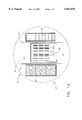
- FIGS. 1A and 1B are a top view and a cross-sectional view, respectively, of the image quality test phantom, in accordance with the present invention
- FIG. 2 illustrates an example of the image quality test phantom positioned in its housing (carrier) for mounting in front of an image intensifier based x-ray system;
- FIG. 3 illustrates an example of the image quality test phantom with its housing, placed on a vascular x-ray system
- FIG. 4 illustrates an example of an actual phantom-based digital image together with overlayed regions of interest automatically identified and determined by imaging software, in accordance with the present invention.
- the present invention proposes an x-ray image quality test phantom which provides an efficient method for very fast and objective image quality measurements and assessments.
- a significant aspect of the present invention is that it provides the capability of measuring critical x-ray system image quality parameters objectively and instantaneously.
- FIGS. 1A and 1B there is illustrated an image quality test phantom 10, in accordance with the present invention.
- Copper sheet 12 generally circular, serves as a base for the composite phantom.
- the purpose of the copper sheet is to provide the mechanical stability for the phantom, an appropriate x-ray beam quality for the image analysis and an appropriate dynamic range.
- a steel mesh 14 is placed on top of the copper sheet 12. A central, approximate square portion, of the mesh is cut out. The remaining part of the steel mesh is used in the computation of resolution uniformity of the x-ray image.
- the composite phantom comprises an insert 15, containing a ten level copper step wedge 16, a resolution pattern 18, a contrast detail phantom 20, and lead line phantoms 22.
- the size of the step wedge 16 is approximately 2.5 cm ⁇ 5.5 cm with steps ranging from 0.254 mm to 2.54 mm.
- the purpose of the step wedge is to access the dynamic range and linearity of the x-ray system.
- the resolution pattern 18 covers spatial frequencies from 0.5 line-pairs/mm (1p/mm) to 5 1p/mm created from 0.1 mm lead foil. It also includes three 1 cm ⁇ 1 cm regions with 0 mm, 0.1 mm and 1.0 mm lead. The resolution pattern is used in the computation of x-ray system modulation transfer function with the square regions used for normalization purposes.
- An aluminum contrast-detail phantom 20 comprises three steps with three apertures on each step.
- the diameters of the three apertures are, for example, 7.6 mm, 3.8 mm, and 1.9 mm, and the respective thicknesses of the steps can be defined as 1 mm, 2 mm and 3 mm.
- the purpose of the contrast-detail phantom is to assess the relative contrast and contrast-to-noise ratio of the x-ray system.
- the aluminum base provides the appropriate contrast levels and form factor.
- Lead line phantoms 22a, 22b, located on the outer edges of the resolution phantom, are used for distortion measurements.
- FIG. 2 An example of the image quality phantom 10 mounted in a carrier 24 is shown in FIG. 2.
- the carrier 24 can include a handle 26 and an alignment pin 28.
- the composite 25 can be sized to attach to the front face of image intensifier 30 or other digital imager.
- the image quality phantom composite 25, comprising the phantom 10 and the carrier 24, is mounted in front of the image intensifier 30.
- An x-ray positioner 32 positions the image intensifier 30 and x-ray tube 34, above and below x-ray table 36, respectively.
- the second integral part of the image quality tool of the present invention is the software functionality.
- the present invention provides software methodology for complete image quality assessment process automation, in conjunction with the test phantom hardware. Once a digital x-ray image of the phantom 10 is acquired, it can then be processed in accordance with the present invention.
- an initial localization of the resolution 18, step wedge 16, contrast-detail 20 and lead wire 22a and 22b phantoms is done by calculating spatial derivatives through cross-sections of the image. Localized filtration with an N ⁇ N edge enhancement kernels are performed in areas around the initial estimates. Additional localization information is obtained by tracking circular edge 38 of the image, as illustrated in FIG. 4.
- An edge linking algorithm such as obtained by dynamic programming, is performed to precisely specify the locations of the resolution 18, step wedge 16, contrast detail 20 and lead wire 22a and 22b phantom edges, where the edges are referenced as 42.
- a second order polynomial warping is performed to automatically predict the location of regions of interest on the image, based upon the known geometry of the image quality phantom 10.
- the desired location of the regions of interest are at the specified frequencies and normalization points on the resolution phantom 18, at each level of the step wedge 16, at each disk and surrounding area of the contrast-detail phantom 20, and in multiple (such as, by way of example, eight) peripheral and two central areas of the mesh pattern 14.
- First and second order statistics and their variates are calculated at each region of interest, to estimate the system resolution, uniformity, dynamic range, contrast-to-noise, and noise.
- System distortion is estimated from polynomial fitting of the edge tracking data from the two lead wires 22a and 22b.
- FIG. 4 there is illustrated an example of the actual digital image of the composite phantom.
- Overlay figures represent the critical image areas used in the assessment of the x-ray system image quality. All regions were identified and isolated by the automatic software without any user interaction. If an additional digital image of a flat copper sheet is available, a digital subtraction of the flat field image from the image quality phantom 10 image is performed to remove bias introduced by system brightness non-uniformities. The subtraction image is then used in the resolution 18, step wedge 16 and contrast-to-noise calculations.
- the approach of the present invention is particularly suitable for use in automatic trending, calibration and optimization of parameters within the x-ray imaging chain.
- the phantom can be imaged at specified intervals, for example, every two weeks, and the resulting data can be recorded and compared to an established data base.
- the results can be made available to an off-site service center, to allow for remote analysis and assessment.
- the phantom provides data needed for automatic closed-loop calibration. Since most adjustments on a modern fluoroscopy system are software controlled, iterative methods, such as least squares, can be used to optimize the system parameters that are measured by the phantom.
- One process of automatic calibration measures the initial system with the image quality phantom 10. If the measured value is out of specification, a software controlled adjustment is performed on the system. The new state is then remeasured with the image quality phantom 10, and an iterative method such as a least squares iterative optimization is used to minimize the difference between the measured value obtained by system adjustments and the engineering specifications.
- the present invention provides for processing of a digital image, analysis of image quality, and assessment of x-ray system image quality.
- a digital subtraction of a flat field image can be performed from the image quality phantom image to remove bias introduced by system brightness non-uniformities.
- the subtraction image can then be used in resolution, step wedge and contrast-to-noise calculations.
- Analysis of image quality of the x-ray system includes automatically providing trending image quality data.
- the trending can be performed by the phantom and can be imaged at specified intervals, for example, every two weeks, and the resulting data can be recorded and compared to an is established data base.
- Assessment of x-ray system image quality can further include remote monitoring of such systems via an off-site service center.
- a first composite x-ray image quality phantom is used to effectively and objectively measure image system parameters such as resolution, uniformity, dynamic range, contrast-to-noise, distortion and noise.
- the present invention further proposes a method of use of the phantom to automatically, without any user interaction, detect the location of individual components in a digital x-ray image of the composite image quality phantom. This involves the use of edge detection and tracking, and polynomial warping to estimate the location of regions of interest within the image quality phantom.
- the present invention provides for the use of the image quality phantom in system image quality trending, both local and remote, and as data feedback of a closed loop design that uses an iterative method to optimize system performance.
Abstract
Description
Claims (7)
Priority Applications (3)
| Application Number | Priority Date | Filing Date | Title |
|---|---|---|---|
| US08/823,037 US5841835A (en) | 1997-03-31 | 1997-03-31 | Apparatus and method for automatic monitoring and assessment of image quality in x-ray systems |
| JP10072963A JPH114822A (en) | 1997-03-31 | 1998-03-23 | Image quality test phantom tool and digital x-ray image processing method |
| EP98302184A EP0874536A1 (en) | 1997-03-31 | 1998-03-24 | Apparatus and method for automatic monitoring and assessment of image quality in X-ray systems |
Applications Claiming Priority (1)
| Application Number | Priority Date | Filing Date | Title |
|---|---|---|---|
| US08/823,037 US5841835A (en) | 1997-03-31 | 1997-03-31 | Apparatus and method for automatic monitoring and assessment of image quality in x-ray systems |
Related Child Applications (1)
| Application Number | Title | Priority Date | Filing Date |
|---|---|---|---|
| US10/624,977 Continuation US6870119B2 (en) | 2000-03-29 | 2003-07-21 | Modular steel concrete reinforcement system |
Publications (1)
| Publication Number | Publication Date |
|---|---|
| US5841835A true US5841835A (en) | 1998-11-24 |
Family
ID=25237630
Family Applications (1)
| Application Number | Title | Priority Date | Filing Date |
|---|---|---|---|
| US08/823,037 Expired - Fee Related US5841835A (en) | 1997-03-31 | 1997-03-31 | Apparatus and method for automatic monitoring and assessment of image quality in x-ray systems |
Country Status (3)
| Country | Link |
|---|---|
| US (1) | US5841835A (en) |
| EP (1) | EP0874536A1 (en) |
| JP (1) | JPH114822A (en) |
Cited By (42)
| Publication number | Priority date | Publication date | Assignee | Title |
|---|---|---|---|---|
| US5994693A (en) * | 1997-01-03 | 1999-11-30 | The Research Foundation Of Suny At Buffalo | Gamma camera quality test pattern |
| EP1062912A1 (en) | 1999-06-24 | 2000-12-27 | General Electric Company | Modular interchangeable phantoms for multiple X-ray systems |
| FR2796480A1 (en) * | 1999-07-15 | 2001-01-19 | Gen Electric | Automatic evaluation of image quality in an X-ray system using multiple spectrums |
| US6315447B1 (en) * | 1998-12-22 | 2001-11-13 | Bio-Imaging Technologies, Inc. | Variable composition phantom simulating varying degrees of body fat for dual energy x-ray machine calibration |
| US6317482B1 (en) * | 1998-06-30 | 2001-11-13 | The United States Of America As Represented By The Secretary Of The Navy | Radiological image quality indicator |
| US20010053199A1 (en) * | 2000-06-16 | 2001-12-20 | Dietmar Sundermann | Collimation device, radiology apparatus, test kit and method of testing a radiology apparatus |
| US6364529B1 (en) | 2000-10-20 | 2002-04-02 | Med-Tec Iowa, Inc. | Radiation phantom |
| FR2819368A1 (en) | 2001-01-09 | 2002-07-12 | Ge Med Sys Global Tech Co Llc | CALIBRATION PROCESS OF A RADIOLOGY APPARATUS, AND RADIOLOGY APPARATUS |
| WO2002061455A2 (en) | 2000-12-29 | 2002-08-08 | Ge Medical Systems Global Technology Company, Llc | A method of monitoring changes in the detective quantum efficiency of an x-ray detector |
| US6493417B1 (en) * | 1999-11-23 | 2002-12-10 | Siemens Aktiengesellschaft | Patient positioning for conducting a medical examination |
| US20030043962A1 (en) * | 2001-08-31 | 2003-03-06 | Ching-Ming Lai | Image positioning method and system for tomosynthesis in a digital X-ray radiography system |
| WO2003052397A1 (en) * | 2001-12-19 | 2003-06-26 | Agresearch Limited | A phantom |
| US20030191387A1 (en) * | 2000-12-28 | 2003-10-09 | Petrick Scott William | Method and apparatus for correcting the offset induced by field effect transistor photo-conductive effects in a solid state X-ray detector |
| US20040179651A1 (en) * | 2003-03-12 | 2004-09-16 | Canon Kabushiki Kaisha | Automated quality control for digital radiography |
| US6813374B1 (en) | 2001-04-25 | 2004-11-02 | Analogic Corporation | Method and apparatus for automatic image quality assessment |
| US20050157848A1 (en) * | 2003-12-22 | 2005-07-21 | Kabushiki Kaisha Toshiba | Image-quality control system |
| US20050259793A1 (en) * | 2004-05-19 | 2005-11-24 | Yeo In H | Medical phantom, holder and method of use thereof |
| US20060212317A1 (en) * | 2005-01-19 | 2006-09-21 | Hahn Jerad J | Mammography operational management system and method |
| US20070183590A1 (en) * | 2006-02-08 | 2007-08-09 | Gray Joel E | Dental image quality and dose analyzer |
| US20080019473A1 (en) * | 2004-11-24 | 2008-01-24 | Koninklijke Philips Electronics N.V. | Computer Tomography Method and Computer Tomograph |
| US20080219410A1 (en) * | 2007-03-07 | 2008-09-11 | Konstanze Gunzert-Marx | Phantom and method for quality monitoring of a medical system |
| GB2449113A (en) * | 2007-05-11 | 2008-11-12 | Cameron Nigel Glenvi Carpenter | Apparatus For Measurement Accuracy Testing Of Radiological Imaging Modalities And Networked Digital Viewing Platforms |
| US20090242744A1 (en) * | 2006-07-11 | 2009-10-01 | Carl Zeiss Industrielle Messtechnik Gmbh | Arrangement for producing electromagnetic radiation and method for operating said arrangement |
| US20090268953A1 (en) * | 2008-04-24 | 2009-10-29 | Apteryx, Inc. | Method for the automatic adjustment of image parameter settings in an imaging system |
| DE102009018596A1 (en) * | 2009-03-03 | 2010-09-09 | Fachhochschule Köln | Test specimen and method for evaluating radiological images of a specimen |
| US20110096911A1 (en) * | 2009-10-27 | 2011-04-28 | Dental Imaging Consultants, LLC | Quality Assurance Phantom for Digital Dental Imaging and Related Method |
| US20110266427A1 (en) * | 2008-06-25 | 2011-11-03 | Australian Nuclear Science And Technology Organisation | Imaging Test Piece for Medium and Large Security X-Ray Scanners |
| CN103052354A (en) * | 2010-07-30 | 2013-04-17 | 国立大学法人东北大学 | Evaluation aid |
| US8708562B1 (en) | 2013-03-05 | 2014-04-29 | Nosil DSC Innovations, Inc. | Phantom systems and methods for diagnostic x-ray equipment |
| US20140153694A1 (en) * | 2012-11-30 | 2014-06-05 | Ge Sensing & Inspection Technologies Gmbh | Method for determining geometric imaging properties of a flat panel detector, correspondingly adapted x-ray inspection system and calibration phantom |
| CN104997525A (en) * | 2015-06-10 | 2015-10-28 | 广州七喜医疗设备有限公司 | Apparatus and method for selecting key parts of digital radiographic image equipment |
| US20160051219A1 (en) * | 2014-08-20 | 2016-02-25 | Canon Kabushiki Kaisha | Evaluation method for radiographing apparatus and phantom used in evaluation |
| US20160084973A1 (en) * | 2013-05-16 | 2016-03-24 | Ibex Innovations Ltd. | Multi-Spectral X-Ray Detection Apparatus |
| US20160084972A1 (en) * | 2013-05-16 | 2016-03-24 | Ibex Innovations Ltd. | X-Ray Detector Apparatus |
| US20160089105A1 (en) * | 2014-09-26 | 2016-03-31 | Samsung Electronics Co., Ltd. | Medical imaging apparatus and control method thereof |
| US20160157794A1 (en) * | 2013-05-16 | 2016-06-09 | Ibex Innovations Ltd. | X-Ray Imaging Apparatus and Methods |
| WO2016138449A1 (en) | 2015-02-27 | 2016-09-01 | Bayer Healthcare Llc | Quantification phantom for use with multiple imaging modalities |
| US9466012B2 (en) | 2013-07-11 | 2016-10-11 | Radiological Imaging Technology, Inc. | Phantom image classification |
| US9934603B2 (en) | 2015-04-22 | 2018-04-03 | The Phantom Laboratory, Incorporated | Three-dimensional resolution gauge for evaluating performance of tomographic imaging systems |
| US9936935B1 (en) | 2014-02-14 | 2018-04-10 | Nosil DSC Innovations, Inc. | Phantom systems and methods for diagnostic radiographic and fluoroscopic X-ray equipment |
| US20180192986A1 (en) * | 2015-08-13 | 2018-07-12 | Paul Jahnke | Model and system for use in an imaging technique |
| US11170500B1 (en) * | 2018-11-09 | 2021-11-09 | United States Of America As Represented By The Administrator Of Nasa | Pyramid image quality indicator (IQI) for X-ray computed tomography |
Families Citing this family (15)
| Publication number | Priority date | Publication date | Assignee | Title |
|---|---|---|---|---|
| JPS6056824A (en) * | 1983-09-06 | 1985-04-02 | Fanuc Ltd | Wire electric discharge machining method |
| US6409383B1 (en) * | 2000-03-14 | 2002-06-25 | Eastman Kodak Company | Automated and quantitative method for quality assurance of digital radiography imaging systems |
| DE10204543A1 (en) | 2002-02-05 | 2003-08-14 | Philips Intellectual Property | Method and device for automatically checking an X-ray system |
| US7006594B2 (en) | 2002-02-25 | 2006-02-28 | Ge Medical Systems Global Technology Company, Llc | Method and apparatus for reconstruction calibration of detector position and source motion based on a multi-pin phantom |
| US7269243B2 (en) | 2002-02-25 | 2007-09-11 | Ge Medical Systems Global Technology Company, Llc | Method and apparatus for controlling electron beam motion based on calibration information |
| JP2003305035A (en) * | 2002-02-25 | 2003-10-28 | Ge Medical Systems Global Technology Co Llc | Method and apparatus for reconstruction calibration of detector position and source motion based on multi-pin phantom |
| KR20040098041A (en) * | 2002-04-05 | 2004-11-18 | 하마마츠 포토닉스 가부시키가이샤 | X-ray tube adjustment apparatus, x-ray tube adjustment system, and x-ray tube adjustment method |
| US6979124B2 (en) | 2003-02-11 | 2005-12-27 | General Electric Company | Image quality vascular uniformity evaluation method and apparatus |
| US7256392B2 (en) | 2003-03-03 | 2007-08-14 | Fujifilm Corporation | Inspection method of radiation imaging system and medical image processing apparatus using the same, and phantom for use of inspection of radiation imaging system |
| US7286631B2 (en) * | 2004-01-09 | 2007-10-23 | General Electric Co. | Method and apparatus for tomosynthesis image quality control |
| JP4863700B2 (en) * | 2005-02-04 | 2012-01-25 | 東芝Itコントロールシステム株式会社 | X-ray inspection equipment |
| JP5459651B2 (en) * | 2008-09-26 | 2014-04-02 | 株式会社東芝 | X-ray imaging apparatus, inspection / maintenance time notification method and apparatus |
| KR101019579B1 (en) | 2009-08-20 | 2011-03-08 | 제주대학교 산학협력단 | Resolution Measurement System of Imaging Device Possible to Evaluate Absolute Value |
| GB201107385D0 (en) * | 2011-05-04 | 2011-06-15 | Materialise Nv | Medical imaging calibration device |
| JP5912205B2 (en) * | 2015-10-14 | 2016-04-27 | 国立大学法人東北大学 | Evaluation aids |
Citations (4)
| Publication number | Priority date | Publication date | Assignee | Title |
|---|---|---|---|---|
| US5276726A (en) * | 1989-11-24 | 1994-01-04 | Thomas Jefferson University | Method of and apparatus for standardizing and monitoring image quality in mammography |
| US5420441A (en) * | 1993-11-23 | 1995-05-30 | Eastman Kodak Company | Automated technique for calibrating a storage phosphor reader |
| US5539799A (en) * | 1992-11-12 | 1996-07-23 | Siemens Aktiengesellschaft | Method and device for acceptance and stability testing of filmless dental radiographic equipment |
| US5544238A (en) * | 1989-11-24 | 1996-08-06 | Thomas Jefferson University | Method of and apparatus for standardizing and monitoring beam quality in mammography |
Family Cites Families (4)
| Publication number | Priority date | Publication date | Assignee | Title |
|---|---|---|---|---|
| DE2903023C3 (en) * | 1979-01-26 | 1983-12-08 | Gerd Prof. Dr.rer.nat. 3006 Burgwedel Hagemann | Test phantom for quality control in X-ray diagnostics |
| US5095499A (en) * | 1989-10-05 | 1992-03-10 | Wentz Virginia R | Oriented mammography phantom |
| US5056130A (en) * | 1989-10-06 | 1991-10-08 | The United States Of America As Represented By The Administrator, National Aeronautics And Space Administration | Computerized tomography calibrator |
| US5416816A (en) * | 1994-01-27 | 1995-05-16 | Boston Test Tool Company | Calibration template for computed radiography |
-
1997
- 1997-03-31 US US08/823,037 patent/US5841835A/en not_active Expired - Fee Related
-
1998
- 1998-03-23 JP JP10072963A patent/JPH114822A/en not_active Withdrawn
- 1998-03-24 EP EP98302184A patent/EP0874536A1/en not_active Withdrawn
Patent Citations (4)
| Publication number | Priority date | Publication date | Assignee | Title |
|---|---|---|---|---|
| US5276726A (en) * | 1989-11-24 | 1994-01-04 | Thomas Jefferson University | Method of and apparatus for standardizing and monitoring image quality in mammography |
| US5544238A (en) * | 1989-11-24 | 1996-08-06 | Thomas Jefferson University | Method of and apparatus for standardizing and monitoring beam quality in mammography |
| US5539799A (en) * | 1992-11-12 | 1996-07-23 | Siemens Aktiengesellschaft | Method and device for acceptance and stability testing of filmless dental radiographic equipment |
| US5420441A (en) * | 1993-11-23 | 1995-05-30 | Eastman Kodak Company | Automated technique for calibrating a storage phosphor reader |
Cited By (76)
| Publication number | Priority date | Publication date | Assignee | Title |
|---|---|---|---|---|
| US5994693A (en) * | 1997-01-03 | 1999-11-30 | The Research Foundation Of Suny At Buffalo | Gamma camera quality test pattern |
| US6317482B1 (en) * | 1998-06-30 | 2001-11-13 | The United States Of America As Represented By The Secretary Of The Navy | Radiological image quality indicator |
| US6315447B1 (en) * | 1998-12-22 | 2001-11-13 | Bio-Imaging Technologies, Inc. | Variable composition phantom simulating varying degrees of body fat for dual energy x-ray machine calibration |
| EP1062912A1 (en) | 1999-06-24 | 2000-12-27 | General Electric Company | Modular interchangeable phantoms for multiple X-ray systems |
| US6231231B1 (en) | 1999-06-24 | 2001-05-15 | General Electric Company | Modular interchangeable phantoms for multiple x-ray systems |
| US6694047B1 (en) * | 1999-07-15 | 2004-02-17 | General Electric Company | Method and apparatus for automated image quality evaluation of X-ray systems using any of multiple phantoms |
| FR2796480A1 (en) * | 1999-07-15 | 2001-01-19 | Gen Electric | Automatic evaluation of image quality in an X-ray system using multiple spectrums |
| US6493417B1 (en) * | 1999-11-23 | 2002-12-10 | Siemens Aktiengesellschaft | Patient positioning for conducting a medical examination |
| US20040223591A1 (en) * | 2000-06-16 | 2004-11-11 | Dietmar Sundermann | Method of testing a radiology apparatus and program |
| US20010053199A1 (en) * | 2000-06-16 | 2001-12-20 | Dietmar Sundermann | Collimation device, radiology apparatus, test kit and method of testing a radiology apparatus |
| US6850596B2 (en) | 2000-06-16 | 2005-02-01 | Ge Medical Systems | Collimation device, radiology apparatus, test kit and method of testing a radiology apparatus |
| US7021118B2 (en) | 2000-06-16 | 2006-04-04 | Ge Medical Systems Global Technology Company Llc | Method of testing a radiology apparatus and computer program |
| US6364529B1 (en) | 2000-10-20 | 2002-04-02 | Med-Tec Iowa, Inc. | Radiation phantom |
| US20030191387A1 (en) * | 2000-12-28 | 2003-10-09 | Petrick Scott William | Method and apparatus for correcting the offset induced by field effect transistor photo-conductive effects in a solid state X-ray detector |
| US6521886B2 (en) | 2000-12-29 | 2003-02-18 | Ge Medical Systems Global Technology Company, Llc | Method of monitoring changes in the detective quantum efficiency of an x-ray detector |
| WO2002061455A2 (en) | 2000-12-29 | 2002-08-08 | Ge Medical Systems Global Technology Company, Llc | A method of monitoring changes in the detective quantum efficiency of an x-ray detector |
| FR2819368A1 (en) | 2001-01-09 | 2002-07-12 | Ge Med Sys Global Tech Co Llc | CALIBRATION PROCESS OF A RADIOLOGY APPARATUS, AND RADIOLOGY APPARATUS |
| US6813374B1 (en) | 2001-04-25 | 2004-11-02 | Analogic Corporation | Method and apparatus for automatic image quality assessment |
| US20030043962A1 (en) * | 2001-08-31 | 2003-03-06 | Ching-Ming Lai | Image positioning method and system for tomosynthesis in a digital X-ray radiography system |
| US6960020B2 (en) * | 2001-08-31 | 2005-11-01 | Analogic Corporation | Image positioning method and system for tomosynthesis in a digital X-ray radiography system |
| WO2003052397A1 (en) * | 2001-12-19 | 2003-06-26 | Agresearch Limited | A phantom |
| US20040179651A1 (en) * | 2003-03-12 | 2004-09-16 | Canon Kabushiki Kaisha | Automated quality control for digital radiography |
| US20050157848A1 (en) * | 2003-12-22 | 2005-07-21 | Kabushiki Kaisha Toshiba | Image-quality control system |
| CN100533446C (en) * | 2003-12-22 | 2009-08-26 | 株式会社东芝 | Image-quality control system |
| US7189000B2 (en) | 2003-12-22 | 2007-03-13 | Kabushiki Kaisha Toshiba | Image-quality control system |
| US20050259793A1 (en) * | 2004-05-19 | 2005-11-24 | Yeo In H | Medical phantom, holder and method of use thereof |
| US20080019473A1 (en) * | 2004-11-24 | 2008-01-24 | Koninklijke Philips Electronics N.V. | Computer Tomography Method and Computer Tomograph |
| US20060212317A1 (en) * | 2005-01-19 | 2006-09-21 | Hahn Jerad J | Mammography operational management system and method |
| US8478610B2 (en) | 2005-01-19 | 2013-07-02 | Atirix Medical Systems | Medical imaging device quality control system and method |
| US8428969B2 (en) * | 2005-01-19 | 2013-04-23 | Atirix Medical Systems, Inc. | System and method for tracking medical imaging quality |
| WO2007092224A3 (en) * | 2006-02-08 | 2007-12-13 | Joel E Gray | Dental image quality and dose analyzer |
| US7503694B2 (en) | 2006-02-08 | 2009-03-17 | Gray Joel E | Dental image quality and dose analyzer |
| WO2007092224A2 (en) * | 2006-02-08 | 2007-08-16 | Gray Joel E | Dental image quality and dose analyzer |
| US20070183590A1 (en) * | 2006-02-08 | 2007-08-09 | Gray Joel E | Dental image quality and dose analyzer |
| US20090242744A1 (en) * | 2006-07-11 | 2009-10-01 | Carl Zeiss Industrielle Messtechnik Gmbh | Arrangement for producing electromagnetic radiation and method for operating said arrangement |
| US8173952B2 (en) | 2006-07-11 | 2012-05-08 | Carl Zeiss Industrielle Messtechnik Gmbh | Arrangement for producing electromagnetic radiation and method for operating said arrangement |
| US20080219410A1 (en) * | 2007-03-07 | 2008-09-11 | Konstanze Gunzert-Marx | Phantom and method for quality monitoring of a medical system |
| US7786433B2 (en) * | 2007-03-07 | 2010-08-31 | Siemens Aktiengesellschaft | Phantom and method for quality monitoring of a medical system |
| GB2449113A (en) * | 2007-05-11 | 2008-11-12 | Cameron Nigel Glenvi Carpenter | Apparatus For Measurement Accuracy Testing Of Radiological Imaging Modalities And Networked Digital Viewing Platforms |
| GB2449113B (en) * | 2007-05-11 | 2012-02-15 | Cameron Nigel Glenville Carpenter | Apparatus for measurement accuracy testing of radiological imaging modalities and networked digital viewing platforms |
| US20090268953A1 (en) * | 2008-04-24 | 2009-10-29 | Apteryx, Inc. | Method for the automatic adjustment of image parameter settings in an imaging system |
| US9720114B2 (en) * | 2008-06-25 | 2017-08-01 | Australian Nuclear Science And Technology Organization | Imaging test piece for medium and large security X-ray scanners |
| US20110266427A1 (en) * | 2008-06-25 | 2011-11-03 | Australian Nuclear Science And Technology Organisation | Imaging Test Piece for Medium and Large Security X-Ray Scanners |
| DE102009018596A1 (en) * | 2009-03-03 | 2010-09-09 | Fachhochschule Köln | Test specimen and method for evaluating radiological images of a specimen |
| US8308362B2 (en) | 2009-10-27 | 2012-11-13 | Dental Imaging Consultants, LLC | Quality assurance phantom for digital dental imaging and related method |
| US20110096911A1 (en) * | 2009-10-27 | 2011-04-28 | Dental Imaging Consultants, LLC | Quality Assurance Phantom for Digital Dental Imaging and Related Method |
| CN103052354B (en) * | 2010-07-30 | 2016-08-24 | 国立大学法人东北大学 | Evaluation aid |
| US8981283B2 (en) | 2010-07-30 | 2015-03-17 | National University Corporation, Tohoku University | Evaluation aid |
| CN106137232B (en) * | 2010-07-30 | 2019-07-26 | 国立大学法人东北大学 | Auxiliary tool is used in evaluation |
| CN103052354A (en) * | 2010-07-30 | 2013-04-17 | 国立大学法人东北大学 | Evaluation aid |
| US9662087B2 (en) | 2010-07-30 | 2017-05-30 | National University Corporation, Tohoku University | Evaluation aid |
| CN106137232A (en) * | 2010-07-30 | 2016-11-23 | 国立大学法人东北大学 | Evaluation Aid |
| US9332957B2 (en) | 2010-07-30 | 2016-05-10 | National University Corporation, Tohoku University | Evaluation aid |
| US20140153694A1 (en) * | 2012-11-30 | 2014-06-05 | Ge Sensing & Inspection Technologies Gmbh | Method for determining geometric imaging properties of a flat panel detector, correspondingly adapted x-ray inspection system and calibration phantom |
| US9146327B2 (en) * | 2012-11-30 | 2015-09-29 | Ge Sensing & Inspection Technologies Gmbh | Method for determining geometric imaging properties of a flat panel detector, correspondingly adapted X-ray inspection system and calibration phantom |
| US8708562B1 (en) | 2013-03-05 | 2014-04-29 | Nosil DSC Innovations, Inc. | Phantom systems and methods for diagnostic x-ray equipment |
| US20160084973A1 (en) * | 2013-05-16 | 2016-03-24 | Ibex Innovations Ltd. | Multi-Spectral X-Ray Detection Apparatus |
| US10120084B2 (en) * | 2013-05-16 | 2018-11-06 | Ibex Innovations Ltd. | X-ray detector apparatus |
| US10180506B2 (en) * | 2013-05-16 | 2019-01-15 | Ibex Innovations Ltd. | Multi-spectral x-ray detection apparatus |
| US20160157794A1 (en) * | 2013-05-16 | 2016-06-09 | Ibex Innovations Ltd. | X-Ray Imaging Apparatus and Methods |
| US10070830B2 (en) * | 2013-05-16 | 2018-09-11 | IBEX Innovations, Ltd. | X-ray imaging apparatus and methods |
| US20160084972A1 (en) * | 2013-05-16 | 2016-03-24 | Ibex Innovations Ltd. | X-Ray Detector Apparatus |
| US9466012B2 (en) | 2013-07-11 | 2016-10-11 | Radiological Imaging Technology, Inc. | Phantom image classification |
| US9936935B1 (en) | 2014-02-14 | 2018-04-10 | Nosil DSC Innovations, Inc. | Phantom systems and methods for diagnostic radiographic and fluoroscopic X-ray equipment |
| US20160051219A1 (en) * | 2014-08-20 | 2016-02-25 | Canon Kabushiki Kaisha | Evaluation method for radiographing apparatus and phantom used in evaluation |
| US20160089105A1 (en) * | 2014-09-26 | 2016-03-31 | Samsung Electronics Co., Ltd. | Medical imaging apparatus and control method thereof |
| US10052080B2 (en) * | 2014-09-26 | 2018-08-21 | Samsung Electronics Co., Ltd. | Medical imaging apparatus and control method thereof |
| WO2016138449A1 (en) | 2015-02-27 | 2016-09-01 | Bayer Healthcare Llc | Quantification phantom for use with multiple imaging modalities |
| US10507003B2 (en) | 2015-02-27 | 2019-12-17 | Bayer Healthcare Llc | Quantification phantom for use with multiple imaging modalities |
| US11246558B2 (en) | 2015-02-27 | 2022-02-15 | Bayer Healthcare Llc | Quantification phantom for use with multiple imaging modalities |
| US9934603B2 (en) | 2015-04-22 | 2018-04-03 | The Phantom Laboratory, Incorporated | Three-dimensional resolution gauge for evaluating performance of tomographic imaging systems |
| CN104997525B (en) * | 2015-06-10 | 2018-02-13 | 广州七喜医疗设备有限公司 | A kind of apparatus and method of digital radiation image documentation equipment critical component type selecting |
| CN104997525A (en) * | 2015-06-10 | 2015-10-28 | 广州七喜医疗设备有限公司 | Apparatus and method for selecting key parts of digital radiographic image equipment |
| US20180192986A1 (en) * | 2015-08-13 | 2018-07-12 | Paul Jahnke | Model and system for use in an imaging technique |
| US10182786B2 (en) * | 2015-08-13 | 2019-01-22 | Paul Jahnke | Model and system for use in an imaging technique |
| US11170500B1 (en) * | 2018-11-09 | 2021-11-09 | United States Of America As Represented By The Administrator Of Nasa | Pyramid image quality indicator (IQI) for X-ray computed tomography |
Also Published As
| Publication number | Publication date |
|---|---|
| EP0874536A1 (en) | 1998-10-28 |
| JPH114822A (en) | 1999-01-12 |
Similar Documents
| Publication | Publication Date | Title |
|---|---|---|
| US5841835A (en) | Apparatus and method for automatic monitoring and assessment of image quality in x-ray systems | |
| US8571290B2 (en) | Automated quantification of digital radiographic image quality | |
| US5276726A (en) | Method of and apparatus for standardizing and monitoring image quality in mammography | |
| Samei et al. | Performance evaluation of computed radiography systems | |
| Bloomquist et al. | Quality control for digital mammography in the ACRIN DMIST trial: part I | |
| JP5602014B2 (en) | X-ray diagnostic equipment | |
| US7680311B2 (en) | System aid for digital radiographic image quality verification | |
| US7480365B1 (en) | Dose reduced digital medical image simulations | |
| US7394925B2 (en) | Radiography apparatus and radiography method | |
| JP5203946B2 (en) | Method and apparatus for automatic exposure control | |
| WO1997001079A1 (en) | Anatomic phantom for evaluation of projection radiographic imaging systems | |
| JP4727022B2 (en) | Method and apparatus for calibrating radiographic image resolution | |
| JP2004243128A (en) | Image quality vascular uniformity evaluation method and apparatus | |
| US9773318B2 (en) | Systems and methods for detecting camera defect caused by exposure to radiation | |
| Kotre | The effect of background structure on the detection of low contrast objects in mammography. | |
| US20070116348A1 (en) | Adaptive image processing and display for digital and computed radiography images | |
| JPH0614911A (en) | X-ray diagnostic method and device therefor | |
| Thijssen et al. | Quality analysis of DSA equipment | |
| van Engen et al. | A supplement to the european guidelines for quality assurance in breast cancer screening and diagnosis | |
| CN108937984A (en) | Radiation-ray camera pick-up device, X-ray camera system and dosage guideline management method | |
| JP2004275769A (en) | Recording method by determining x-ray exposure value and x-ray diagnostic apparatus | |
| EP3708082A1 (en) | Image processing device, image processing method, radiation imaging device, and method and program for controlling radiation imaging device | |
| Workman et al. | Exposure monitoring in photostimulable phosphor computed radiography | |
| Robson et al. | An experimental investigation of the effect of light-box luminance on the detection of low contrast objects in mammography | |
| Baysal et al. | A new CMOS-based digital imaging detector for applications in mammography |
Legal Events
| Date | Code | Title | Description |
|---|---|---|---|
| AS | Assignment |
Owner name: GENERAL ELECTRIC COMPANY, MEDICAL SYSTEMS, WISCONS Free format text: ASSIGNMENT OF ASSIGNORS INTEREST;ASSIGNORS:AUFRICHTIG, RICHARD;TOKMAN, ALEXANDER Y.;REEL/FRAME:008482/0022 Effective date: 19970327 |
|
| FEPP | Fee payment procedure |
Free format text: PAYOR NUMBER ASSIGNED (ORIGINAL EVENT CODE: ASPN); ENTITY STATUS OF PATENT OWNER: LARGE ENTITY |
|
| FPAY | Fee payment |
Year of fee payment: 4 |
|
| REMI | Maintenance fee reminder mailed | ||
| LAPS | Lapse for failure to pay maintenance fees | ||
| STCH | Information on status: patent discontinuation |
Free format text: PATENT EXPIRED DUE TO NONPAYMENT OF MAINTENANCE FEES UNDER 37 CFR 1.362 |
|
| FP | Lapsed due to failure to pay maintenance fee |
Effective date: 20061124 |