US5824313A - Vaccine compositions and method for induction of mucosal immune response via systemic vaccination - Google Patents

Vaccine compositions and method for induction of mucosal immune response via systemic vaccination Download PDF

Info

Publication number
US5824313A
US5824313A US08/480,567 US48056795A US5824313A US 5824313 A US5824313 A US 5824313A US 48056795 A US48056795 A US 48056795A US 5824313 A US5824313 A US 5824313A
Authority
US
United States
Prior art keywords
lymphoid organ
pharmaceutically acceptable
modifying agent
antigen
organ modifying
Prior art date
Legal status (The legal status is an assumption and is not a legal conclusion. Google has not performed a legal analysis and makes no representation as to the accuracy of the status listed.)
Expired - Fee Related
Application number
US08/480,567
Inventor
Raymond A. Daynes
Barbara A. Araneo
Current Assignee (The listed assignees may be inaccurate. Google has not performed a legal analysis and makes no representation or warranty as to the accuracy of the list.)
University of Utah Research Foundation UURF
University of Utah
Original Assignee
University of Utah Research Foundation UURF
Priority date (The priority date is an assumption and is not a legal conclusion. Google has not performed a legal analysis and makes no representation as to the accuracy of the date listed.)
Filing date
Publication date
Priority claimed from US08/123,844 external-priority patent/US5518725A/en
Application filed by University of Utah Research Foundation UURF filed Critical University of Utah Research Foundation UURF
Priority to US08/480,567 priority Critical patent/US5824313A/en
Assigned to UNIVERSITY OF UTAH RESEARCH FOUNDATION reassignment UNIVERSITY OF UTAH RESEARCH FOUNDATION ASSIGNMENT OF ASSIGNORS INTEREST (SEE DOCUMENT FOR DETAILS). Assignors: UTAH UNIVERSITY OF
Assigned to UTAH, UNIVERSITY OF reassignment UTAH, UNIVERSITY OF ASSIGNMENT OF ASSIGNORS INTEREST (SEE DOCUMENT FOR DETAILS). Assignors: ARANEO, BARBARA A., DAYNES, RAYMOND A.
Application granted granted Critical
Publication of US5824313A publication Critical patent/US5824313A/en
Anticipated expiration legal-status Critical
Expired - Fee Related legal-status Critical Current

Links

Images

Classifications

    • AHUMAN NECESSITIES
    • A61MEDICAL OR VETERINARY SCIENCE; HYGIENE
    • A61KPREPARATIONS FOR MEDICAL, DENTAL OR TOILETRY PURPOSES
    • A61K31/00Medicinal preparations containing organic active ingredients
    • A61K31/59Compounds containing 9, 10- seco- cyclopenta[a]hydrophenanthrene ring systems
    • A61K31/5939,10-Secocholestane derivatives, e.g. cholecalciferol, i.e. vitamin D3
    • AHUMAN NECESSITIES
    • A61MEDICAL OR VETERINARY SCIENCE; HYGIENE
    • A61KPREPARATIONS FOR MEDICAL, DENTAL OR TOILETRY PURPOSES
    • A61K31/00Medicinal preparations containing organic active ingredients
    • A61K31/59Compounds containing 9, 10- seco- cyclopenta[a]hydrophenanthrene ring systems
    • AHUMAN NECESSITIES
    • A61MEDICAL OR VETERINARY SCIENCE; HYGIENE
    • A61KPREPARATIONS FOR MEDICAL, DENTAL OR TOILETRY PURPOSES
    • A61K39/00Medicinal preparations containing antigens or antibodies
    • A61K39/02Bacterial antigens
    • AHUMAN NECESSITIES
    • A61MEDICAL OR VETERINARY SCIENCE; HYGIENE
    • A61KPREPARATIONS FOR MEDICAL, DENTAL OR TOILETRY PURPOSES
    • A61K39/00Medicinal preparations containing antigens or antibodies
    • A61K39/02Bacterial antigens
    • A61K39/05Actinobacteria, e.g. Actinomyces, Streptomyces, Nocardia, Bifidobacterium, Gardnerella, Corynebacterium; Propionibacterium
    • AHUMAN NECESSITIES
    • A61MEDICAL OR VETERINARY SCIENCE; HYGIENE
    • A61KPREPARATIONS FOR MEDICAL, DENTAL OR TOILETRY PURPOSES
    • A61K39/00Medicinal preparations containing antigens or antibodies
    • A61K39/02Bacterial antigens
    • A61K39/08Clostridium, e.g. Clostridium tetani
    • AHUMAN NECESSITIES
    • A61MEDICAL OR VETERINARY SCIENCE; HYGIENE
    • A61KPREPARATIONS FOR MEDICAL, DENTAL OR TOILETRY PURPOSES
    • A61K39/00Medicinal preparations containing antigens or antibodies
    • A61K39/02Bacterial antigens
    • A61K39/095Neisseria
    • AHUMAN NECESSITIES
    • A61MEDICAL OR VETERINARY SCIENCE; HYGIENE
    • A61KPREPARATIONS FOR MEDICAL, DENTAL OR TOILETRY PURPOSES
    • A61K39/00Medicinal preparations containing antigens or antibodies
    • A61K39/02Bacterial antigens
    • A61K39/102Pasteurellales, e.g. Actinobacillus, Pasteurella; Haemophilus
    • AHUMAN NECESSITIES
    • A61MEDICAL OR VETERINARY SCIENCE; HYGIENE
    • A61KPREPARATIONS FOR MEDICAL, DENTAL OR TOILETRY PURPOSES
    • A61K39/00Medicinal preparations containing antigens or antibodies
    • A61K39/118Chlamydiaceae, e.g. Chlamydia trachomatis or Chlamydia psittaci
    • AHUMAN NECESSITIES
    • A61MEDICAL OR VETERINARY SCIENCE; HYGIENE
    • A61KPREPARATIONS FOR MEDICAL, DENTAL OR TOILETRY PURPOSES
    • A61K39/00Medicinal preparations containing antigens or antibodies
    • A61K39/12Viral antigens
    • AHUMAN NECESSITIES
    • A61MEDICAL OR VETERINARY SCIENCE; HYGIENE
    • A61KPREPARATIONS FOR MEDICAL, DENTAL OR TOILETRY PURPOSES
    • A61K39/00Medicinal preparations containing antigens or antibodies
    • A61K39/39Medicinal preparations containing antigens or antibodies characterised by the immunostimulating additives, e.g. chemical adjuvants
    • AHUMAN NECESSITIES
    • A61MEDICAL OR VETERINARY SCIENCE; HYGIENE
    • A61KPREPARATIONS FOR MEDICAL, DENTAL OR TOILETRY PURPOSES
    • A61K39/00Medicinal preparations containing antigens or antibodies
    • A61K2039/51Medicinal preparations containing antigens or antibodies comprising whole cells, viruses or DNA/RNA
    • A61K2039/525Virus
    • A61K2039/5252Virus inactivated (killed)
    • AHUMAN NECESSITIES
    • A61MEDICAL OR VETERINARY SCIENCE; HYGIENE
    • A61KPREPARATIONS FOR MEDICAL, DENTAL OR TOILETRY PURPOSES
    • A61K39/00Medicinal preparations containing antigens or antibodies
    • A61K2039/54Medicinal preparations containing antigens or antibodies characterised by the route of administration
    • AHUMAN NECESSITIES
    • A61MEDICAL OR VETERINARY SCIENCE; HYGIENE
    • A61KPREPARATIONS FOR MEDICAL, DENTAL OR TOILETRY PURPOSES
    • A61K39/00Medicinal preparations containing antigens or antibodies
    • A61K2039/545Medicinal preparations containing antigens or antibodies characterised by the dose, timing or administration schedule
    • AHUMAN NECESSITIES
    • A61MEDICAL OR VETERINARY SCIENCE; HYGIENE
    • A61KPREPARATIONS FOR MEDICAL, DENTAL OR TOILETRY PURPOSES
    • A61K39/00Medicinal preparations containing antigens or antibodies
    • A61K2039/55Medicinal preparations containing antigens or antibodies characterised by the host/recipient, e.g. newborn with maternal antibodies
    • AHUMAN NECESSITIES
    • A61MEDICAL OR VETERINARY SCIENCE; HYGIENE
    • A61KPREPARATIONS FOR MEDICAL, DENTAL OR TOILETRY PURPOSES
    • A61K39/00Medicinal preparations containing antigens or antibodies
    • A61K2039/555Medicinal preparations containing antigens or antibodies characterised by a specific combination antigen/adjuvant
    • A61K2039/55505Inorganic adjuvants
    • AHUMAN NECESSITIES
    • A61MEDICAL OR VETERINARY SCIENCE; HYGIENE
    • A61KPREPARATIONS FOR MEDICAL, DENTAL OR TOILETRY PURPOSES
    • A61K39/00Medicinal preparations containing antigens or antibodies
    • A61K2039/555Medicinal preparations containing antigens or antibodies characterised by a specific combination antigen/adjuvant
    • A61K2039/55511Organic adjuvants
    • AHUMAN NECESSITIES
    • A61MEDICAL OR VETERINARY SCIENCE; HYGIENE
    • A61KPREPARATIONS FOR MEDICAL, DENTAL OR TOILETRY PURPOSES
    • A61K39/00Medicinal preparations containing antigens or antibodies
    • A61K2039/57Medicinal preparations containing antigens or antibodies characterised by the type of response, e.g. Th1, Th2
    • CCHEMISTRY; METALLURGY
    • C12BIOCHEMISTRY; BEER; SPIRITS; WINE; VINEGAR; MICROBIOLOGY; ENZYMOLOGY; MUTATION OR GENETIC ENGINEERING
    • C12NMICROORGANISMS OR ENZYMES; COMPOSITIONS THEREOF; PROPAGATING, PRESERVING, OR MAINTAINING MICROORGANISMS; MUTATION OR GENETIC ENGINEERING; CULTURE MEDIA
    • C12N2710/00MICROORGANISMS OR ENZYMES; COMPOSITIONS THEREOF; PROPAGATING, PRESERVING, OR MAINTAINING MICROORGANISMS; MUTATION OR GENETIC ENGINEERING; CULTURE MEDIA dsDNA viruses
    • C12N2710/00011Details
    • C12N2710/16011Herpesviridae
    • C12N2710/16611Simplexvirus, e.g. human herpesvirus 1, 2
    • C12N2710/16634Use of virus or viral component as vaccine, e.g. live-attenuated or inactivated virus, VLP, viral protein
    • CCHEMISTRY; METALLURGY
    • C12BIOCHEMISTRY; BEER; SPIRITS; WINE; VINEGAR; MICROBIOLOGY; ENZYMOLOGY; MUTATION OR GENETIC ENGINEERING
    • C12NMICROORGANISMS OR ENZYMES; COMPOSITIONS THEREOF; PROPAGATING, PRESERVING, OR MAINTAINING MICROORGANISMS; MUTATION OR GENETIC ENGINEERING; CULTURE MEDIA
    • C12N2730/00Reverse transcribing DNA viruses
    • C12N2730/00011Details
    • C12N2730/10011Hepadnaviridae
    • C12N2730/10111Orthohepadnavirus, e.g. hepatitis B virus
    • C12N2730/10134Use of virus or viral component as vaccine, e.g. live-attenuated or inactivated virus, VLP, viral protein
    • CCHEMISTRY; METALLURGY
    • C12BIOCHEMISTRY; BEER; SPIRITS; WINE; VINEGAR; MICROBIOLOGY; ENZYMOLOGY; MUTATION OR GENETIC ENGINEERING
    • C12NMICROORGANISMS OR ENZYMES; COMPOSITIONS THEREOF; PROPAGATING, PRESERVING, OR MAINTAINING MICROORGANISMS; MUTATION OR GENETIC ENGINEERING; CULTURE MEDIA
    • C12N2740/00Reverse transcribing RNA viruses
    • C12N2740/00011Details
    • C12N2740/10011Retroviridae
    • C12N2740/16011Human Immunodeficiency Virus, HIV
    • C12N2740/16111Human Immunodeficiency Virus, HIV concerning HIV env
    • C12N2740/16134Use of virus or viral component as vaccine, e.g. live-attenuated or inactivated virus, VLP, viral protein
    • CCHEMISTRY; METALLURGY
    • C12BIOCHEMISTRY; BEER; SPIRITS; WINE; VINEGAR; MICROBIOLOGY; ENZYMOLOGY; MUTATION OR GENETIC ENGINEERING
    • C12NMICROORGANISMS OR ENZYMES; COMPOSITIONS THEREOF; PROPAGATING, PRESERVING, OR MAINTAINING MICROORGANISMS; MUTATION OR GENETIC ENGINEERING; CULTURE MEDIA
    • C12N2760/00MICROORGANISMS OR ENZYMES; COMPOSITIONS THEREOF; PROPAGATING, PRESERVING, OR MAINTAINING MICROORGANISMS; MUTATION OR GENETIC ENGINEERING; CULTURE MEDIA ssRNA viruses negative-sense
    • C12N2760/00011Details
    • C12N2760/16011Orthomyxoviridae
    • C12N2760/16111Influenzavirus A, i.e. influenza A virus
    • C12N2760/16134Use of virus or viral component as vaccine, e.g. live-attenuated or inactivated virus, VLP, viral protein
    • CCHEMISTRY; METALLURGY
    • C12BIOCHEMISTRY; BEER; SPIRITS; WINE; VINEGAR; MICROBIOLOGY; ENZYMOLOGY; MUTATION OR GENETIC ENGINEERING
    • C12NMICROORGANISMS OR ENZYMES; COMPOSITIONS THEREOF; PROPAGATING, PRESERVING, OR MAINTAINING MICROORGANISMS; MUTATION OR GENETIC ENGINEERING; CULTURE MEDIA
    • C12N2760/00MICROORGANISMS OR ENZYMES; COMPOSITIONS THEREOF; PROPAGATING, PRESERVING, OR MAINTAINING MICROORGANISMS; MUTATION OR GENETIC ENGINEERING; CULTURE MEDIA ssRNA viruses negative-sense
    • C12N2760/00011Details
    • C12N2760/16011Orthomyxoviridae
    • C12N2760/16211Influenzavirus B, i.e. influenza B virus
    • C12N2760/16234Use of virus or viral component as vaccine, e.g. live-attenuated or inactivated virus, VLP, viral protein
    • CCHEMISTRY; METALLURGY
    • C12BIOCHEMISTRY; BEER; SPIRITS; WINE; VINEGAR; MICROBIOLOGY; ENZYMOLOGY; MUTATION OR GENETIC ENGINEERING
    • C12NMICROORGANISMS OR ENZYMES; COMPOSITIONS THEREOF; PROPAGATING, PRESERVING, OR MAINTAINING MICROORGANISMS; MUTATION OR GENETIC ENGINEERING; CULTURE MEDIA
    • C12N2760/00MICROORGANISMS OR ENZYMES; COMPOSITIONS THEREOF; PROPAGATING, PRESERVING, OR MAINTAINING MICROORGANISMS; MUTATION OR GENETIC ENGINEERING; CULTURE MEDIA ssRNA viruses negative-sense
    • C12N2760/00011Details
    • C12N2760/18011Paramyxoviridae
    • C12N2760/18511Pneumovirus, e.g. human respiratory syncytial virus
    • C12N2760/18534Use of virus or viral component as vaccine, e.g. live-attenuated or inactivated virus, VLP, viral protein
    • YGENERAL TAGGING OF NEW TECHNOLOGICAL DEVELOPMENTS; GENERAL TAGGING OF CROSS-SECTIONAL TECHNOLOGIES SPANNING OVER SEVERAL SECTIONS OF THE IPC; TECHNICAL SUBJECTS COVERED BY FORMER USPC CROSS-REFERENCE ART COLLECTIONS [XRACs] AND DIGESTS
    • Y02TECHNOLOGIES OR APPLICATIONS FOR MITIGATION OR ADAPTATION AGAINST CLIMATE CHANGE
    • Y02ATECHNOLOGIES FOR ADAPTATION TO CLIMATE CHANGE
    • Y02A50/00TECHNOLOGIES FOR ADAPTATION TO CLIMATE CHANGE in human health protection, e.g. against extreme weather
    • Y02A50/30Against vector-borne diseases, e.g. mosquito-borne, fly-borne, tick-borne or waterborne diseases whose impact is exacerbated by climate change

Definitions

  • the invention relates to vaccine compositions and methods of vaccination which provide for the induction of mucosal immune responses.
  • the common mucosal immune system by way of the secretory immunoglobulin response, provides a first line of resistance to infection across mucosal surfaces in vertebrates (J. Mestecky (1987). J Clin. Immunol. 7:265-75).
  • the secretory immune response entails, among other events, clonal proliferation of antigen-specific B cells and progressive isotype switching by the B cell progeny to all subclasses of IgG- and IgA-secreting cells.
  • Antigens such as microorganisms, proteins, polysaccharides, etc., that are encountered at a mucosal site can elicit local production of antibodies into the secretions that bathe the mucosal surface at the site, as well as other mucosal sites.
  • the vast majority of secretory IgA is produced by plasma cells distributed in the lamina limbal growth factor of the body. Furthermore, locally produced secretory IgA does not contribute significantly to the circulating pool of IgA.
  • the IgA of secreted body fluids is known to be assembled within IgA-producing cells in a polymeric form before selective transport to external surfaces.
  • the IgA present in plasma and in cerebrospinal fluid is predominantly monomeric in structure (i.e., approximately 7-8S), while the majority of the secreted forms of IgA are dimeric.
  • secretory IgA The protective role of secretory IgA has been well demonstrated in both clinical and experimental systems. As a potent mechanism for the prevention of infectious diseases and for the inhibition of allergic reactions at mucosal surfaces, secreted IgA neutralizes biologically active antigens, prevents uptake of antigens from the intestinal tract, inhibits adherence of bacteria to epithelial surfaces and enhances the antibacterial effects of the innate defense system (see J. Mestecky (1987), supra). Numerous studies have determined that the mucosa are endowed with a specialized lymphoid tissue which induces and regulates the immune response of external secretions.
  • the gut-associated lymphoreticular tissue includes both the Peyer's Patches, which are distinct lymphoid nodules along the small intestines, and the solitary lymphoid nodules. Lymphoid nodules bear a characteristic histologic organization with an epithelium comprised of cuboidal epithelial cells and microfold cells (“M cells”). M cells possess numerous cytoplasmic vesicles and cytoplasmic extensions which surround the lymphoreticular cells in the underlying region, called the dome. It is thought that M cells serve as an antigen-sampling mechanism for the uptake of lumenal antigens and their intact transport across the epithelium.
  • the dome area is rich in MHC class II + cells (macrophages, dendritic cells and B cells), which should create a superb environment for the important function of antigen presentation.
  • the dome also contains many T cells, the function of which is not well-defined. Underneath the dome are situated two follicle areas which represent B cell zones enriched in B cells that can become committed to IgA production. However, unlike germinal centers in other secondary lymphoid tissue, B cell development and differentiation into plasma cells rarely occur in this tissue.
  • IgA-committed B cells Once antigen penetrates the mucosal epithelial cells, antigen-presenting cell-dependent activation of paracortical T cells and germinal center B cells within the Peyer's Patches is observed. However, the inductive stimuli required for differentiation of IgA-committed B cells is deferred until B cells have migrated through efferent lymphatics into the mesenteric lymph nodes after departure from the Peyer's Patches. Ultimately, IgA-committed, antigen-sensitized B cells enter the circulation through the lymph to populate various exocrine glands and mucosal epithelia throughout the body. Under local influences which include information provided by helper T cells, by the antigen and other biochemical mediators, terminal differentiation into IgA-secreting plasma cells occurs.
  • T cells residing in lymphoid tissue that receives drainage from the skin produce high levels of IL-2 and IFN- ⁇ when activated.
  • T cells isolated from lymphoid tissue, including Peyer's Patch, that receive their primary drainage from mucosal surfaces produce a pattern of cytokines enriched in IL-4 and IL-5, but low in IL-2 and IFN- ⁇ , following activation.
  • the ability of cytokines to direct immunoglobulin class switching by activated B cells is a well-studied and well-accepted phenomenon.
  • cytokines biologically active polypeptides that serve as means of cellular communication within the organs and tissues of the vertebrate body.
  • the cytokines are a family of biologic response modifiers that also includes the interleukins and the growth factors, such as the various types of transforming growth factor beta (“TGF- ⁇ ”) epidermal growth factor (“EGF”), nerve growth factor (“NGF”) and platelet-derived growth factors (“PDGF”).
  • TGF- ⁇ transforming growth factor beta
  • EGF epidermal growth factor
  • NNF nerve growth factor
  • PDGF platelet-derived growth factors
  • Interleukins are known to play an important role in directing isotype switching and antibody secretion by antigen-activated B cells (Finkelman et al., supra); and TGF- ⁇ is essential for expression of the IgA isotype (P. Van Vlasselaer (1992). J. Immunol. 148:2062-67; D. Lebman (1990). Proc. Nat. Acad. Sci. USA
  • 1,25-Dihydroxy Vitamin D 3 (1,25(OH) 2 D 3 ) has been shown to promote the synthesis of biologically active forms of TGF- ⁇ in rat cells (D. M. Petkovich et al. (1987). J. Biol. Chem. 262:13424-28; J. Pfeilschrifter et al. (1987). Proc. Nat. Acad. Sci. USA 84:2024-28).
  • Systemic immunization can be induced by systemic administration of an antigen, for example, by way of a parenterally administered vaccine, and such systemic immunization can be effective against systemic infection.
  • parenterally administered vaccines produce little or no increase in secretory antibody titres at mucosal surfaces.
  • Ag antigen-specific secretory immunoglobulin A
  • the antigen is administered orally (for exposure of gastrointestinal mucosa) or intranasally (for exposure of respiratory mucosa) in quantity sufficient to bathe the mucosal surface.
  • Antigen-specific sIgA responses originating at one mucosal site reportedly disseminate to other mucosally associated lymphoid sites.
  • oral and intranasal vaccines may be useful in improving pulmonary immunization against viral respiratory infections, such as influenza.
  • viral respiratory infections such as influenza.
  • the approach of immunization directly across a mucosal surface has been attempted, using a variety of antigenic substances and a variety of mucosal epithelia as the target (Mestecky et al. (1987), supra).
  • nonviable or noninfectious soluble antigens are poor immunogens when administered intranasally or orally.
  • Establishing mucosal immunologic responses and immunologic memory by exposing a mucosal surface with antigen administered by an oral or intranasal route therefore, typically requires milligram amounts of antigen for each subject treated, making such an approach exceedingly costly.
  • Various efforts have been made to reduce the quantity of Ag needed to produce the desired response.
  • a mucosal adjuvant is administered together with the immunogen for which a mucosal response is desired. For example, Vadjy et al. ((1992).
  • the invention relates to a vaccine which comprises an antigen and a lymphoid organ modifying agent.
  • Suitable lymphoid organ modifying agents enhance the production of IL-4, IL-5 or IL-10 in T cells.
  • Such agents include, but are not limited to, 1,25-dihydroxy Vitamin D 3 (1,25(OH) 2 D 3 ), 25-hydroxy Vitamin D 3 (25(OH)D 3 )or biologically active 1,25(OH) 2 D 3 derivatives which are capable of activating the intracellular Vitamin D 3 receptor, all trans-retinoic acid, retinoic acid derivatives, retinol and glucocorticoid.
  • the vaccine composition may further comprise an immune response augmenting agent.
  • the immune response augmenting agent enhances production of IL-2, IL-3, IFN- ⁇ or GM-CSF in T cells.
  • Suitable immune response augmenting agents include, but are not limited to, dehydroepiandrosterone (DHEA), DHEA congeners and DHEA-derivatives.
  • the invention also relates to a method for inducing an antigen-specific mucosal immune response in a subject vertebrate animal which comprises administering a vaccine which comprises an antigen and a lymphoid organ modifying agent to a site which drains into a peripheral lymph compartment.
  • the method comprises separately administering the lymphoid organ modifying agent and a vaccine containing an antigen to the same site.
  • the method may further comprise additionally administering an immune response augmenting agent.
  • the immune response augmenting agent may be administered sequentially or contemporaneously with the lymphoid organ modifying agent.
  • FIGS. 1A-1E are bar graphs showing the effect of topically applied 1,25(OH) 2 D 3 on T cell cytokine production in peripheral lymph nodes in mice.
  • the bars represent the release of the indicated cytokines (IL-2, IFN- ⁇ , IL-4, IL-5 and IL-10) from the lymph node cells isolated from treated and untreated sides of mice in pg/ml.
  • FIG. 1A shows release of IL-2.
  • FIG. 1B shows release of IFN- ⁇ .
  • FIG. 1C shows release of IL-4.
  • FIG. 1D shows release of IL-5.
  • FIG. 1E shows release of IL-10.
  • FIGS. 2A-2D are graphs showing the effect of topically applied 1,25(OH) 2 D 3 on the primary (FIGS. 2A, 2B) and secondary (FIGS. 2C, 2D) serum antibody responses to subcutaneous immunizations with Diphtheria toxoid antigen ("Dt") in mice.
  • Dt Diphtheria toxoid antigen
  • 2B, 2D are shown for treated mice that received 1,25(OH) 2 D 3 on Day 0 following primary immunization (data points ⁇ ), and for treated mice that received 1,25(OH) 2 D 3 on Day 5 following primary immunization (data points ⁇ ), and for untreated mice (data points ⁇ ), which received no 1,25(OH) 2 D 3 .
  • FIG. 2A shows the primary serum IgG response.
  • FIG. 2B shows the primary serum IgA response.
  • FIG. 2C shows the secondary serum IgG response.
  • FIG. 2D shows the secondary serum IgA response.
  • FIGS. 3A-3D are graphs showing the effect of topically applied 1,25(OH) 2 D 3 on the serum antibody responses to primary (FIGS. 3A, 3B) and secondary (FIGS. 3C, 3D) subcutaneous immunizations with a Hemophilus Influenza type b polysaccharide conjugate vaccine ("HibCV"), Hib coupled to Dt, in mice.
  • HibCV Hemophilus Influenza type b polysaccharide conjugate vaccine
  • Results from Dt-specific quantitative ELISA for Dt-specific IgG (FIGS. 3A, 3C) and IgA (FIGS. 3B, 3D) are shown for treated mice (data points ⁇ ), which received 1,25(OH) 2 D 3 on Day 5 following the primary immunization, and for untreated mice (data points ⁇ ), which received no 1,25(OH) 2 D 3 .
  • FIG. 3A shows the primary serum IgG response.
  • FIG. 3B shows the primary serum IgA response.
  • FIG. 3C shows the secondary serum IgG response.
  • FIG. 3D shows the secondary serum IgA response.
  • FIGS. 4A-4D are graphs showing the effect of topically applied 1,25(OH) 2 D 3 on the secretory antibody responses to primary (FIGS. 4A, 4B) and secondary (FIGS. 4C, 4D) subcutaneous immunizations with HibCV in mice.
  • Results from Dt-specific quantitative ELISA for Dt-specific IgG (FIGS. 4A, 4C) and IgA (FIGS. 4B, 4D) are shown for treated mice (data points ⁇ ), which received 1,25(OH) 2 D 3 on Day 5 following the primary immunization, and for untreated mice (data points ⁇ ), which received no 1,25(OH) 2 D 3 .
  • FIG. 4A shows the primary secretory IgG response.
  • FIG. 4B shows the primary secretory IgA response.
  • FIG. 4C shows the secondary secretory IgG response.
  • FIG. 4D shows the secondary secretory IgA response.
  • FIGS. 5A-5D are graphs showing the effect of topically applied 1,25(OH) 2 D 3 on the serum antibody responses to primary (FIGS. 5A, 5B) and secondary (FIGS. 5C, 5D) subcutaneous immunizations with HibCV in mice.
  • Results mean ⁇ S.E.M.
  • Hib-specific quantitative ELISA for IgG FIGS. 5A, 5C
  • IgA FIGS. 5B, 5D
  • FIG. 5A shows the primary serum IgG response.
  • FIG. 5B shows the primary serum IgA response.
  • FIG. 5C shows the secondary serum IgG response.
  • FIG. 5D shows the secondary serum IgA response.
  • FIGS. 6A-6D are graphs showing the effect of topically applied 1,25(OH) 2 D 3 on the secretory antibody responses to primary (FIGS. 6A, 6B) and secondary (FIGS. 6C, 6D) subcutaneous immunizations with HibCV in mice.
  • Results from Hib-specific quantitative ELISA for IgG (FIGS. 6A, 6C) and IgA (FIGS. 6B, 6D) are shown for treated mice (data points ⁇ ), which received 1,25(OH) 2 D 3 on Day 5 following the primary immunization, and for untreated mice (data points ⁇ ), which received no 1,25(OH) 2 D 3 .
  • FIG. 6A shows the primary secretory IgG response.
  • FIG. 6B shows the primary secretory IgA response.
  • FIG. 6C shows the secondary secretory IgG response.
  • FIG. 6D shows the secondary secretory IgA response.
  • FIGS. 7A and 7B are graphs showing the effect of topically applied 1,25(OH) 2 D 3 on the serum antibody responses to immunization with virus vaccine (Influenza A/Beijing; 1.5 ⁇ g) in mice.
  • Results from influenza virus-specific quantitative ELISA for IgG (FIG. 7A) and IgA (FIG. 7B) are shown for treated mice (data points ⁇ ), which received 1.0 ⁇ g of 1,25(OH) 2 D 3 on Day 5 following the primary immunization, and for untreated mice (data points ⁇ ), which received no 1,25(OH) 2 D 3 .
  • FIG. 7A shows the serum IgG response.
  • FIG. 7B shows the serum IgA response.
  • FIGS. 8A-8D are histograms showing the effect of topically applied 1,25(OH) 2 D 3 on mucosal immune responses in two different mucosal compartments to immunization with an influenza virus vaccine (Influenza A/Beijing), in mice.
  • Results of Influenza A/Beijing virus-specific quantitative ELISA for IgG (FIGS. 8A, 8C) and IgA (FIGS. 8B, 8C) from vaginal lavages (FIGS. 8A, 8B) and lung lavages (FIGS. 8C, 8D) are shown for treated mice, which received 1.0 ⁇ g of 1,25(OH) 2 D 3 topically at the vaccination site on Day 5 following vaccination, and for untreated mice, which received no 1,25(OH) 2 D 3 .
  • FIG. 8A shows lung secretory IgG response.
  • FIG. 8B shows lung secretory IgA response.
  • FIG. 8C shows vaginal secretory IgG response.
  • FIG. 8D shows vaginal secretory IgA response.
  • FIGS. 9A and 9B show that IgA production is induced in mucosal tissue with administration of 1,25(OH) 2 D 3 incorporated in the vaccine.
  • the IgA responses are shown for mice without 1,25(OH) 2 D 3 treatment ( ⁇ ) and for mice with 1,25(OH) 2 D 3 treatment ( ⁇ ).
  • FIG. 9A shows the primary mucosal IgA response.
  • FIG. 9B shows the secondary mucosal IgA response.
  • FIGS. 9C and 9D show that IgG production is induced in mucosal tissue with administration of 1,25(OH) 2 D 3 incorporated in the vaccine.
  • the IgG responses are shown for mice without 1,25(OH) 2 D 3 treatment ( ⁇ ) and for mice with 1,25(OH) 2 D 3 treatment ( ⁇ ).
  • FIG. 9C shows the primary mucosal IgG response.
  • FIG. 9D shows the secondary mucosal IgG response.
  • FIGS. 10A and 10B show that IgG production is induced in serum with topical administration of 1,25(OH) 2 D 3 five days after administration of vaccine.
  • the IgG responses are shown for mice without 1,25(OH) 2 D 3 treatment ( ⁇ ) and for mice with 1,25(OH) 2 D 3 treatment ( ⁇ ).
  • FIG. 10A shows the primary serum IgG response.
  • FIG. 10B shows the secondary serum IgG response.
  • FIGS. 10C and 10D show that IgA production is induced in serum with topical administration of 1,25(OH) 2 D 3 five days after administration of vaccine.
  • the IgA responses are shown for mice without 1,25(OH) 2 D 3 treatment ( ⁇ ) and for mice with 1,25(OH) 2 D 3 treatment ( ⁇ ).
  • FIG. 10C shows the primary serum IgA response.
  • FIG. 10D shows the secondary serum IgA response.
  • FIGS. 11A and 11B show that IgA production is induced in mucosal tissue with topical administration of 1,25(OH) 2 D 3 five days after administration of vaccine.
  • the IgA responses are shown for mice without 1,25(OH) 2 D 3 treatment ( ⁇ ) and for mice with 1,25(OH) 2 D 3 treatment ( ⁇ ).
  • FIG. 11A shows the primary mucosal IgA response.
  • FIG. 11B shows the secondary mucosal IgA response.
  • FIGS. 11C and 11D shows that IgG production is induced in mucosal tissue with topical administration of 1,25(OH) 2 D 3 five days after administration of vaccine.
  • the IgG responses are shown for mice without 1,25(OH) 2 D 3 treatment ( ⁇ ) and for mice with 1,25(OH) 2 D 3 treatment ( ⁇ ).
  • FIG. 11C shows the primary mucosal IgG response.
  • FIG. 11D shows the secondary mucosal IgG response.
  • FIG. 12 shows that the antibody response in aged mice, following vaccination with rHBSAg, is enhanced by treatment with DHEA or DHEA-S.
  • the antibody responses are shown for aged mice without treatment ( ⁇ ), aged mice with topical DHEA treatment ( ⁇ ), aged mice with DHEA incorporated in the vaccine ( ⁇ ), and aged mice with DHEA-S incorporated in the vaccine ( ⁇ ).
  • FIGS. 13A and 13B show that serum (systemic) antibody response in mature mice following vaccination with Influenza-A Beijing strain is synergistically enhanced by treatment with DHEA and 1,25(OH) 2 D 3 .
  • Antibody responses at Day 28 are shown for mice without treatment ( ⁇ ), mice treated with 2 ⁇ g DHEA in vaccine ( ⁇ ), mice treated with 0.1 ⁇ g 1,25(OH) 2 D 3 in vaccine ( ⁇ ), and mice treated with 2 ⁇ g DHEA and 0.1 ⁇ g 1,25(OH) 2 D 3 in vaccine ( ⁇ ).
  • FIG. 13A shows the serum IgG response.
  • FIG. 13B show the serum IgA response.
  • FIGS. 13C and 13D show that mucosal antibody response in mature mice following vaccination with Influenza-A Beijing strain is synergistically enhanced by treatment with DHEA and 1,25(OH) 2 D 3 .
  • Antibody responses at Day 28 are shown for mice without treatment ( ⁇ ), mice treated with 2 ⁇ g DHEA in vaccine ( ⁇ ), mice treated with 0.1 ⁇ g 1,25(OH) 2 D 3 in vaccine ( ⁇ ), and mice treated with 2 ⁇ g DHEA and 0.1 ⁇ g 1,25(OH) 2 D 3 in vaccine ( ⁇ ).
  • FIG. 13C shows the mucosal IgG response.
  • FIG. 13D shows the mucosal IgA response.
  • FIGS. 14A and 14B show that serum (systemic) antibody response in mature mice following vaccination with rHBSAg is synergistically enhanced by treatment with DHEA and 1,25(OH) 2 D 3 .
  • Antibody responses at Day 21 are shown for non-immunized mice ( ⁇ ), mice without treatment ( ⁇ ), mice treated with 2 ⁇ g DHEA in vaccine ( ⁇ ), mice treated with 0.1 ⁇ g 1,25(OH) 2 D 3 in vaccine ( ⁇ ), and mice treated with 2 ⁇ g DHEA and 0.1 ⁇ g 1,25(OH) 2 D 3 in vaccine ( ⁇ ).
  • FIG. 14A shows the serum IgG response.
  • FIG. 14B shows the serum IgA response.
  • FIGS. 14C and 14D show that mucosal antibody response in mature mice following vaccination with rHBSAg is synergistically enhanced by treatment with DHEA and 1,25(OH) 2 D 3 .
  • Antibody responses at Day 21 are shown for non-immunized mice ( ⁇ ), mice without treatment ( ⁇ ), mice treated with 2 ⁇ g DHEA in vaccine ( ⁇ ), mice treated with 0.1 ⁇ g 1,25(OH) 2 D 3 in vaccine ( ⁇ ), and mice treated with 2 ⁇ g DHEA and 0.1 ⁇ g 1,25(OH) 2 D 3 in vaccine ( ⁇ ).
  • FIG. 14C shows the mucosal IgG response.
  • FIG. 14D shows the mucosal IgA response.
  • FIGS. 15A and 15B show that serum (systemic) antibody response in mature mice following vaccination with rHBSAg is synergistically enhanced by treatment with DHEA and all trans-retinoic acid.
  • Antibody responses at Day 21 are shown for non-immunized mice ( ⁇ ), mice without treatment ( ⁇ ), mice treated with 5.0 ⁇ g all trans-retinoic acid in vaccine ( ⁇ ), and mice treated with 2 ⁇ g DHEA and 5.0 ⁇ g all trans-retinoic acid in vaccine ( ⁇ ).
  • FIG. 15A shows the serum IgG response.
  • FIG. 15B shows the serum IgA response.
  • FIGS. 15C and 15D show that mucosal antibody response in mature mice following vaccination with rHBSAg is synergistically enhanced by treatment with DHEA and all transretinoic acid.
  • Antibody responses at Day 21 are shown for non-immunized mice ( ⁇ ), mice without treatment ( ⁇ ), mice treated with 5.0 ⁇ g all trans-retinoic acid in vaccine ( ⁇ ), and mice treated with 2 ⁇ g DHEA and 5.0 ⁇ g all trans-retinoic acid in vaccine ( ⁇ ).
  • FIG. 15C shows the mucosal IgG response.
  • FIG. 15D shows the mucosal IgA response.
  • FIG. 16A shows the titre of antigen-specific antibody in nasal wash fluid.
  • FIG. 16B shows the specific neutralizing activity of nasal wash fluid as measured by hemagglutination inhibition.
  • FIG. 16C shows the relative level of infectivity of Influenza type A virus detectable by reverse transcriptase polymerase chain reaction in the tracheae.
  • FIGS. 17A-C show the development of clinical disease over time after infection.
  • FIG. 17A shows clinical disease for control plasmid.
  • FIG. 17B shows clinical disease for gD2 gene containing plasmid.
  • FIG. 17C shows clinical disease for gD2 gene containing plasmid with 1,25(OH) 2 D 3 .
  • FIG. 18 shows the relative titre of virus detectable in vaginal wash fluids over the first 48 hours following infection.
  • FIG. 19 shows the serum neutralizing activity six days after challenge.
  • FIG. 20 shows serum neutralizing antibody titres following intranasal challenge with antigen.
  • FIG. 21 shows nasal wash neutralizing antibody titres following intranasal challenge with antigen.
  • the invention relates to a vaccine which comprises an antigen and a lymphoid organ modifying agent.
  • Suitable lymphoid organ modifying agents enhance the production of IL-4, IL-5 or IL-10 in T cells.
  • Such agents include, but are not limited to, 1,25-dihydroxy Vitamin D 3 (1,25(OH) 2 D 3 ), 25-hydroxy Vitamin D 3 (25(OH)D 3 ) or biologically active 1,2(OH) 2 D 3 derivatives which are capable of activating the intracellular Vitamin D 3 receptor, all transretinoic acid, retinoic acid derivatives, retinol and glucocorticoid.
  • the vaccine composition may further comprise an immune response augmenting agent.
  • the immune response augmenting agent enhances production of IL-2, IL-3, IFN- ⁇ or GM-CSF in T cells.
  • Suitable immune response augmenting agents include, but are not limited to, dehydroepiandrosterone (DHEA), DHEA congeners and DHEA-derivatives.
  • the invention also relates to a method for inducing an antigen-specific mucosal immune response in a subject vertebrate animal which comprises administering a vaccine which comprises an antigen and a lymphoid organ modifying agent to a site which drains into a peripheral lymph compartment.
  • the method comprises separately administering the lymphoid organ modifying agent and a vaccine containing an antigen to the same site.
  • the method may further comprise additionally administering an immune response augmenting agent.
  • the immune response augmenting agent may be administered sequentially or contemporaneously with the lymphoid organ modifying agent.
  • lymphoid organ modifying agents Biologic response modifiers, here termed “lymphoid organ modifying agents” have been identified. These biological response modifiers can affect peripheral lymph node T cell function in a manner that mimics the mucosal lymphoid organ microenvironment, and therefore favors induction of an Ag-specific mucosal antibody response as well as Ag-specific serum antibody production in response to antigen administration to sites which drain into the "treated" lymphoid organ.
  • topically applied 1,25(OH) 2 D 3 serving a function as a lymphoid organ modifying agent with appropriately immunized animals, can be used therapeutically to enhance IgA and IgG production in serum as well as to promote secretory antibody production in mucosal secretions.
  • IL-4 has been shown to have a direct influence on B cell isotype switching to the IgGI isotype (Finkelman et al., supra). Once B cells are activated by antigen at the lymph node, both IL-4 and IL-5 can play important roles in stimulating B cells to secrete antibody (J. Purkerson (1992). J Exp. Med. 175:973-82).
  • 1,25(OH) 2 D 3 is administered to animals prior to in vivo antigen challenge at the peripheral lymph node that drains the site of 1,25(OH) 2 D 3 administration, an Ag-specific secretory immunologic response in the mucosa can result, as well as an Ag-specific serum immunologic response.
  • 1,25(OH) 2 D 3 and in vivo antigen challenge at the same peripheral lymph node is a "seeding" of the lamina intestinal with secretory IgA-committed and secretory IgG-committed B cells at sites throughout the mucosa.
  • the invention features a method for inducing antigen-specific mucosal immunity in a subject vertebrate animal, by administering an effective amount of a lymphoid organ modifying agent to the subject at a site which drains into a peripheral lymphoid organ, and administering an effective amount of the specific antigen to the subject at a site which drains into the same peripheral lymphoid organ.
  • the method further features administering an effective amount of an immune response augmenting agent which enhances T cell lymphokine production, as described in U.S. application Ser. No. 07/779,449, incorporated herein by reference.
  • the term "individual” as used herein refers to a vertebrate and preferably to a member of a species which naturally produces DHEA and DHEA-S and possesses DHEA-S sulfatase activity, and includes, but is not limited to domestic animals, sports animals and primates, including humans.
  • an “antigen” refers to a molecule containing one or more epitopes that will stimulate a host's immune system to make a secretory, humoral and/or cellular antigen-specific response, or to a DNA molecule which is capable of producing such an antigen in a vertebrate.
  • the term is also used interchangeably with "immunogen.”
  • the specific antigen can be a protein, a polysaccharide, a lipopolysaccharide or a lipopeptide; or it can be a combination of any of these.
  • the specific antigen can include a native protein or protein fragment, or a synthetic protein or protein fragment or peptide; it can include glycoprotein, glycopeptide, lipoprotein, lipopeptide, nucleoprotein, nucleopeptide; it can include a peptide-peptide conjugate; it can include a recombinant nucleic acid expression product.
  • the antigen may further be a DNA molecule which produces an antigen in the vertebrate.
  • antigens include, but are not limited to, those that are capable of eliciting an immune response against viral or bacterial hepatitis, influenza, diphtheria, tetanus, pertussis, measles, mumps, rubella, polio, pneumococcus, herpes, respiratory syncytial virus, hemophilus influenza type b, chlamydia, varicella-zoster virus or rabies.
  • Treatment refers to the administration to an individual of a composition which yields a protective immune response, and includes prophylaxis and/or therapy.
  • vaccine composition or “vaccine” is meant an agent used to stimulate the immune system of an individual so that current harm is alleviated, or protection against future harm is provided.
  • Immunization refers to the process of inducing a continuing protective level of antibody and/or cellular immune response which is directed against an antigen to which the organism has been previously exposed.
  • an “immune response augmenting agent”, as used herein, means an agent that is capable, when administered to a vertebrate animal in vivo, of restoring T cell responsiveness to T cell dependent antigens characteristic of normal immune responses to such antigens.
  • Immune response augmenting agents are capable of enhancing T cell production of IL-2, IL-3, IFN- ⁇ or GM-CSF.
  • the immune response augmenting agent can be a substance such as DHEA, DHEA congener or a DHEA-derivative (collectively referred to herein as DHEA congener).
  • DHEA congener is a compound related to DHEA, such as a derivative. Suitable substances can be readily identified by screening for enhanced IL-2, IL-3, IFN- ⁇ or GM-CSF production by T cells as described in Ser. No. 07/779,499.
  • DHEA DHEA congener
  • DHEA-derivative examples include but are not limited to, compounds having the formula ##STR1## wherein X is H or halogen;
  • R 1 , R 2 and R 3 are independently ⁇ O, --OH, --SH, H, halogen, pharmaceutically acceptable ester, pharmaceutically acceptable thioester, pharmaceutically acceptable ether, pharmaceutically acceptable thioether, pharmaceutically acceptable inorganic esters, pharmaceutically acceptable monosaccharide, disaccharide or oligosaccharide, spirooxirane, spirothirane, --OSO 2 R 5 or --OPOR 5 R 6 ;
  • R 5 and R 6 are independently --OH, pharmaceutically acceptable esters or pharmaceutically acceptable ethers
  • DHEA congeners include compounds in which:
  • R 2 is ⁇ O
  • R 3 and X are each H and R 1 is ⁇ O, --OH, pharmaceutically acceptable esters thereof, pharmaceutically acceptable ethers thereof or pharmaceutically acceptable salts;
  • R 2 is ⁇ O, R 3 is H, X is halogen and R 1 is ⁇ O, --OH, pharmaceutically acceptable esters thereof, pharmaceutically acceptable ethers thereof or pharmaceutically acceptable salts;
  • R 2 is ⁇ O
  • R 3 and X are each H and R 1 is --SH, pharmaceutically acceptable thioesters thereof, pharmaceutically acceptable thioethers thereof or pharmaceutically acceptable salts;
  • R 2 is ⁇ O, R 3 is H, X is halogen and R 1 is --SH, pharmaceutically acceptable thioesters thereof, pharmaceutically acceptable thioethers thereof or pharmaceutically acceptable salts;
  • R 2 is ⁇ O
  • X is H and R 1 and R 3 are independently ⁇ O, --OH, pharmaceutically acceptable esters thereof, pharmaceutically acceptable ethers thereof or pharmaceutically acceptable salts;
  • R 2 is ⁇ O
  • X is halogen
  • R 1 and R 3 are independently ⁇ O, --OH, pharmaceutically acceptable esters thereof, pharmaceutically acceptable ethers thereof or pharmaceutically acceptable salts;
  • R 2 is ⁇ O
  • X is H
  • R 1 and R 3 are independently --SH, pharmaceutically acceptable thioesters thereof, pharmaceutically acceptable thioethers thereof or pharmaceutically acceptable salts;
  • R 2 is ⁇ O
  • X is halogen
  • R 1 and R 3 are independently --SH, pharmaceutically acceptable thioesters thereof, pharmaceutically acceptable thioethers thereof or pharmaceutically acceptable salts;
  • R 2 is --OH, R 3 and X are each H and R 1 is ⁇ O, --OH, pharmaceutically acceptable esters thereof, pharmaceutically acceptable ethers thereof or pharmaceutically acceptable salts;
  • R 2 is --OH, R 3 is H, X is halogen and R 1 is ⁇ O, --OH, pharmaceutically acceptable esters thereof, pharmaceutically acceptable ethers thereof or pharmaceutically acceptable salts;
  • R 2 is --OH
  • R 3 and X are each H and R 1 is --SH, pharmaceutically acceptable thioesters thereof, pharmaceutically acceptable thioethers thereof or pharmaceutically acceptable salts;
  • R 2 is --OH, R is H, X is halogen and R 1 is --SH, pharmaceutically acceptable thioesters thereof, pharmaceutically acceptable thioethers thereof or pharmaceutically acceptable salts;
  • R 2 is --OH
  • X is H and R 1 and R 3 are independently ⁇ O, --OH, pharmaceutically acceptable esters thereof, pharmaceutically acceptable ethers thereof or pharmaceutically acceptable salts;
  • R 2 is --OH, X is halogen and R 1 and R 3 are independently ⁇ O, --OH, pharmaceutically acceptable esters thereof, pharmaceutically acceptable ethers thereof or pharmaceutically acceptable salts;
  • R 2 is --OH
  • X is H
  • R 1 and R 3 are independently --SH, pharmaceutically acceptable thioesters thereof, pharmaceutically acceptable thioethers thereof or pharmaceutically acceptable salts;
  • R 2 is --OH, X is halogen and R 1 and R 3 are independently --SH, pharmaceutically acceptable thioesters thereof, pharmaceutically acceptable thioethers thereof or pharmaceutically acceptable salts;
  • R 2 is --SH, R 3 and X are each H and R 1 is ⁇ O, --OH, pharmaceutically acceptable esters thereof, pharmaceutically acceptable ethers thereof or pharmaceutically acceptable salts;
  • R 2 is --SH, R 3 is H, X is halogen and R 1 is ⁇ 0, --OH, pharmaceutically acceptable esters thereof, pharmaceutically acceptable ethers thereof or pharmaceutically acceptable salts;
  • R 2 is --SH
  • R 3 and X are each H and R 1 is --SH, pharmaceutically acceptable thioesters thereof, pharmaceutically acceptable thioethers thereof or pharmaceutically acceptable salts;
  • R 2 is --SH, R 3 is H, X is halogen and R 1 is --SH, pharmaceutically acceptable thioesters thereof, pharmaceutically acceptable thioethers thereof or pharmaceutically acceptable salts;
  • R 2 is --SH, X is H and R 1 and R 3 are independently ⁇ O, --OH, pharmaceutically acceptable esters thereof, pharmaceutically acceptable ethers thereof or pharmaceutically acceptable salts;
  • R 2 is --SH, X is halogen and R 1 and R 3 are independently ⁇ O, --OH, pharmaceutically acceptable esters thereof, pharmaceutically acceptable ethers thereof or pharmaceutically acceptable salts;
  • R 2 is --SH
  • X is H
  • R 1 and R 3 are independently --SH, pharmaceutically acceptable thioesters thereof, pharmaceutically acceptable thioethers thereof or pharmaceutically acceptable salts;
  • R 2 is --SH
  • X is halogen
  • R 1 and R 3 are independently --SH, pharmaceutically acceptable thioesters thereof, pharmaceutically acceptable thioethers thereof or pharmaceutically acceptable salts;
  • X is H and R 1 , R 2 and R 3 are independently ⁇ O, --OH, a sugar residue, pharmaceutically acceptable esters thereof, pharmaceutically acceptable ethers thereof or pharmaceutically acceptable salts, wherein at least one of R 1 , R 2 and R 3 is a sugar residue;
  • X is halogen and R 1 , R 2 and R 3 are independently ⁇ O, --OH, a sugar residue, pharmaceutically acceptable esters thereof, pharmaceutically acceptable ethers thereof or pharmaceutically acceptable salts, wherein at least one of R 1 , R 2 and R 3 is a sugar residue;
  • X is H and R 1 , R 2 and R 3 are independently ⁇ O, --OH, pharmaceutically acceptable inorganic esters thereof or pharmaceutically acceptable salts, wherein at least one of R 1 , R 2 and R 3 is an inorganic ester;
  • X is halogen and R 1 , R 2 and R 3 are independently ⁇ O, --OH, pharmaceutically acceptable inorganic esters thereof or pharmaceutically acceptable salts, wherein at least one of R 1 , R 2 and R 3 is an inorganic ester.
  • esters or thioesters include, but are not limited to, esters or thioesters of the formula --OOCR or --SOCR, wherein R is a pharmaceutically acceptable alkyl, alkenyl, aryl, alkylaryl, arylalkyl, spingosine or substituted spingolipid groups, such as propionate, enanthate, cypionate, succinate, decanoate and phenylpropionate esters.
  • ethers or thioethers include, but are not limited to, ethers or thioethers of the formula --OR or --SR, wherein R is as defined above or enol, or --OR 4 is an unsubstituted or substituted spirooxirane or --SR is a spirothiane.
  • Suitable sugar residues include, but are not limited to monosaccharides, disaccharides and oligosaccharides, such as a glucuronate.
  • compositions include, but are not limited to, inorganic esters of the formula --OSO 2 R 5 or --OPOR 5 R 6 , wherein R 5 and R 6 are independently --OH, pharmaceutically acceptable esters, pharmaceutically acceptable ethers or pharmaceutically acceptable salts.
  • Lymphoid organ modifying agent means a modifier that is capable, when administered to a vertebrate animal in vivo at a peripheral site, of altering the microenvironment of a peripheral lymphoid organ that drains from the administration site, such that activated lymphocytes and macrophages residing within the lymphoid organ exhibit a pattern of cytokines more typical of the microenvironment of a lymphoid organ of the mucosal lymphoid compartment.
  • a pattern of cytokines more typical of a mucosal lymphoid organ is characterized by relatively enhanced production of one or more of active TGF- ⁇ , IL-4, IL-5, and IL-10, and relatively decreased production (or at least no relatively enhanced production) of one or more of IL-2 and IFN- ⁇ .
  • the lymphoid organ modifying agent includes a biological response modifier that can, or a combination of biological response modifiers that together can, when administered to a peripheral lymph organ, result in the lymphoid organ exhibiting a pattern of cytokines more typical of a mucosal lymphoid organ.
  • it can be a substance known to enhance or facilitate production of active TGF- ⁇ , such as, for example, retinoic acid, retinoic acid derivatives, retinol or retinol derivatives; or it can be 1,25(OH) 2 D 3 , 25(OH)D 3 or biologically active 1,25(OH) 2 D 3 derivatives; or glucocorticoids.
  • Biologically active 1,25(OH) 2 D 3 derivatives are those derivatives which are capable of activating the intracellular Vitamin D 3 receptor. Suitable derivatives can thus be readily identified by screening for activation of the intracellular Vitamin D 3 receptor (Minghetti and Norman (1988). FASEB J. 2:3043-3053).
  • suitable derivatives can be readily identified by screening for enhanced IL-4 production by T cells as described in Ser. No. 07/779,499.
  • suitable biologically active Vitamin D 3 derivatives include, but are not limited to, 1,25dihydroxy-16-ene Vitamin D 3 and calcipotriene.
  • the lymphoid organ modifying agent is administered epicutaneously at a peripheral anatomical site (such as, for human subjects, for example, the arm or buttocks or leg); and the specific antigen is administered to the same anatomical site, or to a site known to drain into the same lymphoid organ that receives drainage from the site of administration of the lymphoid organ modifying agent.
  • the lymphoid organ modifying agent can be combined with the specific antigen for simultaneous administration at the same site.
  • the lymphoid organ modifying agent and the specific antigen, or both of them, can be administered by injection.
  • the lymphoid organ modifying agent can be administered at a time concurrent with, prior to, or following the time of administration of the specific antigen.
  • the specific antigen and the lymphoid organ modifying agent can be combined prior to administration and administered together, or they can be administered sequentially.
  • the lymphoid organ modifying agent is administered prior to the administration of the specific antigen, it is administered 0.1 to 24 hours, preferably 0.1 to 6 hours before the antigen.
  • the lymphoid organ modifying agent is administered after the administration of the specific antigen, it is administered 0.1 hour to 6 days, preferably 0.1 hour to 5 days, and most preferably 2 to 3 days after the antigen.
  • the lymphoid organ modifying agent is administered concurrently with the specific antigen, or still more preferably, at a time following administration of the specific antigen.
  • Administration of the specific antigen results initially in a clonal expansion (proliferation) of T cells and B cells in the lymphoid organ that drains the site of administration, and for a time the cells are sequestered in the lymphoid organ; thereafter, the cells are dispatched to the general circulation.
  • the lymphoid organ modifying agent also facilitates B cell homing to mucosal tissues and the generation of mucosal immune responses at all mucosal sites.
  • the lymphoid organ modifying agent can be preferable to administer the lymphoid organ modifying agent at a time after primary immunization when the resulting proliferation of T cells and B cells is well developed, and while, for the most part, the antigen-activated cells remain sequestered in the lymphoid organ.
  • the proliferation of T cells and B cells can be expected to be well developed within about two or three days following primary immunization with the specific antigen; and substantial movement of the cells into the general circulation can be expected about five days following primary immunization, or somewhat later.
  • the lymphoid organ modifying agent reach the draining lymphoid organ at a time following primary immunization with the specific antigen but before a substantial portion of the resulting expanded B cell and T cell populations have moved into the general circulation.
  • This can be accomplished, for example, by first administering the antigen, and thereafter (preferably at least about two or three days thereafter, but not more than about five days thereafter) administering the lymphoid organ modifying agent.
  • this can be accomplished by compounding the lymphoid organ modifying agent with means for delaying its release following administration, and then combining the delayed-release compounded lymphoid organ modifying agent with the specific antigen in a composition for simultaneous administration.
  • Means for delayed release are well-known, and the chemistry or the structure of the means can be adjusted to provide a suitable time period for the delay.
  • Encapsulation of the lymphoid organ modifying agent within liposomes, or within a biodegradable matrix, for example, can effect a delay in delivery of the lymphoid organ modifying agent (relative to the delivery of the antigen) from the administration site to the lymphoid organ.
  • lymphoid organ modifying agent is a quantity of administered lymphoid organ modifying agent that is able to alter, qualitatively, the types of cytokines and growth factors capable of being produced by activated cells residing in a draining lymphoid organ, with the result that representative species produced in the altered lymphoid organ include one or more of active TGF- ⁇ , IL-10, IL-4, and IL-5.
  • amounts can be in the range of about 0.1-500 ⁇ g, preferably 0.5-250 ⁇ g, for a human subject, or in the range of about 0.01-5.0 ⁇ g/kg body weight of the subject animal.
  • Effective amounts for subcutaneous, intramuscular or intracutaneous administration may vary to some degree, but can be in the same general range as for epicutaneous application.
  • An effective amount of antigen is an amount that is the same as that amount which is capable, when administered without a lymphoid organ modifier, of eliciting a demonstrable humoral immune response. For many antigens, this is in the range of about 5-100 ⁇ g for a human subject. The appropriate amount of antigen to be used is dependent on the specific antigen and is well known in the art.
  • An effective amount of an immune response augmenting agent is an amount that restores T cell responsiveness in an immunodeficient individual, such as an immunodeficiency resulting from age or stress.
  • the amount of immune response augmenting agent may be in the range of about 10-1,000 ⁇ g, preferably 10-900 ⁇ g, more preferably 10-500 ⁇ g, and most preferably 20-200 ⁇ g, for a human subject, or in the range of about 0.2-10 ⁇ g/kg body weight of the subject animal, if administered by injection If administered orally, the amount may range from 10-100 mg/day, preferably 20-50 ⁇ g/day for a human subject, or in the range of about 0.5-5 mg/kg/day for an animal subject.
  • the immune response augmenting agent can be administered at a time concurrent with, prior to or following the time of administration of the lymphoid organ modifying agent.
  • the immune response augmenting agent and the lymphoid organ modifying agent can be combined prior to administration of both agents can be combined with the specific antigen prior to administration.
  • the immune response augmenting agent is administered prior to the administration of the lymphoid organ modifying agent, it is administered 0.1 hr. to 14 days, preferably 0.1 hr to 5 days before the lymphoid organ modifying agent.
  • the immune response augmenting agent is administered after the administration of the lymphoid organ modifying agent, it is administered 0.1 to 24 hrs, preferably 0.1 to 6 hrs after the lymphoid organ modifying agent.
  • the immune response augmenting agent be administered prior to the lymphoid organ modifying agent.
  • the immune response augmenting agent is preferably DHEA-S. It is preferred to concurrently administer the lymphoid organ modifying agent and the specific antigen in this embodiment.
  • the invention features a composition for inducing an antigen-specific mucosal immune response, including a lymphoid organ modifying agent and a specific antigen in a pharmaceutically acceptable carrier.
  • the composition may further include an immune response augmenting agent.
  • the lymphoid organ modifying agent includes one or more of 1,25(OH) 2 D 3 , or a retinoid such as all trans-retinoic acid.
  • Biologically active Vitamin D 3 derivatives which are capable of activating the intracellular Vitamin D 3 receptor, glucocorticoid and retinoic acid derivatives, retinol and retinol derivatives, can also be used as lymphoid organ modifying agents.
  • the immune response augmenting agent includes one or more DHEA or DHEA derivative. It has been found that a synergistic effect on the immune response is achieved when the composition includes at least one lymphoid organ modifying agent and at least one immune response augmenting agent.
  • the lymphoid organ modifying agent in the composition is compounded with means for delaying its release following administration, such as encapsulation in a liposome or a biodegradable matrix, so that the movement of the lymphoid organ modifying agent from the delivery site to the draining lymphoid organ is delayed relative to that of the antigen.
  • a lactating mammal that has been treated according to the invention will produce antigen-specific immunoglobulins in the secretions of the mammary glands.
  • a female mammal that is treated according to the invention during pregnancy, or treated prior to pregnancy and then rechallenged with the specific antigen during pregnancy will produce antigen-specific immunoglobulins in the colostrum and in the milk. This provides a means for generating a specific mucosal immune response in the female mammal, without infecting the mother, and then transferring a specific passive immunity from the female mammal to the suckling newborn or infant.
  • the invention features a method for inducing production of antigen-specific antibodies in the mammary secretions of a female mammal, by administering an effective amount of a lymphoid organ modifying agent to the female mammal at a site which drains into a peripheral lymphoid organ, and administering an effective amount of the specific antigen to the female mammal at a site which drains into the same peripheral lymphoid organ.
  • the invention features a method for conferring a specific passive immunity to a suckling mammal, by permitting the suckling mammal to consume mammary secretions from a female so treated.
  • the invention can be used for immunization of any vertebrate animal, and is particularly useful in inducing an antigen-specific mucosal immune response in a human subject.
  • the lymphoid organ modifying agent can be administered before the specific antigen is administered, after the specific antigen is administered or concurrently with the antigen; or the antigen and the lymphoid organ modifying agent can be administered at least partly concurrently.
  • the antigen can be administered to the animal in a manner, and in an amount, suitable for inducing a demonstrable humoral response to the antigen in the animal.
  • the antigen can be applied epicutaneously, for transdermal administration, or it can be administered by injection or infusion, subcutaneously, intramuscularly or intradermally.
  • the lymphoid organ modifying agents can be formulated for a variety of modes of administration, including systemic and topical or localized administration. Techniques and formulations generally may be found in Remington's Pharmaceutical Sciences, Mack Publishing Co., Easton, Pa., latest edition.
  • An effective amount of lymphoid organ modifying agent can vary according to the mode of administration and according to the kind of animal being treated. In general, an effect amount will range from about 0.01-1.0 ⁇ g lymphoid organ modifying agent/kg body weight, or from about 0.1-500 ⁇ g, preferably 0.1-400 ⁇ g, and more preferably 0.5-250 ⁇ g, for human subjects.
  • the lymphoid organ modifying agent and the antigen are applied at the same site, or at sites which drain into the same peripheral lymphoid compartment. Anatomical location of such sites is well known.
  • the lymphoid organ modifying agent can be applied epicutaneously for transdermal administration. Penetrants appropriate to the dermal barrier can be used in the formulation.
  • the lymphoid organ modifying agent can be formulated into ointments, salves, gels or creams, as is generally known in the art.
  • the lymphoid organ modifying agent can be administered by a subcutaneous, intramuscular or intradermal route, as by injection or infusion.
  • the lymphoid organ modifying agent of the invention can be formulated in liquid solutions, preferably in physiologically compatible buffers such as Hank's solution or Ringer's solution.
  • the lymphoid organ modifying agent may be formulated in solid form and redissolved or suspended immediately prior to use.
  • the immune system should be functioning as that of a normal mature adult in order for the lymphoid organ modifying agent to induce a mucosal immune response. If the immune system is not fully competent or the individual is otherwise immunologically deficient, such as results in the very young, the aged, individuals under stress, or the like (see, e.g., U.S. application Ser. No. 07/779,499), it may be necessary to augment the immune system for proper functioning of the lymphoid organ modifying agent.
  • the immune system is augmented by administering an immune response augmenting agent, as described in U.S. application Ser. No. 07/779,449, U.S. application Ser. No. 08/123,843 and U.S. application Ser. No.
  • the immune response augmenting agent is preferably administered for 0.1 hr to 14 days, preferably 0.1 hr to 5 days prior to the administration of the lymphoid organ modifying agent and the administration of the specific antigen.
  • the immune response augmenting agent may be administered prior to, or contemporaneously with, or after the lymphoid organ modifying agent. It can be administered by an epicutaneous, subcutaneous, intramuscular or interdermal route, as by injection or infusion, or it can be administered orally. Alternatively, the individual may be treated with the immune response augmenting agent before induction of the mucosal immune response described above.
  • the immune response augmenting agent may be formulated as described above. In general, an effective amount of immune response augmenting agent may be about 10-1,000 ⁇ g, preferably 10-900 ⁇ g, more preferably 10-500 ⁇ g, most preferably 20-200 ⁇ g, for a human subject, if administered by injection. If administered orally, an effective amount of immune response augmenting agent may be about 10-100 mg/day, preferably 20-50 mg/day.
  • the immune response augmenting agent can be administered orally for one to three weeks prior to or after vaccination, or longer, as necessary.
  • mice Female C3H/HeN MTV - mice were bred and housed in the University of Utah Vivarium from breeding stock originally purchased from the National Cancer Institute. In each experiment, groups of three to five age-matched mice were used.
  • Monoclonal antibody reagents used in the present study were prepared from culture supernatants of appropriate B cell hybridomas adapted to growth under serum-free conditions.
  • the hybridoma clones secreting rat anti-murine IL-2 (S4B6), and rat anti- ⁇ IFN (XMG1.2) were obtained from DNAX (Palo Alto, Calif.).
  • Hybridoma cell clones producing rat antibody specific for murine IL-4 (11B11), or murine IFN- ⁇ (R46A2) were purchased from the ATCC. Purified anti-cytokine antibodies were then used for quantification of murine cytokines in culture supernatants by capture ELISA.
  • rat anti-murine cytokine antibodies were obtained from PharMingen (San Diego, Calif.) and then used for quantification of specific murine cytokines by capture ELISA: biotinylated anti-IL-4 (BVD6-24G2) and anti-murine IL-10 antibodies (Cat. nos. 18141 and 18152), and a set of two rat anti-murine IL-2 monoclonal antibodies.
  • Murine recombinant IL-2, IL-4, IL-5, and IFN- ⁇ were obtained from Genzyme (Cambridge, Mass.) and used as a reference in monospecific ELISA assays.
  • murine recombinant IL-2, IL-4, and IL-5 were derived from culture supernatants of X63Ag8-653 cells transfected with multiple copies of a single murine interleukin gene. After the relative concentration of each lymphokine in culture supernatants was determined by a comparison to a known recombinant standard, these reagents were used as reference lymphokines in appropriate assays.
  • the IL-10 standard was obtained from PharMingen.
  • 1,25(OH) 2 D 3 was received by request from Milan Uskokovic (Hoffman-LaRoche, Nutley, N.J.), dissolved in 95% ethanol as a stock solution of 10 -3 M and then stored at -20° C.
  • 1-2 ⁇ g of 1,25(OH) 2 D 3 was administered in ethanol to the same site as the site of immunization, the dorsal surface of the hind footpad.
  • Capture ELISA Assays for Quantitative Lymphokine Determination the amounts of lymphokine in test supernatants were measured by a capture ELISA. Briefly, 100 ⁇ l of an appropriate capture monoclonal antibody is added to the wells of a 96-well microtest plate (Corning #2581) at a concentration of 1-2 ⁇ g/ml in 0.5M Tris-HCl (pH 9.6). Following extensive washing and the blocking of reactive sites on the plastic with PBS/10% FCS, test supernatants and two-fold serial dilutions of a reference lymphokine (100 ⁇ l/well) were dispensed.
  • Dt Purified Diphtheria toxoid
  • An antibody response to Dt was elicited from mice following a single footpad injection of 10 ⁇ g purified protein delivered in a 25 ⁇ l volume of aluminum hydroxide (273 ⁇ g/ml).
  • Hemophilus Influenza type b conjugate vaccine (HibCV) used for immunization consisting of Hib polysaccharide chemically coupled to Diphtheria toxoid, was purchased at the University of Utah hospital pharmacy. 500 ng of H.
  • Influenza type b polysaccharide (HibCV) coupled to 1 mg of diphtheria toxoid was used as the dose of immunogen.
  • This immunogen was delivered in a 25 ⁇ l volume of aluminum hydroxide (273 ⁇ g/ml). Quantification of specific antibody in a serum sample was performed using an indirect ELISA method, with reagents purchased from Southern Biotechnology Associates (Birmingham, Ala.) and Zymed Laboratories, Inc. (San Francisco, Calif.).
  • purified Dt was diluted in 0.05M Tris-HCl (pH 9.6) at a concentration of 2.0 ⁇ g/ml and then dispensed into 96-well plates.
  • Hib-specific antibodies 100 ⁇ l of a Hib-meningococcal protein conjugate, at a dilution to 200 ng/ml polysaccharide in 0.05M Tris-HCl (pH 9.6) was dispensed into 96-well plates. Following incubation for a minimum of two hours at room temperature or overnight at 4° C., all plates were blocked with PBS-0.05% Tween 20/1.0% bovine serum albumin (BSA) for an additional two-hour incubation at room temperature. Prior to adding the test samples, the plates were washed free of blocking buffer using three washes of distilled water and one wash with PBS/0.05% Tween 20.
  • BSA bovine serum albumin
  • the detection antibody HRP-linked goat anti-mouse Ig specific for IgG and IgA
  • PBS/Tween/10% normal goat serum at a dilution recommended by the manufacturer.
  • the ELISA was developed using ABTS-substrate. O.D. readings were recorded at 405 nM using a Vmax 96-well microtest plate spectrophotometer (Molecular Devices, Menlo Park, Calif.).
  • a simple linear regression analysis of the Ig titration generated a reference curve for calculating the amount of specific antibody contained in the test samples. Data are reported as ng/ml ⁇ SEM.
  • 1,25(OH) 2 D 3 was topically administered to the right front paw (the treated side) and an ethanol vehicle was topically administered to the left front paw of each mouse.
  • mice were sacrificed and lymph node cells from the draining lymph nodes from both the treated and untreated sides of experimental mice were removed, and the resident cells were dissociated and activated in vitro using 1 ⁇ g/ml anti CD 3 ⁇ to stimulate cytokine production.
  • FIGS. 1A-1E The results are shown in FIGS. 1A-1E, in which the bars represent the release of the indicated cytokines (IL-2, IFN- ⁇ , IL-4, IL-5 and IL-10) from the lymph node cells isolated from treated and untreated sides of mice in pg/ml.
  • the results indicate that epicutaneous in vivo treatment of mice with 2 ⁇ g of 1,25(OH) 2 D 3 is sufficient to mediate alterations in the patterns of cytokines produced by activated T cells in the draining lymph node, with IL-2 and IFN- ⁇ being inhibited and IL-4, IL-5 and IL-10 being enhanced.
  • mice were given a primary immunization subcutaneously in the right footpad with 10 ⁇ g Dt in aluminum hydroxide prepared and delivered as set out in Example 1.
  • Two distinct subsets of these mice (“treated mice”) were administered 2 ⁇ g 1,25(OH) 2 D 3 epicutaneously to the right footpad surface on Day 0 and at Day 5 after immunization.
  • the remaining mice (“untreated mice”) received an equal volume of the ethanol carrier.
  • mice were secondarily immunized subcutaneously through an intrapelvic route with Dt and no additional exposure to 1,25(OH) 2 D 3 .
  • mice Serum samples from the treated and untreated groups of mice were collected and all primary and secondary samples were then assayed individually using a Dt-specific, quantitative ELISA for IgG and IgA.
  • the results are shown in FIGS. 2A-2D, in which the mean anti-Diphtheria toxoid antibody response (ng/ml) for each group of mice is presented graphically.
  • the results demonstrate that IgG response to a protein antigen predominates in the serum of Dt-immune mice.
  • the IgA response a much weaker constituent of the response to Dt in the serum, was substantially enhanced by treatment of mice with 1,25(OH) 2 D 3 .
  • mice were administered a primary immunization with the Hemophilus Influenza type b polysaccharide conjugate vaccine, Hib coupled to Dt (HibCV).
  • HibCV Hemophilus Influenza type b polysaccharide conjugate vaccine
  • a subset of these mice were treated epicutaneously with 2 ⁇ g 1,25(OH) 2 D 3 on Day 5 at the same site of immunization.
  • Serum samples and vaginal washings were collected weekly during the primary response and all samples were then assayed individually using Dt-specific and Hib-specific quantitative ELISA for the IgG and IgA isotypes.
  • the results for each immunization regiment are shown in FIGS. 3A-3D (Dt i.e., protein component!
  • the concentration of detectable IgA and IgG specific for both the polysaccharide and the proteins moieties of the vaccine in vaginal secretions changed.
  • the polysaccharide component of the HibCV stimulated an IgA response in the serum that was as strong as the IgG response, and stronger than the IgG response in the secretions.
  • IgA appears to represent a dominant isotype in the antibody response to Hib polysaccharide.
  • Topical administration of 1,25(OH) 2 D 3 five days after a primary immunization with HibCV resulted in a marked enhancement in antibody titres specific for both the polysaccharide and protein toxoid components of the vaccine.
  • a significant elevation of the titres of IgA and IgG antibodies were also found in the vaginal secretions of the hormone treated animals. Similar results were obtained with lung washings as well.
  • the ELISA analyses for Influenza A/Beijing virus antibody employed a protocol identical to the standard protocol described above for Diphtheria toxoid antibody quantification except that influenza virus proteins were employed to coat the microtitre plates. The results are shown in FIGS. 7A and 7B. Twenty-one days following the primary immunization, some of the animals from the treated and untreated groups were sacrificed, and lung lavages were obtained by injecting 0.7 ml of PBS into the trachea, flushing in and out 5-7 times, and finally drawing the lavage fluid into the syringe. Samples were clarified by centrifugation prior to quantitative analysis for IgG and IgA antibody amounts by ELISA.
  • FIGS. 8A-8D demonstrate a mucosal response in two different mucosal compartments (urogenital tract and lungs).
  • mice Ten mature adult C3H mice were immunized with 0.1 ⁇ g inactivated Influenza-A Beijing strain in 25 ⁇ l alum (273 ⁇ g/ml) in the hind footpad. Five days later, half of the immunized mice were administered 2 ⁇ g of 1,25(OH) 2 D 3 topically at the same site of immunization. Individual serum and vaginal lavage samples were collected at weekly intervals during the primary response. All animals were boosted with 0.1 ⁇ g of the inactivated Influenza-A virus in 25 ⁇ l alum in the hind footpad. Recall responses were stimulated without any additional treatment with 1,25(OH) 2 D 3 .
  • FIGS. 10A-10D and 11A-11D The mean quantities of antibody detected in both serum and mucosal secretions during both primary and secondary responses are shown in FIGS. 10A-10D and 11A-11D.
  • FIGS. 10A and 10B show serum IgG production
  • FIGS. 10C and 10D show serum IgA production
  • FIGS. 11A and 11B show mucosal IgA production
  • FIGS. 11C and 11D show IgG production.
  • mice Groups of sex- and age-matched mice (C3H ⁇ C57 BL/6)F1!, greater than 24 months of age, were immunized subcutaneously with rHBSAg (1.0 ⁇ g in 25 ⁇ l alum (273 ⁇ g/ml)). Animals were given a topical administration of 10 ⁇ g DHEA three hours prior to vaccination. Alternatively, 10 ⁇ g DHEA or DHEA-S was incorporated into the vaccine/alum mixture prior to immunization. Untreated aged mice were administered the ethanol vehicle without DHEA. Serum samples were collected from individual mice at multiple times and were evaluated by quantitative ELISA as described above, to determine the amount of HbSAg-specific antibody. The mean antibody response is shown in FIG. 12. The results show that the serum antibody response was enhanced in aged mice when the mice were treated prior to vaccination with topical DHEA ( ⁇ ) or were treated with DHEA ( ⁇ ) or DHEA-S ( ⁇ ) incorporated in the vaccine.
  • mice C3H/HeN! Aged mice C3H/HeN!, approximately 22-27 months of age, were given a topical application of 10 ⁇ g DHEA three hours prior to vaccination with Diphtheria toxoid (Dt, 1.0 ⁇ g), Tetanus toxoid (Tt, 1.0 ⁇ g), or a Hemophilus Influenza-Type b conjugate vaccine coupled to Dt (500 ng of Hib polysaccharide chemically coupled to 1.25 ⁇ g Dt) in standard alum adjuvant. Untreated aged mice and untreated mature adult mice C3H/HeN!, 17-24 weeks of age, were administered the ethanol vehicle without DHEA. Serum samples were collected from individual mice at multiple times during the primary response.
  • Example 9 demonstrates that topical administration of DHEA prior to immunization enhanced the production of serum antibodies in elderly mice. This Example demonstrates that administration of DHEA-S in the antigen vehicle also enhances serum antibody production in elderly mice.
  • Serum samples were collected from individual mice at multiple times during the primary response. Individual serum samples were evaluated by quantitative ELISA as described above, using purified inactivated Influenza A (diluted in 0.05M Tris-HCl (pH 9.6) at a concentration of 0.1 ⁇ g/ml). The mean primary antibody response at Day 28 is shown in Table 2. The results show that the serum antibody response to this antigen was enhanced in aged mice with incorporation of DHEA-S in the vaccine.
  • mice CF-1 Sex- and age-matched mice CF-1! were immunized subcutaneously with the following antigens in alum (273 ⁇ g/ml):
  • Hemophilus Influenza Type b conjugate vaccine coupled to Dt 500 ng of Hib polysaccharide chemically coupled to 1.25 ⁇ g Dt
  • HIV gp 120 (0.5 ⁇ g)
  • Neisseria gonorrhoeae pilin protein (1 ⁇ g)
  • mice One group of mice was administered 2 ⁇ g 1,25(OH) 2 D 3 epicutaneously at the same site on Day 0. Untreated mice were administered the ethanol vehicle without 1,25(OH) 2 D 3 .
  • Serum samples and mucosal samples salival lavage samples (75 ⁇ l of physiological saline)) were collected from individual mice at multiple times during the primary response. Individual serum and mucosal samples were evaluated by quantitative ELISA as described above, using the appropriate antigens.
  • the mean primary antibody responses at Day 28 are shown in Table 3 for the serum (systemic or humoral) antibodies and in Table 4 for the mucosal antibodies. The results show that the serum and mucosal antibody responses were enhanced in mice with topical administration of 1,25(OH) 2 D 3 .
  • Mucosal antibodies (both IgG and IgA) were also detected in other mucosal secretions including lacrimal, rectal, oral and lung. Similar results were obtained when all trans-retinoic acid is used in place of 1,25(OH) 2 D 3 .
  • mice Groups of sex- and age-matched mice C3H/HeN!, 17-24 weeks of age, were immunized with 0.1 ⁇ g monovalent, inactivated Influenza-A virus (Beijing strain) in the hind footpad in a 25 ⁇ l volume of alum (273 ⁇ g/ml). Half of the immunized mice were treated with 1 ⁇ g 1,25(OH) 2 D 3 by topical application at the site of immunization, five days after immunization. After 21 days following immunization, spleens and lungs of two individual mice were dissociated in a balanced salt solution.
  • Beijing strain Beijing strain
  • Lung tissue was further dissociated in collagenase using a modification of the method of Davies and Parsot ((1981), Gut 22:481-488). Detection of antibody-secreting cells was performed on antigen-coated, nitrocellulose-backed 96-well microtitre plates, with biotinylated goat anti-mouse heavy chain-specific antibodies, avidin-alkaline phosphatase, and BCIP and NBT substrates (Sedgwick and Holt (1986). J. Immunol. Meth. 87:37-44). The mean ( ⁇ SEM) number of spot-forming cells/10 6 cells was determined, and the results arc shown in Table 5.
  • mice in each group were immunized with 1.0 ⁇ g HBSAg in 25 ⁇ l alum.
  • the mice in each group were immunized with either vaccine alone, vaccine with 0.1 ⁇ g 1,25(OH) 2 D 3 , or vaccine with 5 ⁇ l all trans-retinoic acid.
  • the agents were incorporated directly into the vaccine mixture.
  • Individual serum (systemic) samples and mucosal samples were collected at weekly intervals during the primary response.
  • the mean quantities of antibodies (IgG and IgA) detected in the serum and mucosal secretions 28 days after a single immunization are shown in Table 7. The results show that both 1,25(OH) 2 D 3 and all trans-retinoic acid enhance both the serum and mucosal antibody response.
  • mice Groups of five mature adult C3H mice were immunized with 0.1 ⁇ g inactivated Influenza-A Beijing strain in 25 ⁇ l of alum (273 ⁇ g/ml) in the hind footpad.
  • the mice in each group were immunized with either vaccine alone, vaccine plus 2 ⁇ g DHEA, vaccine plus 0.1 ⁇ g of 1,25(OH) 2 D 3 , or vaccine with both 2 ⁇ g DHEA and 0.1 ⁇ g 1,25(OH) 2 D 3 .
  • the agents were incorporated directly into the vaccine mixture.
  • Individual serum (systemic) samples and mucosal samples were collected at weekly intervals during the primary response.
  • 13A-13D show the mean quantities of antibody detected in serum (A and B) and mucosal secretions (C and D) 28 days after a single immunization.
  • the results show that co-administration of DHEA and 1,25(OH) 2 D 3 in the vaccine synergistically enhances both the serum and mucosal antibody response.
  • mice Groups of five mature adult CF1 mice were immunized with 1.0 ⁇ g rHBSAg in 25 ⁇ l of alum (273 ⁇ g/ml) in the hind footpad.
  • the mice in each group were immunized with either vaccine alone, vaccine plus 2 ⁇ g DHEA, vaccine plus 0.1 ⁇ g of 1,25(OH) 2 D 3 , or vaccine with both 2 ⁇ g DHEA and 0.1 ⁇ g 1,25(OH) 2 D 3 .
  • the agents were incorporated directly into the vaccine mixture.
  • Individual serum (systemic) samples and mucosal samples were collected at weekly intervals during the primary response.
  • 14A-14D show the mean quantities of antibody detected in serum (A and B) and mucosal secretions (C and D) 21 days after a single immunization.
  • the results show that coadministration of DHEA and 1,25(OH) 2 D 3 in the vaccine synergistically enhances both the serum and mucosal antibody response.
  • mice Groups of five mature adult CF1 mice were immunized with 1.0 ⁇ g rHBSAg in 25 ⁇ l of alum (273 ⁇ g/ml) in the hind footpad. The mice in each group were immunized with either vaccine alone, vaccine plus 5.0 ⁇ g of all trans-retinoic acid, or vaccine with both 2 ⁇ g DHEA and 5.0 ⁇ g all trans-retinoic acid. The agents were incorporated directly into the vaccine mixture. Individual serum (systemic) samples and mucosal samples(vaginal lavages (75 ⁇ l of physiological saline)) were collected at weekly intervals during the primary response. FIGS.
  • 15A-15D show the mean quantities of antibody detected in serum (A and B) and mucosal secretions (C and D) 21 days after a single immunization.
  • the results show that co-administration of DHEA and all trans-retinoic acid in the vaccine synergistically enhances both the serum and mucosal antibody response.
  • mice Groups of mature adult CF1 mice were immunized with 1.0 ⁇ g rHBSAg in 25 ⁇ l of alum (273 ⁇ g/ml) in the hind footpad. The mice in each group were immunized with either vaccine alone, vaccine plus 0.1 ⁇ g 1,25(OH) 2 D 3 , or 0.1 ⁇ g 1,25(OH) 2 -16-ene D 3 . The agents were incorporated directly into the vaccine mixture. Antibody production was followed over 30 days in individual serum samples, fecal samples and mucosal samples (vaginal lavages (75 ⁇ l of physiological saline)). Identical results were obtained with both 1,25(OH) 2 D 3 and 1,25(OH) 2 -16-ene D 3 .
  • mice were then challenged with 2.0 ⁇ g rHBSAg intranasally, and antibody production followed for 10 days.
  • the antibody titres were measured in lung lavages (75 ⁇ l physiological saline) and in feces.
  • the titre of anti-rHBSAg antibodies (IgG and IgA) was elevated for both the vaccines containing 1,25(OH) 2 D 3 or 1,25(OH) 2 -16-ene D 3 compared to vaccine alone.
  • the titre of IgA in feces was also elevated but not as great as in the lung lavages.
  • mice Groups of mature adult mice were immunized with 0.1 ⁇ g rHBSAg in 25 ⁇ l alum (273 ⁇ g/ml) in the hind footpad. One-third of the mice were treated with 0.5 ⁇ g 1,25(OH) 2 D 3 by topical application at the site of immunization, 5 days after immunization. One-third of the mice were similarly treated with 0.5 ⁇ g calcipotriene. Antibody production was followed over 30 days in individual serum samples, fecal samples and mucosal samples (vaginal lavages (75 ⁇ l of physiological saline)).
  • FIG. 16A illustrates the titre of specific antibody in the nasal wash fluid of each mouse in each of three experimental groups.
  • FIG. 16B illustrates the specific neutralizing activity of nasal wash fluids as measured by hemagglutination inhibition.
  • 16C illustrates the relative level of infectivity of Influenza type A virus detectable by reverse phase polymerase chain reaction in the tracheae of mice from each group.
  • Specific ELISA titres and specific neutralizing activity of nasal wash fluids were significantly higher among those mice immunized with antigen and 1,25 Vitamin D 3 .
  • Most convincing was the faster clearance of virus from tracheae in mice vaccinated with Influenza antigen and 1,25 Vitamin D 3 .
  • mice Groups of 10 Balb/c mice were immunized with a DNA vaccine carrying the gene for the gD2 protein of Herpes Simplex Virus type 2. Animals received either a control vaccine which should induce a specific state of immunity or the plasmid containing the gD2 gene with or without 1,25 Vitamin D 3 adjuvant. Following three weekly immunizations, mice were challenged intravaginally with infectious Herpes Simplex type 2 virus.
  • FIGS. 17A-C depict the development of clinical disease over time after infection in each mouse. All 10 mice in the control (no specific immunity) developed severe disease quickly. Of the mice that were immunized with the DNA vaccine with the gD2 gene (specific immunity) 60% developed severe disease.
  • mice immunized with gD2 DNA vaccine plus 1,25 Vitamin D 3 none of the mice developed severe disease.
  • FIG. 8 shows the relative titre of virus detectable in vaginal wash fluids over the first 48 hours following infection.
  • FIG. 19 shows the same neutralizing activity six days after challenge. It was concluded that mice immunized with a vaccine containing 1,25 Vitamin D 3 not only developed specific immunity, as dictated by the immunogen, but developed a better quality of immunity that enhanced the rate of clearance of the pathogen and reduced or prevented significant disease.
  • mice Groups of two-week mice were immunized with 1 ⁇ g respiratory syncytial virus (RSV) F protein intramuscularly. One group was immunized with antigen in vehicle and a second group was immunized with the antigen and 0.1 ⁇ g 1,25(OH) 2 D 3 . Each group was challenged intranasally with RSV F protein. The neutralizing activity of nasal wash fluids and serum was examined three days post-challenge. The results are shown in FIGS. 20 and 21 for serum and nasal washes, respectively. These results demonstrate a significant level of protection in the young mice immunized with the antigen and 1,25(OH) 2 D 3 .
  • RSV respiratory syncytial virus

Landscapes

  • Health & Medical Sciences (AREA)
  • Life Sciences & Earth Sciences (AREA)
  • Chemical & Material Sciences (AREA)
  • Public Health (AREA)
  • Epidemiology (AREA)
  • Pharmacology & Pharmacy (AREA)
  • Animal Behavior & Ethology (AREA)
  • General Health & Medical Sciences (AREA)
  • Medicinal Chemistry (AREA)
  • Veterinary Medicine (AREA)
  • Mycology (AREA)
  • Immunology (AREA)
  • Microbiology (AREA)
  • Virology (AREA)
  • Chemical Kinetics & Catalysis (AREA)
  • General Chemical & Material Sciences (AREA)
  • Medicines Containing Antibodies Or Antigens For Use As Internal Diagnostic Agents (AREA)

Abstract

The invention relates to a vaccine which comprises an antigen and a lymphoid organ modifying agent. Suitable lymphoid organ modifying agents include, but are not limited to, 1,25-dihydroxy Vitamin D3, biologically active Vitamin D3 derivatives which are capable of activating the intracellular Vitamin D3 receptor, all trans-retinoic acid, retinoic acid derivatives, retinol, retinol derivatives and glucocorticoid. The vaccine composition may further comprise an immune response augmenting agent. The immune response augmenting agent enhances T cell lymphokine production. Suitable immune response augmenting agents include, but are not limited to, dehydroepiandrosterone (DHEA), DHEA congeners and DHEA-derivatives. The invention also relates to a method for inducing an antigen-specific mucosal immune response in a subject vertebrate animal which comprises administering a vaccine which comprises an antigen and a lymphoid organ modifying agent to a site which drains into a peripheral lymph compartment. Alternatively, the method comprises separately administering the lymphoid organ modifying agent and a vaccine containing an antigen to the same site. The method may further comprise additionally administering an immune response augmenting agent which enhances T cell lymphokine production. The immune response augmenting agent may be administered sequentially or contemporaneously with the lymphoid organ modifying agent.

Description

CROSS-REFERENCE TO RELATED APPLICATIONS
This Application is a continuation-in-part of U.S. application Ser. No. 08/123,844, filed Sep. 9, 1993, now U.S. Pat. No. 5,518,725, which in turn is a continuation-in-part of U.S. application Ser. No. 08/013,972, filed Feb. 4, 1993, now abandoned, and of U.S. application Ser. No. 07/779,499, filed Oct. 18, 1991, now abandoned. Ser. No. 07/779,499 is a continuation-in-part of U.S. application Ser. No. 07/412,270, filed Sep. 25, 1989 now U.S. Pat. No. 5,540,919. The specification of each of these applications is incorporated by reference herein in its entirety.
TECHNICAL FIELD
The invention relates to vaccine compositions and methods of vaccination which provide for the induction of mucosal immune responses.
BACKGROUND OF THE INVENTION
The publications and other materials used herein to illuminate the background of the invention, or provide additional details respecting its practice, are incorporated by reference.
The Mucosal Immune Response
The vast majority of agents of infection in vertebrates enter the host across a mucosal surface, including generally the mucosa of the alimentary canal (including oral mucosa), the respiratory tract (including olfactory and conjunctival mucosa), the mammary glands, and the genitourinary tract. The common mucosal immune system, by way of the secretory immunoglobulin response, provides a first line of resistance to infection across mucosal surfaces in vertebrates (J. Mestecky (1987). J Clin. Immunol. 7:265-75).
The secretory immune response entails, among other events, clonal proliferation of antigen-specific B cells and progressive isotype switching by the B cell progeny to all subclasses of IgG- and IgA-secreting cells. Antigens such as microorganisms, proteins, polysaccharides, etc., that are encountered at a mucosal site can elicit local production of antibodies into the secretions that bathe the mucosal surface at the site, as well as other mucosal sites.
It is well established that in many species, including humans, the daily combined production of secretory and circulating IgA exceeds that of other immunoglobulin isotypes. Secretory IgA as well as IgM and all subclasses of IgG have been found in virtually all external secretions, including tears, saliva, colostrum and milk, and in the mucous secretions of the respiratory, intestinal and genitourinary tracts. In most species, the systemic and secretory production of immunoglobulins maintain a considerable degree of independence. For example, in humans, the majority of serum IgA is produced in the bone marrow, and is not found in secretions. The vast majority of secretory IgA is produced by plasma cells distributed in the lamina propria of the common mucosal epithelia of the body. Furthermore, locally produced secretory IgA does not contribute significantly to the circulating pool of IgA. The IgA of secreted body fluids is known to be assembled within IgA-producing cells in a polymeric form before selective transport to external surfaces. The IgA present in plasma and in cerebrospinal fluid is predominantly monomeric in structure (i.e., approximately 7-8S), while the majority of the secreted forms of IgA are dimeric.
The protective role of secretory IgA has been well demonstrated in both clinical and experimental systems. As a potent mechanism for the prevention of infectious diseases and for the inhibition of allergic reactions at mucosal surfaces, secreted IgA neutralizes biologically active antigens, prevents uptake of antigens from the intestinal tract, inhibits adherence of bacteria to epithelial surfaces and enhances the antibacterial effects of the innate defense system (see J. Mestecky (1987), supra). Numerous studies have determined that the mucosa are endowed with a specialized lymphoid tissue which induces and regulates the immune response of external secretions. The gut-associated lymphoreticular tissue ("GALT") includes both the Peyer's Patches, which are distinct lymphoid nodules along the small intestines, and the solitary lymphoid nodules. Lymphoid nodules bear a characteristic histologic organization with an epithelium comprised of cuboidal epithelial cells and microfold cells ("M cells"). M cells possess numerous cytoplasmic vesicles and cytoplasmic extensions which surround the lymphoreticular cells in the underlying region, called the dome. It is thought that M cells serve as an antigen-sampling mechanism for the uptake of lumenal antigens and their intact transport across the epithelium. The dome area is rich in MHC class II+ cells (macrophages, dendritic cells and B cells), which should create a superb environment for the important function of antigen presentation. The dome also contains many T cells, the function of which is not well-defined. Underneath the dome are situated two follicle areas which represent B cell zones enriched in B cells that can become committed to IgA production. However, unlike germinal centers in other secondary lymphoid tissue, B cell development and differentiation into plasma cells rarely occur in this tissue.
Once antigen penetrates the mucosal epithelial cells, antigen-presenting cell-dependent activation of paracortical T cells and germinal center B cells within the Peyer's Patches is observed. However, the inductive stimuli required for differentiation of IgA-committed B cells is deferred until B cells have migrated through efferent lymphatics into the mesenteric lymph nodes after departure from the Peyer's Patches. Ultimately, IgA-committed, antigen-sensitized B cells enter the circulation through the lymph to populate various exocrine glands and mucosal epithelia throughout the body. Under local influences which include information provided by helper T cells, by the antigen and other biochemical mediators, terminal differentiation into IgA-secreting plasma cells occurs.
The microenvironment of the Peyer's Patch, which favors development of IgA-committed B cells, has been a focal point of research on the mucosal immune system (J. Bienenstock et al. (1983). Fed. Proc. 42:3215-17; J. Mestecky (1987), supra). The process of isotype-switching which commits Peyer's Patch B cells to IgA production is known to be under the regulation of T cells and specialized T cell effector function (J. Bienenstock et al. (1983), supra). A natural distribution in the pattern of cytokines made by T cells in distinct lymphoid compartments is emerging from recent work (R. Daynes et al. (1990). J Exp. Med. 171:979-96; B. Araneo et al. (1993). J. Inf. Diseases 167:830-840). T cells residing in lymphoid tissue that receives drainage from the skin (that is, from peripheral lymph nodes) produce high levels of IL-2 and IFN-γ when activated. T cells isolated from lymphoid tissue, including Peyer's Patch, that receive their primary drainage from mucosal surfaces (mucosal lymph nodes) produce a pattern of cytokines enriched in IL-4 and IL-5, but low in IL-2 and IFN-γ, following activation. The ability of cytokines to direct immunoglobulin class switching by activated B cells is a well-studied and well-accepted phenomenon. See, e.g., Finkelman et al. (1990). Ann. Rev. Immunol. 8:303-34. In several cases, the molecular mechanism regulating immunoglobulin class-switching has been defined (Finkelman et al. (1990), supra). Investigations by Kiyono et al. ((1984). J Exp. Med. 159:798-811) and H. Kawanishi et al. ((1983). J Exp. Med. 157:433-50) indicate that Peyer's Patch T cells cause switching of IgM-bearing B cells to IgA-bearing B cells. These "switch" T cells do not facilitate class-switching of IgG-bearing B cells to IgA-bearing. Importantly, antigen-specific Peyer's Patch-derived T cell clones are able to mediate maturation and differentiation of IgA-bearing B cells into IgA-secreting cells. Kiyono et al., supra, concluded that growth and differentiation factors made by antigen-specific Peyer's Patch T cells is responsible for the selective enhancement of IgA at gut mucosal surfaces.
As a consequence of the major emphasis on the specialized function and lymphokine-producing potential of Peyer's Patch T cells in the initiation of the mucosal immune process, research in this important field has centered on the delivery of antigen across mucosal surfaces as a requirement for successful vaccination against infectious agents where mucosal immunity is essential. Research efforts, therefore, have been almost universally focused on the induction of mucosal immune responses by providing antigen to and stimulating cells in the local mucosa-draining lymphoid organs.
Biological Response Modifiers
Differentiation of B cells can be effected by cytokines, biologically active polypeptides that serve as means of cellular communication within the organs and tissues of the vertebrate body. The cytokines are a family of biologic response modifiers that also includes the interleukins and the growth factors, such as the various types of transforming growth factor beta ("TGF-β") epidermal growth factor ("EGF"), nerve growth factor ("NGF") and platelet-derived growth factors ("PDGF"). Interleukins are known to play an important role in directing isotype switching and antibody secretion by antigen-activated B cells (Finkelman et al., supra); and TGF-β is essential for expression of the IgA isotype (P. Van Vlasselaer (1992). J. Immunol. 148:2062-67; D. Lebman (1990). Proc. Nat. Acad. Sci. USA 87:3962-66).
1,25-Dihydroxy Vitamin D3 (1,25(OH)2 D3) has been shown to promote the synthesis of biologically active forms of TGF-β in rat cells (D. M. Petkovich et al. (1987). J. Biol. Chem. 262:13424-28; J. Pfeilschrifter et al. (1987). Proc. Nat. Acad. Sci. USA 84:2024-28).
Mucosal Immunologic Memory
Recently, workers in the field of immunization have generally agreed that long-lasting establishment of an immunologic memory at mucosal surfaces could provide an important means for improving host defense against the load of infectious agents that enter the body through mucosal surfaces. Satisfactory means for establishing a mucosal immunologic immunity has not been obtained.
Systemic immunization can be induced by systemic administration of an antigen, for example, by way of a parenterally administered vaccine, and such systemic immunization can be effective against systemic infection. However, parenterally administered vaccines produce little or no increase in secretory antibody titres at mucosal surfaces.
Direct exposure of the mucosal surface to antigen ("Ag") has been proposed as a means of inducing a mucosal immune response, that is, as a means of producing substantial increases in antigen-specific secretory immunoglobulin A ("sIgA") response, at mucosal surfaces. According to such an approach, the antigen is administered orally (for exposure of gastrointestinal mucosa) or intranasally (for exposure of respiratory mucosa) in quantity sufficient to bathe the mucosal surface. Antigen-specific sIgA responses originating at one mucosal site reportedly disseminate to other mucosally associated lymphoid sites. In one proposed immunization strategy, oral and intranasal vaccines may be useful in improving pulmonary immunization against viral respiratory infections, such as influenza. Over the years, the approach of immunization directly across a mucosal surface has been attempted, using a variety of antigenic substances and a variety of mucosal epithelia as the target (Mestecky et al. (1987), supra).
Most nonviable or noninfectious soluble antigens are poor immunogens when administered intranasally or orally. Establishing mucosal immunologic responses and immunologic memory by exposing a mucosal surface with antigen administered by an oral or intranasal route, therefore, typically requires milligram amounts of antigen for each subject treated, making such an approach exceedingly costly. Various efforts have been made to reduce the quantity of Ag needed to produce the desired response. In one approach, a mucosal adjuvant is administered together with the immunogen for which a mucosal response is desired. For example, Vadjy et al. ((1992). Immunology 75:488-92) describes inducing long-term immunological memory to keyhole limpet hemocyanin ("KLH") in the intestinal lamina propria of mice by oral priming immunizations with KLH in combination with cholera toxin ("CT") adjuvant. The induced memory responses to KLH in the gut were not reflected in changes in KLH-specific antibody (Ab) titres in serum in these experiments. CT is a known highly potent mucosal immunogen that additionally has an ability to act as a strong mucosal adjuvant to related as well as unrelated antigens; it also is exceedingly toxic. Abraham et al. ((1992). J Immunol. 149:3719-26) describes obtaining substantial enhancement of bacterial polysaccharide-specific sIgA response in the lungs of mice, by intranasally administering liposomes containing interleukin-2 ("IL-2"), a cytokine known to augment B cell proliferation and progression to immunoglobulin ("Ig") production, together with bacterial polysaccharide from Pseudomonas aeruginosa or Aerobacter levanicum; and an enhancement in those mice of resistance to infection. In contrast, there were no significant changes in bacterial polysaccharide-specific Ig in the serum of the mice immunized with liposomes containing both IL-2 and P. aeruginosa bacterial polysaccharide. Encapsulation of IL-2 in liposomes was thought to avoid the toxicity that results from high systemic levels of this cytokine.
SUMMARY OF THE INVENTION
The invention relates to a vaccine which comprises an antigen and a lymphoid organ modifying agent. Suitable lymphoid organ modifying agents enhance the production of IL-4, IL-5 or IL-10 in T cells. Such agents include, but are not limited to, 1,25-dihydroxy Vitamin D3 (1,25(OH)2 D3), 25-hydroxy Vitamin D3 (25(OH)D3)or biologically active 1,25(OH)2 D3 derivatives which are capable of activating the intracellular Vitamin D3 receptor, all trans-retinoic acid, retinoic acid derivatives, retinol and glucocorticoid. The vaccine composition may further comprise an immune response augmenting agent. The immune response augmenting agent enhances production of IL-2, IL-3, IFN-γ or GM-CSF in T cells. Suitable immune response augmenting agents include, but are not limited to, dehydroepiandrosterone (DHEA), DHEA congeners and DHEA-derivatives.
The invention also relates to a method for inducing an antigen-specific mucosal immune response in a subject vertebrate animal which comprises administering a vaccine which comprises an antigen and a lymphoid organ modifying agent to a site which drains into a peripheral lymph compartment. Alternatively, the method comprises separately administering the lymphoid organ modifying agent and a vaccine containing an antigen to the same site. The method may further comprise additionally administering an immune response augmenting agent. The immune response augmenting agent may be administered sequentially or contemporaneously with the lymphoid organ modifying agent.
BRIEF DESCRIPTION OF THE DRAWINGS
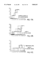
FIGS. 1A-1E are bar graphs showing the effect of topically applied 1,25(OH)2 D3 on T cell cytokine production in peripheral lymph nodes in mice. The bars represent the release of the indicated cytokines (IL-2, IFN-γ, IL-4, IL-5 and IL-10) from the lymph node cells isolated from treated and untreated sides of mice in pg/ml.
FIG. 1A shows release of IL-2.
FIG. 1B shows release of IFN-γ.
FIG. 1C shows release of IL-4.
FIG. 1D shows release of IL-5.
FIG. 1E shows release of IL-10.
FIGS. 2A-2D are graphs showing the effect of topically applied 1,25(OH)2 D3 on the primary (FIGS. 2A, 2B) and secondary (FIGS. 2C, 2D) serum antibody responses to subcutaneous immunizations with Diphtheria toxoid antigen ("Dt") in mice. Results from Dt-specific quantitative ELISA for Dt-specific IgG (FIGS. 2A, 2C) and IgA (FIGS. 2B, 2D) are shown for treated mice that received 1,25(OH)2 D3 on Day 0 following primary immunization (data points ), and for treated mice that received 1,25(OH)2 D3 on Day 5 following primary immunization (data points ▴), and for untreated mice (data points ▪), which received no 1,25(OH)2 D3.
FIG. 2A shows the primary serum IgG response.
FIG. 2B shows the primary serum IgA response.
FIG. 2C shows the secondary serum IgG response.
FIG. 2D shows the secondary serum IgA response.
FIGS. 3A-3D are graphs showing the effect of topically applied 1,25(OH)2 D3 on the serum antibody responses to primary (FIGS. 3A, 3B) and secondary (FIGS. 3C, 3D) subcutaneous immunizations with a Hemophilus Influenza type b polysaccharide conjugate vaccine ("HibCV"), Hib coupled to Dt, in mice. Results from Dt-specific quantitative ELISA for Dt-specific IgG (FIGS. 3A, 3C) and IgA (FIGS. 3B, 3D) are shown for treated mice (data points ), which received 1,25(OH)2 D3 on Day 5 following the primary immunization, and for untreated mice (data points ▪), which received no 1,25(OH)2 D3.
FIG. 3A shows the primary serum IgG response.
FIG. 3B shows the primary serum IgA response.
FIG. 3C shows the secondary serum IgG response.
FIG. 3D shows the secondary serum IgA response.
FIGS. 4A-4D are graphs showing the effect of topically applied 1,25(OH)2 D3 on the secretory antibody responses to primary (FIGS. 4A, 4B) and secondary (FIGS. 4C, 4D) subcutaneous immunizations with HibCV in mice. Results from Dt-specific quantitative ELISA for Dt-specific IgG (FIGS. 4A, 4C) and IgA (FIGS. 4B, 4D) are shown for treated mice (data points ), which received 1,25(OH)2 D3 on Day 5 following the primary immunization, and for untreated mice (data points ▪), which received no 1,25(OH)2 D3.
FIG. 4A shows the primary secretory IgG response.
FIG. 4B shows the primary secretory IgA response.
FIG. 4C shows the secondary secretory IgG response.
FIG. 4D shows the secondary secretory IgA response.
FIGS. 5A-5D are graphs showing the effect of topically applied 1,25(OH)2 D3 on the serum antibody responses to primary (FIGS. 5A, 5B) and secondary (FIGS. 5C, 5D) subcutaneous immunizations with HibCV in mice. Results (mean ±S.E.M.) from Hib-specific quantitative ELISA for IgG (FIGS. 5A, 5C) and IgA (FIGS. 5B, 5D) are shown for treated mice (data points ), which received 1,25(OH)2 D3 on Day 5 following the primary immunization, and for untreated mice (data points ▪), which received no 1,25(OH)2 D3.
FIG. 5A shows the primary serum IgG response.
FIG. 5B shows the primary serum IgA response.
FIG. 5C shows the secondary serum IgG response.
FIG. 5D shows the secondary serum IgA response.
FIGS. 6A-6D are graphs showing the effect of topically applied 1,25(OH)2 D3 on the secretory antibody responses to primary (FIGS. 6A, 6B) and secondary (FIGS. 6C, 6D) subcutaneous immunizations with HibCV in mice. Results from Hib-specific quantitative ELISA for IgG (FIGS. 6A, 6C) and IgA (FIGS. 6B, 6D) are shown for treated mice (data points ), which received 1,25(OH)2 D3 on Day 5 following the primary immunization, and for untreated mice (data points ▪), which received no 1,25(OH)2 D3.
FIG. 6A shows the primary secretory IgG response.
FIG. 6B shows the primary secretory IgA response.
FIG. 6C shows the secondary secretory IgG response.
FIG. 6D shows the secondary secretory IgA response.
FIGS. 7A and 7B are graphs showing the effect of topically applied 1,25(OH)2 D3 on the serum antibody responses to immunization with virus vaccine (Influenza A/Beijing; 1.5 μg) in mice. Results from influenza virus-specific quantitative ELISA for IgG (FIG. 7A) and IgA (FIG. 7B) are shown for treated mice (data points ), which received 1.0 μg of 1,25(OH)2 D3 on Day 5 following the primary immunization, and for untreated mice (data points ▪), which received no 1,25(OH)2 D3.
FIG. 7A shows the serum IgG response.
FIG. 7B shows the serum IgA response.
FIGS. 8A-8D are histograms showing the effect of topically applied 1,25(OH)2 D3 on mucosal immune responses in two different mucosal compartments to immunization with an influenza virus vaccine (Influenza A/Beijing), in mice. Results of Influenza A/Beijing virus-specific quantitative ELISA for IgG (FIGS. 8A, 8C) and IgA (FIGS. 8B, 8C) from vaginal lavages (FIGS. 8A, 8B) and lung lavages (FIGS. 8C, 8D) are shown for treated mice, which received 1.0 μg of 1,25(OH)2 D3 topically at the vaccination site on Day 5 following vaccination, and for untreated mice, which received no 1,25(OH)2 D3.
FIG. 8A shows lung secretory IgG response.
FIG. 8B shows lung secretory IgA response.
FIG. 8C shows vaginal secretory IgG response.
FIG. 8D shows vaginal secretory IgA response.
FIGS. 9A and 9B show that IgA production is induced in mucosal tissue with administration of 1,25(OH)2 D3 incorporated in the vaccine. The IgA responses are shown for mice without 1,25(OH)2 D3 treatment () and for mice with 1,25(OH)2 D3 treatment (▪).
FIG. 9A shows the primary mucosal IgA response.
FIG. 9B shows the secondary mucosal IgA response.
FIGS. 9C and 9D show that IgG production is induced in mucosal tissue with administration of 1,25(OH)2 D3 incorporated in the vaccine. The IgG responses are shown for mice without 1,25(OH)2 D3 treatment (▪) and for mice with 1,25(OH)2 D3 treatment ().
FIG. 9C shows the primary mucosal IgG response.
FIG. 9D shows the secondary mucosal IgG response.
FIGS. 10A and 10B show that IgG production is induced in serum with topical administration of 1,25(OH)2 D3 five days after administration of vaccine. The IgG responses are shown for mice without 1,25(OH)2 D3 treatment (▪) and for mice with 1,25(OH)2 D3 treatment ().
FIG. 10A shows the primary serum IgG response.
FIG. 10B shows the secondary serum IgG response.
FIGS. 10C and 10D show that IgA production is induced in serum with topical administration of 1,25(OH)2 D3 five days after administration of vaccine. The IgA responses are shown for mice without 1,25(OH)2 D3 treatment (▪) and for mice with 1,25(OH)2 D3 treatment ().
FIG. 10C shows the primary serum IgA response.
FIG. 10D shows the secondary serum IgA response.
FIGS. 11A and 11B show that IgA production is induced in mucosal tissue with topical administration of 1,25(OH)2 D3 five days after administration of vaccine. The IgA responses are shown for mice without 1,25(OH)2 D3 treatment (▪) and for mice with 1,25(OH)2 D3 treatment ().
FIG. 11A shows the primary mucosal IgA response.
FIG. 11B shows the secondary mucosal IgA response.
FIGS. 11C and 11D shows that IgG production is induced in mucosal tissue with topical administration of 1,25(OH)2 D3 five days after administration of vaccine. The IgG responses are shown for mice without 1,25(OH)2 D3 treatment (▪) and for mice with 1,25(OH)2 D3 treatment ().
FIG. 11C shows the primary mucosal IgG response.
FIG. 11D shows the secondary mucosal IgG response.
FIG. 12 shows that the antibody response in aged mice, following vaccination with rHBSAg, is enhanced by treatment with DHEA or DHEA-S. The antibody responses are shown for aged mice without treatment (▪), aged mice with topical DHEA treatment (), aged mice with DHEA incorporated in the vaccine (▴), and aged mice with DHEA-S incorporated in the vaccine (♦).
FIGS. 13A and 13B show that serum (systemic) antibody response in mature mice following vaccination with Influenza-A Beijing strain is synergistically enhanced by treatment with DHEA and 1,25(OH)2 D3. Antibody responses at Day 28 are shown for mice without treatment (▪), mice treated with 2 μg DHEA in vaccine (), mice treated with 0.1 μg 1,25(OH)2 D3 in vaccine (▴), and mice treated with 2 μg DHEA and 0.1 μg 1,25(OH)2 D3 in vaccine (♦).
FIG. 13A shows the serum IgG response.
FIG. 13B show the serum IgA response.
FIGS. 13C and 13D show that mucosal antibody response in mature mice following vaccination with Influenza-A Beijing strain is synergistically enhanced by treatment with DHEA and 1,25(OH)2 D3. Antibody responses at Day 28 are shown for mice without treatment (▪), mice treated with 2 μg DHEA in vaccine (), mice treated with 0.1 μg 1,25(OH)2 D3 in vaccine (▴), and mice treated with 2 μg DHEA and 0.1 μg 1,25(OH)2 D3 in vaccine (♦).
FIG. 13C shows the mucosal IgG response.
FIG. 13D shows the mucosal IgA response.
FIGS. 14A and 14B show that serum (systemic) antibody response in mature mice following vaccination with rHBSAg is synergistically enhanced by treatment with DHEA and 1,25(OH)2 D3. Antibody responses at Day 21 are shown for non-immunized mice (★), mice without treatment (), mice treated with 2 μg DHEA in vaccine (♦), mice treated with 0.1 μg 1,25(OH)2 D3 in vaccine (▴), and mice treated with 2 μg DHEA and 0.1 μg 1,25(OH)2 D3 in vaccine (▪).
FIG. 14A shows the serum IgG response.
FIG. 14B shows the serum IgA response.
FIGS. 14C and 14D show that mucosal antibody response in mature mice following vaccination with rHBSAg is synergistically enhanced by treatment with DHEA and 1,25(OH)2 D3. Antibody responses at Day 21 are shown for non-immunized mice (★), mice without treatment (), mice treated with 2 μg DHEA in vaccine (♦), mice treated with 0.1 μg 1,25(OH)2 D3 in vaccine (▴), and mice treated with 2 μg DHEA and 0.1 μg 1,25(OH)2 D3 in vaccine (▪).
FIG. 14C shows the mucosal IgG response.
FIG. 14D shows the mucosal IgA response.
FIGS. 15A and 15B show that serum (systemic) antibody response in mature mice following vaccination with rHBSAg is synergistically enhanced by treatment with DHEA and all trans-retinoic acid. Antibody responses at Day 21 are shown for non-immunized mice (▪), mice without treatment (), mice treated with 5.0 μg all trans-retinoic acid in vaccine (▴), and mice treated with 2 μg DHEA and 5.0 μg all trans-retinoic acid in vaccine (♦).
FIG. 15A shows the serum IgG response.
FIG. 15B shows the serum IgA response.
FIGS. 15C and 15D show that mucosal antibody response in mature mice following vaccination with rHBSAg is synergistically enhanced by treatment with DHEA and all transretinoic acid. Antibody responses at Day 21 are shown for non-immunized mice (▪), mice without treatment (), mice treated with 5.0 μg all trans-retinoic acid in vaccine (▴), and mice treated with 2 μg DHEA and 5.0 μg all trans-retinoic acid in vaccine (♦).
FIG. 15C shows the mucosal IgG response.
FIG. 15D shows the mucosal IgA response.
FIG. 16A shows the titre of antigen-specific antibody in nasal wash fluid.
FIG. 16B shows the specific neutralizing activity of nasal wash fluid as measured by hemagglutination inhibition.
FIG. 16C shows the relative level of infectivity of Influenza type A virus detectable by reverse transcriptase polymerase chain reaction in the tracheae.
FIGS. 17A-C show the development of clinical disease over time after infection.
FIG. 17A shows clinical disease for control plasmid.
FIG. 17B shows clinical disease for gD2 gene containing plasmid.
FIG. 17C shows clinical disease for gD2 gene containing plasmid with 1,25(OH)2 D3.
FIG. 18 shows the relative titre of virus detectable in vaginal wash fluids over the first 48 hours following infection.
FIG. 19 shows the serum neutralizing activity six days after challenge.
FIG. 20 shows serum neutralizing antibody titres following intranasal challenge with antigen.
FIG. 21 shows nasal wash neutralizing antibody titres following intranasal challenge with antigen.
DETAILED DESCRIPTION OF THE PREFERRED EMBODIMENTS
The invention relates to a vaccine which comprises an antigen and a lymphoid organ modifying agent. Suitable lymphoid organ modifying agents enhance the production of IL-4, IL-5 or IL-10 in T cells. Such agents include, but are not limited to, 1,25-dihydroxy Vitamin D3 (1,25(OH)2 D3), 25-hydroxy Vitamin D3 (25(OH)D3) or biologically active 1,2(OH)2 D3 derivatives which are capable of activating the intracellular Vitamin D3 receptor, all transretinoic acid, retinoic acid derivatives, retinol and glucocorticoid. The vaccine composition may further comprise an immune response augmenting agent. The immune response augmenting agent enhances production of IL-2, IL-3, IFN-γ or GM-CSF in T cells. Suitable immune response augmenting agents include, but are not limited to, dehydroepiandrosterone (DHEA), DHEA congeners and DHEA-derivatives.
The invention also relates to a method for inducing an antigen-specific mucosal immune response in a subject vertebrate animal which comprises administering a vaccine which comprises an antigen and a lymphoid organ modifying agent to a site which drains into a peripheral lymph compartment. Alternatively, the method comprises separately administering the lymphoid organ modifying agent and a vaccine containing an antigen to the same site. The method may further comprise additionally administering an immune response augmenting agent. The immune response augmenting agent may be administered sequentially or contemporaneously with the lymphoid organ modifying agent.
Biologic response modifiers, here termed "lymphoid organ modifying agents" have been identified. These biological response modifiers can affect peripheral lymph node T cell function in a manner that mimics the mucosal lymphoid organ microenvironment, and therefore favors induction of an Ag-specific mucosal antibody response as well as Ag-specific serum antibody production in response to antigen administration to sites which drain into the "treated" lymphoid organ.
Particularly, for example, it has been found that topically applied 1,25(OH)2 D3, serving a function as a lymphoid organ modifying agent with appropriately immunized animals, can be used therapeutically to enhance IgA and IgG production in serum as well as to promote secretory antibody production in mucosal secretions.
It has been found that epicutaneous administration of 1,25(OH)2 D3 to animals prior to isolation and in vitro activation of T cells from the draining peripheral lymph node results in emergence in the lymph node T cells of a pattern of cytokines following activation, displaying elevated IL-4, IL-5 and IL-10 production and depressed IL-2 and IFN-γ production. IL-5 and IL-10 are known to synergize with TGF-β to induce differentiation of B cells committed to IgA production (R. Coffman (1989). J. Exp. Med. 170:1039-44; T. Defrance et al. (1992). J. Exp. Med. 175:671-82). IL-4 has been shown to have a direct influence on B cell isotype switching to the IgGI isotype (Finkelman et al., supra). Once B cells are activated by antigen at the lymph node, both IL-4 and IL-5 can play important roles in stimulating B cells to secrete antibody (J. Purkerson (1992). J Exp. Med. 175:973-82).
Moreover, when 1,25(OH)2 D3 is administered to animals prior to in vivo antigen challenge at the peripheral lymph node that drains the site of 1,25(OH)2 D3 administration, an Ag-specific secretory immunologic response in the mucosa can result, as well as an Ag-specific serum immunologic response. Apparently, a result of 1,25(OH)2 D3 and in vivo antigen challenge at the same peripheral lymph node is a "seeding" of the lamina propria with secretory IgA-committed and secretory IgG-committed B cells at sites throughout the mucosa.
In one aspect, in general, the invention features a method for inducing antigen-specific mucosal immunity in a subject vertebrate animal, by administering an effective amount of a lymphoid organ modifying agent to the subject at a site which drains into a peripheral lymphoid organ, and administering an effective amount of the specific antigen to the subject at a site which drains into the same peripheral lymphoid organ. The method further features administering an effective amount of an immune response augmenting agent which enhances T cell lymphokine production, as described in U.S. application Ser. No. 07/779,449, incorporated herein by reference.
The term "individual" as used herein refers to a vertebrate and preferably to a member of a species which naturally produces DHEA and DHEA-S and possesses DHEA-S sulfatase activity, and includes, but is not limited to domestic animals, sports animals and primates, including humans.
An "antigen" refers to a molecule containing one or more epitopes that will stimulate a host's immune system to make a secretory, humoral and/or cellular antigen-specific response, or to a DNA molecule which is capable of producing such an antigen in a vertebrate. The term is also used interchangeably with "immunogen."
The specific antigen can be a protein, a polysaccharide, a lipopolysaccharide or a lipopeptide; or it can be a combination of any of these. Particularly, the specific antigen can include a native protein or protein fragment, or a synthetic protein or protein fragment or peptide; it can include glycoprotein, glycopeptide, lipoprotein, lipopeptide, nucleoprotein, nucleopeptide; it can include a peptide-peptide conjugate; it can include a recombinant nucleic acid expression product. The antigen may further be a DNA molecule which produces an antigen in the vertebrate. Examples of antigens include, but are not limited to, those that are capable of eliciting an immune response against viral or bacterial hepatitis, influenza, diphtheria, tetanus, pertussis, measles, mumps, rubella, polio, pneumococcus, herpes, respiratory syncytial virus, hemophilus influenza type b, chlamydia, varicella-zoster virus or rabies.
"Treatment" refers to the administration to an individual of a composition which yields a protective immune response, and includes prophylaxis and/or therapy.
By "vaccine composition" or "vaccine" is meant an agent used to stimulate the immune system of an individual so that current harm is alleviated, or protection against future harm is provided.
"Immunization" refers to the process of inducing a continuing protective level of antibody and/or cellular immune response which is directed against an antigen to which the organism has been previously exposed.
An "immune response augmenting agent", as used herein, means an agent that is capable, when administered to a vertebrate animal in vivo, of restoring T cell responsiveness to T cell dependent antigens characteristic of normal immune responses to such antigens. Immune response augmenting agents are capable of enhancing T cell production of IL-2, IL-3, IFN-γ or GM-CSF. By way of example, the immune response augmenting agent can be a substance such as DHEA, DHEA congener or a DHEA-derivative (collectively referred to herein as DHEA congener). A DHEA congener is a compound related to DHEA, such as a derivative. Suitable substances can be readily identified by screening for enhanced IL-2, IL-3, IFN-γ or GM-CSF production by T cells as described in Ser. No. 07/779,499.
Examples of "DHEA", "DHEA congener" or "DHEA-derivative", include but are not limited to, compounds having the formula ##STR1## wherein X is H or halogen;
R1, R2 and R3 are independently ═O, --OH, --SH, H, halogen, pharmaceutically acceptable ester, pharmaceutically acceptable thioester, pharmaceutically acceptable ether, pharmaceutically acceptable thioether, pharmaceutically acceptable inorganic esters, pharmaceutically acceptable monosaccharide, disaccharide or oligosaccharide, spirooxirane, spirothirane, --OSO2 R5 or --OPOR5 R6 ;
R5 and R6 are independently --OH, pharmaceutically acceptable esters or pharmaceutically acceptable ethers; and
pharmaceutically acceptable salts.
Thus, examples of suitable DHEA congeners include compounds in which:
(1) R2 is ═O, R3 and X are each H and R1 is ═O, --OH, pharmaceutically acceptable esters thereof, pharmaceutically acceptable ethers thereof or pharmaceutically acceptable salts;
(2) R2 is ═O, R3 is H, X is halogen and R1 is ═O, --OH, pharmaceutically acceptable esters thereof, pharmaceutically acceptable ethers thereof or pharmaceutically acceptable salts;
(3) R2 is ═O, R3 and X are each H and R1 is --SH, pharmaceutically acceptable thioesters thereof, pharmaceutically acceptable thioethers thereof or pharmaceutically acceptable salts;
(4) R2 is ═O, R3 is H, X is halogen and R1 is --SH, pharmaceutically acceptable thioesters thereof, pharmaceutically acceptable thioethers thereof or pharmaceutically acceptable salts;
(5) R2 is ═O, X is H and R1 and R3 are independently ═O, --OH, pharmaceutically acceptable esters thereof, pharmaceutically acceptable ethers thereof or pharmaceutically acceptable salts;
(6) R2 is ═O, X is halogen and R1 and R3 are independently ═O, --OH, pharmaceutically acceptable esters thereof, pharmaceutically acceptable ethers thereof or pharmaceutically acceptable salts;
(7) R2 is ═O, X is H and R1 and R3 are independently --SH, pharmaceutically acceptable thioesters thereof, pharmaceutically acceptable thioethers thereof or pharmaceutically acceptable salts;
(8) R2 is ═O, X is halogen and R1 and R3 are independently --SH, pharmaceutically acceptable thioesters thereof, pharmaceutically acceptable thioethers thereof or pharmaceutically acceptable salts;
(9) R2 is --OH, R3 and X are each H and R1 is ═O, --OH, pharmaceutically acceptable esters thereof, pharmaceutically acceptable ethers thereof or pharmaceutically acceptable salts;
(10) R2 is --OH, R3 is H, X is halogen and R1 is ═O, --OH, pharmaceutically acceptable esters thereof, pharmaceutically acceptable ethers thereof or pharmaceutically acceptable salts;
(11) R2 is --OH, R3 and X are each H and R1 is --SH, pharmaceutically acceptable thioesters thereof, pharmaceutically acceptable thioethers thereof or pharmaceutically acceptable salts;
(12) R2 is --OH, R is H, X is halogen and R1 is --SH, pharmaceutically acceptable thioesters thereof, pharmaceutically acceptable thioethers thereof or pharmaceutically acceptable salts;
(13) R2 is --OH, X is H and R1 and R3 are independently ═O, --OH, pharmaceutically acceptable esters thereof, pharmaceutically acceptable ethers thereof or pharmaceutically acceptable salts;
(14) R2 is --OH, X is halogen and R1 and R3 are independently ═O, --OH, pharmaceutically acceptable esters thereof, pharmaceutically acceptable ethers thereof or pharmaceutically acceptable salts;
(15) R2 is --OH, X is H and R1 and R3 are independently --SH, pharmaceutically acceptable thioesters thereof, pharmaceutically acceptable thioethers thereof or pharmaceutically acceptable salts;
(16) R2 is --OH, X is halogen and R1 and R3 are independently --SH, pharmaceutically acceptable thioesters thereof, pharmaceutically acceptable thioethers thereof or pharmaceutically acceptable salts;
(17) R2 is --SH, R3 and X are each H and R1 is ═O, --OH, pharmaceutically acceptable esters thereof, pharmaceutically acceptable ethers thereof or pharmaceutically acceptable salts;
(18) R2 is --SH, R3 is H, X is halogen and R1 is ═0, --OH, pharmaceutically acceptable esters thereof, pharmaceutically acceptable ethers thereof or pharmaceutically acceptable salts;
(19) R2 is --SH, R3 and X are each H and R1 is --SH, pharmaceutically acceptable thioesters thereof, pharmaceutically acceptable thioethers thereof or pharmaceutically acceptable salts;
(20) R2 is --SH, R3 is H, X is halogen and R1 is --SH, pharmaceutically acceptable thioesters thereof, pharmaceutically acceptable thioethers thereof or pharmaceutically acceptable salts;
(21) R2 is --SH, X is H and R1 and R3 are independently ═O, --OH, pharmaceutically acceptable esters thereof, pharmaceutically acceptable ethers thereof or pharmaceutically acceptable salts;
(22) R2 is --SH, X is halogen and R1 and R3 are independently ═O, --OH, pharmaceutically acceptable esters thereof, pharmaceutically acceptable ethers thereof or pharmaceutically acceptable salts;
(23) R2 is --SH, X is H and R1 and R3 are independently --SH, pharmaceutically acceptable thioesters thereof, pharmaceutically acceptable thioethers thereof or pharmaceutically acceptable salts;
(24) R2 is --SH, X is halogen and R1 and R3 are independently --SH, pharmaceutically acceptable thioesters thereof, pharmaceutically acceptable thioethers thereof or pharmaceutically acceptable salts;
(25) X is H and R1, R2 and R3 are independently ═O, --OH, a sugar residue, pharmaceutically acceptable esters thereof, pharmaceutically acceptable ethers thereof or pharmaceutically acceptable salts, wherein at least one of R1, R2 and R3 is a sugar residue;
(26) X is halogen and R1, R2 and R3 are independently ═O, --OH, a sugar residue, pharmaceutically acceptable esters thereof, pharmaceutically acceptable ethers thereof or pharmaceutically acceptable salts, wherein at least one of R1, R2 and R3 is a sugar residue;
(27) X is H and R1, R2 and R3 are independently ═O, --OH, pharmaceutically acceptable inorganic esters thereof or pharmaceutically acceptable salts, wherein at least one of R1, R2 and R3 is an inorganic ester;
(28) X is halogen and R1, R2 and R3 are independently ═O, --OH, pharmaceutically acceptable inorganic esters thereof or pharmaceutically acceptable salts, wherein at least one of R1, R2 and R3 is an inorganic ester.
Pharmaceutically acceptable esters or thioesters include, but are not limited to, esters or thioesters of the formula --OOCR or --SOCR, wherein R is a pharmaceutically acceptable alkyl, alkenyl, aryl, alkylaryl, arylalkyl, spingosine or substituted spingolipid groups, such as propionate, enanthate, cypionate, succinate, decanoate and phenylpropionate esters.
Pharmaceutically acceptable ethers or thioethers include, but are not limited to, ethers or thioethers of the formula --OR or --SR, wherein R is as defined above or enol, or --OR4 is an unsubstituted or substituted spirooxirane or --SR is a spirothiane.
Suitable sugar residues include, but are not limited to monosaccharides, disaccharides and oligosaccharides, such as a glucuronate.
Pharmaceutically acceptable inorganic esters include, but are not limited to, inorganic esters of the formula --OSO2 R5 or --OPOR5 R6, wherein R5 and R6 are independently --OH, pharmaceutically acceptable esters, pharmaceutically acceptable ethers or pharmaceutically acceptable salts.
"Lymphoid organ modifying agent", as used herein, means a modifier that is capable, when administered to a vertebrate animal in vivo at a peripheral site, of altering the microenvironment of a peripheral lymphoid organ that drains from the administration site, such that activated lymphocytes and macrophages residing within the lymphoid organ exhibit a pattern of cytokines more typical of the microenvironment of a lymphoid organ of the mucosal lymphoid compartment. Particularly, a pattern of cytokines more typical of a mucosal lymphoid organ is characterized by relatively enhanced production of one or more of active TGF-β, IL-4, IL-5, and IL-10, and relatively decreased production (or at least no relatively enhanced production) of one or more of IL-2 and IFN-γ. In preferred embodiments, the lymphoid organ modifying agent includes a biological response modifier that can, or a combination of biological response modifiers that together can, when administered to a peripheral lymph organ, result in the lymphoid organ exhibiting a pattern of cytokines more typical of a mucosal lymphoid organ. By way of example, it can be a substance known to enhance or facilitate production of active TGF-β, such as, for example, retinoic acid, retinoic acid derivatives, retinol or retinol derivatives; or it can be 1,25(OH)2 D3, 25(OH)D3 or biologically active 1,25(OH)2 D3 derivatives; or glucocorticoids. Biologically active 1,25(OH)2 D3 derivatives are those derivatives which are capable of activating the intracellular Vitamin D3 receptor. Suitable derivatives can thus be readily identified by screening for activation of the intracellular Vitamin D3 receptor (Minghetti and Norman (1988). FASEB J. 2:3043-3053). Alternatively, suitable derivatives can be readily identified by screening for enhanced IL-4 production by T cells as described in Ser. No. 07/779,499. Examples of suitable biologically active Vitamin D3 derivatives include, but are not limited to, 1,25dihydroxy-16-ene Vitamin D3 and calcipotriene. Preferably, the lymphoid organ modifying agent is administered epicutaneously at a peripheral anatomical site (such as, for human subjects, for example, the arm or buttocks or leg); and the specific antigen is administered to the same anatomical site, or to a site known to drain into the same lymphoid organ that receives drainage from the site of administration of the lymphoid organ modifying agent. In one mode of administration, the lymphoid organ modifying agent can be combined with the specific antigen for simultaneous administration at the same site. The lymphoid organ modifying agent and the specific antigen, or both of them, can be administered by injection.
The lymphoid organ modifying agent can be administered at a time concurrent with, prior to, or following the time of administration of the specific antigen. The specific antigen and the lymphoid organ modifying agent can be combined prior to administration and administered together, or they can be administered sequentially. When the lymphoid organ modifying agent is administered prior to the administration of the specific antigen, it is administered 0.1 to 24 hours, preferably 0.1 to 6 hours before the antigen. When the lymphoid organ modifying agent is administered after the administration of the specific antigen, it is administered 0.1 hour to 6 days, preferably 0.1 hour to 5 days, and most preferably 2 to 3 days after the antigen.
Preferably, however, the lymphoid organ modifying agent is administered concurrently with the specific antigen, or still more preferably, at a time following administration of the specific antigen. Administration of the specific antigen results initially in a clonal expansion (proliferation) of T cells and B cells in the lymphoid organ that drains the site of administration, and for a time the cells are sequestered in the lymphoid organ; thereafter, the cells are dispatched to the general circulation. Thus, the lymphoid organ modifying agent also facilitates B cell homing to mucosal tissues and the generation of mucosal immune responses at all mucosal sites. In particular, therefore, it can be preferable to administer the lymphoid organ modifying agent at a time after primary immunization when the resulting proliferation of T cells and B cells is well developed, and while, for the most part, the antigen-activated cells remain sequestered in the lymphoid organ. Generally, the proliferation of T cells and B cells can be expected to be well developed within about two or three days following primary immunization with the specific antigen; and substantial movement of the cells into the general circulation can be expected about five days following primary immunization, or somewhat later.
It may be particularly preferred that the lymphoid organ modifying agent reach the draining lymphoid organ at a time following primary immunization with the specific antigen but before a substantial portion of the resulting expanded B cell and T cell populations have moved into the general circulation. This can be accomplished, for example, by first administering the antigen, and thereafter (preferably at least about two or three days thereafter, but not more than about five days thereafter) administering the lymphoid organ modifying agent. Or this can be accomplished by compounding the lymphoid organ modifying agent with means for delaying its release following administration, and then combining the delayed-release compounded lymphoid organ modifying agent with the specific antigen in a composition for simultaneous administration. Means for delayed release are well-known, and the chemistry or the structure of the means can be adjusted to provide a suitable time period for the delay. Encapsulation of the lymphoid organ modifying agent within liposomes, or within a biodegradable matrix, for example, can effect a delay in delivery of the lymphoid organ modifying agent (relative to the delivery of the antigen) from the administration site to the lymphoid organ.
An "effective amount" of lymphoid organ modifying agent is a quantity of administered lymphoid organ modifying agent that is able to alter, qualitatively, the types of cytokines and growth factors capable of being produced by activated cells residing in a draining lymphoid organ, with the result that representative species produced in the altered lymphoid organ include one or more of active TGF-β, IL-10, IL-4, and IL-5. For epicutaneous application of the lymphoid organ modifying agent, amounts can be in the range of about 0.1-500 μg, preferably 0.5-250 μg, for a human subject, or in the range of about 0.01-5.0 μg/kg body weight of the subject animal. Effective amounts for subcutaneous, intramuscular or intracutaneous administration may vary to some degree, but can be in the same general range as for epicutaneous application.
An effective amount of antigen is an amount that is the same as that amount which is capable, when administered without a lymphoid organ modifier, of eliciting a demonstrable humoral immune response. For many antigens, this is in the range of about 5-100 μg for a human subject. The appropriate amount of antigen to be used is dependent on the specific antigen and is well known in the art.
An effective amount of an immune response augmenting agent is an amount that restores T cell responsiveness in an immunodeficient individual, such as an immunodeficiency resulting from age or stress. The amount of immune response augmenting agent may be in the range of about 10-1,000 μg, preferably 10-900 μg, more preferably 10-500 μg, and most preferably 20-200 μg, for a human subject, or in the range of about 0.2-10 μg/kg body weight of the subject animal, if administered by injection If administered orally, the amount may range from 10-100 mg/day, preferably 20-50 μg/day for a human subject, or in the range of about 0.5-5 mg/kg/day for an animal subject.
The immune response augmenting agent can be administered at a time concurrent with, prior to or following the time of administration of the lymphoid organ modifying agent. The immune response augmenting agent and the lymphoid organ modifying agent can be combined prior to administration of both agents can be combined with the specific antigen prior to administration. When the immune response augmenting agent is administered prior to the administration of the lymphoid organ modifying agent, it is administered 0.1 hr. to 14 days, preferably 0.1 hr to 5 days before the lymphoid organ modifying agent. When the immune response augmenting agent is administered after the administration of the lymphoid organ modifying agent, it is administered 0.1 to 24 hrs, preferably 0.1 to 6 hrs after the lymphoid organ modifying agent. It is preferred that the immune response augmenting agent be administered prior to the lymphoid organ modifying agent. When the immune response augmenting agent is administered prior to the lymphoid organ modifying agent, the immune response augmenting agent is preferably DHEA-S. It is preferred to concurrently administer the lymphoid organ modifying agent and the specific antigen in this embodiment.
In another general aspect, the invention features a composition for inducing an antigen-specific mucosal immune response, including a lymphoid organ modifying agent and a specific antigen in a pharmaceutically acceptable carrier. The composition may further include an immune response augmenting agent. In preferred embodiments, the lymphoid organ modifying agent includes one or more of 1,25(OH)2 D3, or a retinoid such as all trans-retinoic acid. Biologically active Vitamin D3 derivatives which are capable of activating the intracellular Vitamin D3 receptor, glucocorticoid and retinoic acid derivatives, retinol and retinol derivatives, can also be used as lymphoid organ modifying agents. The immune response augmenting agent includes one or more DHEA or DHEA derivative. It has been found that a synergistic effect on the immune response is achieved when the composition includes at least one lymphoid organ modifying agent and at least one immune response augmenting agent. In some preferred embodiments, the lymphoid organ modifying agent in the composition is compounded with means for delaying its release following administration, such as encapsulation in a liposome or a biodegradable matrix, so that the movement of the lymphoid organ modifying agent from the delivery site to the draining lymphoid organ is delayed relative to that of the antigen.
Because the method of the invention can result in a common mucosal immune response, secretion of antigen-specific immunoglobulins will occur at all mucosal surfaces. A lactating mammal that has been treated according to the invention will produce antigen-specific immunoglobulins in the secretions of the mammary glands. A female mammal that is treated according to the invention during pregnancy, or treated prior to pregnancy and then rechallenged with the specific antigen during pregnancy, will produce antigen-specific immunoglobulins in the colostrum and in the milk. This provides a means for generating a specific mucosal immune response in the female mammal, without infecting the mother, and then transferring a specific passive immunity from the female mammal to the suckling newborn or infant.
Thus, in another general aspect, the invention features a method for inducing production of antigen-specific antibodies in the mammary secretions of a female mammal, by administering an effective amount of a lymphoid organ modifying agent to the female mammal at a site which drains into a peripheral lymphoid organ, and administering an effective amount of the specific antigen to the female mammal at a site which drains into the same peripheral lymphoid organ. And in another general aspect, the invention features a method for conferring a specific passive immunity to a suckling mammal, by permitting the suckling mammal to consume mammary secretions from a female so treated.
The invention can be used for immunization of any vertebrate animal, and is particularly useful in inducing an antigen-specific mucosal immune response in a human subject.
According to the invention, the lymphoid organ modifying agent can be administered before the specific antigen is administered, after the specific antigen is administered or concurrently with the antigen; or the antigen and the lymphoid organ modifying agent can be administered at least partly concurrently.
The antigen can be administered to the animal in a manner, and in an amount, suitable for inducing a demonstrable humoral response to the antigen in the animal. The antigen can be applied epicutaneously, for transdermal administration, or it can be administered by injection or infusion, subcutaneously, intramuscularly or intradermally.
The lymphoid organ modifying agents can be formulated for a variety of modes of administration, including systemic and topical or localized administration. Techniques and formulations generally may be found in Remington's Pharmaceutical Sciences, Mack Publishing Co., Easton, Pa., latest edition. An effective amount of lymphoid organ modifying agent can vary according to the mode of administration and according to the kind of animal being treated. In general, an effect amount will range from about 0.01-1.0 μg lymphoid organ modifying agent/kg body weight, or from about 0.1-500 μg, preferably 0.1-400 μg, and more preferably 0.5-250 μg, for human subjects.
According to the invention, the lymphoid organ modifying agent and the antigen are applied at the same site, or at sites which drain into the same peripheral lymphoid compartment. Anatomical location of such sites is well known.
The lymphoid organ modifying agent can be applied epicutaneously for transdermal administration. Penetrants appropriate to the dermal barrier can be used in the formulation. The lymphoid organ modifying agent can be formulated into ointments, salves, gels or creams, as is generally known in the art.
Alternatively, the lymphoid organ modifying agent can be administered by a subcutaneous, intramuscular or intradermal route, as by injection or infusion. For injection, the lymphoid organ modifying agent of the invention can be formulated in liquid solutions, preferably in physiologically compatible buffers such as Hank's solution or Ringer's solution. In addition, the lymphoid organ modifying agent may be formulated in solid form and redissolved or suspended immediately prior to use.
It has been found that the immune system should be functioning as that of a normal mature adult in order for the lymphoid organ modifying agent to induce a mucosal immune response. If the immune system is not fully competent or the individual is otherwise immunologically deficient, such as results in the very young, the aged, individuals under stress, or the like (see, e.g., U.S. application Ser. No. 07/779,499), it may be necessary to augment the immune system for proper functioning of the lymphoid organ modifying agent. The immune system is augmented by administering an immune response augmenting agent, as described in U.S. application Ser. No. 07/779,449, U.S. application Ser. No. 08/123,843 and U.S. application Ser. No. 08/487,173 filed concurrently herewith (attorney docket number 23691-108469-1), each of which are incorporated herein by reference. The immune response augmenting agent is preferably administered for 0.1 hr to 14 days, preferably 0.1 hr to 5 days prior to the administration of the lymphoid organ modifying agent and the administration of the specific antigen.
The immune response augmenting agent may be administered prior to, or contemporaneously with, or after the lymphoid organ modifying agent. It can be administered by an epicutaneous, subcutaneous, intramuscular or interdermal route, as by injection or infusion, or it can be administered orally. Alternatively, the individual may be treated with the immune response augmenting agent before induction of the mucosal immune response described above. The immune response augmenting agent may be formulated as described above. In general, an effective amount of immune response augmenting agent may be about 10-1,000 μg, preferably 10-900 μg, more preferably 10-500 μg, most preferably 20-200 μg, for a human subject, if administered by injection. If administered orally, an effective amount of immune response augmenting agent may be about 10-100 mg/day, preferably 20-50 mg/day. The immune response augmenting agent can be administered orally for one to three weeks prior to or after vaccination, or longer, as necessary.
Described below are examples of the present invention which are provided only for illustrative purposes, and not to limit the scope of the present invention. In light of the present disclosure, numerous embodiments within the scope of the claims will be apparent to those of ordinary skill in the art.
EXAMPLE 1 Materials and Methods
Mice. Female C3H/HeN MTV- mice were bred and housed in the University of Utah Vivarium from breeding stock originally purchased from the National Cancer Institute. In each experiment, groups of three to five age-matched mice were used.
Antibodies. Monoclonal antibody reagents used in the present study were prepared from culture supernatants of appropriate B cell hybridomas adapted to growth under serum-free conditions. The hybridoma clones secreting rat anti-murine IL-2 (S4B6), and rat anti-γIFN (XMG1.2) were obtained from DNAX (Palo Alto, Calif.). Hybridoma cell clones producing rat antibody specific for murine IL-4 (11B11), or murine IFN-γ (R46A2) were purchased from the ATCC. Purified anti-cytokine antibodies were then used for quantification of murine cytokines in culture supernatants by capture ELISA. Additionally, the following rat anti-murine cytokine antibodies were obtained from PharMingen (San Diego, Calif.) and then used for quantification of specific murine cytokines by capture ELISA: biotinylated anti-IL-4 (BVD6-24G2) and anti-murine IL-10 antibodies (Cat. nos. 18141 and 18152), and a set of two rat anti-murine IL-2 monoclonal antibodies.
Biological Response Modifiers. Murine recombinant IL-2, IL-4, IL-5, and IFN-γ were obtained from Genzyme (Cambridge, Mass.) and used as a reference in monospecific ELISA assays. In addition, murine recombinant IL-2, IL-4, and IL-5 were derived from culture supernatants of X63Ag8-653 cells transfected with multiple copies of a single murine interleukin gene. After the relative concentration of each lymphokine in culture supernatants was determined by a comparison to a known recombinant standard, these reagents were used as reference lymphokines in appropriate assays. The IL-10 standard was obtained from PharMingen. 1,25(OH)2 D3 was received by request from Milan Uskokovic (Hoffman-LaRoche, Nutley, N.J.), dissolved in 95% ethanol as a stock solution of 10-3 M and then stored at -20° C. For topical use, 1-2 μg of 1,25(OH)2 D3 was administered in ethanol to the same site as the site of immunization, the dorsal surface of the hind footpad.
Capture ELISA Assays for Quantitative Lymphokine Determination. Where indicated, the amounts of lymphokine in test supernatants were measured by a capture ELISA. Briefly, 100 μl of an appropriate capture monoclonal antibody is added to the wells of a 96-well microtest plate (Corning #2581) at a concentration of 1-2 μg/ml in 0.5M Tris-HCl (pH 9.6). Following extensive washing and the blocking of reactive sites on the plastic with PBS/10% FCS, test supernatants and two-fold serial dilutions of a reference lymphokine (100 μl/well) were dispensed. After sufficient incubation and washing, 100 ml of the biotinylated detecting antibody, 1-2 μg/ml, was dispensed into each well. The ELISA was developed, after extensive further washing, using avidin conjugated to horseradish peroxidase and ABTS-substrate. O.D. readings were performed at 405 nM using a Vmax 96-well microtest plate spectrophotometer (Molecular Devices, Menlo Park, Calif.). Results of the analysis of each cytokine are reported in pg/ml±SD.
Antibody Responses. Purified Diphtheria toxoid (Dt) was received as a gift from Connaught Laboratories (London, Ont., Canada). An antibody response to Dt was elicited from mice following a single footpad injection of 10 μg purified protein delivered in a 25 μl volume of aluminum hydroxide (273 μg/ml). Hemophilus Influenza type b conjugate vaccine (HibCV) used for immunization, consisting of Hib polysaccharide chemically coupled to Diphtheria toxoid, was purchased at the University of Utah hospital pharmacy. 500 ng of H. Influenza type b polysaccharide (HibCV) coupled to 1 mg of diphtheria toxoid was used as the dose of immunogen. This immunogen was delivered in a 25 μl volume of aluminum hydroxide (273 μg/ml). Quantification of specific antibody in a serum sample was performed using an indirect ELISA method, with reagents purchased from Southern Biotechnology Associates (Birmingham, Ala.) and Zymed Laboratories, Inc. (San Francisco, Calif.). For detection of Diphtheria toxoid-specific antibodies, purified Dt was diluted in 0.05M Tris-HCl (pH 9.6) at a concentration of 2.0 μg/ml and then dispensed into 96-well plates. For detection of Hib-specific antibodies, 100 μl of a Hib-meningococcal protein conjugate, at a dilution to 200 ng/ml polysaccharide in 0.05M Tris-HCl (pH 9.6) was dispensed into 96-well plates. Following incubation for a minimum of two hours at room temperature or overnight at 4° C., all plates were blocked with PBS-0.05% Tween 20/1.0% bovine serum albumin (BSA) for an additional two-hour incubation at room temperature. Prior to adding the test samples, the plates were washed free of blocking buffer using three washes of distilled water and one wash with PBS/0.05% Tween 20. Individual samples were first diluted in PBS 0.05% Tween 20/1.0% BSA, and then 100 μl was dispensed into appropriate wells of the antigen-coated plates. Included on each plate was an Ig standard: a series of two-fold dilutions of either purified IgG (all subclasses) or IgA (reference standards). The reference Ig were captured by goat anti-murine Ig, which is known to bind all murine Ig isotypes. These plates were incubated at room temperature overnight, followed by 3× wash in distilled water and one wash in PBS/0.05% Tween 20. The detection antibody (HRP-linked goat anti-mouse Ig specific for IgG and IgA) was diluted in PBS/Tween/10% normal goat serum at a dilution recommended by the manufacturer. After a final incubation and wash series the ELISA was developed using ABTS-substrate. O.D. readings were recorded at 405 nM using a Vmax 96-well microtest plate spectrophotometer (Molecular Devices, Menlo Park, Calif.). A simple linear regression analysis of the Ig titration generated a reference curve for calculating the amount of specific antibody contained in the test samples. Data are reported as ng/ml±SEM.
EXAMPLE 2 Administration of 1,25(OH)2 D3 Alters Cytokine Production in Peripheral Lymphoid Organs
To demonstrate the lymphoid organ modifying effect of 1,25(OH)2 D3 in the mouse, i.e., its effect on the pattern of cytokine production in peripheral lymphoid organs, 1,25(OH)2 D3 was topically administered to the right front paw (the treated side) and an ethanol vehicle was topically administered to the left front paw of each mouse. After three hours, mice were sacrificed and lymph node cells from the draining lymph nodes from both the treated and untreated sides of experimental mice were removed, and the resident cells were dissociated and activated in vitro using 1 μg/ml anti CD3 ε to stimulate cytokine production. Culture supernatants were harvested after 24 hours and were then assayed for individual cytokines using monospecific capture ELISAs. The results are shown in FIGS. 1A-1E, in which the bars represent the release of the indicated cytokines (IL-2, IFN-γ, IL-4, IL-5 and IL-10) from the lymph node cells isolated from treated and untreated sides of mice in pg/ml. The results indicate that epicutaneous in vivo treatment of mice with 2 μg of 1,25(OH)2 D3 is sufficient to mediate alterations in the patterns of cytokines produced by activated T cells in the draining lymph node, with IL-2 and IFN-γ being inhibited and IL-4, IL-5 and IL-10 being enhanced.
EXAMPLE 3 Administration of 1,25(OH)2 D3 Enhances Production of IgA in Serum
In a first demonstration of the effect of epicutaneous 1,25(OH)2 D3 administration on immune response to a specific antigen in the mouse, C3H/HeN female mice were given a primary immunization subcutaneously in the right footpad with 10 μg Dt in aluminum hydroxide prepared and delivered as set out in Example 1. Two distinct subsets of these mice ("treated mice") were administered 2 μg 1,25(OH)2 D3 epicutaneously to the right footpad surface on Day 0 and at Day 5 after immunization. The remaining mice ("untreated mice") received an equal volume of the ethanol carrier. After weekly sampling of serum, mice were secondarily immunized subcutaneously through an intrapelvic route with Dt and no additional exposure to 1,25(OH)2 D3. Serum samples from the treated and untreated groups of mice were collected and all primary and secondary samples were then assayed individually using a Dt-specific, quantitative ELISA for IgG and IgA. The results are shown in FIGS. 2A-2D, in which the mean anti-Diphtheria toxoid antibody response (ng/ml) for each group of mice is presented graphically. The results demonstrate that IgG response to a protein antigen predominates in the serum of Dt-immune mice. The IgA response, a much weaker constituent of the response to Dt in the serum, was substantially enhanced by treatment of mice with 1,25(OH)2 D3. These elevations persisted post-secondary immunization as well, even though no additional hormone treatment was given at the time of secondary antigen administration.
EXAMPLE 4 Administration of 1,25(OH)2 D3 Induces Immunoglobulin Production in Serum and Mucosal Tissue
In a further demonstration, groups of C3H/HeN female mice were administered a primary immunization with the Hemophilus Influenza type b polysaccharide conjugate vaccine, Hib coupled to Dt (HibCV). A subset of these mice were treated epicutaneously with 2 μg 1,25(OH)2 D3 on Day 5 at the same site of immunization. Serum samples and vaginal washings were collected weekly during the primary response and all samples were then assayed individually using Dt-specific and Hib-specific quantitative ELISA for the IgG and IgA isotypes. The results for each immunization regiment are shown in FIGS. 3A-3D (Dt i.e., protein component! specific serum antibodies); 4A-4D (Dt specific secretory antibodies); 5A-5D (Hib i.e., polysaccharide component! specific serum antibodies), and 6A-6D (Hib i.e., polysaccharide component! specific secretory antibodies), in which the quantity of antibody produced (mean±SEM) as a result is presented graphically. The results show that the IgA and IgG responses to both the protein and the polysaccharide components of the HibCV in both serum and vaginal secretions was enhanced when the immunized mice were treated with a small amount of 1,25(OH)2 D3 five days after immunization. Most notably, the concentration of detectable IgA and IgG specific for both the polysaccharide and the proteins moieties of the vaccine in vaginal secretions changed. Moreover, the polysaccharide component of the HibCV stimulated an IgA response in the serum that was as strong as the IgG response, and stronger than the IgG response in the secretions. Thus, IgA appears to represent a dominant isotype in the antibody response to Hib polysaccharide.
Topical administration of 1,25(OH)2 D3 five days after a primary immunization with HibCV resulted in a marked enhancement in antibody titres specific for both the polysaccharide and protein toxoid components of the vaccine. In addition, a significant elevation of the titres of IgA and IgG antibodies were also found in the vaginal secretions of the hormone treated animals. Similar results were obtained with lung washings as well.
EXAMPLE 5 Administration of 1,25(OH)2 D3 Induces Immunoglobulin Production in Serum and Mucosal Tissue
In a further demonstration, normal mature adult C3H/HeN strain animals were immunized subcutaneously with 1.5 μg of Influenza virus monovalent A/Beijing, obtained as a gift from Wyeth-Ayerst Labs, Marietta, Pa., with a standard aluminum hydroxide adjuvant. Five days after primary immunization, 1-2 μg of 1,25(OH)2 D3 in ethanol was applied epicutaneously to the immunization site of a "treated" group, and the remaining mice ("untreated mice") received only the ethanol carrier. At weekly intervals thereafter, serum samples were collected from individual animals and the levels of IgG and IgA antibodies specific for influenza virus antigens were analyzed by ELISA. The ELISA analyses for Influenza A/Beijing virus antibody employed a protocol identical to the standard protocol described above for Diphtheria toxoid antibody quantification except that influenza virus proteins were employed to coat the microtitre plates. The results are shown in FIGS. 7A and 7B. Twenty-one days following the primary immunization, some of the animals from the treated and untreated groups were sacrificed, and lung lavages were obtained by injecting 0.7 ml of PBS into the trachea, flushing in and out 5-7 times, and finally drawing the lavage fluid into the syringe. Samples were clarified by centrifugation prior to quantitative analysis for IgG and IgA antibody amounts by ELISA. The remaining animals were sacrificed at 14 days following a secondary immunization (without further treatment with the lymphoid organ modifier) and lung lavage samples were collected and analyzed in a similar manner. The results, shown in FIGS. 8A-8D, demonstrate a mucosal response in two different mucosal compartments (urogenital tract and lungs).
EXAMPLE 6 Administration of 1,25(OH)2 D3 Induces Immunoglobulin Production in Mucosal Tissue
Ten mature adult CBA mice were immunized with 0.1 μg inactivated Influenza-A Beijing strain in 25 μl alum (273 μg/ml) in the hind footpad, wherein 0.1 μg of 1,25(OH)2 D3 was incorporated into the vaccine mixture. Individual samples were collected at weekly intervals during the primary response. All animals were boosted with 0.1 μg of the inactivated Influenza-A virus in 25 μl alum in the hind footpad. Recall responses were stimulated without any additional treatment with 1,25(OH)2 D3. The mean quantities of antibody detected in mucosal secretions during both primary and secondary responses are shown in FIGS. 9A and 9B for IgA, and FIGS. 9C and 9D for IgG, which demonstrate the induction of immunoglobulin production in mucosal tissue.
EXAMPLE 7 Topical Administration of 1,25(OH)2 D3 Induces Immunoglobulin Production in Mucosal Tissue
Ten mature adult C3H mice were immunized with 0.1 μg inactivated Influenza-A Beijing strain in 25 μl alum (273 μg/ml) in the hind footpad. Five days later, half of the immunized mice were administered 2 μg of 1,25(OH)2 D3 topically at the same site of immunization. Individual serum and vaginal lavage samples were collected at weekly intervals during the primary response. All animals were boosted with 0.1 μg of the inactivated Influenza-A virus in 25 μl alum in the hind footpad. Recall responses were stimulated without any additional treatment with 1,25(OH)2 D3. The mean quantities of antibody detected in both serum and mucosal secretions during both primary and secondary responses are shown in FIGS. 10A-10D and 11A-11D. FIGS. 10A and 10B show serum IgG production, and FIGS. 10C and 10D show serum IgA production. FIGS. 11A and 11B show mucosal IgA production, and FIGS. 11C and 11D show IgG production.
EXAMPLE 8 Administration of DHEA or DHEA-S Topically or as a Component of the Vaccine Enhanced Antibody Production
Groups of sex- and age-matched mice (C3H×C57 BL/6)F1!, greater than 24 months of age, were immunized subcutaneously with rHBSAg (1.0 μg in 25 μl alum (273 μg/ml)). Animals were given a topical administration of 10 μg DHEA three hours prior to vaccination. Alternatively, 10 μg DHEA or DHEA-S was incorporated into the vaccine/alum mixture prior to immunization. Untreated aged mice were administered the ethanol vehicle without DHEA. Serum samples were collected from individual mice at multiple times and were evaluated by quantitative ELISA as described above, to determine the amount of HbSAg-specific antibody. The mean antibody response is shown in FIG. 12. The results show that the serum antibody response was enhanced in aged mice when the mice were treated prior to vaccination with topical DHEA () or were treated with DHEA (▴) or DHEA-S (♦) incorporated in the vaccine.
Similar results are obtained when DHEA congeners of formula I as set forth in Table A are used in place of DHEA or DHEA-S.
              TABLE A                                                     
______________________________________                                    
DHEA Congeners Useful for Vaccine Enhanced Antibody Production            
R.sup.1       R.sup.2   R.sup.3      X                                    
______________________________________                                    
O             O         H            H                                    
OOCC.sub.2 H.sub.5                                                        
              O         H            Br                                   
OCH.sub.5     O         H            H                                    
SH            O         H            H                                    
SOC(CH.sub.2).sub.5 CH.sub.3                                              
              O         H            H                                    
SCH.sub.3     O         H            H                                    
O             O         OH           H                                    
OH            O         O            Br                                   
OH            O         OOC(CH.sub.2).sub.2 C.sub.6 H.sub.5               
                                     H                                    
OH            O         OOCC.sub.2 H.sub.5                                
                                     H                                    
OH            O         OH           H                                    
SH            O         SH           H                                    
SCH.sub.3     O         SH           H                                    
OH            OH        H            H                                    
O             OH        H            Br                                   
OH            OOCC.sub.2 H.sub.5                                          
                        H            H                                    
OH            OC.sub.2 H.sub.4                                            
                        H            H                                    
SH            OH        H            H                                    
SCH.sub.3     OH        H            H                                    
O             OH        O            H                                    
OH            OH        O            Br                                   
cypionate     OOCH.sub.3                                                  
                        O            H                                    
OH            OH        OH           H                                    
SH            OH        SH           H                                    
SH            OH        SCH.sub.3    H                                    
OH            SH        H            H                                    
OH            SH        H            Br                                   
OOC(CH.sub.2).sub.2 CO.sub.2 H                                            
              SH        H            H                                    
SH            SH        H            H                                    
SH            SC.sub.2 H.sub.4                                            
                        H            H                                    
O             SH        OH           H                                    
OC.sub.2 H.sub.5                                                          
              SH        OH           H                                    
SOCCH.sub.3   SH        SH           H                                    
SH            SCH.sub.3 SH           H                                    
glucuronate   O         O            H                                    
OSO.sub.2 OH  O         OH           H                                    
OH            OSO.sub.2 OH                                                
                        OH           H                                    
OSO.sub.2 OCC.sub.4 H.sub.9                                               
              O         OH           H                                    
OP(O)(OH)OCCH.sub.3                                                       
              O         O            H                                    
______________________________________                                    
EXAMPLE 9 Topical Administration of DHEA Enhances Serum Antibody Production in Elderly Mice Upon Vaccination
This example demonstrates that topical administration of DHEA prior to vaccination against several different antigens enhances serum antibody production in elderly mice.
Aged mice C3H/HeN!, approximately 22-27 months of age, were given a topical application of 10 μg DHEA three hours prior to vaccination with Diphtheria toxoid (Dt, 1.0 μg), Tetanus toxoid (Tt, 1.0 μg), or a Hemophilus Influenza-Type b conjugate vaccine coupled to Dt (500 ng of Hib polysaccharide chemically coupled to 1.25 μg Dt) in standard alum adjuvant. Untreated aged mice and untreated mature adult mice C3H/HeN!, 17-24 weeks of age, were administered the ethanol vehicle without DHEA. Serum samples were collected from individual mice at multiple times during the primary response. Individual serum samples were evaluated by quantitative ELISA as described above, using purified Dt (diluted in 0.05M Tris-HCl (pH 9.6) at a concentration of 2.0 μg/ml), purified Tt (diluted in 0.05M Tris-HCl (pH 9.6) at a concentration of 2 μg/ml), or Hib-meningococcal protein conjugate (diluted in 0.05M Tris-HCl (pH 9.6) at a concentration of 200 ng/ml polysaccharide). The mean primary antibody response at Day 28 is shown in Table 1. The results show that the serum antibody responses to these antigens were enhanced in aged mice with topical administration of DHEA prior to vaccination. Similar results were obtained when DHEA is incorporated in the vaccine.
              TABLE 1                                                     
______________________________________                                    
Serum Antibody Production                                                 
            Primary Antibody Response (μg/ml)                          
                                 DHEA-                                    
              Mature             treated                                  
Immunogen     Adult       Aged   Aged                                     
______________________________________                                    
Diphtheria    1.65        0.43   5.75                                     
Toxoid (1.0 μg)                                                        
Tetanus       6.8         2.3    4.6                                      
Toxoid (1.0 μg)                                                        
Hemophilus Influenza                                                      
              13.3        6.4    16.2                                     
Type b Conjugate                                                          
Vaccine:                                                                  
Hib Polysaccha-                                                           
ride (500 ng)                                                             
Diphtheria    5.8         3.5    7.8                                      
Toxoid (1.25 μg)                                                       
______________________________________                                    
EXAMPLE 10 Administration of DHEA-S in Vaccine Enhances Serum Antibody Production in Elderly Mice
Example 9 demonstrates that topical administration of DHEA prior to immunization enhanced the production of serum antibodies in elderly mice. This Example demonstrates that administration of DHEA-S in the antigen vehicle also enhances serum antibody production in elderly mice.
Aged mice C3H/HeN!, 22-27 months of age, were vaccinated with inactivated Influenza-A Beijing strain (0.1 μg) in a standard alum adjuvant (25 μl; 273 μg alum/ml) which also contained 10 μg DHEA-S. Untreated aged mice and untreated mature adult mice C3H/HeN!, 17-24 weeks of age, were administered the vaccine without DHEA-S. Serum samples were collected from individual mice at multiple times during the primary response. Individual serum samples were evaluated by quantitative ELISA as described above, using purified inactivated Influenza A (diluted in 0.05M Tris-HCl (pH 9.6) at a concentration of 0.1 μg/ml). The mean primary antibody response at Day 28 is shown in Table 2. The results show that the serum antibody response to this antigen was enhanced in aged mice with incorporation of DHEA-S in the vaccine.
Similar results are obtained for the DHEA congeners of Table 1 when substituted for DHEA.
              TABLE 2                                                     
______________________________________                                    
Serum Antibody Production                                                 
          Primary Antibody Response (mg/ml)                               
                               DHEA-                                      
            Mature             treated                                    
Immunogen   Adult       Aged   Aged                                       
______________________________________                                    
Inactivated 4.7         0.16   4.3                                        
Influenza A                                                               
Virus (0.1 μg)                                                         
______________________________________                                    
EXAMPLE 11 Administration of 1,25(OH)2 D3 Enhances Antibody Response to Vaccinations with Various Antigens
Sex- and age-matched mice CF-1! were immunized subcutaneously with the following antigens in alum (273 μg/ml):
Chlamydia trachomatus peptide (5 μg)
Hemophilus Influenza untypeable (1.0 μg)
Hemophilus Influenza Type b conjugate vaccine coupled to Dt (500 ng of Hib polysaccharide chemically coupled to 1.25 μg Dt)
Respiratory syncytial virus peptide (1 μg)
Hepatitis B Surface Antigen (1 μg)
HIV gp 120 (0.5 μg)
Neisseria gonorrhoeae pilin protein (1 μg)
Diphtheria toxoid (1 μg)
One group of mice was administered 2 μg 1,25(OH)2 D3 epicutaneously at the same site on Day 0. Untreated mice were administered the ethanol vehicle without 1,25(OH)2 D3. Serum samples and mucosal samples (vaginal lavage samples (75 μl of physiological saline)) were collected from individual mice at multiple times during the primary response. Individual serum and mucosal samples were evaluated by quantitative ELISA as described above, using the appropriate antigens. The mean primary antibody responses at Day 28 are shown in Table 3 for the serum (systemic or humoral) antibodies and in Table 4 for the mucosal antibodies. The results show that the serum and mucosal antibody responses were enhanced in mice with topical administration of 1,25(OH)2 D3. Mucosal antibodies (both IgG and IgA) were also detected in other mucosal secretions including lacrimal, rectal, oral and lung. Similar results were obtained when all trans-retinoic acid is used in place of 1,25(OH)2 D3.
              TABLE 3                                                     
______________________________________                                    
Secretory Antibody Production                                             
             Systemic Ig (ng/ml)                                          
                      Vaccine With                                        
             Vaccine    Topical                                           
             Only           1.25(OH).sub.2 D.sub.3                        
               IgG     IgA      IgG  IgA                                  
______________________________________                                    
Chlamydia      <0.02   43       55   85                                   
trachomatus                                                               
peptide (5 μg)                                                         
Hemophilus     3235    69       4712 93.2                                 
influenza                                                                 
untypeable                                                                
Hemophilus Influenza                                                      
type b-CV:                                                                
Hib polysac-   25      12.5     125  76.1                                 
charide (500 ng)                                                          
Diphtheria     853     11.6     1285 20.3                                 
toxoid (1.25 μg)                                                       
Respiratory    2746    225      8440 754                                  
Syncytial virus                                                           
peptide (1 μg)                                                         
Hepatitis B sur-                                                          
               254     58       902  149                                  
face antigen (1 μg)                                                    
HIV gp120 (.5 μg)                                                      
               1356    854      2459 1408                                 
Neisseria gonorrhoeae                                                     
               844     16.3     1841 29.1                                 
pilin protein (1 μg)                                                   
Diphtheria     1233    23       1640 137                                  
toxoid (1 μg)                                                          
______________________________________                                    
              TABLE 4                                                     
______________________________________                                    
Secretory Antibody Production                                             
             Mucosal Ig (ng/ml)                                           
                     Vaccine With                                         
             Vaccine   Topical                                            
             Only      1.25(OH).sub.2 D.sub.3                             
               IgG    IgA      IgG   IgA                                  
______________________________________                                    
Chlamydia      <20    397      759   2234                                 
trachomatus                                                               
peptide (5 μg)                                                         
Hemophilus     719    1384     2081  1865                                 
Influenza                                                                 
untypeable                                                                
Hemophilus Influenza                                                      
type b-CV:                                                                
Hib polysac-   180    440      280   1720                                 
charide (500 ng)                                                          
Diphtheria     310    400      590   1400                                 
toxoid (1.25 μg)                                                       
Respiratory    <20    <20      1544  1264                                 
Syncytial virus                                                           
peptide (1 μg)                                                         
Hepatitis B surface                                                       
               <20    <20      450   250                                  
antigen (1 μg)                                                         
HIV gp120 (.5 μg)                                                      
               35     45       1428  755                                  
Neisseria gonor-                                                          
               6341   1063     10235 5486                                 
rhoeae pilin                                                              
protein (1 μg)                                                         
Diphtheria     <20    60       1.8   1125                                 
toxoid (1 μg)                                                          
______________________________________                                    
The above example was repeated, using 0.1 μg 1,25(OH)2 D3 in the vaccine (in a total volume of 25 μl with alum (250 μg/ml)) instead of topical administration of the 1,25(OH)2 D3. Identical results were obtained as set forth in Tables 3 and 4.
EXAMPLE 12 Analysis of Influenza-Specific Antibodies and Antibody Secreting Cells
Groups of sex- and age-matched mice C3H/HeN!, 17-24 weeks of age, were immunized with 0.1 μg monovalent, inactivated Influenza-A virus (Beijing strain) in the hind footpad in a 25 μl volume of alum (273 μg/ml). Half of the immunized mice were treated with 1 μg 1,25(OH)2 D3 by topical application at the site of immunization, five days after immunization. After 21 days following immunization, spleens and lungs of two individual mice were dissociated in a balanced salt solution. Lung tissue was further dissociated in collagenase using a modification of the method of Davies and Parsot ((1981), Gut 22:481-488). Detection of antibody-secreting cells was performed on antigen-coated, nitrocellulose-backed 96-well microtitre plates, with biotinylated goat anti-mouse heavy chain-specific antibodies, avidin-alkaline phosphatase, and BCIP and NBT substrates (Sedgwick and Holt (1986). J. Immunol. Meth. 87:37-44). The mean (±SEM) number of spot-forming cells/106 cells was determined, and the results arc shown in Table 5.
              TABLE 5                                                     
______________________________________                                    
Analysis of Influenza-Specific Antibody-Secreting Cells by Elispot        
IgG                      IgA                                              
                Vaccine            Vaccine                                
Tissue  Vaccine w/Topical    Vaccine                                      
                                   w/Topical                              
Assayed Only    1,25(OH).sub.2 D.sub.3                                    
                             Only  1,25(OH).sub.2 D.sub.3                 
______________________________________                                    
Spleen  4800    6700         670   4400                                   
        (410)   (730)        (56)  (460)                                  
Lung    20      650          20    320                                    
        (3)     (55)         (2)   (37)                                   
______________________________________                                    
In addition, lacrimal, oral, vaginal and colorectal mucosa were swabbed with a prewetted cotton sponge (1-2 mm diameter), 28 days after immunization. The swab was then rinsed into a small volume (50 μl) of buffer which released the absorbed immunoglobulins. The samples were frozen at -20° C. until assessment. Immunoglobulins were detected using a micro dot-blot analysis with goat anti-mouse heavy chain-specific antibodies. The reactivity of each sample for nitrocellulose-bound antigen (Influenza) was equated with the detection of murine IgG and IgA standards. Negative reactions gave undetectable binding (a predicted value of <10 pg/ml). Positive detection ranged from + to +++, where + had an approximate value of 50-100 pg/ml, ++ had an approximate value of 200 pg/ml, and +++ had an approximate value of 500-1000 pg/ml. The results of this analysis are shown in Table 6, which demonstrates production of secretory immunoglobulin in all mucosal samples tested.
              TABLE 6                                                     
______________________________________                                    
Influenza-Speciflc Secretory Immunoglobulin                               
         IgG              IgA                                             
Mucosal    Vaccine Vaccine    Vaccine                                     
                                    Vaccine                               
Epithelium Only    with D.sub.3                                           
                              Only  with D.sub.3                          
______________________________________                                    
Lacrimal   -       +++        -     +++                                   
Oral       -       +++        -     +++                                   
Vaginal    -       +++        -     +++                                   
Colorectal -       +          -     +                                     
______________________________________                                    
EXAMPLE 13 Comparative Effect of 1,25(OH)2 D3 and All Trans-Retinoic Acid on Immunoglobulin Production
Groups of sex- and age-matched mice CF-1!, 17-24 weeks of age, were immunized with 1.0 μg HBSAg in 25 μl alum. The mice in each group were immunized with either vaccine alone, vaccine with 0.1 μg 1,25(OH)2 D3, or vaccine with 5 μl all trans-retinoic acid. The agents were incorporated directly into the vaccine mixture. Individual serum (systemic) samples and mucosal samples (vaginal lavage samples (75 μl of physiological saline)) were collected at weekly intervals during the primary response. The mean quantities of antibodies (IgG and IgA) detected in the serum and mucosal secretions 28 days after a single immunization are shown in Table 7. The results show that both 1,25(OH)2 D3 and all trans-retinoic acid enhance both the serum and mucosal antibody response.
              TABLE 7                                                     
______________________________________                                    
Antibody Production with 1,25(OH).sub.2 D.sub.3 or All Trans-Retinoic     
Acid                                                                      
             Systemic Ig    Mucosal Ig                                    
Composition    (ng/ml)          (ng/ml)                                   
of Vaccine     IgG    IgA       IgG  IgA                                  
______________________________________                                    
Vaccine only   225    160       35   <20                                  
Vaccine w/ 0.1 μg                                                      
               457    494       352  341                                  
1,25(OH).sub.2 D.sub.3                                                    
Vaccine w/ 5 μg All                                                    
               295    531       437  311                                  
Trans-Retinoic Acid                                                       
______________________________________                                    
EXAMPLE 14 Administration of DHEA and 1,25(OH)2 D3 in Vaccine Enhances Serum and Mucosal Antibody Response
Groups of five mature adult C3H mice were immunized with 0.1 μg inactivated Influenza-A Beijing strain in 25 μl of alum (273 μg/ml) in the hind footpad. The mice in each group were immunized with either vaccine alone, vaccine plus 2 μg DHEA, vaccine plus 0.1 μg of 1,25(OH)2 D3, or vaccine with both 2 μg DHEA and 0.1 μg 1,25(OH)2 D3. The agents were incorporated directly into the vaccine mixture. Individual serum (systemic) samples and mucosal samples (vaginal lavages (75 μl of physiological saline)) were collected at weekly intervals during the primary response. FIGS. 13A-13D show the mean quantities of antibody detected in serum (A and B) and mucosal secretions (C and D) 28 days after a single immunization. The results show that co-administration of DHEA and 1,25(OH)2 D3 in the vaccine synergistically enhances both the serum and mucosal antibody response.
EXAMPLE 15 Administration of DHEA and 1,25(OH)2 D3 in Vaccine Enhances Serum and Mucosal Antibody Response
Groups of five mature adult CF1 mice were immunized with 1.0 μg rHBSAg in 25 μl of alum (273 μg/ml) in the hind footpad. The mice in each group were immunized with either vaccine alone, vaccine plus 2 μg DHEA, vaccine plus 0.1 μg of 1,25(OH)2 D3, or vaccine with both 2 μg DHEA and 0.1 μg 1,25(OH)2 D3. The agents were incorporated directly into the vaccine mixture. Individual serum (systemic) samples and mucosal samples (vaginal lavages (75 μl of physiological saline)) were collected at weekly intervals during the primary response. FIGS. 14A-14D show the mean quantities of antibody detected in serum (A and B) and mucosal secretions (C and D) 21 days after a single immunization. The results show that coadministration of DHEA and 1,25(OH)2 D3 in the vaccine synergistically enhances both the serum and mucosal antibody response.
Similar results are obtained for the DHEA congeners of Table 1 when substituted for DHEA.
EXAMPLE 16 Administration of DHEA and All Trans-Retinoic Acid in Vaccine Enhances Serum and Mucosal Antibody Response
Groups of five mature adult CF1 mice were immunized with 1.0 μg rHBSAg in 25 μl of alum (273 μg/ml) in the hind footpad. The mice in each group were immunized with either vaccine alone, vaccine plus 5.0 μg of all trans-retinoic acid, or vaccine with both 2 μg DHEA and 5.0 μg all trans-retinoic acid. The agents were incorporated directly into the vaccine mixture. Individual serum (systemic) samples and mucosal samples(vaginal lavages (75 μl of physiological saline)) were collected at weekly intervals during the primary response. FIGS. 15A-15D show the mean quantities of antibody detected in serum (A and B) and mucosal secretions (C and D) 21 days after a single immunization. The results show that co-administration of DHEA and all trans-retinoic acid in the vaccine synergistically enhances both the serum and mucosal antibody response.
EXAMPLE 17 Administration of 1,25(OH)2 -16-ene Vitamin D3 in Vaccine Enhances Serum and Mucosal Antibody Response
Groups of mature adult CF1 mice were immunized with 1.0 μg rHBSAg in 25 μl of alum (273 μg/ml) in the hind footpad. The mice in each group were immunized with either vaccine alone, vaccine plus 0.1 μg 1,25(OH)2 D3, or 0.1 μg 1,25(OH)2 -16-ene D3. The agents were incorporated directly into the vaccine mixture. Antibody production was followed over 30 days in individual serum samples, fecal samples and mucosal samples (vaginal lavages (75 μl of physiological saline)). Identical results were obtained with both 1,25(OH)2 D3 and 1,25(OH)2 -16-ene D3. These results were identical to those reported in the previous examples, i.e., an increase in both serum and mucosal IgG and IgA antibody levels when compared to the vaccine alone. In addition, increased IgA antibodies were seen in fecal samples for the vaccines containing 1,25(OH)2 D3 and 1,25(OH)2 -16-ene D3, when compared to the vaccine alone.
The immunized mice were then challenged with 2.0 μg rHBSAg intranasally, and antibody production followed for 10 days. The antibody titres were measured in lung lavages (75 μl physiological saline) and in feces. The titre of anti-rHBSAg antibodies (IgG and IgA) was elevated for both the vaccines containing 1,25(OH)2 D3 or 1,25(OH)2 -16-ene D3 compared to vaccine alone. The titre of IgA in feces was also elevated but not as great as in the lung lavages.
EXAMPLE 18 Topical Administration of Calcipotriene Enhances Serum and Mucosal Antibody Response
Groups of mature adult mice were immunized with 0.1 μg rHBSAg in 25 μl alum (273 μg/ml) in the hind footpad. One-third of the mice were treated with 0.5 μg 1,25(OH)2 D3 by topical application at the site of immunization, 5 days after immunization. One-third of the mice were similarly treated with 0.5 μg calcipotriene. Antibody production was followed over 30 days in individual serum samples, fecal samples and mucosal samples (vaginal lavages (75 μl of physiological saline)). Identical results were obtained with 1,25(OH)2 D3 as previously noted, i.e., increased production of serum and mucosal IgG and IgA antibodies in serum and mucosal secretions when compared to vaccine alone. In addition, increased production of IgA was seen in fecal samples when compared to vaccine alone.
Similar results as seen with 1,25(OH)2 D3 were seen with calcipotriene except the serum IgG levels were not elevated as much as with 1,25(OH)2 D3.
EXAMPLE 19 Administration of 1,25(OH)2 D3 in Vaccine Enhances Neutralizing Activity in Mucosal Secretions
Balb/c mice, aged 7 weeks, were immunized by an intramuscular route with 0.1 gm of Influenza Type A antigen with or without 100 ng of 1,25 Vitamin D3 in saline. A third group was injected with saline alone. After 8 weeks, all mice were challenged with 100 infectious virus of the same type and strain as the vaccine by the intranasal route. The experiment was terminated three days after challenge to collect nasal wash and tracheae from individual mice. FIG. 16A illustrates the titre of specific antibody in the nasal wash fluid of each mouse in each of three experimental groups. FIG. 16B illustrates the specific neutralizing activity of nasal wash fluids as measured by hemagglutination inhibition. FIG. 16C illustrates the relative level of infectivity of Influenza type A virus detectable by reverse phase polymerase chain reaction in the tracheae of mice from each group. Specific ELISA titres and specific neutralizing activity of nasal wash fluids were significantly higher among those mice immunized with antigen and 1,25 Vitamin D3. Most convincing was the faster clearance of virus from tracheae in mice vaccinated with Influenza antigen and 1,25 Vitamin D3.
EXAMPLE 20 Administration of 1,25(OH)2 D3 in DNA Vaccine Enhances Development of Specific Immunity to the Antigen
Groups of 10 Balb/c mice were immunized with a DNA vaccine carrying the gene for the gD2 protein of Herpes Simplex Virus type 2. Animals received either a control vaccine which should induce a specific state of immunity or the plasmid containing the gD2 gene with or without 1,25 Vitamin D3 adjuvant. Following three weekly immunizations, mice were challenged intravaginally with infectious Herpes Simplex type 2 virus. FIGS. 17A-C depict the development of clinical disease over time after infection in each mouse. All 10 mice in the control (no specific immunity) developed severe disease quickly. Of the mice that were immunized with the DNA vaccine with the gD2 gene (specific immunity) 60% developed severe disease. Among the group of mice immunized with gD2 DNA vaccine plus 1,25 Vitamin D3, none of the mice developed severe disease. FIG. 8 shows the relative titre of virus detectable in vaginal wash fluids over the first 48 hours following infection. FIG. 19 shows the same neutralizing activity six days after challenge. It was concluded that mice immunized with a vaccine containing 1,25 Vitamin D3 not only developed specific immunity, as dictated by the immunogen, but developed a better quality of immunity that enhanced the rate of clearance of the pathogen and reduced or prevented significant disease.
EXAMPLE 21 Administration of 1,25(OH)2 D3 in Vaccine Enhances Neutralizing Activity in Serum and Mucosal Secretions
Groups of two-week mice were immunized with 1 μg respiratory syncytial virus (RSV) F protein intramuscularly. One group was immunized with antigen in vehicle and a second group was immunized with the antigen and 0.1 μg 1,25(OH)2 D3. Each group was challenged intranasally with RSV F protein. The neutralizing activity of nasal wash fluids and serum was examined three days post-challenge. The results are shown in FIGS. 20 and 21 for serum and nasal washes, respectively. These results demonstrate a significant level of protection in the young mice immunized with the antigen and 1,25(OH)2 D3.
While the invention has been disclosed in this patent application by reference to the details of preferred embodiments of the invention, it is to be understood that the disclosure is intended in an illustrative rather than in a limiting sense, as it is contemplated that modifications will readily occur to those skilled in the art, within the spirit of the invention and the scope of the appended claims.

Claims (61)

What is claimed is:
1. A method for inducing an antigen-specific mucosal immune response in a vertebrate animal, comprising administering an effective amount of a lymphoid organ modifying agent selected form the group consisting of 1,25-dihydroxy-16-ene Vitamin D3 and calcipotriene to the vertebrate animal at a peripheral, non-mucosal site which drains into a peripheral lymphoid organ or compartment, and administering an effective amount of a specific antigen to the vertebrate animal at a peripheral, non-mucosal site which drains into said peripheral lymphoid organ or compartment.
2. The method of claim 1 wherein said lymphoid organ modifying agent is administered intramuscularly.
3. The method of claim 1 wherein said lymphoid organ modifying agent is administered intradermally.
4. The method of claim 1 wherein said lymphoid organ modifying agent is administered subcutaneously.
5. The method of claim 1 wherein said effective amount of said lymphoid organ modifying agent is 0.01-5.0 μg/kg body weight.
6. The method of claim 1 wherein said effective amount of said lymphoid organ modifying agent is in the range of 0.1-500 μg.
7. The method of claim 1 wherein said lymphoid organ modifying agent administering step commences at a time up to 24 hours earlier than the time said specific antigen administering step commences.
8. The method of claim 1 wherein said lymphoid organ modifying agent administering step commences at about the same time as said specific antigen administering step commences.
9. The method of claim 1 wherein said lymphoid organ modifying agent administering step commences at a time up to 6 days later than the time said specific antigen administering step commences.
10. The method of claim 1 wherein said lymphoid organ modifying agent administering step and said specific antigen administering step are carried out at least partly concurrently.
11. The method of claim 1 wherein said specific antigen and said lymphoid organ modifying agent are combined prior to said administering steps.
12. The method of claim 1 which further comprises administering an effective amount of a dehydroepiandrosterone (DHEA) congener.
13. The method of claim 12 wherein said DHEA congener has the formula ##STR2## wherein X is H or halogen;
R1, R2 and R3 are independently ═O, --OH, --SH, H, halogen, pharmaceutically acceptable ester, pharmaceutically acceptable thioester, pharmaceutically acceptable ether, pharmaceutically acceptable thioether, pharmaceutically acceptable inorganic esters, pharmaceutically acceptable monosaccharide, disaccharide or oligosaccharide, spirooxirane, spirothirane, --OSO2 R5 or --OPOR5 R6 ;
R5 and R6 are independently --OH, pharmaceutically acceptable esters or pharmaceutically acceptable ethers; and
pharmaceutically acceptable salts.
14. The method of claim 12 wherein said effective amount of DHEA congener is 10-1,000 μg when administration is by injection, or 10-100 mg/day when administration is oral.
15. The method of claim 12 wherein said DHEA congener is administered separately from said lymphoid organ modifying agent.
16. The method of claim 15 wherein said DHEA congener is administered up to 14 days prior to said lymphoid organ modifying agent.
17. The method of claim 12 wherein said DHEA congener is administered concurrently with said lymphoid organ modifying agent.
18. The method of claim 12 wherein said DHEA congener is administered separately from said lymphoid organ modifying agent and said antigen.
19. The method of claim 18 wherein said DHEA congener is administered up to 14 days prior to said lymphoid organ modifying agent and said antigen.
20. The method of claim 18 wherein said DHEA congener is administered up to 6 hours after said lymphoid organ modifying agent derivative and said antigen.
21. The method of claim 12 wherein said antigen, lymphoid organ modifying agent and said DHEA congener are combined prior to administration.
22. A method for inducing production of antigen-specific antibodies in the mammary secretions of a female mammal, comprising treating the female mammal according to the method of claim 1.
23. A method for inducing production of antigen-specific antibodies in the mammary secretions of a female mammal, comprising treating the female mammal according to the method of claim 12.
24. A method for conferring a specific passive immunity to a suckling mammal, comprising permitting the suckling mammal to consume mammary secretions from a female mammal treated according to the method of claim 1.
25. A method for conferring a specific passive immunity to a suckling mammal, comprising permitting the suckling mammal to consume mammary secretions from a female mammal treated according to the method of claim 12.
26. A method for inducing an antigen-specific mucosal immune response in a vertebrate animal, which comprises administering an effective amount of at least one lymphoid organ modifying agent selected from the group of 1,25(OH)2 D3 or all trans-retinoic acid to the vertebrate animal at a peripheral, non-mucosal site which drains into a peripheral lymphoid organ or compartment, administering an effective amount of a dehydroepiandrosterone (DHEA) congener to the vertebrate animal and administering an effective amount of a specific antigen to the vertebrate animal at a peripheral, non-mucosal site which drains into said peripheral lymphoid organ or compartment.
27. The method of claim 26 wherein said lymphoid organ modifying agent is 1,25(OH)2 D3.
28. The method of claim 26 wherein said lymphoid organ modifying agent is all trans-retinoic acid.
29. The method of claim 26 wherein said lymphoid organ modifying agent is administered intramuscularly.
30. The method of claim 26 wherein said lymphoid organ modifying agent is administered intradermally.
31. The method of claim 26 wherein said lymphoid organ modifying agent is administered subcutaneously.
32. The method of claim 26 wherein said effective amount of said lymphoid organ modifying agent is 0.01-5.0 μg/kg body weight.
33. The method of claim 26 wherein said effective amount of said lymphoid organ modifying agent is in the range of 0.1-500 μg.
34. The method of claim 26 wherein said lymphoid organ modifying agent administering step, said specific antigen administering step and said DHEA congener administering step are carried out at least partly concurrently.
35. The method of claim 26 wherein said specific antigen, said lymphoid organ modifying agent and said DHEA congener are combined prior to said administering steps.
36. The method of claim 26 wherein said DHEA congener has the formula ##STR3## wherein X is H or halogen;
R1, R2 and R3 are independently ═O, --OH, --SH, H, halogen, pharmaceutically acceptable ester, pharmaceutically acceptable thioester, pharmaceutically acceptable ether, pharmaceutically acceptable thioether, pharmaceutically acceptable inorganic esters, pharmaceutically acceptable monosaccharide, disaccharide or oligosaccharide, spirooxirane, spirothirane, --OSO2 R5 or --OPOR5 R6 ;
R5 and R6 are independently --OH, pharmaceutically acceptable esters or pharmaceutically acceptable ethers; and
pharmaceutically acceptable salts,
with the proviso that when R2 is ═O, R3 is H, and X is H or halogen, then R1 is not --OH, --SH, --OSO2 R5 or --OPOR5 R6, where R5 and R6 are independently a straight or branched C1-14 alkyl.
37. The method of claim 26 wherein said effective amount of DHEA congener is 10-1,000 μg when administration is by injection, or 10-100 mg/day when administration is oral.
38. The method of claim 36 wherein said lymphoid organ modifying agent is 1,25(OH)2 D3.
39. The method of claim 36 wherein said lymphoid organ modifying agent is all trans-retinoic acid.
40. The method of claim 26 wherein said DHEA congener is administered separately from said lymphoid organ modifying agent.
41. The method of claim 40 wherein said DHEA congener is administered up to 14 days prior to said lymphoid organ modifying agent.
42. The method of claim 26 wherein said DHEA congener is administered concurrently with said lymphoid organ modifying agent.
43. The method of claim 26 wherein said DHEA congener is administered separately from said lymphoid organ modifying agent and said antigen.
44. The method of claim 43 wherein said DHEA congener is administered up to 14 days prior to said lymphoid organ modifying agent and said antigen.
45. The method of claim 43 wherein said DHEA congener is administered up to 6 hours days after said lymphoid organ modifying agent and said antigen.
46. The method of claim 26 wherein said antigen, lymphoid organ modifying agent and said DHEA congener are combined prior to administration.
47. A method for inducing production of antigen-specific antibodies in the mammary secretions of a female mammal, comprising treating the female mammal according to the method of claim 26.
48. A method for conferring a specific passive immunity to a suckling mammal, comprising permitting the suckling mammal to consume mammary secretions from a female mammal treated according to the method of claim 26.
49. A vaccine composition comprising an effective amount of a lymphoid organ modifying agent selected form the group consisting of 1,25-dihydroxy-16-ene Vitamin D3 and calcipotriene and an effective amount of a specific antigen in a pharmaceutically acceptable carrier to induce an antigen-specific mucosal immune response.
50. The vaccine composition of claim 49 which further comprises an effective amount of a dehydroepiandrosterone (DHEA) congener to enhance an antigen-specific immune response.
51. The vaccine composition of claim 50 wherein said DHEA congener has the formula ##STR4## wherein X is H or halogen;
R1, R2 and R3 are independently ═O, --OH, --SH, H, halogen, pharmaceutically acceptable ester, pharmaceutically acceptable thioester, pharmaceutically acceptable ether, pharmaceutically acceptable thioether, pharmaceutically acceptable inorganic esters, pharmaceutically acceptable monosaccharide, disaccharide or oligosaccharide, spirooxirane, spirothirane, --OSO2 R5 or --OPOR5 R6 ;
R5 and R6 are independently --OH, pharmaceutically acceptable esters or pharmaceutically acceptable ethers; and
pharmaceutically acceptable salts.
52. The vaccine composition of claim 49 wherein said antigen is capable of eliciting an immune response against viral hepatitis, influenza, diphtheria, tetanus, pertussis, measles, mumps, rubella, polio, pneumococcus, herpes, respiratory syncytial virus, haemophilus influenza type b, varicella-zoster virus or rabies.
53. The vaccine composition of claim 50 wherein said antigen is capable of eliciting an immune response against viral hepatitis, influenza, diphtheria, tetanus, pertussis, measles, mumps, rubella, polio, pneumococcus, herpes, respiratory syncytial virus, haemophilus influenza type b, varicella-zoster virus or rabies.
54. A vaccine composition comprising an effective amount of at least one lymphoid organ modifying agent selected from the group consisting of 1,25-dihydroxy Vitamin D3 (1,25(OH)2 D3) and all trans-retinoic acid, an effective amount of a dehydroepiandrosterone (DHEA) congener, and effective amount of a specific antigen in a pharmaceutically acceptable carrier to induce an antigen-specific mucosal immune response.
55. The vaccine composition of claim 54 wherein said lymphoid organ modifying agent is 1,25(OH)2 D3.
56. The vaccine composition of claim 54 wherein said lymphoid organ modifying agent is all trans-retinoic acid.
57. The vaccine composition of claim 54 wherein said DHEA congener has the formula ##STR5## wherein X is H or halogen;
R1, R2 and R3 are independently ═O, --OH, --SH, H, halogen, pharmaceutically acceptable ester, pharmaceutically acceptable thioester, pharmaceutically acceptable ether, pharmaceutically acceptable thioether, pharmaceutically acceptable inorganic esters, pharmaceutically acceptable monosaccharide, disaccharide or oligosaccharide, spirooxirane, spirothirane, --OSO2 R5 or --OPOR5 R6 ;
R5 and R6 are independently --OH, pharmaceutically acceptable esters or pharmaceutically acceptable ethers; and
pharmaceutically acceptable salts,
with the proviso that when R2 is ═O, R3 is H, and X is H or halogen, then R1 is not --OH, --SH, --OSO2 R5 or --OPOR5 R6, where R5 and R6 are independently a straight or branched C1-14 alkyl.
58. The vaccine composition of claim 57 wherein said lymphoid organ modifying agent is 1,25(OH)2 D3.
59. The vaccine composition of claim 57 wherein said lymphoid organ modifying agent is all trans-retinoic acid.
60. The vaccine composition of claim 54 wherein said antigen is capable of eliciting an immune response against viral hepatitis, influenza, diphtheria, tetanus, pertussis, measles, mumps, rubella, polio, pneumococcus, herpes, respiratory syncytial virus, haemophilus influenza type b, varicella-zoster virus or rabies.
61. The vaccine composition of claim 57 wherein said antigen is capable of eliciting an immune response against viral hepatitis, influenza, diphtheria, tetanus, pertussis, measles, mumps, rubella, polio, pneumococcus, herpes, respiratory syncytial virus, haemophilus influenza type b, varicella-zoster virus or rabies.
US08/480,567 1989-09-25 1995-06-07 Vaccine compositions and method for induction of mucosal immune response via systemic vaccination Expired - Fee Related US5824313A (en)

Priority Applications (1)

Application Number Priority Date Filing Date Title
US08/480,567 US5824313A (en) 1989-09-25 1995-06-07 Vaccine compositions and method for induction of mucosal immune response via systemic vaccination

Applications Claiming Priority (5)

Application Number Priority Date Filing Date Title
US41227089A 1989-09-25 1989-09-25
US77949991A 1991-10-18 1991-10-18
US1397293A 1993-02-04 1993-02-04
US08/123,844 US5518725A (en) 1989-09-25 1993-09-09 Vaccine compositions and method for induction of mucosal immune response via systemic vaccination
US08/480,567 US5824313A (en) 1989-09-25 1995-06-07 Vaccine compositions and method for induction of mucosal immune response via systemic vaccination

Related Parent Applications (1)

Application Number Title Priority Date Filing Date
US08/123,844 Continuation-In-Part US5518725A (en) 1989-09-25 1993-09-09 Vaccine compositions and method for induction of mucosal immune response via systemic vaccination

Publications (1)

Publication Number Publication Date
US5824313A true US5824313A (en) 1998-10-20

Family

ID=27486348

Family Applications (1)

Application Number Title Priority Date Filing Date
US08/480,567 Expired - Fee Related US5824313A (en) 1989-09-25 1995-06-07 Vaccine compositions and method for induction of mucosal immune response via systemic vaccination

Country Status (1)

Country Link
US (1) US5824313A (en)

Cited By (9)

* Cited by examiner, † Cited by third party
Publication number Priority date Publication date Assignee Title
US6358939B1 (en) 1999-12-21 2002-03-19 Northern Lights Pharmaceuticals, Llc Use of biologically active vitamin D compounds for the prevention and treatment of inflammatory bowel disease
EP1363611A1 (en) * 2001-02-02 2003-11-26 Aronex Pharmaceuticals Inc. Retinoid hepatitis therapy
US20040043973A1 (en) * 2000-03-16 2004-03-04 Ahlem Clarence N. Pharmaceutical compositions and treatment methods
US20040138184A1 (en) * 2002-11-18 2004-07-15 Anchel Schwartz Crystallization method for purification of calcipotriene
US20050014734A1 (en) * 2003-07-18 2005-01-20 Genovate Biotechnology Co., Ltd. Modulation of interleukin-10 by DHEA
US6989377B2 (en) 1999-12-21 2006-01-24 Wisconsin Alumni Research Foundation Treating vitamin D responsive diseases
US20060051374A1 (en) * 2004-04-28 2006-03-09 3M Innovative Properties Company Compositions and methods for mucosal vaccination
US20080064669A1 (en) * 2006-08-29 2008-03-13 Rakefet Cohen Stable pharmacologically active compositions including vitamin D-containing and corticosteroid compounds with low pH compatibility
EP1955700A2 (en) 1999-09-30 2008-08-13 Hollis-Eden Pharmaceuticals Inc. Therapeutic treatment of androgen receptor driven conditions

Citations (8)

* Cited by examiner, † Cited by third party
Publication number Priority date Publication date Assignee Title
US4698221A (en) * 1982-11-06 1987-10-06 Bayer Aktiengesellschaft Vaccines containing fat soluble vitamins, zinc compounds and selenium compounds
GB2204237A (en) * 1987-04-16 1988-11-09 Colthurst Ltd Agents for the prophylaxis and therapy of retroviral infections
WO1991004030A1 (en) * 1989-09-25 1991-04-04 University Of Utah Use of steroid hormones in compositions for inducing t cell lymphokine production
US5077284A (en) * 1988-12-30 1991-12-31 Loria Roger M Use of dehydroepiandrosterone to improve immune response
US5098899A (en) * 1989-03-06 1992-03-24 Trustees Of Boston University Method for therapeutically treating psoriatic arthritis using vitamin D analogues and metabolites
US5206008A (en) * 1991-04-15 1993-04-27 Virginia Commonwealth University Enhancement of immune response
US5407684A (en) * 1988-12-30 1995-04-18 Virginia Commonwealth University Use of DHEA as a medicinal
US5518725A (en) * 1989-09-25 1996-05-21 University Of Utah Research Foundation Vaccine compositions and method for induction of mucosal immune response via systemic vaccination

Patent Citations (8)

* Cited by examiner, † Cited by third party
Publication number Priority date Publication date Assignee Title
US4698221A (en) * 1982-11-06 1987-10-06 Bayer Aktiengesellschaft Vaccines containing fat soluble vitamins, zinc compounds and selenium compounds
GB2204237A (en) * 1987-04-16 1988-11-09 Colthurst Ltd Agents for the prophylaxis and therapy of retroviral infections
US5077284A (en) * 1988-12-30 1991-12-31 Loria Roger M Use of dehydroepiandrosterone to improve immune response
US5407684A (en) * 1988-12-30 1995-04-18 Virginia Commonwealth University Use of DHEA as a medicinal
US5098899A (en) * 1989-03-06 1992-03-24 Trustees Of Boston University Method for therapeutically treating psoriatic arthritis using vitamin D analogues and metabolites
WO1991004030A1 (en) * 1989-09-25 1991-04-04 University Of Utah Use of steroid hormones in compositions for inducing t cell lymphokine production
US5518725A (en) * 1989-09-25 1996-05-21 University Of Utah Research Foundation Vaccine compositions and method for induction of mucosal immune response via systemic vaccination
US5206008A (en) * 1991-04-15 1993-04-27 Virginia Commonwealth University Enhancement of immune response

Non-Patent Citations (135)

* Cited by examiner, † Cited by third party
Title
Abe, J. et al. (1989). "A Synthetic Analogue of Vitamin D3, 22-Oxa-1α25-Dihydroxyvitamin D3, is a Potent Modulator of In vivo Immunoregulating Activity Without Inducing Hypercalcemia in Mice," Endocrinol. 124:2645-2647.
Abe, J. et al. (1989). A Synthetic Analogue of Vitamin D 3 , 22 Oxa 1 25 Dihydroxyvitamin D 3 , is a Potent Modulator of In vivo Immunoregulating Activity Without Inducing Hypercalcemia in Mice, Endocrinol. 124:2645 2647. *
Abraham, E. et al. (1992). "Intranasal Immunization with Liposomes Containing IL-2 Enhances Bacterial Polysaccharide Antigen-Specific Pulmonary Secretory Antibody Response," J. Immunol. 149:3719-3726.
Abraham, E. et al. (1992). Intranasal Immunization with Liposomes Containing IL 2 Enhances Bacterial Polysaccharide Antigen Specific Pulmonary Secretory Antibody Response, J. Immunol. 149:3719 3726. *
Araneo, B. A. et al. (1993). "Administration of Dehydroepiandrosterone to Burned Mice Preserves Normal Immunologic Competence," Arch. Surg. 128:318-325.
Araneo, B. A. et al. (1993). Administration of Dehydroepiandrosterone to Burned Mice Preserves Normal Immunologic Competence, Arch. Surg. 128:318 325. *
Araneo, B.A. et al. (1991). "Dihydrotestosterone Exerts a Depressive Influence on the Production of Interleukin-4 (IL-4), IL-5, and γ-Interferon, But Not IL-2 by Activated Murine T Cells," Blood 78:688-699.
Araneo, B.A. et al. (1991). Dihydrotestosterone Exerts a Depressive Influence on the Production of Interleukin 4 (IL 4), IL 5, and Interferon, But Not IL 2 by Activated Murine T Cells, Blood 78:688 699. *
Araneo, B.A. et al. (1993). "Reversal of the Immunosenescent Phenotype by Dehydroepiandrosterone: Hormone Treatment Provides an Adjuvant Effect on the Immunization of Aged Mice with Recombinant Hepatitis B Surface Antigen," J. Infect. Dis. 167:830-840.
Araneo, B.A. et al. (1993). Reversal of the Immunosenescent Phenotype by Dehydroepiandrosterone: Hormone Treatment Provides an Adjuvant Effect on the Immunization of Aged Mice with Recombinant Hepatitis B Surface Antigen, J. Infect. Dis. 167:830 840. *
AyanlarBatuman, O. et al. (1991). "Regulation of Transforming Growth Factor-β1 Gene Expression by Glucocorticoids in Normal Human T Lymphocytes," J. Clin. Invest. 88:1574-1580.
AyanlarBatuman, O. et al. (1991). Regulation of Transforming Growth Factor 1 Gene Expression by Glucocorticoids in Normal Human T Lymphocytes, J. Clin. Invest. 88:1574 1580. *
Bhalla, A.K. (1989). "Hormones and the Immune Response," Am. of Rheumatic Dis. 48:1-6.
Bhalla, A.K. (1989). Hormones and the Immune Response, Am. of Rheumatic Dis. 48:1 6. *
Bienenstock, J. et al. (1983). "Regulation of Lymphoblast Traffic and Localization in Mucosal Tissues, with Emphasis on IgA," Federation Proceedings 42:3213-3217.
Bienenstock, J. et al. (1983). Regulation of Lymphoblast Traffic and Localization in Mucosal Tissues, with Emphasis on IgA, Federation Proceedings 42:3213 3217. *
Bikle, D.D. (1992). "Clinical counterpoint: Vitamin D: New Actions, New Analogs, New Therapeutic Potential," Endocrine Reviews 13:765-784.
Bikle, D.D. (1992). Clinical counterpoint: Vitamin D: New Actions, New Analogs, New Therapeutic Potential, Endocrine Reviews 13:765 784. *
Binderup, L. et al. (1992). "Commentary: Immunological Properties of Vitamin D Analogues and Metabolites," Biochem. Pharmacol. 43:1885-1892.
Binderup, L. et al. (1992). Commentary: Immunological Properties of Vitamin D Analogues and Metabolites, Biochem. Pharmacol. 43:1885 1892. *
Casson, P.R. et al. (1993). "Oral Dehydroepiandrosterone in Physiologic Doses Modulates Immune Function in Postmenopausal Women,"0 Am. J. Obstet. Gynecol. 169:1536-1539.
Casson, P.R. et al. (1993). Oral Dehydroepiandrosterone in Physiologic Doses Modulates Immune Function in Postmenopausal Women, 0 Am. J. Obstet. Gynecol. 169:1536 1539. *
Coffman, R.L. (1989). "T-Helper Heterogeneity and Immune Response Patterns," Hosp. Practice (Aug. 15, 1989):101-133.
Coffman, R.L. (1989). T Helper Heterogeneity and Immune Response Patterns, Hosp. Practice (Aug. 15, 1989):101 133. *
Coffman, R.L. et al. (1989). "Transforming Growth Factor β Specifically Enhances IgA Production by Lipopolysaccharide-Stimulated Murine B Lymphocytes," J. Exp. Med. 170:1039-1044.
Coffman, R.L. et al. (1989). Transforming Growth Factor Specifically Enhances IgA Production by Lipopolysaccharide Stimulated Murine B Lymphocytes, J. Exp. Med. 170:1039 1044. *
Dawson, M.I. et al. (1981). "Aromatic Retinoic Acid Analogues. Synthesis and Pharmacological Activity," J. Med. Chem. 24:583-592.
Dawson, M.I. et al. (1981). "Retinoci Acid Analogues with Ring Modifications. Synthesis and Pharmacological Activity," J. Med. Chem. 24:1214-1223.
Dawson, M.I. et al. (1981). Aromatic Retinoic Acid Analogues. Synthesis and Pharmacological Activity, J. Med. Chem. 24:583 592. *
Dawson, M.I. et al. (1981). Retinoci Acid Analogues with Ring Modifications. Synthesis and Pharmacological Activity, J. Med. Chem. 24:1214 1223. *
Daynes, R.A. and Araneo, B.A. (1992). "Prevention and Reversal of some Age-Associated Changes in Immunologic Responses by Supplemental Dehydroepiandrosterone Sulphate Therapy," Aging: Immunology and Infectious Disease 3:135-154.
Daynes, R.A. and Araneo, B.A. (1992). Prevention and Reversal of some Age Associated Changes in Immunologic Responses by Supplemental Dehydroepiandrosterone Sulphate Therapy, Aging: Immunology and Infectious Disease 3:135 154. *
Daynes, R.A. et al. (1989). "Contrasting Effects of Glucocorticoids on the Capacity of T. Cells to Produce the Growth Factors Interleukin 2 and Interleukin 4," Eur. J. Immunol. 19:2319-2325.
Daynes, R.A. et al. (1989). Contrasting Effects of Glucocorticoids on the Capacity of T. Cells to Produce the Growth Factors Interleukin 2 and Interleukin 4, Eur. J. Immunol. 19:2319 2325. *
Daynes, R.A. et al. (1990). "Regulation of Murine Lymphokine Production In Vivo: II. Dehydroepiandrosterone is a Natural Enhancer of Interleukin 2 Synthesis by Helper T Cells," Eur. J. Immunol. 20:793-802.
Daynes, R.A. et al. (1990). "Regulation of Murine Lymphokine Production In Vivo: III. The Lymphoid Tissue Microenvironment Exerts Regulatory Influences Over T Helper Cell Function," J. Exp. Med. 171:979-996.
Daynes, R.A. et al. (1990). Regulation of Murine Lymphokine Production In Vivo: II. Dehydroepiandrosterone is a Natural Enhancer of Interleukin 2 Synthesis by Helper T Cells, Eur. J. Immunol. 20:793 802. *
Daynes, R.A. et al. (1990). Regulation of Murine Lymphokine Production In Vivo: III. The Lymphoid Tissue Microenvironment Exerts Regulatory Influences Over T Helper Cell Function, J. Exp. Med. 171:979 996. *
Daynes, R.A. et al. (1991). "Locally active steroid hormones may facilitate compartmentalization of immunity by regulating the types of lymphokines produce by helper T cells," Research in Immunology 142:40-45.
Daynes, R.A. et al. (1991). Locally active steroid hormones may facilitate compartmentalization of immunity by regulating the types of lymphokines produce by helper T cells, Research in Immunology 142:40 45. *
Daynes, R.A. et al. (1992). "Natural Regulators of T-Cell Lymphokine Production In Vivo," J. Immunother. 12:174-179.
Daynes, R.A. et al. (1992). Natural Regulators of T Cell Lymphokine Production In Vivo, J. Immunother. 12:174 179. *
Defrance, T. et al. (1992). "Interleukin 10 and Transforming Growth Factor β Co-Operate to Induce Anti-CD40-Activated naive Human B Cells to Secrete Immunoglobulin A," J. Exp. Med. 175:671-682.
Defrance, T. et al. (1992). Interleukin 10 and Transforming Growth Factor Co Operate to Induce Anti CD40 Activated naive Human B Cells to Secrete Immunoglobulin A, J. Exp. Med. 175:671 682. *
Dinarello, C.A. et al. (1987). "Current Concepts: Lymphokines," New Eng. J. Med. 317:940-945.
Dinarello, C.A. et al. (1987). Current Concepts: Lymphokines, New Eng. J. Med. 317:940 945. *
Ernst, P.B. et al. (1987). "Immunithy in Mucosal Tissues," in Basic & Clinical Immunology, Sixth Ed. (eds. Stites, D.P. et al.), (Appleton & Lange, Norwalk, CT), pp. 159-166.
Ernst, P.B. et al. (1987). Immunithy in Mucosal Tissues, in Basic & Clinical Immunology, Sixth Ed. (eds. Stites, D.P. et al.), (Appleton & Lange, Norwalk, CT), pp. 159 166. *
Finkelman, F.D. et al. (1990). "Lymphokine Control of In Vivo Immunoglobulin Isotype Selection," Ann. Rev. Immunol. 8:303-333.
Finkelman, F.D. et al. (1990). Lymphokine Control of In Vivo Immunoglobulin Isotype Selection, Ann. Rev. Immunol. 8:303 333. *
Finkelman, R.D. et al. (1991). "Vitamin D deficiency causes a selective reduction in deposition of transforming growth factor β in rat bone: Possible mechanism for impaired osteoinduction," Proc. Natl. Acad. Sci. USA 88:3657-3660.
Finkelman, R.D. et al. (1991). Vitamin D deficiency causes a selective reduction in deposition of transforming growth factor in rat bone: Possible mechanism for impaired osteoinduction, Proc. Natl. Acad. Sci. USA 88:3657 3660. *
Garg, M. et al. (1993). "Reversal of Age-Associated Decline in Immune Response to Pnu-Immune Vaccine by Supplementation with the Steroid Hormone Dehydroepiandrosterone," Infect. Immun. 61:2238-2241.
Garg, M. et al. (1993). Reversal of Age Associated Decline in Immune Response to Pnu Immune Vaccine by Supplementation with the Steroid Hormone Dehydroepiandrosterone, Infect. Immun. 61:2238 2241. *
Glick, A.B. et al. (1989). "Retinoic acid induces transforming growth factr-β2 in cultural keratinocytes and mouse epidermis," Cell Regulation 1:87-97.
Glick, A.B. et al. (1989). Retinoic acid induces transforming growth factr 2 in cultural keratinocytes and mouse epidermis, Cell Regulation 1:87 97. *
Granner, D.K. (1988). "Hormones of the Adrenal Cortex," Harper's Biochemistry, pp. 511-523.
Granner, D.K. (1988). Hormones of the Adrenal Cortex, Harper s Biochemistry, pp. 511 523. *
Hahn Zoric, M. et al. (1989). The Influence on the Secretory IgA Antibody Levels in Lactating Women of Oal Typhoid and Parenteral Cholera Vaccines Given Alone or in Combination, Scand. J. Infect. Dis. 21:421 426. *
Hahn-Zoric, M. et al. (1989). "The Influence on the Secretory IgA Antibody Levels in Lactating Women of Oal Typhoid and Parenteral Cholera Vaccines Given Alone or in Combination," Scand. J. Infect. Dis. 21:421-426.
Hewison, M. (1992). "Vitamin D and the Immune System," J. Endocrinol. 132:173-175.
Hewison, M. (1992). Vitamin D and the Immune System, J. Endocrinol. 132:173 175. *
Holmgren, J. (1991). "Mucosal immunity and vaccination," FEMS Microbiology Immunology 89:1-10.
Holmgren, J. (1991). Mucosal immunity and vaccination, FEMS Microbiology Immunology 89:1 10. *
Kawanishi, H. et al. (1983). "Mechanisms Regulating IgA Class-Specific Immunoglobulin Production in Murine Gut-Associated Lymphoid Tissues," J. Exp. Med. 157:433-450.
Kawanishi, H. et al. (1983). Mechanisms Regulating IgA Class Specific Immunoglobulin Production in Murine Gut Associated Lymphoid Tissues, J. Exp. Med. 157:433 450. *
Kiyono, H. et al. (1984). "Isotype Specificity of Helper T Cell Clones," J. Exp. Med. 159:798-811.
Kiyono, H. et al. (1984). Isotype Specificity of Helper T Cell Clones, J. Exp. Med. 159:798 811. *
Komori, T. et al. (1985). "The Effect of 1α-Hydroxyvitamin D3 on Primary Antibody Formation in Mice," Immunopharmacol. 9:141-146.
Komori, T. et al. (1985). The Effect of 1 Hydroxyvitamin D 3 on Primary Antibody Formation in Mice, Immunopharmacol. 9:141 146. *
Lebman, D.A. et al. (1990). "Mechanism for Transforming Growth Factor β and IL-2 Enhancement of IgA Expression in Lipopolysaccharide-Stimulated B Cell Cultures," J. Immunology 144:952-959.
Lebman, D.A. et al. (1990). "Molecular Characterization of Germ-Line Immunoglobulin A Transcripts Produced During Transforming Growth Factor Type β-Induced Isotype Switching," Proc. Natl. Acad. Sci. USA 87:3962-3966.
Lebman, D.A. et al. (1990). Mechanism for Transforming Growth Factor and IL 2 Enhancement of IgA Expression in Lipopolysaccharide Stimulated B Cell Cultures, J. Immunology 144:952 959. *
Lebman, D.A. et al. (1990). Molecular Characterization of Germ Line Immunoglobulin A Transcripts Produced During Transforming Growth Factor Type Induced Isotype Switching, Proc. Natl. Acad. Sci. USA 87:3962 3966. *
Lemire, J.M. (1992). "Immunomodulatory Role of 1,25-Dihydroxyvitamin D3," J. Cell. Biochem. 49:26-31.
Lemire, J.M. (1992). Immunomodulatory Role of 1,25 Dihydroxyvitamin D 3 , J. Cell. Biochem. 49:26 31. *
Liew, F.Y. et al. (1984). "Cross-protection in mice infected with influenza A virus by the respiratory route is correlated with local IgA antibody rather than serum antibody or cytotoxic T cell reactivity," Eur. J. Immunol. 14:350-356.
Liew, F.Y. et al. (1984). Cross protection in mice infected with influenza A virus by the respiratory route is correlated with local IgA antibody rather than serum antibody or cytotoxic T cell reactivity, Eur. J. Immunol. 14:350 356. *
Loria, R.M. and Padgett, D.A. (1992). "Mobilization of Cutaneous Immunity for Systemic Protection Against Infections," Ann. N.Y. Acad. Sci. 650:363-366.
Loria, R.M. and Padgett, D.A. (1992). Mobilization of Cutaneous Immunity for Systemic Protection Against Infections, Ann. N.Y. Acad. Sci. 650:363 366. *
M u ller, K. et al. (1992). Inhibition of Human T Lymphocyte Proliferation and Cytokine Production by 1,25 dihydroxyvitamin D3, Differential Effects on CD45RA and CD45RO Cells, Autoimmunity 14:37 43. *
Mbawuike, I.N. et al. (1990). "Enhancement of the Protective Efficacy of Inactivated Influenza A Virus Vaccine in Aged Mice by IL-2 Liposomes," Vaccine 8:347-352.
Mbawuike, I.N. et al. (1990). Enhancement of the Protective Efficacy of Inactivated Influenza A Virus Vaccine in Aged Mice by IL 2 Liposomes, Vaccine 8:347 352. *
McBride, B.W. et al. (1988). "Mucosal antibody response to vaginal infection with herpes simplex virus in pre-vaccinated guinea-pigs," Vaccine 6:414-418.
McBride, B.W. et al. (1988). Mucosal antibody response to vaginal infection with herpes simplex virus in pre vaccinated guinea pigs, Vaccine 6:414 418. *
Meikle, A.W. et al. (1991). "Adrenal Androgen Secretion and Biologic Effects," Endocrin. Metab. Clin. N. Am. 20:381-400.
Meikle, A.W. et al. (1991). Adrenal Androgen Secretion and Biologic Effects, Endocrin. Metab. Clin. N. Am. 20:381 400. *
Mestecky, J. (1987). "The Common Mucosal System and Current Strategies for Induction of Immune Responses in External Secretions," J. Clin. Immunol. 7:265-276.
Mestecky, J. (1987). The Common Mucosal System and Current Strategies for Induction of Immune Responses in External Secretions, J. Clin. Immunol. 7:265 276. *
Minghett, et al., "1,25(OH)2 -Vitamin D3 Receptors: Gene Regulation and Genetic Circuitry" FASEB J. 2:3043-3053, 1988.
Minghett, et al., 1,25(OH) 2 Vitamin D 3 Receptors: Gene Regulation and Genetic Circuitry FASEB J. 2:3043 3053, 1988. *
Muller, K. et al. (1992). "Inhibition of Human T Lymphocyte Proliferation and Cytokine Production by 1,25-dihydroxyvitamin D3, Differential Effects on CD45RA+ and CD45RO+ Cells," Autoimmunity 14:37-43.
Murray, R.K. et al. (eds.) (1988). Harper s Biochemistry (21st Edition), pp. 507 508 (Appleton & Lange, Norwalk, CT). *
Murray, R.K. et al. (eds.) (1988). Harper's Biochemistry (21st Edition), pp. 507-508 (Appleton & Lange, Norwalk, CT).
Nossal, G.J.V. (1987). "Current Concepts: Immunology: The Basic Components of the Immune System," New Eng. J. Med. 316:1320-1325.
Nossal, G.J.V. (1987). Current Concepts: Immunology: The Basic Components of the Immune System, New Eng. J. Med. 316:1320 1325. *
Nunberg, J.H. et al. (1989). "Interleukin 2 Acts as an Adjuvant to Increase the Potency of Inactivated Rabies Virus Vaccine," Proc. Natl. Acad. Sci. USA 86:4240-4243.
Nunberg, J.H. et al. (1989). Interleukin 2 Acts as an Adjuvant to Increase the Potency of Inactivated Rabies Virus Vaccine, Proc. Natl. Acad. Sci. USA 86:4240 4243. *
Petkovich, P.M. et al. (1987). "1,25-Dihydroxyvitamin D3 Increases Epidermal Growth Factor Receptors and Transforming Growth Factor β-Like Activity in a Bone-Derived Cell Line," J. Biol. Chem. 262:13424-13428.
Petkovich, P.M. et al. (1987). 1,25 Dihydroxyvitamin D3 Increases Epidermal Growth Factor Receptors and Transforming Growth Factor Like Activity in a Bone Derived Cell Line, J. Biol. Chem. 262:13424 13428. *
Pfeilschifter, J. et al. (1987). "Modulation of Type β Transforming Growth Factor Activity in Bone Cultures by Osteotropic Hormones," Proc. Natl. Acad. Sci. USA 84:2024-2048.
Pfeilschifter, J. et al. (1987). Modulation of Type Transforming Growth Factor Activity in Bone Cultures by Osteotropic Hormones, Proc. Natl. Acad. Sci. USA 84:2024 2048. *
Quiding, M. et al. (1991). "Intestinal Immune Responses in Humans," J. Clin. Invest. 88:143-148.
Quiding, M. et al. (1991). Intestinal Immune Responses in Humans, J. Clin. Invest. 88:143 148. *
Rasmussen, K.R. et al. (1992). "Dehydroepiandrosterone-Induced Reduction of Cryptosporidium Parvum Infections in Aged Syrian Golden Hamsters," J. Parasitol. 78:554-557.
Rasmussen, K.R. et al. (1992). Dehydroepiandrosterone Induced Reduction of Cryptosporidium Parvum Infections in Aged Syrian Golden Hamsters, J. Parasitol. 78:554 557. *
Rigby, W.F.C. (1988). "The Immunobiology of Vitamin D," Immunol. Today 9:54-57.
Rigby, W.F.C. (1988). The Immunobiology of Vitamin D, Immunol. Today 9:54 57. *
Risdon, G. et al. (1990). "Mechanisms of Chemoprevention by Dietary Dehydroisoandrosterone: Inhibition of Lymphopoiesis," Am. J. Pathol. 136:759-769.
Risdon, G. et al. (1990). Mechanisms of Chemoprevention by Dietary Dehydroisoandrosterone: Inhibition of Lymphopoiesis, Am. J. Pathol. 136:759 769. *
Roberts, A.B. et al. (1992). "Mechanistic Interrelationships Between Two Superfamilies: The Steroid/Retinoid Receptors and Transforming Growth Factor-β," Cancer Surveys 14:205-220.
Roberts, A.B. et al. (1992). Mechanistic Interrelationships Between Two Superfamilies: The Steroid/Retinoid Receptors and Transforming Growth Factor , Cancer Surveys 14:205 220. *
Roitt, I.M. (1988). Essential Immunology, Blackwell Scientific Publications, p. 172. *
Shull, M.M. et al. (1992). "Targeted Disruption of the Mouse Transforming Growth Factor-β-1 Gene Results in Multifocal Inflammatory Disease," Nature 359:693-699.
Shull, M.M. et al. (1992). Targeted Disruption of the Mouse Transforming Growth Factor 1 Gene Results in Multifocal Inflammatory Disease, Nature 359:693 699. *
Suzuki, T. et al. (1991). "Dehydroepiandrosterone Enhances IL2 Production and Cytotoxic Effector Function of Human T Cells," Clin. Immunol. Immunopathol. 61:202-211.
Suzuki, T. et al. (1991). Dehydroepiandrosterone Enhances IL2 Production and Cytotoxic Effector Function of Human T Cells, Clin. Immunol. Immunopathol. 61:202 211. *
Tabata, T. et al. (1986). "The Effect of 1α-Hydroxyvitamin D3 on Cell-Mediated Immunity in Hemodialyzed Patients," J. Clin. Endocrin. Metab. 63:1218-1221.
Tabata, T. et al. (1986). The Effect of 1 Hydroxyvitamin D 3 on Cell Mediated Immunity in Hemodialyzed Patients, J. Clin. Endocrin. Metab. 63:1218 1221. *
van den Wall Bake, A.W.L. et al. (1992). "Transforming Growth Factor-β Inhibits the Production of IgG, IgM, and IgA in Human Lymphocyte Cultures," Cellular Immunology 144:417-428.
van den Wall Bake, A.W.L. et al. (1992). Transforming Growth Factor Inhibits the Production of IgG, IgM, and IgA in Human Lymphocyte Cultures, Cellular Immunology 144:417 428. *
van Vlasselaer, P. et al. (1992). "Transforming Growth Factor-β Directs IgA Switching in Human B Cells," J. Immunol. 148:2062-2067.
van Vlasselaer, P. et al. (1992). Transforming Growth Factor Directs IgA Switching in Human B Cells, J. Immunol. 148:2062 2067. *
Weinberg, A. et al. (1988). "Recombinant Interleukin 2 as an Adjuvant for Vaccine-Induced Protection," J. Immunol. 140:294-299.
Weinberg, A. et al. (1988). Recombinant Interleukin 2 as an Adjuvant for Vaccine Induced Protection, J. Immunol. 140:294 299. *
Weindruch, R. et al. (1984). "Food Intake Reduction and Immunologic Alterations in Mice Fed Dehydroepiandrosterone," Exp. Gerontol. 19:297-304.
Weindruch, R. et al. (1984). Food Intake Reduction and Immunologic Alterations in Mice Fed Dehydroepiandrosterone, Exp. Gerontol. 19:297 304. *
Weksler, M.E. (1993). "Immune senescence and adrenal steroids: immune dysregulation and the action of dehydroepiandrosterone (DHEA) in old animals," Eur. J. Clin. Pharmacol. 45 Suppl. 1!:S21-S23.
Weksler, M.E. (1993). Immune senescence and adrenal steroids: immune dysregulation and the action of dehydroepiandrosterone (DHEA) in old animals, Eur. J. Clin. Pharmacol. 45 Suppl. 1 :S21 S23. *
Wiedmeier, S.E. et al. (1991). "Thymic Modulation of IL-2 and IL-4 Synthesis by Peripheral T Cells," Cell. Immunol. 135:501-518.
Wiedmeier, S.E. et al. (1991). Thymic Modulation of IL 2 and IL 4 Synthesis by Peripheral T Cells, Cell. Immunol. 135:501 518. *
Windholz, M. et al. (eds.) (1976). The Merck Index, pp. 1289 1291 (Merck & Co., Inc., Rahway, N.J.). *
Windholz, M. et al. (eds.) (1976). The Merck Index, pp. 1289-1291 (Merck & Co., Inc., Rahway, N.J.).
Wu, C.Y. et al. (1991). "Glucocorticoids Suppress the Production of Interleukin 4 by Human Lymphocytes," Eur. J. Immunol. 21:2645-2647.
Wu, C.Y. et al. (1991). Glucocorticoids Suppress the Production of Interleukin 4 by Human Lymphocytes, Eur. J. Immunol. 21:2645 2647. *

Cited By (16)

* Cited by examiner, † Cited by third party
Publication number Priority date Publication date Assignee Title
EP1955700A2 (en) 1999-09-30 2008-08-13 Hollis-Eden Pharmaceuticals Inc. Therapeutic treatment of androgen receptor driven conditions
US6989377B2 (en) 1999-12-21 2006-01-24 Wisconsin Alumni Research Foundation Treating vitamin D responsive diseases
US6358939B1 (en) 1999-12-21 2002-03-19 Northern Lights Pharmaceuticals, Llc Use of biologically active vitamin D compounds for the prevention and treatment of inflammatory bowel disease
US6858595B2 (en) * 1999-12-21 2005-02-22 Wisconsin Alumni Research Association Use of biologically active vitamin D compounds for the prevention and treatment of inflammatory bowel disease
US20040043973A1 (en) * 2000-03-16 2004-03-04 Ahlem Clarence N. Pharmaceutical compositions and treatment methods
US7396827B2 (en) 2000-03-16 2008-07-08 Hollis-Eden Pharmaceuticals, Inc. Pharmaceutical compositions and treatment methods
EP1363611A1 (en) * 2001-02-02 2003-11-26 Aronex Pharmaceuticals Inc. Retinoid hepatitis therapy
EP1363611A4 (en) * 2001-02-02 2004-08-11 Aronex Pharmaceuticals Inc Retinoid hepatitis therapy
US7351869B2 (en) 2002-11-18 2008-04-01 Teva Pharmaceutical Industries Ltd Crystallization method for purification of calcipotriene
US20040138184A1 (en) * 2002-11-18 2004-07-15 Anchel Schwartz Crystallization method for purification of calcipotriene
WO2005007876A3 (en) * 2003-07-18 2005-04-21 Genovate Biotechnology Co Ltd Modulation of interleukin-10 by dhea
WO2005007876A2 (en) * 2003-07-18 2005-01-27 Genovate Biotechnology Co., Ltd. Modulation of interleukin-10 by dhea
US20050014734A1 (en) * 2003-07-18 2005-01-20 Genovate Biotechnology Co., Ltd. Modulation of interleukin-10 by DHEA
US20060051374A1 (en) * 2004-04-28 2006-03-09 3M Innovative Properties Company Compositions and methods for mucosal vaccination
WO2006126981A3 (en) * 2004-04-28 2009-04-09 3M Innovative Properties Co Compositions and methods for mucosal vaccination
US20080064669A1 (en) * 2006-08-29 2008-03-13 Rakefet Cohen Stable pharmacologically active compositions including vitamin D-containing and corticosteroid compounds with low pH compatibility

Similar Documents

Publication Publication Date Title
US5518725A (en) Vaccine compositions and method for induction of mucosal immune response via systemic vaccination
US5837269A (en) Vaccine compositions and method for enhancing an immune response
US5562910A (en) Vaccine compositions and method for enhancing an immune response
Araneo et al. Reversal of the immunosenescent phenotype by dehydroepiandrosterone: hormone treatment provides an adjuvant effect on the immunization of aged mice with recombinant hepatitis B surface antigen
Daynes et al. Induction of common mucosal immunity by hormonally immunomodulated peripheral immunization
JP2690620B2 (en) Improvements in or related to vaccines
Czerkinsky et al. Antibody-producing cells in peripheral blood and salivary glands after oral cholera vaccination of humans
US5876721A (en) Vaccines
JP5019494B2 (en) Aqueous immune adjuvant composition of monophosphoryl lipid A
Gockel et al. Transcutaneous immunization induces mucosal and systemic immunity: a potent method for targeting immunity to the female reproductive tract
US6440426B1 (en) Antigen-containing formulation and methods of use thereof
JP4105230B2 (en) Method for treating type I hypersensitivity using monophosphoryl lipid A
CZ116799A3 (en) Adjuvant preparation and vaccine
Enioutina et al. The induction of systemic and mucosal immune responses following the subcutaneous immunization of mature adult mice: characterization of the antibodies in mucosal secretions of animals immunized with antigen formulations containing a vitamin D3 adjuvant
EP0576478A1 (en) Herpes simplex vaccine comprising hsv glycoprotein gd and 3 deacylated monophosphoryl lipid a.
JP2001509491A (en) Antigen delivery system containing monoglyceride or diglyceride as adjuvant
US5824313A (en) Vaccine compositions and method for induction of mucosal immune response via systemic vaccination
CZ20022417A3 (en) Preparation containing antigen
Menge et al. Immune response of the female rat genital tract after oral and local immunization with keyhole limpet hemocyanin conjugated to the cholera toxin B subunit
JP2002526420A (en) Substantially non-toxic, biologically active mucosal adjuvants in vertebrates
US6241995B1 (en) Polygala senega compositions and methods of use
SK59393A3 (en) Use of il-4 to enhance immune response to immunogegens in vaccines
GB2166951A (en) Steroidal immunogens for increasing ovulation in sows
EP0604727A1 (en) Improved immunogenicity of a vaccine by incorporation of a cytokine within an immune-stimulating complex containing an antigen
Talwar Immunological approaches to contraception: the need, basic premise, and overview

Legal Events

Date Code Title Description
AS Assignment

Owner name: UTAH, UNIVERSITY OF, UTAH

Free format text: ASSIGNMENT OF ASSIGNORS INTEREST;ASSIGNORS:DAYNES, RAYMOND A.;ARANEO, BARBARA A.;REEL/FRAME:007701/0793

Effective date: 19951003

Owner name: UNIVERSITY OF UTAH RESEARCH FOUNDATION, UTAH

Free format text: ASSIGNMENT OF ASSIGNORS INTEREST;ASSIGNOR:UTAH UNIVERSITY OF;REEL/FRAME:007701/0807

Effective date: 19951002

REMI Maintenance fee reminder mailed
LAPS Lapse for failure to pay maintenance fees
STCH Information on status: patent discontinuation

Free format text: PATENT EXPIRED DUE TO NONPAYMENT OF MAINTENANCE FEES UNDER 37 CFR 1.362

FP Lapsed due to failure to pay maintenance fee

Effective date: 20021020