US5171663A - Monoclonal antibody against regulatory protein, sgp 120 - Google Patents
Monoclonal antibody against regulatory protein, sgp 120 Download PDFInfo
- Publication number
- US5171663A US5171663A US07/365,772 US36577289A US5171663A US 5171663 A US5171663 A US 5171663A US 36577289 A US36577289 A US 36577289A US 5171663 A US5171663 A US 5171663A
- Authority
- US
- United States
- Prior art keywords
- sgp
- monoclonal antibody
- clones
- igg
- kda
- Prior art date
- Legal status (The legal status is an assumption and is not a legal conclusion. Google has not performed a legal analysis and makes no representation as to the accuracy of the status listed.)
- Expired - Lifetime
Links
Images
Classifications
-
- G—PHYSICS
- G01—MEASURING; TESTING
- G01N—INVESTIGATING OR ANALYSING MATERIALS BY DETERMINING THEIR CHEMICAL OR PHYSICAL PROPERTIES
- G01N33/00—Investigating or analysing materials by specific methods not covered by groups G01N1/00 - G01N31/00
- G01N33/48—Biological material, e.g. blood, urine; Haemocytometers
- G01N33/50—Chemical analysis of biological material, e.g. blood, urine; Testing involving biospecific ligand binding methods; Immunological testing
- G01N33/68—Chemical analysis of biological material, e.g. blood, urine; Testing involving biospecific ligand binding methods; Immunological testing involving proteins, peptides or amino acids
- G01N33/6863—Cytokines, i.e. immune system proteins modifying a biological response such as cell growth proliferation or differentiation, e.g. TNF, CNF, GM-CSF, lymphotoxin, MIF or their receptors
-
- C—CHEMISTRY; METALLURGY
- C07—ORGANIC CHEMISTRY
- C07K—PEPTIDES
- C07K14/00—Peptides having more than 20 amino acids; Gastrins; Somatostatins; Melanotropins; Derivatives thereof
- C07K14/435—Peptides having more than 20 amino acids; Gastrins; Somatostatins; Melanotropins; Derivatives thereof from animals; from humans
- C07K14/46—Peptides having more than 20 amino acids; Gastrins; Somatostatins; Melanotropins; Derivatives thereof from animals; from humans from vertebrates
- C07K14/47—Peptides having more than 20 amino acids; Gastrins; Somatostatins; Melanotropins; Derivatives thereof from animals; from humans from vertebrates from mammals
- C07K14/4701—Peptides having more than 20 amino acids; Gastrins; Somatostatins; Melanotropins; Derivatives thereof from animals; from humans from vertebrates from mammals not used
- C07K14/472—Complement proteins, e.g. anaphylatoxin, C3a, C5a
-
- C—CHEMISTRY; METALLURGY
- C07—ORGANIC CHEMISTRY
- C07K—PEPTIDES
- C07K16/00—Immunoglobulins [IG], e.g. monoclonal or polyclonal antibodies
- C07K16/18—Immunoglobulins [IG], e.g. monoclonal or polyclonal antibodies against material from animals or humans
-
- A—HUMAN NECESSITIES
- A61—MEDICAL OR VETERINARY SCIENCE; HYGIENE
- A61K—PREPARATIONS FOR MEDICAL, DENTAL OR TOILETRY PURPOSES
- A61K38/00—Medicinal preparations containing peptides
-
- G—PHYSICS
- G01—MEASURING; TESTING
- G01N—INVESTIGATING OR ANALYSING MATERIALS BY DETERMINING THEIR CHEMICAL OR PHYSICAL PROPERTIES
- G01N2474/00—Immunochemical assays or immunoassays characterised by detection mode or means of detection
- G01N2474/20—Immunohistochemistry assay
-
- Y—GENERAL TAGGING OF NEW TECHNOLOGICAL DEVELOPMENTS; GENERAL TAGGING OF CROSS-SECTIONAL TECHNOLOGIES SPANNING OVER SEVERAL SECTIONS OF THE IPC; TECHNICAL SUBJECTS COVERED BY FORMER USPC CROSS-REFERENCE ART COLLECTIONS [XRACs] AND DIGESTS
- Y10—TECHNICAL SUBJECTS COVERED BY FORMER USPC
- Y10S—TECHNICAL SUBJECTS COVERED BY FORMER USPC CROSS-REFERENCE ART COLLECTIONS [XRACs] AND DIGESTS
- Y10S435/00—Chemistry: molecular biology and microbiology
- Y10S435/975—Kit
Definitions
- the present invention is related to providing unique monoclonal antibodies which specifically bind to complement regulatory plasma sialoglycoprotein of about 120 kDa molecular weight which is herein referred to as sgp 120.
- sgp 120 complement regulatory plasma sialoglycoprotein of about 120 kDa molecular weight which is herein referred to as sgp 120.
- the nature and properties of sgp 120 have been described by Hammer et al, 1989, J. Biol. Chem. 264:2283-2291.
- the concentration of sgp 120 in plasma is about 300 ⁇ g/ml. It binds to cell-bound C4b and coelutes with C2 from C4b Sepharose from which it is purified. N-terminal amino acid sequence studies have confirmed the uniqueness of sgp 120 protein. It plays a significant complement regulatory role. The intact purified protein inhibits, in a dose responsive and reversible manner, functional hemolytic activity of the classical complement pathway at multiple steps, such as formation of C1, C4, C2 and C3 sites.
- the protein is initially cleaved by kallikrein into 85 kDa and 35 kDa fragments; the 85 kDa fragment is further cleaved into 60 kDa and 25 kDa fragments.
- Cleaved sgp 120 causes increased vascular permeability when injected intradermaly into normal guinea pigs.
- the 85 kDa and 60 kDa fragments share the same N-terminal sequence of amino acids as the intact protein.
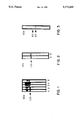
- FIG. 1 demonstrates specificity of antibody-producing clones by Western blotting. Following three separate cell fusions, five antibody-producing clones were identified by ELISA. Western blotting of the culture supernatants of these clones (using Immobilon strips to which human plasma proteins were transblotted from an SDS gel) showed that two clones (lanes 1 and 2) reacted with sgp 120, while the remaining clones (lanes 3, 4, and 5) produced antibodies that bound to a protein of approximately 130 kDa molecular weight.
- FIG. 2 demonstrates Western blot analysis of anti-sgp 120 monoclonal antibody reactivity.
- Human plasma proteins were electrophoretically separated, transblotted onto Immobilon membrane and incubated with B1.9.E-2 culture supernatant (A) and IgG purified from ascites (B). As shown, the monoclonal antibody reacted only with sgp 120.
- FIG. 3 demonstrates reactivity of B1.9.E-2 with sgp 120 fragments.
- Purified sgp 120 was cleaved with kallikrein, fragments separated by SDS-PAGE and blotted onto Immobilon membrane which was subsequently incubated with purified monoclonal IgG at 1:2,000 dilution.
- the monoclonal antibody showed reactivity with 85 kDa and 60 kDa fragments.
- EDTA ethylenediaminetetraacetic acid
- NPGB p-nitrophenylguanidino-benzoate
- SBTI sepsin inhibitor
- GVBS gelatin-containing veronal-buffered saline
- PEG polyethyleneglycol
- PBS phosphate-buffered saline.
- substantially pure means that the product is as pure as can be obtained by standard purification techniques by one of ordinary skill in the art to which this invention belongs.
- mice Female Balb/C mice (Jackson Laboratory, Bar Harbor, ME) were used at the age of 8 weeks.
- sgp 120 was purified according to a modification of the procedure of Hammer et al, supra. Briefly, freshly drawn blood was anti-coagulated with EDTA/NPGB/SBTI solution. sgp 120 was precipitated from plasma with 7.4% to 21.6% (W/V) PEG. Resolubilized protein was applied to a C4b Sepharose column and eluted with GVBS at 15 mS. The 25% PEG cut of the C4b Sepharose eluate was resolubilized and applied to a DEAE-Sephacel column. Recovery of sgp 120 from the column was achieved with a linear salt gradient from 5.6 mS to 15 mS. The protein was about 95% pure as determined by densitometric analysis of an SDS-PAGE gel under non-reducing conditions.
- the amount of antigen for each immunization was about 50 ⁇ g/mouse.
- Three mice were each injected four times at 15-day intervals.
- the antigen emulsified with an equal volume of complete Freund's adjuvant
- the antigen for the first two injections was administered subcutaneously, followed by an intraperitoneal injection of the antigen in incomplete Freund's adjuvant.
- the mice were bled and tested for anti-sgp 120 reactivity by Western blot technique.
- the last boost filter-sterilized sgp 120 in PBS was given by i.v. injection into the tail vein, three days before the fusion.
- mice were killed by cervical dislocation and the spleens were removed aseptically.
- Splenocyte suspension was obtained by perfusing the spleens with the medium (vide infra) through a 25 G needle stuck into the upper pole of the spleen.
- Mouse lymphocytes were isolated by passing the splenocyte suspension over Lympholyte M solution (Catalog # CL5030, Cedarline Laboratories).
- Non-secreting myeloma cells Sp2/0 (American Type Culture Collection, Rockville, Md.) in exponential growth phase and over 95% viable, were used as a fusion partner.
- the isolated primed lymphocytes were subsequently mixed with myeloma cells at a ratio of 1:8 (myeloma cells: lymphocytes) and fused in 50% (w/v) polyethyleneglycol M.W. 1,500 (Aldrich Chemical Company, Inc., Milwaukee, Wis.).
- the suspended cells were then plated out in 5 flatbottomed microtiter plates (Nunc, Roskilde, Denmark) in a 200 ⁇ l/well volume in HAT medium together with 65 ⁇ 10 6 fresh Balb/c thymocytes/ml. The plates were incubated at 37° C. in 5% CO 2 and screened microscopically for growing clones every day after the seventh day of culture.
- the cell culture supernatants were tested for specific reactivity against the immunogen by ELISA assay.
- Antibody producing hybridomas were transferred to 24 well plates. The clones were grown in larger wells until hybridomas covered the whole bottom of the well. Then, 100 ⁇ l samples of supernatants were collected and tested by Western blot.
- One of the clones was subcloned by means of two subsequent limiting dilutions at a concentration of about 0.5 cells/well. The remaining positive clones were frozen in DMSO medium at -70° C.
- the subcloned cell line was expanded and grown as ascites tumor in the pristane-primed peritoneal cavities of Balb/c mice. IgG was purified from ascites by octanoic acid precipitation and using FPLC chromatography (MonoQ, ion-exchange column).
- Class and subclass of the monoclonal antibody were determined by ELISA assay using monospecific rabbit antibodies against mouse IgG 1 , IgG 2a , IgG 2b , IgG 3 , IgA, IgM, kappa and lambda light chains (BIO-RAD Mouse-Typer sub-isotyping kit, Richmond, Calif.).
- DMEM Dulbecco's modified Eagles medium
- glutamine 10 mM
- penicillin 100 U/ml
- streptomycin 100 ⁇ g/ml
- 2-mercaptoethanol 5 ⁇ 10 -5 M
- non-essential amino acids 10 mM
- sodium pyruvate 100 mM
- HEPES buffer 0.18 mM
- Fetal Bovine Serum Hyclone Laboratories, Logan, Utah.
- Selection (HAT) medium contained in addition 1 ⁇ 10 -4 M hypoxanthine, 1.6 ⁇ 10 -5 M thymidine and 4 ⁇ 10 -7 M aminopterin.
- Freezing medium contained 60% HT medium, 30% Fetal Bovine Serum and 10% DMSO (dimethylsulphoxide).
- SDS-PAGE and Immunoblotting SDS-PAGE analysis was performed as described by Maizel, Methods Virol. 5:179-246, 1971, on 7.5% polyacrylamide mini-slab gels. Protein bands were stained with Coomasie blue. Electrophoretically separated proteins were transferred onto Immobilon membranes (Millipore Corporation, Bedford, Mass.) using a semi-dry electro-transblotter (Janssen Life Sciences, Piscataway, N.J.). Unreacted sites on the membranes were blocked with 5% dry skimmed milk solution in PBS (BLOTTO) for 1 hour at RT.
- the membranes were then incubated for 1 hour at RT with culture supernatants diluted 1:10 or purified monoclonal Ig at 1:2,000 in BLOTTO. After three washes with BLOTTO, Immobilon membranes were incubated with gold labeled goat-anti mouse IgG (Janssen Life Sciences, Piscataway, N.J.) for several hours to overnight at RT. After the final three washes in PBS, bands were visualized by incubating the membranes with silver enhancement solution (Catalog # B-2430, Janssen Life Sciences, Piscataway, N.J.).
- B1.9.E-2 Specificity of B1.9.E-2.
- the desired monoclonal antibody designated B1.9.E-22
- B1.9.E-2 was characterized with respect to specificity in indirect ELISA and finally by immunoblotting on SDS gels of human plasma proteins. Both culture supernatant and fractionated ascites showed reactivity only with sgp 120 (FIG. 2).
- a culture of hybridoma B1.9.E-2 was deposited on Mar. 2, 1992 in the American Type Culture Collection (ATTC), 12301 Parklawn Drive, Rockville, Md. 20852, and was given accession number HB 10978. All restrictions on the availability to the public to the culture so deposited will be irrevocably removed upon the granting of the patent.
- Monclonal antibody B1.9.E-2 is of IgG 1 /kappa isotype, as determined by ELISA Mouse-Typer sub-isotyping kit (BIO-RAD).
- sgp 120 specifically cleaved with kallikrein was transferred to Immobilon and incubated with purified monoclonal IgG at 1:2,000 dilution. As shown in FIG. 3, the reactivity is evident only with 85 kDa and 60 kDa fragments, indicating that the epitope to which the monoclonal antibody binds is within the N-terminal portion of sgp 120.
- B1.9.E-2 monoclonal antibody B1.9.E-2 now allows simple purification of substantial amounts of sgp 120 by using standard immunoaffinity chromatographic techniques well known to one of ordinary skill in the art. Furthermore, by employing simple immunological, histological, radiological techniques and combinations thereof, the presence of sgp 120 can be easily detected and localized in biological samples such as cellular components, tissues, plasma and the like, both in vitro and in vivo. In addition, the specific binding property of B1.9.E-2 can be utilized to control the activity of sgp 120 by neutralizing the same with the antibodies.
- kits for detecting the presence of sgp 120 in a biological sample comprises a container containing anti-sgp 120 monoclonal antibody.
- a composition of matter in accordance with the present invention comprises sufficient amount of B1.9.E-2 monoclonal antibody to react with or inactivate sgp 120 in a suitable medium or carrier such as physiological saline, non-toxic buffer, pharmaceutically acceptable vehicle and the like as are well known to one of ordinary skill in the art.
Landscapes
- Health & Medical Sciences (AREA)
- Life Sciences & Earth Sciences (AREA)
- Chemical & Material Sciences (AREA)
- Molecular Biology (AREA)
- Immunology (AREA)
- Organic Chemistry (AREA)
- Engineering & Computer Science (AREA)
- Biochemistry (AREA)
- General Health & Medical Sciences (AREA)
- Proteomics, Peptides & Aminoacids (AREA)
- Medicinal Chemistry (AREA)
- Cell Biology (AREA)
- Biomedical Technology (AREA)
- Biophysics (AREA)
- Urology & Nephrology (AREA)
- Genetics & Genomics (AREA)
- Hematology (AREA)
- Microbiology (AREA)
- Biotechnology (AREA)
- Toxicology (AREA)
- Zoology (AREA)
- Gastroenterology & Hepatology (AREA)
- Food Science & Technology (AREA)
- Physics & Mathematics (AREA)
- Analytical Chemistry (AREA)
- General Physics & Mathematics (AREA)
- Pathology (AREA)
- Preparation Of Compounds By Using Micro-Organisms (AREA)
Abstract
Description
Claims (6)
Priority Applications (1)
| Application Number | Priority Date | Filing Date | Title |
|---|---|---|---|
| US07/365,772 US5171663A (en) | 1989-06-14 | 1989-06-14 | Monoclonal antibody against regulatory protein, sgp 120 |
Applications Claiming Priority (1)
| Application Number | Priority Date | Filing Date | Title |
|---|---|---|---|
| US07/365,772 US5171663A (en) | 1989-06-14 | 1989-06-14 | Monoclonal antibody against regulatory protein, sgp 120 |
Publications (1)
| Publication Number | Publication Date |
|---|---|
| US5171663A true US5171663A (en) | 1992-12-15 |
Family
ID=23440295
Family Applications (1)
| Application Number | Title | Priority Date | Filing Date |
|---|---|---|---|
| US07/365,772 Expired - Lifetime US5171663A (en) | 1989-06-14 | 1989-06-14 | Monoclonal antibody against regulatory protein, sgp 120 |
Country Status (1)
| Country | Link |
|---|---|
| US (1) | US5171663A (en) |
Cited By (1)
| Publication number | Priority date | Publication date | Assignee | Title |
|---|---|---|---|---|
| US5695760A (en) * | 1995-04-24 | 1997-12-09 | Boehringer Inglehiem Pharmaceuticals, Inc. | Modified anti-ICAM-1 antibodies and their use in the treatment of inflammation |
-
1989
- 1989-06-14 US US07/365,772 patent/US5171663A/en not_active Expired - Lifetime
Non-Patent Citations (6)
| Title |
|---|
| Hammer et al., "Isolation and Characterization of a Novel Plasma Protein Which Binds to Activated C4 of the Classical Complement Pathway" J. Biol. Chem. 264(4):2283-2291 (Feb. 5, 1989). |
| Hammer et al., Isolation and Characterization of a Novel Plasma Protein Which Binds to Activated C4 of the Classical Complement Pathway J. Biol. Chem. 264(4):2283 2291 (Feb. 5, 1989). * |
| K hler and Milstein, Continuous Cultures of Fused Cells Secreting Antibody of Predefined Specificity Nature 256:495 497. (1975). * |
| Kohler and Milstein, "Continuous Cultures of Fused Cells Secreting Antibody of Predefined Specificity" Nature 256:495-497. (1975). |
| Sevier et al., "Monodonal Antibodies in Clinical Immunology" Clin. Chem. 27(11):1797-1806 (1981). |
| Sevier et al., Monodonal Antibodies in Clinical Immunology Clin. Chem. 27(11):1797 1806 (1981). * |
Cited By (1)
| Publication number | Priority date | Publication date | Assignee | Title |
|---|---|---|---|---|
| US5695760A (en) * | 1995-04-24 | 1997-12-09 | Boehringer Inglehiem Pharmaceuticals, Inc. | Modified anti-ICAM-1 antibodies and their use in the treatment of inflammation |
Similar Documents
| Publication | Publication Date | Title |
|---|---|---|
| US5955317A (en) | Antibodies to β-amyloids or their derivatives and use thereof | |
| US5750349A (en) | Antibodies to β-amyloids or their derivatives and use thereof | |
| EP0205405B1 (en) | Purified immunoglobulin-related factor, novel monoclonal antibodies, hybridoma cell lines, processes and applications | |
| KR100248315B1 (en) | Monoclonal Antibodies Recognizing the Carboxyl Terminus of Human Cerebral Natriuretic Peptides | |
| NO165556B (en) | MONOCLONAL ANTIBODY AND DERIVATIVES THEREOF WITH HIGH EFFICIENCY TO HUMAN PURE AND STRUCTURALLY LIKE PURE IN IN VITRO APPLICATION. | |
| US5230999A (en) | Monoclonal antibody to endothelin-3 or precursor thereof and use thereof | |
| EP0522159B1 (en) | Antibody to pituitary adenylate cyclase activating peptide-pacap, hybridoma and assay for pacap | |
| JPH06217789A (en) | Monoclonal antibody for human macrophage migration inhibiting factor | |
| JP4374316B2 (en) | Antibody to β-amyloid or a derivative thereof and use thereof | |
| CA2058041A1 (en) | Anti-igf-ii monoclonal antibody | |
| EP0331100A1 (en) | Anti-endothelin antibodies and their use | |
| EP0380443B1 (en) | Monoclonal antibodies specific for thrombin hirudin | |
| US5171663A (en) | Monoclonal antibody against regulatory protein, sgp 120 | |
| EP0287397B1 (en) | Monoclonal antibody specific to human pancreatic phospholipase A2 | |
| EP0345811B1 (en) | Monoclonal abtibodies specific for human fibrinopeptide A | |
| JP3754611B2 (en) | Human aging marker and stress marker test method | |
| US5272059A (en) | Monoclonal antibodies specific for hirudin | |
| EP0479721B1 (en) | Monoclonal antibodies directed against complexes formed by thrombin and thrombin inhibitors | |
| CA2141025C (en) | Anti-mucus glycoprotein monoclonal antibody | |
| US6133427A (en) | Anti-human calcitonin monoclonal antibodies and an immunoassay utilizing said antibodies | |
| EP0417298A1 (en) | Detection of human tissue factor activator | |
| US5843676A (en) | Purified immunoglobulin-related factor, novel monoclonal antibodies, hybridoma cell lines, processes and applications | |
| Chardin et al. | Characterization of axolotl heavy and light immunoglobulin chains by monoclonal antibodies | |
| US5141865A (en) | Monoclonal antibodies which bind thromboxane A2 receptor antagonists and diagnostic methods based thereon | |
| Basta et al. | Production and characterization of the monoclonal antibody against SGP120, a novel serum protein |
Legal Events
| Date | Code | Title | Description |
|---|---|---|---|
| AS | Assignment |
Owner name: UNITED STATES OF AMERICA, THE, AS REPRESENTED BY T Free format text: ASSIGNMENT OF ASSIGNORS INTEREST.;ASSIGNORS:BASTA, MILAN;HAMMER, CARL H.;FRANK, MICHAEL M.;REEL/FRAME:005090/0293 Effective date: 19890609 |
|
| STCF | Information on status: patent grant |
Free format text: PATENTED CASE |
|
| FPAY | Fee payment |
Year of fee payment: 4 |
|
| REMI | Maintenance fee reminder mailed | ||
| FEPP | Fee payment procedure |
Free format text: PAYOR NUMBER ASSIGNED (ORIGINAL EVENT CODE: ASPN); ENTITY STATUS OF PATENT OWNER: LARGE ENTITY |
|
| FPAY | Fee payment |
Year of fee payment: 8 |
|
| FEPP | Fee payment procedure |
Free format text: PAYOR NUMBER ASSIGNED (ORIGINAL EVENT CODE: ASPN); ENTITY STATUS OF PATENT OWNER: LARGE ENTITY Free format text: PAYER NUMBER DE-ASSIGNED (ORIGINAL EVENT CODE: RMPN); ENTITY STATUS OF PATENT OWNER: LARGE ENTITY |
|
| FPAY | Fee payment |
Year of fee payment: 12 |