US5164186A - Drug-releaser - Google Patents
Drug-releaser Download PDFInfo
- Publication number
- US5164186A US5164186A US07/617,376 US61737690A US5164186A US 5164186 A US5164186 A US 5164186A US 61737690 A US61737690 A US 61737690A US 5164186 A US5164186 A US 5164186A
- Authority
- US
- United States
- Prior art keywords
- medicine
- drug
- tumor
- particles
- calcium phosphate
- Prior art date
- Legal status (The legal status is an assumption and is not a legal conclusion. Google has not performed a legal analysis and makes no representation as to the accuracy of the status listed.)
- Expired - Fee Related
Links
- 239000003814 drug Substances 0.000 claims abstract description 50
- 239000002245 particle Substances 0.000 claims abstract description 30
- 239000001506 calcium phosphate Substances 0.000 claims abstract description 14
- 235000011010 calcium phosphates Nutrition 0.000 claims abstract description 14
- QORWJWZARLRLPR-UHFFFAOYSA-H tricalcium bis(phosphate) Chemical compound [Ca+2].[Ca+2].[Ca+2].[O-]P([O-])([O-])=O.[O-]P([O-])([O-])=O QORWJWZARLRLPR-UHFFFAOYSA-H 0.000 claims abstract description 14
- 229910000389 calcium phosphate Inorganic materials 0.000 claims abstract description 13
- AOJJSUZBOXZQNB-TZSSRYMLSA-N Doxorubicin Chemical compound O([C@H]1C[C@@](O)(CC=2C(O)=C3C(=O)C=4C=CC=C(C=4C(=O)C3=C(O)C=21)OC)C(=O)CO)[C@H]1C[C@H](N)[C@H](O)[C@H](C)O1 AOJJSUZBOXZQNB-TZSSRYMLSA-N 0.000 claims description 54
- 206010028980 Neoplasm Diseases 0.000 claims description 29
- 229940009456 adriamycin Drugs 0.000 claims description 27
- 210000001367 artery Anatomy 0.000 claims description 17
- 229940079593 drug Drugs 0.000 claims description 16
- 238000000034 method Methods 0.000 claims description 9
- 239000000203 mixture Substances 0.000 claims description 9
- 238000001354 calcination Methods 0.000 claims description 6
- 239000011575 calcium Substances 0.000 claims description 6
- 150000004676 glycans Chemical class 0.000 claims description 3
- 229920001282 polysaccharide Polymers 0.000 claims description 3
- 239000005017 polysaccharide Substances 0.000 claims description 3
- 230000000259 anti-tumor effect Effects 0.000 claims 3
- 239000013543 active substance Substances 0.000 claims 2
- 230000001093 anti-cancer Effects 0.000 claims 2
- 230000000694 effects Effects 0.000 abstract description 14
- 238000002347 injection Methods 0.000 abstract description 5
- 239000007924 injection Substances 0.000 abstract description 5
- 230000001875 tumorinhibitory effect Effects 0.000 abstract description 5
- 229920001661 Chitosan Polymers 0.000 abstract description 4
- 229920002101 Chitin Polymers 0.000 abstract description 3
- WOBHKFSMXKNTIM-UHFFFAOYSA-N Hydroxyethyl methacrylate Chemical compound CC(=C)C(=O)OCCO WOBHKFSMXKNTIM-UHFFFAOYSA-N 0.000 abstract description 3
- 229940044192 2-hydroxyethyl methacrylate Drugs 0.000 abstract 1
- 238000002513 implantation Methods 0.000 abstract 1
- 239000000126 substance Substances 0.000 description 12
- 239000011148 porous material Substances 0.000 description 9
- 201000011510 cancer Diseases 0.000 description 7
- 239000000463 material Substances 0.000 description 6
- 241000699670 Mus sp. Species 0.000 description 5
- 210000004369 blood Anatomy 0.000 description 5
- 239000008280 blood Substances 0.000 description 5
- 210000004027 cell Anatomy 0.000 description 4
- 102000008186 Collagen Human genes 0.000 description 3
- 108010035532 Collagen Proteins 0.000 description 3
- 239000004372 Polyvinyl alcohol Substances 0.000 description 3
- 229910052586 apatite Inorganic materials 0.000 description 3
- 229910000394 calcium triphosphate Inorganic materials 0.000 description 3
- 229920001436 collagen Polymers 0.000 description 3
- 210000004185 liver Anatomy 0.000 description 3
- VSIIXMUUUJUKCM-UHFFFAOYSA-D pentacalcium;fluoride;triphosphate Chemical compound [F-].[Ca+2].[Ca+2].[Ca+2].[Ca+2].[Ca+2].[O-]P([O-])([O-])=O.[O-]P([O-])([O-])=O.[O-]P([O-])([O-])=O VSIIXMUUUJUKCM-UHFFFAOYSA-D 0.000 description 3
- 229920002451 polyvinyl alcohol Polymers 0.000 description 3
- 235000019422 polyvinyl alcohol Nutrition 0.000 description 3
- MHAJPDPJQMAIIY-UHFFFAOYSA-N Hydrogen peroxide Chemical compound OO MHAJPDPJQMAIIY-UHFFFAOYSA-N 0.000 description 2
- 241000699666 Mus <mouse, genus> Species 0.000 description 2
- 238000010521 absorption reaction Methods 0.000 description 2
- 238000002583 angiography Methods 0.000 description 2
- 210000004204 blood vessel Anatomy 0.000 description 2
- 239000002131 composite material Substances 0.000 description 2
- 230000000968 intestinal effect Effects 0.000 description 2
- 230000005923 long-lasting effect Effects 0.000 description 2
- RFWLACFDYFIVMC-UHFFFAOYSA-D pentacalcium;[oxido(phosphonatooxy)phosphoryl] phosphate Chemical compound [Ca+2].[Ca+2].[Ca+2].[Ca+2].[Ca+2].[O-]P([O-])(=O)OP([O-])(=O)OP([O-])([O-])=O.[O-]P([O-])(=O)OP([O-])(=O)OP([O-])([O-])=O RFWLACFDYFIVMC-UHFFFAOYSA-D 0.000 description 2
- 102000004169 proteins and genes Human genes 0.000 description 2
- 108090000623 proteins and genes Proteins 0.000 description 2
- 238000010926 purge Methods 0.000 description 2
- 210000001519 tissue Anatomy 0.000 description 2
- 239000004604 Blowing Agent Substances 0.000 description 1
- 102100026735 Coagulation factor VIII Human genes 0.000 description 1
- 101000911390 Homo sapiens Coagulation factor VIII Proteins 0.000 description 1
- 206010061218 Inflammation Diseases 0.000 description 1
- 238000009825 accumulation Methods 0.000 description 1
- 239000002246 antineoplastic agent Substances 0.000 description 1
- 229940041181 antineoplastic drug Drugs 0.000 description 1
- 239000002775 capsule Substances 0.000 description 1
- 150000001720 carbohydrates Chemical class 0.000 description 1
- 210000000170 cell membrane Anatomy 0.000 description 1
- 229910010293 ceramic material Inorganic materials 0.000 description 1
- 239000013522 chelant Substances 0.000 description 1
- 230000003247 decreasing effect Effects 0.000 description 1
- 230000007547 defect Effects 0.000 description 1
- 238000001035 drying Methods 0.000 description 1
- 230000008014 freezing Effects 0.000 description 1
- 238000007710 freezing Methods 0.000 description 1
- 206010073071 hepatocellular carcinoma Diseases 0.000 description 1
- 238000004128 high performance liquid chromatography Methods 0.000 description 1
- 229910052739 hydrogen Inorganic materials 0.000 description 1
- 239000001257 hydrogen Substances 0.000 description 1
- 210000003090 iliac artery Anatomy 0.000 description 1
- 230000002163 immunogen Effects 0.000 description 1
- 230000004054 inflammatory process Effects 0.000 description 1
- 230000002401 inhibitory effect Effects 0.000 description 1
- 230000003993 interaction Effects 0.000 description 1
- 210000003734 kidney Anatomy 0.000 description 1
- 239000003446 ligand Substances 0.000 description 1
- 239000002502 liposome Substances 0.000 description 1
- 201000007270 liver cancer Diseases 0.000 description 1
- 208000014018 liver neoplasm Diseases 0.000 description 1
- 229920002521 macromolecule Polymers 0.000 description 1
- 230000007721 medicinal effect Effects 0.000 description 1
- 210000001616 monocyte Anatomy 0.000 description 1
- 210000003205 muscle Anatomy 0.000 description 1
- 231100000344 non-irritating Toxicity 0.000 description 1
- 231100000252 nontoxic Toxicity 0.000 description 1
- 230000003000 nontoxic effect Effects 0.000 description 1
- 239000005416 organic matter Substances 0.000 description 1
- 210000002966 serum Anatomy 0.000 description 1
- 239000002002 slurry Substances 0.000 description 1
- 239000000243 solution Substances 0.000 description 1
- 230000003068 static effect Effects 0.000 description 1
- 230000001954 sterilising effect Effects 0.000 description 1
- 238000004659 sterilization and disinfection Methods 0.000 description 1
- 230000017105 transposition Effects 0.000 description 1
- 238000005303 weighing Methods 0.000 description 1
Images


Classifications
-
- A—HUMAN NECESSITIES
- A61—MEDICAL OR VETERINARY SCIENCE; HYGIENE
- A61K—PREPARATIONS FOR MEDICAL, DENTAL OR TOILETRY PURPOSES
- A61K9/00—Medicinal preparations characterised by special physical form
- A61K9/0012—Galenical forms characterised by the site of application
- A61K9/0019—Injectable compositions; Intramuscular, intravenous, arterial, subcutaneous administration; Compositions to be administered through the skin in an invasive manner
- A61K9/0024—Solid, semi-solid or solidifying implants, which are implanted or injected in body tissue
-
- A—HUMAN NECESSITIES
- A61—MEDICAL OR VETERINARY SCIENCE; HYGIENE
- A61K—PREPARATIONS FOR MEDICAL, DENTAL OR TOILETRY PURPOSES
- A61K9/00—Medicinal preparations characterised by special physical form
- A61K9/20—Pills, tablets, discs, rods
- A61K9/2004—Excipients; Inactive ingredients
- A61K9/2009—Inorganic compounds
-
- A—HUMAN NECESSITIES
- A61—MEDICAL OR VETERINARY SCIENCE; HYGIENE
- A61K—PREPARATIONS FOR MEDICAL, DENTAL OR TOILETRY PURPOSES
- A61K9/00—Medicinal preparations characterised by special physical form
- A61K9/20—Pills, tablets, discs, rods
- A61K9/2004—Excipients; Inactive ingredients
- A61K9/2022—Organic macromolecular compounds
- A61K9/2027—Organic macromolecular compounds obtained by reactions only involving carbon-to-carbon unsaturated bonds, e.g. polyvinyl pyrrolidone, poly(meth)acrylates
-
- A—HUMAN NECESSITIES
- A61—MEDICAL OR VETERINARY SCIENCE; HYGIENE
- A61K—PREPARATIONS FOR MEDICAL, DENTAL OR TOILETRY PURPOSES
- A61K9/00—Medicinal preparations characterised by special physical form
- A61K9/20—Pills, tablets, discs, rods
- A61K9/2004—Excipients; Inactive ingredients
- A61K9/2022—Organic macromolecular compounds
- A61K9/205—Polysaccharides, e.g. alginate, gums; Cyclodextrin
Definitions
- the present invention relates to a drug-releaser which can carry a medicine to a localized affected part of a living body to permit the medicine to stay there for a relatively long time, thereby causing the medical effect long on the localized part of the living body.
- the curing effect will depend on how long it can stay at that localized affected area.
- the living body has a tendency to purge a medicine as a foreign substance from the body in the possible shortest time. Just after putting a medicine in the living body, the circulating blood starts absorption of the medicine, and the medicine will be purged out of the living body after passing through the kidney or the liver.
- a medicine is put in a selected blood vessel little by little at intervals, thereby keeping the concentration of medicine in the blood at a given constant value.
- a given volume of medicine contained in a liposome capsule is put in the living body through the mouth, and then the encapsulated medicine will be gradually absorbed in the intestinal wall while passing through the intestinal canals in ten-odd hours.
- one object of the present invention is to provide a drug-releaser which is free from the defect of conventional dosing methods as described above, permitting the medicine to stay long enough to make full use of its curing effect on the affected area of the body.
- a drug-releaser comprises a porous body of biocompatibility filled with a medicine.
- This porous body may be embeded to be in direct contact with a selected affected part or in the vicinity of the selected affected part.
- a porous body may take the form of minute particle.
- Medicine-filled minute particles may be put in a selected blood vessel which leads to a selected affected part to be treated.
- the requirements of a drug-releaser according to the present invention are: first, it is non-immunogenic, nontoxic, or non-irritating but is biocompatible, and second, its porosity is large enough to hold a relatively large amount of medicine.
- Inorganic matter hydroxycalciumapatite(HAP), calcium-triphosphate(TCP) and other calcium phosphates;
- porous particles weighing one gram can have total surface area ranging from 50 to 500 m 2 in their open pores, thus providing sufficient area in which as much medicine as required can be absorbed and held.
- the substances in group (1) can become porous by mixing with a blowing agent such as hydrogen peroxide, drying the resultant slurry like mixture and baking the dry mixture.
- the substances in groups (2), (3) and (4) can become porous by mixing evenly with certain mediums, and freezing the mixture to be dry.
- HAP and TCP in group (1) have high-absorbing capabilities in a variety of chemically binding forms, for instance chelate bond, hydrogen bond and quadrapole interaction with respect to protein, sugar, proteoglycan and other macromolecular substances. Porous bodies of these substances are appropriate for the purpose of holding an increased amount of medicine such as polysaccharide (anticancer drug) in their open pores and carrying to affected parts of the living body.
- polysaccharide anticancer drug
- HEMA As for HEMA, PVA, chitin chitosan, collagen and their derivatives: these substances in groups (2), (3) and (4) have hydrophilicity and hydrophobicity in balance on their surfaces. These characteristics along with electro static nature on their surfaces cause effective absorption of protein, dye and saccharide. Thus, porous bodies which are made of these materials, are appropriate for the purpose of holding and carrying an increased amount of medicine to affected parts of the living body.
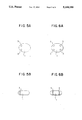
- FIGS. 1 to 6 show different drug-releasers according to the present invention. Particularly, FIGS. 1 (A) to 6 (A) are plane views of the drug-releasers; FIGS. 1 (B) to 3 (B), 5 (B) and 6 (B) are side views of the drug-releasers; and FIG. 4 (B) is a sectional view of the drug-releaser.
- FIG. 7 shows how the ADR concentration in mouse blood varies with time in the chemical treatment using Adriamycin (ADR 1.5 mg/Kg).
- FIG. 8 shows how the tumor size varies with time in the chemical treatment using Adriamycin (ADR 1.5 mg/Kg).
- FIG. 9 shows how the tumor size varies with time in the chemical treatment of selected 15 patients.
- FIGS. 1 and 2 Some examples of porous bodies of the substances described above in groups (1), (2), (3) and (4) are shown in FIGS. 1 and 2.
- the porous disk is subjected to sterilization, and the disk thus sterilized is soaked in a medicine solution.
- a medicine solution When surrounding pressure is decreased, air is replaced by medicine in every pore, and then the medicine is absorbed in the inner wall of the pore.
- a drug-releasing disk 1 results.
- this disk When this disk is implanted in the vicinity of a tumor to be treated, the medicine will continuously apply its effect on the tumor for a relatively long period. Specifically, it ranges from several weeks to several months, depending on the size of the disk, the medicine absorbing-and-holding energy, and other factors.
- Some medicines require the use of ligand, which has an effect to bond the medicine to the inner surface of each pore.
- the porous material of the drug-releasing disk has a very high biocompatibility, and therefore the disk can be held firmly by normal cells or by ingrowthes of intercellularmatrix in the living body. Then, the medicine will be released from every pore to pass through the cell membrane or intercellularmatrix, and arrive at the target cells.
- the medicine can have an inclination to target selected cells when combined with a monoclonal antibody to the selected cells.
- the disk 1 of FIG. 1 has a rounded circumferential edge or else it would hurt surrounding tissue.
- the disk can have a different shape in section as shown in FIG. 2B for the same reason.
- an egg-shaped body 1 as shown in FIG. 3 may be used even more safely.
- Different medicines can be carried by a single drug-releasing disk, thereby making full use of the composite curing effect on the affected parts of the living body.
- a plurality of drug-releasing disks may be implanted in the vicinity of a selected affected part, thereby causing composite curing effects on the same affected part.
- All disks of FIGS. 1 to 6 are several to ten-odd millimeters across. Larger or smaller disks are difficult to be implanted at fixed places in the living body.
- Calcined particles of hydroxycalciumapatite (HAP) or calcium phosphate such as calcium-triphosphate (TCP) may be used. These particles are 10 to 1000 millimicrons across. Particles whose size is below 10 millimicrons across, are easy to be transported by blood when injected in the artery, and therefore there is a fear of accumulation of particles in the kindney. Particles whose size is above 1000 millimicrons across, are difficult to be injected in the artery.
- the P-to-C ratio in the composition of calcium phosphate is preferably equal to or larger than 1.0, but smaller than 2.0 (1.0 ⁇ Ca/P ⁇ 2.0).
- the Ca/P ratio out of the range will make the drug-releasing material easy to melt, lowering the capability of holding and steadily releasing the medicine for an extended period.
- the temperature at which calcium phosphate is calcined preferably ranges from 600 to 1350 degrees Centigrade. Calcination at a temperature below 600 degrees Centigrade is inadequate to form particles, and the calcined material has a poor capability of absorbing macrophage-like monocytes. The calcination at a temperature above 1350 degrees Centigrade will decompose apatite, and therefore no active ceramic material can result.
- drug-releasing particles thus made are injected into an artery leading to a selected tumor such as cancer in the living body.
- the drug-releasing particles reach and stay at the cancer, causing long-lasting curing effect on the cancer, and at the same time, they may accumulate there to block capillary vessels leading to the cancer, thereby preventing the supply of nourishment to the cancer and hence the growth of the cancer.
- Calcium phosphate which was used, was hydroxycalciumapatite (pure apatite; HAP). Its particulars are: calcination temperature: 700 to 800 degrees Centigrade, stoichiometry Ca/P: 1.66 . . . , and average particle size: 50 to 100 millimicrons.
- mice When 2 weeks had passed after grafting tumor on the rear right legs of some selected mice, these mice were separated in two groups. Adriamycin (ADR 1.5 milligrams/kg) was injected in the common iliac arteries on the same side as MethA tumors were grafted in the first group of mice. Likewise, Apatite (HAP) particles filled with Adriamycin (ADR) were arteri-injected in the second group of mice. The ADR concentration was determined on each blood sample which was taken from the fundas venosus plexus of every mouse at intervals (HPLC method), and tumor inhibitory effect was checked.
- ADR 1.5 milligrams/kg was injected in the common iliac arteries on the same side as MethA tumors were grafted in the first group of mice.
- Apatite (HAP) particles filled with Adriamycin (ADR) were arteri-injected in the second group of mice.
- the ADR concentration was determined on each blood sample which was taken from the fundas venosus
- the ADR concentrations in serum reached peak values 2 minutes after the artery injections in the first and second groups.
- the ADR concentration in the HAP+ ADR artery-injected group was significantly low (p ⁇ 0.005), compared with that in the ADR artery-injected group (See FIG. 7).
- the ADR concentration in tumor was significantly high in the HAP+ADR artery-injected group (p ⁇ 0.001): The HAP particles appeared to have functioned as micro-blockader, therby causing the tumor to hold an increased concentration of ADR (See Table 1).
- HAP particles filled with ADR (20 to 50 milligrams) were used.
- the safety injection of HAP-ADR into the stomachduodenum artery was not assured, and therfore necessary injections were effected in the liver artery and selected arteries on the side of the liver. Only one treatment was effected.
- the curing effect was checked in terms of reduction of the tumor area mainly with the aid of angiography, and also the ultrasonic diagonosis state was taken into consideration.
Landscapes
- Health & Medical Sciences (AREA)
- Chemical & Material Sciences (AREA)
- Public Health (AREA)
- Life Sciences & Earth Sciences (AREA)
- Veterinary Medicine (AREA)
- General Health & Medical Sciences (AREA)
- Medicinal Chemistry (AREA)
- Pharmacology & Pharmacy (AREA)
- Epidemiology (AREA)
- Animal Behavior & Ethology (AREA)
- Neurosurgery (AREA)
- Biomedical Technology (AREA)
- Engineering & Computer Science (AREA)
- Dermatology (AREA)
- Inorganic Chemistry (AREA)
- Medicinal Preparation (AREA)
- Medicines That Contain Protein Lipid Enzymes And Other Medicines (AREA)
- Pharmaceuticals Containing Other Organic And Inorganic Compounds (AREA)
Abstract
Disclosed is a drug-releaser comprising a porous body of biocompatibility filled with a medicine. Specifically, such porous body is made of baked calcium phosphate, 2-hydroxyethylmethacrylate, chitin, chitosan or their delivatives. Implantation or artery-injection of such drug-releaser causes a discernible curing effect on affected parts of the living body without causing any irritativeness. The steady release of the medicine from HAP particles to affected parts and the blockade of surrounding capillary vessels leading to such affected parts causes a remarkable tumor inhibitory effect.
Description
This application is a divisional of application Ser. No. 07/257,841 filed on Oct. 14, 1988 now abandoned.
1. Field of the Invention
The present invention relates to a drug-releaser which can carry a medicine to a localized affected part of a living body to permit the medicine to stay there for a relatively long time, thereby causing the medical effect long on the localized part of the living body.
2. Description of the Prior Art
In case that a medicine is applied to inflammation or tumor, the curing effect will depend on how long it can stay at that localized affected area. Generally speaking, the living body has a tendency to purge a medicine as a foreign substance from the body in the possible shortest time. Just after putting a medicine in the living body, the circulating blood starts absorption of the medicine, and the medicine will be purged out of the living body after passing through the kidney or the liver.
In order to keep the curing effect long lasting, it is necessary to retain the medicine in the vicinity of affected area as long as possible to prevent the prompt purge of the medicine from the living body. In an attempt to meet such a need a variety of dosing methods and drug-releasing materials have been proposed and actually used.
As one example, a medicine is put in a selected blood vessel little by little at intervals, thereby keeping the concentration of medicine in the blood at a given constant value. As another example, a given volume of medicine contained in a liposome capsule is put in the living body through the mouth, and then the encapsulated medicine will be gradually absorbed in the intestinal wall while passing through the intestinal canals in ten-odd hours. These conventional dosing methods, however, cannot permit the medicine to stay in the vicinity of the affected areas of the living body, such as tumors, and therefore, the medicine cannot exercise its curing effect on the affected part to its maximum capability.
In view of the above, one object of the present invention is to provide a drug-releaser which is free from the defect of conventional dosing methods as described above, permitting the medicine to stay long enough to make full use of its curing effect on the affected area of the body.
To attain this object a drug-releaser according to the present invention comprises a porous body of biocompatibility filled with a medicine. This porous body may be embeded to be in direct contact with a selected affected part or in the vicinity of the selected affected part. A porous body may take the form of minute particle. Medicine-filled minute particles may be put in a selected blood vessel which leads to a selected affected part to be treated. The requirements of a drug-releaser according to the present invention are: first, it is non-immunogenic, nontoxic, or non-irritating but is biocompatible, and second, its porosity is large enough to hold a relatively large amount of medicine.
Materials to meet such requirements are:
(1) Inorganic matter: hydroxycalciumapatite(HAP), calcium-triphosphate(TCP) and other calcium phosphates;
(2) Organic matter: 2-hydroxyethylmethacrylate(HEMA) or polyvinylalcohol(PVA);
(3) Natural macromolecular matter: chitin or chitosan and chitosan delivatives;and
(4) Natural macromolecular matter: collagen and collagen delivatives
These substances have a biocompatibility, and can be prepared to be porous. In fact, such porous particles weighing one gram can have total surface area ranging from 50 to 500 m2 in their open pores, thus providing sufficient area in which as much medicine as required can be absorbed and held.
Specifically, the substances in group (1) can become porous by mixing with a blowing agent such as hydrogen peroxide, drying the resultant slurry like mixture and baking the dry mixture. The substances in groups (2), (3) and (4) can become porous by mixing evenly with certain mediums, and freezing the mixture to be dry.
HAP and TCP in group (1) have high-absorbing capabilities in a variety of chemically binding forms, for instance chelate bond, hydrogen bond and quadrapole interaction with respect to protein, sugar, proteoglycan and other macromolecular substances. Porous bodies of these substances are appropriate for the purpose of holding an increased amount of medicine such as polysaccharide (anticancer drug) in their open pores and carrying to affected parts of the living body.
As for HEMA, PVA, chitin chitosan, collagen and their derivatives: these substances in groups (2), (3) and (4) have hydrophilicity and hydrophobicity in balance on their surfaces. These characteristics along with electro static nature on their surfaces cause effective absorption of protein, dye and saccharide. Thus, porous bodies which are made of these materials, are appropriate for the purpose of holding and carrying an increased amount of medicine to affected parts of the living body.
FIGS. 1 to 6 show different drug-releasers according to the present invention. Particularly, FIGS. 1 (A) to 6 (A) are plane views of the drug-releasers; FIGS. 1 (B) to 3 (B), 5 (B) and 6 (B) are side views of the drug-releasers; and FIG. 4 (B) is a sectional view of the drug-releaser.
FIG. 7 shows how the ADR concentration in mouse blood varies with time in the chemical treatment using Adriamycin (ADR 1.5 mg/Kg).
FIG. 8 shows how the tumor size varies with time in the chemical treatment using Adriamycin (ADR 1.5 mg/Kg).
FIG. 9 shows how the tumor size varies with time in the chemical treatment of selected 15 patients.
Some examples of porous bodies of the substances described above in groups (1), (2), (3) and (4) are shown in FIGS. 1 and 2.
A drug-releasing porous disk 1 of FIG. 1 is 1 millimeter thick and 5 millimeters across, and has open pores. The pore size ranges from 50 to 500 millimicrons.
First, the porous disk is subjected to sterilization, and the disk thus sterilized is soaked in a medicine solution. When surrounding pressure is decreased, air is replaced by medicine in every pore, and then the medicine is absorbed in the inner wall of the pore. Thus, a drug-releasing disk 1 results. When this disk is implanted in the vicinity of a tumor to be treated, the medicine will continuously apply its effect on the tumor for a relatively long period. Specifically, it ranges from several weeks to several months, depending on the size of the disk, the medicine absorbing-and-holding energy, and other factors.
Some medicines require the use of ligand, which has an effect to bond the medicine to the inner surface of each pore. The porous material of the drug-releasing disk has a very high biocompatibility, and therefore the disk can be held firmly by normal cells or by ingrowthes of intercellularmatrix in the living body. Then, the medicine will be released from every pore to pass through the cell membrane or intercellularmatrix, and arrive at the target cells. The medicine can have an inclination to target selected cells when combined with a monoclonal antibody to the selected cells.
The disk 1 of FIG. 1 has a rounded circumferential edge or else it would hurt surrounding tissue. The disk can have a different shape in section as shown in FIG. 2B for the same reason. Also, an egg-shaped body 1 as shown in FIG. 3 may be used even more safely.
A thin body 1 of FIG. 4 has a small hole 2 for the purpose of facilitating the binding to tissue. Thin bodies of FIGS. 5 and 6 have reentracies 3 for the same purpose.
Different medicines can be carried by a single drug-releasing disk, thereby making full use of the composite curing effect on the affected parts of the living body. A plurality of drug-releasing disks may be implanted in the vicinity of a selected affected part, thereby causing composite curing effects on the same affected part.
All disks of FIGS. 1 to 6 are several to ten-odd millimeters across. Larger or smaller disks are difficult to be implanted at fixed places in the living body.
Next, some examples of making minute particles from drug-releasing material are given.
Calcined particles of hydroxycalciumapatite (HAP) or calcium phosphate such as calcium-triphosphate (TCP) may be used. These particles are 10 to 1000 millimicrons across. Particles whose size is below 10 millimicrons across, are easy to be transported by blood when injected in the artery, and therefore there is a fear of accumulation of particles in the kindney. Particles whose size is above 1000 millimicrons across, are difficult to be injected in the artery.
The P-to-C ratio in the composition of calcium phosphate is preferably equal to or larger than 1.0, but smaller than 2.0 (1.0≦Ca/P<2.0). The Ca/P ratio out of the range will make the drug-releasing material easy to melt, lowering the capability of holding and steadily releasing the medicine for an extended period.
The temperature at which calcium phosphate is calcined, preferably ranges from 600 to 1350 degrees Centigrade. Calcination at a temperature below 600 degrees Centigrade is inadequate to form particles, and the calcined material has a poor capability of absorbing macrophage-like monocytes. The calcination at a temperature above 1350 degrees Centigrade will decompose apatite, and therefore no active ceramic material can result.
Assume that drug-releasing particles thus made are injected into an artery leading to a selected tumor such as cancer in the living body. The drug-releasing particles reach and stay at the cancer, causing long-lasting curing effect on the cancer, and at the same time, they may accumulate there to block capillary vessels leading to the cancer, thereby preventing the supply of nourishment to the cancer and hence the growth of the cancer.
Some examples of applying drug-releasing particles to experimental tumors and malignant tumors in human libers are given below:
The curing effect of drug-releasing particles was studied on MethA tumors which were grafted in the muscles of the rear right legs of some selected mice.
Calcium phosphate which was used, was hydroxycalciumapatite (pure apatite; HAP). Its particulars are: calcination temperature: 700 to 800 degrees Centigrade, stoichiometry Ca/P: 1.66 . . . , and average particle size: 50 to 100 millimicrons.
When 2 weeks had passed after grafting tumor on the rear right legs of some selected mice, these mice were separated in two groups. Adriamycin (ADR 1.5 milligrams/kg) was injected in the common iliac arteries on the same side as MethA tumors were grafted in the first group of mice. Likewise, Apatite (HAP) particles filled with Adriamycin (ADR) were arteri-injected in the second group of mice. The ADR concentration was determined on each blood sample which was taken from the fundas venosus plexus of every mouse at intervals (HPLC method), and tumor inhibitory effect was checked.
The ADR concentrations in serum reached peak values 2 minutes after the artery injections in the first and second groups. The ADR concentration in the HAP+ ADR artery-injected group was significantly low (p<0.005), compared with that in the ADR artery-injected group (See FIG. 7).
The ADR concentration in tumor was significantly high in the HAP+ADR artery-injected group (p<0.001): The HAP particles appeared to have functioned as micro-blockader, therby causing the tumor to hold an increased concentration of ADR (See Table 1).
No tumor inhibitory effect was found in the ADR artery-injected group (1.5 mg/kg), whereas a remarkable inhibitory effect was found in the HAP+ADR artery-injected group. A discernible tumor inhibitory effect was found in the HAP artery-injected group (See FIG. 8).
Selected were 15 patients whose liver cancer could not be treated by surgical operation.
The same calcium phosphate as used in the above examples, was used. Developing tumors were studied by angiography and CT according to the hepatoma dealing standard. Selected were 5 samples in 2 areas; 3 samples in 3 areas; and 7 samples in 4 areas and remote transpositions.
7 samples thus selected were block; 6 samples were tuberous; and 2 samples were infiltrative.
The HAP particles filled with ADR (20 to 50 milligrams) were used. The safety injection of HAP-ADR into the stomacduodenum artery was not assured, and therfore necessary injections were effected in the liver artery and selected arteries on the side of the liver. Only one treatment was effected.
The curing effect was checked in terms of reduction of the tumor area mainly with the aid of angiography, and also the ultrasonic diagonosis state was taken into consideration.
7 examples showed tumor reduction of 50 or more percent(PR); 4 examples showed tumor reduction of 20 to 50 percent(MR); 4 examples showed tumor reduction of 25 or less percent (NC); and no examples showed tumor enlargement of 25 or more percent (PD). Briefly speaking, the curing effect which was equal to or greater than PR, was 47 percent, and that which was equal to or greater than MR, was 73 percent (See FIG. 9).
As is apparent from the above, only one artery-injection of baked particles of hydroxycalciumapatite or calcium-triphosphate caused a discernible curing effect on affected parts of the living body without causing any irritativeness. The steady release of the medicine from HAP particles to affected parts and the blockade of surrounding capillary vessels caused a remarkable tumor inhibitory effect.
              TABLE 1
______________________________________
The ADR concentration in tumor one hour after the chemical
blockade treatment using ADR (1.5 mg/kg)
                 ADR concentration
chemical treatment
                 (μg/g)
______________________________________
ADR alone         4.8
HAP + ADR blockade
                 11.6
______________________________________
Claims (5)
1. A method of treating a selected area of a body which comprises injecting an effective amount of a drug releasing composition which comprises
(a) particles of baked calcium phosphate having an average particle size of from 10 to 1000 μm and wherein the Ca/P ratio is greater than or equal to 1 and less than 2 and wherein the calcination temperature of said calcium phosphate is in the range of from 600° to 1350° C.; and
(b) a medicine dispersed through said particles into an artery leading to said selected area of the body to be treated.
2. The method according to claim 1, wherein said selected area of the body to be treated is a tumor.
3. The method according to claim 1 wherein said baked calcium phosphate is hydroxycalciumapatite.
4. A method of treating a tumor in a selected area of the body which comprises injecting an anti-tumor effective amount of a polysaccharide anticancer active agent-releasing composition into an artery leading to said selected area of the body to be treated, said composition comprising:
(a) particles of baked calcium phosphate having an average particle size of from 10 to 1,000 μm and wherein the Ca/P ratio is greater than or equal to 1 and less than 2 and wherein the calcination temperature of said calcium phosphate is in the range of from 600° to 1350° C.; and
(b) an anti-tumor effective amount of a polysaccharide anticancer active agent dispersed through said particle.
5. A method of treating a tumor in a selected area of the body which comprises injecting an adriamycin-releasing composition into an artery leading to said selected area of the body to be treated, said composition comprising:
(a) particles of baked calcium phosphate having an average particle size of from 10 to 1,000 μm and wherein the Ca/P ratio is greater than or equal to 1 and less than 2 and wherein the calcination temperature of said calcium phosphate is in the range of from 600° to 1350° C.; and
(b) an anti-tumor effective amount of adriamycin dispersed through said particles.
Priority Applications (1)
| Application Number | Priority Date | Filing Date | Title |
|---|---|---|---|
| US07/617,376 US5164186A (en) | 1988-10-13 | 1990-11-21 | Drug-releaser |
Applications Claiming Priority (3)
| Application Number | Priority Date | Filing Date | Title |
|---|---|---|---|
| DE19883834944 DE3834944A1 (en) | 1988-10-13 | 1988-10-13 | Drug-release device |
| US25784188A | 1988-10-14 | 1988-10-14 | |
| US07/617,376 US5164186A (en) | 1988-10-13 | 1990-11-21 | Drug-releaser |
Related Parent Applications (1)
| Application Number | Title | Priority Date | Filing Date |
|---|---|---|---|
| US25784188A Division | 1988-10-13 | 1988-10-14 |
Publications (1)
| Publication Number | Publication Date |
|---|---|
| US5164186A true US5164186A (en) | 1992-11-17 |
Family
ID=27198369
Family Applications (1)
| Application Number | Title | Priority Date | Filing Date |
|---|---|---|---|
| US07/617,376 Expired - Fee Related US5164186A (en) | 1988-10-13 | 1990-11-21 | Drug-releaser |
Country Status (1)
| Country | Link |
|---|---|
| US (1) | US5164186A (en) |
Cited By (14)
| Publication number | Priority date | Publication date | Assignee | Title |
|---|---|---|---|---|
| US5541234A (en) * | 1991-12-20 | 1996-07-30 | Alliedsignal Inc. | Process for making low density hydrogel materials having high surface areas |
| US5704910A (en) * | 1995-06-05 | 1998-01-06 | Nephros Therapeutics, Inc. | Implantable device and use therefor |
| US6602523B1 (en) * | 2000-08-17 | 2003-08-05 | Technology Holding, Llc. | Composite material and process for increasing bioavailability and activity of a beneficial agent |
| US20030204161A1 (en) * | 2002-04-25 | 2003-10-30 | Bozidar Ferek-Petric | Implantable electroporation therapy device and method for using same |
| GB2399499A (en) * | 2003-03-13 | 2004-09-22 | Nanotrend Ino Tech Inc | A porous apatite grain taste-masked oral dosage form |
| WO2005025542A1 (en) * | 2003-09-16 | 2005-03-24 | Ltt Bio-Pharma Co., Ltd. | Fine grain having fat-soluble drug encapsulated therein, process for producing the same and preparation containing the same |
| WO2005039537A1 (en) * | 2003-10-22 | 2005-05-06 | Lidds Ab | Composition comprising biodegradable hydrating ceramics for controlled drug delivery |
| US20070196477A1 (en) * | 2004-04-30 | 2007-08-23 | Withiam Michael C | Rapidly dissolving tablets comprising low surface area calcium phosphates |
| US20070221742A1 (en) * | 2004-06-03 | 2007-09-27 | Flavio Hoerger | Device for impregnating a porous bone replacement material |
| US20080214998A1 (en) * | 2005-10-20 | 2008-09-04 | Ed Kurek | Perfusion Device and Method |
| US20090022878A1 (en) * | 2003-08-08 | 2009-01-22 | Flavio Hoerger | Method to impregnate a porous bone replacement material |
| US8048419B2 (en) | 2006-02-02 | 2011-11-01 | Innovative Biotherapies, Inc. | Extracorporeal cell-based therapeutic device and delivery system |
| US8540658B2 (en) | 2000-08-22 | 2013-09-24 | DePuy Synthes Products, LLC | Bone-regeneration material |
| US9029144B2 (en) | 2008-06-18 | 2015-05-12 | Innovative Bio Therapies, Inc. | Methods for enhanced propagation of cells |
Citations (3)
| Publication number | Priority date | Publication date | Assignee | Title |
|---|---|---|---|---|
| US4322398A (en) * | 1978-02-20 | 1982-03-30 | Battelle Institut E.V. | Implantable drug depot and process for the production thereof |
| US4610692A (en) * | 1981-02-20 | 1986-09-09 | Mundipharma Gmbh | Implant for filling bone cavities and fixing bone fragments in a living body, method of producing the same, and bone implant system |
| US4747845A (en) * | 1983-10-17 | 1988-05-31 | Enquay Pharmaceutical Associates | Synthetic resin matrix system for the extended delivery of drugs |
-
1990
- 1990-11-21 US US07/617,376 patent/US5164186A/en not_active Expired - Fee Related
Patent Citations (3)
| Publication number | Priority date | Publication date | Assignee | Title |
|---|---|---|---|---|
| US4322398A (en) * | 1978-02-20 | 1982-03-30 | Battelle Institut E.V. | Implantable drug depot and process for the production thereof |
| US4610692A (en) * | 1981-02-20 | 1986-09-09 | Mundipharma Gmbh | Implant for filling bone cavities and fixing bone fragments in a living body, method of producing the same, and bone implant system |
| US4747845A (en) * | 1983-10-17 | 1988-05-31 | Enquay Pharmaceutical Associates | Synthetic resin matrix system for the extended delivery of drugs |
Cited By (27)
| Publication number | Priority date | Publication date | Assignee | Title |
|---|---|---|---|---|
| US5541234A (en) * | 1991-12-20 | 1996-07-30 | Alliedsignal Inc. | Process for making low density hydrogel materials having high surface areas |
| US5704910A (en) * | 1995-06-05 | 1998-01-06 | Nephros Therapeutics, Inc. | Implantable device and use therefor |
| US5911704A (en) * | 1995-06-05 | 1999-06-15 | Nephros Therapeutics, Inc. | Implantable device and uses therefor |
| US6572605B1 (en) | 1995-06-05 | 2003-06-03 | Nephros Therapeutics, Inc. | Implantable device and use therefor |
| US6716208B2 (en) | 1995-06-05 | 2004-04-06 | Nephros Therapeutics, Inc. | Implantable device and use therefor |
| US6602523B1 (en) * | 2000-08-17 | 2003-08-05 | Technology Holding, Llc. | Composite material and process for increasing bioavailability and activity of a beneficial agent |
| US20040071784A1 (en) * | 2000-08-17 | 2004-04-15 | Joshi Ashok V. | Composite material and process for increasing bioavailability and activity of a beneficial agent |
| US8679072B2 (en) | 2000-08-22 | 2014-03-25 | DePuy Synthes Products, LLC | Bone-regeneration material |
| US8540658B2 (en) | 2000-08-22 | 2013-09-24 | DePuy Synthes Products, LLC | Bone-regeneration material |
| US20030204161A1 (en) * | 2002-04-25 | 2003-10-30 | Bozidar Ferek-Petric | Implantable electroporation therapy device and method for using same |
| GB2399499B (en) * | 2003-03-13 | 2006-12-27 | Nanotrend Ino Tech Inc | Stable and taste masked pharmaceutical dosage form using porous apatite grains |
| GB2399499A (en) * | 2003-03-13 | 2004-09-22 | Nanotrend Ino Tech Inc | A porous apatite grain taste-masked oral dosage form |
| AU2004200996B2 (en) * | 2003-03-13 | 2005-08-25 | Nanotrend Ino-Tech Inc. | Stable and taste masked pharmaceutical dosage form using porous apatite grains |
| US20090022878A1 (en) * | 2003-08-08 | 2009-01-22 | Flavio Hoerger | Method to impregnate a porous bone replacement material |
| US8382836B2 (en) | 2003-08-08 | 2013-02-26 | Synthes Usa, Llc | Method to impregnate a porous bone replacement material |
| US8632242B2 (en) | 2003-08-08 | 2014-01-21 | DePuy Synthes Products, LLC | Method to impregnate a porous bone replacement material |
| WO2005025542A1 (en) * | 2003-09-16 | 2005-03-24 | Ltt Bio-Pharma Co., Ltd. | Fine grain having fat-soluble drug encapsulated therein, process for producing the same and preparation containing the same |
| WO2005039537A1 (en) * | 2003-10-22 | 2005-05-06 | Lidds Ab | Composition comprising biodegradable hydrating ceramics for controlled drug delivery |
| US9034359B2 (en) | 2003-10-22 | 2015-05-19 | Lidds Ab | Composition comprising biodegradable hydrating ceramics for controlled drug delivery |
| US8124118B2 (en) | 2003-10-22 | 2012-02-28 | Lidds Ab | Composition comprising biodegradable hydrating ceramics for controlled drug delivery |
| US20070196477A1 (en) * | 2004-04-30 | 2007-08-23 | Withiam Michael C | Rapidly dissolving tablets comprising low surface area calcium phosphates |
| US20070221742A1 (en) * | 2004-06-03 | 2007-09-27 | Flavio Hoerger | Device for impregnating a porous bone replacement material |
| US8038962B2 (en) | 2004-06-03 | 2011-10-18 | Synthes Usa, Llc | Device for impregnating a porous bone replacement material |
| US20080214998A1 (en) * | 2005-10-20 | 2008-09-04 | Ed Kurek | Perfusion Device and Method |
| US8641667B2 (en) | 2005-10-20 | 2014-02-04 | DePuy Synthes Products, LLC | Perfusion device and method |
| US8048419B2 (en) | 2006-02-02 | 2011-11-01 | Innovative Biotherapies, Inc. | Extracorporeal cell-based therapeutic device and delivery system |
| US9029144B2 (en) | 2008-06-18 | 2015-05-12 | Innovative Bio Therapies, Inc. | Methods for enhanced propagation of cells |
Similar Documents
| Publication | Publication Date | Title |
|---|---|---|
| US5164186A (en) | Drug-releaser | |
| Uchida et al. | Slow release of anticancer drugs from porous calcium hydroxyapatite ceramic | |
| Anderson et al. | The role of the fibrous capsule in the function of implanted drug‐polymer sustained release systems | |
| US5549915A (en) | Magnetically responsive composition for carrying biologically active substances and methods of production | |
| US6124273A (en) | Chitin hydrogels, methods of their production and use | |
| RU2176525C2 (en) | Surgical implant material or bandage material releasing medication preparation | |
| Yamamura et al. | Synthesis of antibiotic‐loaded hydroxyapatite beads and in vitro drug release testing | |
| Yu et al. | Self‐setting hydroxyapatite cement: A novel skeletal drug‐delivery system for antibiotics | |
| US4670014A (en) | Implantable, biocompatible reservoirs permitting conservation, cellular culturing, or controlled liberation of an active principle | |
| EP1124589B2 (en) | Fibrin tissue adhesive formulation and method for the production thereof | |
| KR900003385B1 (en) | Verapamil dosage form | |
| WO2006084911A2 (en) | Improved device for application of medicaments, manufacturing method therefor, and method of treatment | |
| JPS5836545A (en) | Dry preparation containing fibrinogen, production and use thereof | |
| JP2008542327A (en) | Treatment and pretreatment device involving nitric oxide and method for producing the same | |
| KR20170118948A (en) | Process for making dry and stable hemostatic compositions | |
| WO2006100155A1 (en) | Device for wound care, and manufacturing method thereof, involving the use of nitric oxide | |
| US20090098187A1 (en) | Composition And Its Use For The Manufacture Of A Medicament For Treating, Prophylactically Treating, Preventing Cancer And/Or Infections In The Urinary Tract | |
| CN106693040A (en) | Preparation method of drug-loadable polyvinyl alcohol eluted microspheres | |
| Otsuka et al. | A novel skeletal drug delivery system using self‐setting calcium phosphate cement. 7. Effect of biological factors on lndomethacin release from the cement loaded on bovine bone | |
| Itokazu et al. | Local drug delivery system using ceramics: vacuum method for impregnating a chemotherapeutic agent into a porous hydroxyapatite block | |
| US4293540A (en) | Granular ceramic carrier for administration of medicines and medicine supported therein | |
| Lasserre et al. | Ceramic drug-delivery devices | |
| WO2006084913A2 (en) | Device for treatment of rectal disorders, and manufacturing process for the same, involving nitric oxide | |
| JPH04164456A (en) | Hydroxyl-apatite porous living organism filler and its manufacture | |
| WO2006084914A2 (en) | Device for gastric treatment and manufacturing process for the same |
Legal Events
| Date | Code | Title | Description |
|---|---|---|---|
| FEPP | Fee payment procedure |
Free format text: PAYOR NUMBER ASSIGNED (ORIGINAL EVENT CODE: ASPN); ENTITY STATUS OF PATENT OWNER: SMALL ENTITY |
|
| REMI | Maintenance fee reminder mailed | ||
| LAPS | Lapse for failure to pay maintenance fees | ||
| FP | Lapsed due to failure to pay maintenance fee |
Effective date: 19961120 |
|
| STCH | Information on status: patent discontinuation |
Free format text: PATENT EXPIRED DUE TO NONPAYMENT OF MAINTENANCE FEES UNDER 37 CFR 1.362 |