US4987084A - Method of testing the effect of a molecule on B lymphocyte function - Google Patents
Method of testing the effect of a molecule on B lymphocyte function Download PDFInfo
- Publication number
- US4987084A US4987084A US07/313,108 US31310889A US4987084A US 4987084 A US4987084 A US 4987084A US 31310889 A US31310889 A US 31310889A US 4987084 A US4987084 A US 4987084A
- Authority
- US
- United States
- Prior art keywords
- lymphocyte
- agonist
- antagonist
- calcium ion
- ion flux
- Prior art date
- Legal status (The legal status is an assumption and is not a legal conclusion. Google has not performed a legal analysis and makes no representation as to the accuracy of the status listed.)
- Expired - Fee Related
Links
Images


Classifications
-
- G—PHYSICS
- G01—MEASURING; TESTING
- G01N—INVESTIGATING OR ANALYSING MATERIALS BY DETERMINING THEIR CHEMICAL OR PHYSICAL PROPERTIES
- G01N33/00—Investigating or analysing materials by specific methods not covered by groups G01N1/00 - G01N31/00
- G01N33/48—Biological material, e.g. blood, urine; Haemocytometers
- G01N33/50—Chemical analysis of biological material, e.g. blood, urine; Testing involving biospecific ligand binding methods; Immunological testing
- G01N33/53—Immunoassay; Biospecific binding assay; Materials therefor
- G01N33/569—Immunoassay; Biospecific binding assay; Materials therefor for microorganisms, e.g. protozoa, bacteria, viruses
- G01N33/56966—Animal cells
- G01N33/56972—White blood cells
-
- G—PHYSICS
- G01—MEASURING; TESTING
- G01N—INVESTIGATING OR ANALYSING MATERIALS BY DETERMINING THEIR CHEMICAL OR PHYSICAL PROPERTIES
- G01N33/00—Investigating or analysing materials by specific methods not covered by groups G01N1/00 - G01N31/00
- G01N33/48—Biological material, e.g. blood, urine; Haemocytometers
- G01N33/50—Chemical analysis of biological material, e.g. blood, urine; Testing involving biospecific ligand binding methods; Immunological testing
- G01N33/5005—Chemical analysis of biological material, e.g. blood, urine; Testing involving biospecific ligand binding methods; Immunological testing involving human or animal cells
- G01N33/5008—Chemical analysis of biological material, e.g. blood, urine; Testing involving biospecific ligand binding methods; Immunological testing involving human or animal cells for testing or evaluating the effect of chemical or biological compounds, e.g. drugs, cosmetics
- G01N33/5044—Chemical analysis of biological material, e.g. blood, urine; Testing involving biospecific ligand binding methods; Immunological testing involving human or animal cells for testing or evaluating the effect of chemical or biological compounds, e.g. drugs, cosmetics involving specific cell types
- G01N33/5047—Cells of the immune system
- G01N33/5052—Cells of the immune system involving B-cells
Definitions
- the invention relates to methods of testing the effects of various molecules on B lymphocyte function.
- B lymphocytes the central component of the humoral immune response, arise from pluripotent stem cells and progress through a series of differentiation stages before final maturation into antibody-secreting plasma cells. Numerous genes are turned on and off during this progression with some of these genes being B lineage-restricted. Expression of these B cell-specific genes during differentiation ultimately determines the functional program of the B cells as B cells are activated and regulated through their cell-surface molecules.
- a human B lymphocyte-restricted differentiation antigen CD20 is expressed early during pre-B cell development and persists until plasma cell differentation (Nadler et al., J. Clin. Invest. 74:332 (1984)).
- Antibody binding to CD20 a phosphoprotein, generates a transmembrane signal which is involved in regulating B cell proliferation and differentiation.
- the antibody binding results in a rapid increase in phosphorylation of the CD20 molecule without an increase in intracellular Ca 2+ concentration and is believed to mediate inhibition by blocking a required step of the normal activiation process (Tedder et al., Eur. J. of Immunol. 16:881-887 (1986)).
- Another kind of transmembrane signaling involves the generation of increased levels of intracellular Ca 2+ (MacDougall et al., Cell 54:229 (1988)). Ion channels in lymphocyte plasma membranes have been directly linked with activation and cell cycle progression (Fukushioma et al., Proc. Natl. Acad. Sci. USA 80:2240-2242 (1983)), and both Ca 2+ channel blocking drugs (e.g., diltiazem) and the absence of extracellular Ca 2+ prevent B cell activation (Dugas et al., Eur. J. Immunol. 16:162-167 (1986)).
- Ca 2+ channel blocking drugs e.g., diltiazem
- the invention features a method of testing the effect of an agonist or an antagonist to B lymphocyte cell surface protein CD20 on B lymphocyte function which involves determining calcium ion flux across the B lymphocyte membrane, contacting the B lymphocyte with the agonist or antagonist, and determining the change in calcium ion flux across the membrane after exposure of the B lymphocyte to the agonist or antagonist.
- An agonist is any agent that selectively augments CD20 function
- an antagonist is any agent that selectively inhibits CD20 function.
- the invention features a method of testing the effect of an agonist or antagonist to B lymphocyte cell surface protein CD20 on a modified CD20 protein which involves modifying the CD20 cDNA, transforming a CD20- cell line with the modified CD20 cDNA, expressing modified CD20 protein encoded by the modified cDNA on the surface of a cell of the CD20- cell line, determining the calcium ion flux across the membrane of the transformed cell expressing the modified CD20, contacting the transformed cell with the agonist or antagonist, and determining the change in calcium ion flux across the membrane after exposure of the transformed cell to the agonist or antagonist.
- calcium ion flux is more preferably determined in terms of transmembrane current flow and most preferably determined in terms of the change in cytosolic Ca 2+ concentration.
- a preferred agonist or antagonist is a ligand that binds CD20.
- a preferred agonist or antagonist is antibody to CD20.
- B lymphocytes are contacted with antibody for at least a period of time equivalent to one cell cycle before the change in calcium ion flux is determined.
- CD20 itself is directly involved in Ca 2+ mobility, either as a Ca 2+ channel or as a regulator of conductive Ca 2+ entry across B cell membranes.
- the methods of the invention will permit the accurate determination of the interaction of agents with CD20 by simple indirect tests.
- Ca 2+ channel blocking agents attack all cells with a Ca 2+ channel and have a specific effect on malignant cells because of their faster rate of growth. Therefore, generic Ca 2+ channel blocking agents often have toxic side effects.
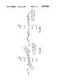
- FIG. 1 shows transmembrane current for a specific applied voltage for CD20+ B lineage cells.
- FIG. 2 shows flow cytometry analysis of CD20 expression.
- FIGS. 3A and 3B show transmembrane current for a specific applied voltage for transfected cells with and without the CD20 cDNA insert.
- HB5 a Mab to the Epstein-Barr Virus C3d receptor used as an isotype matched control for anti-Bl antibody failed to alter the whole cell currents, either acutely or after 24 h in culture.
- the T lymphocyte cell line Jurkat, which does not express CD20, was transfected with CD20 cDNA, CD20+ clones were isolated, and a stable cell line expressing 10,000-20,000 anti-Bl antibody binding sites/cell was developed according to the following procedure. (A clonal line transfected with the vector without CD20 cDNA insert was also isolated as a control.)
- flow cytometry analysis revealed extensive CD20 expression in one of the isolated clones (Jurkat.Bl-21 (solid line)) as compared with Jurkat cells transfected with the vector alone (dashed line). Greater than 90% of the Jurkat.Bl-21 cells expressed CD20. The phenotype of Jurakat.Bl 21 remained constant following transfection with CD20 cDNA, and the cells expressed all of the surface markers that the parent cell line expressed.
- B lymphocyte cell surface protein CD20 functions physiologically as a plasma membrane Ca 2+ channel or an obligate regulator of a Ca 2+ channel.
- Antibody binding to CD20 increases inward Ca 2+ current, which is detectable following culture of lymphocytes in the presence of antibody, and therefore, antibody binding may inhibit cell cycle progression by sustaining a Ca 2+ influx above normal levels.
- CD20 functions in Ca 2+ mobility as described above provides a basis for a simple assay to detect an effect of an agonist or antagonist to CD20 on B lymphocyte function by measuring changes in calcium ion flux across the lymphocyte membrane. First, the resting calcium ion flux across the B lymphocyte membrane is determined. Next, the B lymphocyte is contacted with the agonist or antagonist to be tested. Finally, the change in calcium ion flux following contact with the tested agent is determined.
- One method to measure the ion channel flux is to use flow cytometry analysis of cells cultured with the Ca 2+ chelator and fluorochrome, Fura-2,an indicator that combines an 8-coordinate tetracarboxylate chelating site with a stilbene chromophore (Grynkiewicz et al., J. Biol. Chem. 260:3440 (1985)).
- Clark et al. (Proc. Nat'l Acad. Sci. USA 82:1766-1770 (1985)) describes two well characterized monoclonal antibodies that bind CD20, one inducing B cell proliferation and the other blocking cell cycle progression. These different physiological changes may be caused by differential effects of these antibodies on Ca 2+ transport.
- the assay method described in Example 1 can be used to categorize these differences so that additional populations of antibodies can be surveyed simply and easily.
- Known pharmacologic Ca 2+ channel blocking agents such as Bay K 8644, Verapamil, diltiazem, D-600, nipedipin and 4-aminopyridine, can be assayed for their specific inhibiting or augmenting effect on CD20 function.
- Localization of the functional regions of the CD20 protein can be achieved by testing the effects of various agonists or antagonists to CD20 on modified CD20 molecules.
- a method of testing modified CD20 would include site-directed mutagenesis of CD20 cDNA, transformation of a CD20- cell line with the modified cDNA, expression of the modified CD20 protein encoded by the modified cDNA on the surface of cells of the CD20- cell line, and determination of the changes in calcium ion flux across the membrane of the transformed cell following exposure of the cell to the agonist or antagonist, a procedure analogous to that used by Eldefrawi et al (FASEB J. 1:262-271 (1987)) in targeting drugs towards receptors for ⁇ -aminobutyric acid and voltage-dependent chloride channels.
- Knowledge of the functions of specific regions of CD20 can be applied to develop Ca 2+ channel blocking drugs which specifically target the CD20 cell surface protein without interfering with other cells of the immune system.
- the method of the invention can be used to assay the effects of agonistic and antagonistic drugs on CD20 function in malignant B cells in order to clarify the role that regulating CD20 gene expression might have on limiting the growth advantage of CD20+ malignant cells.
Landscapes
- Health & Medical Sciences (AREA)
- Life Sciences & Earth Sciences (AREA)
- Immunology (AREA)
- Engineering & Computer Science (AREA)
- Hematology (AREA)
- Cell Biology (AREA)
- Biomedical Technology (AREA)
- Chemical & Material Sciences (AREA)
- Urology & Nephrology (AREA)
- Molecular Biology (AREA)
- General Physics & Mathematics (AREA)
- Biochemistry (AREA)
- Biotechnology (AREA)
- Tropical Medicine & Parasitology (AREA)
- Pathology (AREA)
- Food Science & Technology (AREA)
- Medicinal Chemistry (AREA)
- Physics & Mathematics (AREA)
- Analytical Chemistry (AREA)
- Microbiology (AREA)
- General Health & Medical Sciences (AREA)
- Bioinformatics & Cheminformatics (AREA)
- Toxicology (AREA)
- Zoology (AREA)
- Virology (AREA)
- Investigating Or Analysing Biological Materials (AREA)
- Measuring Or Testing Involving Enzymes Or Micro-Organisms (AREA)
- Medicines That Contain Protein Lipid Enzymes And Other Medicines (AREA)
- Preparation Of Compounds By Using Micro-Organisms (AREA)
- Peptides Or Proteins (AREA)
Abstract
Description
Claims (8)
Priority Applications (4)
| Application Number | Priority Date | Filing Date | Title |
|---|---|---|---|
| US07/313,108 US4987084A (en) | 1989-02-21 | 1989-02-21 | Method of testing the effect of a molecule on B lymphocyte function |
| CA002010207A CA2010207A1 (en) | 1989-02-21 | 1990-02-16 | Method of testing the effect of a molecule on b lymphocyte function |
| EP19900301882 EP0384740A3 (en) | 1989-02-21 | 1990-02-21 | Method of testing the effect of a molecule on b lymphocyte function |
| JP2040866A JPH02284062A (en) | 1989-02-21 | 1990-02-21 | Method for testing action of molecule for function of b lymphocyte |
Applications Claiming Priority (1)
| Application Number | Priority Date | Filing Date | Title |
|---|---|---|---|
| US07/313,108 US4987084A (en) | 1989-02-21 | 1989-02-21 | Method of testing the effect of a molecule on B lymphocyte function |
Publications (1)
| Publication Number | Publication Date |
|---|---|
| US4987084A true US4987084A (en) | 1991-01-22 |
Family
ID=23214417
Family Applications (1)
| Application Number | Title | Priority Date | Filing Date |
|---|---|---|---|
| US07/313,108 Expired - Fee Related US4987084A (en) | 1989-02-21 | 1989-02-21 | Method of testing the effect of a molecule on B lymphocyte function |
Country Status (4)
| Country | Link |
|---|---|
| US (1) | US4987084A (en) |
| EP (1) | EP0384740A3 (en) |
| JP (1) | JPH02284062A (en) |
| CA (1) | CA2010207A1 (en) |
Cited By (8)
| Publication number | Priority date | Publication date | Assignee | Title |
|---|---|---|---|---|
| US20020077775A1 (en) * | 2000-05-25 | 2002-06-20 | Schork Nicholas J. | Methods of DNA marker-based genetic analysis using estimated haplotype frequencies and uses thereof |
| US6632619B1 (en) * | 1997-05-16 | 2003-10-14 | The Governors Of The University Of Alberta | Microfluidic system and methods of use |
| US20030195707A1 (en) * | 2000-05-25 | 2003-10-16 | Schork Nicholas J | Methods of dna marker-based genetic analysis using estimated haplotype frequencies and uses thereof |
| US20050014202A1 (en) * | 2003-07-15 | 2005-01-20 | Brown Arthur M. | High throughput assay systems and methods for identifying agents that alter surface expression of integral membrane proteins |
| US6900021B1 (en) | 1997-05-16 | 2005-05-31 | The University Of Alberta | Microfluidic system and methods of use |
| US20050119171A1 (en) * | 2001-08-10 | 2005-06-02 | Genset S.A. | Human cdnas and proteins and uses thereof |
| US20050191719A1 (en) * | 2002-07-23 | 2005-09-01 | Rigel Pharmaceuticals, Inc. | Modulators of B-lymphocyte activation, myosin-1F compositions and methods of use |
| US20070264252A1 (en) * | 2003-10-24 | 2007-11-15 | Li Sau W | Modulators of Ms4a Gene Products |
Families Citing this family (12)
| Publication number | Priority date | Publication date | Assignee | Title |
|---|---|---|---|---|
| US6011068A (en) * | 1991-08-23 | 2000-01-04 | Nps Pharmaceuticals, Inc. | Calcium receptor-active molecules |
| US5858684A (en) * | 1991-08-23 | 1999-01-12 | The Brigham And Women's Hospital, Inc. | Method of screening calcium receptor-active molecules |
| US5763569A (en) * | 1991-08-23 | 1998-06-09 | The Brigham And Women's Hospital, Inc | Calcium receptor-active molecules |
| US6001884A (en) * | 1991-08-23 | 1999-12-14 | Nps Pharmaceuticals, Inc. | Calcium receptor-active molecules |
| US6031003A (en) * | 1991-08-23 | 2000-02-29 | Nps Pharmaceuticals, Inc. | Calcium receptor-active molecules |
| DK1281702T3 (en) * | 1991-08-23 | 2006-04-18 | Nps Pharma Inc | Calcium receptor active molecules |
| US6313146B1 (en) | 1991-08-23 | 2001-11-06 | Nps Pharmaceuticals, Inc. | Calcium receptor-active molecules |
| US5962314A (en) * | 1993-02-23 | 1999-10-05 | Nps Pharmaceuticals, Inc. | Calcium receptor-active molecules |
| US6558916B2 (en) | 1996-08-02 | 2003-05-06 | Axiom Biotechnologies, Inc. | Cell flow apparatus and method for real-time measurements of patient cellular responses |
| US6280967B1 (en) * | 1996-08-02 | 2001-08-28 | Axiom Biotechnologies, Inc. | Cell flow apparatus and method for real-time of cellular responses |
| US5804436A (en) | 1996-08-02 | 1998-09-08 | Axiom Biotechnologies, Inc. | Apparatus and method for real-time measurement of cellular response |
| GB0108143D0 (en) * | 2001-03-31 | 2001-05-23 | Univ Dundee | High-throughput screen |
Family Cites Families (1)
| Publication number | Priority date | Publication date | Assignee | Title |
|---|---|---|---|---|
| US4788137A (en) * | 1985-10-29 | 1988-11-29 | Dana-Farber Cancer Institute, Inc. | Detection of activated T-cells |
-
1989
- 1989-02-21 US US07/313,108 patent/US4987084A/en not_active Expired - Fee Related
-
1990
- 1990-02-16 CA CA002010207A patent/CA2010207A1/en not_active Abandoned
- 1990-02-21 JP JP2040866A patent/JPH02284062A/en active Pending
- 1990-02-21 EP EP19900301882 patent/EP0384740A3/en not_active Withdrawn
Non-Patent Citations (18)
| Title |
|---|
| Dugas, "Human B Cell Activation: Selective Sensitivity of the Early Stages to Calcium Channel-Blocking Drugs," Eur. J. Immunol. 16: 162-167 (1986). |
| Dugas, Human B Cell Activation: Selective Sensitivity of the Early Stages to Calcium Channel Blocking Drugs, Eur. J. Immunol. 16: 162 167 (1986). * |
| Fukishima et al., "Voltage-gated Ca2+ Channel in Mouse Myeloma Cells," Proc. Natl. Acad. Sci. U.S.A. 80: 2240-2242 (1983). |
| Fukishima et al., Voltage gated Ca 2 Channel in Mouse Myeloma Cells, Proc. Natl. Acad. Sci. U.S.A. 80: 2240 2242 (1983). * |
| MacDougall et al., "Detection of Ligand-Activated Conductive Ca21 Channels in Human B Lymphocytes," Cell 54: 229-234 (1988). |
| MacDougall et al., Detection of Ligand Activated Conductive Ca 21 Channels in Human B Lymphocytes, Cell 54: 229 234 (1988). * |
| Nadler et al., "B Cell Origin of Non-T Cell Acute Lymphoblastic Leukemia A Model for Discrete Stages of Neoplastic and Normal Pre-B Cell Differentiation," J. Clin. Invest. 74: 332-340 (1984). |
| Nadler et al., B Cell Origin of Non T Cell Acute Lymphoblastic Leukemia A Model for Discrete Stages of Neoplastic and Normal Pre B Cell Differentiation, J. Clin. Invest. 74: 332 340 (1984). * |
| Tedder et al., "Antibodies Reactive with the B1 Molecule Inhibity Cell Cycle Progression but not Activation of Human B lymphocytes," Eur. J. Immunol. 16: 881-887 (1986). |
| Tedder et al., "Cloning of a Complementary DNA Encoding a New Mouse B Lumphocyte Differentiation Antigen, Homologous to the Human B1 (CD20) Antigen and Localization of the Gene to Chromosome 19," The Journal of Immunology 141: 4388-4394 (1988). |
| Tedder et al., "Isolation and Structure of a cDNA Encoding the Bl (CD20) Cell-Surface Antigen of Human B Lymphocytes," Proc. Natl. Acad. Sci. U.S.A. 85: 208-212 (1988). |
| Tedder et al., "Phosphorylation of the B1 (CD20) Molecule by Normal and Malignmant Human B Lymphocytes," The Journal of Biological Chemistry 263: 10009-10015 (1988). |
| Tedder et al., "The B Cell Surface Molecule B1 is Functionally Linked with B Cell Activation and Differentiation," The Journal of Immunology 135: 973-979 (1985). |
| Tedder et al., Antibodies Reactive with the B1 Molecule Inhibity Cell Cycle Progression but not Activation of Human B lymphocytes, Eur. J. Immunol. 16: 881 887 (1986). * |
| Tedder et al., Cloning of a Complementary DNA Encoding a New Mouse B Lumphocyte Differentiation Antigen, Homologous to the Human B1 (CD20) Antigen and Localization of the Gene to Chromosome 19, The Journal of Immunology 141: 4388 4394 (1988). * |
| Tedder et al., Isolation and Structure of a cDNA Encoding the Bl (CD20) Cell Surface Antigen of Human B Lymphocytes, Proc. Natl. Acad. Sci. U.S.A. 85: 208 212 (1988). * |
| Tedder et al., Phosphorylation of the B1 (CD20) Molecule by Normal and Malignmant Human B Lymphocytes, The Journal of Biological Chemistry 263: 10009 10015 (1988). * |
| Tedder et al., The B Cell Surface Molecule B1 is Functionally Linked with B Cell Activation and Differentiation, The Journal of Immunology 135: 973 979 (1985). * |
Cited By (14)
| Publication number | Priority date | Publication date | Assignee | Title |
|---|---|---|---|---|
| US6900021B1 (en) | 1997-05-16 | 2005-05-31 | The University Of Alberta | Microfluidic system and methods of use |
| US6632619B1 (en) * | 1997-05-16 | 2003-10-14 | The Governors Of The University Of Alberta | Microfluidic system and methods of use |
| US20040178071A1 (en) * | 1997-05-16 | 2004-09-16 | The Governors Of The University Of Alberta | Microfluidic system and methods of use |
| US20030195707A1 (en) * | 2000-05-25 | 2003-10-16 | Schork Nicholas J | Methods of dna marker-based genetic analysis using estimated haplotype frequencies and uses thereof |
| US20020077775A1 (en) * | 2000-05-25 | 2002-06-20 | Schork Nicholas J. | Methods of DNA marker-based genetic analysis using estimated haplotype frequencies and uses thereof |
| US20050119171A1 (en) * | 2001-08-10 | 2005-06-02 | Genset S.A. | Human cdnas and proteins and uses thereof |
| US7285631B2 (en) | 2001-08-10 | 2007-10-23 | Serono Genetics Institute S.A. | Human cDNAs and proteins and uses thereof |
| US20080026427A1 (en) * | 2001-08-10 | 2008-01-31 | Serono Genetics Institute S.A. | HUMAN cDNAs AND PROTEINS AND USES THEREOF |
| US20050191719A1 (en) * | 2002-07-23 | 2005-09-01 | Rigel Pharmaceuticals, Inc. | Modulators of B-lymphocyte activation, myosin-1F compositions and methods of use |
| US7371537B2 (en) * | 2002-07-23 | 2008-05-13 | Rigel Pharmaceuticals, Inc. | Modulators of B-lymphocyte activation, myosin-1F compositions and methods of use |
| US20050014202A1 (en) * | 2003-07-15 | 2005-01-20 | Brown Arthur M. | High throughput assay systems and methods for identifying agents that alter surface expression of integral membrane proteins |
| US7211407B2 (en) * | 2003-07-15 | 2007-05-01 | Chan Test, Inc. | High throughput assay systems and methods for identifying agents that alter surface expression of integral membrane proteins |
| US20070259878A1 (en) * | 2003-07-15 | 2007-11-08 | Chanxpress, Inc. | High throughput assay systems and methods for identifying agents that alter surface expression of integral membrane proteins |
| US20070264252A1 (en) * | 2003-10-24 | 2007-11-15 | Li Sau W | Modulators of Ms4a Gene Products |
Also Published As
| Publication number | Publication date |
|---|---|
| EP0384740A2 (en) | 1990-08-29 |
| CA2010207A1 (en) | 1990-08-21 |
| JPH02284062A (en) | 1990-11-21 |
| EP0384740A3 (en) | 1991-07-10 |
Similar Documents
| Publication | Publication Date | Title |
|---|---|---|
| US4987084A (en) | Method of testing the effect of a molecule on B lymphocyte function | |
| Racioppi et al. | Peptide-major histocompatibility complex class II complexes with mixed agonist/antagonist properties provide evidence for ligand-related differences in T cell receptor-dependent intracellular signaling. | |
| Merker et al. | Uptake and nature of the intracellular binding of cyclosporin A in a murine thymoma cell line, BW5147. | |
| Hünig et al. | Alternative pathway activation of T cells by binding of CD2 to its cell-surface ligand | |
| Cinek et al. | The nature of large noncovalent complexes containing glycosyl-phosphatidylinositol-anchored membrane glycoproteins and protein tyrosine kinases. | |
| Anderson et al. | Cross-linking of T3 (CD3) with T4 (CD4) enhances the proliferation of resting T lymphocytes. | |
| Oviedo‐Orta et al. | Intercellular communication in the immune system: differential expression of connexin40 and 43, and perturbation of gap junction channel functions in peripheral blood and tonsil human lymphocyte subpopulations | |
| Nilsson et al. | Simplified assays of hemolytic activity of the classical and alternative complement pathways | |
| Chalupny et al. | Association of CD8 with p56lck is required for early T cell signalling events. | |
| Font et al. | Elevated soluble CD27 levels in serum of patients with systemic lupus erythematosus | |
| Kepley et al. | Identification of the FcϵRI-activated tyrosine kinases Lyn, Syk, and Zap-70 in human basophils | |
| Shi et al. | Differential tyrosine-specific protein phosphorylation in mouse T lymphocyte subsets. Effect of age. | |
| US6346388B1 (en) | Method of identifying agonist and antagonists for tumor necrosis related receptors TR1 and TR2 | |
| Goldsmith et al. | At least two non-antigen-binding molecules are required for signal transduction by the T-cell antigen receptor. | |
| Smith et al. | Detection of a soluble form of the leukocyte surface antigen CD48 in plasma and its elevation in patients with lymphoid leukemias and arthritis | |
| Reischl et al. | Function and regulation of FcεRI expression on monocytes from non‐atopic donors | |
| Fukai et al. | A critical role for p59fyn in CD2‐based signal transduction | |
| Jüppner et al. | Autoantibodies to parathyroid hormone receptor | |
| Valente et al. | Monoclonal antibodies to the thyrotropin receptor: the identification of blocking and stimulating antibodies | |
| Hahn et al. | Interaction of CD2 with its ligand lymphocyte function-associated antigen-3 induces adenosine 3', 5'-cyclic monophosphate production in T lymphocytes. | |
| Leitner et al. | The relationship between somatostatin binding and cyclic AMP-stimulated protein kinase inhibition | |
| Law et al. | Agonist binding to rat brain somatostatin receptors alters the interaction of the receptors with guanine nucleotide-binding regulatory proteins. | |
| Renard et al. | The CD8β polypeptide is required for the recognition of an altered peptide ligand as an agonist | |
| Ushikubi et al. | Analysis of the defective signal transduction mechanism through the platelet thromboxane A2 receptor in a patient with polycythemia vera | |
| JPH06500775A (en) | Regulation of malignant cell proliferation via a novel 5HT1a receptor |
Legal Events
| Date | Code | Title | Description |
|---|---|---|---|
| AS | Assignment |
Owner name: DANA-FARBER CANCER INSTITUTE, MASSACHUSETTS Free format text: ASSIGNMENT OF ASSIGNORS INTEREST.;ASSIGNORS:TEDDER, THOMAS F.;SCHLOSSMAN, STUART F.;SAITO, HARUO;REEL/FRAME:005094/0689;SIGNING DATES FROM 19890317 TO 19890329 |
|
| AS | Assignment |
Owner name: DANA-FARBER CANCER INSTITUTE, INC., BOSTON, MA A C Free format text: RE-RECORD OF INSTRUMENT PREVIOUSLY RECORDED ON APRIL 10, 1989 AT REEL 5094 FRAME 689 TO CORRECT THE NAME OF THE ASSIGNEE.;ASSIGNORS:TEDDER, THOMAS F.;SCHLOSSMAN, STUART F.;SAITO, HARUO;REEL/FRAME:005262/0845;SIGNING DATES FROM 19890317 TO 19890329 Owner name: DANA-FARBER CANCER INSTITUTE, INC., A CORP. OF MA, Free format text: RE-RECORD OF INSTRUMENT PREVIOUSLY RECORDED ON APRIL 10, 1989 AT REEL 5094 FRAME 689 TO CORRECT THE NAME OF THE ASSIGNEE;ASSIGNORS:TEDDER, THOMAS F.;SCHLOSSMAN, STUART F.;SAITO, HARUO;SIGNING DATES FROM 19890317 TO 19890329;REEL/FRAME:005262/0845 |
|
| FPAY | Fee payment |
Year of fee payment: 4 |
|
| REMI | Maintenance fee reminder mailed | ||
| LAPS | Lapse for failure to pay maintenance fees | ||
| FP | Lapsed due to failure to pay maintenance fee |
Effective date: 19990122 |
|
| STCH | Information on status: patent discontinuation |
Free format text: PATENT EXPIRED DUE TO NONPAYMENT OF MAINTENANCE FEES UNDER 37 CFR 1.362 |