US4965192A - A60-antigen from Mycobacteria and use thereof as tuberculin and as vaccine - Google Patents
A60-antigen from Mycobacteria and use thereof as tuberculin and as vaccine Download PDFInfo
- Publication number
- US4965192A US4965192A US07/187,919 US18791988A US4965192A US 4965192 A US4965192 A US 4965192A US 18791988 A US18791988 A US 18791988A US 4965192 A US4965192 A US 4965192A
- Authority
- US
- United States
- Prior art keywords
- antigen
- mycobacteria
- test
- subject
- antibodies
- Prior art date
- Legal status (The legal status is an assumption and is not a legal conclusion. Google has not performed a legal analysis and makes no representation as to the accuracy of the status listed.)
- Expired - Lifetime
Links
- 239000000427 antigen Substances 0.000 title claims abstract description 160
- 229960001005 tuberculin Drugs 0.000 title description 19
- 229960005486 vaccine Drugs 0.000 title description 2
- 102000036639 antigens Human genes 0.000 claims abstract description 158
- 108091007433 antigens Proteins 0.000 claims abstract description 158
- 102000004169 proteins and genes Human genes 0.000 claims abstract description 16
- 108090000623 proteins and genes Proteins 0.000 claims abstract description 16
- 206010062207 Mycobacterial infection Diseases 0.000 claims abstract description 8
- 208000027531 mycobacterial infectious disease Diseases 0.000 claims abstract description 8
- 238000001962 electrophoresis Methods 0.000 claims abstract description 5
- 239000000203 mixture Substances 0.000 claims abstract description 5
- 150000004676 glycans Chemical class 0.000 claims abstract description 4
- 229920001282 polysaccharide Polymers 0.000 claims abstract description 4
- 239000005017 polysaccharide Substances 0.000 claims abstract description 4
- 238000001556 precipitation Methods 0.000 claims abstract description 3
- 238000000034 method Methods 0.000 claims description 14
- 238000011161 development Methods 0.000 claims description 11
- 206010020751 Hypersensitivity Diseases 0.000 claims description 9
- 239000012530 fluid Substances 0.000 claims description 6
- 238000002347 injection Methods 0.000 claims description 5
- 239000007924 injection Substances 0.000 claims description 5
- 238000002156 mixing Methods 0.000 claims description 2
- 238000010668 complexation reaction Methods 0.000 claims 1
- 230000028993 immune response Effects 0.000 claims 1
- 238000012360 testing method Methods 0.000 abstract description 52
- 241000186366 Mycobacterium bovis Species 0.000 description 23
- 241001467552 Mycobacterium bovis BCG Species 0.000 description 17
- 239000000463 material Substances 0.000 description 16
- 238000010790 dilution Methods 0.000 description 15
- 239000012895 dilution Substances 0.000 description 15
- 235000018102 proteins Nutrition 0.000 description 14
- 229960000190 bacillus calmette–guérin vaccine Drugs 0.000 description 13
- 239000004816 latex Substances 0.000 description 13
- 229920000126 latex Polymers 0.000 description 13
- 201000008827 tuberculosis Diseases 0.000 description 13
- 238000006243 chemical reaction Methods 0.000 description 12
- 230000018109 developmental process Effects 0.000 description 10
- 239000002953 phosphate buffered saline Substances 0.000 description 10
- 230000035945 sensitivity Effects 0.000 description 10
- 238000002360 preparation method Methods 0.000 description 9
- 241001465754 Metazoa Species 0.000 description 8
- 241000699666 Mus <mouse, genus> Species 0.000 description 8
- 239000000872 buffer Substances 0.000 description 8
- 210000002966 serum Anatomy 0.000 description 8
- 241000894006 Bacteria Species 0.000 description 7
- 241000699670 Mus sp. Species 0.000 description 7
- 229920002684 Sepharose Polymers 0.000 description 7
- 108090000631 Trypsin Proteins 0.000 description 7
- 102000004142 Trypsin Human genes 0.000 description 7
- 239000000047 product Substances 0.000 description 7
- 239000000243 solution Substances 0.000 description 7
- 241000894007 species Species 0.000 description 7
- 230000008961 swelling Effects 0.000 description 7
- 239000012588 trypsin Substances 0.000 description 7
- 206010036790 Productive cough Diseases 0.000 description 6
- 230000001086 cytosolic effect Effects 0.000 description 6
- 102000013415 peroxidase activity proteins Human genes 0.000 description 6
- 108040007629 peroxidase activity proteins Proteins 0.000 description 6
- 230000003248 secreting effect Effects 0.000 description 6
- 230000001235 sensitizing effect Effects 0.000 description 6
- 210000003802 sputum Anatomy 0.000 description 6
- 208000024794 sputum Diseases 0.000 description 6
- 241000186367 Mycobacterium avium Species 0.000 description 5
- 241000186362 Mycobacterium leprae Species 0.000 description 5
- 230000000890 antigenic effect Effects 0.000 description 5
- 238000001514 detection method Methods 0.000 description 5
- 238000002405 diagnostic procedure Methods 0.000 description 5
- LOKCTEFSRHRXRJ-UHFFFAOYSA-I dipotassium trisodium dihydrogen phosphate hydrogen phosphate dichloride Chemical compound P(=O)(O)(O)[O-].[K+].P(=O)(O)([O-])[O-].[Na+].[Na+].[Cl-].[K+].[Cl-].[Na+] LOKCTEFSRHRXRJ-UHFFFAOYSA-I 0.000 description 5
- 239000012634 fragment Substances 0.000 description 5
- 238000011534 incubation Methods 0.000 description 5
- 238000005406 washing Methods 0.000 description 5
- QFVHZQCOUORWEI-UHFFFAOYSA-N 4-[(4-anilino-5-sulfonaphthalen-1-yl)diazenyl]-5-hydroxynaphthalene-2,7-disulfonic acid Chemical compound C=12C(O)=CC(S(O)(=O)=O)=CC2=CC(S(O)(=O)=O)=CC=1N=NC(C1=CC=CC(=C11)S(O)(=O)=O)=CC=C1NC1=CC=CC=C1 QFVHZQCOUORWEI-UHFFFAOYSA-N 0.000 description 4
- 102000004190 Enzymes Human genes 0.000 description 4
- 108090000790 Enzymes Proteins 0.000 description 4
- DHMQDGOQFOQNFH-UHFFFAOYSA-N Glycine Chemical compound NCC(O)=O DHMQDGOQFOQNFH-UHFFFAOYSA-N 0.000 description 4
- 241000187482 Mycobacterium avium subsp. paratuberculosis Species 0.000 description 4
- 239000004793 Polystyrene Substances 0.000 description 4
- 239000004365 Protease Substances 0.000 description 4
- 230000004520 agglutination Effects 0.000 description 4
- 238000004220 aggregation Methods 0.000 description 4
- 230000002776 aggregation Effects 0.000 description 4
- 208000026935 allergic disease Diseases 0.000 description 4
- 210000000805 cytoplasm Anatomy 0.000 description 4
- 201000010099 disease Diseases 0.000 description 4
- 208000037265 diseases, disorders, signs and symptoms Diseases 0.000 description 4
- 229940088598 enzyme Drugs 0.000 description 4
- 230000007717 exclusion Effects 0.000 description 4
- 230000009610 hypersensitivity Effects 0.000 description 4
- 230000001900 immune effect Effects 0.000 description 4
- 238000002955 isolation Methods 0.000 description 4
- 239000002245 particle Substances 0.000 description 4
- 229920002223 polystyrene Polymers 0.000 description 4
- 230000003389 potentiating effect Effects 0.000 description 4
- GEYOCULIXLDCMW-UHFFFAOYSA-N 1,2-phenylenediamine Chemical compound NC1=CC=CC=C1N GEYOCULIXLDCMW-UHFFFAOYSA-N 0.000 description 3
- 241000187490 Mycobacterium scrofulaceum Species 0.000 description 3
- 241000187494 Mycobacterium xenopi Species 0.000 description 3
- 108091005804 Peptidases Proteins 0.000 description 3
- 238000003556 assay Methods 0.000 description 3
- 230000001580 bacterial effect Effects 0.000 description 3
- 239000000356 contaminant Substances 0.000 description 3
- 230000003111 delayed effect Effects 0.000 description 3
- 238000002474 experimental method Methods 0.000 description 3
- 238000000760 immunoelectrophoresis Methods 0.000 description 3
- 208000015181 infectious disease Diseases 0.000 description 3
- 230000002401 inhibitory effect Effects 0.000 description 3
- 239000002808 molecular sieve Substances 0.000 description 3
- URGAHOPLAPQHLN-UHFFFAOYSA-N sodium aluminosilicate Chemical compound [Na+].[Al+3].[O-][Si]([O-])=O.[O-][Si]([O-])=O URGAHOPLAPQHLN-UHFFFAOYSA-N 0.000 description 3
- 239000000725 suspension Substances 0.000 description 3
- 208000027930 type IV hypersensitivity disease Diseases 0.000 description 3
- 102000009027 Albumins Human genes 0.000 description 2
- 108010088751 Albumins Proteins 0.000 description 2
- 241000304886 Bacilli Species 0.000 description 2
- BVKZGUZCCUSVTD-UHFFFAOYSA-L Carbonate Chemical compound [O-]C([O-])=O BVKZGUZCCUSVTD-UHFFFAOYSA-L 0.000 description 2
- 241000700198 Cavia Species 0.000 description 2
- 208000006313 Delayed Hypersensitivity Diseases 0.000 description 2
- 239000004471 Glycine Substances 0.000 description 2
- 241000186363 Mycobacterium kansasii Species 0.000 description 2
- 102100037486 Reverse transcriptase/ribonuclease H Human genes 0.000 description 2
- PXIPVTKHYLBLMZ-UHFFFAOYSA-N Sodium azide Chemical compound [Na+].[N-]=[N+]=[N-] PXIPVTKHYLBLMZ-UHFFFAOYSA-N 0.000 description 2
- 230000024932 T cell mediated immunity Effects 0.000 description 2
- 208000024780 Urticaria Diseases 0.000 description 2
- 238000001042 affinity chromatography Methods 0.000 description 2
- 239000007982 barbital buffer Substances 0.000 description 2
- 210000004027 cell Anatomy 0.000 description 2
- 230000001413 cellular effect Effects 0.000 description 2
- 239000001913 cellulose Substances 0.000 description 2
- 229920002678 cellulose Polymers 0.000 description 2
- 238000005119 centrifugation Methods 0.000 description 2
- 230000000536 complexating effect Effects 0.000 description 2
- 239000000470 constituent Substances 0.000 description 2
- ATDGTVJJHBUTRL-UHFFFAOYSA-N cyanogen bromide Chemical compound BrC#N ATDGTVJJHBUTRL-UHFFFAOYSA-N 0.000 description 2
- 238000011156 evaluation Methods 0.000 description 2
- 238000002270 exclusion chromatography Methods 0.000 description 2
- 238000010438 heat treatment Methods 0.000 description 2
- 230000036039 immunity Effects 0.000 description 2
- 238000003018 immunoassay Methods 0.000 description 2
- 230000000984 immunochemical effect Effects 0.000 description 2
- 238000011835 investigation Methods 0.000 description 2
- 230000003287 optical effect Effects 0.000 description 2
- 239000008363 phosphate buffer Substances 0.000 description 2
- XOJVVFBFDXDTEG-UHFFFAOYSA-N pristane Chemical compound CC(C)CCCC(C)CCCC(C)CCCC(C)C XOJVVFBFDXDTEG-UHFFFAOYSA-N 0.000 description 2
- 235000019419 proteases Nutrition 0.000 description 2
- 230000004044 response Effects 0.000 description 2
- 239000007790 solid phase Substances 0.000 description 2
- MJQHZNBUODTQTK-WKGBVCLCSA-N (2s,3r,4s,5r,6r)-2-[[(1s,3s,4s,5s,8r)-3-[(2s,3r,4s,5s,6r)-2-[[(1s,3r,4s,5s,8r)-3,4-dihydroxy-2,6-dioxabicyclo[3.2.1]octan-8-yl]oxy]-3,5-dihydroxy-6-(hydroxymethyl)oxan-4-yl]oxy-4-hydroxy-2,6-dioxabicyclo[3.2.1]octan-8-yl]oxy]-6-(hydroxymethyl)oxane-3,4,5- Chemical compound O[C@@H]1[C@@H](O)[C@@H](O)[C@@H](CO)O[C@H]1O[C@H]1[C@H]2OC[C@@H]1O[C@@H](O[C@@H]1[C@H]([C@H](O[C@H]3[C@H]4OC[C@@H]3O[C@@H](O)[C@H]4O)O[C@H](CO)[C@@H]1O)O)[C@H]2O MJQHZNBUODTQTK-WKGBVCLCSA-N 0.000 description 1
- QKNYBSVHEMOAJP-UHFFFAOYSA-N 2-amino-2-(hydroxymethyl)propane-1,3-diol;hydron;chloride Chemical compound Cl.OCC(N)(CO)CO QKNYBSVHEMOAJP-UHFFFAOYSA-N 0.000 description 1
- GHCZTIFQWKKGSB-UHFFFAOYSA-N 2-hydroxypropane-1,2,3-tricarboxylic acid;phosphoric acid Chemical compound OP(O)(O)=O.OC(=O)CC(O)(C(O)=O)CC(O)=O GHCZTIFQWKKGSB-UHFFFAOYSA-N 0.000 description 1
- 229920000936 Agarose Polymers 0.000 description 1
- 206010003445 Ascites Diseases 0.000 description 1
- 241000193830 Bacillus <bacterium> Species 0.000 description 1
- 241000283690 Bos taurus Species 0.000 description 1
- UXVMQQNJUSDDNG-UHFFFAOYSA-L Calcium chloride Chemical compound [Cl-].[Cl-].[Ca+2] UXVMQQNJUSDDNG-UHFFFAOYSA-L 0.000 description 1
- 241000283707 Capra Species 0.000 description 1
- 108090000317 Chymotrypsin Proteins 0.000 description 1
- 244000026610 Cynodon dactylon var. affinis Species 0.000 description 1
- 238000002965 ELISA Methods 0.000 description 1
- SXRSQZLOMIGNAQ-UHFFFAOYSA-N Glutaraldehyde Chemical compound O=CCCCC=O SXRSQZLOMIGNAQ-UHFFFAOYSA-N 0.000 description 1
- 239000012981 Hank's balanced salt solution Substances 0.000 description 1
- 206010061218 Inflammation Diseases 0.000 description 1
- 206010024229 Leprosy Diseases 0.000 description 1
- 241001529936 Murinae Species 0.000 description 1
- 241000187478 Mycobacterium chelonae Species 0.000 description 1
- 241000187480 Mycobacterium smegmatis Species 0.000 description 1
- 206010029719 Nonspecific reaction Diseases 0.000 description 1
- 229910019142 PO4 Inorganic materials 0.000 description 1
- 108090000526 Papain Proteins 0.000 description 1
- 102000035195 Peptidases Human genes 0.000 description 1
- 229920001213 Polysorbate 20 Polymers 0.000 description 1
- 102000006382 Ribonucleases Human genes 0.000 description 1
- 108010083644 Ribonucleases Proteins 0.000 description 1
- 206010070834 Sensitisation Diseases 0.000 description 1
- BQCADISMDOOEFD-UHFFFAOYSA-N Silver Chemical compound [Ag] BQCADISMDOOEFD-UHFFFAOYSA-N 0.000 description 1
- 241000191967 Staphylococcus aureus Species 0.000 description 1
- 108090000787 Subtilisin Proteins 0.000 description 1
- 238000002835 absorbance Methods 0.000 description 1
- 239000002671 adjuvant Substances 0.000 description 1
- 239000011543 agarose gel Substances 0.000 description 1
- 235000001014 amino acid Nutrition 0.000 description 1
- 150000001413 amino acids Chemical class 0.000 description 1
- 230000003698 anagen phase Effects 0.000 description 1
- 238000004458 analytical method Methods 0.000 description 1
- 239000007864 aqueous solution Substances 0.000 description 1
- 230000002238 attenuated effect Effects 0.000 description 1
- 238000009361 aviculture Methods 0.000 description 1
- 230000006399 behavior Effects 0.000 description 1
- VEZXCJBBBCKRPI-UHFFFAOYSA-N beta-propiolactone Chemical compound O=C1CCO1 VEZXCJBBBCKRPI-UHFFFAOYSA-N 0.000 description 1
- 239000001110 calcium chloride Substances 0.000 description 1
- 229910001628 calcium chloride Inorganic materials 0.000 description 1
- 230000015556 catabolic process Effects 0.000 description 1
- 230000036755 cellular response Effects 0.000 description 1
- 239000003153 chemical reaction reagent Substances 0.000 description 1
- 239000003795 chemical substances by application Substances 0.000 description 1
- VDQQXEISLMTGAB-UHFFFAOYSA-N chloramine T Chemical compound [Na+].CC1=CC=C(S(=O)(=O)[N-]Cl)C=C1 VDQQXEISLMTGAB-UHFFFAOYSA-N 0.000 description 1
- 238000004587 chromatography analysis Methods 0.000 description 1
- 229960002376 chymotrypsin Drugs 0.000 description 1
- 238000010367 cloning Methods 0.000 description 1
- 238000004040 coloring Methods 0.000 description 1
- 230000000052 comparative effect Effects 0.000 description 1
- 230000002860 competitive effect Effects 0.000 description 1
- 230000006835 compression Effects 0.000 description 1
- 238000007906 compression Methods 0.000 description 1
- 238000011109 contamination Methods 0.000 description 1
- 230000006837 decompression Effects 0.000 description 1
- 238000006731 degradation reaction Methods 0.000 description 1
- 230000018044 dehydration Effects 0.000 description 1
- 238000006297 dehydration reaction Methods 0.000 description 1
- 238000003745 diagnosis Methods 0.000 description 1
- 230000009977 dual effect Effects 0.000 description 1
- 230000000694 effects Effects 0.000 description 1
- 230000002255 enzymatic effect Effects 0.000 description 1
- 238000006911 enzymatic reaction Methods 0.000 description 1
- 230000001747 exhibiting effect Effects 0.000 description 1
- 239000012847 fine chemical Substances 0.000 description 1
- 230000006870 function Effects 0.000 description 1
- 239000000499 gel Substances 0.000 description 1
- 230000008348 humoral response Effects 0.000 description 1
- 210000004408 hybridoma Anatomy 0.000 description 1
- 230000003301 hydrolyzing effect Effects 0.000 description 1
- 230000008105 immune reaction Effects 0.000 description 1
- 230000003053 immunization Effects 0.000 description 1
- 238000002649 immunization Methods 0.000 description 1
- 230000002163 immunogen Effects 0.000 description 1
- 230000004054 inflammatory process Effects 0.000 description 1
- 238000011081 inoculation Methods 0.000 description 1
- 238000007912 intraperitoneal administration Methods 0.000 description 1
- 238000012092 latex agglutination test Methods 0.000 description 1
- 230000000670 limiting effect Effects 0.000 description 1
- UEGPKNKPLBYCNK-UHFFFAOYSA-L magnesium acetate Chemical compound [Mg+2].CC([O-])=O.CC([O-])=O UEGPKNKPLBYCNK-UHFFFAOYSA-L 0.000 description 1
- 238000004519 manufacturing process Methods 0.000 description 1
- 238000005259 measurement Methods 0.000 description 1
- 244000005700 microbiome Species 0.000 description 1
- 230000036963 noncompetitive effect Effects 0.000 description 1
- 238000013421 nuclear magnetic resonance imaging Methods 0.000 description 1
- 229940055729 papain Drugs 0.000 description 1
- 235000019834 papain Nutrition 0.000 description 1
- 230000001717 pathogenic effect Effects 0.000 description 1
- 210000003200 peritoneal cavity Anatomy 0.000 description 1
- NBIIXXVUZAFLBC-UHFFFAOYSA-K phosphate Chemical compound [O-]P([O-])([O-])=O NBIIXXVUZAFLBC-UHFFFAOYSA-K 0.000 description 1
- 239000010452 phosphate Substances 0.000 description 1
- 239000000256 polyoxyethylene sorbitan monolaurate Substances 0.000 description 1
- 235000010486 polyoxyethylene sorbitan monolaurate Nutrition 0.000 description 1
- 229960000380 propiolactone Drugs 0.000 description 1
- 238000000746 purification Methods 0.000 description 1
- 230000001698 pyrogenic effect Effects 0.000 description 1
- 238000003908 quality control method Methods 0.000 description 1
- 238000011002 quantification Methods 0.000 description 1
- 230000002285 radioactive effect Effects 0.000 description 1
- 230000036647 reaction Effects 0.000 description 1
- 238000011084 recovery Methods 0.000 description 1
- 230000002829 reductive effect Effects 0.000 description 1
- 108020004418 ribosomal RNA Proteins 0.000 description 1
- 210000003705 ribosome Anatomy 0.000 description 1
- 210000003296 saliva Anatomy 0.000 description 1
- 238000009738 saturating Methods 0.000 description 1
- 238000012216 screening Methods 0.000 description 1
- 238000011896 sensitive detection Methods 0.000 description 1
- 230000008313 sensitization Effects 0.000 description 1
- 238000000926 separation method Methods 0.000 description 1
- 238000013207 serial dilution Methods 0.000 description 1
- 230000000405 serological effect Effects 0.000 description 1
- 229910052709 silver Inorganic materials 0.000 description 1
- 239000004332 silver Substances 0.000 description 1
- HRZFUMHJMZEROT-UHFFFAOYSA-L sodium disulfite Chemical compound [Na+].[Na+].[O-]S(=O)S([O-])(=O)=O HRZFUMHJMZEROT-UHFFFAOYSA-L 0.000 description 1
- 229940001584 sodium metabisulfite Drugs 0.000 description 1
- 235000010262 sodium metabisulphite Nutrition 0.000 description 1
- 238000001179 sorption measurement Methods 0.000 description 1
- 238000001228 spectrum Methods 0.000 description 1
- 230000002269 spontaneous effect Effects 0.000 description 1
- 239000007858 starting material Substances 0.000 description 1
- 238000010254 subcutaneous injection Methods 0.000 description 1
- 239000007929 subcutaneous injection Substances 0.000 description 1
- 239000000126 substance Substances 0.000 description 1
- 239000006228 supernatant Substances 0.000 description 1
- 229960001322 trypsin Drugs 0.000 description 1
- 238000002255 vaccination Methods 0.000 description 1
- 238000012795 verification Methods 0.000 description 1
- 239000011800 void material Substances 0.000 description 1
- 239000007762 w/o emulsion Substances 0.000 description 1
- 230000003442 weekly effect Effects 0.000 description 1
Images


Classifications
-
- C—CHEMISTRY; METALLURGY
- C12—BIOCHEMISTRY; BEER; SPIRITS; WINE; VINEGAR; MICROBIOLOGY; ENZYMOLOGY; MUTATION OR GENETIC ENGINEERING
- C12N—MICROORGANISMS OR ENZYMES; COMPOSITIONS THEREOF; PROPAGATING, PRESERVING, OR MAINTAINING MICROORGANISMS; MUTATION OR GENETIC ENGINEERING; CULTURE MEDIA
- C12N1/00—Microorganisms, e.g. protozoa; Compositions thereof; Processes of propagating, maintaining or preserving microorganisms or compositions thereof; Processes of preparing or isolating a composition containing a microorganism; Culture media therefor
- C12N1/20—Bacteria; Culture media therefor
- C12N1/205—Bacterial isolates
-
- G—PHYSICS
- G01—MEASURING; TESTING
- G01N—INVESTIGATING OR ANALYSING MATERIALS BY DETERMINING THEIR CHEMICAL OR PHYSICAL PROPERTIES
- G01N33/00—Investigating or analysing materials by specific methods not covered by groups G01N1/00 - G01N31/00
- G01N33/48—Biological material, e.g. blood, urine; Haemocytometers
- G01N33/50—Chemical analysis of biological material, e.g. blood, urine; Testing involving biospecific ligand binding methods; Immunological testing
- G01N33/53—Immunoassay; Biospecific binding assay; Materials therefor
- G01N33/531—Production of immunochemical test materials
-
- G—PHYSICS
- G01—MEASURING; TESTING
- G01N—INVESTIGATING OR ANALYSING MATERIALS BY DETERMINING THEIR CHEMICAL OR PHYSICAL PROPERTIES
- G01N33/00—Investigating or analysing materials by specific methods not covered by groups G01N1/00 - G01N31/00
- G01N33/48—Biological material, e.g. blood, urine; Haemocytometers
- G01N33/50—Chemical analysis of biological material, e.g. blood, urine; Testing involving biospecific ligand binding methods; Immunological testing
- G01N33/53—Immunoassay; Biospecific binding assay; Materials therefor
- G01N33/569—Immunoassay; Biospecific binding assay; Materials therefor for microorganisms, e.g. protozoa, bacteria, viruses
- G01N33/56911—Bacteria
- G01N33/5695—Mycobacteria
-
- C—CHEMISTRY; METALLURGY
- C12—BIOCHEMISTRY; BEER; SPIRITS; WINE; VINEGAR; MICROBIOLOGY; ENZYMOLOGY; MUTATION OR GENETIC ENGINEERING
- C12R—INDEXING SCHEME ASSOCIATED WITH SUBCLASSES C12C - C12Q, RELATING TO MICROORGANISMS
- C12R2001/00—Microorganisms ; Processes using microorganisms
- C12R2001/01—Bacteria or Actinomycetales ; using bacteria or Actinomycetales
- C12R2001/32—Mycobacterium
-
- Y—GENERAL TAGGING OF NEW TECHNOLOGICAL DEVELOPMENTS; GENERAL TAGGING OF CROSS-SECTIONAL TECHNOLOGIES SPANNING OVER SEVERAL SECTIONS OF THE IPC; TECHNICAL SUBJECTS COVERED BY FORMER USPC CROSS-REFERENCE ART COLLECTIONS [XRACs] AND DIGESTS
- Y10—TECHNICAL SUBJECTS COVERED BY FORMER USPC
- Y10S—TECHNICAL SUBJECTS COVERED BY FORMER USPC CROSS-REFERENCE ART COLLECTIONS [XRACs] AND DIGESTS
- Y10S435/00—Chemistry: molecular biology and microbiology
- Y10S435/803—Physical recovery methods, e.g. chromatography, grinding
-
- Y—GENERAL TAGGING OF NEW TECHNOLOGICAL DEVELOPMENTS; GENERAL TAGGING OF CROSS-SECTIONAL TECHNOLOGIES SPANNING OVER SEVERAL SECTIONS OF THE IPC; TECHNICAL SUBJECTS COVERED BY FORMER USPC CROSS-REFERENCE ART COLLECTIONS [XRACs] AND DIGESTS
- Y10—TECHNICAL SUBJECTS COVERED BY FORMER USPC
- Y10S—TECHNICAL SUBJECTS COVERED BY FORMER USPC CROSS-REFERENCE ART COLLECTIONS [XRACs] AND DIGESTS
- Y10S435/00—Chemistry: molecular biology and microbiology
- Y10S435/82—Subcellular parts of microorganisms
-
- Y—GENERAL TAGGING OF NEW TECHNOLOGICAL DEVELOPMENTS; GENERAL TAGGING OF CROSS-SECTIONAL TECHNOLOGIES SPANNING OVER SEVERAL SECTIONS OF THE IPC; TECHNICAL SUBJECTS COVERED BY FORMER USPC CROSS-REFERENCE ART COLLECTIONS [XRACs] AND DIGESTS
- Y10—TECHNICAL SUBJECTS COVERED BY FORMER USPC
- Y10S—TECHNICAL SUBJECTS COVERED BY FORMER USPC CROSS-REFERENCE ART COLLECTIONS [XRACs] AND DIGESTS
- Y10S435/00—Chemistry: molecular biology and microbiology
- Y10S435/8215—Microorganisms
- Y10S435/822—Microorganisms using bacteria or actinomycetales
- Y10S435/863—Mycobacterium
- Y10S435/864—Mycobacterium avium
-
- Y—GENERAL TAGGING OF NEW TECHNOLOGICAL DEVELOPMENTS; GENERAL TAGGING OF CROSS-SECTIONAL TECHNOLOGIES SPANNING OVER SEVERAL SECTIONS OF THE IPC; TECHNICAL SUBJECTS COVERED BY FORMER USPC CROSS-REFERENCE ART COLLECTIONS [XRACs] AND DIGESTS
- Y10—TECHNICAL SUBJECTS COVERED BY FORMER USPC
- Y10S—TECHNICAL SUBJECTS COVERED BY FORMER USPC CROSS-REFERENCE ART COLLECTIONS [XRACs] AND DIGESTS
- Y10S435/00—Chemistry: molecular biology and microbiology
- Y10S435/8215—Microorganisms
- Y10S435/822—Microorganisms using bacteria or actinomycetales
- Y10S435/863—Mycobacterium
- Y10S435/866—Mycobacterium smegmatis
-
- Y—GENERAL TAGGING OF NEW TECHNOLOGICAL DEVELOPMENTS; GENERAL TAGGING OF CROSS-SECTIONAL TECHNOLOGIES SPANNING OVER SEVERAL SECTIONS OF THE IPC; TECHNICAL SUBJECTS COVERED BY FORMER USPC CROSS-REFERENCE ART COLLECTIONS [XRACs] AND DIGESTS
- Y10—TECHNICAL SUBJECTS COVERED BY FORMER USPC
- Y10S—TECHNICAL SUBJECTS COVERED BY FORMER USPC CROSS-REFERENCE ART COLLECTIONS [XRACs] AND DIGESTS
- Y10S436/00—Chemistry: analytical and immunological testing
- Y10S436/811—Test for named disease, body condition or organ function
-
- Y—GENERAL TAGGING OF NEW TECHNOLOGICAL DEVELOPMENTS; GENERAL TAGGING OF CROSS-SECTIONAL TECHNOLOGIES SPANNING OVER SEVERAL SECTIONS OF THE IPC; TECHNICAL SUBJECTS COVERED BY FORMER USPC CROSS-REFERENCE ART COLLECTIONS [XRACs] AND DIGESTS
- Y10—TECHNICAL SUBJECTS COVERED BY FORMER USPC
- Y10S—TECHNICAL SUBJECTS COVERED BY FORMER USPC CROSS-REFERENCE ART COLLECTIONS [XRACs] AND DIGESTS
- Y10S436/00—Chemistry: analytical and immunological testing
- Y10S436/815—Test for named compound or class of compounds
-
- Y—GENERAL TAGGING OF NEW TECHNOLOGICAL DEVELOPMENTS; GENERAL TAGGING OF CROSS-SECTIONAL TECHNOLOGIES SPANNING OVER SEVERAL SECTIONS OF THE IPC; TECHNICAL SUBJECTS COVERED BY FORMER USPC CROSS-REFERENCE ART COLLECTIONS [XRACs] AND DIGESTS
- Y10—TECHNICAL SUBJECTS COVERED BY FORMER USPC
- Y10S—TECHNICAL SUBJECTS COVERED BY FORMER USPC CROSS-REFERENCE ART COLLECTIONS [XRACs] AND DIGESTS
- Y10S530/00—Chemistry: natural resins or derivatives; peptides or proteins; lignins or reaction products thereof
- Y10S530/806—Antigenic peptides or proteins
-
- Y—GENERAL TAGGING OF NEW TECHNOLOGICAL DEVELOPMENTS; GENERAL TAGGING OF CROSS-SECTIONAL TECHNOLOGIES SPANNING OVER SEVERAL SECTIONS OF THE IPC; TECHNICAL SUBJECTS COVERED BY FORMER USPC CROSS-REFERENCE ART COLLECTIONS [XRACs] AND DIGESTS
- Y10—TECHNICAL SUBJECTS COVERED BY FORMER USPC
- Y10S—TECHNICAL SUBJECTS COVERED BY FORMER USPC CROSS-REFERENCE ART COLLECTIONS [XRACs] AND DIGESTS
- Y10S530/00—Chemistry: natural resins or derivatives; peptides or proteins; lignins or reaction products thereof
- Y10S530/82—Proteins from microorganisms
- Y10S530/825—Bacteria
Definitions
- This invention relates to an antigen derived from Mycobacteria and is concerned more particularly with an interspecific antigen of Mycobacteria exhibiting upon cross-electrophoresis an immunoelectrophoretic precipitation pattern corresponding to A60 antigen of Mycobacteria bovis strain BCG and to the use of such A60 antigen as a challenge antigen for indicating cell-medicated immunity by delayed hypersensitive reaction.
- the high molecular weight A60 is capable of eliciting as a delayed hypersensitivity as a cellular immune response and also able to serve as a challenge antigen when administered in much smaller amounts for indicating or revealing an established hypersensitivity either artificially or naturally initiated.
- This antigen has the potential for replacing the currently used tubercular-derived materials as a sensitizing agent, or challenge antigen, used in cutaneous testing for such diseases.
- FIG. 1 is an illustration of the reference system (Closs et al, Scand. J. Immunol., 1980, 12, 249-263) for extracellular antigens of M. bovis BCG obtained by bidimensional immunoelectrophoresis and colored with Coomassie blue, the second dimension occurring through a gel containing whole anti-BCG serum;
- FIG. 2 shows the immunoelectrophoretic patterns resulting from exclusion chromatography of a cytoplasmic sample of M. bovis BCG in which A is the starting cytoplasma, B is an exclusion peak constituted substantially entirely of A60-like antigen, and C is the inclusion peak containing all other proteinaceous matter; and
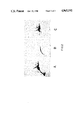
- FIG. 3 is a plot of the degrees of cutaneous reaction, measured by the observed diameters in mm of the swollen areas, resulting from the injection as challenge antigen of varying amounts, expressed in micrograms of the A60 antigen in test guinea pigs pre-sensitized with killed M. bovis bacteria, from which the minimum level of the antigen capable of eliciting a perceivable reaction can be seen.
- the bacterial (with the exception of M. leprae) are multiplied in Dubos medium supplemented with 5% horse serum decomplemented through heating at 56° C. during 30'. They are collected at the end of the growth phase through centrifugation and the bacterial walls are disrupted by compression and decompression in a French cell (Aminco instruments, Silver Spring, Md., U.S.A.) at 8000 p.s.i. and 4° C.
- A60 antigen from various Mycobacteria is easily isolated and purified in a one-step chromatographic procedure. Homogenates of the various Mycobacteria are centrifuged at 20000 r.p.m. for 10! at 4° C. to eliminate cellular debris from the supernatant containing the cytoplasm.
- CIE bidimensional or cross immunoelectrophoresis
- FIG. 1 is obtained by subjecting a test sample, about 24 ⁇ l of the starting cytoplasmic sample in 10 ⁇ l barbital buffer, to electrophoresis in two directions or dimensions with the second direction of electrophoresis being through an agarose gel containing whole anti-BCG serum obtained commercially from Dako of Denmark, and then to coloring by Coomassie blue.
- FIG. 2 shows the result of molecular sieve chromagraphy obtained by passing a cytoplasmic sample of M.
- bovis BCG consisting of 16.5 mg protein in 1 ml on a Sepharose 6B column (Trademark for spherical agarose gel beads with exclusion limits of about 4 ⁇ 10 6 daltons for proteins and 1 ⁇ 10 6 daltons for polysaccharides sold by Pharmacia Fine Chemicals AB).
- the starting sample is indicated at A and has a proteinic pattern of immunoelectrophoresis similar to that of FIG. 1; the exclusion peak is indicated at B which consists substantially of A60 antigen as shown by its immunoelectrophoretic location, and the inclusion peak is at C which includes all other proteinaceous matter, i.e., all other antigens present in the sample.
- peak B in case the presence of some contaminants is still observed after immunoelectrophoretic analysis may be re-chromatographed on the same column, which will yield A60 antigen in an immunochemically homogeneous form.
- A60 antigen purified by bichromatography on a Sepharose 6B column has been chemically analyzed. It is composed of 57% protein and 43% polysaccharides. Its unit of reference was established as follows: 1 unit A60 antigen is the quantity of electrophoretically purified and Coomassie blue colored antigen whose absorbance in solution at 530 nm is:
- This spectrophotometric unit corresponds to 1.5 ⁇ g of the proteinic component and to 1.23 ⁇ g of the polysaccharidic component. It corresponds thus to 2.45 ⁇ g of cytoplasmic A60 antigen.
- Antigen A60 of M. bovis is the slowest migrating component of the bacterial cytoplasm into the electrophoretic field under the following test conditions (1% agarose in Tris-barbital buffer 0.02M at pH 8.6; 8 volts/cm during 1 h at 15° C. in the first dimension and 3 volts/cm during 18 h at 15° C. in the second dimension; detection of the proteinic components by Coomassie blue).
- the bulk of the work was accomplished with M. bovis attenuated strain-Calmette-Guerin Bacillus (BCG). After having determined the optimal conditions for isolation of A60 antigen, similar materials, recognized by a characteristic immunoelectrophoretic pattern corresponding to the A60 antigen of M.
- bovis strain BCG were isolated, through the same simple molecular-sieve chromatographic technique, from a photochromogen (group I, M. kansasii), a scotochromogen (group II, M. scrofulaceum), a nonchromogen (group III, M. xenopi) and a "rapid grower" (group IV, M. chelonei).
- group I M. kansasii
- scotochromogen group II, M. scrofulaceum
- nonchromogen group III, M. xenopi
- group IV M. chelonei
- the several A60 antigen products extracted from these 6 Mycobacteria were recognized by their characteristic immunoelectrophoretic behavior, as specified above. All Mycobacterial species analyzed contained this antigen in large quantities.
- the A60 antigen of M. leprae was recognized to be the principal antigen of lepromin, also named A 7 in former reference systems.
- mice were primed with Pristane (0.50 ml I.P.). Fifteen days later, one ml of a 10% suspension of an aqueous solution of the antigen (10 mg/ml) in Freund incomplete adjuvant was inoculated intraperitoneally. After 15 days, 2 weekly intraperitoneal re-inoculations took place. The mice produced therewith an ascites that secreted large amounts of antibodies. The mice were punctured and drained every 3 days, and they usually died after the 5th to 6th drainage, from dehydration.
- Monoclonal antibodies were obtained in Balb/c mice according to known procedures. (Kohler, G. and Milstein C.: Continuous cultures of fused cells secreting antibody of predefined specificity. Nature, 1975, 256, 495-497).
- Polyclonal antibodies are a heterogenous population of antibodies directed against all epitopes available in an antigen.
- a monoclonal antibody is a homogenous population of antibodies directed against one single epitope of an antigen. This epitope may or may not be shared by other homologous antigens.
- a monoclonal antibody may be specific for a Mycobacterial strain, subspecies, species, genus.
- RIA radioimmunological test was therewith developed for the determination of minimal amounts of antibodies.
- Protein A from Staphylococcus aureus was purchased from Pharmacia (Uppsala, Sweden). This protein has the property to specifically combine with human and mouse IgG even if antigen is bound to them.
- the protein A was adsorbed on the walls of polystyrene tubes. I 125 labeled A60 antigen (about 3000 CMP per ⁇ l) was added to unknown amounts of antibodies and the resulting complex was thereafter contacted with the protein A carrier. After suitable incubation allowing attachment of the anbibodies to the protein A, the amount of radioactivity attached to the walls of the polystyrene was measured.
- PPD Purified Protein Derivative
- A60 antigens of various Mycobacteria species contain at least one epitope common to all species composing the CMN super-group and against which antibodies are made.
- An exploration of the specificity of the various monoclonal antibodies obtained was also made by the were found to be overlapping to some extent.
- one clone secreting an antibody absolutely specific for A60 antigen of M. kansasii and for A60 like antigen of M. scrofulaceum could be isolated as well as four (out of 51 clones) for A60 antigen of M. bovis.
- clone 31 was found to secrete antibodies that reacted with all A60 antigens presented. It had a very large spectrum of immunological activity. No clones secreting antibodies specific for the other Mycobacteria species were obtained, no doubt because the initial number of antibody secreting clones isolated was too small.
- the A60 antigen of M. bovis was adsorbed to the walls of polystyrene microcups (96 per plate) at pH 9.6 in 0.1M carbonate buffer. After adsorption, the wells were filled with a solution of 0.1% in albumin in phosphate buffered saline at 4° C. during 18 h, then the wells were washed with an 0.05% Tween 20 solution in PBS and the wells were finally drained and stored dry in the cold (4° C.). It was found that the A60 antigen adsorbed to the walls and kept dry could be stored at 4° C. during at least 4 months without any observable degradation in its immunological properties.
- Goat antibodies against human IgG were purified by affinity chromatography and labeled with peroxidase following classical procedures.
- This reagent has the dual function to adsorb to human IgG through its antibody moiety and to allow a colored enzymatic reaction through its peroxidase moiety.
- EIA enzymo-immunoassay
- the rapid latex slide agglutination test was normally slightly less sensitive. It picked up only 94% of the positives. However, all cutaneous negatives were also negative with the slide test.
- proteases are available to the art in order to break proteins into fragments.
- the most popular enzymes used for this purpose are trypsin, papain, chymotrypsin and subtilisin, but more esoteric enzymes may be applied as well as chemicals such as CNBr and beta-propiolactone. They split the protein at various specific aminoacid junctions producing thereby different fragments. Experiments were conducted to verify that the splitting with trypsin does not occur precisely at an immunologically important epitopic site. This was done by treating A60 antigen of M. bovis with the protease, and verifying therefore the ability of the fragments to compete for M. bovis antibodies with I 125 labeled antigen in an RIA-test, as in Example I.
- trypsin was suitable and no additional protease was used.
- A60 antigen of M. bovis (15 mg in 15 ml) was treated with trypsin according to known procedures. Briefly, trypsin attached to cellulose via CNBr was incubated with the solutions of A60 antigen in a buffer constituted of Hank's solution at pH 7.2. After 3 hours at 37° C., the cellulose-bound trypsin was removed by centrifugation.
- the solution of degraded A60 antigen of M. bovis was dialysed against an 0.1M glycine buffer at pH 8.0, then contacted with latex particles (0.8 ⁇ in diameter) as in Example II.
- a latex agglutination test was thereby produced that provided identical in sensitivity to a test developed with the intact A60-like antigen. Its superiority resided in the fact that the tendency to spontaneous unspecific aggregation of the latex particles in the presence of serum components was substantially reduced.
- Mycobacteria in the sputum of diseased people can be assessed by mixing the sputum with fluorescently-tagged antibodies developed against A60 antigen on a microscope slide. After suitable incubation, the slide is washed, is mounted and is viewed in a fluorescent microscope. This diagnostic test is too sophisticated to be applied in those geographic areas where the disease is most common; i.e., Spain, Southern Italy, North Africa.
- a less sophisticated test for such bacteria can be carried out as follows: Latex at a concentration of 10% and pH 8.1 was incubated during 72 h at 45° C. with MAb 31, obtained after cloning a hybridoma secreting antibodies against M. bovis A60 antigen. After washing, the latex suspension was assayed in a direct aggregation test for the presence of Mycobacteria in sputum.
- Mycobacteria bovis were mixed at various levels of concentration with the sputum of a disease-free donor to provide standardized test samples of known concentration and the resultant fluids appropriately diluted 1:5 with phosphate buffered saliva at pH 7.5 were mixed with the sensitized latex. Slow rotation was induced by hand during 3 minutes, after which the degree of aggregation of the sensitized latex was read under incident light. The test was not very sensitive.
- a more sensitive detection system for the presence of the bacteria was obtained through an EIA, where MAb 31 obtained as above was adsorbed onto the walls of polystyrene wells as in Example II. After incubation of the diluted sputum into the wells, these were washed and the presence of bacteria attached to the MAb 31 antibody was revealed by incubation of the wells with a peroxidase-labeled second antibody and coloration with O-phenylene diamine, as in Example II. In this test, the sensitivity was increased and the presence of 10,000 or even less bacteria/ml could be detected.
- Sepharose 4B or other molecular sieve material may be as efficient as Sepharose 6B for the separation of A60 antigen from other cytoplasmic constituents.
- Immunologically active fragments of A60 may be isolated in ways other than affinity chromatography. Other enzymes than trypsin may prove more efficient in the obtention of the immunologically active fragments.
- A60 antigen isolated from strain BCG was explored by the measurement of the footpad swelling of primed mice.
- Six-weeks old NMRI female mice were primed by the subcutaneous injection of 75 ⁇ l aliquots of a water-in-oil emulsion (F.I.A.) containing 1.5 ⁇ g/animal of either A60 antigen at a dose 1.5 ⁇ g/animal or tuberculin lot 80 C 12, used undiluted as received from the manufacturer.
- F.I.A. water-in-oil emulsion
- the A60 antigen elicits an hypersensitivity reaction similar to the one observed when tuberculin is used.
- A60 antigen is a potent immunogen eliciting cellular immune responses prompted the investigation of the value of A60 antigen as a replacement for conventional "tuberculins" in cutaneous testing for tuberculosis and its use as a quality control standard for comparing various commercial tuberculin preparations.
- Guinea pigs 400 gr. to 600 gr. were sensitized with 0.1 ⁇ g of killed M. bovis, administered intramuscularly. Four weeks later, various doses of A60 antigen were inoculated intradermally on the shaved animals' sides. A hypersensitive reaction was indicated by an indurated area or wheal which was measured in diameter after another 24 hours.
- the results of this experiment are given in a graph reproduced in FIG. 3.
- the minimal dose at which an established hypersensitivity was manifested or indicated was found to be 15.0 nanograms A60 antigen.
- This experiment demonstrates that a challenge of M. bovis primed animals may be accomplished with A60 antigen in lieu of tuberculin.
- tuberculins were diluted 1:10 and 1:100 in carbonate buffer (0,01M, ph9,6) and absorbed in the presence of glutaraldehyde (0,030M concentration) on the wells of a microtiter plate to serve as the capture antigen for the development of an enzyme-immunological test.
- the tuberculins when used in pure form as sensitizing material inhibit the enzyme-immunological reaction in three cases since the optical density values obtained with them is substantially lower than those obtained when the tuberculins are applied at a 1:10 or even 1:100 dilution.
- the inhibiting effect is best observed when the mouse anti-A60 antigen antibodies are given in a concentrated form (1:1.000 dilution) but the effect is still observable at higher dilutions.
- the immunologically reactive material effective for sensitization of the solid phase is overshadowed by competing contaminants in three out of the five lots tested.
- the potency relative to antibodies against A60 antigen of the different lots of tuberculin is unequal. This is observed when a low concentration of sensitizing or capture antigen is used (e.g., a 1:100 dilution of antigen) and also when diluted antibodies are applied (i.e., a 1:5,000 and 1:10,000 dilution of mouse anti-A60 antigen antibodies).
- a low concentration of sensitizing or capture antigen e.g., a 1:100 dilution of antigen
- diluted antibodies i.e., a 1:5,000 and 1:10,000 dilution of mouse anti-A60 antigen antibodies.
- Lot 85 K 28 is more potent than the other analyzed lots and lot 86 A 20 is the weakest.
- One scheme consists in a competition assay where an enzyme-labelled or radioactivity-labelled A60 antigen competes with the antigen contained in the tuberculin preparation for a limited amount of anti-A60 antigen antibody attached on a solid phase, such as the wells of a microtiter plate.
- the antibody may be monoclonal.
- the availability of a refined detection system for the serology of Mycobacterial infections permitted the investigation of its applicability in several other Mycobacterial infections.
- the A60 antigen is a potent interspecific antigen that reacts with the sera of patients suffering from a variety of Mycobacterial infections, so that the serum from a subject positively identified as infected with tuberculosis will be determined equally positive if A60 antigen isolated from Mycobacterial species other than M. bovis are used as capture antigen.
- A60 antigen isolated by the procedure of the prior application from M. paratuberculosis, M. avium or M. smegmatis may be used instead of A60 antigen isolated from M.
- Bovine sera originating from animals infected with M. bovis and others with M. paratuberculosis were immunologically reactive in the same way in tests where the capture antigen was A60 antigen from M. bovis strain BCG or M. paratuberculosis or M. avium.
- the Pharmacopoea requires that such test preparations produce a delayed hypersensitivity reaction, i.e., a swelling or wheal, at a test dosage which when multiplied 100 fold itself imparts no acquired sensitivity against the test antigen. That is to say, an acceptable test antigen must exhibit a "safety factor" exceeding 100 times between its so-called “revealing dose”, i.e., that amount at which a cellular response indicative of an established sensitivity to the antigen in the test subject, and the amount that will be sufficient to provoke a strong humoral response yielding effective levels of circulating antibodies as normally corresponds with immunization. Tests have established that the present A60 antigen fully satisfies this requirement, particularly since its minimum revealing dose is extremely small as already established above.
- tuberculosis can be distinguished from other Mycobacterial infections, especially that attributable to M. avium by a significantly greater area of swelling or induration at the test site so that a positive diagnosis for tuberculosis can readily be based on this increased diameter of the swelling due to the sensitivity reaction.
- the presence of a hypersensitivity reaction or test swelling will occur within a relatively short time and usually 24-48 hours between the time of administration of the test dose of the antigen and observation of the test site for the usual test responses of swelling, inflammation and the like.
- the A60 antigen of the invention is characterized by important advantages compared with previously known antigens derived from Mycobacteria and especially those associated with tuberculosis.
- typical commercially available tuberculin preparations exhibit a remarkable variation in their immunological activity that denotes a degree of nonhomogeneity that is quite unexpected for commercial test preparations.
- the present antigen material has been found to exhibit quite uniform immunological activity, a number of different batches of the present antigen having now been prepared over time and all found to behave in essentially identical respects.
- a standard reference unit for the A60 antigen can be quantified by an objective reproducible spectrophotometric procedure described above that does not depend upon subjective considerations as is true of the current standardization tests for commercial tuberculosis which depend upon the personal observation of the extent of a cutaneous sensitivity reaction.
- tubercular bacilli While quite specific molecular weight fractions derived from tubercular bacilli have been proposed and are presumably of uniform composition, they are necessarily only recovered in very small amounts and by rather complicated purification techniques. In contrast, the A60 antigen is recovered in usefully large volume from the starting material by a relatively simple procedure. Moreover, the electrophoretic reference system developed by Closs for classifying the various antigens contained in products derived from tubercular bacilli and like organisms provides a readily available way of analyzing the purity and homogeneity of the products of this invention. These products have the virtue of a useful heat stability, being essentially unaffected by heating up to a temperature of about 100° C. Commercial tuberculins in contrast normally contain contaminants, including hydrolytic enzymes and may hence by pyrogenic.
Landscapes
- Health & Medical Sciences (AREA)
- Life Sciences & Earth Sciences (AREA)
- Engineering & Computer Science (AREA)
- Immunology (AREA)
- Chemical & Material Sciences (AREA)
- Biomedical Technology (AREA)
- Molecular Biology (AREA)
- Hematology (AREA)
- Urology & Nephrology (AREA)
- Biotechnology (AREA)
- Microbiology (AREA)
- General Health & Medical Sciences (AREA)
- Biochemistry (AREA)
- Medicinal Chemistry (AREA)
- Physics & Mathematics (AREA)
- Genetics & Genomics (AREA)
- Food Science & Technology (AREA)
- Cell Biology (AREA)
- General Physics & Mathematics (AREA)
- Pathology (AREA)
- Tropical Medicine & Parasitology (AREA)
- Virology (AREA)
- Bioinformatics & Cheminformatics (AREA)
- Analytical Chemistry (AREA)
- Wood Science & Technology (AREA)
- Zoology (AREA)
- Organic Chemistry (AREA)
- General Engineering & Computer Science (AREA)
- Medicines Containing Antibodies Or Antigens For Use As Internal Diagnostic Agents (AREA)
- Peptides Or Proteins (AREA)
- Preparation Of Compounds By Using Micro-Organisms (AREA)
Abstract
An interspecific antigen of Mycobacteria consists essentially of a mixture in substantially immunochemically pure form of a protein having a molecular weight of at least about 4×10⁶ Daltons and polysaccharide having a molecular weight of at least about 1×10⁶ Daltons, and has when subjected to cross-electrophoresis an immunoelectrophoretic precipitation pattern corresponding to A60-antigen of Mycobacteria bovis strain BCG. This antigen is effective for detecting the prior exposure of a subject to Mycobacterial infections by a cutaneous test.
Description
This application is a continuation-in-part of application Ser. No. 678,470, filed Dec. 5, 1984 now U.S. Pat. No. 4,777,130.
This invention relates to an antigen derived from Mycobacteria and is concerned more particularly with an interspecific antigen of Mycobacteria exhibiting upon cross-electrophoresis an immunoelectrophoretic precipitation pattern corresponding to A60 antigen of Mycobacteria bovis strain BCG and to the use of such A60 antigen as a challenge antigen for indicating cell-medicated immunity by delayed hypersensitive reaction.
It is known that the detection of delayed sensitivity reactions which are the basis for cutaneous testing for tuberculosis can be accomplished by a material extracted from Mycobacterium bovis, strain BCG (Bacillus Calmette-Guerin). This BCG derived material, as well as so-called old tuberculin (O.T.) and Purified PROTEIN Derivative (PPD) are heterogeneous in composition. There is, consequently, a need for a purer and more precisely characterized antigenic material, possessing greater and more reliable effectiveness for the cutaneous testing for such diseases.
It was found that a high molecular weight antigenic material related in important immunochemical respects to the A60 antigen of M. bovis strain BCG may be extracted from various Mycobacterial species in substantially immunochemically pure form by the method claimed in U.S. patent application No. 678,170, filed Dec. 5, 1984. This antigenic material proved to possess those properties required to serve as the capture antigen in diagnostic test procedures and resulted in the development of an effective serological diagnostic test also claimed in that application. Taking advantage of the refined and reliable detection test thus made available, it has now been established that the A60 like antigenic material in question can be effectively used as the challenge antigen in delayed sensitivity tests for confirming the acquisition of immunity upon vaccination as well as prior exposure of the subject to tubercular-related infections.
The high molecular weight A60 is capable of eliciting as a delayed hypersensitivity as a cellular immune response and also able to serve as a challenge antigen when administered in much smaller amounts for indicating or revealing an established hypersensitivity either artificially or naturally initiated. This antigen has the potential for replacing the currently used tubercular-derived materials as a sensitizing agent, or challenge antigen, used in cutaneous testing for such diseases.
FIG. 1 is an illustration of the reference system (Closs et al, Scand. J. Immunol., 1980, 12, 249-263) for extracellular antigens of M. bovis BCG obtained by bidimensional immunoelectrophoresis and colored with Coomassie blue, the second dimension occurring through a gel containing whole anti-BCG serum;
FIG. 2 shows the immunoelectrophoretic patterns resulting from exclusion chromatography of a cytoplasmic sample of M. bovis BCG in which A is the starting cytoplasma, B is an exclusion peak constituted substantially entirely of A60-like antigen, and C is the inclusion peak containing all other proteinaceous matter; and
FIG. 3 is a plot of the degrees of cutaneous reaction, measured by the observed diameters in mm of the swollen areas, resulting from the injection as challenge antigen of varying amounts, expressed in micrograms of the A60 antigen in test guinea pigs pre-sensitized with killed M. bovis bacteria, from which the minimum level of the antigen capable of eliciting a perceivable reaction can be seen.
A number of different strains of Mycobacteria were used for the isolation of A60 antigen and were obtained mainly from the National Collection of Cultures of Microorganisms, C.N.C.M., Pasteur Institute, 25, rue du Docteur Roux, 75724 Paris FRANCE. Another source was the Station experimentale d'aviculture B.P. 9, 22440 PLOUFSAGAN, FRANCE.
The bacterial (with the exception of M. leprae) are multiplied in Dubos medium supplemented with 5% horse serum decomplemented through heating at 56° C. during 30'. They are collected at the end of the growth phase through centrifugation and the bacterial walls are disrupted by compression and decompression in a French cell (Aminco instruments, Silver Spring, Md., U.S.A.) at 8000 p.s.i. and 4° C.
A60 antigen from various Mycobacteria is easily isolated and purified in a one-step chromatographic procedure. Homogenates of the various Mycobacteria are centrifuged at 20000 r.p.m. for 10! at 4° C. to eliminate cellular debris from the supernatant containing the cytoplasm. When a cytoplasmic sample of M. bovis BCG is subjected to bidimensional or cross immunoelectrophoresis (CIE), the various antigenic constituents thereof are fractionated in a pattern as illustrated in FIG. 1. Specifically, the reference pattern of FIG. 1 is obtained by subjecting a test sample, about 24 μl of the starting cytoplasmic sample in 10 μl barbital buffer, to electrophoresis in two directions or dimensions with the second direction of electrophoresis being through an agarose gel containing whole anti-BCG serum obtained commercially from Dako of Denmark, and then to coloring by Coomassie blue. FIG. 2 shows the result of molecular sieve chromagraphy obtained by passing a cytoplasmic sample of M. bovis BCG consisting of 16.5 mg protein in 1 ml on a Sepharose 6B column (Trademark for spherical agarose gel beads with exclusion limits of about 4×10⁶ daltons for proteins and 1×10⁶ daltons for polysaccharides sold by Pharmacia Fine Chemicals AB). The starting sample is indicated at A and has a proteinic pattern of immunoelectrophoresis similar to that of FIG. 1; the exclusion peak is indicated at B which consists substantially of A60 antigen as shown by its immunoelectrophoretic location, and the inclusion peak is at C which includes all other proteinaceous matter, i.e., all other antigens present in the sample. If desired, peak B, in case the presence of some contaminants is still observed after immunoelectrophoretic analysis may be re-chromatographed on the same column, which will yield A60 antigen in an immunochemically homogeneous form.
A60 antigen purified by bichromatography on a Sepharose 6B column has been chemically analyzed. It is composed of 57% protein and 43% polysaccharides. Its unit of reference was established as follows: 1 unit A60 antigen is the quantity of electrophoretically purified and Coomassie blue colored antigen whose absorbance in solution at 530 nm is:
______________________________________
1 cm
A = 0.1
530
______________________________________
This spectrophotometric unit corresponds to 1.5 μg of the proteinic component and to 1.23 μg of the polysaccharidic component. It corresponds thus to 2.45 μg of cytoplasmic A60 antigen.
Antigen A60 of M. bovis is the slowest migrating component of the bacterial cytoplasm into the electrophoretic field under the following test conditions (1% agarose in Tris-barbital buffer 0.02M at pH 8.6; 8 volts/cm during 1 h at 15° C. in the first dimension and 3 volts/cm during 18 h at 15° C. in the second dimension; detection of the proteinic components by Coomassie blue). The bulk of the work was accomplished with M. bovis attenuated strain-Calmette-Guerin Bacillus (BCG). After having determined the optimal conditions for isolation of A60 antigen, similar materials, recognized by a characteristic immunoelectrophoretic pattern corresponding to the A60 antigen of M. bovis strain BCG were isolated, through the same simple molecular-sieve chromatographic technique, from a photochromogen (group I, M. kansasii), a scotochromogen (group II, M. scrofulaceum), a nonchromogen (group III, M. xenopi) and a "rapid grower" (group IV, M. chelonei). To these representatives of the various Mycobacterial groups that induce some sort of tuberculosis (M. xenopi and M. scrofulaceum are usually non-pathogenic) was added M. leprae, which had been armadillo-grown, and purchased under the form of lepromin.
About 15 mg of proteins liberated from the cytoplasm of each of these bacteria in 1 ml buffer (CaCl2 - 0.04M; Mg-acetate - 0.015M; Tris-HCl - 0.02M; ph - 7.4) were treated for 10' at 37° C. with 10 units of RNase, then immediately chromatographed twice on a Sepharose 6B column and the A60 antigens from the 6 starting Mycobacteria were brought therewith to immunochemical purity. Samples applied to the column were 1 ml, bed volume of Sepharose was 10 ml and void volume was 3.5 ml.
The several A60 antigen products extracted from these 6 Mycobacteria were recognized by their characteristic immunoelectrophoretic behavior, as specified above. All Mycobacterial species analyzed contained this antigen in large quantities. The A60 antigen of M. leprae was recognized to be the principal antigen of lepromin, also named A 7 in former reference systems.
The production of polyclonal murine antibodies against the isolated A60 antigens was carried out according to known procedures. Such procedures include recovery from the serum of peritoneal cavity of animals inoculated with the antigen. Briefly, Balb/c mice were primed with Pristane (0.50 ml I.P.). Fifteen days later, one ml of a 10% suspension of an aqueous solution of the antigen (10 mg/ml) in Freund incomplete adjuvant was inoculated intraperitoneally. After 15 days, 2 weekly intraperitoneal re-inoculations took place. The mice produced therewith an ascites that secreted large amounts of antibodies. The mice were punctured and drained every 3 days, and they usually died after the 5th to 6th drainage, from dehydration.
Monoclonal antibodies were obtained in Balb/c mice according to known procedures. (Kohler, G. and Milstein C.: Continuous cultures of fused cells secreting antibody of predefined specificity. Nature, 1975, 256, 495-497).
Fifty-one clones each secreting an antibody specific against the A60 antigen derived from M. bovis strain BCG were isolated, and three to six clones for each of the five other isolated A60 antigens.
Polyclonal antibodies are a heterogenous population of antibodies directed against all epitopes available in an antigen. A monoclonal antibody is a homogenous population of antibodies directed against one single epitope of an antigen. This epitope may or may not be shared by other homologous antigens. In the present case, a monoclonal antibody may be specific for a Mycobacterial strain, subspecies, species, genus.
The availability of polyclonal antibodies, of monoclonal anti-bodies and of a large supply of A60 antigens/(except M. leprae) of high purity isolated as described above allowed the development of several diagnostic tests. Their use will be exemplified in the following examples:
120 μg of antigen-like A60 in 120 μl phosphate buffered saline, pH 7.2 (PBS) were incubated with 3 μl of I 125 (300 μCi) and 10 μl of chloramine T (6.5 mg/ml in phosphate buffered saline pH 7.2) during 3 minutes at 20° C. The reaction was stopped by addition of 10 μl sodium metabisulfite (8.45 mg/ml in phosphate buffered saline pH 7.2). The final volume was brought to 200 μl with PBS and chromatographed on 2 ml of Sepharose 6B. The exclusion peak containing I 125 labeled A60 antigen was stored in the presence of 0.2% albumin and 0.02% NaN3.
A radioimmunological (RIA) test was therewith developed for the determination of minimal amounts of antibodies. Protein A from Staphylococcus aureus was purchased from Pharmacia (Uppsala, Sweden). This protein has the property to specifically combine with human and mouse IgG even if antigen is bound to them. The protein A was adsorbed on the walls of polystyrene tubes. I 125 labeled A60 antigen (about 3000 CMP per μl) was added to unknown amounts of antibodies and the resulting complex was thereafter contacted with the protein A carrier. After suitable incubation allowing attachment of the anbibodies to the protein A, the amount of radioactivity attached to the walls of the polystyrene was measured.
This classical radioimmunological assay (RIA) test repeated with the various polyclonal antibodies and the 6 labeled A60 antigen products available established that all polyclonal antibodies obtained by the injection of these antigens as vaccines interreacted to a large extent (7% to 100%) with all A60 labeled antigen products applied in an RIA test.
It was discovered from further tests that the radioactive labeled A60 antigens competed to a variable extent with Purified Protein Derivative (PPD) which is a purified preparation of tuberculin (i.e., an extract of BCG), and surprisingly also with lepromin (an extract of M. leprae) and with Leoprosy-derived-corynebacteria (L.D.C.), for complexing with the 6 polyclonal antibodies tested.
This indicates that A60 antigens of various Mycobacteria species, including those that do not provoke tuberculosis, contain at least one epitope common to all species composing the CMN super-group and against which antibodies are made. An exploration of the specificity of the various monoclonal antibodies obtained was also made by the were found to be overlapping to some extent. However, one clone secreting an antibody absolutely specific for A60 antigen of M. kansasii and for A60 like antigen of M. scrofulaceum could be isolated as well as four (out of 51 clones) for A60 antigen of M. bovis. On the other hand, clone 31 was found to secrete antibodies that reacted with all A60 antigens presented. It had a very large spectrum of immunological activity. No clones secreting antibodies specific for the other Mycobacteria species were obtained, no doubt because the initial number of antibody secreting clones isolated was too small.
The A60 antigen of M. bovis was adsorbed to the walls of polystyrene microcups (96 per plate) at pH 9.6 in 0.1M carbonate buffer. After adsorption, the wells were filled with a solution of 0.1% in albumin in phosphate buffered saline at 4° C. during 18 h, then the wells were washed with an 0.05% Tween 20 solution in PBS and the wells were finally drained and stored dry in the cold (4° C.). It was found that the A60 antigen adsorbed to the walls and kept dry could be stored at 4° C. during at least 4 months without any observable degradation in its immunological properties.
Goat antibodies against human IgG were purified by affinity chromatography and labeled with peroxidase following classical procedures. This reagent has the dual function to adsorb to human IgG through its antibody moiety and to allow a colored enzymatic reaction through its peroxidase moiety.
An enzymo-immunoassay (EIA) was thereby prepared, where human antibodies against A60 antigen contained in a test sample, e.g., patient sera, were specifically adsorbed on the sensitized walls of the microcup by complexing with the adsorbed A60 antigen. After washing with TweenPBS, the presence of the human specific antibodies that attach to the A60 antigen on the wells is revealed by peroxidase-labeled anti-human IgG antibodies. These antibodies will attach to any human IgG present, and after washing, the presence of these antibodies is analyzed by a color reaction based on the enzymatic activity of its peroxidase moiety.
As a parallel test, the same A60 antigen (500 μg/ml) was adsorbed to 20% suspension of latex particles (0.8μ in diameter) in 0.1 m glycine buffer at pH 8.0. After washing the latex particles, verification that agglutination would take place in the presence of a dilution of anti-A60 antiserum in PBS was accomplished by mixture of 50 μl of a dilution of a polyclonal antibody on a slide. After 5 minutes of slow rotation, the limit of dilution of the antibody was recognized to be 1/1200. Competition between the sensitized latex and free A60 antigen for this dilution of antibody showed that 2.6 μg/ml of free A60 antigen were needed to inhibit the latex agglutination induced by the diluted antibody.
These two qualitative tests were used to verify the presence of antibodies against A60 antigen in the sera of patients who were known to be positive for tuberculosis according to a standard cutaneous test. In order to avoid non-specific reactions of the sera with the latex sensitized with A60 antigen, the sera had to be diluted 6 times in PBS before use.
A correlation of 98% was found for positive reactions between cutaneous tests and EIA results just described.
Sera shown to be negative by the standard tuberculin test proved negative by this EIA, although the EIA did produce some false positives (97% correct).
The rapid latex slide agglutination test was normally slightly less sensitive. It picked up only 94% of the positives. However, all cutaneous negatives were also negative with the slide test.
These results indicate that antibodies against the A60 antigens of the invention derived from Mycobacteria are present in large quantities in the serum of patients that showed positive for tuberculosis in a cutaneous tuberculin test and that diagnostic tests--latex agglutination or EIA--based on recognition of such antibodies in serum by the A60 antigen of the invention are valuable diagnostic tools.
Several proteases are available to the art in order to break proteins into fragments. The most popular enzymes used for this purpose are trypsin, papain, chymotrypsin and subtilisin, but more esoteric enzymes may be applied as well as chemicals such as CNBr and beta-propiolactone. They split the protein at various specific aminoacid junctions producing thereby different fragments. Experiments were conducted to verify that the splitting with trypsin does not occur precisely at an immunologically important epitopic site. This was done by treating A60 antigen of M. bovis with the protease, and verifying therefore the ability of the fragments to compete for M. bovis antibodies with I 125 labeled antigen in an RIA-test, as in Example I.
Here, trypsin was suitable and no additional protease was used.
A60 antigen of M. bovis (15 mg in 15 ml) was treated with trypsin according to known procedures. Briefly, trypsin attached to cellulose via CNBr was incubated with the solutions of A60 antigen in a buffer constituted of Hank's solution at pH 7.2. After 3 hours at 37° C., the cellulose-bound trypsin was removed by centrifugation.
The solution of degraded A60 antigen of M. bovis was dialysed against an 0.1M glycine buffer at pH 8.0, then contacted with latex particles (0.8μ in diameter) as in Example II. A latex agglutination test was thereby produced that provided identical in sensitivity to a test developed with the intact A60-like antigen. Its superiority resided in the fact that the tendency to spontaneous unspecific aggregation of the latex particles in the presence of serum components was substantially reduced.
The presence of Mycobacteria in the sputum of diseased people can be assessed by mixing the sputum with fluorescently-tagged antibodies developed against A60 antigen on a microscope slide. After suitable incubation, the slide is washed, is mounted and is viewed in a fluorescent microscope. This diagnostic test is too sophisticated to be applied in those geographic areas where the disease is most common; i.e., Spain, Southern Italy, North Africa.
A less sophisticated test for such bacteria can be carried out as follows: Latex at a concentration of 10% and pH 8.1 was incubated during 72 h at 45° C. with MAb 31, obtained after cloning a hybridoma secreting antibodies against M. bovis A60 antigen. After washing, the latex suspension was assayed in a direct aggregation test for the presence of Mycobacteria in sputum. Mycobacteria bovis were mixed at various levels of concentration with the sputum of a disease-free donor to provide standardized test samples of known concentration and the resultant fluids appropriately diluted 1:5 with phosphate buffered saliva at pH 7.5 were mixed with the sensitized latex. Slow rotation was induced by hand during 3 minutes, after which the degree of aggregation of the sensitized latex was read under incident light. The test was not very sensitive.
The presence of at least 100,000 bacteria/ml sputum was needed to begin to see an aggregation. Nevertheless, despite its insensitivity, the test was functional, and no less sensitive than direct microscopic observation of smears.
A more sensitive detection system for the presence of the bacteria was obtained through an EIA, where MAb 31 obtained as above was adsorbed onto the walls of polystyrene wells as in Example II. After incubation of the diluted sputum into the wells, these were washed and the presence of bacteria attached to the MAb 31 antibody was revealed by incubation of the wells with a peroxidase-labeled second antibody and coloration with O-phenylene diamine, as in Example II. In this test, the sensitivity was increased and the presence of 10,000 or even less bacteria/ml could be detected.
It was also found that in case ribosomes and ribosomal RNA contaminate the A60 antigen prepared from the bacteria cytoplasm; a treatment of the extract with RNAse and DNAse prior to exclusion chromatography considerably improved the purity of the chromatographed product. Sepharose 4B or other molecular sieve material may be as efficient as Sepharose 6B for the separation of A60 antigen from other cytoplasmic constituents. Immunologically active fragments of A60 may be isolated in ways other than affinity chromatography. Other enzymes than trypsin may prove more efficient in the obtention of the immunologically active fragments.
The isolation of the A60 antigen and its use in diagnostic tests designed to detect in simple and easy to perform fashion the presence of antibodies against Mycobacteria, including both IgG and IgM specific antibodies as claimed in U.S. application Ser. No. 678,170, has made possible additional discoveries.
The capacity of A60 antigen isolated from strain BCG to induce delayed hypersensitivity reactions were explored by the measurement of the footpad swelling of primed mice. Six-weeks old NMRI female mice were primed by the subcutaneous injection of 75 μl aliquots of a water-in-oil emulsion (F.I.A.) containing 1.5 μg/animal of either A60 antigen at a dose 1.5 μg/animal or tuberculin lot 80 C 12, used undiluted as received from the manufacturer.
Reactions indicative of delayed hypersensitivity were elicited by injections as challenge antigen of 30 μg of A60 antigen solubilized in phosphate buffered saline or the same amount of pure tuberculin lot 80 C 12 used undiluted, into hind footpads of the primed animals. After 24 hours, the pad thickness was measured by a caliper. Table I resumes the results obtained. Eight mice were used per experimental group:
              TABLE I
______________________________________
Delayed Hypersensitivity Reactions
                        Mean footpad
Injection    Challenge  swelling in mm
______________________________________
A 60         A 60       1.62
Tub 80 C 12  Tub 80 C 12
                        1.40
Buffer       A 60       0.11
A 60         Buffer     0.04
F.I.A. alone A 60       0.01
Tub 80 C 12  A 60       1.34
A 60         Tub 80 C 12
                        1.49
______________________________________
Clearly, the A60 antigen elicits an hypersensitivity reaction similar to the one observed when tuberculin is used.
The discovery that A60 antigen is a potent immunogen eliciting cellular immune responses prompted the investigation of the value of A60 antigen as a replacement for conventional "tuberculins" in cutaneous testing for tuberculosis and its use as a quality control standard for comparing various commercial tuberculin preparations.
The suitability of A60 antigen isolated from BCG as a challenge antigen similar to conventional "tuberculins" of various types was first investigated by means of a classical "tuberculin" evaluation test.
Guinea pigs (400 gr. to 600 gr.) were sensitized with 0.1 μg of killed M. bovis, administered intramuscularly. Four weeks later, various doses of A60 antigen were inoculated intradermally on the shaved animals' sides. A hypersensitive reaction was indicated by an indurated area or wheal which was measured in diameter after another 24 hours.
The results of this experiment are given in a graph reproduced in FIG. 3. The minimal dose at which an established hypersensitivity was manifested or indicated was found to be 15.0 nanograms A60 antigen. This experiment demonstrates that a challenge of M. bovis primed animals may be accomplished with A60 antigen in lieu of tuberculin.
The quality in terms of degree of homogeneity of five commercial preparations or lots of "tuberculins" were analyzed in an enzyme-immunoassay. The tuberculins were diluted 1:10 and 1:100 in carbonate buffer (0,01M, ph9,6) and absorbed in the presence of glutaraldehyde (0,030M concentration) on the wells of a microtiter plate to serve as the capture antigen for the development of an enzyme-immunological test. Mouse antibodies against A60 antigen at various dilutions were applied and the presence of these antibodies in complexed form on the plate was revealed by a peroxidase-labelled antimouse IgG (Dako, Denmark) and subsequent color development with ortho-phenylene diamine and H2 O2 in citric-phosphate buffer pH 5,3. The results are given in Table 2.
              TABLE II
______________________________________
Optical density at 492 nm of a mouse-enzyme-immunological dosage
with five commercial lots of tuberculins used as capture antigen.
          Anti-A60- antigen mouse antiserum dilution
Tuberculin Lot
            1:1,000     1:5,000 1:10,000
______________________________________
86 A 20
Pure        1.05        0.30    0.19
1:10        1.03        0.31    0.20
1:100       0.65        0.23    0.15
84 H 30
Pure        0.41        0.18    0.13
1:10        1.31        0.47    0.28
1:100       1.28        0.47    0.28
83 I 26
Pure        0.35        0.19    0.14
1:10        1.05        0.51    0.36
1:100       1.39        0.42    0.29
85 K 28
Pure        0.98        0.40    0.27
1:10        1.04        0.46    0.39
1:100       1.03        0.51    0.42
80 C 12
Pure        0.84        0.23    0.17
1:10        1.01        0.29    0.19
1:100       0.86        0.28    0.19
______________________________________
An examination of the data leads to the following conclusions:
1. The tuberculins when used in pure form as sensitizing material inhibit the enzyme-immunological reaction in three cases since the optical density values obtained with them is substantially lower than those obtained when the tuberculins are applied at a 1:10 or even 1:100 dilution. The inhibiting effect is best observed when the mouse anti-A60 antigen antibodies are given in a concentrated form (1:1.000 dilution) but the effect is still observable at higher dilutions. Presumably, the immunologically reactive material effective for sensitization of the solid phase is overshadowed by competing contaminants in three out of the five lots tested.
2. The degree of contamination varies for different tuberculin lots since the inhibiting effect is nonexistent for lot 86 A 20, is slight for lot 80 C 12, is more accentuated for lot 85 K 28, is very pronounced for lot 84 H 30 and is maximal for lot 83 I 26.
3. The potency relative to antibodies against A60 antigen of the different lots of tuberculin is unequal. This is observed when a low concentration of sensitizing or capture antigen is used (e.g., a 1:100 dilution of antigen) and also when diluted antibodies are applied (i.e., a 1:5,000 and 1:10,000 dilution of mouse anti-A60 antigen antibodies).
Lot 85 K 28 is more potent than the other analyzed lots and lot 86 A 20 is the weakest.
The same experimental evaluation was applied to A60 antigen isolated from BCG. In this case, the availability of pure material allows the quantification of the amount of sensitizing antigen used. Solutions ranging from 10 μg to 5 ng A60 protein per ml were applied. Table 3 records the results obtained.
              TABLE III
______________________________________
 A60-like
antigen  Anti-A60-like antigen mouse antiserum dilution
concentration
         1:1,000       1:5,000 1:10,000
______________________________________
10 ug    2             1.49    0.85
5 ug     2             1.48    0.86
0.5 ug   2             1.38    0.81
0.05 ug  1.47          0.68    0.32
0.005 ug 0.42          0.29    0.18
______________________________________
(1) Contrary to most lots of tuberculins assayed, large doses of the sensitizing A60 antigen show no inhibiting or competitive effect on the E.I.Assay.
(2) Saturating amounts of the A60 sensitizing antigen seem to be attained when 0.5 μg or more are used, since the sensitivity of the system is not improved with higher concentrations as observed when high dilutions (1:5,000 and 1:10,000) of the anti-A60 mouse antibodies are applied.
(3) The intensity of the observed responses is much greater than that observed for the lots of tuberculins analyzed.
The determination of the potency of a given tuberculin preparation with respect to A60 antigen is thus possible.
One scheme consists in a competition assay where an enzyme-labelled or radioactivity-labelled A60 antigen competes with the antigen contained in the tuberculin preparation for a limited amount of anti-A60 antigen antibody attached on a solid phase, such as the wells of a microtiter plate. The antibody may be monoclonal.
However, the information and materials at hand pointed to the development of an indirect non-competitive test based on an enzyme-labelled anti-mouse antiserum (after U.S. Pat. No. 4,298,687).
Serial dilutions of A60 antigen ranging from 0 to 500 nanograms were made and incubated (0.1 ml) for 3 hours at 37° C. in the presence of 0.1 ml of mouse antiserum against A60 antigen diluted to a 1:5,000 level (final dilution.).
The solutions (0.2 ml) were thereafter incubated for 18 hours (overnight) at room temperature in microtiter wells coated with 500 nanograms of A60 antigen. The wells were washed, an excess of peroxidase-labelled anti-mouse antiserum (Dako, Denmark) was added and incubation was continued for three more hours at room temperature. After washing, the presence of peroxidase was manifested by color development with ortho-phenylene diamine and H2 O2 in citric acid-phosphate buffer.
In this detection system, maximum color development occurs when A60 antigen is absent from the reacting fluid and minimum color development near that of the background is obtained when an amount of A60 antigen sufficient to bind the totality of the antibodies against A60 antigen with the color development intensity varying between the limiting levels in accordance with the amount of A60 antigen between these extremes. Thus, a reference curve based on varying color development levels can be constructed of A60 antigen concentration in unknown fluids and a monoclonal antibody can also be employed for this purpose.
In this way, the same five lots of commercial tuberculins were analyzed at a 1:100 dilution. Their content in A60 material reflected quite accurately the results obtained in Table II. Most potent was lot 85 K 28 (7 μg A60 antigen equivalent/ml) and least concentrated in A60 materials was lot 86 A 20 (0.6 μg A60 antigen equivalent).
The availability of a refined detection system for the serology of Mycobacterial infections permitted the investigation of its applicability in several other Mycobacterial infections. Very unexpectedly, it was found that the A60 antigen is a potent interspecific antigen that reacts with the sera of patients suffering from a variety of Mycobacterial infections, so that the serum from a subject positively identified as infected with tuberculosis will be determined equally positive if A60 antigen isolated from Mycobacterial species other than M. bovis are used as capture antigen. As a convenient example, A60 antigen isolated by the procedure of the prior application from M. paratuberculosis, M. avium or M. smegmatis may be used instead of A60 antigen isolated from M. bovis for the serodiagnosis of tuberculosis in subjects either human or animal. Sera from five M. avium infected patients were obtained from the Cleveland Metropolitan General Hospital (U.S.A.). All five gave positive results in a serodiagnosis test with A60 antigen from M. bovis and also A60 antigen from M. avium and M. paratuberculosis. Sera originating from patients infected with M. xenopi were immunologically reactive with A60 antigen isolated from M. bovis, as were sera from M. kansassii infected patients with A60 antigen isolated from M. bovis.
Bovine sera originating from animals infected with M. bovis and others with M. paratuberculosis were immunologically reactive in the same way in tests where the capture antigen was A60 antigen from M. bovis strain BCG or M. paratuberculosis or M. avium.
As regards the utility of the A60 antigen as a challenge antigen for providing a visible manifestation of an established hypersensitivity in the test subject, it should be noted that the Pharmacopoea requires that such test preparations produce a delayed hypersensitivity reaction, i.e., a swelling or wheal, at a test dosage which when multiplied 100 fold itself imparts no acquired sensitivity against the test antigen. That is to say, an acceptable test antigen must exhibit a "safety factor" exceeding 100 times between its so-called "revealing dose", i.e., that amount at which a cellular response indicative of an established sensitivity to the antigen in the test subject, and the amount that will be sufficient to provoke a strong humoral response yielding effective levels of circulating antibodies as normally corresponds with immunization. Tests have established that the present A60 antigen fully satisfies this requirement, particularly since its minimum revealing dose is extremely small as already established above.
From what has already been said, it will be understood that the hypersensitivity manifestation or indication given by the present A60 antigen will not be confined to tuberculosis but will extend also to various other infections originating from Mycobacteria, including leprosy. This lack of specificity may under some circumstances be perceived as an advantage rather than a disadvantage for example when used for screening tests in less developed countries where a generalized discrimination of Mycobacterial infections in the test population might constitute important medical information in its own right. If a high degree of specificity for the identification of a specific Mycobacterial infection is required, this can be generally obtained by appropriate definition of the discrimination point between a positive or negative conclusion, as is indeed already practiced for known commercial "tuberculin" preparations. That is to say, the degree of the manifestation of sensitivity varies with different infections of the group in question so that as a practical matter, tuberculosis can be distinguished from other Mycobacterial infections, especially that attributable to M. avium by a significantly greater area of swelling or induration at the test site so that a positive diagnosis for tuberculosis can readily be based on this increased diameter of the swelling due to the sensitivity reaction.
As a rule, the presence of a hypersensitivity reaction or test swelling will occur within a relatively short time and usually 24-48 hours between the time of administration of the test dose of the antigen and observation of the test site for the usual test responses of swelling, inflammation and the like.
It will be appreciated that the A60 antigen of the invention is characterized by important advantages compared with previously known antigens derived from Mycobacteria and especially those associated with tuberculosis. As the comparative data provided above shows, typical commercially available tuberculin preparations exhibit a remarkable variation in their immunological activity that denotes a degree of nonhomogeneity that is quite unexpected for commercial test preparations. In contrast, the present antigen material has been found to exhibit quite uniform immunological activity, a number of different batches of the present antigen having now been prepared over time and all found to behave in essentially identical respects. Furthermore, a standard reference unit for the A60 antigen can be quantified by an objective reproducible spectrophotometric procedure described above that does not depend upon subjective considerations as is true of the current standardization tests for commercial tuberculosis which depend upon the personal observation of the extent of a cutaneous sensitivity reaction.
While quite specific molecular weight fractions derived from tubercular bacilli have been proposed and are presumably of uniform composition, they are necessarily only recovered in very small amounts and by rather complicated purification techniques. In contrast, the A60 antigen is recovered in usefully large volume from the starting material by a relatively simple procedure. Moreover, the electrophoretic reference system developed by Closs for classifying the various antigens contained in products derived from tubercular bacilli and like organisms provides a readily available way of analyzing the purity and homogeneity of the products of this invention. These products have the virtue of a useful heat stability, being essentially unaffected by heating up to a temperature of about 100° C. Commercial tuberculins in contrast normally contain contaminants, including hydrolytic enzymes and may hence by pyrogenic.
Claims (4)
1. An interspecific antigen of Mycobacteria consisting essentially of a mixture in substantially immunochemically pure form of a protein having a molecular weight of at least 4×10⁶ Daltons and polysaccharide having a molecular weight of at least about 1×10⁶ Daltons, and having when subjected to cross-electrophoresis an immunoelectrophoretic precipitation pattern corresponding to A60 antigen of Mycobacteria bovis strain BCG.
2. A method of detecting the prior exposure of a subject to Mycobacterial infections which comprises the step of injecting said subject intradermally or cutaneously with an effective amount of the antigen of claim 1 and after passage of a sufficient amount of time for the development by said subject of a hypersensitivity reaction, observing the site of said injection for the presence of significant induration as a positive indication of said prior exposure.
3. The method of detecting the presence of Mycobacteria in the humoral fluids of a subject which comprises mixing a sample of said humoral fluid with an antibody against the antigen of claim 1 and detecting the occurrence of complexation said antibody of said antibody Mycobacteria in said fluid.
4. A method of eliciting an immunological response against the antigen of claim 1 which comprises administering to said subject an immunologically effective amount of the antigen of claim 1.
Priority Applications (1)
| Application Number | Priority Date | Filing Date | Title |
|---|---|---|---|
| US07/187,919 US4965192A (en) | 1984-12-05 | 1988-04-29 | A60-antigen from Mycobacteria and use thereof as tuberculin and as vaccine |
Applications Claiming Priority (2)
| Application Number | Priority Date | Filing Date | Title |
|---|---|---|---|
| US06/678,470 US4777130A (en) | 1984-12-05 | 1984-12-05 | Isolation of mycobacterial a 60 antigen for diagnostic purposes |
| US07/187,919 US4965192A (en) | 1984-12-05 | 1988-04-29 | A60-antigen from Mycobacteria and use thereof as tuberculin and as vaccine |
Related Parent Applications (1)
| Application Number | Title | Priority Date | Filing Date |
|---|---|---|---|
| US06/678,470 Continuation-In-Part US4777130A (en) | 1984-12-05 | 1984-12-05 | Isolation of mycobacterial a 60 antigen for diagnostic purposes |
Related Child Applications (1)
| Application Number | Title | Priority Date | Filing Date |
|---|---|---|---|
| US07/506,046 Division US5068695A (en) | 1988-04-29 | 1990-04-09 | Low dislocation density semiconductor device |
Publications (1)
| Publication Number | Publication Date |
|---|---|
| US4965192A true US4965192A (en) | 1990-10-23 |
Family
ID=24722921
Family Applications (2)
| Application Number | Title | Priority Date | Filing Date |
|---|---|---|---|
| US06/678,470 Expired - Lifetime US4777130A (en) | 1984-12-05 | 1984-12-05 | Isolation of mycobacterial a 60 antigen for diagnostic purposes |
| US07/187,919 Expired - Lifetime US4965192A (en) | 1984-12-05 | 1988-04-29 | A60-antigen from Mycobacteria and use thereof as tuberculin and as vaccine |
Family Applications Before (1)
| Application Number | Title | Priority Date | Filing Date |
|---|---|---|---|
| US06/678,470 Expired - Lifetime US4777130A (en) | 1984-12-05 | 1984-12-05 | Isolation of mycobacterial a 60 antigen for diagnostic purposes |
Country Status (4)
| Country | Link |
|---|---|
| US (2) | US4777130A (en) |
| EP (1) | EP0184511B1 (en) |
| BE (1) | BE1002022A7 (en) |
| DE (1) | DE3587268T2 (en) |
Cited By (13)
| Publication number | Priority date | Publication date | Assignee | Title |
|---|---|---|---|---|
| US5306731A (en) * | 1985-06-14 | 1994-04-26 | Massachusetts Eye And Ear Infirmary | Method and products for treating the eye |
| US5776692A (en) * | 1995-05-23 | 1998-07-07 | Baylor College Of Medicine | Mycobacterial genus-specific DNA probe and its expressed product |
| WO1999039693A2 (en) * | 1998-02-06 | 1999-08-12 | Japan Bcg Laboratory | Methods and compositions for detection and diagnosis of infectious diseases |
| US6013660A (en) * | 1996-10-02 | 2000-01-11 | The Regents Of The University Of California | Externally targeted prophylactic and chemotherapeutic method and agents |
| US6090385A (en) * | 1993-12-10 | 2000-07-18 | Maes; Hubert | Method of treating cancer |
| US6403099B1 (en) * | 1992-03-06 | 2002-06-11 | Chiron S.P.A. | Conjugates formed from heat shock proteins and oligo-or polysaccharides |
| US6599691B1 (en) * | 1995-11-27 | 2003-07-29 | The United States Of America As Represented By The Secretary Of The Navy | Rapid immunoassay to detect infection with Mycobacterium tuberculosis |
| WO2003065002A2 (en) | 2002-01-30 | 2003-08-07 | THE GOVERNMENT OF THE UNITED STATES OF AMERICA as represented by THE NAVY MEDICAL RESEARCH CENTER | Rapid lateral flow assay for determining exposure to mycobacterium tuberculosis and other mycobacteria |
| US6752993B1 (en) | 1993-11-23 | 2004-06-22 | The Regents Of The University Of California | Abundant extracellular product vaccines and methods for their production and use |
| US6761894B1 (en) | 1993-11-23 | 2004-07-13 | The Regents Of The University Of California | Abundant extracellular products and methods for their production and use |
| US20040157802A1 (en) * | 2002-11-15 | 2004-08-12 | Marcus Horwitz | Anti-microbial agents derived from methionine sulfoximine analogues |
| US20040228873A1 (en) * | 1993-11-23 | 2004-11-18 | Horwitz Marcus A. | Abundant extracellular products and methods for their production and use |
| WO2008028489A2 (en) * | 2006-09-05 | 2008-03-13 | Hvidovre Hospital | Ip-i0 based immunological monitoring |
Families Citing this family (9)
| Publication number | Priority date | Publication date | Assignee | Title |
|---|---|---|---|---|
| US4777130A (en) * | 1984-12-05 | 1988-10-11 | Andra Biologicals | Isolation of mycobacterial a 60 antigen for diagnostic purposes |
| US5225324A (en) * | 1987-04-24 | 1993-07-06 | Bioscience International, Inc. | Diagnostics for mycobacteria in public health, medical, and veterinary practice |
| US5091308A (en) * | 1990-01-29 | 1992-02-25 | The Board Of Trustees Of The University Of Illinois | Process for the enzymatic dispersal of mycobacterium bovis - bcg |
| AU1564592A (en) * | 1991-02-12 | 1992-09-07 | Dynagen, Inc. | Agglutination test for mycobacterial antigens in biological samples |
| US5185242A (en) * | 1991-06-24 | 1993-02-09 | Becton Dickinson And Company | Method for lysing mycobacteria using achromopeptidase |
| US5344759A (en) * | 1992-05-11 | 1994-09-06 | Her Majesty The Queen In Right Of Canada, As Represented By The Minister Of Health & Welfare | Glycolipids for serodiagnosis of tuberculosis and leprosy |
| BE1007823A3 (en) * | 1993-12-10 | 1995-10-31 | Anda Biolog Sa | Use of a composition containing at least one antigen and / or one or more fragments of this antigen for obtaining a drug for treating and / or preventing cancer. |
| DE19621488C2 (en) * | 1996-05-29 | 1998-05-14 | Gerlach Gerald F Prof Dr | Diagnostic procedure for the detection of subclinical mammals suffering from paratuberculosis |
| NZ513418A (en) * | 2001-08-07 | 2004-04-30 | Univ Massey | Vaccine comprising proteins from mycobacterium paratuberculosis |
Citations (3)
| Publication number | Priority date | Publication date | Assignee | Title |
|---|---|---|---|---|
| US4410660A (en) * | 1980-05-05 | 1983-10-18 | Montefiore Medical Center | Binding assay for the detection of mycobacteria |
| US4591552A (en) * | 1982-09-29 | 1986-05-27 | New York Blood Center, Inc. | Detection of hepatitis B surface antigen (or antibody to same) with labeled synthetic peptide |
| US4777130A (en) * | 1984-12-05 | 1988-10-11 | Andra Biologicals | Isolation of mycobacterial a 60 antigen for diagnostic purposes |
Family Cites Families (4)
| Publication number | Priority date | Publication date | Assignee | Title |
|---|---|---|---|---|
| US3529057A (en) * | 1964-08-14 | 1970-09-15 | Takeda Chemical Industries Ltd | Purified periodic acid-oxidized mucopeptide of mycobacteria cell walls constituting a vaccine enhancing nonspecific host resistance against pathogenic bacterial infections |
| US3888837A (en) * | 1973-06-28 | 1975-06-10 | Mitsui Pharmaceuticals | Tuberculin active proteins and peptides from the cells of tubercle bacilli |
| US4123427A (en) * | 1976-07-27 | 1978-10-31 | Daniel Thomas M | Method for the purification of mycobacterial protein antigens and resulting product |
| US4351761A (en) * | 1978-05-15 | 1982-09-28 | Research Corporation | Purified antigen to test for Neisseria gonorrhoeae antibodies |
-
1984
- 1984-12-05 US US06/678,470 patent/US4777130A/en not_active Expired - Lifetime
-
1985
- 1985-12-02 EP EP85402376A patent/EP0184511B1/en not_active Expired - Lifetime
- 1985-12-02 DE DE8585402376T patent/DE3587268T2/en not_active Expired - Lifetime
-
1988
- 1988-04-29 US US07/187,919 patent/US4965192A/en not_active Expired - Lifetime
-
1989
- 1989-04-25 BE BE8900456A patent/BE1002022A7/en not_active IP Right Cessation
Patent Citations (3)
| Publication number | Priority date | Publication date | Assignee | Title |
|---|---|---|---|---|
| US4410660A (en) * | 1980-05-05 | 1983-10-18 | Montefiore Medical Center | Binding assay for the detection of mycobacteria |
| US4591552A (en) * | 1982-09-29 | 1986-05-27 | New York Blood Center, Inc. | Detection of hepatitis B surface antigen (or antibody to same) with labeled synthetic peptide |
| US4777130A (en) * | 1984-12-05 | 1988-10-11 | Andra Biologicals | Isolation of mycobacterial a 60 antigen for diagnostic purposes |
Cited By (24)
| Publication number | Priority date | Publication date | Assignee | Title |
|---|---|---|---|---|
| US5306731A (en) * | 1985-06-14 | 1994-04-26 | Massachusetts Eye And Ear Infirmary | Method and products for treating the eye |
| US6403099B1 (en) * | 1992-03-06 | 2002-06-11 | Chiron S.P.A. | Conjugates formed from heat shock proteins and oligo-or polysaccharides |
| US20040228873A1 (en) * | 1993-11-23 | 2004-11-18 | Horwitz Marcus A. | Abundant extracellular products and methods for their production and use |
| US7300660B2 (en) | 1993-11-23 | 2007-11-27 | The Regents Of The University Of California | Abundant extracellular products and methods for their production and use |
| US6752993B1 (en) | 1993-11-23 | 2004-06-22 | The Regents Of The University Of California | Abundant extracellular product vaccines and methods for their production and use |
| US6761894B1 (en) | 1993-11-23 | 2004-07-13 | The Regents Of The University Of California | Abundant extracellular products and methods for their production and use |
| US6090385A (en) * | 1993-12-10 | 2000-07-18 | Maes; Hubert | Method of treating cancer |
| US5776692A (en) * | 1995-05-23 | 1998-07-07 | Baylor College Of Medicine | Mycobacterial genus-specific DNA probe and its expressed product |
| US6599691B1 (en) * | 1995-11-27 | 2003-07-29 | The United States Of America As Represented By The Secretary Of The Navy | Rapid immunoassay to detect infection with Mycobacterium tuberculosis |
| US6013660A (en) * | 1996-10-02 | 2000-01-11 | The Regents Of The University Of California | Externally targeted prophylactic and chemotherapeutic method and agents |
| WO1999039693A2 (en) * | 1998-02-06 | 1999-08-12 | Japan Bcg Laboratory | Methods and compositions for detection and diagnosis of infectious diseases |
| WO1999039693A3 (en) * | 1998-02-06 | 1999-09-23 | Japan Bcg Lab | Methods and compositions for detection and diagnosis of infectious diseases |
| US20050106107A1 (en) * | 1998-02-06 | 2005-05-19 | Nakamura Reiko M. | Methods and compositions for detection and diagnosis of infectious diseases |
| US7655218B2 (en) | 1998-02-06 | 2010-02-02 | Japan Bcg Laboratory | Methods and compositions for detection and diagnosis of infectious diseases |
| CN100584378C (en) * | 1998-02-06 | 2010-01-27 | 日本Bcg制造株式会社 | Use of mycobacterium antigen MPB64 in preparing product for locally detecting active tuberculosis |
| WO2003065002A2 (en) | 2002-01-30 | 2003-08-07 | THE GOVERNMENT OF THE UNITED STATES OF AMERICA as represented by THE NAVY MEDICAL RESEARCH CENTER | Rapid lateral flow assay for determining exposure to mycobacterium tuberculosis and other mycobacteria |
| US6841159B2 (en) | 2002-01-30 | 2005-01-11 | The United States Of America As Represented By The Secretary Of The Navy | Rapid lateral flow assay for determining exposure to Mycobacterium tuberculosis and other mycobacteria |
| US20060142251A1 (en) * | 2002-11-15 | 2006-06-29 | Gunther Harth | Anti-microbial agents derived from methionine sulfoximine analogues |
| US20040157802A1 (en) * | 2002-11-15 | 2004-08-12 | Marcus Horwitz | Anti-microbial agents derived from methionine sulfoximine analogues |
| WO2008028489A2 (en) * | 2006-09-05 | 2008-03-13 | Hvidovre Hospital | Ip-i0 based immunological monitoring |
| WO2008028489A3 (en) * | 2006-09-05 | 2008-05-15 | Hvidovre Hospital | Ip-i0 based immunological monitoring |
| EA020412B1 (en) * | 2006-09-05 | 2014-11-28 | Видовре Хоспитал | Ip-10 based immunological monitoring |
| US20100086950A1 (en) * | 2006-09-05 | 2010-04-08 | Hvidovre Hospital | Ip-10 based immunological monitoring |
| US8026076B2 (en) | 2006-09-05 | 2011-09-27 | Hvidovre Hospital | IP-10 based immunological monitoring |
Also Published As
| Publication number | Publication date |
|---|---|
| DE3587268T2 (en) | 1993-07-29 |
| DE3587268D1 (en) | 1993-05-19 |
| EP0184511A3 (en) | 1988-04-06 |
| BE1002022A7 (en) | 1990-05-22 |
| US4777130A (en) | 1988-10-11 |
| EP0184511A2 (en) | 1986-06-11 |
| EP0184511B1 (en) | 1993-04-14 |
Similar Documents
| Publication | Publication Date | Title |
|---|---|---|
| US4965192A (en) | A60-antigen from Mycobacteria and use thereof as tuberculin and as vaccine | |
| Sada et al. | Detection of mycobacterial antigens in cerebrospinal fluid of patients with tuberculous meningitis by enzyme-linked immunosorbent assay | |
| Murphy et al. | Serological, electrophoretic, and biological properties of Cryptococcus neoformans antigens | |
| Halbert et al. | The use of precipitin analysis in agar for the study of human streptococcal infections: II. Ouchterlony and Oakley technics | |
| Fukazawa et al. | Response and specificity of antibodies for Candida albicans | |
| Jarolim et al. | Monoclonal antibodies against birch pollen allergens: characterization by immunoblotting and use for single-step affinity purification of the major allergen Bet v I | |
| Cocito et al. | Preparation and properties of antigen 60 from Mycobacterium bovis BCG. | |
| US4123427A (en) | Method for the purification of mycobacterial protein antigens and resulting product | |
| Kadival et al. | Characterization of serologic and cell-mediated reactivity of a 38-kDa antigen isolated from Mycobacterium tuberculosis. | |
| Raizman et al. | Detection of circulating antigen in acute experimental infections with Toxoplasma gondii | |
| Ivanyi et al. | Immunodiagnostic assays for tuberculosis and leprosy | |
| Aceti et al. | IgG subclasses in human hydatid disease: prominence of the lgg4 response | |
| Matthews et al. | Immunoblot analysis of serological responses in invasive aspergillosis. | |
| US7279162B1 (en) | Isolated broadly reactive opsonic immunoglobulin for treating a pathogenic coagulase-negative staphylococcus infection | |
| Harthug et al. | Antibody response in group B meningococcal disease determined by enzyme-linked immunosorbent assay with serotype 15 outer membrane antigen | |
| Kronvall et al. | Mycobacterial antigens in antibody responses of leprosy patients | |
| Finger et al. | A comparison of human antisera to purified diphtheria toxoid with antisera to other purified antigens by quantitative precipitin and gel diffusion techniques | |
| Fischetti | Requirements for the opsonic activity of human IgG directed to type 6 group A streptococci: net basic charge and intact Fc region. | |
| Au-Young et al. | Serologic analysis of antigen-specific reactivity in patients with systemic candidiasis | |
| Bevanger | The Ibc proteins of group B streptococci: isolation of the α and β antigens by immunosorbent chromatography and test for human serum antibodies against the two antigens | |
| US5721339A (en) | Gamma antigen of pathogenic group B streptococci | |
| Florvaag et al. | Comparative Studies on Tree Pollen Allergens: VIII. Immunological Properties of the Alder (Alnus incana) Pollen Extract | |
| AU707298B2 (en) | Broadly reactive opsonic antibodies reactive with common staphylococcal antigens | |
| Hearn et al. | Immunolocalization of Aspergillus fumigatus mycelial antigens | |
| Isa et al. | Experimental Inclusion Conjunctivitis in Man: IV. The Nature of the Antibody Response |
Legal Events
| Date | Code | Title | Description |
|---|---|---|---|
| AS | Assignment |
Owner name: ANDA BIOLOGICALS, FRANCE Free format text: ASSIGNMENT OF ASSIGNORS INTEREST.;ASSIGNOR:MAES, ROLAND F.;REEL/FRAME:005252/0358 Effective date: 19880425 |
|
| STCF | Information on status: patent grant |
Free format text: PATENTED CASE |
|
| FPAY | Fee payment |
Year of fee payment: 4 |
|
| FPAY | Fee payment |
Year of fee payment: 8 |
|
| FPAY | Fee payment |
Year of fee payment: 12 |