US4749559A - Method for detecting melanin-containing matter - Google Patents
Method for detecting melanin-containing matter Download PDFInfo
- Publication number
- US4749559A US4749559A US06/869,714 US86971486A US4749559A US 4749559 A US4749559 A US 4749559A US 86971486 A US86971486 A US 86971486A US 4749559 A US4749559 A US 4749559A
- Authority
- US
- United States
- Prior art keywords
- enantiomer
- melanin
- melanoma
- bzz
- binding
- Prior art date
- Legal status (The legal status is an assumption and is not a legal conclusion. Google has not performed a legal analysis and makes no representation as to the accuracy of the status listed.)
- Expired - Fee Related
Links
- XUMBMVFBXHLACL-UHFFFAOYSA-N Melanin Chemical compound O=C1C(=O)C(C2=CNC3=C(C(C(=O)C4=C32)=O)C)=C2C4=CNC2=C1C XUMBMVFBXHLACL-UHFFFAOYSA-N 0.000 title claims abstract description 74
- 238000000034 method Methods 0.000 title claims abstract description 21
- 201000001441 melanoma Diseases 0.000 claims abstract description 32
- 230000027455 binding Effects 0.000 claims abstract description 27
- 230000009870 specific binding Effects 0.000 claims abstract description 7
- DNNMNDDMPUSDQC-UHFFFAOYSA-N 3-methyl-5-phenyl-1,2,4,5-tetrahydro-3-benzazepin-7-ol Chemical compound C1N(C)CCC2=CC=C(O)C=C2C1C1=CC=CC=C1 DNNMNDDMPUSDQC-UHFFFAOYSA-N 0.000 claims abstract description 6
- 230000005855 radiation Effects 0.000 claims abstract description 5
- 231100000433 cytotoxic Toxicity 0.000 claims abstract 2
- 230000001472 cytotoxic effect Effects 0.000 claims abstract 2
- 102000015554 Dopamine receptor Human genes 0.000 claims description 9
- 108050004812 Dopamine receptor Proteins 0.000 claims description 9
- 238000003384 imaging method Methods 0.000 claims description 8
- 239000000427 antigen Substances 0.000 claims description 2
- 108091007433 antigens Proteins 0.000 claims description 2
- 102000036639 antigens Human genes 0.000 claims description 2
- 150000008038 benzoazepines Chemical class 0.000 description 49
- 208000001382 Experimental Melanoma Diseases 0.000 description 23
- 206010028980 Neoplasm Diseases 0.000 description 18
- 239000003446 ligand Substances 0.000 description 17
- 241000699666 Mus <mouse, genus> Species 0.000 description 15
- 238000009825 accumulation Methods 0.000 description 15
- 210000001519 tissue Anatomy 0.000 description 15
- 210000001508 eye Anatomy 0.000 description 11
- 238000001000 micrograph Methods 0.000 description 11
- 241001465754 Metazoa Species 0.000 description 9
- 238000000211 autoradiogram Methods 0.000 description 9
- 239000000049 pigment Substances 0.000 description 7
- 210000004556 brain Anatomy 0.000 description 6
- 210000000981 epithelium Anatomy 0.000 description 6
- 210000001525 retina Anatomy 0.000 description 6
- 241000699670 Mus sp. Species 0.000 description 5
- 238000000376 autoradiography Methods 0.000 description 5
- 210000001159 caudate nucleus Anatomy 0.000 description 5
- 108020003175 receptors Proteins 0.000 description 5
- 102000005962 receptors Human genes 0.000 description 5
- QKNYBSVHEMOAJP-UHFFFAOYSA-N 2-amino-2-(hydroxymethyl)propane-1,3-diol;hydron;chloride Chemical compound Cl.OCC(N)(CO)CO QKNYBSVHEMOAJP-UHFFFAOYSA-N 0.000 description 4
- HBAQYPYDRFILMT-UHFFFAOYSA-N 8-[3-(1-cyclopropylpyrazol-4-yl)-1H-pyrazolo[4,3-d]pyrimidin-5-yl]-3-methyl-3,8-diazabicyclo[3.2.1]octan-2-one Chemical class C1(CC1)N1N=CC(=C1)C1=NNC2=C1N=C(N=C2)N1C2C(N(CC1CC2)C)=O HBAQYPYDRFILMT-UHFFFAOYSA-N 0.000 description 4
- 210000004027 cell Anatomy 0.000 description 4
- 238000001990 intravenous administration Methods 0.000 description 4
- JGBTWNOVLISURM-QFLNOARHSA-N (5r)-8-iodanyl-3-methyl-5-phenyl-1,2,4,5-tetrahydro-3-benzazepin-7-ol Chemical compound C1([C@@H]2C3=CC(O)=C([125I])C=C3CCN(C2)C)=CC=CC=C1 JGBTWNOVLISURM-QFLNOARHSA-N 0.000 description 3
- 150000001875 compounds Chemical class 0.000 description 3
- 229940127089 cytotoxic agent Drugs 0.000 description 3
- 239000002254 cytotoxic agent Substances 0.000 description 3
- 231100000599 cytotoxic agent Toxicity 0.000 description 3
- 238000002474 experimental method Methods 0.000 description 3
- 239000012634 fragment Substances 0.000 description 3
- 238000000338 in vitro Methods 0.000 description 3
- 210000003141 lower extremity Anatomy 0.000 description 3
- 210000004072 lung Anatomy 0.000 description 3
- 230000004048 modification Effects 0.000 description 3
- 238000012986 modification Methods 0.000 description 3
- FGHVSEXHEAUJBT-HFNHQGOYSA-N (z)-but-2-enedioic acid;(5r)-8-chloro-3-methyl-5-phenyl-1,2,4,5-tetrahydro-3-benzazepin-7-ol Chemical compound OC(=O)\C=C/C(O)=O.C1([C@@H]2C3=CC(O)=C(Cl)C=C3CCN(C2)C)=CC=CC=C1 FGHVSEXHEAUJBT-HFNHQGOYSA-N 0.000 description 2
- WZUVPPKBWHMQCE-UHFFFAOYSA-N Haematoxylin Chemical compound C12=CC(O)=C(O)C=C2CC2(O)C1C1=CC=C(O)C(O)=C1OC2 WZUVPPKBWHMQCE-UHFFFAOYSA-N 0.000 description 2
- 239000012981 Hank's balanced salt solution Substances 0.000 description 2
- TWRXJAOTZQYOKJ-UHFFFAOYSA-L Magnesium chloride Chemical compound [Mg+2].[Cl-].[Cl-] TWRXJAOTZQYOKJ-UHFFFAOYSA-L 0.000 description 2
- CSNNHWWHGAXBCP-UHFFFAOYSA-L Magnesium sulfate Chemical compound [Mg+2].[O-][S+2]([O-])([O-])[O-] CSNNHWWHGAXBCP-UHFFFAOYSA-L 0.000 description 2
- 241001529936 Murinae Species 0.000 description 2
- FAPWRFPIFSIZLT-UHFFFAOYSA-M Sodium chloride Chemical compound [Na+].[Cl-] FAPWRFPIFSIZLT-UHFFFAOYSA-M 0.000 description 2
- 238000003556 assay Methods 0.000 description 2
- 229940079593 drug Drugs 0.000 description 2
- 239000003814 drug Substances 0.000 description 2
- 230000000694 effects Effects 0.000 description 2
- 239000000839 emulsion Substances 0.000 description 2
- 238000011534 incubation Methods 0.000 description 2
- 230000004807 localization Effects 0.000 description 2
- 210000001165 lymph node Anatomy 0.000 description 2
- 230000001394 metastastic effect Effects 0.000 description 2
- 206010061289 metastatic neoplasm Diseases 0.000 description 2
- 210000000056 organ Anatomy 0.000 description 2
- DKGZKTPJOSAWFA-UHFFFAOYSA-N spiperone Chemical compound C1=CC(F)=CC=C1C(=O)CCCN1CCC2(C(NCN2C=2C=CC=CC=2)=O)CC1 DKGZKTPJOSAWFA-UHFFFAOYSA-N 0.000 description 2
- 229950001675 spiperone Drugs 0.000 description 2
- -1 spiroperidol Chemical class 0.000 description 2
- 210000003462 vein Anatomy 0.000 description 2
- GOTMKOSCLKVOGG-OAHLLOKOSA-N (5R)-8-chloro-3-methyl-5-phenyl-1,2,4,5-tetrahydro-3-benzazepin-7-ol Chemical compound C1([C@@H]2C3=CC(O)=C(Cl)C=C3CCN(C2)C)=CC=CC=C1 GOTMKOSCLKVOGG-OAHLLOKOSA-N 0.000 description 1
- DQFQCHIDRBIESA-UHFFFAOYSA-N 1-benzazepine Chemical group N1C=CC=CC2=CC=CC=C12 DQFQCHIDRBIESA-UHFFFAOYSA-N 0.000 description 1
- UXVMQQNJUSDDNG-UHFFFAOYSA-L Calcium chloride Chemical compound [Cl-].[Cl-].[Ca+2] UXVMQQNJUSDDNG-UHFFFAOYSA-L 0.000 description 1
- CURLTUGMZLYLDI-UHFFFAOYSA-N Carbon dioxide Chemical compound O=C=O CURLTUGMZLYLDI-UHFFFAOYSA-N 0.000 description 1
- 108010010803 Gelatin Proteins 0.000 description 1
- GOTMKOSCLKVOGG-UHFFFAOYSA-N SCH 23390 Chemical compound C1N(C)CCC2=CC(Cl)=C(O)C=C2C1C1=CC=CC=C1 GOTMKOSCLKVOGG-UHFFFAOYSA-N 0.000 description 1
- 210000001015 abdomen Anatomy 0.000 description 1
- RYXHOMYVWAEKHL-OUBTZVSYSA-N astatine-211 Chemical compound [211At] RYXHOMYVWAEKHL-OUBTZVSYSA-N 0.000 description 1
- 239000003855 balanced salt solution Substances 0.000 description 1
- 239000001110 calcium chloride Substances 0.000 description 1
- 229910001628 calcium chloride Inorganic materials 0.000 description 1
- 235000011089 carbon dioxide Nutrition 0.000 description 1
- 239000003795 chemical substances by application Substances 0.000 description 1
- 239000003086 colorant Substances 0.000 description 1
- 238000012875 competitive assay Methods 0.000 description 1
- 238000002405 diagnostic procedure Methods 0.000 description 1
- 238000001647 drug administration Methods 0.000 description 1
- YQGOJNYOYNNSMM-UHFFFAOYSA-N eosin Chemical compound [Na+].OC(=O)C1=CC=CC=C1C1=C2C=C(Br)C(=O)C(Br)=C2OC2=C(Br)C(O)=C(Br)C=C21 YQGOJNYOYNNSMM-UHFFFAOYSA-N 0.000 description 1
- 230000007717 exclusion Effects 0.000 description 1
- 229920000159 gelatin Polymers 0.000 description 1
- 239000008273 gelatin Substances 0.000 description 1
- 235000019322 gelatine Nutrition 0.000 description 1
- 235000011852 gelatine desserts Nutrition 0.000 description 1
- 210000004209 hair Anatomy 0.000 description 1
- 238000002513 implantation Methods 0.000 description 1
- 238000001727 in vivo Methods 0.000 description 1
- 238000002347 injection Methods 0.000 description 1
- 239000007924 injection Substances 0.000 description 1
- 230000003993 interaction Effects 0.000 description 1
- 230000026045 iodination Effects 0.000 description 1
- 238000006192 iodination reaction Methods 0.000 description 1
- 229910001629 magnesium chloride Inorganic materials 0.000 description 1
- 229910052943 magnesium sulfate Inorganic materials 0.000 description 1
- 239000000463 material Substances 0.000 description 1
- 210000002752 melanocyte Anatomy 0.000 description 1
- 210000001577 neostriatum Anatomy 0.000 description 1
- 230000009871 nonspecific binding Effects 0.000 description 1
- 239000011236 particulate material Substances 0.000 description 1
- 230000019612 pigmentation Effects 0.000 description 1
- 230000000644 propagated effect Effects 0.000 description 1
- 238000000159 protein binding assay Methods 0.000 description 1
- 108090000623 proteins and genes Proteins 0.000 description 1
- 210000002637 putamen Anatomy 0.000 description 1
- 239000002464 receptor antagonist Substances 0.000 description 1
- 229940044551 receptor antagonist Drugs 0.000 description 1
- 238000011160 research Methods 0.000 description 1
- 210000003491 skin Anatomy 0.000 description 1
- 239000011780 sodium chloride Substances 0.000 description 1
- 239000000243 solution Substances 0.000 description 1
- 238000010561 standard procedure Methods 0.000 description 1
- 230000000707 stereoselective effect Effects 0.000 description 1
- 238000012360 testing method Methods 0.000 description 1
Images


Classifications
-
- A—HUMAN NECESSITIES
- A61—MEDICAL OR VETERINARY SCIENCE; HYGIENE
- A61K—PREPARATIONS FOR MEDICAL, DENTAL OR TOILETRY PURPOSES
- A61K51/00—Preparations containing radioactive substances for use in therapy or testing in vivo
- A61K51/02—Preparations containing radioactive substances for use in therapy or testing in vivo characterised by the carrier, i.e. characterised by the agent or material covalently linked or complexing the radioactive nucleus
- A61K51/04—Organic compounds
- A61K51/041—Heterocyclic compounds
- A61K51/044—Heterocyclic compounds having nitrogen as a ring hetero atom, e.g. guanethidine, rifamycins
- A61K51/0468—Heterocyclic compounds having nitrogen as a ring hetero atom, e.g. guanethidine, rifamycins having seven-membered rings, e.g. azelastine, pentylenetetrazole
- A61K51/047—Benzodiazepines
-
- A—HUMAN NECESSITIES
- A61—MEDICAL OR VETERINARY SCIENCE; HYGIENE
- A61K—PREPARATIONS FOR MEDICAL, DENTAL OR TOILETRY PURPOSES
- A61K2123/00—Preparations for testing in vivo
Definitions
- the present invention is related to a method of detecting melanin containing matter. More particularly, the present invention is related to a method of identifying, diagnosing, localizing and/or treating melanin containing cells or tissues.
- Melanin is the dark, amorphous pigment generally found in such body parts as skin, hair, eye, brain and various tumors such as pigmented melanomas and the like.
- Some compounds are known to bind to melanin (Ings, 1984 Drug Metab Rev. 15:1183-1212) and some of these compounds, such as spiroperidol, have also been iodinated.
- the binding encountered with most radiolabeled neuroleptics, including [ 125 I] spiroperidol is not very specific for melanin and a simple, non-invasive test for localizing pigmented tissue comprising an agent which has specific affinity for binding to melanin has not heretofore been known.
- an object of the present invention to provide a method of detecting melanin-containing matter.
- BZZ benzazepines
- FIG. 1A shows accumulation of intraveneously administered [ 125 I]BZZ within the B16 melanoma.
- FIG. 1B is an autoradiogram of a frozen section of a B16 melanoma from a mouse receiving [ 125 I]BZZ corresponding to part A.
- FIG. 1C is a micrograph of the frozen section used in part B. Comparison of the autoradiogram with the light micrograph of the tissue shows a heavy accumulation of the ligand within the tumor and much lower accumulation within the surrounding tissues not invaded by the tumor.
- FIG. 1D is an autoradiogram of a frozen section of a lung from a female C57B1/6NCr mouse receiving 50 ⁇ Ci of [ 125 I]BZZ 10 hours before being killed. Three weeks earlier, this animal had received 5 ⁇ 10 4 B16 melanoma cells intravenously.
- FIG. 1E is a micrograph of the frozen section used in part D. Arrows in D and E indicate the corresponding structures;
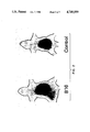
- FIG. 2 shows the utility of [ 125 I]BZZ for noninvasive imaging of a B16 melanoma. Distribution of [ 125 I]in either a mouse which had received a B16 melanoma in the left hind leg two weeks previously (left) or in a control animal. Note the assymetric distribution of radioactivity corresponding to the melanoma in the tumor-treated animal. Both animals received 50 ⁇ Ci of [ 125 I]BZZ approximately 5 hrs before the images were obtained;
- FIG. 3 demonstrates accumulation of [ 125 I]BZZ in retina of pigmented, but not albino, mice.
- FIG. 4 shows the [ 125 I]BZZ binding sites in brain and B16 melanoma.
- FIG. 4A shows the binding of [ 125 I]BZZ to a homogenate of the caudate putamen of the Swiss Webster mouse brain in the presence of the indicated concentrations of either the R- (open symbols) or S-(filled symbols) enantiomer of an unlabeled BZZ(designated SCH23390).
- FIG. 4B shows binding of [ 125 I]BZZ to a homogenate of B16 melanoma propagated in the Swiss Webster mice used for panel A of this figure in the presence of the indicated concentrations of either R- (open symbols) or S-(filled symbols) enantiomer of SCH 23390.
- the amount of bound radioactivity corresponding to 100 percent in panel A of this figure was 23,379 ⁇ 555 cpm/mg wet wt; In panel B, this value was 18,033 ⁇ 3,647 cpm/mg wet weight.
- FIG. 5 shows binding of [ 125 I]BZZ and [ 125 I]-S-BZZ to homogenates of B16 melanoma and caudate nucleus.
- FIG. 6 shows binding of [ 125 I]BZZ enantiomers to frozen sections of B16 melanoma and a human melanoma.
- Frozen sections of either a B16 melanoma (B16) or a human melanoma were incubated in the presence of either [ 125 I]BZZ (R) or [ 125 I]-BZZ (S) and autoradiography was performed as described in Methods.
- the figure at the top of either column is a light micrograph of a sequential frozen section of either tumor.
- a method of detecting melanin containing matter comprising reacting said melanin containing matter with an enantiomer of 2, 3, 4, 5-tetrahydro-3 methyl-5-phenyl-1H-3-benzazepin-7-ol (hereinafter BZZ) and determining the binding of said enantiomer with melanin.
- BZZ an enantiomer of 2, 3, 4, 5-tetrahydro-3 methyl-5-phenyl-1H-3-benzazepin-7-ol
- Radiolabelled ligand [ 125 I]SCH 23982 was obtained from New England Nuclear Corporation. S enantiomer of [ 125 I]SCH 23982 was prepared as a custom iodination by New England Nuclear. All mice were obtained from either Charles River Laboratories (Charlestown, MA) or the NIH Small Animal Section. The B16 melanoma was supplied by the Frederick Cancer Research Facility.
- the B16 melanoma was removed from the host mouse, washed in a balanced salt solution and then cut into small fragments (approximately 2 mm cubes). A tumor fragment was then implanted subcutaneously with a trochar into either the back or hind limb of the recipient mouse (either a C57/B16 or Swiss-Webster). Tumors could be easily seen within 2 to 3 weeks after implantation of the tumor fragment.
- Binding of radiolabeled BZZ ligand (also designated [ 125 I]SCH 23982) to a homogenate of either the caudate-putamen of the mouse or rat brain or the B16 melanoma was determined using minor modifications of a procedure described by Sidhu et al., Eur. J. Pharmacol. 113:437- 440 (1985). A tissue homogenate rather than washed particulate material was used for the assay.
- the assay system (final volume 200 ⁇ ) contained Tris-HCl (pH 7.4), 80 mM; ethyleneglyco-bis-( ⁇ -aminoethyl ether) N,N',tetraacetic (EGTA), 0.8 mM and MgSO 4 , 10 mM; radiolabeled ligand (40,000 cpm, 60 pM) and the unlabeled ligand as required for competitive assay; and incubations were performed at 25° C. for about 60 min. When binding to striatum and to tumor were compared, binding to homogenates of both tissues was determined simultaneously. In order to determine the reproducibility of the results, ligand binding was determined in 3 replicate samples for each experimental condition. The mean of these three values was taken as a single errorless value and the mean of three (or more) of such values from independent experiments is reported. Data represent mean ⁇ SE.
- Radiolabeled ligand was dissolved in Hank's balanced salt solution and administered via the tail vein as a bolus of 100 ⁇ l. At appropriate times, the animals were sacrificed and various organs removed. The amount of 125 I radioactivity within a given organ was determined with a gamma counter.
- tissue sections were preincubated for 15 min in 50 mM Tris-HCl buffer, pH 7.4, for 15 min and then incubated for 20 min in a solution containing 50 mM Tris-HCl, pH 7.4, 120 mM NaCl, 5 mM KCl, 2 mM CaCl 2 , 1 mM MgCl 2 and 200 pM iodinated ligand (either R or S enantiomer).
- Emulsion autoradiography was performed as follows: a flexible coverslip coated with NTB-3 (Kodak) was exposed to the tissue section; when the exposure was complete in about a day (about 24 hrs) the emulsion was developed at 4° for 2 min in D-19 developer. The section was stained with hematoxylin and eosin and the coverslip was reexposed to the stained section.
- NTB-3 Kodak
- mice received a bolus injection of [ 125 I]BZZ (50 ⁇ Ci) dissolved in Hank's balanced salt solution via the tail vein. At several times, the distribution of [ 125 I]was determined by imaging with a pinhole collimeter attached to a gamma camera (Raytheon Nuclear Diagnostics).
- Results shown in FIG. 1 (A) indicate that within 45 min after intravenous administration of [ 125 I]SCH (10 ⁇ Ci) to an albino mouse, the concentration of the radiolabel within the B-16 melanoma was 19-times greater than the concentration of the radioactivity within the nonpigmented eye; this difference was highly significant (p ⁇ 0.001).
- Comparison of a micrograph (FIG. 1, C) of one such tumor with an autoradiograph of the same histological section (FIG. 1B) demonstrated that the radiolabel quickly accumulated within the tumor to a much higher degree than within the surrounding tissues.
- [ 125 I]BZZ was also rapidly accumulated by metastatic murine melanomas.
- [ 125 I]-S-BZZ bound to the pigmented B16 melanoma to the same degree as did its R-enantiomer. It is noted that 2, 3, 4, 5-tetrahydro-8-chloro-3 methyl-5-phenyl-1H-3 benzazepin-7-ol effectively displaced [ 125 I]BZZ from the melanoma tissue. Thus, 8-chloro rather than 8-iodo BZZ can be equally well employed as a ligand depending upon the desired application.
- FIG. 6 (top, right) is a light micrograph of a frozen section of a lymph node removed from a 65 year old, white male who had had a pigmented melanoma removed from his foot 4 years earlier: 5 of the 12 lymph nodes removed with the one shown in FIG. 6 were diagnosed as positive for melanoma.
- FIG. 6 (middle, right) is an autoradiogram of a sequential frozen section incubated in the presence of [ 125 I]R-enantiomer of BZZ;
- FIG. 6 (bottom, right) is an autoradiogram of another frozen section incubated with [ 125 I]-S-enantiomer of BZZ.
- BZZ has specific binding affinity for melanin.
- the striking difference between the binding of BZZ to the pigment epithelium of the normal and albino mouse demonstrates that the ligand binds to melanin, per se, rather than to another product of the c locus, the gene controlling pigmentation in the mouse (Silvers, 1979, The Coat Colors of Mice, pp 45-82).
- the data also indicates that the binding of BZZ to melanin occurs by a mechanism independent of the D-1 dopamine receptor.
- the difference between the enantiomeric selectivity of the D-1 receptor and the melanin-associated binding is remarkably distinctive.
- Another application of the present invention is to combine the specificity of BZZ to melanin with antibodies having specific binding affinity for or directed against melanoma-associated antigens (MAA). Such modifications further enhance the noninvasive localization and imaging of pigmented melanomas, particularly if radiolabelled BZZ is complemented with similarly radiolabelled MAA.
- MAA melanoma-associated antigens
- a further utility of the present invention is in cytotoxically exposing pigmented melanomas to gamma-radiation emitting BZZ. This is accomplished by simply delivering or administering to the site or to the body carrying pigmented melanoma a cytotoxically effective radiation dosage of 125 I, of course, by 125 I-labelled enantiomer of BZZ having specific binding affinity for the melanoma.
Abstract
The present invention discloses a method of detecting melanin containing matter comprising reacting melanin containing matter with an enantiomer of 2, 3, 4, 5-tetrahydro-3-methyl-5 phenyl-1H-3-benzazepin-7-ol and determining the binding of said enantiomer with melanin. The invention also describes a method of delivering cytotoxic level of gamma radiation to pigmented melanoma comprising administering to a body carrying pigmented melanoma a cytotoxically effective radiation dosage of 125 I-enantiomer of 2, 3, 4, 5-tetrahydro-3-methyl-5-phenyl-1H-3-benzazepin-7-ol having specific binding affinity for said melanoma. Other applications of the invention have also been described.
Description
1. Technical Field
The present invention is related to a method of detecting melanin containing matter. More particularly, the present invention is related to a method of identifying, diagnosing, localizing and/or treating melanin containing cells or tissues.
2. State of the Art
Melanin is the dark, amorphous pigment generally found in such body parts as skin, hair, eye, brain and various tumors such as pigmented melanomas and the like. Some compounds are known to bind to melanin (Ings, 1984 Drug Metab Rev. 15:1183-1212) and some of these compounds, such as spiroperidol, have also been iodinated. However, the binding encountered with most radiolabeled neuroleptics, including [125 I] spiroperidol, is not very specific for melanin and a simple, non-invasive test for localizing pigmented tissue comprising an agent which has specific affinity for binding to melanin has not heretofore been known.
It is, therefore, an object of the present invention to provide a method of detecting melanin-containing matter.
It is a further object of the present invention to provide a diagnostic procedure for detecting and localizing pigmented melanomas.
It is yet another object of the present invention to provide a labelled ligand belonging to the group of compounds known as benzazepines (BZZ), said ligand having specificity to bind with melanin.
It is a still further object of the present invention to deliver a cytotoxic agent to melanin containing tissue by having said cytotoxic agent associated with, incorporated in or attached to the benzazepine molecule.
Other objects and advantages of the present invention will become evident as the detailed description of the present invention proceeds.
These and other objects, features and many of the attendant advantages of the invention will be better understood upon a reading of the following detailed description when considered in connection with the accompanying drawings wherein:
FIG. 1A shows accumulation of intraveneously administered [125 I]BZZ within the B16 melanoma.
FIG. 1B is an autoradiogram of a frozen section of a B16 melanoma from a mouse receiving [125 I]BZZ corresponding to part A.
FIG. 1C is a micrograph of the frozen section used in part B. Comparison of the autoradiogram with the light micrograph of the tissue shows a heavy accumulation of the ligand within the tumor and much lower accumulation within the surrounding tissues not invaded by the tumor.
FIG. 1D is an autoradiogram of a frozen section of a lung from a female C57B1/6NCr mouse receiving 50 μCi of [125 I]BZZ 10 hours before being killed. Three weeks earlier, this animal had received 5×10 4 B16 melanoma cells intravenously.
FIG. 1E is a micrograph of the frozen section used in part D. Arrows in D and E indicate the corresponding structures;
FIG. 2 shows the utility of [125 I]BZZ for noninvasive imaging of a B16 melanoma. Distribution of [125 I]in either a mouse which had received a B16 melanoma in the left hind leg two weeks previously (left) or in a control animal. Note the assymetric distribution of radioactivity corresponding to the melanoma in the tumor-treated animal. Both animals received 50 μCi of [125 I]BZZ approximately 5 hrs before the images were obtained;
FIG. 3 demonstrates accumulation of [125 I]BZZ in retina of pigmented, but not albino, mice.
A. Ocular accumulation of [125 I]BZZ.
B. Autoradiographic Localization of [125 I]BZZ within the eye.
Left: Autoradiogram of a frozen section of the eye of an animal which received [125 I]BZZ (50 μCi) 45 min previously showing localized accumulation of radioisotope.
Right: light micrograph of the section of the eye used for autoradiogram.
C. In vitro binding of [125 I]BZZ to pigment epithelium of retina. Comparison of the dark field photomicrograph (bottom) with the light micrograph (top) of the retina shows that the ligand is accumulated predominantly in the pigment epithelium of retina; and
FIG. 4 shows the [125 I]BZZ binding sites in brain and B16 melanoma.
FIG. 4A shows the binding of [125 I]BZZ to a homogenate of the caudate putamen of the Swiss Webster mouse brain in the presence of the indicated concentrations of either the R- (open symbols) or S-(filled symbols) enantiomer of an unlabeled BZZ(designated SCH23390).
FIG. 4B shows binding of [125 I]BZZ to a homogenate of B16 melanoma propagated in the Swiss Webster mice used for panel A of this figure in the presence of the indicated concentrations of either R- (open symbols) or S-(filled symbols) enantiomer of SCH 23390.
The amount of bound radioactivity corresponding to 100 percent in panel A of this figure was 23,379±555 cpm/mg wet wt; In panel B, this value was 18,033±3,647 cpm/mg wet weight.
FIG. 5 shows binding of [125 I]BZZ and [125 I]-S-BZZ to homogenates of B16 melanoma and caudate nucleus.
Binding of the R and S enantiomers of [125 I]BZZ to homogenates of either the B16 melanoma (left) or the caudate nucleus (right) was determined in the absence (Total) and presence (N.S.=nonspecific binding, of unlabeled SCH23390. Data were obtained from 3 separate homogenates of either tissue.
FIG. 6 shows binding of [125 I]BZZ enantiomers to frozen sections of B16 melanoma and a human melanoma. Frozen sections of either a B16 melanoma (B16) or a human melanoma were incubated in the presence of either [125 I]BZZ (R) or [125 I]-BZZ (S) and autoradiography was performed as described in Methods. The figure at the top of either column is a light micrograph of a sequential frozen section of either tumor.
The above and other objects and advantages of the present invention are achieved by a method of detecting melanin containing matter comprising reacting said melanin containing matter with an enantiomer of 2, 3, 4, 5-tetrahydro-3 methyl-5-phenyl-1H-3-benzazepin-7-ol (hereinafter BZZ) and determining the binding of said enantiomer with melanin. Of course, such enantiomorphs as 5-R and 5-S enantiomers of BZZ and such variants as 8-iodo or 8-chloro BZZ can also be employed and said BZZ can be radiolabelled, such as with 125 I and the like, or conjugated with cytotoxic agents such as astatine211 , 125 I and the like.
Various BZZ and methods of preparing the same have been described in such publications as U.S. Pat. No. 3,393,192 and by Sidhu et al (1985) in Eur. J. Pharmac. 113:437- 440 both of which are incorporated herein by reference.
Unless defined otherwise, all scientific or technical terms used herein have the same meaning as generally understood by one of ordinary skill in the art to which this invention belongs.
Radiolabelled ligand [125 I]SCH 23982 was obtained from New England Nuclear Corporation. S enantiomer of [125 I]SCH 23982 was prepared as a custom iodination by New England Nuclear. All mice were obtained from either Charles River Laboratories (Charlestown, MA) or the NIH Small Animal Section. The B16 melanoma was supplied by the Frederick Cancer Research Facility.
Melanoma propagation
The B16 melanoma was removed from the host mouse, washed in a balanced salt solution and then cut into small fragments (approximately 2 mm cubes). A tumor fragment was then implanted subcutaneously with a trochar into either the back or hind limb of the recipient mouse (either a C57/B16 or Swiss-Webster). Tumors could be easily seen within 2 to 3 weeks after implantation of the tumor fragment.
In vitro binding assay
Binding of radiolabeled BZZ ligand (also designated [125 I]SCH 23982) to a homogenate of either the caudate-putamen of the mouse or rat brain or the B16 melanoma was determined using minor modifications of a procedure described by Sidhu et al., Eur. J. Pharmacol. 113:437- 440 (1985). A tissue homogenate rather than washed particulate material was used for the assay. The assay system (final volume 200 μ) contained Tris-HCl (pH 7.4), 80 mM; ethyleneglyco-bis-(β-aminoethyl ether) N,N',tetraacetic (EGTA), 0.8 mM and MgSO4, 10 mM; radiolabeled ligand (40,000 cpm, 60 pM) and the unlabeled ligand as required for competitive assay; and incubations were performed at 25° C. for about 60 min. When binding to striatum and to tumor were compared, binding to homogenates of both tissues was determined simultaneously. In order to determine the reproducibility of the results, ligand binding was determined in 3 replicate samples for each experimental condition. The mean of these three values was taken as a single errorless value and the mean of three (or more) of such values from independent experiments is reported. Data represent mean ± SE.
In vivo drug administration
Radiolabeled ligand was dissolved in Hank's balanced salt solution and administered via the tail vein as a bolus of 100 μl. At appropriate times, the animals were sacrificed and various organs removed. The amount of 125 I radioactivity within a given organ was determined with a gamma counter.
Autoradiography
After animals were killed, the brain, eyes or tumor were immediately removed and frozen on dry ice. Frozen, 16 μthick sections were cut in a cryostat at -16° C., thaw-mounted onto gelatin-coated slides and stored overnight under vacuum at 4° C. The tissue sections were preincubated for 15 min in 50 mM Tris-HCl buffer, pH 7.4, for 15 min and then incubated for 20 min in a solution containing 50 mM Tris-HCl, pH 7.4, 120 mM NaCl, 5 mM KCl, 2 mM CaCl2, 1 mM MgCl2 and 200 pM iodinated ligand (either R or S enantiomer). After incubation, the slides were washed 4 times in ice-cold 50 mM Tris-HCl buffer and dried under a stream of air. Autoradiographic images were produced by placing the slides in X-ray casettes and using them to expose [3 H]ultra film for 15 hours following standard procedure.
Emulsion autoradiography was performed as follows: a flexible coverslip coated with NTB-3 (Kodak) was exposed to the tissue section; when the exposure was complete in about a day (about 24 hrs) the emulsion was developed at 4° for 2 min in D-19 developer. The section was stained with hematoxylin and eosin and the coverslip was reexposed to the stained section.
Noninvasive Imaging of [125 I]
Mice received a bolus injection of [125 I]BZZ (50 μCi) dissolved in Hank's balanced salt solution via the tail vein. At several times, the distribution of [125 I]was determined by imaging with a pinhole collimeter attached to a gamma camera (Raytheon Nuclear Diagnostics).
Results shown in FIG. 1 (A) indicate that within 45 min after intravenous administration of [125 I]SCH (10 μCi) to an albino mouse, the concentration of the radiolabel within the B-16 melanoma was 19-times greater than the concentration of the radioactivity within the nonpigmented eye; this difference was highly significant (p <0.001). Comparison of a micrograph (FIG. 1, C) of one such tumor with an autoradiograph of the same histological section (FIG. 1B) demonstrated that the radiolabel quickly accumulated within the tumor to a much higher degree than within the surrounding tissues. [125 I]BZZ was also rapidly accumulated by metastatic murine melanomas. Comparison of the autoradiogram (FIG. 1, D) and the light micrograph (FIG. 1, E) of the lung from a mouse receiving intravenous B16 melanoma cells 3 weeks earlier and 50 μCi of [125 I]BZZ on the day of the experiment shows that the normal lung tissue does not bind the ligand while the foci of pigmented cells show heavy accumulation of radiolabel.
Because of the accumulation of [125 I]BZZ within the B16 melanoma, it was possible to image the tumor with a noninvasive procedure. Five hours after the intravenous administration of 50 μCi of [125 I]to a pigmented C57B1/6 mouse which 2 weeks earlier had received the B16 melanoma in its left hind leg, sufficient radiolabel accumulated within the tumor that it could be visualized with a pinhole collimator (FIG. 2 left). There was also a substantial accumulation of [125 I]BZZ within the abdomen of the tumor-bearing mouse. This accumulation was unrelated to the presence of the tumor since nontumor-bearing animals also displayed this accumulation of radiolabel (FIG. 2 right).
Binding of [125 I]to melanin
Because [125 I]BZZ permitted noninvasive imaging of a pigmented melanoma, the molecular basis of its selective accumulation by melanocytes was investigated in a series of experiments using the readily accessible pigment epithelium of retina, a tissue rich in melanin. Following intravenous administration of the [125 I]BZZ (10 μCi), radioactivity rapidly accumulated into the eyes of the pigmented C57B1/6 mouse but not in the eyes of the albino Swiss Webster mouse (FIG. 3A). The 79-fold difference between the amount of radiolabel accumulated in the pigmented and albino eyes was highly significant (P<0.001). Autoradiography revealed that radiolabel accumulated predominantly in a narrow band at the back of the pigmented eye (FIG. 3B, compare autoradiogram on left and light micrograph on right); this localized accumulation of radioactivity was not evident in the albino eye (data not shown). In vitro autoradiography (FIG. 3C) confirmed the conclusion that the pigment epithelium of the retina was the ocular structure accumulating the radioisotope. Comparison of light and dark field micrographs of a frozen tissue section exposed to the ligand revealed that radiolabel was highly concentrated within the melanin containing pigment epithelium.
Because [125 I]BZZ could bind to both the D-1 dopamine receptor and melanin, it was important to determine the type of binding site occurring in the pigmented melanoma. Therefore, the binding of the ligand to homogenates of the caudate nucleus and the B16 melanoma was compared. It was found that the D-1 receptor in the caudate nucleus displayed a 100-fold higher affinity for the R-enantiomer of the D-1 receptor antagonist than for the S-enantiomer of this molecule (FIG. 4A). In contrast, the binding site in the melanoma was much less stereoselective with only a 5.1-fold difference between the R- and S-enantiomers (FIG. 4B). Furthermore, 400-fold higher concentrations of R-enantiomer were required to displace [125 I]BZZ from the melanoma binding site than from the D-1 dopamine receptor (Compare FIGS. 4A and B). These data indicate that few, if any, D-1 dopamine receptors occur in the B16 melanoma and that the accumulation of [125 I]BZZ by this tumor is due to the melanin associated with the tumors.
Since the interaction between [125 I]BZZ and the B16 melanoma was not related to the presence of a D-1 receptor, the affinity of the ligand for the D-1 receptor could be viewed as an undesirable side effect. To remove this undesirable side effect from the molecule, the S-enantiomer of BZZ was preferred because the data in FIG. 4 indicated that this molecule retains affinity for melanin but has a much lower affinity for the D-1 receptor. FIG. 5 shows that [125 I]-S-BZZ failed to bind to a homogenate of the caudate nucleus of the rat brain. In contrast, [125 I]-S-BZZ bound to the pigmented B16 melanoma to the same degree as did its R-enantiomer. It is noted that 2, 3, 4, 5-tetrahydro-8-chloro-3 methyl-5-phenyl-1H-3 benzazepin-7-ol effectively displaced [125 I]BZZ from the melanoma tissue. Thus, 8-chloro rather than 8-iodo BZZ can be equally well employed as a ligand depending upon the desired application.
Binding to human melanomas
FIG. 6 (top, right) is a light micrograph of a frozen section of a lymph node removed from a 65 year old, white male who had had a pigmented melanoma removed from his foot 4 years earlier: 5 of the 12 lymph nodes removed with the one shown in FIG. 6 were diagnosed as positive for melanoma. FIG. 6 (middle, right) is an autoradiogram of a sequential frozen section incubated in the presence of [125 I]R-enantiomer of BZZ; FIG. 6 (bottom, right) is an autoradiogram of another frozen section incubated with [125 I]-S-enantiomer of BZZ. For either enantiomer, the highest accumulation of radiolabel within the frozen section was associated with the foci of pigmented metastic melanoma within the node. Both enantiomers of the radiolabeled drug also bound to frozen sections of the murine B16 melanoma (FIG. 6, left). These results indicate that both enantiomers of the radiolabeled BZZ effectively bind to metastatic human melanoma.
It is clear from the data presented herein that BZZ has specific binding affinity for melanin. The striking difference between the binding of BZZ to the pigment epithelium of the normal and albino mouse demonstrates that the ligand binds to melanin, per se, rather than to another product of the c locus, the gene controlling pigmentation in the mouse (Silvers, 1979, The Coat Colors of Mice, pp 45-82). The data also indicates that the binding of BZZ to melanin occurs by a mechanism independent of the D-1 dopamine receptor. The difference between the enantiomeric selectivity of the D-1 receptor and the melanin-associated binding is remarkably distinctive. The results also demonstrate that the binding of [125 I]BZZ to melanin makes it possible to visualize pigmented melanomas by noninvasive imaging techniques. Furthermore, because the S-enantiomer of this molecule retains its affinity for melanin but does not bind to D-1 dopamine receptors as described herein, the present invention allows noninvasive imaging of melanin to the exclusion of D-1 dopamine receptors.
Another application of the present invention is to combine the specificity of BZZ to melanin with antibodies having specific binding affinity for or directed against melanoma-associated antigens (MAA). Such modifications further enhance the noninvasive localization and imaging of pigmented melanomas, particularly if radiolabelled BZZ is complemented with similarly radiolabelled MAA.
Because of the specific binding affinity of BZZ to melanomas, a further utility of the present invention is in cytotoxically exposing pigmented melanomas to gamma-radiation emitting BZZ. This is accomplished by simply delivering or administering to the site or to the body carrying pigmented melanoma a cytotoxically effective radiation dosage of 125 I, of course, by 125 I-labelled enantiomer of BZZ having specific binding affinity for the melanoma.
It is understood that the examples and embodiments described herein are for illustrative purposes only and that various modifications or changes in light thereof will be suggested to persons skilled in the art and are to be included within the spirit and purview of this application and the scope of the appended claims.
Claims (9)
1. A method of detecting melanin-containing matter comprising reacting melanin containing matter with an enantiomer of 2, 3, 4, 5-tetrahydro-3-methyl-5 phenyl-1H-3-benzazepin-7-ol and determining the binding of said enantiomer with melanin.
2. The method of claim 1 wherein said enantiomer is radiolabelled.
3. The method of claim 2 wherein said enantiomer is 125 I-labelled.
4. The method according to claim 3 of detecting melanin-containing matter by radiographic imaging of bound 125 I-labelled enantiomer to said matter.
5. The method of claim 4 wherein said melanin-containing matter is pigmented melanoma.
6. A method of delivering cytotoxic level of gamma radiation to pigmented melanoma comprising administering to a body carrying pigmented melanoma a cytotoxically effective radiation dosage of 125 I-enantiomer of 2, 3, 4, 5-tetrahydro-3-methyl-5 phenyl-1H- 3benzazepin-7-ol having specific binding affinity for said melanoma.
7. The method of claim 6 combining said 125 I-enantiomer with 125 I-labelled antibodies having specific binding affinity with melanoma associated antigens.
8. A method of differentiating between D-1 dopamine receptors and melanin-containing cells without D-1 dopamine receptors, comprising reacting D-1 dopamine receptors and melanin containing cells with radiolabelled S-enantiomer of 2,3,4, 5-tetrahydro-3-methyl-5 phenyl-1H-3-benzazepin-7-ol, wherein binding of said enantiomer occurs only with said cells.
9. The method of claim 8 wherein said enantiomer is 125 I-labelled.
Priority Applications (1)
| Application Number | Priority Date | Filing Date | Title |
|---|---|---|---|
| US06/869,714 US4749559A (en) | 1986-06-02 | 1986-06-02 | Method for detecting melanin-containing matter |
Applications Claiming Priority (1)
| Application Number | Priority Date | Filing Date | Title |
|---|---|---|---|
| US06/869,714 US4749559A (en) | 1986-06-02 | 1986-06-02 | Method for detecting melanin-containing matter |
Publications (1)
| Publication Number | Publication Date |
|---|---|
| US4749559A true US4749559A (en) | 1988-06-07 |
Family
ID=25354122
Family Applications (1)
| Application Number | Title | Priority Date | Filing Date |
|---|---|---|---|
| US06/869,714 Expired - Fee Related US4749559A (en) | 1986-06-02 | 1986-06-02 | Method for detecting melanin-containing matter |
Country Status (1)
| Country | Link |
|---|---|
| US (1) | US4749559A (en) |
Cited By (3)
| Publication number | Priority date | Publication date | Assignee | Title |
|---|---|---|---|---|
| WO1999006074A1 (en) * | 1997-07-30 | 1999-02-11 | Pharmacyclics, Inc. | Use of texaphyrins in detection of melanin and melanin metabolites of melanotic melanoma |
| US20060039858A1 (en) * | 2003-02-11 | 2006-02-23 | Ekaterina Dadachova | Radiolabeled antibodies and peptides for treatment of tumors |
| CN113817796A (en) * | 2020-12-03 | 2021-12-21 | 上海益诺思生物技术股份有限公司 | Application of melanoma cells in determination of drug-melanin binding capacity |
Citations (6)
| Publication number | Priority date | Publication date | Assignee | Title |
|---|---|---|---|---|
| US3393192A (en) * | 1965-04-26 | 1968-07-16 | Schering Corp | Novel benzazepines |
| US4210749A (en) * | 1974-11-12 | 1980-07-01 | Pennwalt Corporation | Substituted 1,2,4,5-tetrahydro-3H,3 benzazepines |
| US4382029A (en) * | 1980-08-07 | 1983-05-03 | Smithkline Beckman Corporation | Process useful for preparing 7,8-acetamido, hydroxy-1-phenyl-2,3,4,5-tetrahydro-1H-3-benzazepines |
| US4460559A (en) * | 1980-03-03 | 1984-07-17 | Goldenberg Milton David | Tumor localization and therapy with labeled antibodies specific to intracellular tumor-associated markers |
| US4549988A (en) * | 1981-04-06 | 1985-10-29 | Hoffmann-La Roche Inc. | 3H-2-Benzazepines |
| US4661347A (en) * | 1982-11-12 | 1987-04-28 | Scripps Clinic | Cytotoxic compositions |
-
1986
- 1986-06-02 US US06/869,714 patent/US4749559A/en not_active Expired - Fee Related
Patent Citations (6)
| Publication number | Priority date | Publication date | Assignee | Title |
|---|---|---|---|---|
| US3393192A (en) * | 1965-04-26 | 1968-07-16 | Schering Corp | Novel benzazepines |
| US4210749A (en) * | 1974-11-12 | 1980-07-01 | Pennwalt Corporation | Substituted 1,2,4,5-tetrahydro-3H,3 benzazepines |
| US4460559A (en) * | 1980-03-03 | 1984-07-17 | Goldenberg Milton David | Tumor localization and therapy with labeled antibodies specific to intracellular tumor-associated markers |
| US4382029A (en) * | 1980-08-07 | 1983-05-03 | Smithkline Beckman Corporation | Process useful for preparing 7,8-acetamido, hydroxy-1-phenyl-2,3,4,5-tetrahydro-1H-3-benzazepines |
| US4549988A (en) * | 1981-04-06 | 1985-10-29 | Hoffmann-La Roche Inc. | 3H-2-Benzazepines |
| US4661347A (en) * | 1982-11-12 | 1987-04-28 | Scripps Clinic | Cytotoxic compositions |
Non-Patent Citations (2)
| Title |
|---|
| O Boyle et al, Structural Determinants of Selective Affinity for Brain D 1 Dopamine Receptors within a Series of 1 phenyl 1H 3 benzazipine Analogs of SK&F 38393 and SCH 23390 , Chemical Abstracts, vol. 104, (2/86) 28373p. * |
| O'Boyle et al, "Structural Determinants of Selective Affinity for Brain D-1 Dopamine Receptors within a Series of 1-phenyl-1H-3-benzazipine Analogs of SK&F 38393 and SCH 23390", Chemical Abstracts, vol. 104, (2/86) #28373p. |
Cited By (5)
| Publication number | Priority date | Publication date | Assignee | Title |
|---|---|---|---|---|
| WO1999006074A1 (en) * | 1997-07-30 | 1999-02-11 | Pharmacyclics, Inc. | Use of texaphyrins in detection of melanin and melanin metabolites of melanotic melanoma |
| US6022526A (en) * | 1997-07-30 | 2000-02-08 | Pharmacyclics, Inc. | Use of texaphyrins in detection of melanin and melanin metabolites diagnostic of melanotic melanoma |
| US20060039858A1 (en) * | 2003-02-11 | 2006-02-23 | Ekaterina Dadachova | Radiolabeled antibodies and peptides for treatment of tumors |
| CN113817796A (en) * | 2020-12-03 | 2021-12-21 | 上海益诺思生物技术股份有限公司 | Application of melanoma cells in determination of drug-melanin binding capacity |
| CN113817796B (en) * | 2020-12-03 | 2023-12-26 | 上海益诺思生物技术股份有限公司 | Application of melanoma cells in drug-melanin binding capacity measurement |
Similar Documents
| Publication | Publication Date | Title |
|---|---|---|
| Lütje et al. | Dual-modality image-guided surgery of prostate cancer with a radiolabeled fluorescent anti-PSMA monoclonal antibody | |
| Sharkey et al. | Biodistribution and radiation dose estimates for yttrium-and iodine-labeled monoclonal antibody IgG and fragments in nude mice bearing human colonic tumor xenografts | |
| Saleh et al. | Phase I trial of the murine monoclonal anti-GD2 antibody 14G2a in metastatic melanoma | |
| Divgi et al. | Phase I and imaging trial of indium 111-labeled anti-epidermal growth factor receptor monoclonal antibody 225 in patients with squamous cell lung carcinoma | |
| Maziere et al. | In vivo characterization of myocardium muscarinic receptors by positron emission tomography | |
| Goldrosen et al. | Biodistribution, pharmacokinetic, and imaging studies with 186Re-labeled NR-LU-10 whole antibody in LS174T colonic tumor-bearing mice | |
| Cheung et al. | Targeting of ganglioside GD2Monoclonal antibody to neuroblastoma | |
| Chen et al. | A comparative autoradiographic study demonstrating differential intratumor localization of monoclonal antibodies to cell surface (Lym-1) and intracellular (TNT-1) antigens | |
| WO1992006685A1 (en) | Enhancement of cellular accumulation of lipophilic cationic organometallic compounds by reduction of intramembrane potential | |
| Blankenberg et al. | Imaging macrophages and the apoptosis of granulocytes in a rodent model of subacute and chronic abscesses with radiolabeled monocyte chemotactic peptide-1 and annexin V | |
| US4749559A (en) | Method for detecting melanin-containing matter | |
| JP2003504395A (en) | Inhibits renal uptake of molecules that may be harmful to the kidney | |
| Griffin et al. | Intraperitoneal immunoconjugates | |
| Epenetos et al. | Antibody-guided irradiation of hepatic metastases using intrahepatically administered radiolabelled anti-CEA antibodies with simultaneous and reversible hepatic blood flow stasis using biodegradable starch microspheres | |
| AU743696B2 (en) | Imaging methods and compositions | |
| Yu et al. | Targeted diagnosis and treatment of superficial bladder cancer with monoclonal antibody BDI-1 | |
| Garkavij et al. | Extracorporeal whole-blood immunoadsorption enhances radioimmunotargeting of iodine-125-labeled BR96-biotin monoclonal antibody | |
| Agui et al. | 125I-iodinated benzazepines bind to melanin: implications for the noninvasive localization of pigmented melanomas | |
| Clarke et al. | Diagnosis of obstructive jaundice | |
| Correa-González et al. | Uptake of 153Sm-DTPA-bis-biotin and 99mTc-DTPA-bis-biotin in rat as-30D-hepatoma cells | |
| Gadina et al. | Preclinical pharmacokinetics and localization studies of the radioiodinated anti-ovarian carcinoma MAb MOv18 | |
| Wahl et al. | Lymphoscintigraphy in melanoma: initial evaluation of a low protein dose monoclonal antibody cocktail | |
| US5840272A (en) | Imaging infectious foci with human IgM 16.88 | |
| Keenan | Immunolymphoscintigraphy | |
| 為政脩 et al. | 99mTc-DL-homocysteine, a potential tumor imaging agent. |
Legal Events
| Date | Code | Title | Description |
|---|---|---|---|
| AS | Assignment |
Owner name: UNITED STATES OF AMERICA THE Free format text: ASSIGNMENT OF ASSIGNORS INTEREST.;ASSIGNOR:KEBABIAN, JOHN W.;REEL/FRAME:004562/0588 Effective date: 19860602 |
|
| REMI | Maintenance fee reminder mailed | ||
| REMI | Maintenance fee reminder mailed | ||
| REMI | Maintenance fee reminder mailed | ||
| FPAY | Fee payment |
Year of fee payment: 4 |
|
| SULP | Surcharge for late payment | ||
| REMI | Maintenance fee reminder mailed | ||
| LAPS | Lapse for failure to pay maintenance fees | ||
| FP | Lapsed due to failure to pay maintenance fee |
Effective date: 19960612 |
|
| STCH | Information on status: patent discontinuation |
Free format text: PATENT EXPIRED DUE TO NONPAYMENT OF MAINTENANCE FEES UNDER 37 CFR 1.362 |