US20200217841A1 - In vitro method for the determination of neurodegenerative diseases - Google Patents
In vitro method for the determination of neurodegenerative diseases Download PDFInfo
- Publication number
- US20200217841A1 US20200217841A1 US16/823,620 US202016823620A US2020217841A1 US 20200217841 A1 US20200217841 A1 US 20200217841A1 US 202016823620 A US202016823620 A US 202016823620A US 2020217841 A1 US2020217841 A1 US 2020217841A1
- Authority
- US
- United States
- Prior art keywords
- kynurenine
- content
- kynurenic acid
- kit according
- detecting
- Prior art date
- Legal status (The legal status is an assumption and is not a legal conclusion. Google has not performed a legal analysis and makes no representation as to the accuracy of the status listed.)
- Abandoned
Links
- 208000015122 neurodegenerative disease Diseases 0.000 title claims abstract description 31
- 230000004770 neurodegeneration Effects 0.000 title claims abstract description 28
- 238000000034 method Methods 0.000 title description 25
- 238000000338 in vitro Methods 0.000 title description 11
- YGPSJZOEDVAXAB-UHFFFAOYSA-N kynurenine Chemical compound OC(=O)C(N)CC(=O)C1=CC=CC=C1N YGPSJZOEDVAXAB-UHFFFAOYSA-N 0.000 claims abstract description 201
- HCZHHEIFKROPDY-UHFFFAOYSA-N kynurenic acid Chemical compound C1=CC=C2NC(C(=O)O)=CC(=O)C2=C1 HCZHHEIFKROPDY-UHFFFAOYSA-N 0.000 claims abstract description 152
- 210000001124 body fluid Anatomy 0.000 claims abstract description 20
- 239000010839 body fluid Substances 0.000 claims abstract description 20
- 238000012360 testing method Methods 0.000 claims description 61
- YGPSJZOEDVAXAB-QMMMGPOBSA-N L-kynurenine Chemical compound OC(=O)[C@@H](N)CC(=O)C1=CC=CC=C1N YGPSJZOEDVAXAB-QMMMGPOBSA-N 0.000 claims description 26
- 239000012491 analyte Substances 0.000 claims description 22
- 239000012528 membrane Substances 0.000 claims description 15
- 238000006243 chemical reaction Methods 0.000 claims description 13
- 239000003153 chemical reaction reagent Substances 0.000 claims description 12
- 150000001875 compounds Chemical class 0.000 claims description 9
- 239000002250 absorbent Substances 0.000 claims description 8
- 230000002745 absorbent Effects 0.000 claims description 8
- 239000007787 solid Substances 0.000 claims description 7
- 238000003118 sandwich ELISA Methods 0.000 claims description 5
- 238000012875 competitive assay Methods 0.000 claims description 4
- 210000003296 saliva Anatomy 0.000 description 58
- 210000002966 serum Anatomy 0.000 description 34
- 208000024827 Alzheimer disease Diseases 0.000 description 19
- QIVBCDIJIAJPQS-VIFPVBQESA-N L-tryptophane Chemical compound C1=CC=C2C(C[C@H](N)C(O)=O)=CNC2=C1 QIVBCDIJIAJPQS-VIFPVBQESA-N 0.000 description 17
- QIVBCDIJIAJPQS-UHFFFAOYSA-N Tryptophan Natural products C1=CC=C2C(CC(N)C(O)=O)=CNC2=C1 QIVBCDIJIAJPQS-UHFFFAOYSA-N 0.000 description 17
- 239000002245 particle Substances 0.000 description 15
- 208000037265 diseases, disorders, signs and symptoms Diseases 0.000 description 12
- 108020004201 indoleamine 2,3-dioxygenase Proteins 0.000 description 11
- 102000006639 indoleamine 2,3-dioxygenase Human genes 0.000 description 11
- 230000037361 pathway Effects 0.000 description 11
- 102000004190 Enzymes Human genes 0.000 description 8
- 108090000790 Enzymes Proteins 0.000 description 8
- 238000003556 assay Methods 0.000 description 8
- 201000010099 disease Diseases 0.000 description 8
- 238000005259 measurement Methods 0.000 description 8
- 239000002207 metabolite Substances 0.000 description 8
- 108091023037 Aptamer Proteins 0.000 description 7
- 208000036110 Neuroinflammatory disease Diseases 0.000 description 7
- SMWDFEZZVXVKRB-UHFFFAOYSA-N anhydrous quinoline Natural products N1=CC=CC2=CC=CC=C21 SMWDFEZZVXVKRB-UHFFFAOYSA-N 0.000 description 7
- 230000003959 neuroinflammation Effects 0.000 description 7
- 201000004810 Vascular dementia Diseases 0.000 description 6
- 230000004913 activation Effects 0.000 description 6
- 208000010877 cognitive disease Diseases 0.000 description 6
- 238000002405 diagnostic procedure Methods 0.000 description 6
- 238000010790 dilution Methods 0.000 description 6
- 239000012895 dilution Substances 0.000 description 6
- 238000005516 engineering process Methods 0.000 description 6
- 239000012530 fluid Substances 0.000 description 6
- 238000012125 lateral flow test Methods 0.000 description 6
- 230000000324 neuroprotective effect Effects 0.000 description 6
- VLTRZXGMWDSKGL-UHFFFAOYSA-N perchloric acid Chemical compound OCl(=O)(=O)=O VLTRZXGMWDSKGL-UHFFFAOYSA-N 0.000 description 6
- 238000001356 surgical procedure Methods 0.000 description 6
- 238000010998 test method Methods 0.000 description 6
- 239000002253 acid Substances 0.000 description 5
- 230000015556 catabolic process Effects 0.000 description 5
- 230000028993 immune response Effects 0.000 description 5
- 230000002757 inflammatory effect Effects 0.000 description 5
- 230000015788 innate immune response Effects 0.000 description 5
- 238000011160 research Methods 0.000 description 5
- 239000000126 substance Substances 0.000 description 5
- 206010061218 Inflammation Diseases 0.000 description 4
- 108010068073 Kynurenine-oxoglutarate transaminase Proteins 0.000 description 4
- 206010027374 Mental impairment Diseases 0.000 description 4
- 239000000427 antigen Substances 0.000 description 4
- 108091007433 antigens Proteins 0.000 description 4
- 102000036639 antigens Human genes 0.000 description 4
- 210000004369 blood Anatomy 0.000 description 4
- 239000008280 blood Substances 0.000 description 4
- 210000004556 brain Anatomy 0.000 description 4
- 230000000875 corresponding effect Effects 0.000 description 4
- 208000035475 disorder Diseases 0.000 description 4
- PCHJSUWPFVWCPO-UHFFFAOYSA-N gold Chemical compound [Au] PCHJSUWPFVWCPO-UHFFFAOYSA-N 0.000 description 4
- 238000003018 immunoassay Methods 0.000 description 4
- 230000004054 inflammatory process Effects 0.000 description 4
- 230000036542 oxidative stress Effects 0.000 description 4
- 230000035945 sensitivity Effects 0.000 description 4
- 206010012289 Dementia Diseases 0.000 description 3
- IAZDPXIOMUYVGZ-UHFFFAOYSA-N Dimethylsulphoxide Chemical compound CS(C)=O IAZDPXIOMUYVGZ-UHFFFAOYSA-N 0.000 description 3
- 208000018737 Parkinson disease Diseases 0.000 description 3
- 208000026301 Postoperative Cognitive Complications Diseases 0.000 description 3
- 238000010171 animal model Methods 0.000 description 3
- 238000003149 assay kit Methods 0.000 description 3
- 230000003247 decreasing effect Effects 0.000 description 3
- 238000006731 degradation reaction Methods 0.000 description 3
- 238000011161 development Methods 0.000 description 3
- 230000018109 developmental process Effects 0.000 description 3
- 230000000694 effects Effects 0.000 description 3
- 239000003797 essential amino acid Substances 0.000 description 3
- 235000020776 essential amino acid Nutrition 0.000 description 3
- 230000003492 excitotoxic effect Effects 0.000 description 3
- 231100000063 excitotoxicity Toxicity 0.000 description 3
- 239000010931 gold Substances 0.000 description 3
- 229910052737 gold Inorganic materials 0.000 description 3
- 239000004816 latex Substances 0.000 description 3
- 229920000126 latex Polymers 0.000 description 3
- 230000002503 metabolic effect Effects 0.000 description 3
- 230000004060 metabolic process Effects 0.000 description 3
- 230000000626 neurodegenerative effect Effects 0.000 description 3
- 231100000189 neurotoxic Toxicity 0.000 description 3
- 230000002887 neurotoxic effect Effects 0.000 description 3
- 230000003950 pathogenic mechanism Effects 0.000 description 3
- 238000001556 precipitation Methods 0.000 description 3
- 210000003079 salivary gland Anatomy 0.000 description 3
- 150000003384 small molecules Chemical class 0.000 description 3
- KAXRWMOLNJZCEW-QRPNPIFTSA-N (2s)-2-amino-4-(2-aminophenyl)-4-oxobutanoic acid;sulfuric acid Chemical compound OS(O)(=O)=O.OC(=O)[C@@H](N)CC(=O)C1=CC=CC=C1N KAXRWMOLNJZCEW-QRPNPIFTSA-N 0.000 description 2
- MZOFCQQQCNRIBI-VMXHOPILSA-N (3s)-4-[[(2s)-1-[[(2s)-1-[[(1s)-1-carboxy-2-hydroxyethyl]amino]-4-methyl-1-oxopentan-2-yl]amino]-5-(diaminomethylideneamino)-1-oxopentan-2-yl]amino]-3-[[2-[[(2s)-2,6-diaminohexanoyl]amino]acetyl]amino]-4-oxobutanoic acid Chemical compound OC[C@@H](C(O)=O)NC(=O)[C@H](CC(C)C)NC(=O)[C@H](CCCN=C(N)N)NC(=O)[C@H](CC(O)=O)NC(=O)CNC(=O)[C@@H](N)CCCCN MZOFCQQQCNRIBI-VMXHOPILSA-N 0.000 description 2
- WJXSWCUQABXPFS-UHFFFAOYSA-N 3-hydroxyanthranilic acid Chemical compound NC1=C(O)C=CC=C1C(O)=O WJXSWCUQABXPFS-UHFFFAOYSA-N 0.000 description 2
- 206010002091 Anaesthesia Diseases 0.000 description 2
- 241000283707 Capra Species 0.000 description 2
- 208000028698 Cognitive impairment Diseases 0.000 description 2
- 108010001336 Horseradish Peroxidase Proteins 0.000 description 2
- 108010033242 Kynurenine 3-monooxygenase Proteins 0.000 description 2
- 241001465754 Metazoa Species 0.000 description 2
- 241000283973 Oryctolagus cuniculus Species 0.000 description 2
- 241001494479 Pecora Species 0.000 description 2
- 208000006011 Stroke Diseases 0.000 description 2
- 108060008682 Tumor Necrosis Factor Proteins 0.000 description 2
- 102000000852 Tumor Necrosis Factor-alpha Human genes 0.000 description 2
- JLCPHMBAVCMARE-UHFFFAOYSA-N [3-[[3-[[3-[[3-[[3-[[3-[[3-[[3-[[3-[[3-[[3-[[5-(2-amino-6-oxo-1H-purin-9-yl)-3-[[3-[[3-[[3-[[3-[[3-[[5-(2-amino-6-oxo-1H-purin-9-yl)-3-[[5-(2-amino-6-oxo-1H-purin-9-yl)-3-hydroxyoxolan-2-yl]methoxy-hydroxyphosphoryl]oxyoxolan-2-yl]methoxy-hydroxyphosphoryl]oxy-5-(5-methyl-2,4-dioxopyrimidin-1-yl)oxolan-2-yl]methoxy-hydroxyphosphoryl]oxy-5-(6-aminopurin-9-yl)oxolan-2-yl]methoxy-hydroxyphosphoryl]oxy-5-(6-aminopurin-9-yl)oxolan-2-yl]methoxy-hydroxyphosphoryl]oxy-5-(6-aminopurin-9-yl)oxolan-2-yl]methoxy-hydroxyphosphoryl]oxy-5-(6-aminopurin-9-yl)oxolan-2-yl]methoxy-hydroxyphosphoryl]oxyoxolan-2-yl]methoxy-hydroxyphosphoryl]oxy-5-(5-methyl-2,4-dioxopyrimidin-1-yl)oxolan-2-yl]methoxy-hydroxyphosphoryl]oxy-5-(4-amino-2-oxopyrimidin-1-yl)oxolan-2-yl]methoxy-hydroxyphosphoryl]oxy-5-(5-methyl-2,4-dioxopyrimidin-1-yl)oxolan-2-yl]methoxy-hydroxyphosphoryl]oxy-5-(5-methyl-2,4-dioxopyrimidin-1-yl)oxolan-2-yl]methoxy-hydroxyphosphoryl]oxy-5-(6-aminopurin-9-yl)oxolan-2-yl]methoxy-hydroxyphosphoryl]oxy-5-(6-aminopurin-9-yl)oxolan-2-yl]methoxy-hydroxyphosphoryl]oxy-5-(4-amino-2-oxopyrimidin-1-yl)oxolan-2-yl]methoxy-hydroxyphosphoryl]oxy-5-(4-amino-2-oxopyrimidin-1-yl)oxolan-2-yl]methoxy-hydroxyphosphoryl]oxy-5-(4-amino-2-oxopyrimidin-1-yl)oxolan-2-yl]methoxy-hydroxyphosphoryl]oxy-5-(6-aminopurin-9-yl)oxolan-2-yl]methoxy-hydroxyphosphoryl]oxy-5-(4-amino-2-oxopyrimidin-1-yl)oxolan-2-yl]methyl [5-(6-aminopurin-9-yl)-2-(hydroxymethyl)oxolan-3-yl] hydrogen phosphate Polymers Cc1cn(C2CC(OP(O)(=O)OCC3OC(CC3OP(O)(=O)OCC3OC(CC3O)n3cnc4c3nc(N)[nH]c4=O)n3cnc4c3nc(N)[nH]c4=O)C(COP(O)(=O)OC3CC(OC3COP(O)(=O)OC3CC(OC3COP(O)(=O)OC3CC(OC3COP(O)(=O)OC3CC(OC3COP(O)(=O)OC3CC(OC3COP(O)(=O)OC3CC(OC3COP(O)(=O)OC3CC(OC3COP(O)(=O)OC3CC(OC3COP(O)(=O)OC3CC(OC3COP(O)(=O)OC3CC(OC3COP(O)(=O)OC3CC(OC3COP(O)(=O)OC3CC(OC3COP(O)(=O)OC3CC(OC3COP(O)(=O)OC3CC(OC3COP(O)(=O)OC3CC(OC3COP(O)(=O)OC3CC(OC3COP(O)(=O)OC3CC(OC3CO)n3cnc4c(N)ncnc34)n3ccc(N)nc3=O)n3cnc4c(N)ncnc34)n3ccc(N)nc3=O)n3ccc(N)nc3=O)n3ccc(N)nc3=O)n3cnc4c(N)ncnc34)n3cnc4c(N)ncnc34)n3cc(C)c(=O)[nH]c3=O)n3cc(C)c(=O)[nH]c3=O)n3ccc(N)nc3=O)n3cc(C)c(=O)[nH]c3=O)n3cnc4c3nc(N)[nH]c4=O)n3cnc4c(N)ncnc34)n3cnc4c(N)ncnc34)n3cnc4c(N)ncnc34)n3cnc4c(N)ncnc34)O2)c(=O)[nH]c1=O JLCPHMBAVCMARE-UHFFFAOYSA-N 0.000 description 2
- 230000009471 action Effects 0.000 description 2
- 230000004075 alteration Effects 0.000 description 2
- 230000037005 anaesthesia Effects 0.000 description 2
- RWZYAGGXGHYGMB-UHFFFAOYSA-N anthranilic acid Chemical compound NC1=CC=CC=C1C(O)=O RWZYAGGXGHYGMB-UHFFFAOYSA-N 0.000 description 2
- 238000013459 approach Methods 0.000 description 2
- 230000002490 cerebral effect Effects 0.000 description 2
- 230000001684 chronic effect Effects 0.000 description 2
- 230000006999 cognitive decline Effects 0.000 description 2
- 230000001149 cognitive effect Effects 0.000 description 2
- 230000003920 cognitive function Effects 0.000 description 2
- 230000003931 cognitive performance Effects 0.000 description 2
- 238000004040 coloring Methods 0.000 description 2
- 230000002596 correlated effect Effects 0.000 description 2
- 230000004064 dysfunction Effects 0.000 description 2
- 239000012634 fragment Substances 0.000 description 2
- 230000006870 function Effects 0.000 description 2
- 230000002068 genetic effect Effects 0.000 description 2
- 238000004128 high performance liquid chromatography Methods 0.000 description 2
- 210000001320 hippocampus Anatomy 0.000 description 2
- 210000004408 hybridoma Anatomy 0.000 description 2
- 230000005934 immune activation Effects 0.000 description 2
- 230000003053 immunization Effects 0.000 description 2
- 239000012535 impurity Substances 0.000 description 2
- 230000028709 inflammatory response Effects 0.000 description 2
- 210000005007 innate immune system Anatomy 0.000 description 2
- 239000003446 ligand Substances 0.000 description 2
- 230000003859 lipid peroxidation Effects 0.000 description 2
- 239000007788 liquid Substances 0.000 description 2
- 238000007726 management method Methods 0.000 description 2
- 239000000463 material Substances 0.000 description 2
- 239000011159 matrix material Substances 0.000 description 2
- 230000007246 mechanism Effects 0.000 description 2
- 208000027061 mild cognitive impairment Diseases 0.000 description 2
- 230000002438 mitochondrial effect Effects 0.000 description 2
- 239000000203 mixture Substances 0.000 description 2
- BMQYVXCPAOLZOK-UHFFFAOYSA-N neopterin Chemical compound OCC(O)C(O)C1=CN=C2NC(N)=NC(=O)C2=N1 BMQYVXCPAOLZOK-UHFFFAOYSA-N 0.000 description 2
- 238000002360 preparation method Methods 0.000 description 2
- 102000004196 processed proteins & peptides Human genes 0.000 description 2
- 108090000765 processed proteins & peptides Proteins 0.000 description 2
- 230000001105 regulatory effect Effects 0.000 description 2
- 230000004044 response Effects 0.000 description 2
- 239000006228 supernatant Substances 0.000 description 2
- 230000001225 therapeutic effect Effects 0.000 description 2
- 230000032258 transport Effects 0.000 description 2
- 230000002792 vascular Effects 0.000 description 2
- 239000002699 waste material Substances 0.000 description 2
- VCKPUUFAIGNJHC-LURJTMIESA-N 3-hydroxy-L-kynurenine Chemical compound NC1=C(O)C=CC=C1C(=O)C[C@H]([NH3+])C([O-])=O VCKPUUFAIGNJHC-LURJTMIESA-N 0.000 description 1
- BGNGWHSBYQYVRX-UHFFFAOYSA-N 4-(dimethylamino)benzaldehyde Chemical compound CN(C)C1=CC=C(C=O)C=C1 BGNGWHSBYQYVRX-UHFFFAOYSA-N 0.000 description 1
- GGZZISOUXJHYOY-UHFFFAOYSA-N 8-amino-4-hydroxynaphthalene-2-sulfonic acid Chemical compound C1=C(S(O)(=O)=O)C=C2C(N)=CC=CC2=C1O GGZZISOUXJHYOY-UHFFFAOYSA-N 0.000 description 1
- 102000013455 Amyloid beta-Peptides Human genes 0.000 description 1
- 108010090849 Amyloid beta-Peptides Proteins 0.000 description 1
- 208000018152 Cerebral disease Diseases 0.000 description 1
- 102000004127 Cytokines Human genes 0.000 description 1
- 108090000695 Cytokines Proteins 0.000 description 1
- 206010011968 Decreased immune responsiveness Diseases 0.000 description 1
- 241000283086 Equidae Species 0.000 description 1
- 241000206602 Eukaryota Species 0.000 description 1
- 206010061216 Infarction Diseases 0.000 description 1
- 108010034143 Inflammasomes Proteins 0.000 description 1
- 102000008070 Interferon-gamma Human genes 0.000 description 1
- 108010074328 Interferon-gamma Proteins 0.000 description 1
- 208000032382 Ischaemic stroke Diseases 0.000 description 1
- 102100037652 Kynurenine 3-monooxygenase Human genes 0.000 description 1
- WHUUTDBJXJRKMK-VKHMYHEASA-N L-glutamic acid Chemical compound OC(=O)[C@@H](N)CCC(O)=O WHUUTDBJXJRKMK-VKHMYHEASA-N 0.000 description 1
- 241000124008 Mammalia Species 0.000 description 1
- 231100000678 Mycotoxin Toxicity 0.000 description 1
- BAWFJGJZGIEFAR-NNYOXOHSSA-O NAD(+) Chemical compound NC(=O)C1=CC=C[N+]([C@H]2[C@@H]([C@H](O)[C@@H](COP(O)(=O)OP(O)(=O)OC[C@@H]3[C@H]([C@@H](O)[C@@H](O3)N3C4=NC=NC(N)=C4N=C3)O)O2)O)=C1 BAWFJGJZGIEFAR-NNYOXOHSSA-O 0.000 description 1
- 108010057466 NF-kappa B Chemical group 0.000 description 1
- 102000003945 NF-kappa B Human genes 0.000 description 1
- 208000012902 Nervous system disease Diseases 0.000 description 1
- 208000025966 Neurological disease Diseases 0.000 description 1
- 239000000020 Nitrocellulose Substances 0.000 description 1
- 108091034117 Oligonucleotide Proteins 0.000 description 1
- 208000037273 Pathologic Processes Diseases 0.000 description 1
- 241000220317 Rosa Species 0.000 description 1
- 230000033540 T cell apoptotic process Effects 0.000 description 1
- 230000006052 T cell proliferation Effects 0.000 description 1
- 210000001744 T-lymphocyte Anatomy 0.000 description 1
- 208000030886 Traumatic Brain injury Diseases 0.000 description 1
- 208000036142 Viral infection Diseases 0.000 description 1
- 241000700605 Viruses Species 0.000 description 1
- 208000027418 Wounds and injury Diseases 0.000 description 1
- 238000002835 absorbance Methods 0.000 description 1
- 230000001154 acute effect Effects 0.000 description 1
- 230000032683 aging Effects 0.000 description 1
- 230000000890 antigenic effect Effects 0.000 description 1
- 244000052616 bacterial pathogen Species 0.000 description 1
- 230000004888 barrier function Effects 0.000 description 1
- 230000000975 bioactive effect Effects 0.000 description 1
- 239000013060 biological fluid Substances 0.000 description 1
- 239000000090 biomarker Substances 0.000 description 1
- 230000015572 biosynthetic process Effects 0.000 description 1
- 230000008499 blood brain barrier function Effects 0.000 description 1
- 230000017531 blood circulation Effects 0.000 description 1
- 210000001218 blood-brain barrier Anatomy 0.000 description 1
- 238000011088 calibration curve Methods 0.000 description 1
- 238000007675 cardiac surgery Methods 0.000 description 1
- 230000030833 cell death Effects 0.000 description 1
- 229920002301 cellulose acetate Polymers 0.000 description 1
- 208000015114 central nervous system disease Diseases 0.000 description 1
- 238000005119 centrifugation Methods 0.000 description 1
- 230000003728 cerebral autoregulation Effects 0.000 description 1
- 230000003727 cerebral blood flow Effects 0.000 description 1
- 210000001175 cerebrospinal fluid Anatomy 0.000 description 1
- 238000003759 clinical diagnosis Methods 0.000 description 1
- 230000015271 coagulation Effects 0.000 description 1
- 238000005345 coagulation Methods 0.000 description 1
- 230000000052 comparative effect Effects 0.000 description 1
- 230000002860 competitive effect Effects 0.000 description 1
- 230000006957 competitive inhibition Effects 0.000 description 1
- 230000001276 controlling effect Effects 0.000 description 1
- 238000011461 current therapy Methods 0.000 description 1
- 230000006378 damage Effects 0.000 description 1
- 230000006735 deficit Effects 0.000 description 1
- 230000001419 dependent effect Effects 0.000 description 1
- 230000003544 deproteinization Effects 0.000 description 1
- 238000013461 design Methods 0.000 description 1
- 238000001514 detection method Methods 0.000 description 1
- 210000003038 endothelium Anatomy 0.000 description 1
- 230000002255 enzymatic effect Effects 0.000 description 1
- 210000003743 erythrocyte Anatomy 0.000 description 1
- 238000011156 evaluation Methods 0.000 description 1
- 230000029142 excretion Effects 0.000 description 1
- 230000001747 exhibiting effect Effects 0.000 description 1
- 230000020764 fibrinolysis Effects 0.000 description 1
- 238000010353 genetic engineering Methods 0.000 description 1
- 229930195712 glutamate Natural products 0.000 description 1
- 230000002209 hydrophobic effect Effects 0.000 description 1
- 230000002519 immonomodulatory effect Effects 0.000 description 1
- 230000036039 immunity Effects 0.000 description 1
- 238000002649 immunization Methods 0.000 description 1
- 238000003364 immunohistochemistry Methods 0.000 description 1
- 230000001506 immunosuppresive effect Effects 0.000 description 1
- 230000001771 impaired effect Effects 0.000 description 1
- 230000006872 improvement Effects 0.000 description 1
- 230000006698 induction Effects 0.000 description 1
- 230000007574 infarction Effects 0.000 description 1
- 208000014674 injury Diseases 0.000 description 1
- 229960003130 interferon gamma Drugs 0.000 description 1
- 230000003834 intracellular effect Effects 0.000 description 1
- 101150006448 kynA gene Proteins 0.000 description 1
- 238000003771 laboratory diagnosis Methods 0.000 description 1
- 210000002540 macrophage Anatomy 0.000 description 1
- 238000004519 manufacturing process Methods 0.000 description 1
- 230000001404 mediated effect Effects 0.000 description 1
- 230000003340 mental effect Effects 0.000 description 1
- 230000037353 metabolic pathway Effects 0.000 description 1
- 239000004005 microsphere Substances 0.000 description 1
- 230000005012 migration Effects 0.000 description 1
- 238000013508 migration Methods 0.000 description 1
- 230000004065 mitochondrial dysfunction Effects 0.000 description 1
- 238000012544 monitoring process Methods 0.000 description 1
- 239000002636 mycotoxin Substances 0.000 description 1
- -1 mycotoxins Chemical class 0.000 description 1
- 230000031990 negative regulation of inflammatory response Effects 0.000 description 1
- 238000011859 neuroprotective therapy Methods 0.000 description 1
- 229920001220 nitrocellulos Polymers 0.000 description 1
- 230000004783 oxidative metabolism Effects 0.000 description 1
- 230000001575 pathological effect Effects 0.000 description 1
- 230000009054 pathological process Effects 0.000 description 1
- 230000007170 pathology Effects 0.000 description 1
- 230000007310 pathophysiology Effects 0.000 description 1
- 230000002093 peripheral effect Effects 0.000 description 1
- 238000002823 phage display Methods 0.000 description 1
- 230000035790 physiological processes and functions Effects 0.000 description 1
- 239000004033 plastic Substances 0.000 description 1
- 238000012123 point-of-care testing Methods 0.000 description 1
- 229920000642 polymer Polymers 0.000 description 1
- 239000011148 porous material Substances 0.000 description 1
- 239000000843 powder Substances 0.000 description 1
- 239000002243 precursor Substances 0.000 description 1
- 238000009597 pregnancy test Methods 0.000 description 1
- 230000002265 prevention Effects 0.000 description 1
- 230000008569 process Effects 0.000 description 1
- 239000000047 product Substances 0.000 description 1
- 238000004393 prognosis Methods 0.000 description 1
- 230000000750 progressive effect Effects 0.000 description 1
- 230000000770 proinflammatory effect Effects 0.000 description 1
- 230000001681 protective effect Effects 0.000 description 1
- 238000012113 quantitative test Methods 0.000 description 1
- 238000009790 rate-determining step (RDS) Methods 0.000 description 1
- 239000003642 reactive oxygen metabolite Substances 0.000 description 1
- 238000005057 refrigeration Methods 0.000 description 1
- 210000003289 regulatory T cell Anatomy 0.000 description 1
- 230000007363 regulatory process Effects 0.000 description 1
- 230000000630 rising effect Effects 0.000 description 1
- 229920006395 saturated elastomer Polymers 0.000 description 1
- 230000028327 secretion Effects 0.000 description 1
- 235000020183 skimmed milk Nutrition 0.000 description 1
- 239000002594 sorbent Substances 0.000 description 1
- 210000005250 spinal neuron Anatomy 0.000 description 1
- 238000002198 surface plasmon resonance spectroscopy Methods 0.000 description 1
- 208000024891 symptom Diseases 0.000 description 1
- 230000005062 synaptic transmission Effects 0.000 description 1
- 230000009897 systematic effect Effects 0.000 description 1
- 238000004885 tandem mass spectrometry Methods 0.000 description 1
- 230000008685 targeting Effects 0.000 description 1
- 230000009529 traumatic brain injury Effects 0.000 description 1
- 208000037820 vascular cognitive impairment Diseases 0.000 description 1
- 208000019553 vascular disease Diseases 0.000 description 1
- 230000006444 vascular growth Effects 0.000 description 1
- 230000006442 vascular tone Effects 0.000 description 1
- 230000009385 viral infection Effects 0.000 description 1
Images


Classifications
-
- G—PHYSICS
- G01—MEASURING; TESTING
- G01N—INVESTIGATING OR ANALYSING MATERIALS BY DETERMINING THEIR CHEMICAL OR PHYSICAL PROPERTIES
- G01N33/00—Investigating or analysing materials by specific methods not covered by groups G01N1/00 - G01N31/00
- G01N33/48—Biological material, e.g. blood, urine; Haemocytometers
- G01N33/50—Chemical analysis of biological material, e.g. blood, urine; Testing involving biospecific ligand binding methods; Immunological testing
- G01N33/53—Immunoassay; Biospecific binding assay; Materials therefor
- G01N33/5308—Immunoassay; Biospecific binding assay; Materials therefor for analytes not provided for elsewhere, e.g. nucleic acids, uric acid, worms, mites
-
- G—PHYSICS
- G01—MEASURING; TESTING
- G01N—INVESTIGATING OR ANALYSING MATERIALS BY DETERMINING THEIR CHEMICAL OR PHYSICAL PROPERTIES
- G01N33/00—Investigating or analysing materials by specific methods not covered by groups G01N1/00 - G01N31/00
- G01N33/48—Biological material, e.g. blood, urine; Haemocytometers
- G01N33/50—Chemical analysis of biological material, e.g. blood, urine; Testing involving biospecific ligand binding methods; Immunological testing
- G01N33/68—Chemical analysis of biological material, e.g. blood, urine; Testing involving biospecific ligand binding methods; Immunological testing involving proteins, peptides or amino acids
- G01N33/6893—Chemical analysis of biological material, e.g. blood, urine; Testing involving biospecific ligand binding methods; Immunological testing involving proteins, peptides or amino acids related to diseases not provided for elsewhere
- G01N33/6896—Neurological disorders, e.g. Alzheimer's disease
-
- G—PHYSICS
- G01—MEASURING; TESTING
- G01N—INVESTIGATING OR ANALYSING MATERIALS BY DETERMINING THEIR CHEMICAL OR PHYSICAL PROPERTIES
- G01N33/00—Investigating or analysing materials by specific methods not covered by groups G01N1/00 - G01N31/00
- G01N33/48—Biological material, e.g. blood, urine; Haemocytometers
- G01N33/50—Chemical analysis of biological material, e.g. blood, urine; Testing involving biospecific ligand binding methods; Immunological testing
- G01N33/53—Immunoassay; Biospecific binding assay; Materials therefor
- G01N33/543—Immunoassay; Biospecific binding assay; Materials therefor with an insoluble carrier for immobilising immunochemicals
- G01N33/54366—Apparatus specially adapted for solid-phase testing
- G01N33/54386—Analytical elements
- G01N33/54387—Immunochromatographic test strips
- G01N33/54388—Immunochromatographic test strips based on lateral flow
-
- G—PHYSICS
- G01—MEASURING; TESTING
- G01N—INVESTIGATING OR ANALYSING MATERIALS BY DETERMINING THEIR CHEMICAL OR PHYSICAL PROPERTIES
- G01N33/00—Investigating or analysing materials by specific methods not covered by groups G01N1/00 - G01N31/00
- G01N33/48—Biological material, e.g. blood, urine; Haemocytometers
- G01N33/50—Chemical analysis of biological material, e.g. blood, urine; Testing involving biospecific ligand binding methods; Immunological testing
- G01N33/53—Immunoassay; Biospecific binding assay; Materials therefor
- G01N33/558—Immunoassay; Biospecific binding assay; Materials therefor using diffusion or migration of antigen or antibody
-
- G—PHYSICS
- G01—MEASURING; TESTING
- G01N—INVESTIGATING OR ANALYSING MATERIALS BY DETERMINING THEIR CHEMICAL OR PHYSICAL PROPERTIES
- G01N33/00—Investigating or analysing materials by specific methods not covered by groups G01N1/00 - G01N31/00
- G01N33/48—Biological material, e.g. blood, urine; Haemocytometers
- G01N33/50—Chemical analysis of biological material, e.g. blood, urine; Testing involving biospecific ligand binding methods; Immunological testing
- G01N33/68—Chemical analysis of biological material, e.g. blood, urine; Testing involving biospecific ligand binding methods; Immunological testing involving proteins, peptides or amino acids
- G01N33/6803—General methods of protein analysis not limited to specific proteins or families of proteins
- G01N33/6806—Determination of free amino acids
-
- G—PHYSICS
- G01—MEASURING; TESTING
- G01N—INVESTIGATING OR ANALYSING MATERIALS BY DETERMINING THEIR CHEMICAL OR PHYSICAL PROPERTIES
- G01N2800/00—Detection or diagnosis of diseases
- G01N2800/28—Neurological disorders
- G01N2800/2814—Dementia; Cognitive disorders
Definitions
- AD Alzheimer's disease
- PD Parkinson's disease
- VD vascular dementia
- Neurodegenerative processes share some common features, which are not disease-specific. While there are still a number of details that await elucidation, there are several common mechanisms that are widely accepted; the role of mitochondrial disturbances, excitotoxicity, neuro-inflammation and oxidative stress appear evident (1, 2). Glutamate excitotoxicity has been implicated in the pathomechanisms of ischemic stroke, traumatic brain injury, and various neurodegenerative disorders (1, 3).
- AD was earlier thought to involve a distinct pathology, which can be clearly distinguished from vascular dementia (VD).
- VD vascular dementia
- the role of a cerebrovascular dysfunction has been linked to the neurodegenerative process of AD, and vascular risk factors have attracted growing attention in connection with AD development and progression.
- AD has been suggested to be a primarily vascular disease (4). Only a small proportion of AD cases have a genetic origin; the majorities are sporadic. The most important risk factor for the development of AD is advancing age, the prevalence and incidence data demonstrating an increasing tendency with rising age (5, 6). Again to be mentioned, kynurenine plays a major role in vascular regulatory processes.
- KP kynurenine pathway
- the KP is the main metabolic route of tryptophan (TRP) degradation in mammals; it is responsible for more than 95% of the TRP catabolism in the human brain (9).
- TRP tryptophan
- the metabolites produced in this metabolic cascade, termed kynurenines, are involved in a number of physiological processes, including neurotransmission and immune responses (10, 11).
- the KP also involves neurotoxic and neuroprotective metabolites, and alterations in their delicate balance have been demonstrated in multiple pathological processes.
- L-KYN L-kynurenine
- KAT neuroprotective kynurenic acid
- KAT kynurenine aminotransferase
- 3-HK 3-hydroxy-L-kynurenine
- Imbalances in the KP are not only relevant in AD, but also in other disorders in which there is a cognitive decline, and influencing this delicate balance may be of utmost therapeutic value (12).
- KP metabolites have also been implicated in vascular cognitive impairment (16). As concerns AD, a substantial amount of evidence demonstrates an altered tryptophan metabolism.
- Postoperative cognitive dysfunction is defined as a newly developed cognitive functional disorder after surgery and anesthesia. Symptoms are subtle and showing manifold patterns. Mechanisms leading to this entity are still not solved entirely. Experimental results show immunological responses of the innate immune system leading to a neuroinflammation. Activation of the inflammatory response and the TNF- ⁇ and NF-kB signal cascades destroy the integrity of the blood-brain-barrier via excretion of different cytokines (20, 21). This enables macrophages migration into the hippocampus and allows the disabling of brain memory response. Anti-inflammatory response could inhibit this proinflammatory action and dysfunction would be prohibited.
- Quinoline acid has been shown to stimulate lipid peroxidation, production of reactive oxygen species, and mitochondrial dysfunction (22, 23). Studies performed in organotypic cultures of rat corticostriatal system indicate that concentrations of QUIN even just slightly higher than physiological concentrations can cause neurodegeneration after a few weeks of exposure (24). Spinal neurons have been found to be especially sensitive to QUIN variations causing cell death with just nanomolar concentrations of this metabolite (25, 26).
- the kynurenine pathway (KP) metabolizes the essential amino acid tryptophan and generates a number of neuroactive metabolites called the kynurenines. Segregated into at least two distinct branches, often termed as the “neurotoxic” and “neuroprotective” arms of the KP, they are regulated by the two enzymes kynurenine 3-monooxygenase (KMO) and kynurenine aminotransferase (KAT), respectively. Interestingly, several enzymes in the pathway are under tight control of inflammatory mediators and even small changes can cause major injuries. Recent years have seen a tremendous increase in our understanding of neuroinflammation in CNS disease.
- a diagnostic method which helps to define more precisely and to very accurately diagnose neurodegenerative diseases, is not available yet but highly desirable.
- WO 2014/177680 discloses a diagnostic method for neurodegenerative disorders.
- the levels of kynurenine in plasma and/or in saliva are compared with the average level of kynurenine measured in comparable individuals who are not affected by such neurodegenerative diseases. Improving the sensitivity and accuracy of diagnostic tests for neurodegenerative disorders, however, is still an object for further research and is an object to be solved by the present invention.
- the present invention is directed to an in vitro method for the determination of a neurodegenerative disease.
- a further subject of the present invention is a test kit for performing such in vitro diagnostic method.
- the present invention relates to the kynurenine pathway, KP.
- Tryptophan (TRP) an essential amino acid
- TRP Tryptophan
- IDO-1 indolamine-2,3-dioxygenase
- the level of kynurenine and kynurenic acid measured in the saliva could be used for the detection of a potential neurodegenerative disease that can otherwise not be easily detected. This is particularly surprising since neurodegenerative diseases relate to the brain that is separated from the rest of the body by the blood-brain (blood/liquor) barrier.
- IDO-1 indole amine 2,3-dioxygenase
- IDO Indole amine 2,3 dioxygenase
- IDO an IFN- ⁇ -inducible intracellular enzyme
- the immunomodulatory effects of IDO are represented by the prevention of T cell proliferation, promotion of T cell apoptosis, induction of T cell ignorance, anergy, and generation of T regulatory cells. While IDO emerges as a regulator of immunity, its role in controlling allo-response is unfolding.
- the present invention provides an in vitro method for the determination of a neurodegenerative disease wherein separately from each other the content of L-kynurenine (L-KYN) and kynurenic acid (KYNA) in a body fluid of a person is determined and the quotient of the content of kynurenine to the content of kynurenic acid is calculated.
- L-KYN L-kynurenine
- KYNA kynurenic acid
- Neurodegenerative diseases in the sense of the present invention comprise in particular Alzheimer's disease, Parkinson's disease, vascular dementia, postoperative cognitive dysfunction and/or age-related depression.
- the method of the present invention for the first time allows to perform a comparatively simple and quick test which provides a meaningful diagnostic result.
- the in vitro test method of the present invention can be performed with a body fluid.
- a body fluid in the sense of the present invention is any liquid that can be derived from a human body.
- the most commonly used body fluids for diagnostic tests are serum or plasma.
- the testing of liquor may also be suitable.
- To obtain a liquor sample is, however, difficult and sometimes dangerous. Therefore, other body fluids, which may be easily obtainable, are preferred.
- the in vitro method of the present invention is performed with saliva since saliva can be most easily obtained and surprisingly the results for tests performed using saliva are very accurate.
- Saliva is a clinically informative, biological fluid that is useful for novel approaches to prognosis, laboratory or clinical diagnosis, and monitoring and management of patients.
- Saliva contains multiple biomarkers and an overview of the principles of salivary gland secretion, methods of collection, and discussion of general uses can be found in a report of a meeting published in the Annals of the New York Academy of Sciences Malamud D, Niedbala R S Oral-based diagnostics NY Acad Sci 2007; Boston Mass.
- saliva samples are not suitable for the determination of the blood sugar level.
- saliva is the watery substance which is secreted by the salivary glands and should not be mistaken for the collective liquid present in the mouth of a person.
- saliva samples are most preferably obtained directly at the salivary glands of a person using a saliva collection device, especially a Salivette® (Sarstedt Ag & Co., NUmbrecht, Germany, www.sarstedt.com).
- a main aspect of the present invention is that it was discovered that meaningful diagnostic predictions can be made from the relation (quotient) of the concentration of kynurenine to the concentration of kynurenic acid in the body fluid, most importantly saliva.
- the same sample is tested with regard to the content of kynurenine and kynurenic acid and the corresponding concentrations are determined individually.
- the in vitro method of the present invention uses two different components, one which specifically binds to kynurenine, and one which specifically binds to kynurenine acid. Both components can be and preferably are located at different sites of a test device in order to allow easy and clear distinction of the test results.
- the term “specifically binding” means that one component binds only to kynurenine (L-kynurenine) and not to kynurenic acid.
- the other component binds specifically only to kynurenic acid and not to L-kynurenine.
- both components should not bind to any other substance or impurity that may be present in the body fluid to be tested. The unspecific binding of the component to any impurity that may be present in the body fluid may influence the test result and is highly undesirable.
- antibodies are used as one or both components that specifically bind to kynurenine or to kynurenic acid.
- Such antibodies can be prepared according to well-known methods, mainly by immunizing a laboratory animal like for example rabbits, goats, horses or sheep and obtain polyclonal antibodies, which may further be purified and used in the test.
- one or more of the components may be a monoclonal antibody, which can be produced by the well-known hybridoma technology. Suitable clones are selected which show the desired binding pattern. When a suitable monoclonal antibody has been identified it is possible to sequence the binding regions and to prepare derivatives of the monoclonal antibody by genetic engineering. It is well-known for example to produce single chain antibodies or diabodies in order to name only a few. Such constructs contain an antigen binding region that fits perfectly to the structure of the target molecule.
- the components that bind specifically to L-kynurenine or to kynurenic acid, respectively are aptamers.
- Aptamers are biocompatible molecules like DNA- or RNA-oligonucleotides or peptides, which enable specific targeting of molecules.
- Aptamers are for example oligonucleotides or peptides that have high sensitivity and robust selectivity towards several types of target molecules including small molecules like L-kynurenine or kynurenic acid, respectively.
- the aptamers contain a variable loop and stem region that bind to a specific pocket or surface structure of the target molecules.
- the aptamers are selected in vitro by using a process called “systematic evolution of ligands by exponential enrichment” (SELEX).
- SELEX systematic evolution of ligands by exponential enrichment
- the target L-kynurenine or kynurenic acid
- the candidates which have the best binding characters, are separated and further improved by slight changes of the binding molecules and further selection of the better candidates.
- an aptamer may be obtained which has high specificity for the target molecules.
- the in vitro method of the present invention is performed as an ELISA (Enzyme Linked Sorbent Test Assay).
- ELISA tests There are different configurations of ELISA tests known. Usually a so-called sandwich ELISA test is performed. In such an ELISA test the compound that binds specifically to L-kynurenine or kynurenic acid is fixed on a solid surface (e.g. the bottom of a microtiter well). Unspecific binding sites are saturated (e.g. with skim milk powder) in order to avoid unspecific binding.
- the ELISA test kit must contain separate entities (e.g. microtiter wells) with components that bind specifically to kynurenine only and in other entities components that bind to kynurenic acid only.
- separate entities e.g. microtiter wells
- microtiter wells are coated with the component in order to allow an easy dilution of the sample for a determination of the content of the analyte.
- the binding of L-kynurenine or kynurenic acid, respectively, to the relevant wells is usually detected with another antibody that binds, however, to another area of the target molecule in order to avoid a negative interference of the binding.
- Such antibody is usually coupled with a signal generating means that may be for example an enzyme like horseradish peroxidase.
- the presence of the analyte to be detected can then be seen by adding a precursor molecule, which is converted to another molecule having different properties by the signal generating molecule.
- the antibody binds to this molecule and with the activity of the signal generating means (e.g.
- a color signal is generated whereby the intensity is proportional to the amount of the bound target molecule (kynurenine or kynurenic acid).
- the reaction can be measured quantitatively and the amount of the analyte to be detected in the body fluid can be determined precisely.
- the in vitro test method is a lateral flow test.
- Lateral flow tests also known as Lateral Flow Immunochromatographic Assays are simple devices intended to detect the presence (or absence) of a target analyte sample without the need for specialized and costly equipment, though many lab based applications exist that are supported by a reading equipment. Typically, these tests are used for medical diagnostics either for home testing, point of care testing, or laboratory use. A widely spread and well known application is e.g. the home pregnancy test.
- the technology is based on a series of capillary beds, such as pieces of porous paper or sintered polymer.
- Each of these elements has the capacity to transport fluid (e.g., saliva) spontaneously.
- the first element acts as a sponge and holds an excess of sample fluid. Once soaked, the fluid migrates to the second element (conjugate pad) in which the manufacturer has stored the so called conjugate, a dried format of bio-active particles (see below) in a salt-sugar matrix that contains everything to guarantee an optimized chemical reaction between the target molecule (e.g., kynurenine) and its chemical partner (e.g., antibody) that has been immobilized on the particle's surface.
- the target molecule e.g., kynurenine
- its chemical partner e.g., antibody
- the sample fluid dissolves the salt-sugar matrix, it also dissolves the particles and in one combined transport action the sample and conjugate mix while flowing through the porous structure.
- the analyte binds to the particles while migrating further through the third capillary bed.
- This material has one or more areas (often called stripes) where a third molecule has been immobilized by the manufacturer. By the time the sample-conjugate mix reaches these strips, analyte has been bound on the particle and the third ‘capture’ molecule binds the complex. After a while, when more and more fluid has passed the stripes, particles accumulate and the stripe-area changes color.
- the control that captures any particle and thereby shows that reaction conditions and technology worked fine
- the second and third contains a specific capture molecule (compound specific for L-kynurenine and kynurenic acid, respectively) and only captures those particles onto which an analyte molecule has been immobilized.
- the fluid After passing these reaction zones the fluid enters the final porous material, the wick, that simply acts as a waste container. Lateral Flow Tests can operate as either competitive or sandwich assays.
- any colored particle can be used, however, latex (blue color) or nanometer sized particles of gold (black/grey, red color) are most commonly used.
- the gold particles are red in color due to localized surface plasmon resonance.
- Fluorescent or magnetic labeled particles can also be used, however these require the use of an electronic reader to assess the test result.
- the sample first encounters colored particles, which are labeled with antibodies raised to the target analyte.
- the test line will also contain antibodies to the same target, although it may bind to a different epitope on the analyte.
- the test line will show as a colored band in positive samples.
- An example of the sandwich assay is the sandwich ELISA.
- test kits preferably incorporate a second line, which contains an antibody that picks up free latex/gold in order to confirm the test has operated correctly.
- the single components of the lateral flow assay are adapted in such a manner that the presence of kynurenine or kynurenic acid is indicated only when more than a certain threshold value of kynurenine is present in the sample.
- a preferred test kit consists of the following components:
- the components of the strip are usually fixed to an inert backing material and may be presented in a simple dipstick format or within a plastic casing with a sample port and reaction window showing the capture and control zones.
- test kits lateral flow immunoassay
- the sample migrates from the sample pad through the conjugate pad where any target analyte present will bind to the conjugate.
- the sample then continues to migrate across the membrane until it reaches the capture zone where the target/conjugate complex will bind to the immobilized antibodies producing a visible line on the membrane.
- the sample migrates further along the strip until it reaches the control zone, where excess conjugate will bind and produce a second visible line on the membrane.
- This control line indicates that the sample has migrated across the membrane as intended.
- Two clear lines on the membrane show a positive result.
- a single line in the control zone is a negative result.
- Double antibody sandwich assays are most suitable for larger analytes, such as bacterial pathogens and viruses, with multiple antigenic sites.
- a suitable pair of antibodies must be selected which bind to different epitopes on kynurenine and kynurenic acid, respectively.
- the term “antibody” means not only antibodies artificially produced for example by immunization of a laboratory animal like rabbit, sheep or goat. It comprises also in a preferred embodiment monoclonal antibodies produced according to the hybridoma technology. Moreover, the term “antibody” comprises also antigen-binding fragments of antibodies such as recombinantly produced antigen-binding fragments. Such constructs can be produced by phage display and technologies derived there from.
- the conjugate pad contains antibodies that are already bound to the target analyte, or to an analogue of it. If the target analyte is present in the sample it will therefore not bind with the conjugate and will remain unlabelled. As the sample migrates along the membrane and reaches the capture zone an excess of unlabelled analyte will bind to the immobilized antibodies and block the capture of the conjugate, so that no visible line is produced. The unbound conjugate will then bind to the antibodies in the control zone producing a visible control line. A single control line on the membrane is a positive result. Two visible lines in the capture and control zones is a negative result.
- the capture zone on the membrane may contain immobilized antigens or enzymes—depending on the target analyte—rather than antibodies. It is also possible to apply multiple capture zones to create a multiplex test.
- Lateral flow immunoassays are simple to be used by untrained operators and generally produce a result within 15 minutes. They are very stable and robust, have a long shelf life and do usually not require refrigeration. They are also relatively inexpensive to produce. These features make them ideal for use at the point-of-care and for testing samples in the field, as well as in the laboratory. However, their sensitivity is limited without additional concentration or culture procedures. There are quantitative tests available, but our target is a qualitative test for saliva within a certain range. Therefore, the preferred test kit is adjusted to measure kynurenine only if present above a certain concentration. Below such concentration the test kit will show a negative result.
- the present invention is based on determining the quotient of the presence of kynurenine compared to the presence of kynurenic acid in the body fluid. Therefore, the lateral flow test is designed to perform two measurements from the sample (preferably saliva) at the same time. In order to make the use of the test simple the test may be calibrated to certain contents of L-kynurenine or kynurenic acid, respectively.
- the lateral flow test is preferably designed in such a manner that when a critical concentration, which has been fixed, previously is reached a color signal can be seen. By using suitable dilutions it is possible to design the lateral flow test in such a manner that it can be easily seen whether the quotient of kynurenine to kynurenic acid is above 1 or below 1.
- a quotient below 1 indicates no neurodegenerative disease whereas a quotient above 1.0 already suggests an increased probability of the presence of such disease.
- Values above 1.25, preferably above 1.3 and most certainly above 1.4 indicate a neurodegenerative disease. Values as high as 2.0 have been observed for patients with established neurodegenerative disease using the in vitro method of the present invention.
- the determination of the concentration of kynurenic acid in a body fluid, especially saliva is performed via a colorimetric or a fluorescence based method (e.g. FLUOstar®, available from BMG Labtech, Ortenberg, Germany). Since according to the present invention, the content of kynurenine and kynurenic acid in the sample are determined individually, in such case kynurenine can be determined either by an immunoassay as described above or by any other method.
- a colorimetric or a fluorescence based method e.g. FLUOstar®, available from BMG Labtech, Ortenberg, Germany.
- test results determined using a fluorescence based test method are very accurate and reliable (e.g. using the method and conditions which is detailed in Example 5).
- kynurenine as well as of kynurenic acid can also be determined by other methods known in the art like HPLC, it is important to consider the solubility properties of kynurenine.
- the determination of the concentration of kynurenine is most preferably performed via kynurenine sulfate which is the soluble form of kynurenine.
- kynurenine sulfate which is the soluble form of kynurenine.
- MS/MS tandem mass spectrometry
- kits for performing the method according to the invention comprises preferably means for the determination of kynurenine and kynurenic acid, respectively, in saliva.
- a kit comprises preferably means for the determination of kynurenine and kynurenic acid, respectively, in saliva.
- Such means may work on different principles. It is possible to use a specific color reagent, which detects the presence of kynurenine and/or kynurenine derivatives.
- the kit may comprise at least one or preferably two antibodies specifically binding to kynurenine or kynurenic acid. Preferably when two antibodies are used, such antibodies do not bind to the same epitope in order to allow the formation or a sandwich formed by the first antibody, kynurenine or its derivative and the second antibody.
- the determination of kynurenine and kynurenic acid is performed by a coloring reaction.
- the sample in the determination test is saliva.
- components, which may negatively affect the correct, and precise test result have to be removed.
- undesired components of saliva that may disturb the correct test result are removed preferably by precipitation of the components that disturb the result of the measurement.
- Such precipitation can preferably be performed by using trichloric acid. It is, however, possible to use other methods for deproteinization of saliva than using trichloric acid. After the disturbing components of saliva have been removed by precipitation it may be necessary to separate the phases by centrifugation.
- the supernatant is then preferably reacted with a coloring reagent that may preferably be Ehrlich's reagent.
- a coloring reagent that may preferably be Ehrlich's reagent.
- the samples are measured by measuring the absorbance at a suitable wavelength.
- the test is performed in a quantitative or semi-quantitative manner. In the test method either a calibration curve can be used or a certain threshold value is fixed in the test kit in order to avoid false positive results.
- FIG. 1 shows a schematic overview of the kynurenine pathway, the major route of tryptophan degradation in higher eukaryotes. Enzymes are indicated in italics. The neurotoxic metabolites QUIN and 3-HK are shown as well as the neuroprotective metabolite KYNA (13).
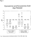
- FIG. 2 shows L-kynurenine and kynurenic acid concentrations in ⁇ M for normal controls and differences between ⁇ 60 and >60 m years of age. Measurements were made in serum and saliva as well. FIG. 2 shows that the mean values of kynurenine and kynurenic acid in cohorts of healthy volunteers are very similar regardless whether the volunteers have an age above or below 60 years.
- FIG. 4 shows values given for the correlation between serum and saliva in kynurenine and kynurenic acid measurement.
- FIG. 9 shows a comparison of the kynurenic acid content in serum and saliva for normal controls and patients as in FIG. 8 . Higher values of kynurenic acid can be observed for the normal control group while the concentration is decreased in patients with ND. The difference between the groups is significant for the value in serum as well as for the value in saliva.
- FIG. 10 shows the values of kynurenine for male and female members of the normal control group.
- the median concentration in serum is 2.63 ⁇ 0.64 ⁇ M (female group) and 2.79 ⁇ 0.64 ⁇ M (male group); the corresponding concentrations in saliva are 0.79 ⁇ 0.37 ⁇ M and 0.88 ⁇ 0.34 ⁇ M. No statistically significant difference was observed in these values for female or male persons.
- the median concentration in serum was 2.69 ⁇ 0.6 ⁇ M, in saliva 0.82 ⁇ 0.28 ⁇ M.
- FIG. 11 B shows the correlation of the kynurenine serum values vs. the kynurenine saliva values.
- a correlation of r 2 0.90 was determined which remained the same also for higher values. This is a clear indication that the saliva values are generally useful for diagnostic purposes.
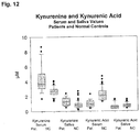
- FIG. 12 shows serum and saliva concentrations for patients and normal control persons (NP). The results show that in the samples of patients the ratio of kynurenine to kynurenic acid has changed to the disadvantage of the neuroprotective substance kynurenic acid.
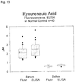
- FIG. 13 shows a comparison of the determination of kynurenic acid via a fluorescence based method compared to a commercially available ELISA test.
- Kynurenine was significantly higher and kynurenic acid lower and ratio was different in patients with neurodegenerative disorders. This could be demonstrated in serum as well as in saliva. The measured values are shown in FIG. 3 .
- kynurenine/kynurenic acid measurement is a tool to identify cerebral disorders as well as to monitor them.
- the measured kynurenine/kynurenic acid quotient is a clear indicator for neurodegenerative diseases if the quotient is 1.0 or higher.
- normal control persons The collective of “normal control persons” is comprised of blood donors. These were healthy persons who consented to the use of serum and saliva samples for the purposes of the present research and examples. Serum was obtained from 326 persons, 302 of which were included in the determination of kynurenine. For 12 persons, no corresponding saliva samples were available or could not be used for the test. For 12 persons, test values in a pathological range were determined, probably based on inflammations (kynurenine above 4.2 in serum). Such persons could not be included in the group of healthy controls.
- Saliva samples generally had lower levels of kynurenine and kynurenic acid.
- kynurenine and kynurenic acid were determined in serum and saliva.
- a determination for both kynurenine and kynurenic acid could be performed in saliva as well as in serum. The results are shown in FIG. 6 .
- Kynurenic acid concentration was determined by a fluorescence based test as described in the following. The test result was compared to the results obtained using a commercially available ELISA test kit (KYNA ELISA human, Cloud-Clone Corporation, 11271 Richmond Avenue Suite H104, Houston Tex. 77082, USA, Lot: L 150525449. The assay employs the competitive inhibition enzyme immunoassay technique and was performed in the manner as described in the instruction manual).
- the fluorescence based test was performed using FluoStar® BMG and the following conditions:
- Kynurenic acid (MW: 189.17, Sigma K3375) standard curve from 10 ⁇ M to 0.156 ⁇ M: KynA (1.89 mg) dissolved in 500 ⁇ l DMSO, then addition of 5 ml H 2 O+4.5 ml HClO 4 6 M: 1000 ⁇ M
- the concentration of KynA is determined based on the calibration line taking into account a dilution factor of 1.1 resulting from the sample preparation
- the result of this comparison is shown in FIG. 13 .
- the fluorescence based test provided more accurate results than the ELISA test kit.
Landscapes
- Health & Medical Sciences (AREA)
- Life Sciences & Earth Sciences (AREA)
- Engineering & Computer Science (AREA)
- Immunology (AREA)
- Molecular Biology (AREA)
- Biomedical Technology (AREA)
- Urology & Nephrology (AREA)
- Chemical & Material Sciences (AREA)
- Hematology (AREA)
- Physics & Mathematics (AREA)
- Medicinal Chemistry (AREA)
- Microbiology (AREA)
- Cell Biology (AREA)
- Food Science & Technology (AREA)
- Biotechnology (AREA)
- Analytical Chemistry (AREA)
- Biochemistry (AREA)
- General Health & Medical Sciences (AREA)
- General Physics & Mathematics (AREA)
- Pathology (AREA)
- Proteomics, Peptides & Aminoacids (AREA)
- Biophysics (AREA)
- Bioinformatics & Computational Biology (AREA)
- Bioinformatics & Cheminformatics (AREA)
- Tropical Medicine & Parasitology (AREA)
- Neurology (AREA)
- Neurosurgery (AREA)
- Investigating Or Analysing Biological Materials (AREA)
Abstract
The present invention is a kit for the determination of a neurodegenerative disease wherein separately from each other the content of kynurenine and kynurenic acid in a body fluid is determined and the quotient of the content of kynurenine to the content of kynurenic acid is calculated.
Description
- Chronic progressive neurodegenerative diseases, such as Alzheimer's disease (AD), Parkinson's disease (PD) and vascular dementia (VD) display an increasing prevalence in parallel with the ongoing aging of the population, and have therefore generated considerable recent research interest. Despite extensive studies on the background of neurodegenerative processes, the exact molecular basis remains still to be clarified. There is accumulating evidence that the innate immune response in the brain is mainly influenced by inflammatory processes.
- Although these devastating diseases have a serious impact on the quality of life of the patients, their management is often challenging. Current therapies offer mostly only symptomatic relief and no neuroprotective therapy is available. The pathomechanisms of different neurodegenerative disorders share a number of common features. Excitotoxicity, neuroinflammation, a mitochondrial disturbance and oxidative stress have been implicated in both acute and chronic neurological disorders (1). Improving the sensitivity and accuracy of diagnostic tests for neurodegenerative disorders, however, is still an object for further research.
- Neurodegenerative processes share some common features, which are not disease-specific. While there are still a number of details that await elucidation, there are several common mechanisms that are widely accepted; the role of mitochondrial disturbances, excitotoxicity, neuro-inflammation and oxidative stress appear evident (1, 2). Glutamate excitotoxicity has been implicated in the pathomechanisms of ischemic stroke, traumatic brain injury, and various neurodegenerative disorders (1, 3).
- AD was earlier thought to involve a distinct pathology, which can be clearly distinguished from vascular dementia (VD). However, in recent years, the role of a cerebrovascular dysfunction has been linked to the neurodegenerative process of AD, and vascular risk factors have attracted growing attention in connection with AD development and progression.
- Overlaps between VD and AD have long been recognized, but in recent years a complete paradigm shift has begun, and AD has been suggested to be a primarily vascular disease (4). Only a small proportion of AD cases have a genetic origin; the majorities are sporadic. The most important risk factor for the development of AD is advancing age, the prevalence and incidence data demonstrating an increasing tendency with rising age (5, 6). Again to be mentioned, kynurenine plays a major role in vascular regulatory processes.
- Similarly, an impaired cerebral blood flow and autoregulation capacity has been observed in animal models of AD, this impairment proving to be associated with oxidative stress (7, 8). These findings link the presence of amyloid beta peptides (Aß) to oxidative stress and neuroinflammation. Today under the new view of innate immune responses we would state that there is an activation of the innate inflammatory response accompanying this disease. In this theory, the Aß molecule could have the role of an alarmin—same like ATP in other diseases—and is responsible for the activation of the inflammation via NALP-3-inflammasome. Second view is the generation of oxygen—radicals under a minimized blood flow.
- In the following the role of the kynurenine pathway (KP) in neurodegenerative diseases and its modulation will be explained in more detail:
- The KP is the main metabolic route of tryptophan (TRP) degradation in mammals; it is responsible for more than 95% of the TRP catabolism in the human brain (9). The metabolites produced in this metabolic cascade, termed kynurenines, are involved in a number of physiological processes, including neurotransmission and immune responses (10, 11). The KP also involves neurotoxic and neuroprotective metabolites, and alterations in their delicate balance have been demonstrated in multiple pathological processes.
- The central intermediate of the KP is L-kynurenine (L-KYN), where the metabolic pathway divides into two different branches. L-KYN is transformed to either the neuroprotective kynurenic acid (KYNA) via kynurenine aminotransferase (KAT), or to 3-hydroxy-L-kynurenine (3-OH-KYN or simply 3-HK), which is further metabolized in a sequence of enzymatic steps to yield finally NAD+ (as shown in
FIG. 1 ). - Imbalances in the KP are not only relevant in AD, but also in other disorders in which there is a cognitive decline, and influencing this delicate balance may be of utmost therapeutic value (12).
- Changes in kynurenine metabolites have additionally been suggested to correlate with the infarct volume, the mortality of stroke patients and the post-stroke cognitive impairment (14).
- In another study, serum kynurenine levels and inflammatory markers were measured in patients undergoing cardiac surgery; the results indicated an association of several kynurenine metabolite levels with the post-surgical cognitive performance (15).
- The results of this paper show increased levels of tryptophan with decreased levels of kynurenine, anthranilic acid and 3-hydroxyanthranilic acid associated with bypass surgery, and a later increase in kynurenic acid. Levels of neopterine and lipid peroxidation products rose after surgery in non-bypass patients whereas TNF-α and S100B levels increased after bypass. Changes of neopterine levels were greater after non-bypass surgery. Cognitive testing showed that the levels of tryptophan, kynurenine, kynurenic acid and the kynurenine/tryptophan ratio, correlated with aspects of post-surgery cognitive function, and were significant predictors of cognitive performance in tasks sensitive to frontal executive function and memory. Thus, anesthesia and major surgery are associated with inflammatory changes (activation of the innate immune response according to generation of free radicals) and alterations in tryptophan oxidative metabolism that predict, and may play a role in, post-surgical cognitive function.
- KP metabolites have also been implicated in vascular cognitive impairment (16). As concerns AD, a substantial amount of evidence demonstrates an altered tryptophan metabolism.
- From the aspect of the peripheral kynurenine metabolism, decreased KYNA levels were measured in the serum, red blood cells and CSF of AD patients (17, 18). Additionally, enhanced IDO activity was demonstrated in the serum of AD patients, as reflected by an increased KYN/TRP ratio, this elevation exhibiting inverse correlation with the rate of cognitive decline. IDO activation was also correlated with several immune markers in the blood, thereby indicating an immune activation, which lends further support to the role of neuroinflammation in the pathomechanism of AD. An increased IDO activity was also confirmed by immunohistochemistry in the hippocampus of AD patients, together with an enhanced quinoline acid (QUIN) immunoreactivity (19).
- Postoperative cognitive dysfunction (POCD) is defined as a newly developed cognitive functional disorder after surgery and anesthesia. Symptoms are subtle and showing manifold patterns. Mechanisms leading to this entity are still not solved entirely. Experimental results show immunological responses of the innate immune system leading to a neuroinflammation. Activation of the inflammatory response and the TNF-α and NF-kB signal cascades destroy the integrity of the blood-brain-barrier via excretion of different cytokines (20, 21). This enables macrophages migration into the hippocampus and allows the disabling of brain memory response. Anti-inflammatory response could inhibit this proinflammatory action and dysfunction would be prohibited.
- Quinoline acid (QUIN) has been shown to stimulate lipid peroxidation, production of reactive oxygen species, and mitochondrial dysfunction (22, 23). Studies performed in organotypic cultures of rat corticostriatal system indicate that concentrations of QUIN even just slightly higher than physiological concentrations can cause neurodegeneration after a few weeks of exposure (24). Spinal neurons have been found to be especially sensitive to QUIN variations causing cell death with just nanomolar concentrations of this metabolite (25, 26).
- As explained above in detail, the kynurenine pathway (KP) metabolizes the essential amino acid tryptophan and generates a number of neuroactive metabolites called the kynurenines. Segregated into at least two distinct branches, often termed as the “neurotoxic” and “neuroprotective” arms of the KP, they are regulated by the two enzymes kynurenine 3-monooxygenase (KMO) and kynurenine aminotransferase (KAT), respectively. Interestingly, several enzymes in the pathway are under tight control of inflammatory mediators and even small changes can cause major injuries. Recent years have seen a tremendous increase in our understanding of neuroinflammation in CNS disease. There is evidence, that neuroinflammation is linked to the innate immune system and the role of NAPL-3 inflammasomes. This finding could be the basis for a protective therapeutic approach in these kind of disorders (28). This theory is supported by the fact that the increased i-protein concentration in patients with ND-disease acts like alarmins. These alarmins are responsible for the activated innate response in the sense of an inflammation.
- A diagnostic method, which helps to define more precisely and to very accurately diagnose neurodegenerative diseases, is not available yet but highly desirable.
- WO 2014/177680 discloses a diagnostic method for neurodegenerative disorders. The levels of kynurenine in plasma and/or in saliva are compared with the average level of kynurenine measured in comparable individuals who are not affected by such neurodegenerative diseases. Improving the sensitivity and accuracy of diagnostic tests for neurodegenerative disorders, however, is still an object for further research and is an object to be solved by the present invention.
- The present invention is directed to an in vitro method for the determination of a neurodegenerative disease.
- A further subject of the present invention is a test kit for performing such in vitro diagnostic method.
- The present invention relates to the kynurenine pathway, KP. Tryptophan (TRP), an essential amino acid, can be metabolized through different pathways, the main metabolic route being the kynurenine pathway. This pathway is illustrated in
FIG. 1 . The first enzyme of the pathway, indolamine-2,3-dioxygenase (IDO-1) is strongly stimulated by inflammatory molecules, particularly interferon-γ. Thus, the kynurenine pathway is often systematically up-regulated when the immune response is activated. The biological significance is that on the one hand the depletion of tryptophan and generation of kynurenines play a key modulating role in the immune response. During the research work that led to the present invention, it was surprisingly found that the level of kynurenine and kynurenic acid measured in the saliva could be used for the detection of a potential neurodegenerative disease that can otherwise not be easily detected. This is particularly surprising since neurodegenerative diseases relate to the brain that is separated from the rest of the body by the blood-brain (blood/liquor) barrier. - The activation of
indole amine 2,3-dioxygenase (IDO-1), the main enzyme involved in the catabolism of tryptophan, generates immunosuppressive metabolites which counter-regulate this immune activation. - Today it is known that the endothelium, once considered to be relatively inert, is involved in various functions such as fibrinolysis, coagulation, vascular tone, growth and immune response. The most common reaction in the human body might be seen in the inflammatory response mediated by the innate immunity.
-
Indole amine - The present invention provides an in vitro method for the determination of a neurodegenerative disease wherein separately from each other the content of L-kynurenine (L-KYN) and kynurenic acid (KYNA) in a body fluid of a person is determined and the quotient of the content of kynurenine to the content of kynurenic acid is calculated.
- Based on the calculated value for the quotient of the contents of kynurenine and kynureic acid, it can be determined whether this person suffers from a neurodegenerative disease.
- Neurodegenerative diseases in the sense of the present invention comprise in particular Alzheimer's disease, Parkinson's disease, vascular dementia, postoperative cognitive dysfunction and/or age-related depression.
- There are many forms of mental impairment that can be designated in a slightly different manner. A clear borderline is, however, difficult to draw. Mental impairments comprise for example amnestic mild cognitive impairment that affects mainly the memory. Furthermore, non-amnestic mild cognitive impairment is also known whereby the memory is not strongly affected, but other mental capabilities are significantly reduced. For Alzheimer's disease there are several known stages and the classification into the several stages depends on the testing methods. It has been assumed that cognitive impairment, dementia and Alzheimer dementia may be gender-specific and dependent on the genetic heritance of the test groups (e.g. Caucasians vs. Afro-Americans). It seems that the mental impairment is definitely age-related. With increasing age, the mental impairment increased significantly whereby the increase starts with the age of 60 to 70 years.
- The method of the present invention for the first time allows to perform a comparatively simple and quick test which provides a meaningful diagnostic result.
- The in vitro test method of the present invention can be performed with a body fluid. A body fluid in the sense of the present invention is any liquid that can be derived from a human body. The most commonly used body fluids for diagnostic tests are serum or plasma. For neurodegenerative diseases the testing of liquor may also be suitable. To obtain a liquor sample is, however, difficult and sometimes dangerous. Therefore, other body fluids, which may be easily obtainable, are preferred. In a particularly preferred embodiment of the present invention the in vitro method of the present invention is performed with saliva since saliva can be most easily obtained and surprisingly the results for tests performed using saliva are very accurate.
- Saliva is a clinically informative, biological fluid that is useful for novel approaches to prognosis, laboratory or clinical diagnosis, and monitoring and management of patients. Saliva contains multiple biomarkers and an overview of the principles of salivary gland secretion, methods of collection, and discussion of general uses can be found in a report of a meeting published in the Annals of the New York Academy of Sciences Malamud D, Niedbala R S Oral-based diagnostics NY Acad Sci 2007; Boston Mass.
- The fact that a determination of kynurenine and kynurenic acid is possible and significant differences between healthy persons and patients affected by neurodegenerative diseases can be reliably identified in saliva samples is, however, quite surprising. It is well known that only some diagnostic determinations are possible in saliva. E.g. as far as viral infections are concerned, while HCV can be accurately detected in saliva, this is not the case for HIV. As a further negative example, saliva samples are not suitable for the determination of the blood sugar level.
- In this context, as a potentially important distinction in terms of test accuracy, saliva is the watery substance which is secreted by the salivary glands and should not be mistaken for the collective liquid present in the mouth of a person. According to the present invention, saliva samples are most preferably obtained directly at the salivary glands of a person using a saliva collection device, especially a Salivette® (Sarstedt Ag & Co., NUmbrecht, Germany, www.sarstedt.com).
- A main aspect of the present invention is that it was discovered that meaningful diagnostic predictions can be made from the relation (quotient) of the concentration of kynurenine to the concentration of kynurenic acid in the body fluid, most importantly saliva. In order to determine these contents or concentrations and the relation thereof, the same sample is tested with regard to the content of kynurenine and kynurenic acid and the corresponding concentrations are determined individually.
- In one embodiment, the in vitro method of the present invention uses two different components, one which specifically binds to kynurenine, and one which specifically binds to kynurenine acid. Both components can be and preferably are located at different sites of a test device in order to allow easy and clear distinction of the test results.
- The term “specifically binding” means that one component binds only to kynurenine (L-kynurenine) and not to kynurenic acid. The other component binds specifically only to kynurenic acid and not to L-kynurenine. Furthermore, both components should not bind to any other substance or impurity that may be present in the body fluid to be tested. The unspecific binding of the component to any impurity that may be present in the body fluid may influence the test result and is highly undesirable.
- In a preferred embodiment, as one or both components that specifically bind to kynurenine or to kynurenic acid, antibodies are used. Such antibodies can be prepared according to well-known methods, mainly by immunizing a laboratory animal like for example rabbits, goats, horses or sheep and obtain polyclonal antibodies, which may further be purified and used in the test.
- In an alternative embodiment, one or more of the components may be a monoclonal antibody, which can be produced by the well-known hybridoma technology. Suitable clones are selected which show the desired binding pattern. When a suitable monoclonal antibody has been identified it is possible to sequence the binding regions and to prepare derivatives of the monoclonal antibody by genetic engineering. It is well-known for example to produce single chain antibodies or diabodies in order to name only a few. Such constructs contain an antigen binding region that fits perfectly to the structure of the target molecule.
- Both polyclonal and monoclonal antibodies specifically binding L-kynurenine or kynurenic acid but also commercially available antibodies can be used in the method of the present invention, as long as their specificity, binding affinity and efficiency provide for satisfactory results which can easily be determined by a calibration test performed with samples having a predetermined content of the test substances.
- In another preferred embodiment the components that bind specifically to L-kynurenine or to kynurenic acid, respectively, are aptamers. Aptamers are biocompatible molecules like DNA- or RNA-oligonucleotides or peptides, which enable specific targeting of molecules. Aptamers are for example oligonucleotides or peptides that have high sensitivity and robust selectivity towards several types of target molecules including small molecules like L-kynurenine or kynurenic acid, respectively. Usually the aptamers contain a variable loop and stem region that bind to a specific pocket or surface structure of the target molecules.
- In preferred embodiments the aptamers are selected in vitro by using a process called “systematic evolution of ligands by exponential enrichment” (SELEX). In the course of a usual SELEX procedure, the target (L-kynurenine or kynurenic acid) is brought into contact with a library of potential ligands. The candidates, which have the best binding characters, are separated and further improved by slight changes of the binding molecules and further selection of the better candidates. After several rounds of enrichment and improvement an aptamer may be obtained which has high specificity for the target molecules.
- In addition thereto it may be advantageous to perform also a negative selection in order to make sure that the aptamer binds exclusively either to L-kynurenine or to kynurenic acid, respectively. In such negative selection those candidates are singled out which do not allow a clear distinction of the target.
- In a preferred embodiment the in vitro method of the present invention is performed as an ELISA (Enzyme Linked Sorbent Test Assay).
- There are different configurations of ELISA tests known. Usually a so-called sandwich ELISA test is performed. In such an ELISA test the compound that binds specifically to L-kynurenine or kynurenic acid is fixed on a solid surface (e.g. the bottom of a microtiter well). Unspecific binding sites are saturated (e.g. with skim milk powder) in order to avoid unspecific binding.
- Preferably the ELISA test kit must contain separate entities (e.g. microtiter wells) with components that bind specifically to kynurenine only and in other entities components that bind to kynurenic acid only. Usually several microtiter wells are coated with the component in order to allow an easy dilution of the sample for a determination of the content of the analyte.
- The binding of L-kynurenine or kynurenic acid, respectively, to the relevant wells is usually detected with another antibody that binds, however, to another area of the target molecule in order to avoid a negative interference of the binding. Such antibody is usually coupled with a signal generating means that may be for example an enzyme like horseradish peroxidase. The presence of the analyte to be detected can then be seen by adding a precursor molecule, which is converted to another molecule having different properties by the signal generating molecule. When for example in one wall kynurenine or kynurenic acid, respectively, is present the antibody binds to this molecule and with the activity of the signal generating means (e.g. horseradish peroxidase) a color signal is generated whereby the intensity is proportional to the amount of the bound target molecule (kynurenine or kynurenic acid). The reaction can be measured quantitatively and the amount of the analyte to be detected in the body fluid can be determined precisely.
- In a further embodiment the in vitro test method is a lateral flow test. Lateral flow tests also known as Lateral Flow Immunochromatographic Assays are simple devices intended to detect the presence (or absence) of a target analyte sample without the need for specialized and costly equipment, though many lab based applications exist that are supported by a reading equipment. Typically, these tests are used for medical diagnostics either for home testing, point of care testing, or laboratory use. A widely spread and well known application is e.g. the home pregnancy test.
- The technology is based on a series of capillary beds, such as pieces of porous paper or sintered polymer. Each of these elements has the capacity to transport fluid (e.g., saliva) spontaneously. The first element (the sample pad) acts as a sponge and holds an excess of sample fluid. Once soaked, the fluid migrates to the second element (conjugate pad) in which the manufacturer has stored the so called conjugate, a dried format of bio-active particles (see below) in a salt-sugar matrix that contains everything to guarantee an optimized chemical reaction between the target molecule (e.g., kynurenine) and its chemical partner (e.g., antibody) that has been immobilized on the particle's surface. While the sample fluid dissolves the salt-sugar matrix, it also dissolves the particles and in one combined transport action the sample and conjugate mix while flowing through the porous structure. In this way, the analyte binds to the particles while migrating further through the third capillary bed. This material has one or more areas (often called stripes) where a third molecule has been immobilized by the manufacturer. By the time the sample-conjugate mix reaches these strips, analyte has been bound on the particle and the third ‘capture’ molecule binds the complex. After a while, when more and more fluid has passed the stripes, particles accumulate and the stripe-area changes color. Typically there are at least three stripes: one (the control) that captures any particle and thereby shows that reaction conditions and technology worked fine, the second and third contains a specific capture molecule (compound specific for L-kynurenine and kynurenic acid, respectively) and only captures those particles onto which an analyte molecule has been immobilized. After passing these reaction zones the fluid enters the final porous material, the wick, that simply acts as a waste container. Lateral Flow Tests can operate as either competitive or sandwich assays.
- In principle, any colored particle can be used, however, latex (blue color) or nanometer sized particles of gold (black/grey, red color) are most commonly used. The gold particles are red in color due to localized surface plasmon resonance. Fluorescent or magnetic labeled particles can also be used, however these require the use of an electronic reader to assess the test result.
- The sample first encounters colored particles, which are labeled with antibodies raised to the target analyte. The test line will also contain antibodies to the same target, although it may bind to a different epitope on the analyte. The test line will show as a colored band in positive samples. An example of the sandwich assay is the sandwich ELISA.
- While not strictly necessary, most test kits preferably incorporate a second line, which contains an antibody that picks up free latex/gold in order to confirm the test has operated correctly.
- In a preferred embodiment the single components of the lateral flow assay are adapted in such a manner that the presence of kynurenine or kynurenic acid is indicated only when more than a certain threshold value of kynurenine is present in the sample.
- A preferred test kit consists of the following components:
-
- 1. Sample pad—an absorbent pad onto the test sample (saliva) is applied
- 2. Conjugate or reagent pad A and B—this contains components (e.g. antibodies) specific to the target (kynurenine and kynurenic acid, respectively) analyte conjugated to colored particles (usually colloidal gold particles, or latex microspheres)
- 3. Reaction membrane—typically a hydrophobic nitrocellulose or cellulose acetate membrane onto which anti-target analyte antibodies are immobilized in a line across the membrane as a capture zone or test line (a control zone may also be present, containing antibodies specific for the conjugate antibodies)
- 4. Wick or waste reservoir—a further absorbent pad designed to draw the sample across the reaction membrane by capillary action and collect it.
- The components of the strip are usually fixed to an inert backing material and may be presented in a simple dipstick format or within a plastic casing with a sample port and reaction window showing the capture and control zones.
- There are two preferred embodiments of the test kits (lateral flow immunoassay) used in the method of the present invention:
- a. Double Antibody Sandwich Assays
- In this format the sample migrates from the sample pad through the conjugate pad where any target analyte present will bind to the conjugate. The sample then continues to migrate across the membrane until it reaches the capture zone where the target/conjugate complex will bind to the immobilized antibodies producing a visible line on the membrane. The sample then migrates further along the strip until it reaches the control zone, where excess conjugate will bind and produce a second visible line on the membrane. This control line indicates that the sample has migrated across the membrane as intended. Two clear lines on the membrane show a positive result. A single line in the control zone is a negative result. Double antibody sandwich assays are most suitable for larger analytes, such as bacterial pathogens and viruses, with multiple antigenic sites. For the present invention a suitable pair of antibodies must be selected which bind to different epitopes on kynurenine and kynurenic acid, respectively.
- When the test methods or kits suitable for performing such method use antibodies which bind specifically to kynurenine, the term “antibody” means not only antibodies artificially produced for example by immunization of a laboratory animal like rabbit, sheep or goat. It comprises also in a preferred embodiment monoclonal antibodies produced according to the hybridoma technology. Moreover, the term “antibody” comprises also antigen-binding fragments of antibodies such as recombinantly produced antigen-binding fragments. Such constructs can be produced by phage display and technologies derived there from.
- b. Competitive Assays
- Competitive assays are primarily used for testing small molecules and differ from the double antibody sandwich format in that the conjugate pad contains antibodies that are already bound to the target analyte, or to an analogue of it. If the target analyte is present in the sample it will therefore not bind with the conjugate and will remain unlabelled. As the sample migrates along the membrane and reaches the capture zone an excess of unlabelled analyte will bind to the immobilized antibodies and block the capture of the conjugate, so that no visible line is produced. The unbound conjugate will then bind to the antibodies in the control zone producing a visible control line. A single control line on the membrane is a positive result. Two visible lines in the capture and control zones is a negative result. However, if an excess of unlabelled target analyte is not present, a weak line may be produced in the capture zone, indicating an inconclusive result. Competitive assays are most suitable for testing for small molecules, such as mycotoxins, unable to bind to more than one antibody simultaneously. There are a number of variations on lateral flow technology. The capture zone on the membrane may contain immobilized antigens or enzymes—depending on the target analyte—rather than antibodies. It is also possible to apply multiple capture zones to create a multiplex test.
- Lateral flow immunoassays are simple to be used by untrained operators and generally produce a result within 15 minutes. They are very stable and robust, have a long shelf life and do usually not require refrigeration. They are also relatively inexpensive to produce. These features make them ideal for use at the point-of-care and for testing samples in the field, as well as in the laboratory. However, their sensitivity is limited without additional concentration or culture procedures. There are quantitative tests available, but our target is a qualitative test for saliva within a certain range. Therefore, the preferred test kit is adjusted to measure kynurenine only if present above a certain concentration. Below such concentration the test kit will show a negative result.
- The present invention is based on determining the quotient of the presence of kynurenine compared to the presence of kynurenic acid in the body fluid. Therefore, the lateral flow test is designed to perform two measurements from the sample (preferably saliva) at the same time. In order to make the use of the test simple the test may be calibrated to certain contents of L-kynurenine or kynurenic acid, respectively. The lateral flow test is preferably designed in such a manner that when a critical concentration, which has been fixed, previously is reached a color signal can be seen. By using suitable dilutions it is possible to design the lateral flow test in such a manner that it can be easily seen whether the quotient of kynurenine to kynurenic acid is above 1 or below 1.
- Usually a quotient below 1 indicates no neurodegenerative disease whereas a quotient above 1.0 already suggests an increased probability of the presence of such disease. Values above 1.25, preferably above 1.3 and most certainly above 1.4 indicate a neurodegenerative disease. Values as high as 2.0 have been observed for patients with established neurodegenerative disease using the in vitro method of the present invention.
- In another preferred embodiment of the invention, the determination of the concentration of kynurenic acid in a body fluid, especially saliva, is performed via a colorimetric or a fluorescence based method (e.g. FLUOstar®, available from BMG Labtech, Ortenberg, Germany). Since according to the present invention, the content of kynurenine and kynurenic acid in the sample are determined individually, in such case kynurenine can be determined either by an immunoassay as described above or by any other method.
- Especially for kynurenic acid, however, it was observed that test results determined using a fluorescence based test method are very accurate and reliable (e.g. using the method and conditions which is detailed in Example 5).
- While the content of kynurenine as well as of kynurenic acid can also be determined by other methods known in the art like HPLC, it is important to consider the solubility properties of kynurenine. The determination of the concentration of kynurenine is most preferably performed via kynurenine sulfate which is the soluble form of kynurenine. Thus, the settings of a HPLC or more preferably a tandem mass spectrometry (MS/MS) test procedure need to be adjusted to detect L-kynurenine sulfate. Values determined in such manner can be used for determining the concentration of kynurenine for healthy persons and also for persons who are possibly afflicted by a neurodegenerative disease.
- In another embodiment the present invention provides suitable kits for performing the method according to the invention. Such a kit comprises preferably means for the determination of kynurenine and kynurenic acid, respectively, in saliva. Such means may work on different principles. It is possible to use a specific color reagent, which detects the presence of kynurenine and/or kynurenine derivatives. Alternatively the kit may comprise at least one or preferably two antibodies specifically binding to kynurenine or kynurenic acid. Preferably when two antibodies are used, such antibodies do not bind to the same epitope in order to allow the formation or a sandwich formed by the first antibody, kynurenine or its derivative and the second antibody.
- In one embodiment of the present invention the determination of kynurenine and kynurenic acid is performed by a coloring reaction. The sample in the determination test is saliva. Before the content of kynurenine or derivatives thereof can be determined, components, which may negatively affect the correct, and precise test result have to be removed. In a preferred embodiment undesired components of saliva that may disturb the correct test result are removed preferably by precipitation of the components that disturb the result of the measurement. Such precipitation can preferably be performed by using trichloric acid. It is, however, possible to use other methods for deproteinization of saliva than using trichloric acid. After the disturbing components of saliva have been removed by precipitation it may be necessary to separate the phases by centrifugation. The supernatant is then preferably reacted with a coloring reagent that may preferably be Ehrlich's reagent. After development of the color the samples are measured by measuring the absorbance at a suitable wavelength. Preferably the test is performed in a quantitative or semi-quantitative manner. In the test method either a calibration curve can be used or a certain threshold value is fixed in the test kit in order to avoid false positive results.
-
FIG. 1 shows a schematic overview of the kynurenine pathway, the major route of tryptophan degradation in higher eukaryotes. Enzymes are indicated in italics. The neurotoxic metabolites QUIN and 3-HK are shown as well as the neuroprotective metabolite KYNA (13). -
FIG. 2 shows L-kynurenine and kynurenic acid concentrations in μM for normal controls and differences between <60 and >60 m years of age. Measurements were made in serum and saliva as well.FIG. 2 shows that the mean values of kynurenine and kynurenic acid in cohorts of healthy volunteers are very similar regardless whether the volunteers have an age above or below 60 years. -
FIG. 3 shows a comparison of kynurenine (measured in serum) in 2 groups: controls (n=194); patients with neurodegenerative disorders (n=42). There was a significant difference for kynurenine and kynurenine acid (p<0.001). -
FIG. 4 shows values given for the correlation between serum and saliva in kynurenine and kynurenic acid measurement. -
FIG. 5 shows the difference of the quotient Kyn/KynA between normal controls (n=194) and patients (n=42) with neurodegenerative (ND) disorders (p<0.0000071) when measured in saliva. -
FIG. 6 shows a comparison of the concentration of kynurenine vs. kynurenic acid for normal controls (n=181). The ratio of both is similar in serum and saliva. -
FIG. 7 shows the results for kynurenine values in serum and saliva in the normal control group. The results are shown for the whole control group and for female (n=143) and male (n=159) members of the group, respectively. -
FIG. 8 shows a comparison of the kynurenine content in serum and saliva for normal controls and for patients (n=49) with long-standing neurodegenerative disease. The difference for serum and for saliva is significant (p<0.001). Kynurenine is significantly increased in the patient group. -
FIG. 9 shows a comparison of the kynurenic acid content in serum and saliva for normal controls and patients as inFIG. 8 . Higher values of kynurenic acid can be observed for the normal control group while the concentration is decreased in patients with ND. The difference between the groups is significant for the value in serum as well as for the value in saliva. -
FIG. 10 shows the values of kynurenine for male and female members of the normal control group. The median concentration in serum is 2.63±0.64 μM (female group) and 2.79±0.64 μM (male group); the corresponding concentrations in saliva are 0.79±0.37 μM and 0.88±0.34 μM. No statistically significant difference was observed in these values for female or male persons. -
FIG. 11 A shows the values for kynurenine in serum and saliva for the normal control group (n=302). The median concentration in serum was 2.69±0.6 μM, in saliva 0.82±0.28 μM. -
FIG. 11 B shows the correlation of the kynurenine serum values vs. the kynurenine saliva values. A correlation of r2=0.90 was determined which remained the same also for higher values. This is a clear indication that the saliva values are generally useful for diagnostic purposes. -
FIG. 12 shows serum and saliva concentrations for patients and normal control persons (NP). The results show that in the samples of patients the ratio of kynurenine to kynurenic acid has changed to the disadvantage of the neuroprotective substance kynurenic acid. -
FIG. 13 shows a comparison of the determination of kynurenic acid via a fluorescence based method compared to a commercially available ELISA test. - The present invention is described in more detail in the Figures and the following Examples.
- Forty-two patients with cerebral dementia (mean age 71+5.3 years, mean MMS-score 22) were enrolled in a comparative study with normal controls (n=194; mean age 48.8 years, range 16-88 years). Aim of our study was to detect changes in the tryptophan metabolism in patients with cerebral dementia, by estimating either kynurenine, kynurenic acid and ratio of kyn/kynA in plasma and in saliva. There was no age related difference between a group I of normal controls (n=93, age>60, mean age 71.3 years, range 60-88) and group II (n=101, age<60, mean age 38.8, range 16-60;
FIG. 2 ). This is demonstrating that the disease must not necessarily be age related in general. The neurodegenerative diseases may be caused by different reasons whereby, however, the frequency of neurodegenerative diseases increases statistically with increasing age. - Kynurenine was significantly higher and kynurenic acid lower and ratio was different in patients with neurodegenerative disorders. This could be demonstrated in serum as well as in saliva. The measured values are shown in
FIG. 3 . - There was a correlation between the values of kynurenine and kynurenic acid in serum compared to saliva (saliva 1:3.5 in serum for kynurenine and 1:3.2 in saliva for kynurenic acid in normal controls,
FIG. 4 ). - Patients with neurodegenerative disease showed a total different pattern: mean values for kynurenine in serum as well as in saliva were significant higher (4.80±0.6 μM for serum and 1.34±0.3 μM for saliva) whereas values for kynurenic acid were significant lower (1.58±0.3 in serum and 1.3±0.2 in saliva,
FIG. 3 ). This is in correspondence with the theory of the pathophysiology of the disease: the neuro-protective part (kynurenic acid) is downregulated and the inflammatory part (kynurenine) is upregulated. - Concerning the measurement of kynurenine and kynurenic acid in serum and saliva we could demonstrate the correlation for serum and saliva. The small numeric difference between both values is related to the different method of measurement.
- We could demonstrate that measurement of kynurenine and kynurenic acid is possible in serum as well as in saliva. There is a relationship between the values in serum compared to the values in saliva.
- Compared to the data evaluated in normal controls, data in patients showed significant different pattern and could be easy identified.
- In total, already in this small group of patients it could be demonstrated, that kynurenine/kynurenic acid measurement is a tool to identify cerebral disorders as well as to monitor them. The measured kynurenine/kynurenic acid quotient is a clear indicator for neurodegenerative diseases if the quotient is 1.0 or higher.
- The collective of “normal control persons” is comprised of blood donors. These were healthy persons who consented to the use of serum and saliva samples for the purposes of the present research and examples. Serum was obtained from 326 persons, 302 of which were included in the determination of kynurenine. For 12 persons, no corresponding saliva samples were available or could not be used for the test. For 12 persons, test values in a pathological range were determined, probably based on inflammations (kynurenine above 4.2 in serum). Such persons could not be included in the group of healthy controls.
- The obtained values corresponded well with values published in the prior art. Saliva samples generally had lower levels of kynurenine and kynurenic acid.
- From the 302 persons of the normal control group, 143 were female and 159 male. The average age was 47.6 (18 to 75 years old) for the female group and 47.1 (18 to 75 years old) for the male group. The age was determined to not be statistically significant. Results are shown in
FIGS. 7 and 10 . - In a second series of tests, kynurenine and kynurenic acid were determined in serum and saliva. For 181 samples a determination for both kynurenine and kynurenic acid could be performed in saliva as well as in serum. The results are shown in
FIG. 6 . - Further test results are shown in the remaining Figures attached herewith.
- Kynurenic acid concentration was determined by a fluorescence based test as described in the following. The test result was compared to the results obtained using a commercially available ELISA test kit (KYNA ELISA human, Cloud-Clone Corporation, 11271 Richmond Avenue Suite H104, Houston Tex. 77082, USA, Lot: L 150525449. The assay employs the competitive inhibition enzyme immunoassay technique and was performed in the manner as described in the instruction manual).
- The fluorescence based test was performed using FluoStar® BMG and the following conditions:
- Perchloric acid (HClO4), 60%=10 M, and 6M (1:1.67 dilution of 10 M acid; e.g. for 10 ml: 4 ml H2O+6 ml HClO4 10M)
- Kynurenic acid (MW: 189.17, Sigma K3375) standard curve from 10 μM to 0.156 μM: KynA (1.89 mg) dissolved in 500 μl DMSO, then addition of 5 ml H2O+4.5 ml HClO4 6 M: 1000 μM
- Dilution 1:50 with HClO4 6M (→20 μM) and further dilution 1:2 using 6M HClO4 up to 0.16 μM
- To 30 μl HClO4 in Eppendorf Tubes® 300 μl serum/saliva are added, centrifuged (10′/15000 g/10° C.) and 200 μl of the clear supernatant are used as sample
- 200 μl of standard/sample are filled into corresponding wells. Fluorescence is measured at Ex=365 nm and Em=460 nm, Gain is adjusted to 65-85 (as assessed from the graphic plot)
- The concentration of KynA is determined based on the calibration line taking into account a dilution factor of 1.1 resulting from the sample preparation
- The result of this comparison is shown in
FIG. 13 . The fluorescence based test provided more accurate results than the ELISA test kit. -
- 1. Zadori D, Klivenyi P, Szalardy L, et al. Mitochondrial disturbances, excitotoxicity, neuroinflammation and kynurenines: novel therapeutic strategies for neurodegenerative disorders. J Neurol Sci, 2012; 322(1-2):187-191
- 2. Sas K, Robotka H, Toldi J, Vecsei L Mitochondria, metabolic disturbances, oxidative stress and the kynurenine system, with focus on neurodegenerative disorders. J Neurol Sci, 2007; 257(1-2):221-239
- 3. Palmer A M, Marion D W, Botscheller M L, et a., Traumatic brain injury-induced excitotoxicity assessed in a controlled cortical impact model. J Neurochem, 1993; 61(6):2015-2024
- 4. de la Torre J C, Alzheimer disease as a vascular disorder: nosological evidence. Stroke, 2002; 33(4):1152-1162
- 5. Hofman A, Rocca W A, Brayne C et al, The prevalence of dementia in Europe: a collaborative study of 1980-1990 findings. Eurodem Prevalence Research Group. Int J Epidemiol, 1991; 20(3):736-748
- 6. Katz M J, Lipton R B, Hall C B, et al. Age-specific and sex-specific prevalence and incidence of mild cognitive impairment, dementia, and Alzheimer dementia in blacks and whites: a report from the Einstein Aging Study. Alzheimer Dis Assoc Disord, 2012; 26(4):335-343
- 7. Iadecola C, Zhang F, Niwa K, et al., SOD1 rescues cerebral endothelial dysfunction in mice overexpressing amyloid precursor protein. Nat Neurosci, 1991; 2(2):157-161
- 8. Niwa K, Kazama K, Younkin L, et al., Cerebrovascular autoregulation is profoundly impaired in mice overexpressing amyloid precursor protein. Am J Physiol Heart Circ Physiol, 2002; 283(1):H315-H323
- 9. Wolf H The effect of hormones and vitamin B6 on urinary excretion of metabolites of the kynurenine pathway. Scand J Clin Lab Invest, 1997; Suppl 136:1-186
- 10. Nemeth H, Toldi J, Vecsei L (2005) Role of kynurenines in the central and peripheral nervous systems. Curr Neurovasc Res, 2005; 2(3):249-260
- 11. Vecsei L, Szalardy L, Fulop F, Toldi J (2013) Kynurenines in the CNS: recent advances and new questions. Nat Rev Drug Discov, 2013; 12(1):64-82
- 12. Majlath Z, Tajti J, Vecsei L (2013) Kynurenines and other novel therapeutic strategies in the treatment of dementia. Ther Adv Neurol Disord 2013; 6(6):386-397.
- 13. Amaral M, Outeiro T F, Scrutton N S et al. The causative role and therapeutic potential of the kynurenine pathway in neurodegenerative disease. J Mol Med, 2013; 91:705-713
- 14. Darlington L G, Mackay G M, Forrest C M et al., Altered kynurenine metabolism correlates with infarct volume in stroke. Eur J Neurosci, 2007; 26(8):2211-2221
- 15. Forrest C M, Mackay G M, Oxford L et al., Kynurenine metabolism predicts cognitive function in patients following cardiac bypass and thoracic surgery. J Neurochem, 2011; 119(1):136-152
- 16. Oxenkrug G F Genetic and hormonal regulation of tryptophan kynurenine metabolism: implications for vascular cognitive impairment, major depressive disorder, and aging. Ann N Y Acad Sci, 2007; 1122:35-49
- 17. Heyes M P, Saito K, Crowley J S et al. Quinolinic acid and kynurenine pathway metabolism in inflammatory and non-inflammatory neurological disease. Brain, 1992; 115(Pt 5): 1249-1273
- 18. Hartai Z, Juhasz A, Rimanoczy A et al. Decreased serum and red blood cell kynurenic acid levels in Alzheimer's disease. Neurochem Int, 2007; 50(2):308-313
- 19. Guillemin G J, Brew B J, Noonan C E et al.
Indoleamine - 20. Terrand N, Eriksson L I, Ryu J K et al., Resolving postoperative neuroinflammation and cognitive decline. Ann Neurol, 2011; 70:986-95
- 21. Rudolph J L, Schreiber K A, Culley D J et al. Measurement of postoperative cognitive dysfunction after cardiac surgery: a systematic review. Acta Anaesthesiol Scand 2010; 54:663-77)
- 22. Behan W M, McDonald M, Darlington L G, Stone T W Oxidative stress as a mechanism for quinolinic acid-induced hippocampal damage: protection by melatonin and deprenyl. Br J Pharm, 1999; 128(8):1754-1760
- 23. Rios C, Santamaria A Quinolinic acid is a potent lipid peroxidant in rat brain homogenates. Neurochem Res, 1991 16(10):1139-1143
- 24. Whetsell W O, Schwarcz R Prolonged exposure to submicromolar concentrations of quinolinic acid causes excitotoxic damage in organotypic cultures of rat corticostriatal system. Neurosci Lett, 1989; 97:271-275
- 25. Stone T W, Mackay G M, Forrest C M et al. Tryptophan metabolites and brain disorders. Clin Chem Lab Med, 2003 41(7):852-859
- 26. Murkin J M, Newman S P, Stump D A, Blumenthal J A. Statement of consensus on assessment of neurobehavioral outcomes after cardiac surgery. Ann Thorac Surg 1995; 59:1289-95.
- 27. Folstein M F, Folstein S E, McHugh P R. “Mini-mental state”. A practical method for grading the cognitive state of patients for the clinician. J Psychiatr Res 1975; 12: 189-98.
- 28. Oxenkrug G F Tryptophan-Kynurenine Metabolism as a Common Mediator of Genetic and Environmental Impacts in Major Depressive Disorder: The Serotonin Hypothesis Revisited 40 Years Later. Isr J Psychiatry Relat Sci. 2010; 47(1): 56-63.
Claims (14)
1.-14. (canceled)
15. A kit for the determination of a neurodegenerative disease wherein a means for detecting the content of kynurenine, a means for detecting the content of kynurenic acid in a body fluid, and optionally means for determining the quotient of the content of kynurenine to the content of kynurenic acid.
16. The kit according to claim 15 , wherein the means for detecting the content of kynurenine is a compound that binds specifically to L-kynurenine and is fixed on a first solid surface, and wherein the means for detecting the content of kynurenic acid specifically binds to kynurenic acid and is fixed on a second solid surface.
17. The kit according to claim 16 , wherein the first solid surface is a surface of a microtiter well.
18. The kit according to claim 16 , wherein the second solid surface is a surface of a microtiter well.
19. The kit according to claim 15 , wherein the means for detecting the content of kynurenic acid in a body fluid is a sandwich ELISA.
20. The kit according to claim 15 , wherein the means for detecting the content of kynurenine in a body fluid is a sandwich ELISA.
21. The kit according to claim 19 , wherein the means for detecting the content of L-kynurenine in a body fluid is a sandwich ELISA.
22. The kit according to claim 17 , wherein the L-kynurenine first solid surface, and an antibody that binds to L-kynurenine at a site different than the binding site of the L-kynurenine to the well.
23. The kit according to claim 17 , wherein the L-kynurenine first solid surface, and an antibody that binds to kynurenic acid at a site different than the binding site of kynurenic acid to the well.
24. The kit according to claim 22 , wherein antibody is coupled to a signal generating means.
25. The kit according to claim 23 , wherein antibody is coupled to a signal generating means.
26. The kit according to claim 15 , wherein the means for detecting the content of kynurenic acid in a body fluid and the means for detecting the content of L-kynurenine in a body fluid is a lateral flow double antibody sandwich test comprising a capillary bed comprising:
(a) an absorbent sample pad upon which to apply a test sample comprising a target analyte,
(b) one or more reagent pads each comprising a compound which specifically binds to a target area of an analyte, wherein the one or more reagent pads are configured to wick the test sample from the absorbent sample pad through the one or more reagent pads,
(c) one or more reaction membranes each comprising an immobilization stripe and a capture compound fixed to the immobilization stripe, wherein the capture compound binds specifically to a target analyte-complex, and wherein each immobilization stripe is in different location on the capillary bed,
(d) and a wick comprising an absorbent pad, wherein the wick is configured to wick the sample through the one or more reagent pads and across the reaction membrane.
27. The kit according to claim 15 , wherein the means for detecting the content of kynurenic acid in a body fluid and the means for detecting the content of L-kynurenine in a body fluid is a lateral flow competitive assay test comprising a capillary bed comprising:
(a) an absorbent sample pad upon which to apply a test sample comprising a target analyte,
(b) one or more reagent pads each comprising a complex of a target analyte or an analogue thereof and a compound which specifically binds to a target analyte, wherein the one or more reagent pads are configured to wick the test sample from the absorbent sample pad through the one or more reagent pads,
(c) one or more reaction membranes each comprising an immobilization stripe and a capture compound fixed to the immobilization stripe, wherein the capture compound binds specifically to a target analyte, and wherein each immobilization stripe is in different location on the capillary bed,
(d) and a wick comprising an absorbent pad, wherein the wick is configured to wick the sample through the one or more reagent pads and across the reaction membrane.
Priority Applications (1)
| Application Number | Priority Date | Filing Date | Title |
|---|---|---|---|
| US16/823,620 US20200217841A1 (en) | 2017-06-08 | 2020-03-19 | In vitro method for the determination of neurodegenerative diseases |
Applications Claiming Priority (4)
| Application Number | Priority Date | Filing Date | Title |
|---|---|---|---|
| EP17174895.7 | 2017-06-08 | ||
| EP17174895.7A EP3413050A1 (en) | 2017-06-08 | 2017-06-08 | In vitro method for the determination of neurodegenerative diseases |
| US16/057,539 US20190086396A1 (en) | 2017-06-08 | 2018-08-07 | In vitro method for the determination of neurodegenerative diseases |
| US16/823,620 US20200217841A1 (en) | 2017-06-08 | 2020-03-19 | In vitro method for the determination of neurodegenerative diseases |
Related Parent Applications (1)
| Application Number | Title | Priority Date | Filing Date |
|---|---|---|---|
| US16/057,539 Division US20190086396A1 (en) | 2017-06-08 | 2018-08-07 | In vitro method for the determination of neurodegenerative diseases |
Publications (1)
| Publication Number | Publication Date |
|---|---|
| US20200217841A1 true US20200217841A1 (en) | 2020-07-09 |
Family
ID=59152635
Family Applications (2)
| Application Number | Title | Priority Date | Filing Date |
|---|---|---|---|
| US16/057,539 Abandoned US20190086396A1 (en) | 2017-06-08 | 2018-08-07 | In vitro method for the determination of neurodegenerative diseases |
| US16/823,620 Abandoned US20200217841A1 (en) | 2017-06-08 | 2020-03-19 | In vitro method for the determination of neurodegenerative diseases |
Family Applications Before (1)
| Application Number | Title | Priority Date | Filing Date |
|---|---|---|---|
| US16/057,539 Abandoned US20190086396A1 (en) | 2017-06-08 | 2018-08-07 | In vitro method for the determination of neurodegenerative diseases |
Country Status (3)
| Country | Link |
|---|---|
| US (2) | US20190086396A1 (en) |
| EP (2) | EP3413050A1 (en) |
| WO (1) | WO2018224696A1 (en) |
Cited By (2)
| Publication number | Priority date | Publication date | Assignee | Title |
|---|---|---|---|---|
| WO2022087694A1 (en) * | 2020-10-26 | 2022-05-05 | Frans Eduardo | Saliva-based alzheimer's diagnosis using the lateral flow assay method |
| EP4535004A4 (en) * | 2022-05-27 | 2025-09-17 | Fujifilm Corp | Methods for assisting in the diagnosis of blood tumors, methods for obtaining data for the diagnosis of blood tumors, and kits for these methods |
Families Citing this family (2)
| Publication number | Priority date | Publication date | Assignee | Title |
|---|---|---|---|---|
| EP3770606A4 (en) * | 2018-03-19 | 2021-12-29 | FUJIFILM Wako Pure Chemical Corporation | Method for judging psychiatric disorder |
| EP4266057B1 (en) | 2022-04-20 | 2026-04-01 | SALION GmbH | Kynurenine: useful biomarker in acute covid-19 and long covid |
Citations (7)
| Publication number | Priority date | Publication date | Assignee | Title |
|---|---|---|---|---|
| US4444879A (en) * | 1981-01-29 | 1984-04-24 | Science Research Center, Inc. | Immunoassay with article having support film and immunological counterpart of analyte |
| US5420016A (en) * | 1992-03-24 | 1995-05-30 | Serim Research Corporation | Test device and kit for detecting helicobacter pylori |
| US6075128A (en) * | 1991-03-29 | 2000-06-13 | Medical College Of Ohio | Materials and methods for isolating IgA immunoglobulins |
| US20120282636A1 (en) * | 2011-05-04 | 2012-11-08 | Pop Test, Llc | Diagnostic Device |
| US20140206956A1 (en) * | 2011-08-04 | 2014-07-24 | Elisha Rabinovitz | Device, system and method for in-vivo immunoassay |
| US20150024415A1 (en) * | 2012-02-16 | 2015-01-22 | The Board Of Trustees Of The University Of Illinois | Detection and quantification of analytes based on signal induced by alkaline phosphate |
| US20160084843A1 (en) * | 2013-05-03 | 2016-03-24 | Salion Gmbh | In vitro method for the early detection of a potential inflammation, in particular associated with rejection of a transplant, a neurodegenerative disorder or a depression |
Family Cites Families (4)
| Publication number | Priority date | Publication date | Assignee | Title |
|---|---|---|---|---|
| US6265442B1 (en) * | 1990-09-13 | 2001-07-24 | The General Hospital Corporation | Treatment of neurological diseases by increasing brain concentrations of kynurenic acid |
| EP2146209B1 (en) * | 2005-04-06 | 2011-11-16 | University of Antwerp | Neurodegenerative markers for Alzheimer's disease |
| GB201322538D0 (en) * | 2013-06-21 | 2014-02-05 | Immusmol Sas | Method for detecting small molecules in a sample |
| JP2018501202A (en) * | 2014-11-19 | 2018-01-18 | ネステク ソシエテ アノニム | Antibodies against serotonin, tryptophan, and kynurenine metabolites and uses thereof |
-
2017
- 2017-06-08 EP EP17174895.7A patent/EP3413050A1/en not_active Withdrawn
-
2018
- 2018-08-07 US US16/057,539 patent/US20190086396A1/en not_active Abandoned
- 2018-08-08 WO PCT/EP2018/071542 patent/WO2018224696A1/en not_active Ceased
- 2018-08-08 EP EP18748947.1A patent/EP3635409A1/en active Pending
-
2020
- 2020-03-19 US US16/823,620 patent/US20200217841A1/en not_active Abandoned
Patent Citations (7)
| Publication number | Priority date | Publication date | Assignee | Title |
|---|---|---|---|---|
| US4444879A (en) * | 1981-01-29 | 1984-04-24 | Science Research Center, Inc. | Immunoassay with article having support film and immunological counterpart of analyte |
| US6075128A (en) * | 1991-03-29 | 2000-06-13 | Medical College Of Ohio | Materials and methods for isolating IgA immunoglobulins |
| US5420016A (en) * | 1992-03-24 | 1995-05-30 | Serim Research Corporation | Test device and kit for detecting helicobacter pylori |
| US20120282636A1 (en) * | 2011-05-04 | 2012-11-08 | Pop Test, Llc | Diagnostic Device |
| US20140206956A1 (en) * | 2011-08-04 | 2014-07-24 | Elisha Rabinovitz | Device, system and method for in-vivo immunoassay |
| US20150024415A1 (en) * | 2012-02-16 | 2015-01-22 | The Board Of Trustees Of The University Of Illinois | Detection and quantification of analytes based on signal induced by alkaline phosphate |
| US20160084843A1 (en) * | 2013-05-03 | 2016-03-24 | Salion Gmbh | In vitro method for the early detection of a potential inflammation, in particular associated with rejection of a transplant, a neurodegenerative disorder or a depression |
Non-Patent Citations (1)
| Title |
|---|
| Sajid et al., Designs, formats and applications of lateral flow assay:A literature review, Journal of Saudi Chemical Society, 2015, 19, pages 689-705. (Year: 2015) * |
Cited By (2)
| Publication number | Priority date | Publication date | Assignee | Title |
|---|---|---|---|---|
| WO2022087694A1 (en) * | 2020-10-26 | 2022-05-05 | Frans Eduardo | Saliva-based alzheimer's diagnosis using the lateral flow assay method |
| EP4535004A4 (en) * | 2022-05-27 | 2025-09-17 | Fujifilm Corp | Methods for assisting in the diagnosis of blood tumors, methods for obtaining data for the diagnosis of blood tumors, and kits for these methods |
Also Published As
| Publication number | Publication date |
|---|---|
| WO2018224696A1 (en) | 2018-12-13 |
| EP3413050A1 (en) | 2018-12-12 |
| US20190086396A1 (en) | 2019-03-21 |
| EP3635409A1 (en) | 2020-04-15 |
Similar Documents
| Publication | Publication Date | Title |
|---|---|---|
| EP3499240B1 (en) | In vitro method for the early detection of a neurodegenerative disorder or a depression using kynurenine as a marker | |
| US20200217841A1 (en) | In vitro method for the determination of neurodegenerative diseases | |
| JP6531171B2 (en) | Methods for enriching CNS derived exosomes | |
| Apilux et al. | based immunosensor with competitive assay for cortisol detection | |
| BR112012014060B1 (en) | method for diagnosing alzheimer's disease in a subject, for detecting mild cognitive impairment or for differentiating alzheimer's disease from mild cognitive impairment | |
| JP2007286053A (en) | Immunoassay method | |
| JP2020500309A (en) | Quantification of exosome subpopulations and diagnosis of neurodegenerative disorders | |
| US7081347B2 (en) | Method for predicting cardiovascular events | |
| Wu et al. | Quantitative brain-derived neurotrophic factor lateral flow assay for point-of-care detection of glaucoma | |
| US20110256559A1 (en) | Method for Detecting Soluble Amyloid Precursor Protein (APP) Alpha and/or Soluble APP Beta | |
| Murakami et al. | Spontaneous intracranial hypotension is diagnosed by a combination of lipocalin-type prostaglandin D synthase and brain-type transferrin in cerebrospinal fluid | |
| TWI706135B (en) | METHODS FOR DIAGNOSING AND TREATING ALZHEIMER'S DISEASE USING G72 PROTEIN AND SLC7A11 mRNA AS BIOMARKERS | |
| Zinellu et al. | Quantification of neurotransmitter amino acids by capillary electrophoresis laser-induced fluorescence detection in biological fluids | |
| CN103983787A (en) | Glutaminyl cyclase as a diagnostic/prognostic indicator for neurodegenerative diseases | |
| KR20100049363A (en) | Apparatus and method for detecting alzheimer's disease using vitamin d binding protein | |
| He et al. | Enhanced performance of a surface plasmon resonance-based immunosensor for the detection of glycocholic acid | |
| CA2571692A1 (en) | Biomarkers of alzheimer's disease | |
| WO2008031190A1 (en) | Alpha-1-antitrypsin as a diagnostic/prognostic indicator for neurodegenerative diseases | |
| WO2006121099A1 (en) | Method of diagnosing alzheimer’s disease using serum glycoprotein as biomarker | |
| US20150111777A1 (en) | Biomarker for early neurodegeneration detection | |
| WO2022250009A1 (en) | Method for assisting differentiation of lewy body disease and alzheimer's disease, biomarker, and reagent kit | |
| JP7106810B2 (en) | Novel lung cancer marker | |
| RU2532381C1 (en) | Method for quantitative measurement of differentiation factor that is prognostic biomarker of normal and pathological individual's vascular system | |
| EP2653872A1 (en) | Biomarker for amyloid-beta -related neurological disorders | |
| CN111323597A (en) | Methods, kits and methods for screening compounds for detection of MCI and/or AD in a subject |
Legal Events
| Date | Code | Title | Description |
|---|---|---|---|
| STPP | Information on status: patent application and granting procedure in general |
Free format text: DOCKETED NEW CASE - READY FOR EXAMINATION |
|
| STPP | Information on status: patent application and granting procedure in general |
Free format text: NON FINAL ACTION MAILED |
|
| STPP | Information on status: patent application and granting procedure in general |
Free format text: NON FINAL ACTION MAILED |
|
| STPP | Information on status: patent application and granting procedure in general |
Free format text: RESPONSE TO NON-FINAL OFFICE ACTION ENTERED AND FORWARDED TO EXAMINER |
|
| STPP | Information on status: patent application and granting procedure in general |
Free format text: DOCKETED NEW CASE - READY FOR EXAMINATION |
|
| STPP | Information on status: patent application and granting procedure in general |
Free format text: NON FINAL ACTION MAILED |
|
| STCB | Information on status: application discontinuation |
Free format text: ABANDONED -- FAILURE TO RESPOND TO AN OFFICE ACTION |