US20200109205A1 - Compositions and methods for targeting and killing alpha-v beta-3-positive cancer stem cells (cscs) and treating drug resistant cancers - Google Patents
Compositions and methods for targeting and killing alpha-v beta-3-positive cancer stem cells (cscs) and treating drug resistant cancers Download PDFInfo
- Publication number
- US20200109205A1 US20200109205A1 US16/499,623 US201816499623A US2020109205A1 US 20200109205 A1 US20200109205 A1 US 20200109205A1 US 201816499623 A US201816499623 A US 201816499623A US 2020109205 A1 US2020109205 A1 US 2020109205A1
- Authority
- US
- United States
- Prior art keywords
- antibody
- polypeptide
- tumor
- αvβ3
- cancer
- Prior art date
- Legal status (The legal status is an assumption and is not a legal conclusion. Google has not performed a legal analysis and makes no representation as to the accuracy of the status listed.)
- Pending
Links
- 206010028980 Neoplasm Diseases 0.000 title claims abstract description 224
- 201000011510 cancer Diseases 0.000 title claims abstract description 109
- 238000000034 method Methods 0.000 title claims abstract description 82
- 239000000203 mixture Substances 0.000 title claims abstract description 81
- 239000003814 drug Substances 0.000 title claims abstract description 40
- 210000000130 stem cell Anatomy 0.000 title claims abstract description 39
- 229940079593 drug Drugs 0.000 title claims abstract description 30
- 230000002147 killing effect Effects 0.000 title claims abstract description 18
- 208000016691 refractory malignant neoplasm Diseases 0.000 title claims abstract description 7
- 230000008685 targeting Effects 0.000 title abstract description 14
- 210000004027 cell Anatomy 0.000 claims abstract description 137
- 108090000765 processed proteins & peptides Proteins 0.000 claims abstract description 107
- 102000004196 processed proteins & peptides Human genes 0.000 claims abstract description 106
- 229920001184 polypeptide Polymers 0.000 claims abstract description 103
- 210000002540 macrophage Anatomy 0.000 claims abstract description 58
- 230000010056 antibody-dependent cellular cytotoxicity Effects 0.000 claims abstract description 41
- 239000005551 L01XE03 - Erlotinib Substances 0.000 claims description 117
- 229960001433 erlotinib Drugs 0.000 claims description 117
- AAKJLRGGTJKAMG-UHFFFAOYSA-N erlotinib Chemical compound C=12C=C(OCCOC)C(OCCOC)=CC2=NC=NC=1NC1=CC=CC(C#C)=C1 AAKJLRGGTJKAMG-UHFFFAOYSA-N 0.000 claims description 115
- 210000004881 tumor cell Anatomy 0.000 claims description 47
- 210000004981 tumor-associated macrophage Anatomy 0.000 claims description 42
- 238000009472 formulation Methods 0.000 claims description 41
- 102000006495 integrins Human genes 0.000 claims description 36
- 108010044426 integrins Proteins 0.000 claims description 36
- 230000004614 tumor growth Effects 0.000 claims description 31
- 239000008194 pharmaceutical composition Substances 0.000 claims description 27
- 239000003112 inhibitor Substances 0.000 claims description 21
- 238000011161 development Methods 0.000 claims description 20
- 230000001965 increasing effect Effects 0.000 claims description 19
- 230000000694 effects Effects 0.000 claims description 18
- 238000002560 therapeutic procedure Methods 0.000 claims description 17
- 239000003102 growth factor Substances 0.000 claims description 16
- 238000001727 in vivo Methods 0.000 claims description 16
- 230000028709 inflammatory response Effects 0.000 claims description 14
- 230000002401 inhibitory effect Effects 0.000 claims description 14
- 230000035945 sensitivity Effects 0.000 claims description 14
- 230000002459 sustained effect Effects 0.000 claims description 11
- 241001529936 Murinae Species 0.000 claims description 9
- 230000002708 enhancing effect Effects 0.000 claims description 9
- 201000009030 Carcinoma Diseases 0.000 claims description 7
- 230000000977 initiatory effect Effects 0.000 claims description 7
- 229930012538 Paclitaxel Natural products 0.000 claims description 6
- NOESYZHRGYRDHS-UHFFFAOYSA-N insulin Chemical compound N1C(=O)C(NC(=O)C(CCC(N)=O)NC(=O)C(CCC(O)=O)NC(=O)C(C(C)C)NC(=O)C(NC(=O)CN)C(C)CC)CSSCC(C(NC(CO)C(=O)NC(CC(C)C)C(=O)NC(CC=2C=CC(O)=CC=2)C(=O)NC(CCC(N)=O)C(=O)NC(CC(C)C)C(=O)NC(CCC(O)=O)C(=O)NC(CC(N)=O)C(=O)NC(CC=2C=CC(O)=CC=2)C(=O)NC(CSSCC(NC(=O)C(C(C)C)NC(=O)C(CC(C)C)NC(=O)C(CC=2C=CC(O)=CC=2)NC(=O)C(CC(C)C)NC(=O)C(C)NC(=O)C(CCC(O)=O)NC(=O)C(C(C)C)NC(=O)C(CC(C)C)NC(=O)C(CC=2NC=NC=2)NC(=O)C(CO)NC(=O)CNC2=O)C(=O)NCC(=O)NC(CCC(O)=O)C(=O)NC(CCCNC(N)=N)C(=O)NCC(=O)NC(CC=3C=CC=CC=3)C(=O)NC(CC=3C=CC=CC=3)C(=O)NC(CC=3C=CC(O)=CC=3)C(=O)NC(C(C)O)C(=O)N3C(CCC3)C(=O)NC(CCCCN)C(=O)NC(C)C(O)=O)C(=O)NC(CC(N)=O)C(O)=O)=O)NC(=O)C(C(C)CC)NC(=O)C(CO)NC(=O)C(C(C)O)NC(=O)C1CSSCC2NC(=O)C(CC(C)C)NC(=O)C(NC(=O)C(CCC(N)=O)NC(=O)C(CC(N)=O)NC(=O)C(NC(=O)C(N)CC=1C=CC=CC=1)C(C)C)CC1=CN=CN1 NOESYZHRGYRDHS-UHFFFAOYSA-N 0.000 claims description 6
- 229960001592 paclitaxel Drugs 0.000 claims description 6
- 102000027426 receptor tyrosine kinases Human genes 0.000 claims description 6
- 108091008598 receptor tyrosine kinases Proteins 0.000 claims description 6
- RCINICONZNJXQF-MZXODVADSA-N taxol Chemical compound O([C@@H]1[C@@]2(C[C@@H](C(C)=C(C2(C)C)[C@H](C([C@]2(C)[C@@H](O)C[C@H]3OC[C@]3([C@H]21)OC(C)=O)=O)OC(=O)C)OC(=O)[C@H](O)[C@@H](NC(=O)C=1C=CC=CC=1)C=1C=CC=CC=1)O)C(=O)C1=CC=CC=C1 RCINICONZNJXQF-MZXODVADSA-N 0.000 claims description 6
- 230000037396 body weight Effects 0.000 claims description 5
- 210000004408 hybridoma Anatomy 0.000 claims description 5
- 229940124617 receptor tyrosine kinase inhibitor Drugs 0.000 claims description 5
- 102000004877 Insulin Human genes 0.000 claims description 3
- 108090001061 Insulin Proteins 0.000 claims description 3
- 239000002136 L01XE07 - Lapatinib Substances 0.000 claims description 3
- 229940122924 Src inhibitor Drugs 0.000 claims description 3
- 230000000340 anti-metabolite Effects 0.000 claims description 3
- 229940100197 antimetabolite Drugs 0.000 claims description 3
- 239000002256 antimetabolite Substances 0.000 claims description 3
- 229960005277 gemcitabine Drugs 0.000 claims description 3
- SDUQYLNIPVEERB-QPPQHZFASA-N gemcitabine Chemical compound O=C1N=C(N)C=CN1[C@H]1C(F)(F)[C@H](O)[C@@H](CO)O1 SDUQYLNIPVEERB-QPPQHZFASA-N 0.000 claims description 3
- 229940125396 insulin Drugs 0.000 claims description 3
- 229960004891 lapatinib Drugs 0.000 claims description 3
- BCFGMOOMADDAQU-UHFFFAOYSA-N lapatinib Chemical compound O1C(CNCCS(=O)(=O)C)=CC=C1C1=CC=C(N=CN=C2NC=3C=C(Cl)C(OCC=4C=C(F)C=CC=4)=CC=3)C2=C1 BCFGMOOMADDAQU-UHFFFAOYSA-N 0.000 claims description 3
- 230000000394 mitotic effect Effects 0.000 claims description 3
- 239000002574 poison Substances 0.000 claims description 3
- 231100000614 poison Toxicity 0.000 claims description 3
- 238000002512 chemotherapy Methods 0.000 claims description 2
- 229940043355 kinase inhibitor Drugs 0.000 claims description 2
- 239000003757 phosphotransferase inhibitor Substances 0.000 claims description 2
- 241000863245 Abraxas Species 0.000 claims 1
- 230000005880 cancer cell killing Effects 0.000 abstract 1
- 239000002502 liposome Substances 0.000 description 38
- 241000699670 Mus sp. Species 0.000 description 36
- 208000005443 Circulating Neoplastic Cells Diseases 0.000 description 33
- 210000001519 tissue Anatomy 0.000 description 31
- 238000011282 treatment Methods 0.000 description 28
- 239000003981 vehicle Substances 0.000 description 24
- 239000000243 solution Substances 0.000 description 19
- 108010047852 Integrin alphaVbeta3 Proteins 0.000 description 17
- -1 e.g. Proteins 0.000 description 17
- 230000014509 gene expression Effects 0.000 description 17
- 239000013543 active substance Substances 0.000 description 14
- 230000018109 developmental process Effects 0.000 description 14
- 208000010507 Adenocarcinoma of Lung Diseases 0.000 description 13
- 150000001875 compounds Chemical class 0.000 description 13
- 150000002632 lipids Chemical class 0.000 description 13
- 201000005249 lung adenocarcinoma Diseases 0.000 description 13
- 238000000684 flow cytometry Methods 0.000 description 12
- 241001465754 Metazoa Species 0.000 description 11
- 239000000427 antigen Substances 0.000 description 11
- 108091007433 antigens Proteins 0.000 description 11
- 102000036639 antigens Human genes 0.000 description 11
- 229960003278 osimertinib Drugs 0.000 description 11
- DUYJMQONPNNFPI-UHFFFAOYSA-N osimertinib Chemical compound COC1=CC(N(C)CCN(C)C)=C(NC(=O)C=C)C=C1NC1=NC=CC(C=2C3=CC=CC=C3N(C)C=2)=N1 DUYJMQONPNNFPI-UHFFFAOYSA-N 0.000 description 11
- 206010061309 Neoplasm progression Diseases 0.000 description 10
- 230000003833 cell viability Effects 0.000 description 10
- 229940121647 egfr inhibitor Drugs 0.000 description 10
- 239000003446 ligand Substances 0.000 description 10
- 230000005751 tumor progression Effects 0.000 description 10
- 241000699666 Mus <mouse, genus> Species 0.000 description 9
- WHGYBXFWUBPSRW-FOUAGVGXSA-N beta-cyclodextrin Chemical compound OC[C@H]([C@H]([C@@H]([C@H]1O)O)O[C@H]2O[C@@H]([C@@H](O[C@H]3O[C@H](CO)[C@H]([C@@H]([C@H]3O)O)O[C@H]3O[C@H](CO)[C@H]([C@@H]([C@H]3O)O)O[C@H]3O[C@H](CO)[C@H]([C@@H]([C@H]3O)O)O[C@H]3O[C@H](CO)[C@H]([C@@H]([C@H]3O)O)O3)[C@H](O)[C@H]2O)CO)O[C@@H]1O[C@H]1[C@H](O)[C@@H](O)[C@@H]3O[C@@H]1CO WHGYBXFWUBPSRW-FOUAGVGXSA-N 0.000 description 9
- 239000012634 fragment Substances 0.000 description 9
- 238000000338 in vitro Methods 0.000 description 9
- 239000002105 nanoparticle Substances 0.000 description 9
- 230000001225 therapeutic effect Effects 0.000 description 9
- 102100040069 Aldehyde dehydrogenase 1A1 Human genes 0.000 description 8
- 238000000692 Student's t-test Methods 0.000 description 8
- 229960004853 betadex Drugs 0.000 description 8
- 210000004979 bone marrow derived macrophage Anatomy 0.000 description 8
- 239000003795 chemical substances by application Substances 0.000 description 8
- ACSIXWWBWUQEHA-UHFFFAOYSA-N clodronic acid Chemical compound OP(O)(=O)C(Cl)(Cl)P(O)(O)=O ACSIXWWBWUQEHA-UHFFFAOYSA-N 0.000 description 8
- 229960002286 clodronic acid Drugs 0.000 description 8
- 210000004072 lung Anatomy 0.000 description 8
- 230000001404 mediated effect Effects 0.000 description 8
- 210000000822 natural killer cell Anatomy 0.000 description 8
- 108010047041 Complementarity Determining Regions Proteins 0.000 description 7
- 206010059866 Drug resistance Diseases 0.000 description 7
- 101000890570 Homo sapiens Aldehyde dehydrogenase 1A1 Proteins 0.000 description 7
- 210000003690 classically activated macrophage Anatomy 0.000 description 7
- 201000010099 disease Diseases 0.000 description 7
- 208000037265 diseases, disorders, signs and symptoms Diseases 0.000 description 7
- 108090000623 proteins and genes Proteins 0.000 description 7
- 102000005962 receptors Human genes 0.000 description 7
- 108020003175 receptors Proteins 0.000 description 7
- IAZDPXIOMUYVGZ-UHFFFAOYSA-N Dimethylsulphoxide Chemical compound CS(C)=O IAZDPXIOMUYVGZ-UHFFFAOYSA-N 0.000 description 6
- PEDCQBHIVMGVHV-UHFFFAOYSA-N Glycerine Chemical compound OCC(O)CO PEDCQBHIVMGVHV-UHFFFAOYSA-N 0.000 description 6
- 108060003951 Immunoglobulin Proteins 0.000 description 6
- 102000008607 Integrin beta3 Human genes 0.000 description 6
- 108010020950 Integrin beta3 Proteins 0.000 description 6
- 231100000002 MTT assay Toxicity 0.000 description 6
- 238000000134 MTT assay Methods 0.000 description 6
- FAPWRFPIFSIZLT-UHFFFAOYSA-M Sodium chloride Chemical compound [Na+].[Cl-] FAPWRFPIFSIZLT-UHFFFAOYSA-M 0.000 description 6
- CZMRCDWAGMRECN-UGDNZRGBSA-N Sucrose Chemical compound O[C@H]1[C@H](O)[C@@H](CO)O[C@@]1(CO)O[C@@H]1[C@H](O)[C@@H](O)[C@H](O)[C@@H](CO)O1 CZMRCDWAGMRECN-UGDNZRGBSA-N 0.000 description 6
- 229930006000 Sucrose Natural products 0.000 description 6
- 235000014113 dietary fatty acids Nutrition 0.000 description 6
- 239000000194 fatty acid Substances 0.000 description 6
- 229930195729 fatty acid Natural products 0.000 description 6
- 150000004665 fatty acids Chemical class 0.000 description 6
- 230000006870 function Effects 0.000 description 6
- 102000018358 immunoglobulin Human genes 0.000 description 6
- 238000002347 injection Methods 0.000 description 6
- 239000007924 injection Substances 0.000 description 6
- 238000002955 isolation Methods 0.000 description 6
- 208000020816 lung neoplasm Diseases 0.000 description 6
- 238000002203 pretreatment Methods 0.000 description 6
- 102000004169 proteins and genes Human genes 0.000 description 6
- 239000005720 sucrose Substances 0.000 description 6
- 239000000725 suspension Substances 0.000 description 6
- 239000002562 thickening agent Substances 0.000 description 6
- 108060001084 Luciferase Proteins 0.000 description 5
- 239000005089 Luciferase Substances 0.000 description 5
- 206010058467 Lung neoplasm malignant Diseases 0.000 description 5
- 206010027476 Metastases Diseases 0.000 description 5
- 230000033115 angiogenesis Effects 0.000 description 5
- 238000013459 approach Methods 0.000 description 5
- 238000003556 assay Methods 0.000 description 5
- 239000007859 condensation product Substances 0.000 description 5
- 239000002552 dosage form Substances 0.000 description 5
- 150000002148 esters Chemical class 0.000 description 5
- 229950009569 etaracizumab Drugs 0.000 description 5
- 239000000499 gel Substances 0.000 description 5
- 238000001990 intravenous administration Methods 0.000 description 5
- 239000007788 liquid Substances 0.000 description 5
- 201000005202 lung cancer Diseases 0.000 description 5
- 230000009401 metastasis Effects 0.000 description 5
- 239000003921 oil Substances 0.000 description 5
- 235000019198 oils Nutrition 0.000 description 5
- 239000000546 pharmaceutical excipient Substances 0.000 description 5
- 229920001223 polyethylene glycol Polymers 0.000 description 5
- 239000000843 powder Substances 0.000 description 5
- 238000002360 preparation method Methods 0.000 description 5
- 239000003755 preservative agent Substances 0.000 description 5
- 235000002639 sodium chloride Nutrition 0.000 description 5
- 239000003765 sweetening agent Substances 0.000 description 5
- 238000012384 transportation and delivery Methods 0.000 description 5
- FWBHETKCLVMNFS-UHFFFAOYSA-N 4',6-Diamino-2-phenylindol Chemical compound C1=CC(C(=N)N)=CC=C1C1=CC2=CC=C(C(N)=N)C=C2N1 FWBHETKCLVMNFS-UHFFFAOYSA-N 0.000 description 4
- 108010035532 Collagen Proteins 0.000 description 4
- 102000008186 Collagen Human genes 0.000 description 4
- FBPFZTCFMRRESA-FSIIMWSLSA-N D-Glucitol Natural products OC[C@H](O)[C@H](O)[C@@H](O)[C@H](O)CO FBPFZTCFMRRESA-FSIIMWSLSA-N 0.000 description 4
- FBPFZTCFMRRESA-JGWLITMVSA-N D-glucitol Chemical compound OC[C@H](O)[C@@H](O)[C@H](O)[C@H](O)CO FBPFZTCFMRRESA-JGWLITMVSA-N 0.000 description 4
- IAYPIBMASNFSPL-UHFFFAOYSA-N Ethylene oxide Chemical compound C1CO1 IAYPIBMASNFSPL-UHFFFAOYSA-N 0.000 description 4
- 229920000084 Gum arabic Polymers 0.000 description 4
- 210000004322 M2 macrophage Anatomy 0.000 description 4
- 108091007491 NSP3 Papain-like protease domains Proteins 0.000 description 4
- 239000002202 Polyethylene glycol Substances 0.000 description 4
- 239000006146 Roswell Park Memorial Institute medium Substances 0.000 description 4
- 229920002472 Starch Polymers 0.000 description 4
- 102100035140 Vitronectin Human genes 0.000 description 4
- 108010031318 Vitronectin Proteins 0.000 description 4
- 235000010489 acacia gum Nutrition 0.000 description 4
- 238000009825 accumulation Methods 0.000 description 4
- 239000003963 antioxidant agent Substances 0.000 description 4
- 235000006708 antioxidants Nutrition 0.000 description 4
- 239000007864 aqueous solution Substances 0.000 description 4
- 239000000872 buffer Substances 0.000 description 4
- 239000002775 capsule Substances 0.000 description 4
- 229920001436 collagen Polymers 0.000 description 4
- 239000003086 colorant Substances 0.000 description 4
- 230000003247 decreasing effect Effects 0.000 description 4
- 239000008298 dragée Substances 0.000 description 4
- 239000000839 emulsion Substances 0.000 description 4
- 238000005516 engineering process Methods 0.000 description 4
- 239000000796 flavoring agent Substances 0.000 description 4
- 235000003599 food sweetener Nutrition 0.000 description 4
- HQKMJHAJHXVSDF-UHFFFAOYSA-L magnesium stearate Chemical compound [Mg+2].CCCCCCCCCCCCCCCCCC([O-])=O.CCCCCCCCCCCCCCCCCC([O-])=O HQKMJHAJHXVSDF-UHFFFAOYSA-L 0.000 description 4
- 238000004519 manufacturing process Methods 0.000 description 4
- 239000012528 membrane Substances 0.000 description 4
- 238000002156 mixing Methods 0.000 description 4
- 210000000056 organ Anatomy 0.000 description 4
- 230000036961 partial effect Effects 0.000 description 4
- 230000008569 process Effects 0.000 description 4
- 239000000047 product Substances 0.000 description 4
- 239000002904 solvent Substances 0.000 description 4
- 239000000600 sorbitol Substances 0.000 description 4
- 239000003381 stabilizer Substances 0.000 description 4
- 235000019698 starch Nutrition 0.000 description 4
- 238000007920 subcutaneous administration Methods 0.000 description 4
- 235000000346 sugar Nutrition 0.000 description 4
- 239000003826 tablet Substances 0.000 description 4
- XLYOFNOQVPJJNP-UHFFFAOYSA-N water Substances O XLYOFNOQVPJJNP-UHFFFAOYSA-N 0.000 description 4
- PUPZLCDOIYMWBV-UHFFFAOYSA-N (+/-)-1,3-Butanediol Chemical compound CC(O)CCO PUPZLCDOIYMWBV-UHFFFAOYSA-N 0.000 description 3
- 244000215068 Acacia senegal Species 0.000 description 3
- 241000416162 Astragalus gummifer Species 0.000 description 3
- 102000004127 Cytokines Human genes 0.000 description 3
- 108090000695 Cytokines Proteins 0.000 description 3
- FBPFZTCFMRRESA-KVTDHHQDSA-N D-Mannitol Chemical compound OC[C@@H](O)[C@@H](O)[C@H](O)[C@H](O)CO FBPFZTCFMRRESA-KVTDHHQDSA-N 0.000 description 3
- 108010010803 Gelatin Proteins 0.000 description 3
- 101001046686 Homo sapiens Integrin alpha-M Proteins 0.000 description 3
- 102100022338 Integrin alpha-M Human genes 0.000 description 3
- 229930195725 Mannitol Natural products 0.000 description 3
- DNIAPMSPPWPWGF-UHFFFAOYSA-N Propylene glycol Chemical compound CC(O)CO DNIAPMSPPWPWGF-UHFFFAOYSA-N 0.000 description 3
- 229920001615 Tragacanth Polymers 0.000 description 3
- 230000035508 accumulation Effects 0.000 description 3
- 108020002494 acetyltransferase Proteins 0.000 description 3
- 230000001464 adherent effect Effects 0.000 description 3
- 239000000443 aerosol Substances 0.000 description 3
- 239000007900 aqueous suspension Substances 0.000 description 3
- 239000003560 cancer drug Substances 0.000 description 3
- 239000012830 cancer therapeutic Substances 0.000 description 3
- 239000001768 carboxy methyl cellulose Substances 0.000 description 3
- 238000000576 coating method Methods 0.000 description 3
- 239000003937 drug carrier Substances 0.000 description 3
- 239000003995 emulsifying agent Substances 0.000 description 3
- 102000052116 epidermal growth factor receptor activity proteins Human genes 0.000 description 3
- 108700015053 epidermal growth factor receptor activity proteins Proteins 0.000 description 3
- 235000013355 food flavoring agent Nutrition 0.000 description 3
- 239000008273 gelatin Substances 0.000 description 3
- 229920000159 gelatin Polymers 0.000 description 3
- 235000019322 gelatine Nutrition 0.000 description 3
- 235000011852 gelatine desserts Nutrition 0.000 description 3
- 230000002209 hydrophobic effect Effects 0.000 description 3
- 210000000987 immune system Anatomy 0.000 description 3
- 208000015181 infectious disease Diseases 0.000 description 3
- 230000005764 inhibitory process Effects 0.000 description 3
- 208000037841 lung tumor Diseases 0.000 description 3
- 239000000594 mannitol Substances 0.000 description 3
- 235000010355 mannitol Nutrition 0.000 description 3
- 239000004005 microsphere Substances 0.000 description 3
- YOHYSYJDKVYCJI-UHFFFAOYSA-N n-[3-[[6-[3-(trifluoromethyl)anilino]pyrimidin-4-yl]amino]phenyl]cyclopropanecarboxamide Chemical compound FC(F)(F)C1=CC=CC(NC=2N=CN=C(NC=3C=C(NC(=O)C4CC4)C=CC=3)C=2)=C1 YOHYSYJDKVYCJI-UHFFFAOYSA-N 0.000 description 3
- 208000002154 non-small cell lung carcinoma Diseases 0.000 description 3
- 230000037361 pathway Effects 0.000 description 3
- 235000013855 polyvinylpyrrolidone Nutrition 0.000 description 3
- 229920000036 polyvinylpyrrolidone Polymers 0.000 description 3
- 239000001267 polyvinylpyrrolidone Substances 0.000 description 3
- 230000004044 response Effects 0.000 description 3
- 239000011780 sodium chloride Substances 0.000 description 3
- 238000010186 staining Methods 0.000 description 3
- 239000008107 starch Substances 0.000 description 3
- 150000003431 steroids Chemical class 0.000 description 3
- 150000008163 sugars Chemical class 0.000 description 3
- 230000004083 survival effect Effects 0.000 description 3
- 230000000699 topical effect Effects 0.000 description 3
- 208000029729 tumor suppressor gene on chromosome 11 Diseases 0.000 description 3
- 239000000080 wetting agent Substances 0.000 description 3
- HDTRYLNUVZCQOY-UHFFFAOYSA-N α-D-glucopyranosyl-α-D-glucopyranoside Natural products OC1C(O)C(O)C(CO)OC1OC1C(O)C(O)C(O)C(CO)O1 HDTRYLNUVZCQOY-UHFFFAOYSA-N 0.000 description 2
- LNAZSHAWQACDHT-XIYTZBAFSA-N (2r,3r,4s,5r,6s)-4,5-dimethoxy-2-(methoxymethyl)-3-[(2s,3r,4s,5r,6r)-3,4,5-trimethoxy-6-(methoxymethyl)oxan-2-yl]oxy-6-[(2r,3r,4s,5r,6r)-4,5,6-trimethoxy-2-(methoxymethyl)oxan-3-yl]oxyoxane Chemical compound CO[C@@H]1[C@@H](OC)[C@H](OC)[C@@H](COC)O[C@H]1O[C@H]1[C@H](OC)[C@@H](OC)[C@H](O[C@H]2[C@@H]([C@@H](OC)[C@H](OC)O[C@@H]2COC)OC)O[C@@H]1COC LNAZSHAWQACDHT-XIYTZBAFSA-N 0.000 description 2
- 108091032973 (ribonucleotides)n+m Proteins 0.000 description 2
- IXPNQXFRVYWDDI-UHFFFAOYSA-N 1-methyl-2,4-dioxo-1,3-diazinane-5-carboximidamide Chemical compound CN1CC(C(N)=N)C(=O)NC1=O IXPNQXFRVYWDDI-UHFFFAOYSA-N 0.000 description 2
- 235000006491 Acacia senegal Nutrition 0.000 description 2
- HRPVXLWXLXDGHG-UHFFFAOYSA-N Acrylamide Chemical compound NC(=O)C=C HRPVXLWXLXDGHG-UHFFFAOYSA-N 0.000 description 2
- CIWBSHSKHKDKBQ-JLAZNSOCSA-N Ascorbic acid Chemical compound OC[C@H](O)[C@H]1OC(=O)C(O)=C1O CIWBSHSKHKDKBQ-JLAZNSOCSA-N 0.000 description 2
- 238000011740 C57BL/6 mouse Methods 0.000 description 2
- KCXVZYZYPLLWCC-UHFFFAOYSA-N EDTA Chemical compound OC(=O)CN(CC(O)=O)CCN(CC(O)=O)CC(O)=O KCXVZYZYPLLWCC-UHFFFAOYSA-N 0.000 description 2
- 241000196324 Embryophyta Species 0.000 description 2
- WSFSSNUMVMOOMR-UHFFFAOYSA-N Formaldehyde Chemical compound O=C WSFSSNUMVMOOMR-UHFFFAOYSA-N 0.000 description 2
- 102000011787 Histone Methyltransferases Human genes 0.000 description 2
- 108010036115 Histone Methyltransferases Proteins 0.000 description 2
- 241000282412 Homo Species 0.000 description 2
- MHAJPDPJQMAIIY-UHFFFAOYSA-N Hydrogen peroxide Chemical compound OO MHAJPDPJQMAIIY-UHFFFAOYSA-N 0.000 description 2
- 108010054477 Immunoglobulin Fab Fragments Proteins 0.000 description 2
- 102000001706 Immunoglobulin Fab Fragments Human genes 0.000 description 2
- 108700005091 Immunoglobulin Genes Proteins 0.000 description 2
- GUBGYTABKSRVRQ-QKKXKWKRSA-N Lactose Natural products OC[C@H]1O[C@@H](O[C@H]2[C@H](O)[C@@H](O)C(O)O[C@@H]2CO)[C@H](O)[C@@H](O)[C@H]1O GUBGYTABKSRVRQ-QKKXKWKRSA-N 0.000 description 2
- 241000699660 Mus musculus Species 0.000 description 2
- WCUXLLCKKVVCTQ-UHFFFAOYSA-M Potassium chloride Chemical compound [Cl-].[K+] WCUXLLCKKVVCTQ-UHFFFAOYSA-M 0.000 description 2
- 238000011529 RT qPCR Methods 0.000 description 2
- 101150047834 SNAI2 gene Proteins 0.000 description 2
- 102100038192 Serine/threonine-protein kinase TBK1 Human genes 0.000 description 2
- 101710106944 Serine/threonine-protein kinase TBK1 Proteins 0.000 description 2
- 229920002125 Sokalan® Polymers 0.000 description 2
- 229930182558 Sterol Natural products 0.000 description 2
- GWEVSGVZZGPLCZ-UHFFFAOYSA-N Titan oxide Chemical compound O=[Ti]=O GWEVSGVZZGPLCZ-UHFFFAOYSA-N 0.000 description 2
- HDTRYLNUVZCQOY-WSWWMNSNSA-N Trehalose Natural products O[C@@H]1[C@@H](O)[C@@H](O)[C@@H](CO)O[C@@H]1O[C@@H]1[C@H](O)[C@@H](O)[C@@H](O)[C@@H](CO)O1 HDTRYLNUVZCQOY-WSWWMNSNSA-N 0.000 description 2
- 238000002835 absorbance Methods 0.000 description 2
- 238000010521 absorption reaction Methods 0.000 description 2
- 239000000205 acacia gum Substances 0.000 description 2
- DPXJVFZANSGRMM-UHFFFAOYSA-N acetic acid;2,3,4,5,6-pentahydroxyhexanal;sodium Chemical compound [Na].CC(O)=O.OCC(O)C(O)C(O)C(O)C=O DPXJVFZANSGRMM-UHFFFAOYSA-N 0.000 description 2
- 102000005421 acetyltransferase Human genes 0.000 description 2
- 239000004480 active ingredient Substances 0.000 description 2
- 102000019997 adhesion receptor Human genes 0.000 description 2
- 108010013985 adhesion receptor Proteins 0.000 description 2
- HDTRYLNUVZCQOY-LIZSDCNHSA-N alpha,alpha-trehalose Chemical compound O[C@@H]1[C@@H](O)[C@H](O)[C@@H](CO)O[C@@H]1O[C@@H]1[C@H](O)[C@@H](O)[C@H](O)[C@@H](CO)O1 HDTRYLNUVZCQOY-LIZSDCNHSA-N 0.000 description 2
- 230000004075 alteration Effects 0.000 description 2
- 238000004458 analytical method Methods 0.000 description 2
- 239000004037 angiogenesis inhibitor Substances 0.000 description 2
- 239000005557 antagonist Substances 0.000 description 2
- 230000002421 anti-septic effect Effects 0.000 description 2
- 230000000259 anti-tumor effect Effects 0.000 description 2
- 230000003078 antioxidant effect Effects 0.000 description 2
- 230000008901 benefit Effects 0.000 description 2
- 230000029918 bioluminescence Effects 0.000 description 2
- 238000005415 bioluminescence Methods 0.000 description 2
- 210000004369 blood Anatomy 0.000 description 2
- 239000008280 blood Substances 0.000 description 2
- 210000004204 blood vessel Anatomy 0.000 description 2
- 150000001720 carbohydrates Chemical class 0.000 description 2
- 235000014633 carbohydrates Nutrition 0.000 description 2
- 239000012876 carrier material Substances 0.000 description 2
- 230000030833 cell death Effects 0.000 description 2
- 239000006285 cell suspension Substances 0.000 description 2
- 229920002678 cellulose Polymers 0.000 description 2
- 239000001913 cellulose Substances 0.000 description 2
- 235000010980 cellulose Nutrition 0.000 description 2
- 239000011248 coating agent Substances 0.000 description 2
- 239000006071 cream Substances 0.000 description 2
- 239000003085 diluting agent Substances 0.000 description 2
- 239000002270 dispersing agent Substances 0.000 description 2
- 239000012636 effector Substances 0.000 description 2
- 210000003743 erythrocyte Anatomy 0.000 description 2
- 238000011156 evaluation Methods 0.000 description 2
- 238000002474 experimental method Methods 0.000 description 2
- 239000000945 filler Substances 0.000 description 2
- 238000001914 filtration Methods 0.000 description 2
- 239000012530 fluid Substances 0.000 description 2
- 230000008014 freezing Effects 0.000 description 2
- 238000007710 freezing Methods 0.000 description 2
- 230000012010 growth Effects 0.000 description 2
- 230000036541 health Effects 0.000 description 2
- BXWNKGSJHAJOGX-UHFFFAOYSA-N hexadecan-1-ol Chemical compound CCCCCCCCCCCCCCCCO BXWNKGSJHAJOGX-UHFFFAOYSA-N 0.000 description 2
- 235000010979 hydroxypropyl methyl cellulose Nutrition 0.000 description 2
- 239000001866 hydroxypropyl methyl cellulose Substances 0.000 description 2
- 229920003088 hydroxypropyl methyl cellulose Polymers 0.000 description 2
- UFVKGYZPFZQRLF-UHFFFAOYSA-N hydroxypropyl methyl cellulose Chemical compound OC1C(O)C(OC)OC(CO)C1OC1C(O)C(O)C(OC2C(C(O)C(OC3C(C(O)C(O)C(CO)O3)O)C(CO)O2)O)C(CO)O1 UFVKGYZPFZQRLF-UHFFFAOYSA-N 0.000 description 2
- 230000006698 induction Effects 0.000 description 2
- 239000008101 lactose Substances 0.000 description 2
- 235000010445 lecithin Nutrition 0.000 description 2
- 239000000787 lecithin Substances 0.000 description 2
- 229940057995 liquid paraffin Drugs 0.000 description 2
- 239000000314 lubricant Substances 0.000 description 2
- 230000002934 lysing effect Effects 0.000 description 2
- 235000019359 magnesium stearate Nutrition 0.000 description 2
- 239000003550 marker Substances 0.000 description 2
- 108020004999 messenger RNA Proteins 0.000 description 2
- 229920000609 methyl cellulose Polymers 0.000 description 2
- 235000010981 methylcellulose Nutrition 0.000 description 2
- 239000001923 methylcellulose Substances 0.000 description 2
- 239000002480 mineral oil Substances 0.000 description 2
- 235000010446 mineral oil Nutrition 0.000 description 2
- 238000012986 modification Methods 0.000 description 2
- 230000004048 modification Effects 0.000 description 2
- 230000007935 neutral effect Effects 0.000 description 2
- 231100000252 nontoxic Toxicity 0.000 description 2
- 230000003000 nontoxic effect Effects 0.000 description 2
- 239000000346 nonvolatile oil Substances 0.000 description 2
- 238000011580 nude mouse model Methods 0.000 description 2
- 230000007170 pathology Effects 0.000 description 2
- 239000000825 pharmaceutical preparation Substances 0.000 description 2
- 239000006187 pill Substances 0.000 description 2
- 239000013612 plasmid Substances 0.000 description 2
- 229920005862 polyol Polymers 0.000 description 2
- 150000003077 polyols Chemical class 0.000 description 2
- 235000010482 polyoxyethylene sorbitan monooleate Nutrition 0.000 description 2
- 239000000244 polyoxyethylene sorbitan monooleate Substances 0.000 description 2
- QELSKZZBTMNZEB-UHFFFAOYSA-N propylparaben Chemical compound CCCOC(=O)C1=CC=C(O)C=C1 QELSKZZBTMNZEB-UHFFFAOYSA-N 0.000 description 2
- 150000003839 salts Chemical class 0.000 description 2
- 239000000523 sample Substances 0.000 description 2
- 235000010413 sodium alginate Nutrition 0.000 description 2
- 239000000661 sodium alginate Substances 0.000 description 2
- 229940005550 sodium alginate Drugs 0.000 description 2
- 235000019812 sodium carboxymethyl cellulose Nutrition 0.000 description 2
- 239000012439 solid excipient Substances 0.000 description 2
- 210000004988 splenocyte Anatomy 0.000 description 2
- 239000007921 spray Substances 0.000 description 2
- PRAKJMSDJKAYCZ-UHFFFAOYSA-N squalane Chemical compound CC(C)CCCC(C)CCCC(C)CCCCC(C)CCCC(C)CCCC(C)C PRAKJMSDJKAYCZ-UHFFFAOYSA-N 0.000 description 2
- 238000007619 statistical method Methods 0.000 description 2
- 150000003432 sterols Chemical class 0.000 description 2
- 235000003702 sterols Nutrition 0.000 description 2
- 239000000126 substance Substances 0.000 description 2
- 239000000829 suppository Substances 0.000 description 2
- 239000000375 suspending agent Substances 0.000 description 2
- 208000024891 symptom Diseases 0.000 description 2
- 239000006188 syrup Substances 0.000 description 2
- 235000020357 syrup Nutrition 0.000 description 2
- 238000012385 systemic delivery Methods 0.000 description 2
- 239000000454 talc Substances 0.000 description 2
- 229910052623 talc Inorganic materials 0.000 description 2
- 229940124597 therapeutic agent Drugs 0.000 description 2
- 125000000101 thioether group Chemical group 0.000 description 2
- 230000003827 upregulation Effects 0.000 description 2
- 230000002792 vascular Effects 0.000 description 2
- 235000015112 vegetable and seed oil Nutrition 0.000 description 2
- 239000008158 vegetable oil Substances 0.000 description 2
- 239000000341 volatile oil Substances 0.000 description 2
- 230000003442 weekly effect Effects 0.000 description 2
- WRIDQFICGBMAFQ-UHFFFAOYSA-N (E)-8-Octadecenoic acid Natural products CCCCCCCCCC=CCCCCCCC(O)=O WRIDQFICGBMAFQ-UHFFFAOYSA-N 0.000 description 1
- DNIAPMSPPWPWGF-GSVOUGTGSA-N (R)-(-)-Propylene glycol Chemical compound C[C@@H](O)CO DNIAPMSPPWPWGF-GSVOUGTGSA-N 0.000 description 1
- ZORQXIQZAOLNGE-UHFFFAOYSA-N 1,1-difluorocyclohexane Chemical compound FC1(F)CCCCC1 ZORQXIQZAOLNGE-UHFFFAOYSA-N 0.000 description 1
- JLPULHDHAOZNQI-ZTIMHPMXSA-N 1-hexadecanoyl-2-(9Z,12Z-octadecadienoyl)-sn-glycero-3-phosphocholine Chemical compound CCCCCCCCCCCCCCCC(=O)OC[C@H](COP([O-])(=O)OCC[N+](C)(C)C)OC(=O)CCCCCCC\C=C/C\C=C/CCCCC JLPULHDHAOZNQI-ZTIMHPMXSA-N 0.000 description 1
- IIZPXYDJLKNOIY-JXPKJXOSSA-N 1-palmitoyl-2-arachidonoyl-sn-glycero-3-phosphocholine Chemical compound CCCCCCCCCCCCCCCC(=O)OC[C@H](COP([O-])(=O)OCC[N+](C)(C)C)OC(=O)CCC\C=C/C\C=C/C\C=C/C\C=C/CCCCC IIZPXYDJLKNOIY-JXPKJXOSSA-N 0.000 description 1
- LQJBNNIYVWPHFW-UHFFFAOYSA-N 20:1omega9c fatty acid Natural products CCCCCCCCCCC=CCCCCCCCC(O)=O LQJBNNIYVWPHFW-UHFFFAOYSA-N 0.000 description 1
- CYDQOEWLBCCFJZ-UHFFFAOYSA-N 4-(4-fluorophenyl)oxane-4-carboxylic acid Chemical compound C=1C=C(F)C=CC=1C1(C(=O)O)CCOCC1 CYDQOEWLBCCFJZ-UHFFFAOYSA-N 0.000 description 1
- QSBYPNXLFMSGKH-UHFFFAOYSA-N 9-Heptadecensaeure Natural products CCCCCCCC=CCCCCCCCC(O)=O QSBYPNXLFMSGKH-UHFFFAOYSA-N 0.000 description 1
- 229920001817 Agar Polymers 0.000 description 1
- 101710133479 Aldehyde dehydrogenase 1A1 Proteins 0.000 description 1
- GUBGYTABKSRVRQ-XLOQQCSPSA-N Alpha-Lactose Chemical compound O[C@@H]1[C@@H](O)[C@@H](O)[C@@H](CO)O[C@H]1O[C@@H]1[C@@H](CO)O[C@H](O)[C@H](O)[C@H]1O GUBGYTABKSRVRQ-XLOQQCSPSA-N 0.000 description 1
- 235000003911 Arachis Nutrition 0.000 description 1
- 244000105624 Arachis hypogaea Species 0.000 description 1
- IYMAXBFPHPZYIK-BQBZGAKWSA-N Arg-Gly-Asp Chemical class NC(N)=NCCC[C@H](N)C(=O)NCC(=O)N[C@@H](CC(O)=O)C(O)=O IYMAXBFPHPZYIK-BQBZGAKWSA-N 0.000 description 1
- 108010011485 Aspartame Proteins 0.000 description 1
- JEBFVOLFMLUKLF-IFPLVEIFSA-N Astaxanthin Natural products CC(=C/C=C/C(=C/C=C/C1=C(C)C(=O)C(O)CC1(C)C)/C)C=CC=C(/C)C=CC=C(/C)C=CC2=C(C)C(=O)C(O)CC2(C)C JEBFVOLFMLUKLF-IFPLVEIFSA-N 0.000 description 1
- 241000894006 Bacteria Species 0.000 description 1
- UXVMQQNJUSDDNG-UHFFFAOYSA-L Calcium chloride Chemical compound [Cl-].[Cl-].[Ca+2] UXVMQQNJUSDDNG-UHFFFAOYSA-L 0.000 description 1
- 241000283707 Capra Species 0.000 description 1
- 229920002134 Carboxymethyl cellulose Polymers 0.000 description 1
- 229920001661 Chitosan Polymers 0.000 description 1
- 102000029816 Collagenase Human genes 0.000 description 1
- 108060005980 Collagenase Proteins 0.000 description 1
- 241000557626 Corvus corax Species 0.000 description 1
- 206010061818 Disease progression Diseases 0.000 description 1
- 108700041152 Endoplasmic Reticulum Chaperone BiP Proteins 0.000 description 1
- 102100021451 Endoplasmic reticulum chaperone BiP Human genes 0.000 description 1
- 102000004190 Enzymes Human genes 0.000 description 1
- 108090000790 Enzymes Proteins 0.000 description 1
- 206010063560 Excessive granulation tissue Diseases 0.000 description 1
- 108010049003 Fibrinogen Proteins 0.000 description 1
- 102000008946 Fibrinogen Human genes 0.000 description 1
- 102100037362 Fibronectin Human genes 0.000 description 1
- 108010067306 Fibronectins Proteins 0.000 description 1
- 102100026121 Flap endonuclease 1 Human genes 0.000 description 1
- 229920002148 Gellan gum Polymers 0.000 description 1
- 101150112743 HSPA5 gene Proteins 0.000 description 1
- 239000012981 Hank's balanced salt solution Substances 0.000 description 1
- 101000913035 Homo sapiens Flap endonuclease 1 Proteins 0.000 description 1
- 108010003272 Hyaluronate lyase Proteins 0.000 description 1
- 102000001974 Hyaluronidases Human genes 0.000 description 1
- 239000004354 Hydroxyethyl cellulose Substances 0.000 description 1
- 229920000663 Hydroxyethyl cellulose Polymers 0.000 description 1
- 229920002153 Hydroxypropyl cellulose Polymers 0.000 description 1
- 206010020751 Hypersensitivity Diseases 0.000 description 1
- 108010009817 Immunoglobulin Constant Regions Proteins 0.000 description 1
- 102000009786 Immunoglobulin Constant Regions Human genes 0.000 description 1
- 108010021625 Immunoglobulin Fragments Proteins 0.000 description 1
- 108010067060 Immunoglobulin Variable Region Proteins 0.000 description 1
- 102000017727 Immunoglobulin Variable Region Human genes 0.000 description 1
- 102100034343 Integrase Human genes 0.000 description 1
- 102000004553 Interleukin-11 Receptors Human genes 0.000 description 1
- 108010017521 Interleukin-11 Receptors Proteins 0.000 description 1
- 229920000161 Locust bean gum Polymers 0.000 description 1
- 238000000585 Mann–Whitney U test Methods 0.000 description 1
- 206010027480 Metastatic malignant melanoma Diseases 0.000 description 1
- 108091092878 Microsatellite Proteins 0.000 description 1
- 101100064567 Mus musculus E2f3 gene Proteins 0.000 description 1
- 101001065556 Mus musculus Lymphocyte antigen 6G Proteins 0.000 description 1
- 241000204031 Mycoplasma Species 0.000 description 1
- 108010057466 NF-kappa B Proteins 0.000 description 1
- 102000003945 NF-kappa B Human genes 0.000 description 1
- 239000005642 Oleic acid Substances 0.000 description 1
- ZQPPMHVWECSIRJ-UHFFFAOYSA-N Oleic acid Natural products CCCCCCCCC=CCCCCCCCC(O)=O ZQPPMHVWECSIRJ-UHFFFAOYSA-N 0.000 description 1
- 108700020796 Oncogene Proteins 0.000 description 1
- 241000283973 Oryctolagus cuniculus Species 0.000 description 1
- 240000007594 Oryza sativa Species 0.000 description 1
- 235000007164 Oryza sativa Nutrition 0.000 description 1
- 206010033128 Ovarian cancer Diseases 0.000 description 1
- 206010061535 Ovarian neoplasm Diseases 0.000 description 1
- 208000002193 Pain Diseases 0.000 description 1
- 229920003171 Poly (ethylene oxide) Polymers 0.000 description 1
- 239000004372 Polyvinyl alcohol Substances 0.000 description 1
- 241000288906 Primates Species 0.000 description 1
- 206010060862 Prostate cancer Diseases 0.000 description 1
- 208000000236 Prostatic Neoplasms Diseases 0.000 description 1
- 206010037660 Pyrexia Diseases 0.000 description 1
- 108010092799 RNA-directed DNA polymerase Proteins 0.000 description 1
- MUPFEKGTMRGPLJ-RMMQSMQOSA-N Raffinose Natural products O(C[C@H]1[C@@H](O)[C@H](O)[C@@H](O)[C@@H](O[C@@]2(CO)[C@H](O)[C@@H](O)[C@@H](CO)O2)O1)[C@@H]1[C@H](O)[C@@H](O)[C@@H](O)[C@@H](CO)O1 MUPFEKGTMRGPLJ-RMMQSMQOSA-N 0.000 description 1
- 241000700159 Rattus Species 0.000 description 1
- 101100111629 Saccharomyces cerevisiae (strain ATCC 204508 / S288c) KAR2 gene Proteins 0.000 description 1
- 206010040070 Septic Shock Diseases 0.000 description 1
- VMHLLURERBWHNL-UHFFFAOYSA-M Sodium acetate Chemical compound [Na+].CC([O-])=O VMHLLURERBWHNL-UHFFFAOYSA-M 0.000 description 1
- DBMJMQXJHONAFJ-UHFFFAOYSA-M Sodium laurylsulphate Chemical compound [Na+].CCCCCCCCCCCCOS([O-])(=O)=O DBMJMQXJHONAFJ-UHFFFAOYSA-M 0.000 description 1
- 244000061456 Solanum tuberosum Species 0.000 description 1
- 235000002595 Solanum tuberosum Nutrition 0.000 description 1
- 235000021307 Triticum Nutrition 0.000 description 1
- 244000098338 Triticum aestivum Species 0.000 description 1
- 206010064390 Tumour invasion Diseases 0.000 description 1
- MUPFEKGTMRGPLJ-UHFFFAOYSA-N UNPD196149 Natural products OC1C(O)C(CO)OC1(CO)OC1C(O)C(O)C(O)C(COC2C(C(O)C(O)C(CO)O2)O)O1 MUPFEKGTMRGPLJ-UHFFFAOYSA-N 0.000 description 1
- 206010052428 Wound Diseases 0.000 description 1
- 208000027418 Wounds and injury Diseases 0.000 description 1
- 240000008042 Zea mays Species 0.000 description 1
- 235000005824 Zea mays ssp. parviglumis Nutrition 0.000 description 1
- 235000002017 Zea mays subsp mays Nutrition 0.000 description 1
- WERKSKAQRVDLDW-ANOHMWSOSA-N [(2s,3r,4r,5r)-2,3,4,5,6-pentahydroxyhexyl] (z)-octadec-9-enoate Chemical compound CCCCCCCC\C=C/CCCCCCCC(=O)OC[C@H](O)[C@@H](O)[C@H](O)[C@H](O)CO WERKSKAQRVDLDW-ANOHMWSOSA-N 0.000 description 1
- 230000009471 action Effects 0.000 description 1
- 230000003213 activating effect Effects 0.000 description 1
- 230000004913 activation Effects 0.000 description 1
- 230000006978 adaptation Effects 0.000 description 1
- 208000037844 advanced solid tumor Diseases 0.000 description 1
- 239000008272 agar Substances 0.000 description 1
- 235000010419 agar Nutrition 0.000 description 1
- 235000010443 alginic acid Nutrition 0.000 description 1
- 229920000615 alginic acid Polymers 0.000 description 1
- 239000000783 alginic acid Substances 0.000 description 1
- 229960001126 alginic acid Drugs 0.000 description 1
- 150000004781 alginic acids Chemical class 0.000 description 1
- 125000002947 alkylene group Chemical group 0.000 description 1
- 208000026935 allergic disease Diseases 0.000 description 1
- 230000007815 allergy Effects 0.000 description 1
- 150000001413 amino acids Chemical group 0.000 description 1
- 238000000540 analysis of variance Methods 0.000 description 1
- 230000002491 angiogenic effect Effects 0.000 description 1
- 238000010171 animal model Methods 0.000 description 1
- 125000000129 anionic group Chemical group 0.000 description 1
- 239000003242 anti bacterial agent Substances 0.000 description 1
- 230000000844 anti-bacterial effect Effects 0.000 description 1
- 239000002260 anti-inflammatory agent Substances 0.000 description 1
- 229940121363 anti-inflammatory agent Drugs 0.000 description 1
- 230000003064 anti-oxidating effect Effects 0.000 description 1
- 229940088710 antibiotic agent Drugs 0.000 description 1
- 238000009175 antibody therapy Methods 0.000 description 1
- 239000002246 antineoplastic agent Substances 0.000 description 1
- 229940064004 antiseptic throat preparations Drugs 0.000 description 1
- 230000006907 apoptotic process Effects 0.000 description 1
- 235000010323 ascorbic acid Nutrition 0.000 description 1
- 229960005070 ascorbic acid Drugs 0.000 description 1
- 239000011668 ascorbic acid Substances 0.000 description 1
- 239000000605 aspartame Substances 0.000 description 1
- 235000010357 aspartame Nutrition 0.000 description 1
- IAOZJIPTCAWIRG-QWRGUYRKSA-N aspartame Chemical compound OC(=O)C[C@H](N)C(=O)N[C@H](C(=O)OC)CC1=CC=CC=C1 IAOZJIPTCAWIRG-QWRGUYRKSA-N 0.000 description 1
- 229960003438 aspartame Drugs 0.000 description 1
- 235000013793 astaxanthin Nutrition 0.000 description 1
- 239000001168 astaxanthin Substances 0.000 description 1
- MQZIGYBFDRPAKN-ZWAPEEGVSA-N astaxanthin Chemical compound C([C@H](O)C(=O)C=1C)C(C)(C)C=1/C=C/C(/C)=C/C=C/C(/C)=C/C=C/C=C(C)C=CC=C(C)C=CC1=C(C)C(=O)[C@@H](O)CC1(C)C MQZIGYBFDRPAKN-ZWAPEEGVSA-N 0.000 description 1
- 229940022405 astaxanthin Drugs 0.000 description 1
- 208000006673 asthma Diseases 0.000 description 1
- 230000003385 bacteriostatic effect Effects 0.000 description 1
- 235000013871 bee wax Nutrition 0.000 description 1
- 239000012166 beeswax Substances 0.000 description 1
- 239000011230 binding agent Substances 0.000 description 1
- 230000033228 biological regulation Effects 0.000 description 1
- 230000036770 blood supply Effects 0.000 description 1
- 230000036760 body temperature Effects 0.000 description 1
- 239000007853 buffer solution Substances 0.000 description 1
- 239000006172 buffering agent Substances 0.000 description 1
- 239000004067 bulking agent Substances 0.000 description 1
- 239000001110 calcium chloride Substances 0.000 description 1
- 229910001628 calcium chloride Inorganic materials 0.000 description 1
- 235000011148 calcium chloride Nutrition 0.000 description 1
- 230000005907 cancer growth Effects 0.000 description 1
- 230000009400 cancer invasion Effects 0.000 description 1
- 235000010948 carboxy methyl cellulose Nutrition 0.000 description 1
- 239000008112 carboxymethyl-cellulose Substances 0.000 description 1
- 229940105329 carboxymethylcellulose Drugs 0.000 description 1
- 239000000969 carrier Substances 0.000 description 1
- 230000021164 cell adhesion Effects 0.000 description 1
- 230000032823 cell division Effects 0.000 description 1
- 230000022534 cell killing Effects 0.000 description 1
- 230000004663 cell proliferation Effects 0.000 description 1
- 102000008372 cell-matrix adhesion mediator activity proteins Human genes 0.000 description 1
- 108040002564 cell-matrix adhesion mediator activity proteins Proteins 0.000 description 1
- 230000005754 cellular signaling Effects 0.000 description 1
- 229940106189 ceramide Drugs 0.000 description 1
- 150000001783 ceramides Chemical class 0.000 description 1
- 229960000541 cetyl alcohol Drugs 0.000 description 1
- 238000006243 chemical reaction Methods 0.000 description 1
- 239000003153 chemical reaction reagent Substances 0.000 description 1
- 238000011260 co-administration Methods 0.000 description 1
- 229940110456 cocoa butter Drugs 0.000 description 1
- 235000019868 cocoa butter Nutrition 0.000 description 1
- 239000003240 coconut oil Substances 0.000 description 1
- 235000019864 coconut oil Nutrition 0.000 description 1
- 229960002424 collagenase Drugs 0.000 description 1
- 230000000295 complement effect Effects 0.000 description 1
- 239000002299 complementary DNA Substances 0.000 description 1
- 238000004624 confocal microscopy Methods 0.000 description 1
- 238000013270 controlled release Methods 0.000 description 1
- 229920001577 copolymer Polymers 0.000 description 1
- 235000005822 corn Nutrition 0.000 description 1
- 239000011243 crosslinked material Substances 0.000 description 1
- 239000013078 crystal Substances 0.000 description 1
- 108010045325 cyclic arginine-glycine-aspartic acid peptide Proteins 0.000 description 1
- GVJHHUAWPYXKBD-UHFFFAOYSA-N d-alpha-tocopherol Natural products OC1=C(C)C(C)=C2OC(CCCC(C)CCCC(C)CCCC(C)C)(C)CCC2=C1C GVJHHUAWPYXKBD-UHFFFAOYSA-N 0.000 description 1
- 229960003901 dacarbazine Drugs 0.000 description 1
- 230000007423 decrease Effects 0.000 description 1
- 210000004443 dendritic cell Anatomy 0.000 description 1
- 230000001419 dependent effect Effects 0.000 description 1
- XXJWXESWEXIICW-UHFFFAOYSA-N diethylene glycol monoethyl ether Chemical compound CCOCCOCCO XXJWXESWEXIICW-UHFFFAOYSA-N 0.000 description 1
- OGQYPPBGSLZBEG-UHFFFAOYSA-N dimethyl(dioctadecyl)azanium Chemical compound CCCCCCCCCCCCCCCCCC[N+](C)(C)CCCCCCCCCCCCCCCCCC OGQYPPBGSLZBEG-UHFFFAOYSA-N 0.000 description 1
- 230000003292 diminished effect Effects 0.000 description 1
- SZXQTJUDPRGNJN-UHFFFAOYSA-N dipropylene glycol Chemical compound OCCCOCCCO SZXQTJUDPRGNJN-UHFFFAOYSA-N 0.000 description 1
- 230000005750 disease progression Effects 0.000 description 1
- 108010007093 dispase Proteins 0.000 description 1
- 239000006185 dispersion Substances 0.000 description 1
- 230000005014 ectopic expression Effects 0.000 description 1
- 230000008030 elimination Effects 0.000 description 1
- 238000003379 elimination reaction Methods 0.000 description 1
- 239000003974 emollient agent Substances 0.000 description 1
- 210000002889 endothelial cell Anatomy 0.000 description 1
- 229940088598 enzyme Drugs 0.000 description 1
- 125000001495 ethyl group Chemical group [H]C([H])([H])C([H])([H])* 0.000 description 1
- 239000010685 fatty oil Substances 0.000 description 1
- 229940012952 fibrinogen Drugs 0.000 description 1
- 108090000062 ficolin Proteins 0.000 description 1
- 229930003935 flavonoid Natural products 0.000 description 1
- 150000002215 flavonoids Chemical class 0.000 description 1
- 235000017173 flavonoids Nutrition 0.000 description 1
- 238000000799 fluorescence microscopy Methods 0.000 description 1
- 238000011010 flushing procedure Methods 0.000 description 1
- 230000037406 food intake Effects 0.000 description 1
- 230000005714 functional activity Effects 0.000 description 1
- 235000010492 gellan gum Nutrition 0.000 description 1
- 239000000216 gellan gum Substances 0.000 description 1
- 230000002068 genetic effect Effects 0.000 description 1
- 125000005456 glyceride group Chemical group 0.000 description 1
- 125000003827 glycol group Chemical group 0.000 description 1
- 239000008187 granular material Substances 0.000 description 1
- 210000001126 granulation tissue Anatomy 0.000 description 1
- 238000000227 grinding Methods 0.000 description 1
- 239000001963 growth medium Substances 0.000 description 1
- 101150028578 grp78 gene Proteins 0.000 description 1
- KLHSDMQFUVANEB-MELZOAELSA-L hexadecyl-[(2r,3r)-4-[hexadecyl(dimethyl)azaniumyl]-2,3-dimethoxybutyl]-dimethylazanium;dibromide Chemical compound [Br-].[Br-].CCCCCCCCCCCCCCCC[N+](C)(C)C[C@@H](OC)[C@H](OC)C[N+](C)(C)CCCCCCCCCCCCCCCC KLHSDMQFUVANEB-MELZOAELSA-L 0.000 description 1
- FBPFZTCFMRRESA-UHFFFAOYSA-N hexane-1,2,3,4,5,6-hexol Chemical compound OCC(O)C(O)C(O)C(O)CO FBPFZTCFMRRESA-UHFFFAOYSA-N 0.000 description 1
- 229960002773 hyaluronidase Drugs 0.000 description 1
- 235000019447 hydroxyethyl cellulose Nutrition 0.000 description 1
- 229940071826 hydroxyethyl cellulose Drugs 0.000 description 1
- 235000010977 hydroxypropyl cellulose Nutrition 0.000 description 1
- 239000001863 hydroxypropyl cellulose Substances 0.000 description 1
- 229940071676 hydroxypropylcellulose Drugs 0.000 description 1
- 230000001900 immune effect Effects 0.000 description 1
- 239000012642 immune effector Substances 0.000 description 1
- 230000008629 immune suppression Effects 0.000 description 1
- 238000010166 immunofluorescence Methods 0.000 description 1
- 238000003125 immunofluorescent labeling Methods 0.000 description 1
- 229940072221 immunoglobulins Drugs 0.000 description 1
- 238000002991 immunohistochemical analysis Methods 0.000 description 1
- 238000011532 immunohistochemical staining Methods 0.000 description 1
- 229940121354 immunomodulator Drugs 0.000 description 1
- 230000004957 immunoregulator effect Effects 0.000 description 1
- 230000001506 immunosuppresive effect Effects 0.000 description 1
- 239000007943 implant Substances 0.000 description 1
- 230000006872 improvement Effects 0.000 description 1
- 238000011534 incubation Methods 0.000 description 1
- 230000001939 inductive effect Effects 0.000 description 1
- 230000008595 infiltration Effects 0.000 description 1
- 238000001764 infiltration Methods 0.000 description 1
- 238000001802 infusion Methods 0.000 description 1
- 230000009545 invasion Effects 0.000 description 1
- QXJSBBXBKPUZAA-UHFFFAOYSA-N isooleic acid Natural products CCCCCCCC=CCCCCCCCCC(O)=O QXJSBBXBKPUZAA-UHFFFAOYSA-N 0.000 description 1
- 235000015110 jellies Nutrition 0.000 description 1
- 239000004922 lacquer Substances 0.000 description 1
- 229940067606 lecithin Drugs 0.000 description 1
- 210000001930 leg bone Anatomy 0.000 description 1
- 230000003902 lesion Effects 0.000 description 1
- 235000010420 locust bean gum Nutrition 0.000 description 1
- 239000000711 locust bean gum Substances 0.000 description 1
- 239000006210 lotion Substances 0.000 description 1
- 239000007937 lozenge Substances 0.000 description 1
- 230000035168 lymphangiogenesis Effects 0.000 description 1
- 239000012931 lyophilized formulation Substances 0.000 description 1
- 239000008176 lyophilized powder Substances 0.000 description 1
- 230000036210 malignancy Effects 0.000 description 1
- 210000005075 mammary gland Anatomy 0.000 description 1
- 239000000463 material Substances 0.000 description 1
- 230000007246 mechanism Effects 0.000 description 1
- 230000010534 mechanism of action Effects 0.000 description 1
- 239000002609 medium Substances 0.000 description 1
- 201000001441 melanoma Diseases 0.000 description 1
- 230000004060 metabolic process Effects 0.000 description 1
- 229910052751 metal Inorganic materials 0.000 description 1
- 239000002184 metal Substances 0.000 description 1
- 230000001394 metastastic effect Effects 0.000 description 1
- 208000021039 metastatic melanoma Diseases 0.000 description 1
- 206010061289 metastatic neoplasm Diseases 0.000 description 1
- 150000002772 monosaccharides Chemical class 0.000 description 1
- JXTPJDDICSTXJX-UHFFFAOYSA-N n-Triacontane Natural products CCCCCCCCCCCCCCCCCCCCCCCCCCCCCC JXTPJDDICSTXJX-UHFFFAOYSA-N 0.000 description 1
- 229920005615 natural polymer Polymers 0.000 description 1
- 230000001613 neoplastic effect Effects 0.000 description 1
- 239000002547 new drug Substances 0.000 description 1
- 231100000344 non-irritating Toxicity 0.000 description 1
- GYCKQBWUSACYIF-UHFFFAOYSA-N o-hydroxybenzoic acid ethyl ester Natural products CCOC(=O)C1=CC=CC=C1O GYCKQBWUSACYIF-UHFFFAOYSA-N 0.000 description 1
- 239000012053 oil suspension Substances 0.000 description 1
- 239000002674 ointment Substances 0.000 description 1
- ZQPPMHVWECSIRJ-KTKRTIGZSA-N oleic acid Chemical compound CCCCCCCC\C=C/CCCCCCCC(O)=O ZQPPMHVWECSIRJ-KTKRTIGZSA-N 0.000 description 1
- 229920001542 oligosaccharide Polymers 0.000 description 1
- 150000002482 oligosaccharides Chemical class 0.000 description 1
- 239000004006 olive oil Substances 0.000 description 1
- 235000008390 olive oil Nutrition 0.000 description 1
- 231100000590 oncogenic Toxicity 0.000 description 1
- 230000002246 oncogenic effect Effects 0.000 description 1
- 239000003960 organic solvent Substances 0.000 description 1
- 239000003791 organic solvent mixture Substances 0.000 description 1
- 239000003002 pH adjusting agent Substances 0.000 description 1
- 239000003973 paint Substances 0.000 description 1
- 239000012188 paraffin wax Substances 0.000 description 1
- 239000002245 particle Substances 0.000 description 1
- 239000006072 paste Substances 0.000 description 1
- 239000008188 pellet Substances 0.000 description 1
- 239000002304 perfume Substances 0.000 description 1
- 230000010412 perfusion Effects 0.000 description 1
- 210000003819 peripheral blood mononuclear cell Anatomy 0.000 description 1
- 239000002953 phosphate buffered saline Substances 0.000 description 1
- 150000003904 phospholipids Chemical class 0.000 description 1
- 230000004962 physiological condition Effects 0.000 description 1
- 239000000049 pigment Substances 0.000 description 1
- 229920000729 poly(L-lysine) polymer Polymers 0.000 description 1
- 239000004584 polyacrylic acid Substances 0.000 description 1
- 229920001515 polyalkylene glycol Polymers 0.000 description 1
- 229920002451 polyvinyl alcohol Polymers 0.000 description 1
- 235000019422 polyvinyl alcohol Nutrition 0.000 description 1
- 238000010837 poor prognosis Methods 0.000 description 1
- 239000001103 potassium chloride Substances 0.000 description 1
- 235000011164 potassium chloride Nutrition 0.000 description 1
- 239000002244 precipitate Substances 0.000 description 1
- 230000002335 preservative effect Effects 0.000 description 1
- 238000012545 processing Methods 0.000 description 1
- 230000035755 proliferation Effects 0.000 description 1
- 230000002035 prolonged effect Effects 0.000 description 1
- 230000000069 prophylactic effect Effects 0.000 description 1
- XJMOSONTPMZWPB-UHFFFAOYSA-M propidium iodide Chemical compound [I-].[I-].C12=CC(N)=CC=C2C2=CC=C(N)C=C2[N+](CCC[N+](C)(CC)CC)=C1C1=CC=CC=C1 XJMOSONTPMZWPB-UHFFFAOYSA-M 0.000 description 1
- 210000002307 prostate Anatomy 0.000 description 1
- 238000000746 purification Methods 0.000 description 1
- 238000011002 quantification Methods 0.000 description 1
- MUPFEKGTMRGPLJ-ZQSKZDJDSA-N raffinose Chemical compound O[C@H]1[C@H](O)[C@@H](CO)O[C@@]1(CO)O[C@@H]1[C@H](O)[C@@H](O)[C@H](O)[C@@H](CO[C@@H]2[C@@H]([C@@H](O)[C@@H](O)[C@@H](CO)O2)O)O1 MUPFEKGTMRGPLJ-ZQSKZDJDSA-N 0.000 description 1
- 230000008672 reprogramming Effects 0.000 description 1
- 238000011160 research Methods 0.000 description 1
- 208000037803 restenosis Diseases 0.000 description 1
- 230000000717 retained effect Effects 0.000 description 1
- 238000010839 reverse transcription Methods 0.000 description 1
- 230000002441 reversible effect Effects 0.000 description 1
- 235000009566 rice Nutrition 0.000 description 1
- 108010038196 saccharide-binding proteins Proteins 0.000 description 1
- 235000019204 saccharin Nutrition 0.000 description 1
- CVHZOJJKTDOEJC-UHFFFAOYSA-N saccharin Chemical compound C1=CC=C2C(=O)NS(=O)(=O)C2=C1 CVHZOJJKTDOEJC-UHFFFAOYSA-N 0.000 description 1
- 229940081974 saccharin Drugs 0.000 description 1
- 239000000901 saccharin and its Na,K and Ca salt Substances 0.000 description 1
- 230000036303 septic shock Effects 0.000 description 1
- 239000008159 sesame oil Substances 0.000 description 1
- 235000011803 sesame oil Nutrition 0.000 description 1
- 230000011664 signaling Effects 0.000 description 1
- 239000002002 slurry Substances 0.000 description 1
- 230000015590 smooth muscle cell migration Effects 0.000 description 1
- 239000001632 sodium acetate Substances 0.000 description 1
- 235000017281 sodium acetate Nutrition 0.000 description 1
- 229920001027 sodium carboxymethylcellulose Polymers 0.000 description 1
- 239000001509 sodium citrate Substances 0.000 description 1
- NLJMYIDDQXHKNR-UHFFFAOYSA-K sodium citrate Chemical compound O.O.[Na+].[Na+].[Na+].[O-]C(=O)CC(O)(CC([O-])=O)C([O-])=O NLJMYIDDQXHKNR-UHFFFAOYSA-K 0.000 description 1
- 239000001540 sodium lactate Substances 0.000 description 1
- 229940005581 sodium lactate Drugs 0.000 description 1
- 235000011088 sodium lactate Nutrition 0.000 description 1
- 235000019333 sodium laurylsulphate Nutrition 0.000 description 1
- 239000007901 soft capsule Substances 0.000 description 1
- 239000007787 solid Substances 0.000 description 1
- 235000011069 sorbitan monooleate Nutrition 0.000 description 1
- 239000001593 sorbitan monooleate Substances 0.000 description 1
- 229940035049 sorbitan monooleate Drugs 0.000 description 1
- 229940083466 soybean lecithin Drugs 0.000 description 1
- 238000001228 spectrum Methods 0.000 description 1
- 150000003408 sphingolipids Chemical class 0.000 description 1
- 125000003003 spiro group Chemical group 0.000 description 1
- 210000000952 spleen Anatomy 0.000 description 1
- 229940032094 squalane Drugs 0.000 description 1
- 230000001954 sterilising effect Effects 0.000 description 1
- 238000004659 sterilization and disinfection Methods 0.000 description 1
- 229960004793 sucrose Drugs 0.000 description 1
- 230000000153 supplemental effect Effects 0.000 description 1
- 229920001059 synthetic polymer Polymers 0.000 description 1
- 230000009885 systemic effect Effects 0.000 description 1
- 235000012222 talc Nutrition 0.000 description 1
- MPLHNVLQVRSVEE-UHFFFAOYSA-N texas red Chemical compound [O-]S(=O)(=O)C1=CC(S(Cl)(=O)=O)=CC=C1C(C1=CC=2CCCN3CCCC(C=23)=C1O1)=C2C1=C(CCC1)C3=[N+]1CCCC3=C2 MPLHNVLQVRSVEE-UHFFFAOYSA-N 0.000 description 1
- 230000025366 tissue development Effects 0.000 description 1
- 239000004408 titanium dioxide Substances 0.000 description 1
- 235000010384 tocopherol Nutrition 0.000 description 1
- 229960001295 tocopherol Drugs 0.000 description 1
- 229930003799 tocopherol Natural products 0.000 description 1
- 239000011732 tocopherol Substances 0.000 description 1
- 230000001988 toxicity Effects 0.000 description 1
- 231100000419 toxicity Toxicity 0.000 description 1
- 235000010487 tragacanth Nutrition 0.000 description 1
- 239000000196 tragacanth Substances 0.000 description 1
- 229940116362 tragacanth Drugs 0.000 description 1
- 230000001960 triggered effect Effects 0.000 description 1
- 230000036326 tumor accumulation Effects 0.000 description 1
- 230000005747 tumor angiogenesis Effects 0.000 description 1
- 231100000588 tumorigenic Toxicity 0.000 description 1
- 230000000381 tumorigenic effect Effects 0.000 description 1
- 235000013311 vegetables Nutrition 0.000 description 1
- 230000035899 viability Effects 0.000 description 1
- 239000011653 vitamin D2 Substances 0.000 description 1
- MECHNRXZTMCUDQ-RKHKHRCZSA-N vitamin D2 Chemical compound C1(/[C@@H]2CC[C@@H]([C@]2(CCC1)C)[C@H](C)/C=C/[C@H](C)C(C)C)=C\C=C1\C[C@@H](O)CCC1=C MECHNRXZTMCUDQ-RKHKHRCZSA-N 0.000 description 1
- 235000001892 vitamin D2 Nutrition 0.000 description 1
- 108010047303 von Willebrand Factor Proteins 0.000 description 1
- 102100036537 von Willebrand factor Human genes 0.000 description 1
- 229960001134 von willebrand factor Drugs 0.000 description 1
- 229920001285 xanthan gum Polymers 0.000 description 1
- 235000010493 xanthan gum Nutrition 0.000 description 1
- 239000000230 xanthan gum Substances 0.000 description 1
- 229940082509 xanthan gum Drugs 0.000 description 1
- GVJHHUAWPYXKBD-IEOSBIPESA-N α-tocopherol Chemical compound OC1=C(C)C(C)=C2O[C@@](CCC[C@H](C)CCC[C@H](C)CCCC(C)C)(C)CCC2=C1C GVJHHUAWPYXKBD-IEOSBIPESA-N 0.000 description 1
Images


Classifications
-
- C—CHEMISTRY; METALLURGY
- C07—ORGANIC CHEMISTRY
- C07K—PEPTIDES
- C07K14/00—Peptides having more than 20 amino acids; Gastrins; Somatostatins; Melanotropins; Derivatives thereof
- C07K14/435—Peptides having more than 20 amino acids; Gastrins; Somatostatins; Melanotropins; Derivatives thereof from animals; from humans
- C07K14/705—Receptors; Cell surface antigens; Cell surface determinants
- C07K14/70546—Integrin superfamily
- C07K14/70557—Integrin beta3-subunit-containing molecules, e.g. CD41, CD51, CD61
-
- A—HUMAN NECESSITIES
- A61—MEDICAL OR VETERINARY SCIENCE; HYGIENE
- A61K—PREPARATIONS FOR MEDICAL, DENTAL OR TOILETRY PURPOSES
- A61K31/00—Medicinal preparations containing organic active ingredients
- A61K31/33—Heterocyclic compounds
- A61K31/395—Heterocyclic compounds having nitrogen as a ring hetero atom, e.g. guanethidine or rifamycins
- A61K31/495—Heterocyclic compounds having nitrogen as a ring hetero atom, e.g. guanethidine or rifamycins having six-membered rings with two or more nitrogen atoms as the only ring heteroatoms, e.g. piperazine or tetrazines
- A61K31/505—Pyrimidines; Hydrogenated pyrimidines, e.g. trimethoprim
- A61K31/517—Pyrimidines; Hydrogenated pyrimidines, e.g. trimethoprim ortho- or peri-condensed with carbocyclic ring systems, e.g. quinazoline, perimidine
-
- A—HUMAN NECESSITIES
- A61—MEDICAL OR VETERINARY SCIENCE; HYGIENE
- A61K—PREPARATIONS FOR MEDICAL, DENTAL OR TOILETRY PURPOSES
- A61K39/00—Medicinal preparations containing antigens or antibodies
- A61K39/395—Antibodies; Immunoglobulins; Immune serum, e.g. antilymphocytic serum
- A61K39/39533—Antibodies; Immunoglobulins; Immune serum, e.g. antilymphocytic serum against materials from animals
- A61K39/39558—Antibodies; Immunoglobulins; Immune serum, e.g. antilymphocytic serum against materials from animals against tumor tissues, cells, antigens
-
- A—HUMAN NECESSITIES
- A61—MEDICAL OR VETERINARY SCIENCE; HYGIENE
- A61K—PREPARATIONS FOR MEDICAL, DENTAL OR TOILETRY PURPOSES
- A61K45/00—Medicinal preparations containing active ingredients not provided for in groups A61K31/00 - A61K41/00
- A61K45/06—Mixtures of active ingredients without chemical characterisation, e.g. antiphlogistics and cardiaca
-
- A—HUMAN NECESSITIES
- A61—MEDICAL OR VETERINARY SCIENCE; HYGIENE
- A61P—SPECIFIC THERAPEUTIC ACTIVITY OF CHEMICAL COMPOUNDS OR MEDICINAL PREPARATIONS
- A61P35/00—Antineoplastic agents
-
- C—CHEMISTRY; METALLURGY
- C07—ORGANIC CHEMISTRY
- C07K—PEPTIDES
- C07K16/00—Immunoglobulins [IGs], e.g. monoclonal or polyclonal antibodies
- C07K16/18—Immunoglobulins [IGs], e.g. monoclonal or polyclonal antibodies against material from animals or humans
- C07K16/28—Immunoglobulins [IGs], e.g. monoclonal or polyclonal antibodies against material from animals or humans against receptors, cell surface antigens or cell surface determinants
- C07K16/2839—Immunoglobulins [IGs], e.g. monoclonal or polyclonal antibodies against material from animals or humans against receptors, cell surface antigens or cell surface determinants against the integrin superfamily
- C07K16/2848—Immunoglobulins [IGs], e.g. monoclonal or polyclonal antibodies against material from animals or humans against receptors, cell surface antigens or cell surface determinants against the integrin superfamily against integrin beta3-subunit-containing molecules, e.g. CD41, CD51, CD61
-
- A—HUMAN NECESSITIES
- A61—MEDICAL OR VETERINARY SCIENCE; HYGIENE
- A61K—PREPARATIONS FOR MEDICAL, DENTAL OR TOILETRY PURPOSES
- A61K39/00—Medicinal preparations containing antigens or antibodies
- A61K2039/505—Medicinal preparations containing antigens or antibodies comprising antibodies
-
- C—CHEMISTRY; METALLURGY
- C07—ORGANIC CHEMISTRY
- C07K—PEPTIDES
- C07K2317/00—Immunoglobulins specific features
- C07K2317/20—Immunoglobulins specific features characterized by taxonomic origin
- C07K2317/21—Immunoglobulins specific features characterized by taxonomic origin from primates, e.g. man
-
- C—CHEMISTRY; METALLURGY
- C07—ORGANIC CHEMISTRY
- C07K—PEPTIDES
- C07K2317/00—Immunoglobulins specific features
- C07K2317/20—Immunoglobulins specific features characterized by taxonomic origin
- C07K2317/24—Immunoglobulins specific features characterized by taxonomic origin containing regions, domains or residues from different species, e.g. chimeric, humanized or veneered
-
- C—CHEMISTRY; METALLURGY
- C07—ORGANIC CHEMISTRY
- C07K—PEPTIDES
- C07K2317/00—Immunoglobulins specific features
- C07K2317/70—Immunoglobulins specific features characterized by effect upon binding to a cell or to an antigen
- C07K2317/73—Inducing cell death, e.g. apoptosis, necrosis or inhibition of cell proliferation
- C07K2317/732—Antibody-dependent cellular cytotoxicity [ADCC]
-
- C—CHEMISTRY; METALLURGY
- C07—ORGANIC CHEMISTRY
- C07K—PEPTIDES
- C07K2317/00—Immunoglobulins specific features
- C07K2317/70—Immunoglobulins specific features characterized by effect upon binding to a cell or to an antigen
- C07K2317/76—Antagonist effect on antigen, e.g. neutralization or inhibition of binding
Definitions
- compositions and methods for treating or ameliorating a cancer by targeting cell surface-expressed ⁇ v ⁇ 3 (avb3) polypeptides in Cancer Stem Cells (CSCs) to kill the CSCs, thus treating ameliorating or slowing the development 20 of cancers caused or initiated by or sustained by cancer or tumor cells, or Cancer Stem Cells (CSCs), expressing ⁇ v ⁇ 3 polypeptides on their cell surfaces.
- CSCs Cancer Stem Cells
- Tumor associated macrophages are involved in regulation of cancer growth and aggressiveness. Whereas M1 macrophages trigger an inflammatory response and inhibit tumor growth, M2 macrophages secrete pro-tumor cytokines into the microenvironment to support tumor progression. A macrophage switch from M1 to M2 has been associated with lung cancer progression, and cancer stem cells have been implicated as a driver of this reprogramming.
- ⁇ v ⁇ 3 avb3 polypeptide-expressing cancer cells or Cancer Stem Cells (CSCs) in an individual in need thereof
- CSCs Cancer Stem Cells
- the macrophage population capable of triggering an inflammatory response and inhibiting tumor growth comprises a tumor associated macrophage (TAM) or an M1 macrophage population,
- TAM tumor associated macrophage
- ⁇ v ⁇ 3 (avb3) polypeptide-expressing cancer or tumor to the effects of therapy, optionally a chemotherapy, optionally therapy with a growth factor inhibitor,
- the antibody or polypeptide has an Fc domain or equivalent domain or moiety capable of binding a macrophage and initiating an antibody-dependent cell-mediated cytotoxicity (ADCC) killing of the cell to which the antibody specifically binds,
- ADCC antibody-dependent cell-mediated cytotoxicity
- ⁇ v ⁇ 3 avb3 polypeptide-expressing cancer cells or Cancer Stem Cells (CSCs) in an individual in need thereof
- CSCs Cancer Stem Cells
- the macrophage population capable of triggering an inflammatory response and inhibiting tumor growth comprises a tumor associated macrophage (TAM) or an M1 macrophage population,
- TAM tumor associated macrophage
- ⁇ v ⁇ 3 (avb3) polypeptide-expressing cancer or tumor to the effects of therapy.
- the antibody or polypeptide is a humanized antibody, optionally a humanized murine antibody; or wherein the antibody or polypeptide is a recombinant or engineered antibody; or wherein the antibody is a human antibody; or, wherein the antibody is monoclonal antibody, or a polyclonal antibody; or wherein the antibody is: monoclonal antibody LM609 (Chemicon Int., Temecula, Calif.) (CVCL KS89) (the murine hybridoma having ATCC accession number HB 9537) (see e.g., U.S. Pat. No.
- the macrophage is a human macrophage, or a tumor associated macrophage (TAM), an M1 macrophage, or a tumor-inhibiting M2 macrophage.
- TAM tumor associated macrophage
- the cancer is an epithelial cancer or epithelial tumor cell; or wherein the cancer is a drug resistant cancer, and optionally the drug is a growth factor inhibitor or a kinase inhibitor, wherein optionally the growth factor inhibitor comprises a Receptor Tyrosine Kinase (RTK) inhibitor, optionally erlotinib.
- RTK Receptor Tyrosine Kinase
- a growth factor inhibitor comprises a Receptor Tyrosine Kinase (RTK) inhibitor, a Src inhibitor, an anti-metabolite inhibitor, a gemcitabine, a GEMZARTM, a mitotic poison, a paclitaxel, a taxol, an ABRAXANETM, an erlotinib, a TARCEVATM, a lapatinib, a TYKERBTM, a cetuxamib, an ERBITUXTM, or an insulin growth factor inhibitor.
- RTK Receptor Tyrosine Kinase
- an antibody or polypeptide capable of specifically binding to an ⁇ v ⁇ 3 (avb3) integrin polypeptide expressed on a cancer or a tumor cell, or on a CSC or, an antibody or polypeptide capable of specifically binding to an ⁇ v ⁇ 3 (avb3) integrin polypeptide expressed on a cancer or a tumor cell, or on a CSC, wherein the antibody has an Fc domain or equivalent domain or moiety capable of binding a macrophage and initiating an antibody-dependent cell-mediated cytotoxicity (ADCC) killing of the cell to which the antibody specifically binds, in the preparation of a medicament for:
- ADCC antibody-dependent cell-mediated cytotoxicity
- ⁇ v ⁇ 3 avb3 polypeptide-expressing cancer cells or Cancer Stem Cells (CSCs) in an individual in need thereof
- CSCs Cancer Stem Cells
- the macrophage population capable of triggering an inflammatory response and inhibiting tumor growth comprises a tumor associated macrophage (TAM) or an M1 macrophage population,
- TAM tumor associated macrophage
- ⁇ v ⁇ 3 (avb3) polypeptide-expressing cancer or tumor to the effects of therapy.
- ⁇ v ⁇ 3 avb3 polypeptide-expressing cancer cells or Cancer Stem Cells (CSCs) in an individual in need thereof
- CSCs Cancer Stem Cells
- the macrophage 10 population capable of triggering an inflammatory response and inhibiting tumor growth comprises a tumor associated macrophage (TAM) or an M1 macrophage population,
- TAM tumor associated macrophage
- composition or a formulation comprises:
- ADCC antibody-dependent cell-mediated cytotoxicity
- FIG. 1A-1 schematically and graphically illustrates evaluation of the pathway of acquired resistance involving lung cancer resistance to EGFR inhibitors, which leads to the upregulation of ⁇ v ⁇ 3 and the acquisition of a drug resistant stem-like fate:
- FIG. 1A schematically illustrates a schematic of the erlotinib resistant xenograft model, i.e., how mice bearing ⁇ v ⁇ 3-negative EGFR-mutant human non-small cell lung cancer (HCC827) subcutaneous xenografts were treated with vehicle or erlotinib, and measured tumor growth over time;
- HCC827 human non-small cell lung cancer
- FIG. 1B graphically illustrates data showing how tumors eventually began to re-grow after several weeks, showing tumor volume as a function of days treated, as compared to control (“Veh”, or vehicle), HCC827 ( ⁇ 3-) xenograft mice were treated with control or erlotinib;
- FIG. 1C illustrates an image of tumor tissues were stained for ⁇ 3;
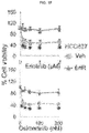
- FIG. 1D graphically illustrates data from a flow cytometry study where cells isolated from erlotinib resistant tissues were stained for ⁇ v ⁇ 3 and ALDH1A1;
- FIG. 1E graphically illustrates data from a flow cytometry where tumor cells isolated from a vehicle treated mouse (veh) and an erlotinib treated mouse (Er1R) were stained for ⁇ v ⁇ 3;
- FIG. 1F graphically illustrates cell viability after cells were treated with erlotinib or osimertinib for 72 hours;
- FIG. 1H illustrates an image of the primary masses of FIG. 1G ;
- FIG. 2A-E schematically and graphically illustrates data showing that LM609 produced a less aggressive phenotype:
- FIG. 2A graphically illustrates LM609 inhibited acquisition of erlotinib resistance, where HCC827 ( ⁇ 3-) xenograft mice were treated with erlotinib or erlotinib and LM609;
- FIG. 2B upper and lower panels graphically and in images illustrate data and stained tissue images showing that LM609 eliminated ⁇ 3-positive cells; where HCC827 ( ⁇ 3-) xenograft mice were treated with Captisol and PBS, Captisol and LM609, erlotinib and PBS, or erlotinib and LM609 for 50 days, and tumor tissues were stained (lower panel) for integrin ⁇ 3, and ⁇ 3-positive area in the tissues were quantified (upper panel);
- FIG. 2C upper panel
- CTCs were stained for ⁇ v ⁇ 3 to quantify ⁇ v ⁇ 3-positive ( ⁇ 3+) and -negative ( ⁇ 3-) CTCs
- FIG. 2C lower panel
- an example of ⁇ v ⁇ 3-positive CTCs is shown ( FIG. 2E );
- FIG. 3A-E graphically illustrate that LM609 eliminated ⁇ v ⁇ 3-positive cells via macrophage-mediated ADCC:
- FIG. 3A-C LM609 eliminated ⁇ v ⁇ 3-positive cells via macrophage-mediated ADCC in vitro; ADCC assays with bone marrow derived macrophages (BMDMs) or NK cells were performed with cancer cells with and without ⁇ v ⁇ 3 integrin treated with the IgG isotype or LM609 10 ⁇ g/mL and/or Fc blocker;
- BMDMs bone marrow derived macrophages
- NK cells were performed with cancer cells with and without ⁇ v ⁇ 3 integrin treated with the IgG isotype or LM609 10 ⁇ g/mL and/or Fc blocker
- FIG. 3D-E LM609 eliminated ⁇ v ⁇ 3-positive tumor growth only in the mice that have macrophages.
- Nude mice were subcutaneously injected with ⁇ 3 ectopically expressing HCC827 cells and treated with control liposome and PBS ( FIG. 3D left panel, control), control liposome and LM609 ( FIG. 3D left panel, LM609), clodronate liposome and PBS ( FIG. 3D right panel, control), or clodronate liposome and LM609 ( FIG. 3D right panel, LM609), tumor growth was monitored for 15 days, and after the treatments, F4/80 staining in the tumor tissues was quantified ( FIG. 3E );
- FIG. 4A-C graphically illustrate that erlotinib resistant tumor cells gained av ⁇ 3 integrin and were resistant to osimertinib:
- FIG. 4B graphically illustrate data showing cells from erlotinib resistant tissue were ⁇ v ⁇ 3-positive while the cells from the vehicle treated animal were not; levels of ⁇ v ⁇ 3 on cells isolated from the vehicle treated (Veh, left panel) and erlotinib treated (Er1R, right panel) animals were measured by flow cytometry;
- FIG. 4C Erlotinib resistant cells were osimertinib resistant. Cells from the vehicle treated (Veh) and erlotinib treated (Er1R) animals were treated with erlotinib (left panel) or osimertinib (right panel) at the indicated doses and MTT assay was performed to measure cell viability;
- FIG. 5A-B graphically illustrate data showing LM609 inhibited ligand binding of ⁇ v ⁇ 3 integrin but not cell viability:
- FIG. 5A graphically illustrates data showing that LM609 inhibited ligand binding of integrin ⁇ v ⁇ 3, wherein M21 cells (av ⁇ 3+) were plated on collagen or vitronectin (av ⁇ 3 ligand) with indicated doses of LM609, and the adherent cell number was measured;
- FIG. 5B graphically illustrates data showing that LM609 did not affect cell viability, paired ⁇ v ⁇ 3-positive ( ⁇ 3+) and -negative ( ⁇ 3 ⁇ ) cells were treated with indicated doses of LM609, and MTT assay was performed to measure cell viability.
- FIG. 6 graphically illustrates data showing that cancer therapeutics enrich ⁇ 3 integrin in epithelial cancer cells: qPCR was performed to detect mRNA expression of integrin a and f 3 subunits in response to increasing dose of hydrogen peroxide (H2O2) for 72 h, expressed as fold relative to vehicle control.
- H2O2 hydrogen peroxide
- compositions and methods for treating or ameliorating a cancer by targeting cell surface-expressed ⁇ v ⁇ 3 (avb3) polypeptides in Cancer Stem Cells (CSCs) to kill the CSCs, thus treating ameliorating or slowing the development of cancers caused or initiated by or sustained by cancer or tumor cells, or Cancer Stem Cells (CSCs), expressing ⁇ v ⁇ 3 polypeptides on their cell surfaces.
- CSCs Cancer Stem Cells
- compositions and methods as provided herein use an antibody or polypeptide that can specifically bind to human ⁇ v ⁇ 3 that also comprises an Fc portion that can mediate antibody-dependent cell-mediated cytotoxicity ADCC) killing of cancer or tumor cells by macrophages; for example, use a humanized antibody to ⁇ v ⁇ 3 that has been modified to include an engineered Fc portion that specifically binds to human macrophages.
- administration of the anti-avb3 antibody can eradicate or reduce the number of highly aggressive, ⁇ v ⁇ 3-positive drug resistant cancers by recruiting tumor associated macrophages which bind to the antibody or polypeptide (which also binds to ⁇ v ⁇ 3), and the macrophages thus kill the cancer by ADCC.
- antibodies e.g., humanized antibodies such as the anti ⁇ v ⁇ 3 antibody LM609, are used to target ⁇ v ⁇ 3 polypeptides in vivo to bind the ⁇ v ⁇ 3-positive cancer cell, e.g., a CSC, and by also binding to macrophages, kill CSCs expressing the ⁇ v ⁇ 3 polypeptide by ADCC.
- anti-av ⁇ 3 antibodies such as LM609 (Chemicon Int., Temecula, Calif.); CBL544, derived from clone 23C6 (MilliporeSigma, Burlington, Mass.); ab7166 (abcam, Cambridge, Mass.); and, monoclonal antibody ab78289 (abcam, Cambridge, Mass.), and humanized versions thereof, are used to prolong cancer drug sensitivity in individuals in need thereof by targeting drug resistant cancer cells via macrophage mediated antibody dependent cell mediated cytotoxicity (ADCC).
- ADCC macrophage mediated antibody dependent cell mediated cytotoxicity
- compositions and methods as provided herein are used to target and kill Cancer Stem Cells (CSCs), which can represent the most aggressive and drug resistant cells within a tumor population.
- CSCs Cancer Stem Cells
- Compositions and methods as provided herein are used to target integrin ⁇ v ⁇ 3, a polypeptide which identifies a CSC population within various epithelial cancers and is both necessary and sufficient to promote drug resistance.
- the anti-av ⁇ 3 integrin antibody, LM609 prolonged sensitivity to erlotinib in an EGFR mutant lung adenocarcinoma xenograft model. Described herein are studies showing that treatment of lung tumor bearing mice with the EGFR inhibitor, erlotinib, while initially suppressing tumor growth, ultimately results in tumor associated ⁇ v ⁇ 3 expression, leading to tumor progression as measured by an increase in circulating tumor cells (CTCs). In addition, erlotinib treated mice showed a marked accumulation of tumor-associated macrophages (TAMs) relative to untreated tumors.
- TAMs tumor-associated macrophages
- LM609 a monoclonal antibody directed to ⁇ v ⁇ 3, destroys the drug resistant CSCs within the primary tumor and eliminates CTCs.
- This anti-tumor effect is macrophage dependent as mice depleted of macrophages are unable to respond to this antibody.
- LM609 in combination with macrophages but not NK cells destroy CSCs due to antibody dependent cellular cytotoxicity (ADCC).
- ADCC antibody dependent cellular cytotoxicity
- mice bearing ⁇ v ⁇ 3-positive tumors with a monoclonal antibody (LM609) targeting this receptor to assess its ability to alter the macrophage phenotype within these tumors.
- LM609 was able to selectively eliminate the ⁇ v ⁇ 3-positive cancer stem cells via antibody-dependent cell-mediated cytotoxicity (ADCC), markedly enhancing the sensitivity of these tumors to the effects of therapy.
- ADCC antibody-dependent cell-mediated cytotoxicity
- compositions and formulations comprising anti-av ⁇ 3 (avb3) antibodies, and methods for: killing or reducing the number of ⁇ v ⁇ 3 (avb3) polypeptide-expressing cancer cells or Cancer Stem Cells (CSCs) in an individual in need thereof, treating, ameliorating or reversing or slowing the development of an ⁇ v ⁇ 3 (avb3) polypeptide-expressing cancer or tumor in an individual in need thereof, ameliorating or slowing the development of 10 cancers caused or initiated by or sustained by cancer or tumor cells, or Cancer Stem Cells (CSCs), expressing ⁇ v ⁇ 3 polypeptides on their cell surfaces, manipulating TAMs in vivo, or enhancing the sensitivity of ⁇ v ⁇ 3 (avb3) polypeptide-expressing cancer or tumor to the effects of therapy.
- CSCs Cancer Stem Cells
- compositions provided herein, and compositions used to practice the methods provided herein are formulated with a pharmaceutically acceptable carrier.
- the pharmaceutical compositions used to practice the methods provided herein can be administered parenterally, topically, orally or by local administration, such as by aerosol or transdermally.
- the pharmaceutical compositions can be formulated in any way and can be administered in a variety of unit dosage forms depending upon the condition or disease and the degree of illness, the general medical condition of each patient, the resulting preferred method of administration and the like. Details on techniques for formulation and administration are well described in the scientific and patent literature, see, e.g., the latest edition of Remington's Pharmaceutical Sciences, Maack Publishing Co, Easton Pa. (“Remington's”).
- Therapeutic agents as provided herein can be administered alone or as a component of a pharmaceutical formulation (composition), or concurrently with, before and/or after administration with another active agent, e.g., a growth factor inhibitor, wherein optionally the growth factor inhibitor comprises a Receptor Tyrosine Kinase (RTK) inhibitor, a Src inhibitor, an anti-metabolite inhibitor, a gemcitabine, a GEMZARTM, a mitotic poison, a paclitaxel, a taxol, an ABRAXANETM, an erlotinib, a TARCEVATM, a lapatinib, a TYKERBTM, a cetuxamib, an ERBITUXTM, or an insulin growth factor inhibitor.
- RTK Receptor Tyrosine Kinase
- compositions and formulations may be formulated for administration in any convenient way for use in human or veterinary medicine.
- Wetting agents, emulsifiers and lubricants, such as sodium lauryl sulfate and magnesium stearate, as well as coloring agents, release agents, coating agents, sweetening, flavoring and perfuming agents, preservatives and antioxidants can also be present in the compositions.
- Formulations of the compositions provided herein and as used to practice the methods provided herein include those suitable for oral/nasal, topical, parenteral, rectal, and/or intravaginal administration.
- the formulations may conveniently be presented in unit dosage form and may be prepared by any methods well known in the art of pharmacy.
- the amount of active ingredient which can be combined with a carrier material to produce a single dosage form will vary depending upon the host being treated, the particular mode of administration.
- the amount of active ingredient which can be combined with a carrier material to produce a single dosage form will generally be that amount of the compound which produces a therapeutic effect.
- compositions provided herein and as used to practice the methods provided herein can be prepared according to any method known to the art for the manufacture of pharmaceuticals.
- Such drugs can contain sweetening agents, flavoring agents, coloring agents and preserving agents.
- a formulation can be admixtured with nontoxic pharmaceutically acceptable excipients which are suitable for manufacture.
- Formulations may comprise one or more diluents, emulsifiers, preservatives, buffers, excipients, etc. and may be provided in such forms as liquids, powders, emulsions, lyophilized powders, sprays, creams, lotions, controlled release formulations, tablets, pills, gels, on patches, in implants, etc.
- compositions for oral administration can be formulated using pharmaceutically acceptable carriers well known in the art in appropriate and suitable dosages. Such carriers enable the pharmaceuticals to be formulated in unit dosage forms as tablets, geltabs, pills, powder, dragees, capsules, liquids, lozenges, gels, syrups, slurries, suspensions, etc., suitable for ingestion by the patient.
- Pharmaceutical preparations for oral use can be formulated as a solid excipient, optionally grinding a resulting mixture, and processing the mixture of granules, after adding suitable additional compounds, if desired, to obtain tablets or dragee cores.
- Suitable solid excipients are carbohydrate or protein fillers include, e.g., sugars, including lactose, sucrose, mannitol, or sorbitol; starch from corn, wheat, rice, potato, or other plants; cellulose such as methyl cellulose, hydroxypropylmethyl-cellulose, or sodium carboxy-methylcellulose; and gums including arabic and tragacanth; and proteins, e.g., gelatin and collagen.
- Disintegrating or solubilizing agents may be added, such as the cross-linked polyvinyl pyrrolidone, agar, alginic acid, or a salt thereof, such as sodium alginate.
- Dragee cores are provided with suitable coatings such as concentrated sugar solutions, which may also contain gum arabic, talc, polyvinylpyrrolidone, carbopol gel, polyethylene glycol, and/or titanium dioxide, lacquer solutions, and suitable organic solvents or solvent mixtures.
- Dyestuffs or pigments may be added to the tablets or dragee coatings for product identification or to characterize the quantity of active compound (i.e., dosage).
- Pharmaceutical preparations provided herein and as used to practice the methods provided herein can also be used orally using, e.g., push-fit capsules made of gelatin, as well as soft, sealed capsules made of gelatin and a coating such as glycerol or sorbitol.
- Push-fit capsules can contain active agents mixed with a filler or binders such as lactose or starches, lubricants such as talc or magnesium stearate, and, optionally, stabilizers.
- the active agents can be dissolved or suspended in suitable liquids, such as fatty oils, liquid paraffin, or liquid polyethylene glycol with or without stabilizers.
- Aqueous suspensions can contain an active agent (e.g., an anti-av ⁇ 3 (avb3) antibody or polypeptide) in admixture with excipients suitable for the manufacture of aqueous suspensions.
- excipients include a suspending agent, such as sodium carboxymethylcellulose, methylcellulose, hydroxypropylmethylcellulose, sodium alginate, polyvinylpyrrolidone, gum tragacanth and gum acacia, and dispersing or wetting agents such as a naturally occurring phosphatide (e.g., lecithin), a condensation product of an alkylene oxide with a fatty acid (e.g., polyoxyethylene stearate), a condensation product of ethylene oxide with a long chain aliphatic alcohol (e.g., heptadecaethylene oxycetanol), a condensation product of ethylene oxide with a partial ester derived from a fatty acid and a hexitol (e.g., polyoxyethylene
- the aqueous suspension can also contain one or more preservatives such as ethyl or n-propyl p-hydroxybenzoate, one or more coloring agents, one or more flavoring agents and one or more sweetening agents, such as sucrose, aspartame or saccharin.
- preservatives such as ethyl or n-propyl p-hydroxybenzoate
- coloring agents such as a coloring agent
- flavoring agents such as aqueous suspension
- sweetening agents such as sucrose, aspartame or saccharin.
- Formulations can be adjusted for osmolarity.
- Oil-based pharmaceuticals are particularly useful for administration hydrophobic active agents (e.g., an anti-av ⁇ 3 (avb3) antibody or polypeptide) used to practice the methods provided herein.
- Oil-based suspensions can be formulated by suspending an active agent in a vegetable oil, such as arachis oil, olive oil, sesame oil or coconut oil, or in a mineral oil such as liquid paraffin; or a mixture of these. See e.g., U.S. Pat. No. 5,716,928 describing using essential oils or essential oil components for increasing bioavailability and reducing inter- and intra-individual variability of orally administered hydrophobic pharmaceutical compounds (see also U.S. Pat. No. 5,858,401).
- the oil suspensions can contain a thickening agent, such as beeswax, hard paraffin or cetyl alcohol.
- Sweetening agents can be added to provide a palatable oral preparation, such as glycerol, sorbitol or sucrose.
- These formulations can be preserved by the addition of an antioxidant such as ascorbic acid.
- an injectable oil vehicle see Minto (1997) J. Pharmacol. Exp. Ther. 281:93-102.
- the pharmaceutical formulations provided herein can also be in the form of oil-in-water emulsions.
- the oily phase can be a vegetable oil or a mineral oil, described above, or a mixture of these.
- Suitable emulsifying agents include naturally-occurring gums, such as gum acacia and gum tragacanth, naturally occurring phosphatides, such as soybean lecithin, esters or partial esters derived from fatty acids and hexitol anhydrides, such as sorbitan mono-oleate, and condensation products of these partial esters with ethylene oxide, such as polyoxyethylene sorbitan mono-oleate.
- the emulsion can also contain sweetening agents and flavoring agents, as in the formulation of syrups and elixirs. Such formulations can also contain a demulcent, a preservative, or a coloring agent.
- the pharmaceutical compounds can also be administered by in intranasal, intraocular and intravaginal routes including suppositories, insufflation, powders and aerosol formulations (for examples of steroid inhalants, see Rohatagi (1995) J. Clin. Pharmacol. 35:1187-1193; Tjwa (1995) Ann. Allergy Asthma Immunol. 75:107-111).
- Suppositories formulations can be prepared by mixing the drug with a suitable non-irritating excipient which is solid at ordinary temperatures but liquid at body temperatures and will therefore melt in the body to release the drug.
- suitable non-irritating excipient which is solid at ordinary temperatures but liquid at body temperatures and will therefore melt in the body to release the drug.
- Such materials are cocoa butter and polyethylene glycols.
- the pharmaceutical compounds can be delivered by transdermally, by a topical route, formulated as applicator sticks, solutions, suspensions, emulsions, gels, creams, ointments, pastes, jellies, paints, powders, and aerosols.
- the pharmaceutical compounds can also be delivered as microspheres for slow release in the body.
- microspheres can be administered via intradermal injection of drug which slowly release subcutaneously; see Rao (1995) J. Biomater Sci. Polym. Ed. 7:623-645; as biodegradable and injectable gel formulations, see, e.g., Gao (1995) Pharm. Res. 12:857-863 (1995); or, as microspheres for oral administration, see, e.g., Eyles (1997) J. Pharm. Pharmacol. 49:669-674.
- the pharmaceutical compounds can be parenterally administered, such as by intravenous (IV) administration or administration into a body cavity or lumen of an organ.
- IV intravenous
- These formulations can comprise a solution of active agent dissolved in a pharmaceutically acceptable carrier.
- Acceptable vehicles and solvents that can be employed are water and Ringer's solution, an isotonic sodium chloride.
- sterile fixed oils can be employed as a solvent or suspending medium.
- any bland fixed oil can be employed including synthetic mono- or diglycerides.
- fatty acids such as oleic acid can likewise be used in the preparation of injectables. These solutions are sterile and generally free of undesirable matter.
- These formulations may be sterilized by conventional, well known sterilization techniques.
- the formulations may contain pharmaceutically acceptable auxiliary substances as required to approximate physiological conditions such as pH adjusting and buffering agents, toxicity adjusting agents, e.g., sodium acetate, sodium chloride, potassium chloride, calcium chloride, sodium lactate and the like.
- concentration of active agent in these formulations can vary widely, and will be selected primarily based on fluid volumes, viscosities, body weight, and the like, in accordance with the particular mode of administration selected and the patient's needs.
- the formulation can be a sterile injectable preparation, such as a sterile injectable aqueous or oleaginous suspension. This suspension can be formulated using those suitable dispersing or wetting agents and suspending agents.
- the sterile injectable preparation can also be a suspension in a nontoxic parenterally-acceptable diluent or solvent, such as a solution of 1,3-butanediol.
- the administration can be by bolus or continuous infusion (e.g., substantially uninterrupted introduction into a blood vessel for a specified period of time).
- the pharmaceutical compounds and formulations provided herein and as used to practice the methods provided herein can be lyophilized.
- stable lyophilized formulations comprising a composition provided herein, which can be made by lyophilizing a solution comprising a pharmaceutical provided herein on and a bulking agent, e.g., mannitol, trehalose, raffinose, and sucrose or mixtures thereof.
- a process for preparing a stable lyophilized formulation can include lyophilizing a solution about 2.5 mg/mL protein, about 15 mg/mL sucrose, about 19 mg/mL NaCl, and a sodium citrate buffer having a pH greater than 5.5 but less than 6.5. See, e.g., U.S. patent app. no. 20040028670.
- compositions and formulations provided herein and as used to practice the methods provided herein can be delivered by the use of liposomes.
- liposomes particularly where the liposome surface carries ligands specific for target cells, or are otherwise preferentially directed to a specific organ, one can focus the delivery of the active agent into target cells in vivo. See, e.g., U.S. Pat. Nos. 6,063,400; 6,007,839; Al-Muhammed (1996) J. Microencapsul. 13:293-306; Chonn (1995) Curr. Opin. Biotechnol. 6:698-708; Ostro (1989) Am. J. Hosp. Pharm. 46:1576-1587.
- compositions are administered to a subject already suffering from a condition, infection or disease in an amount sufficient to cure, alleviate or partially arrest the clinical manifestations of the condition, infection or disease and its complications (a “therapeutically effective amount”).
- compositions provided herein are administered in an amount sufficient to: kill or reduce the number of ⁇ v ⁇ 3 (avb3) polypeptide-expressing cancer cells or Cancer Stem Cells (CSCs) in an individual in need thereof; treating, ameliorating or reversing or slowing the development of an av ⁇ 3 (avb3) polypeptide-expressing cancer or tumor in an individual in need thereof; and/ or ameliorating or slowing the development of cancers caused or initiated by or sustained by cancer or tumor cells, or Cancer Stem Cells (CSCs), expressing ⁇ v ⁇ 3 polypeptides on their cell surfaces.
- CSCs Cancer Stem Cells
- the amount of pharmaceutical composition adequate to accomplish this is defined as a “therapeutically effective dose.”
- the dosage schedule and amounts effective for this use, i.e., the “dosing regimen,” will depend upon a variety of factors, including the stage of the disease or condition, the severity of the disease or condition, the general state of the patient's health, the patient's physical status, age and the like. In calculating the dosage regimen for a patient, the mode of administration also is taken into consideration.
- the dosage regimen also takes into consideration pharmacokinetics parameters well known in the art, i.e., the active agents' rate of absorption, bioavailability, metabolism, clearance, and the like (see, e.g., Hidalgo-Aragones (1996) J. Steroid Biochem. Mol. Biol. 58:611-617; Groning (1996) Pharmazie 51:337-341; Fotherby (1996) Contraception 54:59-69; Johnson (1995) J. Pharm. Sci. 84:1144-1146; Rohatagi (1995) Pharmazie 50:610-613; Brophy (1983) Eur. J. Clin. Pharmacol. 24:103-108; the latest Remington's, supra).
- pharmacokinetics parameters well known in the art, i.e., the active agents' rate of absorption, bioavailability, metabolism, clearance, and the like (see, e.g., Hidalgo-Aragones (1996) J. Steroid Biochem. Mol. Biol. 58:611-617
- formulations can be given depending on the dosage and frequency as required and tolerated by the patient.
- the formulations should provide a sufficient quantity of active agent to effectively treat, prevent or ameliorate a conditions, diseases or symptoms as described herein.
- an exemplary pharmaceutical formulation for oral administration of compositions provided herein or as used to practice the methods provided herein can be in a daily amount of between about 0.1 to 0.5 to about 20, 50, 100 or 1000 or more ug per 25 kilogram of body weight per day.
- dosages are from about 1 mg to about 4 mg per kg of body weight per patient per day are used.
- Lower dosages can be used, in contrast to administration orally, into the blood stream, into a body cavity or into a lumen of an organ.
- Substantially higher dosages can be used in topical or oral administration or administering by powders, spray or inhalation.
- Actual methods for preparing parenterally or non-parenterally administrable formulations will be known or apparent to those skilled in the art and are described in more detail in such publications as Remington's, supra.
- the methods provided herein can further comprise co-administration with other drugs or pharmaceuticals, e.g., compositions for treating cancer, septic shock, infection, fever, pain and related symptoms or conditions.
- other drugs or pharmaceuticals e.g., compositions for treating cancer, septic shock, infection, fever, pain and related symptoms or conditions.
- the methods and/or compositions and formulations provided herein can be co-formulated with and/or co-administered with antibiotics (e.g., antibacterial or bacteriostatic peptides or proteins), particularly those effective against gram negative bacteria, fluids, cytokines, immunoregulatory agents, anti-inflammatory agents, complement activating agents, such as peptides or proteins comprising collagen-like domains or fibrinogen-like domains (e.g., a ficolin), carbohydrate-binding domains, and the like and combinations thereof.
- antibiotics e.g., antibacterial or bacteriostatic peptides or proteins
- cytokines cytokines
- nanoparticles and liposomal membranes comprising compounds (e.g., anti-av ⁇ 3 (avb3) antibodies) used to practice the methods provided herein.
- nanoparticles and liposomal membranes targeting tumor (cancer) stem cells and dysfunctional stem cells are also provided.
- the compositions used to practice the methods provided herein are specifically targeted to cancer cells or cancer stem cells.
- nanoparticles and liposomal membranes comprising (in addition to comprising compounds used to practice the methods provided herein) molecules, e.g., peptides or antibodies, that selectively target abnormally growing, diseased, infected, dysfunctional and/or cancer (tumor) cell receptors.
- molecules e.g., peptides or antibodies
- nanoparticles and liposomal membranes using IL-11 receptor and/or the GRP78 receptor to targeted receptors on cells e.g., on tumor cells, e.g., on prostate or ovarian cancer cells. See, e.g., U.S. patent application publication no. 20060239968.
- nanocells to allow the sequential delivery of two different therapeutic agents with different modes of action or different pharmacokinetics, at least one of which comprises a composition used to practice the methods provided herein.
- a nanocell is formed by encapsulating a nanocore with a first agent inside a lipid vesicle containing a second agent; see, e.g., Sengupta, et al., U.S. Pat. Pub. No. 20050266067.
- the agent in the outer lipid compartment is released first and may exert its effect before the agent in the nanocore is released.
- the nanocell delivery system may be formulated in any pharmaceutical composition for delivery to patients.
- an inhibitor or depleter of an acetyl transferase gene, transcript (message) and/or protein expression or activity is contained in the outer lipid vesicle of the nanocell, and an antiangiogenic agent provided herein is loaded into the nanocore. This arrangement allows active agents to be released first and delivered to the tumor before the tumor's blood supply is cut off by the composition provided herein.
- multilayered liposomes comprising compounds used to practice embodiments provided herein, e.g., for transdermal absorption, e.g., as described in Park, et al., U.S. Pat. Pub. No. 20070082042.
- the multilayered liposomes can be prepared using a mixture of oil-phase components comprising squalane, sterols, ceramides, neutral lipids or oils, fatty acids and lecithins, to about 200 to 5000 nm in particle size, to entrap a composition provided herein.
- a multilayered liposome used to practice embodiments provided herein may further include an antiseptic, an antioxidant, a stabilizer, a thickener, and the like to improve stability.
- Synthetic and natural antiseptics can be used, e.g., in an amount of 0.01% to 20%.
- Antioxidants can be used, e.g., BHT, erysorbate, tocopherol, astaxanthin, vegetable flavonoid, and derivatives thereof, or a plant-derived antioxidizing substance.
- a stabilizer can be used to stabilize liposome structure, e.g., polyols and sugars.
- Exemplary polyols include butylene glycol, polyethylene glycol, propylene glycol, dipropylene glycol and ethyl carbitol; examples of sugars are trehalose, sucrose, mannitol, sorbitol and chitosan, or a monosaccharide or an oligosaccharide, or a high molecular weight starch.
- a thickener can be used for improving the dispersion stability of constructed liposomes in water, e.g., a natural thickener or an acrylamide, or a synthetic polymeric thickener.
- Exemplary thickeners include natural polymers, such as acacia gum, xanthan gum, gellan gum, locust bean gum and starch, cellulose derivatives, such as hydroxy ethylcellulose, hydroxypropyl cellulose and carboxymethyl cellulose, synthetic polymers, such as polyacrylic acid, poly-acrylamide or polyvinylpyrollidone and polyvinylalcohol, and copolymers thereof or cross-linked materials.
- natural polymers such as acacia gum, xanthan gum, gellan gum, locust bean gum and starch
- cellulose derivatives such as hydroxy ethylcellulose, hydroxypropyl cellulose and carboxymethyl cellulose
- synthetic polymers such as polyacrylic acid, poly-acrylamide or polyvinylpyrollidone and polyvinylalcohol, and copolymers thereof or cross-linked materials.
- Liposomes can be made using any method, e.g., as described in Park, et al., U.S. Pat. Pub. No. 20070042031, including method of producing a liposome by encapsulating a therapeutic product comprising providing an aqueous solution in a first reservoir; providing an organic lipid solution in a second reservoir, wherein one of the aqueous solution and the organic lipid solution includes a therapeutic product; mixing the aqueous solution with said organic lipid solution in a first mixing region to produce a liposome solution, wherein the organic lipid solution mixes with said aqueous solution so as to substantially instantaneously produce a liposome encapsulating the therapeutic product; and immediately thereafter mixing the liposome solution with a buffer solution to produce a diluted liposome solution.
- nanoparticles comprising compounds used to practice embodiments provided herein (e.g., anti-av ⁇ 3 (avb3) antibodies) to deliver the composition as a drug-containing nanoparticle (e.g., a secondary nanoparticle), as described, e.g., in U.S. Pat. Pub. No. 20070077286.
- nanoparticles comprising a fat-soluble drug provided herein or a fat-solubilized water-soluble drug to act with a bivalent or trivalent metal salt.
- compositions e.g., anti-av ⁇ 3 (avb3) antibodies
- formulations used to practice embodiments provided herein can be delivered by the use of liposomes.
- liposomes particularly where the liposome surface carries ligands specific for target cells, or are otherwise preferentially directed to a specific organ or cell, e.g., cancer stem cells, one can focus the delivery of the active agent into target cells in vivo. See, e.g., U.S. Patent Nos. 6,063,400; 6,007,839; Al-Muhammed (1996) J. Microencapsul. 13:293-306; Chonn (1995) Curr. Opin. Biotechnol.
- compositions and formulations used to practice embodiments provided herein are delivered by the use of liposomes having rigid lipids having head groups and hydrophobic tails, e.g., as using a polyethyleneglycol-linked lipid having a side chain matching at least a portion the lipid, as described e.g., in US Pat App Pub No. 20080089928.
- compositions and formulations used to practice embodiments provided herein are delivered by the use of amphoteric liposomes comprising a mixture of lipids, e.g., a mixture comprising a cationic amphiphile, an anionic amphiphile and/or neutral amphiphiles, as described e.g., in US Pat App Pub No. 20080088046, or 20080031937.
- compositions and formulations used to practice embodiments provided herein are delivered by the use of liposomes comprising a polyalkylene glycol moiety bonded through a thioether group and an antibody also bonded through a thioether group to the liposome, as described e.g., in US Pat App Pub No.
- compositions and formulations used to practice embodiments provided herein are delivered by the use of liposomes comprising glycerides, glycerol-phospholipids, glycerophosphinolipids, glycerophosphonolipids, sulfolipids, sphingolipids, phospholipids, isoprenolides, steroids, stearines, sterols and/or carbohydrate containing lipids, as described e.g., in US Pat App Pub No. 20070148220.
- compositions and methods comprising antibodies or active fragments thereof, or ⁇ v ⁇ 3-binding polypeptides (e.g., comprising CDRs capable of specifically binding to an ⁇ v ⁇ 3) capable of specifically binding to an ⁇ v ⁇ 3 (avb3) integrin polypeptide expressed on a cancer or a tumor cell, or on a CSC, wherein the antibody or polypeptide has an Fc domain or equivalent domain or moiety capable of binding a macrophage and initiating an antibody-dependent cell-mediated cytotoxicity (ADCC) killing of the cell to which the antibody specifically binds (e.g., anti-av ⁇ 3 (avb3) antibodies).
- ADCC antibody-dependent cell-mediated cytotoxicity
- an antibody or polypeptide for practicing embodiments provided herein can comprise a peptide or polypeptide derived from, modeled after or substantially encoded by an immunoglobulin gene or immunoglobulin genes, or fragments thereof, capable of specifically binding an antigen or epitope, see, e.g. Fundamental Immunology, Third Edition, W. E. Paul, ed., Raven Press, N.Y. (1993); Wilson (1994) J. Immunol. Methods 175:267-273; Yarmush (1992) J. Biochem. Biophys. Methods 25:85-97.
- an antibody or polypeptide for practicing embodiments provided herein includes antigen-binding portions, i.e., “antigen binding sites,” (e.g., fragments, subsequences, complementarity determining regions (CDRs)) that retain capacity to bind antigen, including (i) a Fab fragment, a monovalent fragment consisting of the VL, VH, CL and CH1 domains; (ii) a F(ab′)2 fragment, a bivalent fragment comprising two Fab fragments linked by a disulfide bridge at the hinge region; (iii) a Fd fragment consisting of the VH and CH1 domains; (iv) a Fv fragment consisting of the VL and VH domains of a single arm of an antibody, (v) a dAb fragment (Ward et al., (1989) Nature 341:544-546), which consists of a VH domain; and (vi) an isolated complementarity determining region (CDR).
- method comprise use of any polypeptide capable of specifically binding to an ⁇ v ⁇ 3 (avb3) integrin polypeptide, including polypeptides comprising the ⁇ v ⁇ 3-binding CDR of: monoclonal antibody LM609 (Chemicon Int., Temecula, Calif.) (CVCL KS89) (the murine hybridoma having ATCC accession number HB 9537) (see e.g., U.S. Pat. No.
- humanized antibodies can use “humanized” antibodies, including forms of non-human (e.g., murine) antibodies that are chimeric antibodies comprising minimal sequence (e.g., the antigen binding fragment) derived from non-human immunoglobulin.
- humanized antibodies are human immunoglobulins in which residues from a hypervariable region (HVR) of a recipient (e.g., a human antibody sequence) are replaced by residues from a hypervariable region (HVR) of a non-human species (donor antibody) such as mouse, rat, rabbit or nonhuman primate having the desired specificity, affinity, and capacity.
- HVR hypervariable region
- donor antibody such as mouse, rat, rabbit or nonhuman primate having the desired specificity, affinity, and capacity.
- framework region (FR) residues of the human immunoglobulin are replaced by corresponding non-human residues to improve antigen binding affinity.
- humanized antibodies may comprise residues that are not found in the recipient antibody or the donor antibody. These modifications may be made to improve antibody affinity or functional activity.
- the humanized antibody can comprise substantially all of at least one, and typically two, variable domains, in which all or substantially all of the hypervariable regions correspond to those of a non-human immunoglobulin and all or substantially all of Ab framework regions are those of a human immunoglobulin sequence.
- a humanized antibody used to practice embodiments provided herein can comprise at least a portion of an immunoglobulin constant region (Fc), typically that of or derived from a human immunoglobulin.
- Fc immunoglobulin constant region
- completely human antibodies also can be used to practice embodiments provided herein, including human antibodies comprising amino acid sequence which corresponds to that of an antibody produced by a human.
- This definition of a human antibody specifically excludes a humanized antibody comprising non-human antigen binding residues.
- method comprise use of humanized antibodies capable of specifically binding to an ⁇ v ⁇ 3 (avb3) integrin polypeptide, including humanized versions of the ⁇ v ⁇ 3-binding: monoclonal antibody LM609 (Chemicon Int., Temecula, Calif.) (CVCL KS89) (the murine hybridoma having ATCC accession number HB 9537) (see e.g., U.S. Pat. No.
- antibodies used to practice embodiments provided herein comprise “affinity matured” antibodies, e.g., antibodies comprising with one or more alterations in one or more hypervariable regions which result in an improvement in the affinity of the antibody for antigen; e.g., a histone methyl and/or acetyl transferase, compared to a parent antibody which does not possess those alteration(s).
- antibodies used to practice embodiments provided herein are matured antibodies having nanomolar or even picomolar affinities for the target antigen, e.g., a histone methyl and/or acetyl transferase. Affinity matured antibodies can be produced by procedures known in the art.
- antibodies used to practice methods as provided herein are: the monoclonal antibody LM609 (Chemicon Int., Temecula, Calif.) (CVCL_KS89) (the murine hybridoma having ATCC accession number HB 9537) (see e.g., U.S. Pat. No. 7,115,261); monoclonal antibody CBL544, derived from clone 23C6 (MilliporeSigma, Burlington, Mass.); monoclonal antibody ab7166 (abcam, Cambridge, Mass.); or, monoclonal antibody ab78289 (abcam, Cambridge, Mass.), and humanized versions thereof.
- LM609 Cemicon Int., Temecula, Calif.
- CVCL_KS89 the murine hybridoma having ATCC accession number HB 9537
- LM609 The monoclonal antibody LM609 is described e.g., in Cheresh et al., J Biol Chem. 1987;262(36):17703-11; and U.S. patent number (USPN) 5,753,230, and U.S. Pat. No. 6,590,079.
- LM609 is a murine monoclonal antibody specific for the integrin ⁇ v ⁇ 3, see e.g., Cheresh, D. A., Proc. Natl. Acad. Sci. USA 84:6471-6475 (1987), and Cheresh et al, J. Biol. Chem. 262:17703-17711 (1987).
- LM609 was produced against and is reactive with the M21 cell adhesion receptor now known as the integrin ⁇ v ⁇ 3.
- LM609 inhibits the attachment of M21 cells to ⁇ v ⁇ 3 ligands such as vitronectin, fibrinogen and von Willebrand factor (Cheresh and Spiro, supra) and is also an inhibitor of ⁇ v ⁇ 3-mediated pathologies such as tumor induced angiogenesis (Brooks et al. Cell 79:1157-1164 (1994)), granulation tissue development in cutaneous wound (Clark et al., Am. J. Pathology, 148:1407-1421 (1996)) and smooth muscle cell migration such as that occurring during restenosis (Choi et al., J. Vascular Surg., 19:125-134 (1994); Jones et al., Proc. Natl. Acad. Sci. 93:2482-2487 (1996)).
- ⁇ v ⁇ 3 ligands such as vitronectin, fibrinogen
- kits comprising compositions (e.g., antibodies) for practicing the methods and uses as provided herein, optionally also including instructions for use thereof.
- compositions e.g., antibodies
- kits comprising the monoclonal antibodies or polypeptides capable of specifically binding to an ⁇ v ⁇ 3, as described herein.
- compositions and methods for treating tumors that have become adapted to the effects of therapy and have become highly aggressive and invasive.
- This example describes an approach to prevent this process by exploiting two phenomena that arise during acquired drug resistance.
- EGFR mutant lung tumors treated with the RTK inhibitor erlotinib gain expression of integrin ⁇ v ⁇ 3, and this marker is highly enriched on circulating tumor cells.
- erlotinib-treated tumors show an accumulation of M2 tumor-associated macrophages, demonstrating how the tumor can manipulate the immune system to support its own proliferation and metastasis.
- certain antibodies can mark tumor cells for elimination by antibody-dependent cell-mediated cytotoxicity (ADCC). This process is triggered when the antibody binds to tumor cell antigens and is simultaneously recognized by the FcyR on immune effector cells (6).
- ADCC antibody-dependent cell-mediated cytotoxicity
- TAMs tumor-associated macrophages
- HCC827 human non-small cell lung cancer
- TAMs Tumor-associated macrophages
- M2-type (10) secrete growth factors
- pro-tumorigenic cytokines secrete growth factors
- immunosuppressive enzymes to promote tumor progression through induction of angiogenesis, metastasis, and immune suppression (11-14). Accordingly, the observation of enriched TAMs in tumor tissues is associated with poor prognosis in various types of cancer, including lung adenocarcinoma (7).
- TAMs CD11b-positive, Ly-6G-negative
- M2 type CD206-positive or CD206- and MHC1l-double negative within the TAM population
- LM609 Since etaracizumab was reported to mediate antibody-dependent cell mediated cytotoxicity (ADCC) in vitro (19), we considered LM609 as a unique opportunity to engage TAMs to target the enriched population of ⁇ v ⁇ 3-expressing tumor cells that emerge during acquired resistance to erlotinib in vivo.
- FIG. 2A While erlotinib alone enriched for ⁇ v ⁇ 3 and led to tumor regrowth and the appearance of CTCs, the combination of erlotinib and LM609 maintained erlotinib sensitivity ( FIG. 2A ). Not only did LM609 eliminate ⁇ v ⁇ 3-positive tumor cells ( FIG. 2B ), but it also prevented the increase in ⁇ v ⁇ 3-positive CTCs ( FIG. 2C-E ), suggesting that LM609 prevents both erlotinib resistance and the intravasation/survival of circulating ⁇ v ⁇ 3-positive tumor cells during the course of therapy.
- LM609 only recognizes human integrin av ⁇ 3
- our xenograft model allows us to separate its role in ADCC from any impact on tumor-associated endothelial cells that require this integrin for angiogenesis (20, 21).
- angiogenesis contributes to tumor growth
- LM609 and etaracizumab are IgG1 antibodies with higher affinity for human versus mouse macrophage Fcy receptors (22)
- our preclinical evaluation may underestimate the efficacy of this therapeutic strategy for erlotinib-resistant tumors in humans.
- not only erlotinib but also other cancer therapeutics increase ⁇ 3 integrin expression in epithelial cancer cells ( FIG.
- anti-av ⁇ 3 antibody therapy can be used with various therapeutics.
- anti-av ⁇ 3 antibody with EGFR inhibitors and/or other cancer drugs as a unique therapeutic approach for epithelial cancer patients who progress on a standard of therapy.
- Lung adenocarcinoma (HCC827 in RPMI) cells were obtained from ATCC.
- the melanoma cell line, M21 was a gift from Dr. D. L. Morton (University of California, Los Angeles, USA).
- the lung adenocarcinoma cell line, PC9 was a gift from Dr. Joan Massague (Sloan-Kettering Institute, USA).
- Cell line authentication was performed by the ATCC using short tandem repeat DNA profiles. Upon receipt, each cell line was expanded, cryopreserved as low-passage stocks, and tested routinely for Mycoplasma.
- cells were transfected with vector control (GFP labeled), integrin ⁇ 3, or luciferase using a lentiviral system as previously described (1).
- vector control GFP labeled
- integrin ⁇ 3 integrin ⁇ 3
- luciferase a lentiviral system
- Captisol was obtained from CydexTM (NC0604701) and diluted in water at 6%.
- Erlotinib was obtained from SellekChemTM (S1023) and diluted in DMSO for in vitro or in captisol for in vivo experiments.
- LM609 was produced in this laboratory as previously described (2). The activity was confirmed by adhesion assay as described below. Control and clodronate liposome solutions were obtained from ClodronateLiposome.com.
- M21 (av ⁇ 3+) cells were plated on a vitronectin-coated or collagen-coated plate with and without LM609. Adherent cells were stained with crystal violet and the visible absorbance (600 nm) of each well was quantified using a microplate reader.
- Animals with a tumor volume of 250 ⁇ 700 mm 3 were randomly assigned into groups treated with captisol (oral, six times/week) and PBS (i.p., twice/week), captisol and LM609 (i.p., 10 mg/kg, twice/week), erlotinib (oral, 6.25 mg/kg, six times/week) and PBS, or erlotinib and LM609.
- the mice in the captisol groups were sacrificed on day 15 due to the large tumor size.
- the erlotinib groups were sacrificed on day 50.
- Tumor tissues were divided into three pieces for snap freezing, freezing in OCT compound (VWR, 25608-930), or 10% formalin fixation (Fisher Scientific, 23-313095) for further analyses.
- TAMs were isolated from tumor tissues as previously described (4). Snap frozen tumor tissues were thawed, minced, and dissociated in HBSS containing collagenase IV (Sigma, C5138, 0.5 mg/mL), hyaluronidase (Sigma, H2654, 0.1 30 mg/mL), dispase 11 (Roche, 04942078001, 0.6 U/mL), and DNase IV (Millipore, 260913-10MU, 5 U/mL) at 37° C. for 15 minutes. Cell suspensions were filtered through 70 ⁇ m cell strainer and washed with PBS.
- Flow cytometry was performed on BD LSRFortessaTM, and the ratio of TAMs (CD11b-positive, Ly-6G-negative), M1 (CD206-negative, MHC11-positive within TAMs), and M2 (non-M1 population within TAMs) in the tumor tissue was calculated using the flow cytometry analysis program FlowJoTM (Treestar).
- Immunohistochemical staining was performed on unstained FFPE slides according to the manufacturers recommendations for the VECTASTAIN Elite ABC HRP Kit (Vector Laboratories, PK-6100).
- An anti-integrin ⁇ 3 antibody (Cell Signaling, 13166, 1:200) and a biotinylated goat anti-rabbit antibody (Vector Laboratories, BA-1000, 1:200) were used.
- the stained tissues were imaged on NanoZoomer Slide Scanning SystemTM (Hamamamatsu), and the ⁇ 3-positive area fraction with respect to tumor tissue was calculated utilizing ImageJ (NIH) (5).
- Luciferase- and GFP-positive HCC827 cells (5 ⁇ 10 6 cells in 50 ⁇ l of PBS) were injected into the right lungs of female nu/nu mice (8-10 weeks old). The tumor growth was monitored by IVIS SPECTRUMTM (PerkinElmer) every four weeks. Eight weeks after the injection, the mice were randomly divided into three groups, pre-treatment, erlotinib (oral, 6.25 mg/kg, six times/week) treatment, and the combination of erlotinib and LM609 (i.p., 10 mg/kg, twice/week). The pre-treatment group mice were sacrificed in week 8 for CTC analysis. The treatment group mice were treated with erlotinib and or LM609 for four weeks before being sacrificed.
- PBMCs including CTCs were isolated using LymphoprepTM (STEMCELL Technologies, 85415) and SepMateTM (STEMCELL Technologies, 07801) following the manufacturer's protocol.
- Isolated cells were washed with PBS, plated on poly-L-Lysine (Sigma, P1399) coated 8-well chamber plates, fixed with 4% PFA (VWR, AA43368-9M), and stained with DAPI (Life Technologies, D1306, 1 ⁇ g/mL in 1% BSA in PBS), LM609 (5 ⁇ g/mL in 1% BSA in PBS), and a fluorescently-labeled secondary antibody (Thermo, A21235, 1:500).
- the cells were analyzed utilizing Nikon Eclipse C2TM confocal microscope (Nikon).
- CTCs were defined as DAPI and GFP double positive cells. The numbers of av ⁇ 3-positive and -negative CTCs per mouse were counted. After sacrificing the mice, the tumor mass volume in the lungs were also measured utilizing an OV100TM fluorescence imaging system (Olympus).
- HCC827-R Erlotinib resistant HCC827 (HCC827-R) cells and PC9 (PC9-R) cells were established as previously described (3). Resistance to EGFR inhibitors (erlotinib and AZD9291) was confirmed by MTT assays ( FIG. 1B ).
- BMDM Bone Marrow Derived Macrophage
- BMDMs were aseptically harvested from 8-10 week-old female C57BL/6 mice by flushing leg bones of euthanized mice with RPMI, filtering through 70 ⁇ m cell strainers, and incubating in Red Blood Cell Lysing Buffer Hybri-MaxTM (Sigma, R7757).
- Splenocytes were aseptically harvested from 8-10 week-old female C57BL/6 mice by mincing spleens of euthanized mice with PBS containing 2% FBS and 1 mM EDTA, filtering through 40 ⁇ m strainers, and incubating in Red Blood Cell Lysing Buffer Hybri-MaxTM (Sigma, R7757).
- the NK cells were isolated from the splenocytes using NK Cell Isolation Kit IITM (Miltenyi, 130-096-892).
- Target cells stained with CFSE Cell Division Tracker KitTM BioLegend, 423801) or CellTraceTM Far Red Cell Proliferation KitTM (ThermoFisher, C34564) were co-cultured with BMDMs or NK cells with or without isotype IgG or LM609 for five hours at 37° C. After the incubation, the cells were stained with PI (Sigma, P4864, 1:1000), and flow cytometry was performed on BD LSRFortessaTM. The ratio of dead target cells (PI-positive) to the total target cell population (CFSE- or far red-positive) was calculated as previously described (6).
- Cell pellets were washed blocked with 1% BSA in PBS for 30 minutes at room temperature and stained with LM609 (5 ⁇ g/mL in 1% BSA in PBS) and a fluorescently labeled secondary antibody (Thermo, A21235, 1:500). After the staining, the cells were incubated with PI (Sigma, P4864, 1:1000), and flow cytometry was performed on BD LSRFortessaTM. The levels of ⁇ v ⁇ 3 integrin were analyzed using the flow cytometry analysis program FlowJoTM (Treestar).
- FIG. 1 Erlotinib Resistance Impacted the Primary Tumor, CTC, and Stroma.
- FIG. 1A A schematic of the erlotinib resistant xenograft model.
- FIG. 1B-D Erlotinib resistant tissues display increased ⁇ v ⁇ 3/ALDH1A1-expressing cells.
- FIG. 1E-F Erlotinib resistant ⁇ v ⁇ 3-positive cells were resistant to a third generation EGFR inhibitor, osimertinib.
- Tumor cells isolated from a vehicle treated mouse (veh) and an erlotinib treated mouse (Er1R) were stained for ⁇ v ⁇ 3 for flow cytometry ( FIG. 1E ).
- the graphs are reperesentatives of three experiments. The cells were treated with erlotinib or osimertinib for 72 hours, and cell viability was measured ( FIG. 1F ).
- FIG. 1G-H Erlotinib increased CTCs while the primary tumors are still responding to the treatment.
- the primary masses were visualized ( FIG. 1H ).
- GFP tumor tissue; Bars, 5 mm.
- FIG. 2 LM609 Produced a Less Aggressive Phenotype.
- FIG. 2A LM609 inhibited acquisition of erlotinib resistance.
- the tumor growth was measured twice weekly. Progression free survival was analyzed utilizing Kaplan Meier estimator.
- FIG. 2B LM609 eliminated ⁇ 3-positive cells.
- FIG. 2C-E LM609 decreased numbers of CTCs induced by erlotinib.
- FIG. 2C The pictures are representatives ( FIG. 2D ).
- the CTCs were stained for ⁇ v ⁇ 3 to quantify ⁇ v ⁇ 3-positive ( ⁇ 3+) and -negative ( ⁇ 3-) CTCs ( FIG. 2E ).
- An example of av ⁇ 3-positive CTCs is shown (lower right panel).
- Two-tailed Student's t test was used to determine statistical significance (*P ⁇ 0.05 compared to the pre-treatment). Error bars indicate standard errors (upper panel) and standard deviations (lower panel). Bars, 5 mm (upper panel); bars, 10 ⁇ m (lower panel); blue, DAPI; green, GFP; red, av ⁇ 3.
- FIG. 3 LM609 Eliminated ⁇ v ⁇ 3-Positive Cells via Macrophage-Mediated ADCC.
- FIG. 3A-C LM609 eliminated ⁇ v ⁇ 3-positive cells via macrophage-mediated ADCC in vitro.
- ADCC assays with bone marrow derived macrophages (BMDMs) or NK cells were performed with cancer cells with and without ⁇ v ⁇ 3 integrin treated with the IgG isotype or LM609 10 ⁇ g/mL and/or Fc blocker. Percent cell death of cancer cells was quantified utilizing propidium iodide by flow cytometry. Effector and target cell ratio was 5:1. Two-tailed Student's t test was used to determine statistical significance (*P ⁇ 0.05 compared to IgG isotype controls). Error bars indicate standard errors. Empty vector, EV; ⁇ 3 plasmid, ⁇ 3; Veh, cells isolated from a vehicle treated tissue; Er1R, cells isolated from an erlotinib resistant tissue.
- FIG. 3D-E LM609 eliminated ⁇ v ⁇ 3-positive tumor growth only in the mice that have macrophages.
- FIG. 4 Erlotinib Resistant Tumor Cells Gained ⁇ v ⁇ 3 Integrin and were Resistant to Osimertinib.
- FIG. 4A Erlotinib treated tumor gained resistance.
- FIG. 4B The cells from erlotinib resistant tissue ⁇ v ⁇ 3-positive while the cells from the vehicle treated animal were not. Levels of ⁇ v ⁇ 3 on cells isolated from the vehicle treated (Veh) and erlotinib treated (Er1R) animals were measured by flow cytometry. Blue, secondary control; red, stained with LM609.
- FIG. 4C Erlotinib resistant cells were osimertinib resistant. Cells from the vehicle treated (Veh) and erlotinib treated (Er1R) animals were treated with erlotinib or osimertinib at the indicated doses and MTT assay was performed to measure cell viability.
- FIG. 5 LM609 Inhibited Ligand Binding of ⁇ v ⁇ 3 Integrin but not Cell Viability.
- FIG. 5A LM609 inhibited ligand binding of integrin ⁇ v ⁇ 3.
- M21 cells (av ⁇ 3+) were plated on collagen or vitronectin (av ⁇ 3 ligand) with indicated doses of LM609. The adherent cell number was measured.
- FIG. 5B LM609 did not affect cell viability. Paired ⁇ v ⁇ 3-positive ( ⁇ 3+) and -negative ( ⁇ 3-) cells were treated with indicated doses of LM609, and MTT assay was performed to measure cell viability.
- FIG. 6 graphically illustrates data showing that cancer therapeutics enrich ⁇ 3 integrin in epithelial cancer cells: qPCR was performed to detect mRNA expression of integrin ⁇ and ⁇ subunits in response to increasing dose of hydrogen peroxide (H2O2) for 72 h, expressed as fold relative to vehicle control. Error bars indicate standard deviations. *P ⁇ 0.05 compared to controls.
- Tumor-associated macrophages correlate with response to epidermal growth factor receptor-tyrosine kinase inhibitors in advanced non-small cell lung cancer. Int J Cancer. 2012; 131(3):E227-35.
- Kaneda et al., PI3Kgamma is a molecular switch that controls immune suppression. Nature. 2016; 539(7629):437-42.
- Kaneda M M Messer K S, Ralainirina N, Li H, Leem C J, Gorjestani S, Woo G, Nguyen A V, Figueiredo C C, Foubert P, et al.
- PI3Kgamma is a molecular switch that controls immune suppression. Nature. 2016; 539(7629):437-42.
Abstract
Description
- This Patent Convention Treaty (PCT) International Application claims the benefit of priority to U.S. Provisional Application No. 62/479,768, filed Mar. 31, 2017. The aforementioned application is expressly incorporated herein by reference in its entirety and for all purposes.
- This invention was made with government support under grant numbers T32 OD017853; T32 HL086344; T32 HL098062-03; R-857GC; NCI R01CA045726, awarded by the National Institutes of Health (NIH). The government has certain rights in the invention.
- This invention generally relates to immunology and oncology. In alternative embodiments, provided are compositions and methods for treating or ameliorating a cancer by targeting cell surface-expressed αvβ3 (avb3) polypeptides in Cancer Stem Cells (CSCs) to kill the CSCs, thus treating ameliorating or slowing the
development 20 of cancers caused or initiated by or sustained by cancer or tumor cells, or Cancer Stem Cells (CSCs), expressing αvβ3 polypeptides on their cell surfaces. - Tumor associated macrophages are involved in regulation of cancer growth and aggressiveness. Whereas M1 macrophages trigger an inflammatory response and inhibit tumor growth, M2 macrophages secrete pro-tumor cytokines into the microenvironment to support tumor progression. A macrophage switch from M1 to M2 has been associated with lung cancer progression, and cancer stem cells have been implicated as a driver of this reprogramming.
- In alternative embodiments, provided are methods for:
- killing or reducing the number of αvβ3 (avb3) polypeptide-expressing cancer cells or Cancer Stem Cells (CSCs) in an individual in need thereof,
- treating, ameliorating or reversing or slowing the development of an αvβ3 (avb3) polypeptide-expressing cancer or tumor in an individual in need thereof,
- ameliorating or slowing the development of cancers caused or initiated by or sustained by cancer or tumor cells, or Cancer Stem Cells (CSCs), expressing αvβ3 polypeptides on their cell surfaces,
- increasing a macrophage population capable of triggering an inflammatory response and inhibiting tumor growth in vivo, wherein optionally the macrophage population capable of triggering an inflammatory response and inhibiting tumor growth comprises a tumor associated macrophage (TAM) or an M1 macrophage population,
- enhancing the sensitivity of αvβ3 (avb3) polypeptide-expressing cancer or tumor to the effects of therapy, optionally a chemotherapy, optionally therapy with a growth factor inhibitor,
- the method comprising:
- (a) administering to an individual in need thereof an antibody or polypeptide capable of specifically binding to an αvβ3 (avb3) integrin polypeptide expressed on a cancer or a tumor cell, or on a CSC, or
- (b) (i) providing an antibody or polypeptide capable of specifically binding to an αvβ3 (avb3) integrin polypeptide expressed on a cancer or a tumor cell, or on a CSC,
- wherein the antibody or polypeptide has an Fc domain or equivalent domain or moiety capable of binding a macrophage and initiating an antibody-dependent cell-mediated cytotoxicity (ADCC) killing of the cell to which the antibody specifically binds,
- (ii) administering the antibody or polypeptide to an individual in need thereof,
- thereby resulting in:
- killing or reducing the number of αvβ3 (avb3) polypeptide-expressing cancer cells or Cancer Stem Cells (CSCs) in an individual in need thereof,
- treating, ameliorating or reversing or slowing the development of an αvβ3 (avb3) polypeptide-expressing cancer or tumor in an individual in need thereof,
- ameliorating or slowing the development of cancers caused or initiated by or sustained by cancer or tumor cells, or Cancer Stem Cells (CSCs), expressing αvβ3 polypeptides on their cell surfaces,
- increasing a macrophage population capable of triggering an inflammatory response and inhibiting tumor growth in vivo, wherein optionally the macrophage population capable of triggering an inflammatory response and inhibiting tumor growth comprises a tumor associated macrophage (TAM) or an M1 macrophage population,
- enhancing the sensitivity of αvβ3 (avb3) polypeptide-expressing cancer or tumor to the effects of therapy.
- In alternative embodiments, provided are methods: wherein the antibody or polypeptide is a humanized antibody, optionally a humanized murine antibody; or wherein the antibody or polypeptide is a recombinant or engineered antibody; or wherein the antibody is a human antibody; or, wherein the antibody is monoclonal antibody, or a polyclonal antibody; or wherein the antibody is: monoclonal antibody LM609 (Chemicon Int., Temecula, Calif.) (CVCL KS89) (the murine hybridoma having ATCC accession number HB 9537) (see e.g., U.S. Pat. No. 7,115,261); monoclonal antibody CBL544, derived from clone 23C6 (MilliporeSigma, Burlington, Mass.); monoclonal antibody ab7166 (abcam, Cambridge, Mass.); or, monoclonal antibody ab78289 (abcam, Cambridge, Mass.); or, or any humanized version thereof, or any polypeptide comprising an αvβ3-binding CDR of LM609, CBL544, ab7166 or ab78289.
- In alternative embodiments, provided are methods: wherein the macrophage is a human macrophage, or a tumor associated macrophage (TAM), an M1 macrophage, or a tumor-inhibiting M2 macrophage.
- In alternative embodiments, provided are methods: wherein the cancer is an epithelial cancer or epithelial tumor cell; or wherein the cancer is a drug resistant cancer, and optionally the drug is a growth factor inhibitor or a kinase inhibitor, wherein optionally the growth factor inhibitor comprises a Receptor Tyrosine Kinase (RTK) inhibitor, optionally erlotinib.
- In alternative embodiments, provided are methods: wherein the antibody or polypeptide is administered intravenously, intramuscularly or subcutaneously to the individual in need thereof; or wherein the antibody or polypeptide is formulated as a sterile pharmaceutical composition or formulation, or is formulated for administration intravenously, intramuscularly or subcutaneously; or wherein the dosage of antibody or polypeptide is based on either fixed or body weight-based dosing, or a fixed dose of between about 100 and 1200 mg monthly, or at a dosage of between about 0.3 to 10 mg/kg.
- In alternative embodiments, provided are methods further comprising administration of a growth factor inhibitor, wherein optionally the growth factor inhibitor comprises a Receptor Tyrosine Kinase (RTK) inhibitor, a Src inhibitor, an anti-metabolite inhibitor, a gemcitabine, a GEMZAR™, a mitotic poison, a paclitaxel, a taxol, an ABRAXANE™, an erlotinib, a TARCEVA™, a lapatinib, a TYKERB™, a cetuxamib, an ERBITUX™, or an insulin growth factor inhibitor.
- In alternative embodiments, provided are Uses of: an antibody or polypeptide capable of specifically binding to an αvβ3 (avb3) integrin polypeptide expressed on a cancer or a tumor cell, or on a CSC; or, an antibody or polypeptide capable of specifically binding to an αvβ3 (avb3) integrin polypeptide expressed on a cancer or a tumor cell, or on a CSC, wherein the antibody has an Fc domain or equivalent domain or moiety capable of binding a macrophage and initiating an antibody-dependent cell-mediated cytotoxicity (ADCC) killing of the cell to which the antibody specifically binds, in the preparation of a medicament for:
- killing or reducing the number of αvβ3 (avb3) polypeptide-expressing cancer cells or Cancer Stem Cells (CSCs) in an individual in need thereof,
- treating, ameliorating or reversing or slowing the development of an αvβ3 (avb3) polypeptide-expressing cancer or tumor in an individual in need thereof,
- ameliorating or slowing the development of cancers caused or initiated by or sustained by cancer or tumor cells, or Cancer Stem Cells (CSCs), expressing αvβ3 polypeptides on their cell surfaces,
- increasing a macrophage population capable of triggering an inflammatory response and inhibiting tumor growth in vivo, wherein optionally the macrophage population capable of triggering an inflammatory response and inhibiting tumor growth comprises a tumor associated macrophage (TAM) or an M1 macrophage population,
- enhancing the sensitivity of αvβ3 (avb3) polypeptide-expressing cancer or tumor to the effects of therapy.
- In alternative embodiments, provided are pharmaceutical compositions or formulations for use in a method for:
- killing or reducing the number of αvβ3 (avb3) polypeptide-expressing cancer cells or Cancer Stem Cells (CSCs) in an individual in need thereof,
- treating, ameliorating or reversing or slowing the development of an αvβ3 (avb3) polypeptide-expressing cancer or tumor in an individual in need thereof,
- ameliorating or slowing the development of cancers caused or initiated by or sustained by cancer or tumor cells, or Cancer Stem Cells (CSCs), expressing αvβ3 polypeptides on their cell surfaces,
- increasing a macrophage population capable of triggering an inflammatory response and inhibiting tumor growth in vivo, wherein optionally the
macrophage 10 population capable of triggering an inflammatory response and inhibiting tumor growth comprises a tumor associated macrophage (TAM) or an M1 macrophage population, - enhancing the sensitivity of αvβ3 (avb3) polypeptide-expressing cancer or tumor to the effects of therapy,
- wherein the pharmaceutical composition or a formulation comprises:
- an antibody or polypeptide capable of specifically binding to an αvβ3 (avb3) integrin polypeptide expressed on a cancer or a tumor cell, or on a CSC; or, an antibody capable of specifically binding to an αvβ3 (avb3) integrin polypeptide expressed on a cancer or a tumor cell, or on a CSC, wherein the antibody or polypeptide has an Fc domain or equivalent domain or moiety capable of binding a macrophage and initiating an antibody-dependent cell-mediated cytotoxicity (ADCC) killing of the cell to which the antibody or polypeptide specifically binds.
- The details of one or more exemplary embodiments are set forth in the accompanying drawings and the description below. Other features, objects, and advantages of the invention will be apparent from the description and drawings, and from the claims. All publications, patents, patent applications cited herein are hereby expressly incorporated by reference for all purposes.
- The drawings set forth herein are illustrative of embodiments as provided herein and are not meant to limit the scope of the invention as encompassed by the claims.
-
FIG. 1A-1 schematically and graphically illustrates evaluation of the pathway of acquired resistance involving lung cancer resistance to EGFR inhibitors, which leads to the upregulation of αvβ3 and the acquisition of a drug resistant stem-like fate: -
FIG. 1A : schematically illustrates a schematic of the erlotinib resistant xenograft model, i.e., how mice bearing αvβ3-negative EGFR-mutant human non-small cell lung cancer (HCC827) subcutaneous xenografts were treated with vehicle or erlotinib, and measured tumor growth over time; -
FIG. 1B : graphically illustrates data showing how tumors eventually began to re-grow after several weeks, showing tumor volume as a function of days treated, as compared to control (“Veh”, or vehicle), HCC827 (β3-) xenograft mice were treated with control or erlotinib; -
FIG. 1C : illustrates an image of tumor tissues were stained for β3; -
FIG. 1D : graphically illustrates data from a flow cytometry study where cells isolated from erlotinib resistant tissues were stained for αvβ3 and ALDH1A1; -
FIG. 1E : graphically illustrates data from a flow cytometry where tumor cells isolated from a vehicle treated mouse (veh) and an erlotinib treated mouse (Er1R) were stained for αvβ3; -
FIG. 1F : graphically illustrates cell viability after cells were treated with erlotinib or osimertinib for 72 hours; -
FIG. 1G : graphically illustrates data showing that erlotinib increased CTCs while the primary tumors are still responding to the treatment; where 8 weeks after lung injection of HCC827 (β3−, GFP+, luciferase+) cells, the mice were treated with erlotinib for 30 days before CTCs isolation, and the CTCs were stained for αvβ3, and. avβ3-positive (β3+) and -negative (β3-) CTCs from mice before (pre-treatment, n=3) and after the treatment (post-treatment, n=5) were counted; -
FIG. 1H : illustrates an image of the primary masses ofFIG. 1G ; -
FIG. 1I : graphically illustrates data showing how M2-TAMs were increased in the elrotinib resistant xenograft tissues; percentage of M1 (dark grey), and M2-TAMs (light grey)) in the tumor tissues was compared between the vehicle (n=9) and erlotinib (n=9) treatments after 50 days of the treatments; - as discussed in further detail in Example 1, below.
-
FIG. 2A-E schematically and graphically illustrates data showing that LM609 produced a less aggressive phenotype: -
FIG. 2A graphically illustrates LM609 inhibited acquisition of erlotinib resistance, where HCC827 (β3-) xenograft mice were treated with erlotinib or erlotinib and LM609; -
FIG. 2B upper and lower panels graphically and in images illustrate data and stained tissue images showing that LM609 eliminated β3-positive cells; where HCC827 (β3-) xenograft mice were treated with Captisol and PBS, Captisol and LM609, erlotinib and PBS, or erlotinib and LM609 for 50 days, and tumor tissues were stained (lower panel) for integrin β3, and β3-positive area in the tissues were quantified (upper panel); -
FIG. 2C-E schematically and graphically illustrate that LM609 decreased numbers of CTCs induced by erlotinib: eight weeks after the right lung injection of HCC827 (β3−, GFP+, luciferase+) cells, the mice were treated with erlotinib or the combination of erlotinib and LM609 (erlotinib+LM609) for four weeks before CTCs were isolated, and tumor volume was measured by bioluminescence (FIG. 2D ) before the treatments and after the treatment with erlotinib (Post-erlotinib, n=4) or the combination of erlotinib and LM609 (Post-Er1+LM609, n=5) and quantified (FIG. 2C , upper panel); CTCs were stained for αvβ3 to quantify αvβ3-positive (β3+) and -negative (β3-) CTCs (FIG. 2C , lower panel); an example of αvβ3-positive CTCs is shown (FIG. 2E ); - as discussed in further detail in Example 1, below.
-
FIG. 3A-E graphically illustrate that LM609 eliminated αvβ3-positive cells via macrophage-mediated ADCC: -
FIG. 3A-C : LM609 eliminated αvβ3-positive cells via macrophage-mediated ADCC in vitro; ADCC assays with bone marrow derived macrophages (BMDMs) or NK cells were performed with cancer cells with and without αvβ3 integrin treated with the IgG isotype orLM609 10 μg/mL and/or Fc blocker; -
FIG. 3D-E : LM609 eliminated αvβ3-positive tumor growth only in the mice that have macrophages. Nude mice were subcutaneously injected with β3 ectopically expressing HCC827 cells and treated with control liposome and PBS (FIG. 3D left panel, control), control liposome and LM609 (FIG. 3D left panel, LM609), clodronate liposome and PBS (FIG. 3D right panel, control), or clodronate liposome and LM609 (FIG. 3D right panel, LM609), tumor growth was monitored for 15 days, and after the treatments, F4/80 staining in the tumor tissues was quantified (FIG. 3E ); - as discussed in further detail in Example 1, below.
-
FIG. 4A-C graphically illustrate that erlotinib resistant tumor cells gained avβ3 integrin and were resistant to osimertinib: -
FIG. 4A graphically illustrate data showing that erlotinib treated tumor gained resistance, where PC9 (β3-) subcutaneous xenograft mice were treated with the vehicle (Veh) (n=1) or erlotinib (Er1R, n=1); -
FIG. 4B graphically illustrate data showing cells from erlotinib resistant tissue were αvβ3-positive while the cells from the vehicle treated animal were not; levels of αvβ3 on cells isolated from the vehicle treated (Veh, left panel) and erlotinib treated (Er1R, right panel) animals were measured by flow cytometry; -
FIG. 4C : Erlotinib resistant cells were osimertinib resistant. Cells from the vehicle treated (Veh) and erlotinib treated (Er1R) animals were treated with erlotinib (left panel) or osimertinib (right panel) at the indicated doses and MTT assay was performed to measure cell viability; - as discussed in further detail in Example 1, below.
-
FIG. 5A-B graphically illustrate data showing LM609 inhibited ligand binding of αvβ3 integrin but not cell viability: -
FIG. 5A graphically illustrates data showing that LM609 inhibited ligand binding of integrin αvβ3, wherein M21 cells (avβ3+) were plated on collagen or vitronectin (avβ3 ligand) with indicated doses of LM609, and the adherent cell number was measured; -
FIG. 5B graphically illustrates data showing that LM609 did not affect cell viability, paired αvβ3-positive (β3+) and -negative (β3−) cells were treated with indicated doses of LM609, and MTT assay was performed to measure cell viability. -
FIG. 6 graphically illustrates data showing that cancer therapeutics enrich β3 integrin in epithelial cancer cells: qPCR was performed to detect mRNA expression of integrin a and f3 subunits in response to increasing dose of hydrogen peroxide (H2O2) for 72 h, expressed as fold relative to vehicle control. - Like reference symbols in the various drawings indicate like elements.
- Reference will now be made in detail to various exemplary embodiments, examples of which are illustrated in the accompanying drawings. The following detailed description is provided to give the reader a better understanding of certain details of aspects and embodiments as provided herein, and should not be interpreted as a limitation on the scope of the invention.
- In alternative embodiments, provided are compositions and methods for treating or ameliorating a cancer by targeting cell surface-expressed αvβ3 (avb3) polypeptides in Cancer Stem Cells (CSCs) to kill the CSCs, thus treating ameliorating or slowing the development of cancers caused or initiated by or sustained by cancer or tumor cells, or Cancer Stem Cells (CSCs), expressing αvβ3 polypeptides on their cell surfaces. In alternative embodiments, provided are compositions and methods for targeting and killing αvβ3-positive Cancer Stem Cells (CSCs) and treating drug resistant cancers. In alternative embodiments, compositions and methods as provided herein use an antibody or polypeptide that can specifically bind to human αvβ3 that also comprises an Fc portion that can mediate antibody-dependent cell-mediated cytotoxicity ADCC) killing of cancer or tumor cells by macrophages; for example, use a humanized antibody to αvβ3 that has been modified to include an engineered Fc portion that specifically binds to human macrophages. In alternative embodiments, administration of the anti-avb3 antibody can eradicate or reduce the number of highly aggressive, αvβ3-positive drug resistant cancers by recruiting tumor associated macrophages which bind to the antibody or polypeptide (which also binds to αvβ3), and the macrophages thus kill the cancer by ADCC. In alternative embodiments, antibodies, e.g., humanized antibodies such as the anti αvβ3 antibody LM609, are used to target αvβ3 polypeptides in vivo to bind the αvβ3-positive cancer cell, e.g., a CSC, and by also binding to macrophages, kill CSCs expressing the αvβ3 polypeptide by ADCC.
- While the invention is not limited by any particular mechanism of action, in alternative embodiments anti-avβ3 antibodies such as LM609 (Chemicon Int., Temecula, Calif.); CBL544, derived from clone 23C6 (MilliporeSigma, Burlington, Mass.); ab7166 (abcam, Cambridge, Mass.); and, monoclonal antibody ab78289 (abcam, Cambridge, Mass.), and humanized versions thereof, are used to prolong cancer drug sensitivity in individuals in need thereof by targeting drug resistant cancer cells via macrophage mediated antibody dependent cell mediated cytotoxicity (ADCC).
- Compositions and methods as provided herein are used to target and kill Cancer Stem Cells (CSCs), which can represent the most aggressive and drug resistant cells within a tumor population. Compositions and methods as provided herein are used to target integrin αvβ3, a polypeptide which identifies a CSC population within various epithelial cancers and is both necessary and sufficient to promote drug resistance.
- The anti-avβ3 integrin antibody, LM609, prolonged sensitivity to erlotinib in an EGFR mutant lung adenocarcinoma xenograft model. Described herein are studies showing that treatment of lung tumor bearing mice with the EGFR inhibitor, erlotinib, while initially suppressing tumor growth, ultimately results in tumor associated αvβ3 expression, leading to tumor progression as measured by an increase in circulating tumor cells (CTCs). In addition, erlotinib treated mice showed a marked accumulation of tumor-associated macrophages (TAMs) relative to untreated tumors. Systemic delivery of LM609, a monoclonal antibody directed to αvβ3, destroys the drug resistant CSCs within the primary tumor and eliminates CTCs. This anti-tumor effect is macrophage dependent as mice depleted of macrophages are unable to respond to this antibody. In vitro, LM609 in combination with macrophages but not NK cells destroy CSCs due to antibody dependent cellular cytotoxicity (ADCC). These findings demonstrate that erlotinib, while initially controlling tumor growth, ultimately leads to increased stemness, drug resistance, tumor progression and accumulation of TAMs. Evidence is provided that LM609, by targeting αvβ3, can direct TAMs to destroy the most drug resistant cells within the primary tumor, thereby controlling tumor progression.
- We recently reported that integrin αvβ3 expression is induced on lung adenocarcinoma cells during drug resistance and is both necessary and sufficient to reprogram these tumors to a stem-like state. Given the role that cancer stem cells play in switching M1 to M2 macrophages (macrophage switch from M1 to M2 has been associated with lung cancer progression), we asked whether αvβ3 expression on lung adenocarcinoma cells account for this macrophage conversion. The M1/M2 macrophage ratio in αvβ3-positive tumors was markedly decreased relative to tumors lacking αvβ3. We next treated mice bearing αvβ3-positive tumors with a monoclonal antibody (LM609) targeting this receptor to assess its ability to alter the macrophage phenotype within these tumors. LM609 was able to selectively eliminate the αvβ3-positive cancer stem cells via antibody-dependent cell-mediated cytotoxicity (ADCC), markedly enhancing the sensitivity of these tumors to the effects of therapy. These findings revealed that αvβ3-expressing cancer stem cells enrich the pro-tumor M2 tumor-associated macrophages (TAMs). Eliminating αvβ3-positive cancer stem cells via ADCC serves to prolong tumor sensitivity to therapy.
- In alternative embodiments, provided are pharmaceutical compositions and formulations, e.g., comprising anti-avβ3 (avb3) antibodies, and methods for: killing or reducing the number of αvβ3 (avb3) polypeptide-expressing cancer cells or Cancer Stem Cells (CSCs) in an individual in need thereof, treating, ameliorating or reversing or slowing the development of an αvβ3 (avb3) polypeptide-expressing cancer or tumor in an individual in need thereof, ameliorating or slowing the development of 10 cancers caused or initiated by or sustained by cancer or tumor cells, or Cancer Stem Cells (CSCs), expressing αvβ3 polypeptides on their cell surfaces, manipulating TAMs in vivo, or enhancing the sensitivity of αvβ3 (avb3) polypeptide-expressing cancer or tumor to the effects of therapy.
- In alternative embodiments, compositions provided herein, and compositions used to practice the methods provided herein, are formulated with a pharmaceutically acceptable carrier. In alternative embodiments, the pharmaceutical compositions used to practice the methods provided herein can be administered parenterally, topically, orally or by local administration, such as by aerosol or transdermally. The pharmaceutical compositions can be formulated in any way and can be administered in a variety of unit dosage forms depending upon the condition or disease and the degree of illness, the general medical condition of each patient, the resulting preferred method of administration and the like. Details on techniques for formulation and administration are well described in the scientific and patent literature, see, e.g., the latest edition of Remington's Pharmaceutical Sciences, Maack Publishing Co, Easton Pa. (“Remington's”).
- Therapeutic agents as provided herein, e.g., comprising anti-avβ3 (avb3) antibodies, and antibodies used to practice methods as provided herein, can be administered alone or as a component of a pharmaceutical formulation (composition), or concurrently with, before and/or after administration with another active agent, e.g., a growth factor inhibitor, wherein optionally the growth factor inhibitor comprises a Receptor Tyrosine Kinase (RTK) inhibitor, a Src inhibitor, an anti-metabolite inhibitor, a gemcitabine, a GEMZAR™, a mitotic poison, a paclitaxel, a taxol, an ABRAXANE™, an erlotinib, a TARCEVA™, a lapatinib, a TYKERB™, a cetuxamib, an ERBITUX™, or an insulin growth factor inhibitor.
- Pharmaceutical compositions and formulations, e.g., comprising anti-avβ3 (avb3) antibodies, may be formulated for administration in any convenient way for use in human or veterinary medicine. Wetting agents, emulsifiers and lubricants, such as sodium lauryl sulfate and magnesium stearate, as well as coloring agents, release agents, coating agents, sweetening, flavoring and perfuming agents, preservatives and antioxidants can also be present in the compositions.
- Formulations of the compositions provided herein and as used to practice the methods provided herein include those suitable for oral/nasal, topical, parenteral, rectal, and/or intravaginal administration. The formulations may conveniently be presented in unit dosage form and may be prepared by any methods well known in the art of pharmacy. The amount of active ingredient which can be combined with a carrier material to produce a single dosage form will vary depending upon the host being treated, the particular mode of administration. The amount of active ingredient which can be combined with a carrier material to produce a single dosage form will generally be that amount of the compound which produces a therapeutic effect.
- Pharmaceutical formulations provided herein and as used to practice the methods provided herein can be prepared according to any method known to the art for the manufacture of pharmaceuticals. Such drugs can contain sweetening agents, flavoring agents, coloring agents and preserving agents. A formulation can be admixtured with nontoxic pharmaceutically acceptable excipients which are suitable for manufacture. Formulations may comprise one or more diluents, emulsifiers, preservatives, buffers, excipients, etc. and may be provided in such forms as liquids, powders, emulsions, lyophilized powders, sprays, creams, lotions, controlled release formulations, tablets, pills, gels, on patches, in implants, etc.
- Pharmaceutical formulations for oral administration can be formulated using pharmaceutically acceptable carriers well known in the art in appropriate and suitable dosages. Such carriers enable the pharmaceuticals to be formulated in unit dosage forms as tablets, geltabs, pills, powder, dragees, capsules, liquids, lozenges, gels, syrups, slurries, suspensions, etc., suitable for ingestion by the patient. Pharmaceutical preparations for oral use can be formulated as a solid excipient, optionally grinding a resulting mixture, and processing the mixture of granules, after adding suitable additional compounds, if desired, to obtain tablets or dragee cores. Suitable solid excipients are carbohydrate or protein fillers include, e.g., sugars, including lactose, sucrose, mannitol, or sorbitol; starch from corn, wheat, rice, potato, or other plants; cellulose such as methyl cellulose, hydroxypropylmethyl-cellulose, or sodium carboxy-methylcellulose; and gums including arabic and tragacanth; and proteins, e.g., gelatin and collagen. Disintegrating or solubilizing agents may be added, such as the cross-linked polyvinyl pyrrolidone, agar, alginic acid, or a salt thereof, such as sodium alginate.
- Dragee cores are provided with suitable coatings such as concentrated sugar solutions, which may also contain gum arabic, talc, polyvinylpyrrolidone, carbopol gel, polyethylene glycol, and/or titanium dioxide, lacquer solutions, and suitable organic solvents or solvent mixtures. Dyestuffs or pigments may be added to the tablets or dragee coatings for product identification or to characterize the quantity of active compound (i.e., dosage). Pharmaceutical preparations provided herein and as used to practice the methods provided herein can also be used orally using, e.g., push-fit capsules made of gelatin, as well as soft, sealed capsules made of gelatin and a coating such as glycerol or sorbitol. Push-fit capsules can contain active agents mixed with a filler or binders such as lactose or starches, lubricants such as talc or magnesium stearate, and, optionally, stabilizers. In soft capsules, the active agents can be dissolved or suspended in suitable liquids, such as fatty oils, liquid paraffin, or liquid polyethylene glycol with or without stabilizers.
- Aqueous suspensions can contain an active agent (e.g., an anti-avβ3 (avb3) antibody or polypeptide) in admixture with excipients suitable for the manufacture of aqueous suspensions. Such excipients include a suspending agent, such as sodium carboxymethylcellulose, methylcellulose, hydroxypropylmethylcellulose, sodium alginate, polyvinylpyrrolidone, gum tragacanth and gum acacia, and dispersing or wetting agents such as a naturally occurring phosphatide (e.g., lecithin), a condensation product of an alkylene oxide with a fatty acid (e.g., polyoxyethylene stearate), a condensation product of ethylene oxide with a long chain aliphatic alcohol (e.g., heptadecaethylene oxycetanol), a condensation product of ethylene oxide with a partial ester derived from a fatty acid and a hexitol (e.g., polyoxyethylene sorbitol mono-oleate), or a condensation product of ethylene oxide with a partial ester derived from fatty acid and a hexitol anhydride (e.g., polyoxyethylene sorbitan mono-oleate). The aqueous suspension can also contain one or more preservatives such as ethyl or n-propyl p-hydroxybenzoate, one or more coloring agents, one or more flavoring agents and one or more sweetening agents, such as sucrose, aspartame or saccharin. Formulations can be adjusted for osmolarity.
- Oil-based pharmaceuticals are particularly useful for administration hydrophobic active agents (e.g., an anti-avβ3 (avb3) antibody or polypeptide) used to practice the methods provided herein. Oil-based suspensions can be formulated by suspending an active agent in a vegetable oil, such as arachis oil, olive oil, sesame oil or coconut oil, or in a mineral oil such as liquid paraffin; or a mixture of these. See e.g., U.S. Pat. No. 5,716,928 describing using essential oils or essential oil components for increasing bioavailability and reducing inter- and intra-individual variability of orally administered hydrophobic pharmaceutical compounds (see also U.S. Pat. No. 5,858,401). The oil suspensions can contain a thickening agent, such as beeswax, hard paraffin or cetyl alcohol. Sweetening agents can be added to provide a palatable oral preparation, such as glycerol, sorbitol or sucrose. These formulations can be preserved by the addition of an antioxidant such as ascorbic acid. As an example of an injectable oil vehicle, see Minto (1997) J. Pharmacol. Exp. Ther. 281:93-102. The pharmaceutical formulations provided herein can also be in the form of oil-in-water emulsions. The oily phase can be a vegetable oil or a mineral oil, described above, or a mixture of these. Suitable emulsifying agents include naturally-occurring gums, such as gum acacia and gum tragacanth, naturally occurring phosphatides, such as soybean lecithin, esters or partial esters derived from fatty acids and hexitol anhydrides, such as sorbitan mono-oleate, and condensation products of these partial esters with ethylene oxide, such as polyoxyethylene sorbitan mono-oleate. The emulsion can also contain sweetening agents and flavoring agents, as in the formulation of syrups and elixirs. Such formulations can also contain a demulcent, a preservative, or a coloring agent.
- In practicing embodiment provided herein, the pharmaceutical compounds can also be administered by in intranasal, intraocular and intravaginal routes including suppositories, insufflation, powders and aerosol formulations (for examples of steroid inhalants, see Rohatagi (1995) J. Clin. Pharmacol. 35:1187-1193; Tjwa (1995) Ann. Allergy Asthma Immunol. 75:107-111). Suppositories formulations can be prepared by mixing the drug with a suitable non-irritating excipient which is solid at ordinary temperatures but liquid at body temperatures and will therefore melt in the body to release the drug. Such materials are cocoa butter and polyethylene glycols.
- In practicing embodiments provided herein, the pharmaceutical compounds can be delivered by transdermally, by a topical route, formulated as applicator sticks, solutions, suspensions, emulsions, gels, creams, ointments, pastes, jellies, paints, powders, and aerosols.
- In practicing embodiments provided herein, the pharmaceutical compounds can also be delivered as microspheres for slow release in the body. For example, microspheres can be administered via intradermal injection of drug which slowly release subcutaneously; see Rao (1995) J. Biomater Sci. Polym. Ed. 7:623-645; as biodegradable and injectable gel formulations, see, e.g., Gao (1995) Pharm. Res. 12:857-863 (1995); or, as microspheres for oral administration, see, e.g., Eyles (1997) J. Pharm. Pharmacol. 49:669-674.
- In practicing embodiments provided herein, the pharmaceutical compounds can be parenterally administered, such as by intravenous (IV) administration or administration into a body cavity or lumen of an organ. These formulations can comprise a solution of active agent dissolved in a pharmaceutically acceptable carrier. Acceptable vehicles and solvents that can be employed are water and Ringer's solution, an isotonic sodium chloride. In addition, sterile fixed oils can be employed as a solvent or suspending medium. For this purpose, any bland fixed oil can be employed including synthetic mono- or diglycerides. In addition, fatty acids such as oleic acid can likewise be used in the preparation of injectables. These solutions are sterile and generally free of undesirable matter. These formulations may be sterilized by conventional, well known sterilization techniques. The formulations may contain pharmaceutically acceptable auxiliary substances as required to approximate physiological conditions such as pH adjusting and buffering agents, toxicity adjusting agents, e.g., sodium acetate, sodium chloride, potassium chloride, calcium chloride, sodium lactate and the like. The concentration of active agent in these formulations can vary widely, and will be selected primarily based on fluid volumes, viscosities, body weight, and the like, in accordance with the particular mode of administration selected and the patient's needs. For IV administration, the formulation can be a sterile injectable preparation, such as a sterile injectable aqueous or oleaginous suspension. This suspension can be formulated using those suitable dispersing or wetting agents and suspending agents. The sterile injectable preparation can also be a suspension in a nontoxic parenterally-acceptable diluent or solvent, such as a solution of 1,3-butanediol. The administration can be by bolus or continuous infusion (e.g., substantially uninterrupted introduction into a blood vessel for a specified period of time).
- The pharmaceutical compounds and formulations provided herein and as used to practice the methods provided herein can be lyophilized. Also provided are stable lyophilized formulations comprising a composition provided herein, which can be made by lyophilizing a solution comprising a pharmaceutical provided herein on and a bulking agent, e.g., mannitol, trehalose, raffinose, and sucrose or mixtures thereof. A process for preparing a stable lyophilized formulation can include lyophilizing a solution about 2.5 mg/mL protein, about 15 mg/mL sucrose, about 19 mg/mL NaCl, and a sodium citrate buffer having a pH greater than 5.5 but less than 6.5. See, e.g., U.S. patent app. no. 20040028670.
- The compositions and formulations provided herein and as used to practice the methods provided herein can be delivered by the use of liposomes. By using liposomes, particularly where the liposome surface carries ligands specific for target cells, or are otherwise preferentially directed to a specific organ, one can focus the delivery of the active agent into target cells in vivo. See, e.g., U.S. Pat. Nos. 6,063,400; 6,007,839; Al-Muhammed (1996) J. Microencapsul. 13:293-306; Chonn (1995) Curr. Opin. Biotechnol. 6:698-708; Ostro (1989) Am. J. Hosp. Pharm. 46:1576-1587.
- The formulations provided herein and as used to practice the methods provided herein can be administered for prophylactic and/or therapeutic treatments. In therapeutic applications, compositions are administered to a subject already suffering from a condition, infection or disease in an amount sufficient to cure, alleviate or partially arrest the clinical manifestations of the condition, infection or disease and its complications (a “therapeutically effective amount”). For example, in alternative embodiments, pharmaceutical compositions provided herein are administered in an amount sufficient to: kill or reduce the number of αvβ3 (avb3) polypeptide-expressing cancer cells or Cancer Stem Cells (CSCs) in an individual in need thereof; treating, ameliorating or reversing or slowing the development of an avβ3 (avb3) polypeptide-expressing cancer or tumor in an individual in need thereof; and/ or ameliorating or slowing the development of cancers caused or initiated by or sustained by cancer or tumor cells, or Cancer Stem Cells (CSCs), expressing αvβ3 polypeptides on their cell surfaces. The amount of pharmaceutical composition adequate to accomplish this is defined as a “therapeutically effective dose.” The dosage schedule and amounts effective for this use, i.e., the “dosing regimen,” will depend upon a variety of factors, including the stage of the disease or condition, the severity of the disease or condition, the general state of the patient's health, the patient's physical status, age and the like. In calculating the dosage regimen for a patient, the mode of administration also is taken into consideration.
- The dosage regimen also takes into consideration pharmacokinetics parameters well known in the art, i.e., the active agents' rate of absorption, bioavailability, metabolism, clearance, and the like (see, e.g., Hidalgo-Aragones (1996) J. Steroid Biochem. Mol. Biol. 58:611-617; Groning (1996) Pharmazie 51:337-341; Fotherby (1996) Contraception 54:59-69; Johnson (1995) J. Pharm. Sci. 84:1144-1146; Rohatagi (1995) Pharmazie 50:610-613; Brophy (1983) Eur. J. Clin. Pharmacol. 24:103-108; the latest Remington's, supra). The state of the art allows the clinician to determine the dosage regimen for each individual patient, active agent and disease or condition treated. Guidelines provided for similar compositions used as pharmaceuticals can be used as guidance to determine the dosage regiment, i.e., dose schedule and dosage levels, administered practicing the methods provided herein are correct and appropriate.
- Single or multiple administrations of formulations can be given depending on the dosage and frequency as required and tolerated by the patient. The formulations should provide a sufficient quantity of active agent to effectively treat, prevent or ameliorate a conditions, diseases or symptoms as described herein. For example, an exemplary pharmaceutical formulation for oral administration of compositions provided herein or as used to practice the methods provided herein can be in a daily amount of between about 0.1 to 0.5 to about 20, 50, 100 or 1000 or more ug per 25 kilogram of body weight per day. In an alternative embodiment, dosages are from about 1 mg to about 4 mg per kg of body weight per patient per day are used. Lower dosages can be used, in contrast to administration orally, into the blood stream, into a body cavity or into a lumen of an organ. Substantially higher dosages can be used in topical or oral administration or administering by powders, spray or inhalation. Actual methods for preparing parenterally or non-parenterally administrable formulations will be known or apparent to those skilled in the art and are described in more detail in such publications as Remington's, supra.
- The methods provided herein can further comprise co-administration with other drugs or pharmaceuticals, e.g., compositions for treating cancer, septic shock, infection, fever, pain and related symptoms or conditions. For example, the methods and/or compositions and formulations provided herein can be co-formulated with and/or co-administered with antibiotics (e.g., antibacterial or bacteriostatic peptides or proteins), particularly those effective against gram negative bacteria, fluids, cytokines, immunoregulatory agents, anti-inflammatory agents, complement activating agents, such as peptides or proteins comprising collagen-like domains or fibrinogen-like domains (e.g., a ficolin), carbohydrate-binding domains, and the like and combinations thereof.
- Also provided are nanoparticles and liposomal membranes comprising compounds (e.g., anti-avβ3 (avb3) antibodies) used to practice the methods provided herein. In alternative embodiments, also provided are nanoparticles and liposomal membranes targeting tumor (cancer) stem cells and dysfunctional stem cells. In one aspect, the compositions used to practice the methods provided herein are specifically targeted to cancer cells or cancer stem cells.
- In alternative embodiments, also provided are nanoparticles and liposomal membranes comprising (in addition to comprising compounds used to practice the methods provided herein) molecules, e.g., peptides or antibodies, that selectively target abnormally growing, diseased, infected, dysfunctional and/or cancer (tumor) cell receptors. In alternative embodiments, also provided are nanoparticles and liposomal membranes using IL-11 receptor and/or the GRP78 receptor to targeted receptors on cells, e.g., on tumor cells, e.g., on prostate or ovarian cancer cells. See, e.g., U.S. patent application publication no. 20060239968.
- Also provided are nanocells to allow the sequential delivery of two different therapeutic agents with different modes of action or different pharmacokinetics, at least one of which comprises a composition used to practice the methods provided herein. A nanocell is formed by encapsulating a nanocore with a first agent inside a lipid vesicle containing a second agent; see, e.g., Sengupta, et al., U.S. Pat. Pub. No. 20050266067. The agent in the outer lipid compartment is released first and may exert its effect before the agent in the nanocore is released. The nanocell delivery system may be formulated in any pharmaceutical composition for delivery to patients.
- In one embodiment, an inhibitor or depleter of an acetyl transferase gene, transcript (message) and/or protein expression or activity is contained in the outer lipid vesicle of the nanocell, and an antiangiogenic agent provided herein is loaded into the nanocore. This arrangement allows active agents to be released first and delivered to the tumor before the tumor's blood supply is cut off by the composition provided herein.
- Also provided are multilayered liposomes comprising compounds used to practice embodiments provided herein, e.g., for transdermal absorption, e.g., as described in Park, et al., U.S. Pat. Pub. No. 20070082042. The multilayered liposomes can be prepared using a mixture of oil-phase components comprising squalane, sterols, ceramides, neutral lipids or oils, fatty acids and lecithins, to about 200 to 5000 nm in particle size, to entrap a composition provided herein.
- A multilayered liposome used to practice embodiments provided herein may further include an antiseptic, an antioxidant, a stabilizer, a thickener, and the like to improve stability. Synthetic and natural antiseptics can be used, e.g., in an amount of 0.01% to 20%. Antioxidants can be used, e.g., BHT, erysorbate, tocopherol, astaxanthin, vegetable flavonoid, and derivatives thereof, or a plant-derived antioxidizing substance. A stabilizer can be used to stabilize liposome structure, e.g., polyols and sugars. Exemplary polyols include butylene glycol, polyethylene glycol, propylene glycol, dipropylene glycol and ethyl carbitol; examples of sugars are trehalose, sucrose, mannitol, sorbitol and chitosan, or a monosaccharide or an oligosaccharide, or a high molecular weight starch. A thickener can be used for improving the dispersion stability of constructed liposomes in water, e.g., a natural thickener or an acrylamide, or a synthetic polymeric thickener. Exemplary thickeners include natural polymers, such as acacia gum, xanthan gum, gellan gum, locust bean gum and starch, cellulose derivatives, such as hydroxy ethylcellulose, hydroxypropyl cellulose and carboxymethyl cellulose, synthetic polymers, such as polyacrylic acid, poly-acrylamide or polyvinylpyrollidone and polyvinylalcohol, and copolymers thereof or cross-linked materials.
- Liposomes can be made using any method, e.g., as described in Park, et al., U.S. Pat. Pub. No. 20070042031, including method of producing a liposome by encapsulating a therapeutic product comprising providing an aqueous solution in a first reservoir; providing an organic lipid solution in a second reservoir, wherein one of the aqueous solution and the organic lipid solution includes a therapeutic product; mixing the aqueous solution with said organic lipid solution in a first mixing region to produce a liposome solution, wherein the organic lipid solution mixes with said aqueous solution so as to substantially instantaneously produce a liposome encapsulating the therapeutic product; and immediately thereafter mixing the liposome solution with a buffer solution to produce a diluted liposome solution.
- Also provided are nanoparticles comprising compounds used to practice embodiments provided herein (e.g., anti-avβ3 (avb3) antibodies) to deliver the composition as a drug-containing nanoparticle (e.g., a secondary nanoparticle), as described, e.g., in U.S. Pat. Pub. No. 20070077286. In one embodiment, also provided are nanoparticles comprising a fat-soluble drug provided herein or a fat-solubilized water-soluble drug to act with a bivalent or trivalent metal salt.
- Liposomes
- The compositions (e.g., anti-avβ3 (avb3) antibodies) and formulations used to practice embodiments provided herein can be delivered by the use of liposomes. By using liposomes, particularly where the liposome surface carries ligands specific for target cells, or are otherwise preferentially directed to a specific organ or cell, e.g., cancer stem cells, one can focus the delivery of the active agent into target cells in vivo. See, e.g., U.S. Patent Nos. 6,063,400; 6,007,839; Al-Muhammed (1996) J. Microencapsul. 13:293-306; Chonn (1995) Curr. Opin. Biotechnol. 6:698-708; Ostro (1989) Am. J. Hosp. Pharm. 46:1576-1587. For example, in one embodiment, compositions and formulations used to practice embodiments provided herein are delivered by the use of liposomes having rigid lipids having head groups and hydrophobic tails, e.g., as using a polyethyleneglycol-linked lipid having a side chain matching at least a portion the lipid, as described e.g., in US Pat App Pub No. 20080089928. In another embodiment, compositions and formulations used to practice embodiments provided herein are delivered by the use of amphoteric liposomes comprising a mixture of lipids, e.g., a mixture comprising a cationic amphiphile, an anionic amphiphile and/or neutral amphiphiles, as described e.g., in US Pat App Pub No. 20080088046, or 20080031937. In another embodiment, compositions and formulations used to practice embodiments provided herein are delivered by the use of liposomes comprising a polyalkylene glycol moiety bonded through a thioether group and an antibody also bonded through a thioether group to the liposome, as described e.g., in US Pat App Pub No. 20080014255. In another embodiment, compositions and formulations used to practice embodiments provided herein are delivered by the use of liposomes comprising glycerides, glycerol-phospholipids, glycerophosphinolipids, glycerophosphonolipids, sulfolipids, sphingolipids, phospholipids, isoprenolides, steroids, stearines, sterols and/or carbohydrate containing lipids, as described e.g., in US Pat App Pub No. 20070148220.
- In alternative embodiments, also provided are compositions and methods comprising antibodies or active fragments thereof, or αvβ3-binding polypeptides (e.g., comprising CDRs capable of specifically binding to an αvβ3) capable of specifically binding to an αvβ3 (avb3) integrin polypeptide expressed on a cancer or a tumor cell, or on a CSC, wherein the antibody or polypeptide has an Fc domain or equivalent domain or moiety capable of binding a macrophage and initiating an antibody-dependent cell-mediated cytotoxicity (ADCC) killing of the cell to which the antibody specifically binds (e.g., anti-avβ3 (avb3) antibodies). In alternative embodiments, provided are compositions to administer these antibodies and polypeptides.
- In alternative aspects, an antibody or polypeptide for practicing embodiments provided herein can comprise a peptide or polypeptide derived from, modeled after or substantially encoded by an immunoglobulin gene or immunoglobulin genes, or fragments thereof, capable of specifically binding an antigen or epitope, see, e.g. Fundamental Immunology, Third Edition, W. E. Paul, ed., Raven Press, N.Y. (1993); Wilson (1994) J. Immunol. Methods 175:267-273; Yarmush (1992) J. Biochem. Biophys. Methods 25:85-97. In alternative aspects, an antibody or polypeptide for practicing embodiments provided herein includes antigen-binding portions, i.e., “antigen binding sites,” (e.g., fragments, subsequences, complementarity determining regions (CDRs)) that retain capacity to bind antigen, including (i) a Fab fragment, a monovalent fragment consisting of the VL, VH, CL and CH1 domains; (ii) a F(ab′)2 fragment, a bivalent fragment comprising two Fab fragments linked by a disulfide bridge at the hinge region; (iii) a Fd fragment consisting of the VH and CH1 domains; (iv) a Fv fragment consisting of the VL and VH domains of a single arm of an antibody, (v) a dAb fragment (Ward et al., (1989) Nature 341:544-546), which consists of a VH domain; and (vi) an isolated complementarity determining region (CDR). Single chain antibodies are also included by reference in the term “antibody.”
- In alternative embodiments, method comprise use of any polypeptide capable of specifically binding to an αvβ3 (avb3) integrin polypeptide, including polypeptides comprising the αvβ3-binding CDR of: monoclonal antibody LM609 (Chemicon Int., Temecula, Calif.) (CVCL KS89) (the murine hybridoma having ATCC accession number HB 9537) (see e.g., U.S. Pat. No. 7,115,261); monoclonal antibody CBL544, derived from clone 23C6 (MilliporeSigma, Burlington, Mass.); monoclonal antibody ab7166 (abcam, Cambridge, Mass.); or, monoclonal antibody ab78289 (abcam, Cambridge, Mass.), which can be readily determined by one of skill in the art.
- Alternative embodiments can use “humanized” antibodies, including forms of non-human (e.g., murine) antibodies that are chimeric antibodies comprising minimal sequence (e.g., the antigen binding fragment) derived from non-human immunoglobulin. In alternative embodiments, humanized antibodies are human immunoglobulins in which residues from a hypervariable region (HVR) of a recipient (e.g., a human antibody sequence) are replaced by residues from a hypervariable region (HVR) of a non-human species (donor antibody) such as mouse, rat, rabbit or nonhuman primate having the desired specificity, affinity, and capacity. In alternative embodiments, framework region (FR) residues of the human immunoglobulin are replaced by corresponding non-human residues to improve antigen binding affinity.
- In alternative embodiments, humanized antibodies may comprise residues that are not found in the recipient antibody or the donor antibody. These modifications may be made to improve antibody affinity or functional activity. In alternative embodiments, the humanized antibody can comprise substantially all of at least one, and typically two, variable domains, in which all or substantially all of the hypervariable regions correspond to those of a non-human immunoglobulin and all or substantially all of Ab framework regions are those of a human immunoglobulin sequence.
- In alternative embodiments, a humanized antibody used to practice embodiments provided herein can comprise at least a portion of an immunoglobulin constant region (Fc), typically that of or derived from a human immunoglobulin.
- However, in alternative embodiments, completely human antibodies also can be used to practice embodiments provided herein, including human antibodies comprising amino acid sequence which corresponds to that of an antibody produced by a human. This definition of a human antibody specifically excludes a humanized antibody comprising non-human antigen binding residues.
- In alternative embodiments, method comprise use of humanized antibodies capable of specifically binding to an αvβ3 (avb3) integrin polypeptide, including humanized versions of the αvβ3-binding: monoclonal antibody LM609 (Chemicon Int., Temecula, Calif.) (CVCL KS89) (the murine hybridoma having ATCC accession number HB 9537) (see e.g., U.S. Pat. No. 7,115,261); monoclonal antibody CBL544, derived from clone 23C6 (MilliporeSigma, Burlington, Mass.); monoclonal antibody ab7166 (abcam, Cambridge, Mass.); or, monoclonal antibody ab78289 (abcam, Cambridge, Mass.), where various humanized versions can be readily determined by one of skill in the art.
- In alternative embodiments, antibodies used to practice embodiments provided herein comprise “affinity matured” antibodies, e.g., antibodies comprising with one or more alterations in one or more hypervariable regions which result in an improvement in the affinity of the antibody for antigen; e.g., a histone methyl and/or acetyl transferase, compared to a parent antibody which does not possess those alteration(s). In alternative embodiments, antibodies used to practice embodiments provided herein are matured antibodies having nanomolar or even picomolar affinities for the target antigen, e.g., a histone methyl and/or acetyl transferase. Affinity matured antibodies can be produced by procedures known in the art.
- In alternative embodiments, antibodies used to practice methods as provided herein are: the monoclonal antibody LM609 (Chemicon Int., Temecula, Calif.) (CVCL_KS89) (the murine hybridoma having ATCC accession number HB 9537) (see e.g., U.S. Pat. No. 7,115,261); monoclonal antibody CBL544, derived from clone 23C6 (MilliporeSigma, Burlington, Mass.); monoclonal antibody ab7166 (abcam, Cambridge, Mass.); or, monoclonal antibody ab78289 (abcam, Cambridge, Mass.), and humanized versions thereof. The monoclonal antibody LM609 is described e.g., in Cheresh et al., J Biol Chem. 1987;262(36):17703-11; and U.S. patent number (USPN) 5,753,230, and U.S. Pat. No. 6,590,079. LM609 is a murine monoclonal antibody specific for the integrin αvβ3, see e.g., Cheresh, D. A., Proc. Natl. Acad. Sci. USA 84:6471-6475 (1987), and Cheresh et al, J. Biol. Chem. 262:17703-17711 (1987). LM609 was produced against and is reactive with the M21 cell adhesion receptor now known as the integrin αvβ3. LM609 inhibits the attachment of M21 cells to αvβ3 ligands such as vitronectin, fibrinogen and von Willebrand factor (Cheresh and Spiro, supra) and is also an inhibitor of αvβ3-mediated pathologies such as tumor induced angiogenesis (Brooks et al. Cell 79:1157-1164 (1994)), granulation tissue development in cutaneous wound (Clark et al., Am. J. Pathology, 148:1407-1421 (1996)) and smooth muscle cell migration such as that occurring during restenosis (Choi et al., J. Vascular Surg., 19:125-134 (1994); Jones et al., Proc. Natl. Acad. Sci. 93:2482-2487 (1996)).
- Also provided are kits comprising compositions (e.g., antibodies) for practicing the methods and uses as provided herein, optionally also including instructions for use thereof. In alternative embodiments, also provided are kits comprising the monoclonal antibodies or polypeptides capable of specifically binding to an αvβ3, as described herein.
- The invention will be further described with reference to the following examples; however, it is to be understood that the exemplary embodiments provided herein are or the invention are not limited to such examples.
- This Example and the data presented herein demonstrate the effectiveness of the compositions and methods as provided herein for treating cancers. Provided herein are compositions and methods for treating tumors that have become adapted to the effects of therapy and have become highly aggressive and invasive. This example describes an approach to prevent this process by exploiting two phenomena that arise during acquired drug resistance. First, EGFR mutant lung tumors treated with the RTK inhibitor erlotinib gain expression of integrin αvβ3, and this marker is highly enriched on circulating tumor cells. Second, erlotinib-treated tumors show an accumulation of M2 tumor-associated macrophages, demonstrating how the tumor can manipulate the immune system to support its own proliferation and metastasis. While each of these events drives a more aggressive and drug-resistant tumor phenotype, their coexistence creates a unique opportunity. We demonstrate that systemic delivery of the αvβ3 monoclonal antibody LM609 not only destroys the drug resistant cells within the primary tumor, but eliminates circulating tumor cells. Mechanistically, tumor cell killing occurs by antibody-dependent cell-mediated cytotoxicity (ADCC) for which macrophages are the effector cells in vitro and in vivo. While erlotinib can initially limit tumor growth, this therapy ultimately creates a more aggressive tumor by enriching for αvβ3/ALDH1A1-expressing cells and recruiting tumor-associated macrophages. By targeting αvβ3, compositions and methods as provided herein, including the exemplary use of LM609, exploits the tumor's manipulation of the immune system to destroy the most drug-resistant cells and halt tumor progression.
- Our group previously reported that lung adenocarcinoma patients who had progressed on erlotinib showed a significant increase in expression of integrin αvβ3, that mice bearing αvβ3-negative lung tumors gained expression of αvβ3 as they became resistant to erlotinib (2), and that αvβ3 is highly enriched on metastatic lesions compared to primary tumors from the same patients (3). In fact, we established that integrin αvβ3 is both necessary and sufficient to induce not only erlotinib resistance, but an aggressive stem-like phenotype (2-4). Thus, therapeutically targeting αvβ3 could not only decrease tumor progression but might also eliminate the most drug-resistant cells within the population.
- At the molecular level, we identified a role for αvβ3 in this process that was independent from its function as an adhesion receptor (5). As such, strategies to block the canonical integrin function of αvβ3 using cyclic RGD peptides or similar integrin antagonists that compete for integrin-ligand binding do not impact its ability to drive stemness and drug resistance (2). Instead, we reported that disrupting TANK-binding kinase 1 (TBK1)/NF-κB signaling driven by αvβ3 was able to reverse the phenotype of these aggressive tumor cells (2). Because this approach cuts off only one downstream pathway, we considered alternative approaches to directly target and destroy integrin αvβ3-expressing tumor cells.
- In addition to function-blocking properties, certain antibodies can mark tumor cells for elimination by antibody-dependent cell-mediated cytotoxicity (ADCC). This process is triggered when the antibody binds to tumor cell antigens and is simultaneously recognized by the FcyR on immune effector cells (6). Considering that other groups have also reported an accumulation of tumor-associated macrophages (TAMs) in erlotinib-resistant tumors (2, 7, 8), provided herein is a strategy to exploit this state of enhanced αvβ3 expression and macrophage enrichment as a tactic to eliminate aggressive αvβ3-expressing tumor cells.
- Previous studies in mice and humans reveal that lung cancer resistance to EGFR inhibitors leads to the upregulation of αvβ3 and the acquisition of a drug resistant stem-like fate (2). To evaluate this pathway of acquired resistance, we treated mice bearing αvβ3-negative EGFR-mutant human non-small cell lung cancer (HCC827) subcutaneous xenografts with vehicle or erlotinib, and measured tumor growth over time (
FIG. 1A ). While erlotinib produced the expected tumor growth inhibition within several days, tumors eventually began to re-grow after several weeks (FIG. 1B ). As previously seen in erlotinib-resistant patients (2), expression of integrin αvβ3 was enriched in the erlotinib-resistant tumors compared to the vehicle group, and the αvβ3-expressing cells were positive for the stem marker aldehyde dehydrogenase 1A1, ALDH1A1 (FIG. 1B-D ). HCC827-Er1R, a cell line established from an erlotinib-resistant tumor, retained erlotinib resistance in vitro and was also resistant to the third-generation EGFR inhibitor, osimertinib (FIG. 1E-F ). Importantly, we were able to confirm these findings using αvβ3-negative, EGFR-mutant PC9 xenografts. - Given that αvβ3 integrin has been linked to tumor progression and metastasis for a range of cancers (3, 9), we reasoned that erlotinib-induced αvβ3 expression may facilitate increased tumor invasion. To evaluate this possibility, we examined the number of circulating tumor cells (CTCs) in HCC827 orthotopic tumor-bearing animals during systemic treatment with erlotinib (
FIG. 1A ). Although tumor growth inhibition in the lung was maintained 30 days after initiating erlotinib treatment, we detected a sharp increase in the number of CTCs in the erlotinib-treated group (FIG. 1G-H ), suggesting that increased dissemination of tumor cells to the circulation occurs before acquisition of drug resistance in the primary tumor mass. Importantly the vast majority of the CTCs detected following erlotinib treatment were αvβ3 positive (FIG. 1G-H ). These findings reveal that despite initially controling primary tumor growth, erlotinib has the capacity to convert cells to an αvβ3/ALDH1A1 positive state with the capacity to intravasate and survive in the circulation, highlighting the need to target this population of stem-like, drug-resistant tumor cells that emerges before disease progression is detectable. - Tumor-associated macrophages (TAMs), particularly the M2-type (10), secrete growth factors, pro-tumorigenic cytokines, and immunosuppressive enzymes to promote tumor progression through induction of angiogenesis, metastasis, and immune suppression (11-14). Accordingly, the observation of enriched TAMs in tumor tissues is associated with poor prognosis in various types of cancer, including lung adenocarcinoma (7). As observed for EGFR inhibitor resistant lung adenocarcinoma patient tumors (7, 8), we found a higher number of TAMs (CD11b-positive, Ly-6G-negative), particularly of the M2 type (CD206-positive or CD206- and MHC1l-double negative within the TAM population), within erlotinib-resistant tumors to those from mice treated with a vehicle control (
FIG. 1I ). - The findings above suggest that the erlotinib-resistant tumor environment may be ideally suited for a therapeutic approach that exploits the infiltration of macrophages to target αvβ3-expressing tumor cells. As a strategy to accomplish this, we used an anti-human αvβ3 antibody (LM609) that we previously developed and characterized as an anti-tumor/anti-angiogenic agent in preclinical models (15). In fact, a humanized and affinity-matured form of LM609, etaracizumab, also showed clinical activity in some patients with solid tumors (16-19), although heterogeneity in avβ3 expression may explain the lack of efficacy in others. Since etaracizumab was reported to mediate antibody-dependent cell mediated cytotoxicity (ADCC) in vitro (19), we considered LM609 as a unique opportunity to engage TAMs to target the enriched population of αvβ3-expressing tumor cells that emerge during acquired resistance to erlotinib in vivo.
- While erlotinib alone enriched for αvβ3 and led to tumor regrowth and the appearance of CTCs, the combination of erlotinib and LM609 maintained erlotinib sensitivity (
FIG. 2A ). Not only did LM609 eliminate αvβ3-positive tumor cells (FIG. 2B ), but it also prevented the increase in αvβ3-positive CTCs (FIG. 2C-E ), suggesting that LM609 prevents both erlotinib resistance and the intravasation/survival of circulating αvβ3-positive tumor cells during the course of therapy. While integrin αvβ3 functions as a cell-matrix adhesion molecule (15), treatment with LM609 alone did not affect viability of either αvβ3-positive or -negative cells. This finding is consistent with other studies that showed the role of αvβ3 integrin in tumor cells being independent from its function as an adhesion molecule and rather induction of anchorage independent growth (2, 3). Instead, we found that LM609 increased αvβ3-positive cell death by ADCC, and we determined that this process was mediated by macrophages, but not NK cells (FIG. 3A and 3B ). - Indeed, blockade of Fcy receptors on macrophages diminished the effect of LM609 in vitro (
FIG. 3C ). While LM609 suppressed growth of αvβ3-positive tumors in vivo, depletion of macrophages using clodronate liposomes abolished this effect (FIG. 3D ). Together, these studies point to macrophage-mediated ADCC as a primary mechanism of activity for LM609 both in vitro and in vivo. - Our study demonstrates how erlotinib therapy changes the tumor by enriching for αvβ3 expression, and highlights how the tumor can manipulate the host immune system to support a drug-resistant phenotype. While these adaptations may facilitate tumor progression during treatment with erlotinib, they also provide a unique opportunity to exploit this scenario. Indeed, an anti-avβ3 antibody encourages macrophages to selectively destroy the drug-resistant αvβ3/ALDH1A1-expressing tumor cells within the primary tumor as well as CTCs, together delaying the onset of acquired resistance (
FIG. 2A ). Because LM609 only recognizes human integrin avβ3, our xenograft model allows us to separate its role in ADCC from any impact on tumor-associated endothelial cells that require this integrin for angiogenesis (20, 21). Considering that angiogenesis contributes to tumor growth, and since LM609 and etaracizumab are IgG1 antibodies with higher affinity for human versus mouse macrophage Fcy receptors (22), our preclinical evaluation may underestimate the efficacy of this therapeutic strategy for erlotinib-resistant tumors in humans. Furthermore, given that not only erlotinib but also other cancer therapeutics increase β3 integrin expression in epithelial cancer cells (FIG. 6 ), anti-avβ3 antibody therapy can be used with various therapeutics. In conclusion, provided herein are combinations anti-avβ3 antibody with EGFR inhibitors and/or other cancer drugs as a unique therapeutic approach for epithelial cancer patients who progress on a standard of therapy. - Lung adenocarcinoma (HCC827 in RPMI) cells were obtained from ATCC. The melanoma cell line, M21, was a gift from Dr. D. L. Morton (University of California, Los Angeles, USA). The lung adenocarcinoma cell line, PC9, was a gift from Dr. Joan Massague (Sloan-Kettering Institute, USA). Cell line authentication was performed by the ATCC using short tandem repeat DNA profiles. Upon receipt, each cell line was expanded, cryopreserved as low-passage stocks, and tested routinely for Mycoplasma. For ectopic expression and genetic knockdown, cells were transfected with vector control (GFP labeled), integrin β3, or luciferase using a lentiviral system as previously described (1). Levels of αvβ3 integrin were tested by flow cytometry as described below.
- Captisol was obtained from Cydex™ (NC0604701) and diluted in water at 6%. Erlotinib was obtained from SellekChem™ (S1023) and diluted in DMSO for in vitro or in captisol for in vivo experiments. LM609 was produced in this laboratory as previously described (2). The activity was confirmed by adhesion assay as described below. Control and clodronate liposome solutions were obtained from ClodronateLiposome.com.
- M21 (avβ3+) cells were plated on a vitronectin-coated or collagen-coated plate with and without LM609. Adherent cells were stained with crystal violet and the visible absorbance (600 nm) of each well was quantified using a microplate reader.
- Erlotinib resistant lung adenocarcinoma xenograft model was utilized as previously described (3). Briefly, HCC827 (5×10⁶ tumor cells in 100 μl of 15 RPMI) cells were subcutaneously injected to the right flank of female nu/nu mice (8-10 weeks old). Tumors were measured twice per week with calipers (tumor volume (mm3)=length×width×width/2). Animals with a tumor volume of 250−700 mm3 were randomly assigned into groups treated with captisol (oral, six times/week) and PBS (i.p., twice/week), captisol and LM609 (i.p., 10 mg/kg, twice/week), erlotinib (oral, 6.25 mg/kg, six times/week) and PBS, or erlotinib and LM609. The mice in the captisol groups were sacrificed on
day 15 due to the large tumor size. The erlotinib groups were sacrificed onday 50. Tumor tissues were divided into three pieces for snap freezing, freezing in OCT compound (VWR, 25608-930), or 10% formalin fixation (Fisher Scientific, 23-313095) for further analyses. - TAMs were isolated from tumor tissues as previously described (4). Snap frozen tumor tissues were thawed, minced, and dissociated in HBSS containing collagenase IV (Sigma, C5138, 0.5 mg/mL), hyaluronidase (Sigma, H2654, 0.1 30 mg/mL), dispase 11 (Roche, 04942078001, 0.6 U/mL), and DNase IV (Millipore, 260913-10MU, 5 U/mL) at 37° C. for 15 minutes. Cell suspensions were filtered through 70 μm cell strainer and washed with PBS. Single cell suspensions (106 cells/100 μL in 5% BSA in PBS) were incubated with Mouse BD Fc Block™ (BD Biosciences, 553142, 1:50) for 10 minutes at 4° C. and fluorescently labeled antibodies, CD11b (eBioscience, 17-0112-81, 1:100), Ly-6G (eBioscience, 25-5931, 1:100), CD206 (BioRad, MCA2235PET), and MHC11 (BD Biosciences, 562928) for one hour at 4° C. Flow cytometry was performed on BD LSRFortessa™, and the ratio of TAMs (CD11b-positive, Ly-6G-negative), M1 (CD206-negative, MHC11-positive within TAMs), and M2 (non-M1 population within TAMs) in the tumor tissue was calculated using the flow cytometry analysis program FlowJo™ (Treestar).
- Immunohistochemical staining was performed on unstained FFPE slides according to the manufacturers recommendations for the VECTASTAIN Elite ABC HRP Kit (Vector Laboratories, PK-6100). An anti-integrin β3 antibody (Cell Signaling, 13166, 1:200) and a biotinylated goat anti-rabbit antibody (Vector Laboratories, BA-1000, 1:200) were used. The stained tissues were imaged on NanoZoomer Slide Scanning System™ (Hamamatsu), and the β3-positive area fraction with respect to tumor tissue was calculated utilizing ImageJ (NIH) (5).
- Luciferase- and GFP-positive HCC827 cells (5×10⁶ cells in 50 μl of PBS) were injected into the right lungs of female nu/nu mice (8-10 weeks old). The tumor growth was monitored by IVIS SPECTRUM™ (PerkinElmer) every four weeks. Eight weeks after the injection, the mice were randomly divided into three groups, pre-treatment, erlotinib (oral, 6.25 mg/kg, six times/week) treatment, and the combination of erlotinib and LM609 (i.p., 10 mg/kg, twice/week). The pre-treatment group mice were sacrificed in
week 8 for CTC analysis. The treatment group mice were treated with erlotinib and or LM609 for four weeks before being sacrificed. Whole blood (0.5 mL/mouse) was collected in EDTA tubes, and PBMCs including CTCs were isolated using Lymphoprep™ (STEMCELL Technologies, 85415) and SepMate™ (STEMCELL Technologies, 07801) following the manufacturer's protocol. Isolated cells were washed with PBS, plated on poly-L-Lysine (Sigma, P1399) coated 8-well chamber plates, fixed with 4% PFA (VWR, AA43368-9M), and stained with DAPI (Life Technologies, D1306, 1 μg/mL in 1% BSA in PBS), LM609 (5 μg/mL in 1% BSA in PBS), and a fluorescently-labeled secondary antibody (Thermo, A21235, 1:500). The cells were analyzed utilizing Nikon Eclipse C2™ confocal microscope (Nikon). CTCs were defined as DAPI and GFP double positive cells. The numbers of avβ3-positive and -negative CTCs per mouse were counted. After sacrificing the mice, the tumor mass volume in the lungs were also measured utilizing an OV100™ fluorescence imaging system (Olympus). - Establishment of Erlotinib Induced αvβ3-Positive Cell Lines
- Erlotinib resistant HCC827 (HCC827-R) cells and PC9 (PC9-R) cells were established as previously described (3). Resistance to EGFR inhibitors (erlotinib and AZD9291) was confirmed by MTT assays (
FIG. 1B ). - Cells were plated in 96-well plates, and after appropriate treatments, the cells were incubated in MTT solution (Sigma, M2128, 0.5 mg/mL in growth media) for two hours at 37° C. Then, the MTT solution was removed, and the blue crystalline precipitate in each well was dissolved in DMSO. Visible absorbance of each well at 560 nm was quantified using a microplate reader.
- BMDMs were aseptically harvested from 8-10 week-old female C57BL/6 mice by flushing leg bones of euthanized mice with RPMI, filtering through 70 μm cell strainers, and incubating in Red Blood Cell Lysing Buffer Hybri-Max™ (Sigma, R7757).
- Splenocytes were aseptically harvested from 8-10 week-old female C57BL/6 mice by mincing spleens of euthanized mice with PBS containing 2% FBS and 1 mM EDTA, filtering through 40 μm strainers, and incubating in Red Blood Cell Lysing Buffer Hybri-Max™ (Sigma, R7757). The NK cells were isolated from the splenocytes using NK Cell Isolation Kit II™ (Miltenyi, 130-096-892).
- Target cells stained with CFSE Cell Division Tracker Kit™ (BioLegend, 423801) or CellTrace™ Far Red Cell Proliferation Kit™ (ThermoFisher, C34564) were co-cultured with BMDMs or NK cells with or without isotype IgG or LM609 for five hours at 37° C. After the incubation, the cells were stained with PI (Sigma, P4864, 1:1000), and flow cytometry was performed on BD LSRFortessa™. The ratio of dead target cells (PI-positive) to the total target cell population (CFSE- or far red-positive) was calculated as previously described (6).
- Lung Adenocarcinoma Xenograft Model with Macrophage Depletion
- HCC827-I33 (5×10⁶ tumor cells in 100 μl of RPMI) cells were subcutaneously injected to the right flank of female nu/nu mice (8-10 weeks old). Tumors were measured twice per week with calipers. Animals with a tumor volume of 30-110 mm3 were randomly assigned into groups treated with control liposome (i.p., 200 μL, twice/week) and PBS (i.p., twice/week, n=5), control liposome (i.p., 200 μL, twice/week) and LM609 (i.p., 10 mg/kg, twice/week, n=6), clodronate liposome (i.p., 200 μL, twice/week) and PBS (i.p., twice/week, n=5), or clodronate liposome (i.p., 200 μL, twice/week) and LM609 (i.p., 10 mg/kg, twice/week, n=6). After 15 days of the treatments, the tumor tissues were fixed and frozen in OTC compound.
- Immunofluorescence staining was performed on unstained OCT slides. The slides were permeabilized with 0.1% TritonX-100 (Bio-Rad, 1610407) in PBS for one minute, blocked with 10% NGS (Jackson ImmunoResearch, 005-000-121) in PBS for two hours, and incubated with DAPI (Life Technologies, D1306, 1 μg/mL in 1% BSA in PBS) and an anti-mouse F4/80 antibody (eBioscience, 14-4801, conjugated with TexasRed fluorocore by OneWorldLab) for two hours at room temperature. Confocal microscopy pictures were taken utilizing Nikon Eclipse C2™ confocal microscope (Nikon). F4/80-positive area fraction with respect to tumor tissue was calculated utilizing ImageJ™ (NIH) (5).
- Flow Cytometry for αvβ3 Integrin
- Cell pellets were washed blocked with 1% BSA in PBS for 30 minutes at room temperature and stained with LM609 (5 μg/mL in 1% BSA in PBS) and a fluorescently labeled secondary antibody (Thermo, A21235, 1:500). After the staining, the cells were incubated with PI (Sigma, P4864, 1:1000), and flow cytometry was performed on BD LSRFortessa™. The levels of αvβ3 integrin were analyzed using the flow cytometry analysis program FlowJo™ (Treestar).
- Total RNA was collected using the RNeasy™ RNA Purification kit (Qiagen). cDNA was synthesized with MultiScribe™ reverse transcriptase using random primers (Thermo), and PCR with reverse transcription was carried out on a LightCycler™ with SYBR Green probes (Roche). Expression relative to reference genes was calculated using the 2-ddCT method.
- All research was conducted under protocol S05018 and approved by the UCSD Institutional Animal Care and Use Committee. All studies are in accordance with the guidelines set forth in the NIH Guide for the Care and Use of Laboratory Animals.
- Student's t test, Mann-Whitney U test, or ANOVA was performed to compare independent sample groups. Kaplan-Meier estimator was performed to measure resistance-free survival. Excel (Microsoft) and SPSS (IBM Analytics) were utilized for statistical analysis.
-
FIG. 1A : A schematic of the erlotinib resistant xenograft model. -
FIG. 1B-D : Erlotinib resistant tissues display increased αvβ3/ALDH1A1-expressing cells. HCC827 (β3-) xenograft mice were treated with the vehicle (Veh, n=5), or erlotinib (Er1R, n=9). The tumor growth was measured (FIG. 1B ). The tumor tissues were stained for β3 (FIG. 1C ). Bars, 10 μm. Cells isolated from erlotinib resistant tissues were stained for αvβ3 and ALDH1A1 for flow cytometry (FIG. 1D ). The graph represents two mice. -
FIG. 1E-F : Erlotinib resistant αvβ3-positive cells were resistant to a third generation EGFR inhibitor, osimertinib. Tumor cells isolated from a vehicle treated mouse (veh) and an erlotinib treated mouse (Er1R) were stained for αvβ3 for flow cytometry (FIG. 1E ). The graphs are reperesentatives of three experiments. The cells were treated with erlotinib or osimertinib for 72 hours, and cell viability was measured (FIG. 1F ). -
FIG. 1G-H : Erlotinib increased CTCs while the primary tumors are still responding to the treatment. Eight weeks after the lung injection of HCC827 (β3-, GFP+, luciferase+) cells, the mice were treated with erlotinib for 30 days before CTCs isolation. The CTCs were stained for αvβ3. αvβ3-positive (β3+) and -negative (β3-) CTCs from mice before (pre-treatment, n=3) and after the treatment (post-treatment, n=5) were counted (FIG. 1G ). The primary masses were visualized (FIG. 1H ). GFP, tumor tissue; Bars, 5 mm. -
FIG. 1I : graphically illustrates data showing how M2-TAMs were increased in the elrotinib resistant xenograft tissues; percentage of M1 (dark grey), and M2-TAMs (light grey)) in the tumor tissues was compared between the vehicle (n=9) and erlotinib (n=9) treatments after 50 days of the treatments. Two-tailed Student's t test was used to determine statistical significance (*P<0.05 compared to controls). Error bars indicate standard errors. -
FIG. 2A : LM609 inhibited acquisition of erlotinib resistance. HCC827 (β3-) xenograft mice were treated with erlotinib (Erlotinib, n=9) or erlotinib and LM609 (Erlotinib+LM609, n=10). The tumor growth was measured twice weekly. Progression free survival was analyzed utilizing Kaplan Meier estimator. -
FIG. 2B : LM609 eliminated β3-positive cells. HCC827 (β3-) xenograft mice were treated with Captisol and PBS (control, n=5), Captisol and LM609 (LM609, n=5), erlotinib and PBS (Erlotinib, n=9), or erlotinib and LM609 (Er1+LM609, n=10) for 50 days. Tumor tissues were stained for integrin β3, and β3-positive area in the tissues were quantified (n=9 fields per group) (upper panel). Pictures (lower panel) are representatives. Bars, 10 μm. Two-tailed Student's t test was used to determine statistical significance (*P<0.05). Error bars indicate standard deviations. -
FIG. 2C-E : LM609 decreased numbers of CTCs induced by erlotinib. Eight weeks after the right lung injection of HCC827 (β3−, GFP+, luciferase+) cells, the mice were treated with erlotinib or the combination of erlotinib and LM609 (erlotinib+LM609) for four weeks before CTCs were isolated. The tumor volume was measured by bioluminescence using IVIS Spectrum before the treatments (Pre-treatment, n=14) and after the treatment with erlotinib (Post-erlotinib, n=4) or the combination of erlotinib and LM609 (Post-Er1+LM609, n=5) and quantified (FIG. 2C ). The pictures are representatives (FIG. 2D ). The CTCs were stained for αvβ3 to quantify αvβ3-positive (β3+) and -negative (β3-) CTCs (FIG. 2E ). An example of avβ3-positive CTCs is shown (lower right panel). Two-tailed Student's t test was used to determine statistical significance (*P<0.05 compared to the pre-treatment). Error bars indicate standard errors (upper panel) and standard deviations (lower panel). Bars, 5 mm (upper panel); bars, 10 μm (lower panel); blue, DAPI; green, GFP; red, avβ3. -
FIG. 3 . LM609 Eliminated αvβ3-Positive Cells via Macrophage-Mediated ADCC. -
FIG. 3A-C : LM609 eliminated αvβ3-positive cells via macrophage-mediated ADCC in vitro. ADCC assays with bone marrow derived macrophages (BMDMs) or NK cells were performed with cancer cells with and without αvβ3 integrin treated with the IgG isotype orLM609 10 μg/mL and/or Fc blocker. Percent cell death of cancer cells was quantified utilizing propidium iodide by flow cytometry. Effector and target cell ratio was 5:1. Two-tailed Student's t test was used to determine statistical significance (*P<0.05 compared to IgG isotype controls). Error bars indicate standard errors. Empty vector, EV; β3 plasmid, β3; Veh, cells isolated from a vehicle treated tissue; Er1R, cells isolated from an erlotinib resistant tissue. -
FIG. 3D-E : LM609 eliminated αvβ3-positive tumor growth only in the mice that have macrophages. Nude mice were subcutaneously injected with β3 ectopically expressing HCC827 cells and treated with control liposome and PBS (FIG. 3D left panel, control, n=5), control liposome and LM609 (FIG. 3D left panel, LM609, n=6), clodronate liposome and PBS (right panel, control, n=5), or clodronate liposome and LM609 (FIG. 3D right panel, LM609, n=6). Tumor growth was monitored for 15 days, and after the treatments, F4/80 staining in the tumor tissues was quantified (FIG. 3D right panel, n=10 fields per group). Two-tailed Student's t test was used to determine statistical significance (*P<0.05 compared to the control). Error bards indicate standard errors. -
FIG. 4 : Erlotinib Resistant Tumor Cells Gained αvβ3 Integrin and were Resistant to Osimertinib. -
FIG. 4A : Erlotinib treated tumor gained resistance. PC9 (β3-) subcutaneous xenograft mice were treated with the vehicle (Veh) (n=1) or erlotinib (Er1R, n=1). The tumor growth was measured weekly. -
FIG. 4B : The cells from erlotinib resistant tissue αvβ3-positive while the cells from the vehicle treated animal were not. Levels of αvβ3 on cells isolated from the vehicle treated (Veh) and erlotinib treated (Er1R) animals were measured by flow cytometry. Blue, secondary control; red, stained with LM609. -
FIG. 4C : Erlotinib resistant cells were osimertinib resistant. Cells from the vehicle treated (Veh) and erlotinib treated (Er1R) animals were treated with erlotinib or osimertinib at the indicated doses and MTT assay was performed to measure cell viability. - Two-tailed Student's t test was used to determine statistical significance (*p<0.05 compared to the control). Error bars indicate standard deviations.
-
FIG. 5 : LM609 Inhibited Ligand Binding of αvβ3 Integrin but not Cell Viability. -
FIG. 5A : LM609 inhibited ligand binding of integrin αvβ3. M21 cells (avβ3+) were plated on collagen or vitronectin (avβ3 ligand) with indicated doses of LM609. The adherent cell number was measured. -
FIG. 5B : LM609 did not affect cell viability. Paired αvβ3-positive (β3+) and -negative (β3-) cells were treated with indicated doses of LM609, and MTT assay was performed to measure cell viability. - Two-tailed Student's t test was used to determine statistical significance. Error bars indicate standard deviations. EV, empty vector; β3, β3 plasmid; Veh, cells isolated from the vehicle group (erlotinib sensitive); Er1R, cells isolated from the erlotinib resistant tissue.
-
FIG. 6 graphically illustrates data showing that cancer therapeutics enrich β3 integrin in epithelial cancer cells: qPCR was performed to detect mRNA expression of integrin α and β subunits in response to increasing dose of hydrogen peroxide (H2O2) for 72 h, expressed as fold relative to vehicle control. Error bars indicate standard deviations. *P<0.05 compared to controls. - 1. Holohan et al., Cancer drug resistance: an evolving paradigm. Nat Rev Cancer. 2013; 13(10):714-26.
- 2. Seguin et al., An integrin beta(3)-KRAS-RalB complex drives tumour stemness and resistance to EGFR inhibition. Nat Cell Biol. 2014; 16(5):457-68.
- 3. Desgrosellier et al., An integrin alpha(v)beta(3)-c-Src oncogenic unit promotes anchorage-independence and tumor progression. Nat Med. 2009; 15(10):1163-9.
- 4. Desgrosellier et al., Integrin alphavbeta3 drives slug activation and stemness in the pregnant and neoplastic mammary gland. Dev Cell. 2014; 30(3):295-308.
- 5. Smith J W, and Cheresh D A. Integrin (alpha v beta 3)-ligand interaction. Identification of a heterodimeric RGD binding site on the vitronectin receptor. J Biol Chem. 1990; 265(4):2168-72.
- 6. Carter P. Improving the efficacy of antibody-based cancer therapies. Nat Rev Cancer. 2001; 1(2):118-29.
- 7. Chung et al., Tumor-associated macrophages correlate with response to epidermal growth factor receptor-tyrosine kinase inhibitors in advanced non-small cell lung cancer. Int J Cancer. 2012; 131(3):E227-35.
- 8. Zhang et al., M2-polarized macrophages contribute to the decreased sensitivity of EGFR-TKIs treatment in patients with advanced lung adenocarcinoma. Med Oncol. 2014; 31(8):127.
- 9. Knowles et al.,Integrin alphavbeta3 and fibronectin upregulate Slug in cancer cells to promote clot invasion and metastasis. Cancer Res. 2013; 73(20):6175-84.
- 10. Biswas S K, and Mantovani A. Macrophage plasticity and interaction with lymphocyte subsets: cancer as a paradigm. Nat Immunol. 2010; 11(10):889-96.
- 11. Riabov et al., Role of tumor associated macrophages in tumor angiogenesis and lymphangiogenesis. Front Physiol. 2014; 5(75.
- 12. Trikha et al., E2f3 in tumor macrophages promotes lung metastasis. Oncogene. 2016; 35(28):3636-46.
- 13. Noy R, and Pollard J W. Tumor-associated macrophages: from mechanisms to therapy. Immunity. 2014; 41(1):49-61.
- 14. Kaneda et al., PI3Kgamma is a molecular switch that controls immune suppression. Nature. 2016; 539(7629):437-42.
- 15. Cheresh D A. Human endothelial cells synthesize and express an Arg-Gly-Asp-directed adhesion receptor involved in attachment to fibrinogen and von Willebrand factor. Proc Natl Acad Sci U S A. 1987; 84(18):6471-5.
- 16. Delbaldo et al.,. Phase I and pharmacokinetic study of etaracizumab (Abegrin), a humanized monoclonal antibody against alphavbeta3 integrin receptor, in patients with advanced solid tumors. Invest New Drugs. 2008; 26(1):35-43.
- 17. McNeel et al., Phase I trial of a monoclonal antibody specific for alphavbeta3 integrin (MEDI-522) in patients with advanced malignancies, including an assessment of effect on tumor perfusion. Clin Cancer Res. 2005; 11(21):7851-60.
- 18. Hersey et al., A
randomized phase 2 study of etaracizumab, a monoclonal antibody against integrin alpha(v)beta(3), + or − dacarbazine in patients with stage IV metastatic melanoma. Cancer. 2010; 116(6):1526-34. - 19. Mulgrew et al., Direct targeting of alphavbeta3 integrin on tumor cells with a monoclonal antibody, Abegrin. Mol Cancer Ther. 2006; 5(12):3122-9.
- 20. Brooks et al., Requirement of vascular integrin
alpha v beta 3 for angiogenesis. Science. 1994; 264(5158):569-71. - 21. Brooks et al., Integrin
alpha v beta 3 antagonists promote tumor regression by inducing apoptosis of angiogenic blood vessels. Cell. 1994; 79(7):1157-64. - 22. Guilliams et al., The function of Fcgamma receptors in dendritic cells and macrophages. Nat Rev Immunol. 2014; 14(2):94-108.
- 1. Seguin L, Kato S, Franovic A, Camargo M F, Lesperance J, Elliott K C, Yebra M, Mielgo A, Lowy A M, Husain H, et al. An integrin beta3-KRAS-RalB complex drives tumour stemness and resistance to EGFR inhibition. Nat Cell Biol. 2014; 16(5):457-68.
- 2. Cheresh D A, and Spiro R C. Biosynthetic and functional properties of an Arg-Gly-Asp-directed receptor involved in human melanoma cell attachment to vitronectin, fibrinogen, and von Willebrand factor. J Biol Chem. 1987; 262(36):17703-11.
- 3. Seguin L, Kato S, Franovic A, Camargo M F, Lesperance J, Elliott K C, Yebra M, Mielgo A, Lowy A M, Husain H, et al. An integrin beta(3)-KRAS-RalB complex drives tumour stemness and resistance to EGFR inhibition. Nat Cell Biol. 2014; 16(5):457-68.
- 4. Kaneda M M, Messer K S, Ralainirina N, Li H, Leem C J, Gorjestani S, Woo G, Nguyen A V, Figueiredo C C, Foubert P, et al. PI3Kgamma is a molecular switch that controls immune suppression. Nature. 2016; 539(7629):437-42.
- 5. Shu J Q, G.; Mohammad, I. A Semi-Automatic Image Analysis Tool for Biomarker Detection in Immunohistochemistry Analysis. Seventh International Conference on Image and Graphics. 2013: 937-42.
- 6. Bracher M, Gould H J, Sutton B J, Dombrowicz D, and Karagiannis S N. Three-colour flow cytometric method to measure antibody-dependent tumour cell killing by cytotoxicity and phagocytosis. J Immunol Methods. 2007; 323(2):160-71.
- A number of exemplary embodiments have been described. Nevertheless, it will be understood that various modifications may be made without departing from the spirit and scope of the invention. Accordingly, other embodiments are within the scope of the following claims.
Claims (21)
Priority Applications (1)
| Application Number | Priority Date | Filing Date | Title |
|---|---|---|---|
| US16/499,623 US20200109205A1 (en) | 2017-03-31 | 2018-03-30 | Compositions and methods for targeting and killing alpha-v beta-3-positive cancer stem cells (cscs) and treating drug resistant cancers |
Applications Claiming Priority (3)
| Application Number | Priority Date | Filing Date | Title |
|---|---|---|---|
| US201762479768P | 2017-03-31 | 2017-03-31 | |
| US16/499,623 US20200109205A1 (en) | 2017-03-31 | 2018-03-30 | Compositions and methods for targeting and killing alpha-v beta-3-positive cancer stem cells (cscs) and treating drug resistant cancers |
| PCT/US2018/025470 WO2018183894A1 (en) | 2017-03-31 | 2018-03-30 | Compositions and methods for targeting and killing alpha-v beta-3-positive cancer stem cells (cscs) and treating drug resistant cancers |
Related Parent Applications (1)
| Application Number | Title | Priority Date | Filing Date |
|---|---|---|---|
| PCT/US2018/025470 A-371-Of-International WO2018183894A1 (en) | 2017-03-31 | 2018-03-30 | Compositions and methods for targeting and killing alpha-v beta-3-positive cancer stem cells (cscs) and treating drug resistant cancers |
Related Child Applications (1)
| Application Number | Title | Priority Date | Filing Date |
|---|---|---|---|
| US18/480,778 Continuation US20240067733A1 (en) | 2017-03-31 | 2023-10-04 | Compositions and methods for targeting and killing alpha-v beta-3-positive cancer stem cells (cscs) and treating drug resistant cancers |
Publications (1)
| Publication Number | Publication Date |
|---|---|
| US20200109205A1 true US20200109205A1 (en) | 2020-04-09 |
Family
ID=63676868
Family Applications (2)
| Application Number | Title | Priority Date | Filing Date |
|---|---|---|---|
| US16/499,623 Pending US20200109205A1 (en) | 2017-03-31 | 2018-03-30 | Compositions and methods for targeting and killing alpha-v beta-3-positive cancer stem cells (cscs) and treating drug resistant cancers |
| US18/480,778 Pending US20240067733A1 (en) | 2017-03-31 | 2023-10-04 | Compositions and methods for targeting and killing alpha-v beta-3-positive cancer stem cells (cscs) and treating drug resistant cancers |
Family Applications After (1)
| Application Number | Title | Priority Date | Filing Date |
|---|---|---|---|
| US18/480,778 Pending US20240067733A1 (en) | 2017-03-31 | 2023-10-04 | Compositions and methods for targeting and killing alpha-v beta-3-positive cancer stem cells (cscs) and treating drug resistant cancers |
Country Status (12)
| Country | Link |
|---|---|
| US (2) | US20200109205A1 (en) |
| EP (2) | EP3600422B1 (en) |
| JP (2) | JP2020512978A (en) |
| KR (1) | KR102630070B1 (en) |
| CN (1) | CN110662559A (en) |
| AU (1) | AU2018243670A1 (en) |
| BR (1) | BR112019020451A2 (en) |
| CA (1) | CA3058458A1 (en) |
| EA (1) | EA201992326A1 (en) |
| IL (1) | IL269746A (en) |
| MX (1) | MX2019011717A (en) |
| WO (1) | WO2018183894A1 (en) |
Cited By (1)
| Publication number | Priority date | Publication date | Assignee | Title |
|---|---|---|---|---|
| CN112626118A (en) * | 2020-11-12 | 2021-04-09 | 清华-伯克利深圳学院筹备办公室 | Cell strain and application thereof |
Families Citing this family (5)
| Publication number | Priority date | Publication date | Assignee | Title |
|---|---|---|---|---|
| WO2021015336A1 (en) * | 2019-07-24 | 2021-01-28 | 가톨릭대학교 산학협력단 | SINGLE DOMAIN ANTIBODY TARGETING αVβ3 INTEGRIN |
| KR20220041111A (en) * | 2019-08-02 | 2022-03-31 | 더 리젠츠 오브 더 유니버시티 오브 캘리포니아 | Compositions and methods for targeting and killing alpha-V beta-3-positive cancer stem cells (CSCs) and for treating drug resistant and metastatic cancer |
| EP4110822A4 (en) * | 2020-04-23 | 2024-04-10 | Alpha Beta Holdings Llc | Compositions and methods for treating cancer |
| WO2023056326A1 (en) * | 2021-09-30 | 2023-04-06 | Alpha Beta Holdings, Llc | Compositions and methods for treating cancer |
| CN115960160A (en) * | 2021-10-12 | 2023-04-14 | 南京大学 | Live cell membrane integrin alpha v β 3 Method for in situ sugar chain extension of glycans |
Family Cites Families (29)
| Publication number | Priority date | Publication date | Assignee | Title |
|---|---|---|---|---|
| US5753230A (en) | 1994-03-18 | 1998-05-19 | The Scripps Research Institute | Methods and compositions useful for inhibition of angiogenesis |
| US5716928A (en) | 1995-06-07 | 1998-02-10 | Avmax, Inc. | Use of essential oils to increase bioavailability of oral pharmaceutical compounds |
| US5858401A (en) | 1996-01-22 | 1999-01-12 | Sidmak Laboratories, Inc. | Pharmaceutical composition for cyclosporines |
| US6007839A (en) | 1996-02-16 | 1999-12-28 | The Liposome Company, Inc. | Preparation of pharmaceutical compositions containing etherlipid-containing multiple lipid liposomes |
| RU2194528C2 (en) * | 1996-05-31 | 2002-12-20 | Дзе Скриппс Рисерч Инститьют | Methods and compositions used for inhibiting angiogenesis |
| US6590079B2 (en) | 1997-01-30 | 2003-07-08 | Ixsys, Incorporated | Anti-αvβ3 recombinant human antibodies, nucleic acids encoding same |
| US6630137B1 (en) | 1997-04-28 | 2003-10-07 | Eli Lilly And Company | Activated protein C formulations |
| AU8285998A (en) | 1997-07-02 | 1999-01-25 | Sdg, Inc. | Targeted liposomal constructs for diagnostic and therapeutic uses |
| AU776790B2 (en) * | 1999-02-12 | 2004-09-23 | Lexigen Pharmaceuticals Corporation | Methods for treatment of tumors and metastases using a combination of anti-angiogenic and immuno therapies |
| WO2000064413A1 (en) | 1999-04-23 | 2000-11-02 | Mitsubishi Chemical Corporation | Antibody and polyalkylene glycol-bonded liposome |
| US7671010B2 (en) | 2002-08-30 | 2010-03-02 | The Board Of Regents Of The University Of Texas System | Compositions and methods of use of targeting peptides for diagnosis and therapy of human cancer |
| US7858117B2 (en) | 2002-02-21 | 2010-12-28 | Novosom Ag | Amphoteric liposomes and their use |
| AU2003217930A1 (en) * | 2002-03-04 | 2003-09-22 | Medimmune, Llc | The prevention or treatment of cancer using integrin alphavbeta3 antagonists in combination with other agents |
| EP1547581A1 (en) | 2003-12-23 | 2005-06-29 | Vectron Therapeutics AG | Liposomal vaccine for the treatment of human hematological malignancies |
| JP3903061B2 (en) | 2003-12-24 | 2007-04-11 | 株式会社Lttバイオファーマ | Nanoparticles containing drug, method for producing the same, and preparation for parenteral administration comprising the nanoparticles |
| US20050266067A1 (en) | 2004-03-02 | 2005-12-01 | Shiladitya Sengupta | Nanocell drug delivery system |
| EP1773298A1 (en) | 2004-08-06 | 2007-04-18 | Biospectrum, Inc. | Multiple layered liposome and preparation method thereof |
| JP2008510007A (en) * | 2004-08-16 | 2008-04-03 | メディミューン,インコーポレーテッド | Eph receptor Fc variants with enhanced antibody-dependent cellular cytotoxic activity |
| PT1896071E (en) * | 2005-06-30 | 2015-07-09 | Janssen Biotech Inc | Methods and compositions with enhanced therapeutic activity |
| JP5639338B2 (en) | 2005-07-27 | 2014-12-10 | プロチバ バイオセラピューティクス インコーポレイティッド | Liposome production system and production method |
| US20080088046A1 (en) | 2006-10-13 | 2008-04-17 | Steffen Panzner | Amphoteric liposomes, a method of formulating an amphoteric liposome and a method of loading an amphoteric liposome |
| JP5199108B2 (en) * | 2005-11-14 | 2013-05-15 | ユニヴァーシティー オブ サザン カリフォルニア | Integrin binding small molecule |
| US20080089928A1 (en) | 2006-06-13 | 2008-04-17 | Stavroula Sofou | Liposome drug carriers with ph-sensitivity |
| US20110319335A1 (en) * | 2010-06-23 | 2011-12-29 | Xiaodong Feng | Combined administration of integrin receptor antagonists for anti-angiogenic therapy |
| WO2012167028A2 (en) * | 2011-06-02 | 2012-12-06 | The Regents Of The University Of California | Compositions and methods for treating cancer and diseases and conditions responsive to cell growth inhibition |
| WO2013152313A1 (en) * | 2012-04-05 | 2013-10-10 | The Regents Of The University Of California | Compositions and methods for treating cancer and diseases and conditions responsive to growth factor inhibition |
| JP6527508B2 (en) * | 2013-10-01 | 2019-06-05 | メディミューン リミテッド | Methods for treating and diagnosing cancers that overexpress αVβ6 |
| US20160280787A1 (en) * | 2013-11-11 | 2016-09-29 | Chugai Seiyaku Kabushiki Kaisha | Antigen-binding molecule containing modified antibody variable region |
| WO2016100858A1 (en) * | 2014-12-18 | 2016-06-23 | The Regents Of The University Of California | Methods for inhibiting alpha-v beta-3 expression on cancer stem cells and inhibiting progression to a cancer stem cell phenotype |
-
2018
- 2018-03-30 JP JP2019553167A patent/JP2020512978A/en active Pending
- 2018-03-30 EP EP18774300.0A patent/EP3600422B1/en active Active
- 2018-03-30 WO PCT/US2018/025470 patent/WO2018183894A1/en active Application Filing
- 2018-03-30 BR BR112019020451A patent/BR112019020451A2/en unknown
- 2018-03-30 EP EP23203602.0A patent/EP4353318A2/en active Pending
- 2018-03-30 US US16/499,623 patent/US20200109205A1/en active Pending
- 2018-03-30 CA CA3058458A patent/CA3058458A1/en active Pending
- 2018-03-30 CN CN201880033739.9A patent/CN110662559A/en active Pending
- 2018-03-30 AU AU2018243670A patent/AU2018243670A1/en active Pending
- 2018-03-30 EA EA201992326A patent/EA201992326A1/en unknown
- 2018-03-30 KR KR1020197031332A patent/KR102630070B1/en active IP Right Grant
- 2018-03-30 MX MX2019011717A patent/MX2019011717A/en unknown
-
2019
- 2019-10-02 IL IL26974619A patent/IL269746A/en unknown
-
2021
- 2021-03-29 JP JP2021054730A patent/JP2021095423A/en active Pending
-
2023
- 2023-10-04 US US18/480,778 patent/US20240067733A1/en active Pending
Non-Patent Citations (4)
| Title |
|---|
| Akinrinmade et al., CD64: An Attractive Immunotherapeutic Target for M1-type Macrophage Mediated Chronic inflammatory Diseases, Biomedicines,2017, 5, 56, publication Date: 2017-09-12 (Year: 2017) * |
| Chung et al., Tumor-associated macrophages correlate with response to epidermal growth factor receptor-tyrosine kinase inhibitors in advanced non-small cell lung cancer, International Journal of Cancer, 131, E227-E235, Publication Date: 2011-12-15 (Year: 2011) * |
| Mulgrew et al., Direct targeting of AvB3 integrin on tumor cells with a monoclonal antibody, Abegrin, Mol Cancer Ther 2006, 5(12), 3122-3129, Publication Date: December 2006 (Year: 2006) * |
| Wettersten et al., Targeting integrin avp3-expressing cancer stem cells to manipulate tumor-associated macrophages, Proceedings of the American Association for Cancer Research Annual Meeting 2017; 2017 Apr 1-5; Washington, DC. Philadelphia (PA): AACR; Cancer Res 2017;77(13 Suppl): Abstract 3966 (Year: 2017) * |
Cited By (1)
| Publication number | Priority date | Publication date | Assignee | Title |
|---|---|---|---|---|
| CN112626118A (en) * | 2020-11-12 | 2021-04-09 | 清华-伯克利深圳学院筹备办公室 | Cell strain and application thereof |
Also Published As
| Publication number | Publication date |
|---|---|
| US20240067733A1 (en) | 2024-02-29 |
| EP3600422A1 (en) | 2020-02-05 |
| JP2021095423A (en) | 2021-06-24 |
| EA201992326A1 (en) | 2020-03-13 |
| IL269746A (en) | 2019-11-28 |
| AU2018243670A1 (en) | 2019-10-31 |
| KR102630070B1 (en) | 2024-01-26 |
| MX2019011717A (en) | 2019-12-16 |
| EP3600422A4 (en) | 2021-01-13 |
| CA3058458A1 (en) | 2018-10-04 |
| BR112019020451A2 (en) | 2020-04-28 |
| EP3600422B1 (en) | 2023-10-18 |
| WO2018183894A1 (en) | 2018-10-04 |
| KR20190133205A (en) | 2019-12-02 |
| JP2020512978A (en) | 2020-04-30 |
| CN110662559A (en) | 2020-01-07 |
| EP4353318A2 (en) | 2024-04-17 |
Similar Documents
| Publication | Publication Date | Title |
|---|---|---|
| US20240067733A1 (en) | Compositions and methods for targeting and killing alpha-v beta-3-positive cancer stem cells (cscs) and treating drug resistant cancers | |
| CN106062000B (en) | Methods of treating skin infections by administering IL-4R antagonists | |
| JPWO2020130125A1 (en) | Combination of antibody-drug conjugate and kinase inhibitor | |
| ES2904880T3 (en) | Combination therapy with Notch and PD-1 or PD-L1 inhibitors | |
| EP2882454B1 (en) | Combination therapy for the treatment of glioblastoma | |
| JP2012500180A5 (en) | ||
| MX2014004448A (en) | Methods for treating vascular leak syndrome and cancer. | |
| US20140271644A1 (en) | Combination/adjuvant therapy for wt-1-positive disease | |
| EP2951205B1 (en) | Anti-activin-a compounds for the treatment of ovarian cancer | |
| KR20090108713A (en) | Specific therapy and medicament using integrin ligands for treating cancer | |
| CN105263484A (en) | Pharmaceutical combination comprising metformin and dihydroquercetin and its use for the treatment of cancer | |
| KR20120104491A (en) | Continuous administration of cilengitide in cancer treatments | |
| US11052089B2 (en) | Methods for inhibiting alpha-v beta-3 expression on cancer stem cells and inhibiting progression to a cancer stem cell phenotype | |
| WO2017144548A1 (en) | Combination therapies comprising immuno-oncology agents and doxorubicin-loaded poly(cyanoacrylate) nanoparticles | |
| CA3149324A1 (en) | Combined cancer therapy of anti-galectin-9 antibodies and chemotherapeutics | |
| US20240043543A1 (en) | Anti-galectin-9 antibodies and therapeutic uses thereof | |
| EP3815709A1 (en) | Pharmaceutical compositions and use thereof for relieving anticancer drug resistance and enhancing sensitivity of anticancer drug | |
| CN117015377A (en) | Cancer treatment using bosentan in combination with checkpoint inhibitors | |
| US20220184083A1 (en) | Use of an agent capable of inhibiting the activation of mait cells for the treatment of obesity and obesity-related disorders | |
| JP2022522328A (en) | Administration of SUMO activating enzyme inhibitors and checkpoint inhibitors | |
| AU2014228145A1 (en) | Combination/adjuvant therapy for WT-1-positive disease | |
| US20210060130A1 (en) | Pharmaceutical compositions and use thereof for relieving anticancer drug resistance and enhancing sensitivity of anticancer drug | |
| CN114144181A (en) | Compositions and methods for targeting and killing alpha-v beta-3 (alpha v beta 3) positive Cancer Stem Cells (CSCs) and treating drug-resistant and metastatic cancers | |
| WO2024047196A1 (en) | Use of an agent capable of inhibiting the activation of mait cells for the treatment of rheumatoid arthritis | |
| HAN et al. | Patent 2899889 Summary |
Legal Events
| Date | Code | Title | Description |
|---|---|---|---|
| STPP | Information on status: patent application and granting procedure in general |
Free format text: DOCKETED NEW CASE - READY FOR EXAMINATION |
|
| STPP | Information on status: patent application and granting procedure in general |
Free format text: NON FINAL ACTION MAILED |
|
| STPP | Information on status: patent application and granting procedure in general |
Free format text: RESPONSE TO NON-FINAL OFFICE ACTION ENTERED AND FORWARDED TO EXAMINER |
|
| STPP | Information on status: patent application and granting procedure in general |
Free format text: FINAL REJECTION MAILED |
|
| STPP | Information on status: patent application and granting procedure in general |
Free format text: NON FINAL ACTION MAILED |
|
| STPP | Information on status: patent application and granting procedure in general |
Free format text: FINAL REJECTION MAILED |
|
| STPP | Information on status: patent application and granting procedure in general |
Free format text: DOCKETED NEW CASE - READY FOR EXAMINATION |
|
| STPP | Information on status: patent application and granting procedure in general |
Free format text: NON FINAL ACTION MAILED |
|
| AS | Assignment |
Owner name: THE REGENTS OF THE UNIVERSITY OF CALIFORNIA, CALIFORNIA Free format text: ASSIGNMENT OF ASSIGNORS INTEREST;ASSIGNORS:CHERESH, DAVID;WETTERSTEN, HIROMI;REEL/FRAME:065803/0639 Effective date: 20180314 Owner name: THE REGENTS OF THE UNIVERSITY OF CALIFORNIA, CALIFORNIA Free format text: ASSIGNMENT OF ASSIGNORS INTEREST;ASSIGNORS:CHERESH, DAVID;WETTERSTEN, HIROMI;REEL/FRAME:065803/0990 Effective date: 20180314 |