US20140228551A1 - Compositions, methods of synthesis and use of carbohydrate targeted agents - Google Patents
Compositions, methods of synthesis and use of carbohydrate targeted agents Download PDFInfo
- Publication number
- US20140228551A1 US20140228551A1 US14/128,347 US201214128347A US2014228551A1 US 20140228551 A1 US20140228551 A1 US 20140228551A1 US 201214128347 A US201214128347 A US 201214128347A US 2014228551 A1 US2014228551 A1 US 2014228551A1
- Authority
- US
- United States
- Prior art keywords
- cells
- compound
- linker
- glucose
- media
- Prior art date
- Legal status (The legal status is an assumption and is not a legal conclusion. Google has not performed a legal analysis and makes no representation as to the accuracy of the status listed.)
- Granted
Links
- 238000000034 method Methods 0.000 title claims description 25
- 239000000203 mixture Substances 0.000 title claims description 19
- 238000003786 synthesis reaction Methods 0.000 title description 31
- 230000015572 biosynthetic process Effects 0.000 title description 29
- 150000001720 carbohydrates Chemical class 0.000 title description 14
- 239000003446 ligand Substances 0.000 claims abstract description 28
- 230000008685 targeting Effects 0.000 claims abstract description 16
- 108091006296 SLC2A1 Proteins 0.000 claims abstract description 13
- 125000003178 carboxy group Chemical group [H]OC(*)=O 0.000 claims abstract description 12
- 108090000765 processed proteins & peptides Proteins 0.000 claims abstract description 10
- 125000000217 alkyl group Chemical group 0.000 claims abstract description 9
- 125000002887 hydroxy group Chemical group [H]O* 0.000 claims abstract description 9
- 229910052751 metal Inorganic materials 0.000 claims abstract description 7
- 239000002184 metal Substances 0.000 claims abstract description 7
- 150000002148 esters Chemical class 0.000 claims abstract description 6
- 150000002772 monosaccharides Chemical class 0.000 claims abstract description 5
- 150000001414 amino alcohols Chemical class 0.000 claims abstract description 4
- 125000003277 amino group Chemical group 0.000 claims abstract description 4
- 150000001735 carboxylic acids Chemical class 0.000 claims abstract description 4
- 229910052736 halogen Inorganic materials 0.000 claims abstract description 4
- 150000002367 halogens Chemical class 0.000 claims abstract description 4
- 125000000449 nitro group Chemical group [O-][N+](*)=O 0.000 claims abstract description 4
- 229910052739 hydrogen Inorganic materials 0.000 claims abstract description 3
- 239000001257 hydrogen Substances 0.000 claims abstract description 3
- LYCAIKOWRPUZTN-UHFFFAOYSA-N Ethylene glycol Chemical compound OCCO LYCAIKOWRPUZTN-UHFFFAOYSA-N 0.000 claims abstract 4
- 125000003342 alkenyl group Chemical group 0.000 claims abstract 4
- 125000000304 alkynyl group Chemical group 0.000 claims abstract 4
- 102000058063 Glucose Transporter Type 1 Human genes 0.000 claims abstract 3
- 125000003275 alpha amino acid group Chemical group 0.000 claims abstract 2
- 150000001491 aromatic compounds Chemical class 0.000 claims abstract 2
- 150000001540 azides Chemical class 0.000 claims abstract 2
- 150000002431 hydrogen Chemical group 0.000 claims abstract 2
- WGCNASOHLSPBMP-UHFFFAOYSA-N hydroxyacetaldehyde Natural products OCC=O WGCNASOHLSPBMP-UHFFFAOYSA-N 0.000 claims abstract 2
- 150000001875 compounds Chemical class 0.000 claims description 39
- 238000000163 radioactive labelling Methods 0.000 claims description 12
- 229940045109 genistein Drugs 0.000 claims description 11
- DJSISFGPUUYILV-UHFFFAOYSA-N UNPD161792 Natural products O1C(C(O)=O)C(O)C(O)C(O)C1OC(C(=C1O)O)=CC2=C1C(=O)C=C(C=1C=CC(O)=CC=1)O2 DJSISFGPUUYILV-UHFFFAOYSA-N 0.000 claims description 7
- NPLTVGMLNDMOQE-UHFFFAOYSA-N carthamidin Natural products C1=CC(O)=CC=C1C1OC2=CC(O)=C(O)C(O)=C2C(=O)C1 NPLTVGMLNDMOQE-UHFFFAOYSA-N 0.000 claims description 7
- TZBJGXHYKVUXJN-UHFFFAOYSA-N genistein Natural products C1=CC(O)=CC=C1C1=COC2=CC(O)=CC(O)=C2C1=O TZBJGXHYKVUXJN-UHFFFAOYSA-N 0.000 claims description 7
- 235000006539 genistein Nutrition 0.000 claims description 7
- ZCOLJUOHXJRHDI-CMWLGVBASA-N genistein 7-O-beta-D-glucoside Chemical compound O[C@@H]1[C@@H](O)[C@H](O)[C@@H](CO)O[C@H]1OC1=CC(O)=C2C(=O)C(C=3C=CC(O)=CC=3)=COC2=C1 ZCOLJUOHXJRHDI-CMWLGVBASA-N 0.000 claims description 7
- DJSISFGPUUYILV-ZFORQUDYSA-N scutellarin Chemical compound O1[C@H](C(O)=O)[C@@H](O)[C@H](O)[C@@H](O)[C@@H]1OC(C(=C1O)O)=CC2=C1C(=O)C=C(C=1C=CC(O)=CC=1)O2 DJSISFGPUUYILV-ZFORQUDYSA-N 0.000 claims description 7
- 229930190376 scutellarin Natural products 0.000 claims description 7
- VRYALKFFQXWPIH-PBXRRBTRSA-N (3r,4s,5r)-3,4,5,6-tetrahydroxyhexanal Chemical group OC[C@@H](O)[C@@H](O)[C@H](O)CC=O VRYALKFFQXWPIH-PBXRRBTRSA-N 0.000 claims description 3
- PMMURAAUARKVCB-UHFFFAOYSA-N alpha-D-ara-dHexp Natural products OCC1OC(O)CC(O)C1O PMMURAAUARKVCB-UHFFFAOYSA-N 0.000 claims description 3
- 125000002924 primary amino group Chemical group [H]N([H])* 0.000 claims description 3
- 239000007787 solid Substances 0.000 claims 1
- 230000002194 synthesizing effect Effects 0.000 claims 1
- 210000004027 cell Anatomy 0.000 description 166
- 239000008103 glucose Substances 0.000 description 77
- WQZGKKKJIJFFOK-GASJEMHNSA-N Glucose Natural products OC[C@H]1OC(O)[C@H](O)[C@@H](O)[C@@H]1O WQZGKKKJIJFFOK-GASJEMHNSA-N 0.000 description 73
- WQZGKKKJIJFFOK-VFUOTHLCSA-N beta-D-glucose Chemical compound OC[C@H]1O[C@@H](O)[C@H](O)[C@@H](O)[C@@H]1O WQZGKKKJIJFFOK-VFUOTHLCSA-N 0.000 description 73
- 239000006144 Dulbecco’s modified Eagle's medium Substances 0.000 description 63
- OKKJLVBELUTLKV-UHFFFAOYSA-N Methanol Chemical compound OC OKKJLVBELUTLKV-UHFFFAOYSA-N 0.000 description 36
- ZMXDDKWLCZADIW-UHFFFAOYSA-N N,N-Dimethylformamide Chemical compound CN(C)C=O ZMXDDKWLCZADIW-UHFFFAOYSA-N 0.000 description 36
- 238000006243 chemical reaction Methods 0.000 description 29
- -1 lanthanide metals Chemical class 0.000 description 29
- IAZDPXIOMUYVGZ-UHFFFAOYSA-N Dimethylsulphoxide Chemical compound CS(C)=O IAZDPXIOMUYVGZ-UHFFFAOYSA-N 0.000 description 27
- 108090000623 proteins and genes Proteins 0.000 description 25
- JGFZNNIVVJXRND-UHFFFAOYSA-N N,N-Diisopropylethylamine (DIPEA) Chemical compound CCN(C(C)C)C(C)C JGFZNNIVVJXRND-UHFFFAOYSA-N 0.000 description 23
- 102000004169 proteins and genes Human genes 0.000 description 23
- 239000000243 solution Substances 0.000 description 23
- WDLRUFUQRNWCPK-UHFFFAOYSA-N Tetraxetan Chemical compound OC(=O)CN1CCN(CC(O)=O)CCN(CC(O)=O)CCN(CC(O)=O)CC1 WDLRUFUQRNWCPK-UHFFFAOYSA-N 0.000 description 19
- 230000002285 radioactive effect Effects 0.000 description 19
- 229920005989 resin Polymers 0.000 description 19
- 239000011347 resin Substances 0.000 description 19
- YMWUJEATGCHHMB-UHFFFAOYSA-N Dichloromethane Chemical compound ClCCl YMWUJEATGCHHMB-UHFFFAOYSA-N 0.000 description 17
- 230000035567 cellular accumulation Effects 0.000 description 17
- 206010028980 Neoplasm Diseases 0.000 description 15
- 230000000903 blocking effect Effects 0.000 description 14
- RTZKZFJDLAIYFH-UHFFFAOYSA-N Diethyl ether Chemical compound CCOCC RTZKZFJDLAIYFH-UHFFFAOYSA-N 0.000 description 13
- QTBSBXVTEAMEQO-UHFFFAOYSA-N acetic acid Substances CC(O)=O QTBSBXVTEAMEQO-UHFFFAOYSA-N 0.000 description 13
- 235000014633 carbohydrates Nutrition 0.000 description 13
- HEDRZPFGACZZDS-UHFFFAOYSA-N Chloroform Chemical compound ClC(Cl)Cl HEDRZPFGACZZDS-UHFFFAOYSA-N 0.000 description 12
- 239000010949 copper Substances 0.000 description 12
- 102000042092 Glucose transporter family Human genes 0.000 description 11
- 108091052347 Glucose transporter family Proteins 0.000 description 11
- 102100023536 Solute carrier family 2, facilitated glucose transporter member 1 Human genes 0.000 description 11
- 238000000035 BCA protein assay Methods 0.000 description 10
- 239000012083 RIPA buffer Substances 0.000 description 10
- 239000002738 chelating agent Substances 0.000 description 10
- 229940125898 compound 5 Drugs 0.000 description 10
- 239000003112 inhibitor Substances 0.000 description 10
- 230000004700 cellular uptake Effects 0.000 description 9
- GVOISEJVFFIGQE-YCZSINBZSA-N n-[(1r,2s,5r)-5-[methyl(propan-2-yl)amino]-2-[(3s)-2-oxo-3-[[6-(trifluoromethyl)quinazolin-4-yl]amino]pyrrolidin-1-yl]cyclohexyl]acetamide Chemical compound CC(=O)N[C@@H]1C[C@H](N(C)C(C)C)CC[C@@H]1N1C(=O)[C@@H](NC=2C3=CC(=CC=C3N=CN=2)C(F)(F)F)CC1 GVOISEJVFFIGQE-YCZSINBZSA-N 0.000 description 9
- 238000003756 stirring Methods 0.000 description 9
- VMHLLURERBWHNL-UHFFFAOYSA-M Sodium acetate Chemical compound [Na+].CC([O-])=O VMHLLURERBWHNL-UHFFFAOYSA-M 0.000 description 8
- 238000009825 accumulation Methods 0.000 description 8
- 201000011510 cancer Diseases 0.000 description 8
- 230000008878 coupling Effects 0.000 description 8
- 238000010168 coupling process Methods 0.000 description 8
- 238000005859 coupling reaction Methods 0.000 description 8
- 201000010099 disease Diseases 0.000 description 8
- 208000037265 diseases, disorders, signs and symptoms Diseases 0.000 description 8
- 238000000119 electrospray ionisation mass spectrum Methods 0.000 description 8
- 238000003384 imaging method Methods 0.000 description 8
- 239000000047 product Substances 0.000 description 8
- 210000001519 tissue Anatomy 0.000 description 8
- CBOJBBMQJBVCMW-BTVCFUMJSA-N (2r,3r,4s,5r)-2-amino-3,4,5,6-tetrahydroxyhexanal;hydrochloride Chemical compound Cl.O=C[C@H](N)[C@@H](O)[C@H](O)[C@H](O)CO CBOJBBMQJBVCMW-BTVCFUMJSA-N 0.000 description 7
- 0 CCCCCC(N[C@@](CC(NC(C(*)C1O)OC(CO)C1O)=O)C(O)=[U])=[U] Chemical compound CCCCCC(N[C@@](CC(NC(C(*)C1O)OC(CO)C1O)=O)C(O)=[U])=[U] 0.000 description 7
- GBOGMAARMMDZGR-UHFFFAOYSA-N UNPD149280 Natural products N1C(=O)C23OC(=O)C=CC(O)CCCC(C)CC=CC3C(O)C(=C)C(C)C2C1CC1=CC=CC=C1 GBOGMAARMMDZGR-UHFFFAOYSA-N 0.000 description 7
- 239000007822 coupling agent Substances 0.000 description 7
- GBOGMAARMMDZGR-TYHYBEHESA-N cytochalasin B Chemical compound C([C@H]1[C@@H]2[C@@H](C([C@@H](O)[C@@H]3/C=C/C[C@H](C)CCC[C@@H](O)/C=C/C(=O)O[C@@]23C(=O)N1)=C)C)C1=CC=CC=C1 GBOGMAARMMDZGR-TYHYBEHESA-N 0.000 description 7
- GBOGMAARMMDZGR-JREHFAHYSA-N cytochalasin B Natural products C[C@H]1CCC[C@@H](O)C=CC(=O)O[C@@]23[C@H](C=CC1)[C@H](O)C(=C)[C@@H](C)[C@@H]2[C@H](Cc4ccccc4)NC3=O GBOGMAARMMDZGR-JREHFAHYSA-N 0.000 description 7
- 229960001911 glucosamine hydrochloride Drugs 0.000 description 7
- HBENZIXOGRCSQN-VQWWACLZSA-N (1S,2S,6R,14R,15R,16R)-5-(cyclopropylmethyl)-16-[(2S)-2-hydroxy-3,3-dimethylpentan-2-yl]-15-methoxy-13-oxa-5-azahexacyclo[13.2.2.12,8.01,6.02,14.012,20]icosa-8(20),9,11-trien-11-ol Chemical compound N1([C@@H]2CC=3C4=C(C(=CC=3)O)O[C@H]3[C@@]5(OC)CC[C@@]2([C@@]43CC1)C[C@@H]5[C@](C)(O)C(C)(C)CC)CC1CC1 HBENZIXOGRCSQN-VQWWACLZSA-N 0.000 description 6
- LDIOUQIXNSSOGU-UHFFFAOYSA-N 8-(3-pentylamino)-2-methyl-3-(2-chloro-4-methoxyphenyl)-6,7-dihydro-5h-cyclopenta[d]pyrazolo[1,5-a]pyrimidine Chemical compound CC1=NN2C(NC(CC)CC)=C3CCCC3=NC2=C1C1=CC=C(OC)C=C1Cl LDIOUQIXNSSOGU-UHFFFAOYSA-N 0.000 description 6
- LFQSCWFLJHTTHZ-UHFFFAOYSA-N Ethanol Chemical compound CCO LFQSCWFLJHTTHZ-UHFFFAOYSA-N 0.000 description 6
- ZMANZCXQSJIPKH-UHFFFAOYSA-N Triethylamine Chemical compound CCN(CC)CC ZMANZCXQSJIPKH-UHFFFAOYSA-N 0.000 description 6
- 239000000872 buffer Substances 0.000 description 6
- 238000010511 deprotection reaction Methods 0.000 description 6
- 238000004128 high performance liquid chromatography Methods 0.000 description 6
- NPZTUJOABDZTLV-UHFFFAOYSA-N hydroxybenzotriazole Substances O=C1C=CC=C2NNN=C12 NPZTUJOABDZTLV-UHFFFAOYSA-N 0.000 description 6
- 238000011068 loading method Methods 0.000 description 6
- 238000011362 radionuclide therapy Methods 0.000 description 6
- 239000011541 reaction mixture Substances 0.000 description 6
- 239000007790 solid phase Substances 0.000 description 6
- 125000000999 tert-butyl group Chemical group [H]C([H])([H])C(*)(C([H])([H])[H])C([H])([H])[H] 0.000 description 6
- 229910005267 GaCl3 Inorganic materials 0.000 description 5
- 241000699670 Mus sp. Species 0.000 description 5
- YLEIFZAVNWDOBM-ZTNXSLBXSA-N ac1l9hc7 Chemical compound C([C@H]12)C[C@@H](C([C@@H](O)CC3)(C)C)[C@@]43C[C@@]14CC[C@@]1(C)[C@@]2(C)C[C@@H]2O[C@]3(O)[C@H](O)C(C)(C)O[C@@H]3[C@@H](C)[C@H]12 YLEIFZAVNWDOBM-ZTNXSLBXSA-N 0.000 description 5
- 150000001412 amines Chemical class 0.000 description 5
- 238000004458 analytical method Methods 0.000 description 5
- 238000013459 approach Methods 0.000 description 5
- 150000002337 glycosamines Chemical class 0.000 description 5
- BHAAPTBBJKJZER-UHFFFAOYSA-N p-anisidine Chemical compound COC1=CC=C(N)C=C1 BHAAPTBBJKJZER-UHFFFAOYSA-N 0.000 description 5
- 125000006239 protecting group Chemical group 0.000 description 5
- 239000000700 radioactive tracer Substances 0.000 description 5
- 239000002904 solvent Substances 0.000 description 5
- 235000000346 sugar Nutrition 0.000 description 5
- VUDZSIYXZUYWSC-DBRKOABJSA-N (4r)-1-[(2r,4r,5r)-3,3-difluoro-4-hydroxy-5-(hydroxymethyl)oxolan-2-yl]-4-hydroxy-1,3-diazinan-2-one Chemical compound FC1(F)[C@H](O)[C@@H](CO)O[C@H]1N1C(=O)N[C@H](O)CC1 VUDZSIYXZUYWSC-DBRKOABJSA-N 0.000 description 4
- 125000003088 (fluoren-9-ylmethoxy)carbonyl group Chemical group 0.000 description 4
- 206010006187 Breast cancer Diseases 0.000 description 4
- 208000026310 Breast neoplasm Diseases 0.000 description 4
- 239000004793 Polystyrene Substances 0.000 description 4
- 229940126214 compound 3 Drugs 0.000 description 4
- 238000001816 cooling Methods 0.000 description 4
- UAOMVDZJSHZZME-UHFFFAOYSA-N diisopropylamine Chemical compound CC(C)NC(C)C UAOMVDZJSHZZME-UHFFFAOYSA-N 0.000 description 4
- XBDQKXXYIPTUBI-UHFFFAOYSA-N dimethylselenoniopropionate Natural products CCC(O)=O XBDQKXXYIPTUBI-UHFFFAOYSA-N 0.000 description 4
- 238000011156 evaluation Methods 0.000 description 4
- 230000014509 gene expression Effects 0.000 description 4
- 229960002442 glucosamine Drugs 0.000 description 4
- 238000000338 in vitro Methods 0.000 description 4
- 238000002372 labelling Methods 0.000 description 4
- 238000012633 nuclear imaging Methods 0.000 description 4
- 229920002223 polystyrene Polymers 0.000 description 4
- 238000002600 positron emission tomography Methods 0.000 description 4
- 238000001556 precipitation Methods 0.000 description 4
- 102000004196 processed proteins & peptides Human genes 0.000 description 4
- 150000008163 sugars Chemical class 0.000 description 4
- 238000011282 treatment Methods 0.000 description 4
- ASOKPJOREAFHNY-UHFFFAOYSA-N 1-Hydroxybenzotriazole Chemical compound C1=CC=C2N(O)N=NC2=C1 ASOKPJOREAFHNY-UHFFFAOYSA-N 0.000 description 3
- AVFZOVWCLRSYKC-UHFFFAOYSA-N 1-methylpyrrolidine Chemical compound CN1CCCC1 AVFZOVWCLRSYKC-UHFFFAOYSA-N 0.000 description 3
- 229910019142 PO4 Inorganic materials 0.000 description 3
- VYPSYNLAJGMNEJ-UHFFFAOYSA-N Silicium dioxide Chemical compound O=[Si]=O VYPSYNLAJGMNEJ-UHFFFAOYSA-N 0.000 description 3
- 102000000070 Sodium-Glucose Transport Proteins Human genes 0.000 description 3
- 108010080361 Sodium-Glucose Transport Proteins Proteins 0.000 description 3
- 239000002253 acid Substances 0.000 description 3
- 239000003963 antioxidant agent Substances 0.000 description 3
- 230000003078 antioxidant effect Effects 0.000 description 3
- 235000006708 antioxidants Nutrition 0.000 description 3
- 229910052802 copper Inorganic materials 0.000 description 3
- 238000003745 diagnosis Methods 0.000 description 3
- 238000002059 diagnostic imaging Methods 0.000 description 3
- UXGNZZKBCMGWAZ-UHFFFAOYSA-N dimethylformamide dmf Chemical compound CN(C)C=O.CN(C)C=O UXGNZZKBCMGWAZ-UHFFFAOYSA-N 0.000 description 3
- 239000012467 final product Substances 0.000 description 3
- 239000002773 nucleotide Substances 0.000 description 3
- 125000003729 nucleotide group Chemical group 0.000 description 3
- 239000010452 phosphate Substances 0.000 description 3
- 238000000746 purification Methods 0.000 description 3
- 239000012217 radiopharmaceutical Substances 0.000 description 3
- 229940121896 radiopharmaceutical Drugs 0.000 description 3
- 230000002799 radiopharmaceutical effect Effects 0.000 description 3
- 239000012146 running buffer Substances 0.000 description 3
- 239000000741 silica gel Substances 0.000 description 3
- 229910002027 silica gel Inorganic materials 0.000 description 3
- 238000002603 single-photon emission computed tomography Methods 0.000 description 3
- 238000010532 solid phase synthesis reaction Methods 0.000 description 3
- 108010004034 stable plasma protein solution Proteins 0.000 description 3
- 238000002560 therapeutic procedure Methods 0.000 description 3
- 230000032258 transport Effects 0.000 description 3
- BQHFYSWNHZMMDO-WDSKDSINSA-N (2r)-2-[2-[[(1r)-1-carboxy-2-sulfanylethyl]amino]ethylamino]-3-sulfanylpropanoic acid Chemical compound OC(=O)[C@H](CS)NCCN[C@@H](CS)C(O)=O BQHFYSWNHZMMDO-WDSKDSINSA-N 0.000 description 2
- JFLSOKIMYBSASW-UHFFFAOYSA-N 1-chloro-2-[chloro(diphenyl)methyl]benzene Chemical compound ClC1=CC=CC=C1C(Cl)(C=1C=CC=CC=1)C1=CC=CC=C1 JFLSOKIMYBSASW-UHFFFAOYSA-N 0.000 description 2
- RXACEEPNTRHYBQ-UHFFFAOYSA-N 2-[[2-[[2-[(2-sulfanylacetyl)amino]acetyl]amino]acetyl]amino]acetic acid Chemical compound OC(=O)CNC(=O)CNC(=O)CNC(=O)CS RXACEEPNTRHYBQ-UHFFFAOYSA-N 0.000 description 2
- MSWZFWKMSRAUBD-IVMDWMLBSA-N 2-amino-2-deoxy-D-glucopyranose Chemical compound N[C@H]1C(O)O[C@H](CO)[C@@H](O)[C@@H]1O MSWZFWKMSRAUBD-IVMDWMLBSA-N 0.000 description 2
- KOUZWQLNUJWNIA-UHFFFAOYSA-N 2-hydrazinylpyridine-3-carboxamide Chemical compound NNC1=NC=CC=C1C(N)=O KOUZWQLNUJWNIA-UHFFFAOYSA-N 0.000 description 2
- CIWBSHSKHKDKBQ-JLAZNSOCSA-N Ascorbic acid Chemical compound OC[C@H](O)[C@H]1OC(=O)C(O)=C1O CIWBSHSKHKDKBQ-JLAZNSOCSA-N 0.000 description 2
- OIUIFXXPJVZPPL-PUAQUAIESA-N C[C@H]1C(NC(=O)CCCCCCNC(=O)CCCCCNC(=O)C2(F)CCCCCC3=C2N=NN3CCCNC(=O)CN2CCN(CC(=O)O)CCN(CC(=O)O)CCN(CC(=O)O)CC2)O[C@H](O)[C@@H](O)[C@@H]1O.O=C(O)CN1CCN(CC(=O)O)CCN(C(=O)NCCNC(=O)[C@H]2OC(OC3=CC4OC(OC5=CC=C(O)C=C5)=CC(=O)C4C(O)=C3O)[C@H](O)[C@@H](O)[C@@H]2O)CCN(CC(=O)O)CC1 Chemical compound C[C@H]1C(NC(=O)CCCCCCNC(=O)CCCCCNC(=O)C2(F)CCCCCC3=C2N=NN3CCCNC(=O)CN2CCN(CC(=O)O)CCN(CC(=O)O)CCN(CC(=O)O)CC2)O[C@H](O)[C@@H](O)[C@@H]1O.O=C(O)CN1CCN(CC(=O)O)CCN(C(=O)NCCNC(=O)[C@H]2OC(OC3=CC4OC(OC5=CC=C(O)C=C5)=CC(=O)C4C(O)=C3O)[C@H](O)[C@@H](O)[C@@H]2O)CCN(CC(=O)O)CC1 OIUIFXXPJVZPPL-PUAQUAIESA-N 0.000 description 2
- SRBFZHDQGSBBOR-IOVATXLUSA-N D-xylopyranose Chemical compound O[C@@H]1COC(O)[C@H](O)[C@H]1O SRBFZHDQGSBBOR-IOVATXLUSA-N 0.000 description 2
- 229910052688 Gadolinium Inorganic materials 0.000 description 2
- 239000007821 HATU Substances 0.000 description 2
- OAKJQQAXSVQMHS-UHFFFAOYSA-N Hydrazine Chemical compound NN OAKJQQAXSVQMHS-UHFFFAOYSA-N 0.000 description 2
- QPCDCPDFJACHGM-UHFFFAOYSA-N N,N-bis{2-[bis(carboxymethyl)amino]ethyl}glycine Chemical compound OC(=O)CN(CC(O)=O)CCN(CC(=O)O)CCN(CC(O)=O)CC(O)=O QPCDCPDFJACHGM-UHFFFAOYSA-N 0.000 description 2
- KDLHZDBZIXYQEI-UHFFFAOYSA-N Palladium Chemical compound [Pd] KDLHZDBZIXYQEI-UHFFFAOYSA-N 0.000 description 2
- NQRYJNQNLNOLGT-UHFFFAOYSA-N Piperidine Chemical compound C1CCNCC1 NQRYJNQNLNOLGT-UHFFFAOYSA-N 0.000 description 2
- 102000037054 SLC-Transporter Human genes 0.000 description 2
- 108091006207 SLC-Transporter Proteins 0.000 description 2
- 125000002777 acetyl group Chemical group [H]C([H])([H])C(*)=O 0.000 description 2
- 150000001335 aliphatic alkanes Chemical class 0.000 description 2
- 150000001336 alkenes Chemical class 0.000 description 2
- 150000001345 alkine derivatives Chemical class 0.000 description 2
- 150000001413 amino acids Chemical class 0.000 description 2
- MSWZFWKMSRAUBD-UHFFFAOYSA-N beta-D-galactosamine Natural products NC1C(O)OC(CO)C(O)C1O MSWZFWKMSRAUBD-UHFFFAOYSA-N 0.000 description 2
- 230000001588 bifunctional effect Effects 0.000 description 2
- 239000008280 blood Substances 0.000 description 2
- 210000004369 blood Anatomy 0.000 description 2
- 210000000170 cell membrane Anatomy 0.000 description 2
- 230000009920 chelation Effects 0.000 description 2
- 238000002591 computed tomography Methods 0.000 description 2
- 238000011161 development Methods 0.000 description 2
- 238000000502 dialysis Methods 0.000 description 2
- 239000012154 double-distilled water Substances 0.000 description 2
- 239000003814 drug Substances 0.000 description 2
- 238000009509 drug development Methods 0.000 description 2
- 230000000694 effects Effects 0.000 description 2
- 238000002330 electrospray ionisation mass spectrometry Methods 0.000 description 2
- 125000004185 ester group Chemical group 0.000 description 2
- 238000001704 evaporation Methods 0.000 description 2
- 230000008020 evaporation Effects 0.000 description 2
- 238000000605 extraction Methods 0.000 description 2
- 238000003818 flash chromatography Methods 0.000 description 2
- 150000002334 glycols Chemical class 0.000 description 2
- 150000002391 heterocyclic compounds Chemical class 0.000 description 2
- 239000012216 imaging agent Substances 0.000 description 2
- NOESYZHRGYRDHS-UHFFFAOYSA-N insulin Chemical compound N1C(=O)C(NC(=O)C(CCC(N)=O)NC(=O)C(CCC(O)=O)NC(=O)C(C(C)C)NC(=O)C(NC(=O)CN)C(C)CC)CSSCC(C(NC(CO)C(=O)NC(CC(C)C)C(=O)NC(CC=2C=CC(O)=CC=2)C(=O)NC(CCC(N)=O)C(=O)NC(CC(C)C)C(=O)NC(CCC(O)=O)C(=O)NC(CC(N)=O)C(=O)NC(CC=2C=CC(O)=CC=2)C(=O)NC(CSSCC(NC(=O)C(C(C)C)NC(=O)C(CC(C)C)NC(=O)C(CC=2C=CC(O)=CC=2)NC(=O)C(CC(C)C)NC(=O)C(C)NC(=O)C(CCC(O)=O)NC(=O)C(C(C)C)NC(=O)C(CC(C)C)NC(=O)C(CC=2NC=NC=2)NC(=O)C(CO)NC(=O)CNC2=O)C(=O)NCC(=O)NC(CCC(O)=O)C(=O)NC(CCCNC(N)=N)C(=O)NCC(=O)NC(CC=3C=CC=CC=3)C(=O)NC(CC=3C=CC=CC=3)C(=O)NC(CC=3C=CC(O)=CC=3)C(=O)NC(C(C)O)C(=O)N3C(CCC3)C(=O)NC(CCCCN)C(=O)NC(C)C(O)=O)C(=O)NC(CC(N)=O)C(O)=O)=O)NC(=O)C(C(C)CC)NC(=O)C(CO)NC(=O)C(C(C)O)NC(=O)C1CSSCC2NC(=O)C(CC(C)C)NC(=O)C(NC(=O)C(CCC(N)=O)NC(=O)C(CC(N)=O)NC(=O)C(NC(=O)C(N)CC=1C=CC=CC=1)C(C)C)CC1=CN=CN1 NOESYZHRGYRDHS-UHFFFAOYSA-N 0.000 description 2
- 229910052742 iron Inorganic materials 0.000 description 2
- 229910052747 lanthanoid Inorganic materials 0.000 description 2
- 229910052748 manganese Inorganic materials 0.000 description 2
- 239000012528 membrane Substances 0.000 description 2
- BDAGIHXWWSANSR-UHFFFAOYSA-N methanoic acid Natural products OC=O BDAGIHXWWSANSR-UHFFFAOYSA-N 0.000 description 2
- 238000003333 near-infrared imaging Methods 0.000 description 2
- 210000005170 neoplastic cell Anatomy 0.000 description 2
- 239000002777 nucleoside Substances 0.000 description 2
- 150000003833 nucleoside derivatives Chemical class 0.000 description 2
- 230000005298 paramagnetic effect Effects 0.000 description 2
- 230000008569 process Effects 0.000 description 2
- 235000019260 propionic acid Nutrition 0.000 description 2
- 238000001959 radiotherapy Methods 0.000 description 2
- 108020003175 receptors Proteins 0.000 description 2
- 102000005962 receptors Human genes 0.000 description 2
- 239000000523 sample Substances 0.000 description 2
- 238000001228 spectrum Methods 0.000 description 2
- 239000000126 substance Substances 0.000 description 2
- 150000003871 sulfonates Chemical class 0.000 description 2
- 239000006228 supernatant Substances 0.000 description 2
- WMOVHXAZOJBABW-UHFFFAOYSA-N tert-butyl acetate Chemical compound CC(=O)OC(C)(C)C WMOVHXAZOJBABW-UHFFFAOYSA-N 0.000 description 2
- 238000012360 testing method Methods 0.000 description 2
- 229940124597 therapeutic agent Drugs 0.000 description 2
- RIOQSEWOXXDEQQ-UHFFFAOYSA-N triphenylphosphine Chemical compound C1=CC=CC=C1P(C=1C=CC=CC=1)C1=CC=CC=C1 RIOQSEWOXXDEQQ-UHFFFAOYSA-N 0.000 description 2
- 210000003462 vein Anatomy 0.000 description 2
- QYSXJUFSXHHAJI-YRZJJWOYSA-N vitamin D3 Chemical compound C1(/[C@@H]2CC[C@@H]([C@]2(CCC1)C)[C@H](C)CCCC(C)C)=C\C=C1\C[C@@H](O)CCC1=C QYSXJUFSXHHAJI-YRZJJWOYSA-N 0.000 description 2
- 238000010792 warming Methods 0.000 description 2
- 125000004169 (C1-C6) alkyl group Chemical group 0.000 description 1
- YZUPZGFPHUVJKC-UHFFFAOYSA-N 1-bromo-2-methoxyethane Chemical compound COCCBr YZUPZGFPHUVJKC-UHFFFAOYSA-N 0.000 description 1
- WIEAVXXOSBZYEM-UHFFFAOYSA-N 1-hydroxypyrrolidine-2,5-dione;2-[4,7,10-tris(carboxymethyl)-1,4,7,10-tetrazacyclododec-1-yl]acetic acid Chemical compound ON1C(=O)CCC1=O.OC(=O)CN1CCN(CC(O)=O)CCN(CC(O)=O)CCN(CC(O)=O)CC1 WIEAVXXOSBZYEM-UHFFFAOYSA-N 0.000 description 1
- PBEJTRAJWCNHRS-UHFFFAOYSA-N 2-phenylmethoxybenzaldehyde Chemical compound O=CC1=CC=CC=C1OCC1=CC=CC=C1 PBEJTRAJWCNHRS-UHFFFAOYSA-N 0.000 description 1
- WDBQJSCPCGTAFG-QHCPKHFHSA-N 4,4-difluoro-N-[(1S)-3-[4-(3-methyl-5-propan-2-yl-1,2,4-triazol-4-yl)piperidin-1-yl]-1-pyridin-3-ylpropyl]cyclohexane-1-carboxamide Chemical compound FC1(CCC(CC1)C(=O)N[C@@H](CCN1CCC(CC1)N1C(=NN=C1C)C(C)C)C=1C=NC=CC=1)F WDBQJSCPCGTAFG-QHCPKHFHSA-N 0.000 description 1
- BWGRDBSNKQABCB-UHFFFAOYSA-N 4,4-difluoro-N-[3-[3-(3-methyl-5-propan-2-yl-1,2,4-triazol-4-yl)-8-azabicyclo[3.2.1]octan-8-yl]-1-thiophen-2-ylpropyl]cyclohexane-1-carboxamide Chemical compound CC(C)C1=NN=C(C)N1C1CC2CCC(C1)N2CCC(NC(=O)C1CCC(F)(F)CC1)C1=CC=CS1 BWGRDBSNKQABCB-UHFFFAOYSA-N 0.000 description 1
- OSWFIVFLDKOXQC-UHFFFAOYSA-N 4-(3-methoxyphenyl)aniline Chemical compound COC1=CC=CC(C=2C=CC(N)=CC=2)=C1 OSWFIVFLDKOXQC-UHFFFAOYSA-N 0.000 description 1
- FGUZMCXMRNWZPT-UHFFFAOYSA-N 6-[(2,2,2-trifluoroacetyl)amino]hexanoic acid Chemical compound OC(=O)CCCCCNC(=O)C(F)(F)F FGUZMCXMRNWZPT-UHFFFAOYSA-N 0.000 description 1
- RLGIFVDILDEIHL-UHFFFAOYSA-N 7-iodo-1h-indole-2,3-dione Chemical compound IC1=CC=CC2=C1NC(=O)C2=O RLGIFVDILDEIHL-UHFFFAOYSA-N 0.000 description 1
- WEVYAHXRMPXWCK-UHFFFAOYSA-N Acetonitrile Chemical compound CC#N WEVYAHXRMPXWCK-UHFFFAOYSA-N 0.000 description 1
- 208000010507 Adenocarcinoma of Lung Diseases 0.000 description 1
- 241000321096 Adenoides Species 0.000 description 1
- RXISPKYCDDZXOF-SPGRHIFOSA-K CC(=O)NC1C(NC(=O)C[C@H](NC(=O)CN2CCN(CC(=O)[O-])CCN(CC(=O)[O-])CCN(CC(=O)[O-])CC2)C(=O)O)OC(CO)C(O)C1O.O=C(O)CCN1CCN(CC(=O)O)CCN(CCC(=O)NCC2=CC(O)=C3C(=O)C(C4=CC=C(O)C=C4)COC3=C2)CCN(CC(=O)O)CC1.[H]C1(CO)OC([H]O)C([H])(NC(=O)CCN2CCN(CC(=O)O)CCN(CCC(=O)O)CCN(CC(=O)O)CC2)[C@]([H])(O)[C@]1([H])O.[H]C1(CO)O[C@@]([H])(O)C([H])(NC(=O)CN2CCN(CC(=O)O)CCN(CC(=O)NC3([H])[C@]([H])(O)[C@]([H])(O)C([H])(CO)O[C@@]3([H])O)CCN(CC(=O)O)CC2)[C@]([H])(O)[C@]1([H])O.[H]C1(CO)O[C@@]([H])(O)C([H])(NC(=O)CN2CCN(CC(=O)O)CCN(CC(=O)O)CCN(CC(=O)O)CC2)[C@]([H])(O)[C@]1([H])O Chemical compound CC(=O)NC1C(NC(=O)C[C@H](NC(=O)CN2CCN(CC(=O)[O-])CCN(CC(=O)[O-])CCN(CC(=O)[O-])CC2)C(=O)O)OC(CO)C(O)C1O.O=C(O)CCN1CCN(CC(=O)O)CCN(CCC(=O)NCC2=CC(O)=C3C(=O)C(C4=CC=C(O)C=C4)COC3=C2)CCN(CC(=O)O)CC1.[H]C1(CO)OC([H]O)C([H])(NC(=O)CCN2CCN(CC(=O)O)CCN(CCC(=O)O)CCN(CC(=O)O)CC2)[C@]([H])(O)[C@]1([H])O.[H]C1(CO)O[C@@]([H])(O)C([H])(NC(=O)CN2CCN(CC(=O)O)CCN(CC(=O)NC3([H])[C@]([H])(O)[C@]([H])(O)C([H])(CO)O[C@@]3([H])O)CCN(CC(=O)O)CC2)[C@]([H])(O)[C@]1([H])O.[H]C1(CO)O[C@@]([H])(O)C([H])(NC(=O)CN2CCN(CC(=O)O)CCN(CC(=O)O)CCN(CC(=O)O)CC2)[C@]([H])(O)[C@]1([H])O RXISPKYCDDZXOF-SPGRHIFOSA-K 0.000 description 1
- VEYPBMQAFCZJIX-PAUJIGOPSA-K CC(=O)NC1C(NC(=O)C[C@H](NC(=O)CN2CCN(CC(=O)[O-])CCN(CC(=O)[O-])CCN(CC(=O)[O-])CC2)C(=O)O)OC(CO)C(O)C1O.[H]C1(CO)OC([H]O)C([H])(NC(=O)CCN2CCN(CC(=O)O)CCN(CCC(=O)O)CCN(CC(=O)O)CC2)[C@]([H])(O)[C@]1([H])O.[H]C1(CO)O[C@@]([H])(O)C([H])(NC(=O)CN2CCN(CC(=O)O)CCN(CC(=O)NC3([H])[C@]([H])(O)[C@]([H])(O)C([H])(CO)O[C@@]3([H])O)CCN(CC(=O)O)CC2)[C@]([H])(O)[C@]1([H])O.[H]C1(CO)O[C@@]([H])(O)C([H])(NC(=O)CN2CCN(CC(=O)O)CCN(CC(=O)O)CCN(CC(=O)O)CC2)[C@]([H])(O)[C@]1([H])O Chemical compound CC(=O)NC1C(NC(=O)C[C@H](NC(=O)CN2CCN(CC(=O)[O-])CCN(CC(=O)[O-])CCN(CC(=O)[O-])CC2)C(=O)O)OC(CO)C(O)C1O.[H]C1(CO)OC([H]O)C([H])(NC(=O)CCN2CCN(CC(=O)O)CCN(CCC(=O)O)CCN(CC(=O)O)CC2)[C@]([H])(O)[C@]1([H])O.[H]C1(CO)O[C@@]([H])(O)C([H])(NC(=O)CN2CCN(CC(=O)O)CCN(CC(=O)NC3([H])[C@]([H])(O)[C@]([H])(O)C([H])(CO)O[C@@]3([H])O)CCN(CC(=O)O)CC2)[C@]([H])(O)[C@]1([H])O.[H]C1(CO)O[C@@]([H])(O)C([H])(NC(=O)CN2CCN(CC(=O)O)CCN(CC(=O)O)CCN(CC(=O)O)CC2)[C@]([H])(O)[C@]1([H])O VEYPBMQAFCZJIX-PAUJIGOPSA-K 0.000 description 1
- DKKRYCXXGBJRKI-UHFFFAOYSA-N CCCCCCN(CCC)CC([O-])=[U] Chemical compound CCCCCCN(CCC)CC([O-])=[U] DKKRYCXXGBJRKI-UHFFFAOYSA-N 0.000 description 1
- BVKZGUZCCUSVTD-UHFFFAOYSA-L Carbonate Chemical compound [O-]C([O-])=O BVKZGUZCCUSVTD-UHFFFAOYSA-L 0.000 description 1
- 208000024172 Cardiovascular disease Diseases 0.000 description 1
- 108010078791 Carrier Proteins Proteins 0.000 description 1
- ZZZCUOFIHGPKAK-UHFFFAOYSA-N D-erythro-ascorbic acid Natural products OCC1OC(=O)C(O)=C1O ZZZCUOFIHGPKAK-UHFFFAOYSA-N 0.000 description 1
- KCXVZYZYPLLWCC-UHFFFAOYSA-N EDTA Chemical compound OC(=O)CN(CC(O)=O)CCN(CC(O)=O)CC(O)=O KCXVZYZYPLLWCC-UHFFFAOYSA-N 0.000 description 1
- 108090000790 Enzymes Proteins 0.000 description 1
- 102000004190 Enzymes Human genes 0.000 description 1
- 102000002068 Glycopeptides Human genes 0.000 description 1
- 108010015899 Glycopeptides Proteins 0.000 description 1
- 102000005548 Hexokinase Human genes 0.000 description 1
- 108700040460 Hexokinases Proteins 0.000 description 1
- AVXURJPOCDRRFD-UHFFFAOYSA-N Hydroxylamine Chemical compound ON AVXURJPOCDRRFD-UHFFFAOYSA-N 0.000 description 1
- 102000004877 Insulin Human genes 0.000 description 1
- 108090001061 Insulin Proteins 0.000 description 1
- 241000699666 Mus <mouse, genus> Species 0.000 description 1
- 101100030361 Neurospora crassa (strain ATCC 24698 / 74-OR23-1A / CBS 708.71 / DSM 1257 / FGSC 987) pph-3 gene Proteins 0.000 description 1
- 208000015914 Non-Hodgkin lymphomas Diseases 0.000 description 1
- 208000037273 Pathologic Processes Diseases 0.000 description 1
- 108091006310 SLC2A12 Proteins 0.000 description 1
- 108091006299 SLC2A2 Proteins 0.000 description 1
- 108091006298 SLC2A3 Proteins 0.000 description 1
- 108091006300 SLC2A4 Proteins 0.000 description 1
- WQDUMFSSJAZKTM-UHFFFAOYSA-N Sodium methoxide Chemical compound [Na+].[O-]C WQDUMFSSJAZKTM-UHFFFAOYSA-N 0.000 description 1
- 101710195219 Sodium/glucose cotransporter Proteins 0.000 description 1
- 102100039671 Solute carrier family 2, facilitated glucose transporter member 12 Human genes 0.000 description 1
- 102100023537 Solute carrier family 2, facilitated glucose transporter member 2 Human genes 0.000 description 1
- 102100022722 Solute carrier family 2, facilitated glucose transporter member 3 Human genes 0.000 description 1
- 102100033939 Solute carrier family 2, facilitated glucose transporter member 4 Human genes 0.000 description 1
- 239000007983 Tris buffer Substances 0.000 description 1
- 229930003268 Vitamin C Natural products 0.000 description 1
- BQLUYAHMYOLHBX-XAWYEFCRSA-N [(2r,3s,4r,5r,6s)-3,4,6-triacetyloxy-5-aminooxan-2-yl]methyl acetate;hydrochloride Chemical compound Cl.CC(=O)OC[C@H]1O[C@@H](OC(C)=O)[C@H](N)[C@@H](OC(C)=O)[C@@H]1OC(C)=O BQLUYAHMYOLHBX-XAWYEFCRSA-N 0.000 description 1
- 238000010521 absorption reaction Methods 0.000 description 1
- 230000009056 active transport Effects 0.000 description 1
- 210000002534 adenoid Anatomy 0.000 description 1
- 150000001299 aldehydes Chemical class 0.000 description 1
- 125000005090 alkenylcarbonyl group Chemical group 0.000 description 1
- WQZGKKKJIJFFOK-PHYPRBDBSA-N alpha-D-galactose Chemical compound OC[C@H]1O[C@H](O)[C@H](O)[C@@H](O)[C@H]1O WQZGKKKJIJFFOK-PHYPRBDBSA-N 0.000 description 1
- 150000001408 amides Chemical class 0.000 description 1
- 125000004397 aminosulfonyl group Chemical group NS(=O)(=O)* 0.000 description 1
- 239000012223 aqueous fraction Substances 0.000 description 1
- PYMYPHUHKUWMLA-UHFFFAOYSA-N arabinose Natural products OCC(O)C(O)C(O)C=O PYMYPHUHKUWMLA-UHFFFAOYSA-N 0.000 description 1
- 125000004429 atom Chemical group 0.000 description 1
- QJKVDFRAKMVERC-UHFFFAOYSA-N benzyl 3-bromopropanoate Chemical compound BrCCC(=O)OCC1=CC=CC=C1 QJKVDFRAKMVERC-UHFFFAOYSA-N 0.000 description 1
- 125000001797 benzyl group Chemical group [H]C1=C([H])C([H])=C(C([H])=C1[H])C([H])([H])* 0.000 description 1
- SRBFZHDQGSBBOR-UHFFFAOYSA-N beta-D-Pyranose-Lyxose Natural products OC1COC(O)C(O)C1O SRBFZHDQGSBBOR-UHFFFAOYSA-N 0.000 description 1
- 230000036760 body temperature Effects 0.000 description 1
- 210000004556 brain Anatomy 0.000 description 1
- 210000000481 breast Anatomy 0.000 description 1
- 125000001246 bromo group Chemical group Br* 0.000 description 1
- 239000006227 byproduct Substances 0.000 description 1
- 150000001719 carbohydrate derivatives Chemical class 0.000 description 1
- 230000000747 cardiac effect Effects 0.000 description 1
- 238000009903 catalytic hydrogenation reaction Methods 0.000 description 1
- 230000001413 cellular effect Effects 0.000 description 1
- 210000003679 cervix uteri Anatomy 0.000 description 1
- 239000003795 chemical substances by application Substances 0.000 description 1
- 150000001805 chlorine compounds Chemical class 0.000 description 1
- 238000003776 cleavage reaction Methods 0.000 description 1
- 210000001072 colon Anatomy 0.000 description 1
- 230000006196 deacetylation Effects 0.000 description 1
- 238000003381 deacetylation reaction Methods 0.000 description 1
- 230000001419 dependent effect Effects 0.000 description 1
- 238000013461 design Methods 0.000 description 1
- 238000001514 detection method Methods 0.000 description 1
- 206010012601 diabetes mellitus Diseases 0.000 description 1
- 229940039227 diagnostic agent Drugs 0.000 description 1
- 239000000032 diagnostic agent Substances 0.000 description 1
- 229940127043 diagnostic radiopharmaceutical Drugs 0.000 description 1
- 238000009792 diffusion process Methods 0.000 description 1
- 210000004696 endometrium Anatomy 0.000 description 1
- 238000005516 engineering process Methods 0.000 description 1
- 150000002085 enols Chemical class 0.000 description 1
- 210000000981 epithelium Anatomy 0.000 description 1
- 238000005886 esterification reaction Methods 0.000 description 1
- 239000002360 explosive Substances 0.000 description 1
- 230000002349 favourable effect Effects 0.000 description 1
- 235000019253 formic acid Nutrition 0.000 description 1
- 238000007306 functionalization reaction Methods 0.000 description 1
- 229930182830 galactose Natural products 0.000 description 1
- 230000006377 glucose transport Effects 0.000 description 1
- 230000004190 glucose uptake Effects 0.000 description 1
- 230000034659 glycolysis Effects 0.000 description 1
- 230000002414 glycolytic effect Effects 0.000 description 1
- PCHJSUWPFVWCPO-UHFFFAOYSA-N gold Chemical compound [Au] PCHJSUWPFVWCPO-UHFFFAOYSA-N 0.000 description 1
- 210000003128 head Anatomy 0.000 description 1
- 125000004435 hydrogen atom Chemical group [H]* 0.000 description 1
- 230000007062 hydrolysis Effects 0.000 description 1
- 238000006460 hydrolysis reaction Methods 0.000 description 1
- 238000011534 incubation Methods 0.000 description 1
- 208000015181 infectious disease Diseases 0.000 description 1
- 238000002347 injection Methods 0.000 description 1
- 239000007924 injection Substances 0.000 description 1
- 238000011081 inoculation Methods 0.000 description 1
- 229940125396 insulin Drugs 0.000 description 1
- 150000002602 lanthanoids Chemical class 0.000 description 1
- 238000012417 linear regression Methods 0.000 description 1
- 239000007788 liquid Substances 0.000 description 1
- 210000004185 liver Anatomy 0.000 description 1
- 230000004807 localization Effects 0.000 description 1
- 210000004072 lung Anatomy 0.000 description 1
- 201000005249 lung adenocarcinoma Diseases 0.000 description 1
- 238000002595 magnetic resonance imaging Methods 0.000 description 1
- 230000014759 maintenance of location Effects 0.000 description 1
- 230000001404 mediated effect Effects 0.000 description 1
- 230000004060 metabolic process Effects 0.000 description 1
- 230000001394 metastastic effect Effects 0.000 description 1
- 208000037819 metastatic cancer Diseases 0.000 description 1
- 208000011575 metastatic malignant neoplasm Diseases 0.000 description 1
- 206010061289 metastatic neoplasm Diseases 0.000 description 1
- 125000002496 methyl group Chemical group [H]C([H])([H])* 0.000 description 1
- 239000013580 millipore water Substances 0.000 description 1
- 230000004048 modification Effects 0.000 description 1
- 238000012986 modification Methods 0.000 description 1
- 238000012544 monitoring process Methods 0.000 description 1
- 210000004165 myocardium Anatomy 0.000 description 1
- CMWYAOXYQATXSI-UHFFFAOYSA-N n,n-dimethylformamide;piperidine Chemical compound CN(C)C=O.C1CCNCC1 CMWYAOXYQATXSI-UHFFFAOYSA-N 0.000 description 1
- 210000003739 neck Anatomy 0.000 description 1
- 210000002569 neuron Anatomy 0.000 description 1
- 210000000056 organ Anatomy 0.000 description 1
- 210000001672 ovary Anatomy 0.000 description 1
- 230000002018 overexpression Effects 0.000 description 1
- 150000002923 oximes Chemical class 0.000 description 1
- 210000000496 pancreas Anatomy 0.000 description 1
- 230000009054 pathological process Effects 0.000 description 1
- 230000007310 pathophysiology Effects 0.000 description 1
- 239000008188 pellet Substances 0.000 description 1
- 125000001151 peptidyl group Chemical group 0.000 description 1
- 239000008363 phosphate buffer Substances 0.000 description 1
- 229920000642 polymer Polymers 0.000 description 1
- 239000002952 polymeric resin Substances 0.000 description 1
- 238000002360 preparation method Methods 0.000 description 1
- 230000035755 proliferation Effects 0.000 description 1
- 210000002307 prostate Anatomy 0.000 description 1
- 210000000512 proximal kidney tubule Anatomy 0.000 description 1
- IUVKMZGDUIUOCP-BTNSXGMBSA-N quinbolone Chemical compound O([C@H]1CC[C@H]2[C@H]3[C@@H]([C@]4(C=CC(=O)C=C4CC3)C)CC[C@@]21C)C1=CCCC1 IUVKMZGDUIUOCP-BTNSXGMBSA-N 0.000 description 1
- 230000005855 radiation Effects 0.000 description 1
- 239000002516 radical scavenger Substances 0.000 description 1
- 230000001105 regulatory effect Effects 0.000 description 1
- 238000011160 research Methods 0.000 description 1
- 230000007017 scission Effects 0.000 description 1
- 210000002027 skeletal muscle Anatomy 0.000 description 1
- 210000003491 skin Anatomy 0.000 description 1
- 210000000813 small intestine Anatomy 0.000 description 1
- 229910001415 sodium ion Inorganic materials 0.000 description 1
- 238000000935 solvent evaporation Methods 0.000 description 1
- 241000894007 species Species 0.000 description 1
- 210000002784 stomach Anatomy 0.000 description 1
- 238000007920 subcutaneous administration Methods 0.000 description 1
- 239000000725 suspension Substances 0.000 description 1
- 229920003002 synthetic resin Polymers 0.000 description 1
- 238000002626 targeted therapy Methods 0.000 description 1
- 210000001550 testis Anatomy 0.000 description 1
- 150000003573 thiols Chemical class 0.000 description 1
- 238000004448 titration Methods 0.000 description 1
- 230000009466 transformation Effects 0.000 description 1
- 229910001428 transition metal ion Inorganic materials 0.000 description 1
- 125000002221 trityl group Chemical group [H]C1=C([H])C([H])=C([H])C([H])=C1C([*])(C1=C(C(=C(C(=C1[H])[H])[H])[H])[H])C1=C([H])C([H])=C([H])C([H])=C1[H] 0.000 description 1
- 210000004881 tumor cell Anatomy 0.000 description 1
- 210000003932 urinary bladder Anatomy 0.000 description 1
- 235000019154 vitamin C Nutrition 0.000 description 1
- 239000011718 vitamin C Substances 0.000 description 1
Images


Classifications
-
- A—HUMAN NECESSITIES
- A61—MEDICAL OR VETERINARY SCIENCE; HYGIENE
- A61K—PREPARATIONS FOR MEDICAL, DENTAL OR TOILETRY PURPOSES
- A61K51/00—Preparations containing radioactive substances for use in therapy or testing in vivo
- A61K51/02—Preparations containing radioactive substances for use in therapy or testing in vivo characterised by the carrier, i.e. characterised by the agent or material covalently linked or complexing the radioactive nucleus
- A61K51/04—Organic compounds
- A61K51/041—Heterocyclic compounds
- A61K51/0472—Heterocyclic compounds containing heavy metals, e.g. hemin, hematin, melarsoprol
-
- C—CHEMISTRY; METALLURGY
- C07—ORGANIC CHEMISTRY
- C07H—SUGARS; DERIVATIVES THEREOF; NUCLEOSIDES; NUCLEOTIDES; NUCLEIC ACIDS
- C07H17/00—Compounds containing heterocyclic radicals directly attached to hetero atoms of saccharide radicals
- C07H17/04—Heterocyclic radicals containing only oxygen as ring hetero atoms
- C07H17/06—Benzopyran radicals
- C07H17/065—Benzo[b]pyrans
- C07H17/07—Benzo[b]pyran-4-ones
-
- A—HUMAN NECESSITIES
- A61—MEDICAL OR VETERINARY SCIENCE; HYGIENE
- A61K—PREPARATIONS FOR MEDICAL, DENTAL OR TOILETRY PURPOSES
- A61K51/00—Preparations containing radioactive substances for use in therapy or testing in vivo
- A61K51/02—Preparations containing radioactive substances for use in therapy or testing in vivo characterised by the carrier, i.e. characterised by the agent or material covalently linked or complexing the radioactive nucleus
- A61K51/04—Organic compounds
- A61K51/0491—Sugars, nucleosides, nucleotides, oligonucleotides, nucleic acids, e.g. DNA, RNA, nucleic acid aptamers
-
- A—HUMAN NECESSITIES
- A61—MEDICAL OR VETERINARY SCIENCE; HYGIENE
- A61K—PREPARATIONS FOR MEDICAL, DENTAL OR TOILETRY PURPOSES
- A61K51/00—Preparations containing radioactive substances for use in therapy or testing in vivo
- A61K51/02—Preparations containing radioactive substances for use in therapy or testing in vivo characterised by the carrier, i.e. characterised by the agent or material covalently linked or complexing the radioactive nucleus
- A61K51/04—Organic compounds
- A61K51/0497—Organic compounds conjugates with a carrier being an organic compounds
-
- C—CHEMISTRY; METALLURGY
- C07—ORGANIC CHEMISTRY
- C07H—SUGARS; DERIVATIVES THEREOF; NUCLEOSIDES; NUCLEOTIDES; NUCLEIC ACIDS
- C07H13/00—Compounds containing saccharide radicals esterified by carbonic acid or derivatives thereof, or by organic acids, e.g. phosphonic acids
- C07H13/02—Compounds containing saccharide radicals esterified by carbonic acid or derivatives thereof, or by organic acids, e.g. phosphonic acids by carboxylic acids
- C07H13/04—Compounds containing saccharide radicals esterified by carbonic acid or derivatives thereof, or by organic acids, e.g. phosphonic acids by carboxylic acids having the esterifying carboxyl radicals attached to acyclic carbon atoms
-
- C—CHEMISTRY; METALLURGY
- C07—ORGANIC CHEMISTRY
- C07H—SUGARS; DERIVATIVES THEREOF; NUCLEOSIDES; NUCLEOTIDES; NUCLEIC ACIDS
- C07H15/00—Compounds containing hydrocarbon or substituted hydrocarbon radicals directly attached to hetero atoms of saccharide radicals
- C07H15/26—Acyclic or carbocyclic radicals, substituted by hetero rings
-
- C—CHEMISTRY; METALLURGY
- C07—ORGANIC CHEMISTRY
- C07H—SUGARS; DERIVATIVES THEREOF; NUCLEOSIDES; NUCLEOTIDES; NUCLEIC ACIDS
- C07H17/00—Compounds containing heterocyclic radicals directly attached to hetero atoms of saccharide radicals
- C07H17/04—Heterocyclic radicals containing only oxygen as ring hetero atoms
-
- C—CHEMISTRY; METALLURGY
- C07—ORGANIC CHEMISTRY
- C07H—SUGARS; DERIVATIVES THEREOF; NUCLEOSIDES; NUCLEOTIDES; NUCLEIC ACIDS
- C07H23/00—Compounds containing boron, silicon or a metal, e.g. chelates or vitamin B12
-
- A—HUMAN NECESSITIES
- A61—MEDICAL OR VETERINARY SCIENCE; HYGIENE
- A61K—PREPARATIONS FOR MEDICAL, DENTAL OR TOILETRY PURPOSES
- A61K51/00—Preparations containing radioactive substances for use in therapy or testing in vivo
- A61K51/02—Preparations containing radioactive substances for use in therapy or testing in vivo characterised by the carrier, i.e. characterised by the agent or material covalently linked or complexing the radioactive nucleus
- A61K51/04—Organic compounds
- A61K51/0474—Organic compounds complexes or complex-forming compounds, i.e. wherein a radioactive metal (e.g. 111In3+) is complexed or chelated by, e.g. a N2S2, N3S, NS3, N4 chelating group
- A61K51/0482—Organic compounds complexes or complex-forming compounds, i.e. wherein a radioactive metal (e.g. 111In3+) is complexed or chelated by, e.g. a N2S2, N3S, NS3, N4 chelating group chelates from cyclic ligands, e.g. DOTA
Definitions
- the present invention relates to the field of radiochemistry, nuclear imaging, radionuclide therapy, drug development and chemical synthesis. More particularly, it relates to a strategy for synthesis and radiolabeling of targeted ligands.
- Increased glycolysis is a biochemical hallmark of neoplastic cells (Warburg, 1956). Overexpression of glucose transporters in the cell-membranes of cancer cells results in increased glucose uptake (Kim and Dang, 2006). Furthermore, increased activity of glycolytic enzymes, such as hexokinase, phosphorylates glucose to prohibit its exit from the cell (Parry and Pedersen, 1983). This results in increased accumulation and consumption of glucose by cancer cells.
- Two families of glucose transporters have been reported in humans: GLUT [Solute Carrier family 2 (facilitated glucose transporter), gene name SLC2A] and SGLT [Solute carrier family 5 (sodium/glucose cotransporter); gene name: SLC5A].
- SGLT-1 and SGLT-2 are implicated in transport of sugars.
- Expression of SGLT-1 in normal tissues is restricted to the small intestine and renal proximal tubes, while SGLT-2 is expressed on the apical membranes of cells in the renal convoluted proximal tubules.
- SGLT-1 has high affinity and low capacity for glucose transport, while SGLT-2 has lower affinity and higher capacity.
- Thiorens and Mueckler Increased expression of both SGLT-1 and SGLT-2 have been reported in primary and metastatic cancers (Helmke et al., 2004, Ishikawa et al., 2001, Weihua et al., 2008)
- FDG 2- 18 F-fluoro-2-deoxyglucose
- Radiolabeled sugars have also been developed for SPECT imaging including 99m Tc-Glucosamine-dipicolylamine, US2006/0051291) 99m Tc-tricarbonyl-N-(2′-Hydroxybenzyl)-2-amino-2-deoxy-D-glucose, (US 2006/0051291) 99m Tc Ehtylenedicysteine-glucosamine (US 2007/0248537), 111 In-DOTA-glucosamine, (US2006/0142207A1), 99m Tc-DTPA-deoxyglucose (Chen et al., 2007)
- Solid phase approach is a robust, efficient and reproducible method of synthesis of library of compounds. It was successfully applied for the synthesis of peptides, nucleotides, and glycopeptides.
- synthesized compounds are temporary attached to the insoluble polymer resins, allowing them to be readily separated from coupling agents and by-products during elongation process. After completion of the synthesis, final compound is released from the polymer support with high yield and purity.
- Application of solid phase approach for the development of imaging or therapeutic agents is still limited. SPPS was used for the synthesis peptidyl imaging agents (U.S. Patent Application Publication No. 2008/0089842). Amino-functionalized chelator was immobilized on the resin and coupled to the C-terminus and/or backbone of the peptide. After cleaving from the resin, amino-chelator-peptide was labeled with lanthanide metals.
- Target-specific radiopharmaceuticals consist of a tissue targeting biomolecule attached to a radionuclide.
- Commonly used cyclotron-produced radionuclides i.e. 11 C, 13 N, 15 O and 18 F
- cyclotron-produced radionuclides i.e. 11 C, 13 N, 15 O and 18 F
- complexes consisting of a targeting ligand, bifunctional chelating agent and a radionuclide, can be used for nuclear imaging and therapy.
- the radionuclide most commonly a radiometal, is stably coordinated by the bifunctional chelating agent (BFCA).
- BFCAs for radiometallic chelation include DTPA, hydrazinonicotinamide (HYNIC), mercaptoacetyltriglycine (MAG3), tetraaza compounds (i.e. 1,4,7,10-tetraazacyclododecane-1,4,7,10-tetraacetic acid [DOTA] and macrocyclic derivatives) and ethylenedicysteine (EC).
- ⁇ -emitting radiometals such as 68 Ga and 64 Cu are commonly used for PET, and 99m Tc, 111 In and 67 Ga are commonly used for SPECT. Additionally, ⁇ -emitting ( 225 Ac and 213 Bi) and ⁇ -emitting( 90 Y, 177 Lu, and 186/188 Re) radiometals can be used for radionuclide therapy.
- Diagnostic imaging techniques such as computed tomography (CT) and magnetic resonance imaging (MRI) provide anatomical information about disease sites. While these modalities are commonly-used for monitoring changes in tumor size, they cannot assess functional changes occurring within cells or tumors.
- Functional imaging modalities use radiotracers to image, map and measure biological attributes of disease, such as metabolism, proliferation and surface receptor expression.
- functional imaging techniques such as Positron Emission Tomography (PET) and Single Photon Emission Computed Tomography (SPECT) have been experiencing explosive growth due to advances in molecular imaging technology. New molecular imaging targets for diagnosis and therapy have been developed to visualize disease states and pathological processes without surgical exploration of the body.
- radionuclide therapy utilizes active targeting for improved tumor specificity and localization of radiation in tumor cells, thereby minimizing the effects on normal tissue.
- This approach uses high-affinity cell binding ligands to target radioactivity to tissues expressing a particular receptor.
- Radiolabeled monoclonal antibodies were among the first targeted therapies developed and have shown moderate clinical success, particularly for the treatment of Non-Hodgkin's lymphoma.
- the large size, limited diffusion and slow blood clearance of the tumor-seeking antibodies have limited their clinical utility.
- radiolabeled peptides and low/intermediate molecular weight biologically-active proteins have been employed into the design of novel radionuclide therapies (Pnwar et al., 2005). The smaller size of these molecules confers desirable pharmacokinetic properties that are favorable for therapy, such as higher target-to-background ratios and faster blood clearance.
- Embodiments of the present invention relates to the field of radiochemistry, nuclear imaging, radionuclide therapy, drug development and chemical synthesis. More particularly, embodiments of the invention relate to a strategy for solid phase synthesis and radiolabeling of ligands. Embodiments of the invention further relate to methods of using those radiolabeled ligands for radionuclide therapy and tissue-specific disease imaging
- Embodiments of the present invention provide methods for the synthesis of DO2S compounds conjugated to targeting ligands by solid phase approach.
- the targeting ligands are carbohydrates.
- the DO2S compounds can be conjugated directly to the targeting ligands or through linkers.
- a DO2S derivative has the following general structure:
- R 1 ′, R 2 ′ are each a hydroxyl group, and R 1 is a hydrogen, a linker, or a ligand, and R 3 is a linker or a ligand, and n is an integer from 1 to 10, preferably from 1 to 4.
- the linkers may be amino acids, peptides, amino alcohols, polyethylylene glycols, alkanes, alkenes, alkynes, azide aromatic compounds, carbohydrates, carboxylic acids, esters, phosphororganic compounds, sulfonates.
- the linker may comprise —(CH 2 ) n —X, wherein X is a hydroxyl, an amino, or a carboxyl group, and n is an integer from 1 to 10, preferably from 1 to 4, and wherein the linker may be optionally substituted (e.g., one or more H on the alkyl chain may be replaced with an alkyl (e.g., C 1 -C 6 alkyl), a halogen, a nitro group, a hydroxyl group, an amino group, or a carboxyl group).
- the ligand may be selected from the group consisting of a carbohydrate, peptide, protein, antibody, nucleoside, nucleotide, heterocyclic compound, or alcohol.
- the ligand may be carbohydrates, such as 2-deoxyglucose, 1′ amino-sugar, 2′ amino-sugar, 1′2′-aminosugar, 2′-amino-methylglycoside, etc.
- the ligand may be a glucose.
- the DO2S derivative has the following structure:
- DO2S derivatives may include:
- Some embodiments of the invention comprise a metal or a radionuclide chelated to the DO2S derivative.
- any ⁇ -emitter, ⁇ -emitter, ⁇ -emitter, or ⁇ / ⁇ -emitter may be used in conjunction with embodiments of the invention.
- Preferred ⁇ -emitters include 211 At, 212 Bi and 223 Ra.
- Preferred ⁇ -emitters include 90 Y and 225 Ac.
- Preferred ⁇ / ⁇ -emitters include 67 Cu, 89 Sr, 153 Sm, 166 Ho, 177 Lu, 186 Re and 188 Re.
- Preferred ⁇ -emitters include 62 Cu, 64 Cu, 67 Ga, 68 Ga, 94m Tc, 99m Tc and 111 In.
- the radionuclide may be one selected from 45 Ti, 59 Fe, 60 Cu, 61 Cu, 62 Cu, 64 Cu, 67 Cu, 67 Ga, 68 Ga, 89 Sr, 90 Y, 86 Y, 94m Tc, 99m Tc, 111 In, 149 Pm, 153 Gd, 153 Sm, 166 Ho, 177 Lu, 186 Re, 188 Re, 211 At, 212 Bi, or 225 AC.
- the radionuclide is 68 Ga or 177 Lu.
- Some embodiments of the invention provide methods of radiolabeling DO2S ligand conjugates on solid phase.
- the radiometal is 68 Ga or 177 Lu.
- paramagnetic substances such as Gd, Mn, Cu or Fe
- Gd, Mn, Cu or Fe may be used—i.e., chelated with DO2S derivatives for use in embodiments of the present invention.
- Some embodiments of the invention provide methods for the treatments of medical conditions by administering to subjects compositions comprising DO2S compounds conjugated to GLUT-1 targeting ligands.
- the DO2S compounds may be optionally chelated to a metal species
- Some embodiments of the invention provide methods for diagnostic imaging performed on cellular level by application of DO2S compound conjugated to GLUT-1 targeting ligands.
- the DO2S compound may be optionally labeled with fluorescent, near-infrared imaging probe.
- Some embodiments of the invention provide methods for intra-operative diagnosis and treatment of medical conditions by administering to subjects compositions comprising DO2S compounds conjugated to GLUT-1 targeting ligands.
- the DO2S compound may be optionally labeled with near-infrared imaging probe.
- a method comprises administering to a subject a composition comprising a DO2S compound conjugated to a carbohydrate targeting ligand.
- the DO2S compound may be chelated to a radiometal species.
- kits for the treatments or diagnosis of a subject may comprise a composition comprising a DO2S compound conjugated to a carbohydrate.
- the DO2S compound may be optionally chelated to a metal species.
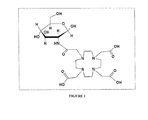
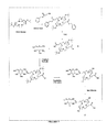
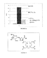
- FIG. 1 illustrates the structure of compound 1a
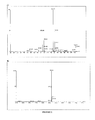
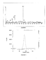
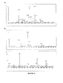
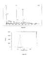
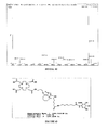
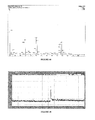
- FIG. 2 illustrates the ESI spectra of the t-Bu ester protected compound 1a before the final deprotection (a) and after final deprotection (b).
- Compound was analyzed using electrospray ionization mass spectroscopy (ES) in the presence of MeOH: 0.2% of formic acid.
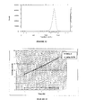
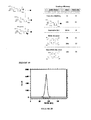
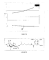
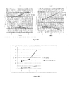
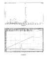
- FIG. 3 illustrates radio-TLC of compound 1a. Retention factor for radiolabeled Glucomedix and 68 GaCl 3 were 0.9 and 0.1, respectively. Radio-TLC (Bioscan) analysis showed the radiochemical purity of tracer was >97%.
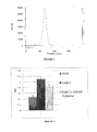
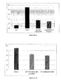
- FIG. 4 illustrates results of cellular uptake study of 68 Ga-labeled compound 1a.
- FIG. 5 illustrates results of cellular uptake study of 177 Lu-labeled compound 1a.
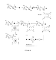
- FIG. 6 illustrates the structure of compound 1b.
- FIG. 7 illustrates a method of synthesis for compound 1b.
- FIG. 9 illustrates the radio-TLC of compound 1b.
- FIG. 10 illustrates the structure of compound 2a
- FIG. 12 illustrates the radio-TLC of compound 2a
- FIG. 13 illustrates the cellular accumulation of 68 Ga-2a.
- FIG. 14 illustrates the cellular accumulation and blocking of 68 Ga-2a
- FIG. 15 shows blocking of accumulation of 68 Ga-2a by glucose transporter inhibitors
- FIG. 16 illustrates the results of cellular uptake and blocking study of 177 Lu-2a.
- FIG. 17 illustrates the structure of compound 3
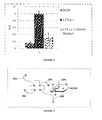
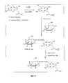
- FIG. 18 illustrates methods of synthesis of compound 3
- FIG. 19 illustrates loading efficiency
- FIG. 20 illustrates the radio-TLC of compound 3
- FIG. 21 illustrates the results of cellular accumulation of 68 Ga-3.
- FIG. 22 illustrates the structure of compound 4.
- FIG. 23 illustrates of a method of synthesis of compound 4
- FIG. 25 illustrates the radio-TLC of compound 4.
- FIG. 26 illustrates cellular accumulation of 68 Ga-4
- FIG. 27 illustrates cellular accumulation of 177 Lu-4
- FIG. 28 Illustrates results of biodistribution study of 177 Lu-4
- FIG. 29 illustrates general approach to the synthesis of DO2S-linker-carbohydrates.
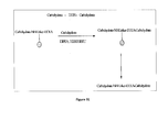
- FIG. 30 illustrates an example of a method used for the synthesis of carbohydrate-DO2S-linker-carbohydrate by SPPS.
- FIG. 31 illustrates an example of method used for the synthesis of di-substituted DO2S-[carbohydrate]2 by SPPS.
- FIG. 32 illustrates structure of compound 5 and predicted physicochemical properties
- FIG. 33 illustrates the ESI-MS spectra of the t-Bu ester protected compound 5 purified by HPLC (C18 column)
- FIG. 34 illustrates the ESI-MS spectra of the t-Bu ester protected compound 5 after purification (precipitation with Et2O)and collecting of supernatant.
- FIG. 35 illustrates the ESI-MS spectra of the t-Bu ester protected compound 5 after purification (precipitation with Et2O)and collecting of pellet.
- FIG. 36 illustrates radio-HPLC chromatogram (UV/Vis detection: 220 nm 275 nm) of the deprotected compound 5.
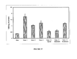
- FIG. 37 illustrates cellular accumulation of 68Ga-5 in SKBr3 breast cancer cell line and results of blocking studies using cold GLUT-1 competitors (genistein, scutellarin, cytochalasin B).
- FIG. 38 illustrates concentration dependent accumulation studies of 68Ga-5 in SKBr3 breast cancer cell line.
- FIG. 39 illustrates an example of method used for the synthesis of DO2S-linker-[carbohydrate]-genistein compound 6
- FIG. 40 illustrates an example of method used for the synthesis of DO2S-linker-[carbohydrate]-compound 7a,b.
- FIG. 41 illustrates structure of compound 7a
- FIG. 42 illustrates the ESI-MS spectra of the compound 7a.
- FIG. 43 illustrates structure of compound 7b
- FIG. 44 illustrates the ESI-MS spectra of the compound 7b.
- FIG. 45 illustrates radio-HPLC chromatogram of compound 7b.
- FIG. 46 illustrates cellular accumulation of 68 Ga-7b in SKBr3 breast cancer cell line. Specificity of binding was confirmed in blocking studies using cold GLUT-1 competitors (glucose, cytochalasin B).
- FIG. 47 illustrates cellular accumulation of 68 Ga-7b, 68 Ga-1 and 68 Ga-copper catalyzed glucosamine conjugate in SKBr3 breast cancer cell line.
- Embodiments of the invention relate to chelator-based carbohydrate derivatives and method of their synthesis.
- Chelator-carbohydrate (DO2S-carbohydrate) maybe synthesized from the amino-sugar is directly conjugated to DO2S compound, or it can be coupled to DO2S through any type of the linker (DO2S-linker-carbohydrate).
- synthesis of DO2S-carbohydrate can be performed manually on solid phase and using automated instrument, such as a peptide synthesizer.
- Resins used for the solid phase synthesis maybe selected from the group consisting of chloro- and bromofunctionalized (Merrifield, 4-bromomethylphenoxy)methyl polystyrene, 2-(4-bromomethylphenoxy)ethyl polystyrene, trityl, 2-chlortrityl chloride, NovaSyn TGT alcohol, NovaSyn TGT bromo), amino- and hydrazine functionalized (AM polystyrene and N-methyl aminomethylpolystyrene, NovaSyn TG amino, MBHA polystyrene, Rink Amide, Siber, amino trityl, sulfamyl-based, WeinrebAM, Fmoc-4-hydrazinobenzoyl, NOVAGel, alkylaminomethyl-indole, hydroxylamine Wang), hydroxyl functionalized (NovSyn hydroxyl, hydroxymethyl-phenyl, oxime), carboxy, al
- Linkers used for the solid phase synthesis of maybe selected from the group of amino acids, peptides, amino alcohols, polyethylylene glycols, alkanes, alkenes, alkynes, azide aromatic compounds, carbohydrates, carboxylic acids, esters, fosfororganic compounds, sulfonates.
- a targeting ligand may be selected from the group consisting of a carbohydrate, peptide, protein, antibody, nucleoside, nucleotide, heterocyclic compound, or alcohol.
- Preferred targeting ligands include carbohydrates, such as 2-deoxyglucose, 1′amino-sugar, 2′amino-sugar, 1′2′-aminosugar, 2′-amino-methylglycoside etc.
- the radiometal may be a transition metal ion or lanthanide series element.
- it may be 45 Ti, 59 Fe, 60 Cu, 61 Cu, 62 Cu, 64 Cu, 67 Cu, 67 Ga, 68 Ga, 89 Sr, 90 Y, 99m Tc, 111 In, 153 Gd, 153 Sm, 166 Ho, 186 Re, 177 Lu, 188 Re, 211 At, 212 Bi, 225 Ac.
- ⁇ -emitters include 211 At, 212 Bi and 223 Ra.
- Preferred ⁇ -emitters include 90 Y and 225 Ac.
- Preferred ⁇ / ⁇ -emitters include 67 Cu, 89 Sr, 153 Sm, 166 Ho, 177 Lu, 186 Re and 188 Re.
- Preferred ⁇ -emitters include 62 Cu, 64 Cu, 67 Ga, 68 Ga, 94m Tc, 99m Tc and 111 In. It is also envisioned that paramagnetic substances, such as Gd, Mn, Cu or Fe can be chelated with DO2S derivatives for use in conjunction with embodiments of the present invention.
- Embodiments of the present invention provide compositions for tissue specific disease imaging and radiotherapy.
- the disease may be cardiovascular disease, infection, diabetes, or cancer.
- the disease is cancer.
- the cancer may be a solid tumor.
- the tumor derives, either primarily or as a metastatic form, from cancers such as of the liver, prostate, pancreas, head and neck, breast, brain, colon, adenoid, oral, skin, lung, testes, ovaries, cervix, endometrium, bladder, stomach, and epithelium.
- Embodiments of the invention also provide kits for preparing a radiopharmaceutical preparation.
- the kit generally includes a sealed vial or bag, or any other kind of appropriate container, containing a predetermined quantity of DO2S-carbohydrate conjugate.
- the components of the kit may be in any appropriate form, such as in liquid, frozen or dry form. In preferred embodiments, the kit components may be provided in lyophilized form.
- the kit may also include an antioxidant and/or a scavenger.
- the antioxidant may be any known antioxidant but is preferably vitamin C. Scavengers may be DTPA, EDTA or DOTA.
- 1,4,7,10-tetraazacyclododecane-1,4,7-tris(t-butyl acetate)-10-acetic acid (1eq.) was dissolved in 2 mL dimethylformamide (DMF), and then N-hydroxybenzotriazole (HOBT) and O-Benzotriazole-N,N,N′,N′-tetramethyl-uronium-hexafluoro-phosphate (HBTU) coupling agents (4eq. each) were added in the presence of 4eq. of N,N-diisopropylamine (DIPEA). Reaction was left at room temperature for 30 min.
- DIPEA N,N-diisopropylamine
- Step b Coupling of Glucosamine Hydrochloride to Activated Chelating Agent
- Glucosaminehydrochloride (4eq) was dissolved in DMF in the presence of DIPEA (4eq) and added to the solution of pre-activated DOTA. Reaction was left stirring for 48 h at room temperature and was monitored by TLC (chloroform:methanol 1:10) and visualized using anisidine solution or dichlorofluoresceine. Conjugate was purified by extraction using CH 2 Cl 2 :H 2 O and organic fraction was collected. The tert-butyl ester protecting groups were removed in the presence of 30% TFA:CH 2 Cl 2 :H 2 O:TIS (950:250:250), and the product was dialyzed using Sep Pak in millipore water for 48 h at room temperature.
- LS174T cells were plated in 12 well plates at a density of 1.5 ⁇ 10 5 cells per well and grown overnight in DMEM containing 5.4 mg/ml glucose and 10% FBS at 37° C., 5% CO 2 . Cells were fasted for 30 min prior to the study with glucose free DMEM. At the start of the study, media were removed from each well and replaced with 0.5 ml glucose free DMEM (Cellgro) containing 0.5 microCi 177 Lu-1a or 177 Lu-DOTA or DMEM media containing 10 mmD-glucose and 0.5 microCi 177 Lu-1a.
- Cellgro 0.5 ml glucose free DMEM
- DO2A tBu ester (1eq., Macrcyclics) was dissolved in CH3CN (5 ml) and benzyl 3-bromopropanoate was added in the presence of Et3N (2.1 ml, 12 mmol) at temp. 0° C. Reaction was slowly warmed up to room temp. and left stirring for 24 h. After evaporation of the solvent under vacuum, product was purified by flash chromatography using 60N silica gel. Deprotection of benzyl ester groups was completed by catalytic hydrogenation under high pressure in the presence of Pd/C during 24 h.
- Modified chelating agent was dissolved in DMF (3 mL) in the presence DIPEA (3eq) and coupling agent, HBTU (3eq.) was added to the solution. The mixture was left for 20 min at r.t. GlcNAc-b-NH 2 (1.3 eq.) was dissolved in DMSO (1 ml). The solution was heated slightly to dissolve the monosaccharide and added to the pre-actived DO2A chelating agent. The mixture was allowed to react at room temp. with constant stirring 12 h. After solvent evaporation, product was purified by extraction using CHCl 3 :MeOH and flash silica gel 60N column.
- iTLC was developed in standard running buffer (0.5M NH4OAC: methanol 1:1 v/v).
- Radio-TLC (Bioscan) analysis showed the radiochemical purity of tracer was >98%.
- 1,4,7,10-tetraazacyclododecane-1,4,7,10-tetraacid acetic acid (1eq.) was dissolved in 2 mL N-methylpyrolidine (NMP) and HATU coupling agent (4eq.) was added in the presence of 4eq. of N,N-diisopropylamine (DIPEA). Reaction was left at room temperature for 20 min.
- NMP N-methylpyrolidine
- DIPEA N,N-diisopropylamine
- Step b Coupling of Glucosamine Hydrochloride to Activated Chelating Agent
- Glucosamine hydrochloride (4eq) was dissolved in NMP in the presence of DIPEA (4eq) and added to the solution of pre-activated DOTA. Reaction was left stirring for one day at room temperature and was traced by TLC (chloroform:methanol 1:10) visualized using anisidine solution or dichlorofluoresceine. After concentration of the reaction under high vacuum, product was extracted using CH2Cl2:H2O and aqueous fraction was collected. Final product was precipitated by addition of ether diethyl on ice.
- iTLC was developed in standard running buffer (0.5M NH4OAC:methanol 1:1 v/v) and is shown below on FIG. 8 .
- Radio-TLC (Bioscan) analysis showed the radiochemical purity of tracer was >99%
- SKBr3 cells were plated in 12 well plates at a density of 2*10 ⁇ 5 cells per well and grown overnight in DMEM containing 5.4 mg/ml glucose and 10% FBS at 37° C., 5% CO2. Cells were fasted for 20 min prior to the study with glucose free DMEM. At the start of the study, media was removed from each well and replaced with 0.5 ml glucose free DMEM (Cellgro) containing 0.5-1 ⁇ Ci 68Ga-2 or 68Ga DOTA. Cells were incubated at 37° C., 5% CO2 for the indicated time. The radioactive media was then removed and cells were washed twice with 1 ml PBS. Cells were then trypsinized and transferred to counting tubes.
- DMEM containing 5.4 mg/ml glucose and 10% FBS at 37° C., 5% CO2. Cells were fasted for 20 min prior to the study with glucose free DMEM. At the start of the study, media was removed from each well and replaced with 0.5 m
- Radioactivity in cells and media were counted at 511 keV using a Perkin Elmer Wizard gamma counter. Cells were then collected and lysed with RIPA buffer (Invitrogen) and protein concentration was determined by Pierce BCA protein assay kit (ThermoFisher). Data is expressed as % ID (cpm cells/cpm media*100)/mg protein. Error bars represent Standard deviation.
- SKBr3 cells were plated in 12 well plates at a density of 2*10 ⁇ 5 cells per well and grown overnight in DMEM containing 5.4 mg/ml glucose and 10% FBS at 37° C., 5% CO2. Cells were fasted for 20 min prior to the study with glucose free DMEM. At the start of the study, media was removed from each well and replaced with 0.5 ml glucose free DMEM (Cellgro) or media that contained 100 mg/ml D or L glucose (Sigma Aldrich) and 0.5-1 ⁇ Ci 68Ga-2. Cells were incubated at 37° C., 5% CO2 for 1 hour. The radioactive media was then removed and cells were washed twice with 1 ml PBS.
- DMEM containing 5.4 mg/ml glucose and 10% FBS at 37° C., 5% CO2.
- SKBr3 cells were plated in 12 well plates at a density of 2*10 ⁇ 5 cells per well and grown overnight in DMEM containing 5.4 mg/ml glucose and 10% FBS at 37° C., 5% CO2. Cells were fasted for 20 min prior to the study with glucose free DMEM. At the start of the study, media was removed from each well and replaced with 0.5 ml glucose free DMEM (Cellgro) or media that contained 100 mg/ml D or L glucose (Sigma Aldrich) or other GLUT1 inhibitors (genistein, cytochalasin B, scutellarin) and 0.5-1 ⁇ Ci 68Ga-2. Cells were incubated at 37° C., 5% CO2 for 1 hour.
- Radioactive media was then removed and cells were washed twice with 1 ml PBS. Cells were then trypsinized and transferred to counting tubes. Radioactivity in cells and media were counted at 511 keV using a Perkin Elmer Wizard gamma counter. Cells were then collected and lysed with RIPA buffer (Invitrogen) and protein concentration was determined by Pierce BCA protein assay kit (ThermoFisher). Data is expressed as % ID (cpm cells/cpm media*100)/mg protein. Error bars represent Standard deviation. Data represents the average of four separate studies performed in triplicate.
- A549 cells were plated in 12 well plates at a density of 1.5*10 ⁇ 5 cells per well and grown overnight in DMEM containing 5.4 mg/ml glucose and 10% FBS at 37° C., 5% CO2. Cells were fasted for 30 min prior to the study with glucose free DMEM. At the start of the study, media was removed from each well and replaced with 0.5 ml glucose free DMEM (Cellgro) containing 0.5 microCi 177 Lu-1a or 177 Lu-DOTA or DMEM media containing 10 mg/ml D-glucose and 0.5 microCi 177 Lu-1a. Cells were incubated at 37° C., 5% CO2 for the indicated time.
- the radioactive media was then removed and cells were washed twice with 1 ml PBS. Cells were then trypsinized and radioactivity was counted. Data is expressed as % ID (cpm cells/cpm media*100). Error bars represent Standard deviation.
- Fmoc-Asn-Glc (0.015 eq.) was immobilized on the 2-chlorotrityl chloride resin (0.01 eq) in the presence of DIPEA (4 eq) in DMF. Reaction is carried out in presence of excess of DIPEA in order to prevent possible hydrolysis of chloro-resin bond and to neutralize HCl, that form during esterification reaction. Reaction is completed with 1-2 h at r.t.
- the unmodified chloride resin was deactivated by addition of ethanol or TFE.
- the Fmoc concentration on the resin was determined by addition of 20% piperidine in DMF to the tested resin.
- the resultant fulvene-piperidine adduct had UV absorption max at 301 nm.
- Free Fmoc amino acid of know concentration was used as a standard.
- Loading efficiency of the compound 3 on the resin was close to 15% after 1 h. Loading increased to 79% when reaction was left for more than 24 h.
- the lower loading of Fmoc-Asn-Glc conjugate relatively to unprotected Asn is probably caused by steric hindrance and close proximity of carboxyl group and Fmoc group.
- Loading efficiency was also determined using Kaiser test, that measure the content of free amines after Fmoc deprotection.
- Conjugate was released from in acid-mediated in mild conditions by addition of AcOH in DCM:H2O:TIS at r.t. in 20-25 min. This process can additionally accelerated in higher temp.
- A549 cells were plated in 12 well plates at a density of 2*10 ⁇ 5 cells per well and grown overnight in DMEM containing 5.4 mg/ml glucose and 10% FBS at 37° C., 5% CO2. Cells were fasted for 30 min prior to the study with glucose free DMEM. At the start of the study, media was removed from each well and replaced with 0.5 ml glucose free DMEM (Cellgro) containing 0.5 microCi 68Ga-3 or 68Ga DOTA. Cells were incubated at 37° C., 5% CO2 for the indicated time. The radioactive media was then removed and cells were washed twice with 1 ml PBS. Cells were then trypsinized and radioactivity was counted. Data is presented as % cpm cells/cpm media.
- A549 cells were plated in 12 well plates at a density of 2*10 ⁇ 5 cells per well and grown overnight in DMEM containing 5.4 mg/ml glucose and 10% FBS at 37° C., 5% CO2. Cells were fasted for 30 min prior to the study with glucose free DMEM. At the start of the study, media was removed from each well and replaced with 0.5 ml glucose free DMEM (Cellgro) containing 0.5 microCi 68Ga-3 or 68Ga DOTA. Cells were incubated at 37° C., 5% CO2 for the indicated time. The radioactive media was then removed and cells were washed twice with 1 ml PBS. Cells were then trypsinized and radioactivity was counted. Data is presented as % cpm cells/cpm media.
- Step 1 Synthesis of ⁇ -N-Acetylglucosaminylamine ( FIG. 23 , compound 3).
- 2-Acetamido-2-deoxy-beta-D-glucopyranosyl azide 3,4,6-triacetate was obtained from Aldrich and was converted to amine (2) by using PPh3 and DCM following the general literature procedure (Carbohyd. Research, 2001, 331, 439).
- Deacetylation To a solution of an acetyl protected amine (2) in dry MeOH, 1-2 drops of ⁇ 1M methanolicNaOMe solution were added, and the reaction mixture was kept at rt until completion of the transformation (monitored by tlc). Amberlyst 15 (H+ form) resin was then added to remove sodium ions, the resin was filtered off, and the solvent removed in vacuo.
- Step3 Synthesis of N-1-( ⁇ -aminocaproyl)- ⁇ -N-acetyl-2-deoxyglucopyranosyl N-[1,4,7,10-Tetraazacyclododecane-1,4,7,10-tetra-(acetyl acid)] ( FIG. 23 ).
- DOTA conjugate (6,) was dissolved in DMSO(1 ml), Et3N (0.3 ml) and coupling agent HATU were added to the solution. The mixture was allowed to preactivate for approx 30 min.
- N( ⁇ -aminocaproyl)-N-acetylglucosaminylamine (5) was dissolved in DMSO and added to the preactivated DOTA solution.
- E.3.Cellular accumulation of 68Ga-4 A549 and SKBr3 cells were plated in 12 well plates at a density of 2*10 ⁇ 5 cells per well and grown overnight in DMEM containing 5.4 mg/ml glucose and 10% FBS at 37° C., 5% CO2. Cells were fasted for 30 min prior to the study with glucose free DMEM. At the start of the study, media was removed from each well and replaced with 0.5 ml glucose free DMEM (Cellgro) containing 0.5-1 ⁇ Ci 68Ga-4 or 68Ga DOTA. Cells were incubated at 37° C., 5% CO2 for the indicated time. The radioactive media was then removed and cells were washed twice with 1 ml PBS.
- Cellgro 0.5 ml glucose free DMEM
- E.4.Cellular accumulation of 177Lu-4 A549 cells were plated in 12 well plates at a density of 2*10 ⁇ 5 cells per well and grown overnight in DMEM containing 5.4 mg/ml glucose and 10% FBS at 37° C., 5% CO2. Cells were fasted for 30 min prior to the study with glucose free DMEM. At the start of the study, media was removed from each well and replaced with 0.5 ml glucose free DMEM (Cellgro) containing 0.5 microCi 177Lu-4 or 177Lu-DOTA. Cells were incubated at 37° C., 5% CO2 for the indicated time. The radioactive media was then removed and cells were washed twice with 1 ml PBS. Cells were then trypsinized and radioactivity was counted. Data is presented as % cpm cells/cpm media
- Tumor xenografts were generated in 6 week old Swiss nu/nu mice using the human lung adenocarcinoma cell line A549. Xenografts were generated by subcutaneous inoculation of 2*10 ⁇ 6 cells per mouse into the right shoulder. Xenografts were allowed to grow for 7 days. Mice were fasted for 8-12 hours prior to the start of the study. On the day of the study, mice were anesthetized with Isofluorane and a tail vein cannula was inserted. The tracer was delivered via tail vein injection. Mice were placed on a warming pad or under a warming lamp to maintain body temperature. Mice were sacrificed at the indicated timepoints and organs were removed, weighed and counted. Data is expressed as % injected dose/gram tissue.
- carbohydrate-genistein (1 eq., 20 ug) was dissolved in 0.5 mL dimethylformamide DMF and dimethylsulfoxide DMSO and N-hydroxybenzotriazole (HOBT) and O-Benzotriazole-N,N,N′,N′-tetramethyl-uronium-hexafluoro-phosphate (HBTU) coupling agents (1.5eq. each) were added in the presence of 4eq. of N,N-diisopropylamine (DIPEA). Reaction was left at room temperature for 30 min.
- DIPEA N,N-diisopropylamine
- Step b Coupling of DOTA-Linker-NH2 to Activated Carbohydrate-Genistein Derivative
- Pre-activated carbohydrate-genistein (1.5eq) was dissolved in DMF in the presence of DIPEA (4eq) and added to the solution DOTA-linker-NH2. Reaction was left stirring for 48 h at room temperature and was traced by TLC (chloroform:methanol 1:10) visualized using anisidine solution or dichlorofluoresceine. After completion of reaction, solution was evaporated and purified on HPLC (C18 column). The tert-butyl ester protecting groups were removed in the presence of 30% TFA:CH 2 Cl 2 :H 2 O:TIS (950:250:250) and product was purified again by HPLC.
- protected compound 5 was purified by precipitation using Et 2 O and fraction of supernatant was collected.
- the tert-butyl ester protecting groups were removed in the presence of 30% TFA:CH 2 Cl 2 :H 2 O:TIS (950:250:250) and product was purified again by precipitation with Et 2 O.
- SKBr3 cells were plated in 12 well plates at a density of 1.5*10 ⁇ 5 cells per well and grown overnight in DMEM containing 5.4 mg/ml glucose and 10% FBS at 37° C., 5% CO2. Cells were fasted for 20 min prior to the study with glucose free DMEM. At the start of the study, media was removed from each well and replaced with 0.5 ml glucose free DMEM (Cellgro) or media that contained 100 mg/ml D or L glucose (Sigma alrich) and 12 uCi/ml of radiolabeled compound. Cells were incubated at 37° C., 5% CO2 for 1 hour. The radioactive media was then removed and cells were washed twice with 1 ml PBS.
- DMEM containing 5.4 mg/ml glucose and 10% FBS at 37° C., 5% CO2.
- Radioactivity in cells and media were counted at 511 keV using a Perkin Elmer Wizard gamma counter. Cells were then collected and lysed with RIPA buffer (Invitrogen) and protein concentration was determined by Pierce BCA protein assay kit (ThermoFisher). Data is expressed as % ID (cpm cells/cpm media*100)/mg protein. Error bars represent Standard deviation. Data represents the average of four separate studies performed in triplicate.
- tris-tBu ester DOTA (1 eq., 20 ug) was dissolved in 0.5 mL dimethylformamide DMF and dimethylsulfoxide DMSO and N-hydroxybenzotriazole (HOBT) and O-Benzotriazole-N,N,N′,N′-tetramethyl-uronium-hexafluoro-phosphate (HBTU) coupling agents (1.5eq. each) were added in the presence of 4eq. of N,N-diisopropylamine (DIPEA). Reaction was left at room temperature for 30 min.
- DIPEA N,N-diisopropylamine
- Step b Coupling of Linker-Genistein to Activated DOTA-tris-tBu Ester
- Pre-activated DOTAtris-tBuester (1.5eq) was dissolved in DMF in the presence of DIPEA (4eq) and combined with solution of genistein-linker-NH2. Reaction was left stirring for 48 h at room temperature and was traced by TLC (chloroform:methanol 1:10) visualized using anisidine solution or dichlorofluoresceine. After completion of reaction, solution was evaporated and purified on rHPLC (C18). The tert-butyl ester protecting groups were removed in the presence of 30% TFA:CH 2 Cl 2 :H 2 O:TIS (950:250:250) and product was purified again by HPLC.
- SKBr3 cells were plated in 12 well plates at a density of 1.5*10 ⁇ 5 cells per well and grown overnight in DMEM containing 5.4 mg/ml glucose and 10% FBS at 37° C., 5% CO2. Cells were fasted for 20 min prior to the study with glucose free DMEM. At the start of the study, media was removed from each well and replaced with 0.5 ml glucose free DMEM (Cellgro) or media that contained 100 mg/ml D or L glucose (Sigma alrich) and 12 uCi/ml of radiolabeled compound. Cells were incubated at 37° C., 5% CO2 for 1 hour. The radioactive media was then removed and cells were washed twice with 1 ml PBS.
- DMEM containing 5.4 mg/ml glucose and 10% FBS at 37° C., 5% CO2.
- SKBr3 cells were plated in 12 well plates at a density of 1.5*10 ⁇ 5 cells per well and grown overnight in DMEM containing glucose and 10% FBS at 37° C., 5% CO2. Cells were fasted for 20 min prior to the study with glucose free DMEM. At the start of the study, media was removed from each well and replaced with 0.5 ml DMEM (Cellgro) containing GLUT-1 inhibitors (scutellarin, glucose, genistein, cytochalasin B) and 12 uCi/ml of radiolabeled compound. Cells were incubated at 37° C., 5% CO2 for 1 hour.
- Radioactive media was then removed and cells were washed twice with 1 ml PBS. Cells were then trypsinized and transferred to counting tubes. Radioactivity in cells and media were counted at 511 keV using a Perkin Elmer Wizard gamma counter. Cells were then collected and lysed with RIPA buffer (Invitrogen) and protein concentration was determined by Pierce BCA protein assay kit (ThermoFisher). Data is expressed as % ID (cpm cells/cpm media*100)/mg protein. Error bars represent Standard deviation. Data represents the average of four separate studies performed in triplicate.
- SKBr3 cells were plated in 12 well plates at a density of 1.5*10 ⁇ 5 cells per well and grown overnight in DMEM containing 5.4 mg/ml glucose and 10% FBS at 37° C., 5% CO2. Cells were fasted for 20 min prior to the study with glucose free DMEM. At the start of the study, media was removed from each well and replaced with 0.5 ml glucose free DMEM (Cellgro) or media that contained 100 mg/ml D or L glucose (Sigma Alrich) and 12 uCi/ml of radiolabeled compound. Cells were incubated at 37° C., 5% CO2 for 1 hour. The radioactive media was then removed and cells were washed twice with 1 ml PBS.
- DMEM containing 5.4 mg/ml glucose and 10% FBS at 37° C., 5% CO2.
- SKBr3 cells were plated in 12 well plates at a density of 1.5*10 ⁇ 5 cells per well and grown overnight in DMEM containing glucose and 10% FBS at 37° C., 5% CO2. Cells were fasted for 20 min prior to the study with glucose free DMEM. At the start of the study, media was removed from each well and replaced with 0.5 ml glucose free DMEM (Cellgro) or media that contained 100 mg/ml D or L glucose (Sigma alrich) or other GLUT1 inhibitors (genistein, scutellarin, cytochalasin B) and 12 uCi/ml of radiolabeled compound. Cells were incubated at 37° C., 5% CO2 for 1 hour.
- Radioactive media was then removed and cells were washed twice with 1 ml PBS. Cells were then trypsinized and transferred to counting tubes. Radioactivity in cells and media were counted at 511 keV using a Perkin Elmer Wizard gamma counter. Cells were then collected and lysed with RIPA buffer (Invitrogen) and protein concentration was determined by Pierce BCA protein assay kit (ThermoFisher). Data is expressed as % ID (cpm cells/cpm media*100)/mg protein. Error bars represent Standard deviation. Data represents the average of four separate studies performed in triplicate.
Landscapes
- Chemical & Material Sciences (AREA)
- Health & Medical Sciences (AREA)
- Life Sciences & Earth Sciences (AREA)
- Organic Chemistry (AREA)
- General Health & Medical Sciences (AREA)
- Engineering & Computer Science (AREA)
- Biochemistry (AREA)
- Biotechnology (AREA)
- Molecular Biology (AREA)
- Genetics & Genomics (AREA)
- Proteomics, Peptides & Aminoacids (AREA)
- Physics & Mathematics (AREA)
- Medicinal Chemistry (AREA)
- Optics & Photonics (AREA)
- Pharmacology & Pharmacy (AREA)
- Epidemiology (AREA)
- Animal Behavior & Ethology (AREA)
- Public Health (AREA)
- Veterinary Medicine (AREA)
- Medicines Containing Antibodies Or Antigens For Use As Internal Diagnostic Agents (AREA)
- Saccharide Compounds (AREA)
Abstract
Description
- The present invention relates to the field of radiochemistry, nuclear imaging, radionuclide therapy, drug development and chemical synthesis. More particularly, it relates to a strategy for synthesis and radiolabeling of targeted ligands.
- Increased glycolysis is a biochemical hallmark of neoplastic cells (Warburg, 1956). Overexpression of glucose transporters in the cell-membranes of cancer cells results in increased glucose uptake (Kim and Dang, 2006). Furthermore, increased activity of glycolytic enzymes, such as hexokinase, phosphorylates glucose to prohibit its exit from the cell (Parry and Pedersen, 1983). This results in increased accumulation and consumption of glucose by cancer cells. Two families of glucose transporters have been reported in humans: GLUT [Solute Carrier family 2 (facilitated glucose transporter), gene name SLC2A] and SGLT [Solute carrier family 5 (sodium/glucose cotransporter); gene name: SLC5A]. While members of the GLUT family of transporters rely on existing concentration gradients to transport glucose from one side of the membrane to the other, members of the SGLT family transport sugars inside the cell against a concentration gradient by active transport. To date, 14 members of the GLUT family have been identified (GLUT1-14) (Thorens and Mueckler). Although there is significant sequence homology among the GLUTs, they display differential expression in tissues and have varying affinities for glucose and related sugars. GLUT1; GLUT2, and GLUTS have been shown to be overexpressed in cancer. GLUT3 is primarily expressed on neurons, while GLUT4 and GLUT12 are primarily expressed in cardiac and skeletal muscle and their presence in the cell membrane is regulated by insulin. Six members of the SGLT family have been identified (SGLT1-6). Of these, SGLT-1 and SGLT-2 are implicated in transport of sugars. Expression of SGLT-1 in normal tissues is restricted to the small intestine and renal proximal tubes, while SGLT-2 is expressed on the apical membranes of cells in the renal convoluted proximal tubules. SGLT-1 has high affinity and low capacity for glucose transport, while SGLT-2 has lower affinity and higher capacity.(Thorens and Mueckler) Increased expression of both SGLT-1 and SGLT-2 have been reported in primary and metastatic cancers (Helmke et al., 2004, Ishikawa et al., 2001, Weihua et al., 2008)
- The increased accumulation of glucose in neoplastic cells has been exploited for development of diagnostic and therapeutic agents. Radiolabeled carbohydrates have been developed for nuclear imaging of cancer. The most successful of these, 2-18F-fluoro-2-deoxyglucose (FDG) is currently the gold standard for positron emission tomography (PET). Other sugar-based PET agents have also been developed, including 18F-Fluoroacetyl-D-glucosamine (Fujiwara et al., 1990), 68Ga-2-amino-2-deoxyglucose-based 3-hydroxy-4pyridonato complexes (Green et al., 2005), 68Ga-tripodal trissalicylaldimine complexes functionalized with xylose, glucose or galactose (Gottschaldt, 2009) 11C-methyl-glucoside (US2010/0008856, Bormons et al, 2003). Radiolabeled sugars have also been developed for SPECT imaging including 99mTc-Glucosamine-dipicolylamine, US2006/0051291) 99mTc-tricarbonyl-N-(2′-Hydroxybenzyl)-2-amino-2-deoxy-D-glucose, (US 2006/0051291) 99mTc Ehtylenedicysteine-glucosamine (US 2007/0248537), 111In-DOTA-glucosamine, (US2006/0142207A1), 99mTc-DTPA-deoxyglucose (Chen et al., 2007)
- Solid phase approach is a robust, efficient and reproducible method of synthesis of library of compounds. It was successfully applied for the synthesis of peptides, nucleotides, and glycopeptides. In solid phase, synthesized compounds are temporary attached to the insoluble polymer resins, allowing them to be readily separated from coupling agents and by-products during elongation process. After completion of the synthesis, final compound is released from the polymer support with high yield and purity. Application of solid phase approach for the development of imaging or therapeutic agents is still limited. SPPS was used for the synthesis peptidyl imaging agents (U.S. Patent Application Publication No. 2008/0089842). Amino-functionalized chelator was immobilized on the resin and coupled to the C-terminus and/or backbone of the peptide. After cleaving from the resin, amino-chelator-peptide was labeled with lanthanide metals.
- Target-specific radiopharmaceuticals consist of a tissue targeting biomolecule attached to a radionuclide. Commonly used cyclotron-produced radionuclides (i.e. 11C, 13N, 15O and 18F) may be covalently linked to targeting molecules. Alternatively, complexes consisting of a targeting ligand, bifunctional chelating agent and a radionuclide, can be used for nuclear imaging and therapy. In this case, the radionuclide, most commonly a radiometal, is stably coordinated by the bifunctional chelating agent (BFCA). Common BFCAs for radiometallic chelation include DTPA, hydrazinonicotinamide (HYNIC), mercaptoacetyltriglycine (MAG3), tetraaza compounds (i.e. 1,4,7,10-tetraazacyclododecane-1,4,7,10-tetraacetic acid [DOTA] and macrocyclic derivatives) and ethylenedicysteine (EC). Each BFCA possesses various combinations of electron-donating atoms for metal chelation. For diagnostic radiopharmaceuticals, γ-emitting radiometals such as 68Ga and 64Cu are commonly used for PET, and 99mTc, 111In and 67Ga are commonly used for SPECT. Additionally, α-emitting (225Ac and 213Bi) and β-emitting(90Y, 177Lu, and 186/188Re) radiometals can be used for radionuclide therapy.
- Diagnostic imaging techniques such as computed tomography (CT) and magnetic resonance imaging (MRI) provide anatomical information about disease sites. While these modalities are commonly-used for monitoring changes in tumor size, they cannot assess functional changes occurring within cells or tumors. Functional imaging modalities use radiotracers to image, map and measure biological attributes of disease, such as metabolism, proliferation and surface receptor expression. As a result, functional imaging techniques such as Positron Emission Tomography (PET) and Single Photon Emission Computed Tomography (SPECT) have been experiencing explosive growth due to advances in molecular imaging technology. New molecular imaging targets for diagnosis and therapy have been developed to visualize disease states and pathological processes without surgical exploration of the body. In particular, targeted radiopharmaceuticals offer promising capabilities for the non-invasive assessment of the pathophysiology of diseases. Schillaci, O. & Simonetti, G., Cancer Biother. Radiopharm. 19: 1-10 (2004); Paulino, et al, Semin Nucl. Med. 33: 238-43 (2003).
- In contrast to traditional radiotherapy methods, radionuclide therapy utilizes active targeting for improved tumor specificity and localization of radiation in tumor cells, thereby minimizing the effects on normal tissue. This approach uses high-affinity cell binding ligands to target radioactivity to tissues expressing a particular receptor. Radiolabeled monoclonal antibodies were among the first targeted therapies developed and have shown moderate clinical success, particularly for the treatment of Non-Hodgkin's lymphoma. However, the large size, limited diffusion and slow blood clearance of the tumor-seeking antibodies have limited their clinical utility. To address these concerns, radiolabeled peptides and low/intermediate molecular weight biologically-active proteins have been employed into the design of novel radionuclide therapies (Pnwar et al., 2005). The smaller size of these molecules confers desirable pharmacokinetic properties that are favorable for therapy, such as higher target-to-background ratios and faster blood clearance.
- Embodiments of the present invention relates to the field of radiochemistry, nuclear imaging, radionuclide therapy, drug development and chemical synthesis. More particularly, embodiments of the invention relate to a strategy for solid phase synthesis and radiolabeling of ligands. Embodiments of the invention further relate to methods of using those radiolabeled ligands for radionuclide therapy and tissue-specific disease imaging
- Embodiments of the present invention provide methods for the synthesis of DO2S compounds conjugated to targeting ligands by solid phase approach. In preferred embodiments, the targeting ligands are carbohydrates. The DO2S compounds can be conjugated directly to the targeting ligands or through linkers.
- A DO2S derivative has the following general structure:
-
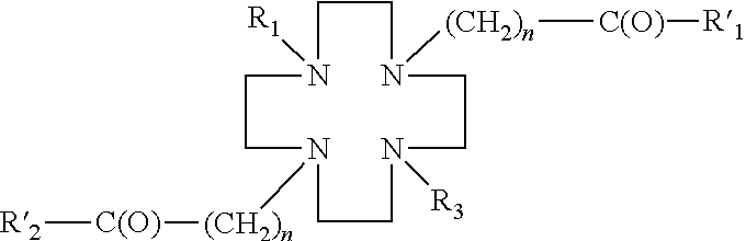
- wherein R1′, R2′ are each a hydroxyl group, and R1 is a hydrogen, a linker, or a ligand, and R3 is a linker or a ligand, and n is an integer from 1 to 10, preferably from 1 to 4. The linkers may be amino acids, peptides, amino alcohols, polyethylylene glycols, alkanes, alkenes, alkynes, azide aromatic compounds, carbohydrates, carboxylic acids, esters, phosphororganic compounds, sulfonates.
- In accordance with embodiments of the invention, the linker may comprise —(CH2)n—X, wherein X is a hydroxyl, an amino, or a carboxyl group, and n is an integer from 1 to 10, preferably from 1 to 4, and wherein the linker may be optionally substituted (e.g., one or more H on the alkyl chain may be replaced with an alkyl (e.g., C1-C6 alkyl), a halogen, a nitro group, a hydroxyl group, an amino group, or a carboxyl group).
- In accordance with embodiments of the invention, the ligand may be selected from the group consisting of a carbohydrate, peptide, protein, antibody, nucleoside, nucleotide, heterocyclic compound, or alcohol. In preferred embodiments, the ligand may be carbohydrates, such as 2-deoxyglucose, 1′ amino-sugar, 2′ amino-sugar, 1′2′-aminosugar, 2′-amino-methylglycoside, etc. In preferred embodiments, the ligand may be a glucose.
- In preferred embodiments, the DO2S derivative has the following structure:
-
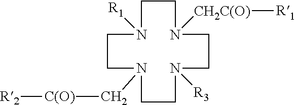
- Examples of DO2S derivatives may include:
-
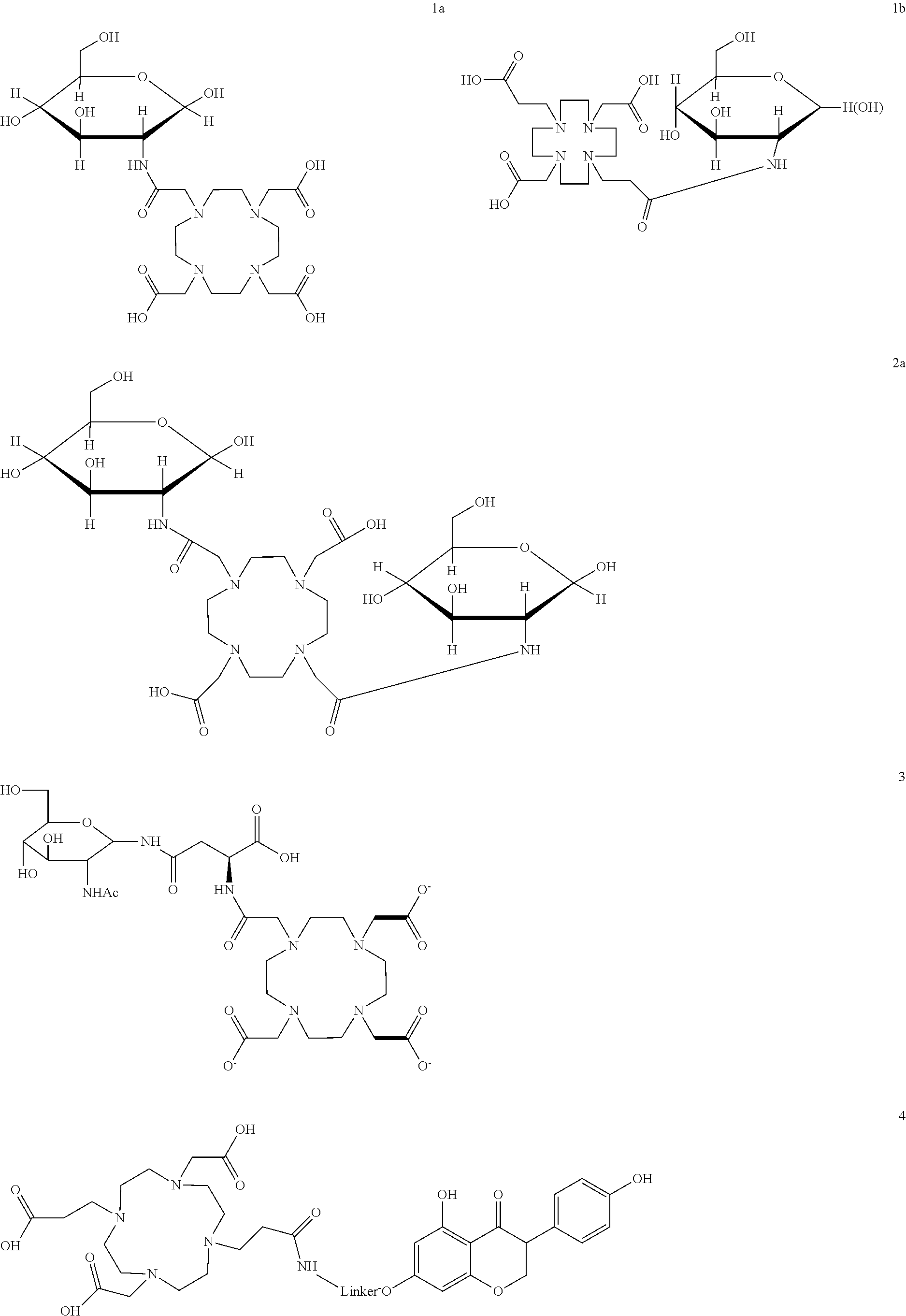
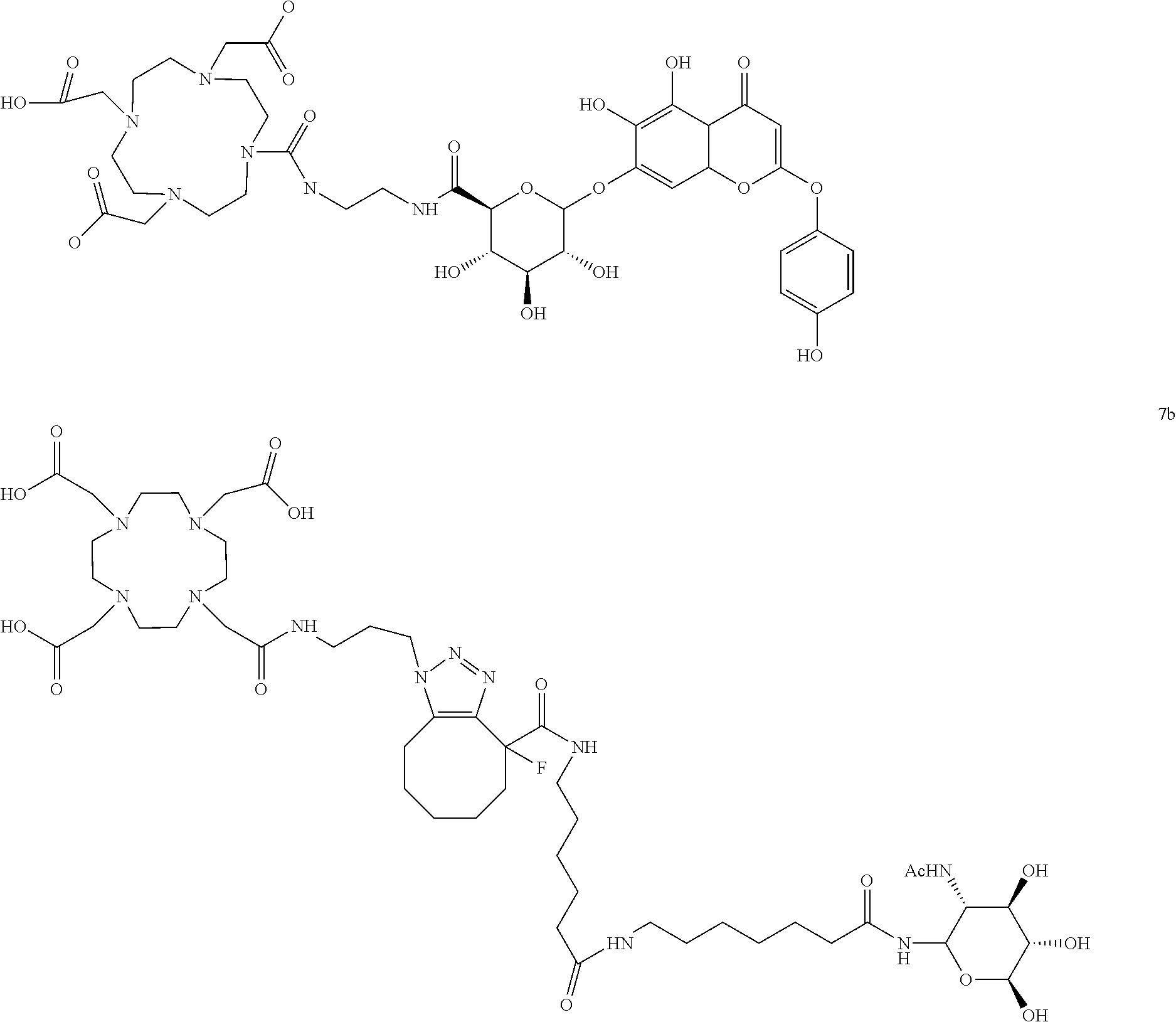
- Some embodiments of the invention comprise a metal or a radionuclide chelated to the DO2S derivative. Generally, any α-emitter, β-emitter, γ-emitter, or β/γ-emitter may be used in conjunction with embodiments of the invention. Preferred α-emitters include 211At, 212Bi and 223Ra. Preferred β-emitters include 90Y and 225Ac. Preferred β/γ-emitters include 67Cu, 89Sr, 153Sm, 166Ho, 177Lu, 186Re and 188Re. Preferred γ-emitters include 62Cu, 64Cu, 67Ga, 68Ga, 94mTc, 99mTc and 111In.
- In accordance with some embodiments of the invention, the radionuclide may be one selected from 45Ti, 59Fe, 60Cu, 61Cu, 62Cu, 64Cu, 67Cu, 67Ga, 68Ga, 89Sr, 90Y, 86Y, 94mTc, 99mTc, 111In, 149Pm, 153Gd, 153Sm, 166Ho, 177Lu, 186Re, 188Re, 211At, 212Bi, or 225AC. In preferred embodiments, the radionuclide is 68Ga or 177Lu.
- Some embodiments of the invention provide methods of radiolabeling DO2S ligand conjugates on solid phase. In preferred embodiments, the radiometal is 68Ga or 177Lu.
- In accordance with some embodiments of the invention, paramagnetic substances, such as Gd, Mn, Cu or Fe, may be used—i.e., chelated with DO2S derivatives for use in embodiments of the present invention.
- Some embodiments of the invention provide methods for the treatments of medical conditions by administering to subjects compositions comprising DO2S compounds conjugated to GLUT-1 targeting ligands. The DO2S compounds may be optionally chelated to a metal species
- Some embodiments of the invention provide methods for diagnostic imaging performed on cellular level by application of DO2S compound conjugated to GLUT-1 targeting ligands. The DO2S compound may be optionally labeled with fluorescent, near-infrared imaging probe.
- Some embodiments of the invention provide methods for intra-operative diagnosis and treatment of medical conditions by administering to subjects compositions comprising DO2S compounds conjugated to GLUT-1 targeting ligands. The DO2S compound may be optionally labeled with near-infrared imaging probe.
- Some embodiments of the invention provide methods for diagnostic imaging of medical conditions. A method comprises administering to a subject a composition comprising a DO2S compound conjugated to a carbohydrate targeting ligand. The DO2S compound may be chelated to a radiometal species.
- Some embodiments of the present invention relate to kits for the treatments or diagnosis of a subject. A kit of the invention may comprise a composition comprising a DO2S compound conjugated to a carbohydrate. The DO2S compound may be optionally chelated to a metal species.
-
FIG. 1 illustrates the structure of compound 1a -
FIG. 2 illustrates the ESI spectra of the t-Bu ester protected compound 1a before the final deprotection (a) and after final deprotection (b).Compound was analyzed using electrospray ionization mass spectroscopy (ES) in the presence of MeOH: 0.2% of formic acid. The ESI spectra showed mass peak corresponding to compound m/z=566.2 (calculated MW+H=565.37). The m/z=734.45 corresponds to compound 1a before the deprotection. -
FIG. 3 illustrates radio-TLC of compound 1a. Retention factor for radiolabeled Glucomedix and 68GaCl3 were 0.9 and 0.1, respectively. Radio-TLC (Bioscan) analysis showed the radiochemical purity of tracer was >97%. -
FIG. 4 illustrates results of cellular uptake study of 68Ga-labeled compound 1a. -
FIG. 5 illustrates results of cellular uptake study of 177Lu-labeled compound 1a. -
FIG. 6 illustrates the structure ofcompound 1b. -
FIG. 7 illustrates a method of synthesis forcompound 1b. -
FIG. 8 illustrates the ESI-MS spectra ofcompound 1b. m/z=593.62 for [M+H] and calculated for C24H43N5O12 [M+1]=595.5 -
FIG. 9 illustrates the radio-TLC ofcompound 1b. -
FIG. 10 illustrates the structure ofcompound 2a -
FIG. 11 illustrates the ESI-MS spectra ofcompound 2a before (a) and after purification (b) shown below m/z=727.33 for [M+H] and calculated for C28H50N6O16 [M]=726.73; -
FIG. 12 illustrates the radio-TLC ofcompound 2a -
FIG. 13 illustrates the cellular accumulation of 68Ga-2a. -
FIG. 14 illustrates the cellular accumulation and blocking of 68Ga-2a -
FIG. 15 shows blocking of accumulation of 68Ga-2a by glucose transporter inhibitors -
FIG. 16 illustrates the results of cellular uptake and blocking study of 177Lu-2a. -
FIG. 17 illustrates the structure ofcompound 3 -
FIG. 18 illustrates methods of synthesis ofcompound 3 -
FIG. 19 illustrates loading efficiency -
FIG. 20 illustrates the radio-TLC ofcompound 3 -
FIG. 21 illustrates the results of cellular accumulation of 68Ga-3. -
FIG. 22 illustrates the structure ofcompound 4 -
FIG. 23 illustrates of a method of synthesis ofcompound 4 -
FIG. 24 illustrates the ESI-MS spectra ofcompound 4, m/z=720.73 for [M+H] and calculated for compound 4 m/z=719.2 -
FIG. 25 illustrates the radio-TLC ofcompound 4 -
FIG. 26 illustrates cellular accumulation of 68Ga-4 -
FIG. 27 illustrates cellular accumulation of 177Lu-4 -
FIG. 28 . Illustrates results of biodistribution study of 177Lu-4 -
FIG. 29 illustrates general approach to the synthesis of DO2S-linker-carbohydrates. -
FIG. 30 illustrates an example of a method used for the synthesis of carbohydrate-DO2S-linker-carbohydrate by SPPS. -
FIG. 31 . illustrates an example of method used for the synthesis of di-substituted DO2S-[carbohydrate]2 by SPPS. -
FIG. 32 . illustrates structure ofcompound 5 and predicted physicochemical properties -
FIG. 33 . illustrates the ESI-MS spectra of the t-Bu ester protectedcompound 5 purified by HPLC (C18 column) -
FIG. 34 . illustrates the ESI-MS spectra of the t-Bu ester protectedcompound 5 after purification (precipitation with Et2O)and collecting of supernatant. -
FIG. 35 . illustrates the ESI-MS spectra of the t-Bu ester protectedcompound 5 after purification (precipitation with Et2O)and collecting of pellet. -
FIG. 36 . illustrates radio-HPLC chromatogram (UV/Vis detection: 220 nm 275 nm) of thedeprotected compound 5. -
FIG. 37 . illustrates cellular accumulation of 68Ga-5 in SKBr3 breast cancer cell line and results of blocking studies using cold GLUT-1 competitors (genistein, scutellarin, cytochalasin B). -
FIG. 38 . illustrates concentration dependent accumulation studies of 68Ga-5 in SKBr3 breast cancer cell line. -
FIG. 39 . illustrates an example of method used for the synthesis of DO2S-linker-[carbohydrate]-genistein compound 6 -
FIG. 40 . illustrates an example of method used for the synthesis of DO2S-linker-[carbohydrate]-compound 7a,b. -
FIG. 41 . illustrates structure ofcompound 7a -
FIG. 42 . illustrates the ESI-MS spectra of thecompound 7a. -
FIG. 43 . illustrates structure ofcompound 7b -
FIG. 44 . illustrates the ESI-MS spectra of thecompound 7b. -
FIG. 45 . illustrates radio-HPLC chromatogram ofcompound 7b. -
FIG. 46 . illustrates cellular accumulation of 68Ga-7b in SKBr3 breast cancer cell line. Specificity of binding was confirmed in blocking studies using cold GLUT-1 competitors (glucose, cytochalasin B). -
FIG. 47 . illustrates cellular accumulation of 68Ga-7b, 68Ga-1 and 68Ga-copper catalyzed glucosamine conjugate in SKBr3 breast cancer cell line. - Embodiments of the invention relate to chelator-based carbohydrate derivatives and method of their synthesis. Chelator-carbohydrate (DO2S-carbohydrate) maybe synthesized from the amino-sugar is directly conjugated to DO2S compound, or it can be coupled to DO2S through any type of the linker (DO2S-linker-carbohydrate).
- In accordance with some embodiments of the invention, synthesis of DO2S-carbohydrate can be performed manually on solid phase and using automated instrument, such as a peptide synthesizer.
- Resins used for the solid phase synthesis maybe selected from the group consisting of chloro- and bromofunctionalized (Merrifield, 4-bromomethylphenoxy)methyl polystyrene, 2-(4-bromomethylphenoxy)ethyl polystyrene, trityl, 2-chlortrityl chloride, NovaSyn TGT alcohol, NovaSyn TGT bromo), amino- and hydrazine functionalized (AM polystyrene and N-methyl aminomethylpolystyrene, NovaSyn TG amino, MBHA polystyrene, Rink Amide, Siber, amino trityl, sulfamyl-based, WeinrebAM, Fmoc-4-hydrazinobenzoyl, NOVAGel, alkylaminomethyl-indole, hydroxylamine Wang), hydroxyl functionalized (NovSyn hydroxyl, hydroxymethyl-phenyl, oxime), carboxy, aldehyde (benzyloxybenzaldehyde, FMPB AM, FMPB NovaGel, NOVAPEG FMBP, FMPE, DFPE, 3-formylindoyl)acetamidomethyl polystyrene—FIA AM resin), enol functionalized (DHP HM resins), thiol functionalized (mercaptomethyl, 3-[4-tritylmercapto)phenylpropionyl AM resins), carbonate functionalized, alkenylcarbonyl functionalized.
- Linkers used for the solid phase synthesis of maybe selected from the group of amino acids, peptides, amino alcohols, polyethylylene glycols, alkanes, alkenes, alkynes, azide aromatic compounds, carbohydrates, carboxylic acids, esters, fosfororganic compounds, sulfonates.
- A targeting ligand may be selected from the group consisting of a carbohydrate, peptide, protein, antibody, nucleoside, nucleotide, heterocyclic compound, or alcohol.
- Preferred targeting ligands include carbohydrates, such as 2-deoxyglucose, 1′amino-sugar, 2′amino-sugar, 1′2′-aminosugar, 2′-amino-methylglycoside etc.
- The radiometal may be a transition metal ion or lanthanide series element. For example, it may be 45Ti, 59Fe, 60Cu, 61Cu, 62Cu, 64Cu, 67Cu, 67Ga, 68Ga, 89Sr, 90Y, 99mTc, 111In, 153Gd, 153Sm, 166Ho, 186Re, 177Lu, 188Re, 211At, 212Bi, 225Ac.
- Generally, it is believed that virtually any α-emitter, β-emitter, γ-emitter, or β/γ-emitter can be used in conjunction with embodiments of the invention. Preferred a-emitters include 211At, 212Bi and 223Ra. Preferred β-emitters include 90Y and 225Ac. Preferred β/γ-emitters include 67Cu, 89Sr, 153Sm, 166Ho, 177Lu, 186Re and 188Re. Preferred γ-emitters include 62Cu, 64Cu, 67Ga, 68Ga, 94mTc, 99mTc and 111In. It is also envisioned that paramagnetic substances, such as Gd, Mn, Cu or Fe can be chelated with DO2S derivatives for use in conjunction with embodiments of the present invention.
- Embodiments of the present invention provide compositions for tissue specific disease imaging and radiotherapy. The disease may be cardiovascular disease, infection, diabetes, or cancer. In a preferred embodiment, the disease is cancer. The cancer may be a solid tumor. In other specific embodiments, the tumor derives, either primarily or as a metastatic form, from cancers such as of the liver, prostate, pancreas, head and neck, breast, brain, colon, adenoid, oral, skin, lung, testes, ovaries, cervix, endometrium, bladder, stomach, and epithelium.
- Embodiments of the invention also provide kits for preparing a radiopharmaceutical preparation. The kit generally includes a sealed vial or bag, or any other kind of appropriate container, containing a predetermined quantity of DO2S-carbohydrate conjugate. The components of the kit may be in any appropriate form, such as in liquid, frozen or dry form. In preferred embodiments, the kit components may be provided in lyophilized form. The kit may also include an antioxidant and/or a scavenger. The antioxidant may be any known antioxidant but is preferably vitamin C. Scavengers may be DTPA, EDTA or DOTA.
- The following examples are provided to illustrate various aspects of the invention, and are not to be construed as limiting the scope of the invention in any manner.
- Sodium methanolate (3eq.) in methanol was added to glucosamine hydrochloride (1eq. Sigma Aldrich) and stirred for 30 min at room temperature. Solvent was evaporated and free glucosamine was added to a solution of DOTA-NHS (1 eq., Macrocyclics) in ddH2O in phosphate buffer pH=7. Reaction was stirred at room temperature for 48 h, followed by evaporation of solvent under high vacuum. Product was purified by dialysis using Sep-Pak dialysis filters with MW cut off 500 and precipitated by addition of diethyl ether.
- To activate the carboxyl groups, 1,4,7,10-tetraazacyclododecane-1,4,7-tris(t-butyl acetate)-10-acetic acid (1eq.) was dissolved in 2 mL dimethylformamide (DMF), and then N-hydroxybenzotriazole (HOBT) and O-Benzotriazole-N,N,N′,N′-tetramethyl-uronium-hexafluoro-phosphate (HBTU) coupling agents (4eq. each) were added in the presence of 4eq. of N,N-diisopropylamine (DIPEA). Reaction was left at room temperature for 30 min.
- Glucosaminehydrochloride (4eq) was dissolved in DMF in the presence of DIPEA (4eq) and added to the solution of pre-activated DOTA. Reaction was left stirring for 48 h at room temperature and was monitored by TLC (chloroform:methanol 1:10) and visualized using anisidine solution or dichlorofluoresceine. Conjugate was purified by extraction using CH2Cl2:H2O and organic fraction was collected. The tert-butyl ester protecting groups were removed in the presence of 30% TFA:CH2Cl2:H2O:TIS (950:250:250), and the product was dialyzed using Sep Pak in millipore water for 48 h at room temperature.
- Compound 1a (1-100 ug) dissolved in 100 ul of ultra pure ddH2O and 500 ul of 0.5M NaOAc buffer (pH=4.4). 0.5-10 mCi of 68GaCl3 was added (eluted from the ITG 68Ge/68Ga generator using 0.05M HCl). The final pH of the reaction was 4.1-4.4. The reaction mixture was heated at 70-75° C. for 20 min. After cooling to room temperature, the reaction was analyzed by radio-TLC.
- a) Cellular uptake study of 68Ga-compound 1a. A549 cells were plated in 12 well plates at a density of 1.5×10⁵ cells per well and grown overnight in DMEM containing 5.4 mg/ml glucose and 10% FBS at 37° C., 5% CO2. Cells were fasted for 30 min prior to the study with glucose free DMEM. At the start of the study, media was removed from each well and replaced with 0.5 ml glucose free DMEM (Cellgro) containing 0.5 microCi 68Ga-1a or 68Ga DOTA or DMEM media containing 10 mmD-glucose (or other GLUT1 inhibitors such as genistein, scutellarin, cytochalasin B) and 0.5 microCi68GA-1a. Cells were incubated at 37° C., 5% CO2 for the indicated time. The radioactive media were then removed and cells were washed twice with 1 ml PBS. Cells were then trypsinized and radioactivities were counted. Data is expressed as % ID (cpm cells/cpm media*100). Error bars represent Standard deviation.
- b). Cellular uptake study of 177Lu-compound 1a. LS174T cells were plated in 12 well plates at a density of 1.5×10⁵ cells per well and grown overnight in DMEM containing 5.4 mg/ml glucose and 10% FBS at 37° C., 5% CO2. Cells were fasted for 30 min prior to the study with glucose free DMEM. At the start of the study, media were removed from each well and replaced with 0.5 ml glucose free DMEM (Cellgro) containing 0.5 microCi 177Lu-1a or 177Lu-DOTA or DMEM media containing 10 mmD-glucose and 0.5 microCi 177Lu-1a. Cells were incubated at 37° C., 5% CO2 for the indicated time. The radioactive media were then removed and cells were washed twice with 1 ml PBS. Cells were then trypsinized and radioactivities were counted. Data is expressed as % ID (cpm cells/cpm media*100). Error bars represent Standard deviation.
- 1,4,7,10-tetraazacyclododecane-1,7-bis(t-butyl acetate), DO2A tBu ester (1eq., Macrcyclics) was dissolved in CH3CN (5 ml) and benzyl 3-bromopropanoate was added in the presence of Et3N (2.1 ml, 12 mmol) at temp. 0° C. Reaction was slowly warmed up to room temp. and left stirring for 24 h. After evaporation of the solvent under vacuum, product was purified by flash chromatography using 60N silica gel. Deprotection of benzyl ester groups was completed by catalytic hydrogenation under high pressure in the presence of Pd/C during 24 h.
- Modified chelating agent was dissolved in DMF (3 mL) in the presence DIPEA (3eq) and coupling agent, HBTU (3eq.) was added to the solution. The mixture was left for 20 min at r.t. GlcNAc-b-NH2 (1.3 eq.) was dissolved in DMSO (1 ml). The solution was heated slightly to dissolve the monosaccharide and added to the pre-actived DO2A chelating agent. The mixture was allowed to react at room temp. with constant stirring 12 h. After solvent evaporation, product was purified by extraction using CHCl3:MeOH and flash silica gel 60N column.
- The tert-butyl ester protecting groups were removed in the presence of 30% TFA:CH2Cl2:H2O:TIS (950:250:250).
- Radiolabeling was performed in 0.5M NaOAc pH=4.4 at 80° C. for 20 min. iTLC was developed in standard running buffer (0.5M NH4OAC: methanol 1:1 v/v). Radio-TLC (Bioscan) analysis showed the radiochemical purity of tracer was >98%.
- This is a two step synthesis performed by coupling of glucosamine hydrochloride to 4,7,10-tetraazacyclodo decane-1,4,7,10-tetraacetic acid.
- To activate the carboxyl groups, 1,4,7,10-tetraazacyclododecane-1,4,7,10-tetraacid acetic acid (1eq.) was dissolved in 2 mL N-methylpyrolidine (NMP) and HATU coupling agent (4eq.) was added in the presence of 4eq. of N,N-diisopropylamine (DIPEA). Reaction was left at room temperature for 20 min.
- Glucosamine hydrochloride (4eq) was dissolved in NMP in the presence of DIPEA (4eq) and added to the solution of pre-activated DOTA. Reaction was left stirring for one day at room temperature and was traced by TLC (chloroform:methanol 1:10) visualized using anisidine solution or dichlorofluoresceine. After concentration of the reaction under high vacuum, product was extracted using CH2Cl2:H2O and aqueous fraction was collected. Final product was precipitated by addition of ether diethyl on ice.
- Radiolabeling was performed in 0.5M NaOAc pH=4.4 at 65° C. for 20 min. iTLC was developed in standard running buffer (0.5M NH4OAC:methanol 1:1 v/v) and is shown below on
FIG. 8 . Radio-TLC (Bioscan) analysis showed the radiochemical purity of tracer was >99% - C.3. Cellular Accumulation of 68Ga-2a
- SKBr3 cells were plated in 12 well plates at a density of 2*10̂5 cells per well and grown overnight in DMEM containing 5.4 mg/ml glucose and 10% FBS at 37° C., 5% CO2. Cells were fasted for 20 min prior to the study with glucose free DMEM. At the start of the study, media was removed from each well and replaced with 0.5 ml glucose free DMEM (Cellgro) containing 0.5-1 μCi 68Ga-2 or 68Ga DOTA. Cells were incubated at 37° C., 5% CO2 for the indicated time. The radioactive media was then removed and cells were washed twice with 1 ml PBS. Cells were then trypsinized and transferred to counting tubes. Radioactivity in cells and media were counted at 511 keV using a Perkin Elmer Wizard gamma counter. Cells were then collected and lysed with RIPA buffer (Invitrogen) and protein concentration was determined by Pierce BCA protein assay kit (ThermoFisher). Data is expressed as % ID (cpm cells/cpm media*100)/mg protein. Error bars represent Standard deviation.
- SKBr3 cells were plated in 12 well plates at a density of 2*10̂5 cells per well and grown overnight in DMEM containing 5.4 mg/ml glucose and 10% FBS at 37° C., 5% CO2. Cells were fasted for 20 min prior to the study with glucose free DMEM. At the start of the study, media was removed from each well and replaced with 0.5 ml glucose free DMEM (Cellgro) or media that contained 100 mg/ml D or L glucose (Sigma Aldrich) and 0.5-1 μCi 68Ga-2. Cells were incubated at 37° C., 5% CO2 for 1 hour. The radioactive media was then removed and cells were washed twice with 1 ml PBS. Cells were then trypsinized and transferred to counting tubes. Radioactivity in cells and media were counted at 511 keV using a Perkin Elmer Wizard gamma counter. Cells were then collected and lysed with RIPA buffer (Invitrogen) and protein concentration was determined by Pierce BCA protein assay kit (ThermoFisher). Data is expressed as % ID (cpm cells/cpm media*100)/mg protein. Error bars represent Standard deviation. Data represents the average of four separate studies performed in triplicate.
- SKBr3 cells were plated in 12 well plates at a density of 2*10̂5 cells per well and grown overnight in DMEM containing 5.4 mg/ml glucose and 10% FBS at 37° C., 5% CO2. Cells were fasted for 20 min prior to the study with glucose free DMEM. At the start of the study, media was removed from each well and replaced with 0.5 ml glucose free DMEM (Cellgro) or media that contained 100 mg/ml D or L glucose (Sigma Aldrich) or other GLUT1 inhibitors (genistein, cytochalasin B, scutellarin) and 0.5-1 μCi 68Ga-2. Cells were incubated at 37° C., 5% CO2 for 1 hour. The radioactive media was then removed and cells were washed twice with 1 ml PBS. Cells were then trypsinized and transferred to counting tubes. Radioactivity in cells and media were counted at 511 keV using a Perkin Elmer Wizard gamma counter. Cells were then collected and lysed with RIPA buffer (Invitrogen) and protein concentration was determined by Pierce BCA protein assay kit (ThermoFisher). Data is expressed as % ID (cpm cells/cpm media*100)/mg protein. Error bars represent Standard deviation. Data represents the average of four separate studies performed in triplicate.
- A549 cells were plated in 12 well plates at a density of 1.5*10̂5 cells per well and grown overnight in DMEM containing 5.4 mg/ml glucose and 10% FBS at 37° C., 5% CO2. Cells were fasted for 30 min prior to the study with glucose free DMEM. At the start of the study, media was removed from each well and replaced with 0.5 ml glucose free DMEM (Cellgro) containing 0.5 microCi 177Lu-1a or 177Lu-DOTA or DMEM media containing 10 mg/ml D-glucose and 0.5 microCi 177Lu-1a. Cells were incubated at 37° C., 5% CO2 for the indicated time. The radioactive media was then removed and cells were washed twice with 1 ml PBS. Cells were then trypsinized and radioactivity was counted. Data is expressed as % ID (cpm cells/cpm media*100). Error bars represent Standard deviation.
- Fmoc-Asn-Glc (0.015 eq.) was immobilized on the 2-chlorotrityl chloride resin (0.01 eq) in the presence of DIPEA (4 eq) in DMF. Reaction is carried out in presence of excess of DIPEA in order to prevent possible hydrolysis of chloro-resin bond and to neutralize HCl, that form during esterification reaction. Reaction is completed with 1-2 h at r.t.
- After attachment of Fmoc-Asn-Glc, the unmodified chloride resin was deactivated by addition of ethanol or TFE.
- The Fmoc concentration on the resin was determined by addition of 20% piperidine in DMF to the tested resin. The resultant fulvene-piperidine adduct had UV absorption max at 301 nm. Free Fmoc amino acid of know concentration was used as a standard. Loading efficiency of the
compound 3 on the resin was close to 15% after 1 h. Loading increased to 79% when reaction was left for more than 24 h. The lower loading of Fmoc-Asn-Glc conjugate relatively to unprotected Asn is probably caused by steric hindrance and close proximity of carboxyl group and Fmoc group. Loading efficiency was also determined using Kaiser test, that measure the content of free amines after Fmoc deprotection. - To Fmoc-Asn-Glc attached to the
resin 20% piperidine/DMF was added and lefty for 30 min followed by wash with 1 ml of DMF. - Radiolabeling was performed on the compound still attached to the resin. 200 uCi of 68Ga was added to the suspension of resin in buffer pH=4.4. reaction was mildly heated at 55° C. for 10-12 min.
- Conjugate was released from in acid-mediated in mild conditions by addition of AcOH in DCM:H2O:TIS at r.t. in 20-25 min. This process can additionally accelerated in higher temp. The after addition of AcOH to the resin and incubation at room, flow through is collected and pH of the eluate is adjusted to 4.4 by addition of NaOAc buffer.
- A549 cells were plated in 12 well plates at a density of 2*10̂5 cells per well and grown overnight in DMEM containing 5.4 mg/ml glucose and 10% FBS at 37° C., 5% CO2. Cells were fasted for 30 min prior to the study with glucose free DMEM. At the start of the study, media was removed from each well and replaced with 0.5 ml glucose free DMEM (Cellgro) containing 0.5 microCi 68Ga-3 or 68Ga DOTA. Cells were incubated at 37° C., 5% CO2 for the indicated time. The radioactive media was then removed and cells were washed twice with 1 ml PBS. Cells were then trypsinized and radioactivity was counted. Data is presented as % cpm cells/cpm media.
- A549 cells were plated in 12 well plates at a density of 2*10̂5 cells per well and grown overnight in DMEM containing 5.4 mg/ml glucose and 10% FBS at 37° C., 5% CO2. Cells were fasted for 30 min prior to the study with glucose free DMEM. At the start of the study, media was removed from each well and replaced with 0.5 ml glucose free DMEM (Cellgro) containing 0.5 microCi 68Ga-3 or 68Ga DOTA. Cells were incubated at 37° C., 5% CO2 for the indicated time. The radioactive media was then removed and cells were washed twice with 1 ml PBS. Cells were then trypsinized and radioactivity was counted. Data is presented as % cpm cells/cpm media.
-
Step 1. Synthesis of β-N-Acetylglucosaminylamine (FIG. 23 , compound 3). 2-Acetamido-2-deoxy-beta-D-glucopyranosyl azide - Step2. Synthesis of N-(ε-aminocaproyl)-β-N-acetylglucosaminylamine (
FIG. 23 , compound 5). 6-Trifluoroacetamido-hexanoic acid (1 eq.) was dissolved in DMSO (0.6 ml). DIEA (6 eq.) and HBTU (1.3 eq.) were added to the solution. The mixture was allowed to pre activate for approximately 10 min. In a separate vial GlcNAc-b-NH2 (1.3 eq) was dissolved in DMSO (1.3 ml). The solution was heated slightly to dissolve the monosaccharide. After the solution was cooled to rt, the monosaccharide solution was added to the activated solution. The mixture was allowed to react to rt with constant stirring for 3 h. Final product was purified by flash using silica gel 60N. - Step3. Synthesis of N-1-(ε-aminocaproyl)-β-N-acetyl-2-deoxyglucopyranosyl N-[1,4,7,10-Tetraazacyclododecane-1,4,7,10-tetra-(acetyl acid)] (
FIG. 23 ). DOTA conjugate (6,) was dissolved in DMSO(1 ml), Et3N (0.3 ml) and coupling agent HATU were added to the solution. The mixture was allowed to preactivate for approx 30 min. In a separate vial N(ε-aminocaproyl)-N-acetylglucosaminylamine (5) was dissolved in DMSO and added to the preactivated DOTA solution. The mixture was allowed to react at rt with constant stirring for 24 h. The reaction mixture was then concentrated under reduced pressure and purified by flash chromatography. Deprotection of tbu ester groups will be performed by addition of TFA (0.5 ml)/DCM (0.5 ml) to the DOTA ester at 0° C. Then the reaction was stirred for 3 h. The solvent was then removed under reduced pressure, precipitated with anhydrous diethyl ether and dried under high vacuum. - E.2. 68Ga-labeling of
compound 4. Radiolabeling was performed in 0.5M NaOAc pH=4.4 at 700 C for 20 min iTLC was developed in standard running buffer (0.5M NH4OAC:methanol 1:1 v/v). Radio-TLC (Bioscan) analysis showed the radiochemical purity of tracer was >99%. - E.3.Cellular accumulation of 68Ga-4. A549 and SKBr3 cells were plated in 12 well plates at a density of 2*10̂5 cells per well and grown overnight in DMEM containing 5.4 mg/ml glucose and 10% FBS at 37° C., 5% CO2. Cells were fasted for 30 min prior to the study with glucose free DMEM. At the start of the study, media was removed from each well and replaced with 0.5 ml glucose free DMEM (Cellgro) containing 0.5-1␣Ci 68Ga-4 or 68Ga DOTA. Cells were incubated at 37° C., 5% CO2 for the indicated time. The radioactive media was then removed and cells were washed twice with 1 ml PBS. Cells were then trypsinized and transferred to counting tubes. Radioactivity in cells and media were counted at 511 keV using a Perkin Elmer Wizard gamma counter. Cells were then collected and lysed with RIPA buffer (Invitrogen) and protein concentration was determined by Pierce BCA protein assay kit (ThermoFisher). Data is expressed as % ID (cpm cells/cpm media*100)/mg protein. Error bars represent Standard deviation. Black lines represent linear regression analysis. Studies were preformed in triplicate.
- E.4.Cellular accumulation of 177Lu-4. A549 cells were plated in 12 well plates at a density of 2*10̂5 cells per well and grown overnight in DMEM containing 5.4 mg/ml glucose and 10% FBS at 37° C., 5% CO2. Cells were fasted for 30 min prior to the study with glucose free DMEM. At the start of the study, media was removed from each well and replaced with 0.5 ml glucose free DMEM (Cellgro) containing 0.5 microCi 177Lu-4 or 177Lu-DOTA. Cells were incubated at 37° C., 5% CO2 for the indicated time. The radioactive media was then removed and cells were washed twice with 1 ml PBS. Cells were then trypsinized and radioactivity was counted. Data is presented as % cpm cells/cpm media
- E.5.Biodistribution study of 177Lu-4. Tumor xenografts were generated in 6 week old Swiss nu/nu mice using the human lung adenocarcinoma cell line A549. Xenografts were generated by subcutaneous inoculation of 2*10̂6 cells per mouse into the right shoulder. Xenografts were allowed to grow for 7 days. Mice were fasted for 8-12 hours prior to the start of the study. On the day of the study, mice were anesthetized with Isofluorane and a tail vein cannula was inserted. The tracer was delivered via tail vein injection. Mice were placed on a warming pad or under a warming lamp to maintain body temperature. Mice were sacrificed at the indicated timepoints and organs were removed, weighed and counted. Data is expressed as % injected dose/gram tissue.
- To activate the carboxyl groups, carbohydrate-genistein (1 eq., 20 ug) was dissolved in 0.5 mL dimethylformamide DMF and dimethylsulfoxide DMSO and N-hydroxybenzotriazole (HOBT) and O-Benzotriazole-N,N,N′,N′-tetramethyl-uronium-hexafluoro-phosphate (HBTU) coupling agents (1.5eq. each) were added in the presence of 4eq. of N,N-diisopropylamine (DIPEA). Reaction was left at room temperature for 30 min.
- Pre-activated carbohydrate-genistein (1.5eq) was dissolved in DMF in the presence of DIPEA (4eq) and added to the solution DOTA-linker-NH2. Reaction was left stirring for 48 h at room temperature and was traced by TLC (chloroform:methanol 1:10) visualized using anisidine solution or dichlorofluoresceine. After completion of reaction, solution was evaporated and purified on HPLC (C18 column). The tert-butyl ester protecting groups were removed in the presence of 30% TFA:CH2Cl2:H2O:TIS (950:250:250) and product was purified again by HPLC.
- Alternatively, protected
compound 5 was purified by precipitation using Et2O and fraction of supernatant was collected. The tert-butyl ester protecting groups were removed in the presence of 30% TFA:CH2Cl2:H2O:TIS (950:250:250) and product was purified again by precipitation with Et2O. - Compound 5 (1-10 ug) dissolved in 100 ul of 0.5M NaOAc buffer (pH=4.4). 0.5-4 mCi of 68GaCl3 was added (eluted from the ITG 68Ge/68Ga generator using 0.05M HCl). The final pH of the reaction was 4.1-4.4. The reaction mixture was heated at 95 C for 20 min After cooling to room temperature reaction was analyzed by radio-TLC.
- a) Cellular uptake study of 68Ga-
compound 7. A549 cells were plated in 12 well plates at a density of 1.5*10̂5 cells per well and grown overnight in DMEM containing 5.4 mg/ml glucose and 10% FBS at 37° C., 5% CO2. Cells were fasted for 30 min prior to the study with glucose free DMEM. At the start of the study, media was removed from each well and replaced with 0.5 ml glucose free DMEM (Cellgro) containing 12 uCi of 68Ga-5/ml or 68Ga DOTA or DMEM media containing 10 mmD-glucose and 12 uCi/ml of 68GA-5. Cells were incubated at 37° C., 5% CO2 for the indicated time. The radioactive media was then removed and cells were washed twice with 1 ml PBS. Cells were then trypsinized and radioactivity was counted. Data is expressed as % ID (cpm cells/cpm media*100). Error bars represent Standard deviation. - SKBr3 cells were plated in 12 well plates at a density of 1.5*10̂5 cells per well and grown overnight in DMEM containing 5.4 mg/ml glucose and 10% FBS at 37° C., 5% CO2. Cells were fasted for 20 min prior to the study with glucose free DMEM. At the start of the study, media was removed from each well and replaced with 0.5 ml glucose free DMEM (Cellgro) or media that contained 100 mg/ml D or L glucose (Sigma alrich) and 12 uCi/ml of radiolabeled compound. Cells were incubated at 37° C., 5% CO2 for 1 hour. The radioactive media was then removed and cells were washed twice with 1 ml PBS. Cells were then trypsinized and transferred to counting tubes. Radioactivity in cells and media were counted at 511 keV using a Perkin Elmer Wizard gamma counter. Cells were then collected and lysed with RIPA buffer (Invitrogen) and protein concentration was determined by Pierce BCA protein assay kit (ThermoFisher). Data is expressed as % ID (cpm cells/cpm media*100)/mg protein. Error bars represent Standard deviation. Data represents the average of four separate studies performed in triplicate.
- F.4. Blocking of accumulation of 68Ga-5 by glucose transporter inhibitors. SKBr3 cells were plated in 12 well plates at a density of 1.5*10̂5 cells per well and grown overnight in DMEM containing glucose transporters inhibitors (scutellarin, glucose, genistein, cytochalasin B) and 10% FBS at 37° C., 5% CO2. Cells were fasted for 20 min prior to the study with glucose free DMEM. At the start of the study, media was removed from each well and replaced with 0.5 ml glucose free DMEM (Cellgro) or media that contained 100 mg/ml D or L glucose (Sigma alrich) and 12 uCi/ml of radiolabeled compound. Cells were incubated at 37° C., 5% CO2 for 1 hour. The radioactive media was then removed and cells were washed twice with 1 ml PBS. Cells were then trypsinized and transferred to counting tubes.
- Radioactivity in cells and media were counted at 511 keV using a Perkin Elmer Wizard gamma counter. Cells were then collected and lysed with RIPA buffer (Invitrogen) and protein concentration was determined by Pierce BCA protein assay kit (ThermoFisher). Data is expressed as % ID (cpm cells/cpm media*100)/mg protein. Error bars represent Standard deviation. Data represents the average of four separate studies performed in triplicate.
- To activate the carboxyl groups, tris-tBu ester DOTA (1 eq., 20 ug) was dissolved in 0.5 mL dimethylformamide DMF and dimethylsulfoxide DMSO and N-hydroxybenzotriazole (HOBT) and O-Benzotriazole-N,N,N′,N′-tetramethyl-uronium-hexafluoro-phosphate (HBTU) coupling agents (1.5eq. each) were added in the presence of 4eq. of N,N-diisopropylamine (DIPEA). Reaction was left at room temperature for 30 min.
- Pre-activated DOTAtris-tBuester (1.5eq) was dissolved in DMF in the presence of DIPEA (4eq) and combined with solution of genistein-linker-NH2. Reaction was left stirring for 48 h at room temperature and was traced by TLC (chloroform:methanol 1:10) visualized using anisidine solution or dichlorofluoresceine. After completion of reaction, solution was evaporated and purified on rHPLC (C18). The tert-butyl ester protecting groups were removed in the presence of 30% TFA:CH2Cl2:H2O:TIS (950:250:250) and product was purified again by HPLC.
- Compound 6 (1-10 ug) dissolved in 100 ul of 0.5M NaOAc buffer (pH=4.4). 0.5-4 mCi of 68GaCl3 was added (eluted from the ITG 68Ge/68Ga generator using 0.05M HCl). The final pH of the reaction was 4.1-4.4. The reaction mixture was heated at 95 C for 20 min. After cooling to room temperature reaction was analyzed by radio-TLC.
- a) Cellular uptake study of 68Ga-compound 6.A549 cells were plated in 12 well plates at a density of 1.5*10̂5 cells per well and grown overnight in DMEM containing 5.4 mg/ml glucose and 10% FBS at 37° C., 5% CO2. Cells were fasted for 30 min prior to the study with glucose free DMEM. At the start of the study, media was removed from each well and replaced with 0.5 ml glucose free DMEM (Cellgro) containing 12 uCi of 68Ga-6/ml or 68Ga DOTA or DMEM media containing 10 mmD-glucose and 12 uCi/ml of 68GA-6. Cells were incubated at 37° C., 5% CO2 for the indicated time. The radioactive media was then removed and cells were washed twice with 1 ml PBS. Cells were then trypsinized and radioactivity was counted. Data is expressed as % ID (cpm cells/cpm media*100). Error bars represent Standard deviation.
- SKBr3 cells were plated in 12 well plates at a density of 1.5*10̂5 cells per well and grown overnight in DMEM containing 5.4 mg/ml glucose and 10% FBS at 37° C., 5% CO2. Cells were fasted for 20 min prior to the study with glucose free DMEM. At the start of the study, media was removed from each well and replaced with 0.5 ml glucose free DMEM (Cellgro) or media that contained 100 mg/ml D or L glucose (Sigma alrich) and 12 uCi/ml of radiolabeled compound. Cells were incubated at 37° C., 5% CO2 for 1 hour. The radioactive media was then removed and cells were washed twice with 1 ml PBS. Cells were then trypsinized and transferred to counting tubes. Radioactivity in cells and media were counted at 511 keV using a Perkin Elmer Wizard gamma counter. Cells were then collected and lysed with RIPA buffer (Invitrogen) and protein concentration was determined by Pierce BCA protein assay kit (ThermoFisher). Data is expressed as % ID (cpm cells/cpm media*100)/mg protein. Error bars represent Standard deviation. Data represents the average of four separate studies performed in triplicate.
- G.5. Blocking of accumulation of 68Ga-6 by glucose transporter inhibitors. SKBr3 cells were plated in 12 well plates at a density of 1.5*10̂5 cells per well and grown overnight in DMEM containing glucose and 10% FBS at 37° C., 5% CO2. Cells were fasted for 20 min prior to the study with glucose free DMEM. At the start of the study, media was removed from each well and replaced with 0.5 ml DMEM (Cellgro) containing GLUT-1 inhibitors (scutellarin, glucose, genistein, cytochalasin B) and 12 uCi/ml of radiolabeled compound. Cells were incubated at 37° C., 5% CO2 for 1 hour. The radioactive media was then removed and cells were washed twice with 1 ml PBS. Cells were then trypsinized and transferred to counting tubes. Radioactivity in cells and media were counted at 511 keV using a Perkin Elmer Wizard gamma counter. Cells were then collected and lysed with RIPA buffer (Invitrogen) and protein concentration was determined by Pierce BCA protein assay kit (ThermoFisher). Data is expressed as % ID (cpm cells/cpm media*100)/mg protein. Error bars represent Standard deviation. Data represents the average of four separate studies performed in triplicate.
- 2-acetamido-N-(ε-minocaproyl)-2-deoxy-β-D-glucosylamine (1eq., 30 ug) was dissolved in 0.5 mL dimethylformamide DMF and dimethylsulfoxide DMF/H2O and were added to MFCO-NHS dissolved in DMF/H2O. Reaction was left at room temperature for 24 h.
-
Compound 7a(1.5eq) and DOTA-linker-N3(10eq) were dissolved in DMF/H2O and reaction was left stirring for 48 h at room temperature. Progress of reaction was monitored by TLC (chloroform:methanol 1:10) visualized using anisidine solution or dichlorofluoresceine. Final product was purified by rHPLC. -
Compound 7b (1-10 ug) dissolved in 100 ul of 0.5M NaOAc buffer (pH=4.4). 0.5-4 mCi of 68GaCl3 was added (eluted from the ITG 68Ge/68Ga generator using 0.05M HCl). The final pH of the reaction was 4.1-4.4. The reaction mixture was heated at 95 C for 20 min. After cooling to room temperature reaction was analyzed by radio-TLC. - a) Cellular uptake study of 68Ga-
compound 7b. A549 cells were plated in 12 well plates at a density of 1.5*10̂5 cells per well and grown overnight in DMEM containing 5.4 mg/ml glucose and 10% FBS at 37° C., 5% CO2. Cells were fasted for 30 min prior to the study with glucose free DMEM. At the start of the study, media was removed from each well and replaced with 0.5 ml glucose free DMEM (Cellgro) containing 12 uCi of 68Ga-7b/ml or 68Ga DOTA or DMEM media containing 10 mmD-glucose and 12 uCi/ml of 68GA-7b. Cells were incubated at 37° C., 5% CO2 for the indicated time. The radioactive media was then removed and cells were washed twice with 1 ml PBS. Cells were then trypsinized and radioactivity was counted. Data is expressed as % ID (cpm cells/cpm media*100). Error bars represent Standard deviation. - SKBr3 cells were plated in 12 well plates at a density of 1.5*10̂5 cells per well and grown overnight in DMEM containing 5.4 mg/ml glucose and 10% FBS at 37° C., 5% CO2. Cells were fasted for 20 min prior to the study with glucose free DMEM. At the start of the study, media was removed from each well and replaced with 0.5 ml glucose free DMEM (Cellgro) or media that contained 100 mg/ml D or L glucose (Sigma Alrich) and 12 uCi/ml of radiolabeled compound. Cells were incubated at 37° C., 5% CO2 for 1 hour. The radioactive media was then removed and cells were washed twice with 1 ml PBS. Cells were then trypsinized and transferred to counting tubes. Radioactivity in cells and media were counted at 511 keV using a Perkin Elmer Wizard gamma counter. Cells were then collected and lysed with RIPA buffer (Invitrogen) and protein concentration was determined by Pierce BCA protein assay kit (ThermoFisher). Data is expressed as % ID (cpm cells/cpm media*100)/mg protein. Error bars represent Standard deviation. Data represents the average of four separate studies performed in triplicate.
- SKBr3 cells were plated in 12 well plates at a density of 1.5*10̂5 cells per well and grown overnight in DMEM containing glucose and 10% FBS at 37° C., 5% CO2. Cells were fasted for 20 min prior to the study with glucose free DMEM. At the start of the study, media was removed from each well and replaced with 0.5 ml glucose free DMEM (Cellgro) or media that contained 100 mg/ml D or L glucose (Sigma alrich) or other GLUT1 inhibitors (genistein, scutellarin, cytochalasin B) and 12 uCi/ml of radiolabeled compound. Cells were incubated at 37° C., 5% CO2 for 1 hour. The radioactive media was then removed and cells were washed twice with 1 ml PBS. Cells were then trypsinized and transferred to counting tubes. Radioactivity in cells and media were counted at 511 keV using a Perkin Elmer Wizard gamma counter. Cells were then collected and lysed with RIPA buffer (Invitrogen) and protein concentration was determined by Pierce BCA protein assay kit (ThermoFisher). Data is expressed as % ID (cpm cells/cpm media*100)/mg protein. Error bars represent Standard deviation. Data represents the average of four separate studies performed in triplicate.
Claims (9)
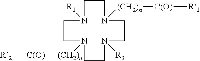


Priority Applications (1)
| Application Number | Priority Date | Filing Date | Title |
|---|---|---|---|
| US14/128,347 US9550001B2 (en) | 2011-06-20 | 2012-06-20 | Compositions, methods of synthesis and use of carbohydrate targeted agents |
Applications Claiming Priority (3)
| Application Number | Priority Date | Filing Date | Title |
|---|---|---|---|
| US201161498950P | 2011-06-20 | 2011-06-20 | |
| US14/128,347 US9550001B2 (en) | 2011-06-20 | 2012-06-20 | Compositions, methods of synthesis and use of carbohydrate targeted agents |
| PCT/US2012/043255 WO2012177701A2 (en) | 2011-06-20 | 2012-06-20 | Compositions, methods of synthesis and use of carbohydrate targeted agents |
Publications (2)
| Publication Number | Publication Date |
|---|---|
| US20140228551A1 true US20140228551A1 (en) | 2014-08-14 |
| US9550001B2 US9550001B2 (en) | 2017-01-24 |
Family
ID=47423183
Family Applications (1)
| Application Number | Title | Priority Date | Filing Date |
|---|---|---|---|
| US14/128,347 Expired - Fee Related US9550001B2 (en) | 2011-06-20 | 2012-06-20 | Compositions, methods of synthesis and use of carbohydrate targeted agents |
Country Status (6)
| Country | Link |
|---|---|
| US (1) | US9550001B2 (en) |
| EP (1) | EP2721045B1 (en) |
| CA (1) | CA2843313C (en) |
| DK (1) | DK2721045T3 (en) |
| PL (1) | PL2721045T3 (en) |
| WO (1) | WO2012177701A2 (en) |
Cited By (8)
| Publication number | Priority date | Publication date | Assignee | Title |
|---|---|---|---|---|
| US9408928B2 (en) | 2007-03-27 | 2016-08-09 | Radiomedix, Inc. | Compositions for targeted imaging and therapy |
| US9550001B2 (en) | 2011-06-20 | 2017-01-24 | Radiomedix Inc. | Compositions, methods of synthesis and use of carbohydrate targeted agents |
| WO2018132751A1 (en) | 2017-01-12 | 2018-07-19 | Radiomedix Inc. | Treatment of cancer cells overexpressing somatostatin receptors using ocreotide derivatives chelated to radioisotopes |
| US10093741B1 (en) | 2017-05-05 | 2018-10-09 | Fusion Pharmaceuticals Inc. | IGF-1R monoclonal antibodies and uses thereof |
| US11191854B2 (en) | 2017-05-05 | 2021-12-07 | Centre For Probe Development And Commercialization | Pharmacokinetic enhancements of bifunctional chelates and uses thereof |
| US11207429B2 (en) * | 2014-05-09 | 2021-12-28 | Arizona Board Of Regents On Behalf Of The University Of Arizona | Method and compositions for orally administered contrast agents for MR imaging |
| US11433148B2 (en) | 2017-05-05 | 2022-09-06 | Centre For Probe Development And Commercialization | IGF-1R monoclonal antibodies and uses thereof |
| CN116655715A (en) * | 2023-07-27 | 2023-08-29 | 北京炫景瑞医药科技有限公司 | A kind of GalNAc derivative, conjugate, composition and their use |
Families Citing this family (8)
| Publication number | Priority date | Publication date | Assignee | Title |
|---|---|---|---|---|
| GB201300707D0 (en) | 2013-01-15 | 2013-02-27 | Novartis Ag | Compounds and processes |
| CA3025988A1 (en) | 2016-06-02 | 2017-12-07 | Sanofi | Conjugates of a pharmaceutical agent and a moiety capable of binding to a glucose sensing protein |
| GB201610738D0 (en) * | 2016-06-20 | 2016-08-03 | Ge Healthcare As | Chelate compounds |
| CN111433193A (en) | 2017-12-01 | 2020-07-17 | 赛诺菲 | Novel conjugates of agents and moieties capable of binding to glucose sensing proteins |
| RU2730507C1 (en) * | 2019-08-27 | 2020-08-24 | Федеральное государственное бюджетное образовательное учреждение высшего образования "Московский государственный университет имени М.В. Ломоносова" (МГУ) | Compound for diagnosing tumours expressing psma and composition based thereon |
| KR20230052958A (en) | 2020-08-19 | 2023-04-20 | 박사이트, 인코포레이티드 | Carrier-protein polysaccharide conjugation method |
| KR20230173150A (en) | 2021-04-20 | 2023-12-26 | 테라파워, 엘엘씨 | Titania-based generator for generating AC-225 |
| CN114853827B (en) * | 2022-06-02 | 2023-11-24 | 南京江原安迪科正电子研究发展有限公司 | Glucose derivative ligand compound and preparation method and application thereof |
Citations (1)
| Publication number | Priority date | Publication date | Assignee | Title |
|---|---|---|---|---|
| US6265483B1 (en) * | 1994-10-05 | 2001-07-24 | Commissariat A L'energie Atomique | Polyazacycloalkanes, tri, tetra- or penta-azamacrocyclic complexes, processes for the production of these substituted or unsubstituted polyazacycloalkanes grafted to a support and uses of polyazacycloalkanes and the aforementioned complexes |
Family Cites Families (42)
| Publication number | Priority date | Publication date | Assignee | Title |
|---|---|---|---|---|
| US4141654A (en) | 1977-05-23 | 1979-02-27 | James V. Massey, III | Standard for calibrating and/or verifying instrument accuracy |
| US5648063A (en) | 1981-07-24 | 1997-07-15 | Schering Aktiengesellschaft | Sterile composition comprising a chelate complex for magnetic resonance imaging |
| US5242679A (en) | 1985-01-14 | 1993-09-07 | Neorx Corporation | Metal radionuclide labeled proteins for diagnosis and therapy |
| US5175343A (en) | 1985-01-14 | 1992-12-29 | Neorx Corporation | Metal radionuclide labeled proteins for diagnosis and therapy |
| US4988496A (en) | 1988-05-31 | 1991-01-29 | Neorx Corporation | Metal radionuclide chelating compounds for improved chelation kinetics |
| ZA894792B (en) | 1988-06-24 | 1991-04-24 | Dow Chemical Co | Macrocyclic bifunctional chelants,complexes thereof and their antibody conjugates |
| US5364613A (en) | 1989-04-07 | 1994-11-15 | Sieving Paul F | Polychelants containing macrocyclic chelant moieties |
| US5605672A (en) | 1993-06-09 | 1997-02-25 | The General Hospital Corporation | Blood pool imaging composition and method of its use |
| US5707605A (en) * | 1995-06-02 | 1998-01-13 | Research Corporation Technologies | Magnetic resonance imaging agents for the detection of physiological agents |
| US6770261B2 (en) * | 1995-06-02 | 2004-08-03 | Research Corporation Technologies | Magnetic resonance imaging agents for the detection of physiological agents |
| US5980862A (en) | 1995-06-02 | 1999-11-09 | Research Corporation Technologies | Magnetic resonance imaging agents for the detection of physiological agents |
| US6713045B1 (en) | 1995-06-02 | 2004-03-30 | Research Corporation Technologies, Inc. | Targeted magnetic resonance imaging agents for the detection of physiological processes |
| US5739313A (en) | 1995-11-13 | 1998-04-14 | Regents Of The University Of Minnesota | Radionuclide labeling of vitamin B12 and coenzymes thereof |
| IT1284046B1 (en) | 1996-06-21 | 1998-05-08 | Bracco Spa | PROCESS FOR THE PREPARATION OF TETRAAZAMACROCYCLES |
| US20040170563A1 (en) | 1997-10-27 | 2004-09-02 | Meade Thomas J. | Magnetic resonance imaging agents for the delivery of therapeutic agents |
| EP1032430A2 (en) | 1997-11-17 | 2000-09-06 | Research Corporation Technologies, Inc. | Magnetic resonance imaging agents for the detection of physiological agents |
| US6071490A (en) | 1998-05-07 | 2000-06-06 | Immunomedics, Inc. | Position emission tomography using gallium-68 chelates |
| WO2000043004A1 (en) | 1999-01-26 | 2000-07-27 | Fox Chase Cancer Center | 1,4,8,11-tetraazacyclotetradecane derivatives as radiodiganostic agents and their use in determining hypoxia and radioresistance of tumors |
| US6692724B1 (en) | 1999-10-25 | 2004-02-17 | Board Of Regents, The University Of Texas System | Ethylenedicysteine (EC)-drug conjugates, compositions and methods for tissue specific disease imaging |
| US6565828B2 (en) | 2000-04-07 | 2003-05-20 | Bristol-Myers Squibb Company | Macrocyclic chelants for metallopharmaceuticals |
| US6673333B1 (en) * | 2000-05-04 | 2004-01-06 | Research Corporation Technologies, Inc. | Functional MRI agents for cancer imaging |
| IL153218A0 (en) | 2000-06-02 | 2003-07-06 | Univ Texas | Ethylenedicysteine (ec) -drug conjugates |
| US6641797B2 (en) | 2000-08-11 | 2003-11-04 | Schering Aktiengesellschaft | Perfluoroalkyl-containing complexes with sugar radicals, process for their production and their use |
| WO2002032291A2 (en) | 2000-10-19 | 2002-04-25 | The General Hospital Corporation | Imaging of enzymatic activity |
| US7138104B2 (en) | 2001-08-08 | 2006-11-21 | Bristol-Myers Squibb Company | Simultaneous imaging of cardiac perfusion and a vitronectin receptor targeted imaging agent |
| WO2003082301A1 (en) | 2002-03-29 | 2003-10-09 | Threshold Pharmaceuticals, Inc. | Compositions and methods for treating cancer |
| US20050232866A1 (en) | 2004-03-26 | 2005-10-20 | Alerion Biomedical Inc. | Biochemically-activated contrast agents for magnetic resonance imaging |
| EP1797106A1 (en) | 2004-09-07 | 2007-06-20 | Triumf, operating as a joint venture by the Governors of the Universities of Alberta, | Synthesis of radiolabeled sugar metal complexes |
| AU2006204045B2 (en) | 2005-01-05 | 2010-10-14 | Board Of Regents, The University Of Texas System | Conjugates for dual imaging and radiochemotherapy: composition, manufacturing, and applications |
| US20060275217A1 (en) | 2005-05-06 | 2006-12-07 | Caravan Peter D | Chemical exchange saturation transfer contrast agents |
| US8845999B2 (en) | 2005-05-23 | 2014-09-30 | The Regents Of The University Of California | Tracers for monitoring the activity of sodium/glucose cotransporters in health and disease |
| US8097237B2 (en) | 2005-08-23 | 2012-01-17 | Stc.Unm | Non-invasive diagnostic agents of cancer and methods of diagnosing cancer, especially leukemia and lymphoma |
| US8758723B2 (en) | 2006-04-19 | 2014-06-24 | The Board Of Regents Of The University Of Texas System | Compositions and methods for cellular imaging and therapy |
| US7586102B2 (en) | 2006-08-14 | 2009-09-08 | Board Of Regents The University Of Texas System | Automated system for formulating radiopharmaceuticals |
| US7842280B2 (en) | 2006-09-06 | 2010-11-30 | Case Western Reserve University | Flexibly labeling peptides |
| MX340566B (en) | 2007-03-27 | 2016-07-14 | Radiomedix Inc | COMPOSITIONS FOR OBJECTIVE IMAGE FORMATION AND THERAPY. |
| WO2009054327A1 (en) | 2007-10-22 | 2009-04-30 | Osaka Foundation For Trade And Industry | Mri probe |
| US20100316566A1 (en) | 2008-02-27 | 2010-12-16 | Radiomedix Inc. | Radiolabeled hedgehog derivatives for imaging and therapy |
| CA2738786C (en) | 2008-09-30 | 2017-12-19 | Mallinckrodt Inc. | A version of fdg detectable by single-photon emission computed tomography |
| US10413621B2 (en) | 2009-10-19 | 2019-09-17 | Case Western Reserve University | Composition and methods for imaging cells |
| PL2721045T3 (en) | 2011-06-20 | 2017-11-30 | Radiomedix Inc. | Compositions, methods of synthesis and use of carbohydrate targeted agents |
| WO2014149069A1 (en) | 2013-03-15 | 2014-09-25 | Purdue Research Foundation | Synthesis and composition of amino acid linking groups conjugated to compounds used for the targeted imaging of tumors |
-
2012
- 2012-06-20 PL PL12803364T patent/PL2721045T3/en unknown
- 2012-06-20 CA CA2843313A patent/CA2843313C/en active Active
- 2012-06-20 WO PCT/US2012/043255 patent/WO2012177701A2/en not_active Ceased
- 2012-06-20 EP EP12803364.4A patent/EP2721045B1/en not_active Not-in-force
- 2012-06-20 US US14/128,347 patent/US9550001B2/en not_active Expired - Fee Related
- 2012-06-20 DK DK12803364.4T patent/DK2721045T3/en active
Patent Citations (1)
| Publication number | Priority date | Publication date | Assignee | Title |
|---|---|---|---|---|
| US6265483B1 (en) * | 1994-10-05 | 2001-07-24 | Commissariat A L'energie Atomique | Polyazacycloalkanes, tri, tetra- or penta-azamacrocyclic complexes, processes for the production of these substituted or unsubstituted polyazacycloalkanes grafted to a support and uses of polyazacycloalkanes and the aforementioned complexes |
Non-Patent Citations (2)
| Title |
|---|
| Altava et al. (Ind. Eng. Chem. Res. 2000, 39, 3589-3595) * |
| Guillier et al. (Chem. Rev. 2000, 100, 2091-2157) * |
Cited By (9)
| Publication number | Priority date | Publication date | Assignee | Title |
|---|---|---|---|---|
| US9408928B2 (en) | 2007-03-27 | 2016-08-09 | Radiomedix, Inc. | Compositions for targeted imaging and therapy |
| US9550001B2 (en) | 2011-06-20 | 2017-01-24 | Radiomedix Inc. | Compositions, methods of synthesis and use of carbohydrate targeted agents |
| US11207429B2 (en) * | 2014-05-09 | 2021-12-28 | Arizona Board Of Regents On Behalf Of The University Of Arizona | Method and compositions for orally administered contrast agents for MR imaging |
| WO2018132751A1 (en) | 2017-01-12 | 2018-07-19 | Radiomedix Inc. | Treatment of cancer cells overexpressing somatostatin receptors using ocreotide derivatives chelated to radioisotopes |
| US11541133B2 (en) | 2017-01-12 | 2023-01-03 | Radiomedixinc. | Treatment of cancer cells overexpressing somatostatin receptors using ocreotide derivatives chelated to radioisotopes |
| US10093741B1 (en) | 2017-05-05 | 2018-10-09 | Fusion Pharmaceuticals Inc. | IGF-1R monoclonal antibodies and uses thereof |
| US11191854B2 (en) | 2017-05-05 | 2021-12-07 | Centre For Probe Development And Commercialization | Pharmacokinetic enhancements of bifunctional chelates and uses thereof |
| US11433148B2 (en) | 2017-05-05 | 2022-09-06 | Centre For Probe Development And Commercialization | IGF-1R monoclonal antibodies and uses thereof |
| CN116655715A (en) * | 2023-07-27 | 2023-08-29 | 北京炫景瑞医药科技有限公司 | A kind of GalNAc derivative, conjugate, composition and their use |
Also Published As
| Publication number | Publication date |
|---|---|
| PL2721045T3 (en) | 2017-11-30 |
| CA2843313A1 (en) | 2012-12-27 |
| EP2721045A2 (en) | 2014-04-23 |
| EP2721045A4 (en) | 2014-11-12 |
| DK2721045T3 (en) | 2017-07-24 |
| CA2843313C (en) | 2018-02-13 |
| US9550001B2 (en) | 2017-01-24 |
| WO2012177701A2 (en) | 2012-12-27 |
| EP2721045B1 (en) | 2017-04-12 |
| WO2012177701A3 (en) | 2013-05-10 |
Similar Documents
| Publication | Publication Date | Title |
|---|---|---|
| US9550001B2 (en) | Compositions, methods of synthesis and use of carbohydrate targeted agents | |
| CN114133434B (en) | Bicyclic peptide nuclide ligands and probes targeting Nectin-4 | |
| CN102911256B (en) | Polypeptides complexe of a kind of radio-labeling and its preparation method and application | |
| CN114773433B (en) | CD25 targeted polypeptide, molecular probe and application | |
| CN111905112A (en) | Polypeptide compound targeting PD-L1 and application thereof | |
| CN110339375B (en) | A kind of rk polypeptide radiopharmaceutical targeting HER2 and preparation method thereof | |
| CN107353323B (en) | Al18F-labeled PSMA targeting inhibitor and preparation method and application thereof | |
| CN110305186B (en) | Prostate cancer PET diagnostic reagent68Ga-DOTA-ANCP-PSMA and preparation method and application thereof | |
| JP7773679B1 (en) | Compounds targeting SSTR2, and methods for their preparation and use | |
| EP4474379A1 (en) | Peptide urea derivative, pharmaceutical composition containing peptide urea derivative, and application of peptide urea derivative | |
| WO2023087871A1 (en) | Granzyme b targeting complex, radiopharmaceutical, preparation method therefor, and use thereof | |
| TW202120016A (en) | Dual-targeted carbonic anhydrase ix complex and a contrast agent thereof | |
| CN110317151B (en) | Prostate cancer PET diagnostic reagent68Ga-HBBED-ANCP-PSMA and preparation method and application thereof | |
| CN117586354A (en) | Targeting GPC3 polypeptide probe and application thereof in preparation of diagnosis and treatment radiopharmaceuticals | |
| US10925981B2 (en) | Hexa-lactoside-triazanonane triacetic acid (NOTA) derivative, method for radiolabeling hexa-lactoside positron emission tomography (PET) imaging agent for liver receptor with Ga-68, and hexa-lactoside PET imaging agent for liver receptor | |
| CN118878582A (en) | A marker precursor and probe for bone metastasis and its preparation method and application | |
| PL239934B1 (en) | Derivatives of PSMA inhibitors for ⁹⁹ᵐTc labelling by HYNIC, radiopharmaceutical kit, radiopharmaceutical preparations and their use in the diagnosis of prostate cancer | |
| CN118852342A (en) | A precursor polypeptide (DOTA) 2-PSMA and (68Ga-DOTA) 2-PSMA molecular probe and its preparation method and application | |
| CN118164956A (en) | Preparation method and application of HSP90 specific radioactive probe | |
| CN118666964A (en) | A PET tracer targeting tumor PD-L1, its labeled precursor, preparation method and application | |
| TWI650138B (en) | Multivalent glyco-complex, imaging agent and uses thereof | |
| CN119101006B (en) | Prostate specific membrane antigen inhibitor, radioactive marker thereof, preparation method and application thereof | |
| TWI852815B (en) | Multivalent glyco-complex, imaging agent and uses thereof | |
| CN114853827B (en) | Glucose derivative ligand compound and preparation method and application thereof | |
| CN116637211B (en) | PET (polyethylene terephthalate) probe coupled with SSTR (SSTR) agonist and inhibitor as well as preparation method and application thereof |
Legal Events
| Date | Code | Title | Description |
|---|---|---|---|
| AS | Assignment |
Owner name: RADIOMEDIX INC., TEXAS Free format text: ASSIGNMENT OF ASSIGNORS INTEREST;ASSIGNORS:TWOROWSKA, IZABELA;SIMS-MOURTADA, JENNIFER;DELPASSAND, EBRAHIM S.;REEL/FRAME:032620/0633 Effective date: 20140304 |
|
| STCF | Information on status: patent grant |
Free format text: PATENTED CASE |
|
| MAFP | Maintenance fee payment |
Free format text: PAYMENT OF MAINTENANCE FEE, 4TH YR, SMALL ENTITY (ORIGINAL EVENT CODE: M2551); ENTITY STATUS OF PATENT OWNER: SMALL ENTITY Year of fee payment: 4 |
|
| AS | Assignment |
Owner name: THE GOVERNMENT OF THE UNITED STATES AS REPRESENTED BY THE SECRETARY OF THE ARMY, MARYLAND Free format text: CONFIRMATORY LICENSE;ASSIGNOR:RADIOMEDIX INC;REEL/FRAME:058609/0921 Effective date: 20200907 |
|
| FEPP | Fee payment procedure |
Free format text: MAINTENANCE FEE REMINDER MAILED (ORIGINAL EVENT CODE: REM.); ENTITY STATUS OF PATENT OWNER: SMALL ENTITY |
|
| LAPS | Lapse for failure to pay maintenance fees |
Free format text: PATENT EXPIRED FOR FAILURE TO PAY MAINTENANCE FEES (ORIGINAL EVENT CODE: EXP.); ENTITY STATUS OF PATENT OWNER: SMALL ENTITY |
|
| STCH | Information on status: patent discontinuation |
Free format text: PATENT EXPIRED DUE TO NONPAYMENT OF MAINTENANCE FEES UNDER 37 CFR 1.362 |
|
| FP | Lapsed due to failure to pay maintenance fee |
Effective date: 20250124 |