US20120184838A1 - Non-invasive deep muscle electromyography - Google Patents
Non-invasive deep muscle electromyography Download PDFInfo
- Publication number
- US20120184838A1 US20120184838A1 US13/387,897 US201013387897A US2012184838A1 US 20120184838 A1 US20120184838 A1 US 20120184838A1 US 201013387897 A US201013387897 A US 201013387897A US 2012184838 A1 US2012184838 A1 US 2012184838A1
- Authority
- US
- United States
- Prior art keywords
- electrodes
- electromyography
- muscle
- muscles
- deep
- Prior art date
- Legal status (The legal status is an assumption and is not a legal conclusion. Google has not performed a legal analysis and makes no representation as to the accuracy of the status listed.)
- Granted
Links
Images

Classifications
-
- A—HUMAN NECESSITIES
- A61—MEDICAL OR VETERINARY SCIENCE; HYGIENE
- A61B—DIAGNOSIS; SURGERY; IDENTIFICATION
- A61B5/00—Measuring for diagnostic purposes; Identification of persons
- A61B5/24—Detecting, measuring or recording bioelectric or biomagnetic signals of the body or parts thereof
- A61B5/25—Bioelectric electrodes therefor
- A61B5/279—Bioelectric electrodes therefor specially adapted for particular uses
- A61B5/296—Bioelectric electrodes therefor specially adapted for particular uses for electromyography [EMG]
-
- A—HUMAN NECESSITIES
- A61—MEDICAL OR VETERINARY SCIENCE; HYGIENE
- A61B—DIAGNOSIS; SURGERY; IDENTIFICATION
- A61B5/00—Measuring for diagnostic purposes; Identification of persons
- A61B5/24—Detecting, measuring or recording bioelectric or biomagnetic signals of the body or parts thereof
- A61B5/316—Modalities, i.e. specific diagnostic methods
- A61B5/389—Electromyography [EMG]
-
- A—HUMAN NECESSITIES
- A61—MEDICAL OR VETERINARY SCIENCE; HYGIENE
- A61B—DIAGNOSIS; SURGERY; IDENTIFICATION
- A61B5/00—Measuring for diagnostic purposes; Identification of persons
- A61B5/45—For evaluating or diagnosing the musculoskeletal system or teeth
- A61B5/4519—Muscles
-
- A—HUMAN NECESSITIES
- A61—MEDICAL OR VETERINARY SCIENCE; HYGIENE
- A61B—DIAGNOSIS; SURGERY; IDENTIFICATION
- A61B5/00—Measuring for diagnostic purposes; Identification of persons
- A61B5/72—Signal processing specially adapted for physiological signals or for diagnostic purposes
- A61B5/7203—Signal processing specially adapted for physiological signals or for diagnostic purposes for noise prevention, reduction or removal
-
- A—HUMAN NECESSITIES
- A61—MEDICAL OR VETERINARY SCIENCE; HYGIENE
- A61B—DIAGNOSIS; SURGERY; IDENTIFICATION
- A61B2562/00—Details of sensors; Constructional details of sensor housings or probes; Accessories for sensors
- A61B2562/04—Arrangements of multiple sensors of the same type
- A61B2562/043—Arrangements of multiple sensors of the same type in a linear array
-
- A—HUMAN NECESSITIES
- A61—MEDICAL OR VETERINARY SCIENCE; HYGIENE
- A61B—DIAGNOSIS; SURGERY; IDENTIFICATION
- A61B5/00—Measuring for diagnostic purposes; Identification of persons
- A61B5/05—Detecting, measuring or recording for diagnosis by means of electric currents or magnetic fields; Measuring using microwaves or radio waves
- A61B5/053—Measuring electrical impedance or conductance of a portion of the body
- A61B5/0536—Impedance imaging, e.g. by tomography
-
- A—HUMAN NECESSITIES
- A61—MEDICAL OR VETERINARY SCIENCE; HYGIENE
- A61B—DIAGNOSIS; SURGERY; IDENTIFICATION
- A61B5/00—Measuring for diagnostic purposes; Identification of persons
- A61B5/103—Measuring devices for testing the shape, pattern, colour, size or movement of the body or parts thereof, for diagnostic purposes
- A61B5/11—Measuring movement of the entire body or parts thereof, e.g. head or hand tremor or mobility of a limb
- A61B5/1107—Measuring contraction of parts of the body, e.g. organ or muscle
-
- A—HUMAN NECESSITIES
- A61—MEDICAL OR VETERINARY SCIENCE; HYGIENE
- A61B—DIAGNOSIS; SURGERY; IDENTIFICATION
- A61B5/00—Measuring for diagnostic purposes; Identification of persons
- A61B5/68—Arrangements of detecting, measuring or recording means, e.g. sensors, in relation to patient
- A61B5/6801—Arrangements of detecting, measuring or recording means, e.g. sensors, in relation to patient specially adapted to be attached to or worn on the body surface
- A61B5/6802—Sensor mounted on worn items
-
- A—HUMAN NECESSITIES
- A61—MEDICAL OR VETERINARY SCIENCE; HYGIENE
- A61B—DIAGNOSIS; SURGERY; IDENTIFICATION
- A61B5/00—Measuring for diagnostic purposes; Identification of persons
- A61B5/68—Arrangements of detecting, measuring or recording means, e.g. sensors, in relation to patient
- A61B5/6801—Arrangements of detecting, measuring or recording means, e.g. sensors, in relation to patient specially adapted to be attached to or worn on the body surface
- A61B5/6813—Specially adapted to be attached to a specific body part
- A61B5/6824—Arm or wrist
-
- A—HUMAN NECESSITIES
- A61—MEDICAL OR VETERINARY SCIENCE; HYGIENE
- A61B—DIAGNOSIS; SURGERY; IDENTIFICATION
- A61B5/00—Measuring for diagnostic purposes; Identification of persons
- A61B5/68—Arrangements of detecting, measuring or recording means, e.g. sensors, in relation to patient
- A61B5/6801—Arrangements of detecting, measuring or recording means, e.g. sensors, in relation to patient specially adapted to be attached to or worn on the body surface
- A61B5/683—Means for maintaining contact with the body
- A61B5/6831—Straps, bands or harnesses
-
- A—HUMAN NECESSITIES
- A61—MEDICAL OR VETERINARY SCIENCE; HYGIENE
- A61B—DIAGNOSIS; SURGERY; IDENTIFICATION
- A61B5/00—Measuring for diagnostic purposes; Identification of persons
- A61B5/72—Signal processing specially adapted for physiological signals or for diagnostic purposes
- A61B5/7235—Details of waveform analysis
- A61B5/7264—Classification of physiological signals or data, e.g. using neural networks, statistical classifiers, expert systems or fuzzy systems
-
- G—PHYSICS
- G06—COMPUTING OR CALCULATING; COUNTING
- G06F—ELECTRIC DIGITAL DATA PROCESSING
- G06F2218/00—Aspects of pattern recognition specially adapted for signal processing
- G06F2218/22—Source localisation; Inverse modelling
Definitions
- This invention relates to non-invasive deep muscle electromyography (EMG) to be used for recording the electrical activity of muscles in order to assess muscle function in a variety of different fields including sports, biomechanics research, physiotherapy and clinical neuro-muscular diagnostics.
- EMG deep muscle electromyography
- the first is a non-invasive surface electromyography (sEMG) in which adhesive bio-electrodes are placed on the surface of the skin directly above the muscle under investigation.
- This category is accordingly limited in application to superficial muscles that are close to the skin's surface.
- the second category is an invasive procedure for obtaining electromyography recordings that utilises fine-wires or needles that physically pierce the skin in order to communicate with a so-called deep muscle that is under investigation. Not only is this technique invasive, but some deep muscles that are located close to major blood vessels, nerves, and viscera should not be investigated at all by such a technique for patient safety reasons.
- Such established invasive EMG techniques are described in a scientific review article by Jasper R Daube and Devon I Rubin (2009) entitled “Needle electromyography” in Muscle & Nerve 39, no. 2: 244-270.
- Invasive electromyography carries with it a number of risks such as the potential for infliction of additional trauma on a patient as well as that of infection. Such risks thus preclude its routine use in sports performance testing and training, particularly for elite athletes who may risk a performance-reducing or livelihood-destroying injury or infection as a result of such a procedure.
- WO 2004107976 there is described a method and device for assessing the function of a deep muscle of a subject in which a patient table, force transducers and an ultrasound device are employed for visualising the deep muscle of interest.
- a computer may be connected to the transducers and ultrasound device to provide cues to the subject during the conduct of an assessment session and to analyse information obtained from the session.
- the device may also include one or more slings for supporting a limb of the subject.
- U.S. Pat. No. 6,185,451 there is described a method and apparatus for assessing the function of deep joint stabilizing muscles in which superficial muscles are monitored using electromyography during performance of an activity known to require recruitment, primarily, of deep stabilizing muscles when performed correctly. If the deep muscle functions adequately, there is little activity of the superficial muscles. Conversely, if the deep muscle function is inadequate, the superficial muscle activity is increased. Monitoring of the superficial muscles using electromyography may be combined with monitoring of the deep joint stabilizing muscle using ultrasound imaging and/or pressure biofeedback.
- the apparatus includes a surface electromyography unit, an ultrasound unit, a pressure biofeedback unit and vitalograph, in combination with a computer programmed to analyze data from them and given an indication of function.
- the sEMG of five specific deep muscles may be recorded on the surface using conventional sEMG methods, with a 15-20% error, as demonstrated by McGill et al (1996, J. Biomechanics, 29(11), p 1503-1507) who recorded psoas, quadratus lumborum, external & internal obliques and transversus abdominus muscles. However this is only applicable to those specific muscles and not generalizable to all deep muscles.
- a method of conducting electromyography of a deep muscle non-invasively comprising the application of an array of suitable surface electromyography electrodes to the skin of a patient and recording electrical potentials of selected electrodes relative to at least one other electrode, the method being characterised in that the array of electrodes is arranged in one or more rings encircling a part of the human body in which a deep muscle being investigated is located, recording the potential of at least selected electrodes relative to another electrode selected from a common reference electrode (mono-polar) and other electrodes (bi-polar) in the array; and processing the recorded potentials of at least some of said selected electrodes in order to determine (optionally using approximations or algorithms, or both) the contribution being made by at least said deep muscle being investigated.
- a common reference electrode mono-polar
- other electrodes bi-polar
- Further features of the invention provide for the contributions to the potentials of the electrodes attributable to each of the encircled muscles including superficial muscles to be determined, typically mathematically by resolving the electromyography signals into their constituent components using independent component analysis (ICA) or any other suitable un-mixing or matrix inversion technique to result in the derivation of the electromyography waveforms of both deep and superficial muscles.
- ICA independent component analysis
- the deep muscles may be indentified by selecting specific movement protocols that may be similar to those used to identify muscles by conventional (sEMG or needle/fine-wire) techniques.
- Still further features of the invention provide for the results of a method as defined above to be integrated with those of a static muscle imaging device that may use the same electrodes to obtain a static tomogram of the muscles encircled; and for the static tomogram to be co-registered with the dipole decomposition or any other suitable 2D or 3D electrical source localisation technique of the ICA-sEMG waveforms.
- the static muscle imaging device may be an electrical impedance tomography device, an ultrasound device, computed tomography device (CT), or a magnetic resonance imaging device (MRI).
- the invention also provides apparatus for carrying out a method as defined above, comprising multiple surface electromyography electrodes having flexible conductors connecting them individually to recording means and computing means adapted to carry out an independent component analysis, or any other suitable un-mixing or matrix inversion technique, on the potentials of the electrodes recorded in order to provide the electromyography waveform of at least one deep muscle encircled by one or more rings of multiple electrodes secured to the skin of the human body, in use.
- FIG. 1 is a schematic side illustration of a human arm illustrating the general positions of the various electrodes
- FIG. 2 a is a schematic section taken through the brachialis region of the arm and showing a single ring of electrodes encircling the arm;
- FIG. 2 b is the same schematic section taken through the brachialis region of the arm and showing two spaced rings of electrodes encircling the arm;
- FIG. 3 is a schematic illustration of the operation of the invention in comparison to that of the prior art
- FIG. 4 is a representation of a static electrical impedance tomogram developed by the processing apparatus
- FIG. 5 is a representation of the electrical impedance tomogram (EIT) of the regions corresponding to the human arm cross-section of FIGS. 2 and 4 co-registered with computed dipoles;
- FIG. 6 is a representation of the five successive stages of the movement protocol used to isolate superficial (biceps) from deep (brachialis) muscle activity, for preliminary clinical results;
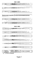
- FIG. 7 illustrates un-processed mono-polar sEMG recordings from preliminary clinical results, using a 2 ring system with 6 electrodes per ring, following the movement protocol represented by FIG. 6 but with a 5 kg bar-bell;
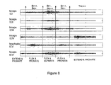
- FIG. 8 illustrates the first six ICA-sEMG components derived from the sEMG recordings from FIG. 7 , from preliminary clinical results;
- FIG. 9 illustrates the root mean square (RMS) values of the ICA-sEMG components of FIG. 8 during each of the five movement stages.
- RMS root mean square
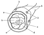
- the example of electromyography of the deep muscle brachialis (c) of the upper arm ( 2 ) as applied to an actual human subject in a laboratory setting is given.
- two arrays of suitable surface electromyography electrodes ( 3 ) are applied to the skin of a patient with the electrodes being arranged in two rings encircling the group of muscles in the appropriate part of the upper arm in which the relevant part of the brachialis is located.
- the two arrays of electrodes comprise a total of 12 electrodes, with 6 electrodes per ring that are arranged generally equally spaced around the periphery of the arm.
- the electrodes that are indicated in FIG. 3 by the Roman numerals (I, II, III, IV, V, VI, VII. VIII, IX, X, XI, XII) sequentially in a clockwise direction around the two rings, are connected to a processing unit ( 4 ) by means of flexible conductors ( 5 ) in the usual way.
- FIG. 3 In so far as FIG. 3 is concerned only one pair of conductors is illustrated as being connected to the electrodes and processing unit, the relevant electrode being that indicated by Roman numeral I.
- a common reference electrode is also usually placed outside the ring, for example on the bony part of the elbow as indicated by numeral ( 3 a ) in FIG. 1 , although it may sometimes be suitable to use one of the ring electrodes as the common reference.
- a ground electrode is placed on a suitable sEMG ground point (e.g. elbow, wrist, etc) for the muscles under investigation. This manner of illustration is employed for the purpose of simplifying the illustration.
- the mono-polar potential of all electrodes relative to the common reference electrode are fed to one of a corresponding set of differential amplifiers ( 6 ), the outputs of which are all fed to a computing device ( 7 ) such as a personal computer or a programmable micro-controller that is programmed to compute the independent component analysis (ICA) or any other suitable un-mixing or matrix inversion technique.
- ICA independent component analysis
- This may be done using approximations or algorithms, or both, and typically along general lines for ICA described by Klemm, Matthias, Jens Haueisen, and Galina Ivanova. in an article entitled “Independent component analysis: comparison of algorithms for the investigation of surface electrical brain activity” in Medical & Biological Engineering & Computing 47, no. 4 (April 2009): 413-423 or any other suitable un-mixing or matrix inversion technique.
- Open-source software called ‘EEGLAB’ that implements some of the ICA algorithms can also be used as may be appropriate.
- the recorded potentials of the electrodes are processed in order to determine the contribution made by at least said deep muscle that is under investigation and generally each of the muscles encircled by the array of electrodes.
- the computing device may also decompose the differential signals and perform other routine software functions such as archiving the data etc.
- the apparatus typically includes a display device ( 8 ) such as a computer monitor that can display the corresponding electromyography waveforms.
- a display device such as a computer monitor that can display the corresponding electromyography waveforms.
- the electromyography device of this invention may thus be used to contemporaneously derive an electromyograph corresponding to both superficial muscles indicated by letters (a, b, d, e) and the deep brachialis muscle (c).
- Prior art invasive needle electromyography of the deep muscle (c), on the other hand, is illustrated schematically by numeral ( 9 ) in which the potential of the deep muscle is measured relative to a surface reference electrode.
- each muscle (a, b, c, d, e) may be measured simultaneously.
- These electrical activities may be represented as Ma, Mb, Mc, Md, and Me in which instance each mono-polar channel would include a weighted sum of components from each (superficial and deep) muscle source, ie.
- sEMG( VII ) AVII.Ma+BVII.Mb+CVII.Mc+DVII.Mc+VII.Md
- ICA independent component analysis
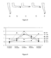
- Each movement should take about 3 seconds and the movement from (A) to (B) activates the brachialis more than the biceps; the movement from (B) to (C) activates the biceps and reduces the brachialis; the movement from (C) to (D) activates the brachialis and reduces the biceps; and the movement from (D) to (E) activates the triceps.
- FIG. 7 illustrates the mono-polar sEMG (voltage in millivolts vs time in seconds) preliminary recordings from two rings positioned as illustrated in FIG. 2 b in which each of the rings had six approximately equally spaced electrodes.
- the movement protocol was as described with reference to FIG. 6 . It is to be noted that these mono-polar sEMG waveforms were dominated by biceps activity (most apparent during SUPINATE) and look similar to each other.
- FIG. 8 illustrates the ICA transformation of the preliminary recordings shown in FIG. 7 and shows the first 6 ICA components (voltage in millivolts vs time in seconds).
- the brachialis component activates on elbow flexion (FLEX) as well as on forearm pronation (PRONATE) and reduces on forearm supination (SUPINATE).
- the biceps consists of 4 components (ICI-ICIII, ICVI) that may be added together, all showing activation on flexion (FLEX), greater activation on forearm supination (SUPINATE) and reduced activation on forearm pronation (PRONATE).
- the triceps component shows activation on extension.
- FIG. 8 thus illustrates that the difference between SUPINATE and PRONATE may be used to identify brachialis (ICV in FIG. 8 ) and biceps (ICI-ICIII, ICVI). Similarly the difference between the elbow flexed conditions (FLEX, SUPINATE, PRONATE) and the end EXTEND condition may be used to identify triceps (ICIV in FIG. 8 ).
- a known static muscle imaging device such as muscle electrical impedance tomography (EIT) that uses the same electrodes as the sEMG ring, may then be applied to obtain a static tomogram of the muscles as illustrated in FIG. 4 and the static tomogram may be co-registered with the dipole decomposition or the electrical source localisation of the ICA-sEMG waveforms derived as described above (see FIG. 5 ) and as described further below.
- EIT muscle electrical impedance tomography
- the ICA-sEMG waveforms may be converted to their corresponding spatial locations (i.e. within the cross-sectional area of the circular electrode ring or rings) using electrical dipole analysis that, as far as applicant is aware, has not previously been applied to electromyography or standard electrical source localisation techniques including those implemented by Jesinger & Stonic (1994) or van den Doel (2008).
- This allows the muscle dipole or electrical source representing the ICA-sEMG to be tagged to a location in the cross-section.
- the dipole or electrical source cross-section may be co-registered with the muscle EIT to identify the location of the ICA-sEMG sources, hence also the identity of the deep muscle.
- FIG. 9 illustrates the root mean square (RMS) values of the ICA-sEMG components during each of the five movement stages, from preliminary clinical results. These values share the same activation and reduction patterns with the biceps being represented by VI; the brachialis by ICV; and the triceps by ICIV.
- RMS root mean square
- the apparatus described for developing the separate electromyographs may be a device on its own, where the ICA-sEMG may be related to specific muscles through pre-defined movement protocols i.e. a protocol that is known to activate a certain deep muscle would therefore also result in a corresponding ICA-sEMG waveform that mimics the activation of that muscle during that movement.
- the apparatus is used in conjunction with an electronic imaging device, the EIT device as indicated above such imaging device could be replaced by another static imaging technology such as those identified above. At least some of those technologies may be expected to provide much more detailed images than EIT but may be cumbersome to use in environments requiring movement typical of functional muscular studies.
- the invention may become usable, or at least approved for use in specific areas where its accuracy has been verified.
- ICA results in statistically independent channels whereas there is not necessarily a one-to-one relationship between an ICA-sEMG component and a specific muscle.
- An ICA-sEMG channel alone may not necessarily be sufficient to spatially localise a source (hence to identify the individual muscle), as its activity may be included in more than one ICA-sEMG component.
- the number of rings of electrodes can be varied with a minimum of one.
- the potential of each electrode need not be measured relative to a reference electrode that the electrodes could be used in a bipolar system in which the potential of electrodes is measured relative to other active electrodes such as a diametrically opposite electrode. Numerous other variations may be made.
Landscapes
- Health & Medical Sciences (AREA)
- Life Sciences & Earth Sciences (AREA)
- Engineering & Computer Science (AREA)
- General Health & Medical Sciences (AREA)
- Veterinary Medicine (AREA)
- Biophysics (AREA)
- Biomedical Technology (AREA)
- Heart & Thoracic Surgery (AREA)
- Medical Informatics (AREA)
- Molecular Biology (AREA)
- Surgery (AREA)
- Animal Behavior & Ethology (AREA)
- Physics & Mathematics (AREA)
- Public Health (AREA)
- Pathology (AREA)
- Signal Processing (AREA)
- Artificial Intelligence (AREA)
- Computer Vision & Pattern Recognition (AREA)
- Physiology (AREA)
- Psychiatry (AREA)
- Dentistry (AREA)
- Oral & Maxillofacial Surgery (AREA)
- Orthopedic Medicine & Surgery (AREA)
- Rheumatology (AREA)
- Measurement And Recording Of Electrical Phenomena And Electrical Characteristics Of The Living Body (AREA)
Abstract
Description
- This invention relates to non-invasive deep muscle electromyography (EMG) to be used for recording the electrical activity of muscles in order to assess muscle function in a variety of different fields including sports, biomechanics research, physiotherapy and clinical neuro-muscular diagnostics.
- There are presently two categories of electromyography recordings. The first is a non-invasive surface electromyography (sEMG) in which adhesive bio-electrodes are placed on the surface of the skin directly above the muscle under investigation. This category is accordingly limited in application to superficial muscles that are close to the skin's surface.
- The second category is an invasive procedure for obtaining electromyography recordings that utilises fine-wires or needles that physically pierce the skin in order to communicate with a so-called deep muscle that is under investigation. Not only is this technique invasive, but some deep muscles that are located close to major blood vessels, nerves, and viscera should not be investigated at all by such a technique for patient safety reasons. Such established invasive EMG techniques are described in a scientific review article by Jasper R Daube and Devon I Rubin (2009) entitled “Needle electromyography” in Muscle & Nerve 39, no. 2: 244-270.
- Invasive electromyography carries with it a number of risks such as the potential for infliction of additional trauma on a patient as well as that of infection. Such risks thus preclude its routine use in sports performance testing and training, particularly for elite athletes who may risk a performance-reducing or livelihood-destroying injury or infection as a result of such a procedure.
- There have been previous attempts to investigate deep muscles non-invasively. In international patent publication number WO 2004107976 there is described a method and device for assessing the function of a deep muscle of a subject in which a patient table, force transducers and an ultrasound device are employed for visualising the deep muscle of interest. A computer may be connected to the transducers and ultrasound device to provide cues to the subject during the conduct of an assessment session and to analyse information obtained from the session. The device may also include one or more slings for supporting a limb of the subject.
- In U.S. Pat. No. 6,185,451 there is described a method and apparatus for assessing the function of deep joint stabilizing muscles in which superficial muscles are monitored using electromyography during performance of an activity known to require recruitment, primarily, of deep stabilizing muscles when performed correctly. If the deep muscle functions adequately, there is little activity of the superficial muscles. Conversely, if the deep muscle function is inadequate, the superficial muscle activity is increased. Monitoring of the superficial muscles using electromyography may be combined with monitoring of the deep joint stabilizing muscle using ultrasound imaging and/or pressure biofeedback. The apparatus includes a surface electromyography unit, an ultrasound unit, a pressure biofeedback unit and vitalograph, in combination with a computer programmed to analyze data from them and given an indication of function.
- The sEMG of five specific deep muscles may be recorded on the surface using conventional sEMG methods, with a 15-20% error, as demonstrated by McGill et al (1996, J. Biomechanics, 29(11), p 1503-1507) who recorded psoas, quadratus lumborum, external & internal obliques and transversus abdominus muscles. However this is only applicable to those specific muscles and not generalizable to all deep muscles.
- Jesinger & Stonic (1994, IEEE DSP Workshop Proc., p 57-60) proposed an inverse finite element modeling (FEM) method to resolve superficial from deep muscle activity. However this method requires a priori information such as a multiple-slice MRI image of the body segment. Such inverse FEM models do not provide unique mathematical solutions unless there are a large number (>>100) of measurement points, and despite being introduced in 1996, to date there is no clinical evidence that this method is practically able to resolve deep from superficial muscle activity. Using the same approach, now termed ‘computed myography’, but with a number of mathematical approximations, van den Doel et al (2008, Inverse Problems, 24, p 1-17) attempted to resolve biceps, brachialis, and triceps activity but was only successful in demonstrating biceps and triceps activity resolution since their posited brachialis component behaved identically to biceps. Triceps and biceps are however both antagonistic superficial muscles that are easily recorded and differentiated using standard sEMG anyway. This inverse FEM method therefore appears to be more suited to academic computer simulated experiments as opposed to a being a viable clinical investigation tool.
- In summary, as far as applicants are aware, aside from five specific deep muscles (McGill et al, 1996), deep muscle activity may currently only be actually recorded using the already established invasive electromyography techniques, and the two hypothesised techniques suggested by the non-invasive patents involving relating deep muscle activity to ultrasound imaging of changes in width and indirect inference of deep muscle activity by monitoring co-agonist superficial muscle EMG as outlined above.
- It is an object of this invention to provide a method and apparatus for conducting electromyography of a deep muscle non-invasively.
- In accordance with this invention there is provided a method of conducting electromyography of a deep muscle non-invasively comprising the application of an array of suitable surface electromyography electrodes to the skin of a patient and recording electrical potentials of selected electrodes relative to at least one other electrode, the method being characterised in that the array of electrodes is arranged in one or more rings encircling a part of the human body in which a deep muscle being investigated is located, recording the potential of at least selected electrodes relative to another electrode selected from a common reference electrode (mono-polar) and other electrodes (bi-polar) in the array; and processing the recorded potentials of at least some of said selected electrodes in order to determine (optionally using approximations or algorithms, or both) the contribution being made by at least said deep muscle being investigated.
- Further features of the invention provide for the contributions to the potentials of the electrodes attributable to each of the encircled muscles including superficial muscles to be determined, typically mathematically by resolving the electromyography signals into their constituent components using independent component analysis (ICA) or any other suitable un-mixing or matrix inversion technique to result in the derivation of the electromyography waveforms of both deep and superficial muscles. The deep muscles may be indentified by selecting specific movement protocols that may be similar to those used to identify muscles by conventional (sEMG or needle/fine-wire) techniques.
- Still further features of the invention provide for the results of a method as defined above to be integrated with those of a static muscle imaging device that may use the same electrodes to obtain a static tomogram of the muscles encircled; and for the static tomogram to be co-registered with the dipole decomposition or any other suitable 2D or 3D electrical source localisation technique of the ICA-sEMG waveforms. The static muscle imaging device may be an electrical impedance tomography device, an ultrasound device, computed tomography device (CT), or a magnetic resonance imaging device (MRI).
- The invention also provides apparatus for carrying out a method as defined above, comprising multiple surface electromyography electrodes having flexible conductors connecting them individually to recording means and computing means adapted to carry out an independent component analysis, or any other suitable un-mixing or matrix inversion technique, on the potentials of the electrodes recorded in order to provide the electromyography waveform of at least one deep muscle encircled by one or more rings of multiple electrodes secured to the skin of the human body, in use.
- Further features of the method and apparatus according to the invention will become more apparent from the following expanded description of the invention and its presently envisaged implementation, the description being made with reference to the accompanying drawings.
- In the drawings:—
-
FIG. 1 is a schematic side illustration of a human arm illustrating the general positions of the various electrodes; -
FIG. 2 a is a schematic section taken through the brachialis region of the arm and showing a single ring of electrodes encircling the arm; -
FIG. 2 b is the same schematic section taken through the brachialis region of the arm and showing two spaced rings of electrodes encircling the arm; -
FIG. 3 is a schematic illustration of the operation of the invention in comparison to that of the prior art; -
FIG. 4 is a representation of a static electrical impedance tomogram developed by the processing apparatus; -
FIG. 5 is a representation of the electrical impedance tomogram (EIT) of the regions corresponding to the human arm cross-section ofFIGS. 2 and 4 co-registered with computed dipoles; -
FIG. 6 is a representation of the five successive stages of the movement protocol used to isolate superficial (biceps) from deep (brachialis) muscle activity, for preliminary clinical results; -
FIG. 7 illustrates un-processed mono-polar sEMG recordings from preliminary clinical results, using a 2 ring system with 6 electrodes per ring, following the movement protocol represented byFIG. 6 but with a 5 kg bar-bell; -
FIG. 8 illustrates the first six ICA-sEMG components derived from the sEMG recordings fromFIG. 7 , from preliminary clinical results; and, -
FIG. 9 illustrates the root mean square (RMS) values of the ICA-sEMG components ofFIG. 8 during each of the five movement stages. - In one implementation of the invention the example of electromyography of the deep muscle brachialis (c) of the upper arm (2) as applied to an actual human subject in a laboratory setting, is given. In this implementation of the invention two arrays of suitable surface electromyography electrodes (3) are applied to the skin of a patient with the electrodes being arranged in two rings encircling the group of muscles in the appropriate part of the upper arm in which the relevant part of the brachialis is located.
- In this particular instance, the two arrays of electrodes comprise a total of 12 electrodes, with 6 electrodes per ring that are arranged generally equally spaced around the periphery of the arm. The electrodes, that are indicated in
FIG. 3 by the Roman numerals (I, II, III, IV, V, VI, VII. VIII, IX, X, XI, XII) sequentially in a clockwise direction around the two rings, are connected to a processing unit (4) by means of flexible conductors (5) in the usual way. - In so far as
FIG. 3 is concerned only one pair of conductors is illustrated as being connected to the electrodes and processing unit, the relevant electrode being that indicated by Roman numeral I. A common reference electrode is also usually placed outside the ring, for example on the bony part of the elbow as indicated by numeral (3 a) inFIG. 1 , although it may sometimes be suitable to use one of the ring electrodes as the common reference. A ground electrode is placed on a suitable sEMG ground point (e.g. elbow, wrist, etc) for the muscles under investigation. This manner of illustration is employed for the purpose of simplifying the illustration. - The mono-polar potential of all electrodes relative to the common reference electrode, are fed to one of a corresponding set of differential amplifiers (6), the outputs of which are all fed to a computing device (7) such as a personal computer or a programmable micro-controller that is programmed to compute the independent component analysis (ICA) or any other suitable un-mixing or matrix inversion technique. This may be done using approximations or algorithms, or both, and typically along general lines for ICA described by Klemm, Matthias, Jens Haueisen, and Galina Ivanova. in an article entitled “Independent component analysis: comparison of algorithms for the investigation of surface electrical brain activity” in Medical & Biological Engineering & Computing 47, no. 4 (April 2009): 413-423 or any other suitable un-mixing or matrix inversion technique. Open-source software called ‘EEGLAB’ that implements some of the ICA algorithms can also be used as may be appropriate.
- In this manner the recorded potentials of the electrodes are processed in order to determine the contribution made by at least said deep muscle that is under investigation and generally each of the muscles encircled by the array of electrodes. The computing device may also decompose the differential signals and perform other routine software functions such as archiving the data etc.
- The apparatus typically includes a display device (8) such as a computer monitor that can display the corresponding electromyography waveforms.
- The electromyography device of this invention may thus be used to contemporaneously derive an electromyograph corresponding to both superficial muscles indicated by letters (a, b, d, e) and the deep brachialis muscle (c).
- It is to be noted that in conventional (non-invasive) surface electromyography of a superficial muscle such as that indicated by the letter (a) the potential between two adjacent electrodes indicated by Roman numerals (I) and (II) would be measured.
- Prior art invasive needle electromyography of the deep muscle (c), on the other hand, is illustrated schematically by numeral (9) in which the potential of the deep muscle is measured relative to a surface reference electrode.
- In exercising the present invention, however, the electrical activity of each muscle (a, b, c, d, e) may be measured simultaneously. These electrical activities may be represented as Ma, Mb, Mc, Md, and Me in which instance each mono-polar channel would include a weighted sum of components from each (superficial and deep) muscle source, ie.
-
sEMG(I)=AI.Ma+BI.Mb+CI.Mc+DI.Mc+EI.Md -
sEMG(II)=AII.Ma+BII.Mb+CII.Mc+DII.Mc+EII.Md -
sEMG(III)=AIII.Ma+BIII.Mb+CIII.Mc+DIII.Mc+EIII.Md -
sEMG(IV)=AIV.Ma+BIV.Mb+CIV.Mc+DIV.Mc+EIV.Md -
sEMG(V)=AV.Ma+BV.Mb+CV.Mc+DV.Mc+EV.Md -
sEMG(VI)=AVI.Ma+BVI.Mb+CVI.Mc+DVI.Mc+EVI.Md -
sEMG(VII)=AVII.Ma+BVII.Mb+CVII.Mc+DVII.Mc+VII.Md -
sEMG(VIII)=AVIII.Ma+BVIII.Mb+CVIII.Mc+DVIII.Mc+EVIII.Md -
sEMG(IX)=AIX.Ma+BIX.Mb+CIX.Mc+DIX.Mc+EIX.Md -
sEMG(X)=AX.Ma+BX.Mb+CX.Mc+DX.Mc+EX.Md -
sEMG(XI)=AXI.Ma+BXI.Mb+CXI.Mc+DXI.Mc+EXI.Md -
sEMG(XII)=AXII.Ma+BXII.Mb+CXII.Mc+DXII.Mc+EXII.Md - If each muscle activation were statistically independent (see discussions on independent component analysis (ICA) limitations below), then ICA may be applied to solve the system of 12 equations for the 5 unknowns. Note that similar ICA components (as measured by e.g. correlation) may be added together to form a single component such as may be done with the four biceps ICAs (ICI-ICIII, ICVI) in
FIG. 8 and this process would ideally unmix the twelve equations such that: -
ICA-sEMG(a)=Ma -
ICA-sEMG(b)=Mb -
ICA-sEMG(c)=Mc -
ICA-sEMG(d)=Md -
ICA-sEMG(e)=Me - The temporal variation of these ICA-sEMG waveforms with pre-determined movement protocols designed to differentiate the deep muscle under investigation from adjacent muscles, is sufficient to determine the identity of the muscle.
- An example of this is provided using the movement protocol of
FIG. 6 in which the sequence is illustrated commencing with the arm in an extended condition as indicated by (A); flexing it to a PRONATE position as indicated by (B); rotating the hand to a SUPINATE position as indicated by (C); returning to the PRONATE position as indicated by (D) and then extending the arm once more as indicated by (E). Each movement should take about 3 seconds and the movement from (A) to (B) activates the brachialis more than the biceps; the movement from (B) to (C) activates the biceps and reduces the brachialis; the movement from (C) to (D) activates the brachialis and reduces the biceps; and the movement from (D) to (E) activates the triceps. -
FIG. 7 illustrates the mono-polar sEMG (voltage in millivolts vs time in seconds) preliminary recordings from two rings positioned as illustrated inFIG. 2 b in which each of the rings had six approximately equally spaced electrodes. The movement protocol was as described with reference toFIG. 6 . It is to be noted that these mono-polar sEMG waveforms were dominated by biceps activity (most apparent during SUPINATE) and look similar to each other. -
FIG. 8 illustrates the ICA transformation of the preliminary recordings shown inFIG. 7 and shows the first 6 ICA components (voltage in millivolts vs time in seconds). It is to be noted that the brachialis component activates on elbow flexion (FLEX) as well as on forearm pronation (PRONATE) and reduces on forearm supination (SUPINATE). The biceps consists of 4 components (ICI-ICIII, ICVI) that may be added together, all showing activation on flexion (FLEX), greater activation on forearm supination (SUPINATE) and reduced activation on forearm pronation (PRONATE). The triceps component shows activation on extension. -
FIG. 8 thus illustrates that the difference between SUPINATE and PRONATE may be used to identify brachialis (ICV inFIG. 8 ) and biceps (ICI-ICIII, ICVI). Similarly the difference between the elbow flexed conditions (FLEX, SUPINATE, PRONATE) and the end EXTEND condition may be used to identify triceps (ICIV inFIG. 8 ). - If complementary muscle identifying information is required, a known static muscle imaging device such as muscle electrical impedance tomography (EIT) that uses the same electrodes as the sEMG ring, may then be applied to obtain a static tomogram of the muscles as illustrated in
FIG. 4 and the static tomogram may be co-registered with the dipole decomposition or the electrical source localisation of the ICA-sEMG waveforms derived as described above (seeFIG. 5 ) and as described further below. - The ICA-sEMG waveforms may be converted to their corresponding spatial locations (i.e. within the cross-sectional area of the circular electrode ring or rings) using electrical dipole analysis that, as far as applicant is aware, has not previously been applied to electromyography or standard electrical source localisation techniques including those implemented by Jesinger & Stonic (1994) or van den Doel (2008). This allows the muscle dipole or electrical source representing the ICA-sEMG to be tagged to a location in the cross-section. The dipole or electrical source cross-section may be co-registered with the muscle EIT to identify the location of the ICA-sEMG sources, hence also the identity of the deep muscle.
-
FIG. 9 illustrates the root mean square (RMS) values of the ICA-sEMG components during each of the five movement stages, from preliminary clinical results. These values share the same activation and reduction patterns with the biceps being represented by VI; the brachialis by ICV; and the triceps by ICIV. - It is to be noted that the apparatus described for developing the separate electromyographs may be a device on its own, where the ICA-sEMG may be related to specific muscles through pre-defined movement protocols i.e. a protocol that is known to activate a certain deep muscle would therefore also result in a corresponding ICA-sEMG waveform that mimics the activation of that muscle during that movement.
- Also, if the apparatus is used in conjunction with an electronic imaging device, the EIT device as indicated above such imaging device could be replaced by another static imaging technology such as those identified above. At least some of those technologies may be expected to provide much more detailed images than EIT but may be cumbersome to use in environments requiring movement typical of functional muscular studies.
- As with all new medical computing techniques the invention may become usable, or at least approved for use in specific areas where its accuracy has been verified.
- It is also to be noted that ICA results in statistically independent channels whereas there is not necessarily a one-to-one relationship between an ICA-sEMG component and a specific muscle. An ICA-sEMG channel alone may not necessarily be sufficient to spatially localise a source (hence to identify the individual muscle), as its activity may be included in more than one ICA-sEMG component.
- Options to overcome this potential limitation include combining ICA-sEMG components that are similar (such as described earlier) by using correlation or any other mathematically equivalent technique; the application of multiple dipole localization techniques such as have been applied to EEG to allow each ICA component to be spatially localized to a single or multiple source; two-dimensional or three-dimensional electrical source localisation techniques. Hence the EMG sourced from specific spatial locations (hence the specific muscles) may be reconstructed using a combination of ICA or any other suitable un-mixing or matrix inversion technique, multiple dipole source localization or any other electrical source technique, superimposed on, for example, a cross sectional tomogram of the region under investigation.
- Numerous variations may be made to the method and apparatus described above without departing from the scope hereof. In particular, it is to be noted that the number of rings of electrodes can be varied with a minimum of one. Also, the potential of each electrode need not be measured relative to a reference electrode that the electrodes could be used in a bipolar system in which the potential of electrodes is measured relative to other active electrodes such as a diametrically opposite electrode. Numerous other variations may be made.
Claims (8)
Applications Claiming Priority (3)
| Application Number | Priority Date | Filing Date | Title |
|---|---|---|---|
| ZA200905309 | 2009-07-30 | ||
| ZA2009/05309 | 2009-07-30 | ||
| PCT/IB2010/001876 WO2011012988A1 (en) | 2009-07-30 | 2010-07-29 | Non-invasive deep muscle electromyography |
Publications (2)
| Publication Number | Publication Date |
|---|---|
| US20120184838A1 true US20120184838A1 (en) | 2012-07-19 |
| US9687168B2 US9687168B2 (en) | 2017-06-27 |
Family
ID=43528829
Family Applications (1)
| Application Number | Title | Priority Date | Filing Date |
|---|---|---|---|
| US13/387,897 Expired - Fee Related US9687168B2 (en) | 2009-07-30 | 2010-07-29 | Non-invasive deep muscle electromyography |
Country Status (6)
| Country | Link |
|---|---|
| US (1) | US9687168B2 (en) |
| EP (1) | EP2459062A4 (en) |
| JP (1) | JP5662443B2 (en) |
| CN (1) | CN102573620B (en) |
| WO (1) | WO2011012988A1 (en) |
| ZA (1) | ZA201109253B (en) |
Cited By (17)
| Publication number | Priority date | Publication date | Assignee | Title |
|---|---|---|---|---|
| WO2019079757A1 (en) | 2017-10-19 | 2019-04-25 | Ctrl-Labs Corporation | Systems and methods for identifying biological structures associated with neuromuscular source signals |
| CN109948640A (en) * | 2018-12-26 | 2019-06-28 | 杭州电子科技大学 | Electromyographic signal classification method based on two-parameter core Optimization-type extreme learning machine |
| US11481031B1 (en) | 2019-04-30 | 2022-10-25 | Meta Platforms Technologies, Llc | Devices, systems, and methods for controlling computing devices via neuromuscular signals of users |
| US11481030B2 (en) | 2019-03-29 | 2022-10-25 | Meta Platforms Technologies, Llc | Methods and apparatus for gesture detection and classification |
| US11493993B2 (en) | 2019-09-04 | 2022-11-08 | Meta Platforms Technologies, Llc | Systems, methods, and interfaces for performing inputs based on neuromuscular control |
| US11567573B2 (en) | 2018-09-20 | 2023-01-31 | Meta Platforms Technologies, Llc | Neuromuscular text entry, writing and drawing in augmented reality systems |
| US11644799B2 (en) | 2013-10-04 | 2023-05-09 | Meta Platforms Technologies, Llc | Systems, articles and methods for wearable electronic devices employing contact sensors |
| US11666264B1 (en) | 2013-11-27 | 2023-06-06 | Meta Platforms Technologies, Llc | Systems, articles, and methods for electromyography sensors |
| US11797087B2 (en) | 2018-11-27 | 2023-10-24 | Meta Platforms Technologies, Llc | Methods and apparatus for autocalibration of a wearable electrode sensor system |
| US11868531B1 (en) | 2021-04-08 | 2024-01-09 | Meta Platforms Technologies, Llc | Wearable device providing for thumb-to-finger-based input gestures detected based on neuromuscular signals, and systems and methods of use thereof |
| US11907423B2 (en) | 2019-11-25 | 2024-02-20 | Meta Platforms Technologies, Llc | Systems and methods for contextualized interactions with an environment |
| US11921471B2 (en) | 2013-08-16 | 2024-03-05 | Meta Platforms Technologies, Llc | Systems, articles, and methods for wearable devices having secondary power sources in links of a band for providing secondary power in addition to a primary power source |
| US11961494B1 (en) | 2019-03-29 | 2024-04-16 | Meta Platforms Technologies, Llc | Electromagnetic interference reduction in extended reality environments |
| EP3709878B1 (en) * | 2017-11-16 | 2025-03-19 | Pautard, Caroline | Device and method for detecting muscular activity of one or more deep muscles by non-invasive measurements |
| US12504816B2 (en) | 2013-08-16 | 2025-12-23 | Meta Platforms Technologies, Llc | Wearable devices and associated band structures for sensing neuromuscular signals using sensor pairs in respective pods with communicative pathways to a common processor |
| US12554325B2 (en) | 2016-07-25 | 2026-02-17 | Meta Platforms Technologies, Llc | Methods and apparatuses for low latency body state prediction based on neuromuscular data |
| US12579768B2 (en) | 2018-01-25 | 2026-03-17 | Meta Platforms Technologies, Llc | Wearable electronic devices, extended reality systems including neuromuscular sensors, and methods for generating text from speech input and modifying the generated text based on neuromuscular data |
Families Citing this family (32)
| Publication number | Priority date | Publication date | Assignee | Title |
|---|---|---|---|---|
| JP6106822B2 (en) * | 2012-05-23 | 2017-04-05 | 地方独立行政法人北海道立総合研究機構 | Muscle activity measuring device |
| US10042422B2 (en) | 2013-11-12 | 2018-08-07 | Thalmic Labs Inc. | Systems, articles, and methods for capacitive electromyography sensors |
| US9880632B2 (en) | 2014-06-19 | 2018-01-30 | Thalmic Labs Inc. | Systems, devices, and methods for gesture identification |
| US11179066B2 (en) | 2018-08-13 | 2021-11-23 | Facebook Technologies, Llc | Real-time spike detection and identification |
| WO2018022602A1 (en) | 2016-07-25 | 2018-02-01 | Ctrl-Labs Corporation | Methods and apparatus for predicting musculo-skeletal position information using wearable autonomous sensors |
| EP3487402B1 (en) | 2016-07-25 | 2021-05-05 | Facebook Technologies, LLC | Methods and apparatus for inferring user intent based on neuromuscular signals |
| US11337652B2 (en) | 2016-07-25 | 2022-05-24 | Facebook Technologies, Llc | System and method for measuring the movements of articulated rigid bodies |
| US11216069B2 (en) | 2018-05-08 | 2022-01-04 | Facebook Technologies, Llc | Systems and methods for improved speech recognition using neuromuscular information |
| WO2018022658A1 (en) | 2016-07-25 | 2018-02-01 | Ctrl-Labs Corporation | Adaptive system for deriving control signals from measurements of neuromuscular activity |
| US10489986B2 (en) | 2018-01-25 | 2019-11-26 | Ctrl-Labs Corporation | User-controlled tuning of handstate representation model parameters |
| US11331045B1 (en) | 2018-01-25 | 2022-05-17 | Facebook Technologies, Llc | Systems and methods for mitigating neuromuscular signal artifacts |
| CN106726357B (en) * | 2017-02-24 | 2020-09-22 | 宁波工程学院 | Standing mode control method of exoskeleton mechanical leg rehabilitation system |
| US10937414B2 (en) | 2018-05-08 | 2021-03-02 | Facebook Technologies, Llc | Systems and methods for text input using neuromuscular information |
| WO2019148002A1 (en) | 2018-01-25 | 2019-08-01 | Ctrl-Labs Corporation | Techniques for anonymizing neuromuscular signal data |
| US10460455B2 (en) | 2018-01-25 | 2019-10-29 | Ctrl-Labs Corporation | Real-time processing of handstate representation model estimates |
| CN112074870A (en) | 2018-01-25 | 2020-12-11 | 脸谱科技有限责任公司 | Visualization of reconstructed hand state information |
| CN111902077B (en) | 2018-01-25 | 2023-08-04 | 元平台技术有限公司 | Calibration techniques for hand state representation modeling using neuromuscular signals |
| EP3743790A4 (en) | 2018-01-25 | 2021-03-17 | Facebook Technologies, Inc. | MANUAL STATE RECONSTRUCTION BASED ON MULTIPLE INPUTS |
| WO2019198828A1 (en) * | 2018-04-13 | 2019-10-17 | 国立研究開発法人理化学研究所 | Myoelectric device, myoelectric measurement method, program, and storage medium |
| US10592001B2 (en) | 2018-05-08 | 2020-03-17 | Facebook Technologies, Llc | Systems and methods for improved speech recognition using neuromuscular information |
| EP3801743B1 (en) | 2018-05-25 | 2024-07-03 | Meta Platforms Technologies, LLC | Methods and apparatus for providing sub-muscular control |
| CN112261907A (en) | 2018-05-29 | 2021-01-22 | 脸谱科技有限责任公司 | Noise reduction shielding technology in surface electromyogram signal measurement and related system and method |
| CN112585600A (en) | 2018-06-14 | 2021-03-30 | 脸谱科技有限责任公司 | User identification and authentication using neuromuscular signatures |
| US11045137B2 (en) | 2018-07-19 | 2021-06-29 | Facebook Technologies, Llc | Methods and apparatus for improved signal robustness for a wearable neuromuscular recording device |
| US10905350B2 (en) | 2018-08-31 | 2021-02-02 | Facebook Technologies, Llc | Camera-guided interpretation of neuromuscular signals |
| EP3857342A4 (en) | 2018-09-26 | 2021-12-01 | Facebook Technologies, LLC. | NEUROMUSCULAR CONTROL OF PHYSICAL OBJECTS IN AN ENVIRONMENT |
| EP3860527B1 (en) | 2018-10-05 | 2024-07-10 | Meta Platforms Technologies, LLC | Use of neuromuscular signals to provide enhanced interactions with physical objects in an augmented reality environment |
| US10905383B2 (en) | 2019-02-28 | 2021-02-02 | Facebook Technologies, Llc | Methods and apparatus for unsupervised one-shot machine learning for classification of human gestures and estimation of applied forces |
| US12089953B1 (en) | 2019-12-04 | 2024-09-17 | Meta Platforms Technologies, Llc | Systems and methods for utilizing intrinsic current noise to measure interface impedances |
| JP6997228B2 (en) * | 2020-01-08 | 2022-01-17 | 和寛 瀧本 | Deep muscle state estimator |
| EP4230141A1 (en) | 2022-02-21 | 2023-08-23 | Blueback | Computer implemented method for classifying physiological signals and use for measuring muscle activity specific to a muscle clustering of a subject |
| KR20240151765A (en) | 2022-02-21 | 2024-10-18 | 블루백 | Computer-implemented physiological signal classification method and its use for measuring specific muscle activity of muscle groups of a subject |
Citations (8)
| Publication number | Priority date | Publication date | Assignee | Title |
|---|---|---|---|---|
| US5513651A (en) * | 1994-08-17 | 1996-05-07 | Cusimano; Maryrose | Integrated movement analyzing system |
| US6185451B1 (en) * | 1997-05-09 | 2001-02-06 | The University Of Queensland | Muscle function assessment apparatus and method |
| US6440067B1 (en) * | 2000-02-28 | 2002-08-27 | Altec, Inc. | System and method for remotely monitoring functional activities |
| US6720984B1 (en) * | 2000-06-13 | 2004-04-13 | The United States Of America As Represented By The Administrator Of The National Aeronautics And Space Administration | Characterization of bioelectric potentials |
| US7254500B2 (en) * | 2003-03-31 | 2007-08-07 | The Salk Institute For Biological Studies | Monitoring and representing complex signals |
| US7409242B2 (en) * | 2002-09-11 | 2008-08-05 | National Institute Of Information And Communications Technology | Active muscle display device |
| US7831302B2 (en) * | 2003-03-22 | 2010-11-09 | Qinetiq Limited | Monitoring electrical muscular activity |
| US8447704B2 (en) * | 2008-06-26 | 2013-05-21 | Microsoft Corporation | Recognizing gestures from forearm EMG signals |
Family Cites Families (8)
| Publication number | Priority date | Publication date | Assignee | Title |
|---|---|---|---|---|
| JPS59200632A (en) * | 1983-04-26 | 1984-11-14 | 工業技術院長 | Apparatus for detecting nerve muscle junction distribution by muscle potential multi-point measurement |
| US6032072A (en) * | 1998-01-30 | 2000-02-29 | Aspect Medical Systems, Inc. | Method for enhancing and separating biopotential signals |
| JP2002287869A (en) * | 2001-03-26 | 2002-10-04 | System Lsi Kk | EMG signal discrimination method and input device using EMG signal |
| US6965794B2 (en) * | 2001-10-05 | 2005-11-15 | Fasstech, Inc. | Apparatus for routing electromyography signals |
| JP2004024769A (en) * | 2002-06-28 | 2004-01-29 | Tokyo Metropolis | Method and apparatus for measuring muscle activity associated with forearm movement |
| AU2003902868A0 (en) | 2003-06-06 | 2003-06-26 | The University Of Queensland | Muscle assessment |
| CN101301250A (en) * | 2008-07-08 | 2008-11-12 | 哈尔滨工业大学 | Control strategy for interactive rehabilitation training of five-degree-of-freedom exoskeleton upper limb rehabilitation robot |
| JP5387837B2 (en) * | 2009-07-29 | 2014-01-15 | 地方独立行政法人北海道立総合研究機構 | Muscle activity measuring device |
-
2010
- 2010-07-29 JP JP2012522271A patent/JP5662443B2/en not_active Expired - Fee Related
- 2010-07-29 CN CN201080030787.6A patent/CN102573620B/en not_active Expired - Fee Related
- 2010-07-29 EP EP10803972.8A patent/EP2459062A4/en not_active Withdrawn
- 2010-07-29 WO PCT/IB2010/001876 patent/WO2011012988A1/en not_active Ceased
- 2010-07-29 US US13/387,897 patent/US9687168B2/en not_active Expired - Fee Related
-
2011
- 2011-12-15 ZA ZA2011/09253A patent/ZA201109253B/en unknown
Patent Citations (8)
| Publication number | Priority date | Publication date | Assignee | Title |
|---|---|---|---|---|
| US5513651A (en) * | 1994-08-17 | 1996-05-07 | Cusimano; Maryrose | Integrated movement analyzing system |
| US6185451B1 (en) * | 1997-05-09 | 2001-02-06 | The University Of Queensland | Muscle function assessment apparatus and method |
| US6440067B1 (en) * | 2000-02-28 | 2002-08-27 | Altec, Inc. | System and method for remotely monitoring functional activities |
| US6720984B1 (en) * | 2000-06-13 | 2004-04-13 | The United States Of America As Represented By The Administrator Of The National Aeronautics And Space Administration | Characterization of bioelectric potentials |
| US7409242B2 (en) * | 2002-09-11 | 2008-08-05 | National Institute Of Information And Communications Technology | Active muscle display device |
| US7831302B2 (en) * | 2003-03-22 | 2010-11-09 | Qinetiq Limited | Monitoring electrical muscular activity |
| US7254500B2 (en) * | 2003-03-31 | 2007-08-07 | The Salk Institute For Biological Studies | Monitoring and representing complex signals |
| US8447704B2 (en) * | 2008-06-26 | 2013-05-21 | Microsoft Corporation | Recognizing gestures from forearm EMG signals |
Non-Patent Citations (5)
| Title |
|---|
| "Spine Anatomy", OrthopaedicsOne Articles. In: OrthopaedicsOne - The Orthopaedic Knowledge Network. http://www.orthopaedicsone.com/display/Main/Spine+anatomy. * |
| Cappellini et al., "Motor Patterns in Human Walking and Running", J Neurophysiol, Volume 95, Issue 6, June 2006, pp. 3426-37 * |
| Kleine et al. âInfluence of motoneuron firing synchronization on SEMG characteristics in dependence of electrode positionâ, Journal of Applied Physiology, Vol. 91, No. 4, October 2001, pp. 1588-1599. * |
| Krouchev et al., "Sequential Activation of Muscle Synergies During Locomotion in the Intact Cat as Revealed by Cluster Analysis and Direct Decomposition", J Neurophysiol, Volume 96, Issue 5, October 2006, pp. 1991-2010 * |
| Nakamura et al., "The application of independent component analysis to the multi-channel surface electromyographic signals for separation of motor unit action potential trains: part I - measuring techniques", Journal of Electromyography and Kinesiology, Volume 14, Issue 4, August 2004, pp. 423-432 * |
Cited By (22)
| Publication number | Priority date | Publication date | Assignee | Title |
|---|---|---|---|---|
| US11921471B2 (en) | 2013-08-16 | 2024-03-05 | Meta Platforms Technologies, Llc | Systems, articles, and methods for wearable devices having secondary power sources in links of a band for providing secondary power in addition to a primary power source |
| US12504816B2 (en) | 2013-08-16 | 2025-12-23 | Meta Platforms Technologies, Llc | Wearable devices and associated band structures for sensing neuromuscular signals using sensor pairs in respective pods with communicative pathways to a common processor |
| US11644799B2 (en) | 2013-10-04 | 2023-05-09 | Meta Platforms Technologies, Llc | Systems, articles and methods for wearable electronic devices employing contact sensors |
| US11666264B1 (en) | 2013-11-27 | 2023-06-06 | Meta Platforms Technologies, Llc | Systems, articles, and methods for electromyography sensors |
| US12554325B2 (en) | 2016-07-25 | 2026-02-17 | Meta Platforms Technologies, Llc | Methods and apparatuses for low latency body state prediction based on neuromuscular data |
| CN112040858A (en) * | 2017-10-19 | 2020-12-04 | 脸谱科技有限责任公司 | System and method for identifying biological structures associated with neuromuscular source signals |
| EP3697297A4 (en) * | 2017-10-19 | 2020-12-16 | Facebook Technologies, Inc. | SYSTEMS AND METHODS FOR IDENTIFYING BIOLOGICAL STRUCTURES ASSOCIATED WITH NEUROMUSCULAR SOURCE SIGNALS |
| US11635736B2 (en) | 2017-10-19 | 2023-04-25 | Meta Platforms Technologies, Llc | Systems and methods for identifying biological structures associated with neuromuscular source signals |
| WO2019079757A1 (en) | 2017-10-19 | 2019-04-25 | Ctrl-Labs Corporation | Systems and methods for identifying biological structures associated with neuromuscular source signals |
| EP3709878B1 (en) * | 2017-11-16 | 2025-03-19 | Pautard, Caroline | Device and method for detecting muscular activity of one or more deep muscles by non-invasive measurements |
| US12579768B2 (en) | 2018-01-25 | 2026-03-17 | Meta Platforms Technologies, Llc | Wearable electronic devices, extended reality systems including neuromuscular sensors, and methods for generating text from speech input and modifying the generated text based on neuromuscular data |
| US11567573B2 (en) | 2018-09-20 | 2023-01-31 | Meta Platforms Technologies, Llc | Neuromuscular text entry, writing and drawing in augmented reality systems |
| US11941176B1 (en) | 2018-11-27 | 2024-03-26 | Meta Platforms Technologies, Llc | Methods and apparatus for autocalibration of a wearable electrode sensor system |
| US11797087B2 (en) | 2018-11-27 | 2023-10-24 | Meta Platforms Technologies, Llc | Methods and apparatus for autocalibration of a wearable electrode sensor system |
| CN109948640A (en) * | 2018-12-26 | 2019-06-28 | 杭州电子科技大学 | Electromyographic signal classification method based on two-parameter core Optimization-type extreme learning machine |
| US11961494B1 (en) | 2019-03-29 | 2024-04-16 | Meta Platforms Technologies, Llc | Electromagnetic interference reduction in extended reality environments |
| US11481030B2 (en) | 2019-03-29 | 2022-10-25 | Meta Platforms Technologies, Llc | Methods and apparatus for gesture detection and classification |
| US11481031B1 (en) | 2019-04-30 | 2022-10-25 | Meta Platforms Technologies, Llc | Devices, systems, and methods for controlling computing devices via neuromuscular signals of users |
| US11493993B2 (en) | 2019-09-04 | 2022-11-08 | Meta Platforms Technologies, Llc | Systems, methods, and interfaces for performing inputs based on neuromuscular control |
| US11907423B2 (en) | 2019-11-25 | 2024-02-20 | Meta Platforms Technologies, Llc | Systems and methods for contextualized interactions with an environment |
| US12591304B2 (en) | 2019-11-25 | 2026-03-31 | Meta Platforms Technologies, Llc | Systems and methods for contextualized interactions with an environment |
| US11868531B1 (en) | 2021-04-08 | 2024-01-09 | Meta Platforms Technologies, Llc | Wearable device providing for thumb-to-finger-based input gestures detected based on neuromuscular signals, and systems and methods of use thereof |
Also Published As
| Publication number | Publication date |
|---|---|
| EP2459062A4 (en) | 2017-04-05 |
| EP2459062A1 (en) | 2012-06-06 |
| CN102573620B (en) | 2014-10-22 |
| CN102573620A (en) | 2012-07-11 |
| WO2011012988A1 (en) | 2011-02-03 |
| JP5662443B2 (en) | 2015-01-28 |
| JP2013500108A (en) | 2013-01-07 |
| US9687168B2 (en) | 2017-06-27 |
| ZA201109253B (en) | 2012-08-29 |
Similar Documents
| Publication | Publication Date | Title |
|---|---|---|
| US9687168B2 (en) | Non-invasive deep muscle electromyography | |
| Clancy et al. | Multiple site electromyograph amplitude estimation | |
| Wikswo Jr et al. | The future of the EEG and MEG | |
| US20110201904A1 (en) | Electro diagnostic functional assessment unit (EFA-2) | |
| Zhang et al. | Machine learning for supporting diagnosis of amyotrophic lateral sclerosis using surface electromyogram | |
| Rathee et al. | Brain–machine interface-driven post-stroke upper-limb functional recovery correlates with beta-band mediated cortical networks | |
| Grab et al. | Robotic TMS mapping of motor cortex in the developing brain | |
| Urbanek et al. | iEMG: Imaging electromyography | |
| Hong et al. | Quantitative evaluation of post-stroke spasticity using neurophysiological and radiological tools: a pilot study | |
| Li et al. | Quantitative and sensitive assessment of neurophysiological status after human spinal cord injury | |
| Gazzoni et al. | Surface EMG in ergonomics and occupational medicine | |
| Maathuis et al. | Motor unit tracking with high-density surface EMG | |
| Donaldson et al. | SEMG evaluations: an overview. | |
| AU2016274615B2 (en) | Non-invasive system and method of spatial localization of specific electrocardiac elements | |
| Lapatki et al. | Three-dimensional amplitude characteristics of masseter motor units and representativeness of extracted motor unit samples | |
| Ng et al. | Clinical applications of power spectral analysis of electromyographic investigations in muscle function | |
| Skublewska-Paszkowska et al. | Comprehensive measurements of human motion parameters in research projects | |
| Sudharsan et al. | RETRACTED ARTICLE: FPGA based peripheral myopathy monitoring using MFCV at dynamic contractions | |
| Cescon et al. | Effect of electrode array position and subcutaneous tissue thickness on conduction velocity estimation in upper trapezius muscle | |
| Chen et al. | ICA-based muscle–tendon units localization and activation analysis during dynamic motion tasks | |
| Mochizuki et al. | Challenging the brain: exploring the link between effort and cortical activation | |
| Sheng et al. | Image-derived skeletal muscle activation metric map: A forearm study using ultra-fast ultrasound imaging and high-density electromyography | |
| Sanders | WS6. 3. Measuring jitter with concentric needle electrodes | |
| Arvanitidis | Control of trunk muscle force in individuals with low back pain: new insights revealed by high-density surface electromyography | |
| Cao et al. | In-Depth Analysis of Functional Corticomuscular Coupling after Stroke Using UEFD-PTE |
Legal Events
| Date | Code | Title | Description |
|---|---|---|---|
| AS | Assignment |
Owner name: SOUTH AFRICAN MEDICAL RESEARCH COUNCIL, SOUTH AFRI Free format text: ASSIGNMENT OF ASSIGNORS INTEREST;ASSIGNOR:JOHN, LESTER RYAN;REEL/FRAME:027718/0812 Effective date: 20100817 Owner name: UNIVERSITY OF CAPE TOWN, SOUTH AFRICA Free format text: ASSIGNMENT OF ASSIGNORS INTEREST;ASSIGNOR:JOHN, LESTER RYAN;REEL/FRAME:027718/0812 Effective date: 20100817 |
|
| STCF | Information on status: patent grant |
Free format text: PATENTED CASE |
|
| FEPP | Fee payment procedure |
Free format text: MAINTENANCE FEE REMINDER MAILED (ORIGINAL EVENT CODE: REM.); ENTITY STATUS OF PATENT OWNER: SMALL ENTITY |
|
| LAPS | Lapse for failure to pay maintenance fees |
Free format text: PATENT EXPIRED FOR FAILURE TO PAY MAINTENANCE FEES (ORIGINAL EVENT CODE: EXP.); ENTITY STATUS OF PATENT OWNER: SMALL ENTITY |
|
| STCH | Information on status: patent discontinuation |
Free format text: PATENT EXPIRED DUE TO NONPAYMENT OF MAINTENANCE FEES UNDER 37 CFR 1.362 |
|
| FP | Lapsed due to failure to pay maintenance fee |
Effective date: 20210627 |