US20120184810A1 - Self-locking cannula - Google Patents
Self-locking cannula Download PDFInfo
- Publication number
- US20120184810A1 US20120184810A1 US13/344,829 US201213344829A US2012184810A1 US 20120184810 A1 US20120184810 A1 US 20120184810A1 US 201213344829 A US201213344829 A US 201213344829A US 2012184810 A1 US2012184810 A1 US 2012184810A1
- Authority
- US
- United States
- Prior art keywords
- cannula
- intended
- intraocular
- intraocular volume
- volume
- Prior art date
- Legal status (The legal status is an assumption and is not a legal conclusion. Google has not performed a legal analysis and makes no representation as to the accuracy of the status listed.)
- Abandoned
Links
Images
Classifications
-
- A—HUMAN NECESSITIES
- A61—MEDICAL OR VETERINARY SCIENCE; HYGIENE
- A61F—FILTERS IMPLANTABLE INTO BLOOD VESSELS; PROSTHESES; DEVICES PROVIDING PATENCY TO, OR PREVENTING COLLAPSING OF, TUBULAR STRUCTURES OF THE BODY, e.g. STENTS; ORTHOPAEDIC, NURSING OR CONTRACEPTIVE DEVICES; FOMENTATION; TREATMENT OR PROTECTION OF EYES OR EARS; BANDAGES, DRESSINGS OR ABSORBENT PADS; FIRST-AID KITS
- A61F9/00—Methods or devices for treatment of the eyes; Devices for putting in contact-lenses; Devices to correct squinting; Apparatus to guide the blind; Protective devices for the eyes, carried on the body or in the hand
- A61F9/007—Methods or devices for eye surgery
-
- A—HUMAN NECESSITIES
- A61—MEDICAL OR VETERINARY SCIENCE; HYGIENE
- A61B—DIAGNOSIS; SURGERY; IDENTIFICATION
- A61B17/00—Surgical instruments, devices or methods
- A61B17/34—Trocars; Puncturing needles
- A61B17/3417—Details of tips or shafts, e.g. grooves, expandable, bendable; Multiple coaxial sliding cannulas, e.g. for dilating
- A61B17/3421—Cannulas
- A61B17/3423—Access ports, e.g. toroid shape introducers for instruments or hands
Definitions
- the present invention relates to an assembly composed of at least one cannula intended to traverse the conjunctiva and/or the sclera of the eye in order to enable the passage of a tool intended to have an action in the intraocular volume, such as a suction instrument, an infusion line, an instrument for cutting—for example at high speed—the vitreous gel contained in the intraocular volume, a light-guide intended to transmit light in order to see what is happening in the intraocular volume or similar, and a tool of the aforementioned type having appropriate dimensions in order to be able to pass through the cannula.
- a tool intended to have an action in the intraocular volume such as a suction instrument, an infusion line, an instrument for cutting—for example at high speed—the vitreous gel contained in the intraocular volume
- a light-guide intended to transmit light in order to see what is happening in the intraocular volume or similar
- a tool of the aforementioned type having appropriate dimensions in order to be able to pass through the can
- the assemblies of this type of the prior art are not easy for the surgeon to use.
- the movement of the surgeon for displacing the tools often lacks precision, and, by virtue of the manipulations of the eye, for example when the surgeon causes an infusion line to pass through the cannula, it may happen that the assembly constituted by cannula and line comes out again towards the outside and physiological liquid comes to be injected underneath the retina, which is not desirable and may cause a detachment of the retina.
- the present invention aims to overcome the drawbacks of the prior art by proposing an assembly of the type mentioned above that gives the surgeon a greater sense of safety when he/she is manoeuvring his/her tool in the intraocular cavity, having the effect of improving safety for the patient and of accelerating the operations being carried out.
- a cannula is as defined in claim 1 , further developments being defined in the dependent claims 2 to 6 .
- the cannula By thus providing for locking the cannula in position when it is positioned through the sclera and/or the conjunctiva in order to form a passage through these walls for the tool, it is ensured that the cannula remains stably in position, which combats an untimely exit of the cannula and the tool that goes through it, notably a perfusion line, during the manipulation of the tool or of the eye, enabling the harmful consequences to be avoided that are linked with such an untimely exit, for example an injection of liquid underneath the retina in the case where the associated tool is a perfusion line.
- the cannula is thus self-locking.
- locking means are provided in the form of an annular groove formed on the outside of the cannula.
- the groove is formed by a narrowing of the external casing of the cannula.
- the groove is formed by two annular stops projecting laterally from the cannula and spaced from one another so as to form the annular groove therebetween.
- the distal end of the cannula that is to say, the end through which the cannula penetrates into the intraocular space—has a bevelled shape.
- This bevel promotes a progressive and non-traumatic positioning of the cannula through the sclera.
- cannula of conical shape is provided over its length, for example from the stop as far as its distal end.
- the present invention also relates to an assembly composed of at least one cannula according to the invention and at least one tool intended to have an action in the intraocular volume, such as a suction instrument, an instrument for cutting—for example at high speed—the vitreous gel contained in the intraocular volume, a light-guide intended to transmit light in order to see what is happening in the intraocular volume or similar, and having appropriate dimensions in order to be able to pass into the channel of the cannula, and also to an assembly composed of at least one cannula according to the invention and at least one tool intended to have an action in the intraocular volume, such as a suction instrument, an instrument for cutting—for example at high speed—the vitreous gel contained in the intraocular volume, a light-guide intended to transmit light in order to see what is happening in the intraocular volume or similar, and passing into the channel of the cannula.
- a suction instrument an instrument for cutting—for example at high speed—the vitreous gel contained in the intraocular volume
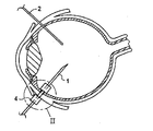
- FIG. 1 is a schematic sectional view of an eye into which two tools have been introduced by means of two cannulae according to the invention
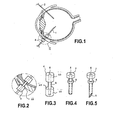
- FIG. 2 is an enlarged view of a part of FIG. 1 ;
- FIG. 3 is a perspective view of the cannula shown in FIGS. 1 and 2 ;
- FIG. 4 is a perspective view of another embodiment of a cannula according to the invention.
- FIG. 5 is a perspective view of yet another embodiment of a cannula according to the invention.
- FIG. 1 a sectional view of an eye is represented schematically.
- Tools 1 and 2 respectively a suction tool 1 and a tool 2 forming a light-guide for enabling the surgeon to see what he/she is doing with the tool 1 , are inserted into the intraocular space of the eye.
- Each tool 1 and 2 passes into the eye by passing into a respective cannula 4 which ensures the crossing of the walls delimiting the intraocular cavity, namely the sclera and/or the conjunctiva.
- the cannula 4 is constituted by a tubular part defining a passage which extends between a proximal opening 5 intended to be outside the intraocular volume and a distal opening opposite the opening 5 and intended to be located in the intraocular volume.
- the cannula 4 is locked in a position in which it traverses the two walls and remains locked there by locking means which are constituted here by a hollowed-out annular groove 6 in the external surface or casing of the cannula.
- locking means which are constituted here by a hollowed-out annular groove 6 in the external surface or casing of the cannula.
- the cannula shown in FIGS. 1 and 2 is represented in more detail in FIG. 3 .
- Said cannula is constituted by a tubular element having a larger outside diameter and defines in its interior a passage channel having an inside diameter.
- the groove 6 is hollowed out on the external surface of the cannula, substantially in its middle. The groove 6 thus forms a narrowing, the outside diameter of which is smaller than the largest outside diameter of the channel and slightly larger than the inside diameter of the channel.
- the extension in length—that is to say, between the proximal and distal openings 5 along the longitudinal direction of the channel of the cannula—of the groove 6 corresponds substantially to the thickness of the two walls formed by the sclera and the conjunctiva, so that once the cannula has been inserted through these two walls these two walls are sandwiched between the two shoulders—the upper shoulder 10 and the lower shoulder 11 —delimiting the groove 6 and thus lock the cannula 4 in position.
- the contact between the plane distal face 12 of the upper or proximal shoulder 10 and the wall of the sclera and/or of the conjunctiva is effected along a plane, at least in part.
- the contact between the proximal plane face 13 of the distal or lower shoulder 11 and the wall delimiting the intraocular space (the sclera and/or the conjunctiva) is effected along a plane, at least in part.
- the distance between the two distal and proximal plane faces 12 and 13 of the proximal and distal shoulders 10 and 11 , respectively, is appreciably equal to or slightly smaller than the thickness of the wall or walls delimiting the intraocular space (the sclera and/or the conjunctiva).
- the lower or distal shoulder 8 has a proximal face equivalent to the face 13 of the shoulder 11 of the embodiment shown in FIGS. 1 to 3
- the shoulder 7 has a distal face equivalent to the face 12 of the shoulder 11
- the two shoulders 7 and 8 sandwich the two walls delimiting the intraocular cavity along respective plane contacts, as in the embodiment shown in FIGS. 1 to 3 .
- the cannula still constituted by a tubular element forming a channel in its interior, includes a head 7 of larger diameter than the diameter of the tubular part of the cannula.
- an annular stop 8 projects outside the cannula.
- an annular groove is formed which has the same function as the groove 6 in the embodiment shown in FIG. 3 .
- the same thickness may be provided as that provided for the groove 6 of the embodiment shown in FIG. 3 .
- an outside diameter of the stop has been represented which is smaller than that of the head.
- identical diameters or, conversely, a larger diameter may be provided for the stop.
- the fact of providing a smaller diameter for the stop presents the additional advantage of facilitating the positioning of the cannula.
- FIG. 5 Represented in FIG. 5 is yet another embodiment corresponding appreciably to that shown in FIG. 4 but in which, in addition, the distal opening 9 of the cannula has a bevelled shape, ensuring a progressive and non-traumatic insertion into the incision of the walls of the sclera and of the conjunctiva when the cannula is inserted for the first time in order to be disposed across these walls.
- This incision may be realised by mounting the cannula onto a knife which incises the scleral+conjunctival thickness, then by means of a clamp the surgeon holds the head of the cannula and withdraws the knife, the cannula being thus placed in position.
- This is the so-called one-step system.
- Other systems exist, for example a system that consists in positioning a gauge that makes it possible for the pars plana (the place where the incision has to be made) to be located, for an incision to be made with the knife, and then for the cannula to be inserted, the gauge helping to find the site of the incision.
- This system is known as the two-step system, by virtue of a tool which is introduced into the cannula and which is then withdrawn once the cannula has been installed.
- the cannula is manufactured from a material that is standard in the field, in particular from metal such as stainless steel or from thermoplastic or plastic materials such as a polyimide.
Landscapes
- Health & Medical Sciences (AREA)
- Surgery (AREA)
- Life Sciences & Earth Sciences (AREA)
- Engineering & Computer Science (AREA)
- Public Health (AREA)
- Biomedical Technology (AREA)
- Heart & Thoracic Surgery (AREA)
- Veterinary Medicine (AREA)
- Nuclear Medicine, Radiotherapy & Molecular Imaging (AREA)
- Animal Behavior & Ethology (AREA)
- General Health & Medical Sciences (AREA)
- Pathology (AREA)
- Ophthalmology & Optometry (AREA)
- Medical Informatics (AREA)
- Molecular Biology (AREA)
- Vascular Medicine (AREA)
- Prostheses (AREA)
- Infusion, Injection, And Reservoir Apparatuses (AREA)
Abstract
Description
- The present invention relates to an assembly composed of at least one cannula intended to traverse the conjunctiva and/or the sclera of the eye in order to enable the passage of a tool intended to have an action in the intraocular volume, such as a suction instrument, an infusion line, an instrument for cutting—for example at high speed—the vitreous gel contained in the intraocular volume, a light-guide intended to transmit light in order to see what is happening in the intraocular volume or similar, and a tool of the aforementioned type having appropriate dimensions in order to be able to pass through the cannula.
- The assemblies of this type of the prior art are not easy for the surgeon to use. In particular, the movement of the surgeon for displacing the tools often lacks precision, and, by virtue of the manipulations of the eye, for example when the surgeon causes an infusion line to pass through the cannula, it may happen that the assembly constituted by cannula and line comes out again towards the outside and physiological liquid comes to be injected underneath the retina, which is not desirable and may cause a detachment of the retina.
- The present invention aims to overcome the drawbacks of the prior art by proposing an assembly of the type mentioned above that gives the surgeon a greater sense of safety when he/she is manoeuvring his/her tool in the intraocular cavity, having the effect of improving safety for the patient and of accelerating the operations being carried out.
- According to the invention, a cannula is as defined in claim 1, further developments being defined in the
dependent claims 2 to 6. - By thus providing for locking the cannula in position when it is positioned through the sclera and/or the conjunctiva in order to form a passage through these walls for the tool, it is ensured that the cannula remains stably in position, which combats an untimely exit of the cannula and the tool that goes through it, notably a perfusion line, during the manipulation of the tool or of the eye, enabling the harmful consequences to be avoided that are linked with such an untimely exit, for example an injection of liquid underneath the retina in the case where the associated tool is a perfusion line. The cannula is thus self-locking.
- According to a preferred and, notably, particularly simple embodiment of the invention, locking means are provided in the form of an annular groove formed on the outside of the cannula.
- In particular, according to a preferred embodiment of the invention the groove is formed by a narrowing of the external casing of the cannula.
- According to another preferred embodiment of the invention, the groove is formed by two annular stops projecting laterally from the cannula and spaced from one another so as to form the annular groove therebetween.
- According to a preferred embodiment of the invention, the distal end of the cannula—that is to say, the end through which the cannula penetrates into the intraocular space—has a bevelled shape.
- This bevel promotes a progressive and non-traumatic positioning of the cannula through the sclera.
- According to another possible embodiment, also enabling a progressive positioning to be promoted, cannula of conical shape is provided over its length, for example from the stop as far as its distal end.
- The present invention also relates to an assembly composed of at least one cannula according to the invention and at least one tool intended to have an action in the intraocular volume, such as a suction instrument, an instrument for cutting—for example at high speed—the vitreous gel contained in the intraocular volume, a light-guide intended to transmit light in order to see what is happening in the intraocular volume or similar, and having appropriate dimensions in order to be able to pass into the channel of the cannula, and also to an assembly composed of at least one cannula according to the invention and at least one tool intended to have an action in the intraocular volume, such as a suction instrument, an instrument for cutting—for example at high speed—the vitreous gel contained in the intraocular volume, a light-guide intended to transmit light in order to see what is happening in the intraocular volume or similar, and passing into the channel of the cannula.
- By way of example, preferred embodiments of the invention will now be described, referring to the drawings in which:
-
FIG. 1 is a schematic sectional view of an eye into which two tools have been introduced by means of two cannulae according to the invention; -
FIG. 2 is an enlarged view of a part ofFIG. 1 ; -
FIG. 3 is a perspective view of the cannula shown inFIGS. 1 and 2 ; -
FIG. 4 is a perspective view of another embodiment of a cannula according to the invention; and -
FIG. 5 is a perspective view of yet another embodiment of a cannula according to the invention. - In
FIG. 1 a sectional view of an eye is represented schematically.Tools 1 and 2, respectively a suction tool 1 and atool 2 forming a light-guide for enabling the surgeon to see what he/she is doing with the tool 1, are inserted into the intraocular space of the eye. Eachtool 1 and 2 passes into the eye by passing into a respective cannula 4 which ensures the crossing of the walls delimiting the intraocular cavity, namely the sclera and/or the conjunctiva. - The cannula 4 is constituted by a tubular part defining a passage which extends between a
proximal opening 5 intended to be outside the intraocular volume and a distal opening opposite theopening 5 and intended to be located in the intraocular volume. - The cannula 4 is locked in a position in which it traverses the two walls and remains locked there by locking means which are constituted here by a hollowed-out
annular groove 6 in the external surface or casing of the cannula. The cannula shown inFIGS. 1 and 2 is represented in more detail inFIG. 3 . Said cannula is constituted by a tubular element having a larger outside diameter and defines in its interior a passage channel having an inside diameter. Thegroove 6 is hollowed out on the external surface of the cannula, substantially in its middle. Thegroove 6 thus forms a narrowing, the outside diameter of which is smaller than the largest outside diameter of the channel and slightly larger than the inside diameter of the channel. The extension in length—that is to say, between the proximal anddistal openings 5 along the longitudinal direction of the channel of the cannula—of thegroove 6 corresponds substantially to the thickness of the two walls formed by the sclera and the conjunctiva, so that once the cannula has been inserted through these two walls these two walls are sandwiched between the two shoulders—theupper shoulder 10 and thelower shoulder 11—delimiting thegroove 6 and thus lock the cannula 4 in position. The contact between the planedistal face 12 of the upper orproximal shoulder 10 and the wall of the sclera and/or of the conjunctiva is effected along a plane, at least in part. The contact between the proximal plane face 13 of the distal orlower shoulder 11 and the wall delimiting the intraocular space (the sclera and/or the conjunctiva) is effected along a plane, at least in part. - The distance between the two distal and proximal plane faces 12 and 13 of the proximal and
distal shoulders - Likewise, in the embodiments shown in
FIG. 4 or 5 the lower ordistal shoulder 8 has a proximal face equivalent to the face 13 of theshoulder 11 of the embodiment shown inFIGS. 1 to 3 , and theshoulder 7 has a distal face equivalent to theface 12 of theshoulder 11, and the twoshoulders FIGS. 1 to 3 . - According to another embodiment, represented in
FIG. 4 , the cannula, still constituted by a tubular element forming a channel in its interior, includes ahead 7 of larger diameter than the diameter of the tubular part of the cannula. On the other side, anannular stop 8 projects outside the cannula. Thus between thehead 7 and theannular stop 8 an annular groove is formed which has the same function as thegroove 6 in the embodiment shown inFIG. 3 . In particular, for this groove the same thickness may be provided as that provided for thegroove 6 of the embodiment shown inFIG. 3 . - In the Figure an outside diameter of the stop has been represented which is smaller than that of the head. However, while remaining within the scope of the invention, identical diameters or, conversely, a larger diameter may be provided for the stop. However, the fact of providing a smaller diameter for the stop presents the additional advantage of facilitating the positioning of the cannula.
- Represented in
FIG. 5 is yet another embodiment corresponding appreciably to that shown inFIG. 4 but in which, in addition, thedistal opening 9 of the cannula has a bevelled shape, ensuring a progressive and non-traumatic insertion into the incision of the walls of the sclera and of the conjunctiva when the cannula is inserted for the first time in order to be disposed across these walls. - This incision may be realised by mounting the cannula onto a knife which incises the scleral+conjunctival thickness, then by means of a clamp the surgeon holds the head of the cannula and withdraws the knife, the cannula being thus placed in position. This is the so-called one-step system. Other systems exist, for example a system that consists in positioning a gauge that makes it possible for the pars plana (the place where the incision has to be made) to be located, for an incision to be made with the knife, and then for the cannula to be inserted, the gauge helping to find the site of the incision. This system is known as the two-step system, by virtue of a tool which is introduced into the cannula and which is then withdrawn once the cannula has been installed.
- The cannula is manufactured from a material that is standard in the field, in particular from metal such as stainless steel or from thermoplastic or plastic materials such as a polyimide.
Claims (8)
Applications Claiming Priority (2)
| Application Number | Priority Date | Filing Date | Title |
|---|---|---|---|
| FR1100141A FR2970412B1 (en) | 2011-01-18 | 2011-01-18 | SELF-LOCKING CANNULA |
| FR1100141 | 2011-01-18 |
Publications (1)
| Publication Number | Publication Date |
|---|---|
| US20120184810A1 true US20120184810A1 (en) | 2012-07-19 |
Family
ID=43920072
Family Applications (1)
| Application Number | Title | Priority Date | Filing Date |
|---|---|---|---|
| US13/344,829 Abandoned US20120184810A1 (en) | 2011-01-18 | 2012-01-06 | Self-locking cannula |
Country Status (4)
| Country | Link |
|---|---|
| US (1) | US20120184810A1 (en) |
| EP (1) | EP2476399B1 (en) |
| ES (1) | ES2756726T3 (en) |
| FR (1) | FR2970412B1 (en) |
Families Citing this family (1)
| Publication number | Priority date | Publication date | Assignee | Title |
|---|---|---|---|---|
| RU2723534C2 (en) * | 2015-11-30 | 2020-06-15 | Мани, Инк. | Cannula provided with piercing needle |
Citations (19)
| Publication number | Priority date | Publication date | Assignee | Title |
|---|---|---|---|---|
| US3528425A (en) * | 1968-09-16 | 1970-09-15 | Surgical Design Corp | Apparatus for performing surgical procedures on the eye |
| US4692142A (en) * | 1986-02-24 | 1987-09-08 | Dignam Bernard J | Sutureless infusion cannula for ophthalmic surgery |
| US5127901A (en) * | 1990-05-18 | 1992-07-07 | Odrich Ronald B | Implant with subconjunctival arch |
| US5300020A (en) * | 1991-05-31 | 1994-04-05 | Medflex Corporation | Surgically implantable device for glaucoma relief |
| US5702414A (en) * | 1995-05-14 | 1997-12-30 | Optonol Ltd | Method of implanting an intraocular implant |
| US5817099A (en) * | 1996-06-06 | 1998-10-06 | Skolik; Stephanie A. | Universal port/seal device for ocular surgery |
| US5830191A (en) * | 1992-06-30 | 1998-11-03 | Ethicon, Inc. | Flexible endoscopic surgical port |
| US20010029335A1 (en) * | 2000-01-03 | 2001-10-11 | Johns Hopkins University | Intraoperative microsurgical ultrasonic device and methods related thereto |
| US6510600B2 (en) * | 1997-11-20 | 2003-01-28 | Optonol, Ltd. | Method for manufacturing a flow regulating implant |
| US20050165432A1 (en) * | 2002-05-09 | 2005-07-28 | Russell Heinrich | Adjustable balloon anchoring trocar |
| US20070005089A1 (en) * | 2005-06-30 | 2007-01-04 | Smith Robert C | Beveled access apparatus with locking ribs elements |
| US20070010827A1 (en) * | 2001-08-28 | 2007-01-11 | Hosheng Tu | Glaucoma stent system |
| US20070179430A1 (en) * | 2005-12-16 | 2007-08-02 | Smith Ronald T | Illuminated infusion cannula |
| JP2007319423A (en) * | 2006-06-01 | 2007-12-13 | Univ Of Tokushima | Vitreous surgery cannula |
| US20080177239A1 (en) * | 2007-01-18 | 2008-07-24 | Yong Li | Trocar cannula system |
| US20080195044A1 (en) * | 2005-03-23 | 2008-08-14 | Akira Nishimura | Ophthalmic Cannula |
| US20090216196A1 (en) * | 2008-02-27 | 2009-08-27 | Entellus Medical, Inc. | Apparatus and method for accessing a sinus cavity |
| US20110152774A1 (en) * | 2009-12-23 | 2011-06-23 | Jose Luis Lopez | Ophthalmic valved trocar cannula |
| US20120157779A1 (en) * | 2010-12-21 | 2012-06-21 | Greg Fischvogt | Access assembly including inflatable seal member |
Family Cites Families (3)
| Publication number | Priority date | Publication date | Assignee | Title |
|---|---|---|---|---|
| FR2830186B1 (en) * | 2001-10-03 | 2004-06-11 | Didier Ducournau | TOOL FOR THE INCISION OF SCLEROTICS FOR THE PLACEMENT OF A BREWING CANNULA AND CORRESPONDING BREWING CANNULA |
| US8066673B2 (en) * | 2006-03-21 | 2011-11-29 | Applied Medical Resources Corporation | Cannula stabilization seal |
| WO2010126076A1 (en) * | 2009-04-30 | 2010-11-04 | マニー株式会社 | Cannula for ophthalmic surgery and method of manufacturing same |
-
2011
- 2011-01-18 FR FR1100141A patent/FR2970412B1/en active Active
- 2011-12-12 ES ES11290571T patent/ES2756726T3/en active Active
- 2011-12-12 EP EP11290571.6A patent/EP2476399B1/en active Active
-
2012
- 2012-01-06 US US13/344,829 patent/US20120184810A1/en not_active Abandoned
Patent Citations (19)
| Publication number | Priority date | Publication date | Assignee | Title |
|---|---|---|---|---|
| US3528425A (en) * | 1968-09-16 | 1970-09-15 | Surgical Design Corp | Apparatus for performing surgical procedures on the eye |
| US4692142A (en) * | 1986-02-24 | 1987-09-08 | Dignam Bernard J | Sutureless infusion cannula for ophthalmic surgery |
| US5127901A (en) * | 1990-05-18 | 1992-07-07 | Odrich Ronald B | Implant with subconjunctival arch |
| US5300020A (en) * | 1991-05-31 | 1994-04-05 | Medflex Corporation | Surgically implantable device for glaucoma relief |
| US5830191A (en) * | 1992-06-30 | 1998-11-03 | Ethicon, Inc. | Flexible endoscopic surgical port |
| US5702414A (en) * | 1995-05-14 | 1997-12-30 | Optonol Ltd | Method of implanting an intraocular implant |
| US5817099A (en) * | 1996-06-06 | 1998-10-06 | Skolik; Stephanie A. | Universal port/seal device for ocular surgery |
| US6510600B2 (en) * | 1997-11-20 | 2003-01-28 | Optonol, Ltd. | Method for manufacturing a flow regulating implant |
| US20010029335A1 (en) * | 2000-01-03 | 2001-10-11 | Johns Hopkins University | Intraoperative microsurgical ultrasonic device and methods related thereto |
| US20070010827A1 (en) * | 2001-08-28 | 2007-01-11 | Hosheng Tu | Glaucoma stent system |
| US20050165432A1 (en) * | 2002-05-09 | 2005-07-28 | Russell Heinrich | Adjustable balloon anchoring trocar |
| US20080195044A1 (en) * | 2005-03-23 | 2008-08-14 | Akira Nishimura | Ophthalmic Cannula |
| US20070005089A1 (en) * | 2005-06-30 | 2007-01-04 | Smith Robert C | Beveled access apparatus with locking ribs elements |
| US20070179430A1 (en) * | 2005-12-16 | 2007-08-02 | Smith Ronald T | Illuminated infusion cannula |
| JP2007319423A (en) * | 2006-06-01 | 2007-12-13 | Univ Of Tokushima | Vitreous surgery cannula |
| US20080177239A1 (en) * | 2007-01-18 | 2008-07-24 | Yong Li | Trocar cannula system |
| US20090216196A1 (en) * | 2008-02-27 | 2009-08-27 | Entellus Medical, Inc. | Apparatus and method for accessing a sinus cavity |
| US20110152774A1 (en) * | 2009-12-23 | 2011-06-23 | Jose Luis Lopez | Ophthalmic valved trocar cannula |
| US20120157779A1 (en) * | 2010-12-21 | 2012-06-21 | Greg Fischvogt | Access assembly including inflatable seal member |
Non-Patent Citations (1)
| Title |
|---|
| Machine translation of JP2007-319423A (Takeshi): Cannula for Vitrectomy * |
Also Published As
| Publication number | Publication date |
|---|---|
| FR2970412B1 (en) | 2013-12-20 |
| ES2756726T3 (en) | 2020-04-27 |
| EP2476399A1 (en) | 2012-07-18 |
| EP2476399B1 (en) | 2019-08-21 |
| FR2970412A1 (en) | 2012-07-20 |
Similar Documents
| Publication | Publication Date | Title |
|---|---|---|
| CN100421741C (en) | injection needle | |
| AU2018311081B2 (en) | Needle and catheter insertion device | |
| EP2600923B1 (en) | Apparatus and method for safely inserting an introducer needle into epidural space | |
| US9227012B2 (en) | Winged medical needle device | |
| US10195019B2 (en) | Intraocular lens insertion apparatus | |
| EP2161053A1 (en) | Needleless connector with displacement correction | |
| KR20050043904A (en) | Needle-less port and method of producing the same | |
| HK1252200A1 (en) | Needles and related assemblies and methods | |
| US8382722B2 (en) | Blunt tip vial access cannula and method for manufacture | |
| WO2016068797A1 (en) | Intravenous catheter assembly | |
| US10130777B2 (en) | Injection needle | |
| US20150157359A1 (en) | Puncture needle | |
| WO1992005816A1 (en) | Hollow needle for medical use, and process for the manufacture of such needles | |
| US20120184810A1 (en) | Self-locking cannula | |
| US20200324086A1 (en) | Introducer needle and related systems and methods | |
| EP3541466B1 (en) | Trough seal | |
| WO2018096549A1 (en) | Intravenous catheter with a self-activating mechanism for preventing back-flow of blood | |
| US11344677B2 (en) | Needle-free injection guide | |
| EP3053552B1 (en) | Cannula | |
| US11472131B2 (en) | Injection nozzle and method of making same | |
| EP2514452B1 (en) | Needle device | |
| US11511082B2 (en) | Catheter having a hard distal tip | |
| JP2017012481A (en) | Implantable catheter port and method for producing implantable catheter port | |
| JP2022512504A (en) | Medical bevel needle | |
| EP3381420B1 (en) | Piercing needle-equipped cannula |
Legal Events
| Date | Code | Title | Description |
|---|---|---|---|
| AS | Assignment |
Owner name: FRANCE CHIRURGIE INSTRUMENTATION SAS (FCI), FRANCE Free format text: ASSIGNMENT OF ASSIGNORS INTEREST;ASSIGNOR:ZECH, JEAN-CHRISTOPHE;REEL/FRAME:027492/0289 Effective date: 20111215 |
|
| STPP | Information on status: patent application and granting procedure in general |
Free format text: DOCKETED NEW CASE - READY FOR EXAMINATION |
|
| STPP | Information on status: patent application and granting procedure in general |
Free format text: NON FINAL ACTION MAILED |
|
| STPP | Information on status: patent application and granting procedure in general |
Free format text: RESPONSE TO NON-FINAL OFFICE ACTION ENTERED AND FORWARDED TO EXAMINER |
|
| STPP | Information on status: patent application and granting procedure in general |
Free format text: FINAL REJECTION MAILED |
|
| STPP | Information on status: patent application and granting procedure in general |
Free format text: DOCKETED NEW CASE - READY FOR EXAMINATION |
|
| STPP | Information on status: patent application and granting procedure in general |
Free format text: NON FINAL ACTION MAILED |
|
| STCB | Information on status: application discontinuation |
Free format text: ABANDONED -- FAILURE TO RESPOND TO AN OFFICE ACTION |
|
| STCB | Information on status: application discontinuation |
Free format text: ABANDONED -- FAILURE TO RESPOND TO AN OFFICE ACTION |