US20080242588A1 - System for seizure suppression - Google Patents
System for seizure suppression Download PDFInfo
- Publication number
- US20080242588A1 US20080242588A1 US11/944,332 US94433207A US2008242588A1 US 20080242588 A1 US20080242588 A1 US 20080242588A1 US 94433207 A US94433207 A US 94433207A US 2008242588 A1 US2008242588 A1 US 2008242588A1
- Authority
- US
- United States
- Prior art keywords
- seizure
- activity
- amiloride
- pctx1
- seizures
- Prior art date
- Legal status (The legal status is an assumption and is not a legal conclusion. Google has not performed a legal analysis and makes no representation as to the accuracy of the status listed.)
- Granted
Links
- 230000001629 suppression Effects 0.000 title abstract description 8
- 206010010904 Convulsion Diseases 0.000 claims abstract description 239
- 108010068806 Acid Sensing Ion Channels Proteins 0.000 claims abstract description 169
- 102000001671 Acid Sensing Ion Channels Human genes 0.000 claims abstract description 169
- 230000000694 effects Effects 0.000 claims abstract description 151
- 239000000203 mixture Substances 0.000 claims abstract description 86
- XSDQTOBWRPYKKA-UHFFFAOYSA-N amiloride Chemical compound NC(=N)NC(=O)C1=NC(Cl)=C(N)N=C1N XSDQTOBWRPYKKA-UHFFFAOYSA-N 0.000 claims abstract description 76
- 238000000034 method Methods 0.000 claims abstract description 62
- 229960002576 amiloride Drugs 0.000 claims abstract description 53
- 229940000406 drug candidate Drugs 0.000 claims abstract description 38
- 230000005764 inhibitory process Effects 0.000 claims abstract description 21
- 229960003965 antiepileptics Drugs 0.000 claims abstract description 19
- 108090000765 processed proteins & peptides Proteins 0.000 claims abstract description 17
- 230000004044 response Effects 0.000 claims abstract description 14
- 230000008859 change Effects 0.000 claims abstract description 11
- 210000004556 brain Anatomy 0.000 claims description 49
- 206010015037 epilepsy Diseases 0.000 claims description 45
- 150000001875 compounds Chemical class 0.000 claims description 42
- 230000002829 reductive effect Effects 0.000 claims description 40
- 238000012360 testing method Methods 0.000 claims description 38
- 238000003556 assay Methods 0.000 claims description 18
- 238000012216 screening Methods 0.000 claims description 12
- 125000000217 alkyl group Chemical group 0.000 claims description 11
- 125000003118 aryl group Chemical group 0.000 claims description 11
- 125000003710 aryl alkyl group Chemical group 0.000 claims description 10
- 150000001732 carboxylic acid derivatives Chemical class 0.000 claims description 8
- 125000001424 substituent group Chemical group 0.000 claims description 8
- 125000001072 heteroaryl group Chemical group 0.000 claims description 6
- 230000002401 inhibitory effect Effects 0.000 claims description 6
- 230000003287 optical effect Effects 0.000 claims description 6
- LFQSCWFLJHTTHZ-UHFFFAOYSA-N Ethanol Chemical compound CCO LFQSCWFLJHTTHZ-UHFFFAOYSA-N 0.000 claims description 5
- 150000002367 halogens Chemical group 0.000 claims description 5
- 125000003282 alkyl amino group Chemical group 0.000 claims description 4
- 150000002148 esters Chemical class 0.000 claims description 4
- 229910052736 halogen Inorganic materials 0.000 claims description 4
- 238000012402 patch clamp technique Methods 0.000 claims description 4
- 125000002924 primary amino group Chemical group [H]N([H])* 0.000 claims description 4
- 125000004663 dialkyl amino group Chemical group 0.000 claims description 3
- 125000003545 alkoxy group Chemical group 0.000 claims description 2
- 125000002887 hydroxy group Chemical group [H]O* 0.000 claims description 2
- 239000008194 pharmaceutical composition Substances 0.000 claims 4
- 101710105611 Acid-sensing ion channel 1A Proteins 0.000 claims 2
- 210000004027 cell Anatomy 0.000 description 65
- 239000002253 acid Substances 0.000 description 40
- 230000001537 neural effect Effects 0.000 description 39
- 210000002569 neuron Anatomy 0.000 description 38
- 229940079593 drug Drugs 0.000 description 35
- 239000003814 drug Substances 0.000 description 35
- 230000028161 membrane depolarization Effects 0.000 description 32
- 108091006146 Channels Proteins 0.000 description 31
- 230000004913 activation Effects 0.000 description 31
- 241000699670 Mus sp. Species 0.000 description 29
- VLSMHEGGTFMBBZ-UHFFFAOYSA-N alpha-Kainic acid Natural products CC(=C)C1CNC(C(O)=O)C1CC(O)=O VLSMHEGGTFMBBZ-UHFFFAOYSA-N 0.000 description 29
- VLSMHEGGTFMBBZ-OOZYFLPDSA-N kainic acid Chemical compound CC(=C)[C@H]1CN[C@H](C(O)=O)[C@H]1CC(O)=O VLSMHEGGTFMBBZ-OOZYFLPDSA-N 0.000 description 29
- 229950006874 kainic acid Drugs 0.000 description 29
- 230000000638 stimulation Effects 0.000 description 29
- 230000009172 bursting Effects 0.000 description 27
- 241001465754 Metazoa Species 0.000 description 24
- 239000000126 substance Substances 0.000 description 23
- 208000027418 Wounds and injury Diseases 0.000 description 22
- 239000001961 anticonvulsive agent Substances 0.000 description 22
- 241000699666 Mus <mouse, genus> Species 0.000 description 21
- 230000006378 damage Effects 0.000 description 21
- 230000001965 increasing effect Effects 0.000 description 21
- 208000014674 injury Diseases 0.000 description 21
- 238000002347 injection Methods 0.000 description 18
- 239000007924 injection Substances 0.000 description 18
- 230000007423 decrease Effects 0.000 description 17
- 230000005284 excitation Effects 0.000 description 17
- 239000003112 inhibitor Substances 0.000 description 16
- 208000005809 status epilepticus Diseases 0.000 description 16
- 238000000537 electroencephalography Methods 0.000 description 15
- 230000000971 hippocampal effect Effects 0.000 description 15
- 238000001727 in vivo Methods 0.000 description 15
- 230000010412 perfusion Effects 0.000 description 15
- 230000009467 reduction Effects 0.000 description 15
- 208000010444 Acidosis Diseases 0.000 description 14
- 230000007950 acidosis Effects 0.000 description 14
- 208000026545 acidosis disease Diseases 0.000 description 14
- 238000000586 desensitisation Methods 0.000 description 14
- 239000011575 calcium Substances 0.000 description 13
- HCZHHEIFKROPDY-UHFFFAOYSA-N kynurenic acid Chemical compound C1=CC=C2NC(C(=O)O)=CC(=O)C2=C1 HCZHHEIFKROPDY-UHFFFAOYSA-N 0.000 description 13
- 210000004295 hippocampal neuron Anatomy 0.000 description 12
- 230000007246 mechanism Effects 0.000 description 12
- 230000001360 synchronised effect Effects 0.000 description 12
- 210000001519 tissue Anatomy 0.000 description 12
- 102000003855 L-lactate dehydrogenase Human genes 0.000 description 11
- 108700023483 L-lactate dehydrogenases Proteins 0.000 description 11
- 210000003169 central nervous system Anatomy 0.000 description 11
- 229960001848 lamotrigine Drugs 0.000 description 11
- PYZRQGJRPPTADH-UHFFFAOYSA-N lamotrigine Chemical compound NC1=NC(N)=NN=C1C1=CC=CC(Cl)=C1Cl PYZRQGJRPPTADH-UHFFFAOYSA-N 0.000 description 11
- 210000004379 membrane Anatomy 0.000 description 11
- 239000012528 membrane Substances 0.000 description 11
- 230000002269 spontaneous effect Effects 0.000 description 11
- 101710099904 Acid-sensing ion channel 1 Proteins 0.000 description 10
- 102100021624 Acid-sensing ion channel 1 Human genes 0.000 description 10
- 102100022094 Acid-sensing ion channel 2 Human genes 0.000 description 10
- 101710099898 Acid-sensing ion channel 3 Proteins 0.000 description 10
- 238000004113 cell culture Methods 0.000 description 10
- 230000000946 synaptic effect Effects 0.000 description 10
- OYPRJOBELJOOCE-UHFFFAOYSA-N Calcium Chemical compound [Ca] OYPRJOBELJOOCE-UHFFFAOYSA-N 0.000 description 9
- WQZGKKKJIJFFOK-GASJEMHNSA-N Glucose Natural products OC[C@H]1OC(O)[C@H](O)[C@@H](O)[C@@H]1O WQZGKKKJIJFFOK-GASJEMHNSA-N 0.000 description 9
- 150000001413 amino acids Chemical group 0.000 description 9
- 229910052791 calcium Inorganic materials 0.000 description 9
- 230000030833 cell death Effects 0.000 description 9
- 208000037265 diseases, disorders, signs and symptoms Diseases 0.000 description 9
- 230000001037 epileptic effect Effects 0.000 description 9
- 230000000763 evoking effect Effects 0.000 description 9
- 238000010304 firing Methods 0.000 description 9
- 230000006870 function Effects 0.000 description 9
- 239000008103 glucose Substances 0.000 description 9
- 210000001320 hippocampus Anatomy 0.000 description 9
- 238000005259 measurement Methods 0.000 description 9
- 239000000243 solution Substances 0.000 description 9
- 102100022097 Acid-sensing ion channel 3 Human genes 0.000 description 8
- 230000000144 pharmacologic effect Effects 0.000 description 8
- 101000901079 Homo sapiens Acid-sensing ion channel 2 Proteins 0.000 description 7
- 102000004310 Ion Channels Human genes 0.000 description 7
- 108090000862 Ion Channels Proteins 0.000 description 7
- 230000003556 anti-epileptic effect Effects 0.000 description 7
- 208000029028 brain injury Diseases 0.000 description 7
- 210000003618 cortical neuron Anatomy 0.000 description 7
- QDERNBXNXJCIQK-UHFFFAOYSA-N ethylisopropylamiloride Chemical compound CCN(C(C)C)C1=NC(N)=C(C(=O)N=C(N)N)N=C1Cl QDERNBXNXJCIQK-UHFFFAOYSA-N 0.000 description 7
- 230000001314 paroxysmal effect Effects 0.000 description 7
- 238000011084 recovery Methods 0.000 description 7
- 230000035945 sensitivity Effects 0.000 description 7
- 238000011282 treatment Methods 0.000 description 7
- 239000002435 venom Substances 0.000 description 7
- 231100000611 venom Toxicity 0.000 description 7
- 210000001048 venom Anatomy 0.000 description 7
- 241000282412 Homo Species 0.000 description 6
- TWRXJAOTZQYOKJ-UHFFFAOYSA-L Magnesium chloride Chemical compound [Mg+2].[Cl-].[Cl-] TWRXJAOTZQYOKJ-UHFFFAOYSA-L 0.000 description 6
- HOKKHZGPKSLGJE-GSVOUGTGSA-N N-Methyl-D-aspartic acid Chemical compound CN[C@@H](C(O)=O)CC(O)=O HOKKHZGPKSLGJE-GSVOUGTGSA-N 0.000 description 6
- 229940024606 amino acid Drugs 0.000 description 6
- 208000035475 disorder Diseases 0.000 description 6
- 238000002474 experimental method Methods 0.000 description 6
- 239000011777 magnesium Substances 0.000 description 6
- 239000002858 neurotransmitter agent Substances 0.000 description 6
- 230000003252 repetitive effect Effects 0.000 description 6
- 230000002459 sustained effect Effects 0.000 description 6
- 230000000699 topical effect Effects 0.000 description 6
- JKMHFZQWWAIEOD-UHFFFAOYSA-N 2-[4-(2-hydroxyethyl)piperazin-1-yl]ethanesulfonic acid Chemical compound OCC[NH+]1CCN(CCS([O-])(=O)=O)CC1 JKMHFZQWWAIEOD-UHFFFAOYSA-N 0.000 description 5
- -1 ASIC2b Proteins 0.000 description 5
- 101150061877 Asic1 gene Proteins 0.000 description 5
- KXDROGADUISDGY-UHFFFAOYSA-N Benzamil hydrochloride Chemical compound C=1C=CC=CC=1CN=C(N)NC(=O)C1=NC(Cl)=C(N)N=C1N KXDROGADUISDGY-UHFFFAOYSA-N 0.000 description 5
- 230000003247 decreasing effect Effects 0.000 description 5
- 239000000975 dye Substances 0.000 description 5
- 208000028329 epileptic seizure Diseases 0.000 description 5
- 238000003209 gene knockout Methods 0.000 description 5
- 230000006698 induction Effects 0.000 description 5
- 238000000185 intracerebroventricular administration Methods 0.000 description 5
- 230000037427 ion transport Effects 0.000 description 5
- 150000002500 ions Chemical class 0.000 description 5
- 230000001404 mediated effect Effects 0.000 description 5
- 239000002609 medium Substances 0.000 description 5
- 230000008062 neuronal firing Effects 0.000 description 5
- 208000024891 symptom Diseases 0.000 description 5
- 230000005062 synaptic transmission Effects 0.000 description 5
- 102100022099 Acid-sensing ion channel 4 Human genes 0.000 description 4
- 101710099897 Acid-sensing ion channel 4 Proteins 0.000 description 4
- 238000011740 C57BL/6 mouse Methods 0.000 description 4
- UXVMQQNJUSDDNG-UHFFFAOYSA-L Calcium chloride Chemical compound [Cl-].[Cl-].[Ca+2] UXVMQQNJUSDDNG-UHFFFAOYSA-L 0.000 description 4
- 241000283073 Equus caballus Species 0.000 description 4
- WSFSSNUMVMOOMR-UHFFFAOYSA-N Formaldehyde Chemical compound O=C WSFSSNUMVMOOMR-UHFFFAOYSA-N 0.000 description 4
- 102000018899 Glutamate Receptors Human genes 0.000 description 4
- 108010027915 Glutamate Receptors Proteins 0.000 description 4
- CSNNHWWHGAXBCP-UHFFFAOYSA-L Magnesium sulfate Chemical compound [Mg+2].[O-][S+2]([O-])([O-])[O-] CSNNHWWHGAXBCP-UHFFFAOYSA-L 0.000 description 4
- 108090001041 N-Methyl-D-Aspartate Receptors Proteins 0.000 description 4
- 208000012902 Nervous system disease Diseases 0.000 description 4
- GQPLMRYTRLFLPF-UHFFFAOYSA-N Nitrous Oxide Chemical compound [O-][N+]#N GQPLMRYTRLFLPF-UHFFFAOYSA-N 0.000 description 4
- 206010061334 Partial seizures Diseases 0.000 description 4
- UIIMBOGNXHQVGW-UHFFFAOYSA-M Sodium bicarbonate Chemical compound [Na+].OC([O-])=O UIIMBOGNXHQVGW-UHFFFAOYSA-M 0.000 description 4
- FAPWRFPIFSIZLT-UHFFFAOYSA-M Sodium chloride Chemical compound [Na+].[Cl-] FAPWRFPIFSIZLT-UHFFFAOYSA-M 0.000 description 4
- 0 [1*]N([2*])/C(=N/C(=O)C1=NC(C)=C(N([7*])[8*])N=C1N([5*])[6*])N([3*])[4*] Chemical compound [1*]N([2*])/C(=N/C(=O)C1=NC(C)=C(N([7*])[8*])N=C1N([5*])[6*])N([3*])[4*] 0.000 description 4
- 230000002159 abnormal effect Effects 0.000 description 4
- 230000036982 action potential Effects 0.000 description 4
- 238000004458 analytical method Methods 0.000 description 4
- 239000005557 antagonist Substances 0.000 description 4
- 230000002238 attenuated effect Effects 0.000 description 4
- 210000003050 axon Anatomy 0.000 description 4
- 239000001110 calcium chloride Substances 0.000 description 4
- 229910001628 calcium chloride Inorganic materials 0.000 description 4
- 230000001684 chronic effect Effects 0.000 description 4
- 230000001419 dependent effect Effects 0.000 description 4
- 230000008579 epileptogenesis Effects 0.000 description 4
- 229960002767 ethosuximide Drugs 0.000 description 4
- HAPOVYFOVVWLRS-UHFFFAOYSA-N ethosuximide Chemical compound CCC1(C)CC(=O)NC1=O HAPOVYFOVVWLRS-UHFFFAOYSA-N 0.000 description 4
- 230000002964 excitative effect Effects 0.000 description 4
- 238000010874 in vitro model Methods 0.000 description 4
- 208000028867 ischemia Diseases 0.000 description 4
- 238000011813 knockout mouse model Methods 0.000 description 4
- JVTAAEKCZFNVCJ-UHFFFAOYSA-N lactic acid Chemical compound CC(O)C(O)=O JVTAAEKCZFNVCJ-UHFFFAOYSA-N 0.000 description 4
- 230000007774 longterm Effects 0.000 description 4
- 210000001577 neostriatum Anatomy 0.000 description 4
- 230000020477 pH reduction Effects 0.000 description 4
- 238000010186 staining Methods 0.000 description 4
- 238000006467 substitution reaction Methods 0.000 description 4
- 206010002091 Anaesthesia Diseases 0.000 description 3
- 241000283690 Bos taurus Species 0.000 description 3
- 208000037158 Partial Epilepsies Diseases 0.000 description 3
- CXOFVDLJLONNDW-UHFFFAOYSA-N Phenytoin Chemical compound N1C(=O)NC(=O)C1(C=1C=CC=CC=1)C1=CC=CC=C1 CXOFVDLJLONNDW-UHFFFAOYSA-N 0.000 description 3
- LHNKBXRFNPMIBR-UHFFFAOYSA-N Picrotoxin Natural products CC(C)(O)C1(O)C2OC(=O)C1C3(O)C4OC4C5C(=O)OC2C35C LHNKBXRFNPMIBR-UHFFFAOYSA-N 0.000 description 3
- HEMHJVSKTPXQMS-UHFFFAOYSA-M Sodium hydroxide Chemical compound [OH-].[Na+] HEMHJVSKTPXQMS-UHFFFAOYSA-M 0.000 description 3
- 241000239292 Theraphosidae Species 0.000 description 3
- 208000028311 absence seizure Diseases 0.000 description 3
- 238000002835 absorbance Methods 0.000 description 3
- 238000009825 accumulation Methods 0.000 description 3
- 230000002378 acidificating effect Effects 0.000 description 3
- 230000001154 acute effect Effects 0.000 description 3
- 210000004727 amygdala Anatomy 0.000 description 3
- 230000037005 anaesthesia Effects 0.000 description 3
- QVGXLLKOCUKJST-UHFFFAOYSA-N atomic oxygen Chemical compound [O] QVGXLLKOCUKJST-UHFFFAOYSA-N 0.000 description 3
- 230000000903 blocking effect Effects 0.000 description 3
- FFGPTBGBLSHEPO-UHFFFAOYSA-N carbamazepine Chemical compound C1=CC2=CC=CC=C2N(C(=O)N)C2=CC=CC=C21 FFGPTBGBLSHEPO-UHFFFAOYSA-N 0.000 description 3
- 229960000623 carbamazepine Drugs 0.000 description 3
- 230000007248 cellular mechanism Effects 0.000 description 3
- 230000036755 cellular response Effects 0.000 description 3
- 230000003727 cerebral blood flow Effects 0.000 description 3
- 210000001175 cerebrospinal fluid Anatomy 0.000 description 3
- 238000006243 chemical reaction Methods 0.000 description 3
- 239000003795 chemical substances by application Substances 0.000 description 3
- 201000010099 disease Diseases 0.000 description 3
- 230000036749 excitatory postsynaptic potential Effects 0.000 description 3
- 230000001747 exhibiting effect Effects 0.000 description 3
- 230000006539 extracellular acidification Effects 0.000 description 3
- 229960003472 felbamate Drugs 0.000 description 3
- WKGXYQFOCVYPAC-UHFFFAOYSA-N felbamate Chemical compound NC(=O)OCC(COC(N)=O)C1=CC=CC=C1 WKGXYQFOCVYPAC-UHFFFAOYSA-N 0.000 description 3
- 239000012530 fluid Substances 0.000 description 3
- 239000001963 growth medium Substances 0.000 description 3
- 238000003384 imaging method Methods 0.000 description 3
- 230000000302 ischemic effect Effects 0.000 description 3
- 230000001535 kindling effect Effects 0.000 description 3
- 229910001629 magnesium chloride Inorganic materials 0.000 description 3
- 230000003278 mimic effect Effects 0.000 description 3
- 230000017074 necrotic cell death Effects 0.000 description 3
- 230000008587 neuronal excitability Effects 0.000 description 3
- 229910052760 oxygen Inorganic materials 0.000 description 3
- 239000001301 oxygen Substances 0.000 description 3
- 230000021962 pH elevation Effects 0.000 description 3
- 230000036961 partial effect Effects 0.000 description 3
- 230000001717 pathogenic effect Effects 0.000 description 3
- 230000002093 peripheral effect Effects 0.000 description 3
- 239000000825 pharmaceutical preparation Substances 0.000 description 3
- 229960002036 phenytoin Drugs 0.000 description 3
- VJKUPQSHOVKBCO-AHMKVGDJSA-N picrotoxin Chemical compound O=C([C@@]12O[C@@H]1C[C@]1(O)[C@@]32C)O[C@@H]3[C@H]2[C@@H](C(=C)C)[C@@H]1C(=O)O2.O=C([C@@]12O[C@@H]1C[C@]1(O)[C@@]32C)O[C@@H]3[C@H]2[C@@H](C(C)(O)C)[C@@H]1C(=O)O2 VJKUPQSHOVKBCO-AHMKVGDJSA-N 0.000 description 3
- 230000001953 sensory effect Effects 0.000 description 3
- 230000011664 signaling Effects 0.000 description 3
- 230000001256 tonic effect Effects 0.000 description 3
- QUTYKIXIUDQOLK-PRJMDXOYSA-N 5-O-(1-carboxyvinyl)-3-phosphoshikimic acid Chemical compound O[C@H]1[C@H](OC(=C)C(O)=O)CC(C(O)=O)=C[C@H]1OP(O)(O)=O QUTYKIXIUDQOLK-PRJMDXOYSA-N 0.000 description 2
- ZKHQWZAMYRWXGA-KQYNXXCUSA-J ATP(4-) Chemical compound C1=NC=2C(N)=NC=NC=2N1[C@@H]1O[C@H](COP([O-])(=O)OP([O-])(=O)OP([O-])([O-])=O)[C@@H](O)[C@H]1O ZKHQWZAMYRWXGA-KQYNXXCUSA-J 0.000 description 2
- 101710099902 Acid-sensing ion channel 2 Proteins 0.000 description 2
- ZKHQWZAMYRWXGA-UHFFFAOYSA-N Adenosine triphosphate Natural products C1=NC=2C(N)=NC=NC=2N1C1OC(COP(O)(=O)OP(O)(=O)OP(O)(O)=O)C(O)C1O ZKHQWZAMYRWXGA-UHFFFAOYSA-N 0.000 description 2
- 206010001497 Agitation Diseases 0.000 description 2
- 201000006474 Brain Ischemia Diseases 0.000 description 2
- 108010033806 Degenerin Sodium Channels Proteins 0.000 description 2
- 206010021143 Hypoxia Diseases 0.000 description 2
- PIWKPBJCKXDKJR-UHFFFAOYSA-N Isoflurane Chemical compound FC(F)OC(Cl)C(F)(F)F PIWKPBJCKXDKJR-UHFFFAOYSA-N 0.000 description 2
- WHUUTDBJXJRKMK-VKHMYHEASA-N L-glutamic acid Chemical compound OC(=O)[C@@H](N)CCC(O)=O WHUUTDBJXJRKMK-VKHMYHEASA-N 0.000 description 2
- 231100000416 LDH assay Toxicity 0.000 description 2
- JVTAAEKCZFNVCJ-UHFFFAOYSA-M Lactate Chemical compound CC(O)C([O-])=O JVTAAEKCZFNVCJ-UHFFFAOYSA-M 0.000 description 2
- 108090000543 Ligand-Gated Ion Channels Proteins 0.000 description 2
- 102000004086 Ligand-Gated Ion Channels Human genes 0.000 description 2
- 102000003939 Membrane transport proteins Human genes 0.000 description 2
- 108090000301 Membrane transport proteins Proteins 0.000 description 2
- 102000004868 N-Methyl-D-Aspartate Receptors Human genes 0.000 description 2
- 208000002193 Pain Diseases 0.000 description 2
- 208000003443 Unconsciousness Diseases 0.000 description 2
- ZHAFUINZIZIXFC-UHFFFAOYSA-N [9-(dimethylamino)-10-methylbenzo[a]phenoxazin-5-ylidene]azanium;chloride Chemical compound [Cl-].O1C2=CC(=[NH2+])C3=CC=CC=C3C2=NC2=C1C=C(N(C)C)C(C)=C2 ZHAFUINZIZIXFC-UHFFFAOYSA-N 0.000 description 2
- 230000004075 alteration Effects 0.000 description 2
- 238000010171 animal model Methods 0.000 description 2
- 230000003935 attention Effects 0.000 description 2
- 230000003542 behavioural effect Effects 0.000 description 2
- 230000008901 benefit Effects 0.000 description 2
- 239000012867 bioactive agent Substances 0.000 description 2
- 239000005388 borosilicate glass Substances 0.000 description 2
- 239000000872 buffer Substances 0.000 description 2
- HUCVOHYBFXVBRW-UHFFFAOYSA-M caesium hydroxide Chemical compound [OH-].[Cs+] HUCVOHYBFXVBRW-UHFFFAOYSA-M 0.000 description 2
- 235000011148 calcium chloride Nutrition 0.000 description 2
- 230000001413 cellular effect Effects 0.000 description 2
- 210000001638 cerebellum Anatomy 0.000 description 2
- 229910052729 chemical element Inorganic materials 0.000 description 2
- 125000001309 chloro group Chemical group Cl* 0.000 description 2
- 230000002566 clonic effect Effects 0.000 description 2
- 230000001054 cortical effect Effects 0.000 description 2
- 231100000135 cytotoxicity Toxicity 0.000 description 2
- 230000003013 cytotoxicity Effects 0.000 description 2
- 238000012217 deletion Methods 0.000 description 2
- 230000037430 deletion Effects 0.000 description 2
- 238000001514 detection method Methods 0.000 description 2
- LBOJYSIDWZQNJS-CVEARBPZSA-N dizocilpine Chemical compound C12=CC=CC=C2[C@]2(C)C3=CC=CC=C3C[C@H]1N2 LBOJYSIDWZQNJS-CVEARBPZSA-N 0.000 description 2
- 231100000673 dose–response relationship Toxicity 0.000 description 2
- 238000001647 drug administration Methods 0.000 description 2
- 238000002651 drug therapy Methods 0.000 description 2
- 210000003527 eukaryotic cell Anatomy 0.000 description 2
- 230000007717 exclusion Effects 0.000 description 2
- 210000001723 extracellular space Anatomy 0.000 description 2
- 230000037406 food intake Effects 0.000 description 2
- YFHXZQPUBCBNIP-UHFFFAOYSA-N fura-2 Chemical compound CC1=CC=C(N(CC(O)=O)CC(O)=O)C(OCCOC=2C(=CC=3OC(=CC=3C=2)C=2OC(=CN=2)C(O)=O)N(CC(O)=O)CC(O)=O)=C1 YFHXZQPUBCBNIP-UHFFFAOYSA-N 0.000 description 2
- 229930195712 glutamate Natural products 0.000 description 2
- 238000010438 heat treatment Methods 0.000 description 2
- 210000001879 hippocampal ca1 region Anatomy 0.000 description 2
- 230000007954 hypoxia Effects 0.000 description 2
- 238000000338 in vitro Methods 0.000 description 2
- 230000002779 inactivation Effects 0.000 description 2
- 238000011534 incubation Methods 0.000 description 2
- 208000015181 infectious disease Diseases 0.000 description 2
- 238000003780 insertion Methods 0.000 description 2
- 230000037431 insertion Effects 0.000 description 2
- 230000001163 intracellular calcium accumulation Effects 0.000 description 2
- 230000003834 intracellular effect Effects 0.000 description 2
- 229960002725 isoflurane Drugs 0.000 description 2
- QWTDNUCVQCZILF-UHFFFAOYSA-N isopentane Chemical compound CCC(C)C QWTDNUCVQCZILF-UHFFFAOYSA-N 0.000 description 2
- 238000002843 lactate dehydrogenase assay Methods 0.000 description 2
- 239000004310 lactic acid Substances 0.000 description 2
- 235000014655 lactic acid Nutrition 0.000 description 2
- 230000002197 limbic effect Effects 0.000 description 2
- 229910052943 magnesium sulfate Inorganic materials 0.000 description 2
- 102000006240 membrane receptors Human genes 0.000 description 2
- 108020004084 membrane receptors Proteins 0.000 description 2
- 238000000520 microinjection Methods 0.000 description 2
- 238000012544 monitoring process Methods 0.000 description 2
- 239000003703 n methyl dextro aspartic acid receptor blocking agent Substances 0.000 description 2
- 150000007523 nucleic acids Chemical class 0.000 description 2
- 102000039446 nucleic acids Human genes 0.000 description 2
- 108020004707 nucleic acids Proteins 0.000 description 2
- 230000036407 pain Effects 0.000 description 2
- 239000002245 particle Substances 0.000 description 2
- 230000001575 pathological effect Effects 0.000 description 2
- 230000007310 pathophysiology Effects 0.000 description 2
- 230000000737 periodic effect Effects 0.000 description 2
- 239000004033 plastic Substances 0.000 description 2
- 229920003023 plastic Polymers 0.000 description 2
- 239000011148 porous material Substances 0.000 description 2
- 230000001242 postsynaptic effect Effects 0.000 description 2
- 238000002360 preparation method Methods 0.000 description 2
- 230000002265 prevention Effects 0.000 description 2
- 238000012545 processing Methods 0.000 description 2
- 238000012342 propidium iodide staining Methods 0.000 description 2
- 108090000623 proteins and genes Proteins 0.000 description 2
- 230000000306 recurrent effect Effects 0.000 description 2
- 238000011160 research Methods 0.000 description 2
- 150000003839 salts Chemical class 0.000 description 2
- 229920006395 saturated elastomer Polymers 0.000 description 2
- 210000003625 skull Anatomy 0.000 description 2
- 229910000030 sodium bicarbonate Inorganic materials 0.000 description 2
- 239000011780 sodium chloride Substances 0.000 description 2
- 241000894007 species Species 0.000 description 2
- 239000007921 spray Substances 0.000 description 2
- 230000004936 stimulating effect Effects 0.000 description 2
- UCSJYZPVAKXKNQ-HZYVHMACSA-N streptomycin Chemical compound CN[C@H]1[C@H](O)[C@@H](O)[C@H](CO)O[C@H]1O[C@@H]1[C@](C=O)(O)[C@H](C)O[C@H]1O[C@@H]1[C@@H](NC(N)=N)[C@H](O)[C@@H](NC(N)=N)[C@H](O)[C@H]1O UCSJYZPVAKXKNQ-HZYVHMACSA-N 0.000 description 2
- 210000002504 synaptic vesicle Anatomy 0.000 description 2
- 201000008914 temporal lobe epilepsy Diseases 0.000 description 2
- 230000004797 therapeutic response Effects 0.000 description 2
- 239000003053 toxin Substances 0.000 description 2
- 231100000765 toxin Toxicity 0.000 description 2
- 108700012359 toxins Proteins 0.000 description 2
- 230000001960 triggered effect Effects 0.000 description 2
- 238000007492 two-way ANOVA Methods 0.000 description 2
- DIWRORZWFLOCLC-HNNXBMFYSA-N (3s)-7-chloro-5-(2-chlorophenyl)-3-hydroxy-1,3-dihydro-1,4-benzodiazepin-2-one Chemical compound N([C@H](C(NC1=CC=C(Cl)C=C11)=O)O)=C1C1=CC=CC=C1Cl DIWRORZWFLOCLC-HNNXBMFYSA-N 0.000 description 1
- TVYLLZQTGLZFBW-ZBFHGGJFSA-N (R,R)-tramadol Chemical compound COC1=CC=CC([C@]2(O)[C@H](CCCC2)CN(C)C)=C1 TVYLLZQTGLZFBW-ZBFHGGJFSA-N 0.000 description 1
- HAPJROQJVSPKCJ-UHFFFAOYSA-N 3-[4-[2-[6-(dibutylamino)naphthalen-2-yl]ethenyl]pyridin-1-ium-1-yl]propane-1-sulfonate Chemical compound C1=CC2=CC(N(CCCC)CCCC)=CC=C2C=C1C=CC1=CC=[N+](CCCS([O-])(=O)=O)C=C1 HAPJROQJVSPKCJ-UHFFFAOYSA-N 0.000 description 1
- UIAGMCDKSXEBJQ-IBGZPJMESA-N 3-o-(2-methoxyethyl) 5-o-propan-2-yl (4s)-2,6-dimethyl-4-(3-nitrophenyl)-1,4-dihydropyridine-3,5-dicarboxylate Chemical compound COCCOC(=O)C1=C(C)NC(C)=C(C(=O)OC(C)C)[C@H]1C1=CC=CC([N+]([O-])=O)=C1 UIAGMCDKSXEBJQ-IBGZPJMESA-N 0.000 description 1
- RPXVIAFEQBNEAX-UHFFFAOYSA-N 6-Cyano-7-nitroquinoxaline-2,3-dione Chemical compound N1C(=O)C(=O)NC2=C1C=C([N+](=O)[O-])C(C#N)=C2 RPXVIAFEQBNEAX-UHFFFAOYSA-N 0.000 description 1
- 241000242759 Actiniaria Species 0.000 description 1
- 241000251468 Actinopterygii Species 0.000 description 1
- 208000000044 Amnesia Diseases 0.000 description 1
- 206010002383 Angina Pectoris Diseases 0.000 description 1
- 208000019901 Anxiety disease Diseases 0.000 description 1
- 101000768857 Arabidopsis thaliana 3-phosphoshikimate 1-carboxyvinyltransferase, chloroplastic Proteins 0.000 description 1
- 206010003628 Atonic seizures Diseases 0.000 description 1
- 238000012935 Averaging Methods 0.000 description 1
- 208000020925 Bipolar disease Diseases 0.000 description 1
- 208000003174 Brain Neoplasms Diseases 0.000 description 1
- 101100163897 Caenorhabditis elegans asic-2 gene Proteins 0.000 description 1
- 102000004657 Calcium-Calmodulin-Dependent Protein Kinase Type 2 Human genes 0.000 description 1
- 108010003721 Calcium-Calmodulin-Dependent Protein Kinase Type 2 Proteins 0.000 description 1
- 241000282465 Canis Species 0.000 description 1
- 244000025254 Cannabis sativa Species 0.000 description 1
- 108010078791 Carrier Proteins Proteins 0.000 description 1
- 241000700199 Cavia porcellus Species 0.000 description 1
- 201000001913 Childhood absence epilepsy Diseases 0.000 description 1
- 206010053398 Clonic convulsion Diseases 0.000 description 1
- 208000033001 Complex partial seizures Diseases 0.000 description 1
- 241000699800 Cricetinae Species 0.000 description 1
- 208000001654 Drug Resistant Epilepsy Diseases 0.000 description 1
- 206010013654 Drug abuse Diseases 0.000 description 1
- 241001269524 Dura Species 0.000 description 1
- 102000004190 Enzymes Human genes 0.000 description 1
- 108090000790 Enzymes Proteins 0.000 description 1
- 241000282324 Felis Species 0.000 description 1
- 241000282326 Felis catus Species 0.000 description 1
- OZLGRUXZXMRXGP-UHFFFAOYSA-N Fluo-3 Chemical compound CC1=CC=C(N(CC(O)=O)CC(O)=O)C(OCCOC=2C(=CC=C(C=2)C2=C3C=C(Cl)C(=O)C=C3OC3=CC(O)=C(Cl)C=C32)N(CC(O)=O)CC(O)=O)=C1 OZLGRUXZXMRXGP-UHFFFAOYSA-N 0.000 description 1
- 206010016845 Foetal alcohol syndrome Diseases 0.000 description 1
- 201000009010 Frontal lobe epilepsy Diseases 0.000 description 1
- 208000034826 Genetic Predisposition to Disease Diseases 0.000 description 1
- 229940122459 Glutamate antagonist Drugs 0.000 description 1
- 208000034308 Grand mal convulsion Diseases 0.000 description 1
- ZRALSGWEFCBTJO-UHFFFAOYSA-N Guanidine Chemical group NC(N)=N ZRALSGWEFCBTJO-UHFFFAOYSA-N 0.000 description 1
- 208000004547 Hallucinations Diseases 0.000 description 1
- 206010019196 Head injury Diseases 0.000 description 1
- 241000238631 Hexapoda Species 0.000 description 1
- 206010063629 Hippocampal sclerosis Diseases 0.000 description 1
- 208000013016 Hypoglycemia Diseases 0.000 description 1
- 206010062767 Hypophysitis Diseases 0.000 description 1
- 101000939659 Hysterocrates gigas Omega-theraphotoxin-Hg1a Proteins 0.000 description 1
- 206010021750 Infantile Spasms Diseases 0.000 description 1
- 206010071082 Juvenile myoclonic epilepsy Diseases 0.000 description 1
- YQEZLKZALYSWHR-UHFFFAOYSA-N Ketamine Chemical compound C=1C=CC=C(Cl)C=1C1(NC)CCCCC1=O YQEZLKZALYSWHR-UHFFFAOYSA-N 0.000 description 1
- CKLJMWTZIZZHCS-REOHCLBHSA-N L-aspartic acid Chemical compound OC(=O)[C@@H](N)CC(O)=O CKLJMWTZIZZHCS-REOHCLBHSA-N 0.000 description 1
- ZDXPYRJPNDTMRX-VKHMYHEASA-N L-glutamine Chemical compound OC(=O)[C@@H](N)CCC(N)=O ZDXPYRJPNDTMRX-VKHMYHEASA-N 0.000 description 1
- 229930182816 L-glutamine Natural products 0.000 description 1
- MKXZASYAUGDDCJ-SZMVWBNQSA-N LSM-2525 Chemical compound C1CCC[C@H]2[C@@]3([H])N(C)CC[C@]21C1=CC(OC)=CC=C1C3 MKXZASYAUGDDCJ-SZMVWBNQSA-N 0.000 description 1
- 201000006792 Lennox-Gastaut syndrome Diseases 0.000 description 1
- 241000270322 Lepidosauria Species 0.000 description 1
- FYYHWMGAXLPEAU-UHFFFAOYSA-N Magnesium Chemical compound [Mg] FYYHWMGAXLPEAU-UHFFFAOYSA-N 0.000 description 1
- 241001508691 Martes zibellina Species 0.000 description 1
- 208000026139 Memory disease Diseases 0.000 description 1
- AJXPJJZHWIXJCJ-UHFFFAOYSA-N Methsuximide Chemical compound O=C1N(C)C(=O)CC1(C)C1=CC=CC=C1 AJXPJJZHWIXJCJ-UHFFFAOYSA-N 0.000 description 1
- 241001529936 Murinae Species 0.000 description 1
- 208000007101 Muscle Cramp Diseases 0.000 description 1
- 229940099433 NMDA receptor antagonist Drugs 0.000 description 1
- 229910021209 NaHPO2 Inorganic materials 0.000 description 1
- 208000025966 Neurological disease Diseases 0.000 description 1
- 208000018737 Parkinson disease Diseases 0.000 description 1
- 241001494479 Pecora Species 0.000 description 1
- 229930182555 Penicillin Natural products 0.000 description 1
- JGSARLDLIJGVTE-MBNYWOFBSA-N Penicillin G Chemical compound N([C@H]1[C@H]2SC([C@@H](N2C1=O)C(O)=O)(C)C)C(=O)CC1=CC=CC=C1 JGSARLDLIJGVTE-MBNYWOFBSA-N 0.000 description 1
- CWRVKFFCRWGWCS-UHFFFAOYSA-N Pentrazole Chemical compound C1CCCCC2=NN=NN21 CWRVKFFCRWGWCS-UHFFFAOYSA-N 0.000 description 1
- 241000009328 Perro Species 0.000 description 1
- 206010034759 Petit mal epilepsy Diseases 0.000 description 1
- 241000790242 Psalmopoeus cambridgei Species 0.000 description 1
- 101710081553 Psalmotoxin-1 Proteins 0.000 description 1
- 241000700159 Rattus Species 0.000 description 1
- 108700008625 Reporter Genes Proteins 0.000 description 1
- 241000283984 Rodentia Species 0.000 description 1
- 235000011449 Rosa Nutrition 0.000 description 1
- 206010070834 Sensitisation Diseases 0.000 description 1
- 206010040703 Simple partial seizures Diseases 0.000 description 1
- 208000010340 Sleep Deprivation Diseases 0.000 description 1
- 229910000831 Steel Inorganic materials 0.000 description 1
- 208000006011 Stroke Diseases 0.000 description 1
- 208000010513 Stupor Diseases 0.000 description 1
- 229930006000 Sucrose Natural products 0.000 description 1
- CZMRCDWAGMRECN-UGDNZRGBSA-N Sucrose Chemical compound O[C@H]1[C@H](O)[C@@H](CO)O[C@@]1(CO)O[C@@H]1[C@H](O)[C@@H](O)[C@H](O)[C@@H](CO)O1 CZMRCDWAGMRECN-UGDNZRGBSA-N 0.000 description 1
- 238000012288 TUNEL assay Methods 0.000 description 1
- 206010043994 Tonic convulsion Diseases 0.000 description 1
- 229920004890 Triton X-100 Polymers 0.000 description 1
- 239000013504 Triton X-100 Substances 0.000 description 1
- 241000251539 Vertebrata <Metazoa> Species 0.000 description 1
- VOXIUXZAOFEFBL-UHFFFAOYSA-N Voacangin Natural products CCC1CC2CN3CC1C(C2)(OC(=O)C)c4[nH]c5ccc(OC)cc5c4C3 VOXIUXZAOFEFBL-UHFFFAOYSA-N 0.000 description 1
- 201000006791 West syndrome Diseases 0.000 description 1
- APERIXFHHNDFQV-UHFFFAOYSA-N [2-[2-[2-[bis(carboxymethyl)amino]-5-methylphenoxy]ethoxy]-4-[3,6-bis(dimethylamino)xanthen-9-ylidene]cyclohexa-2,5-dien-1-ylidene]-bis(carboxymethyl)azanium;chloride Chemical compound [Cl-].C12=CC=C(N(C)C)C=C2OC2=CC(N(C)C)=CC=C2C1=C(C=1)C=CC(=[N+](CC(O)=O)CC(O)=O)C=1OCCOC1=CC(C)=CC=C1N(CC(O)=O)CC(O)=O APERIXFHHNDFQV-UHFFFAOYSA-N 0.000 description 1
- 230000005856 abnormality Effects 0.000 description 1
- 208000003554 absence epilepsy Diseases 0.000 description 1
- OIPILFWXSMYKGL-UHFFFAOYSA-N acetylcholine Chemical compound CC(=O)OCC[N+](C)(C)C OIPILFWXSMYKGL-UHFFFAOYSA-N 0.000 description 1
- 229960004373 acetylcholine Drugs 0.000 description 1
- 230000009858 acid secretion Effects 0.000 description 1
- 230000009471 action Effects 0.000 description 1
- 210000004504 adult stem cell Anatomy 0.000 description 1
- 239000000556 agonist Substances 0.000 description 1
- DKNWSYNQZKUICI-UHFFFAOYSA-N amantadine Chemical compound C1C(C2)CC3CC2CC1(N)C3 DKNWSYNQZKUICI-UHFFFAOYSA-N 0.000 description 1
- 229960003805 amantadine Drugs 0.000 description 1
- 230000006538 anaerobic glycolysis Effects 0.000 description 1
- 230000036506 anxiety Effects 0.000 description 1
- HEHYILNFEUDIQC-UHFFFAOYSA-N apetx2 Chemical compound N1C(=O)CNC(=O)C(CCCNC(N)=N)NC(=O)C(CC(O)=O)NC(=O)C(C(C)O)NC(=O)C2CCCN2C(=O)C(CSSCC(NC(=O)C(CSSCC(NC(=O)C(C)NC(=O)C(NC(=O)CN)C(C)O)C(=O)NC(CO)C(=O)N2)NC(=O)C(NC(=O)CNC(=O)C(CC(C)C)NC(=O)C(CC=3C=CC=CC=3)NC(=O)C(CC=3C=CC(O)=CC=3)NC(=O)C(CCCNC(N)=N)NC3=O)C(C)O)C(=O)NC(C(C)O)C(=O)N4C(CCC4)C(=O)NC(C)C(=O)NC(CC(O)=O)C(O)=O)NC(=O)C(CO)NC(=O)C4CCCN4C(=O)C(CCCNC(N)=N)NC(=O)C(CC=4C=CC(O)=CC=4)NC(=O)C(CC=4C=CC=CC=4)NC(=O)C(CC=4C5=CC=CC=C5NC=4)NC(=O)C(CC=4C=CC(O)=CC=4)NC(=O)C(C(C)CC)NC(=O)CNC(=O)C(CCCCN)NC(=O)C(CO)NC(=O)C(CC(N)=O)NC(=O)CNC(=O)C2CSSCC3NC(=O)C(CO)NC(=O)CNC(=O)C(C(C)O)NC(=O)C1CC1=CC=C(O)C=C1 HEHYILNFEUDIQC-UHFFFAOYSA-N 0.000 description 1
- 230000006907 apoptotic process Effects 0.000 description 1
- 238000013459 approach Methods 0.000 description 1
- 229940009098 aspartate Drugs 0.000 description 1
- 230000002567 autonomic effect Effects 0.000 description 1
- 230000006399 behavior Effects 0.000 description 1
- WQZGKKKJIJFFOK-VFUOTHLCSA-N beta-D-glucose Chemical compound OC[C@H]1O[C@@H](O)[C@H](O)[C@@H](O)[C@@H]1O WQZGKKKJIJFFOK-VFUOTHLCSA-N 0.000 description 1
- 230000000975 bioactive effect Effects 0.000 description 1
- 230000015572 biosynthetic process Effects 0.000 description 1
- 230000036772 blood pressure Effects 0.000 description 1
- 230000036760 body temperature Effects 0.000 description 1
- 230000007177 brain activity Effects 0.000 description 1
- 230000005978 brain dysfunction Effects 0.000 description 1
- 230000003925 brain function Effects 0.000 description 1
- 125000001246 bromo group Chemical group Br* 0.000 description 1
- 239000007853 buffer solution Substances 0.000 description 1
- 230000003139 buffering effect Effects 0.000 description 1
- 230000010221 calcium permeability Effects 0.000 description 1
- 230000003185 calcium uptake Effects 0.000 description 1
- 239000002775 capsule Substances 0.000 description 1
- 150000001720 carbohydrates Chemical class 0.000 description 1
- 235000014633 carbohydrates Nutrition 0.000 description 1
- 125000002091 cationic group Chemical group 0.000 description 1
- 230000005779 cell damage Effects 0.000 description 1
- 208000037887 cell injury Diseases 0.000 description 1
- 230000006727 cell loss Effects 0.000 description 1
- 210000000170 cell membrane Anatomy 0.000 description 1
- 210000004671 cell-free system Anatomy 0.000 description 1
- 210000003710 cerebral cortex Anatomy 0.000 description 1
- 230000002490 cerebral effect Effects 0.000 description 1
- 206010008129 cerebral palsy Diseases 0.000 description 1
- 238000007385 chemical modification Methods 0.000 description 1
- 239000007795 chemical reaction product Substances 0.000 description 1
- 239000003153 chemical reaction reagent Substances 0.000 description 1
- 230000007012 clinical effect Effects 0.000 description 1
- DGBIGWXXNGSACT-UHFFFAOYSA-N clonazepam Chemical compound C12=CC([N+](=O)[O-])=CC=C2NC(=O)CN=C1C1=CC=CC=C1Cl DGBIGWXXNGSACT-UHFFFAOYSA-N 0.000 description 1
- 229960003120 clonazepam Drugs 0.000 description 1
- 230000019771 cognition Effects 0.000 description 1
- 230000001149 cognitive effect Effects 0.000 description 1
- 238000004891 communication Methods 0.000 description 1
- 230000003750 conditioning effect Effects 0.000 description 1
- 239000000470 constituent Substances 0.000 description 1
- 230000036461 convulsion Effects 0.000 description 1
- 230000002920 convulsive effect Effects 0.000 description 1
- 239000012228 culture supernatant Substances 0.000 description 1
- 210000004748 cultured cell Anatomy 0.000 description 1
- 230000001086 cytosolic effect Effects 0.000 description 1
- 230000034994 death Effects 0.000 description 1
- 230000007547 defect Effects 0.000 description 1
- 239000003479 dental cement Substances 0.000 description 1
- 230000030609 dephosphorylation Effects 0.000 description 1
- 239000007933 dermal patch Substances 0.000 description 1
- 210000004207 dermis Anatomy 0.000 description 1
- 238000011161 development Methods 0.000 description 1
- 230000018109 developmental process Effects 0.000 description 1
- 229960001985 dextromethorphan Drugs 0.000 description 1
- NIJJYAXOARWZEE-UHFFFAOYSA-N di-n-propyl-acetic acid Natural products CCCC(C(O)=O)CCC NIJJYAXOARWZEE-UHFFFAOYSA-N 0.000 description 1
- 238000003745 diagnosis Methods 0.000 description 1
- 229960003529 diazepam Drugs 0.000 description 1
- AAOVKJBEBIDNHE-UHFFFAOYSA-N diazepam Chemical compound N=1CC(=O)N(C)C2=CC=C(Cl)C=C2C=1C1=CC=CC=C1 AAOVKJBEBIDNHE-UHFFFAOYSA-N 0.000 description 1
- 239000003085 diluting agent Substances 0.000 description 1
- 238000009826 distribution Methods 0.000 description 1
- 229950004794 dizocilpine Drugs 0.000 description 1
- 210000001671 embryonic stem cell Anatomy 0.000 description 1
- 210000002257 embryonic structure Anatomy 0.000 description 1
- 230000002996 emotional effect Effects 0.000 description 1
- 230000002708 enhancing effect Effects 0.000 description 1
- 238000001952 enzyme assay Methods 0.000 description 1
- 210000002615 epidermis Anatomy 0.000 description 1
- 230000001787 epileptiform Effects 0.000 description 1
- 210000005081 epithelial layer Anatomy 0.000 description 1
- DEFVIWRASFVYLL-UHFFFAOYSA-N ethylene glycol bis(2-aminoethyl)tetraacetic acid Chemical compound OC(=O)CN(CC(O)=O)CCOCCOCCN(CC(O)=O)CC(O)=O DEFVIWRASFVYLL-UHFFFAOYSA-N 0.000 description 1
- 238000011156 evaluation Methods 0.000 description 1
- 239000003257 excitatory amino acid Substances 0.000 description 1
- 230000002461 excitatory amino acid Effects 0.000 description 1
- 231100000318 excitotoxic Toxicity 0.000 description 1
- 230000003492 excitotoxic effect Effects 0.000 description 1
- 210000003191 femoral vein Anatomy 0.000 description 1
- 208000026934 fetal alcohol spectrum disease Diseases 0.000 description 1
- 201000007794 fetal alcohol syndrome Diseases 0.000 description 1
- 239000000835 fiber Substances 0.000 description 1
- 239000007850 fluorescent dye Substances 0.000 description 1
- 125000001153 fluoro group Chemical group F* 0.000 description 1
- 201000007186 focal epilepsy Diseases 0.000 description 1
- 235000013305 food Nutrition 0.000 description 1
- VPSRLGDRGCKUTK-UHFFFAOYSA-N fura-2-acetoxymethyl ester Chemical compound CC(=O)OCOC(=O)CN(CC(=O)OCOC(C)=O)C1=CC=C(C)C=C1OCCOC(C(=C1)N(CC(=O)OCOC(C)=O)CC(=O)OCOC(C)=O)=CC2=C1OC(C=1OC(=CN=1)C(=O)OCOC(C)=O)=C2 VPSRLGDRGCKUTK-UHFFFAOYSA-N 0.000 description 1
- 229960003692 gamma aminobutyric acid Drugs 0.000 description 1
- BTCSSZJGUNDROE-UHFFFAOYSA-N gamma-aminobutyric acid Chemical compound NCCCC(O)=O BTCSSZJGUNDROE-UHFFFAOYSA-N 0.000 description 1
- 239000011521 glass Substances 0.000 description 1
- 229940049906 glutamate Drugs 0.000 description 1
- 239000003825 glutamate receptor antagonist Substances 0.000 description 1
- 125000005843 halogen group Chemical group 0.000 description 1
- 229960003132 halothane Drugs 0.000 description 1
- BCQZXOMGPXTTIC-UHFFFAOYSA-N halothane Chemical compound FC(F)(F)C(Cl)Br BCQZXOMGPXTTIC-UHFFFAOYSA-N 0.000 description 1
- 230000036541 health Effects 0.000 description 1
- 208000013403 hyperactivity Diseases 0.000 description 1
- 230000003166 hypermetabolic effect Effects 0.000 description 1
- 230000002218 hypoglycaemic effect Effects 0.000 description 1
- HSIBGVUMFOSJPD-CFDPKNGZSA-N ibogaine Chemical compound N1([C@@H]2[C@H]3C[C@H](C1)C[C@@H]2CC)CCC1=C3NC2=CC=C(OC)C=C12 HSIBGVUMFOSJPD-CFDPKNGZSA-N 0.000 description 1
- OLOCMRXSJQJJPL-UHFFFAOYSA-N ibogaine Natural products CCC1CC2CC3C1N(C2)C=Cc4c3[nH]c5ccc(OC)cc45 OLOCMRXSJQJJPL-UHFFFAOYSA-N 0.000 description 1
- AREITJMUSRHSBK-UHFFFAOYSA-N ibogamine Natural products CCC1CC2C3CC1CN2CCc4c3[nH]c5ccccc45 AREITJMUSRHSBK-UHFFFAOYSA-N 0.000 description 1
- 208000034287 idiopathic generalized susceptibility to 7 epilepsy Diseases 0.000 description 1
- 238000005286 illumination Methods 0.000 description 1
- 238000007654 immersion Methods 0.000 description 1
- 208000022119 inability to concentrate Diseases 0.000 description 1
- PNDZEEPOYCVIIY-UHFFFAOYSA-N indo-1 Chemical compound CC1=CC=C(N(CC(O)=O)CC(O)=O)C(OCCOC=2C(=CC=C(C=2)C=2N=C3[CH]C(=CC=C3C=2)C(O)=O)N(CC(O)=O)CC(O)=O)=C1 PNDZEEPOYCVIIY-UHFFFAOYSA-N 0.000 description 1
- 230000001939 inductive effect Effects 0.000 description 1
- 230000004941 influx Effects 0.000 description 1
- 230000000977 initiatory effect Effects 0.000 description 1
- 238000011835 investigation Methods 0.000 description 1
- 230000019948 ion homeostasis Effects 0.000 description 1
- 229960003299 ketamine Drugs 0.000 description 1
- 239000000252 konjac Substances 0.000 description 1
- 208000006443 lactic acidosis Diseases 0.000 description 1
- 210000000867 larynx Anatomy 0.000 description 1
- 210000003140 lateral ventricle Anatomy 0.000 description 1
- 230000013016 learning Effects 0.000 description 1
- 230000003902 lesion Effects 0.000 description 1
- 230000000670 limiting effect Effects 0.000 description 1
- 150000002632 lipids Chemical class 0.000 description 1
- 239000012669 liquid formulation Substances 0.000 description 1
- 230000004807 localization Effects 0.000 description 1
- 229960004391 lorazepam Drugs 0.000 description 1
- 210000004072 lung Anatomy 0.000 description 1
- 229910052749 magnesium Inorganic materials 0.000 description 1
- 238000002595 magnetic resonance imaging Methods 0.000 description 1
- 238000002582 magnetoencephalography Methods 0.000 description 1
- 238000012423 maintenance Methods 0.000 description 1
- 238000004519 manufacturing process Methods 0.000 description 1
- 239000000463 material Substances 0.000 description 1
- 230000010534 mechanism of action Effects 0.000 description 1
- 230000009200 mechanosensation Effects 0.000 description 1
- BUGYDGFZZOZRHP-UHFFFAOYSA-N memantine Chemical compound C1C(C2)CC3(C)CC1(C)CC2(N)C3 BUGYDGFZZOZRHP-UHFFFAOYSA-N 0.000 description 1
- 229960004640 memantine Drugs 0.000 description 1
- 230000006984 memory degeneration Effects 0.000 description 1
- 208000023060 memory loss Diseases 0.000 description 1
- 229960003729 mesuximide Drugs 0.000 description 1
- 230000002503 metabolic effect Effects 0.000 description 1
- 230000037323 metabolic rate Effects 0.000 description 1
- DZNKOAWEHDKBEP-UHFFFAOYSA-N methyl 2-[6-[bis(2-methoxy-2-oxoethyl)amino]-5-[2-[2-[bis(2-methoxy-2-oxoethyl)amino]-5-methylphenoxy]ethoxy]-1-benzofuran-2-yl]-1,3-oxazole-5-carboxylate Chemical compound COC(=O)CN(CC(=O)OC)C1=CC=C(C)C=C1OCCOC(C(=C1)N(CC(=O)OC)CC(=O)OC)=CC2=C1OC(C=1OC(=CN=1)C(=O)OC)=C2 DZNKOAWEHDKBEP-UHFFFAOYSA-N 0.000 description 1
- 230000000116 mitigating effect Effects 0.000 description 1
- 230000009456 molecular mechanism Effects 0.000 description 1
- 210000000214 mouth Anatomy 0.000 description 1
- 201000006417 multiple sclerosis Diseases 0.000 description 1
- 230000004118 muscle contraction Effects 0.000 description 1
- 201000006938 muscular dystrophy Diseases 0.000 description 1
- 210000004165 myocardium Anatomy 0.000 description 1
- 230000002151 myoclonic effect Effects 0.000 description 1
- 210000003928 nasal cavity Anatomy 0.000 description 1
- 210000000653 nervous system Anatomy 0.000 description 1
- 210000004498 neuroglial cell Anatomy 0.000 description 1
- 230000000926 neurological effect Effects 0.000 description 1
- 230000002981 neuropathic effect Effects 0.000 description 1
- 230000007171 neuropathology Effects 0.000 description 1
- 230000004112 neuroprotection Effects 0.000 description 1
- 230000003957 neurotransmitter release Effects 0.000 description 1
- 239000002547 new drug Substances 0.000 description 1
- 229960000715 nimodipine Drugs 0.000 description 1
- 239000001272 nitrous oxide Substances 0.000 description 1
- 229960001730 nitrous oxide Drugs 0.000 description 1
- 210000000929 nociceptor Anatomy 0.000 description 1
- 108091008700 nociceptors Proteins 0.000 description 1
- 210000001331 nose Anatomy 0.000 description 1
- 231100000862 numbness Toxicity 0.000 description 1
- 239000002674 ointment Substances 0.000 description 1
- 210000001328 optic nerve Anatomy 0.000 description 1
- 230000003647 oxidation Effects 0.000 description 1
- 238000007254 oxidation reaction Methods 0.000 description 1
- 230000008506 pathogenesis Effects 0.000 description 1
- 230000037361 pathway Effects 0.000 description 1
- LICLJUGDURFZIM-UHFFFAOYSA-N pctx1 Chemical compound N1C(=O)C(CO)NC(=O)C(CCCNC(N)=N)NC(=O)C(CCCNC(N)=N)NC(=O)C(CCCNC(N)=N)NC(=O)C(CCCCN)NC(=O)C(CC=2C3=CC=CC=C3NC=2)NC(=O)C(NC(=O)C(CCC(O)=O)NC(=O)C(CC(C)C)NC(=O)CNC(=O)C(CCC(O)=O)NC2=O)CSSCC(C(=O)NC(C(C)C)C(=O)NC(CC(N)=O)C(=O)NC(CCCNC(N)=N)C(=O)NC(CC=3N=CNC=3)C(=O)NCC(=O)NC(CC(O)=O)C(=O)N3)NC(=O)CNC(=O)C(CCCCN)NC(=O)C(CC=4C5=CC=CC=C5NC=4)NC(=O)C(CCCCN)NC(=O)C4CCCN4C(=O)C(C(C)CC)NC(=O)C(NC(=O)C(CC(O)=O)NC(=O)C(N)CCC(O)=O)CSSCC2NC(=O)C3CSSCC(C(=O)NC(C(C)C)C(=O)N2C(CCC2)C(=O)NC(CCCCN)C(=O)NC(C(C)O)C(=O)N2C(CCC2)C(=O)NC(CCCCN)C(=O)NC(C(C)O)C(O)=O)NC(=O)C(C(C)C)NC(=O)C(CCC(O)=O)NC(=O)C1CC1=CC=CC=C1 LICLJUGDURFZIM-UHFFFAOYSA-N 0.000 description 1
- 229940049954 penicillin Drugs 0.000 description 1
- 229960005152 pentetrazol Drugs 0.000 description 1
- 230000008447 perception Effects 0.000 description 1
- VLTRZXGMWDSKGL-UHFFFAOYSA-M perchlorate Inorganic materials [O-]Cl(=O)(=O)=O VLTRZXGMWDSKGL-UHFFFAOYSA-M 0.000 description 1
- 210000005223 peripheral sensory neuron Anatomy 0.000 description 1
- 239000002831 pharmacologic agent Substances 0.000 description 1
- 230000000079 pharmacotherapeutic effect Effects 0.000 description 1
- 210000003800 pharynx Anatomy 0.000 description 1
- JTJMJGYZQZDUJJ-UHFFFAOYSA-N phencyclidine Chemical compound C1CCCCN1C1(C=2C=CC=CC=2)CCCCC1 JTJMJGYZQZDUJJ-UHFFFAOYSA-N 0.000 description 1
- 229950010883 phencyclidine Drugs 0.000 description 1
- 230000026731 phosphorylation Effects 0.000 description 1
- 238000006366 phosphorylation reaction Methods 0.000 description 1
- 210000003635 pituitary gland Anatomy 0.000 description 1
- 239000000902 placebo Substances 0.000 description 1
- 229940068196 placebo Drugs 0.000 description 1
- 210000001778 pluripotent stem cell Anatomy 0.000 description 1
- 230000010287 polarization Effects 0.000 description 1
- 108010055896 polyornithine Proteins 0.000 description 1
- 239000013641 positive control Substances 0.000 description 1
- 238000002600 positron emission tomography Methods 0.000 description 1
- 230000003389 potentiating effect Effects 0.000 description 1
- 230000035935 pregnancy Effects 0.000 description 1
- 210000002243 primary neuron Anatomy 0.000 description 1
- 210000002248 primary sensory neuron Anatomy 0.000 description 1
- 230000008569 process Effects 0.000 description 1
- 102000004196 processed proteins & peptides Human genes 0.000 description 1
- 210000001236 prokaryotic cell Anatomy 0.000 description 1
- 230000002035 prolonged effect Effects 0.000 description 1
- XJMOSONTPMZWPB-UHFFFAOYSA-M propidium iodide Chemical compound [I-].[I-].C12=CC(N)=CC=C2C2=CC=C(N)C=C2[N+](CCC[N+](C)(CC)CC)=C1C1=CC=CC=C1 XJMOSONTPMZWPB-UHFFFAOYSA-M 0.000 description 1
- 230000009822 protein phosphorylation Effects 0.000 description 1
- 102000004169 proteins and genes Human genes 0.000 description 1
- 238000000746 purification Methods 0.000 description 1
- 210000002763 pyramidal cell Anatomy 0.000 description 1
- 150000003216 pyrazines Chemical class 0.000 description 1
- 239000000376 reactant Substances 0.000 description 1
- 239000011541 reaction mixture Substances 0.000 description 1
- 102000005962 receptors Human genes 0.000 description 1
- 108020003175 receptors Proteins 0.000 description 1
- 230000001105 regulatory effect Effects 0.000 description 1
- 230000008521 reorganization Effects 0.000 description 1
- 230000000284 resting effect Effects 0.000 description 1
- 230000002207 retinal effect Effects 0.000 description 1
- 210000001044 sensory neuron Anatomy 0.000 description 1
- 230000020341 sensory perception of pain Effects 0.000 description 1
- 210000002966 serum Anatomy 0.000 description 1
- 238000004904 shortening Methods 0.000 description 1
- 238000002603 single-photon emission computed tomography Methods 0.000 description 1
- 210000003491 skin Anatomy 0.000 description 1
- 210000002460 smooth muscle Anatomy 0.000 description 1
- NSUPRLHDCFNOKD-UHFFFAOYSA-N snx 482 Chemical compound N1C(=O)C(CCCNC(N)=N)NC(=O)C(NC(=O)CNC(=O)C(C)NC(=O)C(CCCCN)NC(=O)C(CC(O)=O)NC(=O)C(NC(=O)CN)C(C)C)CSSCC(C(N2CCCC2C(=O)NC(CCCNC(N)=N)C(=O)NC(CC(C)C)C(=O)NCC(=O)N2)=O)NC(=O)C(NC(=O)C(CC(O)=O)NC(=O)C(CC(O)=O)NC(=O)C(CC(N)=O)NC(=O)C(C(C)C)NC(=O)C(CO)NC3=O)CSSCC(C(=O)NC(C)C(=O)NC(CC=4C5=CC=CC=C5NC=4)C(=O)NC(CC(O)=O)C(=O)NC(CC(C)C)C(=O)NC(C(C)O)C(=O)NC(CC=4C=CC=CC=4)C(=O)NC(CO)C(=O)NC(CC(O)=O)C(O)=O)NC(=O)C(CC=4C=CC(O)=CC=4)NC(=O)C(CO)NC(=O)C(CC=4C=CC=CC=4)NC(=O)C(CC(C)C)NC(=O)C(CO)NC(=O)C(CC=4N=CNC=4)NC(=O)C2CSSCC3NC(=O)CNC(=O)CNC(=O)C(CC=2C=CC=CC=2)NC(=O)C(CCSC)NC(=O)C1CC1=CC=C(O)C=C1 NSUPRLHDCFNOKD-UHFFFAOYSA-N 0.000 description 1
- 239000002708 spider venom Substances 0.000 description 1
- 210000000278 spinal cord Anatomy 0.000 description 1
- 210000003594 spinal ganglia Anatomy 0.000 description 1
- 239000010959 steel Substances 0.000 description 1
- 210000000130 stem cell Anatomy 0.000 description 1
- 229960005322 streptomycin Drugs 0.000 description 1
- 230000004960 subcellular localization Effects 0.000 description 1
- 208000011117 substance-related disease Diseases 0.000 description 1
- 239000000758 substrate Substances 0.000 description 1
- 239000005720 sucrose Substances 0.000 description 1
- 230000008093 supporting effect Effects 0.000 description 1
- 238000001356 surgical procedure Methods 0.000 description 1
- 210000000225 synapse Anatomy 0.000 description 1
- 230000003956 synaptic plasticity Effects 0.000 description 1
- 208000011580 syndromic disease Diseases 0.000 description 1
- 230000009897 systematic effect Effects 0.000 description 1
- 230000009885 systemic effect Effects 0.000 description 1
- 230000001225 therapeutic effect Effects 0.000 description 1
- 238000002560 therapeutic procedure Methods 0.000 description 1
- 210000003014 totipotent stem cell Anatomy 0.000 description 1
- 229960004380 tramadol Drugs 0.000 description 1
- TVYLLZQTGLZFBW-GOEBONIOSA-N tramadol Natural products COC1=CC=CC([C@@]2(O)[C@@H](CCCC2)CN(C)C)=C1 TVYLLZQTGLZFBW-GOEBONIOSA-N 0.000 description 1
- 238000010361 transduction Methods 0.000 description 1
- 230000026683 transduction Effects 0.000 description 1
- 230000009466 transformation Effects 0.000 description 1
- 230000001052 transient effect Effects 0.000 description 1
- 230000008733 trauma Effects 0.000 description 1
- 229960004453 trimethadione Drugs 0.000 description 1
- IRYJRGCIQBGHIV-UHFFFAOYSA-N trimethadione Chemical compound CN1C(=O)OC(C)(C)C1=O IRYJRGCIQBGHIV-UHFFFAOYSA-N 0.000 description 1
- 238000001665 trituration Methods 0.000 description 1
- 210000004881 tumor cell Anatomy 0.000 description 1
- 238000013042 tunel staining Methods 0.000 description 1
- MSRILKIQRXUYCT-UHFFFAOYSA-M valproate semisodium Chemical compound [Na+].CCCC(C(O)=O)CCC.CCCC(C([O-])=O)CCC MSRILKIQRXUYCT-UHFFFAOYSA-M 0.000 description 1
- 229960000604 valproic acid Drugs 0.000 description 1
- 230000000007 visual effect Effects 0.000 description 1
- 238000005406 washing Methods 0.000 description 1
- 229910052724 xenon Inorganic materials 0.000 description 1
- FHNFHKCVQCLJFQ-UHFFFAOYSA-N xenon atom Chemical compound [Xe] FHNFHKCVQCLJFQ-UHFFFAOYSA-N 0.000 description 1
Images


Classifications
-
- C—CHEMISTRY; METALLURGY
- C40—COMBINATORIAL TECHNOLOGY
- C40B—COMBINATORIAL CHEMISTRY; LIBRARIES, e.g. CHEMICAL LIBRARIES
- C40B30/00—Methods of screening libraries
- C40B30/06—Methods of screening libraries by measuring effects on living organisms, tissues or cells
-
- A—HUMAN NECESSITIES
- A61—MEDICAL OR VETERINARY SCIENCE; HYGIENE
- A61P—SPECIFIC THERAPEUTIC ACTIVITY OF CHEMICAL COMPOUNDS OR MEDICINAL PREPARATIONS
- A61P25/00—Drugs for disorders of the nervous system
- A61P25/08—Antiepileptics; Anticonvulsants
-
- G—PHYSICS
- G01—MEASURING; TESTING
- G01N—INVESTIGATING OR ANALYSING MATERIALS BY DETERMINING THEIR CHEMICAL OR PHYSICAL PROPERTIES
- G01N33/00—Investigating or analysing materials by specific methods not covered by groups G01N1/00 - G01N31/00
- G01N33/48—Biological material, e.g. blood, urine; Haemocytometers
- G01N33/50—Chemical analysis of biological material, e.g. blood, urine; Testing involving biospecific ligand binding methods; Immunological testing
- G01N33/5005—Chemical analysis of biological material, e.g. blood, urine; Testing involving biospecific ligand binding methods; Immunological testing involving human or animal cells
- G01N33/5008—Chemical analysis of biological material, e.g. blood, urine; Testing involving biospecific ligand binding methods; Immunological testing involving human or animal cells for testing or evaluating the effect of chemical or biological compounds, e.g. drugs, cosmetics
- G01N33/5082—Supracellular entities, e.g. tissue, organisms
-
- G—PHYSICS
- G01—MEASURING; TESTING
- G01N—INVESTIGATING OR ANALYSING MATERIALS BY DETERMINING THEIR CHEMICAL OR PHYSICAL PROPERTIES
- G01N33/00—Investigating or analysing materials by specific methods not covered by groups G01N1/00 - G01N31/00
- G01N33/48—Biological material, e.g. blood, urine; Haemocytometers
- G01N33/50—Chemical analysis of biological material, e.g. blood, urine; Testing involving biospecific ligand binding methods; Immunological testing
- G01N33/68—Chemical analysis of biological material, e.g. blood, urine; Testing involving biospecific ligand binding methods; Immunological testing involving proteins, peptides or amino acids
- G01N33/6872—Intracellular protein regulatory factors and their receptors, e.g. including ion channels
-
- G—PHYSICS
- G01—MEASURING; TESTING
- G01N—INVESTIGATING OR ANALYSING MATERIALS BY DETERMINING THEIR CHEMICAL OR PHYSICAL PROPERTIES
- G01N2800/00—Detection or diagnosis of diseases
- G01N2800/28—Neurological disorders
- G01N2800/2857—Seizure disorders; Epilepsy
Definitions
- Epilepsy is a common neurological disorder that affects people of all ages. In children, epilepsy is the second leading cause of disability and death. Across all ages, 2.7 million Americans have epilepsy, which is a greater incidence than for multiple sclerosis, cerebral palsy, muscular dystrophy, and Parkinson's disease combined.
- the present teachings provide systems, including methods and compositions, for seizure suppression, such as inhibition of epileptic seizures.
- the methods may provide a screen for anti-seizure drugs.
- One or more compositions may be selected based on an ability to affect a response of biological cells to a change in extracellular pH and/or to affect an activity of at least one acid sensing ion channel (ASIC).
- ASIC acid sensing ion channel
- Based on the one or more compositions selected at least one drug candidate may be assayed for inhibition of seizure-like electrical activity and/or seizures.
- FIG. 1 is a flowchart of an exemplary method of treating seizures, in accordance with aspects of the present teachings.
- FIG. 2 is a flowchart of an exemplary method of screening for anti-seizure drugs, in accordance with aspects of the present teachings.
- FIGS. 3A-3D are a collection of graphs presenting exemplary electrophysiological data collected by patch-clamp recordings taken from cultured mouse hippocampal neurons exhibiting seizure-like electrical activity in response to removal of kynurenic acid from the culture medium, with the effect of amiloride and PcTX1 venom on the seizure-like electrical activity being tested.
- FIG. 4 is a set of graphs presenting exemplary electrophysiological data collected as patch-clamp recordings of cultured mouse hippocampal neurons treated as in FIG. 3 to induce seizure-like electrical activity.
- FIGS. 5A-5D are a collection of graphs presenting exemplary electrophysiological data collected from mouse hippocampal slices exhibiting repetitive seizure-like electrical activity evoked in low magnesium by a single electrical stimulation or spontaneously, with the effect of amiloride, PcTX1 venom, or synthetic PcTX1 peptide on the seizure-like electrical activity being tested.
- FIGS. 6A and 6B are a set of graphs presenting exemplary electrophysiological data collected using mouse hippocampal slices from which field excitatory postsynaptic potentials (fEPSPs) were measured in the stratum pyramidale and striatum radiatum of the hippocampal CA1 region by stimulating Schaffer collaterals.
- fEPSPs field excitatory postsynaptic potentials
- FIGS. 7A-7E are a set of graphs presenting exemplary electrophysiological data collected from hippocampal slices provided by wild-type (WT) and ASIC1 ⁇ / ⁇ mutant (“ASIC1 KO” (knockout)) mice and exhibiting seizure-like bursting activity (after-discharges) induced by high-frequency stimulus trains, with the effect of amiloride and PcTX1 on the seizure-like electrical activity being tested.
- WT wild-type
- ASIC1 KO ASIC1 ⁇ / ⁇ mutant
- FIGS. 8A-8D are a collection of photographs and graphs presenting exemplary data collected from WT and ASIC1 ⁇ / ⁇ mice treated with kainic acid (KA) to provide an in vivo model of epilepsy.
- KA kainic acid
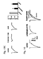
- FIGS. 10A and 10B are a collection of graphs presenting exemplary electrophysiological data collected by patch-clamp recording from neurons repetitively exposed to two different extracellular pHs.
- FIG. 11 is a collection of graphs presenting electrophysiological data collected by patch-clamp recording from mouse cortical neurons shifted to a reduced extracellular pH (pH 6.0), to induce an acid-responsive current, in the presence of various concentrations of the amiloride derivative benzamil.
- FIG. 13 is a set of graphs presenting exemplary electrophysiological data collected by patch-clamp recording from neurons exposed to a drop to pH 6.0 from pH 7.6, followed by different recovery intervals at pH 7.6 before re-exposure to pH 6.0.
- FIG. 14 is a collection of graphs presenting exemplary electrophysiological data collected by patch-clamp recording from neurons exposed to various reduced extracellular pH values at two distinct extracellular Ca 2+ concentrations.
- FIGS. 15A-15C are a set of graphs presenting exemplary electrophysiological data collected by patch-cramp recording from neurons exposed to the antiepileptic drugs ethosuximide, lamotrigine, and felbamate.
- the methods may provide a screen for anti-seizure drugs.
- One or more compositions may be selected based on their ability to affect, such as inhibit, a response of biological cells to a change in extracellular pH, such as one or more drops in extracellular pH, and/or to affect such as selectively inhibit, at least one acid sensing ion channel (ASIC).
- the one or more compositions may be one or more chemical compounds selected based on results of testing a plurality of chemical compounds in an assay designed to measure a cellular response to a drop in extracellular pH and/or to measure an ASIC activity.
- the cellular response and/or ASIC activity may be detected optically and/or electrically, among others.
- At least one drug candidate may be obtained based on the one or more compositions selected.
- the at least one drug candidate may have the same chemical structure as at least one (or all) of the one or more compositions selected and/or may be or include a structural derivative or a set of structural derivatives that are obtained based on at least one of the compositions selected.
- the at least one drug candidate may be assayed for inhibition of experimentally induced seizure-like electrical activity in cells and/or tissues and/or for inhibition of seizures in animals, such as a non-human test species or in humans.
- the methods also or alternatively may provide a treatment for seizures by administration of an effective amount of a drug to a subject prone to seizures and/or having a seizure, in order to suppress seizure activity.
- the drug may include PcTX1 (peptide), a peptide derivative of PcTX1, amiloride, an amiloride derivative or a combination thereof.
- the drug may correspond to a drug candidate identified in a method of screening for anti-seizure drugs, such as described in the preceding paragraph and elsewhere in the present teachings.
- Brain acidosis is a common feature of acute neurological diseases including epileptic seizures and may play an important role in the pathophysiology of neuronal injury. While the pathogenic role of acidosis in brain ischemia has received substantial attention, cellular and molecular mechanisms underlying acid signaling in the epileptic brain and in seizure-induced brain injury have not been described previously.
- PcTX1 also showed inhibition of seizure-like bursting.
- slices prepared from the brains of ASIC1a knockout mice demonstrated reduced sensitivity to low [Mg 2 ⁇ ] o -induced seizure activity.
- the present teachings show an effect of ASIC blockade in an in vivo model of epilepsy.
- Intra-amygdala injection of kainic acid (KA) induced sustained polyspike activity, as measured by EEG, followed by dramatic injury of CA3 neurons.
- Intracerebroventricular injection of PcTX1 reduced both electrographic seizure activity and CA3 neuronal injury.
- compositions that inhibit the activity of ASICs should be antiepileptic.
- FIG. 1 shows a flowchart of an exemplary method 20 of treating seizures.
- a subject prone to seizures and/or having a seizure may be selected, indicated at 22 .
- the subject may be an individual diagnosed as having epilepsy.
- An effective amount of an inhibitor of at least one acid sensing ion channel (ASIC) may be administered to the subject in order to suppress seizure activity, indicated at 24 .
- the inhibitor may be PcTX1 (peptide), a peptide derivative of PcTX1, amiloride, an amiloride derivative, or a combination thereof.
- seizure means an abnormal electrical activity in the brain that results in at least one clinical symptom.
- the electrical activity may be characterized by hypersynchrony, hyperactivity, and/or hyperexcitability of neurons in a portion or all of the brain.
- Exemplary symptoms produced by seizures may include sudden and involuntary muscle contraction (e.g., convulsions), numbness of a part or all of the body, memory loss, loss of consciousness, inability to concentrate, hallucinations, and/or the like. Seizures thus may affect motor, autonomic, cognitive, sensory (visual, auditory, olfactory, taste, feel), and/or emotional function, among others.
- Each seizure may be characterized either as an epileptic seizure, produced by epilepsy, or a non-epileptic seizure with any other cause.
- seizure activity means the abnormal electrical activity associated with at least a portion of one or more seizures.
- seizure means any chronic neurological disorder characterized by recurrent seizures. Each seizure may appear to be unprovoked or may be triggered or provoked by stress, anxiety, sleep deprivation, illness, chemical exposure (e.g., drug abuse or alcohol consumption), photic stimulation (e.g., a flashing/flickering light), and/or the like.
- the disorder may have a cause that is unknown or may be caused, for example, by head trauma, a brain tumor, a genetic predisposition, an infection, a developmental defect, or any combination thereof, among others.
- Exemplary types of epileptic seizures include partial or focal onset seizures, which are localized (at least initially) within the brain, and generalized seizures, which are distributed widely within the brain. Partial seizures may be further categorized as simple partial seizures, which do not affect consciousness, and complex partial seizures, which do affect consciousness. Generalized seizures, which produce a loss of consciousness, may include absence, atonic, clonic, myoclonic, tonic, and tonic-clonic seizures, among others.
- Exemplary seizure syndromes that may be treated include benign focal epilepsies of childhood, childhood absence epilepsy, fetal alcohol syndrome, frontal lobe epilepsy, infantile spasms, juvenile myoclonic epilepsy, Lennox-Gastaut syndrome, occipital lobe epilepsy, or any combination thereof, among others.
- Epilepsy and/or an epileptic seizure may be diagnosed by any suitable technique or combination of techniques including electroencephalography (EEG), magnetoencephalography, magnetic resonance imaging (MRI), positron emission tomography (PET), single photon emission computed tomography (SPECT), or video-EEG, among others.
- EEG electroencephalography
- MRI magnetic resonance imaging
- PET positron emission tomography
- SPECT single photon emission computed tomography
- video-EEG among others.
- to suppress one or more seizures means to reduce the frequency of seizures; to reduce the severity, physical extent, and/or duration of at least one seizure; to substantially prevent at least one seizure; or any combination thereof.
- Seizure suppression for a particular subject may be measurable directly from the subject (e.g., if a seizure is in progress during treatment) and/or, more typically, may be a statistically predicted outcome based on results from controlled tests or clinical trials with a group of subjects.
- seizure-like electrical activity means any electrical activity elicited from isolated cells, a tissue explant, or the brain, in which the electrical activity serves as a model for the electrical activity present in an actual seizure.
- Seizure-like electrical activity may be elicited by exposing cells, tissue, or the brain to electrical stimulation (kindling) (e.g., repetitive and/or high frequency electrical stimulation), a chemical substance(s) (e.g., picrotoxin or kainic acid) (or removal thereof (e.g., removal of kynurenic acid)), photic stimuli (e.g., a flashing/flickering light), or altered ionic conditions (e.g., reduced extracellular Mg 2+ )
- electrical stimulation kindling
- a chemical substance(s) e.g., picrotoxin or kainic acid
- photic stimuli e.g., a flashing/flickering light
- altered ionic conditions e.g.
- drug means a substance, other than food, intended for use in the treatment, prevention, diagnosis, cure, and/or mitigation of a disease, disorder, or condition in humans or animals.
- the drug may be or include a bioactive agent or a mixture of bioactive agents for external or internal use.
- a “candidate drug,” as used herein, means a prospective drug that has not yet been tested fully (or at all) and/or formally approved for use as a drug.
- subject means a many-celled vertebrate or invertebrate organism from the animal kingdom.
- the subject thus may be a person (also termed an individual or a human) or a non-human animal (hereafter, termed only an “animal”).
- exemplary animals include laboratory animals, farm animals, pets, or sport animals, among others.
- An animal subject thus may, for example, be a rodent (such as a mouse, rat, hamster, guinea pig), dog, cow, horse, non-human primate, bird, amphibian, reptile, fish, insect, or the like.
- Non-human subjects may be test species, that is, animals for testing the effect of a composition, chemical compound, or candidate drug, generally prior to human clinical trials and/or use as an approved drug in humans and/or animals.
- non-human: subjects may be drug recipients after a drug has been tested and/or approved, such as for treatment in a veterinary setting.
- prone to seizures means that the subject has been diagnosed as having epilepsy; has a history of seizures, that is, has already suffered one or more seizures; has a disease, disorder, or condition that is known or expected to substantially increase the probability of having a seizure; or any combination thereof.
- the term “having a seizure,” as used herein with respect to a subject, means that the subject currently is suffering a seizure, with abnormal electrical activity in the brain, and is presenting one or more symptoms of seizure. Since seizures have a wide range of durations, subjects having a seizure of longer duration, such as status epilepticus, may be candidates for treatment to attenuate or stop a seizure that is already in progress.
- chemical substance means a material having a definite chemical makeup.
- a chemical substance thus may be a chemical element or a chemical compound.
- chemical compound means a substance consisting of two or more elements chemically bonded in a fixed proportion by mass.
- composition means a chemical substance or a mixture of chemical substances. Accordingly, a composition may be or include one or more chemical elements and/or one or more chemical compounds.
- structural derivative or “derivative,” as used herein with respect to a first chemical compound, means a second chemical compound with a chemical structure that is related to the chemical structure of the first chemical by substitution at one or more positions.
- derivative or “structural derivative” indicate a formal relationship between the chemical structures of the first and second chemical compounds, independent of when and how the first and second chemical compounds were designed and synthesized relative to each other. In other words, according to this definition, if one compound is a derivative of another compound, then both compounds are derivatives of each other.
- the term “effective amount,” as used herein with respect to a substance, means a quantity of the substance that produces a therapeutic response in subjects to which the quantity has been administered.
- the therapeutic response may be produced in any statistically significant proportion of the subjects relative to those receiving placebo, such as at least about 25%, 50%, or 75% of the subjects receiving the effective amount.
- the effective amount may be given to a subject in a single dose or collectively via a plurality of doses.
- pharmaceutical preparation is any pharmaceutically acceptable mixture or composition that contains an effective amount of a drug for administration to a subject in one or more doses.
- pharmaceutically acceptable means approved by a regulatory agency of a federal or state government or listed in the U.S. Pharmacopeia or other generally recognized pharmacopeia for use in animals, and more particularly, in humans.
- the cells may be prokaryotic and/or eukaryotic cells. If eukaryotic cells, the cells may be from any suitable organism, such as cells that are human, non-human primate, murine (mouse), bovine (cow), porcine (pig), ovine (sheep), canine (dog), feline (cat), equine (horse), or the like.
- the cells may be neurons from the brain or elsewhere in the nervous system. Exemplary neurons that may be suitable include hippocampal neurons and/or cortical neurons, among others. Alternatively, or in addition, the cells may be or include non-neurons.
- the cells may be provided by an established cell line, such as tumor cells that are transformed and/or immortalized to facilitate culture, or may be primary cells isolated from tissue, such as by disrupting cell-cell contacts of the tissue, generally without immortalization and/or transformation.
- the cells may be stem cells (e.g., totipotent stem cells, pluripotent stem cells, embryonic stem cells, adult stem cells, and/or the like) or differentiated cells.
- the cells may be neurons or a non-neuronal cell type engineered for use in an ASIC assay.
- the cells may be modified by introduction of foreign nucleic acid to express at least one exogenous ASIC protein (e.g., ASIC1a, ASIC1b, ASIC2a, ASIC2b, ASIC3, ASIC4, or any combination thereof, among others).
- Cells engineered for use in an ASIC assay may be an established cell line or primary cells.
- ASIC acid sensing ion channel
- ASIC1a means any current or future member(s) of the family of proton-activated ion channels.
- ASIC3, and ASIC4 belong to the amiloride-sensitive epithelial Na + -channel/degenerin superfamily. Further aspects of ASICs are described below in Example 6.
- ASIC assay or a grammatical equivalent thereof, as used herein, means any test designed to directly or indirectly measure an activity, such as ion transport, of one or more ASICs.
- the ASIC assay may provide a relatively direct measurement of ASIC activity, such as via an electrical measurement, or may provide a relatively indirect measurement of ASIC activity, such as via detection of a consequence of ion transport.
- the consequence of ion transport may be an immediate consequence or a consequence substantially downstream of the ion transport.
- ASIC assays may be performed on ASICs disposed in whole cells, particularly, live cells, or on cell-free ASICs that are not disposed in whole cells, such as ASICs disposed in isolated membrane patches.
- sample mixture means any mixture that includes a composition to be tested along with one or more reagent (e.g., cells, diluents, buffers, dyes, reactants, substrates. etc.) that permit or facilitate testing.
- reagent e.g., cells, diluents, buffers, dyes, reactants, substrates. etc.
- ASIC inhibitor or a grammatical equivalent thereof, as used herein, means any chemical substance and/or composition capable of substantially reducing (including eliminating) an activity of at least one ASIC, such as the ability of one or more ASICs to conduct an ion current in any suitable ASIC assay.
- An ASIC inhibitor may reduce the magnitude of the ion current and/or the duration of the ion current, among others.
- Substantial reduction of the ion current may be a reduction in magnitude and/or duration of at least about 25%, 50%, 75%, or 90%, among others.
- the ASIC inhibitor also or alternatively may affect the sensitivity of one or more ASICs to activation by protons (pH) and/or may increase the steady-state inactivation or the rate of ASIC desensitization after activation.
- the ASIC inhibitor may be selective or specific for inhibition of ASIC proteins relative to other ion channels, that is, may be capable of inhibiting one or more (or all) ASICs to the substantial exclusion of most (selective) or all (specific) non-ASIC channels.
- the ASIC inhibitor may be selective/specific or nonselective/nonspecific for inhibition within the ASIC family. If specific within the ASIC family, the ASIC inhibitor may be capable of inhibiting one or more ASICs (e.g., ASIC1a only or ASIC1a plus ASIC1b only) to the substantial exclusion of the other ASICs.
- PcTX1 means the spider toxin peptide Psalmotoxin 1 from the tarantula species Psalmopoeus cambridgei (Pc).
- the toxin peptide may be used without substantial purification as part of venom from the tarantula species, may be purified from the venom, may be synthesized chemically, or may be biosynthesized by an engineered organism, among others.
- PcTX1 is effective for inhibiting ASIC1a, the inhibition of ASIC1a has been reported to be specific relative to the other ASIC family members.
- PcTX1 derivative means a peptide with a chemical structure formally related to PcTX1 and distinguished from PcTX1 by one or more amino acid substitutions, deletions, and/or insertions.
- a PcTX1 derivative may be produced de novo or, in some cases, may be synthesized by chemical modification of PcTX1.
- Exemplary derivatives of PcTX1 may include deletions of one or more amino acids, insertions of one or more amino acids, substitution of one or more amino acids with other amino acids or with non-amino acids, or any combination thereof.
- a PcTX1 derivative may have an amino acid sequence with at least about 20%, 40%, 60%, 80%, or 90% amino acid ideentity withh the amino acid sequence of PcTX1, when the sequences are aligned.
- amiloride means a pyrazine derivative that includes a guanidinium moiety, and more particularly having the systematic (IUPAC) name 3,5-diamino-6-chloro-N(diaminomethylidene)pyrazine-2-carboxamide, and corresponding to the structural formula
- Amiloride may be in any suitable nonionic form or ionic form (i.e., as a salt). Amiloride is a blocker of ASICs, although its effect is not specific to ASICs. In particular, over a concentration range at which amiloride is effective for blocking ASICs, amiloride may inhibit other membrane transport proteins, such as Na + /Ca 2+ exchangers, Na + /H + exchangers, and glutamate-operated cationic channels.
- membrane transport proteins such as Na + /Ca 2+ exchangers, Na + /H + exchangers, and glutamate-operated cationic channels.
- amiloride derivative means any structural derivative of amiloride, and more particularly, a chemical compound that is structurally related, to amiloride and distinguished from amiloride by substitution at one or more positions.
- an amiloride derivative is described by the structural formula
- X is a halogen moiety.
- X is typically fluoro, chloro, or bromo. In some embodiments, X is chloro.
- the amino substituents R 1 -R may be selected independently from H, alkyl having 1-12 carbons, arylalkyl having 7-13 carbons, aryl, or heteroaryl.
- substituents R 1 -R 8 is alkyl or arylalkyl
- the alkyl portion of each alkyl or arylalkyl substituent may be optionally and independently further substituted one or more times by halogen, hydroxy, alkoxy having 1-6 carbons, aryl, heteroaryl, amino, alkylamino having 1-6 carbons, dialkylaminio having 2-12 carbons, carboxylic acid, or an ester formally derived from carboxylic acid and an alcohol having 1-6 carbons.
- substituent R 1 -R 8 is aryl, arylalkyl, or heteroaryl the aromatic portion of each aryl, arylalkyl, or heteroaryl substituent may be independently further substituted one or more times by halogen, alkyl having 1-6 carbons, amino, alkylamino having 1-6 carbons, dialkylamino having 2-12 carbons, carboxylic acid, or an ester formally derived from carboxylic acid and an alcohol having 1-6 carbons.
- each of substituents R 1 -R 8 is independently selected from H, alkyl having 1-6 carbons, and arylalkyl, each of which may be further substituted as discussed above.
- amiloride derivative is described by the structural formula
- R 1 , R 2 , R 7 and R 8 are independently H, alkyl having 1-6 carbons, or arylalkyl having 7-13 carbons.
- fect a response of cells means inhibiting (i.e., reducing, slowing, shortening, blocking, etc.), enhancing (i.e., increasing, speeding, lengthening, unblocking, etc.), or otherwise altering any characteristic change in the cells.
- the change may, for example, be a change in membrane potential, current across the membrane, calcium concentration, protein phosphorylation, protein dephosphorylation, reporter gene activity. subcellular localization of a cellular constituent, or any combination thereof among others.
- administering a drug means to give or apply the drug or drug candidate to the subject such that the drug or drug candidate can exert its bioactive effect, if any, on the subject.
- administering a drug may include delivering the drug to a subject by any suitable route, including injection, ingestion, inhalation, topical application, or any combination thereof, among others. Injection may be performed subcutaneously, intradermally, intravenously, intra-arterially, intrathecally, epidurally, subdurally, intracerebroventricularly (i.e., into the brain), intraocularly, intraperitoneally, intra-synovially, or any combination thereof, among others.
- Injection may, for example, be via a needle or may be with a needle-free injector.
- Ingestion may be via a liquid formulation, a capsule, a tablet, or the like.
- Inhalation (or topical application to epithelia in the body) may be via an inhaler, atomizer, sprayer, or the like, and may involve a spray or particles/droplets of any suitable size, such as a spray or particles/droplets configured or sized for delivery to epithelia in the nose, mouth, pharynx, larynx, or lungs, among others.
- Topical application may involve placement of the drug onto an epithelial layer for trans-epithelial uptake.
- Exemplary epithelia for topical application may include external application to the skin or a wound thereof (i.e., direct placement onto the epidermis, dermis, hypodermis, or exposed wound tissue, among others).
- Other exemplary epithelia for topical application may include rectal, vaginal, urethral, oral, nasal, or ocular epithelia, or any combination thereof.
- Topical application may be facilitated by formulating the drug as an ointment and/or by placing the drug onto a dermal patch.
- selecting means to choose, identify, and/or designate one or more members from a set according to one or more criteria. Selecting may be at least substantially or exclusively a cognitive process. Selecting may be performed by one person, at least in part by group discussion or consensus, partly or exclusively by a digital processing device (e g., a computer), or any combination thereof, among others.
- a digital processing device e g., a computer
- obtaining means to come into possession of and/or to bring or cause to fall under the influence of by any suitable means. Accordingly, a chemical compound or composition that is obtained may, for example, be purchased, synthesized, extracted, purified fetched (e.g., from a shelf or stockroom), borrowed, or any combination thereof, among others.
- a subject may be selected to receive an anti-seizure drug. Selection may be performed by any suitable person and/or mechanism. For example, selection may be performed by a medical practitioner, such as a doctor, a nurse, a veterinarian, a medical counselor, and/or the like. Alternatively, or in addition, selection may be performed by the subject himself/herself (e.g., if the drug is available “over the counter” without a prescription from a medical practitioner). In some embodiments, selection may be performed exclusively by, or with the assistance of, a digital processing device (e.g., a computer) that receives data about the subject and analyzes the data using an algorithm to determine whether or not the subject should be selected to receive the anti-seizure drug or drug candidate.
- a digital processing device e.g., a computer
- the subject may be selected based on any suitable criteria. For example, the subject may be selected for being prone to seizures. Selection thus may be based on a medical history of the subject, a previous occurrence of at least one seizure, a clinical test result(s) predicting a relatively higher risk for the subject to suffer a seizure in the future, a present medical condition placing the subject at risk for a seizure, or any combination thereof, among others.
- the medical history may be a written record or an oral communication of the subject's past medical conditions and/or clinical test results.
- the subject may be selected because the subject is currently experiencing a seizure or has one or more signs and/or symptoms indicating a seizure is occurring or is about to occur.
- subjects may be selected to receive a drug candidate, to test the efficacy of the drug candidate in humans or non-human subjects.
- the subjects thus may be selected according to any of the criteria above and/or to provide a representative population for clinical trials or animal testing.
- a drug may be administered to a subject in order to suppress one or more seizures and/or seizure activity.
- the drug may be administered by any of the delivery routes and mechanisms described above in Section I.
- the drug may be administered by a medical practitioner, may be self-administered by the subject, or may be administered by any other person.
- any suitable drug may be administered including PcTX1, a PcTX1 derivative, amiloride, an amiloride derivative, a drug identified in and/or suggested by a screen for ASIC inhibitors, or any combination thereof, among others.
- the drug may be amiloride or an amiloride derivative administered by injection.
- the drug may be PcTX1 or a PcTX1 derivative administered by injection or nasally (i.e., to the nasal cavity).
- nasal administration may promote passage of the drug to the brain.
- the NMDA subtype of glutamate receptors may be involved in seizure-induced brain injury. Activation of ASICs with subsequent membrane depolarization may indirectly facilitate the activation of NMDA receptor channels thus contributing to NMDA receptor-mediated injury. On the other hand, activation of NMDA receptors and subsequent phosphorylation by CaMKII may enhance the activation of ASICs. Blocking ASIC or NMDA activities may therefore induce neuroprotection.
- more effective seizure treatments may be provided by administering, to a subject, both an ASIC inhibitor and an antagonist for NMDA receptor channels, such as amantadine, dextromethorphan, dizocilpine (MK-801), ibogaine, ketamine, memantine, nitrous oxide, phencyclidine, tramadol, or a combination thereof, among others.
- the ASIC inhibitor and NMDA receptor antagonist may be administered in the same pharmaceutical preparation or in distinct pharmaceutical preparations.
- the inhibitor and antagonist may be administered by the same or different delivery mechanisms.
- FIG. 2 shows a flowchart of an exemplary method 30 of screening.
- the steps presented in FIG. 2 may be performed in any suitable order, in any suitable combination, and any suitable number of times. Furthermore, any combination of the steps presented in FIG. 2 may be conflated with any other step(s) described elsewhere in the present teachings to provide a screening method.
- compositions may be tested using an ASIC assay, indicated at 32 .
- the compositions may correspond to a set or library of compositions, with the set/library including any suitable number of compositions, such as at least 2, 10, 100, 1,000, 10,000, or 100,000 compositions.
- Each composition may be or include a different chemical substance and/or chemical compound (or a different mixture of chemical substances/compounds), such that the set/library may be described as a set/library of chemical substances or compounds (e.g., a compound library).
- any suitable chemical compounds may be screened, such as a library of small organic molecules (e.g., with an average molecular weight of less than 5 or 2 kilodaltons), peptides, nucleic acids, lipids, carbohydrates, known ion transport agonist/antagonists, or any combination thereof.
- Members of the library may be tested individually or as mixtures of chemical substances/compounds.
- the compositions may be tested in an array format, such as with microplates providing an array of wells for holding sample mixtures.
- Exemplary microplates that may be suitable include microplates with 24, 96, 384, and 1536 wells.
- two or more members of a library may be tested individually and in parallel on cells using a multiplexed patch-clamp system, such as via planar patch clamp electrodes disposed in an array.
- compositions may be tested as a part of sample mixtures in any suitable assay that reflects a response of the sample mixtures to a change in pH and/or that reflects an ASIC activity in the sample mixtures.
- the assays thus may be performed with cells or in a cell-free system.
- the change in pH may be a reduced pH, such as a reduced extracellular pH.
- Sample mixtures used for testing thus may be exposed to a drop in pH and/or to a pH that is less than physiological pH (e.g., less than pH 7.4).
- a reduced pH may be imposed once or a plurality of times for each sample mixture.
- the sample mixture may be exposed to a repetitively varying pH (such as at least, in order, a pH decrease, a pH increase, and a pH decrease), which may be suitable for identifying compositions that affect (e.g., enhance) ASIC desensitization resulting from a drop in pH and/or that affect (e.g., inhibit) recovery from ASIC desensitization when the pH is raised after the drop (e.g., see Examples 1 and 3).
- Repetitive changes in pH may be pulses of reduced pH, separated by intervals of increased pH. Each pulse of reduced pH may have any suitable length (duration), such as about 0.1-10, 0.2-5, or 0.5-2 seconds.
- each interval of increased pH may have any suitable length, such as about the same as, shorter than, or longer than the length of the reduced pH pulses.
- the pulses and/or intervals may be uniform or nonuniform.
- the pulses and/or intervals may have any suitable frequency, such as at least about 0.2, 0.5, 1, or 2 Hz, or 0.1-10, 0.2-5, or 0.5-2 Hz, among others.
- a drop in pH may be imposed by any suitable technique, such as changing a fluid (e.g., media and/or buffer) in which the sample mixtures and/or cells thereof are disposed, to another fluid of lower pH, or by adding acid to or releasing protons in (e.g., by chemical reaction) the fluid, among others.
- the drop in pH for a sample mixture may be executed over any suitable time interval, such as a sudden, stepwise drop in pH (e.g., in less than 1 or 0.1 second), or a slower, more gradual drop in pH (e.g., in greater than one second).
- the pH of a sample mixture may be reduced by at least about 0.1, 0.2, 0.5, or 1.0 pH unit from the pH value of the sample mixture immediately prior to pH reduction and/or from physiological pH.
- one or more ASICs may be activated by dropping the pH of the sample mixture from 7.4 to 6.5 or 6.0, since an ASIC activity (e.g., an ASIC-mediated current) of readily detectable magnitude may be recorded at these pH values.
- a smaller pH drop e.g., from pH 7.4 to pH 7.2, 7.1, or 7.0
- the smaller pH drop may be more analogous to the pH drop in an actual seizure.
- a current-clamp configuration may be used to study the effect of test compositions and/or candidate drugs on membrane depolarization induced by relatively small pH drops (e.g., to pH 7.1), which may be more relevant to epilepsy.
- Significant membrane depolarization e.g., at least about 10 mV
- any suitable aspect of a sample mixture may be detected when the compositions are tested.
- exemplary aspects may include an optical parameter, such as fluorescence, absorbance, polarization, scattering, reflection, refraction, birefringence, or any combination thereof.
- the optical parameter may be measured in a spatially resolved fashion from a sample mixture, such as by cell imaging, or may be measured from a sample mixture without spatial resolution of the sample mixture.
- the optical parameter may be measured at steady state (an end-point measurement) or while the optical parameter is changing, for example, in a time-resolved fashion (a kinetic measurement(s)).
- the optical parameter may be monitored over any suitable time interval or detected at any suitable time point after a change in pH, such as less than or greater than about 1, 10, or 60 seconds.
- testing may be performed on sample mixtures in the presence of a fluorescent dye, such as a calcium-sensitive dye (e.g., Fura-2, Fura-2AM, Fluo-3, Indo-1, Rhod-2, calcium-sensitive GFP (e.g., Cameleons), etc.), a membrane potential dye (e.g., Di-4-ANEPPS, RH421, DiBAC 4 (3), Tetramethylrhodamine ethyl/methyl ester perchlorate, etc.), or the like.
- a fluorescent dye such as a calcium-sensitive dye (e.g., Fura-2, Fura-2AM, Fluo-3, Indo-1, Rhod-2, calcium-sensitive GFP (e.g., Cameleons), etc.)
- a membrane potential dye e.g., Di-4-ANEPPS, RH421, DiBAC 4 (3), Tetramethylrhodamine ethyl/methyl ester perchlorate, etc.
- an electrical parameter of a sample mixture may be measured electrically such as by patch-clamp analysis of cells and/or membranes. Recording in the patch-clamp analysis may be on a cell-attached patch, an inside-out patch, an outside-out patch, a perforated patch, or may be a whole cell recording, among others. Furthermore, the patch-clamp recording may be performed in a current-clamp or voltage-clamp mode.
- testing may measure an ability, if any, of each composition to inhibit ASIC activity and/or to inhibit a response of cells to a reduced extracellular pH.
- compositions having an effect in the ASIC assay may be selected, indicated at 34 .
- the one or more compositions selected may correspond to one or more individual chemical compounds or at least one chemical compound mixture. If the one or more selected compositions correspond to at least one compound mixture, chemical compounds of the compound mixture may be tested individually using an ASIC assay and/or an assay for a cellular response to reduced pH, in order to identify individual compounds that affect either or both of the assays.
- At least one drug candidate may be obtained based on the one or more compositions selected, indicated at 36 .
- the at least one drug candidate may have the same chemical structure as at least one of the compositions selected or may be a structural derivative thereof.
- a composition may be selected from the step of testing and then may be used to provide a structural basis for obtaining a set of one or more structural derivatives related to the composition.
- the at least one drug candidate may be assayed for an ability to suppress seizure-like electrical activity and/or seizures, indicated at 38 . Accordingly, the at least one drug candidate may be assayed in a seizure model system provided by isolated cells, tissue explants, and/or animals, and/or may be tested in a clinical trial.
- Seizure-like activity may be provided by in vitro preparations (isolated cells and/or tissue explants) that offer a variety of options for studying the mechanisms of the generation, spread, and termination of seizures using methods that are difficult to employ under in vivo conditions.
- in vitro models may permit precise control of temperature and extracellular environment.
- in vitro models generally lack the behavioral and motor components of clinical seizures. Therefore, in vitro models generally rely on “equivalents” of seizures (seizure-like activities) that have been observed in vivo. Such equivalents may be characteristic changes of electrical activity and/or the ionic environment.
- the at least one drug candidate may be assayed on isolated cells.
- primary neurons or an established cell line
- the ability, if any, of the at least one drug candidate to inhibit or otherwise affect the seizure-like electrical activity may be assayed, for example, using a patch-clamp technique.
- the at least one drug candidate may be a plurality of drug candidates assayed in parallel using a multiplexed patch-clamp system, such as with planar patch-clamp electrodes.
- the at least one drug candidate may be assayed on tissue explants (e.g., hippocampal or cortical slices) from brain with seizure-like activity induced by exposure to electrical stimulation, a pharmacological agent (e.g., picrotoxin or kainic acid), a change in ionic environment (e.g., a switch to low Mg 2+ ), a combination thereof, or the like.
- tissue explants e.g., hippocampal or cortical slices
- a pharmacological agent e.g., picrotoxin or kainic acid
- a change in ionic environment e.g., a switch to low Mg 2+
- Reduced Mg 2+ may induce seizure-like bursts and is a commonly used in vitro epilepsy model.
- removing Mg 2+ from artificial CerebroSpinal Fluid may induce spontaneous and triggered interictal-like bursting, followed by spontaneous ictal-like events and finally periodic clustered bursts.
- the ictal-like events may consist of a tonic firing phase and a phase of clustered burst discharges resembling the tonic and clonic phases of seizures.
- This model may be clinically relevant because low levels of Mg 2+ may be associated with human epilepsy and because this model has been used to test the effect of antiepileptic agents.
- tissue explants may have advantages over cultured cells, including (1) more options for inducing seizure-like activity, as described above and elsewhere in the present teaching, and/or (2) more choices for measuring the efficacy of a drug candidate, such as electrophysiological measurements with electrodes, staining the explant to determine the extent of injury (e.g., with propidium iodide), performing an enzyme assay on the explant (e.g., lactate dehydrogenase release to measure cell death), or the like. Further aspects of the use of tissue explants from brain are described below in Example 1.
- the at least one drug candidate may be assayed in animals (e.g., mice) using electrical stimulation (kindling), photic stimulation (e.g., a flashing/flickering light), or pharmacological induction as models of epileptogenesis.
- animals e.g., mice
- electrical stimulation kindling
- photic stimulation e.g., a flashing/flickering light
- pharmacological induction as models of epileptogenesis.
- KA kainic acid
- the KA model may mimic the pathogenesis of human mesial temporal lobe epilepsy, a common form of human focal epilepsy, which is frequently associated with progression to chronic intractable, drug resistant epilepsy.
- status epilepticus may be elicited, which in turn may lead to a characteristic pattern of hippocampal cell death (primarily the CA3 region).
- the KA model may be well suited to study epileptogenesis through electroencephalographic (EEG) monitoring of seizure activities and evaluation of resultant hippocampal cell death.
- EEG monitoring continuous or periodic, among others
- the percentage of time that EEG recordings show polyspike (type-4) activity may be quantified.
- the type-4 EEG activity may correlate best (e.g., linearly) with seizure-induced brain injury. Animals injected with amiloride, PcTX1, or an antiepileptic drug may be used as positive controls for comparison with drug candidates being assayed.
- Amiloride, PcTX1, and a drug candidate suitable for selection as an anti-seizure drug may show no or reduced type-4 activity, and less CA3 neuronal injury in the KA model.
- the brains of the animals also may be analyzed for seizure-induced injury, such as with an apoptosis stain (e.g., propidium iodide staining and/or a TUNEL assay), histological staining (e.g., with cresyl violet), and/or the like.
- an apoptosis stain e.g., propidium iodide staining and/or a TUNEL assay
- histological staining e.g., with cresyl violet
- Example 1 Further aspects of the KA model and its use to assay ASIC inhibitors are described below in Example 1.
- any of the tests/assays described herein, or controls therefore, may be performed in the presence of inhibitors of selected membrane transport proteins.
- blockers for various Ca 2+ -channels e.g., 5 ⁇ M nimodipine for L-type, 1 ⁇ M ⁇ -conotoxin MVIIC for N— and P/Q-type, and 200 nM SNX482 for R-type Ca 2+ -channels
- glutamate receptors MK801 10 ⁇ M, CNQX 20 ⁇ M
- Brain acidosis is a common feature of acute neurological aberrations including epileptic seizures (Chesler et al., 1992; Siesjo et al., 1996; Siesjo et al., 1986; Simon, 2006), and plays an important role in neuronal injury. While the pathogenic role of acidosis, and activation of acid-sensing ion channels (ASICs) (Waldmann et al., 1997), have received substantial attention in brain ischemia (Benveniste et al., 2005; Huang et al., 2004; Xiong et al. 2004), the role of ASICs underlying acid signaling in epileptic brain and in seizure-induced neuronal injury remains unexplored.
- ASICs acid-sensing ion channels
- ASIC1a blockade inhibits synchronous paroxysmal depolarization shifts.
- ASIC1a blockade suppresses low Mg 2+ -induced epileptiform activity, and electrical stimulation-induced after-discharges. Consistent with pharmacologic blockade, slices from ASIC1 knockout mice show resistance to seizure induction.
- ASIC1a blockers In a kainic acid model of status epilepticus, a common in vivo model of limbic seizures, intracerebroventricular injection of ASIC1a blockers, or ASIC1 gene knockout, reduces electrographic seizure activity and CA3 neuronal injury. Thus, activation of ASICs plays an important role in the propagation of epileptic seizures and constitutes a new and novel target for antiepileptic drug therapy.
- the ASIC subunit non-specific blocker amiloride (Waldmann et al., 1997; Xiong et al., 2004), and specific homomeric ASIC1a channel antagonist PcTX1 (Xiong et al. 2004; Escoubas et al., 2000), perfused to cells 2 min before and during Kyn withdrawal resulted in attenuated Kyn withdrawal-induced sustained membrane depolarization and paroxysmal bursts ( FIGS. 3A and 3B ). Without amiloride, Kyn withdrawal-induced sustained depolarization of 39.5 ⁇ 2.1 mV and paroxysmal bursts at 4.0 ⁇ 0.5 Hz.
- Limbic seizures are modeled by removing Mg 2+ from artificial cerebral spinal fluid (aCSF) in slices of hippocampal-entorhinal cortex, which triggers interictal-like bursting activity, or spontaneous ictal-like events (Wong et al., 2001; Anderson et al., 1986).
- aCSF artificial cerebral spinal fluid
- stimulation of Schaffer collaterals induces single population spike or EPSP in the CA1 region of the hippocampus.
- the amplitude of EPSP also increased without generating multiple population spikes or bursting activity.
- Perfusion with Mg 2+ -free aCSF for about 30 min evoked synchronous burst firing with the same stimulation intensity.
- ASIC blockade on seizure-like bursting activity induced by high frequency stimulus trains e.g., after-discharges (Stasheff et al., 1985) was determined as high frequency electric stimulation generates epileptic bursting without disrupting the balance of excitation versus inhibition thus more closely mimicking epileptogenesis in vivo (Stasheff et al., 1985).
- high frequency electric stimulation generates epileptic bursting without disrupting the balance of excitation versus inhibition thus more closely mimicking epileptogenesis in vivo
- FIGS. 7A-7E relatively stable after-discharges were recorded ( FIGS. 7A-7E ).
- ASIC1 ⁇ / ⁇ slices The resistance of ASIC1 ⁇ / ⁇ slices to seizure induction was further demonstrated in detailed plots of stimulation train-dependent development of after-discharges ( FIG. 7D ).
- Seizures produce high frequency synchronous firing of neuronal populations. Since synaptic vesicles are highly acidic (pH ⁇ 5.7) (Miesenbock et al., 1998), release of neurotransmitter is expected to induce high frequency fluctuation of extracellular pH (pH o ) at the synaptic cleft (Chesler et al., 1992) where ASIC1a channels are enriched (Zha et al., 2006). Fluctuation of pH o during seizure activity is expected to activate these channels, facilitating neuronal excitation. However, ASICs undergo desensitization following activation (Hesselager et al., 2004).
- the channels need to recover rapidly from desensitization to be activated repeatedly by high frequency acid pulses.
- ASIC current can be re-activated by acid application (Wang et al., 2006).
- ASIC current induced by short (0.3-0.5 second), high frequency repeated acid pulses.
- ASICs only partially desensitize or no desensitization occurs.
- the channel is activated again by the next acid pulse.
- FIGS. 10A and 10B ASIC current can be repeatedly activated for prolonged periods with little attenuation. This finding provides additional evidence that ASICs can play a role during high frequency acid fluctuation expected to occur in seizure activity.
- ASIC1a activation occurs repeatedly by high frequency acid pulses with little attenuation.
- ASIC1 blockade and ASIC1 gene knockout attenuate seizure propagation.
- FIG. 3 ASIC1 blockade inhibits seizure-like burst activity in a cell culture model of epilepsy.
- A Representative seizure-like burst activity induced by withdrawal of kynurenic acid (Kyn). Following long-term (about 4 weeks) culture of mouse hippocampal neurons in the presence of Kyn and high Mg 2+ , withdrawal of Kyn induces seizure-like activity consisting of high frequency bursts and synchronous depolarization shifts (Furshpan et al., 1989). Bath application of the non-selective ASIC1 blocker amiloride significantly suppresses the seizure-like activity.
- B Extended time-scale showing individual depolarization shifts before and after bath application of 30 ⁇ M amiloride.
- FIG. 4 Representative seizure-like burst activity induced by withdrawal of kynurenic acid (Kyn) in the absence and presence of specific ASIC1a blocker PcTX1 (Xiong et al., 2004). Following long-term ( ⁇ 4 weeks) culture of mouse hippocampal neurons in the presence of Kyn and high Mg 2+ , withdrawal of Kyn induced seizure-like activity consisting of high frequency bursts of synchronous depolarization shifts. Bath perfusion with PcTX1 venom (200 ng/mL total protein) reduced Kyn withdrawal-induced seizure-like synchronous depolarization shifts (see FIGS. 3C and 3D for more detail).
- FIG. 5 ASIC1 blockade inhibits seizure-like burst activity in mouse hippocampal slices.
- A. In the absence of Mg 2+ , single electrical stimulation of a Shaffer collateral pathway can evoke seizure-like busting activity in CA1 region. Bath perfusion of the non-selective ASIC1 blocker amiloride reduces the amplitude and frequency of evoked bursting activity.
- A-a. Representative population spikes evoked by a single electrical stimulation in the absence and presence of 100 ⁇ M amiloride
- B-b. Summary data showing reduction in the amplitude of the first population spike by PcTX1 (n 9, *p ⁇ 0.05).
- FIG. 6 Representative field-EPSPs and summary data showing the lack of inhibition by amiloride on basal synaptic transmission.
- Mg 2+ 1.0 mM
- fEPSP induced field excitatory postsynaptic potential
- FIG. 7 ASIC blockade or ASIC1 gene knockout suppresses stimulus train induced after-discharges.
- A-a Representative after-discharges induced by stimulus trains in the absence and presence of amiloride.
- B-a Representative after-discharges induced by stimulus trains in the absence and presence of 200 ng/mL. PcTX1.
- FIG. 8 ASIC1 blockade or ASIC1 gene knockout inhibits epileptic seizure activity and seizure-induced CA3 neuronal injury in an in vivo model of epilepsy.
- A Representative brain sections showing image of hippocampus CA3 neuronal injury in aCSF-injected but not in PcTX1-injected mice 24 hr following KA injection. Lower panels represent enlarged CA3 region showing TUNEL-positive staining (i.e., cell death) in mice injected with aCSF but not PcTX1. (TUNEL-positive cells are visible in this inverted image as black dots.)
- B A. Representative brain sections showing image of hippocampus CA3 neuronal injury in aCSF-injected but not in PcTX1-injected mice 24 hr following KA injection. Lower panels represent enlarged CA3 region showing TUNEL-positive staining (i.e., cell death) in mice injected with aCSF but not PcTX1. (TUNEL-positive cells are
- C Representative EEG recording showing different types of brain activity. Type 1-baseline; Types 2 and 3-ictal onset; Type 4-polyspike paroxysmal burst.
- FIG. 9 Moderate pH drops reported during seizure activity can cause neuronal excitation through ASIC1a activation.
- A. Representative raw data and summary graphs showing reduction of acid pH 7.0)-induced membrane depolarization and increased firing of action potentials in mouse hippocampal neurons by 100 ⁇ M amiloride (n 4 *p ⁇ 0.05, **p ⁇ 0.01).
- B. Representative raw data and summary graphs showing reduction of acid (pH 7.0)-induced membrane depolarization and increased neuronal firing of action potentials by 20 nM synthetic PcTX1 (n 3, *p ⁇ 0.05).
- FIG. 10 Evidence that ASIC1a channels can be repeatedly activated by high frequency acid pulses.
- A Representative current traces showing ASIC1a current activated by repeated acid pulses (pH 6.5) at different membrane potentials as indicated. The duration of acid pulse is 0.5 sec. The time between the end of the first acid pulse and the beginning of the next acid pulse is 0.5 sec. The same findings were recorded in five cells.
- B Representative current traces showing ASIC1a current activated by repeated acid pulses of 0.3 second in duration, spaced by 0.5 second intervals.
- Cortical neuronal culture Primary neuronal cultures were prepared from embryonic Swiss mice at 15 to 16 days of gestation according to our previously described techniques (Xiong et al., 2004). In brief, cerebral cortices from 10 to 12 embryos were dissected and incubated with 0.05% trypsin-EDTA for 10 min, dissociated by trituration with a fire-polished glass pipette, and plated on poly-L-ornithine-coated culture dishes at a density of 0.5 ⁇ 10 6 cells per dish. Neurons were cultured with Neurobasal medium supplemented with B27 (Invitrogen). The cultures were maintained at 37° C. in a humidified 5% CO 2 atmosphere incubator and were used for experiments after 12-14 days.
- Patch clamp techniques Whole-cell voltage-clamp and current-clamp recordings were performed as described in our previous studies (Xiong et al., 2004). Patch electrodes were constructed from thin-welled borosilicate glass (1.5 mm diameter, WPI, Sarasota, Fla., USA) on a two-stage puller (PP83 Narishige, Tokyo). The resistances of patch electrodes were 1.5-3 M ⁇ when filled with the intracellular solution (see below). Whole-cell currents were recorded using Axopatch 200B amplifiers with pCLAMP software (Axon Instruments, CA, USA). Data were filtered at 2 kHz and digitized at 5 kHz using Digidata 1322 (Axon Instruments).
- Hippocampal slices were obtained from 8- to 10-week-old mice.
- the protocol for brain-slice recording was approved by the Institutional Animal Care and Use Committee of Legacy Research. Animals were deeply anesthetized with isoflurane and decapitated. Brains were quickly removed and placed in ice-cold solution that contained: 125 mM sucrose, 2.5 mM KCl, 26 mM NaHCO 3 , 1.25 mM NaHPO 4 , 6.0 mM MgSO 2 , 0.5 mM CaCl 2 , and 10 mM glucose, and was saturated with 95% O 2 and 5% CO 2 (pH 7.4).
- Hippocamnpal slices were cut in 400 ⁇ m thickness in transverse plane with a vibratome (Leica VT 1000S) and placed in a slice holding chamber (BSC-PC Prechamber, Warner Instrument) and incubated in normal artificial cerebral spinal fluid (aCSF) that contained: 125 mM NaCl, 2.5 mM KCl, 26 mM NaHCO 3 , 1.25 mM NaHPO 2 , 1.0 mM MgSO 4 , 2.0 mM CaCl 2 , and 10 mM glucose, and was saturated with 95% O 2 and 5% CO 2 (pH 7.4).
- aCSF normal artificial cerebral spinal fluid
- Mg 2+ -free aCSF was perfused to the slices for ⁇ 30 min.
- each stimulation train consisted of 120 pulses at 60 Hz and each pulse was 100 ⁇ sec in duration (Stasheff et al., 1985).
- a 10-minute interval of non-stimulation was provided between successive trains of high frequency stimulation.
- Stimulation electrodes were located in striatum radiatum between CA1 and CA3 region. The population spikes were recorded from striatum pyramidale of the CA3 region.
- the stimulus trains generate epileptic bursting by repetitive and strong electrical stimulations without disrupting the system from the balanced neuronal circuits (excitation vs. inhibition).
- electrical stimulation models may more closely mimic epileptogenesis in vivo.
- TUNEL staining Following cryostat sectioning, slices were air-dried, fixed in 10% formalin for 15 min, washed three times in PBS, permeabilized in 3% Triton X-100 for 20 min, and washed three times in PBS. Slices were subsequently incubated with a reaction mixture that contained FITC-dUTP and 300 U/mL terminal deoxy-transferase (TdT) for 90 min at 37° C. Cultures were then viewed with a fluorescent microscope at an excitation/emission wavelength of 500/550 nm (green) for FITC-TUNEL labeled cells.
- TdT terminal deoxy-transferase
- Intracerebroventricular injection in mice The procedure was performed as described in our previous studies (Pignataro et al., 2007; Xiong et al., 2004). A burr hole was drilled through the skull and a cannula inserted in the right lateral ventricle. The cannula was inserted stereotactically at 0.5 mm posterior, 1.0 mm lateral, and 3.0 mm ventral to the bregma, 0.5 ⁇ L of aCSF, amiloride, or PcTX1 was infused 60 min before KA injection.
- mice After anesthesia and catheterization of the femoral vein, animals were placed in a stereotactic frame modified with a headpiece compatible for the mouse (Kopf Instruments) and kept under anesthesia using a mixture of 68.5% N 2 O, 30% O 2 , and 1.5% isoflurane. Rectal body temperature was kept at 37 ⁇ 1° C. during and after the operation using either a heating pad or heating lamp (Harvard Instruments, Holiiston, Mass.). Using dental cement (Plastics One, Inc.), three skull-mounted recording electrodes were affixed to mice (Plastics One, Inc., Roanoke, Va.) and a 26-gauge steel guide cannula over the intact dura.
- mice Twenty-four hours following KA injection, mice were killed and brains were immediately frozen in 2-methyl butane ( ⁇ 30° C.) and sectioned at 12 ⁇ m on a cryostat. Coronal sections at the level of Bregma ⁇ 1.7 mm were air dried (15 min), post-fixed in 10% formalin (10 min), washed twice in PBS, and then processed for histopathology (cresyl violet staining) (Araki et al., 2002).
- the shutter and filter wheel (Lambda 10-2) were also controlled by AIW to allow illumination of cells at either 340 nm or 380 nm excitation wavelengths. Imaging was detected at an emission wavelength of 510 nm. 340/380 nm ratio images were analyzed by averaging pixel ratio values in circumscribed regions of cells in the field of view.
- Organotypic brain slice culture Organotypic brain slices from postnatal C57BL/6, ASIC1a and ASIC2a knockout mice were cultured using an interface method. Whole brains from 10 day-old mice were removed after being anesthetized with halothane. The brains were rapidly removed from the skull and placed in sterile, ice-cold dissecting medium (50% MEM, 50% Hanks BSS, 20 mM Hepes, 6.5 mg/mL glucose, pH 7.15, 10 units/mL penicillirn, and 10 ⁇ g/mL streprtonmycin).
- sterile, ice-cold dissecting medium 50% MEM, 50% Hanks BSS, 20 mM Hepes, 6.5 mg/mL glucose, pH 7.15, 10 units/mL penicillirn, and 10 ⁇ g/mL streprtonmycin.
- the brains were then placed on the stage of a vibratome (Leica VT 1000), and sectioned coronally at 400 microns and floated into dissecting media. With the aid of a sterile red sable brush (Ted Pella, Inc. Redding, Calif.), slices were transferred to Millicell culture plate inserts with microporous membranes (pore size 0.4 ⁇ m, PICM0R50, Millipore, Bedford, Mass.).
- the inserts were then placed in a 6-well dish containing culture medium (50% MEM with Hanks salts and L-glutamine, 25% Hanks BSS, 25% horse serum, 20 mM Hepes, 6.5 mg/mL glucose, 10 units/mL penicillin and 10 ⁇ g/mL streptomycin, pH 7.2).
- culture medium 50% MEM with Hanks salts and L-glutamine, 25% Hanks BSS, 25% horse serum, 20 mM Hepes, 6.5 mg/mL glucose, 10 units/mL penicillin and 10 ⁇ g/mL streptomycin, pH 7.2.
- the slices were maintained in a regular incubator at 37° C. with a 15% CO 2 enriched atmosphere.
- the medium was changed twice a week. Seizure experiments were performed on the slices at 7-10 days in culture.
- LDH Assay Lactate dehydrogenase (LDH) is a stable cytoplasmic enzyme present in all cells. It is rapidly released into the cell culture supernatant upon damage of the plasma membrane and the concentration of LDH released is proportional to the number of cells damaged. Therefore, LDH measurement has been commonly used for cell injury studies. LDH assays were performed using the Cytotoxicity Detection Kit (Cat#1644793, Roche Molecular Biochemicals) and a spectrophotometric mutiwell plate reader (SPECTRAmax, Molecular Devices). Samples (100 ⁇ L) of cell culture or brain slice medium were collected in a 96-well plate(s) and mixed with the reaction solution (100 ⁇ L) from the kit. Absorbance at OD 490 nm was measured 30 min after the reaction. Absorbance at a reference wavelength (OD 620 nm) was subtracted from the measurement.
- SPECTRAmax spectrophotometric mutiwell plate reader
- Mg 2+ -free aCSF was applied to induce epileptic burst discharges.
- Mg 2+ -free aCSF was obtained by simply omitting MgSO 4 from aCSF.
- This example describes patch-clamp experiments performed on mouse cortical neurons in culture to test the effect of the amiloride derivatives benzamil and 5-(N-ethyl-N-isopropyl)-amiloride (EIPA) on an acid-induced ASIC current; see FIGS. 11 and 12 .
- EIPA 5-(N-ethyl-N-isopropyl)-amiloride
- Mouse cortical neurons in culture were monitored electrophysiologically by patch-clamp recording of current at a holding potential of ⁇ 60 mV.
- An ASIC current was induced at this holding potential by dropping the extracellular pH from physiological pH (pH 7.4) to pH 6.0, in the presence of different concentrations of benzamil ( FIG. 11 ) or EIPA ( FIG. 12 ).
- the current traces obtained are presented in the upper part of FIGS. 11 and 12 .
- the amplitude of the measured ASIC current was plotted against the concentration of benzamil or EIPA ( ⁇ M), as shown in the lower parts of FIGS. 11 and 12 to determine an IC 50 (concentration at 50% inhibition) of 9.55 ⁇ M for benzamil and about 20 ⁇ M for EIPA. Therefore, amiloride derivatives inhibit ASIC channel activity and may be used for seizure suppression in subjects prone to seizures and/or having a seizure.
- This example describes experiments performed by patch-clamp analysis of mouse cortical neurons in culture to determine the rate of re-sensitization of ASICs at physiological pH after an ASIC desensitization produced by a drop in extracellular pH; see FIG. 13 .
- Patch electrodes contained (in mM): 140 CsF, 2.0 MgCl2. 1.0 CaCl2, 10 Hepes, 11 EGTA, 2 MgATP, pH 7.3, using CsOH.
- a multi-barrel fast perfusion system (SF-77B, Warner Instrument Co.) was employed to achieve a rapid exchange of solutions.
- ASIC currents were activated by pairs of acid pulses with increasing intervals between the first and the second acid pulse, and a 2 min interval was given for a complete recovery of the channel before the next pair of acid applications. ASIC current was first activated by a 10 sec acid pulse (pH 6.0).
- a short (1 sec) second acid pulse (pH 6.0) was applied to the neuron at different times following the 1st acid application.
- the relative amplitude of the second ASIC current versus the first one was then plotted against the time intervals between the end of the first acid pulse and the start of the second pulse.
- the time constant for the recovery of the ASIC current from its desensitization is then derived by an exponential fit.
- FIG. 13 shows that one second following a complete desensitization of the ASIC current, a significant amount of ASIC current can be activated again by another acid application.
- This example measures the effect of extracellular calcium on ASIC currents in isolated neurons as a function of pH; see FIG. 14 .
- Decreases of [Ca 2+ ] e may be enhanced dramatically during seizure activity.
- pentetrazol-induced seizure for example, a decrease of [Ca 2+ ] e by 0.7-1.0 mM has been recorded.
- the fall of [Ca 2+ ] e often preceded the onset of seizure events, indicating that the fall of [Ca 2+ ] e might be responsible for initiating the seizure activities.
- Decrease of [Ca 2+ ] e has also been observed by others in chronic models of epilepsy including a kindling model and photically induced seizures.
- Decreases in [Ca 2+ ] e are known to increase neuronal excitability. The mechanism by which lowering [Ca 2+] e enhances neuronal excitability is, however, not fully understood, although a host of possibilities has been suggested by others.
- This example describes the effects of three antiepileptic drugs, ethosuximide, felbamate, and lamotrigine, on ASIC currents in isolated neurons; see FIGS. 15A-15C and 16 .
- Drugs effective in control of partial and generalized tonic-clonic seizures are use and voltage-dependent blockers of Na + channels. Examples include phenytoin, carbamazepine, valproic acid, and lamotrigine. These agents selectively dampen pathologic activation of Na + -channels, without affecting normal Na + -channel function. Drugs effective in control of generalized absence seizures likely block low threshold T-type calcium currents. Examples include ethosuximide, trimethadione, and methsuximide. Agents that augment function of GABA A receptors, e.g., diazepam and clonazepam, have broad-spectrum antiepileptic effects.
- LTG lamotrigine
- phenytoin and carbamazepine are ineffective against the absence seizure. Therefore, additional mechanism(s) might be involved in the effects of LTG, to account for its broad clinical efficacy.
- ASICs Activation of ASICs may be involved in increased neuronal excitability and thus seizure generation. Inhibition of ASICs, on the other hand, is expected to be antiepileptic.
- Three commonly used antiepileptic agents were tested on an ASIC current activated by a pH drop from 7.4 to 6.0. As shown in FIG.
- ASICs activated by a drop of the extracellular pH (pH o ) or by an increase of proton concentration, belong to the amiloride-sensitive epithelial Na + -channel/degenerin superfamily. Members of this family of ion channels may contain two transmembrane spanning regions flanked by a large extracellular loop and short intracellular N— and C-termini.
- ASIC1-ASIC4 encoding six different ASIC members have been cloned to date.
- ASIC1a also named ASIC or BNaC2
- ASIC1a may be enriched in primary sensory neurons and in most brain regions.
- ASIC1a channels are reported to be activated by moderate decreases of pH o ; the pH for half-maximal activation (pH 0.5 ) is ⁇ 6.2.
- homomeric ASIC1a channels may be permeable to Ca 2+ ions.
- ASIC1 ⁇ (or ASIC1b), a splice variant of ASIC1a, has been reported to be expressed only in sensory neurons. When expressed in heterologous systems, ASIC1 ⁇ apparently forms homomeric channels with a pH 0.5 of ⁇ 5.9. Different from ASIC1a which is Ca 2+ -permeable, ASIC1 ⁇ is reported to have little Ca 2+ permeability.
- ASIC2a also named MDEG, or BNaC1
- ASIC2a also named MDEG, or BNaC1
- ASIC2b subunits or MDEG2
- ASIC3 also named DRASIC
- DRASIC DRASIC
- Homomeric ASIC3 is thought to respond to pH drops biphasically with a fast desensitizing current followed by a sustained component. Recently cloned ASIC4 apparently shows high levels of expression in the pituitary gland. However, ASIC4 may not form functional acid-sensing channels alone.
- ASICs Like other ligand-gated ion channels, ASICs are believed to assemble from homomultimeric or heteromultimeric subunits. The exact subunit combination of ASICs in native neurons, however, is not clear. In the past five years, the electrophysiological properties and pharmacological profiles of recombinant homomeric and heteromeric ASICs in heterologous expression systems have been investigated extensively. These studies have provided information for elucidating the subunit composition of ASICs in native neurons, since different homomeric and heteromeric ASICs have distinct pH sensitivity, ion selectivity, and channel kinetics.
- ASICs have been implicated in mechanosensation and perception of pain during tissue acidosis, particularly in the ischemic myocardium where ASICs likely transduce anginal pain.
- ASICS activation of ASICS is involved in taste transduction, and maintenance of retinal integrity.
- ASIC1a is involved in synaptic plasticity, learning/memory, and fear conditioning.
- Our own recent studies demonstrated that activation of Ca 2+ -permeable ASIC1a is also responsible for glutamate-independent, acidosis mediated, ischemic brain injury, disclosing a novel therapeutic target for stroke patients.
- Human temporal lobe epilepsy may be associated with hippocampal sclerosis in which dentate hilus, CA3, and CA1 neurons are lost, and may be accompanied by mossy fiber sprouting. Similar neuropathological findings have been reported in in vivo experimental models of epilepsy such as kainic acid-lesioned rat hippocampus, in which CA3 neurons and their synapses on to CA1 pyramidal cells are susceptible to cell death. Whether cell loss, synaptic reorganization, or a combination of these factors causes the epileptic condition remains controversial.
- SE status epilepticus
- SF cerebral blood flow
- CBF cerebral blood flow
- ATP Adenosine triphosphate
- lactate accumulation may be associated with hypermetabolic neuronal necrosis.
- Excitotoxic mechanisms mediated by both N-methyl-D-aspartate (NMDA) and non-NMDA glutamate receptors may open ionic channels permeable to calcium and may play a major role in neuronal injury from SE. Hypoxia, systemic lactic acidosis, CO 2 narcosis, hypoglycemia, and alteration in ion homeostasis may be common and potentially serious complications of SE.
- Normal brain may depend on the complete oxidation of glucose, with the end product of CO 2 and H 2 O for essentially all its energy requirements.
- pathological conditions including hypoxia/ischemia, neurotrauma, and epileptic seizure, increased anaerobic glycolysis due to a reduced oxygen supply (particularly for ischemic patients) and/or increased oxygen demand (particularly for seizure patients due to enhanced neuronal excitation) may lead to lactic acid accumulation.
- Accumulation of lactic acid generally causes a decrease in pH. Extracellular pH typically falls to 6.5 during ischemia. Though riot as severe, a significant drop of brain pH (up to ⁇ 0.3 unit) has been reported during intense neuronal excitation or seizure activity.
- Changes in pH o may modulate the activity of a variety of membrane receptors and ion channels.
- decreased pH o in thought to inhibit while increased pH o is thought to potentiate the activities of the majority of voltage-gated and ligand-gated ion channels.
- NMDA receptor-gated channels may be strongly inhibited by decreases in pH o .
- a method of screening for anti-seizure drugs comprising: (A) testing a library of chemical compounds on biological cells for their ability to inhibit a response of the biological cells to a reduced extracellular pH; (B) selecting one or more of the chemical compounds based on results of the step of testing; and (C) assaying at least one drug candidate for an ability to inhibit seizure-like electrical activity and/or seizures, the at least one drug candidate being based on the one or more chemical compounds selected.
- step of testing includes a step of testing chemical compounds of the library individually.
- step of testing includes a step of testing a mixture of at least two compounds
- step of selecting includes a step of selecting at least the mixture of at least two compounds
- step of assaying includes a step of assaying each of the at least two compounds individually for an ability to inhibit seizure-like electrical activity and/or seizures.
- step of testing includes a step of exposing the biological cells to a drop in extracellular pH of at least 0.2 pH units.
- step of exposing includes steps of reducing the extracellular pH, increasing the extracellular pH, and then reducing the extracellular pH again.
- step of testing includes a step of optically detecting the response.
- step of testing includes a step of exposing the biological cells to a membrane potential dye, a calcium-sensitive dye, or both.
- step of assaying includes a step of assaying at least one drug candidate that has the same chemical structure as a chemical compound that was selected.
- step of assaying includes a step of assaying at least one drug candidate that is a structural derivative of a chemical compound that was selected.
- step of assaying includes a step of detecting one or more results for the at least one drug candidate with a patch-clamp technique.
- a method of screening for anti-seizure drugs comprising: (A) testing a library of chemical compounds for their ability to inhibit at least one acid sensing ion channel (ASIC); (B) selecting one or more of the chemical compounds that inhibit the at least one ASIC; and (C) assaying at least one drug candidate for an ability to inhibit seizure-like electrical activity and/or seizures, the at least one drug candidate being based on the one or more chemical compounds selected.
- ASIC acid sensing ion channel
- step of testing is performed, at least in part, on biological cells engineered to express an ASIC protein.
- step of testing is performed with biological cells and includes a step of detecting the biological cells optically.
- a method of screening for anti-seizure drugs comprising;
- step of suppressing includes a step of administering the effective amount to a person afflicted with status epilepticus.
Landscapes
- Health & Medical Sciences (AREA)
- Life Sciences & Earth Sciences (AREA)
- Engineering & Computer Science (AREA)
- Biomedical Technology (AREA)
- Chemical & Material Sciences (AREA)
- Molecular Biology (AREA)
- Immunology (AREA)
- Cell Biology (AREA)
- Hematology (AREA)
- Urology & Nephrology (AREA)
- Medicinal Chemistry (AREA)
- Biochemistry (AREA)
- Organic Chemistry (AREA)
- General Health & Medical Sciences (AREA)
- Analytical Chemistry (AREA)
- Food Science & Technology (AREA)
- Microbiology (AREA)
- Physics & Mathematics (AREA)
- Biotechnology (AREA)
- General Physics & Mathematics (AREA)
- Pathology (AREA)
- Chemical Kinetics & Catalysis (AREA)
- General Chemical & Material Sciences (AREA)
- Bioinformatics & Cheminformatics (AREA)
- Tropical Medicine & Parasitology (AREA)
- Toxicology (AREA)
- Proteomics, Peptides & Aminoacids (AREA)
- Neurology (AREA)
- Neurosurgery (AREA)
- Nuclear Medicine, Radiotherapy & Molecular Imaging (AREA)
- Pharmacology & Pharmacy (AREA)
- Animal Behavior & Ethology (AREA)
- Public Health (AREA)
- Veterinary Medicine (AREA)
- Pain & Pain Management (AREA)
- Medicines That Contain Protein Lipid Enzymes And Other Medicines (AREA)
- Pharmaceuticals Containing Other Organic And Inorganic Compounds (AREA)
- Acyclic And Carbocyclic Compounds In Medicinal Compositions (AREA)
- Investigating Or Analysing Biological Materials (AREA)
Abstract
Description
- This application is based upon and claims the benefit under 35 U.S.C. §119(e) of U.S. Provisional Patent Application Ser. No. 60/860,522, filed Nov. 21, 2006, and U.S. Provisional Patent Application Ser. No. 60/959,987, filed Jul. 17, 2007. Each of these patent applications is incorporated herein by reference in its entirety for all purposes.
- This invention was made with U.S. Government support from National Institutes of Health R01 grants NS49470, NS47506, and NS50610. The U.S. Government thus may have certain license rights in the invention.
- Epilepsy is a common neurological disorder that affects people of all ages. In children, epilepsy is the second leading cause of disability and death. Across all ages, 2.7 million Americans have epilepsy, which is a greater incidence than for multiple sclerosis, cerebral palsy, muscular dystrophy, and Parkinson's disease combined.
- Epilepsy is a chronic neurological disorder characterized predominantly by recurrent and unpredictable interruptions of normal brain function, called epileptic seizures. An epileptic seizure is a transient occurrence of signs and/or symptoms resulting from abnormal neuronal activity in the brain that is usually excessive, synchronous, or both. Epilepsy is not a singular disease but a variety of disorders reflecting underlying brain dysfunction that may result from many different causes. The disorder may be caused, for example, by abnormality of brain circuitry, imbalance of neurotransmitters, trauma to the brain, infection, or a combination of factors. Regardless of seizure type and cause, the two hallmarks of seizure generation are hyperexcitability of neurons and/or hypersynchrony of neural circuits.
- Current epilepsy therapies rely on surgical removal of epileptic foci (e.g., a portion of the hippocampus) or pharmacological intervention with antiepileptic drugs (AEDs). However, current AEDs do not provide an effective prevention or true pharmacotherapeutic cure for epilepsy. In particular, a third of epilepsy patients are not free of seizures despite drug therapy, and in about 30% of patients, the disorder develops into a form with resistance to current AEDs. Furthermore, current AEDs may have various side effects that substantially limit their use. Therefore, new drugs for suppressing epileptic and non-epileptic seizures and methods of screening for new anti-seizure drugs are needed.
- The present teachings provide systems, including methods and compositions, for seizure suppression, such as inhibition of epileptic seizures. In some embodiments, the methods may provide a screen for anti-seizure drugs. One or more compositions may be selected based on an ability to affect a response of biological cells to a change in extracellular pH and/or to affect an activity of at least one acid sensing ion channel (ASIC). Based on the one or more compositions selected at least one drug candidate may be assayed for inhibition of seizure-like electrical activity and/or seizures. In some embodiments, the methods and compositions may, respectively, administer and provide an effective amount of PcTX1, a peptide derivative of PcTX1, amiloride, an amiloride derivative, or a combination thereof to a subject prone to seizures and/or having a seizure, in order to suppress seizure activity.
-
FIG. 1 is a flowchart of an exemplary method of treating seizures, in accordance with aspects of the present teachings. -
FIG. 2 is a flowchart of an exemplary method of screening for anti-seizure drugs, in accordance with aspects of the present teachings. -
FIGS. 3A-3D are a collection of graphs presenting exemplary electrophysiological data collected by patch-clamp recordings taken from cultured mouse hippocampal neurons exhibiting seizure-like electrical activity in response to removal of kynurenic acid from the culture medium, with the effect of amiloride and PcTX1 venom on the seizure-like electrical activity being tested. -
FIG. 4 is a set of graphs presenting exemplary electrophysiological data collected as patch-clamp recordings of cultured mouse hippocampal neurons treated as inFIG. 3 to induce seizure-like electrical activity. -
FIGS. 5A-5D are a collection of graphs presenting exemplary electrophysiological data collected from mouse hippocampal slices exhibiting repetitive seizure-like electrical activity evoked in low magnesium by a single electrical stimulation or spontaneously, with the effect of amiloride, PcTX1 venom, or synthetic PcTX1 peptide on the seizure-like electrical activity being tested. -
FIGS. 6A and 6B are a set of graphs presenting exemplary electrophysiological data collected using mouse hippocampal slices from which field excitatory postsynaptic potentials (fEPSPs) were measured in the stratum pyramidale and striatum radiatum of the hippocampal CA1 region by stimulating Schaffer collaterals. -
FIGS. 7A-7E are a set of graphs presenting exemplary electrophysiological data collected from hippocampal slices provided by wild-type (WT) and ASIC1−/− mutant (“ASIC1 KO” (knockout)) mice and exhibiting seizure-like bursting activity (after-discharges) induced by high-frequency stimulus trains, with the effect of amiloride and PcTX1 on the seizure-like electrical activity being tested. -
FIGS. 8A-8D are a collection of photographs and graphs presenting exemplary data collected from WT and ASIC1−/− mice treated with kainic acid (KA) to provide an in vivo model of epilepsy. -
FIGS. 9A-9C are a collection of graphs presenting exemplary electrophysiological data collected by patch-clamp recordings from mouse hippocampal neurons exposed to a drop in extracellular pH, in the presence or absence of amiloride or synthetic PcTX1. -
FIGS. 10A and 10B are a collection of graphs presenting exemplary electrophysiological data collected by patch-clamp recording from neurons repetitively exposed to two different extracellular pHs. -
FIG. 11 is a collection of graphs presenting electrophysiological data collected by patch-clamp recording from mouse cortical neurons shifted to a reduced extracellular pH (pH 6.0), to induce an acid-responsive current, in the presence of various concentrations of the amiloride derivative benzamil. -
FIG. 12 is a collection of graphs presenting exemplary electrophysiological data collected as inFIG. 11 but in the presence of various concentrations of the amiloride derivative 5-(N-ethyl-N-isopropyl)-amiloride (EIPA). -
FIG. 13 is a set of graphs presenting exemplary electrophysiological data collected by patch-clamp recording from neurons exposed to a drop to pH 6.0 from pH 7.6, followed by different recovery intervals at pH 7.6 before re-exposure to pH 6.0. -
FIG. 14 is a collection of graphs presenting exemplary electrophysiological data collected by patch-clamp recording from neurons exposed to various reduced extracellular pH values at two distinct extracellular Ca2+ concentrations. -
FIGS. 15A-15C are a set of graphs presenting exemplary electrophysiological data collected by patch-cramp recording from neurons exposed to the antiepileptic drugs ethosuximide, lamotrigine, and felbamate. -
FIG. 16 is a set of graphs presenting exemplary electrophysiological data collected by patch-clamp recording from neurons exposed to the antiepileptic drug lamotrigine. - The present teachings provide systems, including methods and compositions, for seizure suppression, such as inhibition of epileptic seizures.
- The methods may provide a screen for anti-seizure drugs. One or more compositions may be selected based on their ability to affect, such as inhibit, a response of biological cells to a change in extracellular pH, such as one or more drops in extracellular pH, and/or to affect such as selectively inhibit, at least one acid sensing ion channel (ASIC). In some examples, the one or more compositions may be one or more chemical compounds selected based on results of testing a plurality of chemical compounds in an assay designed to measure a cellular response to a drop in extracellular pH and/or to measure an ASIC activity. The cellular response and/or ASIC activity may be detected optically and/or electrically, among others. In any event, at least one drug candidate may be obtained based on the one or more compositions selected. The at least one drug candidate may have the same chemical structure as at least one (or all) of the one or more compositions selected and/or may be or include a structural derivative or a set of structural derivatives that are obtained based on at least one of the compositions selected. The at least one drug candidate may be assayed for inhibition of experimentally induced seizure-like electrical activity in cells and/or tissues and/or for inhibition of seizures in animals, such as a non-human test species or in humans.
- The methods also or alternatively may provide a treatment for seizures by administration of an effective amount of a drug to a subject prone to seizures and/or having a seizure, in order to suppress seizure activity. The drug may include PcTX1 (peptide), a peptide derivative of PcTX1, amiloride, an amiloride derivative or a combination thereof. Alternatively, or in addition, the drug may correspond to a drug candidate identified in a method of screening for anti-seizure drugs, such as described in the preceding paragraph and elsewhere in the present teachings.
- The systems of the present teachings result from data relevant to brain acidosis. Brain acidosis is a common feature of acute neurological diseases including epileptic seizures and may play an important role in the pathophysiology of neuronal injury. While the pathogenic role of acidosis in brain ischemia has received substantial attention, cellular and molecular mechanisms underlying acid signaling in the epileptic brain and in seizure-induced brain injury have not been described previously.
- The present teachings (e.g., see Examples 1-4) provide data on the role of ASICs in seizure model systems. In a cell culture model of epilepsy, brief withdrawal of the NMDA antagonist kynurenic acid induced high frequency bursts and synchronous depolarization shifts. Amiloride a nonspecific blocker of ASICs, and PcTX1, a specific blocker of ASIC1a, both significantly inhibited the increase of neuronal firing and the sustained membrane depolarization. In hippocampal slices, high frequency electrical stimulation or removal of extracellular Mg2+ produced spontaneous seizure-like bursting. Bath perfusion of amiloride significantly decreased the amplitude and the frequency of the seizure-like bursting. PcTX1 also showed inhibition of seizure-like bursting. In contrast to the slices from wild-type animals, slices prepared from the brains of ASIC1a knockout mice demonstrated reduced sensitivity to low [Mg2−]o-induced seizure activity. Finally, the present teachings show an effect of ASIC blockade in an in vivo model of epilepsy. Intra-amygdala injection of kainic acid (KA) induced sustained polyspike activity, as measured by EEG, followed by dramatic injury of CA3 neurons. Intracerebroventricular injection of PcTX1 reduced both electrographic seizure activity and CA3 neuronal injury. Consistent with the in vitro model, ASIC1a knockout mice appeared to be resistant to KA-induced seizure activity and neuronal CA3 injury in vivo. Together, the data presented herein strongly support the hypothesis that activation of ASICs, particularly the ASIC1a channel, is involved in the generation of seizure activity and seizure-mediated neuronal injury. Accordingly, compositions that inhibit the activity of ASICs should be antiepileptic.
-
FIG. 1 shows a flowchart of anexemplary method 20 of treating seizures. A subject prone to seizures and/or having a seizure may be selected, indicated at 22. In some embodiments, the subject may be an individual diagnosed as having epilepsy. An effective amount of an inhibitor of at least one acid sensing ion channel (ASIC) may be administered to the subject in order to suppress seizure activity, indicated at 24. In some embodiments, the inhibitor may be PcTX1 (peptide), a peptide derivative of PcTX1, amiloride, an amiloride derivative, or a combination thereof. - Further aspects of the present teachings are presented in the following sections, including (I) definitions, (II) subject selection, (III) drug administration, (IV) screening for anti-seizure drugs, and (V) examples.
- The term “seizure,” as used herein, means an abnormal electrical activity in the brain that results in at least one clinical symptom. The electrical activity may be characterized by hypersynchrony, hyperactivity, and/or hyperexcitability of neurons in a portion or all of the brain. Exemplary symptoms produced by seizures may include sudden and involuntary muscle contraction (e.g., convulsions), numbness of a part or all of the body, memory loss, loss of consciousness, inability to concentrate, hallucinations, and/or the like. Seizures thus may affect motor, autonomic, cognitive, sensory (visual, auditory, olfactory, taste, feel), and/or emotional function, among others. Each seizure may be characterized either as an epileptic seizure, produced by epilepsy, or a non-epileptic seizure with any other cause.
- The term “seizure activity,” as used herein, means the abnormal electrical activity associated with at least a portion of one or more seizures.
- The term “epilepsy,” as used herein, means any chronic neurological disorder characterized by recurrent seizures. Each seizure may appear to be unprovoked or may be triggered or provoked by stress, anxiety, sleep deprivation, illness, chemical exposure (e.g., drug abuse or alcohol consumption), photic stimulation (e.g., a flashing/flickering light), and/or the like. The disorder may have a cause that is unknown or may be caused, for example, by head trauma, a brain tumor, a genetic predisposition, an infection, a developmental defect, or any combination thereof, among others. Exemplary types of epileptic seizures include partial or focal onset seizures, which are localized (at least initially) within the brain, and generalized seizures, which are distributed widely within the brain. Partial seizures may be further categorized as simple partial seizures, which do not affect consciousness, and complex partial seizures, which do affect consciousness. Generalized seizures, which produce a loss of consciousness, may include absence, atonic, clonic, myoclonic, tonic, and tonic-clonic seizures, among others. Exemplary seizure syndromes that may be treated include benign focal epilepsies of childhood, childhood absence epilepsy, fetal alcohol syndrome, frontal lobe epilepsy, infantile spasms, juvenile myoclonic epilepsy, Lennox-Gastaut syndrome, occipital lobe epilepsy, or any combination thereof, among others. Epilepsy and/or an epileptic seizure may be diagnosed by any suitable technique or combination of techniques including electroencephalography (EEG), magnetoencephalography, magnetic resonance imaging (MRI), positron emission tomography (PET), single photon emission computed tomography (SPECT), or video-EEG, among others.
- The term “to suppress one or more seizures,” “to suppress seizure activity,” and similar terms, as used herein, means to reduce the frequency of seizures; to reduce the severity, physical extent, and/or duration of at least one seizure; to substantially prevent at least one seizure; or any combination thereof. Seizure suppression for a particular subject may be measurable directly from the subject (e.g., if a seizure is in progress during treatment) and/or, more typically, may be a statistically predicted outcome based on results from controlled tests or clinical trials with a group of subjects.
- The term “seizure-like electrical activity,” as used herein, means any electrical activity elicited from isolated cells, a tissue explant, or the brain, in which the electrical activity serves as a model for the electrical activity present in an actual seizure. Seizure-like electrical activity may be elicited by exposing cells, tissue, or the brain to electrical stimulation (kindling) (e.g., repetitive and/or high frequency electrical stimulation), a chemical substance(s) (e.g., picrotoxin or kainic acid) (or removal thereof (e.g., removal of kynurenic acid)), photic stimuli (e.g., a flashing/flickering light), or altered ionic conditions (e.g., reduced extracellular Mg2+) Further examples of mechanisms for eliciting seizure-like electrical activity from cells, hippocampal explants, and the intact brain are described below in Example 1.
- The term “drug,” as used herein, means a substance, other than food, intended for use in the treatment, prevention, diagnosis, cure, and/or mitigation of a disease, disorder, or condition in humans or animals. The drug may be or include a bioactive agent or a mixture of bioactive agents for external or internal use. A “candidate drug,” as used herein, means a prospective drug that has not yet been tested fully (or at all) and/or formally approved for use as a drug.
- The term “subject,” as used herein, means a many-celled vertebrate or invertebrate organism from the animal kingdom. The subject thus may be a person (also termed an individual or a human) or a non-human animal (hereafter, termed only an “animal”). Exemplary animals include laboratory animals, farm animals, pets, or sport animals, among others. An animal subject thus may, for example, be a rodent (such as a mouse, rat, hamster, guinea pig), dog, cow, horse, non-human primate, bird, amphibian, reptile, fish, insect, or the like. Non-human subjects may be test species, that is, animals for testing the effect of a composition, chemical compound, or candidate drug, generally prior to human clinical trials and/or use as an approved drug in humans and/or animals. Alternatively, non-human: subjects may be drug recipients after a drug has been tested and/or approved, such as for treatment in a veterinary setting.
- The term “prone to seizures,” as used herein with respect to a subject, means that the subject has been diagnosed as having epilepsy; has a history of seizures, that is, has already suffered one or more seizures; has a disease, disorder, or condition that is known or expected to substantially increase the probability of having a seizure; or any combination thereof.
- The term “having a seizure,” as used herein with respect to a subject, means that the subject currently is suffering a seizure, with abnormal electrical activity in the brain, and is presenting one or more symptoms of seizure. Since seizures have a wide range of durations, subjects having a seizure of longer duration, such as status epilepticus, may be candidates for treatment to attenuate or stop a seizure that is already in progress.
- The term “chemical substance,” as used herein, means a material having a definite chemical makeup. A chemical substance thus may be a chemical element or a chemical compound.
- The term “chemical compound,” as used herein, means a substance consisting of two or more elements chemically bonded in a fixed proportion by mass.
- The term “composition,” as used herein, means a chemical substance or a mixture of chemical substances. Accordingly, a composition may be or include one or more chemical elements and/or one or more chemical compounds.
- The term “structural derivative” or “derivative,” as used herein with respect to a first chemical compound, means a second chemical compound with a chemical structure that is related to the chemical structure of the first chemical by substitution at one or more positions. The terms “derivative” or “structural derivative” indicate a formal relationship between the chemical structures of the first and second chemical compounds, independent of when and how the first and second chemical compounds were designed and synthesized relative to each other. In other words, according to this definition, if one compound is a derivative of another compound, then both compounds are derivatives of each other.
- The term “effective amount,” as used herein with respect to a substance, means a quantity of the substance that produces a therapeutic response in subjects to which the quantity has been administered. The therapeutic response may be produced in any statistically significant proportion of the subjects relative to those receiving placebo, such as at least about 25%, 50%, or 75% of the subjects receiving the effective amount. The effective amount may be given to a subject in a single dose or collectively via a plurality of doses.
- The term “pharmaceutical preparation,” is used herein is any pharmaceutically acceptable mixture or composition that contains an effective amount of a drug for administration to a subject in one or more doses. The term “pharmaceutically acceptable,” as used herein, means approved by a regulatory agency of a federal or state government or listed in the U.S. Pharmacopeia or other generally recognized pharmacopeia for use in animals, and more particularly, in humans.
- The term “cells” or “biological cells,” as used herein, mean the elementary units of which all known life forms are composed. The cells may be prokaryotic and/or eukaryotic cells. If eukaryotic cells, the cells may be from any suitable organism, such as cells that are human, non-human primate, murine (mouse), bovine (cow), porcine (pig), ovine (sheep), canine (dog), feline (cat), equine (horse), or the like. The cells may be neurons from the brain or elsewhere in the nervous system. Exemplary neurons that may be suitable include hippocampal neurons and/or cortical neurons, among others. Alternatively, or in addition, the cells may be or include non-neurons. The cells may be provided by an established cell line, such as tumor cells that are transformed and/or immortalized to facilitate culture, or may be primary cells isolated from tissue, such as by disrupting cell-cell contacts of the tissue, generally without immortalization and/or transformation. Furthermore, the cells may be stem cells (e.g., totipotent stem cells, pluripotent stem cells, embryonic stem cells, adult stem cells, and/or the like) or differentiated cells. In some embodiments, the cells may be neurons or a non-neuronal cell type engineered for use in an ASIC assay. For example, the cells may be modified by introduction of foreign nucleic acid to express at least one exogenous ASIC protein (e.g., ASIC1a, ASIC1b, ASIC2a, ASIC2b, ASIC3, ASIC4, or any combination thereof, among others). Cells engineered for use in an ASIC assay may be an established cell line or primary cells.
- The term “acid sensing ion channel” or “ASIC,” as used herein, means any current or future member(s) of the family of proton-activated ion channels. The six current members, ASIC1a, ASIC1b, ASIC2a, ASIC2b. ASIC3, and ASIC4 belong to the amiloride-sensitive epithelial Na+-channel/degenerin superfamily. Further aspects of ASICs are described below in Example 6.
- The term “ASIC assay” or a grammatical equivalent thereof, as used herein, means any test designed to directly or indirectly measure an activity, such as ion transport, of one or more ASICs. The ASIC assay may provide a relatively direct measurement of ASIC activity, such as via an electrical measurement, or may provide a relatively indirect measurement of ASIC activity, such as via detection of a consequence of ion transport. The consequence of ion transport may be an immediate consequence or a consequence substantially downstream of the ion transport. ASIC assays may be performed on ASICs disposed in whole cells, particularly, live cells, or on cell-free ASICs that are not disposed in whole cells, such as ASICs disposed in isolated membrane patches.
- The term “sample mixture,” as used herein, means any mixture that includes a composition to be tested along with one or more reagent (e.g., cells, diluents, buffers, dyes, reactants, substrates. etc.) that permit or facilitate testing.
- The term “ASIC inhibitor” or a grammatical equivalent thereof, as used herein, means any chemical substance and/or composition capable of substantially reducing (including eliminating) an activity of at least one ASIC, such as the ability of one or more ASICs to conduct an ion current in any suitable ASIC assay. An ASIC inhibitor may reduce the magnitude of the ion current and/or the duration of the ion current, among others. Substantial reduction of the ion current may be a reduction in magnitude and/or duration of at least about 25%, 50%, 75%, or 90%, among others. The ASIC inhibitor also or alternatively may affect the sensitivity of one or more ASICs to activation by protons (pH) and/or may increase the steady-state inactivation or the rate of ASIC desensitization after activation. The ASIC inhibitor may be selective or specific for inhibition of ASIC proteins relative to other ion channels, that is, may be capable of inhibiting one or more (or all) ASICs to the substantial exclusion of most (selective) or all (specific) non-ASIC channels. Furthermore, the ASIC inhibitor may be selective/specific or nonselective/nonspecific for inhibition within the ASIC family. If specific within the ASIC family, the ASIC inhibitor may be capable of inhibiting one or more ASICs (e.g., ASIC1a only or ASIC1a plus ASIC1b only) to the substantial exclusion of the other ASICs.
- The term “PcTX1,” as used herein, means the spider
toxin peptide Psalmotoxin 1 from the tarantula species Psalmopoeus cambridgei (Pc). The toxin peptide may be used without substantial purification as part of venom from the tarantula species, may be purified from the venom, may be synthesized chemically, or may be biosynthesized by an engineered organism, among others. At a concentration where PcTX1 is effective for inhibiting ASIC1a, the inhibition of ASIC1a has been reported to be specific relative to the other ASIC family members. - The term “PcTX1 derivative,” as used herein, means a peptide with a chemical structure formally related to PcTX1 and distinguished from PcTX1 by one or more amino acid substitutions, deletions, and/or insertions. A PcTX1 derivative may be produced de novo or, in some cases, may be synthesized by chemical modification of PcTX1. Exemplary derivatives of PcTX1 may include deletions of one or more amino acids, insertions of one or more amino acids, substitution of one or more amino acids with other amino acids or with non-amino acids, or any combination thereof. A PcTX1 derivative may have an amino acid sequence with at least about 20%, 40%, 60%, 80%, or 90% amino acid ideentity withh the amino acid sequence of PcTX1, when the sequences are aligned.
- The term “amriloride,” as used herein, means a pyrazine derivative that includes a guanidinium moiety, and more particularly having the systematic (IUPAC)
name 3,5-diamino-6-chloro-N(diaminomethylidene)pyrazine-2-carboxamide, and corresponding to the structural formula -

- Amiloride may be in any suitable nonionic form or ionic form (i.e., as a salt). Amiloride is a blocker of ASICs, although its effect is not specific to ASICs. In particular, over a concentration range at which amiloride is effective for blocking ASICs, amiloride may inhibit other membrane transport proteins, such as Na+/Ca2+ exchangers, Na+/H+ exchangers, and glutamate-operated cationic channels.
- The term “amiloride derivative,” as used herein, means any structural derivative of amiloride, and more particularly, a chemical compound that is structurally related, to amiloride and distinguished from amiloride by substitution at one or more positions. In some embodiments, an amiloride derivative is described by the structural formula
-

- where the X substituent is a halogen moiety. X is typically fluoro, chloro, or bromo. In some embodiments, X is chloro. The amino substituents R1-R[may be selected independently from H, alkyl having 1-12 carbons, arylalkyl having 7-13 carbons, aryl, or heteroaryl. If one or more of substituents R1-R8 is alkyl or arylalkyl, the alkyl portion of each alkyl or arylalkyl substituent may be optionally and independently further substituted one or more times by halogen, hydroxy, alkoxy having 1-6 carbons, aryl, heteroaryl, amino, alkylamino having 1-6 carbons, dialkylaminio having 2-12 carbons, carboxylic acid, or an ester formally derived from carboxylic acid and an alcohol having 1-6 carbons. If one or more of substituent R1-R8 is aryl, arylalkyl, or heteroaryl the aromatic portion of each aryl, arylalkyl, or heteroaryl substituent may be independently further substituted one or more times by halogen, alkyl having 1-6 carbons, amino, alkylamino having 1-6 carbons, dialkylamino having 2-12 carbons, carboxylic acid, or an ester formally derived from carboxylic acid and an alcohol having 1-6 carbons. In some embodiments, each of substituents R1-R8 is independently selected from H, alkyl having 1-6 carbons, and arylalkyl, each of which may be further substituted as discussed above.
- In some embodiments, the amiloride derivative is described by the structural formula
-

- where R1, R2, R7 and R8 are independently H, alkyl having 1-6 carbons, or arylalkyl having 7-13 carbons.
- The term “affect a response of cells,” as used herein, means inhibiting (i.e., reducing, slowing, shortening, blocking, etc.), enhancing (i.e., increasing, speeding, lengthening, unblocking, etc.), or otherwise altering any characteristic change in the cells. The change may, for example, be a change in membrane potential, current across the membrane, calcium concentration, protein phosphorylation, protein dephosphorylation, reporter gene activity. subcellular localization of a cellular constituent, or any combination thereof among others.
- The term “administer,” as used herein with respect to a drug or drug candidate and a subject, means to give or apply the drug or drug candidate to the subject such that the drug or drug candidate can exert its bioactive effect, if any, on the subject. Accordingly, administering a drug may include delivering the drug to a subject by any suitable route, including injection, ingestion, inhalation, topical application, or any combination thereof, among others. Injection may be performed subcutaneously, intradermally, intravenously, intra-arterially, intrathecally, epidurally, subdurally, intracerebroventricularly (i.e., into the brain), intraocularly, intraperitoneally, intra-synovially, or any combination thereof, among others. Injection may, for example, be via a needle or may be with a needle-free injector. Ingestion may be via a liquid formulation, a capsule, a tablet, or the like. Inhalation (or topical application to epithelia in the body) may be via an inhaler, atomizer, sprayer, or the like, and may involve a spray or particles/droplets of any suitable size, such as a spray or particles/droplets configured or sized for delivery to epithelia in the nose, mouth, pharynx, larynx, or lungs, among others. Topical application may involve placement of the drug onto an epithelial layer for trans-epithelial uptake. Exemplary epithelia for topical application may include external application to the skin or a wound thereof (i.e., direct placement onto the epidermis, dermis, hypodermis, or exposed wound tissue, among others). Other exemplary epithelia for topical application may include rectal, vaginal, urethral, oral, nasal, or ocular epithelia, or any combination thereof. Topical application may be facilitated by formulating the drug as an ointment and/or by placing the drug onto a dermal patch.
- The term “selecting,” as used herein, means to choose, identify, and/or designate one or more members from a set according to one or more criteria. Selecting may be at least substantially or exclusively a cognitive process. Selecting may be performed by one person, at least in part by group discussion or consensus, partly or exclusively by a digital processing device (e g., a computer), or any combination thereof, among others.
- The term “obtaining,” as used herein, means to come into possession of and/or to bring or cause to fall under the influence of by any suitable means. Accordingly, a chemical compound or composition that is obtained may, for example, be purchased, synthesized, extracted, purified fetched (e.g., from a shelf or stockroom), borrowed, or any combination thereof, among others.
- The remaining terms used in the present teachings and not explicitly defined herein should be given their ordinary and customary meaning dictated by the context in which they are used.
- A subject may be selected to receive an anti-seizure drug. Selection may be performed by any suitable person and/or mechanism. For example, selection may be performed by a medical practitioner, such as a doctor, a nurse, a veterinarian, a medical counselor, and/or the like. Alternatively, or in addition, selection may be performed by the subject himself/herself (e.g., if the drug is available “over the counter” without a prescription from a medical practitioner). In some embodiments, selection may be performed exclusively by, or with the assistance of, a digital processing device (e.g., a computer) that receives data about the subject and analyzes the data using an algorithm to determine whether or not the subject should be selected to receive the anti-seizure drug or drug candidate.
- The subject may be selected based on any suitable criteria. For example, the subject may be selected for being prone to seizures. Selection thus may be based on a medical history of the subject, a previous occurrence of at least one seizure, a clinical test result(s) predicting a relatively higher risk for the subject to suffer a seizure in the future, a present medical condition placing the subject at risk for a seizure, or any combination thereof, among others. The medical history may be a written record or an oral communication of the subject's past medical conditions and/or clinical test results. In some embodiments, the subject may be selected because the subject is currently experiencing a seizure or has one or more signs and/or symptoms indicating a seizure is occurring or is about to occur.
- In some cases, subjects may be selected to receive a drug candidate, to test the efficacy of the drug candidate in humans or non-human subjects. The subjects thus may be selected according to any of the criteria above and/or to provide a representative population for clinical trials or animal testing.
- A drug may be administered to a subject in order to suppress one or more seizures and/or seizure activity. The drug may be administered by any of the delivery routes and mechanisms described above in Section I. The drug may be administered by a medical practitioner, may be self-administered by the subject, or may be administered by any other person.
- Any suitable drug may be administered including PcTX1, a PcTX1 derivative, amiloride, an amiloride derivative, a drug identified in and/or suggested by a screen for ASIC inhibitors, or any combination thereof, among others. In exemplary embodiments, the drug may be amiloride or an amiloride derivative administered by injection. In other exemplary embodiments, the drug may be PcTX1 or a PcTX1 derivative administered by injection or nasally (i.e., to the nasal cavity). In some embodiments, nasal administration may promote passage of the drug to the brain.
- The NMDA subtype of glutamate receptors may be involved in seizure-induced brain injury. Activation of ASICs with subsequent membrane depolarization may indirectly facilitate the activation of NMDA receptor channels thus contributing to NMDA receptor-mediated injury. On the other hand, activation of NMDA receptors and subsequent phosphorylation by CaMKII may enhance the activation of ASICs. Blocking ASIC or NMDA activities may therefore induce neuroprotection. Accordingly, more effective seizure treatments may be provided by administering, to a subject, both an ASIC inhibitor and an antagonist for NMDA receptor channels, such as amantadine, dextromethorphan, dizocilpine (MK-801), ibogaine, ketamine, memantine, nitrous oxide, phencyclidine, tramadol, or a combination thereof, among others. The ASIC inhibitor and NMDA receptor antagonist may be administered in the same pharmaceutical preparation or in distinct pharmaceutical preparations. Furthermore, the inhibitor and antagonist may be administered by the same or different delivery mechanisms.
- The present teachings provide a method of screening for anti-seizure drugs.
FIG. 2 shows a flowchart of anexemplary method 30 of screening. The steps presented inFIG. 2 may be performed in any suitable order, in any suitable combination, and any suitable number of times. Furthermore, any combination of the steps presented inFIG. 2 may be conflated with any other step(s) described elsewhere in the present teachings to provide a screening method. - Compositions may be tested using an ASIC assay, indicated at 32. The compositions may correspond to a set or library of compositions, with the set/library including any suitable number of compositions, such as at least 2, 10, 100, 1,000, 10,000, or 100,000 compositions. Each composition may be or include a different chemical substance and/or chemical compound (or a different mixture of chemical substances/compounds), such that the set/library may be described as a set/library of chemical substances or compounds (e.g., a compound library). Any suitable chemical compounds may be screened, such as a library of small organic molecules (e.g., with an average molecular weight of less than 5 or 2 kilodaltons), peptides, nucleic acids, lipids, carbohydrates, known ion transport agonist/antagonists, or any combination thereof. Members of the library may be tested individually or as mixtures of chemical substances/compounds. In any event, to promote high throughput, the compositions may be tested in an array format, such as with microplates providing an array of wells for holding sample mixtures. Exemplary microplates that may be suitable include microplates with 24, 96, 384, and 1536 wells. In some embodiments, two or more members of a library may be tested individually and in parallel on cells using a multiplexed patch-clamp system, such as via planar patch clamp electrodes disposed in an array.
- The compositions may be tested as a part of sample mixtures in any suitable assay that reflects a response of the sample mixtures to a change in pH and/or that reflects an ASIC activity in the sample mixtures. The assays thus may be performed with cells or in a cell-free system. The change in pH may be a reduced pH, such as a reduced extracellular pH. Sample mixtures used for testing thus may be exposed to a drop in pH and/or to a pH that is less than physiological pH (e.g., less than pH 7.4). A reduced pH may be imposed once or a plurality of times for each sample mixture. For example, the sample mixture may be exposed to a repetitively varying pH (such as at least, in order, a pH decrease, a pH increase, and a pH decrease), which may be suitable for identifying compositions that affect (e.g., enhance) ASIC desensitization resulting from a drop in pH and/or that affect (e.g., inhibit) recovery from ASIC desensitization when the pH is raised after the drop (e.g., see Examples 1 and 3). Repetitive changes in pH may be pulses of reduced pH, separated by intervals of increased pH. Each pulse of reduced pH may have any suitable length (duration), such as about 0.1-10, 0.2-5, or 0.5-2 seconds. In addition, each interval of increased pH may have any suitable length, such as about the same as, shorter than, or longer than the length of the reduced pH pulses. Furthermore, the pulses and/or intervals may be uniform or nonuniform. The pulses and/or intervals may have any suitable frequency, such as at least about 0.2, 0.5, 1, or 2 Hz, or 0.1-10, 0.2-5, or 0.5-2 Hz, among others.
- A drop in pH may be imposed by any suitable technique, such as changing a fluid (e.g., media and/or buffer) in which the sample mixtures and/or cells thereof are disposed, to another fluid of lower pH, or by adding acid to or releasing protons in (e.g., by chemical reaction) the fluid, among others. The drop in pH for a sample mixture may be executed over any suitable time interval, such as a sudden, stepwise drop in pH (e.g., in less than 1 or 0.1 second), or a slower, more gradual drop in pH (e.g., in greater than one second). In exemplary embodiments, the pH of a sample mixture may be reduced by at least about 0.1, 0.2, 0.5, or 1.0 pH unit from the pH value of the sample mixture immediately prior to pH reduction and/or from physiological pH. In exemplary embodiments, one or more ASICs may be activated by dropping the pH of the sample mixture from 7.4 to 6.5 or 6.0, since an ASIC activity (e.g., an ASIC-mediated current) of readily detectable magnitude may be recorded at these pH values. In some embodiments, a smaller pH drop (e.g., from pH 7.4 to pH 7.2, 7.1, or 7.0) may be suitable because the smaller pH drop may be more analogous to the pH drop in an actual seizure. However, in some cases, the small activity of one or more ASICs with a smaller pH drop may make an ASIC activity difficult to quantify. Accordingly, in some embodiments, a current-clamp configuration may be used to study the effect of test compositions and/or candidate drugs on membrane depolarization induced by relatively small pH drops (e.g., to pH 7.1), which may be more relevant to epilepsy. Significant membrane depolarization (e.g., at least about 10 mV) may be induced by this pH drop.
- Any suitable aspect of a sample mixture may be detected when the compositions are tested. Exemplary aspects may include an optical parameter, such as fluorescence, absorbance, polarization, scattering, reflection, refraction, birefringence, or any combination thereof. The optical parameter may be measured in a spatially resolved fashion from a sample mixture, such as by cell imaging, or may be measured from a sample mixture without spatial resolution of the sample mixture. The optical parameter may be measured at steady state (an end-point measurement) or while the optical parameter is changing, for example, in a time-resolved fashion (a kinetic measurement(s)). The optical parameter may be monitored over any suitable time interval or detected at any suitable time point after a change in pH, such as less than or greater than about 1, 10, or 60 seconds.
- A pH drop sufficient to activate one or more ASICs may induce a membrane depolarization and/or may result in an influx of Ca2+. Accordingly, in exemplary embodiments, testing may be performed on sample mixtures in the presence of a fluorescent dye, such as a calcium-sensitive dye (e.g., Fura-2, Fura-2AM, Fluo-3, Indo-1, Rhod-2, calcium-sensitive GFP (e.g., Cameleons), etc.), a membrane potential dye (e.g., Di-4-ANEPPS, RH421, DiBAC4(3), Tetramethylrhodamine ethyl/methyl ester perchlorate, etc.), or the like.
- In some embodiments, an electrical parameter of a sample mixture may be measured electrically such as by patch-clamp analysis of cells and/or membranes. Recording in the patch-clamp analysis may be on a cell-attached patch, an inside-out patch, an outside-out patch, a perforated patch, or may be a whole cell recording, among others. Furthermore, the patch-clamp recording may be performed in a current-clamp or voltage-clamp mode.
- Any suitable effect of the compositions may be tested using the ASIC assay. For example, testing may measure an ability, if any, of each composition to inhibit ASIC activity and/or to inhibit a response of cells to a reduced extracellular pH.
- One or more compositions having an effect in the ASIC assay may be selected, indicated at 34. The one or more compositions selected may correspond to one or more individual chemical compounds or at least one chemical compound mixture. If the one or more selected compositions correspond to at least one compound mixture, chemical compounds of the compound mixture may be tested individually using an ASIC assay and/or an assay for a cellular response to reduced pH, in order to identify individual compounds that affect either or both of the assays.
- At least one drug candidate may be obtained based on the one or more compositions selected, indicated at 36. The at least one drug candidate may have the same chemical structure as at least one of the compositions selected or may be a structural derivative thereof. In some embodiments, a composition may be selected from the step of testing and then may be used to provide a structural basis for obtaining a set of one or more structural derivatives related to the composition.
- The at least one drug candidate may be assayed for an ability to suppress seizure-like electrical activity and/or seizures, indicated at 38. Accordingly, the at least one drug candidate may be assayed in a seizure model system provided by isolated cells, tissue explants, and/or animals, and/or may be tested in a clinical trial.
- Seizure-like activity may be provided by in vitro preparations (isolated cells and/or tissue explants) that offer a variety of options for studying the mechanisms of the generation, spread, and termination of seizures using methods that are difficult to employ under in vivo conditions. Particularly, in vitro models may permit precise control of temperature and extracellular environment. However, in vitro models generally lack the behavioral and motor components of clinical seizures. Therefore, in vitro models generally rely on “equivalents” of seizures (seizure-like activities) that have been observed in vivo. Such equivalents may be characteristic changes of electrical activity and/or the ionic environment.
- The at least one drug candidate may be assayed on isolated cells. For example, primary neurons (or an established cell line) may be manipulated to provide seizure-like electrical activity as described elsewhere in the present teachings, such as below in Example 1. The ability, if any, of the at least one drug candidate to inhibit or otherwise affect the seizure-like electrical activity may be assayed, for example, using a patch-clamp technique. In some embodiments the at least one drug candidate may be a plurality of drug candidates assayed in parallel using a multiplexed patch-clamp system, such as with planar patch-clamp electrodes.
- The at least one drug candidate may be assayed on tissue explants (e.g., hippocampal or cortical slices) from brain with seizure-like activity induced by exposure to electrical stimulation, a pharmacological agent (e.g., picrotoxin or kainic acid), a change in ionic environment (e.g., a switch to low Mg2+), a combination thereof, or the like. Reduced Mg2+ may induce seizure-like bursts and is a commonly used in vitro epilepsy model. In this model, removing Mg2+ from artificial CerebroSpinal Fluid (aCSF) may induce spontaneous and triggered interictal-like bursting, followed by spontaneous ictal-like events and finally periodic clustered bursts. The ictal-like events may consist of a tonic firing phase and a phase of clustered burst discharges resembling the tonic and clonic phases of seizures. This model may be clinically relevant because low levels of Mg2+ may be associated with human epilepsy and because this model has been used to test the effect of antiepileptic agents. The use of tissue explants may have advantages over cultured cells, including (1) more options for inducing seizure-like activity, as described above and elsewhere in the present teaching, and/or (2) more choices for measuring the efficacy of a drug candidate, such as electrophysiological measurements with electrodes, staining the explant to determine the extent of injury (e.g., with propidium iodide), performing an enzyme assay on the explant (e.g., lactate dehydrogenase release to measure cell death), or the like. Further aspects of the use of tissue explants from brain are described below in Example 1.
- The at least one drug candidate may be assayed in animals (e.g., mice) using electrical stimulation (kindling), photic stimulation (e.g., a flashing/flickering light), or pharmacological induction as models of epileptogenesis.
- A common pharmacological model that may be employed, the kainic acid (KA) model, may be generated by intrahippocampal, intraamygdaloid, or intracerebroventricular administration of one or more doses of kainic acid (KA). The KA model may mimic the pathogenesis of human mesial temporal lobe epilepsy, a common form of human focal epilepsy, which is frequently associated with progression to chronic intractable, drug resistant epilepsy. As a primary consequence of KA injection, status epilepticus may be elicited, which in turn may lead to a characteristic pattern of hippocampal cell death (primarily the CA3 region). The KA model may be well suited to study epileptogenesis through electroencephalographic (EEG) monitoring of seizure activities and evaluation of resultant hippocampal cell death. For example, EEG monitoring (continuous or periodic, among others) may be performed on the animals. The percentage of time that EEG recordings show polyspike (type-4) activity may be quantified. The type-4 EEG activity may correlate best (e.g., linearly) with seizure-induced brain injury. Animals injected with amiloride, PcTX1, or an antiepileptic drug may be used as positive controls for comparison with drug candidates being assayed. Amiloride, PcTX1, and a drug candidate suitable for selection as an anti-seizure drug may show no or reduced type-4 activity, and less CA3 neuronal injury in the KA model. The brains of the animals also may be analyzed for seizure-induced injury, such as with an apoptosis stain (e.g., propidium iodide staining and/or a TUNEL assay), histological staining (e.g., with cresyl violet), and/or the like. Further aspects of the KA model and its use to assay ASIC inhibitors are described below in Example 1.
- Any of the tests/assays described herein, or controls therefore, may be performed in the presence of inhibitors of selected membrane transport proteins. For example, blockers for various Ca2+-channels (e.g., 5 μM nimodipine for L-type, 1 μM φ-conotoxin MVIIC for N— and P/Q-type, and 200 nM SNX482 for R-type Ca2+-channels) and/or glutamate receptors (
MK801 10 μM,CNQX 20 μM) may be added to prevent Ca2+ entry from these sources during the activation of ASICs. - The following Examples describe selected aspects and embodiments of systems for seizure suppression. These Examples are included for illustration and to provide a framework for understanding aspects of the present teachings, and are not intended to limit or define the entire scope of the present teachings.
- Brain acidosis is a common feature of acute neurological aberrations including epileptic seizures (Chesler et al., 1992; Siesjo et al., 1996; Siesjo et al., 1986; Simon, 2006), and plays an important role in neuronal injury. While the pathogenic role of acidosis, and activation of acid-sensing ion channels (ASICs) (Waldmann et al., 1997), have received substantial attention in brain ischemia (Benveniste et al., 2005; Huang et al., 2004; Xiong et al. 2004), the role of ASICs underlying acid signaling in epileptic brain and in seizure-induced neuronal injury remains unexplored. In this Example, we show that mild acidosis, seen during epileptic seizures, induces membrane depolarization and excitation of CNS neurons through ASIC1a activation. In a cell culture model of epilepsy, ASIC1a blockade inhibits synchronous paroxysmal depolarization shifts. In hippocampal slice models of epilepsy, ASIC1a blockade suppresses low Mg2+-induced epileptiform activity, and electrical stimulation-induced after-discharges. Consistent with pharmacologic blockade, slices from ASIC1 knockout mice show resistance to seizure induction. In a kainic acid model of status epilepticus, a common in vivo model of limbic seizures, intracerebroventricular injection of ASIC1a blockers, or ASIC1 gene knockout, reduces electrographic seizure activity and CA3 neuronal injury. Thus, activation of ASICs plays an important role in the propagation of epileptic seizures and constitutes a new and novel target for antiepileptic drug therapy.
- We hypothesized that ASICs are involved in seizure propagation. Using the Furshpan and Potter cell culture model of epilepsy (Meller et al., 2003; Furshpan et al., 1998), we tested ASIC blockade in mouse hippocampal neurons cultured with the glutamate antagonist kynurenate (Kyn) and elevated Mg2+. In the presence of Kyn, the majority of hippocampal neurons remained quiescent. However, Kyn withdrawal resulted in Intense seizure-like activity of synchronous bursts of electrical responses resembling paroxysmal depolarization shifts (Furshpan et at., 1989). Some neurons exhibited sustained depolarizations that nearly abolished the resting potential. The ASIC subunit non-specific blocker, amiloride (Waldmann et al., 1997; Xiong et al., 2004), and specific homomeric ASIC1a channel antagonist PcTX1 (Xiong et al. 2004; Escoubas et al., 2000), perfused to
cells 2 min before and during Kyn withdrawal resulted in attenuated Kyn withdrawal-induced sustained membrane depolarization and paroxysmal bursts (FIGS. 3A and 3B ). Without amiloride, Kyn withdrawal-induced sustained depolarization of 39.5±2.1 mV and paroxysmal bursts at 4.0±0.5 Hz. With 30 μM amiloride, membrane depolarization was reduced to 28.6±2.7 mV (p<0.05, n=5) and burst activity reduced to 2.5±0.4 Hz (p<0.01,FIGS. 3C and 3D ). Similarly, application of PcTX1 reduced membrane depolarization from 30.2±3.7 mV to 10.7±1.8 mV (n=8, p<0.01) and burst activity from 5.5±0.6 Hz to 1.2±0.3 Hz (n=9, p<0.01,FIGS. 3C , 3D, and 4). - Limbic seizures (Avoli et al., 2002) are modeled by removing Mg2+ from artificial cerebral spinal fluid (aCSF) in slices of hippocampal-entorhinal cortex, which triggers interictal-like bursting activity, or spontaneous ictal-like events (Wong et al., 2001; Anderson et al., 1986). In the presence of normal Mg2+ (1.5 mM), stimulation of Schaffer collaterals induces single population spike or EPSP in the CA1 region of the hippocampus. As the stimulation intensity increases, the amplitude of EPSP also increased without generating multiple population spikes or bursting activity. Perfusion with Mg2+-free aCSF for about 30 min evoked synchronous burst firing with the same stimulation intensity. In about, 30% of the slices, spontaneous clustered bursts developed in the absence of electrical stimulation. Bath perfusion of amiloride or PcTX1 for 10 min reduced the amplitude and frequency of evoked bursting activity (
FIGS. 5A and 5B ). Amiloride (100 μM) reduced the amplitude of the first and the second population spikes (from 1.2±0.2 mV to 0.5±0.2 mV and 0.6±0.03 mV to 0.2±0.05 mV, respectively (n=9: p<0.01,FIGS. 5A and 5B ), and the number of population spikes (from 4.7±0.6 to 3.3±0.8, n=9, p<0.05)). Similarly, PcTX1 (200 ng/mL) attenuated the amplitude of the population spike (from 2.1±0.3 to 1.6±0.2 mV, n=9, p<0.05) and the number of spikes (from 4.2±0.6 to 3.0±0.6, p<0.05). - Similar to evoked burst activity, amiloride reduced spontaneous clustered bursts from 29.8±5.6 min−1 to 26.0±5.8 min−1 (reduced to 0.84±0.05 of control, n=8, p<0.05,
FIG. 5C ). PcTX1 reduced the frequency from 80.4±11.1 min−1 to 59.9±11.3 min−1 (reduced to 0.68±0.07 of control, n=9, p<0.01,FIG. 5D ). Application of amiloride did not affect the amplitude of normal fEPSP in the presence of Mg2+ (n=5,FIGS. 6A and 6B ), suggesting that activation of ASIC does not play an important role in basal synaptic transmission (Alvarez de la Rosa et al., 2003). - ASIC blockade on seizure-like bursting activity induced by high frequency stimulus trains, e.g., after-discharges (Stasheff et al., 1985), was determined as high frequency electric stimulation generates epileptic bursting without disrupting the balance of excitation versus inhibition thus more closely mimicking epileptogenesis in vivo (Stasheff et al., 1985). Following approximately five trains of stimulation, relatively stable after-discharges were recorded (
FIGS. 7A-7E ). Amiloride bath perfusion reduced after-discharge duration by about 80% (from 21.0±3.5 sec to 4.7±2.4 sec, n=3; p<0.05) and burst firing frequency by about 90% (from 2.8±0.4 Hz to 0.3±0.1 Hz, n=3, p<0.05,FIG. 7A ). Similarly, PcTX1 reduced after-discharge duration from 29.3±8.5 to 11.5±4.1 sec (reduced to 0.44±0.10 of control, n=4, p<0.05) and burst firing frequency from 0.9±0.1 to 0.5±0.2 Hz (reduced to 0.5±0.1 of control, n=4, p<0.05,FIG. 7B ). Similar to ASIC1a blockade, seizure activity was attenuated in hippocampal slices from ASIC1−/− mice (Xiong et al., 2004) (FIGS. 7C-7E ). In wild-type (WT) mice, five sets of stimulus trains generated bursting activity with a duration of 13.9±2.0 sec and a firing frequency of 1.6±0.2 Hz, whereas in slices from ASIC1−/− mice, identical stimulations generated after-discharges of only 5.8±3.3 sec duration and 0.4±0.2 Hz frequency (n=9 for both groups, p<0.05 for difference in duration and p<0.01 for frequency,FIG. 7E ). The resistance of ASIC1−/− slices to seizure induction was further demonstrated in detailed plots of stimulation train-dependent development of after-discharges (FIG. 7D ). The increased duration and firing frequency developed significantly slower in ASIC1−/− slices than in WT slices (n=9 for both groups, p<0.01, two-way ANOVA). - To determine whether ASIC blockade affects seizure-induced neuronal injury, we tested the effect of ASIC blockade on low Mg2+-induced injury of acute hippocampal slices. 400 μm-thick coronal slices were cut and placed on a Millipore membrane in 6-well plates (2 slices in each well). Slices were randomly divided into three groups treated with the following different solutions (for 2 hr): oxygenated aCSF with normal Mg2+; oxygenated aCSF without Mg2+; oxygenated aCSF without Mg2+ plus 100 μM amiloride. 100 μL medium was collected from each
slice 6 hr following the incubation for the measurement of lactate dehydrogenase (LDH). The LDH value was then normalized to the maximal releasable LDH to give % of cell death. With aCSF containing normal Mg2+, 6.2±0.9% of maximal LDH release was recorded. In the absence of Mg2+, this value was increased to 13.0±2.2%. Addition of aniloride decreased relative LDH release to 6.0±0.4% (n=3). Similar reduction of low Mg2+-induced cell death was observed with a propidium iodide (PI) staining technique. - In an in vivo model of epilepsy (Araki et al., 2002), WT and ASIC1−/− C57BL/6 mice. 20-25 g, had unilateral microinjection of kainic acid (KA) into the basolateral amygdala nucleus. Intracerebroventricular injection (Pignataro et al., 2007), of 0.5 μL of aCSF, or aCSF containing PcTX1 (500 ng/mL) 60 min before the induction of seizure activity resulted in all control mice demonstrating prominent polyspike (type-4 ) EEG activity of 406.7±51.6 sec in 30 min (n=6), but in mice injected with PcTX1 only two out of six showed polyspike EEG activity of 128.3±60.3 sec (n=6, p<0.05,
FIGS. 8C and 8D ). Further, significant CA3 neuronal injury occurred in six out of six control mice at 24 hr after KA injection (TUNEL positive cells were 76.2±7.6), but in mice injected with PcTX1, only two out of six showed any CA3 injury (average TUNEL positive cells were 21.8±14.1, n=6, p<0.01,FIGS. 8A and 8B ). Identical KA injection in ASIC1−/− mice produced attenuated polyspike EEG activity (total duration: 109.8±30.8 sec, n=6, p<0.01 versus WT control,FIG. 8D ). - Unlike ischemia, only moderate pH changes (e.g., ˜0.3 pH unit) are reported in brain parenchyma during seizures (Chesler et al., 1988: Chesler et al., 1992), although pH in the synaptic cleft may be much lower (Chesler et al., 1992; Wemmie et al., 2006). Such small changes of pHo have not been shown to activate significant ASIC current in voltage-clamp recordings at normal condition, though biochemical changes associated with seizures (e.g., low [Ca2+]o) may dramatically increase the sensitivity of the channels to smaller decreases in pHo. One possibility is that voltage-clamp recording has relatively low sensitivity to resolve small current in whole-cell configuration. To provide more evidence that the decreases of pHo to the level seen in epilepsy can indeed activate the ASICs and cause neuronal excitation, we have also used current-clamp recording to study changes of membrane depolarization and neuronal firing by small changes of pHo. Due to the large input resistance of most CNS neurons (e.g., ˜500 MΩ), even a small membrane current (e.g., 10-20 pA, which is difficult to resolve in voltage-clamp recording in general) can induce significant membrane depolarization in current-clamp conditions. Mouse cortical neurons were used for current-
clamp recordings 14 days after the culture. To show that decreases of pHo reported in epilepsy can cause neuronal excitation through ASICs, we examined changes of membrane potential and neuronal firing by small changes of pHo. - As shown in
FIGS. 9A and 98 , perfusion of neurons at pH 7.0 induced approximately 30 mV membrane depolarization and dramatically increased firing rate. Amiloride (100 μM) or synthetic PcTX1 (20 nM) largely and significantly reduced this acid-induced neuronal excitation (n=3-4). Lowering pH to 7.1 also induced a depolarization of approximately 15 mV and increased firing of action potentials, which are sensitive to PcTX1 inhibition, (FIG. 9C ). These data support the notion that a mild drop of pHo, as reported during seizure activity, can cause neuronal excitation through activation of ASICs, thus participating in the propagation of epileptic seizures. - Seizures produce high frequency synchronous firing of neuronal populations. Since synaptic vesicles are highly acidic (pH<5.7) (Miesenbock et al., 1998), release of neurotransmitter is expected to induce high frequency fluctuation of extracellular pH (pHo) at the synaptic cleft (Chesler et al., 1992) where ASIC1a channels are enriched (Zha et al., 2006). Fluctuation of pHo during seizure activity is expected to activate these channels, facilitating neuronal excitation. However, ASICs undergo desensitization following activation (Hesselager et al., 2004). Thus, the channels need to recover rapidly from desensitization to be activated repeatedly by high frequency acid pulses. We have shown that, one second after a complete desensitization following a prolonged acid pulse, >50% of ASIC current can be re-activated by acid application (Wang et al., 2006). To more closely mimic the condition during seizure activity, where acid pulse is expected to be brief, we recorded ASIC current induced by short (0.3-0.5 second), high frequency repeated acid pulses. Here, ASICs only partially desensitize or no desensitization occurs. Following brief recovery in normal pH (0.5 second), the channel is activated again by the next acid pulse. As shown in
FIGS. 10A and 10B , ASIC current can be repeatedly activated for prolonged periods with little attenuation. This finding provides additional evidence that ASICs can play a role during high frequency acid fluctuation expected to occur in seizure activity. - The activity-dependent pH changes are notable for their regional diversity. Although intense neuronal activity in general induces a predominant extracellular acidification in various brain regions, in regions such as cerebellum, cortex, and CA1 and CA3 regions of hippocampus, neuronal activity is accompanied by an initial extracellular alkalinization, followed by acidification. Brief alkalinization may reduce the steady-state inactivation of the ASICs thus increasing the availability of the channels to be activated by a subsequent acid pulse resulting in increased current amplitude.
- Epilepic neuronal excitation induces local acidosis from neurotransmitter release (DeVries, 2001; Krishtal et al., 1987; Miesenbock et al., 1998), induced acid secretion from glial cells (Chesler, 2003), and metabolic production of lactate (Simon, 2006). Although parenchymal pHo drops of about 0.3 units have been reported during seizures (Chesler et al., 1988; Chester et al., 1992; Urbanics et al., 1978), synaptic cleft pH drops maybe pronounced (Chester et al., 1992; Wemmie et al., 2002). Further, extracellular acid buffer and multiple acid transporters in CNS (Chester, 2003) suggest high frequency pH fluctuation at the synaptic cleft during seizures. ASICs are prominent in brain and pathogenic in ischemia (Benveniste et al., 2005; Huang et al., 2004; Xiong et al., 2004), but their role in acid signaling during seizures was substantially unexplored. We show here that moderate acidosis can induce membrane depolarization and excitation of CNS neurons through ASIC1a activation. Further, a simultaneous reduction of [Ca2+]o during seizure activity should further facilitate ASIC activation (Immke et al., 2001). We show ASIC1a activation occurs repeatedly by high frequency acid pulses with little attenuation. In cell culture, brain slice, and in vivo models of epilepsy, we demonstrate that ASIC1 blockade and ASIC1 gene knockout attenuate seizure propagation.
- Though acidosis has been reported to inhibit the function of some ion channels (DeVries, 2001), an increase in neuronal excitation has been well-demonstrated in peripheral (Steen et al., 1992), and in CNS neurons as shown here. The abundance of ASIC1a in brain, its localization at excitatory postsynaptic sites (Zha et al., 2006), sensitivity to mild acidic pH, and capability of activation repeatedly by high frequency acid pulses, suggest the participation of these channels in the propagation of seizures. The results presented in this Example provide strong evidence supporting this hypothesis.
-
FIG. 3 . ASIC1 blockade inhibits seizure-like burst activity in a cell culture model of epilepsy. A. Representative seizure-like burst activity induced by withdrawal of kynurenic acid (Kyn). Following long-term (about 4 weeks) culture of mouse hippocampal neurons in the presence of Kyn and high Mg2+, withdrawal of Kyn induces seizure-like activity consisting of high frequency bursts and synchronous depolarization shifts (Furshpan et al., 1989). Bath application of the non-selective ASIC1 blocker amiloride significantly suppresses the seizure-like activity. B. Extended time-scale showing individual depolarization shifts before and after bath application of 30 μM amiloride. C. Summary data showing reduction in the magnitude of Kyn withdrawal-induced membrane depolarization by amiloride (a; n=8, p<0.05) and PcTX1 (b; n=9, p<0.01). D. Summary data showing reduction in the frequency of Kyn withdrawal-induced synchronous depolarization shifts by amiloride (a; n=8, p<0.01) and PcTX1 (b; n=9, p<0.01). -
FIG. 4 . Representative seizure-like burst activity induced by withdrawal of kynurenic acid (Kyn) in the absence and presence of specific ASIC1a blocker PcTX1 (Xiong et al., 2004). Following long-term (˜4 weeks) culture of mouse hippocampal neurons in the presence of Kyn and high Mg2+, withdrawal of Kyn induced seizure-like activity consisting of high frequency bursts of synchronous depolarization shifts. Bath perfusion with PcTX1 venom (200 ng/mL total protein) reduced Kyn withdrawal-induced seizure-like synchronous depolarization shifts (seeFIGS. 3C and 3D for more detail). -
FIG. 5 . ASIC1 blockade inhibits seizure-like burst activity in mouse hippocampal slices. A. In the absence of Mg2+, single electrical stimulation of a Shaffer collateral pathway can evoke seizure-like busting activity in CA1 region. Bath perfusion of the non-selective ASIC1 blocker amiloride reduces the amplitude and frequency of evoked bursting activity. A-a. Representative population spikes evoked by a single electrical stimulation in the absence and presence of 100 μM amiloride A-b. Summary data showing reduction in the amplitude of the first population spike by amiloride (n=9, **p<0.01). A-c. Summary data showing reduction in total number of population spikes by amiloride (n=9, *p<0.05). B. Bath perfusion with the ASIC1 specific blocker PcTX1 inhibits evoked bursting activity in the absence of Mg2+. B-a. Representative population spikes evoked by a single electrical stimulation in the absence and presence of 200 ng/mL PcTX1 venom. B-b. Summary data showing reduction in the amplitude of the first population spike by PcTX1 (n=9, *p<0.05). B-c. Summary data showing reduction in the total number of population spikes by PcTX1 (n=9). C. Effect of amiloride on spontaneous clustered bursting activity in the absence of Mg2+. Bath perfusion with amiloride reduced the frequency of spontaneous clustered bursting activity (n=8, *p<0.05). D. Effect of PcTX1 on spontaneous bursting activity in the absence of Mg2+. Bath perfusion with 200 ng/mL PcTX1 venom or 20 nM synthetic PcTX1 reduced the frequency of the spontaneous clustered bursting activity (n=9, p<0.01). -
FIG. 6 . Representative field-EPSPs and summary data showing the lack of inhibition by amiloride on basal synaptic transmission. In the presence of Mg2+ (1.0 mM), stimulation of Schaffer collaterals induced field excitatory postsynaptic potential (fEPSP) in the CA1 region of the hippocampus. Application of amiloride (100 μM) did not affect the amplitude or slope of the field-EPSP in the presence of Mg2+ (n=5), suggesting that activation of ASIC does not modify basal synaptic transmission (Alvarez de la Rosa, 2003). -
FIG. 7 . ASIC blockade or ASIC1 gene knockout suppresses stimulus train induced after-discharges. A-a. Representative after-discharges induced by stimulus trains in the absence and presence of amiloride. A-b, A-c. Summary data showing reduction in the duration of after-discharges and the frequency of burst firing by 100 μM amiloride (n=3, *p<0.05). B-a. Representative after-discharges induced by stimulus trains in the absence and presence of 200 ng/mL. PcTX1. B-b, B-c. Summary data showing reduction in the duration of after-discharges and the frequency of burst firing by PcTX1 (n=4, *p<0.05). C. Representative after-discharges induced by 5th or 10th stimulus trains in slices from WT (upper panels) or ASIC1−/− mice (lower panels). D. Summary data showing a stimulus train-dependent increase in the duration of after-discharges and the frequency of burst firing in slices from WT (▪) or ASIC1−/− (◯) mice (n=9 slices in each group, p<0.05 between WT and ASIC1−/− mice for duration and frequency, two-way ANOVA). E. Summary data showing the duration of after-discharges and the frequency of the burst firing induced by 5th stimulus trains in WT and ASIC1−/− mice (n=9 in each group, *p<0.05, **p<0.01 compared with WT control group). -
FIG. 8 . ASIC1 blockade or ASIC1 gene knockout inhibits epileptic seizure activity and seizure-induced CA3 neuronal injury in an in vivo model of epilepsy. A. Representative brain sections showing image of hippocampus CA3 neuronal injury in aCSF-injected but not in PcTX1-injectedmice 24 hr following KA injection. Lower panels represent enlarged CA3 region showing TUNEL-positive staining (i.e., cell death) in mice injected with aCSF but not PcTX1. (TUNEL-positive cells are visible in this inverted image as black dots.) B. Summary data showing the total number of TUNEL positive cells in CA3 region of hippocampus from mice injected with aCSF or PcTX1 (n=6 in each group, **p<0.01). C. Representative EEG recording showing different types of brain activity. Type 1-baseline;Types 2 and 3-ictal onset; Type 4-polyspike paroxysmal burst. D. Summary data showing total duration of polyspike paroxysmal burst activity (Type 4) within 30 min after KA injection in WT mice, ASIC1−/− mice, and in WT mice injected with PcTX1 (n=6 in each group, *p<0.05, **p<0.01). -
FIG. 9 . Moderate pH drops reported during seizure activity can cause neuronal excitation through ASIC1a activation. A. Representative raw data and summary graphs showing reduction of acid pH 7.0)-induced membrane depolarization and increased firing of action potentials in mouse hippocampal neurons by 100 μM amiloride (n=4 *p<0.05, **p<0.01). B. Representative raw data and summary graphs showing reduction of acid (pH 7.0)-induced membrane depolarization and increased neuronal firing of action potentials by 20 nM synthetic PcTX1 (n=3, *p<0.05). C. Representative current-clamp recording showing reduction of acid (pH 7.1)-induced membrane depolarization and increased neuronal firing by PcTX1. -
FIG. 10 . Evidence that ASIC1a channels can be repeatedly activated by high frequency acid pulses. A. Representative current traces showing ASIC1a current activated by repeated acid pulses (pH 6.5) at different membrane potentials as indicated. The duration of acid pulse is 0.5 sec. The time between the end of the first acid pulse and the beginning of the next acid pulse is 0.5 sec. The same findings were recorded in five cells. B. Representative current traces showing ASIC1a current activated by repeated acid pulses of 0.3 second in duration, spaced by 0.5 second intervals. - Cell culture model of epilepsy. Long term cell culture model of epilepsy was performed as described (Furshpan et al., 1989; Meller et al., 2003). Briefly, hippocampal neurons prepared from 1-2 day-old C57BL/6 mice cultured in Neurobasal-A/B27 media supplemented with 10 mM Kyn and 5 mM MgCl2. Culture media was replenished with fresh kynurenic acid/Mg2+ every 3 days. Cells were used for patch-clamp recording after 3 to 4 weeks. Following the formation of a tight seal in a whole-cell configuration, neurons were current-clamped at ˜−60 mV. In the presence of Kyn, the majority of hippocampal neurons remained quiescent. Upon washout of Kyn, almost all neurons displayed seizure-like activity consisting of bursts of synchronous electrical responses that resembled paroxysmal depolarization shifts (Furshpan et al. 1989). The spontaneous electrical behavior of the culture has many of the characteristics of seizure activity in an intact cortex. This system allows seizure-related cellular mechanisms to be studied in long-term cell culture.
- Cortical neuronal culture. Primary neuronal cultures were prepared from embryonic Swiss mice at 15 to 16 days of gestation according to our previously described techniques (Xiong et al., 2004). In brief, cerebral cortices from 10 to 12 embryos were dissected and incubated with 0.05% trypsin-EDTA for 10 min, dissociated by trituration with a fire-polished glass pipette, and plated on poly-L-ornithine-coated culture dishes at a density of 0.5×10⁶ cells per dish. Neurons were cultured with Neurobasal medium supplemented with B27 (Invitrogen). The cultures were maintained at 37° C. in a humidified 5% CO2 atmosphere incubator and were used for experiments after 12-14 days.
- Patch clamp techniques. Whole-cell voltage-clamp and current-clamp recordings were performed as described in our previous studies (Xiong et al., 2004). Patch electrodes were constructed from thin-welled borosilicate glass (1.5 mm diameter, WPI, Sarasota, Fla., USA) on a two-stage puller (PP83 Narishige, Tokyo). The resistances of patch electrodes were 1.5-3 MΩ when filled with the intracellular solution (see below). Whole-cell currents were recorded using Axopatch 200B amplifiers with pCLAMP software (Axon Instruments, CA, USA). Data were filtered at 2 kHz and digitized at 5 kHz using Digidata 1322 (Axon Instruments). During each experiment, a voltage step of −10 mV was applied periodically to monitor the cell capacitance and the access resistance. A multi-barrel perfusion system (SF-77, Warner Instrument Co., Conn., USA) was employed to achieve a rapid exchange of solutions. All experiments were performed at room temperature (22-24° C.).
- Preparation of hippocampal slices and brain slice recording. Hippocampal slices were obtained from 8- to 10-week-old mice. The protocol for brain-slice recording was approved by the Institutional Animal Care and Use Committee of Legacy Research. Animals were deeply anesthetized with isoflurane and decapitated. Brains were quickly removed and placed in ice-cold solution that contained: 125 mM sucrose, 2.5 mM KCl, 26 mM NaHCO3, 1.25 mM NaHPO4, 6.0 mM MgSO2, 0.5 mM CaCl2, and 10 mM glucose, and was saturated with 95% O2 and 5% CO2 (pH 7.4). Hippocamnpal slices were cut in 400 μm thickness in transverse plane with a vibratome (Leica VT 1000S) and placed in a slice holding chamber (BSC-PC Prechamber, Warner Instrument) and incubated in normal artificial cerebral spinal fluid (aCSF) that contained: 125 mM NaCl, 2.5 mM KCl, 26 mM NaHCO3, 1.25 mM NaHPO2, 1.0 mM MgSO4, 2.0 mM CaCl2, and 10 mM glucose, and was saturated with 95% O2 and 5% CO2 (pH 7.4).
- Following ˜60 min of incubation at room temperature, individual slices were transferred to a submerged slice chamber with a volume of 0.5 mL (Warner Instruments, Hamden, Conn.) and perfused with oxygenated aCSF (35° C.) at a rate of 3-4 mL/min. Recording electrodes were pulled from borosilicate glass and filled with aCSF. The population spike and the field excitatory postsynaptic potentials (fEPSP) were recorded in stratum pyramidale and striatum radiatum of hippocampal CA1 region, by stimulating Schaffer Collaterals.
- For low Mg2+ induced epileptic burst activity, Mg2+-free aCSF was perfused to the slices for ˜30 min. For stimulus-train induced bursting discharges (STIB), each stimulation train consisted of 120 pulses at 60 Hz and each pulse was 100 μsec in duration (Stasheff et al., 1985). A 10-minute interval of non-stimulation was provided between successive trains of high frequency stimulation. Stimulation electrodes were located in striatum radiatum between CA1 and CA3 region. The population spikes were recorded from striatum pyramidale of the CA3 region.
- Different from the low Mg2+ model which promotes excitatory synaptic transmission, the stimulus trains generate epileptic bursting by repetitive and strong electrical stimulations without disrupting the system from the balanced neuronal circuits (excitation vs. inhibition). Compared with pharmacological (e.g., picrotoxin) and ionic (e.g., low Mg2+) models, electrical stimulation models may more closely mimic epileptogenesis in vivo.
- TUNEL staining. Following cryostat sectioning, slices were air-dried, fixed in 10% formalin for 15 min, washed three times in PBS, permeabilized in 3% Triton X-100 for 20 min, and washed three times in PBS. Slices were subsequently incubated with a reaction mixture that contained FITC-dUTP and 300 U/mL terminal deoxy-transferase (TdT) for 90 min at 37° C. Cultures were then viewed with a fluorescent microscope at an excitation/emission wavelength of 500/550 nm (green) for FITC-TUNEL labeled cells.
- Intracerebroventricular injection in mice. The procedure was performed as described in our previous studies (Pignataro et al., 2007; Xiong et al., 2004). A burr hole was drilled through the skull and a cannula inserted in the right lateral ventricle. The cannula was inserted stereotactically at 0.5 mm posterior, 1.0 mm lateral, and 3.0 mm ventral to the bregma, 0.5 μL of aCSF, amiloride, or PcTX1 was infused 60 min before KA injection.
- In vivo KA-induced seizure model. A KA-induced in vivo seizure model was implemented as described in our previous studies (Araki et al., 2002). The protocol for seizure-induction in vivo in mice was approved by the Institutional Animal Care and Use Committee of Legacy Research. Adult male wild-type and ASIC1−/− mice with genomic background of C57BL/6, 20-25 g in weight, were used for seizure induction by unilateral microinjection of KA into the basolateral amygdala nucleus based on stereotactic coordinates (AP −0.94 mm, L −2.85 mm, and V −3.75 mm relative to the Bregma). After anesthesia and catheterization of the femoral vein, animals were placed in a stereotactic frame modified with a headpiece compatible for the mouse (Kopf Instruments) and kept under anesthesia using a mixture of 68.5% N2O, 30% O2, and 1.5% isoflurane. Rectal body temperature was kept at 37±1° C. during and after the operation using either a heating pad or heating lamp (Harvard Instruments, Holiiston, Mass.). Using dental cement (Plastics One, Inc.), three skull-mounted recording electrodes were affixed to mice (Plastics One, Inc., Roanoke, Va.) and a 26-gauge steel guide cannula over the intact dura. Anesthesia was discontinued, EEG recordings commenced, and seizures were induced by injection of 0.3 μg KA in 0.2 μL PBS (pH 7.4) into the amygdala via a 31-gauge internal cannula inserted into the lumen of the guide canula. The EEG was monitored for 30 min using a Grass Electroencephalogram (Model 8-16), and lorazepam (6 mg/kg, i.p.) Was then administered to terminate seizures. The EEG was further monitored for up to 30 min to ensure seizure cessation. The duration of polyspike (type 4) EEG activity for each mouse was calculated blind offline. Twenty-four hours following KA injection, mice were killed and brains were immediately frozen in 2-methyl butane (−30° C.) and sectioned at 12 μm on a cryostat. Coronal sections at the level of Bregma −1.7 mm were air dried (15 min), post-fixed in 10% formalin (10 min), washed twice in PBS, and then processed for histopathology (cresyl violet staining) (Araki et al., 2002).
- Calcium inagong. Briefly, cultured neurons were incubated in Fura-2-acetoxymethyl ester (5 μM) for 40-50 min followed by washing 3 times with dye-free solution. Coverslips with Fura-2 loaded cells were transferred to a perfusion chamber on an inverted microscope (Nikon). Cells were illuminated using a xenon lamp (75 W) and observed using a 40× UV fluor oil-immersion objective lens (Nikon). Video images were obtained using a cooled CCD camera (Sensys KAF 1401, Photometrics). Digitized images were acquired using a PC-type computer controlled by Axon Imaging Workbench software (AIW2.1. Axon Instruments). The shutter and filter wheel (Lambda 10-2) were also controlled by AIW to allow illumination of cells at either 340 nm or 380 nm excitation wavelengths. Imaging was detected at an emission wavelength of 510 nm. 340/380 nm ratio images were analyzed by averaging pixel ratio values in circumscribed regions of cells in the field of view.
- Organotypic brain slice culture. Organotypic brain slices from postnatal C57BL/6, ASIC1a and ASIC2a knockout mice were cultured using an interface method. Whole brains from 10 day-old mice were removed after being anesthetized with halothane. The brains were rapidly removed from the skull and placed in sterile, ice-cold dissecting medium (50% MEM, 50% Hanks BSS, 20 mM Hepes, 6.5 mg/mL glucose, pH 7.15, 10 units/mL penicillirn, and 10 μg/mL streprtonmycin). The brains were then placed on the stage of a vibratome (Leica VT 1000), and sectioned coronally at 400 microns and floated into dissecting media. With the aid of a sterile red sable brush (Ted Pella, Inc. Redding, Calif.), slices were transferred to Millicell culture plate inserts with microporous membranes (pore size 0.4 μm, PICM0R50, Millipore, Bedford, Mass.). The inserts were then placed in a 6-well dish containing culture medium (50% MEM with Hanks salts and L-glutamine, 25% Hanks BSS, 25% horse serum, 20 mM Hepes, 6.5 mg/mL glucose, 10 units/mL penicillin and 10 μg/mL streptomycin, pH 7.2). The slices were maintained in a regular incubator at 37° C. with a 15% CO2 enriched atmosphere. The medium was changed twice a week. Seizure experiments were performed on the slices at 7-10 days in culture.
- LDH Assay. Lactate dehydrogenase (LDH) is a stable cytoplasmic enzyme present in all cells. It is rapidly released into the cell culture supernatant upon damage of the plasma membrane and the concentration of LDH released is proportional to the number of cells damaged. Therefore, LDH measurement has been commonly used for cell injury studies. LDH assays were performed using the Cytotoxicity Detection Kit (Cat#1644793, Roche Molecular Biochemicals) and a spectrophotometric mutiwell plate reader (SPECTRAmax, Molecular Devices). Samples (100 μL) of cell culture or brain slice medium were collected in a 96-well plate(s) and mixed with the reaction solution (100 μL) from the kit. Absorbance at OD490 nm was measured 30 min after the reaction. Absorbance at a reference wavelength (OD620 nm) was subtracted from the measurement.
- Pharmacological and electrical bursting models. To induce epileptic burst discharges, Mg2+-free aCSF was applied. Mg2+-free aCSF was obtained by simply omitting MgSO4 from aCSF.
- The following references are cited in the preceding subsections of Example 1 and are incorporated herein by reference
- Alvarez de la Rosa, D., Krueger S. R., Kolar A., Shao D., Fitzsimonds R. M., Canessa C. M. Distribution, subcellular localization and ontogeny of ASIC1 in the mammalian central nervous system. J. Physiol. 546, 77-87 (2003).
- Anderson, W. W., Lewis, D. V., Swartzwelder, H. S., and Wilson, W. A. Magnesium-free medium activates seizure-like events in the rat hippocampal slice, Brain Res. 398, 215-219 (1986).
- Araki, T., Simon, R. P., Taki, W., Lan, J. Q., and Henshall, D. C. Characterization of neuronal death induced by focally evoked limbic seizures in the C57BL/6 mouse. J. Neurosci. Res. 69, 614-621 (2002).
- Avoli, M., D'Antuono, M., Louvel, J., Kohling, R., Biagini, G., Pumain R., D'Arcangelo G., and Tancredi, V. Network and pharmacological mechanisms leading to epileptiform synchronization in the limbic system in vitro. Prog. Neurobiol. 68, 167-207 (2002).
- Benveniste, M., and Dingledine, R. Limiting stroke-induced damage by targeting an acid channel. N. Engl. J. Med. 352. 85-86 (2005).
- Chesler, M., and Chan, C. Y. Stimulus-induced extracellular pH transients in the in vitro turtle cerebellum. Neuroscience. 27, 941-948 (1988).
- Chesler, M., and Kaila, K. Modulation of pH by neuronal activity. Trends.
Neurosci 15, 396-402 (1992). - Chesler, M. Regulation and modulation of pH in the brain. Physiol Rev. 83, 1183-1221 (2003).
- DeVries, S. H. Exocytosed protons feedback to suppress the Ca2+ current in mammalian cone photoreceptors. Neuron. 32, 1107-1117 (2001).
- Escoubas, P., De Weille, J. R., Lecoq, A., Diochot, S., Waldmann, R., Champigny, G., Moinier, D., Ménez, A., Lazduriski, M. Isolation of a tarantula toxin specific for a class of proton-gated Na+ channels. J. Biol. Chem. 275, 25116-25121 (2000).
- Furshpan, E. J., and Potter, D. D. Seizure-like activity and cellular damage in rat hippocampal neurons in cell culture. Neuron. 3, 199-207 (1989).
- Hesselager, M., Timmermann, D. B., and Ahring, P. K. pH Dependency and desensitization kinetics of heterologously expressed combinations of acid-sensing ion channel subunits. J. Biol. Chem. 2 79, 11006-11015 (2004).
- Huang, Y., and McNamara. J. O. Ischemic stroke: “acidotoxicity” is a perpetrator. Cell 118, 665-666 (2004).
- Immke, D. C., and McCleskey, E. W. Lactate enhances the acid-sensing Na+ channel on ischemia-sensing neurons. Nat.
Neurosci 4, 869-870 (2001). - Krishtal, O. A., Osipchuk, Y. V., Shelest, T. N., and Smirnoff, S. V. Rapid extracellular pH transients related to synaptic transmission in rat hippocampal slices. Brain Res. 436, 352-356 (1987).
- McNamara, J. O., Huang, Y. Z., and Leonard, A. S. Molecular signaling mechanisms underlying epileptogenesis. Sci. STKE. 2006. re12 (2006).
- Meldrum, B. S., and Rogawski, M. A. Molecular targets for antiepileptic drug development. Neurother, 4, 18-61 (2007).
- Meller. R, Schindler, C. K., Chu, X. P., Xiong, Z. G., Cameron, J. A., Simon, R. P., and Henshall, D. C. Seizure-like activity leads to the release of BAD from 14-3-3 protein and cell death in hippocampal neurons in vitro. Cell Death Differ 10, 539-547 (2003)
- Miesenbock, G., De Angelis, D. A., and Rothman, J. E. Visualizing secretion and synaptic transmission with pH-sensitive green fluorescent proteins. Nature. 394, 192-195 (1998).
- Pignataro, G., Simon, R. P., and Xiong, Z. G. Prolonged activation of ASIC1a and the time window for neuroprotection in cerebral ischaemia. Brain. 130, 151-158 (2007),
- Pitkanen, A., and Halonen, T. Prevention of epilepsy. Trends Pharmacol. Sci. 19, 253-255 (1998).
- Siesjo, B. K., and Wieloch, T. Epileptic brain damage: pathophysiology and neurochemical pathology. Adv. Neurol. 44, 813-847 (1986).
- Siesjo, B. K., Katsura, K. and Kristian, T. Acidosis-related damage. Adv. Neurol. 71, 209-233 (1996).
- Simon, R. P. Status epilepticus mechanisms and management. Wasterlain, C. G., and Treiman, D. M. (eds.): pp. 149-162 (The MIT Press, 2006).
- Stasheff, S. F., Bragdon. A. C., and Wilson, W. A. Induction of epileptiform activity in hippocampal slices by trains of electrical stimuli. Brain Res. 344, 296-302 (1985).
- Steen, K. H., Reeh, P. W., Anton, F., and Handwerker, H. O. Protons selectively induce lasting excitation and sensitization to mechanical stimulation of nociceptors in rat skin, in vitro, J. Neurosci. 12, 86-95 (1992).
- Urbanics, R., Leniger-Follert, E., and Lübbers. D. W. Time course of changes of extracellular H+ and K+ activities during and after direct electrical stimulation of the brain cortex. Pflugers Arch. 378, 47-53 (1978).
- Waldmann, R., Champigny, G., Bassilana, F., Heurteaux, C. and Lazdunski, M. A proton-gated cation channel involved in acid-sensing. Nature 386, 173-177 (1997).
- Wang, W. Z., Chu, X. P., Li, M. H., Seeds, J., Simon, R. P., Xiong, Z. G. Modulation of acid-sensing ion channel currents, acid-induced increase of intracellular Ca2+, and acidosis-mediated neuronal injury by intracellular pH. J. Biol. Chem. 281, 29369-29378 (2006).
- Wemmie, J. A, et al. The acid-activated ion channel ASIC contributes to synaptic plasticity, learning, and memory. Neuron. 34, 463-477 (2002).
- Wemmie, J. A., Price, M. P., and Welsh, M. J. Acid-sensing ion channels: advances, questions and therapeutic opportunities. Trends Neurosci. 29, 578-586 (2006).
- Wong, M., and Yamada, K. A. Developmental characteristics of epileptiform activity in immature rat neocortex: a comparison of four in vitro seizure models. Brain Res. Dev. Brain Res. 128, 113-120 (2001).
- Xiong, Z. G., Zhu, X. M., Chu, X. P., Minami, M., Hey, J., Wei, W. L., MacDonald, J. F., Wemmie, J. A., Price, M. P., Welsh, M. J., and Simon R. P. Neuroprotection in ischemia: blocking calcium-permeable acid-sensing ion channels. Cell 118, 687-698 (2004).
- Zha, X. M., Wemmie, J. A., Green, S. H., and Welsh, M. J. Acid-sensing ion channel 1a is a postsynaptic proton receptor that affects the density of dendritic spines. Proc. Natl. Acad. Sci. U.S.A. 103, 16556-16561 (2006).
- This example describes patch-clamp experiments performed on mouse cortical neurons in culture to test the effect of the amiloride derivatives benzamil and 5-(N-ethyl-N-isopropyl)-amiloride (EIPA) on an acid-induced ASIC current; see
FIGS. 11 and 12 . - Mouse cortical neurons in culture were monitored electrophysiologically by patch-clamp recording of current at a holding potential of −60 mV. An ASIC current was induced at this holding potential by dropping the extracellular pH from physiological pH (pH 7.4) to pH 6.0, in the presence of different concentrations of benzamil (
FIG. 11 ) or EIPA (FIG. 12 ). The current traces obtained are presented in the upper part ofFIGS. 11 and 12 . The amplitude of the measured ASIC current was plotted against the concentration of benzamil or EIPA (μM), as shown in the lower parts ofFIGS. 11 and 12 to determine an IC50 (concentration at 50% inhibition) of 9.55 μM for benzamil and about 20 μM for EIPA. Therefore, amiloride derivatives inhibit ASIC channel activity and may be used for seizure suppression in subjects prone to seizures and/or having a seizure. - This example describes experiments performed by patch-clamp analysis of mouse cortical neurons in culture to determine the rate of re-sensitization of ASICs at physiological pH after an ASIC desensitization produced by a drop in extracellular pH; see
FIG. 13 . - Increased neurotransmission as demonstrated by high frequency synchronous firing by populations of neurons is one of the hallmarks of epilepsy. Since synaptic vesicles are highly acidic (pH <5.7), and release of neurotransmitter likely ejects protons into the synaptic cleft, it is expected that pH in the synaptic cleft and extracellular space may undergo fluctuation (up and down) during high frequency synaptic activities. Indeed, in hippocampal slices, others have measured a brief (a few milliseconds) extracellular acidification accompanying EPSPs. Although the measured acid transients in the extracellular space were relatively small (˜0.2 pH units), localized changes in the microdomain of the synaptic cleft might be more pronounced. Moreover, a pair of sequential stimuli were reported to reduce pH further, suggesting that acidification might be more prominent when multiple vesicles are released within a short time, for example during seizure activity.
- Since ASIC currents may rapidly decay or desensitize with time, it would be interesting to know how fast the ASIC channels can recover from the desensitization process for activation repeatedly by subsequent acid pulses. For this reason, we have performed a study to determine the rate of recovery of the ASICs in cultured mouse cortical neurons (14 days in culture) using whole-cell patch-clamp recording and a fast-perfusion technique. Normal extracellular solutions contained (in mM): 140 NaCl, 5.4 KCl, 25 Hepes, 10 Glucose, 2 CaCl2, 1.0 MgCl2, pH 7.4 using NaOH. For solutions with pH <6.0, MES instead of Hepes was used for more reliable pH buffering. Patch electrodes contained (in mM): 140 CsF, 2.0 MgCl2. 1.0 CaCl2, 10 Hepes, 11 EGTA, 2 MgATP, pH 7.3, using CsOH. A multi-barrel fast perfusion system (SF-77B, Warner Instrument Co.) was employed to achieve a rapid exchange of solutions. ASIC currents were activated by pairs of acid pulses with increasing intervals between the first and the second acid pulse, and a 2 min interval was given for a complete recovery of the channel before the next pair of acid applications. ASIC current was first activated by a 10 sec acid pulse (pH 6.0). Following a complete desensitization of the current at the end of the first acid pulse, a short (1 sec) second acid pulse (pH 6.0) was applied to the neuron at different times following the 1st acid application. The relative amplitude of the second ASIC current versus the first one was then plotted against the time intervals between the end of the first acid pulse and the start of the second pulse. The time constant for the recovery of the ASIC current from its desensitization is then derived by an exponential fit.
-
FIG. 13 shows that one second following a complete desensitization of the ASIC current, a significant amount of ASIC current can be activated again by another acid application. A detailed analysis yields an average time constant (τ) for the recovery of the current from desensitization of 0.69 sec (n=4). This finding strongly suggests that ASICs can be repeatedly activated by the high frequency acid fluctuation expected to occur in epileptic seizures. - This example measures the effect of extracellular calcium on ASIC currents in isolated neurons as a function of pH; see
FIG. 14 . - Repetitive electrical stimulation and intense neuronal activity may produce dramatic decreases (up to 0.5 mM) of extracellular calcium ([Ca2+]e) in the central nervous system. Decreases in [Ca2+]e have also been evoked by iontophoretic applications of excitatory amino acids. These decreases of [Ca2+]e may be due largely to excessive release of excitatory neurotransmitters and/or activation of postsynaptic glutamate receptors.
- Decreases of [Ca2+]e may be enhanced dramatically during seizure activity. In pentetrazol-induced seizure, for example, a decrease of [Ca2+]e by 0.7-1.0 mM has been recorded. It has been noted that the fall of [Ca2+]e often preceded the onset of seizure events, indicating that the fall of [Ca2+]e might be responsible for initiating the seizure activities. Decrease of [Ca2+]e has also been observed by others in chronic models of epilepsy including a kindling model and photically induced seizures. Decreases in [Ca2+]e are known to increase neuronal excitability. The mechanism by which lowering [Ca2+] e enhances neuronal excitability is, however, not fully understood, although a host of possibilities has been suggested by others.
- Recent studies have shown that the activities of ASIC3 and ASIC1a channels are dramatically modulated by [Ca2+]e. Studies by others demonstrated that ASIC3 channels are normally inhibited by physiological concentrations of Ca2+. When [Ca2+]e is reduced, ASIC activity increases dramatically. Modulation of ASIC1a channels by [Ca2+]e has also been shown by two recent studies. For example, two negatively charged residues near the entrance of the channel pore, E425 and D432, may be crucial for the Ca2+ blockade of the ASIC1a channel. Although the effect of [Ca2+]e on ASIC3 and ASIC1a have been documented, the effect of [Ca2+]e on ASICs in native neurons in the CNS has not been studied in detail. Delineating the detailed effects of [Ca2+]e on the properties of ASICs in native CNS neurons may be important for understanding the precise role of these channels in epileptic seizures where [Ca2+]e is dramatically altered. A combination of homomeric ASIC1a, heteromeric ASIC1a/ASIC2a and likely ASIC1a/ASIC2b channels have been reported to exist in the neurons of the CNS. Heteromeric channels, in general, may have different electrophysiological and pharmacological properties from the homomeric channels.
- We have recorded the effect of lowering Ca2+ on the ASIC current in mouse hippocampal neurons. Lowering [Ca2+]e from 1.3 to 0.5 mM significantly increased the amplitude of the ASIC current (
FIG. 14 ). The increase was more pronounced when the current was activated With a small pH drop (e.g., from 7.4 to 7.0) (FIG. 14 , n=6). In addition, an apparent shift of the pH dose-response relationship was observed with reduced [Ca2+]e. For example, in the presence of normal [Ca2+]e activation of ASIC current required a pH drop to <7.0. However, in the presence of low [Ca2+]e, detectable current could be recorded when the pH was dropped to >7.1. Our studies thus suggest that a leftward shift in the pH dose-response relationship is likely involved in the modulation of the ASICs by lowering [Ca2+]e. This finding may suggest that during seizure activity where a significant decrease of [Ca2+]e occurs, ASICs can be activated by even a slight pH drop (e.g. by ˜0.3 unit). This combination of low [Ca2+]e and low pH may induce dramatic membrane depolarization thus facilitating the generation of seizure activity. - In addition to decreased [Ca2+]e, a significant drop in the glucose concentration may be associated with epilepsy, particularly in status epilepticus. Our previous report that decreases in extracellular glucose concentration significantly enhance the activity of ASICs further suggests that activation of ASICs plays an important role in the pathophysiology of epilepsy.
- This example describes the effects of three antiepileptic drugs, ethosuximide, felbamate, and lamotrigine, on ASIC currents in isolated neurons; see
FIGS. 15A-15C and 16. - Current frontline antiepileptic drugs fall into several cellular mechanistic categories. Drugs effective in control of partial and generalized tonic-clonic seizures are use and voltage-dependent blockers of Na+ channels. Examples include phenytoin, carbamazepine, valproic acid, and lamotrigine. These agents selectively dampen pathologic activation of Na+-channels, without affecting normal Na+-channel function. Drugs effective in control of generalized absence seizures likely block low threshold T-type calcium currents. Examples include ethosuximide, trimethadione, and methsuximide. Agents that augment function of GABAA receptors, e.g., diazepam and clonazepam, have broad-spectrum antiepileptic effects. Although most AEDs fall into these categories, some AEDs may have clinical antiepileptic effects that cannot be easily explained by the above-mentioned mechanisms. As an example, lamotrigine (LTG) has a broad spectrum of clinical effects against various types of epilepsy. It is effective against both partial and generalized seizures, including absence seizures. Furthermore, LTG has also been used for the treatment of bipolar disorder and pain. The primarily documented cellular mechanism of action is Na+-channel blockade, a mechanism shared by many other antiepileptic agents including phenytoin and carbamazepine. Unlike LTG, however, phenytoin and carbamazepine are ineffective against the absence seizure. Therefore, additional mechanism(s) might be involved in the effects of LTG, to account for its broad clinical efficacy.
- Activation of ASICs may be involved in increased neuronal excitability and thus seizure generation. Inhibition of ASICs, on the other hand, is expected to be antiepileptic. To test this hypothesis further, we performed a study to examine the effect of currently available antiepileptic agents—particularly those agents for which the mechanism of action is not fully understood—on ASIC currents. We suspected that some of the current antiepileptic agents may, in fact, exert their antiepileptic effect partially through their inhibition of an ASIC current. Three commonly used antiepileptic agents were tested on an ASIC current activated by a pH drop from 7.4 to 6.0. As shown in
FIG. 15A , bath application of ethosuximide (1 mM) for 5 min did not affect the amplitude of the ASIC current (n=3). Perfusion of lamotrigine (1 mM), however, inhibited the current by 43±13% (n=3, p<0.05,FIG. 15B ), whereas perfusion of 1 mM felbamate reduced the amplitude of the ASIC current by nearly 60% (n=3.FIG. 15C ). In addition to decreasing the amplitude of the ASIC current, lamotrigine also enhances the desensitization of the current (FIG. 16 ). Together, these data suggest that inhibition of the ASIC current may be a mechanism underlying the antiepileptic effect of some antiepileptic drugs. - This example provides additional background on ASICs, seizure-induced cell death, and brain acidosis. Citations identifying scientific publications that support statements in Example 6 and elsewhere in the present teachings have been omitted for the sake of brevity. However, U.S. Provisional Patent Application Ser. No. 60/959,987, which is incorporated herein by reference, includes most or all of the omitted citations in its Appendix.
- ASICs, activated by a drop of the extracellular pH (pHo) or by an increase of proton concentration, belong to the amiloride-sensitive epithelial Na+-channel/degenerin superfamily. Members of this family of ion channels may contain two transmembrane spanning regions flanked by a large extracellular loop and short intracellular N— and C-termini. Four genes (ASIC1-ASIC4) encoding six different ASIC members have been cloned to date. ASIC1a (also named ASIC or BNaC2) may be enriched in primary sensory neurons and in most brain regions. ASIC1a channels are reported to be activated by moderate decreases of pHo; the pH for half-maximal activation (pH0.5) is ˜6.2. In addition to being selective for Na+, homomeric ASIC1a channels may be permeable to Ca2+ ions. ASIC1β (or ASIC1b), a splice variant of ASIC1a, has been reported to be expressed only in sensory neurons. When expressed in heterologous systems, ASIC1β apparently forms homomeric channels with a pH0.5 of ˜5.9. Different from ASIC1a which is Ca2+-permeable, ASIC1β is reported to have little Ca2+ permeability. Similar to the ASIC1 gene, the ASIC2 gene may be alternatively spliced to code for two variants: ASIC2a and 2b. ASIC2a (also named MDEG, or BNaC1) apparently has a widespread distribution in both peripheral sensory and central neurons. Homomeric ASIC2a channels are reported to have a relatively low sensitivity to H+with a pH0.5 of 4.4, ASIC2b subunits (or MDEG2) apparently are expressed in both peripheral sensory and central neurons. They may not form functional proton-gated channels by themselves, but may associate with other ASIC subunits (e.g., ASIC3) to form heteromultimeric channels. ASIC3 (also named DRASIC) may be expressed predominantly in neurons of dorsal root ganglia. Homomeric ASIC3 is thought to respond to pH drops biphasically with a fast desensitizing current followed by a sustained component. Recently cloned ASIC4 apparently shows high levels of expression in the pituitary gland. However, ASIC4 may not form functional acid-sensing channels alone.
- Like other ligand-gated ion channels, ASICs are believed to assemble from homomultimeric or heteromultimeric subunits. The exact subunit combination of ASICs in native neurons, however, is not clear. In the past five years, the electrophysiological properties and pharmacological profiles of recombinant homomeric and heteromeric ASICs in heterologous expression systems have been investigated extensively. These studies have provided information for elucidating the subunit composition of ASICs in native neurons, since different homomeric and heteromeric ASICs have distinct pH sensitivity, ion selectivity, and channel kinetics. The recent findings that tarantula toxin PcTX1 specifically blocks homomeric ASIC1a channels, while sea anemone peptide APETx2 specifically blocks the ASIC3 channels, have provided additional means by which one can investigate the subunit composition of native ASICs. More significant findings related to the subunit composition and functions of ASICs in the brain have been made by gene knockout approaches, combined with electrophysiological recordings and behavioral testing.
- The detailed role that ASICs can play is still under active investigation. In peripheral sensory neurons, ASICs have been implicated in mechanosensation and perception of pain during tissue acidosis, particularly in the ischemic myocardium where ASICs likely transduce anginal pain. Recent studies also suggested that activation of ASICS is involved in taste transduction, and maintenance of retinal integrity. The presence of ASICs in the brain, which lacks nociceptors, suggests that these channels in the CNS have functions beyond nociception. Indeed, recent studies have indicated that ASIC1a is involved in synaptic plasticity, learning/memory, and fear conditioning. Our own recent studies demonstrated that activation of Ca2+-permeable ASIC1a is also responsible for glutamate-independent, acidosis mediated, ischemic brain injury, disclosing a novel therapeutic target for stroke patients.
- Since acidosis is also a common feature of epileptic seizures, particularly during status epilepticus, and since intracellular calcium accumulation is a component of the cytotoxicity of seizure-induced brain injury, activation of ASICs and subsequent membrane depolarization and intracellular Ca2+ accumulation may be involved in the neuropathology of epilepsy. Data presented in Example 1 strongly supports this hypothesis.
- Human temporal lobe epilepsy may be associated with hippocampal sclerosis in which dentate hilus, CA3, and CA1 neurons are lost, and may be accompanied by mossy fiber sprouting. Similar neuropathological findings have been reported in in vivo experimental models of epilepsy such as kainic acid-lesioned rat hippocampus, in which CA3 neurons and their synapses on to CA1 pyramidal cells are susceptible to cell death. Whether cell loss, synaptic reorganization, or a combination of these factors causes the epileptic condition remains controversial.
- More severe brain injury in epilepsy patients may be caused by status epilepticus (SE). In humans, SF may be consistently associated with widespread neuronal necrosis in the hippocampus and other brain regions. In animal models, convulsive SE also may cause extensive neuronal necrosis. Nonconvulsive SE in adult animals also may lead to widespread neuronal injury in vulnerable regions, although lesions may develop more slowly than they would in the presence of convulsive seizures. In SE, glutamate, aspartate, and acetylcholine may play major roles as excitatory neurotransmitters, and GABA may be the dominant inhibitory neurotransmitter. Major increases in cerebral blood flow (CBF) may protect the brain in early SE, but CBF may fall in late SE as blood pressure falls. At the same time, large increases in the cerebral metabolic rate for glucose and oxygen may continue throughout SE. Adenosine triphosphate (ATP) depletion and lactate accumulation may be associated with hypermetabolic neuronal necrosis. Excitotoxic mechanisms mediated by both N-methyl-D-aspartate (NMDA) and non-NMDA glutamate receptors may open ionic channels permeable to calcium and may play a major role in neuronal injury from SE. Hypoxia, systemic lactic acidosis, CO2 narcosis, hypoglycemia, and alteration in ion homeostasis may be common and potentially serious complications of SE.
- Normal brain may depend on the complete oxidation of glucose, with the end product of CO2 and H2O for essentially all its energy requirements. During pathological conditions including hypoxia/ischemia, neurotrauma, and epileptic seizure, increased anaerobic glycolysis due to a reduced oxygen supply (particularly for ischemic patients) and/or increased oxygen demand (particularly for seizure patients due to enhanced neuronal excitation) may lead to lactic acid accumulation. Accumulation of lactic acid generally causes a decrease in pH. Extracellular pH typically falls to 6.5 during ischemia. Though riot as severe, a significant drop of brain pH (up to ˜0.3 unit) has been reported during intense neuronal excitation or seizure activity. It is also expected that, in the local region of the synaptic cleft, a more severe pH drop may occur. In brain regions such as the adult spinal cord and optic nerve, intense neuronal activity induced by electrical stimulation may induce a predominant extracellular acidification (pHo). In some regions such as cerebellum cortex, and CA1 and CA3 regions of the hippocampus, however, neuronal activity may be accompanied by an initial extracellular alkalinization, followed by acidification.
- Changes in pHomay modulate the activity of a variety of membrane receptors and ion channels. In general, decreased pHo in thought to inhibit while increased pHo is thought to potentiate the activities of the majority of voltage-gated and ligand-gated ion channels. For example, NMDA receptor-gated channels may be strongly inhibited by decreases in pHo.
- In contrast to its inhibition of membrane receptors and ion channels, an increase in neuronal excitation by acid, as demonstrated by membrane depolarization and repetitive firing of action potentials, has also been demonstrated, though the exact mechanism is unclear.
- This example describes selected embodiments of the present teachings, presented as a series of indexed paragraphs.
- 1. A method of screening for anti-seizure drugs, comprising: (A) testing a library of chemical compounds on biological cells for their ability to inhibit a response of the biological cells to a reduced extracellular pH; (B) selecting one or more of the chemical compounds based on results of the step of testing; and (C) assaying at least one drug candidate for an ability to inhibit seizure-like electrical activity and/or seizures, the at least one drug candidate being based on the one or more chemical compounds selected.
- 2. The method of
paragraph 1, and wherein the step of testing includes a step of testing chemical compounds of the library individually. - 3. The method of
paragraph 1, wherein the step of testing includes a step of testing a mixture of at least two compounds, wherein the step of selecting includes a step of selecting at least the mixture of at least two compounds, and wherein the step of assaying includes a step of assaying each of the at least two compounds individually for an ability to inhibit seizure-like electrical activity and/or seizures. - 4. The method of
paragraph 1, wherein the step of testing includes a step of exposing the biological cells to a drop in extracellular pH of at least 0.2 pH units. - 5. The method of
paragraph 1, wherein the step of exposing includes steps of reducing the extracellular pH, increasing the extracellular pH, and then reducing the extracellular pH again. - 6. The method of
paragraph 1, wherein the step of testing includes a step of optically detecting the response. - 7. The method of
paragraph 1, wherein the step of testing includes a step of exposing the biological cells to a membrane potential dye, a calcium-sensitive dye, or both. - 8. The method of
paragraph 1, wherein the step of testing is performed on biological cells engineered to express an acid sensing ion channel. - 9. The method of
paragraph 1, wherein the step of assaying includes a step of assaying at least one drug candidate that has the same chemical structure as a chemical compound that was selected. - 10. The method of
paragraph 1, wherein the step of assaying includes a step of assaying at least one drug candidate that is a structural derivative of a chemical compound that was selected. - 11. The method of
paragraph 1, wherein the step of assaying includes a step of detecting one or more results for the at least one drug candidate with a patch-clamp technique. - 12. A method of screening for anti-seizure drugs, comprising: (A) testing a library of chemical compounds for their ability to inhibit at least one acid sensing ion channel (ASIC); (B) selecting one or more of the chemical compounds that inhibit the at least one ASIC; and (C) assaying at least one drug candidate for an ability to inhibit seizure-like electrical activity and/or seizures, the at least one drug candidate being based on the one or more chemical compounds selected.
- 13. The method of
paragraph 12, wherein the step of testing is performed, at least in part, on biological cells engineered to express an ASIC protein. - 14. The method of
paragraph 12, wherein the step of testing is performed with biological cells and includes a step of detecting the biological cells optically. - 15. A method of screening for anti-seizure drugs, comprising;
- testing a plurality of compositions for an ability to affect a response of biological cells to a reduced extracellular pH and/or to affect an activity of at least one acid sensing ion channel; and
- selecting a drug for seizure suppression from one or more drug candidates that were chosen, at least in part, based on results of the step of testing.
- 16. A method of treating seizures, comprising:
- suppressing one or more seizures in a subject prone to seizures and/or having a seizure with an effective amount of PcTX1, a peptide derivative of PcTX1, amiloride, an amiloride derivative, or a combination thereof.
- 17. The method of
paragraph 16, wherein the step of suppressing includes a step of administering the effective amount to a person afflicted with status epilepticus. - 18. A method of treating seizures, comprising:
- administering, to a subject prone to seizures and/or having a seizure in order to suppress seizure activity, an effective amount of a drug selected, at least in part, based on results of a screen for ASIC inhibitors.
- The disclosure set forth above may encompass multiple distinct inventions with independent utility. Although each of these inventions has been disclosed in its preferred form(s), the specific embodiments thereof as disclosed and illustrated herein are not to be considered in a limiting sense, because numerous variations are possible. The subject matter of the inventions includes all novel and nonobvious combinations and subcombinations of the various elements, features, functions, and/or properties disclosed herein. The following claims particularly point out certain combinations and subcombinations regarded as novel and nonobvious. Inventions embodied in other combinations and subcombinations of features, functions, elements, and/or properties may be claimed in applications claiming priority from this or a related application. Such claims, whether directed to a different invention or to the same invention, and whether broader, narrower, equal, or different in scope to the original claims, also are regarded as included within the subject matter of the inventions of the present disclosure.
Claims (19)

Priority Applications (1)
| Application Number | Priority Date | Filing Date | Title |
|---|---|---|---|
| US11/944,332 US7892764B2 (en) | 2006-11-21 | 2007-11-21 | System for seizure suppression |
Applications Claiming Priority (3)
| Application Number | Priority Date | Filing Date | Title |
|---|---|---|---|
| US86052206P | 2006-11-21 | 2006-11-21 | |
| US95998707P | 2007-07-17 | 2007-07-17 | |
| US11/944,332 US7892764B2 (en) | 2006-11-21 | 2007-11-21 | System for seizure suppression |
Publications (2)
| Publication Number | Publication Date |
|---|---|
| US20080242588A1 true US20080242588A1 (en) | 2008-10-02 |
| US7892764B2 US7892764B2 (en) | 2011-02-22 |
Family
ID=39430606
Family Applications (1)
| Application Number | Title | Priority Date | Filing Date |
|---|---|---|---|
| US11/944,332 Active 2028-01-28 US7892764B2 (en) | 2006-11-21 | 2007-11-21 | System for seizure suppression |
Country Status (2)
| Country | Link |
|---|---|
| US (1) | US7892764B2 (en) |
| WO (1) | WO2008064340A2 (en) |
Cited By (10)
| Publication number | Priority date | Publication date | Assignee | Title |
|---|---|---|---|---|
| US20130310899A1 (en) * | 2011-01-24 | 2013-11-21 | Newsouth Innovations Pty Limited | Stimulation method for maintaining the responsiveness of electrically excitable cells to repeated electrical stimulation |
| US20140323924A1 (en) * | 2013-04-26 | 2014-10-30 | David J. Mishelevich | Targeted optogenetic neuromodulation for treatment of clinical conditions |
| WO2015026339A1 (en) | 2013-08-21 | 2015-02-26 | Morehouse School Of Medicine | Composition for reducing nervous system injury and method of making and use thereof |
| WO2018125975A1 (en) * | 2016-12-28 | 2018-07-05 | Expesicor Llc | A non-human animal model of neurodegenerative disorders |
| US10016416B2 (en) | 2004-09-16 | 2018-07-10 | Morehouse School Of Medicine | Composition for reducing nervous system injury and method of making and use thereof |
| US10603316B2 (en) | 2013-08-21 | 2020-03-31 | Morehouse School Of Medicine | Composition and methods for preventing or reducing the incidence of transient ischemic attacks |
| RU2759892C1 (en) * | 2020-12-09 | 2021-11-18 | федеральное государственное автономное образовательное учреждение высшего образования "Казанский (Приволжский) федеральный университет" (ФГАОУ ВО КФУ) | Perfusion chamber, system and method for studying the brain activity in vivo |
| US20220182717A1 (en) * | 2020-12-08 | 2022-06-09 | Beijing Bytedance Network Technology Co., Ltd. | Multimedia data processing method, apparatus and electronic device |
| WO2026004593A1 (en) * | 2024-06-26 | 2026-01-02 | 富士フイルム株式会社 | Drug discovery assistance device, method for operating drug discovery assistance device, program for operating drug discovery assistance device, and evaluation method |
| WO2026004594A1 (en) * | 2024-06-26 | 2026-01-02 | 富士フイルム株式会社 | Drug discovery support device, method for operating drug discovery support device, program for operating drug discovery support device, and evaluation method |
Families Citing this family (50)
| Publication number | Priority date | Publication date | Assignee | Title |
|---|---|---|---|---|
| US8484081B2 (en) * | 2007-03-29 | 2013-07-09 | The Nielsen Company (Us), Llc | Analysis of marketing and entertainment effectiveness using central nervous system, autonomic nervous system, and effector data |
| US9886981B2 (en) * | 2007-05-01 | 2018-02-06 | The Nielsen Company (Us), Llc | Neuro-feedback based stimulus compression device |
| EP2142082A4 (en) * | 2007-05-01 | 2015-10-28 | Neurofocus Inc | NEUROINFORMATIC REFERENCE SYSTEM |
| US20090024449A1 (en) * | 2007-05-16 | 2009-01-22 | Neurofocus Inc. | Habituation analyzer device utilizing central nervous system, autonomic nervous system and effector system measurements |
| US8392253B2 (en) | 2007-05-16 | 2013-03-05 | The Nielsen Company (Us), Llc | Neuro-physiology and neuro-behavioral based stimulus targeting system |
| WO2008141340A1 (en) * | 2007-05-16 | 2008-11-20 | Neurofocus, Inc. | Audience response measurement and tracking system |
| US8494905B2 (en) * | 2007-06-06 | 2013-07-23 | The Nielsen Company (Us), Llc | Audience response analysis using simultaneous electroencephalography (EEG) and functional magnetic resonance imaging (fMRI) |
| US20090030287A1 (en) * | 2007-06-06 | 2009-01-29 | Neurofocus Inc. | Incented response assessment at a point of transaction |
| EP2152155A4 (en) * | 2007-06-06 | 2013-03-06 | Neurofocus Inc | SYSTEM FOR MONITORING REACTIONS TO MULTIMARCHED PROGRAMS AND ADVERTISEMENTS USING MEASUREMENTS OF NEUROLOGICAL REACTIONS |
| US20090036755A1 (en) * | 2007-07-30 | 2009-02-05 | Neurofocus, Inc. | Entity and relationship assessment and extraction using neuro-response measurements |
| EP2170161B1 (en) | 2007-07-30 | 2018-12-05 | The Nielsen Company (US), LLC. | Neuro-response stimulus and stimulus attribute resonance estimator |
| US8392254B2 (en) * | 2007-08-28 | 2013-03-05 | The Nielsen Company (Us), Llc | Consumer experience assessment system |
| US8635105B2 (en) * | 2007-08-28 | 2014-01-21 | The Nielsen Company (Us), Llc | Consumer experience portrayal effectiveness assessment system |
| US8386313B2 (en) | 2007-08-28 | 2013-02-26 | The Nielsen Company (Us), Llc | Stimulus placement system using subject neuro-response measurements |
| US8392255B2 (en) * | 2007-08-29 | 2013-03-05 | The Nielsen Company (Us), Llc | Content based selection and meta tagging of advertisement breaks |
| US20090083129A1 (en) | 2007-09-20 | 2009-03-26 | Neurofocus, Inc. | Personalized content delivery using neuro-response priming data |
| US8494610B2 (en) * | 2007-09-20 | 2013-07-23 | The Nielsen Company (Us), Llc | Analysis of marketing and entertainment effectiveness using magnetoencephalography |
| US8464288B2 (en) | 2009-01-21 | 2013-06-11 | The Nielsen Company (Us), Llc | Methods and apparatus for providing personalized media in video |
| US9357240B2 (en) | 2009-01-21 | 2016-05-31 | The Nielsen Company (Us), Llc | Methods and apparatus for providing alternate media for video decoders |
| US8270814B2 (en) | 2009-01-21 | 2012-09-18 | The Nielsen Company (Us), Llc | Methods and apparatus for providing video with embedded media |
| US20100250325A1 (en) | 2009-03-24 | 2010-09-30 | Neurofocus, Inc. | Neurological profiles for market matching and stimulus presentation |
| US20110046502A1 (en) * | 2009-08-20 | 2011-02-24 | Neurofocus, Inc. | Distributed neuro-response data collection and analysis |
| US20110046473A1 (en) * | 2009-08-20 | 2011-02-24 | Neurofocus, Inc. | Eeg triggered fmri signal acquisition |
| US8655437B2 (en) * | 2009-08-21 | 2014-02-18 | The Nielsen Company (Us), Llc | Analysis of the mirror neuron system for evaluation of stimulus |
| US10987015B2 (en) * | 2009-08-24 | 2021-04-27 | Nielsen Consumer Llc | Dry electrodes for electroencephalography |
| US8209224B2 (en) | 2009-10-29 | 2012-06-26 | The Nielsen Company (Us), Llc | Intracluster content management using neuro-response priming data |
| US20110106750A1 (en) | 2009-10-29 | 2011-05-05 | Neurofocus, Inc. | Generating ratings predictions using neuro-response data |
| US9560984B2 (en) * | 2009-10-29 | 2017-02-07 | The Nielsen Company (Us), Llc | Analysis of controlled and automatic attention for introduction of stimulus material |
| US8335716B2 (en) * | 2009-11-19 | 2012-12-18 | The Nielsen Company (Us), Llc. | Multimedia advertisement exchange |
| US8335715B2 (en) * | 2009-11-19 | 2012-12-18 | The Nielsen Company (Us), Llc. | Advertisement exchange using neuro-response data |
| WO2011133548A2 (en) | 2010-04-19 | 2011-10-27 | Innerscope Research, Inc. | Short imagery task (sit) research method |
| US8655428B2 (en) | 2010-05-12 | 2014-02-18 | The Nielsen Company (Us), Llc | Neuro-response data synchronization |
| US8392250B2 (en) | 2010-08-09 | 2013-03-05 | The Nielsen Company (Us), Llc | Neuro-response evaluated stimulus in virtual reality environments |
| US8392251B2 (en) | 2010-08-09 | 2013-03-05 | The Nielsen Company (Us), Llc | Location aware presentation of stimulus material |
| US8396744B2 (en) | 2010-08-25 | 2013-03-12 | The Nielsen Company (Us), Llc | Effective virtual reality environments for presentation of marketing materials |
| US9259432B1 (en) * | 2011-01-31 | 2016-02-16 | Parminder J. S. Vig | Composition and methods for targeted delivery of a therapeutic compound to the brain or spinal cord of a subject for treatment of neurodegenerative diseases |
| US9569986B2 (en) | 2012-02-27 | 2017-02-14 | The Nielsen Company (Us), Llc | System and method for gathering and analyzing biometric user feedback for use in social media and advertising applications |
| US9060671B2 (en) | 2012-08-17 | 2015-06-23 | The Nielsen Company (Us), Llc | Systems and methods to gather and analyze electroencephalographic data |
| US9320450B2 (en) | 2013-03-14 | 2016-04-26 | The Nielsen Company (Us), Llc | Methods and apparatus to gather and analyze electroencephalographic data |
| US9622702B2 (en) | 2014-04-03 | 2017-04-18 | The Nielsen Company (Us), Llc | Methods and apparatus to gather and analyze electroencephalographic data |
| US9950194B2 (en) | 2014-09-09 | 2018-04-24 | Mevion Medical Systems, Inc. | Patient positioning system |
| US9936250B2 (en) | 2015-05-19 | 2018-04-03 | The Nielsen Company (Us), Llc | Methods and apparatus to adjust content presented to an individual |
| US10067565B2 (en) | 2016-09-29 | 2018-09-04 | Intel Corporation | Methods and apparatus for identifying potentially seizure-inducing virtual reality content |
| WO2019060298A1 (en) | 2017-09-19 | 2019-03-28 | Neuroenhancement Lab, LLC | Method and apparatus for neuroenhancement |
| US11717686B2 (en) | 2017-12-04 | 2023-08-08 | Neuroenhancement Lab, LLC | Method and apparatus for neuroenhancement to facilitate learning and performance |
| US11318277B2 (en) | 2017-12-31 | 2022-05-03 | Neuroenhancement Lab, LLC | Method and apparatus for neuroenhancement to enhance emotional response |
| US12280219B2 (en) | 2017-12-31 | 2025-04-22 | NeuroLight, Inc. | Method and apparatus for neuroenhancement to enhance emotional response |
| US11364361B2 (en) | 2018-04-20 | 2022-06-21 | Neuroenhancement Lab, LLC | System and method for inducing sleep by transplanting mental states |
| CN113382683A (en) | 2018-09-14 | 2021-09-10 | 纽罗因恒思蒙特实验有限责任公司 | System and method for improving sleep |
| US11786694B2 (en) | 2019-05-24 | 2023-10-17 | NeuroLight, Inc. | Device, method, and app for facilitating sleep |
Citations (3)
| Publication number | Priority date | Publication date | Assignee | Title |
|---|---|---|---|---|
| US5800459A (en) * | 1996-12-24 | 1998-09-01 | The United States Of America As Represented By The Secretary Of The Navy | Electric field control of epiletiform activity |
| US20030186860A1 (en) * | 2002-03-29 | 2003-10-02 | Welsh Michael J. | Novel compositions and methods for modulating the acid-sensing ion channel (ASIC) |
| US20090291150A1 (en) * | 2008-05-21 | 2009-11-26 | University Of Iowa Research Foundation | Method and compositions for treating and preventing seizures by modulating acid-sensing ion channel activity |
Family Cites Families (1)
| Publication number | Priority date | Publication date | Assignee | Title |
|---|---|---|---|---|
| US7132505B1 (en) * | 2001-05-10 | 2006-11-07 | Centre National De La Recherche Scientifique - Cnrs | Polypeptide inhibiting a proton-gated Na+ channel, a nucleic acid coding for such polypeptide and a method of manufacturing an ASIC1a channel blocker |
-
2007
- 2007-11-21 WO PCT/US2007/085438 patent/WO2008064340A2/en not_active Ceased
- 2007-11-21 US US11/944,332 patent/US7892764B2/en active Active
Patent Citations (3)
| Publication number | Priority date | Publication date | Assignee | Title |
|---|---|---|---|---|
| US5800459A (en) * | 1996-12-24 | 1998-09-01 | The United States Of America As Represented By The Secretary Of The Navy | Electric field control of epiletiform activity |
| US20030186860A1 (en) * | 2002-03-29 | 2003-10-02 | Welsh Michael J. | Novel compositions and methods for modulating the acid-sensing ion channel (ASIC) |
| US20090291150A1 (en) * | 2008-05-21 | 2009-11-26 | University Of Iowa Research Foundation | Method and compositions for treating and preventing seizures by modulating acid-sensing ion channel activity |
Cited By (21)
| Publication number | Priority date | Publication date | Assignee | Title |
|---|---|---|---|---|
| US10016416B2 (en) | 2004-09-16 | 2018-07-10 | Morehouse School Of Medicine | Composition for reducing nervous system injury and method of making and use thereof |
| US11559524B2 (en) | 2004-09-16 | 2023-01-24 | Morehouse School Of Medicine | Composition for reducing nervous system injury and method of making and use thereof |
| US10953006B2 (en) | 2004-09-16 | 2021-03-23 | Morehouse School Of Medicine | Composition for reducing nervous system injury and method of making and use thereof |
| US10406156B2 (en) | 2004-09-16 | 2019-09-10 | Morehouse School Of Medicine | Composition for reducing nervous system injury and method of making and use thereof |
| US10201539B2 (en) | 2004-09-16 | 2019-02-12 | Morehouse School Of Medicine | Composition for reducing nervous system injury and method of making and use thereof |
| US10028951B2 (en) | 2004-09-16 | 2018-07-24 | Morehouse School Of Medicine | Composition for reducing nervous system injury and method of making and use thereof |
| US9669213B2 (en) * | 2011-01-24 | 2017-06-06 | David Tsai | Stimulation method for maintaining the responsiveness of electrically excitable cells to repeated electrical stimulation |
| US20130310899A1 (en) * | 2011-01-24 | 2013-11-21 | Newsouth Innovations Pty Limited | Stimulation method for maintaining the responsiveness of electrically excitable cells to repeated electrical stimulation |
| US20140323924A1 (en) * | 2013-04-26 | 2014-10-30 | David J. Mishelevich | Targeted optogenetic neuromodulation for treatment of clinical conditions |
| CN105592849A (en) * | 2013-08-21 | 2016-05-18 | 莫尔豪斯医学院 | Composition for alleviating nervous system damage and its production method and use |
| US10603316B2 (en) | 2013-08-21 | 2020-03-31 | Morehouse School Of Medicine | Composition and methods for preventing or reducing the incidence of transient ischemic attacks |
| WO2015026339A1 (en) | 2013-08-21 | 2015-02-26 | Morehouse School Of Medicine | Composition for reducing nervous system injury and method of making and use thereof |
| US11160802B2 (en) | 2013-08-21 | 2021-11-02 | Morehouse School Of Medicine | Composition and methods for preventing or reducing the incidence of transient ischemic attacks |
| EP3035931A4 (en) * | 2013-08-21 | 2017-05-31 | Morehouse School of Medicine | Composition for reducing nervous system injury and method of making and use thereof |
| US11826366B2 (en) | 2013-08-21 | 2023-11-28 | Morehouse School Of Medicine | Composition and methods for preventing or reducing the incidence of transient ischemic attacks |
| WO2018125975A1 (en) * | 2016-12-28 | 2018-07-05 | Expesicor Llc | A non-human animal model of neurodegenerative disorders |
| US20220182717A1 (en) * | 2020-12-08 | 2022-06-09 | Beijing Bytedance Network Technology Co., Ltd. | Multimedia data processing method, apparatus and electronic device |
| US12075118B2 (en) * | 2020-12-08 | 2024-08-27 | Beijing Bytedance Network Technology Co., Ltd. | Multimedia data processing method, apparatus and electronic device |
| RU2759892C1 (en) * | 2020-12-09 | 2021-11-18 | федеральное государственное автономное образовательное учреждение высшего образования "Казанский (Приволжский) федеральный университет" (ФГАОУ ВО КФУ) | Perfusion chamber, system and method for studying the brain activity in vivo |
| WO2026004593A1 (en) * | 2024-06-26 | 2026-01-02 | 富士フイルム株式会社 | Drug discovery assistance device, method for operating drug discovery assistance device, program for operating drug discovery assistance device, and evaluation method |
| WO2026004594A1 (en) * | 2024-06-26 | 2026-01-02 | 富士フイルム株式会社 | Drug discovery support device, method for operating drug discovery support device, program for operating drug discovery support device, and evaluation method |
Also Published As
| Publication number | Publication date |
|---|---|
| US7892764B2 (en) | 2011-02-22 |
| WO2008064340A3 (en) | 2008-08-14 |
| WO2008064340A2 (en) | 2008-05-29 |
Similar Documents
| Publication | Publication Date | Title |
|---|---|---|
| US7892764B2 (en) | System for seizure suppression | |
| Yamada et al. | Cl− uptake promoting depolarizing GABA actions in immature rat neocortical neurones is mediated by NKCC1 | |
| Ziemann et al. | Seizure termination by acidosis depends on ASIC1a | |
| Liu et al. | Chromodomain Y-like protein–mediated histone crotonylation regulates stress-induced depressive behaviors | |
| Massa et al. | Small molecule BDNF mimetics activate TrkB signaling and prevent neuronal degeneration in rodents | |
| Leinekugel et al. | Ca2+ oscillations mediated by the synergistic excitatory actions of GABAA and NMDA receptors in the neonatal hippocampus | |
| Pellegrino et al. | Knocking down of the KCC2 in rat hippocampal neurons increases intracellular chloride concentration and compromises neuronal survival | |
| McCarson et al. | Nociceptive regulation of GABAB receptor gene expression in rat spinal cord | |
| Matheus et al. | Decreased synaptic plasticity in the medial prefrontal cortex underlies short-term memory deficits in 6-OHDA-lesioned rats | |
| Aschner et al. | Environmental factors in neurodevelopmental and neurodegenerative disorders | |
| Jiang et al. | Characterization of acid-sensing ion channels in medium spiny neurons of mouse striatum | |
| EP2198922A1 (en) | CNS chloride modulation and uses thereof | |
| Javed et al. | Smith–Magenis syndrome protein RAI1 regulates body weight homeostasis through hypothalamic BDNF-producing neurons and neurotrophin downstream signalling | |
| Singh et al. | Mechanisms of spontaneous electrical activity in the developing cerebral cortex—mouse subplate zone | |
| Li et al. | Astrocytes regulate developmental changes in the chloride ion gradient of embryonic rat ventral spinal cord neurons in culture | |
| Deisz et al. | Components of neuronal chloride transport in rat and human neocortex | |
| US6846641B2 (en) | In vitro ischemia model | |
| Chu et al. | Acidosis decreases low Ca2+‐induced neuronal excitation by inhibiting the activity of calcium‐sensing cation channels in cultured mouse hippocampal neurons | |
| US20180149639A1 (en) | Modeling neural network dysfunction | |
| WO2006034035A2 (en) | Treatment of ischemia | |
| Ding et al. | The role of β-arrestin-2 on Fear/anxious-related memory in a rat model of Post-traumatic stress disorder | |
| WO2020178226A1 (en) | Neuronal network and method for monitoring excitatory and inhibitory balance | |
| Molnar et al. | Alterations in Neuronal γ-Aminobutyric AcidAReceptor Responsiveness in Genetic Models of Seizure Susceptibility with Different Expression Patterns | |
| Ma et al. | Fetal growth restriction mice are more likely to exhibit depression‐like behaviors due to stress‐induced loss of dopaminergic neurons in the VTA | |
| Mao et al. | Prefrontal dysfunction and a monkey model of schizophrenia |
Legal Events
| Date | Code | Title | Description |
|---|---|---|---|
| AS | Assignment |
Owner name: LEGACY EMANUEL HOSPITAL & HEALTH CENTER, OREGON Free format text: ASSIGNMENT OF ASSIGNORS INTEREST;ASSIGNORS:XIONG, ZHIGANG;SIMON, ROGER P.;REEL/FRAME:021409/0736 Effective date: 20080521 |
|
| STCF | Information on status: patent grant |
Free format text: PATENTED CASE |
|
| FPAY | Fee payment |
Year of fee payment: 4 |
|
| MAFP | Maintenance fee payment |
Free format text: PAYMENT OF MAINTENANCE FEE, 8TH YR, SMALL ENTITY (ORIGINAL EVENT CODE: M2552); ENTITY STATUS OF PATENT OWNER: SMALL ENTITY Year of fee payment: 8 |
|
| MAFP | Maintenance fee payment |
Free format text: PAYMENT OF MAINTENANCE FEE, 12TH YR, SMALL ENTITY (ORIGINAL EVENT CODE: M2553); ENTITY STATUS OF PATENT OWNER: SMALL ENTITY Year of fee payment: 12 |