US20030192068A1 - Production of recombinant polypeptides by bovine species and transgenic methods - Google Patents
Production of recombinant polypeptides by bovine species and transgenic methods Download PDFInfo
- Publication number
- US20030192068A1 US20030192068A1 US10/170,221 US17022102A US2003192068A1 US 20030192068 A1 US20030192068 A1 US 20030192068A1 US 17022102 A US17022102 A US 17022102A US 2003192068 A1 US2003192068 A1 US 2003192068A1
- Authority
- US
- United States
- Prior art keywords
- transgene
- sequence
- bovine
- transgenic
- milk
- Prior art date
- Legal status (The legal status is an assumption and is not a legal conclusion. Google has not performed a legal analysis and makes no representation as to the accuracy of the status listed.)
- Abandoned
Links
Images


Classifications
-
- C—CHEMISTRY; METALLURGY
- C12—BIOCHEMISTRY; BEER; SPIRITS; WINE; VINEGAR; MICROBIOLOGY; ENZYMOLOGY; MUTATION OR GENETIC ENGINEERING
- C12N—MICROORGANISMS OR ENZYMES; COMPOSITIONS THEREOF; PROPAGATING, PRESERVING, OR MAINTAINING MICROORGANISMS; MUTATION OR GENETIC ENGINEERING; CULTURE MEDIA
- C12N15/00—Mutation or genetic engineering; DNA or RNA concerning genetic engineering, vectors, e.g. plasmids, or their isolation, preparation or purification; Use of hosts therefor
- C12N15/09—Recombinant DNA-technology
- C12N15/63—Introduction of foreign genetic material using vectors; Vectors; Use of hosts therefor; Regulation of expression
- C12N15/79—Vectors or expression systems specially adapted for eukaryotic hosts
- C12N15/85—Vectors or expression systems specially adapted for eukaryotic hosts for animal cells
- C12N15/8509—Vectors or expression systems specially adapted for eukaryotic hosts for animal cells for producing genetically modified animals, e.g. transgenic
-
- A—HUMAN NECESSITIES
- A01—AGRICULTURE; FORESTRY; ANIMAL HUSBANDRY; HUNTING; TRAPPING; FISHING
- A01K—ANIMAL HUSBANDRY; AVICULTURE; APICULTURE; PISCICULTURE; FISHING; REARING OR BREEDING ANIMALS, NOT OTHERWISE PROVIDED FOR; NEW BREEDS OF ANIMALS
- A01K67/00—Rearing or breeding animals, not otherwise provided for; New or modified breeds of animals
- A01K67/027—New or modified breeds of vertebrates
- A01K67/0275—Genetically modified vertebrates, e.g. transgenic
- A01K67/0278—Knock-in vertebrates, e.g. humanised vertebrates
-
- A—HUMAN NECESSITIES
- A23—FOODS OR FOODSTUFFS; TREATMENT THEREOF, NOT COVERED BY OTHER CLASSES
- A23C—DAIRY PRODUCTS, e.g. MILK, BUTTER OR CHEESE; MILK OR CHEESE SUBSTITUTES; PREPARATION THEREOF
- A23C9/00—Milk preparations; Milk powder or milk powder preparations
- A23C9/20—Dietetic milk products not covered by groups A23C9/12 - A23C9/18
-
- C—CHEMISTRY; METALLURGY
- C07—ORGANIC CHEMISTRY
- C07K—PEPTIDES
- C07K14/00—Peptides having more than 20 amino acids; Gastrins; Somatostatins; Melanotropins; Derivatives thereof
- C07K14/435—Peptides having more than 20 amino acids; Gastrins; Somatostatins; Melanotropins; Derivatives thereof from animals; from humans
- C07K14/46—Peptides having more than 20 amino acids; Gastrins; Somatostatins; Melanotropins; Derivatives thereof from animals; from humans from vertebrates
- C07K14/47—Peptides having more than 20 amino acids; Gastrins; Somatostatins; Melanotropins; Derivatives thereof from animals; from humans from vertebrates from mammals
- C07K14/4701—Peptides having more than 20 amino acids; Gastrins; Somatostatins; Melanotropins; Derivatives thereof from animals; from humans from vertebrates from mammals not used
- C07K14/4732—Casein
-
- C—CHEMISTRY; METALLURGY
- C07—ORGANIC CHEMISTRY
- C07K—PEPTIDES
- C07K14/00—Peptides having more than 20 amino acids; Gastrins; Somatostatins; Melanotropins; Derivatives thereof
- C07K14/435—Peptides having more than 20 amino acids; Gastrins; Somatostatins; Melanotropins; Derivatives thereof from animals; from humans
- C07K14/76—Albumins
- C07K14/765—Serum albumin, e.g. HSA
-
- C—CHEMISTRY; METALLURGY
- C07—ORGANIC CHEMISTRY
- C07K—PEPTIDES
- C07K14/00—Peptides having more than 20 amino acids; Gastrins; Somatostatins; Melanotropins; Derivatives thereof
- C07K14/435—Peptides having more than 20 amino acids; Gastrins; Somatostatins; Melanotropins; Derivatives thereof from animals; from humans
- C07K14/79—Transferrins, e.g. lactoferrins, ovotransferrins
-
- C—CHEMISTRY; METALLURGY
- C12—BIOCHEMISTRY; BEER; SPIRITS; WINE; VINEGAR; MICROBIOLOGY; ENZYMOLOGY; MUTATION OR GENETIC ENGINEERING
- C12N—MICROORGANISMS OR ENZYMES; COMPOSITIONS THEREOF; PROPAGATING, PRESERVING, OR MAINTAINING MICROORGANISMS; MUTATION OR GENETIC ENGINEERING; CULTURE MEDIA
- C12N15/00—Mutation or genetic engineering; DNA or RNA concerning genetic engineering, vectors, e.g. plasmids, or their isolation, preparation or purification; Use of hosts therefor
- C12N15/09—Recombinant DNA-technology
- C12N15/63—Introduction of foreign genetic material using vectors; Vectors; Use of hosts therefor; Regulation of expression
- C12N15/79—Vectors or expression systems specially adapted for eukaryotic hosts
- C12N15/85—Vectors or expression systems specially adapted for eukaryotic hosts for animal cells
-
- C—CHEMISTRY; METALLURGY
- C12—BIOCHEMISTRY; BEER; SPIRITS; WINE; VINEGAR; MICROBIOLOGY; ENZYMOLOGY; MUTATION OR GENETIC ENGINEERING
- C12N—MICROORGANISMS OR ENZYMES; COMPOSITIONS THEREOF; PROPAGATING, PRESERVING, OR MAINTAINING MICROORGANISMS; MUTATION OR GENETIC ENGINEERING; CULTURE MEDIA
- C12N9/00—Enzymes; Proenzymes; Compositions thereof; Processes for preparing, activating, inhibiting, separating or purifying enzymes
- C12N9/14—Hydrolases (3)
- C12N9/24—Hydrolases (3) acting on glycosyl compounds (3.2)
- C12N9/2402—Hydrolases (3) acting on glycosyl compounds (3.2) hydrolysing O- and S- glycosyl compounds (3.2.1)
- C12N9/2462—Lysozyme (3.2.1.17)
-
- C—CHEMISTRY; METALLURGY
- C12—BIOCHEMISTRY; BEER; SPIRITS; WINE; VINEGAR; MICROBIOLOGY; ENZYMOLOGY; MUTATION OR GENETIC ENGINEERING
- C12N—MICROORGANISMS OR ENZYMES; COMPOSITIONS THEREOF; PROPAGATING, PRESERVING, OR MAINTAINING MICROORGANISMS; MUTATION OR GENETIC ENGINEERING; CULTURE MEDIA
- C12N9/00—Enzymes; Proenzymes; Compositions thereof; Processes for preparing, activating, inhibiting, separating or purifying enzymes
- C12N9/14—Hydrolases (3)
- C12N9/48—Hydrolases (3) acting on peptide bonds (3.4)
- C12N9/50—Proteinases, e.g. Endopeptidases (3.4.21-3.4.25)
- C12N9/64—Proteinases, e.g. Endopeptidases (3.4.21-3.4.25) derived from animal tissue
- C12N9/6421—Proteinases, e.g. Endopeptidases (3.4.21-3.4.25) derived from animal tissue from mammals
- C12N9/6424—Serine endopeptidases (3.4.21)
- C12N9/6464—Protein C (3.4.21.69)
-
- C—CHEMISTRY; METALLURGY
- C12—BIOCHEMISTRY; BEER; SPIRITS; WINE; VINEGAR; MICROBIOLOGY; ENZYMOLOGY; MUTATION OR GENETIC ENGINEERING
- C12Y—ENZYMES
- C12Y304/00—Hydrolases acting on peptide bonds, i.e. peptidases (3.4)
- C12Y304/21—Serine endopeptidases (3.4.21)
- C12Y304/21069—Protein C activated (3.4.21.69)
-
- A—HUMAN NECESSITIES
- A01—AGRICULTURE; FORESTRY; ANIMAL HUSBANDRY; HUNTING; TRAPPING; FISHING
- A01K—ANIMAL HUSBANDRY; AVICULTURE; APICULTURE; PISCICULTURE; FISHING; REARING OR BREEDING ANIMALS, NOT OTHERWISE PROVIDED FOR; NEW BREEDS OF ANIMALS
- A01K2207/00—Modified animals
- A01K2207/15—Humanized animals
-
- A—HUMAN NECESSITIES
- A01—AGRICULTURE; FORESTRY; ANIMAL HUSBANDRY; HUNTING; TRAPPING; FISHING
- A01K—ANIMAL HUSBANDRY; AVICULTURE; APICULTURE; PISCICULTURE; FISHING; REARING OR BREEDING ANIMALS, NOT OTHERWISE PROVIDED FOR; NEW BREEDS OF ANIMALS
- A01K2217/00—Genetically modified animals
-
- A—HUMAN NECESSITIES
- A01—AGRICULTURE; FORESTRY; ANIMAL HUSBANDRY; HUNTING; TRAPPING; FISHING
- A01K—ANIMAL HUSBANDRY; AVICULTURE; APICULTURE; PISCICULTURE; FISHING; REARING OR BREEDING ANIMALS, NOT OTHERWISE PROVIDED FOR; NEW BREEDS OF ANIMALS
- A01K2217/00—Genetically modified animals
- A01K2217/05—Animals comprising random inserted nucleic acids (transgenic)
-
- A—HUMAN NECESSITIES
- A01—AGRICULTURE; FORESTRY; ANIMAL HUSBANDRY; HUNTING; TRAPPING; FISHING
- A01K—ANIMAL HUSBANDRY; AVICULTURE; APICULTURE; PISCICULTURE; FISHING; REARING OR BREEDING ANIMALS, NOT OTHERWISE PROVIDED FOR; NEW BREEDS OF ANIMALS
- A01K2217/00—Genetically modified animals
- A01K2217/07—Animals genetically altered by homologous recombination
- A01K2217/072—Animals genetically altered by homologous recombination maintaining or altering function, i.e. knock in
-
- A—HUMAN NECESSITIES
- A01—AGRICULTURE; FORESTRY; ANIMAL HUSBANDRY; HUNTING; TRAPPING; FISHING
- A01K—ANIMAL HUSBANDRY; AVICULTURE; APICULTURE; PISCICULTURE; FISHING; REARING OR BREEDING ANIMALS, NOT OTHERWISE PROVIDED FOR; NEW BREEDS OF ANIMALS
- A01K2227/00—Animals characterised by species
- A01K2227/10—Mammal
- A01K2227/101—Bovine
-
- A—HUMAN NECESSITIES
- A01—AGRICULTURE; FORESTRY; ANIMAL HUSBANDRY; HUNTING; TRAPPING; FISHING
- A01K—ANIMAL HUSBANDRY; AVICULTURE; APICULTURE; PISCICULTURE; FISHING; REARING OR BREEDING ANIMALS, NOT OTHERWISE PROVIDED FOR; NEW BREEDS OF ANIMALS
- A01K2227/00—Animals characterised by species
- A01K2227/10—Mammal
- A01K2227/105—Murine
-
- A—HUMAN NECESSITIES
- A01—AGRICULTURE; FORESTRY; ANIMAL HUSBANDRY; HUNTING; TRAPPING; FISHING
- A01K—ANIMAL HUSBANDRY; AVICULTURE; APICULTURE; PISCICULTURE; FISHING; REARING OR BREEDING ANIMALS, NOT OTHERWISE PROVIDED FOR; NEW BREEDS OF ANIMALS
- A01K2267/00—Animals characterised by purpose
- A01K2267/01—Animal expressing industrially exogenous proteins
-
- C—CHEMISTRY; METALLURGY
- C12—BIOCHEMISTRY; BEER; SPIRITS; WINE; VINEGAR; MICROBIOLOGY; ENZYMOLOGY; MUTATION OR GENETIC ENGINEERING
- C12N—MICROORGANISMS OR ENZYMES; COMPOSITIONS THEREOF; PROPAGATING, PRESERVING, OR MAINTAINING MICROORGANISMS; MUTATION OR GENETIC ENGINEERING; CULTURE MEDIA
- C12N2800/00—Nucleic acids vectors
- C12N2800/30—Vector systems comprising sequences for excision in presence of a recombinase, e.g. loxP or FRT
-
- C—CHEMISTRY; METALLURGY
- C12—BIOCHEMISTRY; BEER; SPIRITS; WINE; VINEGAR; MICROBIOLOGY; ENZYMOLOGY; MUTATION OR GENETIC ENGINEERING
- C12N—MICROORGANISMS OR ENZYMES; COMPOSITIONS THEREOF; PROPAGATING, PRESERVING, OR MAINTAINING MICROORGANISMS; MUTATION OR GENETIC ENGINEERING; CULTURE MEDIA
- C12N2830/00—Vector systems having a special element relevant for transcription
- C12N2830/008—Vector systems having a special element relevant for transcription cell type or tissue specific enhancer/promoter combination
-
- C—CHEMISTRY; METALLURGY
- C12—BIOCHEMISTRY; BEER; SPIRITS; WINE; VINEGAR; MICROBIOLOGY; ENZYMOLOGY; MUTATION OR GENETIC ENGINEERING
- C12N—MICROORGANISMS OR ENZYMES; COMPOSITIONS THEREOF; PROPAGATING, PRESERVING, OR MAINTAINING MICROORGANISMS; MUTATION OR GENETIC ENGINEERING; CULTURE MEDIA
- C12N2830/00—Vector systems having a special element relevant for transcription
- C12N2830/15—Vector systems having a special element relevant for transcription chimeric enhancer/promoter combination
-
- C—CHEMISTRY; METALLURGY
- C12—BIOCHEMISTRY; BEER; SPIRITS; WINE; VINEGAR; MICROBIOLOGY; ENZYMOLOGY; MUTATION OR GENETIC ENGINEERING
- C12N—MICROORGANISMS OR ENZYMES; COMPOSITIONS THEREOF; PROPAGATING, PRESERVING, OR MAINTAINING MICROORGANISMS; MUTATION OR GENETIC ENGINEERING; CULTURE MEDIA
- C12N2830/00—Vector systems having a special element relevant for transcription
- C12N2830/42—Vector systems having a special element relevant for transcription being an intron or intervening sequence for splicing and/or stability of RNA
-
- C—CHEMISTRY; METALLURGY
- C12—BIOCHEMISTRY; BEER; SPIRITS; WINE; VINEGAR; MICROBIOLOGY; ENZYMOLOGY; MUTATION OR GENETIC ENGINEERING
- C12N—MICROORGANISMS OR ENZYMES; COMPOSITIONS THEREOF; PROPAGATING, PRESERVING, OR MAINTAINING MICROORGANISMS; MUTATION OR GENETIC ENGINEERING; CULTURE MEDIA
- C12N2830/00—Vector systems having a special element relevant for transcription
- C12N2830/80—Vector systems having a special element relevant for transcription from vertebrates
- C12N2830/85—Vector systems having a special element relevant for transcription from vertebrates mammalian
-
- C—CHEMISTRY; METALLURGY
- C12—BIOCHEMISTRY; BEER; SPIRITS; WINE; VINEGAR; MICROBIOLOGY; ENZYMOLOGY; MUTATION OR GENETIC ENGINEERING
- C12N—MICROORGANISMS OR ENZYMES; COMPOSITIONS THEREOF; PROPAGATING, PRESERVING, OR MAINTAINING MICROORGANISMS; MUTATION OR GENETIC ENGINEERING; CULTURE MEDIA
- C12N2840/00—Vectors comprising a special translation-regulating system
- C12N2840/44—Vectors comprising a special translation-regulating system being a specific part of the splice mechanism, e.g. donor, acceptor
Definitions
- the invention relates to the production of recombinant polypeptides by transgenic bovine species and to methods for producing transgenic non-human mammals having a desired phenotype.
- transgenic sheep (Nancarrow, et al. (1987) Theriogenology 27:263 (transgenic sheep containing bovine growth hormone gene) Clark, A. J. et al. (1989) Bio/Technology 7:487-482 and Simons, J., et al. (1988) Bio/Technology 6:179-183 (human factor IX and ⁇ -1 antitrypsin CONA in ovine species), and rabbit (Hanover, S.
- t-PA Human tissue plasminogen activator
- U.S. Pat. No. 4,873,316 issued Oct. 10, 1989 discloses the use of 9 kb of 5′ sequence from the bovine ⁇ S1 casein gene including the casein signal peptide and several casein codons fused to a mature t-PA sequence.
- the transgenic mice obtained with this construct reportedly produced about 0.2-0.5 ⁇ g/ml of a t-PA fusion protein in their milk.
- transgenic mouse having mammary secretory cells incorporating a recombinant expression system comprising a bovine ⁇ -lactalbumin gene fused to interleukin-2
- European Pat. Pub. No. 0 279 582 published Aug. 24, 1988
- tissue-specific expression of chloramphenicol acetyltransferase under control of rat ⁇ -casein promoter in transgenic mice and PCT Pub. No. W088/10118 published Dec. 29, 1988 (transgenic mice and sheep containing transgene encoding bovine ⁇ S1 casein promoter and signal sequence fused to t-PA).
- transgenic bovine species which are capable of producing recombinant polypeptides such as human milk proteins and human serum proteins in the milk of such transgenic mammals.
- transgenic bovine species which are capable of producing recombinant polypeptides which are maintained intracellularly or are secreted extracellularly.
- transgenic bovine species which are capable of producing recombinant polypeptides such as human milk proteins and human serum proteins in the milk of such transgenic animals.
- transgenes which are capable of directing the production of recombinant polypeptides in the milk of transgenic bovine species.
- the invention includes transgenes for producing recombinant polypeptides in the milk of transgenic bovine species.
- the production of such transgenic bovine milk containing one or more recombinant polypeptides is desirable since it provides a matrix wherein little or no purification is necessary for human consumption-
- the transgene comprises a secretory DNA sequence encoding a secretory signal sequence which is functional in mammary secretory cells of the bovine species of interest and a recombinant DNA sequence encoding the recombinant polypeptide. These sequences are operably linked to form a secretory-recombinant DNA sequence.
- At least one expression regulation sequence functional in the mammary secretory cells of the bovine species, is operably linked to the secretory-recombinant DNA sequence.
- the transgene so constructed is capable of directing the expression of the secretory-recombinant DNA sequence in mammary secretory cells of bovine species containing the transgene. Such expression produces a form of recombinant polypeptide which is secreted from the mammary secretory cells into the milk of the transgenic bovine species.
- the invention includes methods for producing such transgenic bovine species.
- the method includes introducing the above transgene into an embryonal target cell of a bovine species, transplanting the transgenic embryonic target cell formed thereby into a recipient bovine parent and identifying at least one female offspring which is capable of producing the recombinant polypeptide in its milk.
- the invention also includes transgenic bovine species capable of producing recombinant polypeptides in the milk of lactating females of said species, the milk from such transgenic bovine species containing such recombinant polypeptides and food formulations containing the transgenic milk in liquid or dried form, as well as food formulations supplemented with one or more recombinant polypeptides from such transgenic milk.
- the invention includes transgenes and transgenic bovine species containing transgenes that are capable of producing a recombinant polypeptide.
- transgenes are similar to the aforementioned transgenes for milk secretion and are characterized by having an expression regulation sequence which targets the expression of the DNA encoding the recombinant polypeptide to a particular cell or tissue type, e.g. expression of human serum albumin in the liver of a transgenic bovine species.
- a secretory DNA sequence encoding a secretory signal sequence functional in the particular targeted cell or tissue is operably linked to the recombinant DNA sequence encoding the recombinant polypeptide, e.g. secretion of human serum albumin from bovine liver into the bovine circulatory system.
- the invention includes methods for producing transgenic non-human mammals having a desirable phenotype.
- the method comprises first causing the methylation of a transgene capable of conferring the desirable phenotype when incorporated into the cells of a transgenic non-human animal, e.g., by transforming an appropriate bacterium, such as E. coli MM 294, with a plasmid containing the transgene.
- the methylated transgene is then excised and introduced into fertilized oocytes of the non-human animal to permit integration into the genome.
- the oocytes are then cultured to form pre-implantation embryos thereby replicating the genome of each of the fertilized oocytes.
- At least one cell is removed from each of the pre-implantation embryos and treated to release the DNA contained therein.
- Each of the released DNAs are then digested with a restriction endonuclease capable of cleaving the methylated transgene but incapable of cleaving the unmethylated form of the transgene formed after integration into and replication of the genomic DNA.
- Those pre-implantation embryos which have integrated the transgene contain DNA which is resistant to cleavage by the restriction endonuclease in the region containing the transgene. This resistance to digestion, which can be detected by electrophoresis of the digest after PCR amplification of the DNA and hybridization with a labelled probe for the transgene, facilitates the identification of successful transgenesis.
- the invention also includes a method to produce a population of transgenic offspring having the same genotype.
- This method utilizes a specific embodiment of the above method for detecting early transgenesis.
- a methylated transgene is introduced into fertilized oocytes which are cultured to pre-implantation embryos. Thereafter, each pre-implantation embryo is divided to form first and second hemi-embryos. Each of the first hemi-embryos are then analyzed for transgenesis as described above.
- the second untreated hemi-embryo which contains the integrated transgene, is cloned to form a multiplicity of clonal transgenic blastocysts or hemi-blastocysts, each of which have the same genotype.
- the transgenic embryos are thereafter transplanted into one or more recipient female parents to produce a population of transgenic non-human mammals having the same genotype.
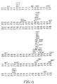
- FIG. 1 depicts the DNA (Seq. ID No.: 1) and amino acid (Seq. ID No.: 2) sequence for a human lactoferrin clone derived from a human mammary cDNA library as described herein except that the sequence between nucleotides 1557-1791 and 2050-2119 corresponds to the previously published sequence (Rado et al. (1987) Blood 70:989-993).
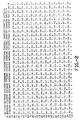
- FIG. 2 depicts the complete DNA (Seq. ID No.: 3) and amino acid (Seq. ID No.: 4) sequence of human lactoferrin including 5,′ and 3′ untranslated sequence as well as the complete human lactoferrin signal sequence.
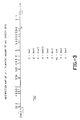
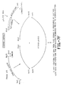
- FIG. 3 is a restriction map of a clone of a 5′-flanking region of bovine ⁇ S1 casein gene.
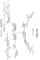
- FIG. 4 is a restriction map of a clone of a 3′-flanking region of bovine ⁇ S1 casein gene
- FIGS. 5A, 5B and 5 C depict the construction of pSI3′5′CAT and pSI5′CAT.
- FIG. 6 depicts pMH-1.
- FIGS. 7A through 7F depict the construction of expression vectors containing sequences encoding human lactoferrin.
- FIG. 8 depicts the genome of human serum albumin, the fragments used to generate transgenic mice contained in this genomic DNA and the identification of the fragment sizes which would be obtained upon the digestion of genomic DNA from a transgenic mouse with the restriction enzymes BstE-II and Nco-I or with Nco-I and Hindi-III.
- FIG. 9 depicts an alternate pathway for the construction of a transgene of the invention encoding human lactoferrin.
- FIG. 10 depicts the construction of a plasmid pPC containing a transgene encoding Protein C.
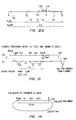
- FIG. 11 depicts the DNA sequence for a hybrid intervening sequence used in a preferred embodiment of the invention.
- the predicted intervening sequence (shown in lower case) consists of the 5′-end of IVS-1 from bovine ⁇ S1 casein (from position +54 to +180 with respect to the start of transcription) fused to the 3′-end of a human IgG splice sequence.
- the Hind III site (in bold type and underlined) derives from the IgG sequence and marks the junction between the ⁇ S1 and IgG splice sequences.
- the 5′-end upper case sequence depicts the complete exon one of the bovine ⁇ S1 casein gene.
- the 3′-end upper case sequence represents the splice junction of the IgG gene through to the Pst I site (CTGCAG) incorporated in the cloning vector, pMH1.
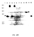
- FIG. 12A is a restriction map or a bovine ⁇ S1 casein promoter hLF cDNA transgene.
- FIG. 12B shows a Southern blot analysis of DNA isolated from various bovine and murine tissues using an hLF cDNA probe.
- FIG. 13 depicts restriction maps of hLF genomic clones 13.1 and 13.2.
- FIG. 14 depicts the BamHI fragment from genomic hLF subcloned into plasmid pUC19.
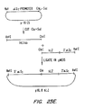
- FIG. 15A depicts a restriction map of the 8hLFgen9k or 16hLFgen9k construct containing the 8 or 16 kb ⁇ S1 casein promoter, a ClaI-ApaI synthetic linker and the 9 kb (i.e., 8.9 kb) ApaI-SalI genomic hLF fragment.
- FIG. 15B depicts the DNA sequence of the ClaI-ApaI synthetic sequence shown in FIG. 15A.
- FIG. 15C depicts the IVS and the structure of exon 1 and part of exon 2 of the genomic hLF construct shown in FIG. 15A through FIG. 17.
- FIG. 16 depicts the coinjection of the NotI-SalI fragment from the 8hLFgen9k or 16hLFgen9k construct (as shown in FIG. 15A) with the 3′ ClaI fragment of genomic hLF.
- FIG. 17 depicts the generation of a genomic 8hLF transgene by linking the NotI-MluI fragment from the 8hLFgen9k construction (shown in FIG. 15A), the MluI-ClaI fragment from clone 13.2 depicted in FIG. 13 and a ClaI-NotI linker.
- FIG. 17 also depicts the DNA sequence of the ClaI-NotI linker.
- FIGS. 18 - 20 depict the generation of the ⁇ LG-hLFgen and ⁇ LG-hLFgen37 constructs.
- FIG. 21 depicts the design of the 16,8hLZ expression vector.
- FIG. 22 depicts the design of the 16,8hLZ3 expression vector.
- FIGS. 23 A- 23 E depict the pathway for the construction of plasmid p16,8hLZ.
- FIG. 24 depicts a comparison between the DNA of bovine ⁇ LG and sheep ⁇ LG.
- the top sequence represents the bovine sequence.
- FIG. 25 shows the linker GP 278/279.
- FIG. 26 depicts the p16,8A hLZ3 expression vector.
- FIG. 27 depicts the 16,A hLZ3 expression vector.
- non-human mammals comprise all non-human mammals capable of producing a “transgenic non-human mammal” having a “desirable phenotype”.
- Such mammals include non-human primates, murine species, bovine species, canine species, etc.
- Preferred non-human animals include be, porcine and ovine species, most preferably bovine species.
- Desirable phenotypes for transgenic non-human mammals include, but are not limited to, the production of recombinant polypeptides in the milk of female transgenic non-human mammals, the production of animal models for the study of disease, the production of animals with higher resistance to disease (e.g. diseases of the mammary gland such as mastitis) and the production of recombinant polypeptides in the blood, urine or other suitable body fluid or tissue of the animal.
- -transgenic bovine species are disclosed which are capable of producing recombinant human lactoferrin, human serum albumin and human Protein C in the milk of lactating females or human serum albumin in the liver of the transgenic animal.
- transgenic non-human mammals of the invention are produced by introducing a “transgene” into an embryonal target cell of the animal of choice.
- a transgene is a DNA sequence which is capable of producing a desirable phenotype when contained in the genome of cells of a transgenic non-human mammal.
- the transgene comprises a “recombinant DNA sequence” encoding a “recombinant polypeptide”. In such cases, the transgene is capable of being expressed to produce the recombinant polypeptide.
- a “recombinant polypeptide” (or the recombinant DNA sequence encoding the same) is either a “heterologous polypeptide” or a “homologous polypeptide”.
- Heterologous polypeptides are polypeptides which are not normally produced by the transgenic animal. Examples of heterologous polypeptides include human milk proteins such as lactoferrin, lysozyme, secreted immunoglobulins, lactalbumin, bile salt-stimulated lipase, etc., human serum proteins such as albumin, immunoglobulins, Factor VIII, Factor IX, protein C, etc.
- the recombinant DNA sequences include genomic and cDNA sequences encoding the recombinant polypeptide.
- the transgene may be integrated in a random manner into the genome of the species used for transgenesis.
- transgenes encoding human lactoferrin, human serum albumin and human Protein C in conjunction with a ⁇ S1 casein secretory signal sequence under control of ⁇ S1 casein expression regulation sequences are designed to produce and secrete these heterologous polypeptides from the mammary gland of a lactating transgenic mammal into its milk.
- a homologous polypeptide is one which is endogenous to the particular transgenic species.
- endogenous polypeptides from bovine species include bovine milk proteins such as ⁇ S1, ⁇ S2, ⁇ - and ⁇ -casein, ⁇ -lactoglobulin lactoferrin, lysozyme, cholesterol hydrolase, serum proteins such as serum albumin and proteinaceous hormones such as growth hormones.
- the transgene is preferably integrated in a random manner into the genome of the species used for transgenesis.
- transgenic animal which contains not only the transgene encoding the endogenous polypeptide but also the corresponding endogenous genomic DNA sequence. Accordingly, such transgenic non-human mammals are readily characterized by an increase in the copy number of genes encoding the endogenous polypeptide. Further, the transgene will generally be located at a position which is different from the endogenous gene.
- the transgenic animal When DNA encoding a homologous polypeptide is expressed, for example, in bovine species, the transgenic animal is characterized by an increase in the amount of the homologous polypeptide in either the endogenous tissue or fluid in which it is normally found and/or by its presence in a tissue and/or body fluid which either does not normally contain the homologous polypeptide or produces it at significantly lower levels.
- bovine cholesterol hydrolase is normally present in the colostrum for about the first 15-20 days of lactation.
- This naturally,occurring endogenous polypeptide increases calf weight.
- This protein is also a homologous polypeptide when, for example, its expression in mammary secretory cells is placed under the control of expression regulation sequences, such as those obtained from bovine casein genes, which facilitate the expression of the homologous polypeptide beyond the lactation period that it is normally present.
- bovine cholesterol hydrolase expression is maintained in transgenic bovine milk by placing the expression of cholesterol hydrolase recombinant DNA (either cDNA or genomic) under the control of bovine ⁇ S1 casein expression regulation sequences.
- a genomic recombinant DNA When a genomic recombinant DNA is used, it is engineered such that it has appropriate restriction sites (e.g. ClaI and SalI) at the 5′ and 3′ end of the structural gene such that it is capable of being inserted into an appropriate transgene genomic cassette (e.g. p-16 kb, CS which is described in Example 15).
- a recombinant DNA encoding bovine cholesterol hydrolase derived from cDNA may be placed under control of bovine ⁇ S1 casein expression regulation sequence by substituting the human lactoferrin sequences in a plasmid such as p16, 8HLF3 (containing a hybrid intervening sequence) or p16, 8HLF4 (containing a homologous ⁇ S1 casein intervening sequence).
- a plasmid such as p16, 8HLF3 (containing a hybrid intervening sequence) or p16, 8HLF4 (containing a homologous ⁇ S1 casein intervening sequence).
- bovine lactoferrin is normally present in only trace amounts in cow's milk. When, however, bovine lactoferrin is expressed under control of other regulatory sequences, for example, obtained from an ⁇ S1 casein gene, higher amounts of lactoferrin in the milk of transgenic bovine species are obtained.
- a transgene comprising DNA encoding homologous bovine growth hormone is incorporated into the bovine genome to confer superior growth characteristics to the transgenic animal.
- homologous polypeptides include, for example, a polypeptide which normally is maintained intracellularly in a particular species but which is secreted into the milk or other extracellular compartment of the transgenic species, such as the circulatory system.
- Each of the heterologous or homologous polypeptides are characterized by specific amino acid and nucleic acid sequences. It is to be understood, however, that such sequences include naturally occurring allelic variations thereof and variants produced by recombinant methods wherein such nucleic acid and polypeptide sequences have been modified by the substitution, insertion and/or deletion of one or more nucleotides in such nucleic acids to cause the substitution, insertion or deletion of one ore more amino acid residues in the recombinant polypeptide.
- the transgene When expression of the DNA of the transgene is necessary to generate a desired phenotype, e.g. to produce a recombinant polypeptide, the transgene typically includes at least a 5′ and preferably additional 3′ “expression regulation sequences” each operably linked to a recombinant or secretory-recombinant DNA as defined hereinafter.
- expression regulation sequences in addition to controlling transcription also contribute to RNA stability and processing, at least to the extent they are also transcribed.
- Such expression regulation sequences are chosen to produce tissue-specific or cell type-specific expression of the recombinant or secretory-recombinant DNA. Once a tissue or cell type is chosen for expression, 5′ and optional 3′ expression regulation sequences are chosen. Generally, such expression regulation sequences are derived from genes that are expressed primarily in the tissue or cell type chosen. Preferably, the genes from which these expression regulation sequences are obtained are expressed substantially only in the tissue or cell type chosen, although secondary expression in other tissue and/or cell types is acceptable if expression of the recombinant DNA in the transgene in such tissue or cell type is not detrimental to the transgenic animal. Particularly preferred expression regulation sequences are those endogenous to the species of animal to be manipulated.
- expression regulation sequences from other species such as those from human genes may also be used.
- Particularly preferred expression regulation sequences from human genes are human lactoferrin (hLF) sequences.
- the expression regulation sequences and the recombinant DNA sequences are from the same species, e.g., each from bovine species or from a human source. In such cases, the expression regulation sequence and the recombinant DNA sequence are homologous to each other.
- the expression regulation sequences and recombinant DNA sequences are obtained from different species, e.g., an expression regulation sequence from bovine species and a recombinant DNA sequence from a human source). In such cases, the expression regulation and recombinant DNA sequence are heterologous to each other.
- the following defines expression regulation sequences from endogenous genes. Such definitions are also applicable to expression regulation sequences from non-endogenous, heterologous genes.
- the 5′ expression regulation sequence includes the transcribed portion of the endogenous gene upstream from the translation initiation sequence (the 5′ untranslated region or 5′ UTR) and those flanking sequences upstream therefrom which comprise a functional promoter.
- a “functional promoter” includes those necessary untranscribed DNA sequences which direct the binding of RNA polymerase to the endogenous gene to promote transcription.
- Such sequences typically comprise a TATA sequence or box located generally about 25 to 30 nucleotides from the transcription initiation site.
- the TATA box is also sometimes referred to the proximal signal.
- the promoter further comprises one or more distal signals located upstream from the proximal signal (TATA box) which are necessary to initiate transcription.
- Such promoter sequences are generally contained within the first 100 to 200 nucleotides located upstream from the transcription initiation site, but may extend up to 500 to 600 nucleotides from the transcription initiation site. Such sequences are either readily apparent to those skilled in the art or readily identifiable by standard methods. Such promoter sequences alone or in combination with the 5′ untranslated region are referred to herein as “proximal 5′ expression regulation sequences”.
- distal 5′ expression regulation sequences In addition to such proximal 5′ expression regulation sequences, it is preferred that additional 5′ flanking sequences (referred to herein as “distal 5′ expression regulation sequences”) also be included in the transgene.
- Such distal 5′ expression regulation sequences are believed to contain one or more enhancer and/or other sequences which facilitate expression of the endogenous gene and as a consequence facilitate the expression of the recombinant or secretory-recombinant DNA sequence operably linked to the distal and proximal 5′ expression regulation sequences. The amount of distal 5′ expression regulation sequence depends upon the endogenous gene from which the expression regulation sequences are derived.
- such sequences comprise 5′ flanking regions of approximately 1 kb, more preferably 16 kb and most preferably about 30 kb of 5′ flanking sequence.
- the determination of the optimal amount of distal 5′ expression regulation sequence used from any particular endogenous gene is readily determined by varying the amount of distal 5′ expression regulation sequence to obtain maximal expression.
- the distal 5′ expression regulation sequence will not be so large as to extend into an adjacent gene and will not include DNA sequences which adversely effect the level of transgene expression.
- 3′ expression regulation sequences also- be included to supplement tissue or cell-type specific expression.
- Such 3′ expression regulation sequences include 3′ proximal and 3′ distal expression regulation sequences from an appropriate endogenous gene.
- the 3′ proximal expression regulation sequences include transcribed but untranslated DNA positioned downstream from the translation stop signal in the recombinant DNA sequence (also referred to as the 3′ untranslated region or 3′ UTR).
- Such sequences generally terminate at a polyadenylation sequence (either from the endogenous gene or from other sources such as SV40) and sequences that may affect RNA stability.
- 3′ UTR's comprise about 100 to 500 nucleotides downstream from the translation stop signal in the gene from which the 3′ regulation sequence is derived.
- Distal 3′ expression regulation sequences include flanking DNA sequences downstream from the proximal 3′ expression regulation sequence. Some of these distal sequences are transcribed, but do not form part of the mRNA while other sequences in this distal 3′ expression regulation sequence are not transcribed at all.
- Such distal 3′ expression regulation sequences are believed to contain enhancer and/or other sequences which enhance expression.
- Such sequences are believed to be necessary for efficient polydenylation and contain transcription termination sequences
- such sequences comprise about 2 kb, more preferably 8 kb and most preferably about 15 kb of 3′ flanking sequence.
- a preferred 3′ flanking sequence is the 3′ flanking sequence of the human lactoferrin (hLF) gene.
- hLF human lactoferrin
- Transgenic animals containing transgenes that include about 9 kb of hLF 3′ flanking sequences show enhanced expression of recombinant polypeptides in milk compared to animals containing transgenes that include 1 kb or less of hLF 3′ flanking sequence, due to an enhancer or other enhancing sequence located in this region.
- the human lactoferrin 3′ flanking sequence will be at least 1 kb in length up to about 9 kb in length or longer, typically 3 to 7 kb, more typically 4 to 5 kb.
- enhancers or enhancing sequences can be isolated and used in combination with various amounts of homologous or heterologous sequences.
- the enhancing sequences can range in length from about 50 basepairs to about 2 kb, more typically from about 100 basepairs to about 500 basepairs.
- transgene having a 5′ expression regulation sequence and a 3′ flanking sequence that originate from the same gene.
- the 5′ expression regulation sequence and 3′ flanking sequence are from the bovine ⁇ S1-casein gene.
- a genomic sequence such as a human genomic clone or clones, can be introduced into an animal to produce a transgenic animal containing a transgene that has the sequence of the human gene, including all or part of the 5′ expression regulation sequences, coding sequences, introns, and 3′ untranslated and flanking sequences.
- the human lactoferrin genomic sequence is used in its entirety, but various components can be substituted with components from other mammary gland specific genes.
- endogenous 3′ regulation sequences are not used.
- the 3′ proximal expression regulation sequences normally associated with the genomic DNA encoded by the recombinant DNA sequence are used to direct polyadenylation.
- distal 3′ regulation sequences from the genomic DNA encoding the recombinant polypeptide may also be employed preferably in the same amounts as set forth for endogenous 3′ expression regulation sequences.
- the recombinant polypeptide encoded by the transgene may comprise either genomic DNA or a double stranded DNA derived from cDNA.
- the optimal amount of 3′ expression regulation sequence may be readily determined by varying the amount of 3′ flanking sequence to obtain maximal expression of the recombinant polypeptide.
- the distal 3′ regulation sequence be it from an endogenous gene or a heterologous gene, will not extend into the adjacent gene from which is derived and will exclude any sequences which adversely effect the level of transgene expression.
- the transgenes of the invention preferably also comprise a “recombinant intervening sequence” which interrupts the transcribed but untranslated 5′ region of the transgene.
- intervening sequences can be derived, for example, from bovine ⁇ S1 casein and from human lactoferrin.
- sequences as used herein are “homologous recombinant intervening sequences” in that the 5′ and 3′ RNA splice signals in such recombinant intervening sequences are those normally found in an intervening sequence from an endogenous or heterologous gene.
- Recombinant intervening sequences may, however, also comprise a “hybrid intervening sequence”.
- hybrid intervening sequences comprise a 5′ RNA splice signal and 3′ RNA splice signal from intervening sequences from different sources.
- hybrid intervening sequences comprise at least one “permissive RNA splice sequence”.
- a permissive RNA splice signal is an RNA splice signal sequence, preferably a 3′ RNA splice signal, from an intron contained within a repertoire of germ line DNA segments which undergo rearrangement during cell differentiation.
- Such gene repertoires include the immunoglobulin super gene family, including the immunoglobulins and T-cell antigen receptors as well as the repertoire of the major histocompatibility complex (MHC) genes and others.
- Particularly preferred permissive splice sequences are those obtained from the immunoglobulin repertoire, preferably of the IgG class, and more preferably those 3′ splice signal sequences associated with the J-C segment rearrangement of the Ig heavy and light chain, most preferably the heavy chain.
- a particularly preferred permissive splice sequence comprises that portion of the sequence as shown downstream of the HindIII site in FIG. 11.
- a particularly preferred hybrid intervening sequence comprises the entire sequence shown in FIG. 11 which includes a 5′ portion of an intervening sequence from bovine ⁇ S1 casein and a 3′ sequence portion of an IgG heavy chain intervening sequence.
- Such hybrid intervening sequences containing permissive RNA splice signals are preferably used when the recombinant DNA corresponds to a cDNA sequence.
- a transgenic mouse was obtained which produced approximately 1330 ⁇ g/ml of hLF in the transgenic milk.
- This amount of recombinant polypeptide far exceeds the previously reported amounts for production of various protein in transgenic mouse milk of generally less than 10 ⁇ g/ml and in one case approximately 50 ⁇ g/ml. It also exceeds the maximum of 8 ⁇ g/ml of hLF produced herein when the same transgene was used that contained a homologous bovine intervening sequence rather than the hybrid intervening sequence.
- hybrid intervening sequences are not limited to transgenes utilizing cDNA sequence. Rather, hybrid intervening sequences are also useful when the recombinant polypeptide is encoded by a genomic sequence. Based on the results obtained with the cDNA recombinant DNA and the general expectation that genomic DNA sequences express at higher levels than sequences derived from cDNA, it is expected that such hybrid intervening sequences used in conjunction with genomic recombinant DNA will further enhance expression levels above that which would otherwise be obtained with genomic sequence alone.
- transgenes include large amounts of 5′ and 3′ expression regulation sequences.
- the recombinant DNA is preferably derived from genomic clones which may be tens to hundreds of kilobases in length. Based on the present technology for cloning and manipulating DNA, the construction and microinjection of transgenes is practically limited to linearized DNA having a length not greater than about 50 kb. However, the transgenes of the invention, especially those having a length greater than about 50 kb, may be readily generated by introducing two or more overlapping fragments of the desired transgene into an embryonal target cell.
- the overlapping fragments undergo homologous recombination which results in integration of the fully reconstituted transgene in the genome of the target cell.
- it is preferred that such overlapping transgene fragments have 100% homology in those regions which overlap.
- lower sequence homology may be tolerated provided efficient homologous recombination occurs.
- the non-homology does exist between the homologous sequence portions, it is preferred that the non-homology not be spread throughout the homologous sequence portion but rather be located in discrete areas. Although as few as 14 base pairs at 100% homology are sufficient for homologous recombination in mammalian cells (Rubnitz, J. and Subramani, S. (1984) Mol. Cell. Biol. 4:2253-2258), longer homologous sequence portions are preferred, e.g. 500 bp, more preferably 1000 bp, next most preferably 2000 bp and most preferably greater than 2000 bp for each homologous sequence portion.
- transgene so generated has a unit length of 38 kb, there is no known practical limit to the size of the transgene which may be formed using larger and/or greater numbers of overlapping transgene fragments. In particular, it is expected that transgenes may be formed by this approach having lengths between about 50 to 1000 kb and more preferably between 50 and 500 kb.
- transgenic bovine species containing transgenes incorporating recombinant DNA comprising genomic DNA which otherwise could not be incorporated into a pronucleus to form a transgenic animal.
- genomic transgenes are expected to produce higher expression levels in transgenic cows as compared to that which is produced by transgenes encoding recombinant cDNA.
- a “secretory DNA sequence” encoding a functional secretion signal peptide is also operably linked within the transgene to direct secretion of the recombinant polypeptide from one or more cell types within the transgenic animal.
- Secretory DNA sequences in general are derived from genes encoding secreted proteins of the same species of the transgenic animal. Such secretory DNA sequences are preferably derived from genes encoding polypeptides secreted from the cell type targeted for tissue-specific expression, e.g. secreted milk proteins for expression in and secretion from mammary secretory cells. Secretory DNA sequences, however, are not limited to such sequences.
- Secretory DNA sequences from proteins secreted from other cell types within the species of transgenic animal may also be used, e.g., the native signal sequence of a homologous gene encoding a protein secreted other than in the mammary glands.
- heterologous secretory DNA sequences which encode signal secretion peptides from species other than the transgenic animals my also be used e.g., human t-PA, human serum albumin human lactoferrin and human lactalbumin and secretion signals from microbial genes encoding secreted polypeptides such as from yeast, filamentous fungi, and bacteria.
- a secretory DNA sequence may be defined functionally as any DNA sequence which when operably linked to a recombinant DNA sequence encodes a signal peptide which is capable of causing the secretion of the recombinant polypeptide.
- a secretory DNA sequence encoding a secretory signal sequence functional in the mammary secretory cells of bovine species is used to cause secretion of recombinant polypeptide from bovine mammary secretory cells.
- the secretory DNA sequence is operably linked to the recombinant DNA sequence. Examples of such secretory DNA sequences include DNA sequences encoding signal secretion sequences for bovine ⁇ S1 casein, murine lactoferrin and human transferrin.
- the preferred secretory DNA sequence is that encoding the secretory sequence of ⁇ S1 casein from bovine species. The use of this secretory DNA sequence is described in more detail in the Examples.
- “Operably linked” in the context of linking a secretory DNA sequence to a recombinant DNA sequence means that the secretory DNA sequence (comprising codons encoding the secretory signal peptide sequence) is covalently coupled to the recombinant DNA sequence so that the resultant secretory-recombinant DNA sequence encodes 5′ to 3′ for the secretory signal sequence and recombinant polypeptide. Accordingly, the reading frame for the secretory sequence and the recombinant DNA sequence must be covalently combined such that an open reading frame exists from the 5′ end of the mRNA sequence formed after transcription and processing of the primary RNA transcript.
- This open reading frame in the RNA contains a 5′ sequence portion encoding the secretory signal peptide and a 3′ sequence portion encoding the recombinant polypeptide.
- the recombinant polypeptide produced upon expression of the secretory-recombinant DNA sequence is of a form which is capable of being secreted from targeted cells which express the DNA sequence.
- the signal peptide generally is removed in vivo during secretion to produce an extracellular form of the recombinant polypeptide.
- a secretory-recombinant DNA sequence is expressed predominantly in the mammary secretory cells of transgenic bovine species.
- tissue-specific expression is obtained by operably linking mammary specific expression regulation DNA sequences to the above secretory-recombinant DNA sequence.
- mammary specific regulation sequences include the aforementioned regulation sequences contained in various bovine genes preferentially expressed in the mammary secretory cells of the species.
- mammary specific genes include ⁇ S1 casein; ⁇ S2-casein; ⁇ -casein; K-casein; ⁇ -lactalbumin; and ⁇ -lactoglobulin.
- Preferred expression regulation sequences are derived from ⁇ S1 casein as described more in detail in the Examples.
- the transgenes of the invention that are designed to secrete the recombinant polypeptide into transgenic bovine milk are capable of causing such secretion at levels significantly higher than that previously reported for transgenic mice and sheep.
- the recombinant polypeptide is encoded by a recombinant DNA corresponding to, or derived from, cDNA
- the molar concentration of the recombinant polypeptide is preferably greater than about 1.0 ⁇ M, more preferably greater than about 100 ⁇ M, and most preferably greater than 100 ⁇ M.
- the amount of recombinant polypeptide is preferably greater than 50 ⁇ g/mi, more preferably greater than about 500 ⁇ g/ml and most preferably greater than about 1000 ⁇ g/ml (1 mg/ml).
- the transgene of the invention encodes a recombinant polypeptide that is encoded by recombinant DNA derived from or corresponding to genomic DNA (or comprised substantially of such genomic sequences, e.g. greater than about 50%, more preferably greater than about 75%, most preferably greater than 90% of the codons encoding the recombinant polypeptide are from genomic sequences)
- the molar concentrations and protein levels in bovine transgenic milk are the same as for cDNA or higher.
- the molar concentration of the recombinant polypeptide in such transgenic milk is preferably greater than about 50 ⁇ M, more preferably greater than about 50 ⁇ M, most preferably greater than about 500 ⁇ M.
- the levels are preferably greater than about 10 mg/ml, more preferably greater than about 2.5 mg/ml, most preferably greater than 5 mg/ml.
- bovine transgenic milk will vary depending upon the molecular weight of the particular recombinant polypeptide.
- a particular advantage of producing a recombinant polypeptide in bovine transgenic milk is that relatively large molecular weight polypeptides may be so produced which are otherwise difficult to produce in large quantities in other systems such as prokaryotic expression systems.
- any recombinant polypeptide may be produced in bovine transgenic milk according to the invention, it is generally preferred that such recombinant polypeptides have a molecular weight greater than about 10,000 Daltons.
- recombinant polypeptides having molecular weights of greater than 15,000, greater than 20,000 and greater than 60,000 Daltons may also be expressed in transgenic bovine milk.
- human lysozyme having a molecular weight of 17,000 Daltons and lactoferrin having a molecular weight of 79,000 Daltons may be readily produced in the transgenic milk of bovine species according to the disclosure of the invention.
- the recombinant polypeptides of the invention have a wide range of molecular weights.
- the foregoing preferred molar concentrations of recombinant polypeptides are adjusted when higher molecular weight recombinant polypeptides are produced. Such adjustment is made by converting the molar concentration to the amount of protein produced and adjusting the molar concentrations so that the recombinant protein level is within the following preferred concentrations.
- transgenic mice Most of the previous reports relating to the production of polypeptides in transgenic milk involve transgenic mice. The mouse, however, normally produces between 55 to 80 milligrams of protein per ml of milk. A cow, on the other hand, normally produces between 30 to 34 milligrams of protein per ml.
- the recombinant polypeptide concentration be between about 3 and 50% of the normal bovine milk protein concentration (i.e., between about 1 and 17 milligrams of recombinant polypeptide per ml of transgenic milk), more preferably between 10 to 20% (i.e., between 3 to about 7 milligrams per ml) and most preferably between 10 and 15% (i.e., between about 3 and 5 milligrams per ml) of the normal amount of protein produced in bovine milk.
- Such preferred ranges also provide a preferred maximum limit to the aforementioned levels of protein produced in transgenic bovine milk.
- transgene of the invention are performed by standard methods known to those skilled in the art or as described herein. Once the transgene or overlapping homologous fragments encoding the transgene are constructed as described they are used to make transgenic non-human animals.
- Methods of introducing transgenes or overlapping transgene fragments into embryonal target cells include microinjection of the transgene into the pronuclei of fertilized oocytes or nuclei of ES cells of the non-human animal. Such methods for murine species are well known to those skilled in the art.
- the transgene may be introduced into an animal by infection of zygotes with a retrovirus containing the transgene (Jaenisch, R. (1976) Proc. Natl. Acad. Sci. USA 73:1260-1264).
- the preferred method is microinjection of the fertilized oocyte.
- the fertilized oocytes are first microinjected by standard techniques.
- pre-implantation embryos preferably contain approximately 16 to 150 cells.
- the 16 to 32 cell stage of an embryo is commonly referred to as a morula.
- blastocysts Those pre-implantation embryos containing more than 32 cells are commonly referred to as blastocysts. They are generally characterized as demonstrating the development of a blastocoel cavity typically at the 64 cell stage.
- Methods for culturing fertilized oocytes to the pre-implantation stage include those described by Gordon, et al. (1984) Methods in Enzymology 101:414; Hogan, et al. (1986) in Manipulating the Mouse Embryo, Cold Spring Harbor Laboratory Press, Cold Spring Harbor, N.Y.
- Such pre-implantation embryos are thereafter transferred to an appropriate female by standard methods to permit the birth of a transgenic or chimeric animal depending upon the stage of development when the transgene is introduced.
- mosaic animals can be bred to form true germline transgenic animals.
- the detection of-transgene integration in the pre-implantation embryo is highly desirable.
- methods are provided for identifying embryos wherein transgenesis has occurred and which permit implantation of transgenic embryos to form transgenic animals.
- one or more cells are removed from the pre-implantation embryo.
- the embryo is preferably not cultivated past the morula stage (32 cells). Division of the pre-implantation embryo (reviewed by Williams et al.
- hemi-embryos hemi-morula or hemi-blastocyst
- hemi-embryos hemi-morula or hemi-blastocyst
- each of the hemi-embryos formed by division of pre-implantation embryos is analyzed to determine if the transgene has been integrated into the genome of the organism.
- Each of the other hemi-embryos is maintained for subsequent implantation into a recipient female of the species.
- a preferred method for detecting transgenesis at this early stage in the embryo's development uses these hemi-embryos in connection with a unique property of the restriction endonuclease Dpn I. This enzyme recognizes the sequence GATC in double-stranded DNA but only when the adenine in each strand within this sequence is methylated at N-6.
- the transgene containing the sequence GATC is methylated prior to microinjection either by transferring the transgene on an appropriate plasmid through a DAM + strain of microorganisms such as E. coli MM294 or by directly methylating the transgene with dam methylase.
- the methylated transgene (preferably without any exogenous sequences such as plasmid vector) is then microinjected into fertilized oocytes (approximately 10 to 500 copies per pronucleus, more preferably 50 to 100 copies per pronucleus).
- fertilized oocytes so obtained re cultured in vitro to the pre-implantation stage During this early growth and cell division phase, the genomic DNA is replicated.
- the identification of the pre-implantation embryos containing the integrated transgene is achieved by analyzing the DNA from each of the hemi-embryos. Such DNA is typically obtained by lysing the hemi-embryo and analyzing the thus released DNA after treatment as described by Ninomiy, T. et al. (1989) Molecular Reproduction and Development 1:242-248. Each of the DNA samples is treated with Dpn I. Thereafter, a polymerase chain reaction (Saiki, et al. (1985) Science 230:1350-1354) is preformed to amplify all or part of the transgene.
- extension primers each complimentary to opposite strands at opposing ends of the transgene are used for amplification.
- extension primers are chosen such that the amplified gene product spans the Dpn I site in the transgene. If Dpn I cleavage has not occurred, PCR amplification results in amplified sequences having a predetermined size whereas primer extension for those transgenes which have been cleaved will not result in exponential amplification.
- the Dpn I/PCR amplified DNA from the hemi-embryo is subjected to electrophoresis followed by hybridization with labeled probe complimentary to the region of the transgene between the two extension primers.
- This facilities the determination of the size of the amplified DNA sequences, if any, and provides an indication of whether the transgene has been integrated into the pre-implantation embryo from which the hemi-embryo was obtained (now called a “transgenic hemi-embryo”). If it has, the remaining untreated transgenic hemi-embryo is transplanted into a recipient parent.
- the transgenic non-human animal having the desired phenotype conferred by the integrated transgene is identified by an appropriate method in utero or after birth.
- restriction endonucleases capable of cleaving a methylated DNA sequence but incapable of cleaving the unmethylated form of a recognition sequence may be used in the aforementioned method.
- the above described methods for the detection of transgenesis in pre-implantation embryos provide economical and time saving method for generating transgenic non-human animals since they significantly decrease the number of pregnancies required to produce a transgenic animal and substantially increase the likelihood that an implanted embryo will produce a transgenic non-human animal. Such methods are especially important for those animals for which very low or non-existent frequencies of transgenesis have been obtained, e.g. bovine species.
- transgenic embryos and/or non-human transgenic animals having the same “genotype” means that the genomic DNA is substantially identical between the individuals of the embryo and/or transgenic animal population. It is to be understood, however, that during mitosis various somatic mutations may occur which may produce variations in the genotype of one or more cells and/or animals. Thus, a population having the same genotype may demonstrate individual or subpopulation variations.
- transgenic hemi-embryo After a hemi-embryo is identified as a transgenic hemi-embryo, it is cloned. Such embryo cloning may be performed by several different approaches. In one cloning method, the transgenic hemi-embryo is cultured in the same or in a similar media as used to culture individual oocytes to the pre-implantation stage. The “transgenic embryo” so formed (preferably a transgenic morula) is then divided into “transgenic hemi-embryos” which can then be implanted into a recipient female to form a clonal population of two transgenic non-human animals.
- the two transgenic hemi-embryos obtained may be again cultivated to the pre-implantation stage, divided, and recultivated to the transgenic embryo stage. This procedure is repeated until the desired number of clonal transgenic embryos having the same genotype are obtained. Such transgenic embryos may then be implanted into recipient females to produce a clonal population of transgenic non-human animals.
- the transgenic embryo is cloned by nuclear transfer according to the techniques of Prather, et al. (1988) Biol. Reprod. 37:59-86; Roble, et al. (1987) J. Anim. Sci. 64:642-664.
- nuclei of the transgenic embryo are transplanted into enucleated oocytes, each of which is thereafter cultured to the blastocyst stage.
- the transgenic embryos may be resubjected to another round of cloning by nuclear transplantation or may be transferred to a recipient parent for production of transgenic offspring having the same genotype.
- transgenesis in addition to the foregoing methods for detecting early transgenesis, other methods may be used to detect transgenesis. Such methods include in utero and post partum analysis of tissue. In utero analysis is performed by several techniques. In one, transvaginal puncture of the amniotic cavity is performed under echoscopic guidance (Bowgso, et al. (1975) Bet. Res. 96:124-127; Rumsey, et al. (1974) J. Anim. Sci. 39:386-391). This involves recovering about 15 to 20 milliliters of amniotic fluid between about day 35 and day 100 of gestation.
- This volume of amniotic fluid contains about 1000 to 12,000 cells per ml originating from the urogenital tract, the skin and possibly the lungs of the developing embryo. Most of these cells are dead. Such cells, however, contain genomic DNA which is subjected to PCR analysis for the transgene as an indication of a successful transgenesis. Alternatively, fetal cells may be recovered by chorion puncture. This method also may be performed transvaginally and under echoscopic guidance. In this method, a needle is used to puncture the recipient animal's placenta, particularly the placentonal structures, which are fixed against the vaginal wall. Such sampling may be performed around day 60 of gestation in bovine species. Chorion cells, if necessary, are separated from maternal tissue and subjected to PCR analysis for the transgene as an indication of successful transgenesis.
- Transgenesis may also be detected after birth.
- transgene integration can be detected by taking an appropriate tissue biopsy such as from the ear or tail of the putative transgenic animal. About one to two centimeters of tail or about five to ten square millimeters of ear are obtained followed by southern blotting with a probe for the transgene according to the method of Hogan, et al. (1986) Manipulating the Mouse Embryo, Cold Spring Harbor Laboratory.
- Transgenesis can also be determined by using the southern blot technique with DNA obtained from other tissues.
- semen from a recombinant bull will be useful for identifying transgenic animals.
- Transgenesis may also by detected by assaying for expression of the recombinant polypeptide in a tissue, secretion (e.g., saliva), or other body fluid.
- a tissue secretion (e.g., saliva)
- saliva secretion
- the goal is expression of a recombinant polypeptide in milk of cows it will be especially useful to assay the saliva of bulls for expression levels. This is because some mammary specific promoters may also cause salivary gland expression, albeit at low levels. See, e.g., Archibald et al. (1990) Proc. Nat. Acad. Sci. USA 87Z:5178-5182.
- the transgenic milk so obtained may be either used as is or further treated to purify the recombinant polypeptide. This depends, in part, on the recombinant polypeptide contained in the transgenic milk and the ultimate use for that protein. Thus, when the recombinant polypeptide is secreted into transgenic milk to increase the nutritional value of the bovine milk, no further purification is generally necessary.
- An example of such a situation involves one of the preferred embodiments wherein human lactoferrin is produced in the milk of bovine species as a supplement to control intestinal tract infections in newborn human infants and to improve iron absorption.
- human lactoferrin produced in transgenic bovine milk may be partially purified by acidifying the milk to about pH 4-5 to precipitate caseins.
- the soluble fraction (the whey) contains the human lactoferrin which is partially purified.
- the recombinant polypeptide contained in bovine transgenic milk may also be used in food formulations.
- a particularly useful food formulation comprises an infant formula containing one or more recombinant polypeptides from transgenic bovine milk which have either nutritional or other beneficial value.
- an infant formula containing human lactoferrin from transgenic bovine milk made according to the present invention provides a bacteriostatic effect which aids in controlling diarrhea in newborn.
- recombinant polypeptides such as human casein and human lysozyme may also be generated in transgenic bovine milk to provide nutritional value.
- Table 2 sets forth the constituents of a typical infant formula.
- the protein content varies between about 1.8 and 4.5 grams of protein per 100 kilocalories of formula.
- the total protein including recombinant polypeptide should lie between the values at least based on regulatory requirements in the United States from which the formulation in Table 2 is based.
- the amount of total protein including recombinant polypeptide may vary from the foregoing depending upon the local regulations where the particular formula is intended to be used.
- recombinant polypeptides from transgenic bovine milk may also be supplemented with recombinant polypeptides from transgenic bovine milk.
- recombinant polypeptides may be used to supplement common diet formulations.
- purification methods consistent with such an application are called for. Such purification methods will depend on the particular recombinant polypeptide to be purified and are generally known to those skilled in the art. Such methods typically include a partial purification by casein fractionation followed by chromatography of the appropriate fraction containing the recombinant polypeptide. Such chromotography includes affinity chromatography, ion exchange chromotography, gel filtration and HPLC.
- transgenes are provided for producing human lactoferrin in the milk of transgenic bovine species.
- Human lactoferrin (HLF) is a single chain glycoprotein which binds two ferric ions. Secreted by exocrine glands (Mason, et al. (1978) J. Clin. Path. 31:316-327; Tenovuo, et al. (1986) Infect. Immun. 51:49-53) and polymorphonuclear neutrophil granulocytes (Mason, et al. (1969) J. Exp. Med. 130:643-658), this protein functions as part of a host non-specific defense system by inhibiting the growth of a diverse spectrum-of bacteria.
- HLF exhibits a bacteriostatic effect by chelation of the available iron in the media, making this essential metal inaccessible to the invading microorganisms (Bullen, et al. (1972) Br. Med. J. 1:69-75; Griffiths, et al. (1977) Infect. Immun. 15:396-401; Spik, et al. (1978) Immunoloy 8:663-671; Stuart, et al. (1984) Int. J. Biochem. 16:1043-1947). This effect is blocked if the protein is saturated with ferric ions.
- HLF displays a direct bacteriocidal effect on certain microorganisms (Arnold, et al.
- Lactoferrin is the major iron binding protein in human milk (present at a concentration of about 1.5-1.7 mg/ml) and may play a role in the absorption of iron by the small intestine. All of the iron present in breast milk is thought to be bound to hLF and is taken up at very high efficiencies compared to formula (Hide, D. W., et al. (1981) Arch. Dis. Child. 56:172). It has been postulated that the high uptake of the hLF bound iron is due to a receptor in the jejunum and data has been presented suggesting existence of receptors in Rhesus monkeys (Cox, et al. (1979) BBA 588:120; Davidson, L. A., et al.
- human lactoferrin in the milk of transgenic bovine species provides a source of human lactoferrin.
- Such lactoferrin may be purified from the transgenic milk for formulation purposes.
- the whole transgenic milk may be used, preferably after pasteurization, in either liquid or dried form.
- the beneficial action of human lactoferrin may be potentiated by combining the human lactoferrin or the transgenic milk containing it with human lysozyme.
- the human lysozyme may be simultaneously produced in the transgenic cow by introducing a second transgene simultaneously with the HLF transgene to produce a transgenic cow capable of producing more than one recombinant polypeptide in the transgenic milk.
- the transgenes may be sequentially introduced into bovine species.
- a transgenic bovine species is obtained containing one of the transgenes.
- embryonic cells, such as eggs are obtained from the transgenic female and treated so as to incorporate the second transgene encoding the second polypeptide.
- the egg is fertilized, followed by microinjection of the pronucleus of the zygote so obtained.
- the foregoing combination of more than two recombinant polypeptides in transgenic bovine milk is not limited to the aforementioned human lactoferrin and lysozyme combination.
- the invention contemplates the production of transgenic bovine species and transgenic milk wherein more than one recombinant polypeptide is produced by such a transgenic animal in the transgenic milk.
- HLF complete amino acid sequence of HLF has been determined (Metz-Boutigue et al. (1984) Eur. J. Biochem. 1451:659-676). HLF comprises two domains, each containing one iron-binding site and one N-linked glycosylation site. These domains show homology between each other, indicative of an ancestral gene duplication and fusion event. In addition, HLF shares extensive homology with other members of the transferrin family (Metz-Boutigue, supra; Pentecost, et al. (1987) J. Biol. Chem. 262:10134-10139). Location of the amino acids involved in the iron-binding sites has been determined by X-ray crystallography (Anderson et al. (1987) Proc. Natl.
- human lactoferrin comprises a polypeptide having the amino acid sequence substantially as described by Metz-Boutigue, et al. (1984) Eur. J. Biochem. 1451:659-676 and as set forth in FIG. 2. It is noted, however, that an earlier partial sequence of the human lactoferrin sequence disclosed a number of discrepancies between the published sequence and that obtained herein. Specifically, the following discrepancies exist (amino acid numbering is from the sequence in FIG.
- human lactoferrin is also defined by the sequence shown in FIG. 1 which combines the sequence differences obtained herein with the published sequence.
- the term human lactoferrin also includes allelic variations of either of these sequences or recombinant human lactoferrin variants wherein one or more amino acids have been modified by the substitution, insertion or deletion of one or more amino acid residues.
- human lactoferrin may be produced in milk with all or part of a secretory signal sequence covalently attached thereto.
- a “human lactoferrin DNA sequence” is a DNA sequence which encodes human lactoferrin as defined above. Such a human lactoferrin DNA sequence may be obtained from a human mammary gland cDNA library or may be derived from the human genome.
- Example 2 herein describes the cloning and nucleotide sequence of human lactoferrin derived from a human mammary gland cDNA library. The DNA sequence of this human lactoferrin is shown in FIG. 1 and FIG. 2 and is substantially the same as that described by Rado, et al. (1987) Blood 70:989-993.
- the construction of plasmids containing an expressible transgene encoding hLF is described in the examples.
- One of these plasmids is cGP1HLF also sometimes referred to as 16,8HLF3 contains a transgene designed for tissue-specific expression in bovine mammary secretory cells.
- transgenes are provided for producing human serum albumin in the milk of transgenic bovine species.
- Human serum albumin is a serum protein which contains 584 amino acid residues (Minghetti, et al. (1986) J. Biol. Chem. 261:6747). It is the most abundant protein in human serum and performs two very important physiological functions. Serum albumin is responsible for about 80% of the total osmolarity of blood and it transports fatty acids between adipose tissues.
- Human serum albumin is used primarily to expand plasma volume by restoring osmotic pressure in the circulatory system.
- a heat treated serum derived hSA fraction is infused in most shock and trauma victims, including most of the patients undergoing extensive surgery.
- HSA is presently derived from human blood plasma as a by-product from blood fractionation processes to obtain rare blood proteins such as factor VIII and IX.
- the recently developed technology of producing such factors by biotechnological means threatens the source of human serum albumin.
- human serum albumin comprises a polypeptide having the amino acid sequence substantially as that described by Minghetti, et al., ibid; Lawn, et al. (1981) Nucl. Acids Res. 9:6103. Also included are variations thereof including recombinant human serum albumin variants wherein one or more amino acids have been modified by the substitution, insertion or deletion of one or more amino acid residues (Minghetti, et al. (1986) J. Biol. Chem. 261:6747-6757). In some instances, human serum albumin may be produced in milk by expressing a transgene which contains DNA encoding the secretory signal sequence of hSA.
- human serum albumin may be produced in and secreted from liver cells of a transgene animal utilizing a completely heterologous transgene comprising human genomic DNA encoding 5′ expression regulation sequences, the human serum albumin secretion signal and structural gene and 3′ expression regulation sequences.
- transgenes containing this heterologous sequence were formed by in vivo homologous recombination of overlapping transgene fragments to reconstitute the hSA gene in the transgenic animal. The so formed transgenic animal produced human serum albumin in its circulatory system.
- a “human serum albumin DNA sequence” is a DNA sequence which encodes human serum albumin as defined above.
- Such a human serum albumin DNA sequence may be obtained from ⁇ HAL-HAI, ⁇ HAL-3W and ⁇ HAL-HI4 as described by Urano, et al. (1986) J. Biol. Chem. 261:3244-3251 and Urano, et al. (1984) Gene 32:255-261 and in the Examples herein.
- the human serum albumin DNA sequence was cloned as described in Example 10 herein and subsequently manipulated to substitute for the human lactoferrin gene encoded in plasmid cGP1HLF (also referred to as p16,8HLF4). From this plasmid a transgene is obtained containing 16 kb of the 5′ expression regulation sequence of the bovine ⁇ S1 casein gene, human serum albumin DNA sequence and approximately 8 kb of the 3′-flanking region of the ⁇ S1 casein bovine gene. This transgene is used to microinject fertilized oocytes from bovine species.
- blastocysts containing the hSA transgene are implanted into a recipient female bovine species and brought to term.
- the following is presented by way of example and is not to be construed as any limitation on the scope of the invention.
- Placental tissue was obtained from the slaughterhouse. Surrounding connective tissue was removed and pieces of about 30 grams were quickly frozen in liquid N 2 . Chromosomal DNA was isolated as follows: 30 grams of tissue was homogenized (on ice) with 35 ml of Buffer 1 containing 300 mM Sucrose; 60 mM KCl; 15 mM NaCl; 60 mM Tris.HCl pH 8.2; 0.5 mM spermidine; 0.15 mM spermine; 2 mM EDTA; 0.5 mM EGTA. 65 ml of icecold buffer 1 containing 1% NP40 was added and the mixture was incubated for five minutes on ice.
- RNAse A and T1 were added to final concentrations of 0.4 mg/ml and 6 u/ml respectively. After incubation at 37° C. for three hours, Proteinase K was added to a final concentration of 0.1 mg/ml. This mixture was incubated for 15 hours at 37° C. The mixture was then carefully extracted with phenol.
- the aqueous phase was isolated and ⁇ fraction (1/30) ⁇ volume of 3M NaOAc pH 5.2 and one volume of isopropylalcohol was added.
- the precipitate (DNA) was rinsed with 70% ethanol and slowly dissolved in 0.5 ml of 10 mM Tris.HCl pH 8.0; 1 mM EDTA, at 4° C.
- Primer #2 was located at position +164 relative to the major transcription initiation site and had the following sequence: 5′-TGA AGC TTG CTA ACA GTA TAT CAT AGG-3′ (Seq. ID. No.: 6).
- the first eight nucleotides of this primer are not encoded by the bovine genome, but contain a HindIII restriction site to facilitate subsequent cloning steps. These primers were annealed to the chromosomal DNA and extended in the presence of deoxynucleotides by TAQ-polymerase. After three minutes the mixture was denatured for one minute at 92° C., reannealed at 50° C. for 1.5 minutes and again incubated at extension temperature (68° C.) for 2 minutes.
- the other primer was located at position 1070 of the cDNA sequence and had the following sequence: 5′-GCA CAC AAT TAT TTG ATA TG-3′ (Seq. ID No.: 8). These primers were annealed to the chromosomal DNA and the region between these primers was amplified as described above. The resulting fragment was ⁇ 900 bp longer then expected. Sequence analysis showed that an intervening sequence of this size was present between nucleotide 737 and 738 of the cDNA. The amplified fragment was treated with Klenow-polymerase to repair any overhanging ends and treated with kinase to attach phosphate groups to the ends of the fragment. The fragment was then ligated into pUC19 previously cut with SmaI.
- the bacteriophage particle titre was determined on Escherichia coli MB406 a permissive host strain (Stratagene Inc.). For this, several dilutions of the phage stock were made in SM buffer (50 mM Tris.HCl pH 7.5, 100 mM NaCl, 10 mM MgSO4, 0.01% gelatin) and mixed with 200 ⁇ l MB406 (O.D.
- phages were then plated by adding the required amount of phage stock to 400 ⁇ l MB406. The subsequent plating was as described as above. The next step was transfer of the phage to nitrocellulose filters. Plates were placed at 4° C. for one hour. Nitrocellulose filters (S&S) were placed on the top agarose layer and exact position was marked. After lifting, the filters were soaked for (1) 30 minutes in denaturation buffer (1.5M NaCl, 0.5M NaOH); (2) 5 minutes in neutralizing buffer (1.5M NaCl, 0.5M Tris.HCl pH 8.0). After rinsing with 2 ⁇ SSPE (360 mM NaCl, 20 mM NaH 2 PO 4 , 2 mM EDTA), the filters were baked under vacuum at 80° C. for two hours.
- denaturation buffer 1.5M NaCl, 0.5M NaOH
- neutralizing buffer 1.5M NaCl, 0.5M Tris.HCl pH 8.0
- Prehybridization of the filters was performed in a buffer containing 50% formamide, 5 ⁇ Denhardt's solution (0.1% Ficoll, 0.1% polyvinylpydrolidone, 0.1% bovine serum albumin), 5 ⁇ SSPE, 0.1% SDS and 100 ⁇ g/ml denatured salmon sperm DNA at 42° C. for two hours. Hybridization was performed in same buffer at 42° C. overnight in a shaking waterbath. The probe, generated as previously described, was labelled using the Random Primed labelling kit from Boehringer Mannheim. After overnight hybridization the filters were washed three times with 2 ⁇ SSC, 0.1% SDS at room temperature.
- Duplicate nitrocellulose filters from the initial phage plating used for isolating 5′ clones were screened with the 3′ ⁇ S1-casein probe using the same hybridization conditions previously described. Eight positive clones were identified after two rounds of screening. Phage DNA was prepared as described. Subsequent restriction digests with SaII, EcoRI and SaI/EcoRI and Southern hybridization with the 3′ ⁇ S1 probe showed identical inserts in seven of the eight clones. One clone containing an 18.5 kb EcoRI insert was further analyzed with the restriction enzymes BsteII and BamHI. A restriction map of that clone is shown in FIG. 4.
- the human mammary gland library was screened by standard plaque hybridization technique (Maniatis, et al. (1982) Molecular Cloning: A Laboratory Manual ) with three synthetic oligomers. Two of the oligomers were 30-mers corresponding to the cDNA sequence of Rado et al., supra, at amino acid positions 436-445 and 682-691. The third was a 21-mer “best guess” probe based on human codon bias and coding for amino acid sequence of HLF between amino acid residues 18 and 24. Respectively, they were: (Seq. ID No.: 9) (1) 5′-CTTGCTGTGGCCGTGGTTAGGAGATCAGAC-3′ (Seq. ID No.: 10) (2) 5′-CTCCTGGAAGCCTGTGAATTCCTCAGGAAG-3′, and (Seq. ID No.: 11) (3) 5′-ACCAAGTGCTTCCAGTGGCAG-3′.
- the probes were radiolabeled (Crouse et al. (1983) Methods Enzymol. 101, 78-98) and used to screen duplicate filters. Filters were washed at a final stringency of 2 ⁇ SSC, 37° C.
- DNA fragments were isolated by use of low-melting agarose (Crouse et al, supra) and subcloned into bacteriophase M13mp18 or M13mp19 (Messing et al. (1983) Methods Enzymol. 101, 20-78). The sequence was determined using the Sequenase enzyme (modified T7 DNA polymerase) (Tabor et al. (1987) Proc. Natl. Acad. Sci. USA 84, 4767-4771). All reactions were carried out according to -the manufacturer's specifications (US Biochemicals). The sequence is shown in FIG. 1.
- the hLF sequence was digested with HindIII and EcoRI (present in the surrounding phage sequences) and subcloned into the HindIII and EcoRI site of pUC19 to form pUS119 Lacto 4.1. This clone contained the entire coding sequence of the mature form of hLF, but lacked the complete signal sequence.
- fragment 2 Approximately 1.6 kb (fragment 2 , FIG. 5A) of ⁇ S1-casein 3′-flanking sequence was isolated by PCR amplification from a bovine 3′-flanking genomic clone from Example 1. This region contained the previously described splice within the 3′ untranslated region of ⁇ S1-casein gene. Fragment 2 was subcloned into the SmaI site of pUC19. The primer sequences consisted of: 5′-GAGGGACTCCACAGTTATGG-3′ and (Seq. ID No.: 14) 5′-GCACACAATTATTTGATATG-3′ (Seq. ID No.: 15)
- a hybrid splicing signal comprising the 3′ splice site of an immunoglobulin gene (Bothwell et al. (1981) Cell, 24, 625-637) was synthetically prepared and inserted into pUC18 along with unique restriction sites flanking either side to produce pMH-1. This plasmid is shown in FIG. 6. NcoI and HindIII sites were designed such that ligation with fragment 1 from the bovine 5′ genomic clone would result in the functional hybrid splice sequence. See FIG. 11.
- a polyadenylation sequence was obtained from SV40 virus as a BamHI-DraI fragment (fragment 3 in FIG. 5A) isolated from pRSVcat (Gorman, C. M., et al. (1982), Proc. Natl. Acad. Sci., 79, 6777-6781).
- a bacterial CAT coding sequence was subcloned into pUC19 as a PstI-BamHI fragment.
- Fragment 1 of ⁇ S1-casein promoter was subcloned into pMH-1 (FIG. 6) between the NcoI and HindIII sites to form pMHS15′flank.
- fragment 3 The SV40 polyadenylation sequence (fragment 3 ) was subcloned as a BamHI-DraI fragment into pUC19 immediately 3′ to the 3′ ⁇ S1-casein flanking sequence (fragment 2 ) to form pUC19 3′ UTR/SV40. This allowed for the removal of a continuous EcoRI-SalI fragment (containing the 3′-flanking sequence and poly (A) sequence) that was subcloned into pMH-1 to derive pMHS13′UTR (FIG. 5B) which was used later to construct pMHSI 3′UTR hlf which contains sequences encoding human lactoferrin.
- Each of these CAT plasmids were transfected into human 293S cells (Graham, F. L., et al. (1977), J. Gen. Virol., 36, 59-72) by the calcium phosphate co-precipitation method (Gorman, C. M., et al. (1983), Science, 221, 551; Graham, F. L., et al. (1973), Virology, 52, 456-467). Cells were harvested 44 hours after transfection and cell extracts were assayed for CAT activity (Gorman, C. M., et al. (1982), Mol. Cell. Biol., 2, 1011; deCrombrugghe, B., et al.
- pS13′5′CAT was expressed in these cells at a level which was approximately 30-100 fold lower than the control plasmid, but significantly higher than background. Primer extension analysis indicated that transcription had initiated predominantly in the expected region.
- bovine ⁇ S1-casein 5′-flanking sequence from Example 1 was isolated from the bovine genomic library (phage GP1) as a SalI-BglII fragment.
- the BglII site lies at the junction of the first intron and second exon of the ⁇ S1-casein gene.
- Bovine ⁇ S1-casein signal sequence (Stewart et al. (1984) Nucl. Acids Res. 12, 3895) was prepared from synthetic DNA synthesized on a Cylone Plus® DNA Synthesizer (Millgen/Biosearch I) and contained the entire signal sequence plus XhoI and Cla I sites attached to the 5′-end, and NaeI to the 3′-end (fragment 8 , FIG. 7B).
- a synthetic linker was prepared that contained the last five codons of hLF beginning at the EcoRI site and extending for four bases beyond the stop codon.
- a KpnI site was added to the 3′-end (fragment 7 in FIG. 7C).
- Cosmid cGP1HLF was prepared from a 3-way ligation (FIG. 7F):
- Fragments from (a), (b), and (c) were ligated together and transfected into bacteria using commercial lambda packaging extracts (Stratagene, Inc.) to produce cGP1HLF.
- pS13′UTRhLF The HindIII-SalI fragment of pS13′UTRhLF was subcloned into the same sites in pMHS15′flank to form pS13′5′hLF (FIG. 7E).
- This plasmid contains 681 bp of bovine ⁇ S1-casein promoter sequence, the ⁇ S1-casein/IgG hybrid intron, the ⁇ S1-casein signal sequence, the hLF coding region, approximately 1.6 kb of ⁇ S1-casein 3′-flanking sequence, and the SV40 late region polyadenylation sequence.
- Plasmid pS13′5′hLF (FIG. 7E) was cut with KpnI and BamHI which border the ⁇ S1-casein 1.6 kb 3 ′-flanking sequence. The larger vector fragment was purified, made blunt ended with Klenow, and self-ligated to form pS15′hLF.
- Sepharose suspensions (0.3 ml) were incubated for five hours at room temperature by head-over-head rotation with samples (usually 50 ⁇ l) in 2-ml polystyrene tubes. Sepharose beads were then washed with saline (five times with 1.5 ml) and incubated for 16 hours at room temperature with 50 ⁇ l (1 kBq) of 125 I-labeled-affinity-purified polyclonal rabbit anti human lactoferrin antibodies, together with 0.5 ml of PBS, 0.1% ( w /v) Tween-20. Thereafter the Sepharose was washed again with saline (four times with 1.5 ml) and bound radio activity was measured. Results were expressed as percent binding of the labelled antibodies added.
- lactoferrin levels were expressed in nanomolar, using purified human milk lactoferrin as a standard (serial dilutions in PBS, 10 mM EDTA, 0.1% ( w /v) Tween-20.
- hLF is expressed in the cells and secreted into the medium. In both cases, the levels are about 0.4 ⁇ g/ml medium using about 3 ⁇ 10 6 cells
- the proteins behave identical to hLF in a human milk sample in a dose response assay measuring the amount of 125 I-anti-lactoferrin bound as a function of the amount of sample used.
- the protein has about the same size ( ⁇ 80 kD) as in a human milk sample as judged by Western blotting.
- the hLF RNA produced in the cells has the correct size and its level is similar for both plasmids as judged by Northern-blotting.
- Immature oocytes are obtained in large quantity (400-600/day) by aspirating follicles of ovaries obtained at abbatoirs. Immature oocytes are cultured for a period in vitro before they are competent to be fertilized. Once “matured”, oocytes are fertilized with sperm which has also been matured, or “capacitated” in vitro. The pronuclei of the fertilized oocyte (or zygote) is then injected with the transgene encoding for the expression and secretion of human lactoferrin.
- the zygotes are substantially synchronous such that greater than about 30, 50, 70, 90 or 95% of zygotes are in S-phase at the time of injection.
- Zygotes resulting from this in vitro fertilization and microinjection are then cultured to the late morula or blastocyst stage (5-6 days) in medium prepared, or “conditioned” by oviductal tissue. Blastocysts are then transferred non-surgically to recipient cattle for the balance of gestation or analyzed for integration of the transgene as described herein.
- IMM In vitro maturation
- Ovaries are obtained immediately after slaughter at local abbatoirs and oocytes are recovered.
- oocytes are obtained from living cattle by surgical, endoscopic, or transvaginal ultrasonic approaches. In all cases, oocytes are aspirated from ovarian follicles (2-10 mm diameter). After washing, oocytes are placed in a maturation medium consisting of M199 supplemented with 10% fetal calf serum, and incubated for 24 hours at 39° C. Sirard et al. (1988) Biol. Reprod. 39, 546-552.
- IVF In vitro fertilization
- Matured oocytes are fertilized with either fresh or thawed sperm.
- Sperm are prepared for fertilization by first obtaining a population of sperm enriched for motility by a “swim-up” separation technique (Parrish et al. (1986) Theriogenology 25, 591-600).
- Motil sperm are then added to a fertilization media, consisting of a modified Tyrode's solution (Parrish et al. (1986) supra.) supplemented with heparin to induce sperm capacitation (Parrish et al. (1988) Biol. Reprod. 38, 1171-1180).
- Capacitation constitutes the final sperm maturation process which is essential for fertilization.
- Sperm and oocytes are co-cultured for 18 hours.
- a useful feature of this IVF method is that (in the case of frozen sperm) consistent, repeatable results are obtained once optimal fertilization conditions for a particular ejaculate have been defined (Parrish et al. (1986) supra.).
- IVC In vitro culture
- Conventional culture systems which support development of murine, rabbit, or human ova, do not support development of bovine embryos past the 8-16 cell stage. This problem has been overcome by pre-conditioning culture media with oviductal tissue. Oviduct-conditioned medium will support bovine embryos past the 8-16 cell stage to the blastocyst stage in vitro (Eyestone and First (1989) J. Reprod. Fert. 85, 715-720).
- Bovine embryos have proved refractory to in vitro culture. This in part stems from the existence of a “block” to cleavage in vitro at the 8-16 cell stage. This block may be alleviated by culturing embryos in the oviducts of rabbits (reviewed by Boland (1984) Theriogenology 21, 126-137) or sheep (Willadeen (1982) in: Mammalian Egg Transfer, (E. Adams, ed., pp. 185-210)); Eyestone et al. (1987) Theriogenology 28, 1-7).
- Bovine embryos did not yield to attempts to culture them in vitro past the 8-16 cell “block” until Camous et al. (1984) J. Reprod. Fert. 72, 479-485 demonstrated cleavage to 216 cells when embryos were co-cultured with trophoblastic tissue.
- Blastocysts have been produced in this system after superovulation and artificial insemination, or by in vitro maturation (IVM), and fertilization (IVF) of immature oocytes. Blastocysts produced in this fashion resulted in pregnancies and live calves after transfer to recipient animals. The results obtained were as follows: Efficiency Number Step (%) (per 100) IVM 90 90 IVF 80 72 IVC 30 22 Embryo transfer 50 11 (% pregnant)
- Tissue suspension can be used for embryo-co-culture.
- media may be conditioned for 48 h; after centrifuging the suspension, the supernatant may be used as embryo culture medium.
- Conditioned medium may be stored at ⁇ 70° C., if desired.
- Conditioned medium should be used at full strength for embryo culture (no dilution) (Eyestone (1989) ibid).
- the DNA fragment containing the hLF expression unit is excised from the vector by digestion with the appropriate restriction enzyme(s) and separated on agarose gels. The fragment is purified by electroelution, phenol and chloroform extraction and ethanol precipitation (Maniatis et al.). The DNA fragment is dissolved in and dialyzed in 10 mM tris, 0.1 mM EDTA pH 7.2 at a concentration of 1 to 2 ⁇ g/ml. Microinjection needles are filled with the dialyzed DNA solution.
- Bovine pronuclei are injected in principle as murine pronuclei (Hogan, B. et al. (1986) in: Manipulating the mouse embryo, Cold Spring Harbor Laboratory) with an additional centrifugation step in order to visualize the pronuclei.
- the injection takes place 18-24 hours after fertilization. The time varies depending on the bull used as a source of semen. Different batches of semen cause the nuclei to become visible at different times.
- Bovine oocytes matured and fertilized in vitro, are spun in an eppendorf tube in 1 ml of tyrodes-hepes solution (Parrish (1987)) at 14500 g for eight minutes (Wall et al. (1985) Biol. Reprod. 32, 645-651). The embryos are transferred to a drop of tyrodes-hepes solution on a microscope slide covered with paraffin oil. Using a hydraulic system the oocytes are fixed to the egg holder in such a way that both the pronuclei are visible (using interference-contrast or phase contrast optics).
- the oocytes are rolled to change their position on the egg holder to visualize the pronuclei.
- the injection needle is brought into the same sharp focus of one of the pronuclei.
- the needle is then advanced through the zona pellucida, cytoplasm into the pronucleus.
- a small volume of 1-3 pl is injected (containing 20-100 DNA copies) into the pronucleus either by using a constant flow or a pulse flow (using a switch) of DNA solution out of the needle.
- two cell stage embryos are spun as described and the nuclei of both blastomers are injected as described.
- the injected embryos are then transferred to a drop of co-culture medium as described in Example 6 in order to develop to the morula or blastocyst stage.
- PCR is performed as described previously (Ninomiy, T. et al. (1979) Molecular Reprod. and Devel. 1, 242-248) with sets of two primers, one in ⁇ S1 and the other in hLF cDNA sequence. For example, in a PCR where the forward primer (30 mer) ⁇ S1 sequence is
- the reverse primer (30 mer) in hLF sequence is GGG TTT TCG AGG GTG CCC CCG AGG ATG GAT (Seq. ID No.: 17); 971-1000 of FIG. 1), a 990 bp fragment will be generated.
- This fragment contains the hitherto inactivated DpNI site by loss of adenosine-methylation, at 934 bp away from the start of the forward primer.
- Bovine morula developed from microinjected oocytes are split according to the method of Donahue (Donahue, S. (1986) Genetic Engineering of Animals, ed. J. Warren Evans et al., Plenum). One half of the morula is kept in culture to develop into blastocysts. The other half is subjected to the DNA analysis as described in Example 8. When the result of this analysis is known, the morula kept in culture are developed into a blastocyst or as a source for nuclear transfer into enucleated zygotes. Blastocyst transfer into synchronized cows is performed according to the method of Betteridge (Betteridge, K. J. (1977) in: Embryo transfer in farm animals: a review of techniques and applications).
- hLF is detected in the milk of lactating transgenic offspring using the RIA of Example 5.
- phage clones that contain the complete hSA gene are used to construct an expression vector for hSA. They are designated ⁇ HAL-HA1, ⁇ HAL-3W and ⁇ HAL-H14. They are described in Urano, et al. (1986), J. Biol. Chem., 261, 3244-3251; and Urano, et al. (1984), Gene, 32, 255-261. The sequence of the gene plus some surrounding regions is published in Minghetti, et al. (1986), J. Biol. Chem., 261, 6747-6757. A single phage containing the complete hSA gene is constructed as follows:
- Clone HA-1 is cut with BstEII and AhaII.
- the ⁇ 1400 bp fragment running from position 1784 (in the first exon, just downstream of the ATG) to 3181 is isolated and a synthetic linker is attached to the BstEII site at the 5′ end containing the first few amino acids that are cut off with BstEII as well as the sequence surrounding the ATG as well as a few convenient restriction sites. This fragment is called fragment #1.
- Clone 3W is cut with AhaII and SacI the ⁇ 13.1 kb fragment running from position 3181 to 16322 is isolated and a synthetic linker is attached to the SacI site to facilitate cloning in phage EMBL3. This fragment is called fragment #2.
- the expression vector so formed contains, (1) 16 kb of promoter sequences derived from the ⁇ S1-casein gene, (2) the first exon and intervening sequence of this gene both present in GP1, (3) the signal sequence of the hSA gene the complete genomic gene coding for hSA including 2.5 kb downstream of that gene, and (4) ⁇ 8 kb of 3′-flanking sequence derived from the ⁇ S1-casein gene.
- This transgene is used to produce transgenic bovine species producing hSA in their milk in a manner analogous to that used to produce hLF in the milk of bovine species.
- Chromatographic techniques are preferred for the purification of hSA from cow milk. This approach produces a better recovery and higher albumin purity as well as a lower content of albumin polymers as compared with ethanol fractionation (Curling (1980) in: “Methods of Plasma Protein Fractionation”, Curling, ed., Academic Press London, UK; Curling et al. (1982) J. Parenteral Sci. Technol. 36, 59; Berglof et al and Martinache et al. (1982) Joint Meeting IHS-ISBT, Budapest). The specific transport role of hSA as well as its major role in maintaining intravascular osmotic pressure may also be better preserved upon chromatographic purification (Steinbruch (1982), Joint Meeting ISH-ISBT, Budapest).
- steps 3-6 are essentially identical to the method described by Curling and others (Curling (1980) op. cit.; Curling et al. (1982) op. cit.; Berglöf et al. (1982) op. cit.) for the purification of hSA from plasma.
- FIG. 8 Three overlapping genomic hSA clones were used to generate the hSA gene in transgenic mice, ⁇ HAL-HA1, ⁇ HAL-H14 and ⁇ HAL-3W, are shown in FIG. 8 as reported by Urano, et al. (1984), Gene. 32, 255-261 and Urano, et al. (1986), J. Biol. Chem., 261 3244-3251. Briefly, a genomic library was constructed from a partial EcoRI digest of human fibroblast DNA.
- this library was screened with 32 P-labeled human albumin genomic clones by hybridization in 1 M NaCl, 50 mM Tris-HCl (pH 8.0), 10 mM EDTA, 0.1% SDS, 100 ug/ml of sheared salmon sperm DNA and 10 ⁇ Denhardt's solution at 65° C. overnight after prehybridization in 3 ⁇ SSC and 10 ⁇ Denhardt's solution. Following hybridization, filters were washed in 0.2 ⁇ SSC and 0.1% SDS at 65° C. The isolation of the ⁇ HAL-HA1 clone was identical except that a 0.9 kb BglII-EcoRI fragment from the 5′ end of ⁇ HAL-3W was used to screen the human fibroblast library.
- the blunt ended fragments were then treated with bacterial alkaline phosphatase to remove the 5′ phosphate groups from each fragment.
- the overlapping DNA fragments were next concentrated then coinjected into the male pronuclei of fertilized mouse eggs according to published methods (Hogan, et al. (1986) in “Manipulating the Mouse Embryo: A Laboratory Manual”, Cold Spring Harbor Laboratory). While the number of molecules injected varied from ⁇ 25 to ⁇ 100 per egg cell, the ratio of the individual fragments was approximately 1:1:1.
- Embryos were implanted into the uteri of pseudo pregnant female mice according to the methods of Hogan, et al., supra.
- genomic DNA from the newborn pups was subject to the following specific digestions followed by Southern hybridization with HSA cDNA probes:
- Bst EII cuts outside the HSA gene region and yields an 18 kb band if correct recombination occurred
- Nco I cuts outside the overlapping regions and yields bands of 8.0 and 9.3 kb if correct recombination occurred;
- Nco I+Hind III cuts at several positions outside the region of overlap, indicative of the presence of intact fragments
- Hinc II cuts in the overlapping regions, yielding several bands indicative of correct arrangement in these regions.
- This example describes the construction of two hLF transgenes wherein the first contains approximately 16 kb of ⁇ S1 casein 5′ expression regulation sequence (pGP1hLF (16 kb) also referred to as p16,8HLF4) and the second contains approximately 7.9 kb of ⁇ S1 casein 5′ expression regulation sequence (pGP1hLF (8 kb) also referred to as p8.8HLF4).
- pGP1hLF 16 kb
- p8HLF4 7.9 kb of ⁇ S1 casein 5′ expression regulation sequence
- a 1.8 kb EcoRI-BglII fragment (fragment C in FIG. 9) was isolated from phage clone GP1. This fragment runs from position ⁇ 100 of the transcription start site into the second exon of the ⁇ S1 casein gene.
- the BglII site lies at the junction of the first entron and second exon of the ⁇ S1 casein gene.
- the 3′ end containing the BglII site was ligated to a synthetic BglII-ClaI linker and subcloned into the plasmid pUC19.
- the resulting plasmid is designated pEBS.
- Fragment B in FIG. 9 was isolated as an EcoRI fragment and cloned into the EcoRI site of pEBS. Fragment B includes sequences from position ⁇ 7500 to position ⁇ 100 of the transcription start site in the ⁇ S1 casein gene. The plasmid so formed is designated pEB3S and contains the combination of fragments B and C is the 8.9 kb EcoRI-ClaI fragment running from position ⁇ 7500 to position +1400 of the transcription start site.
- the insert from pNE was isolated as a NotI-EcoRI fragment and together with the EcoRI-ClaI insert from pNE3BS was ligated into the cloning vector pKUN2.
- the resulting plasmid pGP1 ( ⁇ 2ex) contains 16 kb of ⁇ S1 casein promoter plus the 5′ end of the gene to the BglII site at the border of the second exon.
- the final plasmid (16,8HLF4) containing the transgene was assembled using the NotI-ClaI fragment from clone pGPI ( ⁇ 2ex) and the Xho-NotI fragment from clone pHLF 3′ 10 kb.
- the structure of this transgene is the same as previously described herein.
- the hLF sequence is now surrounded by two unique restriction sites (ClaI and SalI) and can be replaced by any recombinant ANA sequence that has a ClaI-site at the 5′-end and a SalI-site at the 3′-end.
- transgene was constructed that is identical to the foregoing except that it contains only about 8 kb of 5′ ⁇ S1 casein expression regulation sequence. It was constructed by taking the NotI-ClaI fragment from pNE3BS and fusing it directly into Xho-otI fragment from clone pHLF 3′ 10 kb. The resulting plasmid was designated pGPIhLF (7 kb) (also referred to as p8.8HLF4).
- Plasmid 16,8hLF4 was modified to contain a hybrid splice signal ( ⁇ S1 casein-IgG) described in examples 3 and 5.
- the resulting plasmid was designated 16,8hLF3 and is identical to 16,8hLF4 except for the presence of a hybrid intron versus a “natural” casein intron in the 5′-UTR.
- the hLF signal sequence can also be used in all of the cDNA constructs disclosed herein instead of the casein signal sequence. This can be done in the following way: A synthetic oligo was made that contains the complete hLF signal sequence (see FIG. 2) plus a ClaI restriction site at the 5′-end and an EagI restriction site at the 3′-end. These restriction sites also border the casein-signal sequence in the other plasmids (e.g., p16,8hLF4). A fragment containing the hLF-cDNA surrounded by ClaI and SalI sites was cloned in pGEM7 (Stratagene, Inc.) containing a ClaI and SalI site.
- pGEM7 Stratagene, Inc.
- the resulting plasmid was digested with ClaI and EagI and used as a vector to accommodate the ClaI-EagI fragment containing the hLF sequence.
- the cDNA with its own sequence, was excised as a ClaI-SalI fragment and inserted in ClaI-SalI digested p16,8hLF4 to generate p16,8hLF5.
- this ClaI-SalI fragment containing the hLF-cDNA plus hLF signal sequence can be inserted in any hLF cDNA vector.
- Transgenic mice were generated utilizing several of the transgenes identified in the examples herein.
- the transgenes used are identified in Tables 3 and 4.
- the 5′ and 3′ expression regulation sequences were from the bovine ⁇ S1 casein gene
- the RNA splice signal in the 5′ untranslated region was either homologous from the ⁇ S1 casein gene or a hybrid casein-IgG intervening sequence.
- the recombinant DNA in each case was derived from cDNA clones.
- the transgene containing 26 kb of 5′ ⁇ S1 casein expression regulation sequence was generated by in vivo homologous recombination of overlapping fragments. Briefly, a phage clone containing an approximately 14 kb SalI insert was identified. This insert contains about 11.5 kb of sequence upstream from the 5′ casein sequence contained in 16,8hLF4 and about 2.5 kb of overlapping sequence. The NotI insert from 16,8hLF4 and the SalI phage insert were coinjected to produce the 26.8hLF4 mice.
- 16,8hLF4 is expressed at high level (in same range as 16,8hLF3). However, (in mice) this only occurs in a small number of cases and 1/16 when 8,8hLF4 and 26,8hLF4 are included). Similar results were obtained using a hSA cDNA.
- the 16,8hSA4 transgene was constructed by digesting p16,8hLF4 with ClaI and SalI to remove the hLF cDNA sequence.
- hSA cDNA was excised from a clone with EcoRI.
- a ClaI synthetic linker was added to the 5′ (upstream) end and a SalI linker to the 3′ (downstream) end.
- 16,8hSA4 was formed from which the NotI insert was excised and used for microinjection.
- the 16,8hSA4 construct yield 9 lines. One of the 9 lines gave high level expression (100 ⁇ g/ml)), while the remaining 8 of 9 gave low expression (0.01-0.05 ⁇ g/ml). This indicates that the level and the frequency of hLF expression in the mouse mammary gland are not determined by the particular cDNA used, but are an inherent characteristic of the 16,8 ⁇ 4 construct (i.e., the 16 kb 5′ and 8 kb 3′ flanking regions of the ⁇ -S1 casein gene combined with the heterologous IVS).
- RNA analysis has shown that expression of the cDNA constructs is tissue-specific and stage-specific (i.e., 1s expression is only observed in the lactating mammary gland), that the transcripts are correctly sized and that RNA and protein levels correlate.
- Bovine oocytes were collected by aspiration of follicles present on ovaries obtained from slaughterhouses and transported in an insulated container at 30-32° C. Oocytes, together with follicular fluid, were aspirated from 2-8 mm diameter follicles and pooled into 50 ml conical tubes. Cumulus-oocyte complexes (COC) were allowed to settle into a pellet, after which the supernatant was discarded and the pellet washed in 50 ml TL-Hepes (Vander Shaws, et al. (1991) Theriogenology 35, 288 (Abstr.).
- COC Cumulus-oocyte complexes
- COC containing several intact, unexpanded cumulus cell layers, were selected and isolated under a dissecting microscope at 15 ⁇ magnification, washed four times in 10 ml TL-Hepes, once in 2-3 ml TCM199+10% fetal calf serum (M199) and then paraffin oil (20 COC/droplet). COC were incubated for 23 h in a humidified atmosphere of 5% CO 2 in air at 39° C.
- a total of about 2500 oocytes were used. On average, two aspiration sessions occurred per week. The yield of aspirated oocytes was highly variable from day to day, with a mean daily number of about 150. Maturation and fertilization were analyzed by cytological analysis. Maturation was defined as the breakdown of-the nuclear membrane, the appearance of the first polar body and a metaphase plate. Oocytes were fertilized in vitro with frozen thawed-sperm obtained from three different bulls with excellent characteristics with respect to genetic background, field performance and ease of calving. Sperm capacitation was facilitated with heparin. Parrish, J. et al. (1986) Theriogenology 25:591-600.
- sperm preparations were enriched for live, motile sperm by a “swim-up” procedure (Parrish, J. et al.
- sperm were centrifuged through a percoll gradient. After isolation of the motile portion, sperm were counted on a hemocytometer, diluted to an appropriate concentration to yield a 25-fold concentrated stock.
- the fertilization medium consisted of TALP medium (Banister, Bethal Biol. Reprod. 28:235-247) supplemented with 2.0-10.0 mg/ml heparin (from porcine intestinal mucosa, 177 IU/mg; Sigma) and if the cumulus was removed prior to fertilization, 1 mM hypotaurine, 10 mM penicillamine, 20 mM epinephrine and 2 mM sodium metabisulfite.
- Matured COC were selected on the basis of expanded cumulus masses for fertilization, washed once in 10 ml fertilization medium, and either added directly to fertilization droplets, or first stripped of their cumulus investment by gentle pipetting through a small-bore, fire-polished pipet and then added to the droplets. Finally, sperm cells were added to a final concentration of 1 ⁇ 10 6 ⁇ 2.0 ⁇ 10 6 /ml. After 16 ⁇ 24 h, presumptive zygotes were removed from fertilization droplets.
- Embryo culture Embryos were cultured from the zygote to the compact morula or blastocyst stage in oviductal-tissue conditioned medium. Eyestone et al., (1991) J. Reprod. Fert. 92:59-64. Oviducts were obtained at slaughter and transported at ambient temperature. Luminal tissue from 2-4 oviducts (1-2 cows) was harvested by gently scraping intact oviducts on the outside with a glass slide. The extruded material was washed 5 times in 10 ml TALP-Hepes and diluted in M199 to a tissue:media ratio of 1:50.
- Media were conditioned in 50 ml “T” flasks containing five ml of oviduct tissue suspension. Conditioned media frequently contained a proteinaceous precipitate after thawing, which was removed by centrifugation. Droplets were covered with paraffin oil and were incubated for 2 h to permit pH to equilibrate prior to adding zygotes. Zygotes were placed in culture droplets within 2 h after microinjection. Initial cleavage (>2 cells) was assessed 42 h after adding sperm. Media were not changed during the course of incubations. Criteria for normal development consisted of attainment of the compact morula or blastocyst stage.
- Embryo transfer The synchronization schedule was set up so that recipients started estrous on the same day at which oocytes were aspirated from slaughterhouse ovaries (i.e., start of maturation is day 1). Estrous in recipient cattle was synchronized with a 9-day Norgestamet (Intervert, Boxmeer, The Netherlands) treatment (administered in an ear implant according to the manufacturer), and a 500 ⁇ g dose of cloprostanol given on day 7 of the Norgestamet treatment. Estrous occurred within 2-3 days after implant removal. Embryos were transferred non-surgically to recipient heifers 5-7 days after estrous (1-2 embryos/uterine horn).
- Norgestamet Intervert, Boxmeer, The Netherlands
- Pregnancy was determined by rectal palpation at 45 to 60 days of gestation. A total of 21 pregnancies were established (confirmed by rectal palpation 45-460 days after transfer). During pregnancy, 2 fetuses were lost. One recipient aborted spontaneously for unknown reasons at 7.5 months of gestation. The second fetus, collected at slaughter of the recipient at 3 weeks after the calculated day of parturition, was a full grown dead calf having an abnormal embryonic development called ‘schistosoma reflexum’. In both cases, no intact DNA could be isolated for analysis. Nineteen calves were born after normal pregnancies.
- FIG. 12A the coding sequence of the hLF cDNA is depicted by a hatched box. The position of the translational start and stop codon is indicated. The 5′ and 3′ untranslated regions are encoded by ⁇ S1 casein exons (open boxes). Intervening sequences interrupting these exons are represented by a single line. The expression unit is surrounded by flanking sequences derived from the bovine ⁇ S1 casein gene (indicated by a double line). Positions of restriction enzyme sites are indicated by the following symbols: R, EcoRI; A, Asp718; N, NotI.
- the NotI sites is not present at the indicated positions in the bovine ⁇ S1 casein gene itself, but were introduced by synthetic linkers.
- the black bar represents the position of the probe used to detect the presence of the transgene. Sizes of the fragments (in kbp) obtained after digestion with EcoRI or Asp718 are shown at the bottom.
- DNA analysis DNA was isolated from placenta, blood and ear tissue from all calves. A Southern blot analysis of the extracted DNA is shown in FIG. 12B. Ten ⁇ g of DNA was loaded per lane. Fragment size markers are in kbp (HindIII digest of lamda DNA) are indicated on the left.
- Lane 1 EcoRI digested human DNA (isolated from blood), lane 2, Eco RI digested DNA from calf #4 isolated from blood; lane 3, Asp718 digested DNA from calf #4 isolated from blood; lane 4, EcoRI digested placental DNA from calf #4; lane 5, Asp718 digested placental DNA from calf 14; lane 6, EcoRI digested DNA from calf #15 isolated from blood; lane 7, Asp718 digested DNA from calf #15 isolated from blood; lane 8, EcoRI digested DNA from calf #15 isolated from ear tissue; lane 9, Asp718 digested DNA from calf #15 isolated from ear tissue; lane 10, EcoRI digested placental DNA from calf #15; lane 11, Asp718 digested placental DNA from calf #15; lane 12, EcoRI digested DNA isolated from the tail of a transgenic mouse harboring the same construct.
- DNA extraction, Southern blot analysis and hybridization were performed according to standard procedures.
- the probe used in the Southern blotting experiment was a 758 bp EcoRV-EcoRI fragment covering the 3′ part of the hLF cDNA of FIG. 2.
- Southern blot analysis using hLF cDNA as a probe indicated that in tissues of two calves (#4 and #15) transgene sequences had been integrated into the host genome. Calf #15 (a female) was mosaic for integration of the transgene. Placental tissue was positive, is whereas in blood and ear tissue no hLF sequences could be detected. The copy number in the placenta was 1-2.
- the restriction enzyme map of the transgene was different from that expected based on the map of the casein-hLF plasmid (FIG. 12A) and based on the pattern obtained in many individual transgenic mice (data not shown). Hence, a rearrangement had occurred involving a deletion of part of the DNA construct. It is not clear whether this rearrangement event is related to the fact that the transgene could not be detected in all tissues. In mice, it has been shown that over 30% of all transgenic animals born are mosaic.
- Calf #4 (a male) showed, in all three tissues, the same hybridization pattern that was identical to the expected one. Restriction digestions with different enzymes indicated that head-to-tail concatamers of intact copies had integrated and there was no indication of rearrangements. Copy numbers were estimated by comparing the intensities of the transgenic band with bands resulting from hybridization of the hLF probe to human DNA (FIG. 12B). In calf #4, between 5 and 10 copies of the transgene had integrated in all three tissues examined.
- expression cassettes where the flanking regions including the transcription initiation site of the ⁇ S1 casein gene are operably linked to the untranslated regions of the gene to be expressed.
- Such an expression cassette is p-16 kb, CS and was constructed as follows: plasmid pS1 3′5′hLF was used as a template in a PCR experiment.
- This plasmid contains 680 bp of promoter sequence of the ⁇ S1 casein gene as well as its first exon. The rest of this plasmid is not relevant for this experiment.
- the upstream primer was located just upstream of the insert in the plasmid moiety (just upstream of a NotI restriction site). Its sequence is: 5′-CGA CGT TGT AAA ACC ACGG-3′.
- the downstream primer was located in exon 1. Its sequence matches the first 19 bp of the exon exactly and also has a non-hydridizing region of 17 bp containing a ClaI and a SalI site. It has the following sequence:
- the amplified fragment was digested with NotI and SalI and ligated into pKUN2 (see Example 13).
- the resulting plasmid (p-680CS) therefore harbors a proximal promoter fragment from ⁇ 680 to +19, plus two restriction sites just downstream of those 19 bp
- This plasmid was digested with NotI (just upstream of ⁇ 680) and NsiI (at ⁇ 280) and used as a vector to ligate to a fragment running from a NotI site (just upstream of ⁇ 16 kb) to NsiI ( ⁇ 280) isolated from p16,8hLF4 (Example 13).
- This plasmid (p ⁇ 16 kb, CS) therefore harbors a promoter fragment from ⁇ 16,000 to +19. It can be used to insert genomic genes that carry their own UTR's and poly(A) ⁇ signal. After insertion of the genomic gene as a ClaI-SalI fragment, the ⁇ S1 casein 3′-flanking region can be inserted as a SalI-fragment.
- a human genomic library in EMBL-3 (Clonotech) is probed with a sequence specific for protein C.
- a purified phage DNA prep containing the complete Protein C gene is isolated.
- the phage is isolated from an E. coli strain having the Dam phenotype, such a strain GM113. This results in cloned DNA which is not methylated and as such all ClaI restriction sites can be cleaved.
- fragment I A ClaI NheI fragment running from positions +1333 to 11483 is isolated. This is designated fragment I.
- pGEM7 (Stratogene, Inc.) is digested with SphI and SmaI. The region in between is replaced by the corresponding region of plasmid pKUN ( Gene (1986) 46, 269-276). The resulting plasmid is designated pGEM7A and has the following restriction map in the relevant region: HindIII ClaI XbaI SalI SpeI
- Primer GP125 has the following sequence: Primer GP125 has the following sequence: 5′-CAA ATC GAT TGA ACT TGC AGT ATC TCC ACG AC-3′ ClaI Primer GP 126 has the following sequence: 5′-GGG ATC GAT CAG ATT CTG TCC CCC AT-3′ ClaI
- Primer GP125 has an overlap with exon 0 (position 654 to 675 of the Protein C gene) and introduces a ClaI site in the 5′ untranslated region. Exon 0 is the exon not identified by Foster, et al. Primer GP126 overlaps the region from 1344 to 1315 in the Protein C gene. This region contains a ClaI site.
- the region between position 654 and 1344 is amplified using either human DNA or phage DNA as a template.
- the so amplified material is digested with ClaI and cloned in vector pGEN7a to form pPCCC.
- This vector is propagated in a dam negative strain such as GM113 and partially cut with ClaI (only the plasmids that are cut once with ClaI at position 1340 are of interest) and completely with XbaI.
- the ClaI NheI fragment (fragment 1) is cloned into this vector.
- the resultant plasmid is designated pPC. Its structure is shown in FIG. 10.
- the Protein C transgene is isolated as a ClaI-SalI fragment and ligated into p16 kb, CS (See Example 15) to generate a transgene capable of expressing Protein C in bovine milk, this plasmid is designated p16 kb, CS, PC.
- transgenic bovine species are capable of producing protein C in their milk.
- hLF genomic clone two human genomic cosmid libraries were screened using an hLF cDNA clone described herein as a probe.
- 2 clones contained the entire hLF gene as determined by hybridization with primers specific for the first and last (17th) hLF exons and by DNA sequencing.
- the insert sizes of these hLF genomic clones was 42 kbp for clone 13.1 and 43 kbp for clone 13.2.
- Clones 13.1 and 13.2 contain 5 kbp and 13 kbp of 5′ flanking sequences, respectively.
- the 3′ flanking region of clone 13.2 is between 1 kbp and 3 kbp; clone 13.1 contains 7 kbp of additional 3′ flanking sequence.
- hLF clones were confirmed by sequencing several exons (incl. first and last) and comparing these sequences and the promoter region to the hLF cDNA sequence shown in FIG. 2. In addition, the clones were transfected into human kidney 293 cells and hLF expression was detected, indicating that both clones were functional.
- FIGS. 13 - 16 illustrate the overall procedure for generating the ⁇ S1 casein/genomic hLF transgene.
- the most 5′ ApaI site in the structural hLF gene is located in exon I, in the hLF signal sequence.
- the 400 bp region immediately 5′ of exon I was sequenced. This region contains the transcription initiation site of the hLF gene and a TATA-box. This region also includes a BamHI restriction site.
- the 8 kbp or 16 kbp ⁇ S1 5′ casein promoter and flanking sequence was fused to 9 kb of the 5′ region of the structural hLF gene (FIG. 15A) and this fragment was coinjected with an overlapping hLF fragment containing about 33 to 34 kbp of the 3′ sequence of generic hLF clone 13.1 obtained by ClaI digestion. See FIGS. 13 and 16.
- tissue-specificity of hLF expression was determined by isolating total RNA from a large number of tissues and analyzing for the presence and levels of transgene derived mRNA. Based on this analysis, hLF mRNA only occurs in the lactating mammary gland and expression is tissue- and stage-specific.
- RNA levels were below the threshold of detection in lines 950 and 951, but were high in high expressing lines and correlated with bovine ⁇ S1 casein expression levels. This was determined by Northern blot analysis of both bovine lactating mammary gland RNA and mammary gland RNA from lactating transgenic mice. A 24 bp synthetic oligomer which hybridizes to exactly the same sequence in the 5′ UTR of bovine ⁇ S1 casein RNA and in the transgene RNA was used as a probe. Expression levels were compared directly by quantification of the amount of labelled probe hybridized to the transgene- and ⁇ S1 RNA.
- the ratio of mRNA to protein was in the same range for bovine ⁇ S1 casein and hLF. This indicates that translation and secretion of the transgene derived hLF is not impaired.
- the length of the hLF mRNA was as expected (about 2.5 kb) but in mouse line 937 a longer band (3-3.5 kb) of slightly less intensity was also observed. The occurrence of this band may be related to the homologous recombination process. It remains to be determined if this RNA translates into bona fine hHL.
- casein promoters are less favorable for obtaining high level expression than other milk specific promoters.
- present data show that this is not the case.
- the transgenes containing ⁇ S1 casein sequences perform better than any other mammary gland specific transgene reported.
- hLF genomic transgenes have also been generated by conventional ligations in cosmids.
- the first construct 8hLFgen is similar to the transgene generated by coinjection, but contains the 3° ClaI fragment from hLF clone 13.2. The size of this fragment is about 26-27 kb.
- the second construct 16hLFgen is identical to 8hLFgen, but contains a larger stretch of ⁇ S1 casein promoter sequences.
- the NotI-MluI fragment from the construct depicted in FIG. 15A (referred to as 8hLFgen9k) was used to prepare the 8hLFgen construct.
- This NotI-MluI fragment contains the synthetic ClaI-ApaI fragment depicted in FIG. 15B.
- This synthetic sequence contains 24 bp of the hLF 5′-UTR and encodes for most of the hLF signal sequence (see FIG. 15C).
- This NotI-MluI was ligated with the 3′ MluI-ClaI fragment from clone 13.2 and a ClaI-NotI linker as shown in FIG. 17.
- the cloning vector was cosmid pWE15 cut with NotI, from which the internal ClaI and SalI sites had been deleted.
- the first intron of the hLF gene is located 4 bp downstream of the ApaI site in the signal sequence.
- the DNA sequence encoding the 19 aa signal sequence is partly located in exon 1 (43 bp, encoding 14 aa and 1 codon partially) and in exon 2 (the first 14 bp, encoding 4 aa and 1 codon partially).
- the exact position of hLF intron 1 was determined by DNA sequencing and comparing the genomic sequence to the hLF cDNA sequence.
- the sequence upstream of the translation initiation site (355 bp, containing the hLF 5′ UTR and 5 ′ flanking region) was also sequenced.
- the hLF transcription initiation site was not included in the genomic hLF constructs as shown. Instead, they contain the bovine ⁇ S1 casein gene transcription initiation site. Although the exact position of the hLF ‘cap’ site has not been determined, it is probably located about 30 bp downstream of the ‘TATA’ box, as is the case for the vast majority of eukaryotic genes. In addition, for the mouse LF gene the transcription initiation site has been mapped (Shirsat, et al. (1992) Gene 110, 229-234; Liu and Teng (1991) J. of Biol. Chem. 32, 21880-21885). On the basis of homology between the mLF and hLF 5′ UTR, it is concluded that genomic hLF constructs herein do not contain the hLF transcription initiation site.
- the cDNA contains a Thr codon (ACA) at aa position 130 (see FIG. 2).
- the corresponding region in genomic hLF clones 13.1 and 13.2 (exon 4, plus parts of intron 3 and 4) have been sequenced. These clones contain the sequence ATA, which encodes isoleucine.
- the cDNA also contains a Cys codon (TGC) at position 404 (see FIG. 2). In hLF clones 13.1 and 13.2 this is a GGC, encoding glycine.
- the 5′ NotI-MluI fragment from the construct depicted in FIG. 15A (called 8hLFgen9k) was ligated to the 3′ MluI-ClaI fragment from clone 13.1, combined with a ClaI-NotI linker (compare FIG. 17: read 13.1 instead of 13.2).
- the cloning vector was cosmid pWE15, from which the internal ClaI and SalI sites had been deleted, cut with NotI. Prior to microinjection, vector sequences were removed via NotI digestion.
- mice containing 8hLFgen and 5 mice containing 16hLFgen were generated.
- Preliminary expression date in milk are as follows: Max. hLF expression Construct Line in milk (mg/ml) 8hLFgen 1089 0.95 1252 1.2 1401 1.4 16 hLFgen 1112 2.8 1113 ND 1134 0.3 1185 ND 1191 ND 8hLF37 1507 4.1 1556 8.7
- the bovine ⁇ LG-promoter (beta-lactoglobulin) was used to construct a transgene encoding for the expression of hLF. Briefly, the ⁇ S1 promoter in the genomic hLF constructs 8hLFgen and 8hLFgen37 were replaced with the bovine ⁇ LG-promoter. The resulting constructs are referred to as ⁇ LG-hLFgen and ⁇ LG-hLFgen37. The overall strategy for these constructions are depicted in FIGS. 18 - 20 .
- a 3.2 kb NotI-SacI fragment was isolated.
- the NotI site was derived from the polylinker of the cloning vector.
- the SacI site lies 15 bp downstream of the BLG transcription initiation site.
- a PvuII site is located five bp upstream of the translation initiation site.
- a fragment representing the region between the SacI and PvuII sites (including these sites) was generated by synthesizing and annealing the 30-mer and 37-mer DNA oligomers depicted in FIG. 18. This fragment also contains a ClaI and a SalI site directly downstream of the PvuII site (FIG. 18).
- HLF is normally expressed at relatively high levels (1-2 mg/ml) in human milk.
- the intact hLF gene under control of its own promoter was microinjected using standard procedures.
- the cosmid 13.2 was linearized at the PvuI site 0.5 to 0.8 kb upstream of the 5′ region of the hLF insert (FIG. 20) and subsequently treated with the exonuclease Bal31, thereby removing approximately 1 kb of cosmid and 0.2 to 0.5 of 5′ hLF sequence. Subsequently, the DNA was treated with T4 polymerase to create blunt ends and cut with MluI.
- the approximately 19 kb (12.5 5′ flanking sequences +6.2 kb hLF gene) blunt end-MluI cut plasmid vector (pKUN6deltaCla, SmaI-MluI), resulted in plasmid phLF5′M gene 37.
- This plasmid contains a NotI site directly 5′ of the SmaI site.
- the 19 kb NotI-MluI fragment was isolated and ligated with the 30 kb MluI-NotI 3′ fragment from construct 8hLFgen37 into a NotI cut pWE15 cosmid, resulting in p5′hLFgen37 (FIG. 20).
- clone ⁇ 7.2.1 contains 14 kb insert comprising 8.7 kb of 5′ flanking sequences and 5.3 kb of the genomic hLZ gene.
- Exon 4 is only partly included: clone ⁇ 7.2.1 stops at one of the Sau3A sites at position 5333 and 5350 (numbering according to Peters, et al., op. cit.). The region downstream of position 5333/5350 (532 or 549 bp of exon 4 sequences) is missing. These sequences are non-coding and represent part of the 3′ UTR of the hLZ gene. All hLZ coding sequences are present in ⁇ 7.2.1.
- the design of expression vector 16,8hLZ, shown in FIG. 21, is as follows.
- the 5′ flanking region (including the promoter) of the hLZ gene was removed and replaced with the bovine ⁇ S1 casein gene promoter by subcloning into the plasmid p-16kbCS which is described in Example 16.
- the fusion site is located in the 5′ UTR of the hLZ gene (exon 1), such that in addition to 23 bp of casein 5′ UTR most of the hLZ 5′ UTR is present. All coding sequences in this construct, including the signal sequence, are derived from hLZ clone ⁇ 7.2.1 (FIG. 23A).
- the 3′ UTR of the hLZ gene in clone ⁇ 7.2.1 was fused to the 3′UTR +flanking region of the bovine ⁇ S1 casein gene described previously.
- the resulting 3′UTR of construct 16,8hLZ is therefore derived partly from the hLZ gene (exon 4, running from bp 4761 to bp 5333/5350) and partly (including par of exon 8 and all of exon 9) from the bovine ⁇ S1 casein gene.
- the 3′ flanking region (8 kb) is derived entirely from the bovine ⁇ S1 casein gene.
- the resulting synthetic DNA fragment has artificial 5′ KpnI-HincII fragment and the 5.3 kb HincII-SalI fragment were subcloned into a KpnI-SalI cut pKUN-1 plasmid (FIG. 23A). From the resulting 9.3 kb plasmid (pKHLys3′5.3) the 5.3 kb ClaI-SalI fragment was isolated and subcloned into a ClaI-SalI cut p-0.7kbCS plasmid (the equivalent of p-16CS but containing less 5′ flanking sequences), resulting in pKhLZ0.7.
- the 13.3 kb ClaI-NotI fragment was isolated and ligated with the 14.5 kn NotI-ClaI fragment from p-16CS into a NotI cut pWE15 cosmid (FIG. 23E). From the resulting construct (named 16,8hLZ in FIG. 23E) the 27.8 kb NotI insert was isolated, purified and microinjected into murine and bovine zygotes following standard procedures.
- the design of expression vector 16,8hLZ3, shown in FIG. 22, is as follows.
- Previously described expression vector 16,8hLF3 was used in the construction of 16,8hLZ3.
- the vector 16,8hLZ3 contains not only the bovine ⁇ S1 casein gene promoter, but also the complete first exon and part of the first intron of the bovine ⁇ S1 gene. In addition, it contains part of the first intron plus the splice acceptor site of an immunoglobulin gene.
- the signal sequence and part of the 3′ UTR and the complete 3′ flanking region are also derived from the bovine ⁇ S1 casein gene.
- the hLF cDNA and the ⁇ S1 casein signal sequence are excised from this vector by ClaI-SalI double digestion.
- the ClaI site is located 5 bp 5′ to the translation initiation codon.
- the 16,8hLZ expression cassette vector sequence was removed by NotI digestion, subsequently purified according to standard procedures and microinjected into mouse zygotes.
- mice [0373] Four independent transgenic mice were generated for covalent 16,8hLZ3. The following expression data are available from mouse lines 905 and 907. Mouse line Expression ( ⁇ g/ml) (max) 905 475 907 10
- 16,8hLZ3 can be expressed at relatively high levels (0.36 mg/ml compares to ⁇ 1.8 mg/ml hLF). However, as also shown, 16,8hLZ3 does not always express at high levels. Although the number of mice analyzed is very low, constructs 16,8hLZ and 16,8hLZ3 seem to behave more or less similar with regard to frequency of expression and expression levels. It should be noted, however, that another 7 lines of mice transgenic for 16,8hLZ also contain the 16,8hLF3 construct. (See below.) None of these lines expressed as high as 0.36 mg/ml. Therefore, 16,8hLZ3 appears to be a more efficient construct then 16,8hLZ. This could be caused by the heterologous splice site (which does enhance hLF cDNA expression levels).
- Mouse hLZ expression hLF expression line ( ⁇ g/ml) ( ⁇ g/ml) 649 150-250 (max: 311) 500-2000 (max: 2100) 650 10-30 1-9 651 1-2.5 1-4.3 657 1-6 1-15 658 0.5 1 659 ⁇ 0.1 0.1 660 5-25 300-1260
- these constructs can be expressed at relatively high levels (0.2-0.5 mg/ml range) with approximately 20-25% of the resulting transgenic mice expressing at these high levels (3/13; 7 coinjections+6 single inj.). Also coinjection with 16,8hLF3 does not appear to influence expression of 16,8hLZ.
- the low expressors which produced levels are from 0.1 to 5 ⁇ g/ml (8/13) and
- mice having the coinjected fragments express at high levels. This is similar to the frequency of high level expression observed upon injection of one fragment (5/13).
- both 16,8hLF3/16,8hLZ mice lines (649 and 660) express hLF at much higher levels than observed previously. This indicates that the presence of the hLZ construct stimulates expression of the 16,8hLF3 construct.
- the high hLF levels are accompanied by high hLZ levels.
- RNA analysis reveals that the 16,8hLZ transgene in line 660 is transcriptionally at least as active as the hLF transgene.
- genomic hLZ constructs are transcribed at much higher levels than the hLF cDNA constructs, and expressed in the same range as the genomic hLF transgenes.
- hLZ transgenes expressing at 0.25 mg/ml is comparable to a protein level of 5 mg/ml, a level of 50 ⁇ g/ml equivalent to 1 mg/ml.
- mouse line 649 hLZ mRNA levels exceeded bovine ⁇ S1 mRNA levels—which had been 10-fold diluted—several fold. Since bovine ⁇ S-casein is expressed at ⁇ 12 mg/ml (and is of similar size as hLZ), these hLZ RNA levels would be equivalent to an expression level of several mg/ml.
- Construct 16,8 A hLZ3 is a derivative of 16,8 hLZ3.
- 16.8 A hLZ3 the hLZ 5′ UTR sequences and the hLZ signal sequence have been replaced with the corresponding sequences from the bovine ⁇ S1-casein gene.
- Construct 16,A hLZ3 is a derivative of 16,8 A hLZ3.
- 16.A hLZ3 the bovine ⁇ S1-casein gene 3′ UTR and flanking sequences have been replaced with the hLZ3′ UTR and 4.5 kb of hLZ3′′ flanking sequences.
- Vector p0.7.8hLZ (FIG. 23B) was digested with Cla1 and Sal1.
- the 4.7 kb fragment (comprising 0.7 kb of the ⁇ S1-casein 5′ flanking sequences and the plasmid vector) was isolated and ligated to linker GP 278/279 (FIG. 25).
- This DNA sequence comprises part of the bovine ⁇ S1-casein 5′ UTR, the complete bovine ⁇ S1-casein signal sequence and 25 bp of hLZ sequence, encoding the N-terminal region of adult hLZ.
- the ligation product was isolated and ligated to a 5.3 kb Bal1-Sal1 fragment from pKHLys3′5.3 (which is depicted in FIG. 23A).
- the resulting constructs is pO.7AhLZ ⁇ 3′.
- the 5.3 kb Cla1-Sal1 fragment was isolated and inserted into a Cla1-Sal1 vector, derived from p16, 8hLF3 (also used in construction of 16,8 hLZ3).
- the resulting construct is designated p16,8A hLZ3 (FIG. 26).
- transgene without plasmid sequences was isolated as a Not1 fragment (16,8A hLZ3; 27.8 kb; 16,AhLZ3; 24.3 kb), purified and microinjected into fertilized mouse oocytes following standard procedures.
- construct 16 A hLZ3 yields much higher expression levels than any other hLZ construct tested.
- sequences of the Y-chromosome specific primers are: Forward primer: 5′-GGA TCC GAG ACA CAG AAC AGG-3′ Reverse primer: 5′-GCT AAT CCA TCC ATC CTA TAG-3′
- Expression levels were as follows: animal sample 1 (ng/ml) sample 2 (ng/ml) 9772 25 18 9773 3 1.4 9774 1.2 nd
Landscapes
- Health & Medical Sciences (AREA)
- Life Sciences & Earth Sciences (AREA)
- Chemical & Material Sciences (AREA)
- Organic Chemistry (AREA)
- Genetics & Genomics (AREA)
- Engineering & Computer Science (AREA)
- Zoology (AREA)
- General Health & Medical Sciences (AREA)
- Wood Science & Technology (AREA)
- Bioinformatics & Cheminformatics (AREA)
- Biotechnology (AREA)
- Biochemistry (AREA)
- Molecular Biology (AREA)
- General Engineering & Computer Science (AREA)
- Biomedical Technology (AREA)
- Medicinal Chemistry (AREA)
- Biophysics (AREA)
- Environmental Sciences (AREA)
- Microbiology (AREA)
- Proteomics, Peptides & Aminoacids (AREA)
- Gastroenterology & Hepatology (AREA)
- Toxicology (AREA)
- Veterinary Medicine (AREA)
- Physics & Mathematics (AREA)
- Animal Behavior & Ethology (AREA)
- Animal Husbandry (AREA)
- Biodiversity & Conservation Biology (AREA)
- Plant Pathology (AREA)
- Polymers & Plastics (AREA)
- Food Science & Technology (AREA)
- Nutrition Science (AREA)
- Micro-Organisms Or Cultivation Processes Thereof (AREA)
- Preparation Of Compounds By Using Micro-Organisms (AREA)
Abstract
Description
- This a continuation-in-part of U.S. patent application Ser. No. 08/077,788, filed Jun. 15, 1993, which is a continuation-in-part of U.S. patent application Ser. No. 07/898,956, filed Jun. 15, 1992, which is a continuation-in-part of U.S. patent application Ser. No. 07/619,131 filed Nov. 27, 1990, which is a continuation-in-part of U.S. patent application Ser. No. 07/444,745 filed Dec. 1, 1989 (now abandoned). Each of the above applications is incorporated by reference in its entirety for all purposes.
- The invention relates to the production of recombinant polypeptides by transgenic bovine species and to methods for producing transgenic non-human mammals having a desired phenotype.
- There is a plethora of literature relating to the expression of heterologous genes in lower organisms such as unicellular bacteria, yeast and filamentous fungi, and in higher cell types such as mammalian cells. There are also numerous reports on the production of transgenic animals, most of which relate to the production of transgenic mice. See, e.g., U.S. Pat. No. 4,736,866 (transgenic mice containing activated oncogene); Andres, A., et al. (1987) Proc. Natl. Acad. Sci. USA 84:1299-1303 (HA-RAS oncogene under control of whey acid protein promoter); Schoenberger, C. A., et al. (1987) Experientia 43:644 and (1988) EMBO J. 7:169-175 (C-myc oncogene under control of whey acid protein promoter); and Muller, W. J., et al. (1988) Cell 54:105-115 (C-myc oncogene under control of the mouse mammary tumor virus promoter). Several laboratories have also reported the production of transgenic porcine species (Miller, K. F., et al. (1989) J. Endocrin. 120:481-488 (expression of human or bovine growth hormone gene in transgenic swine); Vize, P. D., et al. (1988) J. Cell Sci. 90:295-300 (porcine growth hormone fusion gene in transgenic pigs); and Ebert, K. et al. (1988) Mol. Endocrin. 2:277-283 (MMLV-rat somatotropin fusion gene in transgenic pigs)), transgenic sheep (Nancarrow, et al. (1987) Theriogenology 27:263 (transgenic sheep containing bovine growth hormone gene) Clark, A. J. et al. (1989) Bio/Technology 7:487-482 and Simons, J., et al. (1988) Bio/Technology 6:179-183 (human factor IX and α-1 antitrypsin CONA in ovine species), and rabbit (Hanover, S. V., et al. (1987) Deutche Tierarztliche Wochenschrift 94,:476-478 (production of transgenic rabbits by injection of uteroglobin-promoter-CAT fusion gene into fertilized rabbit oocytes). A number of reports have also suggested the production of transgenic cattle (Wagner, et al. (1984) Theriogenology 21:29-44) with one reporting some progress in microinjection techniques (Lohse, J. K., et al. (1985) Theriogenology 23:205). However, little, if any, success has been achieved in producing transgenic cows. Scientific articles which clearly demonstrate the actual production of a transgenic cow capable of producing a heterologous protein are presently unknown. This, despite the statements that one transgenic cow was produced in Canada which expressed human β-interferon (Van Brunt, J. (1988) Bio/Technology 6:1149-1155) and that transient expression of human α-fetoprotein in liver and blood was obtained on one occasion (Church, R. B. (1986) Biotechnology News Watch 6 (15), 4). One reference reports that bovine papilloma virus was apparently integrated but not expressed in a transgenic cow (Roschlau, et al- (1988) Arch. Tierz., Berlin 31:3-8). A recent article has summarized the genetic engineering of livestock. (Pursel, V. G. et al. (1989) Science 244:1281-1288)
- A number of laboratories have reported tissue-specific expression of DNA encoding various proteins in the mammary gland or the production of various proteins in the milk of transgenic mice and sheep. For example, Simmons, J. P., et al. (1987) Nature 328:530-532 report the microinjection of a 16.2 kb genomic fragment encoding β-lactoglobulin (BLG) including 4 kb of 5′ sequence, 4.9 kb of the BLG transcription unit and 7.3 kb of 3′ flanking sequence into fertilized mouse eggs. According to these authors, the sheep BLG was expressed in mammary tissue and produced BLG in the milk of the transgenic mice at concentrations ranging from about 3.0 to about 23 mg/ml. When, however, cDNA encoding human factor IX or human α1-antitrypsin was inserted into the 5′ untranslated region of the BLG gene and microinjected into sheep (Simmons, J. P., et al. (1988) Bio/Technology 6:179-183) the production of factor IX or α1-antitrypsin was significantly reduced (25ng/ml for factor IX and 10mg/ml for α1-antitrypsin; see Clark, A. J., et al. (1989) Bio/Technology 7:487-492).
- In a similar approach, a 14 kb genomic clone containing the entire 7.5 kb rat β-casein together with 3.5 kb of 5′ and 3.0 kb of 3′ flanking DNA was reportedly microinjected into fertilized mouse oocytes. Lee, et al. (1988) Nucl. Acids Res. 16:1027-1041. Yet, in this case, the level of expression of the rat β-transgene in the lactating mammary gland of transgenic mice was reported to be at a level of 0.01-1% of the endogenous mouse β-casein gene.
- Human tissue plasminogen activator (t-PA) reportedly was produced in transgenic mouse milk at the levels between 0.2 and about 0.4 μg/ml when a cDNA encoding a human t-PA with its endogenous secretion sequence was expressed under control of a 2.6
kb 5′ sequence of the murine whey acid protein gene. Gordon, K., et al. (1987) Bio/Technology 5:1183-1187. Subsequent experiments using the same or similar construction reportedly produced t-PA in different mouse lines arranging from less than 20 ng of t-PA per ml of milk to about 50 μg/ml. Pittius, C. W., et al. (1988) Proc. Natl. Acad. Sci. USA 85:5874-5878. - U.S. Pat. No. 4,873,316 issued Oct. 10, 1989, discloses the use of 9 kb of 5′ sequence from the bovine αS1 casein gene including the casein signal peptide and several casein codons fused to a mature t-PA sequence. The transgenic mice obtained with this construct reportedly produced about 0.2-0.5 μg/ml of a t-PA fusion protein in their milk.
- In addition, a number of patent publications purportedly describe the production of specific proteins in the milk of transgenic mice and sheep. See, e.g. European Patent Publication No. 0 264 166 published Apr. 20, 1988 (hepatitis B surface antigen and t-PA genes under control of the whey acid promoter protein for mammary tissue specific expression in mice); PCT Publication No. W088/00239 published Jan. 14, 1988 (tissue specific expression of a transgene encoding factor IX under control of a whey protein promoter in sheep); PCT Publication No. W088/01648 published Mar. 10, 1988 (transgenic mouse having mammary secretory cells incorporating a recombinant expression system comprising a bovine α-lactalbumin gene fused to interleukin-2); European Pat. Pub. No. 0 279 582 published Aug. 24, 1988 (tissue-specific expression of chloramphenicol acetyltransferase under control of rat β-casein promoter in transgenic mice); and PCT Pub. No. W088/10118 published Dec. 29, 1988 (transgenic mice and sheep containing transgene encoding bovine αS1 casein promoter and signal sequence fused to t-PA).
- Given the state of the transgenic art, it is apparent that a need exists for methods which enable the efficient production of transgenic mammals, especially transgenic mammals other than transgenic mice.
- Further, it is apparent that a need exists for methods for producing transgenic bovine species which are capable of producing recombinant polypeptides such as human milk proteins and human serum proteins in the milk of such transgenic mammals.
- Accordingly, it is an object herein to provide methods for detecting the transgenesis of fertilized oocytes prior to implantation.
- In addition, it is an object herein to provide transgenic bovine species which are capable of producing recombinant polypeptides which are maintained intracellularly or are secreted extracellularly.
- It is also an object herein to provide transgenic bovine species which are capable of producing recombinant polypeptides such as human milk proteins and human serum proteins in the milk of such transgenic animals.
- Further, it is an object herein to provide milk from a transgenic bovine species containing such recombinant polypeptides.
- Still further, it is an object herein to provide food formulations supplemented with recombinant polypeptides from such transgenic milk such as human infant formula supplemented with human lactoferrin.
- Further, it is an object herein to provide transgenes which are capable of directing the production of recombinant polypeptides in the milk of transgenic bovine species.
- The references discussed herein are provided solely for their disclosure prior to the filing date of the present application. Nothing herein is to be construed as an admission that the inventors are not entitled to antedate such disclosure by priority based on earlier filed applications.
- In accordance with the above objects, the invention includes transgenes for producing recombinant polypeptides in the milk of transgenic bovine species. The production of such transgenic bovine milk containing one or more recombinant polypeptides is desirable since it provides a matrix wherein little or no purification is necessary for human consumption- The transgene comprises a secretory DNA sequence encoding a secretory signal sequence which is functional in mammary secretory cells of the bovine species of interest and a recombinant DNA sequence encoding the recombinant polypeptide. These sequences are operably linked to form a secretory-recombinant DNA sequence. At least one expression regulation sequence, functional in the mammary secretory cells of the bovine species, is operably linked to the secretory-recombinant DNA sequence. The transgene so constructed is capable of directing the expression of the secretory-recombinant DNA sequence in mammary secretory cells of bovine species containing the transgene. Such expression produces a form of recombinant polypeptide which is secreted from the mammary secretory cells into the milk of the transgenic bovine species.
- In addition, the invention includes methods for producing such transgenic bovine species. The method includes introducing the above transgene into an embryonal target cell of a bovine species, transplanting the transgenic embryonic target cell formed thereby into a recipient bovine parent and identifying at least one female offspring which is capable of producing the recombinant polypeptide in its milk.
- The invention also includes transgenic bovine species capable of producing recombinant polypeptides in the milk of lactating females of said species, the milk from such transgenic bovine species containing such recombinant polypeptides and food formulations containing the transgenic milk in liquid or dried form, as well as food formulations supplemented with one or more recombinant polypeptides from such transgenic milk.
- In addition to the foregoing, the invention includes transgenes and transgenic bovine species containing transgenes that are capable of producing a recombinant polypeptide. Such transgenes are similar to the aforementioned transgenes for milk secretion and are characterized by having an expression regulation sequence which targets the expression of the DNA encoding the recombinant polypeptide to a particular cell or tissue type, e.g. expression of human serum albumin in the liver of a transgenic bovine species. When the recombinant polypeptide is to be secreted from such targeted cells or tissues, a secretory DNA sequence encoding a secretory signal sequence functional in the particular targeted cell or tissue is operably linked to the recombinant DNA sequence encoding the recombinant polypeptide, e.g. secretion of human serum albumin from bovine liver into the bovine circulatory system.
- Further, the invention includes methods for producing transgenic non-human mammals having a desirable phenotype. The method comprises first causing the methylation of a transgene capable of conferring the desirable phenotype when incorporated into the cells of a transgenic non-human animal, e.g., by transforming an appropriate bacterium, such as E. coli MM 294, with a plasmid containing the transgene. The methylated transgene is then excised and introduced into fertilized oocytes of the non-human animal to permit integration into the genome. The oocytes are then cultured to form pre-implantation embryos thereby replicating the genome of each of the fertilized oocytes. Thereafter, at least one cell is removed from each of the pre-implantation embryos and treated to release the DNA contained therein. Each of the released DNAs are then digested with a restriction endonuclease capable of cleaving the methylated transgene but incapable of cleaving the unmethylated form of the transgene formed after integration into and replication of the genomic DNA. Those pre-implantation embryos which have integrated the transgene contain DNA which is resistant to cleavage by the restriction endonuclease in the region containing the transgene. This resistance to digestion, which can be detected by electrophoresis of the digest after PCR amplification of the DNA and hybridization with a labelled probe for the transgene, facilitates the identification of successful transgenesis.
- The invention also includes a method to produce a population of transgenic offspring having the same genotype. This method utilizes a specific embodiment of the above method for detecting early transgenesis. In this method, a methylated transgene is introduced into fertilized oocytes which are cultured to pre-implantation embryos. Thereafter, each pre-implantation embryo is divided to form first and second hemi-embryos. Each of the first hemi-embryos are then analyzed for transgenesis as described above. After identifying successful transgenesis in at least one first hemi-embryo, the second untreated hemi-embryo which contains the integrated transgene, is cloned to form a multiplicity of clonal transgenic blastocysts or hemi-blastocysts, each of which have the same genotype. The transgenic embryos are thereafter transplanted into one or more recipient female parents to produce a population of transgenic non-human mammals having the same genotype.
- The accompanying drawings, which are incorporated in and form a part of the specification, illustrate embodiments of the present invention and, together with the description, serve to explain the principles of the invention. In the drawings:
- FIG. 1 depicts the DNA (Seq. ID No.: 1) and amino acid (Seq. ID No.: 2) sequence for a human lactoferrin clone derived from a human mammary cDNA library as described herein except that the sequence between nucleotides 1557-1791 and 2050-2119 corresponds to the previously published sequence (Rado et al. (1987) Blood 70:989-993).
- FIG. 2 depicts the complete DNA (Seq. ID No.: 3) and amino acid (Seq. ID No.: 4) sequence of human lactoferrin including 5,′ and 3′ untranslated sequence as well as the complete human lactoferrin signal sequence.
- FIG. 3 is a restriction map of a clone of a 5′-flanking region of bovine αS1 casein gene.
- FIG. 4 is a restriction map of a clone of a 3′-flanking region of bovine αS1 casein gene
- FIGS. 5A, 5B and 5C depict the construction of pSI3′5′CAT and pSI5′CAT.
- FIG. 6 depicts pMH-1.
- FIGS. 7A through 7F depict the construction of expression vectors containing sequences encoding human lactoferrin.
- FIG. 8 depicts the genome of human serum albumin, the fragments used to generate transgenic mice contained in this genomic DNA and the identification of the fragment sizes which would be obtained upon the digestion of genomic DNA from a transgenic mouse with the restriction enzymes BstE-II and Nco-I or with Nco-I and Hindi-III.
- FIG. 9 depicts an alternate pathway for the construction of a transgene of the invention encoding human lactoferrin.
- FIG. 10 depicts the construction of a plasmid pPC containing a transgene encoding Protein C.
- FIG. 11 depicts the DNA sequence for a hybrid intervening sequence used in a preferred embodiment of the invention. The predicted intervening sequence (shown in lower case) consists of the 5′-end of IVS-1 from bovine αS1 casein (from position +54 to +180 with respect to the start of transcription) fused to the 3′-end of a human IgG splice sequence. The Hind III site (in bold type and underlined) derives from the IgG sequence and marks the junction between the αS1 and IgG splice sequences. The 5′-end upper case sequence depicts the complete exon one of the bovine αS1 casein gene. The 3′-end upper case sequence represents the splice junction of the IgG gene through to the Pst I site (CTGCAG) incorporated in the cloning vector, pMH1.
- FIG. 12A is a restriction map or a bovine αS1 casein promoter hLF cDNA transgene.
- FIG. 12B shows a Southern blot analysis of DNA isolated from various bovine and murine tissues using an hLF cDNA probe.
- FIG. 13 depicts restriction maps of hLF genomic clones 13.1 and 13.2.
- FIG. 14 depicts the BamHI fragment from genomic hLF subcloned into plasmid pUC19.
- FIG. 15A depicts a restriction map of the 8hLFgen9k or 16hLFgen9k construct containing the 8 or 16 kb αS1 casein promoter, a ClaI-ApaI synthetic linker and the 9 kb (i.e., 8.9 kb) ApaI-SalI genomic hLF fragment.
- FIG. 15B depicts the DNA sequence of the ClaI-ApaI synthetic sequence shown in FIG. 15A.
- FIG. 15C depicts the IVS and the structure of
exon 1 and part ofexon 2 of the genomic hLF construct shown in FIG. 15A through FIG. 17. - FIG. 16 depicts the coinjection of the NotI-SalI fragment from the 8hLFgen9k or 16hLFgen9k construct (as shown in FIG. 15A) with the 3′ ClaI fragment of genomic hLF.
- FIG. 17 depicts the generation of a genomic 8hLF transgene by linking the NotI-MluI fragment from the 8hLFgen9k construction (shown in FIG. 15A), the MluI-ClaI fragment from clone 13.2 depicted in FIG. 13 and a ClaI-NotI linker. FIG. 17 also depicts the DNA sequence of the ClaI-NotI linker.
- FIGS. 18-20 depict the generation of the βLG-hLFgen and βLG-hLFgen37 constructs.
- FIG. 21 depicts the design of the 16,8hLZ expression vector.
- FIG. 22 depicts the design of the 16,8hLZ3 expression vector.
- FIGS. 23A-23E depict the pathway for the construction of plasmid p16,8hLZ.
- FIG. 24 depicts a comparison between the DNA of bovine βLG and sheep βLG. The top sequence represents the bovine sequence.
- FIG. 25 shows the linker GP 278/279.
- FIG. 26 depicts the p16,8A hLZ3 expression vector.
- FIG. 27 depicts the 16,A hLZ3 expression vector.
- The “non-human mammals” of the invention comprise all non-human mammals capable of producing a “transgenic non-human mammal” having a “desirable phenotype”. Such mammals include non-human primates, murine species, bovine species, canine species, etc. Preferred non-human animals include be, porcine and ovine species, most preferably bovine species.
- Desirable phenotypes for transgenic non-human mammals include, but are not limited to, the production of recombinant polypeptides in the milk of female transgenic non-human mammals, the production of animal models for the study of disease, the production of animals with higher resistance to disease (e.g. diseases of the mammary gland such as mastitis) and the production of recombinant polypeptides in the blood, urine or other suitable body fluid or tissue of the animal. In the preferred embodiments,-transgenic bovine species are disclosed which are capable of producing recombinant human lactoferrin, human serum albumin and human Protein C in the milk of lactating females or human serum albumin in the liver of the transgenic animal.
- The transgenic non-human mammals of the invention are produced by introducing a “transgene” into an embryonal target cell of the animal of choice. In one aspect of the invention, a transgene is a DNA sequence which is capable of producing a desirable phenotype when contained in the genome of cells of a transgenic non-human mammal. In specific embodiments, the transgene comprises a “recombinant DNA sequence” encoding a “recombinant polypeptide”. In such cases, the transgene is capable of being expressed to produce the recombinant polypeptide.
- As used herein, a “recombinant polypeptide” (or the recombinant DNA sequence encoding the same) is either a “heterologous polypeptide” or a “homologous polypeptide”. Heterologous polypeptides are polypeptides which are not normally produced by the transgenic animal. Examples of heterologous polypeptides include human milk proteins such as lactoferrin, lysozyme, secreted immunoglobulins, lactalbumin, bile salt-stimulated lipase, etc., human serum proteins such as albumin, immunoglobulins, Factor VIII, Factor IX, protein C, etc. and industrial enzymes such as proteases, lipases, chitinases, and liginases from procaryotic and eucaryotic sources. The recombinant DNA sequences include genomic and cDNA sequences encoding the recombinant polypeptide.
- When recombinant DNA sequences encoding a heterologous polypeptide are used, the transgene may be integrated in a random manner into the genome of the species used for transgenesis. As disclosed in the Examples, transgenes encoding human lactoferrin, human serum albumin and human Protein C in conjunction with a αS1 casein secretory signal sequence under control of αS1 casein expression regulation sequences are designed to produce and secrete these heterologous polypeptides from the mammary gland of a lactating transgenic mammal into its milk.
- As used herein, a homologous polypeptide is one which is endogenous to the particular transgenic species. Examples of endogenous polypeptides from bovine species include bovine milk proteins such as αS1, αS2, β- and κ-casein, β-lactoglobulin lactoferrin, lysozyme, cholesterol hydrolase, serum proteins such as serum albumin and proteinaceous hormones such as growth hormones. When recombinant DNA sequences encoding a homologous polypeptide are used, the transgene is preferably integrated in a random manner into the genome of the species used for transgenesis. Such random integration results in a transgenic animal which contains not only the transgene encoding the endogenous polypeptide but also the corresponding endogenous genomic DNA sequence. Accordingly, such transgenic non-human mammals are readily characterized by an increase in the copy number of genes encoding the endogenous polypeptide. Further, the transgene will generally be located at a position which is different from the endogenous gene.
- When DNA encoding a homologous polypeptide is expressed, for example, in bovine species, the transgenic animal is characterized by an increase in the amount of the homologous polypeptide in either the endogenous tissue or fluid in which it is normally found and/or by its presence in a tissue and/or body fluid which either does not normally contain the homologous polypeptide or produces it at significantly lower levels.
- Thus, for example, bovine cholesterol hydrolase is normally present in the colostrum for about the first 15-20 days of lactation. This naturally,occurring endogenous polypeptide increases calf weight. This protein, however, is also a homologous polypeptide when, for example, its expression in mammary secretory cells is placed under the control of expression regulation sequences, such as those obtained from bovine casein genes, which facilitate the expression of the homologous polypeptide beyond the lactation period that it is normally present. Thus, according to one aspect of the invention, bovine cholesterol hydrolase expression is maintained in transgenic bovine milk by placing the expression of cholesterol hydrolase recombinant DNA (either cDNA or genomic) under the control of bovine αS1 casein expression regulation sequences. When a genomic recombinant DNA is used, it is engineered such that it has appropriate restriction sites (e.g. ClaI and SalI) at the 5′ and 3′ end of the structural gene such that it is capable of being inserted into an appropriate transgene genomic cassette (e.g. p-16 kb, CS which is described in Example 15). Alternatively, a recombinant DNA encoding bovine cholesterol hydrolase derived from cDNA may be placed under control of bovine αS1 casein expression regulation sequence by substituting the human lactoferrin sequences in a plasmid such as p16, 8HLF3 (containing a hybrid intervening sequence) or p16, 8HLF4 (containing a homologous αS1 casein intervening sequence). When these particular plasmids are used, the cDNA clone is engineered such that it has appropriate ClaI and SalI restriction sites at the ends of the recombinant DNA.
- By way of further example, bovine lactoferrin is normally present in only trace amounts in cow's milk. When, however, bovine lactoferrin is expressed under control of other regulatory sequences, for example, obtained from an αS1 casein gene, higher amounts of lactoferrin in the milk of transgenic bovine species are obtained. In another example, a transgene comprising DNA encoding homologous bovine growth hormone is incorporated into the bovine genome to confer superior growth characteristics to the transgenic animal. In other instances, homologous polypeptides include, for example, a polypeptide which normally is maintained intracellularly in a particular species but which is secreted into the milk or other extracellular compartment of the transgenic species, such as the circulatory system.
- Each of the heterologous or homologous polypeptides are characterized by specific amino acid and nucleic acid sequences. It is to be understood, however, that such sequences include naturally occurring allelic variations thereof and variants produced by recombinant methods wherein such nucleic acid and polypeptide sequences have been modified by the substitution, insertion and/or deletion of one or more nucleotides in such nucleic acids to cause the substitution, insertion or deletion of one ore more amino acid residues in the recombinant polypeptide.
- When expression of the DNA of the transgene is necessary to generate a desired phenotype, e.g. to produce a recombinant polypeptide, the transgene typically includes at least a 5′ and preferably additional 3′ “expression regulation sequences” each operably linked to a recombinant or secretory-recombinant DNA as defined hereinafter. Such expression regulation sequences in addition to controlling transcription also contribute to RNA stability and processing, at least to the extent they are also transcribed.
- Such expression regulation sequences are chosen to produce tissue-specific or cell type-specific expression of the recombinant or secretory-recombinant DNA. Once a tissue or cell type is chosen for expression, 5′ and optional 3′ expression regulation sequences are chosen. Generally, such expression regulation sequences are derived from genes that are expressed primarily in the tissue or cell type chosen. Preferably, the genes from which these expression regulation sequences are obtained are expressed substantially only in the tissue or cell type chosen, although secondary expression in other tissue and/or cell types is acceptable if expression of the recombinant DNA in the transgene in such tissue or cell type is not detrimental to the transgenic animal. Particularly preferred expression regulation sequences are those endogenous to the species of animal to be manipulated. However, expression regulation sequences from other species such as those from human genes may also be used. Particularly preferred expression regulation sequences from human genes are human lactoferrin (hLF) sequences. In some instances, the expression regulation sequences and the recombinant DNA sequences (either genomic or CDNA) are from the same species, e.g., each from bovine species or from a human source. In such cases, the expression regulation sequence and the recombinant DNA sequence are homologous to each other. Alteratively, the expression regulation sequences and recombinant DNA sequences (either cDNA or genomic) are obtained from different species, e.g., an expression regulation sequence from bovine species and a recombinant DNA sequence from a human source). In such cases, the expression regulation and recombinant DNA sequence are heterologous to each other. The following defines expression regulation sequences from endogenous genes. Such definitions are also applicable to expression regulation sequences from non-endogenous, heterologous genes.
- In general, the 5′ expression regulation sequence includes the transcribed portion of the endogenous gene upstream from the translation initiation sequence (the 5′ untranslated region or 5′ UTR) and those flanking sequences upstream therefrom which comprise a functional promoter. As used herein, a “functional promoter” includes those necessary untranscribed DNA sequences which direct the binding of RNA polymerase to the endogenous gene to promote transcription. Such sequences typically comprise a TATA sequence or box located generally about 25 to 30 nucleotides from the transcription initiation site. The TATA box is also sometimes referred to the proximal signal. In many instances, the promoter further comprises one or more distal signals located upstream from the proximal signal (TATA box) which are necessary to initiate transcription. Such promoter sequences are generally contained within the first 100 to 200 nucleotides located upstream from the transcription initiation site, but may extend up to 500 to 600 nucleotides from the transcription initiation site. Such sequences are either readily apparent to those skilled in the art or readily identifiable by standard methods. Such promoter sequences alone or in combination with the 5′ untranslated region are referred to herein as “proximal 5′ expression regulation sequences”.
- In addition to such proximal 5′ expression regulation sequences, it is preferred that additional 5′ flanking sequences (referred to herein as “distal 5′ expression regulation sequences”) also be included in the transgene. Such distal 5′ expression regulation sequences are believed to contain one or more enhancer and/or other sequences which facilitate expression of the endogenous gene and as a consequence facilitate the expression of the recombinant or secretory-recombinant DNA sequence operably linked to the distal and proximal 5′ expression regulation sequences. The amount of distal 5′ expression regulation sequence depends upon the endogenous gene from which the expression regulation sequences are derived. In general, however, such sequences comprise 5′ flanking regions of approximately 1 kb, more preferably 16 kb and most preferably about 30 kb of 5′ flanking sequence. The determination of the optimal amount of distal 5′ expression regulation sequence used from any particular endogenous gene is readily determined by varying the amount of distal 5′ expression regulation sequence to obtain maximal expression. In general, the distal 5′ expression regulation sequence will not be so large as to extend into an adjacent gene and will not include DNA sequences which adversely effect the level of transgene expression.
- In addition, it is preferred that 3′ expression regulation sequences also- be included to supplement tissue or cell-type specific expression. Such 3′ expression regulation sequences include 3′ proximal and 3′ distal expression regulation sequences from an appropriate endogenous gene. The 3′ proximal expression regulation sequences include transcribed but untranslated DNA positioned downstream from the translation stop signal in the recombinant DNA sequence (also referred to as the 3′ untranslated region or 3′ UTR). Such sequences generally terminate at a polyadenylation sequence (either from the endogenous gene or from other sources such as SV40) and sequences that may affect RNA stability. Generally, 3′ UTR's comprise about 100 to 500 nucleotides downstream from the translation stop signal in the gene from which the 3′ regulation sequence is derived. Distal 3′ expression regulation sequences include flanking DNA sequences downstream from the proximal 3′ expression regulation sequence. Some of these distal sequences are transcribed, but do not form part of the mRNA while other sequences in this distal 3′ expression regulation sequence are not transcribed at all. Such distal 3′ expression regulation sequences are believed to contain enhancer and/or other sequences which enhance expression. Such sequences are believed to be necessary for efficient polydenylation and contain transcription termination sequences Preferably, such sequences comprise about 2 kb, more preferably 8 kb and most preferably about 15 kb of 3′ flanking sequence.
- A preferred 3′ flanking sequence is the 3′ flanking sequence of the human lactoferrin (hLF) gene. Transgenic animals containing transgenes that include about 9 kb of
hLF 3′ flanking sequences show enhanced expression of recombinant polypeptides in milk compared to animals containing transgenes that include 1 kb or less ofhLF 3′ flanking sequence, due to an enhancer or other enhancing sequence located in this region. Usually thehuman lactoferrin 3′ flanking sequence will be at least 1 kb in length up to about 9 kb in length or longer, typically 3 to 7 kb, more typically 4 to 5 kb. It will also be possible, and sometimes desirable, to use standard methods (e.g., deletion analysis) to identify regions contained within the 9kb 3′ flanking sequence that enhance mammary gland expression of recombinant polypeptides. These enhancers or enhancing sequences can be isolated and used in combination with various amounts of homologous or heterologous sequences. Typically the enhancing sequences can range in length from about 50 basepairs to about 2 kb, more typically from about 100 basepairs to about 500 basepairs. - It will often be desirable to use a transgene having a 5′ expression regulation sequence and a 3′ flanking sequence that originate from the same gene. In a preferred embodiment, the 5′ expression regulation sequence and 3′ flanking sequence are from the bovine αS1-casein gene.
- In an alternative embodiment a genomic sequence, such as a human genomic clone or clones, can be introduced into an animal to produce a transgenic animal containing a transgene that has the sequence of the human gene, including all or part of the 5′ expression regulation sequences, coding sequences, introns, and 3′ untranslated and flanking sequences. In a preferred embodiment, the human lactoferrin genomic sequence is used in its entirety, but various components can be substituted with components from other mammary gland specific genes.
- Although the use of both 5′ and 3′ expression regulation sequences are preferred, in some embodiments of the invention, endogenous 3′ regulation sequences are not used. In such cases, the 3′ proximal expression regulation sequences normally associated with the genomic DNA encoded by the recombinant DNA sequence are used to direct polyadenylation. In addition, distal 3′ regulation sequences from the genomic DNA encoding the recombinant polypeptide may also be employed preferably in the same amounts as set forth for
endogenous 3′ expression regulation sequences. In such cases, it is to be understood that the recombinant polypeptide encoded by the transgene may comprise either genomic DNA or a double stranded DNA derived from cDNA. As with the 5′ expression regulation sequences, the optimal amount of 3′ expression regulation sequence may be readily determined by varying the amount of 3′ flanking sequence to obtain maximal expression of the recombinant polypeptide. In general, the distal 3′ regulation sequence, be it from an endogenous gene or a heterologous gene, will not extend into the adjacent gene from which is derived and will exclude any sequences which adversely effect the level of transgene expression. - Examples of expression regulation sequences are provided in Table I.
TABLE 1 Expression Regulation Tissue Animal Sequence Specificity Species 16 kb of bovine αS1 Mammary bovine casein 5′ to structural secretory gene and 8 kb 3′ tocells structural gene ≈15 kb 5′ to albuminLiver murine gene ≈15 kb 5′ to α-actinMuscle murine gene ≈15 kb upstream of Spermatids murine protamine gene - In addition to the 5′ and 3′ expression regulation sequences and the recombinant DNA (either genomic or derived from cDNA) the transgenes of the invention preferably also comprise a “recombinant intervening sequence” which interrupts the transcribed but untranslated 5′ region of the transgene. Such intervening sequences can be derived, for example, from bovine αS1 casein and from human lactoferrin. Such sequences as used herein are “homologous recombinant intervening sequences” in that the 5′ and 3′ RNA splice signals in such recombinant intervening sequences are those normally found in an intervening sequence from an endogenous or heterologous gene. Recombinant intervening sequences may, however, also comprise a “hybrid intervening sequence”. Such hybrid intervening sequences comprise a 5′ RNA splice signal and 3′ RNA splice signal from intervening sequences from different sources. In some aspects of the invention, such hybrid intervening sequences comprise at least one “permissive RNA splice sequence”. As used herein, a permissive RNA splice signal is an RNA splice signal sequence, preferably a 3′ RNA splice signal, from an intron contained within a repertoire of germ line DNA segments which undergo rearrangement during cell differentiation. Examples of such gene repertoires include the immunoglobulin super gene family, including the immunoglobulins and T-cell antigen receptors as well as the repertoire of the major histocompatibility complex (MHC) genes and others. Particularly preferred permissive splice sequences are those obtained from the immunoglobulin repertoire, preferably of the IgG class, and more preferably those 3′ splice signal sequences associated with the J-C segment rearrangement of the Ig heavy and light chain, most preferably the heavy chain. A particularly preferred permissive splice sequence comprises that portion of the sequence as shown downstream of the HindIII site in FIG. 11. A particularly preferred hybrid intervening sequence comprises the entire sequence shown in FIG. 11 which includes a 5′ portion of an intervening sequence from bovine αS1 casein and a 3′ sequence portion of an IgG heavy chain intervening sequence.
- Such hybrid intervening sequences containing permissive RNA splice signals are preferably used when the recombinant DNA corresponds to a cDNA sequence. As indicated in the Examples, when 16 kb of 5′ expression regulation sequence from the αS1 casein gene was used in conjunction with an αS1 casein-IgG hybrid intervening sequence to express human lactoferrin cDNA operably linked to the αS1 casein secretory signal sequence a transgenic mouse was obtained which produced approximately 1330 μg/ml of hLF in the transgenic milk. This amount of recombinant polypeptide far exceeds the previously reported amounts for production of various protein in transgenic mouse milk of generally less than 10 μg/ml and in one case approximately 50 μg/ml. It also exceeds the maximum of 8μg/ml of hLF produced herein when the same transgene was used that contained a homologous bovine intervening sequence rather than the hybrid intervening sequence.
- However, such hybrid intervening sequences are not limited to transgenes utilizing cDNA sequence. Rather, hybrid intervening sequences are also useful when the recombinant polypeptide is encoded by a genomic sequence. Based on the results obtained with the cDNA recombinant DNA and the general expectation that genomic DNA sequences express at higher levels than sequences derived from cDNA, it is expected that such hybrid intervening sequences used in conjunction with genomic recombinant DNA will further enhance expression levels above that which would otherwise be obtained with genomic sequence alone.
- Based on the foregoing, it is apparent that preferred transgenes include large amounts of 5′ and 3′ expression regulation sequences. Further, the recombinant DNA is preferably derived from genomic clones which may be tens to hundreds of kilobases in length. Based on the present technology for cloning and manipulating DNA, the construction and microinjection of transgenes is practically limited to linearized DNA having a length not greater than about 50 kb. However, the transgenes of the invention, especially those having a length greater than about 50 kb, may be readily generated by introducing two or more overlapping fragments of the desired transgene into an embryonal target cell. When so introduced, the overlapping fragments undergo homologous recombination which results in integration of the fully reconstituted transgene in the genome of the target cell. In general, it is preferred that such overlapping transgene fragments have 100% homology in those regions which overlap. However, lower sequence homology may be tolerated provided efficient homologous recombination occurs. If non-homology does exist between the homologous sequence portions, it is preferred that the non-homology not be spread throughout the homologous sequence portion but rather be located in discrete areas. Although as few as 14 base pairs at 100% homology are sufficient for homologous recombination in mammalian cells (Rubnitz, J. and Subramani, S. (1984) Mol. Cell. Biol. 4:2253-2258), longer homologous sequence portions are preferred, e.g. 500 bp, more preferably 1000 bp, next most preferably 2000 bp and most preferably greater than 2000 bp for each homologous sequence portion.
- As indicated in the examples, three overlapping fragments of the human serum albumin gene were microinjected into the pronuclei of mouse zygotes in approximately equal molar portions. These fragments successfully recombined and integrated into the mouse genome as confirmed by analysis of the integrated DNA by Southern blotting procedures and by detection of RNA transcript and human serum albumin in the serum of the transgenic mouse. Although the transgene so generated has a unit length of 38 kb, there is no known practical limit to the size of the transgene which may be formed using larger and/or greater numbers of overlapping transgene fragments. In particular, it is expected that transgenes may be formed by this approach having lengths between about 50 to 1000 kb and more preferably between 50 and 500 kb. Further, the use of homologous recombination of overlapping fragments is expected to be fruitful in the generation of larger transgenic animals, such as transgenic bovine species, containing transgenes incorporating recombinant DNA comprising genomic DNA which otherwise could not be incorporated into a pronucleus to form a transgenic animal. Such genomic transgenes are expected to produce higher expression levels in transgenic cows as compared to that which is produced by transgenes encoding recombinant cDNA.
- When, the ultimate object is to secrete a recombinant polypeptide, a “secretory DNA sequence” encoding a functional secretion signal peptide is also operably linked within the transgene to direct secretion of the recombinant polypeptide from one or more cell types within the transgenic animal. Secretory DNA sequences in general are derived from genes encoding secreted proteins of the same species of the transgenic animal. Such secretory DNA sequences are preferably derived from genes encoding polypeptides secreted from the cell type targeted for tissue-specific expression, e.g. secreted milk proteins for expression in and secretion from mammary secretory cells. Secretory DNA sequences, however, are not limited to such sequences. Secretory DNA sequences from proteins secreted from other cell types within the species of transgenic animal may also be used, e.g., the native signal sequence of a homologous gene encoding a protein secreted other than in the mammary glands. In addition, “heterologous secretory DNA sequences” which encode signal secretion peptides from species other than the transgenic animals my also be used e.g., human t-PA, human serum albumin human lactoferrin and human lactalbumin and secretion signals from microbial genes encoding secreted polypeptides such as from yeast, filamentous fungi, and bacteria. In general, a secretory DNA sequence may be defined functionally as any DNA sequence which when operably linked to a recombinant DNA sequence encodes a signal peptide which is capable of causing the secretion of the recombinant polypeptide.
- In one of the preferred embodiments, a secretory DNA sequence encoding a secretory signal sequence functional in the mammary secretory cells of bovine species is used to cause secretion of recombinant polypeptide from bovine mammary secretory cells. The secretory DNA sequence is operably linked to the recombinant DNA sequence. Examples of such secretory DNA sequences include DNA sequences encoding signal secretion sequences for bovine αS1 casein, murine lactoferrin and human transferrin. The preferred secretory DNA sequence is that encoding the secretory sequence of αS1 casein from bovine species. The use of this secretory DNA sequence is described in more detail in the Examples.
- “Operably linked” in the context of linking a secretory DNA sequence to a recombinant DNA sequence means that the secretory DNA sequence (comprising codons encoding the secretory signal peptide sequence) is covalently coupled to the recombinant DNA sequence so that the resultant secretory-recombinant DNA sequence encodes 5′ to 3′ for the secretory signal sequence and recombinant polypeptide. Accordingly, the reading frame for the secretory sequence and the recombinant DNA sequence must be covalently combined such that an open reading frame exists from the 5′ end of the mRNA sequence formed after transcription and processing of the primary RNA transcript. This open reading frame in the RNA contains a 5′ sequence portion encoding the secretory signal peptide and a 3′ sequence portion encoding the recombinant polypeptide. When so constructed, the recombinant polypeptide produced upon expression of the secretory-recombinant DNA sequence is of a form which is capable of being secreted from targeted cells which express the DNA sequence. The signal peptide generally is removed in vivo during secretion to produce an extracellular form of the recombinant polypeptide.
- In the preferred embodiments of the invention, a secretory-recombinant DNA sequence is expressed predominantly in the mammary secretory cells of transgenic bovine species. Such tissue-specific expression is obtained by operably linking mammary specific expression regulation DNA sequences to the above secretory-recombinant DNA sequence. Such mammary specific regulation sequences include the aforementioned regulation sequences contained in various bovine genes preferentially expressed in the mammary secretory cells of the species. Such mammary specific genes include αS1 casein; αS2-casein; β-casein; K-casein; α-lactalbumin; and β-lactoglobulin. Preferred expression regulation sequences are derived from αS1 casein as described more in detail in the Examples.
- In general, the transgenes of the invention that are designed to secrete the recombinant polypeptide into transgenic bovine milk are capable of causing such secretion at levels significantly higher than that previously reported for transgenic mice and sheep. When the recombinant polypeptide is encoded by a recombinant DNA corresponding to, or derived from, cDNA, the molar concentration of the recombinant polypeptide is preferably greater than about 1.0 μM, more preferably greater than about 100 μM, and most preferably greater than 100 μM. When viewed from the perspective of the level of recombinant polypeptide present in the transgenic milk, the amount of recombinant polypeptide is preferably greater than 50 μg/mi, more preferably greater than about 500 μg/ml and most preferably greater than about 1000 μg/ml (1 mg/ml).
- When the transgene of the invention encodes a recombinant polypeptide that is encoded by recombinant DNA derived from or corresponding to genomic DNA (or comprised substantially of such genomic sequences, e.g. greater than about 50%, more preferably greater than about 75%, most preferably greater than 90% of the codons encoding the recombinant polypeptide are from genomic sequences), the molar concentrations and protein levels in bovine transgenic milk are the same as for cDNA or higher. In general, the molar concentration of the recombinant polypeptide in such transgenic milk is preferably greater than about 50 μM, more preferably greater than about 50 μM, most preferably greater than about 500 μM. When viewed from the level of protein in the transgenic milk, the levels are preferably greater than about 10 mg/ml, more preferably greater than about 2.5 mg/ml, most preferably greater than 5 mg/ml.
- The foregoing molar concentration and protein levels in bovine transgenic milk will vary depending upon the molecular weight of the particular recombinant polypeptide. A particular advantage of producing a recombinant polypeptide in bovine transgenic milk is that relatively large molecular weight polypeptides may be so produced which are otherwise difficult to produce in large quantities in other systems such as prokaryotic expression systems. Although any recombinant polypeptide may be produced in bovine transgenic milk according to the invention, it is generally preferred that such recombinant polypeptides have a molecular weight greater than about 10,000 Daltons. However, other recombinant polypeptides having molecular weights of greater than 15,000, greater than 20,000 and greater than 60,000 Daltons may also be expressed in transgenic bovine milk. For example, human lysozyme having a molecular weight of 17,000 Daltons and lactoferrin having a molecular weight of 79,000 Daltons may be readily produced in the transgenic milk of bovine species according to the disclosure of the invention. Thus, the recombinant polypeptides of the invention have a wide range of molecular weights.
- As a consequence, the foregoing preferred molar concentrations of recombinant polypeptides are adjusted when higher molecular weight recombinant polypeptides are produced. Such adjustment is made by converting the molar concentration to the amount of protein produced and adjusting the molar concentrations so that the recombinant protein level is within the following preferred concentrations.
- Most of the previous reports relating to the production of polypeptides in transgenic milk involve transgenic mice. The mouse, however, normally produces between 55 to 80 milligrams of protein per ml of milk. A cow, on the other hand, normally produces between 30 to 34 milligrams of protein per ml. Since exceptionally high levels of recombinant polypeptide production may adversely affect the production of endogenous milk protein and/or have adverse effects upon the mammary secretory gland, it is preferred that the recombinant polypeptide concentration be between about 3 and 50% of the normal bovine milk protein concentration (i.e., between about 1 and 17 milligrams of recombinant polypeptide per ml of transgenic milk), more preferably between 10 to 20% (i.e., between 3 to about 7 milligrams per ml) and most preferably between 10 and 15% (i.e., between about 3 and 5 milligrams per ml) of the normal amount of protein produced in bovine milk. Such preferred ranges also provide a preferred maximum limit to the aforementioned levels of protein produced in transgenic bovine milk.
- The above described linking of various DNA sequences to form the transgene of the invention are performed by standard methods known to those skilled in the art or as described herein. Once the transgene or overlapping homologous fragments encoding the transgene are constructed as described they are used to make transgenic non-human animals.
- Methods of introducing transgenes or overlapping transgene fragments into embryonal target cells include microinjection of the transgene into the pronuclei of fertilized oocytes or nuclei of ES cells of the non-human animal. Such methods for murine species are well known to those skilled in the art. Alternatively, the transgene may be introduced into an animal by infection of zygotes with a retrovirus containing the transgene (Jaenisch, R. (1976) Proc. Natl. Acad. Sci. USA 73:1260-1264). The preferred method is microinjection of the fertilized oocyte. In this preferred embodiment, the fertilized oocytes are first microinjected by standard techniques. They are thereafter cultured in vitro until a “pre-implantation embryo” is obtained. Such pre-implantation embryos preferably contain approximately 16 to 150 cells. The 16 to 32 cell stage of an embryo is commonly referred to as a morula. Those pre-implantation embryos containing more than 32 cells are commonly referred to as blastocysts. They are generally characterized as demonstrating the development of a blastocoel cavity typically at the 64 cell stage. Methods for culturing fertilized oocytes to the pre-implantation stage include those described by Gordon, et al. (1984) Methods in Enzymology 101:414; Hogan, et al. (1986) in Manipulating the Mouse Embryo, Cold Spring Harbor Laboratory Press, Cold Spring Harbor, N.Y. (for the mouse embryo); and Hammer, et al. (1985) Nature 315:680 (for rabbit and porcine embryos) Gandolfi, et al. (1987) J. Reprod. Fert. 81:23-28; Rexroad, et al. (1988) J. Anim. Sci. 66:947-953 (for ovine embryos) and Eyestone, W. H. et al. (1989) J. Reprod. Fert. 85:715-720; Camous., et al. (1984) J. Reprod. Fert. 72:779-785; and Heyman, Y., et al. (1987) Theriogenology 27:5968 (for bovine embryos). Such pre-implantation embryos are thereafter transferred to an appropriate female by standard methods to permit the birth of a transgenic or chimeric animal depending upon the stage of development when the transgene is introduced. As is well known, mosaic animals can be bred to form true germline transgenic animals.
- Since the frequency of transgene incorporation is often low, the detection of-transgene integration in the pre-implantation embryo is highly desirable. In one aspect of the invention methods are provided for identifying embryos wherein transgenesis has occurred and which permit implantation of transgenic embryos to form transgenic animals. In this method, one or more cells are removed from the pre-implantation embryo. When equal division is used, the embryo is preferably not cultivated past the morula stage (32 cells). Division of the pre-implantation embryo (reviewed by Williams et al. (1986) Theriogenology 22:521-531) results in two “hemi-embryos” (hemi-morula or hemi-blastocyst) one of which is capable of subsequent development after implantation into the appropriate female to develop in utero to term. Although equal division of the pre-implantation embryo is preferred, it is to be understood that such an embryo may be unequally divided either intentionally or unintentionally into two hemi-embryos which are not necessarily of equal cell number. Essentially, all that is required is that one of the embryos which is not analyzed as hereinafter described be of sufficient cell number to develop to full term in utero. In a specific embodiment, the hemi-embryo which is not analyzed as described herein, if shown to be transgenic, is used to generate a clonal population of transgenic non-human animals.
- One of each of the hemi-embryos formed by division of pre-implantation embryos is analyzed to determine if the transgene has been integrated into the genome of the organism. Each of the other hemi-embryos is maintained for subsequent implantation into a recipient female of the species. A preferred method for detecting transgenesis at this early stage in the embryo's development uses these hemi-embryos in connection with a unique property of the restriction endonuclease Dpn I. This enzyme recognizes the sequence GATC in double-stranded DNA but only when the adenine in each strand within this sequence is methylated at N-6. When using this preferred method, the transgene containing the sequence GATC is methylated prior to microinjection either by transferring the transgene on an appropriate plasmid through a DAM + strain of microorganisms such as E. coli MM294 or by directly methylating the transgene with dam methylase. The methylated transgene (preferably without any exogenous sequences such as plasmid vector) is then microinjected into fertilized oocytes (approximately 10 to 500 copies per pronucleus, more preferably 50 to 100 copies per pronucleus). The fertilized oocytes so obtained =re cultured in vitro to the pre-implantation stage During this early growth and cell division phase, the genomic DNA is replicated. Accordingly, those copies of the methylated transgene integrated into the genome of the fertilized oocyte are unmethylated after replication whereas any non-integrated transgenes which may still exist after replication will remain methylated. (Lacks, S., et al. (1977) J. Mol. Biol. 114:153.) This differential methylation pattern for integrated versus non-integrated transgene permits the identification of which fertilized oocytes have integrated the transgene into the genome.
- The identification of the pre-implantation embryos containing the integrated transgene is achieved by analyzing the DNA from each of the hemi-embryos. Such DNA is typically obtained by lysing the hemi-embryo and analyzing the thus released DNA after treatment as described by Ninomiy, T. et al. (1989) Molecular Reproduction and Development 1:242-248. Each of the DNA samples is treated with Dpn I. Thereafter, a polymerase chain reaction (Saiki, et al. (1985) Science 230:1350-1354) is preformed to amplify all or part of the transgene. When the entire transgene is amplified, two extension primers each complimentary to opposite strands at opposing ends of the transgene are used for amplification. When, however, less than the entire transgene is amplified, such extension primers are chosen such that the amplified gene product spans the Dpn I site in the transgene. If Dpn I cleavage has not occurred, PCR amplification results in amplified sequences having a predetermined size whereas primer extension for those transgenes which have been cleaved will not result in exponential amplification. Generally, the Dpn I/PCR amplified DNA from the hemi-embryo is subjected to electrophoresis followed by hybridization with labeled probe complimentary to the region of the transgene between the two extension primers. This facilities the determination of the size of the amplified DNA sequences, if any, and provides an indication of whether the transgene has been integrated into the pre-implantation embryo from which the hemi-embryo was obtained (now called a “transgenic hemi-embryo”). If it has, the remaining untreated transgenic hemi-embryo is transplanted into a recipient parent. After in utero development, the transgenic non-human animal having the desired phenotype conferred by the integrated transgene is identified by an appropriate method in utero or after birth. Of course, other restriction endonucleases capable of cleaving a methylated DNA sequence but incapable of cleaving the unmethylated form of a recognition sequence may be used in the aforementioned method.
- The above described method using Dpn I requires that the sequence GATC be present in the transgene of interest. In those cases when such a sequence is not present, it may be readily introduced into the transgene by site directed mutagenesis (Kunkel, T. A. (1985) Proc. Natl. Acad. Sci. 82:488) or cassette mutagenesis (Wells, J. A., et al. (1985) Gene 34:315) provided such mutagenesis does not change the amino acid sequence encoded by the transgene (or causes an inconsequential change in amino acid sequence) and that any codons so generated are functional in the transgenic non-human animal of interest.
- The above described methods for the detection of transgenesis in pre-implantation embryos provide economical and time saving method for generating transgenic non-human animals since they significantly decrease the number of pregnancies required to produce a transgenic animal and substantially increase the likelihood that an implanted embryo will produce a transgenic non-human animal. Such methods are especially important for those animals for which very low or non-existent frequencies of transgenesis have been obtained, e.g. bovine species.
- In an alternate embodiment, the above described method for detecting transgenesis in pre-implantation embryos is combined with embryonic cloning steps to generate a clonal population of transgenic embryos which may thereafter be implanted into recipient females to produce a clonal population of transgenic non-human animals also having the same genotype. In this regard, it is to be understood that transgenic embryos and/or non-human transgenic animals having the same “genotype” means that the genomic DNA is substantially identical between the individuals of the embryo and/or transgenic animal population. It is to be understood, however, that during mitosis various somatic mutations may occur which may produce variations in the genotype of one or more cells and/or animals. Thus, a population having the same genotype may demonstrate individual or subpopulation variations.
- After a hemi-embryo is identified as a transgenic hemi-embryo, it is cloned. Such embryo cloning may be performed by several different approaches. In one cloning method, the transgenic hemi-embryo is cultured in the same or in a similar media as used to culture individual oocytes to the pre-implantation stage. The “transgenic embryo” so formed (preferably a transgenic morula) is then divided into “transgenic hemi-embryos” which can then be implanted into a recipient female to form a clonal population of two transgenic non-human animals. Alternatively, the two transgenic hemi-embryos obtained may be again cultivated to the pre-implantation stage, divided, and recultivated to the transgenic embryo stage. This procedure is repeated until the desired number of clonal transgenic embryos having the same genotype are obtained. Such transgenic embryos may then be implanted into recipient females to produce a clonal population of transgenic non-human animals.
- In a preferred cloning method, the transgenic embryo is cloned by nuclear transfer according to the techniques of Prather, et al. (1988) Biol. Reprod. 37:59-86; Roble, et al. (1987) J. Anim. Sci. 64:642-664. According to this method, nuclei of the transgenic embryo are transplanted into enucleated oocytes, each of which is thereafter cultured to the blastocyst stage. At this point, the transgenic embryos may be resubjected to another round of cloning by nuclear transplantation or may be transferred to a recipient parent for production of transgenic offspring having the same genotype.
- In addition to the foregoing methods for detecting early transgenesis, other methods may be used to detect transgenesis. Such methods include in utero and post partum analysis of tissue. In utero analysis is performed by several techniques. In one, transvaginal puncture of the amniotic cavity is performed under echoscopic guidance (Bowgso, et al. (1975) Bet. Res. 96:124-127; Rumsey, et al. (1974) J. Anim. Sci. 39:386-391). This involves recovering about 15 to 20 milliliters of amniotic fluid between about day 35 and day 100 of gestation. This volume of amniotic fluid contains about 1000 to 12,000 cells per ml originating from the urogenital tract, the skin and possibly the lungs of the developing embryo. Most of these cells are dead. Such cells, however, contain genomic DNA which is subjected to PCR analysis for the transgene as an indication of a successful transgenesis. Alternatively, fetal cells may be recovered by chorion puncture. This method also may be performed transvaginally and under echoscopic guidance. In this method, a needle is used to puncture the recipient animal's placenta, particularly the placentonal structures, which are fixed against the vaginal wall. Such sampling may be performed around day 60 of gestation in bovine species. Chorion cells, if necessary, are separated from maternal tissue and subjected to PCR analysis for the transgene as an indication of successful transgenesis.
- Transgenesis may also be detected after birth. In such cases, transgene integration can be detected by taking an appropriate tissue biopsy such as from the ear or tail of the putative transgenic animal. About one to two centimeters of tail or about five to ten square millimeters of ear are obtained followed by southern blotting with a probe for the transgene according to the method of Hogan, et al. (1986) Manipulating the Mouse Embryo, Cold Spring Harbor Laboratory.
- Transgenesis can also be determined by using the southern blot technique with DNA obtained from other tissues. In particular, semen from a recombinant bull will be useful for identifying transgenic animals.
- Transgenesis may also by detected by assaying for expression of the recombinant polypeptide in a tissue, secretion (e.g., saliva), or other body fluid. In the case where the goal is expression of a recombinant polypeptide in milk of cows it will be especially useful to assay the saliva of bulls for expression levels. This is because some mammary specific promoters may also cause salivary gland expression, albeit at low levels. See, e.g., Archibald et al. (1990) Proc. Nat. Acad. Sci. USA 87Z:5178-5182.
- In those embodiments where a recombinant polypeptide is expressed and secreted into the milk of transgenic bovine species, the transgenic milk so obtained may be either used as is or further treated to purify the recombinant polypeptide. This depends, in part, on the recombinant polypeptide contained in the transgenic milk and the ultimate use for that protein. Thus, when the recombinant polypeptide is secreted into transgenic milk to increase the nutritional value of the bovine milk, no further purification is generally necessary. An example of such a situation involves one of the preferred embodiments wherein human lactoferrin is produced in the milk of bovine species as a supplement to control intestinal tract infections in newborn human infants and to improve iron absorption. In other situations, a partial purification may be desired to isolate a particular recombinant polypeptide for its nutritional value. Thus, for example, human lactoferrin produced in transgenic bovine milk may be partially purified by acidifying the milk to about pH 4-5 to precipitate caseins. The soluble fraction (the whey) contains the human lactoferrin which is partially purified.
- The recombinant polypeptide contained in bovine transgenic milk may also be used in food formulations. A particularly useful food formulation comprises an infant formula containing one or more recombinant polypeptides from transgenic bovine milk which have either nutritional or other beneficial value. For example, an infant formula containing human lactoferrin from transgenic bovine milk made according to the present invention provides a bacteriostatic effect which aids in controlling diarrhea in newborn. Similarly, recombinant polypeptides such as human casein and human lysozyme may also be generated in transgenic bovine milk to provide nutritional value. Table 2 sets forth the constituents of a typical infant formula. As indicated therein, the protein content varies between about 1.8 and 4.5 grams of protein per 100 kilocalories of formula. Thus, the total protein including recombinant polypeptide should lie between the values at least based on regulatory requirements in the United States from which the formulation in Table 2 is based. The amount of total protein including recombinant polypeptide, of course, may vary from the foregoing depending upon the local regulations where the particular formula is intended to be used.
TABLE 2 Nutrient Minimuma Maximumb Protein (gm)a 1.8b 4.5 Fat: gm 3.3 6.0 percent cal 30.0 54.0 Essential fatty acids (linoleate): percent cal 2.7 mg 300.0 Vitamins: (A) (IU) 250.0 (75 μg)a 750.0 (225 μg)c D (IU) 40.0 100.0 K (μg) 4.0 E (IU) 0.7 (with 0.7 IU/gm lineoleic acid) C (ascorbic acid (mg) 8.0 B1 (thiamine (μg) 40.0 B2 (riboflavin) (μg) 60.0 B4 (pyridoxine) (μg) 35.0 (with 15 μg/gm of protein in formula) B12 (μg) 0.15 Niacin (μg) 250.0 Folic acid (μg) 4.0 Pantothenic acid (g) 300.0 Biotin (μg) 1.5a Choline (mg) 7.0b Inositol (mg) 4.0c Minerals: Calcium (mg) 50.0a Phosphorus (mg) 25.0b Magnesium (mg) 6.0 Iron (mg) 0.15 Iodine (μg) 5.0 Zinc (mg) 0.5 Copper (μg) 60.0 Manganese (μg) 5.0 Sodium (mg) 20.0 60.0 Potassium (mg) 80.0 200.0 Chloride (mg) 55.0 150.0 - In addition to infant formulas, other food formulations may also be supplemented with recombinant polypeptides from transgenic bovine milk. For example, such recombinant polypeptides may be used to supplement common diet formulations.
- When the recombinant polypeptide is intended to be used pharmaceutically, purification methods consistent with such an application are called for. Such purification methods will depend on the particular recombinant polypeptide to be purified and are generally known to those skilled in the art. Such methods typically include a partial purification by casein fractionation followed by chromatography of the appropriate fraction containing the recombinant polypeptide. Such chromotography includes affinity chromatography, ion exchange chromotography, gel filtration and HPLC.
- In a specific embodiment of the invention, transgenes are provided for producing human lactoferrin in the milk of transgenic bovine species. Human lactoferrin (HLF) is a single chain glycoprotein which binds two ferric ions. Secreted by exocrine glands (Mason, et al. (1978) J. Clin. Path. 31:316-327; Tenovuo, et al. (1986) Infect. Immun. 51:49-53) and polymorphonuclear neutrophil granulocytes (Mason, et al. (1969) J. Exp. Med. 130:643-658), this protein functions as part of a host non-specific defense system by inhibiting the growth of a diverse spectrum-of bacteria. HLF exhibits a bacteriostatic effect by chelation of the available iron in the media, making this essential metal inaccessible to the invading microorganisms (Bullen, et al. (1972) Br. Med. J. 1:69-75; Griffiths, et al. (1977) Infect. Immun. 15:396-401; Spik, et al. (1978) Immunoloy 8:663-671; Stuart, et al. (1984) Int. J. Biochem. 16:1043-1947). This effect is blocked if the protein is saturated with ferric ions. Several studies suggest that HLF displays a direct bacteriocidal effect on certain microorganisms (Arnold, et al. (1980) Infect. Immun. 28:893-698; Arnold, et al. (1977) Science 197:263-265; Arnold, et al. (1981) Infect. Immun. 32:655-660; Arnold, et al. (1982) Infect. Immun. 35:792-797; Bortner, et al. (1986) Infect. Immun. 51:373-377). The bacteriocidal effect is also inhibited by iron saturation of the protein. No mechanism for the bactericidal effect of HLF has been postulated, although it has been demonstrated that it can damage the outer membrane and alter outer membrane permeability in gram-negative bacteria (Ellison, et al. (1988) Infect. Immun. 56:2774-2781).
- Lactoferrin is the major iron binding protein in human milk (present at a concentration of about 1.5-1.7 mg/ml) and may play a role in the absorption of iron by the small intestine. All of the iron present in breast milk is thought to be bound to hLF and is taken up at very high efficiencies compared to formula (Hide, D. W., et al. (1981) Arch. Dis. Child. 56:172). It has been postulated that the high uptake of the hLF bound iron is due to a receptor in the jejunum and data has been presented suggesting existence of receptors in Rhesus monkeys (Cox, et al. (1979) BBA 588:120; Davidson, L. A., et al. (1985) Fed. Proc. 18:901). There is also evidence for specific lactoferrin receptors on mucosal cells of the small intestine of human adults (Cox, et al. (1979) Biochem. Biophys. Acta. 588:120-128). Free iron levels have been implicated in the control of the intestinal flora (Mevissen-Verhage, et al. (1985) Eur. J. Clin. Microbiol. 4:14). Breast fed infants, compared with infants fed cow's milk, with and without added iron, were shown to have substantially reduced coliform and, elevated bifidobacteria and clostridia counts in fecal samples. In in vitro studies, human milk has been shown to have a specific inhibitory effect on E. coli (Brock, et al. (1983) Infect. and Immunit. 40:453). Human milk has also been shown to have a specific inhibitory effect on E. coli in small intestine due to its high content of iron binding protein, predominantly hLF (Bullen, et al. (1972) British Med. J. i:69).
- Thus, the production of human lactoferrin in the milk of transgenic bovine species provides a source of human lactoferrin. Such lactoferrin may be purified from the transgenic milk for formulation purposes. Alternatively, the whole transgenic milk may be used, preferably after pasteurization, in either liquid or dried form. In addition, the beneficial action of human lactoferrin may be potentiated by combining the human lactoferrin or the transgenic milk containing it with human lysozyme. The human lysozyme may be simultaneously produced in the transgenic cow by introducing a second transgene simultaneously with the HLF transgene to produce a transgenic cow capable of producing more than one recombinant polypeptide in the transgenic milk. Alternatively, the transgenes may be sequentially introduced into bovine species. When such is the case, a transgenic bovine species is obtained containing one of the transgenes. Thereafter, embryonic cells, such as eggs, are obtained from the transgenic female and treated so as to incorporate the second transgene encoding the second polypeptide. Preferably, the egg is fertilized, followed by microinjection of the pronucleus of the zygote so obtained. It is to be understood that the foregoing combination of more than two recombinant polypeptides in transgenic bovine milk is not limited to the aforementioned human lactoferrin and lysozyme combination. Thus, the invention contemplates the production of transgenic bovine species and transgenic milk wherein more than one recombinant polypeptide is produced by such a transgenic animal in the transgenic milk.
- The complete amino acid sequence of HLF has been determined (Metz-Boutigue et al. (1984) Eur. J. Biochem. 1451:659-676). HLF comprises two domains, each containing one iron-binding site and one N-linked glycosylation site. These domains show homology between each other, indicative of an ancestral gene duplication and fusion event. In addition, HLF shares extensive homology with other members of the transferrin family (Metz-Boutigue, supra; Pentecost, et al. (1987) J. Biol. Chem. 262:10134-10139). Location of the amino acids involved in the iron-binding sites has been determined by X-ray crystallography (Anderson et al. (1987) Proc. Natl. Acad. Sci. 84:1769-1773). A partial cDNA sequence for neutrophil HLF was published by Rado, et al. (1987) Blood 70:989-993. There was a >98% agreement between the amino acid sequence deduced from the cDNA and that which was determined by direct analysis of lactoferrin from human milk. The structure of the iron-saturated and iron-free form of human lactoferrin have recently been published. (Anderson, et al., (1989) J. Mol. Biol. 209:711-734; Anderson, et al. (1990) Nature:784-787.)
- As used herein, “human lactoferrin” comprises a polypeptide having the amino acid sequence substantially as described by Metz-Boutigue, et al. (1984) Eur. J. Biochem. 1451:659-676 and as set forth in FIG. 2. It is noted, however, that an earlier partial sequence of the human lactoferrin sequence disclosed a number of discrepancies between the published sequence and that obtained herein. Specifically, the following discrepancies exist (amino acid numbering is from the sequence in FIG. 1 with DNA position in parenthesis):
Amino Acid Position In Metz-Boutique Arg 122 (418) Absent Thr 130 (442) Ile Gln 151 (505) Arg Ser 184 (604) Leu Tyr 189 (619) Lys Ser 372 (1169) TrP between Ala 391 (1122) 13 amino acids and Met Cys 403 (1225) Gly Gln 512 (1588) Glu Lys 675 (2077) Arg - Accordingly, human lactoferrin is also defined by the sequence shown in FIG. 1 which combines the sequence differences obtained herein with the published sequence. The term human lactoferrin also includes allelic variations of either of these sequences or recombinant human lactoferrin variants wherein one or more amino acids have been modified by the substitution, insertion or deletion of one or more amino acid residues. In some instances human lactoferrin may be produced in milk with all or part of a secretory signal sequence covalently attached thereto.
- As used herein, a “human lactoferrin DNA sequence” is a DNA sequence which encodes human lactoferrin as defined above. Such a human lactoferrin DNA sequence may be obtained from a human mammary gland cDNA library or may be derived from the human genome. Example 2 herein describes the cloning and nucleotide sequence of human lactoferrin derived from a human mammary gland cDNA library. The DNA sequence of this human lactoferrin is shown in FIG. 1 and FIG. 2 and is substantially the same as that described by Rado, et al. (1987) Blood 70:989-993. The construction of plasmids containing an expressible transgene encoding hLF is described in the examples. One of these plasmids is cGP1HLF also sometimes referred to as 16,8HLF3) contains a transgene designed for tissue-specific expression in bovine mammary secretory cells.
- In a second embodiment of the invention, transgenes are provided for producing human serum albumin in the milk of transgenic bovine species. Human serum albumin is a serum protein which contains 584 amino acid residues (Minghetti, et al. (1986) J. Biol. Chem. 261:6747). It is the most abundant protein in human serum and performs two very important physiological functions. Serum albumin is responsible for about 80% of the total osmolarity of blood and it transports fatty acids between adipose tissues.
- Human serum albumin is used primarily to expand plasma volume by restoring osmotic pressure in the circulatory system. Currently, a heat treated serum derived hSA fraction is infused in most shock and trauma victims, including most of the patients undergoing extensive surgery. HSA is presently derived from human blood plasma as a by-product from blood fractionation processes to obtain rare blood proteins such as factor VIII and IX. The recently developed technology of producing such factors by biotechnological means, however, threatens the source of human serum albumin.
- As used herein “human serum albumin” comprises a polypeptide having the amino acid sequence substantially as that described by Minghetti, et al., ibid; Lawn, et al. (1981) Nucl. Acids Res. 9:6103. Also included are variations thereof including recombinant human serum albumin variants wherein one or more amino acids have been modified by the substitution, insertion or deletion of one or more amino acid residues (Minghetti, et al. (1986) J. Biol. Chem. 261:6747-6757). In some instances, human serum albumin may be produced in milk by expressing a transgene which contains DNA encoding the secretory signal sequence of hSA. Alternatively, human serum albumin may be produced in and secreted from liver cells of a transgene animal utilizing a completely heterologous transgene comprising human
genomic DNA encoding 5′ expression regulation sequences, the human serum albumin secretion signal and structural gene and 3′ expression regulation sequences. As indicated in the Examples, transgenes containing this heterologous sequence were formed by in vivo homologous recombination of overlapping transgene fragments to reconstitute the hSA gene in the transgenic animal. The so formed transgenic animal produced human serum albumin in its circulatory system. - As used herein, a “human serum albumin DNA sequence” is a DNA sequence which encodes human serum albumin as defined above. Such a human serum albumin DNA sequence may be obtained from λHAL-HAI, λHAL-3W and λHAL-HI4 as described by Urano, et al. (1986) J. Biol. Chem. 261:3244-3251 and Urano, et al. (1984) Gene 32:255-261 and in the Examples herein.
- The human serum albumin DNA sequence was cloned as described in Example 10 herein and subsequently manipulated to substitute for the human lactoferrin gene encoded in plasmid cGP1HLF (also referred to as p16,8HLF4). From this plasmid a transgene is obtained containing 16 kb of the 5′ expression regulation sequence of the bovine αS1 casein gene, human serum albumin DNA sequence and approximately 8 kb of the 3′-flanking region of the αS1 casein bovine gene. This transgene is used to microinject fertilized oocytes from bovine species. After early detection of transgenesis, blastocysts containing the hSA transgene are implanted into a recipient female bovine species and brought to term. The following is presented by way of example and is not to be construed as any limitation on the scope of the invention.
- Construction of a Probe Specific for Bovine αS1 Casein Sequences
- A. Isolation of Chromosomal DNA
- Placental tissue was obtained from the slaughterhouse. Surrounding connective tissue was removed and pieces of about 30 grams were quickly frozen in liquid N 2. Chromosomal DNA was isolated as follows: 30 grams of tissue was homogenized (on ice) with 35 ml of
Buffer 1 containing 300 mM Sucrose; 60 mM KCl; 15 mM NaCl; 60 mM Tris.HCl pH 8.2; 0.5 mM spermidine; 0.15 mM spermine; 2 mM EDTA; 0.5 mM EGTA. 65 ml oficecold buffer 1 containing 1% NP40 was added and the mixture was incubated for five minutes on ice. After centrifugation for five minutes at 3000 xg the pellet was rinsed withbuffer 1 containing 1% NP40. After repeating the centrifugation step the pellet was resuspended in 5 ml ofbuffer 1. 5 ml 0.5 M EDTA was quickly added. Final volume was now 15 ml. 0.15 ml of a 10% SDS solution was added. After mixing, RNAse A and T1 were added to final concentrations of 0.4 mg/ml and 6 u/ml respectively. After incubation at 37° C. for three hours, Proteinase K was added to a final concentration of 0.1 mg/ml. This mixture was incubated for 15 hours at 37° C. The mixture was then carefully extracted with phenol. The aqueous phase was isolated and {fraction (1/30)} volume of 3M NaOAc pH 5.2 and one volume of isopropylalcohol was added. The precipitate (DNA) was rinsed with 70% ethanol and slowly dissolved in 0.5 ml of 10 mM Tris.HCl pH 8.0; 1 mM EDTA, at 4° C. - B. Amplification of Sequences from the 5′-Flanking Region of the αS1-Casein Gene
- Two DNA-primers were synthesized based on the sequence published by Yu-Lee et al., (1986) Nucl. Acids Res. 14, 1883-1902.
Primer 1 was located at position-681 relative to the major transcription initiation site and had the following sequence: - 5′-TCC ATG GGG GTC ACA AAG AAC TGG AC-3′. (Seq. ID No.: 5)
-
Primer # 2 was located at position +164 relative to the major transcription initiation site and had the following sequence: 5′-TGA AGC TTG CTA ACA GTA TAT CAT AGG-3′ (Seq. ID. No.: 6). The first eight nucleotides of this primer are not encoded by the bovine genome, but contain a HindIII restriction site to facilitate subsequent cloning steps. These primers were annealed to the chromosomal DNA and extended in the presence of deoxynucleotides by TAQ-polymerase. After three minutes the mixture was denatured for one minute at 92° C., reannealed at 50° C. for 1.5 minutes and again incubated at extension temperature (68° C.) for 2 minutes. This cycle was repeated 30 times. After the last cycle DNA was checked for the presence of the expected EcoRI sites. Both the size of the fragment and the presence of EcoRI sites was as expected. The fragment was then treated with Klenow enzyme to repair any overhanging ends, treated with kinase to attach phosphate groups at the ends of the fragment, incubated at 65° C. for 10 minutes to inactivate the kinase and klenow enzymes and finally digested with HindIII. This fragment was then subcloned in pUC19 (Yanisch-Perron, et al. (1985), Gene, 33, 103-109) digested with SmaI and HindIII. Formal proof of the identity of this fragment was obtained by sequencing parts of this subclone (after re-cloning into M13 vector). The determined sequence was identical to the published sequence. This probe was then used to screen a bovine genomic library to obtain clones specific for the 5′-flanking region of the αS1-casein gene. - C. Amplification of Sequences from the 3′-Flanking Region of the αS1-Casein Gene
- A similar approach was taken as described above. Two primers were designed based on the sequence published by Stewart et al (1984) Nucl. Acids Res. 12, 3895-3907. The 5′-primer was located just downstream of the coding sequence starting at position 713 of the cDNA sequence. It had the following sequence:
- 5′-GAG GGA CTC CAC AGT TAT GG-3′ (Seq. ID No.: 7).
- The other primer was located at position 1070 of the cDNA sequence and had the following sequence: 5′-GCA CAC AAT TAT TTG ATA TG-3′ (Seq. ID No.: 8). These primers were annealed to the chromosomal DNA and the region between these primers was amplified as described above. The resulting fragment was ≈900 bp longer then expected. Sequence analysis showed that an intervening sequence of this size was present between
nucleotide 737 and 738 of the cDNA. The amplified fragment was treated with Klenow-polymerase to repair any overhanging ends and treated with kinase to attach phosphate groups to the ends of the fragment. The fragment was then ligated into pUC19 previously cut with SmaI. - D. Screening of a Bovine Phage Library for αS1-Casein Flanking Sequences
- A bovine genomic library, constructed in EMBL3, was obtained from Dr. M. Groenen, Agricultural University Wageningen, Netherlands, and was screened in the following way. The bacteriophage particle titre was determined on Escherichia coli MB406 a permissive host strain (Stratagene Inc.). For this, several dilutions of the phage stock were made in SM buffer (50 mM Tris.HCl pH 7.5, 100 mM NaCl, 10 mM MgSO4, 0.01% gelatin) and mixed with 200 μl MB406 (O.D.550=0.9); after 20 minutes at 37° C., 3 ml top agarose (Luria-Bertani medium, 0.8% agarose, 10 mM MgCl2) was added and this was plated on LB plates and incubated overnight at 37° C.
- Approximately 600,000 phages were then plated by adding the required amount of phage stock to 400 μl MB406. The subsequent plating was as described as above. The next step was transfer of the phage to nitrocellulose filters. Plates were placed at 4° C. for one hour. Nitrocellulose filters (S&S) were placed on the top agarose layer and exact position was marked. After lifting, the filters were soaked for (1) 30 minutes in denaturation buffer (1.5M NaCl, 0.5M NaOH); (2) 5 minutes in neutralizing buffer (1.5M NaCl, 0.5M Tris.HCl pH 8.0). After rinsing with 2×SSPE (360 mM NaCl, 20 mM NaH 2PO4, 2 mM EDTA), the filters were baked under vacuum at 80° C. for two hours.
- Prehybridization of the filters was performed in a buffer containing 50% formamide, 5× Denhardt's solution (0.1% Ficoll, 0.1% polyvinylpydrolidone, 0.1% bovine serum albumin), 5×SSPE, 0.1% SDS and 100 μg/ml denatured salmon sperm DNA at 42° C. for two hours. Hybridization was performed in same buffer at 42° C. overnight in a shaking waterbath. The probe, generated as previously described, was labelled using the Random Primed labelling kit from Boehringer Mannheim. After overnight hybridization the filters were washed three times with 2×SSC, 0.1% SDS at room temperature.
- Overnight exposure of Kodak XAR films was performed with amplifying screens (Dupont) at −70° C. Putative positives were plugged out of the plates and put overnight in SM buffer at 4° C. These were plated out as described above and DNA was isolated following the plate lysate method (Maniatis, T., et al. (1982), Molecular Cloning: A Laboratory Manual, Cold Spring Harbor, N.Y.). 5 ml SM buffer was added to the top agarose layer; after two hours gentle shaking buffer was removed and spun at 4000 rpm at 4° C. for 10 minutes. Supernatant was transferred to sterile tubes and RNase A and DNaseI (both
final concentration 1 μg/ml) was added, this was incubated at 37° C. for 30 minutes. One volume of a 20% polyethyleneglycol, 2.5 M NaCl solution was added and put on ice for one hour. Centrifugation at 4000 rpm for 30 minutes at 4° C. left precipitated bacteriophage particles. These were resuspended in 500 ml SM buffer, SDS (final concentration 0.1%) and EDTA (final concentration 5 mM) was added, this was incubated at 68° C. for 15 minutes. Protein was removed with one phenol and one chloroform extraction step. Precipitation of phage DNA was performed with one volume isopropanol. Phage DNA was washed once with 70% ethanol and dissolved in 50 ml Tris.HCl pH 7.5, 1 mM EDTA buffer. - Restriction enzyme analysis, agarose gel electrophoresis, transfer of DNA from gel to nitrocellulose filter and Southern blotting were all done according to standard procedures (Maniatis (1982), Molecular Cloning: A Laboratory Manual). Hybridization with probes (described hereinafter) was performed according to the same procedure as the screening conditions described above.
- E. Isolation of Clones Containing 5′-Flanking Region of Bovine S1-Casein
- Three putative clones were identified using the probe and procedures as described above. After another round of screening, clean recombinant bacteriophage was analyzed. Digestion of cloned DNA with SalI, EcoRI and SalI/EcoRI (double digestion) and hybridization with the probe described above showed identical inserts in all three clones. The insert consisted of an 18 kb (partial Sau3A fragment excised with SalI). Transciptional orientation in the clone was determined with hybridization of above described restriction fragments with (1)
probe 1 described above, and (2) the NcoI-NsiI fragment ofprobe 1. This showed a region of about 16 kb upstream of transcription start. Downstream from the transcription start was another 1.9 kbp. Sequencing of part of the latter region showed the presence ofexon 2 and part ofintron 2 of the bovine αS1-casein gene. Additional sequencing of the region—103—+300 confirmed the identity of the clone. The ethidium-bromide pattern of the described restriction fragments also showed the orientation of the clone in the EMBL vector. Subsequent analysis of the clone with the following restriction enzymes (NcoI, PstI, KpnI, BamHI, HindIII, BaqlII) resulted in the restriction map of 5′ flanking region of bovine S1-casein gene as shown in FIG. 3. - F. Isolation of Clones Containing 3′-Flanking Region of Bovine αS1-Casein
- Duplicate nitrocellulose filters from the initial phage plating used for isolating 5′ clones were screened with the 3′ αS1-casein probe using the same hybridization conditions previously described. Eight positive clones were identified after two rounds of screening. Phage DNA was prepared as described. Subsequent restriction digests with SaII, EcoRI and SaI/EcoRI and Southern hybridization with the 3′ αS1 probe showed identical inserts in seven of the eight clones. One clone containing an 18.5 kb EcoRI insert was further analyzed with the restriction enzymes BsteII and BamHI. A restriction map of that clone is shown in FIG. 4.
- Cloning of Human Lactoferrin Gene
- A. Materials
- Restriction endonucleases, T4 ligase, and T7 polynucleotide kinase were obtained from Boehringer-Mannheim, New England Biolabs, or Bethesda Research Laboratories. Radio-isotopes were purchased from Amersham. A human mammary gland cDNA library in bacteriophage λgt11 was obtained from Clontech, Inc., Palo Alto, Calif.
- B. Isolation of the Human Lactoferrin Gene
- The human mammary gland library was screened by standard plaque hybridization technique (Maniatis, et al. (1982) Molecular Cloning: A Laboratory Manual) with three synthetic oligomers. Two of the oligomers were 30-mers corresponding to the cDNA sequence of Rado et al., supra, at amino acid positions 436-445 and 682-691. The third was a 21-mer “best guess” probe based on human codon bias and coding for amino acid sequence of HLF between
amino acid residues 18 and 24. Respectively, they were:(Seq. ID No.: 9) (1) 5′-CTTGCTGTGGCCGTGGTTAGGAGATCAGAC-3′ (Seq. ID No.: 10) (2) 5′-CTCCTGGAAGCCTGTGAATTCCTCAGGAAG-3′, and (Seq. ID No.: 11) (3) 5′-ACCAAGTGCTTCCAGTGGCAG-3′. - The probes were radiolabeled (Crouse et al. (1983) Methods Enzymol. 101, 78-98) and used to screen duplicate filters. Filters were washed at a final stringency of 2 ×SSC, 37° C.
- C. Nucleotide Sequence Analysis
- DNA fragments were isolated by use of low-melting agarose (Crouse et al, supra) and subcloned into bacteriophase M13mp18 or M13mp19 (Messing et al. (1983) Methods Enzymol. 101, 20-78). The sequence was determined using the Sequenase enzyme (modified T7 DNA polymerase) (Tabor et al. (1987) Proc. Natl. Acad. Sci. USA 84, 4767-4771). All reactions were carried out according to -the manufacturer's specifications (US Biochemicals). The sequence is shown in FIG. 1. The hLF sequence was digested with HindIII and EcoRI (present in the surrounding phage sequences) and subcloned into the HindIII and EcoRI site of pUC19 to form pUS119 Lacto 4.1. This clone contained the entire coding sequence of the mature form of hLF, but lacked the complete signal sequence.
- Construction of Bovine αS1-Casein CAT Vectors
- In order to determine whether the αS1 casein fragments obtained in Example 1 had promoter and other properties needed to express a heterologous gene, expression plasmids were constructed containing variable amounts of 5-′ and 3′-flanking regions from the αS1-casein gene. The chloramphenicol Acetyl transferase gene (CAT) was used as a heterologous gene in these vector constructs. The CAT gene is useful to detect the expression level for a heterologous gene construct since it is not normally present in mammalian cells and confers a readily detectable enzymatic activity (see Gorman, C. N., et al. (1983), Mol. Cell. Biol., 2, 1044-1051) which can be quantified in the cells or animals containing an expressible gene.
- A. DNA Sequences
- 681 bp of a αS1-casein promoter plus the first non-coding exon plus approximately 150 bp of the first intervening sequence (IVS) were isolated from a 5′-flanking genomic clone from Example 1 by PCR amplification as an NcoI-HindIII fragment (approximately 830 bp). This fragment is identified as
fragment 1 in FIG. 5A. The primer sequences consisted of:(Seq. ID No.: 12) 5′-TCCATGGGGGTCACAAAGAACTGGAC-3′ and (Seq. ID No.: 13) 5′-TGAAGCTTGCTAACAGTATATCATAGG-3′ - that were designed from a sequence published by Yu-Lee et al. (1986) Nuc. Acids Res. 14, 1883-1902.
- Approximately 1.6 kb (
fragment 2, FIG. 5A) of αS1-casein 3′-flanking sequence was isolated by PCR amplification from a bovine 3′-flanking genomic clone from Example 1. This region contained the previously described splice within the 3′ untranslated region of αS1-casein gene.Fragment 2 was subcloned into the SmaI site of pUC19. The primer sequences consisted of:5′-GAGGGACTCCACAGTTATGG-3′ and (Seq. ID No.: 14) 5′-GCACACAATTATTTGATATG-3′ (Seq. ID No.: 15) - that were designed from a sequence published by Stewart et al. (1984) Nucl. Acids Res. 12, 3895-3907.
- A hybrid splicing signal comprising the 3′ splice site of an immunoglobulin gene (Bothwell et al. (1981) Cell, 24, 625-637) was synthetically prepared and inserted into pUC18 along with unique restriction sites flanking either side to produce pMH-1. This plasmid is shown in FIG. 6. NcoI and HindIII sites were designed such that ligation with
fragment 1 from the bovine 5′ genomic clone would result in the functional hybrid splice sequence. See FIG. 11. - A polyadenylation sequence was obtained from SV40 virus as a BamHI-DraI fragment (
fragment 3 in FIG. 5A) isolated from pRSVcat (Gorman, C. M., et al. (1982), Proc. Natl. Acad. Sci., 79, 6777-6781). - A bacterial CAT coding sequence was subcloned into pUC19 as a PstI-BamHI fragment.
- B. Construction of pS13′ 5° CAT
-
Fragment 1 of αS1-casein promoter was subcloned into pMH-1 (FIG. 6) between the NcoI and HindIII sites to form pMHS15′flank. - The SV40 polyadenylation sequence (fragment 3) was subcloned as a BamHI-DraI fragment into pUC19 immediately 3′ to the 3′ αS1-casein flanking sequence (fragment 2) to form
pUC19 3′ UTR/SV40. This allowed for the removal of a continuous EcoRI-SalI fragment (containing the 3′-flanking sequence and poly (A) sequence) that was subcloned into pMH-1 to derive pMHS13′UTR (FIG. 5B) which was used later to constructpMHSI 3′UTR hlf which contains sequences encoding human lactoferrin. - The EcoRI-SalI sequence (
fragments 2 and 3) were subcloned into the EcoRI-SalI sites of pMHS15′flank to form pS13′5′flank. - The PstI-BamHI CAT fragment (
fragment 4 in FIG. 5B), after blunting the BamHI site with Klenow, was subcloned into pS13′5′flank (FIG. 5B) between the PstI and SmaI sites to form pS13′5°CAT. - C. Construction of pS15′CAT
- The CAT fragment (
fragment 4 in FIG. 5B, PstI-BamHI) and SV40 polyadenylation fragment (fragment 3 in FIG. 5A, BamHI-DraI) were subcloned into the PstI and SmaI sites of pMHS15′flank to form pS15′CAT (FIG. 5C). - D. Assay for CAT Production
- Each of these CAT plasmids were transfected into human 293S cells (Graham, F. L., et al. (1977), J. Gen. Virol., 36, 59-72) by the calcium phosphate co-precipitation method (Gorman, C. M., et al. (1983), Science, 221, 551; Graham, F. L., et al. (1973), Virology, 52, 456-467). Cells were harvested 44 hours after transfection and cell extracts were assayed for CAT activity (Gorman, C. M., et al. (1982), Mol. Cell. Biol., 2, 1011; deCrombrugghe, B., et al. (1973), Nature [London], 241, 237-251, as modified by Nordeen, S. K., et al. (1987), DNA, 6, 173-178). A control plasmid expressing CAT driven by the Cytomegalovirus Immediate early promoter (Boshart, M., et al. (1985), Cell, 41, 521) was transfected into human 293 S cells to assay for transfected efficiency.
- pS13′5′CAT was expressed in these cells at a level which was approximately 30-100 fold lower than the control plasmid, but significantly higher than background. Primer extension analysis indicated that transcription had initiated predominantly in the expected region.
- When pS15′CAT was transfected into 293S cells, expression was also detected.
- Bovine αS1-Casein/Human Lactoferrin Expression Cosmid cGP1HLF
- A. Construction of DNA Sequences.
- 16 kb of bovine αS1-
casein 5′-flanking sequence from Example 1 was isolated from the bovine genomic library (phage GP1) as a SalI-BglII fragment. The BglII site lies at the junction of the first intron and second exon of the αS1-casein gene. - Bovine αS1-casein signal sequence (Stewart et al. (1984) Nucl. Acids Res. 12, 3895) was prepared from synthetic DNA synthesized on a Cylone Plus® DNA Synthesizer (Millgen/Biosearch I) and contained the entire signal sequence plus XhoI and Cla I sites attached to the 5′-end, and NaeI to the 3′-end (
fragment 8, FIG. 7B). - Cleavage of pUC119 Lacto 4.1 with EaeI precisely opened the plasmid at the codon for the first amino acid of mature hLF. Treatment with Klenow was used to fill in the
overhanging 5′-end. Further digestion with AccI and EcoRI gave two fragments: (a) an EaeI-AccI fragment containing the first 243 bp of mature hLF (fragment 5, FIG. 7C), and (b) a contiguous AccI-EcoRI fragment (fragment 6, FIG. 7C) of 1815 bp that contained all but five terminal codons of the remaining coding sequence. - A synthetic linker was prepared that contained the last five codons of hLF beginning at the EcoRI site and extending for four bases beyond the stop codon. A KpnI site was added to the 3′-end (
fragment 7 in FIG. 7C). - An 8.5
kb EcoRI 3′-fragment was isolated from the bovine genomic library (FIG. 4) containing sequences beginning just downstream of the coding region of αS1-casein and a BstEII site approximately 350 bp from the 5′-end. This fragment was subcloned into pMH-1 at the EcoRI site to form pMH3′E10 (FIG. 7A). A SalI site is adjacent to the 3′-EcoRI site in pMH3′E10. - B. Construction of cGP1HLF
- The
hLF 3′-linker (fragment 7, FIG. 7C) was subcloned into the EcoRI-KpnI sites of pMH3′UTR (FIG. 7A) to produce pMH3′UTRhLF2linker (FIG. 7A). - The synthetic bovine αS1-casein signal sequence (fragment 8) was then subcloned into the XhoI and SmaI sites of pMH3′UTRhLF2linker to make pS13′hLF1/2L (FIG. 7B).
- The two hLF coding fragments (fragments 5 and 6 in FIG. 7C) were subcloned into the NaeI and EcoRI sites of pS13′hLF1/2L (FIG. 7B) to make pS13′UTRhLF (FIG. 7C).
- The large αS1-
casein 3′UTR fragment from pMH3′E10 (FIG. 7A) was isolated as a BstEII-SalI fragment and subcloned into the same sites of pS13′UTRhLF to form phLF3′10 kb (FIG. 7D). - Cosmid cGP1HLF was prepared from a 3-way ligation (FIG. 7F):
- (1) the 16
kb 5′-flanking sequence from phage GP1 (Example 1, FIG. 3) was modified by attaching two linker adapters. The SalI site at the 5′-end was ligated to a NotI-SalI linker. The BglII site at the 3′-end was ligated to a BglII-XhoI linker; - (2) the hLF coding region, flanked on the 5′-end by the αS1-casein signal sequence and on the 3′-end by approximately 8.5 kb of αS1-
casein 3′-flanking sequence, was isolated as a XhoI-SalI fragment from phLF3′10 kb. The SalI site at the 5′-end was ligated to a SalI-NotI linker; - (c) Cosmid pWE15 (Stratagene, Inc.) was linearized with NotI.
- Fragments from (a), (b), and (c) were ligated together and transfected into bacteria using commercial lambda packaging extracts (Stratagene, Inc.) to produce cGP1HLF.
- Bovine αS1-Casein/hLF Expression Plasmids.
- A. Construction of pS13′5′hLF
- The HindIII-SalI fragment of pS13′UTRhLF was subcloned into the same sites in pMHS15′flank to form pS13′5′hLF (FIG. 7E). This plasmid contains 681 bp of bovine αS1-casein promoter sequence, the αS1-casein/IgG hybrid intron, the αS1-casein signal sequence, the hLF coding region, approximately 1.6 kb of αS1-
casein 3′-flanking sequence, and the SV40 late region polyadenylation sequence. - B. pS15′hLF
- Plasmid pS13′5′hLF (FIG. 7E) was cut with KpnI and BamHI which border the αS1-casein 1.6
kb 3 ′-flanking sequence. The larger vector fragment was purified, made blunt ended with Klenow, and self-ligated to form pS15′hLF. - C. Radioimmunoassay for hLF
- An immunoglobulin-enriched fraction of ascites fluid of a monoclonal antibody against human lactoferrin, which does not cross-react with the bovine or murine protein, was prepared by 50% ammonium sulfate precipitation and coupled to CNBr-activated Sepharose 4B (20 mg of protein to 1 g of Sepharose). The Sepharose beads were suspended (2 mg/ml) in phosphate-buffered saline (PBS; 10 mM sodium phosphate, 0.14 M NaCl containing 10 mM EDTA, 0.1% ( w/v) Polylorene and 0.02% (w/v) NaN3, pH 7.4. Sepharose suspensions (0.3 ml) were incubated for five hours at room temperature by head-over-head rotation with samples (usually 50 μl) in 2-ml polystyrene tubes. Sepharose beads were then washed with saline (five times with 1.5 ml) and incubated for 16 hours at room temperature with 50 μl (1 kBq) of 125I-labeled-affinity-purified polyclonal rabbit anti human lactoferrin antibodies, together with 0.5 ml of PBS, 0.1% (w/v) Tween-20. Thereafter the Sepharose was washed again with saline (four times with 1.5 ml) and bound radio activity was measured. Results were expressed as percent binding of the labelled antibodies added. Levels of lactoferrin in test samples were expressed in nanomolar, using purified human milk lactoferrin as a standard (serial dilutions in PBS, 10 mM EDTA, 0.1% (w/v) Tween-20.
- Repeated testing of standard on separate occasions revealed that this RiA was highly reproducible, intra- and inter assay coefficients of variation ranged from 5-10%. As little as 0.1 nanogram human lactoferrin is easily detected by this RIA.
- D. Expression in 293S Cells
- 293S cells were transfected with the above hLF plasmids as described (1 μg of a CMV-CAT plasmid was co-transfected as control for transfection efficiency). Forty-four hours after transfection medium was removed from the cells and assayed for hLF as described supra, RNA was isolated as described by Stryker, et al. (1989) EMBO J. 8, 2669. The results can be summarized as follows:
- 1. Transfection efficiencies are identical for the two hLF plasmids;
- 2. hLF is expressed in the cells and secreted into the medium. In both cases, the levels are about 0.4 μg/ml medium using about 3×10 6 cells
- 3. The proteins behave identical to hLF in a human milk sample in a dose response assay measuring the amount of 125I-anti-lactoferrin bound as a function of the amount of sample used.
- 4. The protein has about the same size ( −80 kD) as in a human milk sample as judged by Western blotting.
- 5. The hLF RNA produced in the cells has the correct size and its level is similar for both plasmids as judged by Northern-blotting.
- These data indicate that these two expression plasmids are able to express hLF. By all standards used so far, the protein is identical to hLF present in human milk. The heterologous signal sequence is functional in that it promotes secretion of the protein from the cells into the medium. Further, the casein regulatory sequences used in these plasmids are able to promote expression of a heterologous gene.
- In vitro Maturation, Fertilization and Culture of Bovine Oocytes
- Immature oocytes are obtained in large quantity (400-600/day) by aspirating follicles of ovaries obtained at abbatoirs. Immature oocytes are cultured for a period in vitro before they are competent to be fertilized. Once “matured”, oocytes are fertilized with sperm which has also been matured, or “capacitated” in vitro. The pronuclei of the fertilized oocyte (or zygote) is then injected with the transgene encoding for the expression and secretion of human lactoferrin. Preferably the zygotes are substantially synchronous such that greater than about 30, 50, 70, 90 or 95% of zygotes are in S-phase at the time of injection. Zygotes resulting from this in vitro fertilization and microinjection are then cultured to the late morula or blastocyst stage (5-6 days) in medium prepared, or “conditioned” by oviductal tissue. Blastocysts are then transferred non-surgically to recipient cattle for the balance of gestation or analyzed for integration of the transgene as described herein.
- In vitro maturation (IVM). Ovaries are obtained immediately after slaughter at local abbatoirs and oocytes are recovered. Alternatively, oocytes are obtained from living cattle by surgical, endoscopic, or transvaginal ultrasonic approaches. In all cases, oocytes are aspirated from ovarian follicles (2-10 mm diameter). After washing, oocytes are placed in a maturation medium consisting of M199 supplemented with 10% fetal calf serum, and incubated for 24 hours at 39° C. Sirard et al. (1988) Biol. Reprod. 39, 546-552.
- In vitro fertilization (IVF). Matured oocytes are fertilized with either fresh or thawed sperm. Sperm are prepared for fertilization by first obtaining a population of sperm enriched for motility by a “swim-up” separation technique (Parrish et al. (1986) Theriogenology 25, 591-600). Motil sperm are then added to a fertilization media, consisting of a modified Tyrode's solution (Parrish et al. (1986) supra.) supplemented with heparin to induce sperm capacitation (Parrish et al. (1988) Biol. Reprod. 38, 1171-1180). Capacitation constitutes the final sperm maturation process which is essential for fertilization. Sperm and oocytes are co-cultured for 18 hours. A useful feature of this IVF method is that (in the case of frozen sperm) consistent, repeatable results are obtained once optimal fertilization conditions for a particular ejaculate have been defined (Parrish et al. (1986) supra.).
- In vitro culture (IVC). Conventional culture systems, which support development of murine, rabbit, or human ova, do not support development of bovine embryos past the 8-16 cell stage. This problem has been overcome by pre-conditioning culture media with oviductal tissue. Oviduct-conditioned medium will support bovine embryos past the 8-16 cell stage to the blastocyst stage in vitro (Eyestone and First (1989) J. Reprod. Fert. 85, 715-720).
- Bovine embryos have proved refractory to in vitro culture. This in part stems from the existence of a “block” to cleavage in vitro at the 8-16 cell stage. This block may be alleviated by culturing embryos in the oviducts of rabbits (reviewed by Boland (1984) Theriogenology 21, 126-137) or sheep (Willadeen (1982) in: Mammalian Egg Transfer, (E. Adams, ed., pp. 185-210)); Eyestone et al. (1987) Theriogenology 28, 1-7). However, these in vivo alternatives have been less than ideal, in that: (1) they require the maintenance of large numbers of recipient animals, (2) they require surgery to gain access to the oviducts for transfer, and a second surgery (or sacrifice) to recover the embryos, (3) all transferred embryos are seldom recovered, and (4) access to embryos during culture for observation or treatment is entirely precluded. The lack of in vitro culture systems has hampered the development of various manipulation techniques (such as gene transfer by pronuclear injection) by preventing accumulation of basic information of the chronology and ontogeny of bovine development, and by complicating the process of culturing embryos to a stage compatible with non-surgical embryo transfer and cryopreservation techniques (e.g., late blastocyst stages).
- Bovine embryos did not yield to attempts to culture them in vitro past the 8-16 cell “block” until Camous et al. (1984) J. Reprod. Fert. 72, 479-485 demonstrated cleavage to 216 cells when embryos were co-cultured with trophoblastic tissue.
- The co-culture procedure was extended to oviductal tissue, based on the ability of homo- or hetero-oviducts to support development from zygote to blastocyst. Thus, bovine embryos co-cultured with oviductal tissue, or in medium conditioned by oviductal tissue, developed from zygote to blastocyst in vitro (Eyestone and First, (1989) J. Reprod. Fert. 85, 715-720; Eyestone W. H. (1989) “Factors affecting the development of early bovine embryos in vivo and in vitro.” Ph.D. Thesis, University of Wisconsin). Blastocysts have been produced in this system after superovulation and artificial insemination, or by in vitro maturation (IVM), and fertilization (IVF) of immature oocytes. Blastocysts produced in this fashion resulted in pregnancies and live calves after transfer to recipient animals. The results obtained were as follows:
Efficiency Number Step (%) (per 100) IVM 90 90 IVF 80 72 IVC 30 22 Embryo transfer 50 11 (% pregnant) - Therefore, from an initial daily harvest of 500 oocytes, it is expected the approximately 55 pregnancies will result.
- Preparation of Oviduct Tissue Co-Culture and Conditioned Medium
- 1. Obtain bovine oviducts after slaughter or by salpingectomy.
- 2. Harvest lumenal tissue by scraping intact oviduct gently with a glass slide.
- 3. Wash
tissue 5 times in 10 ml modified tyrodes-hepes solution (Parrish et al. (1988) Biol. Reprod. 38, 1171-1180). - 4. Resuspend final tissue pellet in M199+10% fetal calf serum at a ratio of 1 volume tissue:50 volumes of media.
- 5. Tissue suspension can be used for embryo-co-culture.
- 6. Alternatively, media may be conditioned for 48 h; after centrifuging the suspension, the supernatant may be used as embryo culture medium. Conditioned medium may be stored at −70° C., if desired. Conditioned medium should be used at full strength for embryo culture (no dilution) (Eyestone (1989) ibid).
- Microinjection of hLF Transgene into Bovine Pronuclei
- The DNA fragment containing the hLF expression unit is excised from the vector by digestion with the appropriate restriction enzyme(s) and separated on agarose gels. The fragment is purified by electroelution, phenol and chloroform extraction and ethanol precipitation (Maniatis et al.). The DNA fragment is dissolved in and dialyzed in 10 mM tris, 0.1 mM EDTA pH 7.2 at a concentration of 1 to 2 μg/ml. Microinjection needles are filled with the dialyzed DNA solution.
- Before in vitro fertilization, cumulus cells are removed from the egg by either vortexing at maximal speed for 2 minutes or pipetting the eggs up and down several times in a standard micropipet. Bovine pronuclei are injected in principle as murine pronuclei (Hogan, B. et al. (1986) in: Manipulating the mouse embryo, Cold Spring Harbor Laboratory) with an additional centrifugation step in order to visualize the pronuclei. The injection takes place 18-24 hours after fertilization. The time varies depending on the bull used as a source of semen. Different batches of semen cause the nuclei to become visible at different times.
- Bovine oocytes, matured and fertilized in vitro, are spun in an eppendorf tube in 1 ml of tyrodes-hepes solution (Parrish (1987)) at 14500 g for eight minutes (Wall et al. (1985) Biol. Reprod. 32, 645-651). The embryos are transferred to a drop of tyrodes-hepes solution on a microscope slide covered with paraffin oil. Using a hydraulic system the oocytes are fixed to the egg holder in such a way that both the pronuclei are visible (using interference-contrast or phase contrast optics). If necessary, the oocytes are rolled to change their position on the egg holder to visualize the pronuclei. The injection needle is brought into the same sharp focus of one of the pronuclei. The needle is then advanced through the zona pellucida, cytoplasm into the pronucleus. A small volume of 1-3 pl is injected (containing 20-100 DNA copies) into the pronucleus either by using a constant flow or a pulse flow (using a switch) of DNA solution out of the needle. Alternatively, two cell stage embryos are spun as described and the nuclei of both blastomers are injected as described. The injected embryos are then transferred to a drop of co-culture medium as described in Example 6 in order to develop to the morula or blastocyst stage.
- Early Detection of Transgenesis with hLF Transgene
- Upon the microinjection of a construct, the oocyte is cultured. A proper site of each embryo is cleaved and subjected to lysis (King, D. et al. (1988) Molecular Reproduction and
Development 1, 57-62), proteolysis (Higuchi, R., (1989) “Amplifications (A forum for PCR Users.” 2, 1-3) and DPNI digestion. PCR is performed as described previously (Ninomiy, T. et al. (1979) Molecular Reprod. and Devel. 1, 242-248) with sets of two primers, one in αS1 and the other in hLF cDNA sequence. For example, in a PCR where the forward primer (30 mer) αS1 sequence is - ATG AAA CTT ATC CTC ACC TGT CTT GTG (Seq. ID No.: 16)
- and the reverse primer (30 mer) in hLF sequence is GGG TTT TCG AGG GTG CCC CCG AGG ATG GAT (Seq. ID No.: 17); 971-1000 of FIG. 1), a 990 bp fragment will be generated. This fragment contains the hitherto inactivated DpNI site by loss of adenosine-methylation, at 934 bp away from the start of the forward primer.
- Production of hLF in Milk of Bovine Species
- Bovine morula developed from microinjected oocytes are split according to the method of Donahue (Donahue, S. (1986) Genetic Engineering of Animals, ed. J. Warren Evans et al., Plenum). One half of the morula is kept in culture to develop into blastocysts. The other half is subjected to the DNA analysis as described in Example 8. When the result of this analysis is known, the morula kept in culture are developed into a blastocyst or as a source for nuclear transfer into enucleated zygotes. Blastocyst transfer into synchronized cows is performed according to the method of Betteridge (Betteridge, K. J. (1977) in: Embryo transfer in farm animals: a review of techniques and applications).
- hLF is detected in the milk of lactating transgenic offspring using the RIA of Example 5.
- Bovine αS1 Casein/hSA Expression Plasmids
- Three overlapping phage clones that contain the complete hSA gene are used to construct an expression vector for hSA. They are designated λHAL-HA1, λHAL-3W and λHAL-H14. They are described in Urano, et al. (1986), J. Biol. Chem., 261, 3244-3251; and Urano, et al. (1984), Gene, 32, 255-261. The sequence of the gene plus some surrounding regions is published in Minghetti, et al. (1986), J. Biol. Chem., 261, 6747-6757. A single phage containing the complete hSA gene is constructed as follows:
- Clone HA-1 is cut with BstEII and AhaII. The ≈1400 bp fragment running from position 1784 (in the first exon, just downstream of the ATG) to 3181 is isolated and a synthetic linker is attached to the BstEII site at the 5′ end containing the first few amino acids that are cut off with BstEII as well as the sequence surrounding the ATG as well as a few convenient restriction sites. This fragment is called
fragment # 1. - Clone 3W is cut with AhaII and SacI the ≈13.1 kb fragment running from position 3181 to 16322 is isolated and a synthetic linker is attached to the SacI site to facilitate cloning in phage EMBL3. This fragment is called
fragment # 2. - These two fragments are ligated and cloned in phage EMBL3. After identification of the correct phage, a fragment running from just upstream of the BstEII site (where unique restriction sites have been introduced) to the SacI site are isolated and ligated from a SacI to SalI fragment (running from position 16322 to
- ≈21200 isolated from clone H-14. These two fragments are then ligated and cloned in EMBL4.
- After cutting with ClaI (just upstream of the BstEII site, newly introduced) and BamHI (just downstream of the SalI site in the phage DNA) this new clone yields a fragment containing the complete hSA gene with about 2.5
kb 3′-flanking sequence. - To construct an expression vector for hSA cosmid cGP1HLF is partially digested with ClaI and BamHI. This removes the signal sequence, the coding sequence of hLF, the 3′-UTR and poly(A) addition region of αS1-casein as well as a
small region 3′ of the casein gene. - This is ligated to the hSA fragment described above and the resulting cosmid is called cGP1HSA.
- The expression vector so formed contains, (1) 16 kb of promoter sequences derived from the αS1-casein gene, (2) the first exon and intervening sequence of this gene both present in GP1, (3) the signal sequence of the hSA gene the complete genomic gene coding for hSA including 2.5 kb downstream of that gene, and (4) ≈8 kb of 3′-flanking sequence derived from the αS1-casein gene.
- This transgene is used to produce transgenic bovine species producing hSA in their milk in a manner analogous to that used to produce hLF in the milk of bovine species.
- Purification of HSA from the Milk of Bovine Species
- Purification of heterologous proteins from milk is facilitated by the fact that, following casein precipitation, those proteins, for the most part, are found in the whey fraction which is less contaminated than the production media used in microbial or cell-based systems.
- Chromatographic techniques are preferred for the purification of hSA from cow milk. This approach produces a better recovery and higher albumin purity as well as a lower content of albumin polymers as compared with ethanol fractionation (Curling (1980) in: “Methods of Plasma Protein Fractionation”, Curling, ed., Academic Press London, UK; Curling et al. (1982) J. Parenteral Sci. Technol. 36, 59; Berglof et al and Martinache et al. (1982) Joint Meeting IHS-ISBT, Budapest). The specific transport role of hSA as well as its major role in maintaining intravascular osmotic pressure may also be better preserved upon chromatographic purification (Steinbruch (1982), Joint Meeting ISH-ISBT, Budapest).
- The following steps are used to recover hSA produced in the milk of transgenic cows:
- 1. Precipitation of caseins (about 80% of milk protein) and essentially all the milk fat at pH 4.5 and/or by adding chymosin. The whey fraction contains the albumin;
- 2. Affinity-chromatography of albumin on Cibacron blue 3GA-Sepharose CL-6B (Harvey (1980) in: Methods of Plasma Protein Fractionation, op. cit.) This step serves both to remove proteins other than albumin and to decrease the volume to be handled about 30-fold. Albumin is eluted from this matrix with 0.15 M NaCl and 20 mM sodium salicylate at pH 7.5;
- 3. Buffer-exchange on Sephadex G-25: desalting into 0.025 M sodium acetate, adjustment to pH 5.2, followed by filtration;
- 4. Anion-exchange chromatography on DEAE-Sepharose CL-6B. Desorption of albumin at pH 4.5;
- 5. Cation-exchange chromatography on CM-Sepharose CL-6B. Albumin elution with 0.11 M sodium acetate, pH 5.5 and concentration of albumin at a 6% (w/v) solution by ultrafiltration; and
- 6. Gel filtration on Sephacryl S-200. Fraction of high-molecular weight protein (e.g. albumin polymers, pyrogens) is discarded. The main fraction (albumin monomers) is concentrated by ultrafiltration and formulated.
- It is to be noted that steps 3-6 are essentially identical to the method described by Curling and others (Curling (1980) op. cit.; Curling et al. (1982) op. cit.; Berglöf et al. (1982) op. cit.) for the purification of hSA from plasma.
- Transgenic Mice Containing the Human Serum Albumin (hSA) Transgene Generated by Homologous Recombination
- Three overlapping genomic hSA clones were used to generate the hSA gene in transgenic mice, λHAL-HA1, λHAL-H14 and λHAL-3W, are shown in FIG. 8 as reported by Urano, et al. (1984), Gene. 32, 255-261 and Urano, et al. (1986), J. Biol. Chem., 261 3244-3251. Briefly, a genomic library was constructed from a partial EcoRI digest of human fibroblast DNA. For the clones λHAL-H14 and λHAL-3W, this library was screened with 32P-labeled human albumin genomic clones by hybridization in 1 M NaCl, 50 mM Tris-HCl (pH 8.0), 10 mM EDTA, 0.1% SDS, 100 ug/ml of sheared salmon sperm DNA and 10× Denhardt's solution at 65° C. overnight after prehybridization in 3×SSC and 10× Denhardt's solution. Following hybridization, filters were washed in 0.2×SSC and 0.1% SDS at 65° C. The isolation of the λHAL-HA1 clone was identical except that a 0.9 kb BglII-EcoRI fragment from the 5′ end of λHAL-3W was used to screen the human fibroblast library.
- These three hSA phage clones were used to generate three overlapping linear DNA fragments, which in composite comprised the whole HSA gene and flanking regions. The 5′ most fragment I was a EcoRI-EcoRI fragment isolated from λHAL-HA1; the middle fragment II was a Acyl (=AhaII)−SacI fragment of λHAL-3W; and the 3′ most fragment III was a XhoI-SalI fragment of λHAL-H14 (FIG. 7). The fragments were treated with klenow DNA polymerase and dNTP's to fill in overhanging sticky ends. In some experiments, the blunt ended fragments were then treated with bacterial alkaline phosphatase to remove the 5′ phosphate groups from each fragment. The overlapping DNA fragments were next concentrated then coinjected into the male pronuclei of fertilized mouse eggs according to published methods (Hogan, et al. (1986) in “Manipulating the Mouse Embryo: A Laboratory Manual”, Cold Spring Harbor Laboratory). While the number of molecules injected varied from ≈25 to ≈100 per egg cell, the ratio of the individual fragments was approximately 1:1:1. Embryos were implanted into the uteri of pseudo pregnant female mice according to the methods of Hogan, et al., supra.
- To assay correct homologous recombination of the three overlapping fragments and integration of the nascent transgene into the mouse genome, genomic DNA from the newborn pups was subject to the following specific digestions followed by Southern hybridization with HSA cDNA probes:
- Bst EII: cuts outside the HSA gene region and yields an 18 kb band if correct recombination occurred;
- Nco I: cuts outside the overlapping regions and yields bands of 8.0 and 9.3 kb if correct recombination occurred;
- Nco I+Hind III: cuts at several positions outside the region of overlap, indicative of the presence of intact fragments;
- Hinc II: cuts in the overlapping regions, yielding several bands indicative of correct arrangement in these regions.
- In an initial experiment of 28 transgenic animals born, 22 had correctly recombined all three fragments. From 20 out of those 22 animals blood was collected and assayed for the presence of hSA protein using a radio immuno assay. 15 out of those 20 animals showed hSA expression at levels between 0.5 and 5 μg/mL. None of the animals that had no recombination or that were not transgenic showed any expression. Using RNA blots, only two (the two with the highest protein level) showed a band. We are currently performing blots on RNA that has been enriched for the presence of mRNA (i.e., poly(A)+RNA). Using reverse transcriptase to synthesize cDNA, followed by PCR, we have observed a perfect relationship between the presence of RNA and protein. However, in this experiment we could not determine the size(s) of the RNA.
- Alternate Construction of Transgenes Encoding hLF
- This example describes the construction of two hLF transgenes wherein the first contains approximately 16 kb of
αS1 casein 5′ expression regulation sequence (pGP1hLF (16 kb) also referred to as p16,8HLF4) and the second contains approximately 7.9 kb ofαS1 casein 5′ expression regulation sequence (pGP1hLF (8 kb) also referred to as p8.8HLF4). The overall strategy for these constructions is depicted in FIG. 9. - A 1.8 kb EcoRI-BglII fragment (fragment C in FIG. 9) was isolated from phage clone GP1. This fragment runs from position −100 of the transcription start site into the second exon of the αS1 casein gene. The BglII site lies at the junction of the first entron and second exon of the αS1 casein gene. The 3′ end containing the BglII site was ligated to a synthetic BglII-ClaI linker and subcloned into the plasmid pUC19. The resulting plasmid is designated pEBS.
- Fragment B in FIG. 9 was isolated as an EcoRI fragment and cloned into the EcoRI site of pEBS. Fragment B includes sequences from position −7500 to position −100 of the transcription start site in the αS1 casein gene. The plasmid so formed is designated pEB3S and contains the combination of fragments B and C is the 8.9 kb EcoRI-ClaI fragment running from position −7500 to position +1400 of the transcription start site. The 8.9 kb EcoRI-ClaI fragment from pEB3, obtained by complete digestion with ClaI and partial digestion with EcoRI was isolated and subcloned into EcoRI-ClaI cut pKUN2 (a derivative of pKUN; Gene (1986) 46, 269-276 containing a NotI restriction site) to form pNE3BS.
- An 8.5 kb ClaI-EcoRI fragment (fragment A in FIG. 9) running from position −16000 to position −7500 of the transcription start site was isolated from phage GP1. It was thereafter subcloned into pUC19 to form pSE. Using synthetic oligonucleotide, a unique NotI site was introduced into the ClaI site thereby destroying it. The resulting plasmid is designated pNE.
- The insert from pNE was isolated as a NotI-EcoRI fragment and together with the EcoRI-ClaI insert from pNE3BS was ligated into the cloning vector pKUN2. The resulting plasmid pGP1 (Δ2ex) contains 16 kb of αS1 casein promoter plus the 5′ end of the gene to the BglII site at the border of the second exon.
- The final plasmid (16,8HLF4) containing the transgene was assembled using the NotI-ClaI fragment from clone pGPI (Δ2ex) and the Xho-NotI fragment from
clone pHLF 3′ 10 kb. The structure of this transgene is the same as previously described herein. - As a minor modification to this plasmid the SalI site of this plasmid was removed by cutting with SalI and inserting a linker that contains a NotI site, but not a SalI site. Subsequently, a SalI site was introduced just downstream of the hLF sequence by cutting the KpnI site as that position adding the following linker:
5′-CGTCGACAGTAC-3′ (Seq. ID No.: 18) CATGGCAGCTGT-5′ (sEQ. id nO.: 19) - In effect, the hLF sequence is now surrounded by two unique restriction sites (ClaI and SalI) and can be replaced by any recombinant ANA sequence that has a ClaI-site at the 5′-end and a SalI-site at the 3′-end.
- Another transgene was constructed that is identical to the foregoing except that it contains only about 8 kb of 5′ αS1 casein expression regulation sequence. It was constructed by taking the NotI-ClaI fragment from pNE3BS and fusing it directly into Xho-otI fragment from
clone pHLF 3′ 10 kb. The resulting plasmid was designated pGPIhLF (7 kb) (also referred to as p8.8HLF4). -
Plasmid 16,8hLF4 was modified to contain a hybrid splice signal (αS1 casein-IgG) described in examples 3 and 5. The resulting plasmid was designated 16,8hLF3 and is identical to 16,8hLF4 except for the presence of a hybrid intron versus a “natural” casein intron in the 5′-UTR. - The hLF signal sequence can also be used in all of the cDNA constructs disclosed herein instead of the casein signal sequence. This can be done in the following way: A synthetic oligo was made that contains the complete hLF signal sequence (see FIG. 2) plus a ClaI restriction site at the 5′-end and an EagI restriction site at the 3′-end. These restriction sites also border the casein-signal sequence in the other plasmids (e.g., p16,8hLF4). A fragment containing the hLF-cDNA surrounded by ClaI and SalI sites was cloned in pGEM7 (Stratagene, Inc.) containing a ClaI and SalI site. The resulting plasmid was digested with ClaI and EagI and used as a vector to accommodate the ClaI-EagI fragment containing the hLF sequence. From the positive clones, the cDNA, with its own sequence, was excised as a ClaI-SalI fragment and inserted in ClaI-SalI digested p16,8hLF4 to generate p16,8hLF5. Similarly, this ClaI-SalI fragment containing the hLF-cDNA plus hLF signal sequence can be inserted in any hLF cDNA vector.
- Production of Recombinant Human Lactoferrin and Human Serum Albumin in the Milk of Transgenic Mice
- Transgenic mice were generated utilizing several of the transgenes identified in the examples herein. The transgenes used are identified in Tables 3 and 4. In each case, the 5′ and 3′ expression regulation sequences were from the bovine αS1 casein gene, the RNA splice signal in the 5′ untranslated region was either homologous from the αS1 casein gene or a hybrid casein-IgG intervening sequence. The recombinant DNA in each case was derived from cDNA clones.
- The transgene containing 26 kb of 5′ αS1 casein expression regulation sequence was generated by in vivo homologous recombination of overlapping fragments. Briefly, a phage clone containing an approximately 14 kb SalI insert was identified. This insert contains about 11.5 kb of sequence upstream from the 5′ casein sequence contained in 16,8hLF4 and about 2.5 kb of overlapping sequence. The NotI insert from 16,8hLF4 and the SalI phage insert were coinjected to produce the 26.8hLF4 mice.
TABLE 3* Length Plasmid of 5′ - Length Maximum Range from expression of 3′ - expression of mean which regulator expression Number levels levels transgene segment regulator of recorded recorded excised (kb) segment IVS strains (μg/ml) (μg/ml) p0.7,8 0.68 8 homologous 6 0.0-0.8 0.0-0.1 hLF4 p8,8 6.2 8 homologous 6 5-36 2.5-16 hLF4 ‡p16,8 14.5 8 homologous 5 0.3-3.6 0.0-1.8 hLF4 p26,8 26 8 homologous 5 0.6-10 0.2-1.7 hLF4 p16,8 14.5 8 heterologous 13 0.0-708 0.0-200 hLF3 -
TABLE 4 16, 8hLF 3 Expression DataMean level of Maximum level of Mouse expression range expression range line No. (μg/ml) (μg/ml) 5/13 - High Expressors: 27 33.5 97.5 29 37.5 66.0 32 21.2 148.0 33 200.0 708.0 38 25.0 126.0 8/13 - Low Expressors: 0.0-1.7 0.2-18 - The data in Tables 3 and 4 demonstrates that the hybrid intron+heterologous splice acceptor site dramatically increases expression levels in a significant number of cases (5/13).
- The
construct 16,8hLF4 is expressed at high level (in same range as 16,8hLF3). However, (in mice) this only occurs in a small number of cases and 1/16 when 8,8hLF4 and 26,8hLF4 are included). Similar results were obtained using a hSA cDNA. - Briefly, the 16,8hSA4 transgene was constructed by digesting p16,8hLF4 with ClaI and SalI to remove the hLF cDNA sequence. hSA cDNA was excised from a clone with EcoRI. A ClaI synthetic linker was added to the 5′ (upstream) end and a SalI linker to the 3′ (downstream) end. After insertion into the ClaI/SalI digested 16,8hLF4 vector, 16,8hSA4 was formed from which the NotI insert was excised and used for microinjection.
- The 16,
8hSA4 construct yield 9 lines. One of the 9 lines gave high level expression (100 μg/ml)), while the remaining 8 of 9 gave low expression (0.01-0.05 μg/ml). This indicates that the level and the frequency of hLF expression in the mouse mammary gland are not determined by the particular cDNA used, but are an inherent characteristic of the 16,8×4 construct (i.e., the 16kb 5′ and 8kb 3′ flanking regions of the α-S1 casein gene combined with the heterologous IVS). - The data also show that 0.7 kb of 5′ α-51 casein flanking sequence does not drive high level expression and that 8 (6.2), 16 (14.5) and 26 kb are more effective. In this respect, 8 kb is slightly more effective than 16 or 26 kb of 5′ flanking sequence.
- Also, RNA analysis has shown that expression of the cDNA constructs is tissue-specific and stage-specific (i.e., 1s expression is only observed in the lactating mammary gland), that the transcripts are correctly sized and that RNA and protein levels correlate.
- Generation of hLF Transgenic Cattle
- Transgenesis in the bovine system was obtained utilizing the p16,8hLF4 transgene described in Example 13.
- Oocyte Maturation and Fertilization
- Bovine oocytes were collected by aspiration of follicles present on ovaries obtained from slaughterhouses and transported in an insulated container at 30-32° C. Oocytes, together with follicular fluid, were aspirated from 2-8 mm diameter follicles and pooled into 50 ml conical tubes. Cumulus-oocyte complexes (COC) were allowed to settle into a pellet, after which the supernatant was discarded and the pellet washed in 50 ml TL-Hepes (Vander Shaws, et al. (1991) Theriogenology 35, 288 (Abstr.). COC, containing several intact, unexpanded cumulus cell layers, were selected and isolated under a dissecting microscope at 15× magnification, washed four times in 10 ml TL-Hepes, once in 2-3 ml TCM199+10% fetal calf serum (M199) and then paraffin oil (20 COC/droplet). COC were incubated for 23 h in a humidified atmosphere of 5% CO2 in air at 39° C.
- A total of about 2500 oocytes were used. On average, two aspiration sessions occurred per week. The yield of aspirated oocytes was highly variable from day to day, with a mean daily number of about 150. Maturation and fertilization were analyzed by cytological analysis. Maturation was defined as the breakdown of-the nuclear membrane, the appearance of the first polar body and a metaphase plate. Oocytes were fertilized in vitro with frozen thawed-sperm obtained from three different bulls with excellent characteristics with respect to genetic background, field performance and ease of calving. Sperm capacitation was facilitated with heparin. Parrish, J. et al. (1986) Theriogenology 25:591-600. Since sperm from individual bulls respond differently to specific fertilization conditions, semen from each lot was tested in advance to determine optimal heparin and sperm concentration required to maximize normal fertilization frequency and to minimize polyspermy. Fertilization conditions for a given bull were selected after screening at heparin concentrations of 0.0, 1.0 and 10.0 mg heparin/ml, and at 1.0, 2.0 and 4.0×10⁶ motile sperm/ml. Since the proportion of sperm that survives freezing and thawing varies from bull to bull (approximately 30-60% for the bulls was used here) sperm preparations were enriched for live, motile sperm by a “swim-up” procedure (Parrish, J. et al. Ibid), alternatively, sperm were centrifuged through a percoll gradient. After isolation of the motile portion, sperm were counted on a hemocytometer, diluted to an appropriate concentration to yield a 25-fold concentrated stock. The fertilization medium consisted of TALP medium (Banister, Bethal Biol. Reprod. 28:235-247) supplemented with 2.0-10.0 mg/ml heparin (from porcine intestinal mucosa, 177 IU/mg; Sigma) and if the cumulus was removed prior to fertilization, 1 mM hypotaurine, 10 mM penicillamine, 20 mM epinephrine and 2 mM sodium metabisulfite. Matured COC were selected on the basis of expanded cumulus masses for fertilization, washed once in 10 ml fertilization medium, and either added directly to fertilization droplets, or first stripped of their cumulus investment by gentle pipetting through a small-bore, fire-polished pipet and then added to the droplets. Finally, sperm cells were added to a final concentration of 1×10⁶−2.0×10⁶/ml. After 16˜24 h, presumptive zygotes were removed from fertilization droplets. At this point, 20˜30 zygotes for each experiment were fixed in 3:1 ethanol:acetic acid for 24 h, stained with 1% aceto-orcein (in 40% acetic acid), and examined to determine fertilization frequency (percentage of sample with 2 pronuclei and a sperm tail). For each batch of semen, the ‘in vitro’ fertilization conditions (heparin concentration and sperm number) were optimized to obtain normal fertilization rates ranging from 50 to 70% as determined by the presence of two pronuclei and a sperm tail as described above. Either one of two techniques were used for selection of motile sperm: the swim-up technique and centrifugation through a Percoll gradient. No significant differences in fertilization rates between these methods were recorded. The efficiencies of these and the following steps are shown in Table 5. The remaining oocyte were then prepared for microinjection.
TABLE 5 Efficiencies of the steps involved in the process from immature bovine oocytes to transqenic calves Step Total No. Percent* oocytes 2470 — matured 2297 93 fertilized 1358 61 injected 1154 85 survival 981 85 cleavage 687 70 transferred 129‡ 1.9 pregnant 21 2.1 integration 2 1.0 - Microinjection. The 26 kbp casein-hLF transgene (from p16,8hLF4) used for microinjection was released by NotI digestion and purified by agarose gel electrophoresis and electroelution. The final DNA concentration was adjusted to 2.5 μg/ml. Batches of 50 cumulus-intact fertilized oocytes were stripped either as described above or by
vortexing 2 minutes in 2ml TL-hepes medium in a 10 ml conical tube. In order to visualize the pronuclei, cumulus free oocytes were centrifuged in 1 ml TL-hepes medium 8 minutes at 14,500×g in an Eppendorf centrifuge. Wall, R. et al. Biol. Reprod. 32:645-651. Microinjection was performed essentially as described by Hogan B. et al. (1985) Manipulating the Mouse Embryo:A Laboratory Manual, Cold Spring Harbor Laboratory Press, Cold Spring Harbor, N.Y.. - Embryo culture. Embryos were cultured from the zygote to the compact morula or blastocyst stage in oviductal-tissue conditioned medium. Eyestone et al., (1991) J. Reprod. Fert. 92:59-64. Oviducts were obtained at slaughter and transported at ambient temperature. Luminal tissue from 2-4 oviducts (1-2 cows) was harvested by gently scraping intact oviducts on the outside with a glass slide. The extruded material was washed 5 times in 10 ml TALP-Hepes and diluted in M199 to a tissue:media ratio of 1:50. Media were conditioned in 50 ml “T” flasks containing five ml of oviduct tissue suspension. Conditioned media frequently contained a proteinaceous precipitate after thawing, which was removed by centrifugation. Droplets were covered with paraffin oil and were incubated for 2 h to permit pH to equilibrate prior to adding zygotes. Zygotes were placed in culture droplets within 2 h after microinjection. Initial cleavage (>2 cells) was assessed 42 h after adding sperm. Media were not changed during the course of incubations. Criteria for normal development consisted of attainment of the compact morula or blastocyst stage.
- Embryo transfer. The synchronization schedule was set up so that recipients started estrous on the same day at which oocytes were aspirated from slaughterhouse ovaries (i.e., start of maturation is day 1). Estrous in recipient cattle was synchronized with a 9-day Norgestamet (Intervert, Boxmeer, The Netherlands) treatment (administered in an ear implant according to the manufacturer), and a 500 μg dose of cloprostanol given on
day 7 of the Norgestamet treatment. Estrous occurred within 2-3 days after implant removal. Embryos were transferred non-surgically to recipient heifers 5-7 days after estrous (1-2 embryos/uterine horn). Recipients received 9-day old embryos, at which time they have developed to the compact morula or early blastocyst stage. These embryos are one day ahead in development compared to the stage of the estrous cycle of the recipients. In case of two microinjection sessions on subsequent days, one group of recipients was used that were in synchrony with the first batch of oocytes collected. Transfers of embryos that developed from oocytes aspirated on the day of the start of estrous gave better results than embryos from oocytes obtained one day later. Due to the somewhat delayed development of microinjected embryos, there appeared to be a better synchrony between the recipients and the first group of embryos. Recipients received two embryos when the quality grade (according to Linder and Wright, Theriogenology 20:407-416) was fair to poor and only one single embryo when the quality grade was excellent to good. Each pregnant recipient that received 2 embryos carried only one fetus to term. The overall pregnancy rate was 21%, which is significantly less than the rates reported by others with non-microinjected embryos which had developed in vivo (Linder and Wright, Ibid and Massy et al. (1984) Theriogenology 21:196-217). In the experiments described here, no transfers with non-injected embryos were performed. - Pregnancy was determined by rectal palpation at 45 to 60 days of gestation. A total of 21 pregnancies were established (confirmed by rectal palpation 45-460 days after transfer). During pregnancy, 2 fetuses were lost. One recipient aborted spontaneously for unknown reasons at 7.5 months of gestation. The second fetus, collected at slaughter of the recipient at 3 weeks after the calculated day of parturition, was a full grown dead calf having an abnormal embryonic development called ‘schistosoma reflexum’. In both cases, no intact DNA could be isolated for analysis. Nineteen calves were born after normal pregnancies. One of these calves died during parturition, and a second, 24 hours after birth, because of pneumonia following accidental inhalation of milk. A third calf, born after a pregnancy of 10 months and with a body weight of 70 kg was euthanized at an age of 3 weeks. Pathological analysis indicated that the animal was suffering from sepsis due to chronic omphalophlebitis. Tissues that could be analyzed from the three dead calves contained no integrated human lactoferrin (hLF) sequences. Therefore, the cause of their death is unlikely to be related to transaene integration. The remaining 16 calves are in excellent health.
- Structure of the transgene. In FIG. 12A, the coding sequence of the hLF cDNA is depicted by a hatched box. The position of the translational start and stop codon is indicated. The 5′ and 3′ untranslated regions are encoded by αS1 casein exons (open boxes). Intervening sequences interrupting these exons are represented by a single line. The expression unit is surrounded by flanking sequences derived from the bovine αS1 casein gene (indicated by a double line). Positions of restriction enzyme sites are indicated by the following symbols: R, EcoRI; A, Asp718; N, NotI. The NotI sites is are not present at the indicated positions in the bovine αS1 casein gene itself, but were introduced by synthetic linkers. The black bar represents the position of the probe used to detect the presence of the transgene. Sizes of the fragments (in kbp) obtained after digestion with EcoRI or Asp718 are shown at the bottom.
- DNA analysis. DNA was isolated from placenta, blood and ear tissue from all calves. A Southern blot analysis of the extracted DNA is shown in FIG. 12B. Ten μg of DNA was loaded per lane. Fragment size markers are in kbp (HindIII digest of lamda DNA) are indicated on the left.
Lane 1, EcoRI digested human DNA (isolated from blood),lane 2, Eco RI digested DNA fromcalf # 4 isolated from blood;lane 3, Asp718 digested DNA fromcalf # 4 isolated from blood;lane 4, EcoRI digested placental DNA fromcalf # 4;lane 5, Asp718 digested placental DNA fromcalf 14;lane 6, EcoRI digested DNA fromcalf # 15 isolated from blood;lane 7, Asp718 digested DNA fromcalf # 15 isolated from blood;lane 8, EcoRI digested DNA fromcalf # 15 isolated from ear tissue;lane 9, Asp718 digested DNA fromcalf # 15 isolated from ear tissue;lane 10, EcoRI digested placental DNA fromcalf # 15;lane 11, Asp718 digested placental DNA fromcalf # 15;lane 12, EcoRI digested DNA isolated from the tail of a transgenic mouse harboring the same construct. DNA extraction, Southern blot analysis and hybridization were performed according to standard procedures. The probe used in the Southern blotting experiment was a 758 bp EcoRV-EcoRI fragment covering the 3′ part of the hLF cDNA of FIG. 2. Southern blot analysis using hLF cDNA as a probe indicated that in tissues of two calves (#4 and #15) transgene sequences had been integrated into the host genome. Calf #15 (a female) was mosaic for integration of the transgene. Placental tissue was positive, is whereas in blood and ear tissue no hLF sequences could be detected. The copy number in the placenta was 1-2. The restriction enzyme map of the transgene was different from that expected based on the map of the casein-hLF plasmid (FIG. 12A) and based on the pattern obtained in many individual transgenic mice (data not shown). Apparently, a rearrangement had occurred involving a deletion of part of the DNA construct. It is not clear whether this rearrangement event is related to the fact that the transgene could not be detected in all tissues. In mice, it has been shown that over 30% of all transgenic animals born are mosaic. - Calf #4 (a male) showed, in all three tissues, the same hybridization pattern that was identical to the expected one. Restriction digestions with different enzymes indicated that head-to-tail concatamers of intact copies had integrated and there was no indication of rearrangements. Copy numbers were estimated by comparing the intensities of the transgenic band with bands resulting from hybridization of the hLF probe to human DNA (FIG. 12B). In
calf # 4, between 5 and 10 copies of the transgene had integrated in all three tissues examined. - An analysis of sperm producted by
calf # 4 detected no abnormalities. DNA was subsequently isolated from the sperm and analyzed for the presence of the hLF-transgene. It appeared that the copynumber of the transgene (2-3) was the same in sperm as in other tissues indicating thatcalf # 4 is not mosaic and should be able to transmit the transgene to 50% of his offspring. - Construction of Transgene Cassette for Genomic Recombinant DNA
- The plasmids described so far all contain regions derived from the bovine αS1-casein untranscribed regions (including intervening sequences). When a genomic gene is to be expressed that already contains untranslated regions and intervening sequences permissive for high expression, it is preferable to use expression cassettes where the flanking regions including the transcription initiation site of the αS1 casein gene are operably linked to the untranslated regions of the gene to be expressed. Such an expression cassette is p-16 kb, CS and was constructed as follows:
plasmid pS1 3′5′hLF was used as a template in a PCR experiment. This plasmid contains 680 bp of promoter sequence of the αS1 casein gene as well as its first exon. The rest of this plasmid is not relevant for this experiment. The upstream primer was located just upstream of the insert in the plasmid moiety (just upstream of a NotI restriction site). Its sequence is: 5′-CGA CGT TGT AAA ACC ACGG-3′. - The downstream primer was located in
exon 1. Its sequence matches the first 19 bp of the exon exactly and also has a non-hydridizing region of 17 bp containing a ClaI and a SalI site. It has the following sequence: - 5′-ATTGTCGACTTATCGATGGGTTGATGATCAAGGTGA-3′
- The amplified fragment was digested with NotI and SalI and ligated into pKUN2 (see Example 13). The resulting plasmid (p-680CS) therefore harbors a proximal promoter fragment from −680 to +19, plus two restriction sites just downstream of those 19 bp
- This plasmid was digested with NotI (just upstream of −680) and NsiI (at−280) and used as a vector to ligate to a fragment running from a NotI site (just upstream of −16 kb) to NsiI (−280) isolated from p16,8hLF4 (Example 13). This plasmid (p−16 kb, CS) therefore harbors a promoter fragment from ≈−16,000 to +19. It can be used to insert genomic genes that carry their own UTR's and poly(A)−signal. After insertion of the genomic gene as a ClaI-SalI fragment, the
αS1 casein 3′-flanking region can be inserted as a SalI-fragment. - Construction of Transgene for Production of Protein C
- The genomic sequence of Protein C has been published. Foster, et al. (1985) Proc. Natl. Acad. Sci. USA 82, 4673-4677. This sequence, however, does not include the first exon which was identified through the cDNA sequence published by Beckman, et al. (1985) Nucl. Acids Res. 13, 5233-5247. The first exon of Protein C is located at position −1499 to −1448 in the Foster sequence. The transgene for expressing and secreting Protein C into the milk of bovine species is shown in FIG. 10. This transgene was constructed as follows.
- A human genomic library in EMBL-3 (Clonotech) is probed with a sequence specific for protein C. A purified phage DNA prep containing the complete Protein C gene is isolated. The phage is isolated from an E. coli strain having the Dam phenotype, such a strain GM113. This results in cloned DNA which is not methylated and as such all ClaI restriction sites can be cleaved.
- A ClaI NheI fragment running from positions +1333 to 11483 is isolated. This is designated fragment I.
- pGEM7 (Stratogene, Inc.) is digested with SphI and SmaI. The region in between is replaced by the corresponding region of plasmid pKUN ( Gene (1986) 46, 269-276). The resulting plasmid is designated pGEM7A and has the following restriction map in the relevant region:
HindIII ClaI XbaI SalI SpeI - Two primers are synthesized. Primer GP125 has the following sequence:
Primer GP125 has the following sequence: 5′-CAA ATC GAT TGA ACT TGC AGT ATC TCC ACG AC-3′ ClaI Primer GP 126 has the following sequence: 5′-GGG ATC GAT CAG ATT CTG TCC CCC AT-3′ ClaI - Primer GP125 has an overlap with exon 0 (
position 654 to 675 of the Protein C gene) and introduces a ClaI site in the 5′ untranslated region.Exon 0 is the exon not identified by Foster, et al. Primer GP126 overlaps the region from 1344 to 1315 in the Protein C gene. This region contains a ClaI site. - The region between
position 654 and 1344 is amplified using either human DNA or phage DNA as a template. The so amplified material is digested with ClaI and cloned in vector pGEN7a to form pPCCC. This vector is propagated in a dam negative strain such as GM113 and partially cut with ClaI (only the plasmids that are cut once with ClaI at position 1340 are of interest) and completely with XbaI. The ClaI NheI fragment (fragment 1) is cloned into this vector. The resultant plasmid is designated pPC. Its structure is shown in FIG. 10. From this plasmid, the Protein C transgene is isolated as a ClaI-SalI fragment and ligated into p16 kb, CS (See Example 15) to generate a transgene capable of expressing Protein C in bovine milk, this plasmid is designated p16 kb, CS, PC. - The transgene contained within
plasmid p 16 kb, CS, PC is excised with NotI and used to generate transgenic bovine species as previously described. Such transgenic animals are capable of producing protein C in their milk. - Human Lactoferrin Transgene Formed by In Vivo Homologous Recombination: Microinjection of Two Overlapping DNA Fragments
- To obtain the entire hLF genomic clone, two human genomic cosmid libraries were screened using an hLF cDNA clone described herein as a probe. Of 14 clones isolated, 2 clones (designated 13.1 and 13.2; one from each human cosmid library) contained the entire hLF gene as determined by hybridization with primers specific for the first and last (17th) hLF exons and by DNA sequencing. The insert sizes of these hLF genomic clones was 42 kbp for clone 13.1 and 43 kbp for clone 13.2. Clones 13.1 and 13.2 contain 5 kbp and 13 kbp of 5′ flanking sequences, respectively. The 3′ flanking region of clone 13.2 is between 1 kbp and 3 kbp; clone 13.1 contains 7 kbp of additional 3′ flanking sequence. The size of the structural hLF gene (=introns+exons) is approximately 30 kb.
- The identity of the hLF clones was confirmed by sequencing several exons (incl. first and last) and comparing these sequences and the promoter region to the hLF cDNA sequence shown in FIG. 2. In addition, the clones were transfected into human kidney 293 cells and hLF expression was detected, indicating that both clones were functional.
- A comparison of the 13.1 and 13.2 clones (derived from independent libraries) by restriction mapping and Southern blotting revealed no differences in the corresponding regions (i.e. in the structural hLF gene). Southern blotting experiments revealed that the hLF gene is a single copy gene in the human genome. FIGS. 13-16 illustrate the overall procedure for generating the αS1 casein/genomic hLF transgene.
- The most 5′ ApaI site in the structural hLF gene is located in exon I, in the hLF signal sequence. The 400 bp region immediately 5′ of exon I was sequenced. This region contains the transcription initiation site of the hLF gene and a TATA-box. This region also includes a BamHI restriction site.
- To construct a mammary gland specific expression vector it was necessary to fuse the 8 (6.2) kbp or 16 (14.5) kbp αS1 bovine casein promoter region to the genomic hLF clone. However, the total size of such a construct, about 50 or 60 kb (6.2 or 14.5 kb from the casein gene promoter +8 kb from the cosmid vector and 35-40 kb from the hLF genomic clone, i.e., about 50-63 kb), renders the use of conventional cloning vectors difficult. Therefore, the 8 kbp or 16
kbp αS1 5′ casein promoter and flanking sequence was fused to 9 kb of the 5′ region of the structural hLF gene (FIG. 15A) and this fragment was coinjected with an overlapping hLF fragment containing about 33 to 34 kbp of the 3′ sequence of generic hLF clone 13.1 obtained by ClaI digestion. See FIGS. 13 and 16. - The BamHI fragment (containing exon I) from clone 13.2 was subcloned into the plasmid pUC19 (FIG. 14). From this clone, a 8.9 kbp ApaI-SalI fragment was isolated by ApaI (partial digest) and SalI digestion. This fragment lacks most of the hLF signal sequence and all of the
hLF 5′ UTR. A synthetic sequence (FIG. 15B) representing this missing region was obtained by synthesizing 2 complementary DNA strands (a 68-mer and a 62-mer) which runs from the 5′ ApaI site into the downstream region from the hLF TATA-box. After annealing these primers a DNA fragment was generated which has a 5′ ClaI overhang and a 3′ ApaI overhang. Subsequent sequencing of the cbI-ApcI fragment showed that it has the sequence given in FIG. 15B, which differs at one position from the native sequence. This synthetic ClaI-ApaI fragment and the 8.9 kbp ApaI-SalI fragment described above were ligated into p-16 kbCS and into a similar plasmid, containing 8 kbp instead of 16 kbp of the αS1 casein promoter. This yields two plasmids, containing 16 kbp or 8 kbp of bovine αS1 casein promoter, fused to the 5′ part (9 kbp) of the hLF genomic gene. See FIG. 15A. These fragments were cut out (NotI-SalI) and coinjected with the 3′ 33 to 34 kbp ClaI fragment from hLF cosmid clone 13.1. The coinjected fragments had an overlap of 5.4 kbp. - Upon coinjection of the constructs containing the 8 kbp αS1 casein promoter, 8 independent transgenic mice were identified by tail-DNA blotting. To determine if homologous recombination had occurred, chromosomal DNA (from tails of founders and offspring) was digested with ApaI and analyzed by Southern blotting. The 2.7 kb ClaI-MluI fragment (see FIG. 13 or FIG. 17) that is located in the overlap was used as a probe. When homologous recombination occurred, a band of 7.5+0.3=7.8 kb is generated and detected with this probe. This band is also present in human chromosomal DNA, which was used as a control in the analysis. If homologous recombination has not occurred, the probe detects bands of varying size, depending on the location of ApaI sites around the site of integration.
- The diagnostic 7.8 kb band was detected in all 8 transgenic mouse lines, indicating that each transgenic mouse contained recombined fragments. For these 8 mouse lines (founder no's: 936, 937, 950, 951, 982, 983, 984 and 985), milk was collected from lactating females (founder and/or offspring) and assayed for hLF protein expression. The data on 7 mouse lines is shown below.
Expression Level Mouse line (max.) (mghLF/ml) 936 4.5 937 6.0 950 0.003 951 0.010 982 5.9 983 similar to 982 and 937 on day 984 2.8 985 6.6 - *: This mouse died (by accident) on
day 4 of lactation. At this time, hLF expression had reached a level of 0.3 mghLF/ml. This is exactly the level found for other high expressors (e.g. lines 937, 982, 984) at this early stage of lactation. This phenomenon of gradual increase of hLF expression at the beginning of, and in particular, the- first lactation has been commonly observed by us in the mice generated herein. Therefore, mouse 983 is classified as a high level expresser. - The tissue-specificity of hLF expression was determined by isolating total RNA from a large number of tissues and analyzing for the presence and levels of transgene derived mRNA. Based on this analysis, hLF mRNA only occurs in the lactating mammary gland and expression is tissue- and stage-specific.
- RNA levels were below the threshold of detection in lines 950 and 951, but were high in high expressing lines and correlated with bovine αS1 casein expression levels. This was determined by Northern blot analysis of both bovine lactating mammary gland RNA and mammary gland RNA from lactating transgenic mice. A 24 bp synthetic oligomer which hybridizes to exactly the same sequence in the 5′ UTR of bovine αS1 casein RNA and in the transgene RNA was used as a probe. Expression levels were compared directly by quantification of the amount of labelled probe hybridized to the transgene- and αS1 RNA. When a correction was made for the size difference between bovine αS1 casein (20 kD) and hLF (80 kD), the ratio of mRNA to protein was in the same range for bovine αS1 casein and hLF. This indicates that translation and secretion of the transgene derived hLF is not impaired. The length of the hLF mRNA was as expected (about 2.5 kb) but in mouse line 937 a longer band (3-3.5 kb) of slightly less intensity was also observed. The occurrence of this band may be related to the homologous recombination process. It remains to be determined if this RNA translates into bona fine hHL.
- It has been suggested that casein promoters are less favorable for obtaining high level expression than other milk specific promoters. However, the present data show that this is not the case. With respect to both expression level and percentage of expressing animals, the transgenes containing αS1 casein sequences perform better than any other mammary gland specific transgene reported.
- The above data compared to those obtained with constructs containing hLF cDNA provide the following observation. The best cDNA expression vector herein (16, 8hLF3) always expresses at much lower levels as compared to the genomic hLF construct. Of 13 cDNA lines generated, 8 expressed at very low levels (1-5 μg/ml), 5 expressed from 40 to 200 μg/ml. These relatively low levels (although high for cDNA expression) as compared to that observed for genomic hLF (containing the same flanking sequences) indicate that genomic sequences produce consistently higher expression levels.
- Generation of Genomic Human Lactoferrin Transgenes by Conventional Cosmid Ligation Techniques
- hLF genomic transgenes have also been generated by conventional ligations in cosmids. The first construct 8hLFgen is similar to the transgene generated by coinjection, but contains the 3° ClaI fragment from hLF clone 13.2. The size of this fragment is about 26-27 kb. The second construct 16hLFgen is identical to 8hLFgen, but contains a larger stretch of αS1 casein promoter sequences.
- Construction Detail:
- The NotI-MluI fragment from the construct depicted in FIG. 15A (referred to as 8hLFgen9k) was used to prepare the 8hLFgen construct. This NotI-MluI fragment contains the synthetic ClaI-ApaI fragment depicted in FIG. 15B. This synthetic sequence contains 24 bp of the
hLF 5′-UTR and encodes for most of the hLF signal sequence (see FIG. 15C). This NotI-MluI was ligated with the 3′ MluI-ClaI fragment from clone 13.2 and a ClaI-NotI linker as shown in FIG. 17. The cloning vector was cosmid pWE15 cut with NotI, from which the internal ClaI and SalI sites had been deleted. - The first intron of the hLF gene is located 4 bp downstream of the ApaI site in the signal sequence. As a result, the DNA sequence encoding the 19 aa signal sequence is partly located in exon 1 (43 bp, encoding 14 aa and 1 codon partially) and in exon 2 (the first 14 bp, encoding 4 aa and 1 codon partially). The exact position of
hLF intron 1 was determined by DNA sequencing and comparing the genomic sequence to the hLF cDNA sequence. The sequence upstream of the translation initiation site (355 bp, containing thehLF 5′ UTR and 5′ flanking region) was also sequenced. - The hLF transcription initiation site was not included in the genomic hLF constructs as shown. Instead, they contain the bovine αS1 casein gene transcription initiation site. Although the exact position of the hLF ‘cap’ site has not been determined, it is probably located about 30 bp downstream of the ‘TATA’ box, as is the case for the vast majority of eukaryotic genes. In addition, for the mouse LF gene the transcription initiation site has been mapped (Shirsat, et al. (1992)
Gene 110, 229-234; Liu and Teng (1991) J. of Biol. Chem. 32, 21880-21885). On the basis of homology between the mLF andhLF 5′ UTR, it is concluded that genomic hLF constructs herein do not contain the hLF transcription initiation site. - The cDNA contains a Thr codon (ACA) at aa position 130 (see FIG. 2). The corresponding region in genomic hLF clones 13.1 and 13.2 (
exon 4, plus parts ofintron 3 and 4) have been sequenced. These clones contain the sequence ATA, which encodes isoleucine. The cDNA also contains a Cys codon (TGC) at position 404 (see FIG. 2). In hLF clones 13.1 and 13.2 this is a GGC, encoding glycine. - By using the NotI-MluI fragment from 16hLFgen9k instead of from 8hLFgen9k, 16hLFgen was generated.
- Construction of 8hLFgen37:
- The 5′ NotI-MluI fragment from the construct depicted in FIG. 15A (called 8hLFgen9k) was ligated to the 3′ MluI-ClaI fragment from clone 13.1, combined with a ClaI-NotI linker (compare FIG. 17: read 13.1 instead of 13.2). The cloning vector was cosmid pWE15, from which the internal ClaI and SalI sites had been deleted, cut with NotI. Prior to microinjection, vector sequences were removed via NotI digestion.
- All constructs were cut from the vector using NotI, and microinjected.
- Expression Data:
- Three mice containing 8hLFgen and 5 mice containing 16hLFgen were generated. Preliminary expression date in milk are as follows:
Max. hLF expression Construct Line in milk (mg/ml) 8hLFgen 1089 0.95 1252 1.2 1401 1.4 16 hLFgen 1112 2.8 1113 ND 1134 0.3 1185 ND 1191 ND 8hLF37 1507 4.1 1556 8.7 - Bovine βLG/Human Lactoferrin Transgenes
- The bovine βLG-promoter (beta-lactoglobulin) was used to construct a transgene encoding for the expression of hLF. Briefly, the αS1 promoter in the genomic hLF constructs 8hLFgen and 8hLFgen37 were replaced with the bovine βLG-promoter. The resulting constructs are referred to as βLG-hLFgen and βLG-hLFgen37. The overall strategy for these constructions are depicted in FIGS. 18-20.
- Isolation of the Bovine βLG-Promoter
- The charon 28 phage clone λβLG-13, described by Silva et al., (1990) Nucl. Acids Res. 18:3051, was obtained from Dr. Carl A. Batt. This clone was isolated from a bovine genomic library by screening with a βLG cDNA probe. It contains the structural βLG gene and about 8 kb of 5′ flanking region. From this clone, a 4.3 kb EcoRI fragment was isolated and subcloned into plasmid pKUN5 using standard procedures (see FIG. 18).
- From this plasmid, a 3.2 kb NotI-SacI fragment was isolated. The NotI site was derived from the polylinker of the cloning vector. The SacI site lies 15 bp downstream of the BLG transcription initiation site. A PvuII site is located five bp upstream of the translation initiation site. A fragment representing the region between the SacI and PvuII sites (including these sites) was generated by synthesizing and annealing the 30-mer and 37-mer DNA oligomers depicted in FIG. 18. This fragment also contains a ClaI and a SalI site directly downstream of the PvuII site (FIG. 18). The 3.2 kb NotI-SacI fragment and the synthetic SacI-SalI fragment were ligated into a pKUN plasmid (pKUN1deltaC), from which the internal ClaI site had previously been removed by cutting with ClaI and subsequent treatment of the cut vector with Klenow enzyme. This ligation resulted in plasmid pBLG3.2.
- The 734 bp region directly upstream of the translation initiation site was sequenced and compared to the corresponding region of the published sequence of the sheep BLG promoter (see FIG. 24). Overall homology was 91%, indicating that the sheep- and bovine BLG-promoters are very similar.
- Generation of βLG-HLF Constructs:
- The 8.9 ClaI-SalI fragment from construct 8hLFgen9k (Example 15A) was isolated and cloned into pβLG3.2 after cutting this vector with ClaI and SalI. This ligation resulted in construct pβLGhLFgen9k (FIG. 19). From this construct the 9.4 kb NotI-MluI fragment was isolated and, together with the 23-24 kb MluI-NotI fragment isolated from 8hLFgen, ligated into a NotI cut pWE15 cosmid, resulting in pβLG-hLFgen (FIG. 19). The 34 kb NotI insert was isolated from the cosmid by NotI digestion and microinjected following standard procedures.
- For the generation of pβLG-hLFgen37 the 9.4 kb NotI-MluI fragment from pβLGhLFgen9k was ligated with the 30
kb 3′ MluI-ClaI fragment from hLF clone 13.1, combined with a ClaI-NotI linker into a NotI cut pWE15 cosmid vector. - The βLG-hLFgen insert was isolated and microinjected following standard procedure.
- Expression data:
- βLG-hLFgen (the shorter of the 2 constructs) was injected and 7 independent mouse lines were produced. Expression data for hLF product in milk is available for the following lines:
Max. hLF expression Construct Line in milk (mg/ml) βLG-hLFgen 1106 0.02 1107 1.9 1108 0.8 1110 6.2 1111 1.3 1155 2.1 1156 2.2 βLG-hLFgen37 1591 0.05 1592 27 1593 5.9 - Isolation of a Genomic hLF Fragment Containing Both the Structural Gene and the hLF Promoter
- HLF is normally expressed at relatively high levels (1-2 mg/ml) in human milk. To determine whether the hLF promoter can drive high level hLF expression in the milk of transgenic animals, the intact hLF gene under control of its own promoter was microinjected using standard procedures.
- Construction Details:
- Two important points determined the construction route. Since the cosmid vector C2RB (FIG. 13) containing the genomic hLF clones does not contain unique restriction sites flanking the hLF insert, the intact insert could not be isolated directly from this cosmid. It was desirable to include all 5′ and 3′ flanking sequences present in hLF clones 13.1 and 13.2 into the transgene. Since clone 13.2 (FIG. 13) contains the most 5′ flanking sequences (13 kb) and clone 13.1 the most 3′ flanking sequences (7 kb more than 13.2), the 5′ part of 13.2 was combined with the 3′ part of 13.1.
- The cosmid 13.2 was linearized at the PvuI site 0.5 to 0.8 kb upstream of the 5′ region of the hLF insert (FIG. 20) and subsequently treated with the exonuclease Bal31, thereby removing approximately 1 kb of cosmid and 0.2 to 0.5 of 5′ hLF sequence. Subsequently, the DNA was treated with T4 polymerase to create blunt ends and cut with MluI. The approximately 19 kb (12.5 5′ flanking sequences +6.2 kb hLF gene) blunt end-MluI cut plasmid vector (pKUN6deltaCla, SmaI-MluI), resulted in plasmid phLF5′
M gene 37. This plasmid contains a NotI site directly 5′ of the SmaI site. From this plasmid, the 19 kb NotI-MluI fragment was isolated and ligated with the 30 kb MluI-NotI 3′ fragment from construct 8hLFgen37 into a NotI cut pWE15 cosmid, resulting in p5′hLFgen37 (FIG. 20). - The 49 kb NotI insert was isolated and microinjected following standard procedures.
- Expression Data on Construct p5′ h1LFgen37:
- Eight independent founder mice have been generated for the p5′hLFgen37 construct; expression data are available for 6 lines.
Max. hLF expression Construct Line in milk (mg/ml) p5″hLFgen37 1491 ND 1492 2.5 1493 4.2 1495 6.5 1496 18 1497 ND 1506 6.3 1551 6.4 - Generation of Mammary Gland Specific hLZ Expression Cassettes
- The structure and sequence of the human lysozyme gene has been described (Peters, et al. (1989) Eur. J. Biochem 182:507-516). The structural hLZ gene contains 4 exons and is 5.3 kb in size.
- Using a 91-mer synthetic DNA sequence complementary to part of
exon 2 of the hLZ gene as a probe, several independent hLZ clones were isolated from a human genomic phage library. The clone λ7.2.1 contains 14 kb insert comprising 8.7 kb of 5′ flanking sequences and 5.3 kb of the genomic hLZ gene.Exon 4 is only partly included: clone λ7.2.1 stops at one of the Sau3A sites at position 5333 and 5350 (numbering according to Peters, et al., op. cit.). The region downstream of position 5333/5350 (532 or 549 bp ofexon 4 sequences) is missing. These sequences are non-coding and represent part of the 3′ UTR of the hLZ gene. All hLZ coding sequences are present in λ7.2.1. -
Expression Vector 16,8hLZ - The design of
expression vector 16,8hLZ, shown in FIG. 21, is as follows. The 5′ flanking region (including the promoter) of the hLZ gene was removed and replaced with the bovine αS1 casein gene promoter by subcloning into the plasmid p-16kbCS which is described in Example 16. The fusion site is located in the 5′ UTR of the hLZ gene (exon 1), such that in addition to 23 bp ofcasein 5′ UTR most of thehLZ 5′ UTR is present. All coding sequences in this construct, including the signal sequence, are derived from hLZ clone λ7.2.1 (FIG. 23A). - The 3′ UTR of the hLZ gene in clone λ7.2.1 was fused to the 3′UTR +flanking region of the bovine αS1 casein gene described previously. The resulting 3′UTR of
construct 16,8hLZ is therefore derived partly from the hLZ gene (exon 4, running from bp 4761 to bp 5333/5350) and partly (including par ofexon 8 and all of exon 9) from the bovine αS1 casein gene. The 3′ flanking region (8 kb) is derived entirely from the bovine αS1 casein gene. - Construction Details for 16,8hLZ: 16,hLZ:
- The 6 bp directly 5′ to the AUG codon in
hLZ exon 1 constitute a HincII site. A SalI phage polylinker site is located directly 3′ of the λ7.2.1 insert. These sites were used to isolate a 5.3 kb HincII-SalI insert (FIG. 23). The sequence running from +3 (relative to the transcription initiation site at ÷1) to the HincII site, was synthesized by annealing the 31-mer and 35-mer depicted in FIG. 23A. The resulting synthetic DNA fragment has artificial 5′ KpnI-HincII fragment and the 5.3 kb HincII-SalI fragment were subcloned into a KpnI-SalI cut pKUN-1 plasmid (FIG. 23A). From the resulting 9.3 kb plasmid (pKHLys3′5.3) the 5.3 kb ClaI-SalI fragment was isolated and subcloned into a ClaI-SalI cut p-0.7kbCS plasmid (the equivalent of p-16CS but containing less 5′ flanking sequences), resulting in pKhLZ0.7. - The 8 kb bovine αS1 casein gene EcoRI fragment containing the 3′ casein UTR and ca 6.6 kb of flanking sequences, was isolated from plasmid pKE3′ E10 (described previously) as an 8
kb 5′-XhoI-SalI-3′ fragment (FIG. 23B). This fragment was subcloned into the SalI site of pKhLZ0.7, resulting in p0.7,8hLZ. After this, the SalI site of p0.7,8hLZ was replaced with a NotI site by insertion of linker S1/S2 (FIG. 23C), yielding plasmid p0.7,8hLZNt (FIG. 23D). From this plasmid, the 13.3 kb ClaI-NotI fragment was isolated and ligated with the 14.5 kn NotI-ClaI fragment from p-16CS into a NotI cut pWE15 cosmid (FIG. 23E). From the resulting construct (named 16,8hLZ in FIG. 23E) the 27.8 kb NotI insert was isolated, purified and microinjected into murine and bovine zygotes following standard procedures. -
Expression Vector 16,8hLZ3 - The design of
expression vector 16,8hLZ3, shown in FIG. 22, is as follows. Previously describedexpression vector 16,8hLF3 was used in the construction of 16,8hLZ3. Thevector 16,8hLZ3 contains not only the bovine αS1 casein gene promoter, but also the complete first exon and part of the first intron of the bovine αS1 gene. In addition, it contains part of the first intron plus the splice acceptor site of an immunoglobulin gene. The signal sequence and part of the 3′ UTR and the complete 3′ flanking region are also derived from the bovine αS1 casein gene. The hLF cDNA and the αS1 casein signal sequence are excised from this vector by ClaI-SalI double digestion. The ClaI site is located 5bp 5′ to the translation initiation codon. - An 5.3 kb ClaI-SalI hLZ fragment was isolated from plasmid pKhLZ0.7 and subcloned into a ClaI-SalI cut 16,8hLF3 vector from which the hLF cDNA had been removed by ClaI-SalI double digestion.
- The 16,8hLZ expression cassette vector sequence was removed by NotI digestion, subsequently purified according to standard procedures and microinjected into mouse zygotes.
- Expression Data:
-
Construct 16,8hLZ: - Seven transgenic mice were generated for
construct 16,8hLZ. Expression data are available for 6 independent mouse lines (data from lactating offspring, using our standard hLZ assay on milk samples).Max. hLF expression Construct Line in milk (mg/ml) 16,8 hLZ 645 10 647 0.7 661 260 662 7.4 1069 60 1070 28 - The above data illustrates that 16,8hLZ expresses at relatively high levels. In human milk, hLZ levels are only 50 μg/ml (max). Since hLZ is a 15 kD protein, a level of 0.26 mg/ml hLZ compares to ˜1.3 mg/ml of hLF (hLF is 80 kD).
-
Construct 16,8hLZ3: - Four independent transgenic mice were generated for covalent 16,8hLZ3. The following expression data are available from
mouse lines 905 and 907.Mouse line Expression (μg/ml) (max) 905 475 907 10 - The data show that 16,8hLZ3 can be expressed at relatively high levels (0.36 mg/ml compares to ˜1.8 mg/ml hLF). However, as also shown, 16,8hLZ3 does not always express at high levels. Although the number of mice analyzed is very low, constructs 16,8hLZ and 16,8hLZ3 seem to behave more or less similar with regard to frequency of expression and expression levels. It should be noted, however, that another 7 lines of mice transgenic for 16,8hLZ also contain the 16,8hLF3 construct. (See below.) None of these lines expressed as high as 0.36 mg/ml. Therefore, 16,8hLZ3 appears to be a more efficient construct then 16,8hLZ. This could be caused by the heterologous splice site (which does enhance hLF cDNA expression levels).
- Transgenic Mice Containing Transgenes Encoding Genomic hLZ and hLF cDNA
- Coinjection of 16,8hLF3 and 16,8hLZ
- To assess the feasibility of simultaneously expressing hLF and hLZ in the milk of transgenic animals, the appropriate isolated and purified 16,8hLF3 and 16,8hLZ constructs were coinjected into murine zygotes.
- Seven independent mouse lines transgenic for both constructs were generated. The expression data available for each line are as follows:
Mouse hLZ expression hLF expression line (μg/ml) (μg/ml) 649 150-250 (max: 311) 500-2000 (max: 2100) 650 10-30 1-9 651 1-2.5 1-4.3 657 1-6 1-15 658 0.5 1 659 <0.1 0.1 660 5-25 300-1260 - Conclusions:
- Only line 649 (1/7) expresses hLZ at relatively high levels. Line 649 and mouse line 660 (2/7) express high levels of hLF.
- Comparison to Data Obtained from Single Construct Injections: for 16,8hLZ:
- The hLZ expression level of mouse line 649, coinjected with the 16,8hLF3 and 16,8hLZ expression cassettes is comparable to that of line 661 injected only with 16,8hLZ.
- In most cases, high level expression of hLZ is not obtained upon coinjection (1/7: high expression (line 649); 2/7 (650 and 660): intermediate-low expression; 4/7: low expression). Upon injection of single hLZ transgene, similar data are obtained (1/4: high expression (line 661); 1/4: intermediate; 2/4: low expression). Therefore, behavior of the 16,8hLZ transgene is not measurably influenced by the presence of the 16,8hLF3 transgene.
- Note that none of the 7 lines expressed as high as line 905 (construct 16,8hLZ3), although the level of 649 is in the same range.
- In conclusion, these constructs can be expressed at relatively high levels (0.2-0.5 mg/ml range) with approximately 20-25% of the resulting transgenic mice expressing at these high levels (3/13; 7 coinjections+6 single inj.). Also coinjection with 16,8hLF3 does not appear to influence expression of 16,8hLZ.
- For 16,8hLF3:
- The single injection of 16,8hLF3 resulted in 13 independent transgenic mouse lines which can be divided into 2 groups:
- (1) the low expressors which produced levels are from 0.1 to 5 μg/ml (8/13) and
- (2) the high expressors which produced levels from 40 to 200 μg/ml (5/13).
- Of the mice having the coinjected fragments, 2/7 express at high levels. This is similar to the frequency of high level expression observed upon injection of one fragment (5/13). However, both 16,8hLF3/16,8hLZ mice lines (649 and 660) express hLF at much higher levels than observed previously. This indicates that the presence of the hLZ construct stimulates expression of the 16,8hLF3 construct. In line 649, the high hLF levels are accompanied by high hLZ levels. For line 660, this is less clear as hLZ levels are intermediate. However, as illustrated below, RNA analysis reveals that the 16,8hLZ transgene in line 660 is transcriptionally at least as active as the hLF transgene.
- Results from Expression Analysis at the mRNA Level:
- Northern blot analysis was performed on both bovine lactating mammary gland total RNA and mammary gland total RNA from lactating transgenic mice (including mice transgenic for genomic hLF, 16,8hLF3 and 16,8hLF3+16,8hLz). A 24 bp synthetic oligomer which hybridizes to exactly the same sequence in the 5′ UTR of bovine αS1 casein RNA and in all transgene derived RNA was used as a probe. Expression levels were compared directly by quantification of the amount of labelled probe hybridized to the transgene- and bovine αS1 RNA.
- It appeared that the ratio of hLZ- to hLF-mRNA and of hLZ- to bovine αS1 mRNA was much higher than expected from the hLZ- and hLF protein levels. For example, line 649 expressed ˜0.2 mg/ml of hLZ and −1−2 mg/ml of hLF. After correcting for protein size (factor 5), hLZ and hLF mRNA levels are expected to be within the same range, with hLF levels about 2-fold higher than hLZ RNA levels. However, in line 649 hLZ mRNA levels were 20-fold higher than the hLF mRNA levels. Comparative RNA analysis of lines 650, 661, 662 and bovine mammary gland RNA confirmed these data.
- It can therefore be concluded that transcriptionally, very high levels of hLZ expression are obtained using genomic hLZ sequences and the bovine αS1 casein gene based expression system of the invention. The genomic hLZ constructs are transcribed at much higher levels than the hLF cDNA constructs, and expressed in the same range as the genomic hLF transgenes.
- To compare the performance of different hLF and hLZ transgenes at the translational level, a 20-fold correction should be made. The transcriptional activity of hLZ transgenes expressing at 0.25 mg/ml is comparable to a protein level of 5 mg/ml, a level of 50 μg/ml equivalent to 1 mg/ml. In-addition, mouse line 649 hLZ mRNA levels exceeded bovine αS1 mRNA levels—which had been 10-fold diluted—several fold. Since bovine αS-casein is expressed at −12 mg/ml (and is of similar size as hLZ), these hLZ RNA levels would be equivalent to an expression level of several mg/ml.
- Generation of 16,8 A hLZ:
-
Construct 16,8 A hLZ3 is a derivative of 16,8 hLZ3. In 16.8 A hLZ3 thehLZ 5′ UTR sequences and the hLZ signal sequence have been replaced with the corresponding sequences from the bovine αS1-casein gene. -
Construct 16,A hLZ3 is a derivative of 16,8 A hLZ3. In 16.A hLZ3 the bovine αS1-casein gene 3′ UTR and flanking sequences have been replaced with the hLZ3′ UTR and 4.5 kb of hLZ3″ flanking sequences. - Construction Details:
- Vector p0.7.8hLZ (FIG. 23B) was digested with Cla1 and Sal1. The 4.7 kb fragment (comprising 0.7 kb of the αS1-
casein 5′ flanking sequences and the plasmid vector) was isolated and ligated to linker GP 278/279 (FIG. 25). This DNA sequence comprises part of the bovine αS1-casein 5′ UTR, the complete bovine αS1-casein signal sequence and 25 bp of hLZ sequence, encoding the N-terminal region of adult hLZ. The ligation product was isolated and ligated to a 5.3 kb Bal1-Sal1 fragment from pKHLys3′5.3 (which is depicted in FIG. 23A). The resulting constructs is pO.7AhLZΔ3′. From this construct the 5.3 kb Cla1-Sal1 fragment was isolated and inserted into a Cla1-Sal1 vector, derived from p16, 8hLF3 (also used in construction of 16,8 hLZ3). The resulting construct is designated p16,8A hLZ3 (FIG. 26). - For construction of 16,A hLZ3 the vector p0.7AhLZΔ3′ was digested with Xba1 and Sal1, and a Xba1-Not1-Sal1 linker was inserted (FIG. 27). This vector was linearized with Xba1, and the 6.5 kb Xba1 fragment from λHLYS1 (described by Peters et al., Eur. J. Biochem. 182, 507-516, 1989) was inserted in the sense orientation. This resulted in vector p0.7AhLZ. From this vector, the 9.8 kb Cla1-Not1 hLZ fragment was isolated and, together with the 14.5 kb Not1-Cla1 fragment from p16.8hLZ3, inserted into the Not1 digested pWE15 cosmid vector.
- In both cases, the transgene without plasmid sequences was isolated as a Not1 fragment (16,8A hLZ3; 27.8 kb; 16,AhLZ3; 24.3 kb), purified and microinjected into fertilized mouse oocytes following standard procedures.
- Four independent transgenic founder mice have been generated with
construct 16,8 A hLZ3 and 6 mice were generated with construct 16 A hLZ3. - Preliminary Expression Data:
max. hLZ expression Mouse line (μg/ml in milk) 16,8 A hLZ3: 1711 56 1783 20 16 A hLZ3: 1806 267 1809 2400 - From these results it can be concluded that construct 16 A hLZ3 yields much higher expression levels than any other hLZ construct tested.
- Preliminary quantitative Northern blotting data combined with data on hLZ protein expression levels indicate that the discrepancy between RNA and protein levels as observed for
constructs 16,8hLZ and 16,8hLZ3 does not occur with construct 16 A hLZ3. - Transmission Experiments with ″
Calf # 4 - Three heifers were super-ovulated using normal procedures used in cattle breeding (described in Diekman, S. J. et al. (1989) Theriogenology 31:473-487). These animals were subsequently inseminated with sperm from “
calf # 4” as described in Example 15.Calf # 4 was judged to be transgenic as described in Example 15. The insemination resulted in two pregnancies. - These two animals were slaughtered four weeks after insemination and the embryos recovered from the uterus. Total DNA was isolated from these embryos following procedures as described in Maniatis et al. (1982), digested with EcoRI, and analyzed by “Southern Blot technique”. The blot was hybridized to a probe specific for the hLF gene (same protocol as in example 15). Of the 12 embryos recovered 6 (50%) showed an hLF-specific band. In all cases the band was of the expected size and intensity. This indicates that:
- (a) the transgene transmits with an efficiency of appr. 50%
- (b) the copy number is the same as in the founder (˜3)
- (c) no gross rearrangements have occurred during transmission of the six transgenic embryos, five were male and one was female according to a PCR-analysis with primers specific for a bovine Y-chromosome repeat. These data demonstrate that the transgene can be transmitted to both males and females and has not integrated in the Y-chromosome.
- The sequences of the Y-chromosome specific primers are:
Forward primer: 5′-GGA TCC GAG ACA CAG AAC AGG-3′ Reverse primer: 5′-GCT AAT CCA TCC ATC CTA TAG-3′ - Expression of Recombinant Proteins in Saliva of Calves
- Ten animals were born from oocytes co- injected with the hLF transgene (as described in Example 18) and a hLz transgene (16,8hLZ; described in Example 22). None of these animals appeared transgenic as judged by Southern Blot, but four of them (all males) were judged to be mosaic based on PCR with 0.5 μg DNA from blood and ear. Primers for this PCR-experiment were located in
exon 8 of the hLF gene. The sequences of the primers are - 5′-TTT GGA AAG GAC AAG TCA CCG-3′ and
- 5′-CTC ACT TTT CCT CAA GTT CTG-3′
- All ten animals were tested for hLF and hLZ expression in saliva. Epithelial cells in the salivary gland are structurally and functionally similar to such cells in the mammary gland, and some milk protein genes may also be expressed in salivary gland (albeit at much lower levels than in mammary gland).
- Approximately 2 ml of saliva was collected from the mouth of the animal and levels of protein were determined in these samples using a radioimmunoassay as described in Example 5. Of the ten animals, three showed expression of hLF above the lower limit of detection. All three animals were part of the group of four animals judged to be mosaic.
- Expression levels were as follows:
animal sample 1 (ng/ml) sample 2 (ng/ml) 9772 25 18 9773 3 1.4 9774 1.2 nd - All 10 animals were also tested for hLZ expression. Only animal 9772 showed expression of hLZ in saliva. The amount detected was 2 ng/ml.
- Of the 21 animals born in the experiment described in Example 15, one animal (male) was judged to be mosaic based on the fact that it was immunotolerant for hLF. This animal showed an hLF expression in saliva of 100 ng/ml.
- These data show that the transgenes used are capable of expressing hLF (and hLZ) in bovines.
- Having described the preferred embodiments of the present invention, it will appear to those ordinarily skilled in the art that various modifications may be made to the disclosed embodiments, and that such modifications are intended to be within the scope of the present invention.
- All references cited herein are expressly incorporated in their entirety by reference for all purposes.
-
1 38 2319 base pairs nucleic acid single linear DNA (genomic) sig_peptide 1..54 mat_peptide 55..2130 CDS 1..2130 1 GGA CTT GTC TTC CTC GTC CTG CTG TTC CTC GGG GCC CTC GGA CTG TGT 48 Gly Leu Val Phe Leu Val Leu Leu Phe Leu Gly Ala Leu Gly Leu Cys -18 -15 -10 -5 CTG GCT GGC CGT AGG AGA AGG AGT GTT CAG TGG TGC GCC GTA TCC CAA 96 Leu Ala Gly Arg Arg Arg Arg Ser Val Gln Trp Cys Ala Val Ser Gln 1 5 10 CCC GAG GCC ACA AAA TGC TTC CAA TGG CAA AGG AAT ATG AGA AAA GTG 144 Pro Glu Ala Thr Lys Cys Phe Gln Trp Gln Arg Asn Met Arg Lys Val 15 20 25 30 CTG GGC CCT CCT GTC AGC TGC ATA AAG AGA GAC TCC CCC ATC CAG TGT 192 Leu Gly Pro Pro Val Ser Cys Ile Lys Arg Asp Ser Pro Ile Gln Cys 35 40 45 ATC CAG GCC ATT GCG GAA AAC AGG GCC GAT GCT GTG ACC CTT GAT GGT 240 Ile Gln Ala Ile Ala Glu Asn Arg Ala Asp Ala Val Thr Leu Asp Gly 50 55 60 GGT TTC ATA TAC GAG GCA GGC CTG GCC CCC TAC AAA CTG CGA CCT GTA 288 Gly Phe Ile Tyr Glu Ala Gly Leu Ala Pro Tyr Lys Leu Arg Pro Val 65 70 75 GCG GCG GAA GTC TAC GGG ACC GAA AGA CAG CCA CGA ACT CAC TAT TAT 336 Ala Ala Glu Val Tyr Gly Thr Glu Arg Gln Pro Arg Thr His Tyr Tyr 80 85 90 CGG GTG GCT GTG GTG AAG AAG GGC GGC AGC TTT CAG CTG AAC GAA CTG 384 Arg Val Ala Val Val Lys Lys Gly Gly Ser Phe Gln Leu Asn Glu Leu 95 100 105 110 CAA GGT CTG AAG TCC TGC CAC ACA GGC CTT CGC AGG ACC GCT GGA TGG 432 Gln Gly Leu Lys Ser Cys His Thr Gly Leu Arg Arg Thr Ala Gly Trp 115 120 125 AAT GTC CCT ACA GGG ACA CTT CGT CCA TTC TTG AAT TGG ACG GGT CCA 480 Asn Val Pro Thr Gly Thr Leu Arg Pro Phe Leu Asn Trp Thr Gly Pro 130 135 140 CCT GAG CCC ATT GAG GCA GCT GTG CAG TTC TTC TCA GCC AGC TGT GTT 528 Pro Glu Pro Ile Glu Ala Ala Val Gln Phe Phe Ser Ala Ser Cys Val 145 150 155 CCC GGT GCA GAT AAA GGA CAG TTC CCC AAC CTG TGT CGC CTG TGT GCG 576 Pro Gly Ala Asp Lys Gly Gln Phe Pro Asn Leu Cys Arg Leu Cys Ala 160 165 170 GGG ACA GGG GAA AAC AAA TGT GCC TTC TCC TCC CAG GAA CCG TAC TTC 624 Gly Thr Gly Glu Asn Lys Cys Ala Phe Ser Ser Gln Glu Pro Tyr Phe 175 180 185 190 AGC TAC TCT GGT GCC TTC AAG TGT CTG AGA GAC GGG GCT GGA GAC GTG 672 Ser Tyr Ser Gly Ala Phe Lys Cys Leu Arg Asp Gly Ala Gly Asp Val 195 200 205 GCT TTT ATC AGA GAG AGC ACA GTG TTT GAG GAC CTG TCA GAC GAG GCT 720 Ala Phe Ile Arg Glu Ser Thr Val Phe Glu Asp Leu Ser Asp Glu Ala 210 215 220 GAA AGG GAC GAG TAT GAG TTA CTC TGC CCA GAC AAC ACT CGG AAG CCA 768 Glu Arg Asp Glu Tyr Glu Leu Leu Cys Pro Asp Asn Thr Arg Lys Pro 225 230 235 GTG GAC AAG TTC AAA GAC TGC CAT CTG GCC CGG GTC CCT TCT CAT GCC 816 Val Asp Lys Phe Lys Asp Cys His Leu Ala Arg Val Pro Ser His Ala 240 245 250 GTT GTG GCA CGA AGT GTG AAT GGC AAG GAG GAT GCC ATC TGG AAT CTT 864 Val Val Ala Arg Ser Val Asn Gly Lys Glu Asp Ala Ile Trp Asn Leu 255 260 265 270 CTC CGC CAG GCA CAG GAA AAG TTT GGA AAG GAC AAG TCA CCG AAA TTC 912 Leu Arg Gln Ala Gln Glu Lys Phe Gly Lys Asp Lys Ser Pro Lys Phe 275 280 285 CAG CTC TTT GGC TCC CCT AGT GGG CAG AAA GAT CTG CTG TTC AAG GAC 960 Gln Leu Phe Gly Ser Pro Ser Gly Gln Lys Asp Leu Leu Phe Lys Asp 290 295 300 TCT GCC ATT GGG TTT TCG AGG GTG CCC CCG AGG ATA GAT TCT GGG CTG 1008 Ser Ala Ile Gly Phe Ser Arg Val Pro Pro Arg Ile Asp Ser Gly Leu 305 310 315 TAC CTT GGC TCC GGC TAC TTC ACT GCC ATC CAG AAC TTG AGG AAA AGT 1056 Tyr Leu Gly Ser Gly Tyr Phe Thr Ala Ile Gln Asn Leu Arg Lys Ser 320 325 330 GAG GAG GAA GTG GCT GCC CGG CGT GCG CGG GTC GTG TGG TGT GCG GTG 1104 Glu Glu Glu Val Ala Ala Arg Arg Ala Arg Val Val Trp Cys Ala Val 335 340 345 350 GGC GAG CAG GAG CTG CGC AAG TGT AAC CAG TGG AGT GGC TTG AGC GAA 1152 Gly Glu Gln Glu Leu Arg Lys Cys Asn Gln Trp Ser Gly Leu Ser Glu 355 360 365 GGC AGC GTG ACC TGC TCC TCG GCC TCC ACC ACA GAG GAC TGC ATC GCC 1200 Gly Ser Val Thr Cys Ser Ser Ala Ser Thr Thr Glu Asp Cys Ile Ala 370 375 380 CTG GTG CTG AAA GGA GAA GCT GAT GCC ATG AGT TTG GAT GGA GGA TAT 1248 Leu Val Leu Lys Gly Glu Ala Asp Ala Met Ser Leu Asp Gly Gly Tyr 385 390 395 GTG TAC ACT GCA TGC AAA TGT GGT TTG GTG CCT GTC CTG GCA GAG AAC 1296 Val Tyr Thr Ala Cys Lys Cys Gly Leu Val Pro Val Leu Ala Glu Asn 400 405 410 TAC AAA TCC CAA CAA AGC AGT GAC CCT GAT CCT AAC TGT GTG GAT AGA 1344 Tyr Lys Ser Gln Gln Ser Ser Asp Pro Asp Pro Asn Cys Val Asp Arg 415 420 425 430 CCT GTG GAA GGA TAT CTT GCT GTG GCG GTG GTT AGG AGA TCA GAC ACT 1392 Pro Val Glu Gly Tyr Leu Ala Val Ala Val Val Arg Arg Ser Asp Thr 435 440 445 AGC CTT ACC TGG AAC TCT GTG AAA GGC AAG AAG TCC TGC CAC ACC GCC 1440 Ser Leu Thr Trp Asn Ser Val Lys Gly Lys Lys Ser Cys His Thr Ala 450 455 460 GTG GAC AGG ACT GCA GGC TGG AAT ATC CCC ATG GGC CTG CTC TTC AAC 1488 Val Asp Arg Thr Ala Gly Trp Asn Ile Pro Met Gly Leu Leu Phe Asn 465 470 475 CAG ACG GGC TCC TGC AAA TTT GAT GAA TAT TTC AGT CAA AGC TGT GCC 1536 Gln Thr Gly Ser Cys Lys Phe Asp Glu Tyr Phe Ser Gln Ser Cys Ala 480 485 490 CCT GGG TCT GAC CCG AGA TCT AAT CTC TGT GCT CTG TGT ATT GGC GAC 1584 Pro Gly Ser Asp Pro Arg Ser Asn Leu Cys Ala Leu Cys Ile Gly Asp 495 500 505 510 GAG CAG GGT GAG AAT AAG TGC GTG CCC AAC AGC AAT GAG AGA TAC TAC 1632 Glu Gln Gly Glu Asn Lys Cys Val Pro Asn Ser Asn Glu Arg Tyr Tyr 515 520 525 GGC TAC ACT GGG GCT TTC CGG TGC CTG GCT GAG AAT GCT GGA GAC GTT 1680 Gly Tyr Thr Gly Ala Phe Arg Cys Leu Ala Glu Asn Ala Gly Asp Val 530 535 540 GCA TTT GTG AAA GAT GTC ACT GTC TTG CAG AAC ACT GAT GGA AAT AAC 1728 Ala Phe Val Lys Asp Val Thr Val Leu Gln Asn Thr Asp Gly Asn Asn 545 550 555 AAT GAG GCA TGG GCT AAG GAT TTG AAG CTG GCA GAC TTT GCG CTG CTG 1776 Asn Glu Ala Trp Ala Lys Asp Leu Lys Leu Ala Asp Phe Ala Leu Leu 560 565 570 TGC CTC GAT GGC AAA CGG AAG CCT GTG ACT GAG GCT AGA AGC TGC CAT 1824 Cys Leu Asp Gly Lys Arg Lys Pro Val Thr Glu Ala Arg Ser Cys His 575 580 585 590 CTT GCC ATG GCC CCG AAT CAT GCC GTG GTG TCT CGG ATG GAT AAG GTG 1872 Leu Ala Met Ala Pro Asn His Ala Val Val Ser Arg Met Asp Lys Val 595 600 605 GAA CGC CTG AAA CAG GTG CTG CTC CAC CAA CAG GCT AAA TTT GGG AGA 1920 Glu Arg Leu Lys Gln Val Leu Leu His Gln Gln Ala Lys Phe Gly Arg 610 615 620 AAT GGA TCT GAC TGC CCG GAC AAG TTT TGC TTA TTC CAG TCT GAA ACC 1968 Asn Gly Ser Asp Cys Pro Asp Lys Phe Cys Leu Phe Gln Ser Glu Thr 625 630 635 AAA AAC CTT CTG TTC AAT GAC AAC ACT GAG TGT CTG GCC AGA CTC CAT 2016 Lys Asn Leu Leu Phe Asn Asp Asn Thr Glu Cys Leu Ala Arg Leu His 640 645 650 GGC AAA ACA ACA TAT GAA AAA TAT TTG GGA CCA CAG TAT GTC GCA GGC 2064 Gly Lys Thr Thr Tyr Glu Lys Tyr Leu Gly Pro Gln Tyr Val Ala Gly 655 660 665 670 ATT ACT AAT CGT AAA AAG TGC TCA ACC TCC CCC CTC CTG GAA GCC TGT 2112 Ile Thr Asn Arg Lys Lys Cys Ser Thr Ser Pro Leu Leu Glu Ala Cys 675 680 685 GAA TTC CTC AGG AAG TAAAACCGAA GAAGATGGCC CAGCTCCCCA AGAAAGCCTC 2167 Glu Phe Leu Arg Lys 690 AGCCATTCAC TGCCCCCAGC TCTTCTCCCC AGGTGTGTTG GGGCCTTGGC TCCCCTGCTG 2227 AAGGTGGGGA TTGCCCATCC ATCTGCTTAC AATTCCCTGC TGTCGTCTTA GCAAGAAGTA 2287 AAATGAGAAA TTTTGTTGAT ATTCAAAAAA AA 2319 709 amino acids amino acid linear protein 2 Gly Leu Val Phe Leu Val Leu Leu Phe Leu Gly Ala Leu Gly Leu Cys -18 -15 -10 -5 Leu Ala Gly Arg Arg Arg Arg Ser Val Gln Trp Cys Ala Val Ser Gln 1 5 10 Pro Glu Ala Thr Lys Cys Phe Gln Trp Gln Arg Asn Met Arg Lys Val 15 20 25 30 Leu Gly Pro Pro Val Ser Cys Ile Lys Arg Asp Ser Pro Ile Gln Cys 35 40 45 Ile Gln Ala Ile Ala Glu Asn Arg Ala Asp Ala Val Thr Leu Asp Gly 50 55 60 Gly Phe Ile Tyr Glu Ala Gly Leu Ala Pro Tyr Lys Leu Arg Pro Val 65 70 75 Ala Ala Glu Val Tyr Gly Thr Glu Arg Gln Pro Arg Thr His Tyr Tyr 80 85 90 Arg Val Ala Val Val Lys Lys Gly Gly Ser Phe Gln Leu Asn Glu Leu 95 100 105 110 Gln Gly Leu Lys Ser Cys His Thr Gly Leu Arg Arg Thr Ala Gly Trp 115 120 125 Asn Val Pro Thr Gly Thr Leu Arg Pro Phe Leu Asn Trp Thr Gly Pro 130 135 140 Pro Glu Pro Ile Glu Ala Ala Val Gln Phe Phe Ser Ala Ser Cys Val 145 150 155 Pro Gly Ala Asp Lys Gly Gln Phe Pro Asn Leu Cys Arg Leu Cys Ala 160 165 170 Gly Thr Gly Glu Asn Lys Cys Ala Phe Ser Ser Gln Glu Pro Tyr Phe 175 180 185 190 Ser Tyr Ser Gly Ala Phe Lys Cys Leu Arg Asp Gly Ala Gly Asp Val 195 200 205 Ala Phe Ile Arg Glu Ser Thr Val Phe Glu Asp Leu Ser Asp Glu Ala 210 215 220 Glu Arg Asp Glu Tyr Glu Leu Leu Cys Pro Asp Asn Thr Arg Lys Pro 225 230 235 Val Asp Lys Phe Lys Asp Cys His Leu Ala Arg Val Pro Ser His Ala 240 245 250 Val Val Ala Arg Ser Val Asn Gly Lys Glu Asp Ala Ile Trp Asn Leu 255 260 265 270 Leu Arg Gln Ala Gln Glu Lys Phe Gly Lys Asp Lys Ser Pro Lys Phe 275 280 285 Gln Leu Phe Gly Ser Pro Ser Gly Gln Lys Asp Leu Leu Phe Lys Asp 290 295 300 Ser Ala Ile Gly Phe Ser Arg Val Pro Pro Arg Ile Asp Ser Gly Leu 305 310 315 Tyr Leu Gly Ser Gly Tyr Phe Thr Ala Ile Gln Asn Leu Arg Lys Ser 320 325 330 Glu Glu Glu Val Ala Ala Arg Arg Ala Arg Val Val Trp Cys Ala Val 335 340 345 350 Gly Glu Gln Glu Leu Arg Lys Cys Asn Gln Trp Ser Gly Leu Ser Glu 355 360 365 Gly Ser Val Thr Cys Ser Ser Ala Ser Thr Thr Glu Asp Cys Ile Ala 370 375 380 Leu Val Leu Lys Gly Glu Ala Asp Ala Met Ser Leu Asp Gly Gly Tyr 385 390 395 Val Tyr Thr Ala Cys Lys Cys Gly Leu Val Pro Val Leu Ala Glu Asn 400 405 410 Tyr Lys Ser Gln Gln Ser Ser Asp Pro Asp Pro Asn Cys Val Asp Arg 415 420 425 430 Pro Val Glu Gly Tyr Leu Ala Val Ala Val Val Arg Arg Ser Asp Thr 435 440 445 Ser Leu Thr Trp Asn Ser Val Lys Gly Lys Lys Ser Cys His Thr Ala 450 455 460 Val Asp Arg Thr Ala Gly Trp Asn Ile Pro Met Gly Leu Leu Phe Asn 465 470 475 Gln Thr Gly Ser Cys Lys Phe Asp Glu Tyr Phe Ser Gln Ser Cys Ala 480 485 490 Pro Gly Ser Asp Pro Arg Ser Asn Leu Cys Ala Leu Cys Ile Gly Asp 495 500 505 510 Glu Gln Gly Glu Asn Lys Cys Val Pro Asn Ser Asn Glu Arg Tyr Tyr 515 520 525 Gly Tyr Thr Gly Ala Phe Arg Cys Leu Ala Glu Asn Ala Gly Asp Val 530 535 540 Ala Phe Val Lys Asp Val Thr Val Leu Gln Asn Thr Asp Gly Asn Asn 545 550 555 Asn Glu Ala Trp Ala Lys Asp Leu Lys Leu Ala Asp Phe Ala Leu Leu 560 565 570 Cys Leu Asp Gly Lys Arg Lys Pro Val Thr Glu Ala Arg Ser Cys His 575 580 585 590 Leu Ala Met Ala Pro Asn His Ala Val Val Ser Arg Met Asp Lys Val 595 600 605 Glu Arg Leu Lys Gln Val Leu Leu His Gln Gln Ala Lys Phe Gly Arg 610 615 620 Asn Gly Ser Asp Cys Pro Asp Lys Phe Cys Leu Phe Gln Ser Glu Thr 625 630 635 Lys Asn Leu Leu Phe Asn Asp Asn Thr Glu Cys Leu Ala Arg Leu His 640 645 650 Gly Lys Thr Thr Tyr Glu Lys Tyr Leu Gly Pro Gln Tyr Val Ala Gly 655 660 665 670 Ile Thr Asn Arg Lys Lys Cys Ser Thr Ser Pro Leu Leu Glu Ala Cys 675 680 685 Glu Phe Leu Arg Lys 690 2619 base pairs nucleic acid single linear DNA (genomic) sig_peptide 295..351 mat_peptide 352..2430 CDS 295..2430 3 GACTCCTAGG GGCTTGCAGA CCTAGTGGGA GAGAAAGAAC ATCGCAGCAG CCAGGCAGAA 60 CCAGGACAGG TGAGGTGCAG GCTGGCTTTC CTCTCGCAGC GCGGTGTGGA GTCCTGTCCT 120 GCCTCAGGGC TTTTCGGAGC CTGGATCCTC AAGGAACAAG TAGACCTGGC CGCGGGGAGT 180 GGGGAGGGAA GGGGTGTCTA TTGGGCAACA GGGCGGCAAA GCCCTGAATA AAGGGGCGCA 240 GGGCAGGCGC AAGTGCAGAG CCTTCGTTTG CCAAGTCGCC TCCAGACCGC AGAC ATG 297 Met -19 AAA CTT GTC TTC CTC GTC CTG CTG TTC CTC GGG GCC CTC GGA CTG TGT 345 Lys Leu Val Phe Leu Val Leu Leu Phe Leu Gly Ala Leu Gly Leu Cys -15 -10 -5 CTG GCT GGC CGT AGG AGA AGG AGT GTT CAG TGG TGC GCC GTA TCC CAA 393 Leu Ala Gly Arg Arg Arg Arg Ser Val Gln Trp Cys Ala Val Ser Gln 1 5 10 CCC GAG GCC ACA AAA TGC TTC CAA TGG CAA AGG AAT ATG AGA AAA GTG 441 Pro Glu Ala Thr Lys Cys Phe Gln Trp Gln Arg Asn Met Arg Lys Val 15 20 25 30 CGT GGC CCT CCT GTC AGC TGC ATA AAG AGA GAC TCC CCC ATC CAG TGT 489 Arg Gly Pro Pro Val Ser Cys Ile Lys Arg Asp Ser Pro Ile Gln Cys 35 40 45 ATC CAG GCC ATT GCG GAA AAC AGG GCC GAT GCT GTG ACC CTT GAT GGT 537 Ile Gln Ala Ile Ala Glu Asn Arg Ala Asp Ala Val Thr Leu Asp Gly 50 55 60 GGT TTC ATA TAC GAG GCA GGC CTG GCC CCC TAC AAA CTG CGA CCT GTA 585 Gly Phe Ile Tyr Glu Ala Gly Leu Ala Pro Tyr Lys Leu Arg Pro Val 65 70 75 GCG GCG GAA GTC TAC GGG ACC GAA AGA CAG CCA CGA ACT CAC TAT TAT 633 Ala Ala Glu Val Tyr Gly Thr Glu Arg Gln Pro Arg Thr His Tyr Tyr 80 85 90 GCC GTG GCT GTG GTG AAG AAG GGC GGC AGC TTT CAG CTG AAC GAA CTG 681 Ala Val Ala Val Val Lys Lys Gly Gly Ser Phe Gln Leu Asn Glu Leu 95 100 105 110 CAA GGT CTG AAG TCC TGC CAC ACA GGC CTT CGC AGG ACC GCT GGA TGG 729 Gln Gly Leu Lys Ser Cys His Thr Gly Leu Arg Arg Thr Ala Gly Trp 115 120 125 AAT GTC CCT ACA GGG ACA CTT CGT CCA TTC TTG AAT TGG ACG GGT CCA 777 Asn Val Pro Thr Gly Thr Leu Arg Pro Phe Leu Asn Trp Thr Gly Pro 130 135 140 CCT GAG CCC ATT GAG GCA GCT GTG GCC AGG TTC TTC TCA GCC AGC TGT 825 Pro Glu Pro Ile Glu Ala Ala Val Ala Arg Phe Phe Ser Ala Ser Cys 145 150 155 GTT CCC GGT GCA GAT AAA GGA CAG TTC CCC AAC CTG TGT CGC CTG TGT 873 Val Pro Gly Ala Asp Lys Gly Gln Phe Pro Asn Leu Cys Arg Leu Cys 160 165 170 GCG GGG ACA GGG GAA AAC AAA TGT GCC TTC TCC TCC CAG GAA CCG TAC 921 Ala Gly Thr Gly Glu Asn Lys Cys Ala Phe Ser Ser Gln Glu Pro Tyr 175 180 185 190 TTC AGC TAC TCT GGT GCC TTC AAG TGT CTG AGA GAC GGG GCT GGA GAC 969 Phe Ser Tyr Ser Gly Ala Phe Lys Cys Leu Arg Asp Gly Ala Gly Asp 195 200 205 GTG GCT TTT ATC AGA GAG AGC ACA GTG TTT GAG GAC CTG TCA GAC GAG 1017 Val Ala Phe Ile Arg Glu Ser Thr Val Phe Glu Asp Leu Ser Asp Glu 210 215 220 GCT GAA AGG GAC GAG TAT GAG TTA CTC TGC CCA GAC AAC ACT CGG AAG 1065 Ala Glu Arg Asp Glu Tyr Glu Leu Leu Cys Pro Asp Asn Thr Arg Lys 225 230 235 CCA GTG GAC AAG TTC AAA GAC TGC CAT CTG GCC CGG GTC CCT TCT CAT 1113 Pro Val Asp Lys Phe Lys Asp Cys His Leu Ala Arg Val Pro Ser His 240 245 250 GCC GTT GTG GCA CGA AGT GTG AAT GGC AAG GAG GAT GCC ATC TGG AAT 1161 Ala Val Val Ala Arg Ser Val Asn Gly Lys Glu Asp Ala Ile Trp Asn 255 260 265 270 CTT CTC CGC CAG GCA CAG GAA AAG TTT GGA AAG GAC AAG TCA CCG AAA 1209 Leu Leu Arg Gln Ala Gln Glu Lys Phe Gly Lys Asp Lys Ser Pro Lys 275 280 285 TTC CAG CTC TTT GGC TCC CCT AGT GGG CAG AAA GAT CTG CTG TTC AAG 1257 Phe Gln Leu Phe Gly Ser Pro Ser Gly Gln Lys Asp Leu Leu Phe Lys 290 295 300 GAC TCT GCC ATT GGG TTT TCG AGG GTG CCC CCG AGG ATA GAT TCT GGG 1305 Asp Ser Ala Ile Gly Phe Ser Arg Val Pro Pro Arg Ile Asp Ser Gly 305 310 315 CTG TAC CTT GGC TCC GGC TAC TTC ACT GCC ATC CAG AAC TTG AGG AAA 1353 Leu Tyr Leu Gly Ser Gly Tyr Phe Thr Ala Ile Gln Asn Leu Arg Lys 320 325 330 AGT GAG GAG GAA GTG GCT GCC CGG CGT GCG CGG GTC GTG TGG TGT GCG 1401 Ser Glu Glu Glu Val Ala Ala Arg Arg Ala Arg Val Val Trp Cys Ala 335 340 345 350 GTG GGC GAG CAG GAG CTG CGC AAG TGT AAC CAG TGG AGT GGC TTG AGC 1449 Val Gly Glu Gln Glu Leu Arg Lys Cys Asn Gln Trp Ser Gly Leu Ser 355 360 365 GAA GGC AGC GTG ACC TGC TCC TCG GCC TCC ACC ACA GAG GAC TGC ATC 1497 Glu Gly Ser Val Thr Cys Ser Ser Ala Ser Thr Thr Glu Asp Cys Ile 370 375 380 GCC CTG GTG CTG AAA GGA GAA GCT GAT GCC ATG AGT TTG GAT GGA GGA 1545 Ala Leu Val Leu Lys Gly Glu Ala Asp Ala Met Ser Leu Asp Gly Gly 385 390 395 TAT GTG TAC ACT GCA TGC AAA TGT GGT TTG GTG CCT GTC CTG GCA GAG 1593 Tyr Val Tyr Thr Ala Cys Lys Cys Gly Leu Val Pro Val Leu Ala Glu 400 405 410 AAC TAC AAA TCC CAA CAA AGC AGT GAC CCT GAT CCT AAC TGT GTG GAT 1641 Asn Tyr Lys Ser Gln Gln Ser Ser Asp Pro Asp Pro Asn Cys Val Asp 415 420 425 430 AGA CCT GTG GAA GGA TAT CTT GCT GTG GCG GTG GTT AGG AGA TCA GAC 1689 Arg Pro Val Glu Gly Tyr Leu Ala Val Ala Val Val Arg Arg Ser Asp 435 440 445 ACT AGC CTT ACC TGG AAC TCT GTG AAA GGC AAG AAG TCC TGC CAC ACC 1737 Thr Ser Leu Thr Trp Asn Ser Val Lys Gly Lys Lys Ser Cys His Thr 450 455 460 GCC GTG GAC AGG ACT GCA GGC TGG AAT ATC CCC ATG GGC CTG CTC TCC 1785 Ala Val Asp Arg Thr Ala Gly Trp Asn Ile Pro Met Gly Leu Leu Ser 465 470 475 AAC CAG ACG GGC TCC TGC AAA TTT GAT GAA TAT TTC AGT CAA AGC TGT 1833 Asn Gln Thr Gly Ser Cys Lys Phe Asp Glu Tyr Phe Ser Gln Ser Cys 480 485 490 GCC CCT GGG TCT GAC CCG AGA TCT AAT CTC TGT GCT CTG TGT ATT GGC 1881 Ala Pro Gly Ser Asp Pro Arg Ser Asn Leu Cys Ala Leu Cys Ile Gly 495 500 505 510 GAC GAG CAG GGT GAG AAT AAG TGC GTG CCC AAC AGC AAC GAG AGA TAC 1929 Asp Glu Gln Gly Glu Asn Lys Cys Val Pro Asn Ser Asn Glu Arg Tyr 515 520 525 TAC GGC TAC ACT GGG GCT TTC CGG TGC CTG GCT GAG AAT GCT GGA GAC 1977 Tyr Gly Tyr Thr Gly Ala Phe Arg Cys Leu Ala Glu Asn Ala Gly Asp 530 535 540 GTT GCA TTT GTG AAA GAT GTC ACT GTC TTG CAG AAC ACT GAT GGA AAT 2025 Val Ala Phe Val Lys Asp Val Thr Val Leu Gln Asn Thr Asp Gly Asn 545 550 555 AAC AAT GAG GCA TGG GCT AAG GAT TTG AAC CTG GCA GAC TTT GCG CTG 2073 Asn Asn Glu Ala Trp Ala Lys Asp Leu Asn Leu Ala Asp Phe Ala Leu 560 565 570 CTG TGC CTC GAT GGC AAA CGG AAG CCT GTG ACT GAC GCT AGA AGC TGC 2121 Leu Cys Leu Asp Gly Lys Arg Lys Pro Val Thr Asp Ala Arg Ser Cys 575 580 585 590 CAT CTT GCC ATG GCC CCG AAT CAT GCC GTG GTG TCT CGG ATG GAT AAG 2169 His Leu Ala Met Ala Pro Asn His Ala Val Val Ser Arg Met Asp Lys 595 600 605 GTG GAA CGC CTG AAA CAG GTG CTG CTC CAC CAA CAG GCT AAA TTT GGG 2217 Val Glu Arg Leu Lys Gln Val Leu Leu His Gln Gln Ala Lys Phe Gly 610 615 620 AGA AAT GGA TCT GAC TGC CCG CAG AAG TTT TGC TTA TTC CAG TCT GAA 2265 Arg Asn Gly Ser Asp Cys Pro Gln Lys Phe Cys Leu Phe Gln Ser Glu 625 630 635 ACC AAA AAC CTT CTG TTC AAT GAC AAC ACT GAG TGT CTG GCC AGA CTC 2313 Thr Lys Asn Leu Leu Phe Asn Asp Asn Thr Glu Cys Leu Ala Arg Leu 640 645 650 CAT GGC AAA ACA ACA TAT GAA AAA TAT TTG GGA CCA CAG TAT GTC GCA 2361 His Gly Lys Thr Thr Tyr Glu Lys Tyr Leu Gly Pro Gln Tyr Val Ala 655 660 665 670 GGC ATT ACT AAT CTG AAA AAG TGC TCA ACC TCC CCC CTC CTG GAA GCC 2409 Gly Ile Thr Asn Leu Lys Lys Cys Ser Thr Ser Pro Leu Leu Glu Ala 675 680 685 TGT GAA TTC CTC AGG AAG TAAAACCGAA GAAGATGGCC CAGCTCCCCA 2457 Cys Glu Phe Leu Arg Lys 690 AGAAAGCCTC AGCCATTCAC TGCCCCCAGC TCTTCTCCCC AGGTGTGTTG GGGCCTTGGC 2517 TCCCCTGCTG AAGGTGGGGA TTGCCCATCC ATCTGCTTAC AATTCCCTGC TGTCGTCTTA 2577 GCAAGAAGTA AAATGAGAAA TTTTGTTGAT ATTCAAAAAA AA 2619 711 amino acids amino acid linear protein 4 Met Lys Leu Val Phe Leu Val Leu Leu Phe Leu Gly Ala Leu Gly Leu -19 -15 -10 -5 Cys Leu Ala Gly Arg Arg Arg Arg Ser Val Gln Trp Cys Ala Val Ser 1 5 10 Gln Pro Glu Ala Thr Lys Cys Phe Gln Trp Gln Arg Asn Met Arg Lys 15 20 25 Val Arg Gly Pro Pro Val Ser Cys Ile Lys Arg Asp Ser Pro Ile Gln 30 35 40 45 Cys Ile Gln Ala Ile Ala Glu Asn Arg Ala Asp Ala Val Thr Leu Asp 50 55 60 Gly Gly Phe Ile Tyr Glu Ala Gly Leu Ala Pro Tyr Lys Leu Arg Pro 65 70 75 Val Ala Ala Glu Val Tyr Gly Thr Glu Arg Gln Pro Arg Thr His Tyr 80 85 90 Tyr Ala Val Ala Val Val Lys Lys Gly Gly Ser Phe Gln Leu Asn Glu 95 100 105 Leu Gln Gly Leu Lys Ser Cys His Thr Gly Leu Arg Arg Thr Ala Gly 110 115 120 125 Trp Asn Val Pro Thr Gly Thr Leu Arg Pro Phe Leu Asn Trp Thr Gly 130 135 140 Pro Pro Glu Pro Ile Glu Ala Ala Val Ala Arg Phe Phe Ser Ala Ser 145 150 155 Cys Val Pro Gly Ala Asp Lys Gly Gln Phe Pro Asn Leu Cys Arg Leu 160 165 170 Cys Ala Gly Thr Gly Glu Asn Lys Cys Ala Phe Ser Ser Gln Glu Pro 175 180 185 Tyr Phe Ser Tyr Ser Gly Ala Phe Lys Cys Leu Arg Asp Gly Ala Gly 190 195 200 205 Asp Val Ala Phe Ile Arg Glu Ser Thr Val Phe Glu Asp Leu Ser Asp 210 215 220 Glu Ala Glu Arg Asp Glu Tyr Glu Leu Leu Cys Pro Asp Asn Thr Arg 225 230 235 Lys Pro Val Asp Lys Phe Lys Asp Cys His Leu Ala Arg Val Pro Ser 240 245 250 His Ala Val Val Ala Arg Ser Val Asn Gly Lys Glu Asp Ala Ile Trp 255 260 265 Asn Leu Leu Arg Gln Ala Gln Glu Lys Phe Gly Lys Asp Lys Ser Pro 270 275 280 285 Lys Phe Gln Leu Phe Gly Ser Pro Ser Gly Gln Lys Asp Leu Leu Phe 290 295 300 Lys Asp Ser Ala Ile Gly Phe Ser Arg Val Pro Pro Arg Ile Asp Ser 305 310 315 Gly Leu Tyr Leu Gly Ser Gly Tyr Phe Thr Ala Ile Gln Asn Leu Arg 320 325 330 Lys Ser Glu Glu Glu Val Ala Ala Arg Arg Ala Arg Val Val Trp Cys 335 340 345 Ala Val Gly Glu Gln Glu Leu Arg Lys Cys Asn Gln Trp Ser Gly Leu 350 355 360 365 Ser Glu Gly Ser Val Thr Cys Ser Ser Ala Ser Thr Thr Glu Asp Cys 370 375 380 Ile Ala Leu Val Leu Lys Gly Glu Ala Asp Ala Met Ser Leu Asp Gly 385 390 395 Gly Tyr Val Tyr Thr Ala Cys Lys Cys Gly Leu Val Pro Val Leu Ala 400 405 410 Glu Asn Tyr Lys Ser Gln Gln Ser Ser Asp Pro Asp Pro Asn Cys Val 415 420 425 Asp Arg Pro Val Glu Gly Tyr Leu Ala Val Ala Val Val Arg Arg Ser 430 435 440 445 Asp Thr Ser Leu Thr Trp Asn Ser Val Lys Gly Lys Lys Ser Cys His 450 455 460 Thr Ala Val Asp Arg Thr Ala Gly Trp Asn Ile Pro Met Gly Leu Leu 465 470 475 Ser Asn Gln Thr Gly Ser Cys Lys Phe Asp Glu Tyr Phe Ser Gln Ser 480 485 490 Cys Ala Pro Gly Ser Asp Pro Arg Ser Asn Leu Cys Ala Leu Cys Ile 495 500 505 Gly Asp Glu Gln Gly Glu Asn Lys Cys Val Pro Asn Ser Asn Glu Arg 510 515 520 525 Tyr Tyr Gly Tyr Thr Gly Ala Phe Arg Cys Leu Ala Glu Asn Ala Gly 530 535 540 Asp Val Ala Phe Val Lys Asp Val Thr Val Leu Gln Asn Thr Asp Gly 545 550 555 Asn Asn Asn Glu Ala Trp Ala Lys Asp Leu Asn Leu Ala Asp Phe Ala 560 565 570 Leu Leu Cys Leu Asp Gly Lys Arg Lys Pro Val Thr Asp Ala Arg Ser 575 580 585 Cys His Leu Ala Met Ala Pro Asn His Ala Val Val Ser Arg Met Asp 590 595 600 605 Lys Val Glu Arg Leu Lys Gln Val Leu Leu His Gln Gln Ala Lys Phe 610 615 620 Gly Arg Asn Gly Ser Asp Cys Pro Gln Lys Phe Cys Leu Phe Gln Ser 625 630 635 Glu Thr Lys Asn Leu Leu Phe Asn Asp Asn Thr Glu Cys Leu Ala Arg 640 645 650 Leu His Gly Lys Thr Thr Tyr Glu Lys Tyr Leu Gly Pro Gln Tyr Val 655 660 665 Ala Gly Ile Thr Asn Leu Lys Lys Cys Ser Thr Ser Pro Leu Leu Glu 670 675 680 685 Ala Cys Glu Phe Leu Arg Lys 690 26 base pairs nucleic acid single linear DNA 5 TCCATGGGGG TCACAAAGAA CTGGAC 26 27 base pairs nucleic acid single linear DNA 6 TGAAGCTTGC TAACAGTATA TCATAGG 27 20 base pairs nucleic acid single linear DNA 7 GAGGGACTCC ACAGTTATGG 20 20 base pairs nucleic acid single linear DNA 8 GCACACAATT ATTTGATATG 20 30 base pairs nucleic acid single linear DNA 9 CTTGCTGTGG CGGTGGTTAG GAGATCAGAC 30 30 base pairs nucleic acid single linear DNA 10 CTCCTGGAAG CCTGTGAATT CCTCAGGAAG 30 21 base pairs nucleic acid single linear DNA 11 ACCAAGTGCT TCCAGTGGCA G 21 26 base pairs nucleic acid single linear DNA 12 TCCATGGGGG TCACAAAGAA CTGGAC 26 27 base pairs nucleic acid single linear DNA 13 TGAAGCTTGC TAACAGTATA TCATAGG 27 20 base pairs nucleic acid single linear DNA 14 GAGGGACTCC ACAGTTATGG 20 20 base pairs nucleic acid single linear DNA 15 GCACACAATT ATTTGATATG 20 27 base pairs nucleic acid single linear DNA 16 ATGAAACTTA TCCTCACCTG TCTTGTG 27 30 base pairs nucleic acid single linear DNA 17 GGGTTTTCGA GGGTGCCCCC GAGGATGGAT 30 12 base pairs nucleic acid single linear DNA 18 CGTCGACAGT AC 12 12 base pairs nucleic acid single linear DNA 19 TGTCGACGGT AC 12 19 base pairs nucleic acid single linear DNA 20 CGACGTTGTA AAACGACGG 19 36 base pairs nucleic acid single linear DNA 21 ATTGTCGACT TATCGATGGG TTGATGATCA AGGTGA 36 32 base pairs nucleic acid single linear DNA 22 CAAATCGATT GAACTTGCAG TATCTCCACG AC 32 26 base pairs nucleic acid single linear DNA 23 GGGATCGATC AGATTCTGTC CCCCAT 26 21 base pairs nucleic acid single linear DNA 24 GGATCCGAGA CACAGAACAG G 21 21 base pairs nucleic acid single linear DNA 25 GCTAATCCAT CCATCCTATA G 21 21 base pairs nucleic acid single linear DNA 26 TTTGGAAAGG ACAAGTCACC G 21 21 base pairs nucleic acid single linear DNA 27 CTCACTTTTC CTCAAGTTCT G 21 807 base pairs nucleic acid single linear DNA (genomic) 28 GGAAGTGCCT GGAGATTAAA ATGTGAGAGT GGAGTGGAGG TTGGGTCCTG TAGGCCTTCC 60 CATCCCACGT GCCTCACGGA GCCCTAGTGC TACTCAGTCA TGCCCCCGCA GCAGGGGTCA 120 GGTCACTTTC CCATCCTGGG GGTTATTATG ACTGTTGTCA TTGTTGTTGC CATTTTTGCT 180 ACCCTAACTG GGCAGCGGGT GCTTGCAGAG CCCTCGATAC TGACCAGGTT CCCCCCTCGG 240 AGCTCGACCT GAACCCCATG TCACCCTCGC CCCAGCCTGC AGAGGGTGGG TGACTGCAGA 300 GATCCCTTTA CCCAAGGCCA CAGTCACATG GTTTGGAGGA GATGGTGCCC AAGGCAGAAG 360 CCACCCTCCA GACACACCTG CCCCCAGTGC TGGCTCTGAC CTGTCCTTGT CTAAGAGGCT 420 GACCCCAGAA GTGTTCCTGG CGCTGGCAGC CAGCCTGGAC CCAGAGCCTG GACACCCCCT 480 GCGCCCCCAC TTCTGGGGGC GTACCAGGAA CCGTCCAGGC CCAGAGGGCC TTCCTGCTTG 540 GCCTCGAATG GAAGAAGGCC TCCTATTGTC CTTCGTAGAG GAAGCAACCC CAGGGCCCAA 600 GGATAGGCCA GGGGGGATTC GGGGAACCGC GTGGCTCCGG CGCGGCCCGG GCTGGCTGGC 660 TGGCCCTCCT CCTGTATAAG GCCCCGAGCC CGCTGTCTCA GCCCTCCACT CCCTGCAGAG 720 CTCAGAAGCG TGACCCCAGC TGCAGCCATG AAGTGCCTCC TGCTTGCCCT GGCCCTCACC 780 TGTGGCGCCC AGGCCCTCAT CGTCACC 807 824 base pairs nucleic acid single linear DNA (genomic) 29 GGAAGTGTCC TGGGAGATTT AAAATGTGAG AGGCGGGAGG TGGGAGGTTG GGCCCTGTGG 60 GCCTGCCCAT CCCACGTGCC TGCATTAGCC CCAGTGCTGC TCAGCCGTGC CCCCGCCGCA 120 GGGGTCAGGT CACTTTCCCG TCCTGGGGTT ATTATGACTC TTGTCATTGC CATTGCCATT 180 TTTGCTACCC TAACTGGGCA GCAGGTGCTT GCAGAGCCCT CGATACCGAC CAGGTCCTCC 240 CTCGGAGCTC GACCTGAACC CCATGTCACC CTTGCCCCAG CCTGCAGAGG GTGGGTGACT 300 GCAGAGATCC CTTCACCCAA GGCCACGGTC ACATGGTTTG GAGGAGCTGG TGCCCAAGGC 360 AGAGGCCACC CTCCAGGACA CACCTGTCCC CAGTGCTGGC TCTGACCTGT CCTTGTCTAA 420 GAGGCTGACC CCGGAAGTGT TCCTGGCACT GGCAGCCAGC CTGGACCCAG AGTCCAGACA 480 CCCACCTGTG CCCCCGCTTC TGGGGTCTAC CAGGAACCGT CTAGGCCCAG AGGGGGACTT 540 CCTGCTTGGC CTTGGATGGA AGAAGGCCTC CTATTGTCCT CGTAGAGGAA GCCACCCCGG 600 GGCCTGAGGA TGAGCCAAGT GGGATTCCGG GAACCGCGTG GCTGGGGGCC CAGCCCGGGC 660 TGGCTGGCCT GCATGCGCCT CCTGTATAAG GCCCCAAGCC TGCCTGTCTC AGCCCTCCAC 720 TCCCTGCAGA GCTCAGAAGC ACGACCCCAG CTGCAGCCAT GAAGTGCCTC CTGCTTGCCC 780 TGGGCCTGGC CCTCGCCTGT GGCGTCCAGG CCATCATCGT CACC 824 288 base pairs nucleic acid single linear DNA 30 ATCACCTTGA TCATCAACCC AGCTTGCTGC TTCTTCCCAG TCTTGGGTTC AAGGTATTAT 60 GTATACATAT AACAAAATTT CTATGATTTT CCTATGTCTC ATCTTTCATT CTTCACTAAT 120 ACGCAGTTGT AACTTTTCTA TGTGATTGCA AGTATTGGTA CTTTCCTATG ATATACTGTT 180 AGCAAGCTTG AGGTGTGGCA GGCTTGAGAT CTGGCCATAC ACTTGAGTGA CAATGACATC 240 CACTTTGCCT TTCTCTCCAC AGGTGTCCAC TCCCAGGTCC AACTGCAG 288 68 base pairs nucleic acid double linear DNA misc_feature 1..3, 64..68 /note= “Overhang” 31 CGATACCAAG TCGCCTCCAG ACCGCAGACA TGAAACTTGT CTTCCTCGTC CTGCTGTTCC 60 TCGGGGCC 68 14 base pairs nucleic acid single linear DNA 32 GACTGTGTCT GGCT 14 16 base pairs nucleic acid double linear DNA misc_feature 1..2 /note= “Overhang” misc_feature 17..20 /note= “Overhang on complementary strand” 33 CGGTCGACAT CGATGC 16 30 base pairs nucleic acid double linear DNA misc_feature -1..-4, 31..34 /note= “Overhangs on complementary strand” 34 CAGAAGCGTG ACCCCAGTAT CGATACCTGG 30 31 base pairs nucleic acid double linear DNA misc_feature -1..-4, /note= “Overhang on complementary strand” 35 CATCGATCCC TAGCACTCTG ACCTAGCAGT C 31 38 base pairs nucleic acid double linear DNA misc_feature 1..4 /note= “Overhang” misc_feature 39..42 /note= “Overhang on complementary strand” 36 TCGAGCGGCC GCCGGACCGG GCCGCCTCGG CCTCGCGA 38 51 base pairs nucleic acid double linear DNA misc_feature 1..2 /note= “Overhang” 37 CGATAACCAT GAAACTTCTT ATCCTCACCT GTCTTGTGGC TGTTGCTCTT G 51 27 base pairs nucleic acid double linear DNA 38 CCAAGGTCTT TGAAAGGTGT GAGTTGC 27
Claims (97)
Priority Applications (2)
| Application Number | Priority Date | Filing Date | Title |
|---|---|---|---|
| US10/170,221 US20030192068A1 (en) | 1989-12-01 | 2002-06-11 | Production of recombinant polypeptides by bovine species and transgenic methods |
| US10/987,587 US20050198698A1 (en) | 1989-12-01 | 2004-11-12 | Production of recombinant polypeptides by bovine species and transgenic methods |
Applications Claiming Priority (28)
| Application Number | Priority Date | Filing Date | Title |
|---|---|---|---|
| US44474589A | 1989-12-01 | 1989-12-01 | |
| PH41666 | 1990-05-30 | ||
| US61913190A | 1990-11-27 | 1990-11-27 | |
| EP91901026.4 | 1990-11-30 | ||
| OAPV60265 | 1990-11-30 | ||
| MYPI9002116 | 1990-11-30 | ||
| LK10403 | 1990-11-30 | ||
| NOP922996 | 1990-11-30 | ||
| FI923485 | 1990-11-30 | ||
| CA2,075,206 | 1990-11-30 | ||
| WOPCT/US90/06874 | 1990-11-30 | ||
| PCT/US1990/006874 WO1991008216A1 (en) | 1989-12-01 | 1990-11-30 | Production of recombinant polypeptides by bovine species and transgenic methods |
| AU69608/91 | 1990-11-30 | ||
| RU5052392.13 | 1990-11-30 | ||
| EP90109733.0 | 1990-12-01 | ||
| PK498/90 | 1990-12-01 | ||
| NZ236310 | 1990-12-03 | ||
| TW79110238 | 1990-12-05 | ||
| IN1037/CAL/90 | 1990-12-18 | ||
| IDP-001607 | 1992-01-21 | ||
| US89895692A | 1992-06-15 | 1992-06-15 | |
| US7778893A | 1993-06-15 | 1993-06-15 | |
| PCT/US1993/005724 WO1993025567A1 (en) | 1992-06-15 | 1993-06-15 | Production of recombinant polypeptides by bovine species and transgenic methods |
| WOPCT/US93/05724 | 1993-06-15 | ||
| US08/154,019 US5633076A (en) | 1989-12-01 | 1993-11-16 | Method of producing a transgenic bovine or transgenic bovine embryo |
| US08/476,798 US6140552A (en) | 1989-12-01 | 1995-06-07 | Production of recombinant polypeptides by bovine species and transgenic methods |
| US42659199A | 1999-10-26 | 1999-10-26 | |
| US10/170,221 US20030192068A1 (en) | 1989-12-01 | 2002-06-11 | Production of recombinant polypeptides by bovine species and transgenic methods |
Related Parent Applications (1)
| Application Number | Title | Priority Date | Filing Date |
|---|---|---|---|
| US42659199A Continuation | 1989-12-01 | 1999-10-26 |
Related Child Applications (1)
| Application Number | Title | Priority Date | Filing Date |
|---|---|---|---|
| US10/987,587 Continuation US20050198698A1 (en) | 1989-12-01 | 2004-11-12 | Production of recombinant polypeptides by bovine species and transgenic methods |
Publications (1)
| Publication Number | Publication Date |
|---|---|
| US20030192068A1 true US20030192068A1 (en) | 2003-10-09 |
Family
ID=27536159
Family Applications (7)
| Application Number | Title | Priority Date | Filing Date |
|---|---|---|---|
| US08/154,019 Expired - Lifetime US5633076A (en) | 1989-12-01 | 1993-11-16 | Method of producing a transgenic bovine or transgenic bovine embryo |
| US08/464,167 Expired - Lifetime US6013857A (en) | 1989-12-01 | 1995-06-05 | Transgenic bovines and milk from transgenic bovines |
| US08/461,333 Expired - Lifetime US5741957A (en) | 1989-12-01 | 1995-06-05 | Transgenic bovine |
| US08/476,798 Expired - Lifetime US6140552A (en) | 1989-12-01 | 1995-06-07 | Production of recombinant polypeptides by bovine species and transgenic methods |
| US09/158,313 Expired - Fee Related US6066725A (en) | 1989-12-01 | 1998-09-21 | Production of recombinant polypeptides by bovine species and transgenic methods |
| US10/170,221 Abandoned US20030192068A1 (en) | 1989-12-01 | 2002-06-11 | Production of recombinant polypeptides by bovine species and transgenic methods |
| US10/987,587 Abandoned US20050198698A1 (en) | 1989-12-01 | 2004-11-12 | Production of recombinant polypeptides by bovine species and transgenic methods |
Family Applications Before (5)
| Application Number | Title | Priority Date | Filing Date |
|---|---|---|---|
| US08/154,019 Expired - Lifetime US5633076A (en) | 1989-12-01 | 1993-11-16 | Method of producing a transgenic bovine or transgenic bovine embryo |
| US08/464,167 Expired - Lifetime US6013857A (en) | 1989-12-01 | 1995-06-05 | Transgenic bovines and milk from transgenic bovines |
| US08/461,333 Expired - Lifetime US5741957A (en) | 1989-12-01 | 1995-06-05 | Transgenic bovine |
| US08/476,798 Expired - Lifetime US6140552A (en) | 1989-12-01 | 1995-06-07 | Production of recombinant polypeptides by bovine species and transgenic methods |
| US09/158,313 Expired - Fee Related US6066725A (en) | 1989-12-01 | 1998-09-21 | Production of recombinant polypeptides by bovine species and transgenic methods |
Family Applications After (1)
| Application Number | Title | Priority Date | Filing Date |
|---|---|---|---|
| US10/987,587 Abandoned US20050198698A1 (en) | 1989-12-01 | 2004-11-12 | Production of recombinant polypeptides by bovine species and transgenic methods |
Country Status (1)
| Country | Link |
|---|---|
| US (7) | US5633076A (en) |
Cited By (15)
| Publication number | Priority date | Publication date | Assignee | Title |
|---|---|---|---|---|
| WO2014164253A1 (en) * | 2013-03-09 | 2014-10-09 | Moderna Therapeutics, Inc. | Heterologous untranslated regions for mrna |
| US8980864B2 (en) | 2013-03-15 | 2015-03-17 | Moderna Therapeutics, Inc. | Compositions and methods of altering cholesterol levels |
| US8999380B2 (en) | 2012-04-02 | 2015-04-07 | Moderna Therapeutics, Inc. | Modified polynucleotides for the production of biologics and proteins associated with human disease |
| US9107886B2 (en) | 2012-04-02 | 2015-08-18 | Moderna Therapeutics, Inc. | Modified polynucleotides encoding basic helix-loop-helix family member E41 |
| US9181319B2 (en) | 2010-08-06 | 2015-11-10 | Moderna Therapeutics, Inc. | Engineered nucleic acids and methods of use thereof |
| US9186372B2 (en) | 2011-12-16 | 2015-11-17 | Moderna Therapeutics, Inc. | Split dose administration |
| US9283287B2 (en) | 2012-04-02 | 2016-03-15 | Moderna Therapeutics, Inc. | Modified polynucleotides for the production of nuclear proteins |
| US9334328B2 (en) | 2010-10-01 | 2016-05-10 | Moderna Therapeutics, Inc. | Modified nucleosides, nucleotides, and nucleic acids, and uses thereof |
| US9428535B2 (en) | 2011-10-03 | 2016-08-30 | Moderna Therapeutics, Inc. | Modified nucleosides, nucleotides, and nucleic acids, and uses thereof |
| US9464124B2 (en) | 2011-09-12 | 2016-10-11 | Moderna Therapeutics, Inc. | Engineered nucleic acids and methods of use thereof |
| US9533047B2 (en) | 2011-03-31 | 2017-01-03 | Modernatx, Inc. | Delivery and formulation of engineered nucleic acids |
| US9572897B2 (en) | 2012-04-02 | 2017-02-21 | Modernatx, Inc. | Modified polynucleotides for the production of cytoplasmic and cytoskeletal proteins |
| US9597380B2 (en) | 2012-11-26 | 2017-03-21 | Modernatx, Inc. | Terminally modified RNA |
| US10323076B2 (en) | 2013-10-03 | 2019-06-18 | Modernatx, Inc. | Polynucleotides encoding low density lipoprotein receptor |
| US10849920B2 (en) | 2015-10-05 | 2020-12-01 | Modernatx, Inc. | Methods for therapeutic administration of messenger ribonucleic acid drugs |
Families Citing this family (377)
| Publication number | Priority date | Publication date | Assignee | Title |
|---|---|---|---|---|
| IL94183A (en) * | 1989-05-05 | 2003-09-17 | Baylor College Medicine | cDNA SEQUENCE CODING FOR HUMAN LACTOFERRIN PROTEIN OR PORTION THEREOF AND LACTOFERRIN PROTEIN PRODUCED FROM SAID SEQUENCE |
| US5633076A (en) * | 1989-12-01 | 1997-05-27 | Pharming Bv | Method of producing a transgenic bovine or transgenic bovine embryo |
| US6448469B1 (en) * | 1991-10-02 | 2002-09-10 | Genzyme Corporation | Production of membrane proteins in the milk of transgenic nonhuman mammals |
| US20060021070A1 (en) * | 1996-04-01 | 2006-01-26 | University of Massachusetts, a Public Institution of Higher Education of the Commonwealth of MA | Production of chimeric bovine or porcine animals using cultured inner cell mass |
| US6011197A (en) | 1997-03-06 | 2000-01-04 | Infigen, Inc. | Method of cloning bovines using reprogrammed non-embryonic bovine cells |
| US6080912A (en) * | 1997-03-20 | 2000-06-27 | Wisconsin Alumni Research Foundation | Methods for creating transgenic animals |
| AU756620B2 (en) | 1997-07-14 | 2003-01-16 | University Of Liege | Mutations in the myostation gene cause double-muscling in mammals |
| US7087420B1 (en) | 1997-07-17 | 2006-08-08 | Cambia | Microbial β-glucuronidase genes, gene products and uses thereof |
| US6391547B1 (en) * | 1997-09-09 | 2002-05-21 | Center For The Application Of Molecular Biology To International Agriculture | Microbial β-glucuronidase genes, gene products and uses thereof |
| US6891082B2 (en) | 1997-08-01 | 2005-05-10 | The Johns Hopkins University School Of Medicine | Transgenic non-human animals expressing a truncated activintype II receptor |
| US6699672B1 (en) * | 1997-09-02 | 2004-03-02 | Insight Biopharmaceuticals Ltd. | Heparanase specific molecular probes and their use research and medical applications |
| US20010006630A1 (en) * | 1997-09-02 | 2001-07-05 | Oron Yacoby-Zeevi | Introducing a biological material into a patient |
| US20040213789A1 (en) * | 1997-09-02 | 2004-10-28 | Oron Yacoby-Zeevi | Heparanase activity neutralizing anti-heparanase monoclonal antibody and other anti-heparanase antibodies |
| US6177545B1 (en) * | 1997-09-02 | 2001-01-23 | Insight Strategy & Marketing Ltd. | Heparanase specific molecular probes and their use in research and medical applications |
| US20020088019A1 (en) * | 1997-09-02 | 2002-07-04 | Oron Yacoby-Zeevi | Methods of and pharmaceutical compositions for improving implantation of embryos |
| US20040019923A1 (en) * | 1997-10-16 | 2004-01-29 | Ivarie Robert D. | Exogenous proteins expressed in avians and their eggs |
| US20090074718A1 (en) * | 1997-10-16 | 2009-03-19 | Ivarie Robert D | Avian derived erythropoietin |
| US8563803B2 (en) * | 1997-10-16 | 2013-10-22 | Synageva Biopharma Corp. | Method of making protein in an egg of a transgenic chicken |
| US7511120B2 (en) * | 1997-10-16 | 2009-03-31 | Synageva Biopharma Corp. | Glycosylated G-CSF obtained from a transgenic chicken |
| JP2001520009A (en) * | 1997-10-16 | 2001-10-30 | ユニバーシティ・オブ・ジョージア・リサーチ・ファウンデイション・インコーポレイテッド | Vector containing a magnum-specific promoter for avian transgenes |
| US7129390B2 (en) | 1997-10-16 | 2006-10-31 | Avigenics, Inc | Poultry Derived Glycosylated Interferon Alpha 2b |
| DE69840671D1 (en) * | 1997-11-14 | 2009-04-30 | Cedars Sinai Medical Center | TRANSFECTION AND TRANSFER OF NON-HUMAN MALE GERM CELLS FOR GENERATING TRANSGENERED NON-HUMAN MAMMALS |
| US7179892B2 (en) | 2000-12-06 | 2007-02-20 | Neuralab Limited | Humanized antibodies that recognize beta amyloid peptide |
| US7964192B1 (en) | 1997-12-02 | 2011-06-21 | Janssen Alzheimer Immunotherapy | Prevention and treatment of amyloidgenic disease |
| US6509187B2 (en) * | 1998-05-25 | 2003-01-21 | Agrobiogen Gmbh | Method and device for collection and preparation of tissue samples for molecular genetic diagnostics |
| SE9802213D0 (en) * | 1998-06-18 | 1998-06-18 | Amersham Pharm Biotech Ab | A method for the removal / purification of serum albumins and means for use in the method |
| US20030217375A1 (en) * | 1998-08-31 | 2003-11-20 | Eyal Zcharia | Transgenic animals expressing heparanase and uses thereof |
| US20040117863A1 (en) * | 1998-09-18 | 2004-06-17 | Edge Michael D. | Transgenically produced fusion proteins |
| US20040019920A1 (en) * | 1998-11-19 | 2004-01-29 | Gala Design, Inc. | Transgenic animals |
| US6700037B2 (en) | 1998-11-24 | 2004-03-02 | Infigen, Inc. | Method of cloning porcine animals |
| US6258998B1 (en) | 1998-11-24 | 2001-07-10 | Infigen, Inc. | Method of cloning porcine animals |
| US6682736B1 (en) * | 1998-12-23 | 2004-01-27 | Abgenix, Inc. | Human monoclonal antibodies to CTLA-4 |
| US7109003B2 (en) | 1998-12-23 | 2006-09-19 | Abgenix, Inc. | Methods for expressing and recovering human monoclonal antibodies to CTLA-4 |
| CZ302706B6 (en) | 1998-12-23 | 2011-09-14 | Pfizer Inc. | Human monoclonal antibody, pharmaceutical composition containing thereof, cell line producing the antibody, isolated molecule encoding heavy or light chain of said antibody, host cell containing said isolated molecule and use of said antibody |
| US7208576B2 (en) | 1999-01-06 | 2007-04-24 | Merrimack Pharmaceuticals, Inc. | Non-glycosylated human alpha-fetoprotein, methods of production, and uses thereof |
| CA2354638C (en) * | 1999-01-06 | 2013-03-19 | Atlantic Biopharmaceuticals, Inc. | Expression of secreted human alpha-fetoprotein in transgenic animals |
| EP1147184A4 (en) * | 1999-01-26 | 2002-10-21 | Tranxenogen Inc | Controlled expression of heterologous proteins in the mammary gland of a transgenic animal |
| AU2878600A (en) * | 1999-03-01 | 2000-09-21 | Hadasit Medical Research Services & Development Company Ltd | Polynucleotide encoding a polypeptide having heparanase activity and expression of same in genetically modified cells |
| US20020012660A1 (en) * | 1999-03-04 | 2002-01-31 | Alan Colman | Method of preparing a somatic cells for nuclear transfer |
| US6864235B1 (en) | 1999-04-01 | 2005-03-08 | Eva A. Turley | Compositions and methods for treating cellular response to injury and other proliferating cell disorders regulated by hyaladherin and hyaluronans |
| US6911429B2 (en) * | 1999-04-01 | 2005-06-28 | Transition Therapeutics Inc. | Compositions and methods for treating cellular response to injury and other proliferating cell disorders regulated by hyaladherin and hyaluronans |
| AU781915B2 (en) * | 1999-04-14 | 2005-06-23 | Gtc Biotherapeutics, Inc. | Method of purifying heterologous proteins |
| US9486429B2 (en) * | 1999-06-01 | 2016-11-08 | Vanderbilt University | Therapeutic methods employing nitric oxide precursors |
| UA81216C2 (en) | 1999-06-01 | 2007-12-25 | Prevention and treatment of amyloid disease | |
| US6346382B1 (en) | 1999-06-01 | 2002-02-12 | Vanderbilt University | Human carbamyl phosphate synthetase I polymorphism and diagnostic methods related thereto |
| AU5890400A (en) * | 1999-06-24 | 2001-01-09 | Baylor College Of Medicine | System and method to detect compounds and mutations that upregulate and downregulate expression of nucleotide sequences |
| US7994278B1 (en) | 1999-08-06 | 2011-08-09 | Nobel Biosciences Llc | Biologically active polypeptides derived from a novel early stage pregnancy factor designated maternin (MA) |
| US7605238B2 (en) | 1999-08-24 | 2009-10-20 | Medarex, Inc. | Human CTLA-4 antibodies and their uses |
| EP1212422B1 (en) | 1999-08-24 | 2007-02-21 | Medarex, Inc. | Human ctla-4 antibodies and their uses |
| DE69933707T2 (en) | 1999-08-25 | 2007-08-23 | Accuplex, L.L.C. | SERUM AMYLOID A AS A MARKER FOR INFLAMMATORY RESPONSE, MILK QUALITY AND THE PRESENCE OF KOLOSTRUM IN MILK |
| US6534300B1 (en) | 1999-09-14 | 2003-03-18 | Genzyme Glycobiology Research Institute, Inc. | Methods for producing highly phosphorylated lysosomal hydrolases |
| US6770468B1 (en) | 1999-09-14 | 2004-08-03 | Genzyme Glycobiology Research Institute, Inc. | Phosphodiester-α-GlcNAcase of the lysosomal targeting pathway |
| US6790660B1 (en) | 2001-09-18 | 2004-09-14 | Lexicon Genetics Incorporated | Human kielin-like proteins and polynucleotides encoding the same |
| US6426362B1 (en) | 1999-10-08 | 2002-07-30 | Galileo Laboratories, Inc. | Formulations of tocopherols and methods of making and using them |
| CA2387907A1 (en) * | 1999-10-22 | 2001-05-03 | The Board Of Regents Of The University Of Nebraska | Serum amyloid a isoform from colostrum |
| US7820878B2 (en) * | 1999-11-19 | 2010-10-26 | Kyowa Hakko Kirin Co., Ltd. | Production of ungulates, preferably bovines that produce human immunoglobulins |
| US7074983B2 (en) | 1999-11-19 | 2006-07-11 | Kirin Beer Kabushiki Kaisha | Transgenic bovine comprising human immunoglobulin loci and producing human immunoglobulin |
| WO2001035735A1 (en) * | 1999-11-19 | 2001-05-25 | Hematech, Llc | Production of ungulates, preferably bovines that produce human immunoglobulins |
| US7414170B2 (en) * | 1999-11-19 | 2008-08-19 | Kirin Beer Kabushiki Kaisha | Transgenic bovines capable of human antibody production |
| US7067713B2 (en) * | 2000-01-31 | 2006-06-27 | Pharming Intellectual Property B.V. | C1 Inhibitor produced in the milk of transgenic non-human mammals |
| US20050255554A1 (en) * | 2000-03-20 | 2005-11-17 | Ashutosh Chilkoti | Fusion peptides isolatable by phase transition |
| US6852834B2 (en) | 2000-03-20 | 2005-02-08 | Ashutosh Chilkoti | Fusion peptides isolatable by phase transition |
| NZ522971A (en) * | 2000-05-05 | 2005-02-25 | Gtc Biotherapeutics Inc | Transgenically produced decorin in the mammary gland of a transgenic animal |
| AU2001261968A1 (en) | 2000-05-25 | 2001-12-03 | Oregon Health Sciences University | Pt32 sperm protein, sperm c-yes, oocyte cytoplasmic c-yes, and uses thereof |
| US20030172394A1 (en) * | 2001-08-01 | 2003-09-11 | Holzer George L | Selection and cloning methods |
| EA013564B1 (en) * | 2000-08-03 | 2010-06-30 | Терапеутик Хьюман Поликлоналз Инк. | Humanized immunoglobulin and pharmaceutical composition comprising thereof |
| US6677500B2 (en) * | 2000-08-15 | 2004-01-13 | Board Of Trustees Of The University Of Illinois | Ungulates expressing exogenous IGF-I in their milk |
| US6673623B1 (en) | 2000-09-12 | 2004-01-06 | Novocure, Inc. | Methods and compositions that control lipid production |
| US20040226053A1 (en) * | 2000-10-13 | 2004-11-11 | Meade Harry M. | Methods of producing a target molecule in a transgenic animal and purification of the target molecule |
| PE20020574A1 (en) | 2000-12-06 | 2002-07-02 | Wyeth Corp | HUMANIZED ANTIBODIES THAT RECOGNIZE THE AMYLOID PEPTIDE BETA |
| US7491534B2 (en) * | 2000-12-22 | 2009-02-17 | Kirin Holdings Kabushiki Kaisha | Methods for altering cell fate to generate T-cells specific for an antigen of interest |
| MXPA03005624A (en) * | 2000-12-22 | 2004-12-03 | Vaurox Llc | Methods for cloning mammals using reprogrammed donor chromatin or donor cells. |
| US20020142397A1 (en) | 2000-12-22 | 2002-10-03 | Philippe Collas | Methods for altering cell fate |
| WO2002053701A2 (en) * | 2000-12-29 | 2002-07-11 | Vanderbilt University | Epididymal lipocalin gene and uses thereof |
| ME00502B (en) | 2001-01-05 | 2011-10-10 | Amgen Fremont Inc | Antibodies to insulin-like growth factor i receptor |
| US20050177883A1 (en) | 2001-01-30 | 2005-08-11 | Rebholtz Paul C. | Systems of transferring embryos and managing recipients |
| WO2002085924A2 (en) * | 2001-04-23 | 2002-10-31 | Abgenix, Inc. | ANTI-α3(IV)NC1 MONOCLONAL ANTIBODIES AND ANIMAL MODEL FOR HUMAN ANTI-GLOMERULAR BASEMENT MEMBRANE AUTOANTIBODY DISEASE |
| JP2005519580A (en) * | 2001-05-16 | 2005-07-07 | アルバート アインシュタイン カレッジ オブ メディシン オブ イエシバ ユニバーシティ | Human anti-pneumococcal antibody derived from non-human animals |
| JP2004536081A (en) * | 2001-06-13 | 2004-12-02 | トーラス エイチエスエイ エルエルシー | Purification of human serum albumin |
| US6949691B2 (en) * | 2001-06-15 | 2005-09-27 | New Century Pharmaceuticals Inc. | Human albumin animal models for drug evaluation, toxicology and immunogenicity studies |
| US6867189B2 (en) * | 2001-07-26 | 2005-03-15 | Genset S.A. | Use of adipsin/complement factor D in the treatment of metabolic related disorders |
| US7220445B2 (en) * | 2001-10-26 | 2007-05-22 | Genecor International, Inc. | Phytase enzymes, nucleic acid sequences encoding phytase enzymes and vectors and host cells incorporating same |
| US7510831B2 (en) * | 2001-10-26 | 2009-03-31 | Genencor International, Inc. | Trichoderma reesei phytase enzymes, nucleic acids encoding such phytase enzymes, vectors and host cells incorporating same and methods of making and using same |
| US6671189B2 (en) * | 2001-11-09 | 2003-12-30 | Minebea Co., Ltd. | Power converter having primary and secondary side switches |
| AR039067A1 (en) | 2001-11-09 | 2005-02-09 | Pfizer Prod Inc | ANTIBODIES FOR CD40 |
| DE10155984A1 (en) * | 2001-11-15 | 2003-05-28 | Boehringer Ingelheim Pharma | Method for reducing ligand leakage from affinity chromatography matrices |
| WO2003044174A2 (en) * | 2001-11-21 | 2003-05-30 | Board Of Regents Of The University Of Nebraska | Mammary-associated serum amyloid a3 promoter sequences and uses for same |
| AR037456A1 (en) | 2001-11-30 | 2004-11-10 | Biogen Inc | ANTIBODIES AGAINST MONOCITARY CHEMOTHOTAL PROTEINS |
| US20030124652A1 (en) * | 2001-12-21 | 2003-07-03 | Novazyme Pharmaceuticals, Inc. | Methods of producing high mannose glycoproteins in complex carbohydrate deficient cells |
| WO2003054182A2 (en) * | 2001-12-21 | 2003-07-03 | Nexia Biotechnologies, Inc. | Production of butyrylcholinesterases in transgenic mammals |
| US6905856B2 (en) * | 2001-12-21 | 2005-06-14 | Genzyme Glycobiology Research Institute, Inc. | Soluble GlcNAc phosphotransferase |
| US6800472B2 (en) * | 2001-12-21 | 2004-10-05 | Genzyme Glycobiology Research Institute, Inc. | Expression of lysosomal hydrolase in cells expressing pro-N-acetylglucosamine-1-phosphodiester α-N-acetyl glucosimanidase |
| JP2005538685A (en) * | 2001-12-21 | 2005-12-22 | トランザイム, インコーポレイテッド | Methods and compositions for generating genetically modified animals using lentiviral vectors |
| US6578724B1 (en) * | 2001-12-29 | 2003-06-17 | United States Can Company | Connector for use in packaging aerosol containers |
| US8071096B2 (en) | 2002-01-25 | 2011-12-06 | G2 Therapies Ltd | Anti-C5aR antibodies and uses thereof |
| EP1585386A4 (en) * | 2002-02-20 | 2009-09-02 | American Integrated Biolog Inc | Transgenic production in saliva |
| AR038568A1 (en) | 2002-02-20 | 2005-01-19 | Hoffmann La Roche | ANTI-A BETA ANTIBODIES AND ITS USE |
| US20030213007A1 (en) * | 2002-03-27 | 2003-11-13 | Slattery Charles Wilbur | Human milk produced by human mammary tissue implanted in non-human host animals and uses thereof |
| NZ536420A (en) * | 2002-04-12 | 2008-04-30 | Medarex Inc | Methods of treatment using CTLA-4 antibodies |
| DE60335195D1 (en) * | 2002-08-12 | 2011-01-13 | Danisco Us Inc | MUTANT E. COLI-APPA-PHYTASEENZYME AND NATURAL VARIANTS THEREOF, SUCH NUCLEIC ACIDS CODING PHYTASEASE SYMBOLS, VECTORS AND HOST CELLS CONTAINING THEREOF, AND METHOD FOR THE PREPARATION AND USE THEREOF |
| KR100546819B1 (en) * | 2002-08-16 | 2006-01-26 | 재단법인서울대학교산학협력재단 | Transgenic cloned cow producing human prourokinase and method for producing the same |
| DE10238433A1 (en) * | 2002-08-16 | 2004-03-04 | Justus-Liebig-Universität Giessen | Method for determining the allelic state of the 5 'end of the alpha S1 casein gene |
| US7169548B2 (en) * | 2002-09-13 | 2007-01-30 | Xy, Inc. | Sperm cell processing and preservation systems |
| AU2003301332B2 (en) * | 2002-10-17 | 2008-06-12 | Pharming Intellectual Property B.V. | Protein modification |
| US20040077077A1 (en) * | 2002-10-18 | 2004-04-22 | Xiangzhong Yang | Novel methods for the production of cloned mammals, mammals cloned according to the methods, and methods of use of same |
| TW200509968A (en) | 2002-11-01 | 2005-03-16 | Elan Pharm Inc | Prevention and treatment of synucleinopathic disease |
| US8506959B2 (en) | 2002-11-01 | 2013-08-13 | Neotope Biosciences Limited | Prevention and treatment of synucleinopathic and amyloidogenic disease |
| US7429690B2 (en) | 2002-11-08 | 2008-09-30 | Kirin Holdings Kabushiki Kaisha | Transgenic bovines having reduced prion protein production |
| KR101025707B1 (en) * | 2002-11-23 | 2011-03-30 | 재단법인서울대학교산학협력재단 | Transformed clones producing human alpha1-antitrypsin and methods for producing them |
| US20040194156A1 (en) * | 2002-12-27 | 2004-09-30 | Califa Llc | Transgenic aquatic invertebrates as a bioreactor for production of recombinant polypeptides |
| US20040231010A1 (en) * | 2003-01-09 | 2004-11-18 | Murray James D. | Lysozyme transgenic ungulates |
| US20040210953A1 (en) * | 2003-01-09 | 2004-10-21 | Murray James D. | Lysozyme transgenic ungulates |
| US7368546B2 (en) * | 2003-01-21 | 2008-05-06 | The Board Of Regents Of The University Of Nebraska | Human SAA3 nucleic acid molecule, protein, and methods of use for same |
| US7803362B2 (en) * | 2003-01-24 | 2010-09-28 | Synageva Biopharma Corp. | Glycosylated interferon alpha |
| TWI353991B (en) | 2003-05-06 | 2011-12-11 | Syntonix Pharmaceuticals Inc | Immunoglobulin chimeric monomer-dimer hybrids |
| US20050069918A1 (en) * | 2003-05-29 | 2005-03-31 | Francois Claret | JAB1 as a prognostic marker and a therapeutic target for human cancer |
| CA2531197A1 (en) * | 2003-05-30 | 2005-01-06 | The Board Of Trustees Of The University Of Illinois | Gene expression profiles that identify genetically elite ungulate mammals |
| US20060269552A1 (en) * | 2003-06-09 | 2006-11-30 | Oron Yacoby-Zeevi | Heparanase activity neutralizing anti-heparanase monclonal antibody and other anti-heparanase antibodies |
| HN2004000285A (en) | 2003-08-04 | 2006-04-27 | Pfizer Prod Inc | ANTIBODIES DIRECTED TO c-MET |
| CA2537273A1 (en) * | 2003-09-05 | 2005-03-17 | Gtc Biotherapeutics, Inc. | Method for the production of fusion proteins in transgenic mammal milk |
| AR045563A1 (en) | 2003-09-10 | 2005-11-02 | Warner Lambert Co | ANTIBODIES DIRECTED TO M-CSF |
| RU2390562C2 (en) * | 2003-09-30 | 2010-05-27 | Стерренбелд Байотекнолоджи Норт Америка, Инк. | Method of creating transgenic mammals which produce exogenous proteins in milk, and transgenic mammals created using said method |
| US7807862B2 (en) * | 2003-09-30 | 2010-10-05 | Sterrenbeld Biotechnologie North America, Inc. | Transgenic bovine comprising human growth hormone in its serum and methods of making |
| MX370489B (en) * | 2004-01-09 | 2019-12-16 | Pfizer | ANTIBODIES TO MAdCAM. |
| US20050197496A1 (en) * | 2004-03-04 | 2005-09-08 | Gtc Biotherapeutics, Inc. | Methods of protein fractionation using high performance tangential flow filtration |
| US7667089B2 (en) * | 2004-04-09 | 2010-02-23 | National Chung Hsing University | Transgenic mammal secreting B-domain deleted human FVII in its milk |
| AU2005237494B2 (en) * | 2004-04-22 | 2010-06-10 | Kyowa Hakko Kirin Co., Ltd | Transgenic animals and uses thereof |
| WO2005113604A2 (en) * | 2004-05-14 | 2005-12-01 | Hematech, Llc | Methods for immunoglobulin purification |
| AU2005259221B2 (en) | 2004-07-01 | 2011-02-10 | Innate Pharma | Antibodies binding to receptors KIR2DL1, -2, 3 but not KIR2DS4 and their therapeutic use |
| KR20080019733A (en) * | 2004-07-16 | 2008-03-04 | 화이자 프로덕츠 인코포레이티드 | Combination therapy for non-hematologic malignancies using anti-IGF-1AL antibody |
| US20100132058A1 (en) * | 2004-07-23 | 2010-05-27 | Diatchenko Luda B | Methods and materials for determining pain sensitivity and predicting and treating related disorders |
| EA013752B1 (en) | 2004-08-09 | 2010-06-30 | Элан Фармасьютикалз, Инк. | Prevention and treatment of synucleinopathic and amyloidogenic disease |
| WO2006037604A1 (en) * | 2004-10-01 | 2006-04-13 | MAX-PLANCK-Gesellschaft zur Förderung der Wissenschaften e.V. | Novel antibodies directed to the mammalian eag1 ion channel protein |
| AU2005294131A1 (en) | 2004-10-05 | 2006-04-20 | Wyeth | Methods and compositions for improving recombinant protein production |
| WO2006040153A2 (en) * | 2004-10-13 | 2006-04-20 | Ablynx N.V. | Single domain camelide anti -amyloid beta antibodies and polypeptides comprising the same for the treatment and diagnosis of degenarative neural diseases such as alzheimer's disease |
| JP4271122B2 (en) * | 2004-10-15 | 2009-06-03 | 財団法人 ひろしま産業振興機構 | Polynucleotides for recombinant protein production in silkworm |
| WO2006057466A1 (en) * | 2004-11-23 | 2006-06-01 | Korea Research Institute Of Bioscience And Biotechnology | Beta-casein gene targeting vector using homologous recombination |
| US20060123500A1 (en) * | 2004-12-07 | 2006-06-08 | Gtc Biotherapeutics, Inc. | Methods of prescreening cells for nuclear transfer procedures |
| US20060130159A1 (en) * | 2004-12-09 | 2006-06-15 | Nick Masiello | Method of purifying recombinant MSP 1-42 derived from Plasmodium falciparum |
| US7745389B2 (en) | 2005-02-14 | 2010-06-29 | University Of Iowa Research Foundation | Methods for treatment of age-related macular degeneration |
| US20080019905A9 (en) * | 2005-02-18 | 2008-01-24 | Strome Scott E | Method of using an anti-CD137 antibody as an agent for radioimmunotherapy or radioimmunodetection |
| ES2569409T3 (en) | 2005-03-08 | 2016-05-10 | Pfizer Products Inc. | Anti-CTLA-4 antibody compositions |
| JP5225069B2 (en) | 2005-03-23 | 2013-07-03 | ゲンマブ エー/エス | Antibodies against CD38 for the treatment of multiple myeloma |
| US8323645B2 (en) | 2005-03-24 | 2012-12-04 | Millennium Pharmaceuticals, Inc. | Antibodies that bind OV064 and methods of use therefor |
| PT2343320T (en) | 2005-03-25 | 2018-01-23 | Gitr Inc | Anti-gitr antibodies and uses thereof |
| EP1877075A4 (en) * | 2005-04-25 | 2008-07-30 | Pfizer | ANTIBODIES DIRECTED AGAINST MYOSTATIN |
| CA2763671A1 (en) | 2005-04-26 | 2006-11-02 | Pfizer Inc. | P-cadherin antibodies |
| US20070011752A1 (en) * | 2005-05-06 | 2007-01-11 | American Integrated Biologics, Inc. | Production of human proteins in transgenic animal saliva |
| EP1907001B1 (en) | 2005-06-17 | 2015-07-15 | Merck Sharp & Dohme Corp. | Ilt3 binding molecules and uses therefor |
| US8124732B2 (en) | 2005-06-24 | 2012-02-28 | Synageva Biopharma Corp. | Composition comprising isolated human CTLA4-Fc fusion protein produced in a transgenic chicken |
| WO2007052096A2 (en) * | 2005-06-29 | 2007-05-10 | Cameron Malcolm Lang Clokie | Production of bone morphogenic proteins (bmps) in transgenic mammals |
| JP2009504659A (en) * | 2005-08-08 | 2009-02-05 | オンコノン・リミテッド・ライアビリティ・カンパニー | Antibody composition, method of treating neoplastic disease, and method of modulating fertility |
| EP3138403A1 (en) | 2005-08-09 | 2017-03-08 | Revivicor, Inc. | Transgenic ungulates expressing ctla4-ig and uses thereof |
| NZ566774A (en) | 2005-09-07 | 2011-11-25 | Pfizer | Human monoclonal antibodies to activin receptor-like kinase-1 |
| KR20140133958A (en) * | 2005-10-21 | 2014-11-20 | 엘에프비 유에스에이, 인크. | Antibodies with enhanced antibody-dependent cellular cytotoxicity activity, methods of their production and use |
| CA2631675A1 (en) * | 2005-11-30 | 2007-06-21 | The University Of North Carolina At Chapel Hill | Identification of genetic polymorphic variants associated with somatosensory disorders and methods of using the same |
| US8110194B2 (en) | 2005-12-07 | 2012-02-07 | Medarex, Inc. | CTLA-4 antibody dosage escalation regimens |
| US9181344B2 (en) | 2005-12-15 | 2015-11-10 | Genmab A/S | Use of effector-function-deficient antibodies for treatment of auto-immune diseases |
| ATE501169T1 (en) | 2005-12-30 | 2011-03-15 | Evonik Roehm Gmbh | LACTOFERRIN PEPTIDES, SUITABLE AS CELL-PENETRATING PEPTIDES |
| US7709227B2 (en) * | 2006-01-04 | 2010-05-04 | Phasebio Pharmaceuticals, Inc. | Multimeric ELP fusion constructs |
| WO2007092777A2 (en) * | 2006-02-02 | 2007-08-16 | Wyeth | Cloning, characterization, and application of tnfrsf19 in neurological disorders |
| US7531632B2 (en) * | 2006-02-16 | 2009-05-12 | Gtc Biotherapeutics, Inc. | Clarification of transgenic milk using depth filtration |
| US7812127B2 (en) | 2006-03-17 | 2010-10-12 | Synageva Biopharma Corp. | Glycosylated human G-CSF |
| KR20090029184A (en) * | 2006-04-07 | 2009-03-20 | 더 가브먼트 오브 더 유나이티드 스테이츠 오브 아메리카, 리프리젠티드 바이 더 세크러테리, 디파트먼트 오브 헬쓰 앤드 휴먼 서비씨즈 | Antibody Compositions and Methods of Treatment of Neoplastic Diseases |
| AU2007240316A1 (en) | 2006-04-20 | 2007-11-01 | Becton, Dickinson And Company | Thermostable proteins and methods of making and using thereof |
| CA2655903A1 (en) * | 2006-06-19 | 2008-08-07 | Tolerx, Inc. | Ilt3 binding molecules and uses therefor |
| US9006510B2 (en) * | 2006-06-29 | 2015-04-14 | Mayo Foundation For Medical Education And Research | Genetically modified heart valve xenografts |
| US20090208480A1 (en) * | 2006-08-04 | 2009-08-20 | Yue Huang | Long half-life recombinant butyrylcholinesterase |
| US8350011B2 (en) | 2006-08-04 | 2013-01-08 | Medimmune Limited | Antibodies to ErbB2 |
| ES2570153T3 (en) * | 2006-08-22 | 2016-05-17 | Novo Nordisk As | Anti-C5aR antibodies with improved properties |
| PE20081216A1 (en) | 2006-09-01 | 2008-09-04 | Therapeutic Human Polyclonals Inc | ENHANCED EXPRESSION OF HUMAN OR HUMANIZED IMMUNOGLOBULIN IN NON-HUMAN TRANSGENIC ANIMALS |
| WO2008037257A2 (en) | 2006-09-26 | 2008-04-03 | Genmab A/S | Anti-cd38 plus corticosteroids plus a non-corticosteroid chemotherapeutic for treating tumors |
| EP2076287A2 (en) * | 2006-10-12 | 2009-07-08 | Wyeth | Methods and compositions with reduced opalescence |
| EP2094733A1 (en) | 2006-11-03 | 2009-09-02 | U3 Pharma GmbH | Fgfr4 antibodies |
| US20090105462A1 (en) * | 2006-11-09 | 2009-04-23 | Ivarie Robert D | Glycosylated erythropoietin |
| US8440185B2 (en) * | 2006-12-26 | 2013-05-14 | The Johns Hopkins University | Compositions and methods for the treatment of immunologic disorders |
| US7989173B2 (en) | 2006-12-27 | 2011-08-02 | The Johns Hopkins University | Detection and diagnosis of inflammatory disorders |
| EP2124998B8 (en) * | 2006-12-27 | 2015-05-06 | The Johns Hopkins University | Methods for detecting inflammation and auto-immune diseases |
| US20090142342A1 (en) * | 2006-12-27 | 2009-06-04 | Johns Hopkins University | B7-h4 receptor agonist compositions and methods for treating inflammation and auto-immune diseases |
| WO2008091311A1 (en) | 2007-01-26 | 2008-07-31 | Synageva Biopharma Corp | Transgene expression in avians |
| HUE043966T2 (en) | 2007-02-23 | 2019-09-30 | Prothena Biosciences Ltd | Prevention and treatment of synucleinopathic and amyloidogenic disease |
| PL2583978T3 (en) | 2007-02-23 | 2016-07-29 | Prothena Biosciences Ltd Co | Prevention and treatment of synucleinopathic and amyloidogenic disease |
| ES2593484T3 (en) | 2007-03-29 | 2016-12-09 | Genmab A/S | Bispecific antibodies and their production methods |
| FR2915398B1 (en) * | 2007-04-25 | 2012-12-28 | Lab Francais Du Fractionnement | "SET OF MEANS FOR THE TREATMENT OF MALIGNANT PATHOLOGY, AUTOIMMUNE DISEASE OR INFECTIOUS DISEASE" |
| JP6071165B2 (en) | 2007-05-31 | 2017-02-01 | ゲンマブ エー/エス | Stable IgG4 antibody |
| US20090228999A1 (en) * | 2007-06-13 | 2009-09-10 | Sterrenbeld Biotechnologie North America, Inc. | Process of Making Transgenic Mammals That Produce Exogenous Proteins in Milk and Transgenic Mammals Produced Thereby |
| US8591886B2 (en) | 2007-07-12 | 2013-11-26 | Gitr, Inc. | Combination therapies employing GITR binding molecules |
| EP2030980A1 (en) * | 2007-08-28 | 2009-03-04 | AM-Pharma B.V. | Mutants of lactoferrin |
| CA2715078C (en) * | 2007-09-26 | 2019-07-23 | Intrexon Corporation | Synthetic 5'utrs, expression vectors, and methods for increasing transgene expression |
| JP5769969B2 (en) | 2007-11-12 | 2015-08-26 | ユー3・ファーマ・ゲーエムベーハー | AXL antibody |
| US20090169549A1 (en) * | 2007-12-19 | 2009-07-02 | The Board Of Regents Of The University Of Texas System | Conformational isomers of alpha-synuclein, antibodies thereto and methods of their manufacture and use |
| BRPI0906429B1 (en) | 2008-01-10 | 2021-08-03 | Research Development Foundation | METHOD OF IDENTIFYING AN E. CHAFFEENSIS INFECTION IN AN INDIVIDUAL, USE OF ONE OR MORE SYNTHETIC POLYPEPTIDE AND KIT |
| KR20170005155A (en) * | 2008-01-31 | 2017-01-11 | 반더빌트유니버시티 | Therapeutic treatment for lung conditions |
| JP5623916B2 (en) | 2008-01-31 | 2014-11-12 | バンダービルト・ユニバーシティVanderbilt University | Pharmaceutical composition for aneurysmal subarachnoid hemorrhage and vasospasm |
| RU2010138612A (en) | 2008-02-20 | 2012-03-27 | Джи2 ИНФЛЕММЕЙШН ПТИ ЛТД (AU) | HUMANIZED ANTIBODIES AGAINST C5aR |
| EP2279003A4 (en) * | 2008-05-01 | 2013-04-03 | Gtc Biotherapeutics Inc | ANTI-CD137 ANTIBODIES AS AN AGENT IN THE TREATMENT OF INFLAMMATORY CONDITIONS |
| EA201001734A1 (en) | 2008-05-02 | 2011-10-31 | Новартис Аг | IMPROVED BINDING MOLECULES BASED ON FIBRONECTIN AND THEIR APPLICATION |
| EP2285409B1 (en) | 2008-05-30 | 2016-04-20 | XBiotech, Inc | Il-1 alpha antibodies |
| KR102050040B1 (en) * | 2008-06-25 | 2019-11-28 | 에스바테크 - 어 노바티스 컴파니 엘엘씨 | STABLE AND SOLUBLE ANTIBODIES INHIBITING TNFα |
| PL2307458T3 (en) | 2008-06-25 | 2018-08-31 | Esbatech, An Alcon Biomedical Research Unit Llc | Humanization of rabbit antibodies using a universal antibody framework |
| UA109633C2 (en) | 2008-12-09 | 2015-09-25 | HUMAN ANTIBODY AGAINST TISSUE FACTOR | |
| KR20110101212A (en) | 2008-12-17 | 2011-09-15 | 제넨테크, 인크. | Hepatitis C Virus Combination Therapy |
| EP2270053A1 (en) | 2009-05-11 | 2011-01-05 | U3 Pharma GmbH | Humanized AXL antibodies |
| US20100304873A1 (en) * | 2009-05-28 | 2010-12-02 | Lipa Markowitz | Bowling Ball and Football Game Controller |
| US8399625B1 (en) | 2009-06-25 | 2013-03-19 | ESBATech, an Alcon Biomedical Research Unit, LLC | Acceptor framework for CDR grafting |
| CA2772204A1 (en) | 2009-08-31 | 2011-03-03 | Amplimmune, Inc. | Methods and compositions for the inhibition of transplant rejection |
| SG10201407757XA (en) | 2009-10-23 | 2015-01-29 | Millennium Pharm Inc | Anti-gcc antibody molecules and related compositions and methods |
| WO2011051327A2 (en) | 2009-10-30 | 2011-05-05 | Novartis Ag | Small antibody-like single chain proteins |
| WO2011051466A1 (en) | 2009-11-02 | 2011-05-05 | Novartis Ag | Anti-idiotypic fibronectin-based binding molecules and uses thereof |
| US9420770B2 (en) | 2009-12-01 | 2016-08-23 | Indiana University Research & Technology Corporation | Methods of modulating thrombocytopenia and modified transgenic pigs |
| CA2784861A1 (en) | 2009-12-21 | 2011-07-14 | Pharmathene, Inc. | Recombinant butyrylcholinesterases and truncates thereof |
| NZ582981A (en) * | 2010-01-27 | 2013-03-28 | Vialactia Biosciences Nz Ltd | Marker assisted selection of a bovines for lactoferrin production phenotype |
| EP2384766A1 (en) | 2010-05-03 | 2011-11-09 | Helmholtz Zentrum München Deutsches Forschungszentrum für Gesundheit und Umwelt (GmbH) | Novel antibody to a carbonic anhydrase |
| WO2011139488A2 (en) | 2010-05-06 | 2011-11-10 | Mayo Foundation For Medical Education And Research | Methods and materials for reducing cardiac xenograft rejection |
| SMT202000031T1 (en) | 2010-06-09 | 2020-03-13 | Genmab As | ANTIBODIES AGAINST HUMAN CD38 |
| BR112012031727B1 (en) | 2010-06-15 | 2022-03-29 | Genmab A/S | DRUG-ANTIBODY CONJUGATE, PHARMACEUTICAL COMPOSITION, AND, USE OF DRUG-ANTIBODY CONJUGATE |
| TW201217527A (en) | 2010-07-09 | 2012-05-01 | Biogen Idec Hemophilia Inc | Processable single chain molecules and polypeptides made using same |
| WO2012019061A2 (en) | 2010-08-05 | 2012-02-09 | Stem Centrx, Inc. | Novel effectors and methods of use |
| EP2621951A1 (en) | 2010-09-29 | 2013-08-07 | Université de Liège | Combination of an agonistic anti-cd40 monoclonal antibody or a cd40 ligand and inactivated or attenuated bacteria for use in the treatment and/or prevention of mastitis |
| UY33679A (en) | 2010-10-22 | 2012-03-30 | Esbatech | STABLE AND SOLUBLE ANTIBODIES |
| SG191716A1 (en) | 2010-11-19 | 2013-08-30 | Toshio Imai | Neutralizing anti-ccl20 antibodies |
| CN102533965B (en) * | 2010-12-30 | 2013-05-15 | 中国检验检疫科学研究院 | Fluorescent quantitative PCR kit and its application for detecting transgenic dairy cows with human lactoferrin |
| ES2680636T3 (en) | 2011-02-14 | 2018-09-10 | Revivicor Inc. | Genetically modified pigs for xenotransplantation of vascularized xenografts and derivatives thereof |
| SA112330278B1 (en) | 2011-02-18 | 2015-10-09 | ستيم سينتركس، انك. | Novel modulators and methods of use |
| US9499610B2 (en) | 2011-04-08 | 2016-11-22 | H. Lundbeck A/S | Antibodies specific to pyroglutamated Aβ |
| US9150644B2 (en) | 2011-04-12 | 2015-10-06 | The United States Of America, As Represented By The Secretary, Department Of Health And Human Services | Human monoclonal antibodies that bind insulin-like growth factor (IGF) I and II |
| NZ713461A (en) | 2011-04-15 | 2017-02-24 | Compugen Ltd | Polypeptides and polynucleotides, and uses thereof for treatment of immune related disorders and cancer |
| DK3424953T3 (en) | 2011-06-06 | 2020-11-02 | Novo Nordisk As | Terapeutiske antistoffer |
| WO2013012733A1 (en) | 2011-07-15 | 2013-01-24 | Biogen Idec Ma Inc. | Heterodimeric fc regions, binding molecules comprising same, and methods relating thereto |
| BR112014003315A2 (en) | 2011-08-15 | 2017-03-01 | Univ Chicago | antibody-related compositions and methods for staphylococcal protein |
| US20130058947A1 (en) | 2011-09-02 | 2013-03-07 | Stem Centrx, Inc | Novel Modulators and Methods of Use |
| US20150064163A1 (en) | 2011-09-02 | 2015-03-05 | Lifenet Health | BMP Peptides & Methods of Use |
| WO2013040501A1 (en) | 2011-09-16 | 2013-03-21 | Pharmathene, Inc. | Compositions and combinations of organophosphorus bioscavengers and hyaluronan-degrading enzymes, and uses thereof |
| EP2581387B1 (en) | 2011-10-11 | 2015-09-16 | Université d'Aix-Marseille | An anti-HIV-1 Tat monoclonal antibody |
| WO2013068902A1 (en) | 2011-11-08 | 2013-05-16 | Pfizer Inc. | Methods of treating inflammatory disorders using anti-m-csf antibodies |
| US9676849B2 (en) | 2012-01-10 | 2017-06-13 | Biogen Ma Inc. | Enhancement of transport of therapeutic molecules across the blood brain barrier |
| BR112014020826A8 (en) | 2012-02-24 | 2017-09-19 | Stem Centrx Inc | ANTIBODY SPECIFICALLY BINDING TO AN Epitope, NUCLEIC ACID, VECTOR OR HOST CELL, DRUG CONJUGATE ANTIBODY, PHARMACEUTICAL COMPOSITION COMPRISING SAID ANTIBODY, USE THEREOF, KIT AND METHOD OF PREPARATION OF THE CONJUGATE |
| CN104703622B (en) | 2012-04-26 | 2017-05-24 | 芝加哥大学 | Compositions and methods related to antibodies that neutralize coagulase activity during staphylococcus aureus disease |
| US9156915B2 (en) | 2012-04-26 | 2015-10-13 | Thomas Jefferson University | Anti-GCC antibody molecules |
| SG11201406769RA (en) | 2012-05-10 | 2014-11-27 | Massachusetts Inst Technology | Agents for influenza neutralization |
| IN2014KN02831A (en) | 2012-05-24 | 2015-05-08 | Mountgate Group Ltd | |
| CA2875247A1 (en) | 2012-06-08 | 2013-12-12 | Biogen Idec Ma Inc. | Chimeric clotting factors |
| KR101704893B1 (en) | 2012-06-15 | 2017-02-08 | 화이자 인코포레이티드 | Improved antagonist antibodies against gdf-8 and uses therefor |
| MX366886B (en) | 2012-08-07 | 2019-07-30 | Massachusetts Inst Technology | Anti-dengue virus antibodies and uses thereof. |
| UA118441C2 (en) | 2012-10-08 | 2019-01-25 | Протена Біосаєнсиз Лімітед | Antibodies recognizing alpha-synuclein |
| TWI635098B (en) | 2013-02-01 | 2018-09-11 | 再生元醫藥公司 | Antibody containing chimeric constant region |
| EP3594230A1 (en) | 2013-02-13 | 2020-01-15 | Laboratoire Français du Fractionnement et des Biotechnologies | Highly galactosylated anti-tnf-alpha antibodies and uses thereof |
| US10034921B2 (en) * | 2013-02-13 | 2018-07-31 | Laboratoire Français Du Fractionnement Et Des Biotechnologies | Proteins with modified glycosylation and methods of production thereof |
| JP2016506974A (en) | 2013-02-15 | 2016-03-07 | エスバテック − ア ノバルティスカンパニー エルエルシー | Acceptor framework for CDR grafting |
| WO2014127215A1 (en) | 2013-02-15 | 2014-08-21 | Biogen Idec Ma Inc. | Optimized factor viii gene |
| CA2896174A1 (en) | 2013-02-20 | 2014-08-28 | Esbatech - A Novartis Company Llc | Acceptor framework for cdr grafting |
| WO2014130922A1 (en) | 2013-02-25 | 2014-08-28 | Trustees Of Boston University | Compositions and methods for treating fungal infections |
| WO2014144549A1 (en) | 2013-03-15 | 2014-09-18 | Biogen Idec Ma Inc. | Factor ix polypeptide formulations |
| US9944992B2 (en) | 2013-03-15 | 2018-04-17 | The University Of Chicago | Methods and compositions related to T-cell activity |
| US10526375B2 (en) | 2013-06-05 | 2020-01-07 | Massachusetts Institute Of Technology | Human Adaptation of H7 HA |
| CN105555303A (en) | 2013-06-28 | 2016-05-04 | 贝勒研究院 | Dendritic cell ASGPR targeting immunotherapeutics for multiple sclerosis |
| WO2015003114A1 (en) | 2013-07-05 | 2015-01-08 | University Of Washington Through Its Center For Commercialization | Soluble mic neutralizing monoclonal antibody for treating cancer |
| US9850302B2 (en) | 2013-07-12 | 2017-12-26 | Prothena Biosciences Limited | Antibodies that recognize IAPP |
| WO2015004633A1 (en) | 2013-07-12 | 2015-01-15 | Neotope Biosciences Limited | Antibodies that recognize islet-amyloid polypeptide (iapp) |
| CN105792836A (en) | 2013-08-28 | 2016-07-20 | 施特姆森特克斯股份有限公司 | Novel SEZ6 modulators and methods of use |
| CN120939216A (en) | 2014-01-13 | 2025-11-14 | 贝勒研究院 | New vaccines against HPV and HPV-related diseases |
| KR20160119806A (en) | 2014-02-11 | 2016-10-14 | 메사추세츠 인스티튜트 오브 테크놀로지 | Novel full spectrum anti-dengue antibody |
| JP2017514458A (en) | 2014-03-12 | 2017-06-08 | プロセナ バイオサイエンシーズ リミテッド | Anti-laminin 4 antibody specific for LG4-5 |
| KR20160131082A (en) | 2014-03-12 | 2016-11-15 | 프로테나 바이오사이언시즈 리미티드 | Anti-laminin4 antibodies specific for lg1-3 |
| CA2944402A1 (en) | 2014-04-08 | 2015-10-15 | Prothena Biosciences Limited | Blood-brain barrier shuttles containing antibodies recognizing alpha-synuclein |
| JP2017518737A (en) | 2014-04-21 | 2017-07-13 | ミレニアム ファーマシューティカルズ, インコーポレイテッドMillennium Pharmaceuticals, Inc. | Anti-pSYK antibody molecules and their use for SYK targeted therapeutics |
| US11008561B2 (en) | 2014-06-30 | 2021-05-18 | Bioverativ Therapeutics Inc. | Optimized factor IX gene |
| CN107073297B (en) | 2014-07-08 | 2021-09-14 | 纽约大学 | Tau imaging ligands and their use in diagnosis and treatment of tauopathies |
| CN107073070A (en) | 2014-08-15 | 2017-08-18 | 格纳西尼有限公司 | The method for treating cervical carcinoma |
| KR102485788B1 (en) | 2014-08-27 | 2023-01-09 | 메모리얼 슬로안 케터링 캔서 센터 | Antibodies, compositions, and uses |
| US20170246259A1 (en) | 2014-09-10 | 2017-08-31 | Georgetown University | Compositions and Methods of Using Interleukin-4 Induced Gene 1 (IL-4I1) |
| ES2748295T3 (en) | 2014-09-16 | 2020-03-16 | Symphogen As | Anti-MET antibodies and compositions |
| JP2017531642A (en) | 2014-10-03 | 2017-10-26 | マサチューセッツ インスティテュート オブ テクノロジー | Antibodies that bind to Ebola glycoprotein and uses thereof |
| MA40835A (en) | 2014-10-23 | 2017-08-29 | Biogen Ma Inc | ANTI-GPIIB / IIIA ANTIBODIES AND THEIR USES |
| MA40861A (en) | 2014-10-31 | 2017-09-05 | Biogen Ma Inc | ANTI-GLYCOPROTEIN IIB / IIIA ANTIBODIES |
| TWI769570B (en) | 2015-01-28 | 2022-07-01 | 愛爾蘭商普羅佘納生物科技有限公司 | Anti-transthyretin antibodies |
| TWI786505B (en) | 2015-01-28 | 2022-12-11 | 愛爾蘭商普羅佘納生物科技有限公司 | Anti-transthyretin antibodies |
| TWI718121B (en) | 2015-01-28 | 2021-02-11 | 愛爾蘭商普羅佘納生物科技有限公司 | Anti-transthyretin antibodies |
| KR20180026659A (en) | 2015-03-18 | 2018-03-13 | 더 존스 홉킨스 유니버시티 | A novel monoclonal antibody inhibitor targeting the potassium channel KCNK9 |
| FR3038517B1 (en) | 2015-07-06 | 2020-02-28 | Laboratoire Francais Du Fractionnement Et Des Biotechnologies | USE OF MODIFIED FC FRAGMENTS IN IMMUNOTHERAPY |
| GB201512203D0 (en) | 2015-07-13 | 2015-08-19 | Lundbeck & Co As H | Agents,uses and methods |
| GB201512215D0 (en) | 2015-07-13 | 2015-08-19 | Lundbeck & Co As H | Agents,uses and methods |
| JO3711B1 (en) | 2015-07-13 | 2021-01-31 | H Lundbeck As | Antibodies specific for hyperphosphorylated tau and methods of use thereof |
| EA201890423A1 (en) | 2015-08-03 | 2018-07-31 | Биовератив Терапьютикс Инк. | SLIGHT PROTEINS OF THE FACTOR IX, METHODS OF THEIR RECEPTION AND APPLICATION |
| WO2017027685A2 (en) | 2015-08-13 | 2017-02-16 | New York University | Antibody-based molecules specific for the truncated asp421 epitope of tau and their uses in the diagnosis and treatment of tauopathy |
| WO2017027691A1 (en) | 2015-08-13 | 2017-02-16 | New York University | Antibody-based molecules selective for the {p}ser404 epitope of tau and their uses in the diagnosis and treatment of tauopathy |
| EP3341725B1 (en) | 2015-08-25 | 2021-08-18 | Prothena Biosciences Limited | Methods for detecting phosphorylated alpha-synuclein |
| CA3007301A1 (en) | 2015-12-04 | 2017-06-08 | Board Of Regents, The University Of Texas System | Slc45a2 peptides for immunotherapy |
| CA3012695A1 (en) | 2016-02-01 | 2017-08-10 | Bioverativ Therapeutics Inc. | Optimized factor viii genes |
| MY198194A (en) | 2016-05-02 | 2023-08-09 | Prothena Biosciences Ltd | Antibodies Recognizing Tau |
| MX2018013386A (en) | 2016-05-02 | 2019-02-28 | Prothena Biosciences Ltd | Antibodies recognizing tau. |
| JP7016470B2 (en) | 2016-07-02 | 2022-02-07 | プロセナ バイオサイエンシーズ リミテッド | Anti-transthyretin antibody |
| WO2018007924A2 (en) | 2016-07-02 | 2018-01-11 | Prothena Biosciences Limited | Anti-transthyretin antibodies |
| WO2018007922A2 (en) | 2016-07-02 | 2018-01-11 | Prothena Biosciences Limited | Anti-transthyretin antibodies |
| ES2883182T3 (en) | 2016-07-06 | 2021-12-07 | Prothena Biosciences Ltd | Assay for the detection of total and phosphorylated alpha-synuclein in S129 |
| PH12018502613B1 (en) | 2016-07-12 | 2023-02-01 | H Lundbeck As | Antibodies specific for hyperphosphorylated tau and methods of use thereof |
| US11168148B2 (en) | 2016-09-07 | 2021-11-09 | The Regents Of The University Of California | Antibodies to oxidation-specific epitopes |
| MX2019004371A (en) | 2016-10-13 | 2019-11-18 | Massachusetts Inst Technology | ANTIBODIES THAT BIND WITH THE ZIKA VIRUS ENVELOPE PROTEIN AND USES THEM. |
| WO2018085967A1 (en) * | 2016-11-08 | 2018-05-17 | Guangzhou Institutes Of Biomedicine And Health, Chinese Academy Of Sciences | Genetically humanized mammals expressing human serum albumin and uses thereof |
| BR112018014281A2 (en) | 2016-11-15 | 2018-12-18 | H Lundbeck As | agents, uses and methods for the treatment of synucleinopathy |
| EP3555127B1 (en) | 2016-12-16 | 2024-12-18 | H. Lundbeck A/S | Agents, uses and methods |
| US10364286B2 (en) | 2016-12-22 | 2019-07-30 | H. Lundbeck A/S | Monoclonal anti-alpha-synuclein antibodies for preventing tau aggregation |
| JP7062669B2 (en) | 2016-12-23 | 2022-05-06 | ブリストル-マイヤーズ スクイブ カンパニー | Design of therapeutic immunoglobulin G4 for improved bioanalytic and bioprocessing properties |
| CN106834443B (en) * | 2016-12-27 | 2021-01-15 | 中国科学院西北高原生物研究所 | Identification method for birth sex and proportion of Tibetan antelope |
| CN110267985B (en) | 2017-01-04 | 2023-05-23 | H.隆德贝克有限公司 | Antibodies specific for hyperphosphorylated tau protein for the treatment of ophthalmic diseases |
| WO2018129284A1 (en) | 2017-01-05 | 2018-07-12 | The Johns Hopkins University | Development of new monoclonal antibodies recognizing human prostate-specific membrane antigen (psma) |
| EA201991768A1 (en) | 2017-01-31 | 2020-01-22 | Байоверетив Терапьютикс Инк. | FUSIONED PROTEINS BASED ON FACTOR IX AND METHODS OF PRODUCING THEM AND WAYS OF APPLICATION |
| KR102784294B1 (en) | 2017-05-02 | 2025-03-19 | 프로테나 바이오사이언시즈 리미티드 | Tau recognition antibody |
| EP3652211A1 (en) | 2017-07-14 | 2020-05-20 | Pfizer Inc. | Antibodies to madcam |
| US10894833B2 (en) | 2017-07-20 | 2021-01-19 | H. Lundbeck A/S | Agents, uses and methods for treatment |
| NZ762100A (en) | 2017-08-09 | 2025-12-19 | Bioverativ Therapeutics Inc | Nucleic acid molecules and uses thereof |
| EP3444275A1 (en) | 2017-08-16 | 2019-02-20 | Exiris S.r.l. | Monoclonal antibody anti-fgfr4 |
| AU2018345807B2 (en) | 2017-10-06 | 2025-02-06 | Novo Nordisk A/S | Methods of detecting transthyretin |
| JP2021500878A (en) | 2017-10-12 | 2021-01-14 | ボード・オブ・リージエンツ,ザ・ユニバーシテイ・オブ・テキサス・システム | T cell receptor for immunotherapy |
| EP3706795A4 (en) | 2017-11-09 | 2021-10-13 | Pinteon Therapeutics Inc. | Methods and compositions for the generation and use of humanized conformation-specific phosphorylated tau antibodies |
| CN119930831A (en) | 2018-03-14 | 2025-05-06 | 纪念斯隆凯特琳癌症中心 | Anti-polysialic acid antibodies and uses thereof |
| UA129352C2 (en) | 2018-05-07 | 2025-03-26 | Генмаб А/С | Methods of treating cancer with a combination of an anti-pd-1 antibody and an anti-tissue factor antibody-drug conjugate |
| TW202506732A (en) | 2018-05-07 | 2025-02-16 | 丹麥商珍美寶股份有限公司 | Methods of treating cancer with a combination of an anti-pd-1 antibody and an anti-tissue factor antibody-drug conjugate |
| TW202016144A (en) | 2018-06-21 | 2020-05-01 | 日商第一三共股份有限公司 | Compositions including cd3 antigen binding fragments and uses thereof |
| EP3833766A1 (en) | 2018-08-09 | 2021-06-16 | Bioverativ Therapeutics Inc. | Nucleic acid molecules and uses thereof for non-viral gene therapy |
| BR112021006153A2 (en) | 2018-10-04 | 2021-06-29 | Georg-August-Universität Göttingen Stiftung Öffentlichen Rechts, Universitätsmedizin | humanized anti-n-truncated beta amyloid monoclonal antibody |
| TWI844571B (en) | 2018-10-30 | 2024-06-11 | 丹麥商珍美寶股份有限公司 | Methods of treating cancer with a combination of an anti-vegf antibody and an anti-tissue factor antibody-drug conjugate |
| CU24781B1 (en) | 2018-11-08 | 2026-02-09 | Prothena Biosciences Ltd | HUMANIZED ANTIBODIES THAT RECOGNIZE TAU |
| EA202190986A1 (en) | 2018-11-26 | 2021-12-14 | Фоти Севен, Инк. | HUMANIZED ANTIBODIES TO c-Kit |
| GB201820006D0 (en) | 2018-12-07 | 2019-01-23 | Lifearc | Humanised anti-IL17BR antibody |
| JP7630834B2 (en) | 2019-03-03 | 2025-02-18 | プロセナ バイオサイエンシーズ リミテッド | Tau recognition antibody |
| EP3938400B1 (en) | 2019-03-11 | 2025-07-30 | Memorial Sloan Kettering Cancer Center | Cd22 antibodies and methods of using the same |
| KR20220097891A (en) | 2019-09-30 | 2022-07-08 | 바이오버라티브 테라퓨틱스 인크. | Lentiviral vector formulation |
| WO2021091815A1 (en) | 2019-11-04 | 2021-05-14 | Seagen Inc. | Anti-cd30 antibody-drug conjugates and their use for the treatment of hiv infection |
| TW202132343A (en) | 2019-11-07 | 2021-09-01 | 丹麥商珍美寶股份有限公司 | Methods of treating cancer with a combination of an anti-pd-1 antibody and an anti-tissue factor antibody-drug conjugate |
| TW202131954A (en) | 2019-11-07 | 2021-09-01 | 丹麥商珍美寶股份有限公司 | Methods of treating cancer with a combination of a platinum-based agent and an anti-tissue factor antibody-drug conjugate |
| AU2020397416A1 (en) | 2019-12-03 | 2022-07-14 | Evotec International Gmbh | Interferon-associated antigen binding proteins for use in treating hepatitis B infection |
| US20230242655A1 (en) | 2019-12-03 | 2023-08-03 | Evotec International Gmbh | Interferon-associated antigen binding proteins and uses thereof |
| JP7460094B2 (en) | 2020-04-24 | 2024-04-02 | ジェネクシン・インコーポレイテッド | How to treat cervical cancer |
| JP2023523145A (en) | 2020-04-27 | 2023-06-02 | ザ リージェンツ オブ ザ ユニバーシティ オブ カリフォルニア | Isotype-independent antibody against lipoprotein (a) |
| EP3909601A1 (en) | 2020-05-11 | 2021-11-17 | LeukoCom GmbH | A novel antibody binding specifically to human ceacam1/3/5 and use thereof |
| MX2023000156A (en) | 2020-06-24 | 2023-02-16 | Bioverativ Therapeutics Inc | METHODS FOR THE ELIMINATION OF FREE FACTOR VIII FROM PREPARATIONS OF LENTIVIRAL VECTORS MODIFIED TO EXPRESS SAID PROTEIN. |
| BR112022026508A2 (en) | 2020-06-24 | 2023-03-07 | Prothena Biosciences Ltd | ANTIBODIES THAT RECOGNIZE SORTILIN |
| EP4171652A1 (en) | 2020-06-29 | 2023-05-03 | Genmab A/S | Anti-tissue factor antibody-drug conjugates and their use in the treatment of cancer |
| EP4175982A1 (en) | 2020-06-30 | 2023-05-10 | Fondazione Toscana Life Sciences | Neutralizing antibodies to sars coronavirus-2 |
| IT202000015754A1 (en) | 2020-06-30 | 2021-12-30 | Fond Toscana Life Sciences | ANTIBODIES TO CORONAVIRUS |
| US10947552B1 (en) | 2020-09-30 | 2021-03-16 | Alpine Roads, Inc. | Recombinant fusion proteins for producing milk proteins in plants |
| AU2021353004A1 (en) | 2020-09-30 | 2023-04-13 | Nobell Foods, Inc. | Recombinant milk proteins and food compositions comprising the same |
| US10894812B1 (en) | 2020-09-30 | 2021-01-19 | Alpine Roads, Inc. | Recombinant milk proteins |
| AU2021383611A1 (en) | 2020-11-17 | 2023-06-29 | Peter Maccallum Cancer Institute | Methods of treating cancer with a combination of tucatinib and an anti-pd-1/anti-pd-l1 antibody |
| US20220211018A1 (en) | 2020-11-20 | 2022-07-07 | Revivicor, Inc. | Multi-transgenic pigs with growth hormone receptor knockout for xenotransplantation |
| MX2023008285A (en) | 2021-01-13 | 2023-09-12 | Memorial Sloan Kettering Cancer Center | Anti-dll3 antibody-drug conjugate. |
| CA3204628A1 (en) | 2021-01-13 | 2022-07-21 | John T. POIRIER | Antibody-pyrrolobenzodiazepine derivative conjugate |
| MX2023007788A (en) | 2021-01-24 | 2023-11-17 | Michael David Forrest | Inhibitors of atp synthase - cosmetic and therapeutic uses. |
| GB202105110D0 (en) | 2021-04-09 | 2021-05-26 | Cancer Research Tech Ltd | Anti-CD73 antibodies |
| CN113151159B (en) * | 2021-05-06 | 2023-09-26 | 内蒙古大学 | An additive for oocyte maturation culture fluid in vitro and its application |
| EP4351732A1 (en) | 2021-06-09 | 2024-04-17 | Evotec International GmbH | Interferon-associated antigen binding proteins for use for the treatment or prevention of coronavirus infection |
| CA3225141A1 (en) | 2021-07-13 | 2023-01-19 | Keystone Bio, Inc. | Diagnostics for porphyromonas gingivalis |
| WO2023021003A1 (en) | 2021-08-18 | 2023-02-23 | Philip Morris Products S.A. | Antibody and antigen binding fragments thereof |
| AU2022332276A1 (en) | 2021-08-23 | 2024-04-04 | Bioverativ Therapeutics Inc. | Optimized factor viii genes |
| EP4392445A1 (en) | 2021-08-23 | 2024-07-03 | Bioverativ Therapeutics Inc. | Closed-end dna production with inverted terminal repeat sequences |
| CA3232376A1 (en) | 2021-09-20 | 2023-03-23 | Maria KOKKINAKI | Multitransgenic pigs comprising ten genetic modifications for xenotransplantation |
| TW202346327A (en) | 2021-09-30 | 2023-12-01 | 美商百歐維拉提夫治療公司 | Nucleic acids encoding factor viii polypeptides with reduced immunogenicity |
| MX2024005080A (en) | 2021-10-29 | 2024-05-13 | Seagen Inc | Methods of treating cancer with a combination of an anti-pd-1 antibody and an anti-cd30 antibody-drug conjugate. |
| WO2023094980A1 (en) | 2021-11-23 | 2023-06-01 | Fondazione Toscana Life Sciences | Antibodies to coronavirus |
| WO2023213960A1 (en) | 2022-05-06 | 2023-11-09 | Genmab A/S | Methods of treating cancer with anti-tissue factor antibody-drug conjugates |
| EP4568995A2 (en) | 2022-08-12 | 2025-06-18 | Takeda Vaccines, Inc. | Anti-denv3 antibodies |
| EP4612495A1 (en) | 2022-11-02 | 2025-09-10 | Takeda Vaccines, Inc. | A method for determining the infectivity of a virus |
| WO2024126293A1 (en) | 2022-12-13 | 2024-06-20 | Evotec International Gmbh | Interferon-associated antigen binding proteins for use for the treatment or prevention of respiratory syncytial virus infection |
| WO2024126289A1 (en) | 2022-12-13 | 2024-06-20 | Evotec International Gmbh | Interferon-associated antigen binding proteins for use for the treatment or prevention of influenza virus infection |
| WO2024126294A1 (en) | 2022-12-13 | 2024-06-20 | Evotec International Gmbh | Interferon-associated antigen binding proteins for use for the treatment or prevention of parainfluenza virus infection |
| IT202300000924U1 (en) | 2023-01-23 | 2024-12-22 | Philomena Dove | ALL-UP STITCHING SEAMLESS LEGGINGS |
| WO2024213635A1 (en) | 2023-04-12 | 2024-10-17 | Evotec International Gmbh | Interferon-associated antigen binding proteins for use for the treatment or prevention of hepatitis delta virus infection |
| EP4713360A1 (en) | 2023-05-19 | 2026-03-25 | Les Laboratoires Servier | Anti-met antibodies, antibody-drug conjugates, compositions and uses thereof |
| IT202300015579A1 (en) | 2023-07-25 | 2025-01-25 | Fond Toscana Life Sciences | ANTIBODIES AGAINST COVID-19 AND OTHER HUMAN CORONAVIRUSES |
| WO2025046393A1 (en) | 2023-08-25 | 2025-03-06 | Fondazione Per L’Istituto Oncologico Di Ricerca (Ior) | Reagents against human endogenous retroviruses to target cancer cells |
| TW202530246A (en) | 2023-09-15 | 2025-08-01 | 愛爾蘭商歐賽爾普羅希那有限公司 | Cell penetrating agents and uses thereof |
| WO2025059491A2 (en) | 2023-09-15 | 2025-03-20 | Prothena Platform Technologies Limited | Methods, compositions, and kits including cell-penetrating agents |
| WO2025059486A1 (en) | 2023-09-15 | 2025-03-20 | Prothena Biosciences Limited | Anti-tdp-43 antibodies and uses thereof |
| AR133834A1 (en) | 2023-09-15 | 2025-11-05 | Prothena Biosciences Ltd | CELL PENETRATION AGENTS AND THEIR USES |
| IT202300026115A1 (en) | 2023-12-06 | 2025-06-06 | Fond Per L’Istituto Oncologico Di Ricerca Ior | An Antibody-Based Strategy Targeting Activated Oncogenic ERG in Prostate Cancer |
| EP4635983A1 (en) | 2024-04-15 | 2025-10-22 | Ymmunobio AG | A novel antibody binding specifically to nptxr and use thereof |
Family Cites Families (31)
| Publication number | Priority date | Publication date | Assignee | Title |
|---|---|---|---|---|
| US4873191A (en) * | 1981-06-12 | 1989-10-10 | Ohio University | Genetic transformation of zygotes |
| US4870009A (en) * | 1982-11-22 | 1989-09-26 | The Salk Institute For Biological Studies | Method of obtaining gene product through the generation of transgenic animals |
| US4767703A (en) * | 1984-04-03 | 1988-08-30 | Wisconsin Alumni Research Foundation | Method for assessing the fertility of male mammals |
| US4736866B1 (en) * | 1984-06-22 | 1988-04-12 | Transgenic non-human mammals | |
| LU85479A1 (en) * | 1984-07-25 | 1986-02-12 | Oleofina Sa | ANTIBACTERIAL FOOD COMPOSITIONS FOR ANIMALS AND PROCESS FOR PREPARING THE SAME |
| JPS61145200A (en) * | 1984-12-19 | 1986-07-02 | Snow Brand Milk Prod Co Ltd | Separation and purification of bovine lactoferrin |
| DE122007000007I2 (en) * | 1986-04-09 | 2010-12-30 | Genzyme Corp | Genetically transformed animals secreting a desired protein in milk |
| GB8615942D0 (en) * | 1986-06-30 | 1986-08-06 | Animal & Food Research Council | Peptide production |
| AU7879987A (en) * | 1986-08-28 | 1988-03-24 | Immunex Corp. | Expression of heterologous proteins by transgenic lactating mammals |
| AU613316B2 (en) * | 1986-09-12 | 1991-08-01 | Genentech Inc. | Improved recombinant expression |
| US4994384A (en) * | 1986-12-31 | 1991-02-19 | W. R. Grace & Co.-Conn. | Multiplying bovine embryos |
| EP0832981A1 (en) * | 1987-02-17 | 1998-04-01 | Pharming B.V. | DNA sequences to target proteins to the mammary gland for efficient secretion |
| US4977137B1 (en) * | 1987-06-03 | 1994-06-28 | Baylor College Medicine | Lactoferrin as a dietary ingredient promoting the growth of the gastrointestinal tract |
| US4873316A (en) * | 1987-06-23 | 1989-10-10 | Biogen, Inc. | Isolation of exogenous recombinant proteins from the milk of transgenic mammals |
| US5750172A (en) * | 1987-06-23 | 1998-05-12 | Pharming B.V. | Transgenic non human mammal milk |
| US5213979A (en) * | 1987-12-30 | 1993-05-25 | W. R. Grace & Co.-Conn. | In vitro culture of bovine embryos |
| JPH03503841A (en) * | 1988-01-29 | 1991-08-29 | エイビーエス・グローバル・インコーポレーテツド | In vitro culture of bovine embryos |
| IE881133L (en) * | 1988-04-14 | 1989-10-14 | Univ Dublin | In-vitro culture of bovine embryos |
| GB8826446D0 (en) * | 1988-11-11 | 1988-12-14 | Agricultural & Food Res | Peptide production |
| US5571691A (en) * | 1989-05-05 | 1996-11-05 | Baylor College Of Medicine | Production of recombinant lactoferrin and lactoferrin polypeptides using CDNA sequences in various organisms |
| US5633076A (en) * | 1989-12-01 | 1997-05-27 | Pharming Bv | Method of producing a transgenic bovine or transgenic bovine embryo |
| CA2351200A1 (en) * | 1989-12-01 | 1991-06-13 | Gene Pharming Europe Bv | Production of recombinant polypeptides by bovine species and transgenic methods |
| JPH04365487A (en) * | 1990-04-11 | 1992-12-17 | Consortium Elektrochem Ind Gmbh | Recombinant dna structure, recombinant vector, method for obtaining protein from milk of transgenic animal, method for producing transgenic animal and milk of transgenic animal |
| US5096822A (en) * | 1990-07-26 | 1992-03-17 | W. R. Grace & Co.- Conn. | Bovine embryo medium |
| DE69133557D1 (en) * | 1990-08-29 | 2007-03-15 | Pharming Intellectual Pty Bv | HOMOLOGOUS RECOMBINATION IN MAMMALIAN CELLS |
| WO1992022670A1 (en) * | 1991-06-12 | 1992-12-23 | Genpharm International, Inc. | Early detection of transgenic embryos |
| AU663101B2 (en) * | 1991-08-13 | 1995-09-28 | Wisconsin Milk Marketing Board | DNA sequence encoding bovine alpha-lactalbumin and methods of use |
| WO1993004171A1 (en) * | 1991-08-19 | 1993-03-04 | Symbicom Aktiebolag | Human beta-casein, process for producing it and use thereof |
| DK8892D0 (en) * | 1992-01-23 | 1992-01-23 | Symbicom Ab | HUMANT PROTEING |
| AU667507B2 (en) * | 1992-03-04 | 1996-03-28 | Abs Global, Inc. | Improved procedure for bovine nuclear transfer |
| ZA932568B (en) * | 1992-04-24 | 1993-11-12 | Baylor College Midecine A Non | Production of recombinant human lactoferrin |
-
1993
- 1993-11-16 US US08/154,019 patent/US5633076A/en not_active Expired - Lifetime
-
1995
- 1995-06-05 US US08/464,167 patent/US6013857A/en not_active Expired - Lifetime
- 1995-06-05 US US08/461,333 patent/US5741957A/en not_active Expired - Lifetime
- 1995-06-07 US US08/476,798 patent/US6140552A/en not_active Expired - Lifetime
-
1998
- 1998-09-21 US US09/158,313 patent/US6066725A/en not_active Expired - Fee Related
-
2002
- 2002-06-11 US US10/170,221 patent/US20030192068A1/en not_active Abandoned
-
2004
- 2004-11-12 US US10/987,587 patent/US20050198698A1/en not_active Abandoned
Cited By (51)
| Publication number | Priority date | Publication date | Assignee | Title |
|---|---|---|---|---|
| US9447164B2 (en) | 2010-08-06 | 2016-09-20 | Moderna Therapeutics, Inc. | Engineered nucleic acids and methods of use thereof |
| US9181319B2 (en) | 2010-08-06 | 2015-11-10 | Moderna Therapeutics, Inc. | Engineered nucleic acids and methods of use thereof |
| US9937233B2 (en) | 2010-08-06 | 2018-04-10 | Modernatx, Inc. | Engineered nucleic acids and methods of use thereof |
| US9657295B2 (en) | 2010-10-01 | 2017-05-23 | Modernatx, Inc. | Modified nucleosides, nucleotides, and nucleic acids, and uses thereof |
| US9334328B2 (en) | 2010-10-01 | 2016-05-10 | Moderna Therapeutics, Inc. | Modified nucleosides, nucleotides, and nucleic acids, and uses thereof |
| US10064959B2 (en) | 2010-10-01 | 2018-09-04 | Modernatx, Inc. | Modified nucleosides, nucleotides, and nucleic acids, and uses thereof |
| US9701965B2 (en) | 2010-10-01 | 2017-07-11 | Modernatx, Inc. | Engineered nucleic acids and methods of use thereof |
| US9950068B2 (en) | 2011-03-31 | 2018-04-24 | Modernatx, Inc. | Delivery and formulation of engineered nucleic acids |
| US9533047B2 (en) | 2011-03-31 | 2017-01-03 | Modernatx, Inc. | Delivery and formulation of engineered nucleic acids |
| US9464124B2 (en) | 2011-09-12 | 2016-10-11 | Moderna Therapeutics, Inc. | Engineered nucleic acids and methods of use thereof |
| US10022425B2 (en) | 2011-09-12 | 2018-07-17 | Modernatx, Inc. | Engineered nucleic acids and methods of use thereof |
| US10751386B2 (en) | 2011-09-12 | 2020-08-25 | Modernatx, Inc. | Engineered nucleic acids and methods of use thereof |
| US9428535B2 (en) | 2011-10-03 | 2016-08-30 | Moderna Therapeutics, Inc. | Modified nucleosides, nucleotides, and nucleic acids, and uses thereof |
| US9271996B2 (en) | 2011-12-16 | 2016-03-01 | Moderna Therapeutics, Inc. | Formulation and delivery of PLGA microspheres |
| US9186372B2 (en) | 2011-12-16 | 2015-11-17 | Moderna Therapeutics, Inc. | Split dose administration |
| US9295689B2 (en) | 2011-12-16 | 2016-03-29 | Moderna Therapeutics, Inc. | Formulation and delivery of PLGA microspheres |
| US9301993B2 (en) | 2012-04-02 | 2016-04-05 | Moderna Therapeutics, Inc. | Modified polynucleotides encoding apoptosis inducing factor 1 |
| US9107886B2 (en) | 2012-04-02 | 2015-08-18 | Moderna Therapeutics, Inc. | Modified polynucleotides encoding basic helix-loop-helix family member E41 |
| US9255129B2 (en) | 2012-04-02 | 2016-02-09 | Moderna Therapeutics, Inc. | Modified polynucleotides encoding SIAH E3 ubiquitin protein ligase 1 |
| US9254311B2 (en) | 2012-04-02 | 2016-02-09 | Moderna Therapeutics, Inc. | Modified polynucleotides for the production of proteins |
| US9220792B2 (en) | 2012-04-02 | 2015-12-29 | Moderna Therapeutics, Inc. | Modified polynucleotides encoding aquaporin-5 |
| US9283287B2 (en) | 2012-04-02 | 2016-03-15 | Moderna Therapeutics, Inc. | Modified polynucleotides for the production of nuclear proteins |
| US9221891B2 (en) | 2012-04-02 | 2015-12-29 | Moderna Therapeutics, Inc. | In vivo production of proteins |
| US10501512B2 (en) | 2012-04-02 | 2019-12-10 | Modernatx, Inc. | Modified polynucleotides |
| US9303079B2 (en) | 2012-04-02 | 2016-04-05 | Moderna Therapeutics, Inc. | Modified polynucleotides for the production of cytoplasmic and cytoskeletal proteins |
| US9220755B2 (en) | 2012-04-02 | 2015-12-29 | Moderna Therapeutics, Inc. | Modified polynucleotides for the production of proteins associated with blood and lymphatic disorders |
| US9216205B2 (en) | 2012-04-02 | 2015-12-22 | Moderna Therapeutics, Inc. | Modified polynucleotides encoding granulysin |
| US9192651B2 (en) | 2012-04-02 | 2015-11-24 | Moderna Therapeutics, Inc. | Modified polynucleotides for the production of secreted proteins |
| US9149506B2 (en) | 2012-04-02 | 2015-10-06 | Moderna Therapeutics, Inc. | Modified polynucleotides encoding septin-4 |
| US9114113B2 (en) | 2012-04-02 | 2015-08-25 | Moderna Therapeutics, Inc. | Modified polynucleotides encoding citeD4 |
| US9572897B2 (en) | 2012-04-02 | 2017-02-21 | Modernatx, Inc. | Modified polynucleotides for the production of cytoplasmic and cytoskeletal proteins |
| US9587003B2 (en) | 2012-04-02 | 2017-03-07 | Modernatx, Inc. | Modified polynucleotides for the production of oncology-related proteins and peptides |
| US8999380B2 (en) | 2012-04-02 | 2015-04-07 | Moderna Therapeutics, Inc. | Modified polynucleotides for the production of biologics and proteins associated with human disease |
| US9233141B2 (en) | 2012-04-02 | 2016-01-12 | Moderna Therapeutics, Inc. | Modified polynucleotides for the production of proteins associated with blood and lymphatic disorders |
| US9675668B2 (en) | 2012-04-02 | 2017-06-13 | Moderna Therapeutics, Inc. | Modified polynucleotides encoding hepatitis A virus cellular receptor 2 |
| US9095552B2 (en) | 2012-04-02 | 2015-08-04 | Moderna Therapeutics, Inc. | Modified polynucleotides encoding copper metabolism (MURR1) domain containing 1 |
| US9782462B2 (en) | 2012-04-02 | 2017-10-10 | Modernatx, Inc. | Modified polynucleotides for the production of proteins associated with human disease |
| US9814760B2 (en) | 2012-04-02 | 2017-11-14 | Modernatx, Inc. | Modified polynucleotides for the production of biologics and proteins associated with human disease |
| US9827332B2 (en) | 2012-04-02 | 2017-11-28 | Modernatx, Inc. | Modified polynucleotides for the production of proteins |
| US9828416B2 (en) | 2012-04-02 | 2017-11-28 | Modernatx, Inc. | Modified polynucleotides for the production of secreted proteins |
| US9878056B2 (en) | 2012-04-02 | 2018-01-30 | Modernatx, Inc. | Modified polynucleotides for the production of cosmetic proteins and peptides |
| US9089604B2 (en) | 2012-04-02 | 2015-07-28 | Moderna Therapeutics, Inc. | Modified polynucleotides for treating galactosylceramidase protein deficiency |
| US9061059B2 (en) | 2012-04-02 | 2015-06-23 | Moderna Therapeutics, Inc. | Modified polynucleotides for treating protein deficiency |
| US9050297B2 (en) | 2012-04-02 | 2015-06-09 | Moderna Therapeutics, Inc. | Modified polynucleotides encoding aryl hydrocarbon receptor nuclear translocator |
| US9597380B2 (en) | 2012-11-26 | 2017-03-21 | Modernatx, Inc. | Terminally modified RNA |
| WO2014164253A1 (en) * | 2013-03-09 | 2014-10-09 | Moderna Therapeutics, Inc. | Heterologous untranslated regions for mrna |
| US8980864B2 (en) | 2013-03-15 | 2015-03-17 | Moderna Therapeutics, Inc. | Compositions and methods of altering cholesterol levels |
| US10323076B2 (en) | 2013-10-03 | 2019-06-18 | Modernatx, Inc. | Polynucleotides encoding low density lipoprotein receptor |
| US10849920B2 (en) | 2015-10-05 | 2020-12-01 | Modernatx, Inc. | Methods for therapeutic administration of messenger ribonucleic acid drugs |
| US11590157B2 (en) | 2015-10-05 | 2023-02-28 | Modernatx, Inc. | Methods for therapeutic administration of messenger ribonucleic acid drugs |
| US12246030B2 (en) | 2015-10-05 | 2025-03-11 | Modernatx, Inc. | Methods for therapeutic administration of messenger ribonucleic acid drugs |
Also Published As
| Publication number | Publication date |
|---|---|
| US6013857A (en) | 2000-01-11 |
| US6066725A (en) | 2000-05-23 |
| US6140552A (en) | 2000-10-31 |
| US5741957A (en) | 1998-04-21 |
| US20050198698A1 (en) | 2005-09-08 |
| US5633076A (en) | 1997-05-27 |
Similar Documents
| Publication | Publication Date | Title |
|---|---|---|
| US5741957A (en) | Transgenic bovine | |
| AU656720B2 (en) | Production of recombinant polypeptides by bovine species and transgenic methods | |
| JP3670003B2 (en) | Production of recombinant polypeptides by bovine species and transgenic methods | |
| WO1993025567A9 (en) | Production of recombinant polypeptides by bovine species and transgenic methods | |
| EP0625197B1 (en) | Dna encoding kappa-casein, process for obtaining the protein and use thereof | |
| Krimpenfort et al. | Generation of transgenic dairy cattle using ‘in vitro’embryo production | |
| US5750172A (en) | Transgenic non human mammal milk | |
| EP0347431B1 (en) | Expression of proteins in milk | |
| US5739407A (en) | Human β-casein, process for producing it and use thereof | |
| CA2093659C (en) | Dna sequence encoding bovine .alpha.-lactalbumin and methods of use | |
| Shamay et al. | Production of the mouse whey acidic protein in transgenic pigs during lactation | |
| JP2002528117A (en) | Transgenic and cloned mammals | |
| JPH04365487A (en) | Recombinant dna structure, recombinant vector, method for obtaining protein from milk of transgenic animal, method for producing transgenic animal and milk of transgenic animal | |
| Behboodi et al. | Transgenic production from in vivo‐derived embryos: Effect on calf birth weight and sex ratio | |
| HK1014545A (en) | Production of recombinant polypeptides by bovine species and transgenic methods | |
| DEYKIN et al. | RESEARCH RESULTS IN PHARMACOLOGY | |
| US20050043530A1 (en) | Seminal vesicle tissue-specific promoters and uses thereof | |
| Jänne et al. | Transgenic dairy cattle |
Legal Events
| Date | Code | Title | Description |
|---|---|---|---|
| STCB | Information on status: application discontinuation |
Free format text: ABANDONED -- FAILURE TO RESPOND TO AN OFFICE ACTION |
|
| AS | Assignment |
Owner name: GENE PHARMING EUROPE BV, NETHERLANDS Free format text: ASSIGNMENT OF ASSIGNORS INTEREST;ASSIGNOR:GENPHARM INTERNATIONAL, INC.;REEL/FRAME:017215/0977 Effective date: 19940214 Owner name: GENPHARM INTERNATIONAL, INC., CALIFORNIA Free format text: ASSIGNMENT OF ASSIGNORS INTEREST;ASSIGNORS:DEBOER, HERMAN A.;STRIJKER, REIN;HEYNEKER, HERBERT L.;AND OTHERS;REEL/FRAME:017223/0014;SIGNING DATES FROM 19931116 TO 19951222 |
|
| AS | Assignment |
Owner name: PHARMING HOLDING N.V., NETHERLANDS Free format text: CHANGE OF NAME;ASSIGNOR:PHARMING B.V.;REEL/FRAME:017223/0445 Effective date: 19970529 Owner name: PHARMING B.V., NETHERLANDS Free format text: CHANGE OF NAME;ASSIGNOR:GENE PHARMING EUROPE B.V.;REEL/FRAME:017223/0437 Effective date: 19950428 |
|
| AS | Assignment |
Owner name: KING GEORGE HOLDINGS LUXEMBOURG IIA S.A.R.L., LUXE Free format text: GRANT OF A SECURITY INTEREST IN GRANTOR'S (DEBTOR'S) OWNERSHIP RIGHTS TO GRANTEE (SECURED PARTY);ASSIGNOR:PHARMING INTELLECTUAL PROPERTY B.V.;REEL/FRAME:017247/0850 Effective date: 20060223 Owner name: PHARMING INTELLECTUAL PROPERTY B.V., NETHERLANDS Free format text: ASSIGNMENT OF ASSIGNORS INTEREST;ASSIGNOR:PHARMING HOLDING N.V.;REEL/FRAME:017240/0918 Effective date: 19980529 |
|
| AS | Assignment |
Owner name: GENPHARM INTERNATIONAL, INC., CALIFORNIA Free format text: CORRECTIVE ASSIGNMENT TO CORRECT THE DATE HERMAN A. DEBOER SIGNED DOCUMENT PREVIOUSLY RECORDED ON REEL 017223 FRAME 0014;ASSIGNORS:DEBOER, HERMAN A.;STRIJKER, REIN;HEYNEKER, HERBERT L.;AND OTHERS;REEL/FRAME:017336/0267;SIGNING DATES FROM 19931116 TO 19940108 |