US20030191456A1 - Laser refractive surgical procedure method and apparatus - Google Patents
Laser refractive surgical procedure method and apparatus Download PDFInfo
- Publication number
- US20030191456A1 US20030191456A1 US10/114,834 US11483402A US2003191456A1 US 20030191456 A1 US20030191456 A1 US 20030191456A1 US 11483402 A US11483402 A US 11483402A US 2003191456 A1 US2003191456 A1 US 2003191456A1
- Authority
- US
- United States
- Prior art keywords
- blade
- cornea
- approximately
- membrane
- revolutions
- Prior art date
- Legal status (The legal status is an assumption and is not a legal conclusion. Google has not performed a legal analysis and makes no representation as to the accuracy of the status listed.)
- Abandoned
Links
- 238000000034 method Methods 0.000 title claims abstract description 34
- 238000001356 surgical procedure Methods 0.000 title claims abstract description 13
- 210000004045 bowman membrane Anatomy 0.000 claims abstract description 16
- 210000002469 basement membrane Anatomy 0.000 claims abstract description 13
- 210000004087 cornea Anatomy 0.000 claims description 23
- 210000000981 epithelium Anatomy 0.000 claims description 6
- 230000010355 oscillation Effects 0.000 claims description 5
- 210000001519 tissue Anatomy 0.000 claims description 4
- 230000000694 effects Effects 0.000 claims 2
- 230000001678 irradiating effect Effects 0.000 claims 2
- 230000005855 radiation Effects 0.000 claims 2
- 210000005081 epithelial layer Anatomy 0.000 abstract description 11
- 238000005520 cutting process Methods 0.000 abstract description 7
- 238000002360 preparation method Methods 0.000 abstract description 5
- 210000000695 crystalline len Anatomy 0.000 description 5
- 210000001525 retina Anatomy 0.000 description 4
- LFQSCWFLJHTTHZ-UHFFFAOYSA-N Ethanol Chemical compound CCO LFQSCWFLJHTTHZ-UHFFFAOYSA-N 0.000 description 3
- 201000009310 astigmatism Diseases 0.000 description 2
- 230000004442 axial hyperopia Effects 0.000 description 2
- 230000004329 axial myopia Effects 0.000 description 2
- 231100000481 chemical toxicant Toxicity 0.000 description 2
- 231100000252 nontoxic Toxicity 0.000 description 2
- 230000003000 nontoxic effect Effects 0.000 description 2
- 230000003287 optical effect Effects 0.000 description 2
- 239000003440 toxic substance Substances 0.000 description 2
- 230000004308 accommodation Effects 0.000 description 1
- 238000013459 approach Methods 0.000 description 1
- 238000012937 correction Methods 0.000 description 1
- 210000002555 descemet membrane Anatomy 0.000 description 1
- 230000036040 emmetropia Effects 0.000 description 1
- 210000003038 endothelium Anatomy 0.000 description 1
- 230000004438 eyesight Effects 0.000 description 1
- 238000011065 in-situ storage Methods 0.000 description 1
- 238000004519 manufacturing process Methods 0.000 description 1
- 239000000463 material Substances 0.000 description 1
- 238000012986 modification Methods 0.000 description 1
- 230000004048 modification Effects 0.000 description 1
- 208000014733 refractive error Diseases 0.000 description 1
- 230000001105 regulatory effect Effects 0.000 description 1
- 238000000926 separation method Methods 0.000 description 1
- 229910001220 stainless steel Inorganic materials 0.000 description 1
- 239000010935 stainless steel Substances 0.000 description 1
- 231100000331 toxic Toxicity 0.000 description 1
- 230000002588 toxic effect Effects 0.000 description 1
- 238000013519 translation Methods 0.000 description 1
Images


Classifications
-
- A—HUMAN NECESSITIES
- A61—MEDICAL OR VETERINARY SCIENCE; HYGIENE
- A61F—FILTERS IMPLANTABLE INTO BLOOD VESSELS; PROSTHESES; DEVICES PROVIDING PATENCY TO, OR PREVENTING COLLAPSING OF, TUBULAR STRUCTURES OF THE BODY, e.g. STENTS; ORTHOPAEDIC, NURSING OR CONTRACEPTIVE DEVICES; FOMENTATION; TREATMENT OR PROTECTION OF EYES OR EARS; BANDAGES, DRESSINGS OR ABSORBENT PADS; FIRST-AID KITS
- A61F9/00—Methods or devices for treatment of the eyes; Devices for putting in contact-lenses; Devices to correct squinting; Apparatus to guide the blind; Protective devices for the eyes, carried on the body or in the hand
- A61F9/007—Methods or devices for eye surgery
- A61F9/013—Instruments for compensation of ocular refraction ; Instruments for use in cornea removal, for reshaping or performing incisions in the cornea
- A61F9/0133—Knives or scalpels specially adapted therefor
Definitions
- This invention relates generally to the field of refractive surgery and, more particularly, to a device and method for performing laser refractive surgery.
- the human eye in its simplest terms functions to provide vision by transmitting light through a clear outer portion called the cornea, and focusing the image by way of a crystalline lens onto a retina.
- the quality of the focused image depends on many factors including the size and shape of the eye, and the transparency of the cornea and the lens.
- the optical power of the eye is determined by the optical power of the cornea and the crystalline lens.
- sharp images are formed on the retina (emmetropia).
- images are either formed in front of the retina because the eye is abnormally long (axial myopia), or formed in back of the retina because the eye is abnormally short (axial hyperopia).
- the cornea also may be asymmetric or toric, resulting in an uncompensated cylindrical refractive error referred to as corneal astigmatism.
- the eye may become presbyopic resulting in the need for a bifocal or multifocal correction device.
- LASEK Laser Epithelial Keratomileusis
- the present invention improves upon the prior art by providing a method for the removal of the epithelial layer and underlying Basement Membrane while leave a smooth and undisturbed Bowman's Membrane in preparation for a laser refractive surgical procedure.
- the method of the present invention uses a microkeratome having a blade that is capable of cutting through the epithelial layer and Basement Membrane, but not capable of cutting through Bowman's Membrane.
- the method of the present invention does not require the use of a toxic chemical such as alcohol.
- one objective of the present invention is to provide a safe and nontoxic method for the removal of the epithelial layer and underlying Basement Membrane in preparation for a laser refractive surgical procedure.
- Another objective of the present invention is to provide a method for the removal of the epithelial layer in preparation for a laser refractive surgical procedure without the use of toxic chemicals.
- Another objective of the present invention is to provide a device that provides the safe and non-toxic method for the removal of the epithelial layer and underlying Basement Membrane in preparation for a laser refractive surgical procedure.
- FIG. 1 is a schematic representation of a microkeratome that may be used with the invention of the present method.
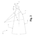
- FIG. 2 is an enlarged partial side view of a microkeratome blade that may be used with the method of the present invention.
- FIG. 3 is a partial cross-sectional view of a human cornea.
- one microkeratome 34 that may be used with the method of the present invention generally includes suction ring 10 sized and shaped so as to affixed to eye 12 .
- Ring 10 includes guides 14 / 16 opposite eye 12 that guide cutting head 40 across ring 10 .
- Ring 10 is connected through translation member 26 to stepper motor 28 for providing linear movement of cutting head 40 across ring 10 .
- Cutting head 40 contains blade 38 that is eccentrically connected to motor 36 contained within housing 42 of microkeratome 34 .
- Microkeratome 34 is well-known in the art (see for example U.S. Pat. No. 6,071,293 (Krumeich), the entire contents of which being incorporated herein by reference), and commercially available from sources such as Alcon Laboratories, Inc., Fort Worth, Tex.
- blade 38 that may be used with the method of the present invention generally includes relatively flat side 100 , tapered side 110 containing rounded section 112 and blunt tip 114 connecting flat side 100 and rounded section 112 .
- Rounded section 112 generally has a radius of between about 0.025 millimeters and 0.200 millimeters and is rounded through of angle ⁇ of between approximately 5 degrees and 60 degrees.
- Blunt tip 114 generally has a length L of between approximately 0.001 millimeters and 0.050 millimeters, with between about 0.005 millimeters and 0.025 millimeters being preferred, and is ground at an offset angle ⁇ relative to rounded portion 112 at between approximately between 0 degrees and 60 degrees, with between approximately between 0 degrees and 20 degrees being preferred.
- Blade 38 may be made of any suitable material, such as 400 Series stainless steel and may be made using conventional surgical blade manufacturing techniques well-known in the art.
- human cornea 200 has several layers. The outermost layer is epithelium 210 , followed by Basement Membrane 220 , Bowman's Membrane 230 , substantia intestinal or stroma 240 , Descemet's Membrane 250 and endothelium 260 .
- the method of the present invention involves the use of microkeratome 34 having blade 38 to remove epithelium 210 and Basement Membrane 220 while leaving Bowman's Membrane 230 relatively intact.
- the method of the present invention uses microkeratome 34 in a conventional manner well known to those skilled in the art.
- the oscillation frequency of blade 38 preferably is approximately between 5,000 revolutions/minute and 20,000 revolutions/minute, with approximately between 8,000 revolutions/minute and 14,000 revolutions/minute being most preferred.
- the speed of blade 38 as it traverses or advances across cornea 200 preferably is approximately between 1.0 millimeter/second and 2.0 millimeters/second, with approximately between 1.5 millimeters/second being most preferred.
- blunt tip 114 penetrates epithelium 210 and Basement Membrane 220 , but is insufficiently sharp to penetrate Bowman's Membrane 230 .
- Bowman's Membrane 230 and stroma 240 are irradiated as in a conventional laser refractive surgical procedure, see for example, U.S. Pat. No. 4,784,135 (Blum, et al.) and U.S. Pat. No. 4,903,695 C1 (Warner, et al.), the entire contents of which being incorporated herein by reference.
Landscapes
- Health & Medical Sciences (AREA)
- Ophthalmology & Optometry (AREA)
- Heart & Thoracic Surgery (AREA)
- Surgery (AREA)
- Engineering & Computer Science (AREA)
- Biomedical Technology (AREA)
- Nuclear Medicine, Radiotherapy & Molecular Imaging (AREA)
- Vascular Medicine (AREA)
- Life Sciences & Earth Sciences (AREA)
- Animal Behavior & Ethology (AREA)
- General Health & Medical Sciences (AREA)
- Public Health (AREA)
- Veterinary Medicine (AREA)
- Laser Surgery Devices (AREA)
- Laser Beam Processing (AREA)
Abstract
A method for the removal of the epithelial layer and underlying Basement Membrane while leave a smooth and undisturbed Bowman's Membrane in preparation for a laser refractive surgical procedure. The method of the present invention uses a microkeratome having a blade that is capable of cutting through the epithelial layer and Basement Membrane, but not capable of cutting through Bowman's Membrane.
Description
- This invention relates generally to the field of refractive surgery and, more particularly, to a device and method for performing laser refractive surgery.
- The human eye in its simplest terms functions to provide vision by transmitting light through a clear outer portion called the cornea, and focusing the image by way of a crystalline lens onto a retina. The quality of the focused image depends on many factors including the size and shape of the eye, and the transparency of the cornea and the lens.
- The optical power of the eye is determined by the optical power of the cornea and the crystalline lens. In the normal, healthy eye, sharp images are formed on the retina (emmetropia). In many eyes, images are either formed in front of the retina because the eye is abnormally long (axial myopia), or formed in back of the retina because the eye is abnormally short (axial hyperopia). The cornea also may be asymmetric or toric, resulting in an uncompensated cylindrical refractive error referred to as corneal astigmatism. In addition, due to age-related reduction in lens accommodation, the eye may become presbyopic resulting in the need for a bifocal or multifocal correction device.
- In the past, axial myopia, axial hyperopia and corneal astigmatism generally have been corrected by spectacles or contact lenses, but there are several refractive surgical procedures that have been investigated and used since 1949. Jose Barraquer, M.D. investigated a procedure called keratomileusis that reshaped the cornea using a microkeratome and a cryolathe. This procedure was never widely accepted by surgeons. Another procedure that has gained widespread acceptance is radial and/or transverse incisional keratotomy (RK or AK, respectively). In the 1990s, the use of photablative lasers to reshape the surface of the cornea (photorefractive keratectomy or PRK) or for mid-stromal photoablation (Laser-Assisted In Situ Keratomileusis or LASIK) have been approved by regulatory authorities in the U.S. and other countries. Recently, a new version of PRK called Laser Epithelial Keratomileusis (LASEK) has been developed wherein the epithelial layer is soaked in alcohol so as to release it from Bowman's Membrane and the epithelial layer is non-destructively rolled aside and the underlying stromal tissue is ablated in a manner similar to PRK. This procedure does not always allow for the smooth removal of the epithelial layer in a single sheet. In addition, alcohol is toxic to corneal tissue.
- Accordingly, a need continues to exist for a device and method for the safe, consistent removal of the epithelial layer and Basement Membrane during the a laser refractive surgical procedure.
- The present invention improves upon the prior art by providing a method for the removal of the epithelial layer and underlying Basement Membrane while leave a smooth and undisturbed Bowman's Membrane in preparation for a laser refractive surgical procedure. The method of the present invention uses a microkeratome having a blade that is capable of cutting through the epithelial layer and Basement Membrane, but not capable of cutting through Bowman's Membrane. The method of the present invention does not require the use of a toxic chemical such as alcohol.
- Accordingly, one objective of the present invention is to provide a safe and nontoxic method for the removal of the epithelial layer and underlying Basement Membrane in preparation for a laser refractive surgical procedure.
- Another objective of the present invention is to provide a method for the removal of the epithelial layer in preparation for a laser refractive surgical procedure without the use of toxic chemicals.
- Another objective of the present invention is to provide a device that provides the safe and non-toxic method for the removal of the epithelial layer and underlying Basement Membrane in preparation for a laser refractive surgical procedure.
- These and other advantages and objectives of the present invention will become apparent from the detailed description and claims that follow.
- FIG. 1 is a schematic representation of a microkeratome that may be used with the invention of the present method.
- FIG. 2 is an enlarged partial side view of a microkeratome blade that may be used with the method of the present invention.
- FIG. 3 is a partial cross-sectional view of a human cornea.
- As best seen in FIG. 1, one
microkeratome 34 that may be used with the method of the present invention generally includessuction ring 10 sized and shaped so as to affixed toeye 12.Ring 10 includes guides 14/16opposite eye 12 thatguide cutting head 40 acrossring 10.Ring 10 is connected throughtranslation member 26 tostepper motor 28 for providing linear movement of cuttinghead 40 acrossring 10. Cuttinghead 40 containsblade 38 that is eccentrically connected tomotor 36 contained withinhousing 42 ofmicrokeratome 34. Microkeratome 34 is well-known in the art (see for example U.S. Pat. No. 6,071,293 (Krumeich), the entire contents of which being incorporated herein by reference), and commercially available from sources such as Alcon Laboratories, Inc., Fort Worth, Tex. - As best seen in FIG. 2,
blade 38 that may be used with the method of the present invention generally includes relativelyflat side 100,tapered side 110 containingrounded section 112 andblunt tip 114 connectingflat side 100 androunded section 112.Rounded section 112 generally has a radius of between about 0.025 millimeters and 0.200 millimeters and is rounded through of angle δ of between approximately 5 degrees and 60 degrees.Blunt tip 114 generally has a length L of between approximately 0.001 millimeters and 0.050 millimeters, with between about 0.005 millimeters and 0.025 millimeters being preferred, and is ground at an offset angle Θ relative torounded portion 112 at between approximately between 0 degrees and 60 degrees, with between approximately between 0 degrees and 20 degrees being preferred.Blade 38 may be made of any suitable material, such as 400 Series stainless steel and may be made using conventional surgical blade manufacturing techniques well-known in the art. - As best seen in FIG. 3,
human cornea 200 has several layers. The outermost layer isepithelium 210, followed byBasement Membrane 220, Bowman's Membrane 230, substantia propria orstroma 240, Descemet'sMembrane 250 andendothelium 260. The method of the present invention involves the use ofmicrokeratome 34 havingblade 38 to removeepithelium 210 and Basement Membrane 220 while leaving Bowman's Membrane 230 relatively intact. The method of the present invention usesmicrokeratome 34 in a conventional manner well known to those skilled in the art. The oscillation frequency ofblade 38 preferably is approximately between 5,000 revolutions/minute and 20,000 revolutions/minute, with approximately between 8,000 revolutions/minute and 14,000 revolutions/minute being most preferred. The speed ofblade 38 as it traverses or advances acrosscornea 200 preferably is approximately between 1.0 millimeter/second and 2.0 millimeters/second, with approximately between 1.5 millimeters/second being most preferred. Asblade 38 approachescornea 200,blunt tip 114 penetratesepithelium 210 andBasement Membrane 220, but is insufficiently sharp to penetrate Bowman'sMembrane 230. As a result,blunt tip 114 androunded portion 112 scrape along the surface of Bowman'sMembrane 230, separatingepithelium 210 andBasement Membrane 220 from Bowman'sMembrane 230 without damaging Bowman'sMembrane 230. Following such separation, Bowman's Membrane 230 andstroma 240 are irradiated as in a conventional laser refractive surgical procedure, see for example, U.S. Pat. No. 4,784,135 (Blum, et al.) and U.S. Pat. No. 4,903,695 C1 (Warner, et al.), the entire contents of which being incorporated herein by reference. - This description is given for purposes of illustration and explanation. It will be apparent to those skilled in the relevant art that changes and modifications may be made to the invention described above without departing from its scope or spirit.
Claims (12)
1. A method of performing a laser refractive surgical procedure, comprising the steps of:
a) contacting a cornea with a blade;
b) advancing the blade across the cornea so that the blade penetrates an epithelium and a Basement Membrane of the cornea so as to expose but not penetrate a Bowman's Membrane of the cornea; and
c) irradiating the Bowman's Membrane and underlying stromal tissue with ablative laser radiation to effect a refractive change in the cornea.
2. The method of claim 1 wherein the blade contains a blunt tip.
3. The method of claim 2 wherein the blade has a flat section and a rounded section and the blunt tip separates the flat section from the rounded section.
4. The method of claim 1 wherein the blade is oscillated at an oscillation frequency of approximately between 5,000 revolutions/minute and approximately 20,000 revolutions/minute.
5. The method of claim 4 wherein the oscillation frequency is approximately between 8,000 revolutions/minute and 14,000 revolutions/minute.
6. The method of claim 1 wherein the blade is advanced across the cornea at a speed of approximately between 1.0 millimeter/second and 2.0 millimeters/second.
7. The method of claim 6 wherein the blade is advanced across the cornea at a speed of approximately 1.5 millimeters/second.
8. A method of performing a laser refractive surgical procedure, comprising the steps of:
a) contacting a cornea with a blade, the blade oscillated at an oscillation frequency of approximately between 5,000 revolutions/minute and 20,000 revolutions/minute;
b) advancing the blade across the cornea at a speed of approximately between 1.0 millimeter/second and 2.0 millimeters/second so that the blade penetrates an epithelium and a Basement Membrane of the cornea so as to expose but not penetrate a Bowman's Membrane of the cornea; and
c) irradiating the Bowman's Membrane and underlying stromal tissue with ablative laser radiation to effect a refractive change in the cornea.
9. The method of claim 8 wherein the blade contains a blunt tip.
10. The method of claim 9 wherein the blade has a flat section and a rounded section and the blunt tip separates the flat section from the rounded section.
11. The method of claim 8 wherein the oscillation frequency is approximately between 8,000 revolutions/minute and 14,000 revolutions/minute.
12. The method of claim 8 wherein the blade is advanced across the cornea at a speed of approximately 1.5 millimeters/second.
Priority Applications (5)
| Application Number | Priority Date | Filing Date | Title |
|---|---|---|---|
| US10/114,834 US20030191456A1 (en) | 2002-04-03 | 2002-04-03 | Laser refractive surgical procedure method and apparatus |
| US10/235,302 US20030191457A1 (en) | 2002-04-03 | 2002-09-05 | Laser refractive surgical procedure method and apparatus |
| PCT/US2003/007495 WO2003084387A2 (en) | 2002-04-03 | 2003-03-13 | Laser refractive surgical procedure method and apparatus |
| AU2003220181A AU2003220181A1 (en) | 2002-04-03 | 2003-03-13 | Laser refractive surgical procedure method and apparatus |
| US10/454,845 US20030208191A1 (en) | 2002-04-03 | 2003-06-04 | Laser refractive surgical procedure method |
Applications Claiming Priority (1)
| Application Number | Priority Date | Filing Date | Title |
|---|---|---|---|
| US10/114,834 US20030191456A1 (en) | 2002-04-03 | 2002-04-03 | Laser refractive surgical procedure method and apparatus |
Related Child Applications (1)
| Application Number | Title | Priority Date | Filing Date |
|---|---|---|---|
| US10/235,302 Continuation-In-Part US20030191457A1 (en) | 2002-04-03 | 2002-09-05 | Laser refractive surgical procedure method and apparatus |
Publications (1)
| Publication Number | Publication Date |
|---|---|
| US20030191456A1 true US20030191456A1 (en) | 2003-10-09 |
Family
ID=28673712
Family Applications (2)
| Application Number | Title | Priority Date | Filing Date |
|---|---|---|---|
| US10/114,834 Abandoned US20030191456A1 (en) | 2002-04-03 | 2002-04-03 | Laser refractive surgical procedure method and apparatus |
| US10/235,302 Abandoned US20030191457A1 (en) | 2002-04-03 | 2002-09-05 | Laser refractive surgical procedure method and apparatus |
Family Applications After (1)
| Application Number | Title | Priority Date | Filing Date |
|---|---|---|---|
| US10/235,302 Abandoned US20030191457A1 (en) | 2002-04-03 | 2002-09-05 | Laser refractive surgical procedure method and apparatus |
Country Status (3)
| Country | Link |
|---|---|
| US (2) | US20030191456A1 (en) |
| AU (1) | AU2003220181A1 (en) |
| WO (1) | WO2003084387A2 (en) |
Family Cites Families (2)
| Publication number | Priority date | Publication date | Assignee | Title |
|---|---|---|---|---|
| US6030398A (en) * | 1997-05-30 | 2000-02-29 | Summit Technology, Inc. | Surgical microtomes |
| DE69718633T2 (en) * | 1997-08-06 | 2003-09-11 | Sis Ltd. Surgical Instrument Systems, Bruegg B. Biel | Microkeratome for corneal surgery using laser |
-
2002
- 2002-04-03 US US10/114,834 patent/US20030191456A1/en not_active Abandoned
- 2002-09-05 US US10/235,302 patent/US20030191457A1/en not_active Abandoned
-
2003
- 2003-03-13 AU AU2003220181A patent/AU2003220181A1/en not_active Abandoned
- 2003-03-13 WO PCT/US2003/007495 patent/WO2003084387A2/en not_active Ceased
Also Published As
| Publication number | Publication date |
|---|---|
| US20030191457A1 (en) | 2003-10-09 |
| AU2003220181A1 (en) | 2003-10-20 |
| WO2003084387A2 (en) | 2003-10-16 |
| WO2003084387A3 (en) | 2003-12-04 |
| AU2003220181A8 (en) | 2003-10-20 |
Similar Documents
| Publication | Publication Date | Title |
|---|---|---|
| US4923467A (en) | Apparatus and process for application and adjustable reprofiling of synthetic lenticules for vision correction | |
| Bas et al. | Excimer laser in situ keratomileusis for myopia | |
| US20030014042A1 (en) | Method of creating stromal pockets for corneal implants | |
| US5833701A (en) | Procedure and device for corrective and therapeutic eye treatment | |
| JP4114036B2 (en) | Ophthalmic device for corneal curvature correction | |
| US5156622A (en) | Apparatus and process for application and adjustable reprofiling of synthetic lenticules for vision correction | |
| EP0764432A2 (en) | System for surgically correcting presbyopia | |
| US8256431B2 (en) | Methods for treating hyperopia and presbyopia via laser tunneling | |
| US7878204B2 (en) | Methods for treating hyperopia and presbyopia via laser tunneling | |
| EP0997122B1 (en) | Apparatus for Improving LASIK flap adherence | |
| US20040044355A1 (en) | Minimally invasive corneal surgical procedure for the treatment of hyperopia | |
| JP2002500522A (en) | Methods of corneal laser surgery | |
| Maldonado et al. | Advances in technologies for laser-assisted in situ keratomileusis (LASIK) surgery | |
| CA2331223C (en) | A method of corneal surgery by laser incising a contoured corneal flap | |
| US5104408A (en) | Apparatus and process for application and adjustable reprofiling of synthetic lenticules for vision correction | |
| US6350272B1 (en) | Method and apparatus for cutting an oblong corneal flap | |
| US6805698B2 (en) | Microkeratome blade | |
| US6551306B1 (en) | Refractive laser ablation through topography | |
| US6582445B1 (en) | Trephine for lamellar keratectomy | |
| US20040002722A1 (en) | Ultrasonic microkeratome | |
| US6358261B1 (en) | Lamellar dissecting instrument | |
| US20030191456A1 (en) | Laser refractive surgical procedure method and apparatus | |
| Viestenz et al. | Evaluation of corneal flap dimensions and cut quality using the SKBM automated microkeratome | |
| US20030208191A1 (en) | Laser refractive surgical procedure method | |
| Nair et al. | Evolution of refractive surgery |
Legal Events
| Date | Code | Title | Description |
|---|---|---|---|
| AS | Assignment |
Owner name: ALCON, INC., SWITZERLAND Free format text: ASSIGNMENT OF ASSIGNORS INTEREST;ASSIGNORS:AUSTIN, PETER;KOCAR, GEORGE A.;SACHAROFF, ALEX C.;AND OTHERS;REEL/FRAME:012781/0976 Effective date: 20020402 |
|
| STCB | Information on status: application discontinuation |
Free format text: ABANDONED -- FAILURE TO RESPOND TO AN OFFICE ACTION |