US20030068825A1 - System and method of determining proteomic differences - Google Patents
System and method of determining proteomic differences Download PDFInfo
- Publication number
- US20030068825A1 US20030068825A1 US10/195,774 US19577402A US2003068825A1 US 20030068825 A1 US20030068825 A1 US 20030068825A1 US 19577402 A US19577402 A US 19577402A US 2003068825 A1 US2003068825 A1 US 2003068825A1
- Authority
- US
- United States
- Prior art keywords
- peptide
- mass
- peptides
- analysis
- sequence
- Prior art date
- Legal status (The legal status is an assumption and is not a legal conclusion. Google has not performed a legal analysis and makes no representation as to the accuracy of the status listed.)
- Abandoned
Links
- DYYZDPZMBCSHTQ-UHFFFAOYSA-N COC1=CC(C(C)C)=C([N+](=O)[O-])C=C1C(C)C Chemical compound COC1=CC(C(C)C)=C([N+](=O)[O-])C=C1C(C)C DYYZDPZMBCSHTQ-UHFFFAOYSA-N 0.000 description 3
- CREMABGTGYGIQB-UHFFFAOYSA-N C.C Chemical compound C.C CREMABGTGYGIQB-UHFFFAOYSA-N 0.000 description 1
Images


Classifications
-
- G—PHYSICS
- G01—MEASURING; TESTING
- G01N—INVESTIGATING OR ANALYSING MATERIALS BY DETERMINING THEIR CHEMICAL OR PHYSICAL PROPERTIES
- G01N33/00—Investigating or analysing materials by specific methods not covered by groups G01N1/00 - G01N31/00
- G01N33/48—Biological material, e.g. blood, urine; Haemocytometers
- G01N33/50—Chemical analysis of biological material, e.g. blood, urine; Testing involving biospecific ligand binding methods; Immunological testing
- G01N33/68—Chemical analysis of biological material, e.g. blood, urine; Testing involving biospecific ligand binding methods; Immunological testing involving proteins, peptides or amino acids
- G01N33/6803—General methods of protein analysis not limited to specific proteins or families of proteins
- G01N33/6848—Methods of protein analysis involving mass spectrometry
- G01N33/6851—Methods of protein analysis involving laser desorption ionisation mass spectrometry
-
- G—PHYSICS
- G01—MEASURING; TESTING
- G01N—INVESTIGATING OR ANALYSING MATERIALS BY DETERMINING THEIR CHEMICAL OR PHYSICAL PROPERTIES
- G01N33/00—Investigating or analysing materials by specific methods not covered by groups G01N1/00 - G01N31/00
- G01N33/48—Biological material, e.g. blood, urine; Haemocytometers
- G01N33/50—Chemical analysis of biological material, e.g. blood, urine; Testing involving biospecific ligand binding methods; Immunological testing
- G01N33/58—Chemical analysis of biological material, e.g. blood, urine; Testing involving biospecific ligand binding methods; Immunological testing involving labelled substances
-
- G—PHYSICS
- G01—MEASURING; TESTING
- G01N—INVESTIGATING OR ANALYSING MATERIALS BY DETERMINING THEIR CHEMICAL OR PHYSICAL PROPERTIES
- G01N33/00—Investigating or analysing materials by specific methods not covered by groups G01N1/00 - G01N31/00
- G01N33/48—Biological material, e.g. blood, urine; Haemocytometers
- G01N33/50—Chemical analysis of biological material, e.g. blood, urine; Testing involving biospecific ligand binding methods; Immunological testing
- G01N33/68—Chemical analysis of biological material, e.g. blood, urine; Testing involving biospecific ligand binding methods; Immunological testing involving proteins, peptides or amino acids
- G01N33/6803—General methods of protein analysis not limited to specific proteins or families of proteins
- G01N33/6818—Sequencing of polypeptides
-
- G—PHYSICS
- G16—INFORMATION AND COMMUNICATION TECHNOLOGY [ICT] SPECIALLY ADAPTED FOR SPECIFIC APPLICATION FIELDS
- G16B—BIOINFORMATICS, i.e. INFORMATION AND COMMUNICATION TECHNOLOGY [ICT] SPECIALLY ADAPTED FOR GENETIC OR PROTEIN-RELATED DATA PROCESSING IN COMPUTATIONAL MOLECULAR BIOLOGY
- G16B20/00—ICT specially adapted for functional genomics or proteomics, e.g. genotype-phenotype associations
- G16B20/30—Detection of binding sites or motifs
-
- G—PHYSICS
- G16—INFORMATION AND COMMUNICATION TECHNOLOGY [ICT] SPECIALLY ADAPTED FOR SPECIFIC APPLICATION FIELDS
- G16B—BIOINFORMATICS, i.e. INFORMATION AND COMMUNICATION TECHNOLOGY [ICT] SPECIALLY ADAPTED FOR GENETIC OR PROTEIN-RELATED DATA PROCESSING IN COMPUTATIONAL MOLECULAR BIOLOGY
- G16B20/00—ICT specially adapted for functional genomics or proteomics, e.g. genotype-phenotype associations
- G16B20/50—Mutagenesis
-
- G—PHYSICS
- G01—MEASURING; TESTING
- G01N—INVESTIGATING OR ANALYSING MATERIALS BY DETERMINING THEIR CHEMICAL OR PHYSICAL PROPERTIES
- G01N30/00—Investigating or analysing materials by separation into components using adsorption, absorption or similar phenomena or using ion-exchange, e.g. chromatography or field flow fractionation
- G01N30/02—Column chromatography
- G01N2030/022—Column chromatography characterised by the kind of separation mechanism
- G01N2030/027—Liquid chromatography
-
- G—PHYSICS
- G01—MEASURING; TESTING
- G01N—INVESTIGATING OR ANALYSING MATERIALS BY DETERMINING THEIR CHEMICAL OR PHYSICAL PROPERTIES
- G01N2458/00—Labels used in chemical analysis of biological material
- G01N2458/15—Non-radioactive isotope labels, e.g. for detection by mass spectrometry
-
- G—PHYSICS
- G01—MEASURING; TESTING
- G01N—INVESTIGATING OR ANALYSING MATERIALS BY DETERMINING THEIR CHEMICAL OR PHYSICAL PROPERTIES
- G01N30/00—Investigating or analysing materials by separation into components using adsorption, absorption or similar phenomena or using ion-exchange, e.g. chromatography or field flow fractionation
- G01N30/02—Column chromatography
- G01N30/62—Detectors specially adapted therefor
- G01N30/72—Mass spectrometers
- G01N30/7233—Mass spectrometers interfaced to liquid or supercritical fluid chromatograph
- G01N30/724—Nebulising, aerosol formation or ionisation
- G01N30/7266—Nebulising, aerosol formation or ionisation by electric field, e.g. electrospray
-
- G—PHYSICS
- G16—INFORMATION AND COMMUNICATION TECHNOLOGY [ICT] SPECIALLY ADAPTED FOR SPECIFIC APPLICATION FIELDS
- G16B—BIOINFORMATICS, i.e. INFORMATION AND COMMUNICATION TECHNOLOGY [ICT] SPECIALLY ADAPTED FOR GENETIC OR PROTEIN-RELATED DATA PROCESSING IN COMPUTATIONAL MOLECULAR BIOLOGY
- G16B20/00—ICT specially adapted for functional genomics or proteomics, e.g. genotype-phenotype associations
-
- Y—GENERAL TAGGING OF NEW TECHNOLOGICAL DEVELOPMENTS; GENERAL TAGGING OF CROSS-SECTIONAL TECHNOLOGIES SPANNING OVER SEVERAL SECTIONS OF THE IPC; TECHNICAL SUBJECTS COVERED BY FORMER USPC CROSS-REFERENCE ART COLLECTIONS [XRACs] AND DIGESTS
- Y10—TECHNICAL SUBJECTS COVERED BY FORMER USPC
- Y10T—TECHNICAL SUBJECTS COVERED BY FORMER US CLASSIFICATION
- Y10T436/00—Chemistry: analytical and immunological testing
- Y10T436/13—Tracers or tags
-
- Y—GENERAL TAGGING OF NEW TECHNOLOGICAL DEVELOPMENTS; GENERAL TAGGING OF CROSS-SECTIONAL TECHNOLOGIES SPANNING OVER SEVERAL SECTIONS OF THE IPC; TECHNICAL SUBJECTS COVERED BY FORMER USPC CROSS-REFERENCE ART COLLECTIONS [XRACs] AND DIGESTS
- Y10—TECHNICAL SUBJECTS COVERED BY FORMER USPC
- Y10T—TECHNICAL SUBJECTS COVERED BY FORMER US CLASSIFICATION
- Y10T436/00—Chemistry: analytical and immunological testing
- Y10T436/24—Nuclear magnetic resonance, electron spin resonance or other spin effects or mass spectrometry
Definitions
- This invention relates to systems and methods for automatically calculating information received from a mass spectrometer. More specifically, this invention relates to systems and methods that determine proteomic differences between two samples by comparing mass spectrometer data from each sample.
- mRNA messenger RNA
- biological and computational techniques have been used to correlate specific biological functions or cellular activities with these expressed gene sequences.
- Proteins are essential for the control and execution of virtually every biological process. The rate of synthesis and the half-life which dictate a particular peptide's expression level are typically controlled post-transcriptionally. Furthermore, the activity of a peptide is frequently modulated by post-translational modifications and is thus dependent on the association of the peptide with other molecules. Examples of associated molecules include DNA, RNA, sugar residues and other peptides. Neither the level of expression nor the state of activity of peptides is therefore directly apparent from the gene sequence or even the expression level of the corresponding mRNA transcript. It is therefore essential that a complete description of a biological system include measurements that indicate the identity, quantity and the state of activity of the peptides which constitute the system. This requirement for large-scale (ultimately global) analysis of peptides expressed in a cell or tissue has been termed proteome analysis (Pennington et al., Trends Cell Bio 7:168-173 (1997)).
- proteome analysis is based on the separation of complex peptide samples by two-dimensional gel electrophoresis (2DE) and the subsequent sequential identification of the separated peptide species (Ducret et al., Prot Sci 7:706-719 (1998); Garrels et al., Electrophoresis 18:1347-1360 (1997); Link et al., Electrophoresis 18:1314-1334 (1997); Shevchenko et al., Proc Natl Acad Sci USA 93:14440-14445 (1996); Gygi et al., Electrophoresis 20:310-319 (1999); Boucherie et al., Electrophoresis 17:1683-1699 (1996)).
- 2DE two-dimensional gel electrophoresis
- Mass spectrometry based techniques for peptide identification identify peptide fragments based on a spectral signature uniquely generated for each peptide sequence.
- a peptide mixture is separated using a first mass spectrometer which separates the peptides according to their mass and charge characteristics to produce a spectrum indicative of the component peptides of the peptide mixture.
- Each separated peptide is then further subjected to a second tandem mass analysis where the peptide is fragmented and a second mass spectrum is produced.
- the second mass spectrum comprises a series of peaks (peptide signature) formed as a result of differences in the mass-to-charge ratios of fragments of the peptide. For peptides with differing sequences, the series of peaks uniquely identifies the particular sequence of the peptide undergoing analysis.
- Computational methods for sequencing peptides subjected to mass analysis involve comparing the spectrum generated by the peptide of interest with known spectra.
- the peptide spectrum is associated with a known sequence to indicate sequence homology.
- the results of the analysis typically contain many values and statistical correlations that identify associations between the peptide signature and the known spectra.
- the analysis may also include candidate sequences that are likely to match the experimental spectrum, as well as, correlation scores and probabilities indicating the degree of confidence of the match.
- U.S. Pat. No. 6,017,693 describes a system for correlating a peptide fragment mass spectrum with amino acid sequences derived from a database.
- This is one example of a conventional mass spectrometry-based method for peptide identification which compares an experimental peptide spectrum with a known database of spectra.
- mass spectra from an experiment are input into a computer containing a database of sequence-associated spectrum.
- the computer then performs a search of the database and outputs results of the search to the investigator in the form of an output file or summary.
- the resulting output file must then be reviewed and interpreted manually by the investigator to determine the peptide sequence.
- Such a system may have the analytical capability to process a relatively small sample peptide population, however, its utility is severely diminished when assessing the many thousands of proteins or peptides typically present in a cell or tissue extract. The resulting amount of time an investigator must devote to reviewing the output files therefore represents a significant bottleneck in the analytical process which must be alleviated if complex mixed-populations of peptides are to be assessed.
- a further difficulty presented by the aforementioned peptide sequencing and identification methods relate to their limitations when applied to differential analysis. Differential analysis correlates protein expression between multiple populations of cells or tissues to identify differences between them. Such comparisons are essential to understand regulatory patterns and identify novel peptides or pathways.
- Existing mass spectroscopy based technologies typically asses each sample independently and are subject to experimental and instrumental variability between samples. This results in difficulties in correlating all of the components from each sample relative to one another and limits the utility of these techniques in assessing differential peptide expression on a global scale.
- Embodiments of this invention include systems and methods for rapidly determining and quantifying proteomic differences between two or more biological samples.
- proteomic analysis is performed by differentially labeling the two or more samples and subsequently quantifying the peptide levels or abundance in each sample. Differential labeling of the peptides derived from each sample provides a discernable means to identify each peptide population during the analysis and to provide a consistent, calculable molecular weight difference that can be observed during mass spectrometry of a mixed population peptide sample.
- the mixed population peptide sample is passed through a peptide separation column and subjected to mass spectroscopy-based techniques. Knowledge of the difference in mass between the two populations, permits the system to identify pairs of the same (analogous) peptide from the mass spectrometry data, and determine their relative quantities or abundances. This results in the ability to rapidly and reliably calculate proteomic differences between the biological samples.
- the approach described herein can be used for the quantitative analysis of peptide expression in complex samples (such as cells, tissues, and fractions thereof). Furthermore, the invention provides a suitable mechanism for differential expression analysis between multiple samples and the identification of novel peptides. Using a peptide labeling technique in conjunction with peptide separation and mass analysis methodologies, the peptide identification system resolves complex mixtures of peptides which are identified by database similarity lookups rather than traditional sequencing reactions. Additionally, this system evaluates peptide expression and regulation patterns in a rapid and quantifiable manner.
- Embodiments of the invention include a mass spectrometry-based system and method for rapidly and quantitatively analyzing peptides in complex mixtures or isolates.
- the system also features automated processing capabilities used to analyze differentially expressed peptides in a single sample in order to reduce variability and increase accuracy.
- Differentially expressed peptides are identified by changes in expression patterns which, for example, may be affected by a stimulus (e.g., administration of a drug or contact with a potentially toxic material), by a change in environment (e.g., nutrient level, temperature, passage of time) or by a change in condition or cell state (e.g., disease state, malignancy, site-directed mutation, gene knockouts) of the cell, tissue or organism from which the sample originated.
- a stimulus e.g., administration of a drug or contact with a potentially toxic material
- a change in environment e.g., nutrient level, temperature, passage of time
- a change in condition or cell state e.g., disease
- FIG. 1 is a flow diagram illustrating a differential peptide identification methodology.
- FIG. 2 is a block diagram illustrating a data analysis system used to identify differential peptide expression.
- FIG. 3 is a flowchart illustrating a method of qualitative analysis of complex peptide mixtures.
- FIG. 4 is a simplified mass spectrum intensity curve for a differentially labeled peptide in which markers create a mass differential between analogous peptides.
- FIG. 5 is a flowchart illustrating a correlation process used for identifying differentially labeled peptides.
- FIGS. 6 A-E are simplified mass spectrum scans illustrating states of differential expression that may be identified by the data analysis system.
- FIG. 7 is a flow diagram illustrating a method for identifying and quantitating chromatographic peaks from a differentially labeled mass spectrum analysis.
- FIG. 8 is a flow diagram illustrating a method for parallel processing of mass spectrum and sequence data.
- FIG. 9 is a flow diagram illustrating computational activities performed by nodes within a parallel architecture that are used to resolve and quantitate differentially expressed peptides.
- FIG. 10 is a chart showing the FPLC spectrum from the purification the synthesized PEPTag.
- FIG. 11 a is a printout showing the mass spectrum of the synthesized PEPTag.
- FIG. 11 b is a printout showing the mass spectrum from MS/MS experiment to sequence PEPTag.
- FIGS. 12 a,b show printouts of the MALDI MS analysis of PEPTag captured BSA peptides.
- FIG. 12 a is a printout wherein peaks are cysteinyl tryptic peptides from tagged BSA, which are captured by HA matrix and cleaved off by TEV.
- FIG. 12 b is a printout showing a control analysis of untagged BSA. The main peak in this spectrum is from TEV protease.
- FIGS. 13 a,b show the ⁇ LC MS/MS analysis of PEPTag captured BSA peptides.
- FIG. 13 a is a printout showing the base peak ion current profiles of all peptides released by TEV protease.
- FIGS. 14 a,b show the MS and MS/MS spectra of the PEPTag modified peptide.
- FIG. 14 a is a printout showing the full-scan (600-1,500 m/z) mass spectrum at time 29.49 min of ⁇ LC-MS and ⁇ LC-MS/MS analysis.
- FIG. 15 is a printout showing the MALDI mass spectrum of a pair of PEPTag labeled peptides of identical sequences. The m/z difference depends on the charge state. It is either 14 or 7 for charge state one or two.
- FIGS. 16 a - c show the ⁇ LC-MS/MS analysis of captured peptides labeled by differential PEPTags.
- FIG. 16 a is a printout showing base peak ion current profiles of all the peptides released by TEV protease from combined two protein mixtures.
- FIG. 16 b is a printout showing the reconstructed ion chromatograms (m/z 1034.0-1035.0) of a cysteinyl peptide labeled by PEPTag 1a.
- FIG. 16 c is a printout showing the reconstructed ion chromatograms (m/z 1027.0-1028.0) of the same cysteinyl peptide labeled by PEPTag 1b.
- FIG. 17 is a printout of the ESI mass spectrum of the pair of PEPTag labeled peptides of identical sequences. The m/z difference is 7 for doubly charged ions.
- each population is labeled with an identifiable label or marker to resolve the mixed-population of peptides within the same sample or analysis.
- the resulting combined analysis provides improved resolution and identification capabilities and is not subject to the degree of instrumental or cross-sample experimental variations which confound conventional peptide identification techniques.
- the peptide identification system further implements an automated sequencing routine in which tandem mass spectra identification resolves protein sequences by querying and correlation against a spectral database of known peptide spectra. This feature significantly improves data acquisition and sequencing throughput and provides a mechanism by which peptides within the mixed-population can be readily identified without additional sequencing steps or reactions.
- an affinity labeling procedure is used to selectively isolate peptides that contain a desired label or tag.
- the isolated proteins, peptides, or reaction products are then characterized by mass spectrometry (MS) based techniques.
- MS mass spectrometry
- sequence of isolated peptides is determined using tandem MS (MS) n techniques which are correlated with known peptide spectrum produced by the tandem MS (MS) n techniques.
- the system for peptide identification and differential comparison incorporates a chromatographic/separation technique, such as microcapillary liquid chromatography or gas chromatography. These chromatographic techniques separate the mixed peptide sample or solution of interest thereby permitting selective analysis of each peptide sequence.
- the sample is introduced into a mass spectrometer which serves as a detector of the individual components.
- the spectral database comprises a collection of tandem mass spectra which have been previously associated with known peptide sequences.
- a mass spectral database is described in U.S. Pat. No. 5,538,897 to Yates, et al.
- Software comparison and identification routines correlate the output spectrum from mass spectrometry of the sample with those spectrum contained in the spectral database and returns the peptide identity of each peptide in the sample. Using these methods the spectrum of a complex peptide mixture is readily resolved and the corresponding sequences of the constituent peptides are identified as will be described in greater detail hereinbelow.
- FIG. 1 illustrates an overview of one embodiment of a peptide identification and differential analysis technique used to resolve, sequence, and identify complex peptide mixtures derived from two or more peptide populations.
- a typical comparison of differential expression is made using a starting cell population 105 .
- One portion of the cell population 105 is separated into a control cell population 109 A, while another portion of the population 105 is treated with a test compound to become test cell population 109 B.
- the test cell population 109 B is treated with one or more conditions or treatments for which proteomic differences are to be identified.
- the cell population 105 is analyzed by comparing the proteomes of the control population 109 A with the treated cell population 109 B.
- the protein or peptide populations from each cell are isolated to yield a control peptide population 107 and a treated peptide population 108 .
- the peptide isolation procedure may additionally incorporate processing or purification steps designed to remove undesirable or contaminating biomolecules and chemicals. For example, during the harvest of peptides from a cell or tissue, biomolecules such as RNA, DNA, and proteases, as well as, extraction reagents and buffers may be removed from the peptide isolate to prevent interference with detection of the peptide molecules.
- a subsequent labeling reaction is used to label each peptide population 107 , 108 with an identifiable peptide labeling moiety or label 122 , 124 which aids in resolving the peptide populations 107 during mass analysis.
- the labels 122 , 124 comprise multi-functional synthetic peptide sequences with differing masses.
- the peptide populations 107 , 108 are made differentially identifiable by incorporating the first label 122 into the first peptide population 107 and incorporating the second label 124 into the second peptide population 108 .
- the peptides 107 , 108 derived from each condition or treatment 110 are made to contain an identifiable label 122 , 124 of known mass.
- the difference in molecular weight between the first label 122 and the second label 124 serves as a basis for determining the peptide population 107 , 108 of origin from which an identified peptide is derived by creating a mass differential between the two peptide populations. Examples of differential labels are described below.
- the labels 122 , 124 may additionally contain a peptide epitope tag or motif used for affinity purification of the labeled peptides 107 , 108 .
- This feature of the labels 122 , 124 is useful for isolating only those peptides which have been labeled and may further serve as a means for enriching the peptide populations 107 , 108 . Enrichment of the peptide populations 107 , 108 increases the sensitivity of the mass detection procedure and removes background “noise” that may be contributed by unlabeled or undesirable peptides.
- the treated peptide population 108 might be labeled in order for each peptide in the treated population to have a different mass from the control population. Additionally, it is contemplated that the peptides can be metabolically labeled prior to isolation from the cells or tissues. In this alternative method, discernable peptide populations 107 , 108 are created through the use of isotopic labeling to create peptide populations 107 , 108 with differing masses.
- a heavy isotope label such as a nitrogen isotope ( 15 N)
- a lighter nitrogen isotope such as 14 N
- the different isotopes are incorporated in-vivo to label all of the amino acids to create the discernable peptide populations without the requirement of a subsequent labeling step.
- a specific protease site may further be incorporated into the label 122 , 124 to facilitate the release of the affinity purified labeled peptides from an affinity matrix. Additional details of the chemical composition of the labels 122 , 124 as well as details of the specialized peptide epitope motifs for purification of the peptide populations 107 , 108 are described below.
- the mixed peptide sample 130 is subjected to proteolysis to fragment the peptides 107 , 108 into smaller molecules which are of suitable size for use in mass spectrometry-based techniques. Furthermore, protease cleavage can be used to release labeled peptides 107 , 108 from the aforementioned affinity matrix.
- protease enzymes which may be used for peptide digestion include: TEB protease, chymotrypsin, endopeptidease Arg-C, endopeptidease Asp-N, trypsin, Staphylococcus aureus protease, thermolysin, and pepsin.
- protease selection may be directed by the type of label incorporated into the labeled peptides 107 , 108 .
- These labels 122 , 124 may contain amino acid sequences which define specific protease cleavage sites which are designed to release the labeled peptides from the affinity matrix to provide a purified or enriched peptide sample.
- Quantitation of peptide expression levels is performed using mass analysis techniques which determine peptide quantities within the differentially labeled mixed-population peptide sample 130 .
- the mixed-population sample 130 is first subjected to a preliminary separation step using liquid or gas chromatography methods or 2-dimensional gel electrophoresis.
- multidimensional protein identification technology ModPIT (Washburn et al., Nature Biotechnology, 19: 242-247 (2001)) is used as a preliminary means to separate the peptide components resulting from the aforementioned proteolysis reactions.
- the MudPIT technique utilizes a fused-silica microcapillary column packed with a reverse-phase material (XDB-C18, Hewlett-Packard, Calif.) in addition to a strong cation exchange material (Partisphere SCX, Whatman, N.J.).
- XDB-C18 reverse-phase material
- Hewlett-Packard Calif.
- Partisphere SCX Partisphere SCX, Whatman, N.J.
- the mixed-peptide sample is loaded onto the packed column and placed in-line with the mass spectrometer and a buffer solution is passed through the column to elute the peptides.
- the resulting peptide eluate provides a preliminary separation means for the peptides which are then passed through the mass spectrometer resulting in further separation of the peptides according to their mass-to-charge ratio.
- the mass spectrometer in addition to serving as a peptide-separation means, acts as a detector to provide information useful in the identification of each peptide species contained within the mixed-population sample 130 .
- Mass analysis in this manner, provides a suitable method to compare expression levels between similar peptides 107 , 108 derived from different sources, conditions, or treatments as will be described in greater detail hereinbelow.
- mass analysis techniques may be applied to the resolution and identification of the mixed-population peptide sample 130 .
- suitable mass analysis techniques include: electron ionization, fast atom/ion bombardment, matrix-assisted laser desorption/ionization (MALDI), and electrospray ionization.
- MALDI spectroscopy techniques in particular possess a number of desirable characteristics which improve the quality of the mass analysis.
- MS (MS) n tandem mass analysis
- MS (MS) n spectrum 147 are desirably acquired for each resolved peptide 146 using an automated procedure wherein the individual spectrum 147 are acquired and stored for later processing and sequence identification.
- MS(MS) n spectrum 147 are generated (at least one for each resolved peptide 146 ). While it is possible to visualize, review, and identify each spectrum manually, it is impractical and time consuming for an entire peptide population to be analyzed in this manner. Instead the MS(MS) n spectrum 147 are well suited to be processed by an automated method using computer assisted identification in conjunction with a spectral or correlative database, as will be described in greater detail hereinbelow.
- differential peptide analysis compares peptides present in two or more biological samples.
- the peptides are labeled with a discernable marker to allow the peptides from each biological sample to be identifiable from one another when they are combined.
- Combination of the samples is desirable as it permits simultaneous analysis of the peptides and provides a means of directly comparing related peptides.
- Direct peptide comparison is further useful in identifying expression differences between related peptides within the two or more biological samples and aids in the detection of novel peptides.
- a composition of the two peptide populations will be related (i.e. both cells will contain identical peptides which may be expressed at different levels).
- the differential peptide analysis identifies and quantitates the relative concentrations of the related peptides in these populations to provide information about the overall peptide expression state of each biological sample. This analysis further identifies differences in peptide expression between the two biological samples which are useful in determining the effect of a treatment or condition upon a cell or tissue.
- Peptides are identified using mass analytical methods in which the peptides undergoing analysis are bombarded with an electron beam to produce identifiable fragments (cations and radical cations) that are accelerated in a vacuum through a magnetic field and are sorted on the basis of mass-to-charge ratios. Peptides are identified on the basis of the mass-to-charge ratio which is related to the molecular weight of the fragments produced. Subsequent tandem mass analysis produces a unique spectral signature for each identified fragment which is compared to a database of known spectral signatures and used to identify the sequences of the collection of peptide fragments.
- One device for performing this function is a tandem mass spectrometer LCQ Deca from Thermo Finnigan (San Jose, Calif.). See http://www.thermofinnigan.com on the Internet for more information.
- This embodiment of the invention therefore is an automated method for identifying the many thousands of component peptides (i.e.: the proteome) of a biological sample. Furthermore, the expression levels of the component peptides can be rapidly quantitated and compared between samples to give a better understanding of global peptide expression within biological systems.
- component peptides i.e.: the proteome
- FIG. 2 illustrates components of a data analysis system 200 which interact with instrumentation 205 used to perform the differential peptide analysis.
- the data analysis system 200 comprises a plurality of modules 210 that operate in conjunction with a microprocessor 215 to receive and process data output 208 produced by the mass analysis and MS (MS) n techniques. Using these modules 210 , the data analysis system 200 identifies the peptide constituents whose mass spectrum and associated information make up the data output 208 and subsequently processes the data to obtain detailed sequence and expression information.
- MS mass analysis and MS
- an instrument control/data acquisition (ICDA) module 220 acts as an interface between the instrumentation 205 and the data analysis system 200 .
- the ICDA module 220 receives the data output 208 and performs necessary handshaking and error correcting functions to insure data integrity.
- the ICDA module 220 is further equipped to recognize and process various data types associated with the data output 208 which are native to the instrumentation being used 205 .
- the ICDA module 220 may additionally issue control signals 209 which coordinate run-time activities associated with the instrumentation 205 .
- the control signals 209 may be used to modify configuration settings or parameters the instrumentation 205 , as well as, manage operational modes such as starting/stopping sample analysis.
- control signals 209 may be issued by the data analysis system 200 to direct a plurality of mass spectral analysis scans to be acquired by the instrumentation 205 over a specified time period or with a particular frequency.
- the mixed-peptide population 130 is eluted from the preliminary separation means and passed through the mass analysis instrumentation over a time period of approximately 1-10 minutes.
- mass spectral scans are taken with a frequency of approximately 50 scans/sec generating a plurality of mass spectral scans which are representative of the peptide composition at various points throughout the peptide elution.
- this method of multiscan mass analysis is used to construct peptide elution profiles for each of the peptides in the mixed population and improves the ability of the data analysis system 200 to identify and quantify proteomic differences.
- a data processing (DP) module 225 receives the data output 208 from the instruments 205 , formats the data output 208 , and stores it in a working database 226 in a suitable form for later retrieval and processing. Functions of the DP module 225 may include rearranging or organizing the data output 208 , performing operations to transform or change the format of the data output 208 , or other tasks to prepare the data output 208 for subsequent analysis.
- the DP module 225 additionally interacts with a working database 226 (used to store raw data and information) and a bioinformatic database or data warehouse 227 (used to archive the experimental results after the data has been processed and the mixed-peptide population analyzed, quantitated, and compared) to organize, categorize and store the data output 208 in a form that may be easily sorted, queried, and retrieved.
- a working database 226 used to store raw data and information
- a bioinformatic database or data warehouse 227 used to archive the experimental results after the data has been processed and the mixed-peptide population analyzed, quantitated, and compared
- the working database 226 and the bioinformatic database 227 are desirably implemented using relational schemas to provide flexible analytical querying and data mining capabilities. Furthermore, use of the databases 226 , 227 provide a means by which the data output 208 and expression results may be correlated with other information creating an integrated bioinformatic system.
- the databases 226 , 227 may be implemented using applications designed for relational database development and implementation, such as those sold by Oracle Corporation (Redwood Shores, Calif.), Sybase Corporation (Emeryville, Calif.), and MySQL AB (Postgirot, Sweden).
- the databases 226 , 227 comprise database designs implemented using numerous other programming languages such as JAVA, C/C++, Basic, Fortran, or the like, wherein the database structure, tables, and associations are defined by code of the programming languages.
- databases 226 , 227 may be implemented as a single database with separate tables or as other data structures that are well known in the art such as linked lists, binary trees, and so forth. Additionally, the databases 226 , 227 may be implemented as a plurality of databases which are collectively administered to store and analyze the data of the data analysis system 200 .
- a communications module 235 of the data analysis system 200 interacts with a spectral database 250 to aid in the determination of the origin and sequence for each peptide component of the mixed peptide population under study.
- the spectral database 250 comprises stored spectra of known peptide sequences used to identify peptides from experimental tandem mass spectrum data 255 .
- the data analysis system 200 desirably utilizes a computer program or search routine to identify the peptides by comparison of tandem mass spectrum data 255 with the spectral database 255 .
- SEQUEST peptide identification program developed at the University of Washington (http://www.washington.edu). Information on the SEQUEST program and system can be found on the Internet at http://thompson.mbt.washington.edu.
- peptide-correlated output files 260 containing the putative identities of the peptides determined from the spectral data analysis are then returned to the data analysis system 200 for further processing.
- communication between the data analysis system 200 and the spectral database 250 occurs by way of a communications medium 252 , such as the Internet, with the communications module 235 providing functionality for sending and receiving data through a suitable means, such as a TCP/IP based protocol.
- the communications module may additionally provide accessibility to other remotely located bioinformatic information systems 254 such as GenBank, SwissProt, Entrez, PubMed, and the like to acquire other information which may be associated with the peptide-correlated output files 260 and information stored in the databases 226 , 227 .
- a quantitation module 230 is used by the data analysis system 200 to determine more precise relationships between the peptides identified in the mixed-population and their relative expression levels. This module confirms the identity of each peptide in the mixed population of peptides by evaluating the results of the peptide correlated output files 260 and the mass spectrum data 208 .
- the quantitation module 230 evaluates the peptide-correlated output files 260 and identifies peaks or intensity curves corresponding to resolved peptides in the mass spectrum data 208 .
- the quantitation module 230 also quantitates the amount of peptide associated with a particular resolved peak 146 or intensity curve within the mass spectrum data 208 by area calculations. Additionally, the quantitation module 230 identifies and evaluates the peaks corresponding to the same peptide from both control and treated samples. This process will be described in greater detail hereinbelow.
- peptides from the control population and the treated population may be determined by the differential masses of the labels 122 , 124 which are integrated into each peptide undergoing analysis.
- the use of the label 122 , 124 distinguishes analogous peptides from different samples which have similar spectrum 208 by creating a mass differential between the analogous peptides containing different labels 122 , 124 .
- Identification of the peptides derived from each treatment or condition provides a means for the quantitation module 230 to perform cross-sample comparisons and identify changes in peptide expression.
- the IR module 240 provides additional insight into the mixed population peptide samples under study by retrieving information from other bioinformatic databases 254 that may be correlated with peptide sequences identified by the data analysis system 200 .
- the IR module 240 may read information stored in the working database 226 or the bioinformatic database 227 and perform automated information search queries directed towards collecting additional information about the identified peptides.
- the IR module 240 therefore, provides an additional means for automatically associating bioinformatic information from other informational sources and repositories with the experimentally identified peptides to yield a detailed collection of information.
- peptide expression data is acquired for the mixed population of differentially labeled peptides 130 and subsequently processed to identify the peptide constituents of the mixed population sample.
- the system 200 formats and stores the data in an organized manner and extracts relevant information to use to query the spectral database 250 .
- the spectral database 250 then returns correlated tandem mass spectra 260 which are associated with the spectra of individual peptides in the mixed population undergoing analysis.
- specialized modules 210 of the system 200 provide instructions which parse and process the correlated tandem mass spectra 260 in a rapid and efficient manner and store the results of the analysis in the bioinformatic database 227 for subsequent evaluation by the investigator.
- the aforementioned automated analysis and correlation features of the data analysis system 200 free investigators from having to perform lengthy searches and associations on an individual basis. Furthermore, the data analysis system 200 provides a more complete collection of data and information to which subsequent data mining techniques can be applied to further investigate the components of the mixed-peptide population.
- FIG. 3 further illustrates a method 300 for analyzing complex peptide mixtures using the aforementioned metabolic labeling or tagging methods to distinguish between different cell types or conditions.
- the process begins at a start state 302 and then moves to a state 304 wherein one cell population is treated differently from another cell population. Once the cell populations are treated, their peptides are isolated and labeled at a state 306 .
- the labeling method may include metabolic labeling methods incorporating isotopes directly into the peptides or subsequent post-growth labeling methods with incorporate peptides of known sequence and mass into the peptides.
- metabolic labeling methods incorporating isotopes directly into the peptides
- subsequent post-growth labeling methods with incorporate peptides of known sequence and mass into the peptides.
- the peptides are then processed and separated by mass spectroscopy-based techniques at a state 308 .
- the mass spectroscopy-based techniques are preceded by the aforementioned MudPIT two-dimensional liquid chromatography methodology for separating the mixed-peptide population.
- the mixed-peptide sample is eluted off the column in a series of buffer washes (see Washburn et al., Nature Biotechnology, 19: 242-247 (2001) for additional information).
- Mass analysis of the eluted sample takes place as a plurality of independent “mass analysis snapshots” or scans which are performed sequentially over the time it takes for the mixed-peptide population to be eluted from the MudPIT column.
- mass analysis of the mixed-peptide eluate is performed at a rate of approximately 50 scans per second with approximately 9000 scans being acquired during the run of a typical mixed-peptide sample.
- the acquisition of sequential mass spectrum scans form a parent ion map or peptide elution profile for each of the peptides in the mixed population.
- peptide signatures or tandem mass spectrum are further generated by directing a portion of each eluted peptide through a second tandem mass analysis instrument to identify and characterize the peptides present in each parent mass spectrum scan.
- the data analysis system 200 identifies the intensity of each of the peptide peaks within a particular mass spectrum scan or ion map and directs a tandem mass analysis to be performed for the most intense peaks using MS (MS) n .
- MS MS
- a similar process of identification of peak intensity is performed.
- the mass analysis system 200 determines if the most intense peaks have already been identified in the previous mass spectrum scan and, if so, selects new peaks with lesser intensities to perform tandem mass analysis on.
- the data analysis system 200 avoids performing redundant tandem mass analysis on peptides which are eluted over the time for which a plurality of mass analysis scans have been acquired to reduce the size of the data set which must be subsequently processed.
- tandem mass spectrum may be acquired for each peak within a particular mass spectrum scan or tandem mass spectrum may be acquired in another user-defined manner as desired. In this manner, data acquisition is facilitated, yet comprehensive information may be readily obtained to aid in the subsequent sequence identification.
- the data analysis system 200 facilitates the analysis of the peptide-correlated output files 260 by automating a number of the sorting and organizational tasks required to analyze the results returned from the spectrum comparison state 312 thereby reducing the burden to the investigator in identifying the components of the mixed-peptide population.
- the peptide data returned from the output files 260 is parsed and are stored to the working database 226 . This process is explained more completely below.
- a subsequent quantitation is performed in state 315 to determine the relative abundance of the peptides originating from the different samples which have been mixed together at the onset of the analysis.
- identity of each peptide that was subjected to a spectrum analysis is retrieved from the working database 226 and correlated with the mass spectrum peak heights and areas to determine the relative abundance of the identified peptide.
- Differential comparisons are additionally performed to correlate the expression of analogous peptides arising from the different peptide samples within the mixed population.
- the data analysis system 200 may further employ advanced processes to identify spectral peaks which were not positively correlated by spectral comparison. For example, in the analysis of a whole cell lysate containing many thousands of individual peptide components, the mass spectra data 208 produced vary greatly from one to the next in terms of quality and information. In some instances, the spectral peak 146 may not possess sufficient signal strength to be positively identified by the component identification 145 and spectrum comparison process.
- the data analysis system 200 provides functionality to correlate these weak or diminished spectral peaks 146 with analogous spectral peaks arising from the same peptide from a different peptide population within the sample. Thus, low abundance peptides can be positively identified based on an analogous peptide with a different label 122 , 124 . This feature of the data analysis system 200 improves the analysis of the peptide-correlated output files 260 and increases the sensitivity of the system in detecting and identifying low abundance peptides within the mixed-peptide population.
- the resulting peptide identification and expression data is stored in the relational database 227 where it may be subsequently retrieved by the investigator and further utilized in a data mining operations state 320 .
- the process 300 then ends at an end state 325 .
- the abovementioned peptide analysis method 300 desirably resolves the differentially labeled mixed-peptide population to produce a plurality of primary mass spectrum indicative of the individual components of the mixed population which are distributed based on their mass-to-charge ratio. Moreover, the mass analytical technique which produces the plurality of primary spectra possesses sufficient resolution capabilities to separate the mixed-peptide population into discrete and quantifiable units.
- a subsequent tandem mass analysis is performed to generate a spectrum “signature” indicative of the peptide sequence of the separated peptide.
- the spectrum signatures are used as queries to interrogate the spectral database 250 which contains a plurality of previously associated peptide-correlated spectra. Typically, these queries produce a large number of results which must be correlated with the original spectrum signatures to verify the peptide sequence.
- the peptide analysis method 300 comprises a series of instructions that determine the necessary associations between the spectrum signatures and the peptide-correlated spectra to identify each peptide in the mixed population. Furthermore, these instructions quantitate the individual peptides represented in the primary spectra and identify related peptides in the mixed-peptide population to assess differential expression in a manner that will be discussed in greater detail hereinbelow.
- FIG. 4 illustrates a simplified mass spectrum scan diagram 400 for identical but differentially labeled peptides 402 A, 402 B.
- the mass spectrum scan 400 comprises a plurality of individual mass analysis scans which are acquired over a designated time frame. Each individual mass analysis scan yields a snapshot of the peptides which are present in the portion of the eluate for which the mass analysis is conducted.
- an intensity curve 407 is generated for each peptide component of the mixed-peptide population.
- the intensity curve further represents the relative amount of the peptide component present at designated points in the mass analysis scan.
- intensity measurements are assessed for a first peptide 402 A containing a first marker and a second peptide 402 B containing a second marker.
- the intensity for the first peptide 402 A has an approximate value of “73” (read from the y-axis of the mass spectrum scan diagram) and an approximate mass-to-charge value of “1028” (read from the x-axis of the mass spectrum scan diagram).
- the second peptide 402 B has an approximate value of “98” and an approximate mass-to-charge value of “1035”.
- this method of data acquisition and comparison thus provides a means to compare the relative amounts of the two peptides 402 A, B at any point where a mass analysis scan is performed. Furthermore, expression levels for each peptide 402 A, B can be mapped over the time course of the elution and the maximal expression levels identified. In one embodiment, tracking of the maximal peptide expression levels as indicated by the intensity curves 407 is useful in improving the accuracy and sensitivity of peptides identification as will be discussed in greater detail hereinbelow.
- a further feature of the data analysis system 200 resides in the mass differential created by analogous peptides whose sequence may be identical but whose mass-to-charge ratio differs as a result of the incorporated markers 122 , 124 .
- This mass differential represents a known or expected value which may be used to identify analogous peptides on the basis of the mass-to-charge distribution with or without supplemental peptide-correlated sequence information 260 .
- the data analysis system 200 identifies mass spectral scans comprising two or more peaks of interest where peptides 402 A, B are compared. Assessing the mass-to-charge value a first peptide peak 405 associated with the first peptide 402 A labeled with the first marker 122 yields a value of approximately 1027.6 mass-to-charge units while a second peptide peak 410 associated with the second peptide 402 A labeled with the second marker 124 yields a peak at approximately 1034.5 mass-to-charge units.
- the mass-to-charge difference between the first peptide peak 405 and the second peptide peak 410 is observed as a displacement, or offset, of approximately “7” mass units 425 .
- This displacement between the two peaks 405 , 410 arises from the mass difference between the first and the second markers 122 , 124 used to label each identical or analogous peptide 402 A, B prior to mass analysis.
- the mass differential 420 may be used to identify peptides whose relative concentration within the mixed-peptide population is too low to be positively correlated with known peptide sequences within the spectral database 250 . Further details describing aspects of the differential labeling method used to discriminate analogous peptides based on the mass differential are described in the section entitled “Peptide Labeling Methods”.
- the mass differential created by the markers 122 , 124 may be used by the data analysis system 200 to determine the region of the primary spectrum which should be scanned for analogous peptides rather than comparing each spectrum signature with all others produced by peptides of the primary spectrum scans. As will be subsequently shown, this feature is useful in dividing the comparison and quantitation calculations into smaller subsets that may be operated on in parallel to improve acquisition of experimental results.
- FIG. 5 illustrates one embodiment of a correlation process 500 used by the data analysis system 200 to identify and correlate peptide peaks corresponding to resolved peptides 146 obtained by mass analysis.
- the process begins at a start state 502 and proceeds to a state 503 where scanning of the primary mass spectra 208 takes place.
- the primary mass spectra 208 comprises a plurality of mass analysis scans corresponding to sequential time points in the elution of the mixed-peptide population. Each mass analysis scan further corresponds to an ion map, snapshot, or image of the proteins which are present in the eluate during the time at which the mass analysis scan was performed.
- eluted peptides that are detected in the primary mass spectra 208 are further analyzed be tandem mass analysis to generate peptide signatures characteristic of each of the peptide sequences.
- the collection of signatures are then used to query the spectral database 250 to aid in the identification of the peptides by correlation with tandem mass analysis spectrum of known sequences.
- peptide matching against the spectral database 250 takes place in a batch process where peptides associated with the first discernable population are processed and the results stored in the working database 226 . Subsequently, peptides associated with the second discernable population are then processed and results similarly stored in the database 226 .
- the data analysis system 200 may recognize peptides arising from each peptide population by identifying the characteristic mass difference between the peaks in the mass spectrum scans.
- the results 260 obtained from the queries of the spectral database 250 include information which aids in the identification of each peptide sequence.
- One component of the query result 260 comprises a correlation result which identifies a known peptide sequence that is likely to be similar to the experimental peptide sequence from which the query was formed. Additionally, a correlation score may be used to indicate the degree of certainty of the correlation result. A high correlation score is indicative of a high degree of certainty for the identification of the experimental peptide sequence. In a similar manner a lower correlation score is indicative of a lesser degree of certainty for the identification of the experimental peptide sequence.
- the value of the correlation score is desirably used in conjunction with the mass-differential created by the peptide markers 122 , 124 to identify the peptide components of the mixed-population and determine the proteonomic differences as will be described in greater detail hereinbelow.
- the process of peptide correlation 500 continues in a state 505 where the elution profile for each of the peptides is assessed.
- the peptide peak intensity across the plurality of mass analysis scans obtained during the time course of the elution is evaluated to produce an intensity curve indicative of the relative abundance of the protein during the elution.
- quantitation of the peptide can be made by evaluating the summation of the peak intensities for all mass analysis scans along the intensity curve where the peptide is found.
- the data analysis system 200 further identifies the time frame of the elution corresponding to a particular mass analysis scan where the intensity of the peptide is maximal and stores this value in the working database 226 for use in identifying analogous peptides labeled with different markers 122 , 124 .
- a decision state 510 the correlation process 500 scans each mass spectrum scan incrementally and upon identifying a peptide, determines if a corresponding analogous peptide or partner exists in the spectral vicinity.
- corresponding analogous peptides can be identified by scanning for peaks displaced by an appropriate mass distance, dependent on the marker or label 122 , 124 used to tag the mixed-peptide population. For example, as shown in the previous illustration, the correlation process 500 identifies the first peak 405 and scans the primary mass spectrum in the regions that are displaced approximately 7 mass units away from the first peak of interest to determine if the second peptide peak 410 is present.
- the process 500 proceeds to a state 515 where the sequence identity of both peaks 405 , 410 is confirmed.
- the process 500 proceeds to a state 535 where the correlation score for the identified peptide is reviewed (see section below entitled Un-matched Peptide Correlation).
- sequence confirmation state 515 the peptide sequences for each identified peptide are confirmed using information obtained from the MS (MS) n analysis and subsequent peptide-correlated output files 260 .
- MS MS
- sequence confirmation state 515 the data analysis system processes correlate analogous peptides by both sequence-related information, as well as, expected mass differences to establish the relationship between the two discernibly labeled peptides with a high degree of certainty.
- the sequence confirmation state 515 additionally incorporates an intensity scanning feature that is useful in identifying peptides of low abundance or whose tandem mass analysis scans produce inconclusive results.
- the data analysis system 200 may proceed identify a different region of the intensity curve 407 for the particular peptide of interest which is associated with a different mass analysis scan.
- the region of the intensity curve 407 selected corresponds to a region where the peptide is present in greater abundance (as indicated by a higher intensity).
- the data analysis system 200 may then review the results of the tandem mass analysis taken in this higher intensity region and any spectral database queries performed for the peptide to improve the positive identification of peptide sequences and facilitate analogous peptide identification.
- the data analysis system 200 is able to acquire useful peptide sequence information from other regions or mass analysis scans which may be correlated with the region where the tandem mass analysis of the peptide produced inconclusive results.
- the data acquisition system 200 may utilize the plurality of mass analysis scans and tandem mass analysis taken over different times to better resolve the each peptide sequence and confirm the sequence identities between two analogous peptides.
- the process 500 proceeds to a state 520 where peak or intensity curve areas for analogous peptides are determined. As previously indicated, these calculations are representative of the amount of peptide present in the mixed-population sample and may be used to determine changes in peptide expression by computing the difference between analogous peptides. As will be described in greater detail in subsequent illustrations and discussion, the analysis of the peak area and intensity curves desirably employs a specialized method for identifying and resolving each peptide associated data set to improve the quantitation and integration of the area defined by the bounds of the data set.
- the quantitation methods used in this state 520 desirably provide improved accuracy in assessing the relative abundance of each peptide in the mixed population and aid in identifying proteomic differences in the cells or tissues under comparison. Additionally, the quantitation methods may be used to identify peptide abundance at specific times during the elution of the peptide (corresponding to individual mass analysis scans), as well as, across the overall time frame for which the elution of the peptide takes place (corresponding to the plurality of mass analysis scans).
- the process 500 proceeds to a state 525 where the peptide abundances or concentrations are compared.
- differences in abundance between the analogous peptides are identified by calculating the difference between the quantities of peptides determined in state 520 .
- the process 500 then proceeds to a state 530 where the results of the aforementioned calculations are stored within the relational database 227 .
- the relational database 227 may comprise a plurality of tables or fields which may be interrelated via associations. These associations are used to generate meaningful queries, such as those used to produce reports, which display the associations between analogous peptides in the cell or tissue samples.
- the use of the relational database 227 also provides a means of interrelating data obtained from a plurality of different mass analysis experiments and aids in data mining operations used to evaluate and associate differential peptide expression in various conditions and biological samples of interest.
- the peptide calculations may include a confidence score which is used to order the results based on the degree of confidence with which the peptide identification and/or comparison is made.
- other identifiers or relationships can be stored in the relational database 227 , including information that correlates the identified peptides to other resolved peptides within the mass analysis spectrum. As previously discussed, at least a portion of this information may be obtained from other bioinformatic databases 254 which are queried by the data analysis system 200 and the results stored with the associated peptide sequence and quantitation results.
- the process 500 proceeds to a state 535 wherein the correlation score of the peptide comparison is reviewed.
- results in the form of peptide-correlated output files
- the process 500 proceeds to a decision state 540 wherein an assessment of the results of the spectral database queries is made.
- the data analysis system 200 identifies if significant correlation exists between the resolved peptide and any mass analysis spectrum in the spectral database 250 . If a significant correlation is determined to exist between the resolved peptide and an entry in the spectral database 250 , the process 500 moves to the state 530 wherein the putative sequence of the resolved peptide is stored along with an indicator of the relative confidence level of the correlation.
- the process 500 moves to a state 545 wherein novel or unmatched peptides (which are identified by a lack of significant correlation with existing entries in the spectral database 250 ) are stored in the relational database 227 with an appropriate identifier denoting that the peptide is unidentifiable or possesses a low correlation score indicating that the resolved peptide's sequence was not known with certainty.
- the aforementioned correlation process 500 therefore implements a method to identify each peptide in the primary mass analysis spectrum and, if possible, associate analogous peptides labeled with the different markers 122 , 124 . Furthermore, the correlation process 500 quantitates the relative abundance of each peptide and may use this information to aid in the determination of proteomic differences. Proteomic differences between analogous peptides are subsequently used to identify changes in peptide expression or abundance corresponding to the treatment or condition which the cells or tissues were exposed to and provides an important tool for investigators to use in assessing complex peptide populations and biological processes.
- the correlation process 500 is desirably implemented in a clustered environment to improve computing performance and yield results more quickly.
- the correlation process 500 is performed in a parallel computational manner where the work of identifying and comparing peptides is subdivided and distributed across a plurality of computing devices configured to process the spectra in a distributed manner.
- FIGS. 6 A- 6 F illustrate a collection of exemplary mass spectrum scans depicting states of differential expression which may be identified by the data analysis system 200 .
- a collection of peaks 605 is shown with each peak indicative of a peptide component of the mixed-population that has been separated by mass analysis.
- the correlation process 500 subsequently identifies a first peak 405 and a corresponding partner or analogous second peak 410 . Confirmation of both the appropriate mass difference (seven mass units in the illustrated embodiment) and the tandem mass spectrum (not shown in the illustration) results in the comparison process 500 identifying these peaks 405 , 410 as analogous and having the same peptide composition with different labels or tags.
- Confirmation further prevents other peaks 610 in the mass spectrum from being inappropriately associated with the two analogous peaks 405 , 410 .
- the data analysis system 200 upon confirming the relationship between the peaks 405 , 410 the data analysis system 200 performs a quantitation of peak areas and intensity values to determine the relative amount of peptide within the sample and compares these values to one another to determine proteomic differences.
- a first peak area 615 is associated with the first peak 405 and has a value of “1000” with a second peak area 620 associated with the second peak 410 also having a value of “1000’.
- FIG. 6B illustrates an exemplary mass spectrum scan for a labeled peptide having an up-regulated expression pattern.
- the data analysis system 200 identifies the first peak 405 and the second peak 410 as analogous based on their mass difference and tandem mass spectrum.
- the first peak 405 possesses a substantially reduced peak area 615 compared to the area 620 of the second peak 410 .
- the data analysis system therefore recognizes this pattern of expression as being up-regulated when comparing the quantity of peptide 402 labeled with the first label 122 relative to the quantity of peptide 402 labeled with the second label (see FIG. 4).
- peptide down-regulation as illustrated in FIG. 6C, may be determined by the data analysis system 200 when the first peak 405 possesses a substantially increased peak area 615 relative the area 620 of the second peak 410 .
- FIG. 6D illustrates an exemplary mass spectrum scan for a labeled peptide exhibiting de-novo expression.
- the lack of the first peak at the expected position 630 in the mass spectrum in addition to the presence of the unpaired second peak 410 is indicative of only the peptide population labeled with the second label 124 containing the indicated peptide.
- an expression pattern where an unmatched peak is present in the mass spectrum scan may indicate de-novo expression of a peptide which is potentially of significant interest to investigators.
- FIG. 6E illustrates and exemplary mass spectrum scan for a labeled peptide exhibiting repression.
- the presence of the first peak 405 in addition to the lack of a corresponding or paired second peak at the indicated position 635 may identify a peptide that is found only in the first peptide population labeled with the first label 122 .
- FIG. 6F illustrates an exemplary mass spectrum where low signal strength in the second peptide peak 410 may be correlated with a positive identification of the first peptide peak 405 to yield a putative identification of an otherwise unidentifiable peptide.
- the second peak possesses a peak area 620 indicative of a peptide whose low abundance prevents identification by tandem mass spectroscopy.
- the peak analysis process 500 however is able to associate the second peak 420 with the first peak 405 on the basis of the mass differential. In the absence of confirming tandem mass spectroscopy data, this type of identification can be important in identifying peptides which fall below the threshold of detectability of the instrumentation in one mixed peptide population but are readily detectable, in a second peptide population.
- the aforementioned exemplary mass spectra demonstrate an overview of how peptide expression between two or more samples may be correlated to identify differences in peptide expression. Based upon the identification of analogous peaks 405 , 410 that are appropriately displaced by incorporation of the markers 122 , 124 , the data analysis system quantitates relative amounts of peptide expression and readily compares these values in the cells or tissues under study. Comparison of peptide expression in this manner provides important insight into changes or alterations in differential peptide expression and may identify peptide expression states of interest.
- Another useful feature of this system relates to the aspects of analysis whereby the majority of peptides contained within a cell or tissue of interest may be analyzed simultaneously. This feature provides a global assessment of peptide expression which is in many cases necessary to better understand important biological relationships between related peptides and pathways.
- a further feature of this system relates to the simultaneous analysis of two or more peptide populations within the sample mixed population sample. Analysis within the same sample desirably reduces problems associated with background, noise, and spurious or stray data which might otherwise confound differential expression analysis. These problems are commonly found in experimental mass analysis where each peptide population is evaluated independently of one another and increases the difficulty in positively and accurately identifying and associating peptides across multiple sample sets.
- the aforementioned mass spectra depict mass spectrum scans taken at particular time intervals during the elution of the mixed peptide population.
- the principles and methods for mass spectral analysis to identify proteomic differences can additionally be carried out using the intensity curves 407 formed from the aggregate of the plurality of mass spectral scans taken over a designated time interval.
- peptides are quantitated and compared based on the total peptide concentrations within the mixed population sample.
- This method of proteomic analysis desirably normalizes the difference analysis over the plurality of mass analysis scans and reduces quantitation errors which might arise from slight differences in elution at particular times during the mass spectrum acquisition process.
- the intensity curves 407 may be used for analogous peptide comparison.
- proteomic differences, peptide identification, and peptide quantitation can be performed both on individual mass analysis scans and on the intensity curves as a whole.
- FIG. 7 illustrates a flow diagram used by the data analysis system 200 to identify and quantitate the chromatographic scans of the mass spectra associated with the differentially labeled peptides.
- the process of identification and quantitation is a computationally demanding task as there are typically thousands of individual scans which must be analyzed to associate and identify analogous peptides. Furthermore, the relative abundance of the peptides represented in each scan must be evaluated and correlated between analogous, but differentially labeled, peptides.
- parallelization of tasks is used to improve computational performance by distributing the computational work to be performed among a network of computers.
- the data analysis system 200 can be readily adapted to process the mass spectra in a non-parallel manner, such a system may lack the improvement in performance gained by distributing the computational workload over a number of computers within a cluster.
- Parallel computational methods utilize a plurality of independent microprocessors and/or computers to solve complex problems in a more rapid manner than can be accomplished using a single computer or processing device.
- computers are typically interconnected by networking connections forming a plurality of nodes within a clustered environment which exchange information and operate in a coordinated manner using a parallel computational language.
- the parallel computational language is designed to implement specialized programming and communication requirements necessary for solving problems in a distributed manner. Examples of commonly utilized parallel computational paradigms include Parallel Virtual Machine (PVM), Message Passing Interface (MPI), load sharing facility (LSF), or other similar methods to create programming instructions and processes that can be simultaneously executed on a plurality of computational devices to solve problems rapidly and efficiently.
- PVM Parallel Virtual Machine
- MPI Message Passing Interface
- LSF load sharing facility
- the data analysis system 200 typically stores the necessary information about each chromatographic peak and intensity curve 407 in one or more tables of the working database 226 .
- This information includes the results 260 of the sequence queries directed towards the spectral database 250 .
- these queries are created by the data analysis system 200 using the tandem mass spectra 147 generated from each resolved peptide 146 .
- the resulting peptide-correlated output files 260 obtained by comparison of the tandem mass spectrum 147 against the spectral database 250 provides a preliminary basis of knowledge and information used to evaluate the sequence and composition of the resolved peptides 146 .
- the data analysis system 200 receives the peptide-correlated output files 260 the associated information is stored in the aforementioned database 226 where it is subsequently processed in a manner that will be described in greater detail hereinbelow.
- Additional information which may be stored in the database 226 includes information identifying chromatographic peak or intensity curve areas, mass-to-charge ratios, peptide-correlated data output, or other information useful in associating or pairing the differentially labeled peptides from the mixed-population.
- this information is stored in tables or arrays within the database 226 to facilitate cataloging, sorting, querying, and storage/retrieval of the information used to determine the peptide sequences and proteomic differences in the biological samples. These tables may additionally be arranged according to the results of the tandem mass spectroscopy obtained for each condition, cell treatment, peptide-population, and/or label and are used to distinguish between the peptides in the mixed-population that underwent mass analysis.
- two tables are generated and compared which correspond to a first table containing information relating to the wild-type condition and a second table containing information relating to the mutant condition.
- the process 700 for identification and quantitation of the chromatographic peaks and intensity curves proceeds from a start state 702 to a state 710 where the data analysis system 200 reads data from the tables and acquires information contained in the fields of interest.
- the process 700 then moves to a state 715 wherein a first summary file is created containing information necessary to perform the peptide identification and quantitation analysis, while removing unnecessary information which might otherwise reduce the performance of the parallel processing routines.
- the process then proceeds to a state 720 where the quantitation summary is broken into a plurality of data sub-sections 720 to divide the data into smaller pieces which may be operated upon individually.
- the creation of data subsections at the state 720 additionally facilitates the distribution of the experimental data across the plurality of nodes improving the ability to perform the identification and quantitation in parallel.
- the identification of the peptides commences when the data sub-sections are processed in a state 725 and distributed across the plurality of nodes within a computing cluster. After receiving the data sub-sections, the process 700 proceeds to a state 730 where each node quantifies the chromatographic peaks and intensity curves. The quantitated data is then sent back to the database 226 in state 735 where results are captured and collated.
- the process 700 moves to a state 740 wherein a comparison function is performed to identify any chromatographic peaks whose tandem mass analysis spectrum can not be correlated with an associated entry in the spectral database 250 , thus indicating that the peptide may not be identified accurately.
- the process 700 proceeds to a new state 745 where the chromatographic peaks and their associated information fields are used to build a second summary table which is redistributed for parallel processing in the aforementioned manner.
- the process 700 then moves to a state 750 wherein the peaks and intensity curves 407 are requantified by extrapolation to improve the level of confidence of the identification of the peptide.
- the extrapolation state 750 is performed by identifying the paired or analogous peptide which reside an appropriate number of mass units away from the unidentified peptide (mass shift), depending on the differential mass labeling technique chosen.
- differentially labeled peptides which are analogous (having similar sequences but different labels and derived from different biological samples) are identified based upon knowledge of the expected mass differential between the markers 122 , 124 used to label the two or more peptide population being compared.
- the process advances to an end state 757 where quantitation is completed and the results stored in the relational database 227 .
- the data analysis system may proceed through a first collection of resolved peptides whose sequence identity are confirmed by spectral database 250 comparison. Furthermore, these peptides may be associated with partner (analogous) peptides whose mass-to-charge ratio is displaced or offset from that of the resolved peptide.
- the data analysis system 200 confirms the relationship between the resolved peptide and the analogous peptide by verifying that the mass difference between the two peptides occurs with an expected value dependent upon the markers 122 , 124 incorporated into the peptide populations. Furthermore, the data analysis system 200 may confirm the peptide-correlated output files 260 for the two peptides are consistent with the peptides having the same sequence.
- the data analysis system 200 is able to identify and associate peptides with similar sequences that have been derived from different cells, tissues, treatments, and/or conditions.
- the results of this identification procedure are then stored in the aforementioned database 226 where they may be formatted, queried, and presented in user-defined manners.
- a subsequent identification process may be attempted in order to maximize the chances for identifying the peptide sequence.
- the data analysis system 200 reviews the primary mass analysis scans and identifies the unknown peak or intensity curve. Subsequently, the data analysis system 200 scans the mass-to-charge region of the spectra coinciding with a region where an analogous peptide (containing the different marker) might be expected.
- the data analysis system 200 may correlate the tandem mass spectrum of the peptides and determine if the spectra are similar enough to associate the sequence information of the analogous peptide with that of the unidentified peptide.
- the tandem mass spectrum produced for the peptide is of low resolution or quality. This is typically due to a low abundance or concentration of the peptide in the eluate which was used to generate the tandem mass spectrum.
- the resulting low resolution tandem mass spectrum may contribute to a low confidence sequence match with the spectral database 250 .
- the data analysis system 200 may scan through the intensity curve of the peptide and locate an area or region where the peptide intensity is maximal. The data analysis system 200 may then assess the tandem mass spectrum for the peptide taken in this region to improve the quality or resolution of the spectrum which may be subsequently compared against the spectrum database 250 .
- This process desirably improves sequence identification and increases the confidence of matches.
- the data analysis system 200 may correlate this information with the mass spectrum scan having low peptide abundance or concentration to identify each peptide with greater accuracy and sensitivity.
- the intensity curve scanning technique described above can be applied to instances where analogous peptides are difficult to determine in a particular mass spectrum scan.
- the data analysis system 200 may scan peptide intensity curves for both the peptide of interest and the putative analogous peptide to identify areas of maximal intensity. In these regions of maximal intensity, the tandem mass spectra can be assessed to improve the accuracy and sensitivity of the identification of each peptide. The results of the identification can then be correlated with one another to aid in identification of the analogous peptides and proteomic differences.
- the aforementioned method 700 for identifying and quantitating data uses parallelizable tasks to improve the ability of the data analysis system 200 to process the large numbers of peptides that might be found within an entire organism or tissue sample.
- each parallelizable task is desirably divided in such a way so as to associate the specific data files and information required for analysis of the resolved peptides 146 . This association of information improves the computational efficiency of identifying and quantitating the resolved peptides and reduces the amount of data that must be transferred between nodes.
- FIG. 8 illustrates a flow diagram of a process 800 in which the data output comprising the mass spectrum information 208 is analyzed by the data analysis system 200 .
- the process proceeds to a state 805 where analysis of the labeled mixed-peptide population 130 takes place.
- the primary mass analysis is performed to separate the components of the mixed-peptide population 130 .
- the subsequent tandem mass analysis is performed on each resolved peptide to generate the unique mass spectrum which is dependent on the sequence or composition of the peptide.
- the resulting spectral information including the primary mass spectrum and the plurality of tandem mass spectra, as well as, associated data and information produced by the instrumentation 205 are received by the data acquisition module 220 of the data analysis system 200 in a state 810 .
- the spectral data and information may be re-arranged, cataloged, formatted, or otherwise processed into a form suitable for storage in the working database 226 .
- the data processing module 225 of the data analysis system 200 may associate the spectral data and information with informational identifiers such as investigator-input descriptions of the experimental conditions, cell types, sample quantities, markers used, and other information which is useful in identifying and assessing the spectral data.
- Processed spectral data and information is stored in the database 226 according to an organizational schema that separates the data into component parts and stores it within the database 227 in a plurality of data tables and fields as will be subsequently illustrated in greater detail.
- the process 800 proceeds to a state 812 where the spectral database query is prepared.
- the data processing module 225 retrieves information from the database 226 including experimental tandem spectra and associated information from one or more of the resolved peptides. This information is further formatted and organized to form a query command or file which is submitted by the communications module 235 to the spectral database 250 .
- the data analysis system 200 forms and submits a combined or composite query in which a plurality of spectrum and information to be analyzed is submitted as a batch file to be processed by the spectral database 250 . Additionally, the spectrum and information can be reviewed by the investigator and customized queries developed which are submitted in a manner similar to the automated queries generated by the data analysis system 200 .
- Queries which are received by the spectral database 250 are then compared against the plurality of mass spectra with known peptide sequences.
- the results of the query comprise one or more peptide-correlated output files 260 which contain information indicating the correlation between the experimentally resolved peptide and those contained in the spectral database 250 .
- the output files 260 are sent back to the data analysis system 200 in a subsequent step 815 where they are processed and stored in the database 226 .
- each output file 260 typically comprises numerous fields and types of information which are associated with the analysis and identification of each peptide.
- the data analysis system 200 desirably performs a number of steps of the analysis in parallel 818 .
- parallel processing comprises subdividing or partitioning the analysis into sub-processes that may be independently operated upon by a plurality of nodes within a clustered computer environment.
- Parallelization of the data analysis commences in a state 820 where both the experimental mass analysis data and the results returned from the spectral database query 260 are split into jobs that are operated on by nodes within the cluster.
- this state 820 information is extracted and stored in fields of tables which are integrated into the database schema. As shown in subsequent figures, these tables are populated with information which characterize each peptide component and provide links or associations to allow the information stored in the tables to be analyzed and correlated.
- the information retrieval module 210 of the data analysis system 210 may additionally acquire supplemental information from other external or bioinformatic databases 254 which is desirably associated with the experimental results and peptide-correlated output file information.
- This supplemental information may, for example, include descriptions and information further detailing the matched peptides from FASTA databases, as well as, other sources of information such as GenBank search results and nucleic acid expression data.
- Additional information may be computed by the data analysis system 200 in a state 830 where parameter calculations based on the associated data are made.
- the information contained in the fields of the tables may be used to calculate information such as the molecular weight of the peptides undergoing analysis, charge distributions, or other information which may be of interest to the investigators.
- links or associations may be created within the tables which serve as pointers or hyperlinks to the stored mass spectra or peptide-correlated output files 260 to facilitate subsequent investigator retrieval of the information stored in the database 226 .
- the process enters a state 835 where the information is uploaded to the database 226 .
- This state 835 utilizes the database 226 as a centralized storage area to organize the data output 208 , peptide-correlated output files 260 , and any newly created information/associations in a manner that is readily accessible to the investigator.
- the informational upload 835 to the database 226 prepares the data analysis system 200 for subsequent operations in which differential analysis and proteomic expression evaluation are performed.
- the process 800 subsequently reaches an end state 842 where the informational processing and upload is complete and the data analysis system 200 made ready to perform other functions.
- the foregoing method of parallel data processing efficiently acquires the necessary data and information to associate the experimentally obtained mass spectra with spectra obtained from known peptide sequences.
- This method may further be scaled up or down as necessary to accommodate various amounts of data and provides an improved method for populating the bioinformatic database 227 so as reduce the amount of time necessary to complete the analysis of the experimental results.
- a distinctive feature of the data analysis system 200 resides in its ability to dynamically create links or identifiers during the processing of the data output 208 and sequence-correlated data output files 260 . These links are automatically created and stored in the bioinformatic database 227 in response to a number of definable events which the data analysis system 200 is programmed to recognize. In one aspect, when a particular database match or sequence homology is encountered with a peptide undergoing analysis. The data analysis system 200 may create the identifier which flags the data of interest for subsequent review by the investigator.
- the identifier may additionally comprise a hyperlink to an actual image of the spectrum stored in the database 227 whereby the investigator can quickly review the visual representation (picture) of the mass analysis.
- These identifiers are desirably stored in the database 227 and may be subsequently used by the investigator to selectively retrieve data of interest. Additionally, the investigator may create similar links or identifiers in a user-defined manner to flag desired data or information selectively.
- the hyperlinked association of data and information can also be represented by a link which contains the address of a computer that runs script to generate an image of the spectrum on the fly, based upon the numerical values of the mass spectrum analysis.
- actual images of the spectrum need not necessarily be stored in the database 227 and may instead be generated upon request of the investigator.
- images of the experimental spectrum are desirably stored within the database to provide an additional source of information which may be used for data analysis.
- neural network analysis of the images of the experimental spectrum may be performed to aid in the identification of proteomic differences and data mining operations.
- information is analyzed by methods such as pattern recognition or data classification.
- the neural network is an adaptive process that “learns” or creates associations based on previously encountered data input.
- the storage of images within the database 227 therefore may be desirably used in conjunction with the neural network processing paradigm to provide improved information analysis as compared to using more traditional processing methodologies alone.
- storage of images within the database 227 improves access times for investigators wishing to view the mass spectrum compared to that of rendering the images from the numerical representations of the data and information.
- FIG. 9 provides a detailed flow diagram of a quantification method 900 used by each node during parallel peptide assessment. Beginning in a start state 902 the process advances to a state 905 where quantification is performed by extracting peptide information from the relevant correlated database files 260 and comparing this information with the peptide associated peak or intensity curve 407 undergoing analysis.
- One component of the correlated database file 260 comprises a summary of expected peaks and intensities at various charge states for the associated known peptide sequence. These peaks and intensities are extracted in a subsequent state 910 to within one atomic mass unit (amu) of the calculated masses of the peptide at the different charge states which the peptide exists as during the mass analysis.
- this state 910 appropriate peaks are isolated from the spectrum to isolate and identify relevant portions of the spectrum from which quantitation will subsequently be made.
- peptides resolved in the primary mass spectrum are present in a number of different charge states. These charge states are indicative of states of ionization of the peptide when subjected to the energy of the mass analysis. Each ionization state results in a different mass-to-charge ratio for the peptide and results in a plurality of independently resolved peaks or charge intensities appearing in the primary spectrum. The exact number of peaks or charge intensities is therefore dependent on the number of different charges states possible for each peptide.
- a significant feature of the quantification method 900 resides in its ability to identify the aforementioned charge states for each peptide and determine which charge states are appropriate for assessing quantitation.
- the quantification method 900 enters a state 915 to determine the most abundant charge state of the peptide undergoing analysis based on the expected charge states for the associated known peptide.
- the most abundant charge state is identified by extracting stored peptide intensities from the correlated database file 260 to identify peaks in the mass spectrum which correlate with the plurality of charge states of the peptide under analysis.
- the node identifies the highest intensity charge state and takes the peak 146 associated with this charge state to be the most relevant for the purposes of quantitation.
- the quantification method 900 Upon identifying the peak 146 of the mass spectrum to be quantified, the quantification method 900 proceeds to a state 920 where a numerical filter is used to smooth the data contained in the identified peak 146 of the mass spectrum.
- the numerical filter comprises a Butterworth or Chebyshev filter applied to the peaks 146 of the mass spectrum to isolate each peak of interest from any intervening peaks or background noise.
- the method proceeds to a new state 925 wherein an endpoint determination is made to define the bounds of the peak area to be quantified.
- the peak smoothing and endpoint identification states 920 , 925 are useful in isolating the peptide-associated peak of interest, for which quantitation of peak area will be made, from any background noise or other closely positioned peaks within the mass spectrum.
- the method 900 then proceeds to a state 930 where an area determination is made to determine the relative amount of peptide present.
- Information related to the calculated peak area and quantitation of the peptide is subsequently summarized to a file or table in a new state 935 and is written back to the working database 226 for storage in the bioinformatic database 227 .
- the method 900 contains an additional module for optimizing the peptide data stored in the correlated database file 226 .
- the additional peptide module is configured to detect identical peptides (with the same marker or label) that have been identified in immediately adjacent peaks. This result may be due, for example, to a long elution time for a particular peptide, so that the measured peak for the peptide extends beyond the dynamic exclusion window specified for the analysis. Thus, the area beyond the exclusion window is detected as a separate, second peak, even though it relates to the same peptide as the prior peak.
- the module detects that the second peak is in fact the tail end of the first. In that case, the module will combine their areas and record the combined value as the actual area of the first peak while eliminating the second peak from the data set.
- Another optional module can also be implemented with the method 900 to double check the accuracy of the Sequest peptide identifications.
- This check module is designed to eliminate duplicate Sequest peptide identity files from the collected data, and also to ensure that the most accurate peptide identity is used for each peak.
- Two data loops run within this module.
- a first outer loop gathers and stores to a “consensus” table all of the Sequest peptide data that comes from a first run of a sample through the system.
- Each entry in the table includes a peak identifier, and a step and charge state for each peak, along with the Sequest Xcorr score and peptide that was identified for the peak.
- a Sequest data from a second run of sample through the system is stored to a second data table.
- each entry for each peak is then matched against all entries in the consensus table in order to find matches. If a peak from the first run is matched with a peak from the second run, the module determines whether the step and charge states for the compared peaks are the same. If they are the same, the module determines whether the correlation (Xcorr) score is greater for the data in the consensus table, or the second table. The data with the highest Xcorr score is retained in the consensus table so that at the completion of the process, the consensus table has a list of the Sequest data having the highest correlation to particular peptides for each peak. This ensures that each peak is assigned to a correct peptide, and artifacts are not entered into the database.
- the module determines whether the charge state is plus 2 for each set of data. If the charge state of the data from the second run is not plus 2, then the data stored in the consensus table from the first run is maintained. However, if the charge state of the data from the second run is plus 2, then the data from the second run is copied into the consensus table for that peak.
- the aforementioned quantitation method 900 defines a principle functionality of the distributed node processing for each resolved peptide 146 in the primary mass spectrum.
- This method 900 features an efficient peak isolation and quantitation approach that identifies the most relevant peak associated with a peptide having a plurality of charge states. Furthermore, the identified mass spectrum associated with each peptide of interest is isolated from the surrounding information contained in the spectrum so that an accurate assessment of the peak area may be obtained.
- This feature of the invention contributes to increased sensitivity in identifying relative peptide abundances and improves the determination of proteomic differences when comparing analogous peptides within the mass spectrum.
- the following pseudocode illustrates one example for implementing a parallel processing routine for analysis of the primary mass spectrum and subsequent determination of peptide quantitation and proteomic differences.
- a master/slave paradigm is used to perform the calculations associated with the data analysis and, as previously indicated, the functions are implemented in a parallel programming language such as PVM, MPI or LSF.
- the comments provided within the pseudocode describe the functionality of the procedure calls used to perform the data analysis which can be coded in numerous different ways as will be appreciated by one of skill in the art.
- the software of the data analysis system 200 therefore desirably provides easy and open access to data contained within the relational database 227 and is designed to be independent of system architecture. These features permit the software to be readily extended to larger scale installations to accommodate the vast quantities of data which are typically associated with identifying and comparing the many thousands of peptides found in most biological samples. a.
- PSEUDOCODE FOR PARALLEL PROCESSING (MASTER) /* start by building the parallel virtual machine - see how many nodes (slaves) are available and what is their computational load; launch slave tasks on the remote nodes */ initiate (parallel_virtual_machine) ; /* the master node first compiles a list of all the output files from the spectral database search; these files (*.out) contain information regarding the matched peptides from a given database such as the correlation score, the preliminary score, the sequence, the number of matched ions and so on */ read(*.out files) ; /* once this list has been compiled, workload packets need to be constructed; these are sublists of output files, computed such that the total number of matches per packet is constant.
- PSEUDOCODE FOR PARALLEL PROCESSING (SLAVE) /* once the slave process has been started, it needs to know the general parametes of the parallel job */ receive (broadcasted parameters) ; /* signal to the master node that we are ready to begin */ 1 communicate (availability to master) ; /* meet all the communication requirements imposed by the master: get ready to receive workload packet. . .
- Tables illustrate a schema that may be used in the relational database 227 for storing and processing the aforementioned mass spectra.
- Experimental information, data output and subsequent results from spectral database queries are stored in fields of these Tables and are used in the identification of proteomic differences between the two or more biological samples.
- these Tables are desirably implemented using a specialized database programming language such as SQL or MySQL in order to permit the fields and information stored in these Tables to be flexibly associated.
- This implementation also provides search, query, and processing routines used to identify the primary mass spectrum peaks.
- the information retrieved from the spectral database 250 and stored in the Tables is further used to associate peptide-specific sequences with the primary mass spectrum peaks, and assess differential peptide expression between analogous peptides in the mixed-population.
- Tables illustrate one of many possible schemas that may be used to process and analyze the mass spectral data and evaluate peptide expression. As such, other implementations and Table schemas should be considered to be but other embodiments of the present invention.
- Tables 1 and 2 illustrate peptide and peptide tables or entities that store information about the peptides and peptides identified by mass spectral analysis.
- the peptide and peptide entities are defined by a plurality of fields which identify features and information related to the peptide.
- the peptide and peptide entities, as well as other related entities, serve as a basis for storing and associating information useful in identifying the peptides, relating the peptides with the mass spectra information, and describing information that may be of interest to the investigator.
- Each field may additionally be associated with a number of database properties or attributes used to define the type of data in the table and describe functionality used by the relational database to manipulate the information within the table.
- each field of the table may be associated with attributes including: Type, Null, Key, Default, and Extra.
- the Type attribute defines the type of information or value which is to be stored within the table such as an integer, character, text, or other variable identifier.
- the Null attribute indicates whether the field must contain an associated data value or may be stored within the relational database as an empty field.
- the Key attribute defines a unique instance of the entity and is used by the relational database 227 to maintain links or associations in the table and interrelate the table with other tables in the database 226 .
- the Default attribute defines the contents of the field when an instance of the Table is created in the database 226 , 227 .
- the Extra attribute defines properties or functionality which the database programming language uses to perform operations on fields of the table such as auto incrementing values to facilitate user interaction.
- Table 1 further comprises a peptide_id field (defines a unique peptide identifier for the matched peptide), a name field (defines the name of the peptide), and a sequence field (defines the peptide sequence).
- peptide_id field defineds a unique peptide identifier for the matched peptide
- name field defineds the name of the peptide
- sequence field defineds the peptide sequence.
- Table 2 comprises a peptide_id field (defines the unique peptide identifier for the matched peptide), a name field (defines the name of the peptide sequence, with the corresponding peptide belonging to the named peptide), and a peptide_id field (defines a unique peptide identifier for the corresponding peptide).
- Table 3 illustrates a global table that is used in conjunction with peptide and peptide tables to store and relate information used in the processing of the tandem mass spectra obtained from the spectral database 250 .
- the fields of this table comprise: a peptide_id field (defines a peptide identifier similar to that of the peptide and peptide tables), a species field (defines species, conditions, or treatments of the biological samples), a charge_state field (defines the charge state of the peptide of interest), a quantitation_value field (defines the computed quantitation value), a ratio field (defines the relative abundance of one biological sample to another), a mass field (defines the mass of the peptide), a identified_charge_state field (defines the charge state of the peptide as identified by the spectral database or the data analysis program 200 ), and a duplicate field (defines whether or not the peptide has been found elsewhere in the mass spectrum or database).
- Table 4 illustrates a quantitation table used by the data analysis program 200 to maintain state information and run indicators used in the identification and quantitation of the peaks of the primary mass spectrum.
- the fields of this table comprise: a run_id field (defines the identifiers used by the data analysis program 200 to determine what operations are being performed), a Qvalue field (defines the quantitation value obtained by the data analysis program), a start_scan field (defines a number corresponding to the scan number where the peak under analysis starts), end_scan (defines a number corresponding to the scan number where the peak under analysis ends), a duplicate field (defines whether or not the peptide is a duplicate), a xcorr field (defines a correlation score as computed by the spectral database analysis), a DCn field (defines a delta Cn value as computed by the spectral database analysis), a valley field (defines whether or not the start_scan analysis commences in a valley of the spectrum),
- Table 5 illustrates a node table used by the data analysis system 200 as a data structure to pass information between nodes of the parallel computing distributed system for data analysis.
- the fields of this table comprise: a dirname field (defines a name of a directory which contains the data files 260 produced by the spectral database 250 ), a filename field (defines the filenames of the data files 260 files produced by the spectral database 250 and may include a hyperlink to the actual raw spectrum data), a charge state field (defines the charge state [1, 2 or 3] for the top rated peptide in a given data file 260 ), a mass field (defines the mass of the peptide), a tol field (defines the mass tolerance of the analysis), a tot_icurrent field (defines the total ion current per mass spectrum), a Xcorr field (defines the correlation score for the peptide), a dCn field (defines the delta Cn between the peptide and one defined
- Embodiments of this invention provide analytical reagents and mass spectrometry-based methods using these reagents for the rapid and quantitative analysis of proteins or protein function in mixtures of proteins.
- the analytical method can be used for qualitative and particularly for quantitative analysis of global protein expression profiles in cells and tissues, i.e., the quantitative analysis of proteomes.
- the method can also be employed to screen for and identify proteins whose expression level in cells, tissue or biological fluids is affected by a stimulus (e.g., administration of a drug or contact with a potentially toxic material), by a change in environment (e.g., nutrient level, temperature, passage of time) or by a change in condition or cell state (e.g., disease state, malignancy, site-directed mutation, gene knockouts) of the cell, tissue or organism from which the sample originated.
- a stimulus e.g., administration of a drug or contact with a potentially toxic material
- a change in environment e.g., nutrient level, temperature, passage of time
- a change in condition or cell state e.g., disease state, malignancy, site-directed mutation, gene knockouts
- the proteins identified in such a screen can function as markers for the changed state. For example, comparisons of protein expression profiles of normal and malignant cells can result in the identification of proteins whose presence or absence is characteristic and diagnostic of the malignancy.
- the methods herein can be employed to screen for changes in the expression or state of enzymatic activity of specific proteins. These changes may be induced by a variety of chemicals, including pharmaceutical agonists or antagonists, or potentially harmful or toxic materials. The knowledge of such changes may be useful for diagnosing enzyme-based diseases and for investigating complex regulatory networks in cells.
- the methods herein can also be used to implement a variety of clinical and diagnostic analyses to detect the presence, absence, deficiency or excess of a given protein or protein function in a biological fluid (e.g., blood), or in cells or tissue.
- a biological fluid e.g., blood
- the method is particularly useful in the analysis of complex mixtures of proteins, i.e., those containing 5 or more distinct proteins or protein functions.
- One method employs affinity-labeled protein reactive reagents that allow for the selective isolation of peptide fragments or the products of reaction with a given protein (e.g., products of enzymatic reaction) from complex mixtures.
- the isolated peptide fragments or reaction products are characteristic of the presence of a protein or the presence of a protein function, e.g., an enzymatic activity, respectively, in those mixtures.
- Isolated peptides or reaction products are characterized by mass spectrometric (MS) techniques.
- MS mass spectrometric
- sequence of isolated peptides can be determined using tandem MS (MS) n techniques, and by application of sequence database searching techniques, the protein from which the sequenced peptide originated can be identified.
- Embodiments of the present invention provide trifunctional synthetic reagents that can be used for reducing the complexity of peptide mixtures by labeling peptides at a specific amino acid residue and then selectively enriching only those peptides containing the labeled amino acid. By preparing this reagent in two forms with detectably different masses, this technique can be used to provide accurate relative quantification of peptide amounts using mass spectrometry.
- peptide labeling reagents are used that consist of heavier isotopes of atoms normally found in those reagents.
- cells or tissues that will be used to prepare proteins for the control or the experimental protein samples are grown with reagents containing 15 N, whereas cells or tissues that will be used to prepare proteins for the other sample are grown with reagents containing 14 N.
- These reagents can be amino acids or amino acid precursors containing the required nitrogen isotope.
- Peptides from biological samples grown with 15 N containing reagents will be heavier and distinguishable from peptides from other samples grown with 14 N reagents when the peptide samples are mixed and analyzed with ms/ms techniques.
- the peptide labeling moiety consists of a lysine residue modified with an iodoacetamide functional group on the ⁇ -amino group of the side chain.
- the synthetic peptides contain two additional motifs: a peptide epitope tag for high affinity purification; and a highly specific protease site for releasing the affinity purified labeled peptides from the affinity matrix.
- these synthetic peptides can readily be prepared as isoforms of two different masses by the simple expedient of using an ornithine in place of lysine to introduce a 14 mass unit difference in the carboxyl terminal acid.
- the peptide labeling moiety consists of a molecule modified with an iodo-containing organic substituent, which may be an iodide on a primary carbon, an acid iodide, or an iodoacetamide functional group.
- the peptide labeling moiety comprises a substituted benzyl moiety, which undergoes heterolytic cleavage upon exposure to light of a certain wavelength.
- these molecules can readily be prepared as isoforms of two different masses by the simple expedient of using an alkylene chain that has additional methylene groups or is missing methylene groups to introduce an integer multiple of 14 mass unit difference in the carboxyl terminal acid.
- the invention provides a compound of Formula I
- Immobilization Site is selected from the group consisting of an epitope tag, a linker to a solid surface, a metal chelating site, a magnetic site, and a specific oligonucleotide sequence, or a combination thereof;
- Cleavage Site is selected from the group consisting of a protease cleavage site, a photocleavable linker, a restriction enzyme cleavage site, a chemical cleavage site, and a thermal cleavage site, or a combination thereof;
- Link is selected from the group consisting of an amino acid reactive site and a mass variance site, or a combination thereof.
- the compounds of the present invention are immobilized on, for example, a surface, such that they do not move when washed with a fluid.
- the surface on which the compounds are immobilized may be a solid surface. Examples, without limitation of solid surfaces include beads (glass, plastic or other material), plastic, glass, silicon chip, multi-well plates, and membranes (such as PVDF or nylon).
- the solid surface may comprise an amino acid sequence.
- the Immobilization Site of the compounds of the present invention will then comprise another amino acid sequence which is the epitope tag of the amino acid sequence on the surface.
- An epitope tag binds exclusively to its target amino acid sequence.
- the solid surface may comprise a metal chelating column, comprising for example nickel atoms.
- the Immobilization Site of the compounds of the invention may then comprise, for example, amino acid residues, such as histidines, or other residues, such as ethylenediaminetetraacetate, that will chelate to the metal atom on the column.
- the solid surface can be an oligonucleotide and the Immobilization Site can be the complimentary oligonucleotide. Those skilled in the art and familiar with metal affinity chromatography will know which chelating groups are best used with which metals on the column to be used.
- the solid surface may comprise magnetic residues.
- the Immobilization Site of the compounds of the present invention will also comprise magnetic residues that are designed to bind magnetically to the magnetic residues of the solid surface.
- the Immobilization Site is a direct link between the solid surface and the compounds of the present invention.
- the direct link may be an acyl group or other chemical moieties that are capable of reacting with the solid surface, in some cases reversibly, so that the compounds of the present invention are immobilized on the surface.
- the Cleavage Site is a part of the compound of the present invention that is capable of breaking the molecule in two different parts: One part of the molecule remains immobilized on the solid surface, while the other part of the molecule can move away from the solid surface by a wash fluid.
- the Cleavage Site may be an amino acid sequence, comprising at least one amino acid residue, which is a cleavage site for a protease.
- the Cleavage Site may be a photocleavable linker.
- a photocleavable linker is a residue that breaks in two parts, either heterolytically or homolytically, when exposed to light of a certain wavelength, whether visible, infrared, or ultraviolet.
- a Cleavage Site which comprises a polynucleotide residue, of at least two nucleotides in length, that can be cleaved with a restriction enzyme.
- the Cleavage Site is a site that can be chemically cleaved, for example, by addition of an acid or a base.
- the Cleavage Site may be cleaved thermally.
- This embodiment may include a Cleavage Site that comprises a polynucleotide reside that can hybridize to another polynucleotide residue connected to the Immobilization Site. Heating the compounds can then result in the hybridized polynucleotides to “melt” and separate, as a DNA double helix would.
- the Link comprises a residue that can react with an amino acid.
- the Link may react with a side-chain of an amino acid, or with the N- or C-terminus of a polypeptide.
- the Link residue comprises a reactive group.
- the reactive group may be a moiety that can undergo nucleophilic substitution with a portion of the amino acid, or can form an amide or an ester bond with the amino acid.
- the invention contemplates any reactive group that can form a bond with any part of an amino acid.
- the Link comprises a portion that allows mass variance to be introduced into a series of molecules.
- the Link residue comprises a alkylene group, which may be a methylene in one embodiment, an ethylene in another embodiment, and a propylene in yet another embodiment, thereby introducing a mass difference of a multiple of 14 mass units between the different embodiments.
- the mass variance portion of the Link residue may be a series of methylene residues, or a series of —NH— residues, or a series of amide bonds, —NH—C(O)—. Any other repeating unit may work for introducing mass variance.
- the mass variance may be a variance that is measurable under the conditions of the experiment.
- mass variances in the range of 1 to 1000 mass units, or in the range of about 1 to about 500 mass units, or in the range of about 1 to about 250 mass units, or in the range of about 1 to about 100, or in the range of about 1 to about 50, or in the range of about 1 to about 30, or in the range of about 1 to about 20, or in the range of about 3 to about 20, or in the range of about 4 to about 20 are contemplated.
- the mass variance portion of the Link affects chromatographic properties of the compound of the invention consistently.
- the invention provides a compound of Formula II or III:
- A is an integer from 0 to 12;
- X is selected from the group consisting of an amide bond of formula —C(O)—NR—, a carbonyl of formula —C(O)—, and an amino acid sequence comprising between 10 to 30 amino acids, where R is hydrogen or lower alkyl;
- Y is an amide bond of formula —C(O)—NR—, where R is hydrogen or lower alkyl, or Y is an amino acid sequence comprising between 0 to 20 amino acids;
- Z is selected from the group consisting of an amide bond of formula —(CH 2 ) B —C(O)—NR—, an amide bond of formula —(CH 2 ) B —NR—C(O)—, and an amino acid sequence comprising between 0 to 3 amino acids,
- R is hydrogen or lower alkyl
- B is an integer from 0 to 20;
- alk is straight or branched chain of alkylene comprising between 0 and 20 carbon atoms
- Ph is a phenyl group optionally substituted with one or more electron withdrawing groups ortho or para to the —CH 2 — group;
- Link is selected from the group consisting of —(CH 2 ) C —I, —(CH 2 ) D —CH(—(CH 2 ) E CH 3 )—(CH 2 ) F —X—I, Lys- ⁇ -iodoacetamide, Arg- ⁇ -iodoacetamide, and Orn- ⁇ -iodoacetamide
- C, D, E, and F are each independently an integer from 0 to 20;
- Epitope Tag Site is a sequence of amino acids
- amino acid sequence of each Epitope Tag Site can be the same or different
- Protease Cleavage Site is a sequence of amino acids that is a cleavage site for a highly specific protease enzyme.
- Acyl it is meant a chemical substituent of the formula R—C(O)—, where R is an organic group selected from the group consisting of straight chain, branched, or cyclic alkyl, aryl, and five-membered or six-membered heteroaryl, each being optionally substituted with one or more protected substituents, which are selected from the group consisting of hydroxyl (—OH), sulfhydryl (—SH), amino (—NH 2 ), nitro (—NO 2 ), carboxyl (—COOH), ester (—COOR), and carboxamido (—CONH 2 ).
- R is an organic group selected from the group consisting of straight chain, branched, or cyclic alkyl, aryl, and five-membered or six-membered heteroaryl, each being optionally substituted with one or more protected substituents, which are selected from the group consisting of hydroxyl (—OH), sulfhydryl (—SH), amino (—NH 2 ),
- Electron withdrawing groups are well-known to those of skill in the art. These groups include, without limitation, —OH, —OR, —NO 2 , —N(CH 3 ) 3 + , —CN, —COOH, —COOR, —SO 3 H, —CHO, and —CRO. In general, these groups are the ones that increase the rate of nucleophilic aromatic substitution when they are located at the ortho or para position with respect to the site of attack.
- One of the functional groups of the compounds is the Epitope Tag Site.
- Suitable Epitope Tag Sites bind selectively either covalently or non-covalently and with high affinity to a capture reagent.
- the “capture reagent” is an amino acid sequence bound to solid support.
- the solid supports, with the capture reagent attached thereto, are packed into a column, preferably a column for chromatography.
- the amino acid sequence of the capture reagent and the amino acid sequence of the Epitope Tag Site are designed to bind to each other with high selectivity and high affinity.
- the binding may be either covalently or non-covalently. Examples of non-covalent binding include ionic interactions, van der Waals interactions, and hydrophobic or hydrophilic interactions.
- the binding between the Epitope Tag Site and the capture reagent may be similar to the binding of an antibody to an epitope of a protein for which the antibody is specific.
- the interaction or bond between the Epitope Tag Site and the capture agent preferably remains intact after extensive and multiple washings with a variety of solutions to remove non-specifically bound components.
- the Epitope Tag Site binds minimally or preferably not at all to components in the assay system, except the capture agent, and does not significantly bind to surfaces of reaction vessels. Any non-specific interaction of the Epitope Tag Site with other components or surfaces should be disrupted by multiple washes that leave Epitope Tag Site-capture agent interaction intact. Further, the interaction of Epitope Tag Site and the capture agent can be disrupted to release peptide, substrates or reaction products, for example, by addition of a displacing ligand or by changing the temperature or solvent conditions. Preferably, neither capture agent nor Epitope Tag Site react chemically with other components in the assay system and both groups should be chemically stable over the time period of an assay or experiment.
- the Epitope Tag Site is preferably soluble in the sample liquid to be analyzed and the capture reagent should remain soluble in the sample liquid even though attached to an insoluble resin such as Agarose.
- the term “soluble” means that the capture reagent is sufficiently hydrated or otherwise solvated such that it functions properly for binding to the Epitope Tag Site.
- the capture reagent or capture reagent-containing conjugates should not be present in the sample to be analyzed, except when added to capture the Epitope Tag Site.
- a displacement ligand is optionally used to displace the Epitope Tag Site from the capture reagent. Suitable displacement ligands are not typically present in samples unless added.
- the displacement ligand should be chemically and enzymatically stable in the sample to be analyzed and should not react with or bind to components (other than the capture reagent) in samples or bind non-specifically to reaction vessel walls.
- the displacement ligand preferably does not undergo peptide-like fragmentation during mass spectral analysis, and its presence in sample should not significantly suppress the ionization of tagged peptide, substrate or reaction product conjugates.
- Protease Cleavage Site Another functional group of the compounds disclosed herein is the Protease Cleavage Site.
- This site is an amino acid sequence, which in some embodiments comprises between 1 and 15 amino acids, and in other embodiments comprises between 4 and 8 amino acids, while in certain other embodiments comprises at least four amino acids.
- the Protease Cleavage Site is an amino acid sequence of formula ENLYFQG (SEQ ID NO: 1).
- the Protease Cleavage Site is designed to be cleaved once it is exposed to a highly specific protease enzyme.
- the protease enzyme is selected from the group consisting of TEV protease, chymotrypsin, endoproteinase Arg-C, endoproteinase Asp-N, trypsin, Staphylococcus aureus protease, thermolysin, and pepsin.
- the protease enzyme is TEV protease.
- the Protease Cleavage Site is not cleaved by the enzyme for the initial proteolysis of the lysed cell sample, nor would the cleavage site be lysed by any contaminating proteases from the cell sample.
- the third functional group of the compounds disclosed herein is the protein reactive group, designated as “Link” in the above formula.
- This group may selectively react with certain protein functional groups or may be a substrate of an enzyme of interest. Any selectively reactive protein reactive group should react with a functional group of interest that is present in at least a portion of the proteins in a sample. Reaction of Link with functional groups on the protein should occur under conditions that do not lead to substantial degradation of the compounds in the sample to be analyzed.
- Examples of selectively reactive Links suitable for use in the affinity tagged reagents include those which react with sulfhydryl groups to tag proteins containing cysteine, those that react with amino groups, carboxylate groups, ester groups, phosphate reactive groups, and aldehyde and/or ketone reactive groups or, after fragmentation with CNBr, with homoserine lactone.
- Thiol reactive groups include epoxides, ⁇ -haloacyl groups, nitriles, sulfonated alkyls or aryl thiols and maleimides.
- Amino reactive groups tag amino groups in proteins and include sulfonyl halides, isocyanates, isothiocyantes, active esters, including tetrafluorophenyl esters, and N-hydroxysuccinimidyl esters, acid halides, and acid anyhydrides.
- amino reactive groups include aldehydes or ketones in the presence or absence of NaBH 4 or NaCNBH 3 .
- Carboxylic acid reactive groups include amines or alcohols in the presence of a coupling agent such as dicyclohexylcarbodiimide, or 2,3,5,6-tetrafluorophenyl trifluoroacetate and in the presence or absence of a coupling catalyst such as 4-dimethylaminopyridine; and transition metal-diamine complexes including Cu(II)phenanthroline.
- a coupling agent such as dicyclohexylcarbodiimide, or 2,3,5,6-tetrafluorophenyl trifluoroacetate
- a coupling catalyst such as 4-dimethylaminopyridine
- transition metal-diamine complexes including Cu(II)phenanthroline.
- Ester reactive groups include amines which, for example, react with homoserine lactone.
- Phosphate reactive groups include chelated metal where the metal is, for example Fe(III) or Ga(III), chelated to, for example, nitrilotriacetiac acid or iminodiacetic acid.
- Aldehyde or ketone reactive groups include amine plus NaBH 4 or NaCNBH 3 , or these reagents after first treating a carbohydrate with periodate to generate an aldehyde or ketone.
- the Link group should be soluble in the sample liquid to be analyzed and it should be stable with respect to chemical reaction, e.g., substantially chemically inert, with components of the sample as well as the Epitope Tag Site, Protease Cleavage Site, and the capture reagent groups.
- the Link group when bound to the molecule should not interfere with the specific interaction of the Epitope Tag Site with the capture reagent or interfere with the displacement of the Epitope Tag Site from the capture reagent by a displacing ligand or by a change in temperature or solvent.
- the Link group should bind minimally or preferably not at all to other components in the system, to reaction vessel surfaces or to the capture reagent. Any non-specific interactions of the Link group should be broken after multiple washes which leave the Epitope Tag Site-capture reagent complex intact.
- the Link group may be selected from a group of substituents that differ from one another by the presence or absence of one or more repeating units, such as methylene (—CH 2 —) groups.
- groups that contain straight chain alkylene moieties within them are particularly well-suited for this purpose.
- the invention contemplates using lysine, ornithine, or arginine, coupled with iodoacetamide, as the Link group.
- Orn is the three letter designation for “L-ornithine,” which is (S)-(+)-2,5-diaminopentanoic acid, H 2 N(CH 2 ) 3 CH(NH 2 )COOH.
- Iodoacetamide is an organic substituent group with the structure I—CH 2 —C(O)—NH—.
- the iodoacetamide group When an amino acid group of a compound is derivatized by the iodoacetamide group, the iodoacetamide group is chemically bound to the side-chain amino group of the amino acid moiety.
- the designation “ ⁇ ” or “ ⁇ ” following the amino acids in the above formula designate the position at which the amino acid is derivatized by the iodoacetamide group.
- Lys- ⁇ -iodoacetamide has the formula
- Lys- ⁇ -iodoacetamide and Lys-iodoacetamide K-iodoacetamide
- Arg- ⁇ -iodoacetamide and Arg-iodoacetamide R-iodoacetamide
- Orn- ⁇ -iodoacetamide and Orn-iodoacetamide refer to the same compound or moiety, respectively.
- Link moiety is a non-amino acid organic group.
- the Link moiety is —(CH 2 ) C —I or —(CH 2 ) D —CH(—(CH 2 ) E CH 3 )—(CH 2 ) F —X—I, where C, D, E, and F are each independently an integer from 0 to 20, and X is as defined herein.
- the Link group is iodoacetamide.
- the Link group is selected from the group consisting of —CH(CH 2 C(O)I)CH 2 CH 3 , —C(C(O)I)CH 2 CH 2 CH 3 , —CH(CH 2 I)CH 2 CH 3 , —CH 2 CH(CH 2 I)CH 2 CH 2 CH 3 .
- alk is a straight or branched chain of alkylene comprising between 0 and 20, between 0 and 15, between 0 and 10, between 0 and 5, or between 0 and 3 carbon atoms carbon atoms.
- alk is a straight chain of alkylene.
- alk may be selected from the group consisting of methylene, ethylene, propylene, n-butylene, and n-pentylene. In certain embodimets, alk is propylene.
- Ph is a substituted phenyl group. It may be substituted with electron withdrawing groups. The substitutions may take place at positions ortho or para to the methylene group to which Ph is connected. In certain embodiments, the substituents on Ph are methoxy or nitro. In some embodiments, Ph is the following:
- the Ph groups is such that when the molecule is exposed to a light of certain wavelength, for example ultraviolet light, the bond between the CH 2 group and Z undergoes heterolytic cleavage. Therefore, the substituents on Ph are situated to stabilize the resulting benzylic free radical.
- Z is an amino acid sequence comprising between 1 and 3 amino acids.
- Z is a single amino acid. It may be any of the natural or synthetic amino acids known in the art.
- Z is selected from the group consisting of glycine, alanine, and valine.
- Z may be a synthetic amino acid, where the amino group in a position other than ⁇ to the carboxyl group. For instance, the amino group may be ⁇ , ⁇ , ⁇ , ⁇ , or ⁇ , or any other position, to the carboxyl group.
- Z is ⁇ -aminobutyric acid.
- the invention provides for a method for simultaneously identifying and determining the levels of expression of cysteine-containing proteins in normal and perturbed cells, comprising:
- A is an integer from 0 to 12;
- X is selected from the group consisting of an amide bond of formula —C(O)—NR—, a carbonyl of formula —C(O)—, and an amino acid sequence comprising between 10 to 30 amino acids, where R is hydrogen or lower alkyl;
- Y is an amide bond of formula —C(O)—NR—, where R is hydrogen or lower alkyl, or Y is an amino acid sequence comprising between 0 to 20 amino acids;
- Z is selected from the group consisting of an amide bond of formula —(CH 2 ) B —C(O)—NR—, an amide bond of formula —(CH 2 ) B —NR—C(O)—, and an amino acid sequence comprising between 0 to 3 amino acids,
- R is hydrogen or lower alkyl
- B is an integer from 0 to 20;
- alk is straight or branched chain of alkylene comprising between 0 and 20 carbon atoms
- Ph is a phenyl group optionally substituted with one or more electron withdrawing groups ortho or para to the —CH 2 — group;
- Link is selected from the group consisting of —(CH 2 ) C —I, —(CH 2 ) D —CH(—(CH 2 ) E CH 3 )—(CH 2 ) F —X—I, Lys- ⁇ -iodoacetamide, Arg- ⁇ -iodoacetamide, and Orn- ⁇ -iodoacetamide
- C, D, E, and F are each independently an integer from 0 to 20;
- Epitope Tag Site is a sequence of amino acids
- Protease Cleavage Site is a sequence of amino acids that is a cleavage site for a highly specific protease enzyme
- step e) subjecting the combined protein samples or the combined peptide samples from step e) to proteolysis at a site on the protein samples or at a site on the peptide samples, the site being other than the Protease Cleavage Site;
- step f) subjecting the proteolyzed combined protein samples or the proteolyzed peptide samples from step f) to an affinity chromatography system comprising a second amino acid sequence attached to a solid, thereby forming bound proteins and non-bound proteins,
- step l) comparing the results of step l) to:
- the invention provides for a method for simultaneously identifying and determining the levels of expression of cysteine-containing proteins in normal and perturbed cells, comprising:
- A is an integer from 0 to 12;
- X is selected from the group consisting of an amide bond of formula —C(O)—NR—, a carbonyl of formula —C(O)—, and an amino acid sequence comprising between 10 to 30 amino acids, where R is hydrogen or lower alkyl;
- Y is an amide bond of formula —C(O)—NR—, where R is hydrogen or lower alkyl, or Y is an amino acid sequence comprising between 0 to 20 amino acids;
- Z is selected from the group consisting of an amide bond of formula —(CH 2 ) B —C(O)—NR—, an amide bond of formula —(CH 2 ) B —NR—C(O)—, and an amino acid sequence comprising between 0 to 3 amino acids,
- R is hydrogen or lower alkyl
- B is an integer from 0 to 20;
- alk is straight or branched chain of alkylene comprising between 0 and 20 carbon atoms
- Ph is a phenyl group optionally substituted with one or more electron withdrawing groups ortho or para to the —CH 2 — group;
- Link is selected from the group consisting of —(CH 2 ) C —I, —(CH 2 ) D —CH(—(CH 2 ) E CH 3 )—(CH 2 ) F —X—I, Lys- ⁇ -iodoacetamide, Arg- ⁇ -iodoacetamide, and Orn- ⁇ -iodoacetamide
- C, D, E, and F are each independently an integer from 0 to 20;
- Epitope Tag Site is a sequence of amino acids
- Protease Cleavage Site is a sequence of amino acids that is a cleavage site for a highly specific protease enzyme
- A is an integer from 0 to 12;
- X is selected from the group consisting of an amide bond of formula —C(O)—NR—, a carbonyl of formula —C(O)—, and an amino acid sequence comprising between 10 to 30 amino acids, where R is hydrogen or lower alkyl;
- Y is an amide bond of formula —C(O)—NR—, where R is hydrogen or lower alkyl, or Y is an amino acid sequence comprising between 0 to 20 amino acids;
- Z is selected from the group consisting of an amide bond of formula —(CH 2 ) B —C(O)—NR—, an amide bond of formula —(CH 2 ) B —NR—C(O)—, and an amino acid sequence comprising between 0 to 3 amino acids,
- R is hydrogen or lower alkyl
- B is an integer from 0 to 20;
- alk is straight or branched chain of alkylene comprising between 0 and 20 carbon atoms
- Ph is a phenyl group optionally substituted with one or more electron withdrawing groups ortho or para to the —CH 2 — group;
- Link is selected from the group consisting of —(CH 2 ) C —I, —(CH 2 ) D —CH(—(CH 2 ) E CH 3 )—(CH 2 ) F —X—I, Lys- ⁇ -iodoacetamide, Arg- ⁇ -iodoacetamide, and Orn- ⁇ -iodoacetamide
- C, D, E, and F are each independently an integer from 0 to 20;
- Epitope Tag Site is a sequence of amino acids
- Protease Cleavage Site is a sequence of amino acids that is a cleavage site for a highly specific protease enzyme
- step e) subjecting the combined protein samples or the combined peptide samples from step e) to proteolysis at a site on the protein samples or at a site on the peptide samples, the site being other than the Protease Cleavage Site;
- step f) subjecting the proteolyzed combined protein samples or the proteolyzed peptide samples from step f) to an affinity chromatography system comprising a second amino acid sequence attached to a solid, thereby forming bound proteins and non-bound proteins,
- step n) comparing the results of step n) to:
- step b) in the above method Link is Lys- ⁇ -iodoacetamide, then in step d) Link is Orn- ⁇ -iodoacetamide.
- step b) Link is Orn- ⁇ -iodoacetamide, then in step d) Link is Lys- ⁇ -iodoacetamide.
- the Z substituent in the first reagent i.e., in step b) has a molecular weight that is an integer multiple of 14 atomic mass units different than the Z substituent in the second reagent, i.e., in step d).
- the Z in the first reagent contains valine whereas the Z in the second reagent contains leucine instead of valine, all the other amino acids in Z, if any, remaining the same between the two reagents.
- the reagent of step b) is selected from the group consisting of
- the reagent of step b) or of step d) reacts with the reactive side chain of one or more of the amino acid residues of the proteins in the first or second protein sample.
- reactive side chain it is meant the amino acid side chain that is functionalized, or an amino acid side chain that is other than straight chain or branched alkyl. Therefore, the reagent reacts with the first or second protein at an amino acid residue selected from the group consisting of tyrosine, tryptophan, cysteine, methionine, proline, serine, threonine, lysine, histidine, arginine, aspartic acid, glutamic acid, asparagine, and glutamine. In certain embodiments, the reagent reacts at an amino acid residue selected from the group consisting of tyrosine, cysteine, proline, and histidine. In another embodiment, the site of reaction is a cysteine.
- the chromatographic separation of step l) is a multi-dimensional liquid chromatographic separation, which may be a two-dimensional liquid chromatographic separation or a three-dimensional liquid chromatographic separation.
- the dimensions of the multi-dimensional liquid chromatographic separation are selected from the group consisting of size differentiation, charge differentiation, hydrophobicity, hydrophilicity, and polarity.
- at least one dimension of the multi-dimensional liquid chromatographic separation is separation using size differentiation.
- Embodiments of the invention include those in which one dimension of the multi-dimensional liquid chromatographic separation is separation using charge differentiation. In other embodiments, one dimension of the multi-dimensional liquid chromatographic separation is separation using hydrophobicity or hydrophilicity.
- the mass analysis of step m) is a multi-dimensional mass analysis, which may be a two-dimensional mass analysis (i.e., tandem mass spectrometry).
- Multi-dimensional chromatography is also well-known in the art, where multiple columns are used in tandem, or the same column is packed with segments of different material that can separate the sample using different criteria. See, for example, Link et al., (1999) or Opitek et al. (1997), above.
- Multi-dimensional mass analysis is a technique known to those skilled in the art as well. In this technique, following an initial ionization, an ion of interest is selected.
- each fragment (known as “daughter ion” or “progeny ion”) is now capable of being either analyzed or be subjected to further fragmentation.
- the technique is fully described in Siuzdak, Mass Spectrometry for Biotechnology, Academic Press, San Diego, Calif., 1996, which is incorporated by reference herein in its entirety.
- the preparation of proteins from step a) is subjected to orthogonal chromatography before proceeding with the labeling in step b).
- Orthogonal chromatography is a technique well-known in the art.
- Quantitative relative amounts of proteins in one or more different samples containing protein mixtures can be determined using chemically similar, affinity tagged and differentially labeled reagents to affinity tag and differentially label proteins in the different samples.
- the label may be differentiated by having additional methylene groups, which would result in the mass of the two labels be different by an integer multiple of 14.
- each sample to be compared is treated with a different labeled reagent to tag certain proteins therein with the affinity label.
- the treated samples are then combined, preferably in equal amounts, and the proteins in the combined sample are enzymatically digested, if necessary, to generate peptides.
- Some of the peptides are affinity tagged and in addition tagged peptides originating from different samples are differentially labeled.
- affinity labeled peptides are isolated, released from the capture reagent and analyzed by (LC/MS). Peptides characteristic of their protein origin are sequenced using (MS) n techniques allowing identification of proteins in the samples.
- the relative amounts of a given protein in each sample is determined by comparing relative abundance of the ions generated from any differentially labeled peptides originating from that protein.
- the method can be used to assess relative amounts of known proteins in different samples. The method is described in U.S. Pat. No. 5,538,897, issued Jul. 23, 1996, to Yates et al., which is incorporated herein by reference in its entirety, including any drawings.
- the method since the method does not require any prior knowledge of the type of proteins that may be present in the samples, it can be used to identify proteins which are present at different levels in the samples examined. More specifically, the method can be applied to screen for and identify proteins which exhibit differential expression in cells, tissue or biological fluids. It is also possible to determine the absolute amount of specific proteins in a complex mixture. In this case, a known amount of internal standard, one for each specific protein in the mixture to be quantified, is added to the sample to be analyzed.
- the internal standard is an affinity tagged peptide that is identical in chemical structure to the affinity tagged peptide to be quantified except that the internal standard is differentially labeled, either in the peptide or in the affinity tagged portion, to distinguish it from the affinity tagged peptide to be quantified.
- the internal standard can be provided in the sample to be analyzed in other ways. For example, a specific protein or set of proteins can be chemically tagged with a labeled affinity tagging reagent. A known amount of this material can be added to the sample to be analyzed. Alternatively, a specific protein or set of proteins may be labeled with additional methylene groups and then derivatized with an affinity tagging reagent.
- affinity tagging reagents used to derivatize proteins present in different affinity tagged peptides from different samples can be selectively quantified by mass spectrometry. This may be achieved by using reagents whose molecular mass varies from one sample to another by an integer multiple of 14. So, for example, the Link group in one reagent may feature ornithine whereas the Link group in another reagent may feature arginine or lysine.
- the Z groups in the different reagent may vary such that the molecular mass of the reagent varies by an integer multiple of 14. It is also understood that other amino acids may also be featured.
- the lighter reagent may have valine whereas the heavier reagent may feature leucine or isoluecine in its stead. The same would be true for having asparagine in the lighter reagent and glutamine in the heavier reagent, or aspartic acid in the lighter reagent and glutamic acid in the heavier reagent.
- the method provides for quantitative measurement of specific proteins in biological fluids, cells or tissues and can be applied to determine global protein expression profiles in different cells and tissues.
- the same general strategy can be broadened to achieve the proteome-wide, qualitative and quantitative analysis of the state of modification of proteins, by employing affinity reagents with differing specificity for reaction with proteins.
- the method and reagents can be used to identify low abundance proteins in complex mixtures and can be used to selectively analyze specific groups or classes of proteins such as membrane or cell surface proteins, or proteins contained within organelles, sub-cellular fractions, or biochemical fractions such as immunoprecipitates. Further, these methods can be applied to analyze differences in expressed proteins in different cell states.
- the methods and reagents herein can be employed in diagnostic assays for the detection of the presence or the absence of one or more proteins indicative of a disease state, such as cancer.
- the methods described herein can also be applied to determine the relative quantities of one or more proteins in two or more protein samples.
- the proteins in each sample are reacted with affinity tagging reagents which are substantially chemically identical but differentially labeled.
- the samples are combined and processed as one.
- the relative quantity of each tagged peptide which reflects the relative quantity of the protein from which the peptide originates is determined by the integration of the respective mass peaks by mass spectrometry.
- Samples that can be analyzed by methods of this invention include cell homogenates; cell fractions; biological fluids including urine, blood, and cerebrospinal fluid; tissue homogenates; tears; feces; saliva; lavage fluids such as lung or peritoneal ravages; mixtures of biological molecules including proteins, lipids, carbohydrates and nucleic acids generated by partial or complete fractionation of cell or tissue homogenates.
- MS and (MS) n methods employ MS and (MS) n methods. While a variety of MS and (MS) n are available and may be used in these methods, Matrix Assisted Laser Desorption Ionization MS (MALDI/MS) and Electrospray ionization MS (ESI/MS) methods are preferred.
- MALDI/MS Matrix Assisted Laser Desorption Ionization MS
- ESI/MS Electrospray ionization MS
- Another aspect of the present invention relates to a method for proteomic analysis, comprising:
- A is an integer from 1 to 12;
- X is an amide bond of formula —C(O)—NR—, where R is hydrogen or lower alkyl, or X is an amino acid sequence comprising between 10 to 30 amino acids;
- Y is an amide bond of formula —C(O)—NR—, where R is hydrogen or lower alkyl, or Y is an amino acid sequence comprising between 0 to 20 amino acids;
- Z is an amide bond of formula —C(O)—NR—, where R is hydrogen or lower alkyl, or Z is an amino acid sequence comprising between 0 to 3 amino acids;
- Link is selected from the group consisting of Lys- ⁇ -iodoacetamide, Arg- ⁇ -iodoacetamide, and Orn- ⁇ -iodoacetamide;
- Epitope Tag Site is a sequence of amino acids
- Protease Cleavage Site is a sequence of amino acids that is a cleavage site for a highly specific protease enzyme
- step b) subjecting the reacted proteins or peptides from step b) to proteolysis at a site on the protein samples or at a site on the peptide samples, the site being other than the Protease Cleavage Site;
- step c) subjecting the proteolyzed reacted proteins or the proteolyzed reacted peptides from step c) to an affinity chromatography system comprising a second amino acid sequence attached to a solid support, thereby forming bound proteins and non-bound proteins,
- step i) comparing the results of step i) to:
- proteome analysis refers to identifying the proteome of a cell.
- the “proteome” of a cell is the collection of all the proteins expressed by the cell at the time the proteomic analysis is undertaken. It is understood that, unlike the genome of a cell, which is invariable, the proteome of a cell varies depending on many factors, including the age of the cell, the environmental conditions surrounding the cell, and the position of the cell in its life cycle.
- the reagent reacts with the reactive side chain of one or more of the amino acid residues of the first or second protein. Therefore, the reagent reacts with the protein at an amino acid residue selected from the group consisting of tyrosine, tryptophan, cysteine, methionine, proline, serine, threonine, lysine, histidine, arginine, aspartic acid, glutamic acid, asparagine, and glutamine. In certain embodiments, the reagent reacts at an amino acid residue selected from the group consisting of tyrosine, cysteine, proline, and histidine. In another preferred embodiment, the site of reaction is a cysteine.
- the chromatographic separation of step i) is a multi-dimensional liquid chromatographic separation, which may be a two-dimensional liquid chromatographic separation or a three-dimensional liquid chromatographic separation.
- the dimensions of the multi-dimensional liquid chromatographic separation are selected from the group consisting of size differentiation, charge differentiation, hydrophobicity, hydrophilicity, and polarity.
- at least one dimension of the multi-dimensional liquid chromatographic separation is separation using size differentiation.
- Embodiments of the invention include those in which one dimension of the multi-dimensional liquid chromatographic separation is separation using charge differentiation. In other embodiments, one dimension of the multi-dimensional liquid chromatographic separation is separation using hydrophobicity or hydrophilicity.
- the mass analysis of step i) is a multi-dimensional mass analysis, which more preferably, may be a two-dimensional mass analysis.
- the preparation of proteins from step a) is subjected to orthogonal chromatography before proceeding with the labeling in step b).
- the invention provides a mass spectrometric method for identification and quantification of one or more proteins in a complex mixture which employs affinity labeled reagents in which the Link group is a group that selectively reacts with certain groups that are typically found in peptides (e.g., sulfhydryl, amino, carboxy, homoserine, or lactone groups).
- affinity labeled reagents with different Link groups are introduced into a mixture containing proteins and the reagents react with certain proteins to tag them with the affinity label. It may be necessary to pretreat the protein mixture to reduce disulfide bonds or otherwise facilitate affinity labeling.
- proteins in the complex mixture are cleaved, e.g., enzymatically, into a number of peptides. This digestion step may not be necessary, if the proteins are relatively small.
- Peptides that remain tagged with the affinity label are isolated by an affinity isolation method, e.g., affinity chromatography, via their selective binding to the capture reagent. Isolated peptides are released from the capture reagent by displacement of the Epitope Tag Site or cleavage of the linker, and released materials are analyzed by liquid chromatography/mass spectrometry (LC/MS). The sequence of one or more tagged peptides is then determined by (MS) n techniques. At least one peptide sequence derived from a protein will be characteristic of that protein and be indicative of its presence in the mixture. Thus, the sequences of the peptides typically provide sufficient information to identify one or more proteins present in a mixture.
- the method comprises the following steps:
- Each sample is derivatized with a different reagent having a different mass. Derivatization of SH groups is preferably performed under slightly basic conditions (pH 8.5) for 90 min at about room temperature.
- reference sample and “sample” are derivatized with two different reagents, whose molecular mass differs by an integer multiple of 14.
- sample For the comparative analysis of several samples one sample is designated a reference to which the other samples are related.
- Protein digestion The proteins in the sample mixture are digested, typically with trypsin. Alternative proteases are also compatible with the procedure as in fact are chemical fragmentation procedures. In cases in which the preceding steps were performed in the presence of high concentrations of denaturing solubilizing agents, the sample mixture is diluted until the denaturant concentration is compatible with the activity of the proteases used. This step may be omitted in the analysis of small proteins.
- the tagged peptides are isolated on anti-HA antibodies-agarose. After digestion the pH of the peptide samples is lowered to 6.5 and the tagged peptides are immobilized on beads coated with anti-HA. The beads are extensively washed. The last washing solvent includes 10% methanol to remove residual SDS.
- the mass spectrometer in a dual mode in which it alternates in successive scans between measuring the relative quantities of peptides eluting from the capillary column and recording the sequence information of selected peptides.
- Peptides are quantified by measuring in the MS mode the relative signal intensities for pairs of peptide ions of identical sequence that are tagged with the lighter or heavier forms of the reagent, respectively, and which therefore differ in mass by the mass differential encoded within the affinity tagged reagent.
- Peptide sequence information is automatically generated by selecting peptide ions of a particular mass-to-charge (m/z) ratio for collision-induced dissociation (CID) in the mass spectrometer operating in the (MS) n mode.
- This method can also be practiced using other affinity tags and other protein reactive groups, including amino reactive groups, carboxyl reactive groups, or groups that react with homoserine lactones.
- the peptide labeling moiety consists of a lysine residue modified with an iodoacetamido functional group on the ⁇ -amino side chain.
- the synthetic chemistry necessary for this modification reaction is readily available in the literature.
- the synthetic peptides contain two additional motifs: a peptide epitope tag for high affinity purification; and a highly specific protease site for releasing the affinity purified labeled peptides from the affinity matrix.
- these synthetic peptides can readily be prepared as isoforms of two different masses by the simple expedient of using an ornithine in place of lysine to introduce a 14 mass unit difference in the carboxyl terminal acid.
- the peptide sequence in the square brackets is an Epitope Tag Site and the sequence in parentheses is a Protease Cleavage Site.
- the peptide sequence YPYDVPDYA (SEQ ID NO: 38) is an influenza hemagglutinin (HA) epitope tag.
- HA hemagglutinin
- This part of the reagent could be replaced by any other epitope tag, or multiple copies of a single tag for higher efficiency purification, or parallel copies of different tags for higher specificity purification.
- Examples of other Epitope Tag Sites include Flag, His-6, and c-myc.
- the protease cleavage site shown here is that of TEV protease, which is commercially available. This enzyme has been shown to cleave at only one protein site in the entire yeast genome, thus indicating that the enzyme is highly specific for an extremely rare sequence. This part of the reagent could be replaced by any other highly specific protease cleavage site, either commercially available, such as Factor Xa, or Pharmacia Prescission Enzyme, or one that is newly discovered.
- the amino acid indicated in bold is used to provide a site of attachment for the iodoacetamide group, hence we have used lysine which contains an ⁇ -amino side chain that is suitable for the purpose.
- This amino acid is also used to introduce a differential mass between the two reagents, and this can be readily accomplished by using ornithine in place of lysine.
- Ornithine is commercially available and differs from lysine only by the presence of one additional methyl group, which makes it 14 amu (atomic mass unit) heavier than lysine.
- Arginine is also commercially available and its molecular weight is 28 amu (i.e., 2 ⁇ 14) heavier than lysine.
- This part of the reagent could be replaced with any other amino acid or similar molecule that provided an attachment site for the iodoacetamide group.
- the integral difference of 14 amu could be further enhanced by the choice of two amino acids differing by 14 amu (e.g., valine and leucine) in the Z portion of the peptide labeling moiety.
- the methods of the invention may be used to determine the proteomic differences in an organism or cell based on the change in the cell's environmental condition.
- one may compare the proteome of the cells of two plants of the same species, one having encountered high salt concentrations and the other low salt concentrations, thereby determining the effect of salt concentration on the plant's proteome.
- Another aspect of the invention relates to a process for preparing a fusion protein of Formula IV or V:
- A, X, Y, Z, alk, Ph, Link, Epitope Tag Site, and Protease Cleavage Site are as defined herein
- the invention relates to a process for preparing a fusion protein of Formula VI:
- A, X, Y, Z, alk, Ph, Link, Epitope Tag Site, and Protease Cleavage Site are as defined herein
- the data analysis system 200 upon receiving the results of the quantitation of the resolved peptides 146 , compares the relative peptide expression levels for the analogous peptides with different markers 122 , 124 . Using the quantitation module 230 , the system 200 then identifies each recognizable peak or intensity curve 407 and associates any differentially tagged partner peptides (analogs). These tagged partner peptides can be recognized as peaks or intensity curves 407 that are present at a predicted mass displacement distance, based on the mass differential created by the markers 122 , 124 .
- the peptide-correlated output files 260 may be used to confirm or deny the sequences of the peptides to establish if peptides being compared are partners. This process is repeated until all possible pairs of peptide partners have been identified in the data set.
- the data processing module 225 then integrates the area contained by each peak or intensity curve 407 and calculates the ratio of the quantitated peaks to identify differences in peptide expression.
- the data output comprising the identified differences in peptide expression can be sorted and presented to the investigator in the form of one or more reports. These reports may be categorized by identification of the peptide constituents of the mixed-peptide population, ratios of peptides containing different markers 122 , 124 , names of the peptides identified by the data analysis system 200 , or other user-defined criteria. Additionally, the identification reports may list any unpaired peaks in the mass spectrum ordered by confidence level, peptide name, or other user-defined criteria.
- the data analysis system 200 and related methods feature a significantly improved means of identifying proteomic differences between two or more biological samples.
- the use of markers 122 , 124 with similar chemical and physical properties further serves as a basis for selective identification of peptides originating from each biological sample and permits the samples to be mixed for simultaneous mass analysis. Analysis in this manner not only improves the throughput of identification but also provides an ideal mutual internal standard for quantification which helps to increase identification accuracy and sensitivity.
Abstract
The present invention relates to a system and methods for identifying differential peptide expression in one or more peptide populations. Each population is labeled with a discernable label and provides a mechanism to resolve mixed peptide populations using mass spectroscopy-based techniques. Spectra produced by the peptide sample are used to interrogate a spectral database in which peptide sequences of known spectra are stored. In addition to providing sequence information, the methods presented herein may be used to determine qualitative and quantitative measurements of peptide expression. These measurements may further be used to determine proteomic differences and novel peptide expression.
Description
- This application claims priority from U.S. Provisional Application No. 60/305,169, filed on Jul. 13, 2001 and U.S. Provisional Application No. 60/359,524 filed on Feb. 21, 2002.
- This invention relates to systems and methods for automatically calculating information received from a mass spectrometer. More specifically, this invention relates to systems and methods that determine proteomic differences between two samples by comparing mass spectrometer data from each sample.
- Recent advances in nucleotide sequencing and detection have made it possible to determine the complete DNA sequence for an entire genome of a living organism. With the sequencing of the human genome complete, as well as that of numerous other lower organisms, the attention of many researchers has turned towards how these sequences may be used to answer complex biological questions and provide useful information in the treatment of disease states.
- More recently, comparative cDNA array analysis and related high-throughput nucleotide identification technologies have been used to globally assess gene expression at the messenger RNA (mRNA) level. These technologies are capable of quantitatively and simultaneously measuring mRNA levels for virtually every gene expressed in a cell or tissue to provide a complete expression profile for an organism. Furthermore, biological and computational techniques have been used to correlate specific biological functions or cellular activities with these expressed gene sequences.
- While knowledge of expressed gene sequences or mRNAs is important to understanding biological mechanisms and states of a living organism, the interpretation of the data obtained by these techniques represents a formidable challenge and cannot be solely relied upon to answer many biological questions. In particular, it has become apparent that knowledge of nucleotide expression patterns must be correlated with peptide expression patterns in order to more thoroughly understand and explain the numerous mechanisms related to biological processes.
- Proteins are essential for the control and execution of virtually every biological process. The rate of synthesis and the half-life which dictate a particular peptide's expression level are typically controlled post-transcriptionally. Furthermore, the activity of a peptide is frequently modulated by post-translational modifications and is thus dependent on the association of the peptide with other molecules. Examples of associated molecules include DNA, RNA, sugar residues and other peptides. Neither the level of expression nor the state of activity of peptides is therefore directly apparent from the gene sequence or even the expression level of the corresponding mRNA transcript. It is therefore essential that a complete description of a biological system include measurements that indicate the identity, quantity and the state of activity of the peptides which constitute the system. This requirement for large-scale (ultimately global) analysis of peptides expressed in a cell or tissue has been termed proteome analysis (Pennington et al., Trends Cell Bio 7:168-173 (1997)).
- At present no peptide analytical technology approaches the throughput and level of automation of genomic technology. The most common implementation of proteome analysis is based on the separation of complex peptide samples by two-dimensional gel electrophoresis (2DE) and the subsequent sequential identification of the separated peptide species (Ducret et al., Prot Sci 7:706-719 (1998); Garrels et al., Electrophoresis 18:1347-1360 (1997); Link et al., Electrophoresis 18:1314-1334 (1997); Shevchenko et al., Proc Natl Acad Sci USA 93:14440-14445 (1996); Gygi et al., Electrophoresis 20:310-319 (1999); Boucherie et al., Electrophoresis 17:1683-1699 (1996)). This approach has been assisted by the development of mass spectrometric techniques and computational methods which correlate peptide and peptide mass spectral data with computer databases in order identify peptides (Eng et al., J Am Soc Mass Spectrom 5:976-980 (1994); Mann and Wilm, Anal Chem 66:4390-4399 (1994); Yates et al., Anal Chem 67:1426-1436 (1995)).
- Mass spectrometry based techniques for peptide identification identify peptide fragments based on a spectral signature uniquely generated for each peptide sequence. In this procedure, a peptide mixture is separated using a first mass spectrometer which separates the peptides according to their mass and charge characteristics to produce a spectrum indicative of the component peptides of the peptide mixture. Each separated peptide is then further subjected to a second tandem mass analysis where the peptide is fragmented and a second mass spectrum is produced. The second mass spectrum comprises a series of peaks (peptide signature) formed as a result of differences in the mass-to-charge ratios of fragments of the peptide. For peptides with differing sequences, the series of peaks uniquely identifies the particular sequence of the peptide undergoing analysis.
- Computational methods for sequencing peptides subjected to mass analysis involve comparing the spectrum generated by the peptide of interest with known spectra. In these methods, the peptide spectrum is associated with a known sequence to indicate sequence homology. The results of the analysis typically contain many values and statistical correlations that identify associations between the peptide signature and the known spectra. The analysis may also include candidate sequences that are likely to match the experimental spectrum, as well as, correlation scores and probabilities indicating the degree of confidence of the match.
- In conventional systems the results of the statistical analysis are reviewed and interpreted by an investigator to validate the peptide sequence. Sequence interpretation in this manner is a time consuming process and requires highly skilled individuals trained to understand the significance of the statistical analysis and correlation scores. Furthermore, validation of the peptide sequences can be inaccurate and is prone to investigator bias. As a result, analysis of increasingly complex peptide mixtures becomes impractical due to the inherent limitations in interpreting the data. Additionally, quantitating and comparing peptide concentrations in a mixed peptide population is also particularly time consuming due to the need to transform and interpret the results by hand.
- U.S. Pat. No. 6,017,693 describes a system for correlating a peptide fragment mass spectrum with amino acid sequences derived from a database. This is one example of a conventional mass spectrometry-based method for peptide identification which compares an experimental peptide spectrum with a known database of spectra. In this system, mass spectra from an experiment are input into a computer containing a database of sequence-associated spectrum. The computer then performs a search of the database and outputs results of the search to the investigator in the form of an output file or summary. The resulting output file must then be reviewed and interpreted manually by the investigator to determine the peptide sequence. Such a system may have the analytical capability to process a relatively small sample peptide population, however, its utility is severely diminished when assessing the many thousands of proteins or peptides typically present in a cell or tissue extract. The resulting amount of time an investigator must devote to reviewing the output files therefore represents a significant bottleneck in the analytical process which must be alleviated if complex mixed-populations of peptides are to be assessed.
- Thus, in the analysis of complex mixed peptide samples, there is a need for an automated method for processing mass spectral data in which peptide signatures generated during an experiment can be automatically queried against a database of spectral information to generate sequence information. Additionally, there is a need for a system which receives the results from the peptide sequence analysis and interprets the results automatically. Such a system is useful when identifying and comparing large numbers of proteins or peptides as are typically found in whole cell or tissue extracts. Furthermore, this system should be adapted to store the information in a central database permitting the comparison of results obtained from many experiments to facilitate global proteomic comparisons and data mining operations.
- A further difficulty presented by the aforementioned peptide sequencing and identification methods relate to their limitations when applied to differential analysis. Differential analysis correlates protein expression between multiple populations of cells or tissues to identify differences between them. Such comparisons are essential to understand regulatory patterns and identify novel peptides or pathways. Existing mass spectroscopy based technologies typically asses each sample independently and are subject to experimental and instrumental variability between samples. This results in difficulties in correlating all of the components from each sample relative to one another and limits the utility of these techniques in assessing differential peptide expression on a global scale.
- It is therefore apparent that current technologies are not suitable for rapidly quantitating nor determining the state of activity of each peptide within a complex mixture. Furthermore, existing technologies are not able to efficiently and accurately perform simultaneous analysis of more than one peptide population hindering the investigator's ability to conduct differential analysis. Accordingly, it would be useful to provide an efficient system for performing differential analysis which is capable of measuring peptide or protein expression changes between two or more biological samples. Such an analytical tool can provide important insight into how peptides interact and is useful in determining unknown peptide functions.
- Embodiments of this invention include systems and methods for rapidly determining and quantifying proteomic differences between two or more biological samples. In one embodiment, proteomic analysis is performed by differentially labeling the two or more samples and subsequently quantifying the peptide levels or abundance in each sample. Differential labeling of the peptides derived from each sample provides a discernable means to identify each peptide population during the analysis and to provide a consistent, calculable molecular weight difference that can be observed during mass spectrometry of a mixed population peptide sample.
- During the analysis, the mixed population peptide sample is passed through a peptide separation column and subjected to mass spectroscopy-based techniques. Knowledge of the difference in mass between the two populations, permits the system to identify pairs of the same (analogous) peptide from the mass spectrometry data, and determine their relative quantities or abundances. This results in the ability to rapidly and reliably calculate proteomic differences between the biological samples.
- The approach described herein can be used for the quantitative analysis of peptide expression in complex samples (such as cells, tissues, and fractions thereof). Furthermore, the invention provides a suitable mechanism for differential expression analysis between multiple samples and the identification of novel peptides. Using a peptide labeling technique in conjunction with peptide separation and mass analysis methodologies, the peptide identification system resolves complex mixtures of peptides which are identified by database similarity lookups rather than traditional sequencing reactions. Additionally, this system evaluates peptide expression and regulation patterns in a rapid and quantifiable manner.
- Embodiments of the invention include a mass spectrometry-based system and method for rapidly and quantitatively analyzing peptides in complex mixtures or isolates. The system also features automated processing capabilities used to analyze differentially expressed peptides in a single sample in order to reduce variability and increase accuracy. Differentially expressed peptides are identified by changes in expression patterns which, for example, may be affected by a stimulus (e.g., administration of a drug or contact with a potentially toxic material), by a change in environment (e.g., nutrient level, temperature, passage of time) or by a change in condition or cell state (e.g., disease state, malignancy, site-directed mutation, gene knockouts) of the cell, tissue or organism from which the sample originated.
- These and other aspects, advantages, and novel features of the invention will become apparent upon reading the following detailed description and upon reference to the accompanying drawings. In the drawings, same elements have the same reference numerals in which:
- FIG. 1 is a flow diagram illustrating a differential peptide identification methodology.
- FIG. 2 is a block diagram illustrating a data analysis system used to identify differential peptide expression.
- FIG. 3 is a flowchart illustrating a method of qualitative analysis of complex peptide mixtures.
- FIG. 4 is a simplified mass spectrum intensity curve for a differentially labeled peptide in which markers create a mass differential between analogous peptides.
- FIG. 5 is a flowchart illustrating a correlation process used for identifying differentially labeled peptides.
- FIGS. 6A-E are simplified mass spectrum scans illustrating states of differential expression that may be identified by the data analysis system.
- FIG. 7 is a flow diagram illustrating a method for identifying and quantitating chromatographic peaks from a differentially labeled mass spectrum analysis.
- FIG. 8 is a flow diagram illustrating a method for parallel processing of mass spectrum and sequence data.
- FIG. 9 is a flow diagram illustrating computational activities performed by nodes within a parallel architecture that are used to resolve and quantitate differentially expressed peptides.
- FIG. 10 is a chart showing the FPLC spectrum from the purification the synthesized PEPTag.
- FIG. 11 a is a printout showing the mass spectrum of the synthesized PEPTag.
- FIG. 11 b is a printout showing the mass spectrum from MS/MS experiment to sequence PEPTag.
- FIGS. 12 a,b show printouts of the MALDI MS analysis of PEPTag captured BSA peptides. FIG. 12a is a printout wherein peaks are cysteinyl tryptic peptides from tagged BSA, which are captured by HA matrix and cleaved off by TEV. FIG. 12b is a printout showing a control analysis of untagged BSA. The main peak in this spectrum is from TEV protease.
- FIGS. 13 a,b show the μLC MS/MS analysis of PEPTag captured BSA peptides. FIG. 13a is a printout showing the base peak ion current profiles of all peptides released by TEV protease. FIG. 13b is a printout showing the reconstructed ion chromatograms from A (m/z 956.0-957.0) of the eluted peptide, which is doubly charged ion (m/z=956.4).
- FIGS. 14 a,b show the MS and MS/MS spectra of the PEPTag modified peptide. FIG. 14a is a printout showing the full-scan (600-1,500 m/z) mass spectrum at time 29.49 min of μLC-MS and μLC-MS/MS analysis. FIG. 14b is a printout showing the tandem mass spectrum (250-1925 m/z) of the (M+2H)2+ of the eluted peptide (m/z=957.25).
- FIG. 15 is a printout showing the MALDI mass spectrum of a pair of PEPTag labeled peptides of identical sequences. The m/z difference depends on the charge state. It is either 14 or 7 for charge state one or two.
- FIGS. 16 a-c show the μLC-MS/MS analysis of captured peptides labeled by differential PEPTags. FIG. 16a is a printout showing base peak ion current profiles of all the peptides released by TEV protease from combined two protein mixtures. FIG. 16b is a printout showing the reconstructed ion chromatograms (m/z 1034.0-1035.0) of a cysteinyl peptide labeled by PEPTag 1a. FIG. 16c is a printout showing the reconstructed ion chromatograms (m/z 1027.0-1028.0) of the same cysteinyl peptide labeled by PEPTag 1b.
- FIG. 17 is a printout of the ESI mass spectrum of the pair of PEPTag labeled peptides of identical sequences. The m/z difference is 7 for doubly charged ions.
- The system and methods presented herein are useful in identifying protein or peptide components when comparing mixed peptide populations for differential expression. In one embodiment, each population is labeled with an identifiable label or marker to resolve the mixed-population of peptides within the same sample or analysis. The resulting combined analysis provides improved resolution and identification capabilities and is not subject to the degree of instrumental or cross-sample experimental variations which confound conventional peptide identification techniques.
- The peptide identification system further implements an automated sequencing routine in which tandem mass spectra identification resolves protein sequences by querying and correlation against a spectral database of known peptide spectra. This feature significantly improves data acquisition and sequencing throughput and provides a mechanism by which peptides within the mixed-population can be readily identified without additional sequencing steps or reactions.
- As described below, in one embodiment an affinity labeling procedure is used to selectively isolate peptides that contain a desired label or tag. The isolated proteins, peptides, or reaction products are then characterized by mass spectrometry (MS) based techniques. In particular, the sequence of isolated peptides is determined using tandem MS (MS) n techniques which are correlated with known peptide spectrum produced by the tandem MS (MS)n techniques.
- Prior to spectrometric analysis, the system for peptide identification and differential comparison incorporates a chromatographic/separation technique, such as microcapillary liquid chromatography or gas chromatography. These chromatographic techniques separate the mixed peptide sample or solution of interest thereby permitting selective analysis of each peptide sequence. Following the preliminary separation of the components, the sample is introduced into a mass spectrometer which serves as a detector of the individual components. Such a coupling between of these two technologies provides an efficient and high resolution method to identify the individual peptide components contained in the sample of interest.
- The spectral database comprises a collection of tandem mass spectra which have been previously associated with known peptide sequences. One example of a mass spectral database is described in U.S. Pat. No. 5,538,897 to Yates, et al. Software comparison and identification routines correlate the output spectrum from mass spectrometry of the sample with those spectrum contained in the spectral database and returns the peptide identity of each peptide in the sample. Using these methods the spectrum of a complex peptide mixture is readily resolved and the corresponding sequences of the constituent peptides are identified as will be described in greater detail hereinbelow.
- The following discussion provides examples of differential comparisons that are made based on treated and untreated cell or tissue populations. However, it will be appreciated that the peptide identification methods presented herein provide a flexible means for conducting comparisons between many different types of samples. Thus, these methods are applicable to a variety of instances where it is desirable to study differential peptide expression between two or more peptide populations. For example, in addition to comparing a treated versus untreated cell or tissue population, comparisons between different cell or tissue types may also be made. Furthermore, the analytical methods described herein can be used for multiplex analysis to simultaneously assess a complex mixture of peptides derived from more than two samples or peptide populations.
- A. System Overview
- FIG. 1 illustrates an overview of one embodiment of a peptide identification and differential analysis technique used to resolve, sequence, and identify complex peptide mixtures derived from two or more peptide populations. A typical comparison of differential expression is made using a starting
cell population 105. One portion of thecell population 105 is separated into acontrol cell population 109A, while another portion of thepopulation 105 is treated with a test compound to becometest cell population 109B. - The
test cell population 109B is treated with one or more conditions or treatments for which proteomic differences are to be identified. In one exemplary embodiment, thecell population 105 is analyzed by comparing the proteomes of thecontrol population 109A with the treatedcell population 109B. - Once the cells have been treated, the protein or peptide populations from each cell are isolated to yield a
control peptide population 107 and a treatedpeptide population 108. During this stage of analysis the peptide isolation procedure may additionally incorporate processing or purification steps designed to remove undesirable or contaminating biomolecules and chemicals. For example, during the harvest of peptides from a cell or tissue, biomolecules such as RNA, DNA, and proteases, as well as, extraction reagents and buffers may be removed from the peptide isolate to prevent interference with detection of the peptide molecules. - A subsequent labeling reaction is used to label each
peptide population label peptide populations 107 during mass analysis. In one aspect, thelabels peptide populations first label 122 into thefirst peptide population 107 and incorporating thesecond label 124 into thesecond peptide population 108. Thus, thepeptides treatment 110 are made to contain anidentifiable label first label 122 and thesecond label 124 serves as a basis for determining thepeptide population - The
labels peptides labels peptide populations peptide populations - Of course, it is not required to label both populations of peptides. Accordingly, only the treated
peptide population 108 might be labeled in order for each peptide in the treated population to have a different mass from the control population. Additionally, it is contemplated that the peptides can be metabolically labeled prior to isolation from the cells or tissues. In this alternative method,discernable peptide populations peptide populations first peptide population 107 and a lighter nitrogen isotope, such as 14N, may be incorporated into thesecond peptide population 108. The different isotopes are incorporated in-vivo to label all of the amino acids to create the discernable peptide populations without the requirement of a subsequent labeling step. - When using the peptide epitope tag for affinity purification, a specific protease site may further be incorporated into the
label labels peptide populations - Following peptide labeling, cleanup and purification procedures may be used to prepare the
peptide populations population peptide sample 130. Combining the uniquely labeledpeptide populations population labels - Furthermore run-to-run inconsistencies, experimental variabilities, and user-induced inaccuracies are minimized by combining the
peptide samples peptide populations - In preparation for mass analysis, the
mixed peptide sample 130 is subjected to proteolysis to fragment thepeptides peptides - Proteolysis is desirably conducted using a highly specific protease enzyme. Examples of protease enzymes which may be used for peptide digestion include: TEB protease, chymotrypsin, endopeptidease Arg-C, endopeptidease Asp-N, trypsin, Staphylococcus aureus protease, thermolysin, and pepsin. As described in greater detail below, protease selection may be directed by the type of label incorporated into the labeled
peptides labels - Quantitation of peptide expression levels is performed using mass analysis techniques which determine peptide quantities within the differentially labeled mixed-
population peptide sample 130. As discussed above, in one embodiment, the mixed-population sample 130 is first subjected to a preliminary separation step using liquid or gas chromatography methods or 2-dimensional gel electrophoresis. In another embodiment multidimensional protein identification technology (MudPIT) (Washburn et al., Nature Biotechnology, 19: 242-247 (2001)) is used as a preliminary means to separate the peptide components resulting from the aforementioned proteolysis reactions. - The MudPIT technique utilizes a fused-silica microcapillary column packed with a reverse-phase material (XDB-C18, Hewlett-Packard, Calif.) in addition to a strong cation exchange material (Partisphere SCX, Whatman, N.J.). The mixed-peptide sample is loaded onto the packed column and placed in-line with the mass spectrometer and a buffer solution is passed through the column to elute the peptides. The resulting peptide eluate provides a preliminary separation means for the peptides which are then passed through the mass spectrometer resulting in further separation of the peptides according to their mass-to-charge ratio.
- As will be appreciated by one of skill in the art, numerous methodologies exist which may be used to provide a preliminary separation means for resolving the mixed-peptide sample prior to mass analysis. Thus, these preliminary separation means used in conjunction with the mass analysis techniques described herein represent alternate embodiments of the present invention.
- The mass spectrometer, in addition to serving as a peptide-separation means, acts as a detector to provide information useful in the identification of each peptide species contained within the mixed-
population sample 130. Mass analysis, in this manner, provides a suitable method to compare expression levels betweensimilar peptides - As will be appreciated by one of skill in the art, a number of mass analysis techniques may be applied to the resolution and identification of the mixed-
population peptide sample 130. Examples of suitable mass analysis techniques include: electron ionization, fast atom/ion bombardment, matrix-assisted laser desorption/ionization (MALDI), and electrospray ionization. MALDI spectroscopy techniques in particular possess a number of desirable characteristics which improve the quality of the mass analysis. These characteristics include: large mass range of the input peptide species (greater the 300,000 daltons), high sensitivity (low picomole detectability), soft ionization (producing little or no observed fragmentation of the peptides), salt tolerance (in millimolar concentrations), and the ability to analyze complex mixtures of peptides in a resolvable manner. - Following the initial separation/quantitation step, a subsequent component analysis step is performed in which resolved
peptides 146 undergo tandem mass analysis (MS (MS)n) to produce a unique spectrum 147 characteristic of the particular sequence of thepeptide 146. In one embodiment, MS (MS)n spectrum 147 are desirably acquired for each resolvedpeptide 146 using an automated procedure wherein the individual spectrum 147 are acquired and stored for later processing and sequence identification. - In a typical differential expression and characterization analysis, a large number of MS(MS) n spectrum 147 are generated (at least one for each resolved peptide 146). While it is possible to visualize, review, and identify each spectrum manually, it is impractical and time consuming for an entire peptide population to be analyzed in this manner. Instead the MS(MS)n spectrum 147 are well suited to be processed by an automated method using computer assisted identification in conjunction with a spectral or correlative database, as will be described in greater detail hereinbelow.
- Based on the aforementioned overview, differential peptide analysis compares peptides present in two or more biological samples. The peptides are labeled with a discernable marker to allow the peptides from each biological sample to be identifiable from one another when they are combined. Combination of the samples is desirable as it permits simultaneous analysis of the peptides and provides a means of directly comparing related peptides. Direct peptide comparison is further useful in identifying expression differences between related peptides within the two or more biological samples and aids in the detection of novel peptides.
- For example, in a peptide population A and a peptide population B derived from a similar cell or tissue type, it will be expected that the composition of the two peptide populations will be related (i.e. both cells will contain identical peptides which may be expressed at different levels). The differential peptide analysis identifies and quantitates the relative concentrations of the related peptides in these populations to provide information about the overall peptide expression state of each biological sample. This analysis further identifies differences in peptide expression between the two biological samples which are useful in determining the effect of a treatment or condition upon a cell or tissue.
- Peptides are identified using mass analytical methods in which the peptides undergoing analysis are bombarded with an electron beam to produce identifiable fragments (cations and radical cations) that are accelerated in a vacuum through a magnetic field and are sorted on the basis of mass-to-charge ratios. Peptides are identified on the basis of the mass-to-charge ratio which is related to the molecular weight of the fragments produced. Subsequent tandem mass analysis produces a unique spectral signature for each identified fragment which is compared to a database of known spectral signatures and used to identify the sequences of the collection of peptide fragments. One device for performing this function is a tandem mass spectrometer LCQ Deca from Thermo Finnigan (San Jose, Calif.). See http://www.thermofinnigan.com on the Internet for more information.
- This embodiment of the invention therefore is an automated method for identifying the many thousands of component peptides (i.e.: the proteome) of a biological sample. Furthermore, the expression levels of the component peptides can be rapidly quantitated and compared between samples to give a better understanding of global peptide expression within biological systems.
- B. The Data Analysis System
- FIG. 2 illustrates components of a
data analysis system 200 which interact withinstrumentation 205 used to perform the differential peptide analysis. Thedata analysis system 200 comprises a plurality ofmodules 210 that operate in conjunction with amicroprocessor 215 to receive andprocess data output 208 produced by the mass analysis and MS (MS)n techniques. Using thesemodules 210, thedata analysis system 200 identifies the peptide constituents whose mass spectrum and associated information make up thedata output 208 and subsequently processes the data to obtain detailed sequence and expression information. - In the illustrated embodiment, an instrument control/data acquisition (ICDA)
module 220 acts as an interface between theinstrumentation 205 and thedata analysis system 200. TheICDA module 220 receives thedata output 208 and performs necessary handshaking and error correcting functions to insure data integrity. TheICDA module 220 is further equipped to recognize and process various data types associated with thedata output 208 which are native to the instrumentation being used 205. TheICDA module 220 may additionally issue control signals 209 which coordinate run-time activities associated with theinstrumentation 205. For example, the control signals 209 may be used to modify configuration settings or parameters theinstrumentation 205, as well as, manage operational modes such as starting/stopping sample analysis. Furthermore, control signals 209 may be issued by thedata analysis system 200 to direct a plurality of mass spectral analysis scans to be acquired by theinstrumentation 205 over a specified time period or with a particular frequency. In this embodiment, the mixed-peptide population 130 is eluted from the preliminary separation means and passed through the mass analysis instrumentation over a time period of approximately 1-10 minutes. During this time, mass spectral scans are taken with a frequency of approximately 50 scans/sec generating a plurality of mass spectral scans which are representative of the peptide composition at various points throughout the peptide elution. As will be described in greater detail hereinbelow, this method of multiscan mass analysis is used to construct peptide elution profiles for each of the peptides in the mixed population and improves the ability of thedata analysis system 200 to identify and quantify proteomic differences. - A data processing (DP)
module 225 receives thedata output 208 from theinstruments 205, formats thedata output 208, and stores it in a workingdatabase 226 in a suitable form for later retrieval and processing. Functions of theDP module 225 may include rearranging or organizing thedata output 208, performing operations to transform or change the format of thedata output 208, or other tasks to prepare thedata output 208 for subsequent analysis. TheDP module 225 additionally interacts with a working database 226 (used to store raw data and information) and a bioinformatic database or data warehouse 227 (used to archive the experimental results after the data has been processed and the mixed-peptide population analyzed, quantitated, and compared) to organize, categorize and store thedata output 208 in a form that may be easily sorted, queried, and retrieved. - The working
database 226 and thebioinformatic database 227 are desirably implemented using relational schemas to provide flexible analytical querying and data mining capabilities. Furthermore, use of thedatabases data output 208 and expression results may be correlated with other information creating an integrated bioinformatic system. In one embodiment, thedatabases databases - It is also recognized that other types of databases may be used, such as object oriented databases, flat file databases, and so forth. Furthermore, the
databases databases data analysis system 200. - As will be subsequently described in greater detail, a
communications module 235 of thedata analysis system 200 interacts with aspectral database 250 to aid in the determination of the origin and sequence for each peptide component of the mixed peptide population under study. Thespectral database 250 comprises stored spectra of known peptide sequences used to identify peptides from experimental tandemmass spectrum data 255. Thedata analysis system 200 desirably utilizes a computer program or search routine to identify the peptides by comparison of tandemmass spectrum data 255 with thespectral database 255. One such program for determining the identity of a peptide by matching tandem mass spectrum data with stored peptide spectra is the SEQUEST peptide identification program developed at the University of Washington (http://www.washington.edu). Information on the SEQUEST program and system can be found on the Internet at http://thompson.mbt.washington.edu. - Once the
system 200 has searched thespectral database 250 in order to match tandem mass spec data with storedspectral data 208, peptide-correlatedoutput files 260 containing the putative identities of the peptides determined from the spectral data analysis are then returned to thedata analysis system 200 for further processing. - In one embodiment, communication between the
data analysis system 200 and thespectral database 250 occurs by way of acommunications medium 252, such as the Internet, with thecommunications module 235 providing functionality for sending and receiving data through a suitable means, such as a TCP/IP based protocol. The communications module may additionally provide accessibility to other remotely locatedbioinformatic information systems 254 such as GenBank, SwissProt, Entrez, PubMed, and the like to acquire other information which may be associated with the peptide-correlatedoutput files 260 and information stored in thedatabases - A
quantitation module 230 is used by thedata analysis system 200 to determine more precise relationships between the peptides identified in the mixed-population and their relative expression levels. This module confirms the identity of each peptide in the mixed population of peptides by evaluating the results of the peptide correlatedoutput files 260 and themass spectrum data 208. - More specifically, the
quantitation module 230 evaluates the peptide-correlatedoutput files 260 and identifies peaks or intensity curves corresponding to resolved peptides in themass spectrum data 208. Thequantitation module 230 also quantitates the amount of peptide associated with a particular resolvedpeak 146 or intensity curve within themass spectrum data 208 by area calculations. Additionally, thequantitation module 230 identifies and evaluates the peaks corresponding to the same peptide from both control and treated samples. This process will be described in greater detail hereinbelow. - As previously indicated, peptides from the control population and the treated population may be determined by the differential masses of the
labels label similar spectrum 208 by creating a mass differential between the analogous peptides containingdifferent labels quantitation module 230 to perform cross-sample comparisons and identify changes in peptide expression. - The
IR module 240 provides additional insight into the mixed population peptide samples under study by retrieving information from otherbioinformatic databases 254 that may be correlated with peptide sequences identified by thedata analysis system 200. For example, theIR module 240 may read information stored in the workingdatabase 226 or thebioinformatic database 227 and perform automated information search queries directed towards collecting additional information about the identified peptides. TheIR module 240, therefore, provides an additional means for automatically associating bioinformatic information from other informational sources and repositories with the experimentally identified peptides to yield a detailed collection of information. - Based on the aforementioned system architecture, peptide expression data is acquired for the mixed population of differentially labeled
peptides 130 and subsequently processed to identify the peptide constituents of the mixed population sample. Thesystem 200 formats and stores the data in an organized manner and extracts relevant information to use to query thespectral database 250. Thespectral database 250 then returns correlatedtandem mass spectra 260 which are associated with the spectra of individual peptides in the mixed population undergoing analysis. - Typically, many thousands of queries are generated by the
system 200 and the amount of information returned from thespectral database 250 necessitates an automated method for identifying and quantitating the peptide constituents of themixed population 130. To this end,specialized modules 210 of thesystem 200 provide instructions which parse and process the correlatedtandem mass spectra 260 in a rapid and efficient manner and store the results of the analysis in thebioinformatic database 227 for subsequent evaluation by the investigator. - As will be appreciated by one of skill in the art, the aforementioned automated analysis and correlation features of the
data analysis system 200 free investigators from having to perform lengthy searches and associations on an individual basis. Furthermore, thedata analysis system 200 provides a more complete collection of data and information to which subsequent data mining techniques can be applied to further investigate the components of the mixed-peptide population. - C. Analyzing Complex Mixtures
- FIG. 3 further illustrates a
method 300 for analyzing complex peptide mixtures using the aforementioned metabolic labeling or tagging methods to distinguish between different cell types or conditions. The process begins at astart state 302 and then moves to astate 304 wherein one cell population is treated differently from another cell population. Once the cell populations are treated, their peptides are isolated and labeled at astate 306. - As previously indicated, the labeling method may include metabolic labeling methods incorporating isotopes directly into the peptides or subsequent post-growth labeling methods with incorporate peptides of known sequence and mass into the peptides. Several examples of labeling peptides are provided below.
- Following labeling, the peptides are then processed and separated by mass spectroscopy-based techniques at a
state 308. In one embodiment, the mass spectroscopy-based techniques are preceded by the aforementioned MudPIT two-dimensional liquid chromatography methodology for separating the mixed-peptide population. Upon applying the mixed-peptide sample to the MudPIT column, the mixed-peptide sample is eluted off the column in a series of buffer washes (see Washburn et al., Nature Biotechnology, 19: 242-247 (2001) for additional information). Mass analysis of the eluted sample takes place as a plurality of independent “mass analysis snapshots” or scans which are performed sequentially over the time it takes for the mixed-peptide population to be eluted from the MudPIT column. In one aspect, mass analysis of the mixed-peptide eluate is performed at a rate of approximately 50 scans per second with approximately 9000 scans being acquired during the run of a typical mixed-peptide sample. - As the mixed-peptide population is eluted, the acquisition of sequential mass spectrum scans form a parent ion map or peptide elution profile for each of the peptides in the mixed population. Subsequently, peptide signatures or tandem mass spectrum are further generated by directing a portion of each eluted peptide through a second tandem mass analysis instrument to identify and characterize the peptides present in each parent mass spectrum scan. In one embodiment, the
data analysis system 200 identifies the intensity of each of the peptide peaks within a particular mass spectrum scan or ion map and directs a tandem mass analysis to be performed for the most intense peaks using MS (MS)n. The resulting tandem mass spectrum or peptide signature is therefore generated for a limited number of intense peaks in the mass spectrum scan and the results of the scan are stored in the workingdatabase 226. - In a subsequent mass spectrum scan a similar process of identification of peak intensity is performed. The
mass analysis system 200 determines if the most intense peaks have already been identified in the previous mass spectrum scan and, if so, selects new peaks with lesser intensities to perform tandem mass analysis on. Thus, thedata analysis system 200 avoids performing redundant tandem mass analysis on peptides which are eluted over the time for which a plurality of mass analysis scans have been acquired to reduce the size of the data set which must be subsequently processed. Furthermore, by performing tandem mass analysis on a limited number of intense peaks, thedata analysis system 200 improves the likelihood that each resolved peptide will undergo tandem mass analysis during the point in the elution where the peak intensity corresponding to the peptide concentration or abundance is of sufficient intensity to generate a useful high resolution tandem mass spectrum or peptide signature. Alternatively, tandem mass spectrum may be acquired for each peak within a particular mass spectrum scan or tandem mass spectrum may be acquired in another user-defined manner as desired. In this manner, data acquisition is facilitated, yet comprehensive information may be readily obtained to aid in the subsequent sequence identification. - When this method is applied to each mass spectrum scan acquired during the elution process, a plurality of tandem mass spectra are obtained which correspond to the plurality of resolved
peptides 146. These spectra then undergo spectrum comparison at astate 312 by matching the spectrum from each peptide with thespectral database 250. - In the analysis of whole cell lysates it is not uncommon to identify in excess of 40000 individual spectral peaks corresponding to different resolved peptides which are to be desirably processed. The
spectrum comparison state 312 likewise produces a very large number of peptide-correlatedoutput files 260 to be subsequently processed by thedata analysis system 200. - The
data analysis system 200 facilitates the analysis of the peptide-correlatedoutput files 260 by automating a number of the sorting and organizational tasks required to analyze the results returned from thespectrum comparison state 312 thereby reducing the burden to the investigator in identifying the components of the mixed-peptide population. In one aspect of this automation, the peptide data returned from the output files 260 is parsed and are stored to the workingdatabase 226. This process is explained more completely below. - Following analysis and storage of the spectral data, a subsequent quantitation is performed in
state 315 to determine the relative abundance of the peptides originating from the different samples which have been mixed together at the onset of the analysis. During thequantitation state 315 the identity of each peptide that was subjected to a spectrum analysis is retrieved from the workingdatabase 226 and correlated with the mass spectrum peak heights and areas to determine the relative abundance of the identified peptide. Differential comparisons are additionally performed to correlate the expression of analogous peptides arising from the different peptide samples within the mixed population. - During the analysis of the peptide-correlated output files and quantitation steps, the
data analysis system 200 may further employ advanced processes to identify spectral peaks which were not positively correlated by spectral comparison. For example, in the analysis of a whole cell lysate containing many thousands of individual peptide components, themass spectra data 208 produced vary greatly from one to the next in terms of quality and information. In some instances, thespectral peak 146 may not possess sufficient signal strength to be positively identified by the component identification 145 and spectrum comparison process. - The
data analysis system 200 provides functionality to correlate these weak or diminishedspectral peaks 146 with analogous spectral peaks arising from the same peptide from a different peptide population within the sample. Thus, low abundance peptides can be positively identified based on an analogous peptide with adifferent label data analysis system 200 improves the analysis of the peptide-correlatedoutput files 260 and increases the sensitivity of the system in detecting and identifying low abundance peptides within the mixed-peptide population. - Upon completion of the analysis and quantitation of the mixed-peptide population, the resulting peptide identification and expression data is stored in the
relational database 227 where it may be subsequently retrieved by the investigator and further utilized in a datamining operations state 320. Theprocess 300 then ends at anend state 325. - The abovementioned
peptide analysis method 300 desirably resolves the differentially labeled mixed-peptide population to produce a plurality of primary mass spectrum indicative of the individual components of the mixed population which are distributed based on their mass-to-charge ratio. Moreover, the mass analytical technique which produces the plurality of primary spectra possesses sufficient resolution capabilities to separate the mixed-peptide population into discrete and quantifiable units. - For each of the separated peptides, a subsequent tandem mass analysis is performed to generate a spectrum “signature” indicative of the peptide sequence of the separated peptide. The spectrum signatures are used as queries to interrogate the
spectral database 250 which contains a plurality of previously associated peptide-correlated spectra. Typically, these queries produce a large number of results which must be correlated with the original spectrum signatures to verify the peptide sequence. - The
peptide analysis method 300 comprises a series of instructions that determine the necessary associations between the spectrum signatures and the peptide-correlated spectra to identify each peptide in the mixed population. Furthermore, these instructions quantitate the individual peptides represented in the primary spectra and identify related peptides in the mixed-peptide population to assess differential expression in a manner that will be discussed in greater detail hereinbelow. - FIG. 4 illustrates a simplified mass spectrum scan diagram 400 for identical but differentially labeled
peptides mass spectrum scan 400 comprises a plurality of individual mass analysis scans which are acquired over a designated time frame. Each individual mass analysis scan yields a snapshot of the peptides which are present in the portion of the eluate for which the mass analysis is conducted. By combining the results of the mass analysis scans anintensity curve 407 is generated for each peptide component of the mixed-peptide population. The intensity curve further represents the relative amount of the peptide component present at designated points in the mass analysis scan. - As shown in the illustrated embodiment, intensity measurements are assessed for a
first peptide 402A containing a first marker and asecond peptide 402B containing a second marker. At a designated scan number with a value of “178” (read from the z-axis of the mass spectrum scan diagram) the intensity for thefirst peptide 402A has an approximate value of “73” (read from the y-axis of the mass spectrum scan diagram) and an approximate mass-to-charge value of “1028” (read from the x-axis of the mass spectrum scan diagram). In a similar manner, at the same scan number “178”, thesecond peptide 402B has an approximate value of “98” and an approximate mass-to-charge value of “1035”. Using this method of data acquisition and comparison thus provides a means to compare the relative amounts of the twopeptides 402A, B at any point where a mass analysis scan is performed. Furthermore, expression levels for eachpeptide 402A, B can be mapped over the time course of the elution and the maximal expression levels identified. In one embodiment, tracking of the maximal peptide expression levels as indicated by the intensity curves 407 is useful in improving the accuracy and sensitivity of peptides identification as will be discussed in greater detail hereinbelow. - A further feature of the
data analysis system 200 resides in the mass differential created by analogous peptides whose sequence may be identical but whose mass-to-charge ratio differs as a result of the incorporatedmarkers sequence information 260. - In an exemplary method demonstrating how the analogous peptide comparison feature may be applied, the
data analysis system 200 identifies mass spectral scans comprising two or more peaks of interest wherepeptides 402A, B are compared. Assessing the mass-to-charge value afirst peptide peak 405 associated with thefirst peptide 402A labeled with thefirst marker 122 yields a value of approximately 1027.6 mass-to-charge units while asecond peptide peak 410 associated with thesecond peptide 402A labeled with thesecond marker 124 yields a peak at approximately 1034.5 mass-to-charge units. The mass-to-charge difference between thefirst peptide peak 405 and thesecond peptide peak 410 is observed as a displacement, or offset, of approximately “7”mass units 425. This displacement between the twopeaks second markers analogous peptide 402A, B prior to mass analysis. - Thus, when analogous peptides derived from different biological samples or
peptide populations 109A, B are labeled withdiscernable markers peptides 402A, B intodiscrete peaks labels mass differential 420 may be used to identify peptides whose relative concentration within the mixed-peptide population is too low to be positively correlated with known peptide sequences within thespectral database 250. Further details describing aspects of the differential labeling method used to discriminate analogous peptides based on the mass differential are described in the section entitled “Peptide Labeling Methods”. - Differential labeling of the mixed-population of peptides in the aforementioned manner provides a means for identifying peptides derived from each peptide population that are mixed prior to mass analysis. The separation distance of the exemplary analogous peptides illustrated in the
mass analysis scan 400 is proportional to the mass of themarkers data analysis system 200 to validate that two peptide peaks found in the primary spectrum are analogous. Without a differential mass label, analogous peptides from each sample would have identical mass-to-charge ratios and thus be indistinguishable from one another. The resulting spectrum would therefore lack any discernable differences which could be used to identify analogous peptides and difficulties would arise in determining how much peptide was being contributed from each cell or tissue type under comparison. - Additionally, the mass differential created by the
markers data analysis system 200 to determine the region of the primary spectrum which should be scanned for analogous peptides rather than comparing each spectrum signature with all others produced by peptides of the primary spectrum scans. As will be subsequently shown, this feature is useful in dividing the comparison and quantitation calculations into smaller subsets that may be operated on in parallel to improve acquisition of experimental results. - 1. Correlation of Mass Spectral Information
- Matched Peptide Correlation
- FIG. 5 illustrates one embodiment of a
correlation process 500 used by thedata analysis system 200 to identify and correlate peptide peaks corresponding to resolvedpeptides 146 obtained by mass analysis. The process begins at astart state 502 and proceeds to astate 503 where scanning of theprimary mass spectra 208 takes place. Theprimary mass spectra 208 comprises a plurality of mass analysis scans corresponding to sequential time points in the elution of the mixed-peptide population. Each mass analysis scan further corresponds to an ion map, snapshot, or image of the proteins which are present in the eluate during the time at which the mass analysis scan was performed. - As will be described in greater detail in subsequent figures, eluted peptides that are detected in the
primary mass spectra 208 are further analyzed be tandem mass analysis to generate peptide signatures characteristic of each of the peptide sequences. The collection of signatures are then used to query thespectral database 250 to aid in the identification of the peptides by correlation with tandem mass analysis spectrum of known sequences. - In one embodiment, peptide matching against the
spectral database 250 takes place in a batch process where peptides associated with the first discernable population are processed and the results stored in the workingdatabase 226. Subsequently, peptides associated with the second discernable population are then processed and results similarly stored in thedatabase 226. Thedata analysis system 200 may recognize peptides arising from each peptide population by identifying the characteristic mass difference between the peaks in the mass spectrum scans. - The
results 260 obtained from the queries of thespectral database 250 include information which aids in the identification of each peptide sequence. One component of thequery result 260 comprises a correlation result which identifies a known peptide sequence that is likely to be similar to the experimental peptide sequence from which the query was formed. Additionally, a correlation score may be used to indicate the degree of certainty of the correlation result. A high correlation score is indicative of a high degree of certainty for the identification of the experimental peptide sequence. In a similar manner a lower correlation score is indicative of a lesser degree of certainty for the identification of the experimental peptide sequence. The value of the correlation score is desirably used in conjunction with the mass-differential created by thepeptide markers - The process of
peptide correlation 500 continues in astate 505 where the elution profile for each of the peptides is assessed. During thisstate 505, the peptide peak intensity across the plurality of mass analysis scans obtained during the time course of the elution is evaluated to produce an intensity curve indicative of the relative abundance of the protein during the elution. Using the information obtained from the intensity curve, quantitation of the peptide can be made by evaluating the summation of the peak intensities for all mass analysis scans along the intensity curve where the peptide is found. Additionally, in evaluating theintensity profile 505 for each peptide, thedata analysis system 200 further identifies the time frame of the elution corresponding to a particular mass analysis scan where the intensity of the peptide is maximal and stores this value in the workingdatabase 226 for use in identifying analogous peptides labeled withdifferent markers - In a
decision state 510, thecorrelation process 500 scans each mass spectrum scan incrementally and upon identifying a peptide, determines if a corresponding analogous peptide or partner exists in the spectral vicinity. In one aspect, corresponding analogous peptides can be identified by scanning for peaks displaced by an appropriate mass distance, dependent on the marker orlabel correlation process 500 identifies thefirst peak 405 and scans the primary mass spectrum in the regions that are displaced approximately 7 mass units away from the first peak of interest to determine if thesecond peptide peak 410 is present. - While in the
decision state 510, if thedata analysis system 200 determines that the identified peptide possesses a potentially analogous partner, as indicated by the presence of thesecond peak 410 with the appropriate mass difference, theprocess 500 proceeds to astate 515 where the sequence identity of bothpeaks data analysis system 200 determines that the identified peptide does not possess and analogous partner, theprocess 500 proceeds to astate 535 where the correlation score for the identified peptide is reviewed (see section below entitled Un-matched Peptide Correlation). - In the case of identified peptide partners where the
process 500 has reached thesequence confirmation state 515, the peptide sequences for each identified peptide are confirmed using information obtained from the MS (MS)n analysis and subsequent peptide-correlated output files 260. During thesequence confirmation state 515, the data analysis system processes correlate analogous peptides by both sequence-related information, as well as, expected mass differences to establish the relationship between the two discernibly labeled peptides with a high degree of certainty. - The
sequence confirmation state 515 additionally incorporates an intensity scanning feature that is useful in identifying peptides of low abundance or whose tandem mass analysis scans produce inconclusive results. Using this feature, thedata analysis system 200 may proceed identify a different region of theintensity curve 407 for the particular peptide of interest which is associated with a different mass analysis scan. Typically, the region of theintensity curve 407 selected corresponds to a region where the peptide is present in greater abundance (as indicated by a higher intensity). Thedata analysis system 200 may then review the results of the tandem mass analysis taken in this higher intensity region and any spectral database queries performed for the peptide to improve the positive identification of peptide sequences and facilitate analogous peptide identification. Additionally, when using this method, thedata analysis system 200 is able to acquire useful peptide sequence information from other regions or mass analysis scans which may be correlated with the region where the tandem mass analysis of the peptide produced inconclusive results. Thus, if one peptide is below the threshold of resolvability of the MS (MS)n analysis at a particular time point or if the peptide-correlatedoutput files 260 do not imply a clear sequence identity, thedata acquisition system 200 may utilize the plurality of mass analysis scans and tandem mass analysis taken over different times to better resolve the each peptide sequence and confirm the sequence identities between two analogous peptides. - Following the
confirmation state 515, theprocess 500 proceeds to astate 520 where peak or intensity curve areas for analogous peptides are determined. As previously indicated, these calculations are representative of the amount of peptide present in the mixed-population sample and may be used to determine changes in peptide expression by computing the difference between analogous peptides. As will be described in greater detail in subsequent illustrations and discussion, the analysis of the peak area and intensity curves desirably employs a specialized method for identifying and resolving each peptide associated data set to improve the quantitation and integration of the area defined by the bounds of the data set. The quantitation methods used in thisstate 520 desirably provide improved accuracy in assessing the relative abundance of each peptide in the mixed population and aid in identifying proteomic differences in the cells or tissues under comparison. Additionally, the quantitation methods may be used to identify peptide abundance at specific times during the elution of the peptide (corresponding to individual mass analysis scans), as well as, across the overall time frame for which the elution of the peptide takes place (corresponding to the plurality of mass analysis scans). - After quantitating the analogous peptides the
process 500 proceeds to astate 525 where the peptide abundances or concentrations are compared. In thisstate 525, differences in abundance between the analogous peptides are identified by calculating the difference between the quantities of peptides determined instate 520. This information provides valuable insight into proteomic differences between analogous peptides in the mixed-population and serves as an indicator of differences in expression or regulation of the peptides as will be shown in greater detail in subsequent figures. - The
process 500 then proceeds to astate 530 where the results of the aforementioned calculations are stored within therelational database 227. As will be appreciated by one of skill in the art, therelational database 227 may comprise a plurality of tables or fields which may be interrelated via associations. These associations are used to generate meaningful queries, such as those used to produce reports, which display the associations between analogous peptides in the cell or tissue samples. The use of therelational database 227 also provides a means of interrelating data obtained from a plurality of different mass analysis experiments and aids in data mining operations used to evaluate and associate differential peptide expression in various conditions and biological samples of interest. In one aspect, the peptide calculations may include a confidence score which is used to order the results based on the degree of confidence with which the peptide identification and/or comparison is made. Furthermore, other identifiers or relationships can be stored in therelational database 227, including information that correlates the identified peptides to other resolved peptides within the mass analysis spectrum. As previously discussed, at least a portion of this information may be obtained from otherbioinformatic databases 254 which are queried by thedata analysis system 200 and the results stored with the associated peptide sequence and quantitation results. - Un-Matched Peptide Correlation
- In those instances where the
correlation process 500 reaches thedecision state 510 and determines that the resolved peptide does not possess an identifiable partner (analogous peptide), theprocess 500 proceeds to astate 535 wherein the correlation score of the peptide comparison is reviewed. In thisstate 535, results (in the form of peptide-correlated output files) are obtained from queries of the spectral database 250 (corresponding to the tandem mass analysis spectrum of the resolved peptide). Theprocess 500 proceeds to adecision state 540 wherein an assessment of the results of the spectral database queries is made. In thisstate 540, thedata analysis system 200 identifies if significant correlation exists between the resolved peptide and any mass analysis spectrum in thespectral database 250. If a significant correlation is determined to exist between the resolved peptide and an entry in thespectral database 250, theprocess 500 moves to thestate 530 wherein the putative sequence of the resolved peptide is stored along with an indicator of the relative confidence level of the correlation. - If a significant correlation is not found at the
decision state 540, theprocess 500 moves to astate 545 wherein novel or unmatched peptides (which are identified by a lack of significant correlation with existing entries in the spectral database 250) are stored in therelational database 227 with an appropriate identifier denoting that the peptide is unidentifiable or possesses a low correlation score indicating that the resolved peptide's sequence was not known with certainty. - Upon storing the results for analogous or identifiable peptides in
state 520 or storing the results for peptides with little or no sequence homology instate 545 the process proceeds to adecision state 550 and determines if all resolved peptides have been assessed. If additional peptides remain to be correlated, the process returns to thescan spectrum state 503 and performs the indicated functions. When all peptides have been processed in the aforementioned manner, theprocess 500 proceeds to astate 560 where the results of the analysis may be output to the investigator. In thisstate 560 data summaries and automated calculations may be made which are subsequently output in a user-defined manner to provide the investigator with one or more flexible reports of the experimental results including peptide sequence identifications and correlation, differential expression analysis of analogous peptides, novel peptide identification, and confidence level assessments for the peptide correlations. Finally, the process proceeds to anend state 562 completing thepeak analysis process 500. - The
aforementioned correlation process 500 therefore implements a method to identify each peptide in the primary mass analysis spectrum and, if possible, associate analogous peptides labeled with thedifferent markers correlation process 500 quantitates the relative abundance of each peptide and may use this information to aid in the determination of proteomic differences. Proteomic differences between analogous peptides are subsequently used to identify changes in peptide expression or abundance corresponding to the treatment or condition which the cells or tissues were exposed to and provides an important tool for investigators to use in assessing complex peptide populations and biological processes. - As will be subsequently described in greater detail, the amount of data which must be analyzed during the correlation process is quite large. As a result, the time required to perform the analysis can take many hours to complete. Although it is possible to perform the necessary calculations on a single computing device, the
correlation process 500 is desirably implemented in a clustered environment to improve computing performance and yield results more quickly. In the clustered computing environment thecorrelation process 500 is performed in a parallel computational manner where the work of identifying and comparing peptides is subdivided and distributed across a plurality of computing devices configured to process the spectra in a distributed manner. - 2. Exemplary Mass Spectra Data
- FIGS. 6A-6F illustrate a collection of exemplary mass spectrum scans depicting states of differential expression which may be identified by the
data analysis system 200. In each figure, a collection ofpeaks 605 is shown with each peak indicative of a peptide component of the mixed-population that has been separated by mass analysis. Thecorrelation process 500 subsequently identifies afirst peak 405 and a corresponding partner or analogoussecond peak 410. Confirmation of both the appropriate mass difference (seven mass units in the illustrated embodiment) and the tandem mass spectrum (not shown in the illustration) results in thecomparison process 500 identifying thesepeaks other peaks 610 in the mass spectrum from being inappropriately associated with the twoanalogous peaks peaks data analysis system 200 performs a quantitation of peak areas and intensity values to determine the relative amount of peptide within the sample and compares these values to one another to determine proteomic differences. - In FIG. 6A, a
first peak area 615 is associated with thefirst peak 405 and has a value of “1000” with asecond peak area 620 associated with thesecond peak 410 also having a value of “1000’. A calculation of the difference between thepeak areas analogous peaks - FIG. 6B illustrates an exemplary mass spectrum scan for a labeled peptide having an up-regulated expression pattern. Similar to the manner of identification and confirmation as described above, the
data analysis system 200 identifies thefirst peak 405 and thesecond peak 410 as analogous based on their mass difference and tandem mass spectrum. In the case of up-regulated expression thefirst peak 405 possesses a substantially reducedpeak area 615 compared to thearea 620 of thesecond peak 410. The data analysis system therefore recognizes this pattern of expression as being up-regulated when comparing the quantity of peptide 402 labeled with thefirst label 122 relative to the quantity of peptide 402 labeled with the second label (see FIG. 4). Conversely, peptide down-regulation as illustrated in FIG. 6C, may be determined by thedata analysis system 200 when thefirst peak 405 possesses a substantially increasedpeak area 615 relative thearea 620 of thesecond peak 410. - FIG. 6D illustrates an exemplary mass spectrum scan for a labeled peptide exhibiting de-novo expression. As shown in the illustrated embodiment, the lack of the first peak at the expected position 630 in the mass spectrum in addition to the presence of the unpaired
second peak 410 is indicative of only the peptide population labeled with thesecond label 124 containing the indicated peptide. In one aspect, an expression pattern where an unmatched peak is present in the mass spectrum scan may indicate de-novo expression of a peptide which is potentially of significant interest to investigators. - Alternatively, FIG. 6E illustrates and exemplary mass spectrum scan for a labeled peptide exhibiting repression. As shown in the illustrated embodiment, the presence of the
first peak 405 in addition to the lack of a corresponding or paired second peak at theindicated position 635 may identify a peptide that is found only in the first peptide population labeled with thefirst label 122. - In the case of unpaired peptides encountered in the mass analysis, further characterization by the
correlation process 500 may be performed to determine if there is significant correlation between the tandem mass spectrum of the peptide with those in thespectral database 250. This information is useful in identifying peptides with novel sequences, as well as, flagging those peptides whose level of expression changes dramatically when comparing the two peptide populations. - FIG. 6F illustrates an exemplary mass spectrum where low signal strength in the
second peptide peak 410 may be correlated with a positive identification of thefirst peptide peak 405 to yield a putative identification of an otherwise unidentifiable peptide. As shown in the illustrated embodiment the second peak possesses apeak area 620 indicative of a peptide whose low abundance prevents identification by tandem mass spectroscopy. Thepeak analysis process 500 however is able to associate thesecond peak 420 with thefirst peak 405 on the basis of the mass differential. In the absence of confirming tandem mass spectroscopy data, this type of identification can be important in identifying peptides which fall below the threshold of detectability of the instrumentation in one mixed peptide population but are readily detectable, in a second peptide population. - The aforementioned exemplary mass spectra demonstrate an overview of how peptide expression between two or more samples may be correlated to identify differences in peptide expression. Based upon the identification of
analogous peaks markers - Another useful feature of this system relates to the aspects of analysis whereby the majority of peptides contained within a cell or tissue of interest may be analyzed simultaneously. This feature provides a global assessment of peptide expression which is in many cases necessary to better understand important biological relationships between related peptides and pathways.
- A further feature of this system relates to the simultaneous analysis of two or more peptide populations within the sample mixed population sample. Analysis within the same sample desirably reduces problems associated with background, noise, and spurious or stray data which might otherwise confound differential expression analysis. These problems are commonly found in experimental mass analysis where each peptide population is evaluated independently of one another and increases the difficulty in positively and accurately identifying and associating peptides across multiple sample sets.
- In one embodiment the aforementioned mass spectra depict mass spectrum scans taken at particular time intervals during the elution of the mixed peptide population. As will be appreciated by those of skill in the art, the principles and methods for mass spectral analysis to identify proteomic differences can additionally be carried out using the intensity curves 407 formed from the aggregate of the plurality of mass spectral scans taken over a designated time interval. In this embodiment, peptides are quantitated and compared based on the total peptide concentrations within the mixed population sample. This method of proteomic analysis desirably normalizes the difference analysis over the plurality of mass analysis scans and reduces quantitation errors which might arise from slight differences in elution at particular times during the mass spectrum acquisition process. In a manner similar to that used in comparing analogous peptides in the mass analysis scans, the intensity curves 407 may be used for analogous peptide comparison. Thus, proteomic differences, peptide identification, and peptide quantitation can be performed both on individual mass analysis scans and on the intensity curves as a whole.
- 3. Quantitating Sample Differences in Parallel
- FIG. 7 illustrates a flow diagram used by the
data analysis system 200 to identify and quantitate the chromatographic scans of the mass spectra associated with the differentially labeled peptides. The process of identification and quantitation is a computationally demanding task as there are typically thousands of individual scans which must be analyzed to associate and identify analogous peptides. Furthermore, the relative abundance of the peptides represented in each scan must be evaluated and correlated between analogous, but differentially labeled, peptides. In the illustrated embodiment parallelization of tasks is used to improve computational performance by distributing the computational work to be performed among a network of computers. Although, thedata analysis system 200 can be readily adapted to process the mass spectra in a non-parallel manner, such a system may lack the improvement in performance gained by distributing the computational workload over a number of computers within a cluster. - Parallel computational methods utilize a plurality of independent microprocessors and/or computers to solve complex problems in a more rapid manner than can be accomplished using a single computer or processing device. In a parallel architecture, computers are typically interconnected by networking connections forming a plurality of nodes within a clustered environment which exchange information and operate in a coordinated manner using a parallel computational language. The parallel computational language is designed to implement specialized programming and communication requirements necessary for solving problems in a distributed manner. Examples of commonly utilized parallel computational paradigms include Parallel Virtual Machine (PVM), Message Passing Interface (MPI), load sharing facility (LSF), or other similar methods to create programming instructions and processes that can be simultaneously executed on a plurality of computational devices to solve problems rapidly and efficiently. For additional details relating to these parallel implementations the reader is directed to the following references: Pvm: Parallel Virtual Machine: A Users' Guide and Tutorial for Networked Parallel Computing, Al Geist, MIT Press (1994); Using Mpi: Portable Parallel Programming With the Message-Passing Interface, William Gropp, Ewing Lusk, Anthony Skjellum, MIT Press (1999); Parallel Programming: Techniques and Applications Using Networked Workstations and Parallel Computers, Barry Wilkinson, C. Michael Allen, Prentice Hall (1998).
- The
data analysis system 200 typically stores the necessary information about each chromatographic peak andintensity curve 407 in one or more tables of the workingdatabase 226. This information includes theresults 260 of the sequence queries directed towards thespectral database 250. As previously discussed, these queries are created by thedata analysis system 200 using the tandem mass spectra 147 generated from each resolvedpeptide 146. The resulting peptide-correlated output files 260 obtained by comparison of the tandem mass spectrum 147 against thespectral database 250 provides a preliminary basis of knowledge and information used to evaluate the sequence and composition of the resolvedpeptides 146. As thedata analysis system 200 receives the peptide-correlatedoutput files 260 the associated information is stored in theaforementioned database 226 where it is subsequently processed in a manner that will be described in greater detail hereinbelow. - Additional information which may be stored in the
database 226 includes information identifying chromatographic peak or intensity curve areas, mass-to-charge ratios, peptide-correlated data output, or other information useful in associating or pairing the differentially labeled peptides from the mixed-population. In one aspect, this information is stored in tables or arrays within thedatabase 226 to facilitate cataloging, sorting, querying, and storage/retrieval of the information used to determine the peptide sequences and proteomic differences in the biological samples. These tables may additionally be arranged according to the results of the tandem mass spectroscopy obtained for each condition, cell treatment, peptide-population, and/or label and are used to distinguish between the peptides in the mixed-population that underwent mass analysis. - In an exemplary differential analysis comparing a wild-type peptide population with a mutant or treated peptide population, two tables are generated and compared which correspond to a first table containing information relating to the wild-type condition and a second table containing information relating to the mutant condition.
- Thus, the
process 700 for identification and quantitation of the chromatographic peaks and intensity curves proceeds from astart state 702 to astate 710 where thedata analysis system 200 reads data from the tables and acquires information contained in the fields of interest. Theprocess 700 then moves to astate 715 wherein a first summary file is created containing information necessary to perform the peptide identification and quantitation analysis, while removing unnecessary information which might otherwise reduce the performance of the parallel processing routines. The process then proceeds to astate 720 where the quantitation summary is broken into a plurality ofdata sub-sections 720 to divide the data into smaller pieces which may be operated upon individually. The creation of data subsections at thestate 720 additionally facilitates the distribution of the experimental data across the plurality of nodes improving the ability to perform the identification and quantitation in parallel. - The identification of the peptides commences when the data sub-sections are processed in a
state 725 and distributed across the plurality of nodes within a computing cluster. After receiving the data sub-sections, theprocess 700 proceeds to astate 730 where each node quantifies the chromatographic peaks and intensity curves. The quantitated data is then sent back to thedatabase 226 instate 735 where results are captured and collated. - After the initial quantification is complete, the
process 700 moves to astate 740 wherein a comparison function is performed to identify any chromatographic peaks whose tandem mass analysis spectrum can not be correlated with an associated entry in thespectral database 250, thus indicating that the peptide may not be identified accurately. - Subsequently, the
process 700 proceeds to a new state 745 where the chromatographic peaks and their associated information fields are used to build a second summary table which is redistributed for parallel processing in the aforementioned manner. Theprocess 700 then moves to astate 750 wherein the peaks and intensity curves 407 are requantified by extrapolation to improve the level of confidence of the identification of the peptide. - The
extrapolation state 750 is performed by identifying the paired or analogous peptide which reside an appropriate number of mass units away from the unidentified peptide (mass shift), depending on the differential mass labeling technique chosen. Duringstate 750, differentially labeled peptides which are analogous (having similar sequences but different labels and derived from different biological samples) are identified based upon knowledge of the expected mass differential between themarkers end state 757 where quantitation is completed and the results stored in therelational database 227. - During the identification and correlation of analogous peptides, the data analysis system may proceed through a first collection of resolved peptides whose sequence identity are confirmed by
spectral database 250 comparison. Furthermore, these peptides may be associated with partner (analogous) peptides whose mass-to-charge ratio is displaced or offset from that of the resolved peptide. Thedata analysis system 200 confirms the relationship between the resolved peptide and the analogous peptide by verifying that the mass difference between the two peptides occurs with an expected value dependent upon themarkers data analysis system 200 may confirm the peptide-correlatedoutput files 260 for the two peptides are consistent with the peptides having the same sequence. In this manner, thedata analysis system 200 is able to identify and associate peptides with similar sequences that have been derived from different cells, tissues, treatments, and/or conditions. The results of this identification procedure are then stored in theaforementioned database 226 where they may be formatted, queried, and presented in user-defined manners. - For those peptides whose sequence cannot be identified with certainty based upon the peptide-correlated
output file 260, a subsequent identification process may be attempted in order to maximize the chances for identifying the peptide sequence. In this process thedata analysis system 200 reviews the primary mass analysis scans and identifies the unknown peak or intensity curve. Subsequently, thedata analysis system 200 scans the mass-to-charge region of the spectra coinciding with a region where an analogous peptide (containing the different marker) might be expected. If an analogous peptide peak or intensity curve is identified, thedata analysis system 200 may correlate the tandem mass spectrum of the peptides and determine if the spectra are similar enough to associate the sequence information of the analogous peptide with that of the unidentified peptide. - In certain instances, the tandem mass spectrum produced for the peptide is of low resolution or quality. This is typically due to a low abundance or concentration of the peptide in the eluate which was used to generate the tandem mass spectrum. The resulting low resolution tandem mass spectrum may contribute to a low confidence sequence match with the
spectral database 250. To improve in the identification of peptides which posses such low resolution spectra, thedata analysis system 200 may scan through the intensity curve of the peptide and locate an area or region where the peptide intensity is maximal. Thedata analysis system 200 may then assess the tandem mass spectrum for the peptide taken in this region to improve the quality or resolution of the spectrum which may be subsequently compared against thespectrum database 250. This process desirably improves sequence identification and increases the confidence of matches. Upon identifying the sequence of the peptide in the region of maximal intensity, thedata analysis system 200 may correlate this information with the mass spectrum scan having low peptide abundance or concentration to identify each peptide with greater accuracy and sensitivity. - Furthermore, the intensity curve scanning technique described above can be applied to instances where analogous peptides are difficult to determine in a particular mass spectrum scan. Using this method, the
data analysis system 200 may scan peptide intensity curves for both the peptide of interest and the putative analogous peptide to identify areas of maximal intensity. In these regions of maximal intensity, the tandem mass spectra can be assessed to improve the accuracy and sensitivity of the identification of each peptide. The results of the identification can then be correlated with one another to aid in identification of the analogous peptides and proteomic differences. - Peptides which are identified using the intensity curve scanning methods are requantified and the results summarized and returned as before. Those peptides which cannot be conclusively identified are flagged during the quantification procedure and the results returned to the working
database 226 where they may be summarized independently. Unidentified peptides are significant in that they may represent novel peptides whose expression cannot be correlated with information in existing spectral databases and are typically of interest to investigators. - The
aforementioned method 700 for identifying and quantitating data uses parallelizable tasks to improve the ability of thedata analysis system 200 to process the large numbers of peptides that might be found within an entire organism or tissue sample. To improve the efficiency of processing, each parallelizable task is desirably divided in such a way so as to associate the specific data files and information required for analysis of the resolvedpeptides 146. This association of information improves the computational efficiency of identifying and quantitating the resolved peptides and reduces the amount of data that must be transferred between nodes. - FIG. 8 illustrates a flow diagram of a
process 800 in which the data output comprising themass spectrum information 208 is analyzed by thedata analysis system 200. Beginning in astart state 802 the process proceeds to astate 805 where analysis of the labeled mixed-peptide population 130 takes place. In thisstate 805, the primary mass analysis is performed to separate the components of the mixed-peptide population 130. Furthermore, the subsequent tandem mass analysis is performed on each resolved peptide to generate the unique mass spectrum which is dependent on the sequence or composition of the peptide. - The resulting spectral information including the primary mass spectrum and the plurality of tandem mass spectra, as well as, associated data and information produced by the
instrumentation 205 are received by thedata acquisition module 220 of thedata analysis system 200 in astate 810. In thisstate 810, the spectral data and information may be re-arranged, cataloged, formatted, or otherwise processed into a form suitable for storage in the workingdatabase 226. Additionally, thedata processing module 225 of thedata analysis system 200 may associate the spectral data and information with informational identifiers such as investigator-input descriptions of the experimental conditions, cell types, sample quantities, markers used, and other information which is useful in identifying and assessing the spectral data. Processed spectral data and information is stored in thedatabase 226 according to an organizational schema that separates the data into component parts and stores it within thedatabase 227 in a plurality of data tables and fields as will be subsequently illustrated in greater detail. - Upon completion of the aforementioned database population, the
process 800 proceeds to astate 812 where the spectral database query is prepared. In thisstate 812 thedata processing module 225 retrieves information from thedatabase 226 including experimental tandem spectra and associated information from one or more of the resolved peptides. This information is further formatted and organized to form a query command or file which is submitted by thecommunications module 235 to thespectral database 250. In one embodiment, thedata analysis system 200 forms and submits a combined or composite query in which a plurality of spectrum and information to be analyzed is submitted as a batch file to be processed by thespectral database 250. Additionally, the spectrum and information can be reviewed by the investigator and customized queries developed which are submitted in a manner similar to the automated queries generated by thedata analysis system 200. - Queries which are received by the
spectral database 250 are then compared against the plurality of mass spectra with known peptide sequences. As previously discussed, the results of the query comprise one or more peptide-correlatedoutput files 260 which contain information indicating the correlation between the experimentally resolved peptide and those contained in thespectral database 250. The output files 260 are sent back to thedata analysis system 200 in asubsequent step 815 where they are processed and stored in thedatabase 226. - In an experiment where many thousands of peptides are simultaneously assessed, the amount of information contained in the uploaded
output files 260 is quite large. Furthermore, eachoutput file 260 typically comprises numerous fields and types of information which are associated with the analysis and identification of each peptide. In order to more efficiently complete the analysis of the mixed-peptide population, thedata analysis system 200 desirably performs a number of steps of the analysis in parallel 818. As previously indicated, parallel processing comprises subdividing or partitioning the analysis into sub-processes that may be independently operated upon by a plurality of nodes within a clustered computer environment. - Parallelization of the data analysis commences in a
state 820 where both the experimental mass analysis data and the results returned from thespectral database query 260 are split into jobs that are operated on by nodes within the cluster. In thisstate 820, information is extracted and stored in fields of tables which are integrated into the database schema. As shown in subsequent figures, these tables are populated with information which characterize each peptide component and provide links or associations to allow the information stored in the tables to be analyzed and correlated. - In a
subsequent state 825 theinformation retrieval module 210 of thedata analysis system 210 may additionally acquire supplemental information from other external orbioinformatic databases 254 which is desirably associated with the experimental results and peptide-correlated output file information. This supplemental information may, for example, include descriptions and information further detailing the matched peptides from FASTA databases, as well as, other sources of information such as GenBank search results and nucleic acid expression data. - Additional information may be computed by the
data analysis system 200 in astate 830 where parameter calculations based on the associated data are made. In thisstate 830, the information contained in the fields of the tables may be used to calculate information such as the molecular weight of the peptides undergoing analysis, charge distributions, or other information which may be of interest to the investigators. Furthermore, links or associations may be created within the tables which serve as pointers or hyperlinks to the stored mass spectra or peptide-correlatedoutput files 260 to facilitate subsequent investigator retrieval of the information stored in thedatabase 226. - As each node completes the aforementioned operations to prepare and analyze the subset of information which has been distributed to it, the process enters a
state 835 where the information is uploaded to thedatabase 226. Thisstate 835 utilizes thedatabase 226 as a centralized storage area to organize thedata output 208, peptide-correlated output files 260, and any newly created information/associations in a manner that is readily accessible to the investigator. Additionally, the informational upload 835 to thedatabase 226 prepares thedata analysis system 200 for subsequent operations in which differential analysis and proteomic expression evaluation are performed. Theprocess 800 subsequently reaches anend state 842 where the informational processing and upload is complete and thedata analysis system 200 made ready to perform other functions. - The foregoing method of parallel data processing efficiently acquires the necessary data and information to associate the experimentally obtained mass spectra with spectra obtained from known peptide sequences. This method may further be scaled up or down as necessary to accommodate various amounts of data and provides an improved method for populating the
bioinformatic database 227 so as reduce the amount of time necessary to complete the analysis of the experimental results. - A distinctive feature of the
data analysis system 200 resides in its ability to dynamically create links or identifiers during the processing of thedata output 208 and sequence-correlated data output files 260. These links are automatically created and stored in thebioinformatic database 227 in response to a number of definable events which thedata analysis system 200 is programmed to recognize. In one aspect, when a particular database match or sequence homology is encountered with a peptide undergoing analysis. Thedata analysis system 200 may create the identifier which flags the data of interest for subsequent review by the investigator. - The identifier may additionally comprise a hyperlink to an actual image of the spectrum stored in the
database 227 whereby the investigator can quickly review the visual representation (picture) of the mass analysis. These identifiers are desirably stored in thedatabase 227 and may be subsequently used by the investigator to selectively retrieve data of interest. Additionally, the investigator may create similar links or identifiers in a user-defined manner to flag desired data or information selectively. - The hyperlinked association of data and information can also be represented by a link which contains the address of a computer that runs script to generate an image of the spectrum on the fly, based upon the numerical values of the mass spectrum analysis. Thus, actual images of the spectrum need not necessarily be stored in the
database 227 and may instead be generated upon request of the investigator. - In one embodiment, images of the experimental spectrum are desirably stored within the database to provide an additional source of information which may be used for data analysis. For example, neural network analysis of the images of the experimental spectrum may be performed to aid in the identification of proteomic differences and data mining operations. In a neural network processing paradigm, information is analyzed by methods such as pattern recognition or data classification. Furthermore, the neural network is an adaptive process that “learns” or creates associations based on previously encountered data input. The storage of images within the
database 227 therefore may be desirably used in conjunction with the neural network processing paradigm to provide improved information analysis as compared to using more traditional processing methodologies alone. Furthermore, storage of images within thedatabase 227 improves access times for investigators wishing to view the mass spectrum compared to that of rendering the images from the numerical representations of the data and information. - FIG. 9 provides a detailed flow diagram of a
quantification method 900 used by each node during parallel peptide assessment. Beginning in astart state 902 the process advances to astate 905 where quantification is performed by extracting peptide information from the relevant correlated database files 260 and comparing this information with the peptide associated peak orintensity curve 407 undergoing analysis. One component of the correlateddatabase file 260 comprises a summary of expected peaks and intensities at various charge states for the associated known peptide sequence. These peaks and intensities are extracted in asubsequent state 910 to within one atomic mass unit (amu) of the calculated masses of the peptide at the different charge states which the peptide exists as during the mass analysis. During thisstate 910, appropriate peaks are isolated from the spectrum to isolate and identify relevant portions of the spectrum from which quantitation will subsequently be made. - As will be appreciated by those of skill in the art, during mass analysis, peptides resolved in the primary mass spectrum are present in a number of different charge states. These charge states are indicative of states of ionization of the peptide when subjected to the energy of the mass analysis. Each ionization state results in a different mass-to-charge ratio for the peptide and results in a plurality of independently resolved peaks or charge intensities appearing in the primary spectrum. The exact number of peaks or charge intensities is therefore dependent on the number of different charges states possible for each peptide.
- A significant feature of the
quantification method 900 resides in its ability to identify the aforementioned charge states for each peptide and determine which charge states are appropriate for assessing quantitation. To accomplish this task, thequantification method 900 enters astate 915 to determine the most abundant charge state of the peptide undergoing analysis based on the expected charge states for the associated known peptide. In one embodiment, the most abundant charge state is identified by extracting stored peptide intensities from the correlateddatabase file 260 to identify peaks in the mass spectrum which correlate with the plurality of charge states of the peptide under analysis. During thisstate 915, the node identifies the highest intensity charge state and takes thepeak 146 associated with this charge state to be the most relevant for the purposes of quantitation. - Upon identifying the
peak 146 of the mass spectrum to be quantified, thequantification method 900 proceeds to astate 920 where a numerical filter is used to smooth the data contained in the identifiedpeak 146 of the mass spectrum. In one aspect the numerical filter comprises a Butterworth or Chebyshev filter applied to thepeaks 146 of the mass spectrum to isolate each peak of interest from any intervening peaks or background noise. Subsequently, the method proceeds to anew state 925 wherein an endpoint determination is made to define the bounds of the peak area to be quantified. The peak smoothing and endpoint identification states 920, 925 are useful in isolating the peptide-associated peak of interest, for which quantitation of peak area will be made, from any background noise or other closely positioned peaks within the mass spectrum. - The
method 900 then proceeds to astate 930 where an area determination is made to determine the relative amount of peptide present. Information related to the calculated peak area and quantitation of the peptide is subsequently summarized to a file or table in anew state 935 and is written back to the workingdatabase 226 for storage in thebioinformatic database 227. - In another embodiment, the
method 900 contains an additional module for optimizing the peptide data stored in the correlateddatabase file 226. The additional peptide module is configured to detect identical peptides (with the same marker or label) that have been identified in immediately adjacent peaks. This result may be due, for example, to a long elution time for a particular peptide, so that the measured peak for the peptide extends beyond the dynamic exclusion window specified for the analysis. Thus, the area beyond the exclusion window is detected as a separate, second peak, even though it relates to the same peptide as the prior peak. By comparing the back border value of the first peak with the front border value of the second peak, the module detects that the second peak is in fact the tail end of the first. In that case, the module will combine their areas and record the combined value as the actual area of the first peak while eliminating the second peak from the data set. - Another optional module can also be implemented with the
method 900 to double check the accuracy of the Sequest peptide identifications. This check module is designed to eliminate duplicate Sequest peptide identity files from the collected data, and also to ensure that the most accurate peptide identity is used for each peak. Two data loops run within this module. A first outer loop gathers and stores to a “consensus” table all of the Sequest peptide data that comes from a first run of a sample through the system. Each entry in the table includes a peak identifier, and a step and charge state for each peak, along with the Sequest Xcorr score and peptide that was identified for the peak. Once this data is stored, a Sequest data from a second run of sample through the system is stored to a second data table. - Each entry for each peak is then matched against all entries in the consensus table in order to find matches. If a peak from the first run is matched with a peak from the second run, the module determines whether the step and charge states for the compared peaks are the same. If they are the same, the module determines whether the correlation (Xcorr) score is greater for the data in the consensus table, or the second table. The data with the highest Xcorr score is retained in the consensus table so that at the completion of the process, the consensus table has a list of the Sequest data having the highest correlation to particular peptides for each peak. This ensures that each peak is assigned to a correct peptide, and artifacts are not entered into the database. If the step and charge states for the two peaks are not the same, the module determines whether the charge state is plus 2 for each set of data. If the charge state of the data from the second run is not plus 2, then the data stored in the consensus table from the first run is maintained. However, if the charge state of the data from the second run is plus 2, then the data from the second run is copied into the consensus table for that peak.
- The
aforementioned quantitation method 900 defines a principle functionality of the distributed node processing for each resolvedpeptide 146 in the primary mass spectrum. Thismethod 900 features an efficient peak isolation and quantitation approach that identifies the most relevant peak associated with a peptide having a plurality of charge states. Furthermore, the identified mass spectrum associated with each peptide of interest is isolated from the surrounding information contained in the spectrum so that an accurate assessment of the peak area may be obtained. This feature of the invention contributes to increased sensitivity in identifying relative peptide abundances and improves the determination of proteomic differences when comparing analogous peptides within the mass spectrum. - 4. Exemplary Pseudocode for Parallel Processing
- The following pseudocode illustrates one example for implementing a parallel processing routine for analysis of the primary mass spectrum and subsequent determination of peptide quantitation and proteomic differences. A master/slave paradigm is used to perform the calculations associated with the data analysis and, as previously indicated, the functions are implemented in a parallel programming language such as PVM, MPI or LSF. The comments provided within the pseudocode describe the functionality of the procedure calls used to perform the data analysis which can be coded in numerous different ways as will be appreciated by one of skill in the art.
- The software of the
data analysis system 200 therefore desirably provides easy and open access to data contained within therelational database 227 and is designed to be independent of system architecture. These features permit the software to be readily extended to larger scale installations to accommodate the vast quantities of data which are typically associated with identifying and comparing the many thousands of peptides found in most biological samples.a. PSEUDOCODE FOR PARALLEL PROCESSING (MASTER) /* start by building the parallel virtual machine - see how many nodes (slaves) are available and what is their computational load; launch slave tasks on the remote nodes */ initiate (parallel_virtual_machine) ; /* the master node first compiles a list of all the output files from the spectral database search; these files (*.out) contain information regarding the matched peptides from a given database such as the correlation score, the preliminary score, the sequence, the number of matched ions and so on */ read(*.out files) ; /* once this list has been compiled, workload packets need to be constructed; these are sublists of output files, computed such that the total number of matches per packet is constant. This guarantees a fair workload for all the slaves in the cluster */ compute (workload) ; /* next the summary parameters are broadcasted to the slaves, e.g. - FASTA database used for search and/or description, database to be uploaded with the results from the search */ broadcast (parameters) ; /* here the main work of the master begins: keep sending workload packets to the nodes */ while (there is work to be done) { wait (request from slave) ; send (workload_packet, slave) ; receive (acknowledgement) ; } /* when there is no more work to be done, signals are sent to the slaves in the cluster so they can exit gracefully */ shutdown (slaves) ; /* and the process is finished */ exit; b. PSEUDOCODE FOR PARALLEL PROCESSING (SLAVE) /* once the slave process has been started, it needs to know the general parametes of the parallel job */ receive (broadcasted parameters) ; /* signal to the master node that we are ready to begin */ 1 communicate (availability to master) ; /* meet all the communication requirements imposed by the master: get ready to receive workload packet. . . */ receive (workload_packet) ; /* acknowledge the transmission */ send (acknowledgement) ; /* examine the workload packet; open the corresponding output files */ forall (files in workload_packet) { open (file) ; /* make connection with the database that stores the search summary */ initiate (database_connection) ; /* and now start the real work: get all the details for each hit. . . */ forall (entries in file) { get_search_results (entry) ; compute_peptide_molecular_weight (entry) ; get_description_from_fasta-db(entry) ; /* this is the database that Sequest used */ /* and upload the details */ upload_db (tablename, entry.details) ; } } /* done with this packet of data - communicate the master that we are ready for more */ goto (1) ; - D. Exemplary Data Tables for Storing Spectral Data
- The following Tables illustrate a schema that may be used in the
relational database 227 for storing and processing the aforementioned mass spectra. Experimental information, data output and subsequent results from spectral database queries are stored in fields of these Tables and are used in the identification of proteomic differences between the two or more biological samples. As previously described, these Tables are desirably implemented using a specialized database programming language such as SQL or MySQL in order to permit the fields and information stored in these Tables to be flexibly associated. This implementation also provides search, query, and processing routines used to identify the primary mass spectrum peaks. The information retrieved from thespectral database 250 and stored in the Tables is further used to associate peptide-specific sequences with the primary mass spectrum peaks, and assess differential peptide expression between analogous peptides in the mixed-population. It will be appreciated that the following combination of Tables illustrate one of many possible schemas that may be used to process and analyze the mass spectral data and evaluate peptide expression. As such, other implementations and Table schemas should be considered to be but other embodiments of the present invention. - Tables 1 and 2 illustrate peptide and peptide tables or entities that store information about the peptides and peptides identified by mass spectral analysis. In these tables, the peptide and peptide entities are defined by a plurality of fields which identify features and information related to the peptide. The peptide and peptide entities, as well as other related entities, serve as a basis for storing and associating information useful in identifying the peptides, relating the peptides with the mass spectra information, and describing information that may be of interest to the investigator.
- Each field may additionally be associated with a number of database properties or attributes used to define the type of data in the table and describe functionality used by the relational database to manipulate the information within the table. For example, each field of the table may be associated with attributes including: Type, Null, Key, Default, and Extra. The Type attribute defines the type of information or value which is to be stored within the table such as an integer, character, text, or other variable identifier. The Null attribute indicates whether the field must contain an associated data value or may be stored within the relational database as an empty field. The Key attribute defines a unique instance of the entity and is used by the
relational database 227 to maintain links or associations in the table and interrelate the table with other tables in thedatabase 226. The Default attribute defines the contents of the field when an instance of the Table is created in thedatabase - Table 1 further comprises a peptide_id field (defines a unique peptide identifier for the matched peptide), a name field (defines the name of the peptide), and a sequence field (defines the peptide sequence). These fields define attributes of the Peptide entity which may be associated with other fields of other tables or entities to aid in the organization of the database schema. In a similar manner, Table 2 comprises a peptide_id field (defines the unique peptide identifier for the matched peptide), a name field (defines the name of the peptide sequence, with the corresponding peptide belonging to the named peptide), and a peptide_id field (defines a unique peptide identifier for the corresponding peptide).
- Table 3 illustrates a global table that is used in conjunction with peptide and peptide tables to store and relate information used in the processing of the tandem mass spectra obtained from the
spectral database 250. The fields of this table comprise: a peptide_id field (defines a peptide identifier similar to that of the peptide and peptide tables), a species field (defines species, conditions, or treatments of the biological samples), a charge_state field (defines the charge state of the peptide of interest), a quantitation_value field (defines the computed quantitation value), a ratio field (defines the relative abundance of one biological sample to another), a mass field (defines the mass of the peptide), a identified_charge_state field (defines the charge state of the peptide as identified by the spectral database or the data analysis program 200), and a duplicate field (defines whether or not the peptide has been found elsewhere in the mass spectrum or database). - Table 4 illustrates a quantitation table used by the
data analysis program 200 to maintain state information and run indicators used in the identification and quantitation of the peaks of the primary mass spectrum. The fields of this table comprise: a run_id field (defines the identifiers used by thedata analysis program 200 to determine what operations are being performed), a Qvalue field (defines the quantitation value obtained by the data analysis program), a start_scan field (defines a number corresponding to the scan number where the peak under analysis starts), end_scan (defines a number corresponding to the scan number where the peak under analysis ends), a duplicate field (defines whether or not the peptide is a duplicate), a xcorr field (defines a correlation score as computed by the spectral database analysis), a DCn field (defines a delta Cn value as computed by the spectral database analysis), a valley field (defines whether or not the start_scan analysis commences in a valley of the spectrum), and an extrapolation field (defines whether or not extrapolation has been performed during the analysis). - Table 5 illustrates a node table used by the
data analysis system 200 as a data structure to pass information between nodes of the parallel computing distributed system for data analysis. The fields of this table comprise: a dirname field (defines a name of a directory which contains the data files 260 produced by the spectral database 250), a filename field (defines the filenames of the data files 260 files produced by the spectral database 250 and may include a hyperlink to the actual raw spectrum data), a charge state field (defines the charge state [1, 2 or 3] for the top rated peptide in a given data file 260), a mass field (defines the mass of the peptide), a tol field (defines the mass tolerance of the analysis), a tot_icurrent field (defines the total ion current per mass spectrum), a Xcorr field (defines the correlation score for the peptide), a dCn field (defines the delta Cn between the peptide and one defined in the data file 260), a Sp field (defines a preliminary scoring of the peptide under analysis), a RSp field (defines a ranking for the preliminary scoring of the peptide under analysis), a IonsMatch field (defines the number of matched ions found in the mass spectrum), a IonsTot field (defines the total number of ions expected), a SpecLink field (defines a hyperlink to a plot of the actual spectrum), a PeptideWeight field (defines the weight of the peptide under study), a resultPI field (defines the pH of the peptide at the specified temperature), a Ref field (defines a database reference for the matched peptide), a DuplicateCount field (defines a number of places where the peptide occurs and may further contain a hyperlink to other information such as BLAST sequence information), a tryptic field (defines the tryptic nature of the peptide), a Sequence field (defines the actual sequence of the peptide under study), and a PeptideHeader field (defines references and annotations for the matched peptide). - The aforementioned tables and descriptors summarize some of the primary fields and attributes associated with performing the data analysis used to identify the sequence of each peak within the primary mass spectrum. Furthermore, these tables are used by the
data analysis system 200 to store the information useful in comparing the analogous peptides in the mixed-population and to identify proteomic differences using the data analysis system peak identification algorithms.TABLE 1 PEPTIDE Field Type Null Key Default Extra peptide_id int (11) PRI NULL auto_increment name varchar (255) YES NULL sequence mediumtext YES NULL -
TABLE 2 PEPTIDE Field Type Null Key Default Extra peptide_id int (11) 0 sequence varchar (255) YES NULL peptide_id int (11) PRI NULL auto_increment -
TABLE 3 GLOBAL Field Type Null Key Default Extra peptide_id int (11) 0 species tinyint (4) YES NULL charge state tinyint (4) YES NULL quantitation_value float YES NULL ratio float YES NULL mass float YES NULL identified_charge_state tinyint (4) YES NULL duplicate tinyint (4) YES NULL -
TABLE 4 QUANTITATION Field Type Null Key Default Extra run_id tinyint (4) YES NULL qvalue float YES NULL start_scan smallint (6) YES NULL end_scan smallint (6) YES NULL duplicate tinyint (4) YES NULL XCorr float YES NULL DCn float YES NULL valley tinyint (4) YES NULL extrapolation tinyint (4) YES NULL -
TABLE 5 NODE Field Type Null Key Default Extra dirname varchar (255) YES NULL filename varchar (255) YES NULL charge_state tinyint (4) YES NULL Mass float YES NULL tol float YES NULL tot_icurrent float YES NULL XCorr float YES NULL dCn float YES NULL Sp float YES NULL RSp smallint (6) YES NULL IonsMatch smallint (6) YES NULL IonsTot smallint (6) YES NULL SpecLink varchar (255) YES NULL PeptideWeight mediumint (9) YES NULL resultPI float YES NULL Ref text YES NULL DuplicateCount varchar (255) YES NULL tryptic tinyint (4) YES NULL Sequence text YES NULL PeptideHeader text YES NULL - E. Peptide Labeling Methods
- Embodiments of this invention provide analytical reagents and mass spectrometry-based methods using these reagents for the rapid and quantitative analysis of proteins or protein function in mixtures of proteins. The analytical method can be used for qualitative and particularly for quantitative analysis of global protein expression profiles in cells and tissues, i.e., the quantitative analysis of proteomes. The method can also be employed to screen for and identify proteins whose expression level in cells, tissue or biological fluids is affected by a stimulus (e.g., administration of a drug or contact with a potentially toxic material), by a change in environment (e.g., nutrient level, temperature, passage of time) or by a change in condition or cell state (e.g., disease state, malignancy, site-directed mutation, gene knockouts) of the cell, tissue or organism from which the sample originated. The proteins identified in such a screen can function as markers for the changed state. For example, comparisons of protein expression profiles of normal and malignant cells can result in the identification of proteins whose presence or absence is characteristic and diagnostic of the malignancy.
- In an exemplary embodiment, the methods herein can be employed to screen for changes in the expression or state of enzymatic activity of specific proteins. These changes may be induced by a variety of chemicals, including pharmaceutical agonists or antagonists, or potentially harmful or toxic materials. The knowledge of such changes may be useful for diagnosing enzyme-based diseases and for investigating complex regulatory networks in cells.
- The methods herein can also be used to implement a variety of clinical and diagnostic analyses to detect the presence, absence, deficiency or excess of a given protein or protein function in a biological fluid (e.g., blood), or in cells or tissue. The method is particularly useful in the analysis of complex mixtures of proteins, i.e., those containing 5 or more distinct proteins or protein functions.
- One method employs affinity-labeled protein reactive reagents that allow for the selective isolation of peptide fragments or the products of reaction with a given protein (e.g., products of enzymatic reaction) from complex mixtures. The isolated peptide fragments or reaction products are characteristic of the presence of a protein or the presence of a protein function, e.g., an enzymatic activity, respectively, in those mixtures. Isolated peptides or reaction products are characterized by mass spectrometric (MS) techniques. In particular, the sequence of isolated peptides can be determined using tandem MS (MS) n techniques, and by application of sequence database searching techniques, the protein from which the sequenced peptide originated can be identified.
- I. Peptide Labeling Reagents
- Embodiments of the present invention provide trifunctional synthetic reagents that can be used for reducing the complexity of peptide mixtures by labeling peptides at a specific amino acid residue and then selectively enriching only those peptides containing the labeled amino acid. By preparing this reagent in two forms with detectably different masses, this technique can be used to provide accurate relative quantification of peptide amounts using mass spectrometry.
- In one embodiment of the invention, peptide labeling reagents are used that consist of heavier isotopes of atoms normally found in those reagents. In a preferred embodiment, cells or tissues that will be used to prepare proteins for the control or the experimental protein samples are grown with reagents containing 15N, whereas cells or tissues that will be used to prepare proteins for the other sample are grown with reagents containing 14N. These reagents can be amino acids or amino acid precursors containing the required nitrogen isotope. Peptides from biological samples grown with 15N containing reagents will be heavier and distinguishable from peptides from other samples grown with 14N reagents when the peptide samples are mixed and analyzed with ms/ms techniques.
- In some embodiments of the invention, the peptide labeling moiety consists of a lysine residue modified with an iodoacetamide functional group on the ε-amino group of the side chain. The synthetic peptides contain two additional motifs: a peptide epitope tag for high affinity purification; and a highly specific protease site for releasing the affinity purified labeled peptides from the affinity matrix. In addition, these synthetic peptides can readily be prepared as isoforms of two different masses by the simple expedient of using an ornithine in place of lysine to introduce a 14 mass unit difference in the carboxyl terminal acid.
- In other embodiments of the invention, the peptide labeling moiety consists of a molecule modified with an iodo-containing organic substituent, which may be an iodide on a primary carbon, an acid iodide, or an iodoacetamide functional group. In addition, the peptide labeling moiety comprises a substituted benzyl moiety, which undergoes heterolytic cleavage upon exposure to light of a certain wavelength. In addition, these molecules can readily be prepared as isoforms of two different masses by the simple expedient of using an alkylene chain that has additional methylene groups or is missing methylene groups to introduce an integer multiple of 14 mass unit difference in the carboxyl terminal acid.
- Thus, in a first aspect, the invention provides a compound of Formula I
- Immobilization Site-Cleavage Site-Link (I)
- where:
- Immobilization Site is selected from the group consisting of an epitope tag, a linker to a solid surface, a metal chelating site, a magnetic site, and a specific oligonucleotide sequence, or a combination thereof;
- Cleavage Site is selected from the group consisting of a protease cleavage site, a photocleavable linker, a restriction enzyme cleavage site, a chemical cleavage site, and a thermal cleavage site, or a combination thereof;
- Link is selected from the group consisting of an amino acid reactive site and a mass variance site, or a combination thereof.
- At some point during their use, the compounds of the present invention are immobilized on, for example, a surface, such that they do not move when washed with a fluid. The surface on which the compounds are immobilized may be a solid surface. Examples, without limitation of solid surfaces include beads (glass, plastic or other material), plastic, glass, silicon chip, multi-well plates, and membranes (such as PVDF or nylon).
- There are a number of ways by which the compounds of the invention may be immobilized. For instance, the solid surface may comprise an amino acid sequence. The Immobilization Site of the compounds of the present invention will then comprise another amino acid sequence which is the epitope tag of the amino acid sequence on the surface. An epitope tag binds exclusively to its target amino acid sequence.
- In other embodiments, the solid surface may comprise a metal chelating column, comprising for example nickel atoms. The Immobilization Site of the compounds of the invention may then comprise, for example, amino acid residues, such as histidines, or other residues, such as ethylenediaminetetraacetate, that will chelate to the metal atom on the column. The solid surface can be an oligonucleotide and the Immobilization Site can be the complimentary oligonucleotide. Those skilled in the art and familiar with metal affinity chromatography will know which chelating groups are best used with which metals on the column to be used.
- In other embodiments of the present invention, the solid surface may comprise magnetic residues. In this case, the Immobilization Site of the compounds of the present invention will also comprise magnetic residues that are designed to bind magnetically to the magnetic residues of the solid surface.
- In certain other embodiments, the Immobilization Site is a direct link between the solid surface and the compounds of the present invention. The direct link may be an acyl group or other chemical moieties that are capable of reacting with the solid surface, in some cases reversibly, so that the compounds of the present invention are immobilized on the surface.
- The Cleavage Site is a part of the compound of the present invention that is capable of breaking the molecule in two different parts: One part of the molecule remains immobilized on the solid surface, while the other part of the molecule can move away from the solid surface by a wash fluid.
- In certain embodiments, the Cleavage Site may be an amino acid sequence, comprising at least one amino acid residue, which is a cleavage site for a protease.
- In other embodiments, the Cleavage Site may be a photocleavable linker. A photocleavable linker is a residue that breaks in two parts, either heterolytically or homolytically, when exposed to light of a certain wavelength, whether visible, infrared, or ultraviolet.
- Other embodiments of the invention include a Cleavage Site which comprises a polynucleotide residue, of at least two nucleotides in length, that can be cleaved with a restriction enzyme.
- In certain other embodiments, the Cleavage Site is a site that can be chemically cleaved, for example, by addition of an acid or a base.
- In other embodiments, the Cleavage Site may be cleaved thermally. This embodiment may include a Cleavage Site that comprises a polynucleotide reside that can hybridize to another polynucleotide residue connected to the Immobilization Site. Heating the compounds can then result in the hybridized polynucleotides to “melt” and separate, as a DNA double helix would.
- The Link comprises a residue that can react with an amino acid. The Link may react with a side-chain of an amino acid, or with the N- or C-terminus of a polypeptide. Thus, the Link residue comprises a reactive group. The reactive group may be a moiety that can undergo nucleophilic substitution with a portion of the amino acid, or can form an amide or an ester bond with the amino acid. However, in general, the invention contemplates any reactive group that can form a bond with any part of an amino acid.
- Optionally, the Link comprises a portion that allows mass variance to be introduced into a series of molecules. Thus, for example, the Link residue comprises a alkylene group, which may be a methylene in one embodiment, an ethylene in another embodiment, and a propylene in yet another embodiment, thereby introducing a mass difference of a multiple of 14 mass units between the different embodiments. The mass variance portion of the Link residue may be a series of methylene residues, or a series of —NH— residues, or a series of amide bonds, —NH—C(O)—. Any other repeating unit may work for introducing mass variance. The mass variance may be a variance that is measurable under the conditions of the experiment. Thus, mass variances in the range of 1 to 1000 mass units, or in the range of about 1 to about 500 mass units, or in the range of about 1 to about 250 mass units, or in the range of about 1 to about 100, or in the range of about 1 to about 50, or in the range of about 1 to about 30, or in the range of about 1 to about 20, or in the range of about 3 to about 20, or in the range of about 4 to about 20 are contemplated. In general, the mass variance portion of the Link affects chromatographic properties of the compound of the invention consistently.
- In another aspect, the invention provides a compound of Formula II or III:
- Acyl-NH—X-[Epitope Tag Site]A-Y-[Protease Cleavage Site]-Z-Link (II)
- Acyl-NH—X-alk-O-Ph-CH2-Z-Link (III)
- where:
- A is an integer from 0 to 12;
- X is selected from the group consisting of an amide bond of formula —C(O)—NR—, a carbonyl of formula —C(O)—, and an amino acid sequence comprising between 10 to 30 amino acids, where R is hydrogen or lower alkyl;
- Y is an amide bond of formula —C(O)—NR—, where R is hydrogen or lower alkyl, or Y is an amino acid sequence comprising between 0 to 20 amino acids;
- Z is selected from the group consisting of an amide bond of formula —(CH 2)B—C(O)—NR—, an amide bond of formula —(CH2)B—NR—C(O)—, and an amino acid sequence comprising between 0 to 3 amino acids,
- where R is hydrogen or lower alkyl, and
- where B is an integer from 0 to 20;
- alk is straight or branched chain of alkylene comprising between 0 and 20 carbon atoms;
- Ph is a phenyl group optionally substituted with one or more electron withdrawing groups ortho or para to the —CH 2— group;
- Link is selected from the group consisting of —(CH 2)C—I, —(CH2)D—CH(—(CH2)ECH3)—(CH2)F—X—I, Lys-ε-iodoacetamide, Arg-δ-iodoacetamide, and Orn-δ-iodoacetamide
- where C, D, E, and F are each independently an integer from 0 to 20;
- Epitope Tag Site is a sequence of amino acids,
- where when A is two or more, the amino acid sequence of each Epitope Tag Site can be the same or different; and
- Protease Cleavage Site is a sequence of amino acids that is a cleavage site for a highly specific protease enzyme.
- By “Acyl” it is meant a chemical substituent of the formula R—C(O)—, where R is an organic group selected from the group consisting of straight chain, branched, or cyclic alkyl, aryl, and five-membered or six-membered heteroaryl, each being optionally substituted with one or more protected substituents, which are selected from the group consisting of hydroxyl (—OH), sulfhydryl (—SH), amino (—NH 2), nitro (—NO2), carboxyl (—COOH), ester (—COOR), and carboxamido (—CONH2). These substituents may be protected by any common organic protecting group as set forth in, for example, Greene & Wutts, Protective Groups in Organic Chemistry, 3rd Ed., John Wiley & Sons, New York, N.Y., 1999.
- Electron withdrawing groups are well-known to those of skill in the art. These groups include, without limitation, —OH, —OR, —NO 2, —N(CH3)3 +, —CN, —COOH, —COOR, —SO3H, —CHO, and —CRO. In general, these groups are the ones that increase the rate of nucleophilic aromatic substitution when they are located at the ortho or para position with respect to the site of attack.
- One of the functional groups of the compounds is the Epitope Tag Site. Suitable Epitope Tag Sites bind selectively either covalently or non-covalently and with high affinity to a capture reagent. The “capture reagent” is an amino acid sequence bound to solid support. The solid supports, with the capture reagent attached thereto, are packed into a column, preferably a column for chromatography. The amino acid sequence of the capture reagent and the amino acid sequence of the Epitope Tag Site are designed to bind to each other with high selectivity and high affinity. The binding may be either covalently or non-covalently. Examples of non-covalent binding include ionic interactions, van der Waals interactions, and hydrophobic or hydrophilic interactions. The binding between the Epitope Tag Site and the capture reagent may be similar to the binding of an antibody to an epitope of a protein for which the antibody is specific.
- The interaction or bond between the Epitope Tag Site and the capture agent preferably remains intact after extensive and multiple washings with a variety of solutions to remove non-specifically bound components. The Epitope Tag Site binds minimally or preferably not at all to components in the assay system, except the capture agent, and does not significantly bind to surfaces of reaction vessels. Any non-specific interaction of the Epitope Tag Site with other components or surfaces should be disrupted by multiple washes that leave Epitope Tag Site-capture agent interaction intact. Further, the interaction of Epitope Tag Site and the capture agent can be disrupted to release peptide, substrates or reaction products, for example, by addition of a displacing ligand or by changing the temperature or solvent conditions. Preferably, neither capture agent nor Epitope Tag Site react chemically with other components in the assay system and both groups should be chemically stable over the time period of an assay or experiment.
- The Epitope Tag Site is preferably soluble in the sample liquid to be analyzed and the capture reagent should remain soluble in the sample liquid even though attached to an insoluble resin such as Agarose. In the case of the capture reagent, the term “soluble” means that the capture reagent is sufficiently hydrated or otherwise solvated such that it functions properly for binding to the Epitope Tag Site. The capture reagent or capture reagent-containing conjugates should not be present in the sample to be analyzed, except when added to capture the Epitope Tag Site.
- A displacement ligand is optionally used to displace the Epitope Tag Site from the capture reagent. Suitable displacement ligands are not typically present in samples unless added. The displacement ligand should be chemically and enzymatically stable in the sample to be analyzed and should not react with or bind to components (other than the capture reagent) in samples or bind non-specifically to reaction vessel walls. The displacement ligand preferably does not undergo peptide-like fragmentation during mass spectral analysis, and its presence in sample should not significantly suppress the ionization of tagged peptide, substrate or reaction product conjugates.
- Another functional group of the compounds disclosed herein is the Protease Cleavage Site. This site is an amino acid sequence, which in some embodiments comprises between 1 and 15 amino acids, and in other embodiments comprises between 4 and 8 amino acids, while in certain other embodiments comprises at least four amino acids. In one embodiment, the Protease Cleavage Site is an amino acid sequence of formula ENLYFQG (SEQ ID NO: 1).
- The Protease Cleavage Site is designed to be cleaved once it is exposed to a highly specific protease enzyme. In certain embodiments, the protease enzyme is selected from the group consisting of TEV protease, chymotrypsin, endoproteinase Arg-C, endoproteinase Asp-N, trypsin, Staphylococcus aureus protease, thermolysin, and pepsin. In other embodiments, the protease enzyme is TEV protease. Preferably, the Protease Cleavage Site is not cleaved by the enzyme for the initial proteolysis of the lysed cell sample, nor would the cleavage site be lysed by any contaminating proteases from the cell sample.
- The third functional group of the compounds disclosed herein is the protein reactive group, designated as “Link” in the above formula. This group may selectively react with certain protein functional groups or may be a substrate of an enzyme of interest. Any selectively reactive protein reactive group should react with a functional group of interest that is present in at least a portion of the proteins in a sample. Reaction of Link with functional groups on the protein should occur under conditions that do not lead to substantial degradation of the compounds in the sample to be analyzed. Examples of selectively reactive Links suitable for use in the affinity tagged reagents include those which react with sulfhydryl groups to tag proteins containing cysteine, those that react with amino groups, carboxylate groups, ester groups, phosphate reactive groups, and aldehyde and/or ketone reactive groups or, after fragmentation with CNBr, with homoserine lactone.
- Thiol reactive groups include epoxides, α-haloacyl groups, nitriles, sulfonated alkyls or aryl thiols and maleimides. Amino reactive groups tag amino groups in proteins and include sulfonyl halides, isocyanates, isothiocyantes, active esters, including tetrafluorophenyl esters, and N-hydroxysuccinimidyl esters, acid halides, and acid anyhydrides. In addition, amino reactive groups include aldehydes or ketones in the presence or absence of NaBH 4 or NaCNBH3.
- Carboxylic acid reactive groups include amines or alcohols in the presence of a coupling agent such as dicyclohexylcarbodiimide, or 2,3,5,6-tetrafluorophenyl trifluoroacetate and in the presence or absence of a coupling catalyst such as 4-dimethylaminopyridine; and transition metal-diamine complexes including Cu(II)phenanthroline.
- Ester reactive groups include amines which, for example, react with homoserine lactone.
- Phosphate reactive groups include chelated metal where the metal is, for example Fe(III) or Ga(III), chelated to, for example, nitrilotriacetiac acid or iminodiacetic acid.
- Aldehyde or ketone reactive groups include amine plus NaBH 4 or NaCNBH3, or these reagents after first treating a carbohydrate with periodate to generate an aldehyde or ketone.
- The Link group should be soluble in the sample liquid to be analyzed and it should be stable with respect to chemical reaction, e.g., substantially chemically inert, with components of the sample as well as the Epitope Tag Site, Protease Cleavage Site, and the capture reagent groups. The Link group when bound to the molecule should not interfere with the specific interaction of the Epitope Tag Site with the capture reagent or interfere with the displacement of the Epitope Tag Site from the capture reagent by a displacing ligand or by a change in temperature or solvent. The Link group should bind minimally or preferably not at all to other components in the system, to reaction vessel surfaces or to the capture reagent. Any non-specific interactions of the Link group should be broken after multiple washes which leave the Epitope Tag Site-capture reagent complex intact.
- The Link group may be selected from a group of substituents that differ from one another by the presence or absence of one or more repeating units, such as methylene (—CH 2—) groups. Thus, groups that contain straight chain alkylene moieties within them are particularly well-suited for this purpose.
- In certain embodiments, the invention contemplates using lysine, ornithine, or arginine, coupled with iodoacetamide, as the Link group. “Orn” is the three letter designation for “L-ornithine,” which is (S)-(+)-2,5-diaminopentanoic acid, H 2N(CH2)3CH(NH2)COOH. “Iodoacetamide” is an organic substituent group with the structure I—CH2—C(O)—NH—. When an amino acid group of a compound is derivatized by the iodoacetamide group, the iodoacetamide group is chemically bound to the side-chain amino group of the amino acid moiety. Thus, the designation “ε” or “δ” following the amino acids in the above formula designate the position at which the amino acid is derivatized by the iodoacetamide group. For example, Lys-ε-iodoacetamide has the formula
- ICH2C(O)NH(CH2)4CH(NH2)COOH
- It is also understood within the context of the invention that the incorporation of the designation “ε” or “δ” is optional. Therefore, Lys-δ-iodoacetamide and Lys-iodoacetamide (K-iodoacetamide), Arg-δ-iodoacetamide and Arg-iodoacetamide (R-iodoacetamide), and Orn-δ-iodoacetamide and Orn-iodoacetamide refer to the same compound or moiety, respectively.
- Specific embodiments provided herein include, but are in no way limited to, the following compounds:
- Acyl-NH-AYPYDVPDYASENLYFQGK-iodoacetamide (SEQ ID NO: 2),
- Acyl-NH-AYPYDVPDYASENLYFQGGK-iodoacetamide (SEQ ID NO: 3),
- Acyl-NH-AYPYDVPDYASENLYFQGAK-iodoacetamide (SEQ ID NO: 4),
- Acyl-NH-AYPYDVPDYASENLYFQG(GABA)K-iodoacetamide (SEQ ID NO: 5),
- Acyl-NH-AYPYDVPDYASENLYFQGVK-iodoacetamide (SEQ ID NO: 6),
- Acyl-NH-AYPYDVPDYASENLYFQGOrn-iodoacetamide (SEQ ID NO: 7),
- Acyl-NH-AYPYDVPDYASENLYFQGGOrn-iodoacetamide (SEQ ID NO: 8),
- Acyl-NH-AYPYDVPDYASENLYFQGAOrn-iodoacetamide (SEQ ID NO: 9),
- Acyl-NH-AYPYDVPDYASENLYFQG(GABA)Orn-iodoacetamide (SEQ ID NO: 10),
- Acyl-NH-AYPYDVPDYASENLYFQGVOrn-iodoacetamide (SEQ ID NO: 11),
- Acyl-NH-AYPYDVPDYASENLYFQGR-iodoacetamide (SEQ ID NO: 12),
- Acyl-NH-AYPYDVPDYASENLYFQGGR-iodoacetamide (SEQ ID NO: 13),
- Acyl-NH-AYPYDVPDYASENLYFQGAR-iodoacetamide (SEQ ID NO: 14),
- Acyl-NH-AYPYDVPDYASENLYFQG(GABA)R-iodoacetamide (SEQ ID NO: 15), and
- Acyl-NH-AYPYDVPDYASENLYFQGVR-iodoacetamide (SEQ ID NO: 16).
- Other specific embodiments include:
- Acyl-NH-CASENLYFQGK-CH 2CH2CH2CH2—NH—C(O)—CH2I,
- Acyl-NH-CASENLYFQGOrn-CH 2CH2CH2—NH—C(O)—CH2I,
- Acyl-NH-CASENLYFQGPK-CH 2CH2CH2CH2—NH—C(O)—CH2I, and
- Acyl-NH-CASENLYFQGPOrn-CH 2CH2CH2CH2—NH—C(O)—CH2I.
- Other embodiments of the invention include compounds in which the Link moiety is a non-amino acid organic group. In these embodiments, the Link moiety is —(CH 2)C—I or —(CH2)D—CH(—(CH2)ECH3)—(CH2)F—X—I, where C, D, E, and F are each independently an integer from 0 to 20, and X is as defined herein. In some embodiments, the Link group is iodoacetamide. In other embodiments, the Link group is selected from the group consisting of —CH(CH2C(O)I)CH2CH3, —C(C(O)I)CH2CH2CH3, —CH(CH2I)CH2CH3, —CH2CH(CH2I)CH2CH2CH3.
- In other embodiments, the invention relates to a compound of Formula III. In some embodiments, alk is a straight or branched chain of alkylene comprising between 0 and 20, between 0 and 15, between 0 and 10, between 0 and 5, or between 0 and 3 carbon atoms carbon atoms. In some embodiments alk is a straight chain of alkylene. alk may be selected from the group consisting of methylene, ethylene, propylene, n-butylene, and n-pentylene. In certain embodimets, alk is propylene.
- In some embodiments Ph is a substituted phenyl group. It may be substituted with electron withdrawing groups. The substitutions may take place at positions ortho or para to the methylene group to which Ph is connected. In certain embodiments, the substituents on Ph are methoxy or nitro. In some embodiments, Ph is the following:

- The Ph groups is such that when the molecule is exposed to a light of certain wavelength, for example ultraviolet light, the bond between the CH 2 group and Z undergoes heterolytic cleavage. Therefore, the substituents on Ph are situated to stabilize the resulting benzylic free radical.
- In embodiments, Z is an amino acid sequence comprising between 1 and 3 amino acids. In certain embodiments, Z is a single amino acid. It may be any of the natural or synthetic amino acids known in the art. In some embodiments, Z is selected from the group consisting of glycine, alanine, and valine. In certain other embodiments, Z may be a synthetic amino acid, where the amino group in a position other than α to the carboxyl group. For instance, the amino group may be β, δ, ε, φ, or γ, or any other position, to the carboxyl group. In some embodiments Z is γ-aminobutyric acid.
- Certain other specific embodiments of the invention include, without limitation,
- Acyl-CH 2CH2CH2—O-Ph-CH2-G-NH—C(O)—CH2I,
- Acyl-CH 2CH2CH2—O-Ph-CH2-A-NH—C(O)—CH2I,
- Acyl-CH 2CH2CH2—O-Ph-CH2-γ-aminobutyric acid-NH—C(O)—CH2I, and
- Acyl-CH 2CH2CH2—O-Ph-CH2-V-NH—C(O)—CH2I,
- where Ph is
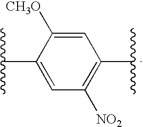
- II. Peptide Labeling Process
- In another aspect, the invention provides for a method for simultaneously identifying and determining the levels of expression of cysteine-containing proteins in normal and perturbed cells, comprising:
- a) preparing a first protein sample or a first peptide sample from the normal cells;
- b) reacting the first protein sample or the first peptide sample with a reagent of Formula II or III:
- Acyl-NH—X-[Epitope Tag Site]A-Y-[Protease Cleavage Site]-Z-Link (II)
- Acyl-NH—X-alk-O-Ph-CH2-Z-Link (III)
- where:
- A is an integer from 0 to 12;
- X is selected from the group consisting of an amide bond of formula —C(O)—NR—, a carbonyl of formula —C(O)—, and an amino acid sequence comprising between 10 to 30 amino acids, where R is hydrogen or lower alkyl;
- Y is an amide bond of formula —C(O)—NR—, where R is hydrogen or lower alkyl, or Y is an amino acid sequence comprising between 0 to 20 amino acids;
- Z is selected from the group consisting of an amide bond of formula —(CH 2)B—C(O)—NR—, an amide bond of formula —(CH2)B—NR—C(O)—, and an amino acid sequence comprising between 0 to 3 amino acids,
- where R is hydrogen or lower alkyl, and
- where B is an integer from 0 to 20;
- alk is straight or branched chain of alkylene comprising between 0 and 20 carbon atoms;
- Ph is a phenyl group optionally substituted with one or more electron withdrawing groups ortho or para to the —CH 2— group;
- Link is selected from the group consisting of —(CH 2)C—I, —(CH2)D—CH(—(CH2)ECH3)—(CH2)F—X—I, Lys-ε-iodoacetamide, Arg-δ-iodoacetamide, and Orn-δ-iodoacetamide
- where C, D, E, and F are each independently an integer from 0 to 20;
- Epitope Tag Site is a sequence of amino acids,
- where when A is two or more, the amino acid sequence of each Epitope Tag Site can be the same or different; and
- Protease Cleavage Site is a sequence of amino acids that is a cleavage site for a highly specific protease enzyme;
- c) preparing a second protein sample or a second peptide sample from the perturbed cells;
- d) reacting the second protein sample or the second peptide sample of step c) with a second reagent of Formula II or III:
- Acyl-NH—X-[Epitope Tag Site]A-Y-[Protease Cleavage Site]-Z-Link (II)
- Acyl-NH—X-alk-O-Ph-CH2-Z-Link (III)
- where:
- A is an integer from 0 to 12;
- X is selected from the group consisting of an amide bond of formula —C(O)—NR—, a carbonyl of formula —C(O)—, and an amino acid sequence comprising between 10 to 30 amino acids, where R is hydrogen or lower alkyl;
- Y is an amide bond of formula —C(O)—NR—, where R is hydrogen or lower alkyl, or Y is an amino acid sequence comprising between 0 to 20 amino acids;
- Z is selected from the group consisting of an amide bond of formula —(CH 2)B—C(O)—NR—, an amide bond of formula —(CH2)B—NR—C(O)—, and an amino acid sequence comprising between 0 to 3 amino acids,
- where R is hydrogen or lower alkyl, and
- where B is an integer from 0 to 20;
- alk is straight or branched chain of alkylene comprising between 0 and 20 carbon atoms;
- Ph is a phenyl group optionally substituted with one or more electron withdrawing groups ortho or para to the —CH 2— group;
- Link is selected from the group consisting of —(CH 2)C—I, —(CH2)D—CH(—(CH2)ECH3)—(CH2)F—X—I, Lys-ε-iodoacetamide, Arg-δ-iodoacetamide, and Orn-δ-iodoacetamide
- where C, D, E, and F are each independently an integer from 0 to 20;
- Epitope Tag Site is a sequence of amino acids,
- where when A is two or more, the amino acid sequence of each Epitope Tag Site can be the same or different; and
- Protease Cleavage Site is a sequence of amino acids that is a cleavage site for a highly specific protease enzyme,
- such that the molecular weight of the first reagent and the molecular weight of the second reagent are different by an integer multiple of 14 atomic mass units;
- e) combining the reacted the first and the second protein samples or the reacted the first and the second peptide sample from steps b) and d);
- f) subjecting the combined protein samples or the combined peptide samples from step e) to proteolysis at a site on the protein samples or at a site on the peptide samples, the site being other than the Protease Cleavage Site;
- g) subjecting the proteolyzed combined protein samples or the proteolyzed peptide samples from step f) to an affinity chromatography system comprising a second amino acid sequence attached to a solid, thereby forming bound proteins and non-bound proteins,
- where the Epitope Tag Site of the reagent and the second amino acid sequence bind with high specificity to each other;
- h) eluting the non-bound proteins from the affinity chromatography system;
- i) subjecting the affinity chromatography system from step h) to a protease specific for the Protease Cleavage Site, thereby forming a cleaved protein mixture;
- j) eluting the cleaved protein mixture from the affinity chromatography system of step i);
- k) isolating the eluted protein mixture obtained from step j);
- l) subjecting the eluted protein mixture from step k) to chromatographic separation, followed by mass analysis;
- m) comparing the results of step l) to:
- 1) determining the ratio of amounts of compounds in the two samples, where the molecular weights thereof are separated by an integer multiple of 14 atomic mass units; and
- 2) comparing the results obtained for each compound to protein databases containing chromatographic and molecular weight correlations.
- In another aspect, the invention provides for a method for simultaneously identifying and determining the levels of expression of cysteine-containing proteins in normal and perturbed cells, comprising:
- a) preparing a first protein sample or a first peptide sample from the normal cells;
- b) subjecting the first protein sample or the first peptide sample from step a) to proteolysis;
- c) reacting the proteolyzed first protein sample or the proteolyzed first peptide sample with a reagent of Formula II or III:
- Acyl-NH—X-[Epitope Tag Site]A-Y-[Protease Cleavage Site]-Z-Link (II)
- Acyl-NH-X-alk-O-Ph-CH2-Z-Link (III)
- where:
- A is an integer from 0 to 12;
- X is selected from the group consisting of an amide bond of formula —C(O)—NR—, a carbonyl of formula —C(O)—, and an amino acid sequence comprising between 10 to 30 amino acids, where R is hydrogen or lower alkyl;
- Y is an amide bond of formula —C(O)—NR—, where R is hydrogen or lower alkyl, or Y is an amino acid sequence comprising between 0 to 20 amino acids;
- Z is selected from the group consisting of an amide bond of formula —(CH 2)B—C(O)—NR—, an amide bond of formula —(CH2)B—NR—C(O)—, and an amino acid sequence comprising between 0 to 3 amino acids,
- where R is hydrogen or lower alkyl, and
- where B is an integer from 0 to 20;
- alk is straight or branched chain of alkylene comprising between 0 and 20 carbon atoms;
- Ph is a phenyl group optionally substituted with one or more electron withdrawing groups ortho or para to the —CH 2— group;
- Link is selected from the group consisting of —(CH 2)C—I, —(CH2)D—CH(—(CH2)ECH3)—(CH2)F—X—I, Lys-ε-iodoacetamide, Arg-δ-iodoacetamide, and Orn-δ-iodoacetamide
- where C, D, E, and F are each independently an integer from 0 to 20;
- Epitope Tag Site is a sequence of amino acids,
- where when A is two or more, the amino acid sequence of each Epitope Tag Site can be the same or different; and
- Protease Cleavage Site is a sequence of amino acids that is a cleavage site for a highly specific protease enzyme;
- d) preparing a second protein sample or a second peptide sample from the perturbed cells;
- e) subjecting the second protein sample or the second peptide sample from step d) to proteolysis;
- f) reacting the proteolyzed second protein sample or the proteolyzed second peptide sample of step e) with a second reagent of Formula II or III:
- Acyl-NH—X-[Epitope Tag Site]A-Y-[Protease Cleavage Site]-Z-Link (II)
- Acyl-NH—X-alk-O-Ph-CH2-Z-Link (III)
- where:
- A is an integer from 0 to 12;
- X is selected from the group consisting of an amide bond of formula —C(O)—NR—, a carbonyl of formula —C(O)—, and an amino acid sequence comprising between 10 to 30 amino acids, where R is hydrogen or lower alkyl;
- Y is an amide bond of formula —C(O)—NR—, where R is hydrogen or lower alkyl, or Y is an amino acid sequence comprising between 0 to 20 amino acids;
- Z is selected from the group consisting of an amide bond of formula —(CH 2)B—C(O)—NR—, an amide bond of formula —(CH2)B—NR—C(O)—, and an amino acid sequence comprising between 0 to 3 amino acids,
- where R is hydrogen or lower alkyl, and
- where B is an integer from 0 to 20;
- alk is straight or branched chain of alkylene comprising between 0 and 20 carbon atoms;
- Ph is a phenyl group optionally substituted with one or more electron withdrawing groups ortho or para to the —CH 2— group;
- Link is selected from the group consisting of —(CH 2)C—I, —(CH2)D—CH(—(CH2)ECH3)—(CH2)F—X—I, Lys-ε-iodoacetamide, Arg-δ-iodoacetamide, and Orn-δ-iodoacetamide
- where C, D, E, and F are each independently an integer from 0 to 20;
- Epitope Tag Site is a sequence of amino acids,
- where when A is two or more, the amino acid sequence of each Epitope Tag Site can be the same or different; and
- Protease Cleavage Site is a sequence of amino acids that is a cleavage site for a highly specific protease enzyme,
- such that the molecular weight of the first reagent and the molecular weight of the second reagent are different by an integer multiple of 14 atomic mass units;
- g) combining the reacted the first and the second protein samples or the reacted the first and the second peptide sample from steps c) and f);
- h) subjecting the combined protein samples or the combined peptide samples from step e) to proteolysis at a site on the protein samples or at a site on the peptide samples, the site being other than the Protease Cleavage Site;
- i) subjecting the proteolyzed combined protein samples or the proteolyzed peptide samples from step f) to an affinity chromatography system comprising a second amino acid sequence attached to a solid, thereby forming bound proteins and non-bound proteins,
- where the Epitope Tag Site of the reagent and the second amino acid sequence bind with high specificity to each other;
- j) eluting the non-bound proteins from the affinity chromatography system;
- k) subjecting the affinity chromatography system from step j) to a protease specific for the Protease Cleavage Site, thereby forming a cleaved protein mixture;
- l) eluting the cleaved protein mixture from the affinity chromatography system of step k);
- m) isolating the eluted protein mixture obtained from step l);
- n) subjecting the eluted protein mixture from step m) to chromatographic separation, followed by mass analysis;
- o) comparing the results of step n) to:
- 1) determining the ratio of amounts of compounds in the two samples, where the molecular weights thereof are separated by an integer multiple of 14 atomic mass units; and
- 2) comparing the results obtained for each compound to protein databases containing chromatographic and molecular weight correlations.
- In certain embodiments, if in step b) in the above method Link is Lys-ε-iodoacetamide, then in step d) Link is Orn-δ-iodoacetamide. Alternatively, if in step b) Link is Orn-δ-iodoacetamide, then in step d) Link is Lys-ε-iodoacetamide. In another embodiment, the Z substituent in the first reagent, i.e., in step b) has a molecular weight that is an integer multiple of 14 atomic mass units different than the Z substituent in the second reagent, i.e., in step d). For example, and without limitation, the Z in the first reagent contains valine whereas the Z in the second reagent contains leucine instead of valine, all the other amino acids in Z, if any, remaining the same between the two reagents.
- In an embodiment, the reagent of step b) is selected from the group consisting of
- Acyl-NH-AYPYDVPDYASENLYFQGK-iodoacetamide (SEQ ID NO: 17),
- Acyl-NH-AYPYDVPDYASENLYFQGGK-iodoacetamide (SEQ ID NO: 18),
- Acyl-NH-AYPYDVPDYASENLYFQGAK-iodoacetamide (SEQ ID NO: 19),
- Acyl-NH-AYPYDVPDYASENLYFQG(GABA)K-iodoacetamide (SEQ ID NO: 20),
- Acyl-NH-AYPYDVPDYASENLYFQGVK-iodoacetamide (SEQ ID NO: 21),
- Acyl-NH-AYPYDVPDYASENLYFQGR-iodoacetamide (SEQ ID NO: 22),
- Acyl-NH-AYPYDVPDYASENLYFQGGR-iodoacetamide (SEQ ID NO: 23),
- Acyl-NH-AYPYDVPDYASENLYFQGAR-iodoacetamide (SEQ ID NO: 24),
- Acyl-NH-AYPYDVPDYASENLYFQG(GABA)R-iodoacetamide (SEQ ID NO: 25),
- Acyl-NH-AYPYDVPDYASENLYFQGVR-iodoacetamide (SEQ ID NO: 26),
- Acyl-NH-AYPYDVPDYASENLYFQGOrn-iodoacetamide (SEQ ID NO: 27),
- Acyl-NH-AYPYDVPDYASENLYFQGGOrn-iodoacetamide (SEQ ID NO: 28),
- Acyl-NH-AYPYDVPDYASENLYFQGAOrn-iodoacetamide (SEQ ID NO: 29),
- Acyl-NH-AYPYDVPDYASENLYFQG(GABA)Orn-iodoacetamide (SEQ ID NO: 30), and
- Acyl-NH-AYPYDVPDYASENLYFQGVOrn-iodoacetamide (SEQ ID NO: 31).
- Therefore, by way of example only, if the reagent of step b) is
- Acyl-NH-AYPYDVPDYASENLYPQGK-iodoacetamide (SEQ ID NO: 32) the reagent of step d) would be
- Acyl-NH-AYPYDVPDYASENLYPQGOrn-iodoacetamide (SEQ ID NO: 33);
- and if the reagent of step b) is
- Acyl-NH-AYPYDVPDYASENLYPQGOrn-iodoacetamide (SEQ ID NO: 34),
- the reagent of step d) would be
- Acyl-NH-AYPYDVPDYASENLYPQGK-iodoacetamide (SEQ ID NO: 35).
- Preferably, the reagent of step b) or of step d) reacts with the reactive side chain of one or more of the amino acid residues of the proteins in the first or second protein sample. By “reactive side chain” it is meant the amino acid side chain that is functionalized, or an amino acid side chain that is other than straight chain or branched alkyl. Therefore, the reagent reacts with the first or second protein at an amino acid residue selected from the group consisting of tyrosine, tryptophan, cysteine, methionine, proline, serine, threonine, lysine, histidine, arginine, aspartic acid, glutamic acid, asparagine, and glutamine. In certain embodiments, the reagent reacts at an amino acid residue selected from the group consisting of tyrosine, cysteine, proline, and histidine. In another embodiment, the site of reaction is a cysteine.
- In some embodiments of the present invention, the chromatographic separation of step l) is a multi-dimensional liquid chromatographic separation, which may be a two-dimensional liquid chromatographic separation or a three-dimensional liquid chromatographic separation. The dimensions of the multi-dimensional liquid chromatographic separation are selected from the group consisting of size differentiation, charge differentiation, hydrophobicity, hydrophilicity, and polarity. In some embodiments, at least one dimension of the multi-dimensional liquid chromatographic separation is separation using size differentiation. Embodiments of the invention include those in which one dimension of the multi-dimensional liquid chromatographic separation is separation using charge differentiation. In other embodiments, one dimension of the multi-dimensional liquid chromatographic separation is separation using hydrophobicity or hydrophilicity.
- In another embodiment the mass analysis of step m) is a multi-dimensional mass analysis, which may be a two-dimensional mass analysis (i.e., tandem mass spectrometry).
- It is well-known in the art to separate fragments of a solution using chromatography and, in tandem thereto, analyze the mass spectra of each fragment. The technique is formally known in the art as LC-MS or LC-MS/MS analysis. Multi-dimensional chromatography is also well-known in the art, where multiple columns are used in tandem, or the same column is packed with segments of different material that can separate the sample using different criteria. See, for example, Link et al., (1999) or Opitek et al. (1997), above. Multi-dimensional mass analysis is a technique known to those skilled in the art as well. In this technique, following an initial ionization, an ion of interest is selected. The selected ion is fragmented and each fragment (known as “daughter ion” or “progeny ion”) is now capable of being either analyzed or be subjected to further fragmentation. The technique is fully described in Siuzdak, Mass Spectrometry for Biotechnology, Academic Press, San Diego, Calif., 1996, which is incorporated by reference herein in its entirety.
- In certain embodiments, the preparation of proteins from step a) is subjected to orthogonal chromatography before proceeding with the labeling in step b). Orthogonal chromatography is a technique well-known in the art.
- Quantitative relative amounts of proteins in one or more different samples containing protein mixtures (e.g., biological fluids, cell or tissue lysates, etc.) can be determined using chemically similar, affinity tagged and differentially labeled reagents to affinity tag and differentially label proteins in the different samples. The label may be differentiated by having additional methylene groups, which would result in the mass of the two labels be different by an integer multiple of 14.
- In this method, each sample to be compared is treated with a different labeled reagent to tag certain proteins therein with the affinity label. The treated samples are then combined, preferably in equal amounts, and the proteins in the combined sample are enzymatically digested, if necessary, to generate peptides. Some of the peptides are affinity tagged and in addition tagged peptides originating from different samples are differentially labeled. As described above, affinity labeled peptides are isolated, released from the capture reagent and analyzed by (LC/MS). Peptides characteristic of their protein origin are sequenced using (MS) n techniques allowing identification of proteins in the samples. The relative amounts of a given protein in each sample is determined by comparing relative abundance of the ions generated from any differentially labeled peptides originating from that protein. The method can be used to assess relative amounts of known proteins in different samples. The method is described in U.S. Pat. No. 5,538,897, issued Jul. 23, 1996, to Yates et al., which is incorporated herein by reference in its entirety, including any drawings.
- Further, since the method does not require any prior knowledge of the type of proteins that may be present in the samples, it can be used to identify proteins which are present at different levels in the samples examined. More specifically, the method can be applied to screen for and identify proteins which exhibit differential expression in cells, tissue or biological fluids. It is also possible to determine the absolute amount of specific proteins in a complex mixture. In this case, a known amount of internal standard, one for each specific protein in the mixture to be quantified, is added to the sample to be analyzed. The internal standard is an affinity tagged peptide that is identical in chemical structure to the affinity tagged peptide to be quantified except that the internal standard is differentially labeled, either in the peptide or in the affinity tagged portion, to distinguish it from the affinity tagged peptide to be quantified. The internal standard can be provided in the sample to be analyzed in other ways. For example, a specific protein or set of proteins can be chemically tagged with a labeled affinity tagging reagent. A known amount of this material can be added to the sample to be analyzed. Alternatively, a specific protein or set of proteins may be labeled with additional methylene groups and then derivatized with an affinity tagging reagent.
- Also, it is possible to quantify the levels of specific proteins in multiple samples in a single analysis (multiplexing). For example, a set of five different samples can be reacted with one of SEQ ID NO:27-SEQ ID NO:31, then follow with subsequent steps as described herein. In this case, affinity tagging reagents used to derivatize proteins present in different affinity tagged peptides from different samples can be selectively quantified by mass spectrometry. This may be achieved by using reagents whose molecular mass varies from one sample to another by an integer multiple of 14. So, for example, the Link group in one reagent may feature ornithine whereas the Link group in another reagent may feature arginine or lysine. Similarly, the Z groups in the different reagent may vary such that the molecular mass of the reagent varies by an integer multiple of 14. It is also understood that other amino acids may also be featured. For example, the lighter reagent may have valine whereas the heavier reagent may feature leucine or isoluecine in its stead. The same would be true for having asparagine in the lighter reagent and glutamine in the heavier reagent, or aspartic acid in the lighter reagent and glutamic acid in the heavier reagent.
- In this aspect of the invention, the method provides for quantitative measurement of specific proteins in biological fluids, cells or tissues and can be applied to determine global protein expression profiles in different cells and tissues. The same general strategy can be broadened to achieve the proteome-wide, qualitative and quantitative analysis of the state of modification of proteins, by employing affinity reagents with differing specificity for reaction with proteins. The method and reagents can be used to identify low abundance proteins in complex mixtures and can be used to selectively analyze specific groups or classes of proteins such as membrane or cell surface proteins, or proteins contained within organelles, sub-cellular fractions, or biochemical fractions such as immunoprecipitates. Further, these methods can be applied to analyze differences in expressed proteins in different cell states. For example, the methods and reagents herein can be employed in diagnostic assays for the detection of the presence or the absence of one or more proteins indicative of a disease state, such as cancer.
- The methods described herein can also be applied to determine the relative quantities of one or more proteins in two or more protein samples. The proteins in each sample are reacted with affinity tagging reagents which are substantially chemically identical but differentially labeled. The samples are combined and processed as one.
- The relative quantity of each tagged peptide which reflects the relative quantity of the protein from which the peptide originates is determined by the integration of the respective mass peaks by mass spectrometry.
- The methods described herein can be applied to the analysis or comparison of multiple different samples. Samples that can be analyzed by methods of this invention include cell homogenates; cell fractions; biological fluids including urine, blood, and cerebrospinal fluid; tissue homogenates; tears; feces; saliva; lavage fluids such as lung or peritoneal ravages; mixtures of biological molecules including proteins, lipids, carbohydrates and nucleic acids generated by partial or complete fractionation of cell or tissue homogenates.
- The methods described herein employ MS and (MS) n methods. While a variety of MS and (MS)n are available and may be used in these methods, Matrix Assisted Laser Desorption Ionization MS (MALDI/MS) and Electrospray ionization MS (ESI/MS) methods are preferred.
- III. Analytical Methodology
- Another aspect of the present invention relates to a method for proteomic analysis, comprising:
- a) preparing a protein sample or a peptide sample from cells;
- b) reacting the protein sample or the peptide sample with a reagent of the formula:
- Acyl-NH—X-[Epitope Tag Site]A-Y-[Protease Cleavage Site]-Z-Link
- where:
- A is an integer from 1 to 12;
- X is an amide bond of formula —C(O)—NR—, where R is hydrogen or lower alkyl, or X is an amino acid sequence comprising between 10 to 30 amino acids;
- Y is an amide bond of formula —C(O)—NR—, where R is hydrogen or lower alkyl, or Y is an amino acid sequence comprising between 0 to 20 amino acids;
- Z is an amide bond of formula —C(O)—NR—, where R is hydrogen or lower alkyl, or Z is an amino acid sequence comprising between 0 to 3 amino acids;
- Link is selected from the group consisting of Lys-ε-iodoacetamide, Arg-δ-iodoacetamide, and Orn-δ-iodoacetamide;
- Epitope Tag Site is a sequence of amino acids, and
- Protease Cleavage Site is a sequence of amino acids that is a cleavage site for a highly specific protease enzyme;
- c) subjecting the reacted proteins or peptides from step b) to proteolysis at a site on the protein samples or at a site on the peptide samples, the site being other than the Protease Cleavage Site;
- d) subjecting the proteolyzed reacted proteins or the proteolyzed reacted peptides from step c) to an affinity chromatography system comprising a second amino acid sequence attached to a solid support, thereby forming bound proteins and non-bound proteins,
- where the Epitope Tag Site of the reagent and the second amino acid sequence bind with high specificity to each other;
- e) eluting the non-bound proteins from the affinity chromatography system;
- f) subjecting the affinity chromatography system from step e) to a protease specific for the Protease Cleavage Site, thereby forming a cleaved protein mixture;
- g) eluting the cleaved protein mixture from the affinity chromatography system of step f);
- h) isolating the cleaved protein mixture obtained from step g);
- i) subjecting the cleaved protein mixture from step h) to chromatographic separation, followed by mass analysis;
- j) comparing the results of step i) to:
- 1) determine the ratio of amounts of compounds in the sample separated by a molecular weight of 14 atomic mass units; and
- 2) identify the various modified proteins by comparing the results obtained for each modified protein to protein databases containing chromatographic and molecular weight correlations.
- “Proteomic analysis” refers to identifying the proteome of a cell. The “proteome” of a cell is the collection of all the proteins expressed by the cell at the time the proteomic analysis is undertaken. It is understood that, unlike the genome of a cell, which is invariable, the proteome of a cell varies depending on many factors, including the age of the cell, the environmental conditions surrounding the cell, and the position of the cell in its life cycle.
- In the above methods, the reagent reacts with the reactive side chain of one or more of the amino acid residues of the first or second protein. Therefore, the reagent reacts with the protein at an amino acid residue selected from the group consisting of tyrosine, tryptophan, cysteine, methionine, proline, serine, threonine, lysine, histidine, arginine, aspartic acid, glutamic acid, asparagine, and glutamine. In certain embodiments, the reagent reacts at an amino acid residue selected from the group consisting of tyrosine, cysteine, proline, and histidine. In another preferred embodiment, the site of reaction is a cysteine.
- In some embodiments of the present invention, the chromatographic separation of step i) is a multi-dimensional liquid chromatographic separation, which may be a two-dimensional liquid chromatographic separation or a three-dimensional liquid chromatographic separation. The dimensions of the multi-dimensional liquid chromatographic separation are selected from the group consisting of size differentiation, charge differentiation, hydrophobicity, hydrophilicity, and polarity. In some embodiments, at least one dimension of the multi-dimensional liquid chromatographic separation is separation using size differentiation. Embodiments of the invention include those in which one dimension of the multi-dimensional liquid chromatographic separation is separation using charge differentiation. In other embodiments, one dimension of the multi-dimensional liquid chromatographic separation is separation using hydrophobicity or hydrophilicity.
- In another embodiment the mass analysis of step i) is a multi-dimensional mass analysis, which more preferably, may be a two-dimensional mass analysis. In certain embodiments, the preparation of proteins from step a) is subjected to orthogonal chromatography before proceeding with the labeling in step b).
- In one aspect, the invention provides a mass spectrometric method for identification and quantification of one or more proteins in a complex mixture which employs affinity labeled reagents in which the Link group is a group that selectively reacts with certain groups that are typically found in peptides (e.g., sulfhydryl, amino, carboxy, homoserine, or lactone groups). One or more affinity labeled reagents with different Link groups are introduced into a mixture containing proteins and the reagents react with certain proteins to tag them with the affinity label. It may be necessary to pretreat the protein mixture to reduce disulfide bonds or otherwise facilitate affinity labeling. After reaction with the affinity labeled reagents, proteins in the complex mixture are cleaved, e.g., enzymatically, into a number of peptides. This digestion step may not be necessary, if the proteins are relatively small. Peptides that remain tagged with the affinity label are isolated by an affinity isolation method, e.g., affinity chromatography, via their selective binding to the capture reagent. Isolated peptides are released from the capture reagent by displacement of the Epitope Tag Site or cleavage of the linker, and released materials are analyzed by liquid chromatography/mass spectrometry (LC/MS). The sequence of one or more tagged peptides is then determined by (MS) n techniques. At least one peptide sequence derived from a protein will be characteristic of that protein and be indicative of its presence in the mixture. Thus, the sequences of the peptides typically provide sufficient information to identify one or more proteins present in a mixture.
- IV. Proteome Analysis Methodology
- The method comprises the following steps:
- Reduction. Disulfide bonds of proteins in the sample and reference mixtures are chemically reduced to free SH groups. The preferred reducing agent is tri-n-butylphosphine which is used under standard conditions. Alternative reducing agents include mercaptoethanol, 2-methylthioethanol, 2-methylthio-1-hexanol, and dithiothreitol. If required, this reaction can be performed in the presence of solubilizing agents including high concentrations of urea and detergents to maintain protein solubility. The reference and sample protein mixtures to be compared are processed separately, applying identical reaction conditions.
- Derivatization of SH groups with an affinity tag. Free SH groups of the sample protein are derivatized with a reagent of the invention. The reagent reacts with the free SH group through the Link group.
- Each sample is derivatized with a different reagent having a different mass. Derivatization of SH groups is preferably performed under slightly basic conditions (pH 8.5) for 90 min at about room temperature. For the quantitative, comparative analysis of two samples, one sample each (termed “reference sample” and “sample”) are derivatized with two different reagents, whose molecular mass differs by an integer multiple of 14. For the comparative analysis of several samples one sample is designated a reference to which the other samples are related.
- Combination of labeled samples. After completion of the affinity tagging reaction defined aliquots of the samples labeled with different reagents are combined and all the subsequent steps are performed on the pooled samples. Combination of the differentially labeled samples at this early stage of the procedure eliminates variability due to subsequent reactions and manipulations. Preferably equal amounts of each sample are combined.
- Removal of excess affinity tagged reagent. Excess reagent is adsorbed, for example, by adding an excess of SH-containing beads to the reaction mixture after protein SH groups are completely derivatized. Beads are added to the solution to achieve about a 5-fold molar excess of SH groups over the reagent added and incubated for 30 min at about room temperature. After the reaction the beads are removed by centrifugation.
- Protein digestion. The proteins in the sample mixture are digested, typically with trypsin. Alternative proteases are also compatible with the procedure as in fact are chemical fragmentation procedures. In cases in which the preceding steps were performed in the presence of high concentrations of denaturing solubilizing agents, the sample mixture is diluted until the denaturant concentration is compatible with the activity of the proteases used. This step may be omitted in the analysis of small proteins.
- Affinity Isolation of the Affinity Tagged Peptides by Interaction with a Capture Reagent.
- The tagged peptides are isolated on anti-HA antibodies-agarose. After digestion the pH of the peptide samples is lowered to 6.5 and the tagged peptides are immobilized on beads coated with anti-HA. The beads are extensively washed. The last washing solvent includes 10% methanol to remove residual SDS.
- Release of the captured peptides with specific protease. A solution of TEV in TRIS at pH 7.5 is added to the column and digestion is allowed to proceed. The bound peptides are cleaved from the column by incubation at 30° C. for 6 hours.
- Analysis of the isolated, derivatized peptides by μLC-(MS) n or CE-(MS)n with data dependent fragmentation. Methods and instrument control protocols well-known in the art and described, for example, in Ducret et al. (1998); Figeys and Aebersold (1998); Figeys et al. (1996); or Haynes et al. (Electrophoresis 19:939-945 (1998)) are used. In this last step, both the quantity and sequence identity of the proteins from which the tagged peptides originated can be determined by automated multistage MS. This is achieved by the operation of the mass spectrometer in a dual mode in which it alternates in successive scans between measuring the relative quantities of peptides eluting from the capillary column and recording the sequence information of selected peptides. Peptides are quantified by measuring in the MS mode the relative signal intensities for pairs of peptide ions of identical sequence that are tagged with the lighter or heavier forms of the reagent, respectively, and which therefore differ in mass by the mass differential encoded within the affinity tagged reagent. Peptide sequence information is automatically generated by selecting peptide ions of a particular mass-to-charge (m/z) ratio for collision-induced dissociation (CID) in the mass spectrometer operating in the (MS)n mode. (Link et al. Electrophoresis 18:1314-1334 (1997); Gygi et al. Nature Biotechnol 17:994-999 (1999); Gygi et al., Cell Biol 19:1720-1730 (1999)). The resulting CID spectra are then automatically correlated with sequence databases to identify the protein from which the sequenced peptide originated. Combination of the results generated by MS and (MS)n analyses of affinity tagged and differentially labeled peptide samples therefore determines the relative quantities as well as the sequence identities of the components of protein mixtures in a single, automated operation.
- This method can also be practiced using other affinity tags and other protein reactive groups, including amino reactive groups, carboxyl reactive groups, or groups that react with homoserine lactones.
- The approach employed herein for quantitative proteome analysis is based on two principles. First, a short sequence of contiguous amino acids from a protein contains sufficient information to uniquely identify that protein. Protein identification by (MS) n is accomplished by correlating the sequence information contained in the CID mass spectrum with sequence databases, using sophisticated computer searching algorithms (Yates, III et al. U.S. Pat. No. 5,538,897). Second, pairs of peptides tagged with lighter and heavier Link groups or Z groups, respectively, are chemically similar and therefore serve as mutual internal standards for accurate quantification. The MS measurement readily differentiates between peptides originating from different samples, representing for example different cell states, because of the difference between the distinct reagents attached to the peptides. The ratios between the intensities of the differing weight components of these pairs or sets of peaks provide an accurate measure of the relative abundance of the peptides (and hence the proteins) in the original cell pools.
- Specifically, the peptide labeling moiety consists of a lysine residue modified with an iodoacetamido functional group on the ε-amino side chain. The synthetic chemistry necessary for this modification reaction is readily available in the literature. The synthetic peptides contain two additional motifs: a peptide epitope tag for high affinity purification; and a highly specific protease site for releasing the affinity purified labeled peptides from the affinity matrix. In addition, these synthetic peptides can readily be prepared as isoforms of two different masses by the simple expedient of using an ornithine in place of lysine to introduce a 14 mass unit difference in the carboxyl terminal acid.
- Examples of the reagents (SEQ ID NO: 36 and SEQ ID NO: 37) are thus:

- The peptide sequence in the square brackets is an Epitope Tag Site and the sequence in parentheses is a Protease Cleavage Site. In the case shown here, the peptide sequence YPYDVPDYA (SEQ ID NO: 38) is an influenza hemagglutinin (HA) epitope tag. This part of the reagent could be replaced by any other epitope tag, or multiple copies of a single tag for higher efficiency purification, or parallel copies of different tags for higher specificity purification. Examples of other Epitope Tag Sites include Flag, His-6, and c-myc.
- The protease cleavage site shown here is that of TEV protease, which is commercially available. This enzyme has been shown to cleave at only one protein site in the entire yeast genome, thus indicating that the enzyme is highly specific for an extremely rare sequence. This part of the reagent could be replaced by any other highly specific protease cleavage site, either commercially available, such as Factor Xa, or Pharmacia Prescission Enzyme, or one that is newly discovered. The amino acid indicated in bold is used to provide a site of attachment for the iodoacetamide group, hence we have used lysine which contains an ε-amino side chain that is suitable for the purpose. This amino acid is also used to introduce a differential mass between the two reagents, and this can be readily accomplished by using ornithine in place of lysine. Ornithine is commercially available and differs from lysine only by the presence of one additional methyl group, which makes it 14 amu (atomic mass unit) heavier than lysine. Arginine is also commercially available and its molecular weight is 28 amu (i.e., 2×14) heavier than lysine. This part of the reagent could be replaced with any other amino acid or similar molecule that provided an attachment site for the iodoacetamide group. Finally, the integral difference of 14 amu could be further enhanced by the choice of two amino acids differing by 14 amu (e.g., valine and leucine) in the Z portion of the peptide labeling moiety.
- In addition to the above methods, the methods of the invention may be used to determine the proteomic differences in an organism or cell based on the change in the cell's environmental condition. Thus, for example, one may compare the proteome of the cells of two plants of the same species, one having encountered high salt concentrations and the other low salt concentrations, thereby determining the effect of salt concentration on the plant's proteome.
- It is also within the scope of the present invention that the two modes of analysis discussed herein, i.e., the qualitative and quantitative proteome analyses, are exercised in conjunction with each other. Thus, by way of example only, one may compare the proteome of the cells of two plants of the same species, one having encountered higher temperatures than the other, thereby not only determining the effect of heat on the proteome in terms of which proteins are expressed, but also determining the effect of heat on the level of expression of each protein of interest.
- In practicing the present invention to achieve the above end, one may use a number of different compounds of the present invention, having different masses (yet all within an integer multiple of 14 from each other), and mark different proteins of the cells with the different reagents. By applying the multidimensional LC/MS techniques described herein, one is able to determine which proteins, and to what extent, are expressed in the cells.
- V. Fusion Protein Preparation
- Another aspect of the invention relates to a process for preparing a fusion protein of Formula IV or V:
- Protein-Acyl-N—X-[Epitope Tag Site]A-Y-[Protease Cleavage Site]-Z-[Lys-δ-N-iodoacetamide] (IV)
- Protein-Acyl-NH—X-alk-O-Ph-CH2-Z-Link (V)
- where A, X, Y, Z, alk, Ph, Link, Epitope Tag Site, and Protease Cleavage Site are as defined herein
- comprising,
- a) preparing a fusion protein sample of Formula II or III from cells
- Protein-Acyl-NH—X-[Epitope Tag Site]A-Y-[Protease Cleavage Site]-Z-Orn-δ-NHCOCH2 (II)
- Acyl-NH—X-alk-O-Ph-CH2-Z-NHCOCH2 (III)
- b) reacting the protein sample with a Link or with iodoacetamide.
- In another aspect, the invention relates to a process for preparing a fusion protein of Formula VI:
- Protein-Acyl-N—X-[Epitope Tag Site]A-Y-[Protease Cleavage Site]-Z-[Lys-δ-N-iodoacetamide] (VI)
- where A, X, Y, Z, alk, Ph, Link, Epitope Tag Site, and Protease Cleavage Site are as defined herein
- comprising,
- a) preparing a fusion protein sample of Formula VII from cells
- Protein-Acyl-NH—X-[Epitope Tag Site]A-Y-[Protease Cleavage Site]-Z-Lys-δ-NHCOCH2 (VII)
- b) reacting the protein sample with iodoacetamide.
- Markers that are useful in plant breeding, genetics, and diagnostics are disclosed in U.S. Provisional Patent Application No. 60/264,226, entitled “Cereal Simple Sequence Repeat Markers,” filed on Jan. 26, 2001 (Attorney Docket No. NADII.026PR), which is hereby incorporated by reference in its entirety.
- Briefly summarizing one embodiment of the present invention, upon receiving the results of the quantitation of the resolved
peptides 146, thedata analysis system 200 compares the relative peptide expression levels for the analogous peptides withdifferent markers quantitation module 230, thesystem 200 then identifies each recognizable peak orintensity curve 407 and associates any differentially tagged partner peptides (analogs). These tagged partner peptides can be recognized as peaks or intensity curves 407 that are present at a predicted mass displacement distance, based on the mass differential created by themarkers intensity curve 407 is found, the peptide-correlatedoutput files 260 may be used to confirm or deny the sequences of the peptides to establish if peptides being compared are partners. This process is repeated until all possible pairs of peptide partners have been identified in the data set. Thedata processing module 225 then integrates the area contained by each peak orintensity curve 407 and calculates the ratio of the quantitated peaks to identify differences in peptide expression. - In a subsequent analysis stage, the data output comprising the identified differences in peptide expression can be sorted and presented to the investigator in the form of one or more reports. These reports may be categorized by identification of the peptide constituents of the mixed-peptide population, ratios of peptides containing
different markers data analysis system 200, or other user-defined criteria. Additionally, the identification reports may list any unpaired peaks in the mass spectrum ordered by confidence level, peptide name, or other user-defined criteria. - The
data analysis system 200 and related methods feature a significantly improved means of identifying proteomic differences between two or more biological samples. The use ofmarkers - Although the foregoing description of the invention has shown, described and pointed out novel features of the invention, it will be understood that various omissions, substitutions, and changes in the form of the detail of the apparatus as illustrated, as well as the uses thereof, may be made by those skilled in the art without departing from the spirit of the present invention. Consequently the scope of the invention should not be limited to the foregoing discussion but should be defined by the appended claims.
Claims (48)
1. A method for determining peptide expression levels between a first biological sample and a second biological sample, comprising:
providing a peptide mixture comprising first labeled peptides from a first biological sample and second labeled peptides from a second biological sample, wherein peptides having the same amino acid sequence in the first biological sample and in the second biological sample have a predetermined mass difference;
calculating the weight of peptides in the peptide mixture;
identifying a peptide pair in the peptide mixture by determining two peptides whose weight differs by the predetermined mass difference; and
quantifying the abundance of each peptide in the peptide pair.
2. The method of claim 1 , wherein calculating the weight of the peptides comprises performing a primary mass analysis to produce a primary spectrum of peaks characteristic of the peptide mixture, wherein each peak corresponds to one labeled peptide in the peptide mixture.
3. The method of claim 2 , comprising performing a secondary mass analysis on each peak in order to produce a secondary spectra characteristic of the individual peptide correlated with the peak.
4. The method of claim 3 wherein the secondary mass analysis comprises a tandem mass analytical technique selected from the group consisting of: electrospray mass analysis, fast atom bombardment mass analysis and liquid secondary ion mass analysis.
5. The method of claim 3 , comprising identifying the peptide correlated with the peak by comparing the secondary spectra with a database of known peptide spectra.
6. The method of claim 2 , wherein quantitating the abundance of each peptide comprises assessing the size of peaks in the primary spectrum to generate values representative of a relative amount of each peptide present in the peptide mixture.
7. The method of claim 6 , wherein quantitating the abundance of each peptide is performed using parallel computational methods.
8. The method of claim 1 , wherein the first labeled peptides are labeled with a first chemical group, and the second labeled peptides are labeled with a second chemical group, and wherein the first chemical group and the second chemical group have a predetermined mass difference.
9. The method of claim 8 , wherein the first chemical group comprises a lysine residue modified with an iodoacetamide functional group on the ε-amino group of the lysine residue side chain
10. The method of claim 9 , wherein the second chemical group comprises an ornithine residue modified with an iodoacetamide functional group on the ε-amino group of the ornithine residue side chain.
11. The method of claim 9 , wherein the first chemical group is 15N and the second chemical group is 14N.
12. The method of claim 1 wherein calculating the weight of peptides comprises mass analytical techniques selected from the group consisting of: electron ionization mass analysis, fast atom/ion bombardment mass analysis, matrix-assisted laser desorption/ionization mass analysis and electrospray ionization mass analysis.
13. The method of claim 1 , wherein the first biological sample and the second biological sample are taken from the same starting cell population, but the first biological sample is untreated, whereas the second biological sample is treated with a test compound.
14. The method of claim 13 , wherein the starting cell population is selected from the group consisting of: plant cells, animal cells, bacterial cells and fungal cells.
15. A method for quantitative proteomic analysis of two or more peptide populations, the method comprising:
differentially labeling the two or more peptide populations;
combining the two or more peptide populations to form a mixed peptide population;
proteolyzing the mixed peptide population to generate a collection of mixed peptide fragments of suitable size to be resolved by mass analysis;
separating the collection of mixed peptide fragments by mass analysis into discrete peptide fragments while producing a primary mass spectrum with peptide peak intensities indicative of the presence of the discrete peptide fragments;
analyzing the discrete peptide fragments using tandem mass analysis to generate a plurality of tandem mass spectrum characteristic of each discrete peptide fragment;
comparing the tandem mass spectrum against a database of sequence-correlated mass spectra thereby determining a putative sequence identity for the tandem mass spectrum generated by the discrete peptide fragments;
identifying the discrete peptide fragments derived from the differentially labeled peptide populations which are indicative of analogous peptides; and
assessing the peptide peak intensities of the discrete peptide fragments derived from the analogous peptides to identify proteomic differences.
16. The method for quantitative proteomic analysis of claim 15 wherein a sequence prediction process is used to compare the tandem mass spectrum against the database of sequence-collated mass spectra.
17. The method for quantitative proteomic analysis of claim 16 wherein the sequence prediction process produces a plurality of sequence-correlated data files and a peak detection process is used process and associate the sequence-correlated data files with the peptide peak intensities of the primary mass spectrum to identify the discrete peptide fragments.
18. The method for quantitative proteomic analysis of claim 17 wherein the peak detection process operates by:
(a) extracting information from the sequence-correlated data file corresponding to intensities for known charge states of peptide associated with the sequence-correlated mass spectrum;
(b) identifying the highest intensity charge state of the peptide associated with the sequence-correlated mass spectrum;
(c) identifying the peptide peak intensity in the primary mass spectrum which is associated with the highest intensity charge state of the peptide associated with the sequence-correlated mass spectrum;
(d) performing a data filtering operation on the peptide peak intensity to remove background noise and intervening peak intensities; and
(e) performing a determination of a quantitation value to be associated with the peptide peak intensity.
19. The method for quantitative proteomic analysis of claim 16 wherein the peak detection process further identifies proteomic differences between analogous peptides by comparing the quantitation values for the associated discrete peptide fragments.
20. The method for quantitative proteomic analysis of claim 17 wherein the identified proteomic differences correspond to differences in peptide concentration associated with up-regulation, down-regulation, unchanged regulation, increased peptide concentration, decreased peptide concentration, equivalent peptide concentration, peptide repression, and peptide induction.
21. A system for determining peptide expression levels between a first biological sample and a second biological sample, comprising:
a peptide mixture comprising first labeled peptides from a first biological sample and second labeled peptides from a second biological sample, wherein peptides having the same amino acid sequence in the first biological sample and in the second biological sample have a predetermined mass difference;
a first module configured to calculate the weight of peptides in the peptide mixture;
a second module configured to identify a peptide pair in the peptide mixture by determining two peptides whose weight differs by the predetermined mass difference; and
a third module configured to quantify the abundance of each peptide in the peptide pair.
22. The system of claim 21 , wherein the first module is configured to perform a primary mass analysis to produce a primary spectrum of peaks characteristic of the peptide mixture, wherein each peak corresponds to one labeled peptide in the peptide mixture.
23. The system of claim 22 , wherein the first module is configured to perform a secondary mass analysis on each peak in order to produce a secondary spectra characteristic of the individual peptide correlated with the peak.
24. The system of claim 23 wherein the secondary mass analysis comprises a tandem mass analytical technique selected from the group consisting of: electrospray mass analysis, fast atom bombardment mass analysis and liquid secondary ion mass analysis.
25. The system of claim 23 , wherein the second module is configured to identify the peptide correlated with the peak by comparing the secondary spectra with a database of known peptide spectra.
26. The system of claim 22 , wherein the third module is configured to assess the size of peaks in the primary spectrum and generate values representative of a relative amount of each peptide present in the peptide mixture.
27. The system of claim 26 , wherein the third module is configured to use parallel computational means.
28. The system of claim 21 , wherein the first labeled peptides have been labeled with a first chemical group, and the second labeled peptides have been labeled with a second chemical group, and wherein the first chemical group and the second chemical group have a predetermined mass difference.
29. The system of claim 28 , wherein the first chemical group comprises a lysine residue modified with an iodoacetamide functional group on the ε-amino group of the lysine residue side chain
30. The system of claim 29 , wherein the second chemical group comprises a ornithine residue modified with an iodoacetamide functional group on the ε-amino group of the ornithine residue side chain.
31. The system of claim 29 , wherein the first chemical group is 15N and the second chemical group is 14N.
32. The system of claim 31 wherein the first module is configured to us mass analytical techniques selected from the group consisting of: electron ionization mass analysis, fast atom/ion bombardment mass analysis, matrix-assisted laser desorption/ionization mass analysis and electrospray ionization mass analysis.
33. The system of claim 31 , wherein the first biological sample and the second biological sample are taken from the same starting cell population, but the first biological sample is untreated, whereas the second biological sample is treated with a test compound.
34. The system of claim 33 , wherein the starting cell population is selected from the group consisting of: plant cells, animal cells, bacterial cells and fungal cells.
35. A system for quantitative proteomic analysis of two or more peptide populations, the system comprising:
a collection of differentially labeled peptides fragments of suitable size to be resolved by mass analysis;
means for separating the collection of mixed peptide fragments by mass analysis into discrete peptide fragments while producing a primary mass spectrum with peptide peak intensities indicative of the presence of the discrete peptide fragments;
means for analyzing the discrete peptide fragments using tandem mass analysis to generate a plurality of tandem mass spectrum characteristic of each discrete peptide fragment;
means for comparing the tandem mass spectrum against a database of sequence-correlated mass spectra thereby determining a putative sequence identity for the tandem mass spectrum generated by the discrete peptide fragments;
means for identifying the discrete peptide fragments derived from the differentially labeled peptide populations which are indicative of analogous peptides; and
assessing the peptide peak intensities of the discrete peptide fragments derived from the analogous peptides to identify proteomic differences.
36. The system for quantitative proteomic analysis of claim 35 wherein a sequence prediction process is used as the means to compare the tandem mass spectrum against the database of sequence-collated mass spectra.
37. The system for quantitative proteomic analysis of claim 36 wherein the sequence prediction process produces a plurality of sequence-correlated data files and a peak detection process is used process and associate the sequence-correlated data files with the peptide peak intensities of the primary mass spectrum to identify the discrete peptide fragments.
38. The system for quantitative proteomic analysis of claim 37 wherein the peak detection process operates by:
(a) extracting information from the sequence-correlated data file corresponding to intensities for known charge states of peptide associated with the sequence-correlated mass spectrum;
(b) identifying the highest intensity charge state of the peptide associated with the sequence-correlated mass spectrum;
(c) identifying the peptide peak intensity in the primary mass spectrum which is associated with the highest intensity charge state of the peptide associated with the sequence-correlated mass spectrum;
(d) performing a data filtering operation on the peptide peak intensity to remove background noise and intervening peak intensities; and
(e) performing a determination of a quantitation value to be associated with the peptide peak intensity.
39. The system for quantitative proteomic analysis of claim 36 wherein the peak detection process further identifies proteomic differences between analogous peptides by comparing the quantitation values for the associated discrete peptide fragments.
40. The system for quantitative proteomic analysis of claim 37 wherein the identified proteomic differences correspond to differences in peptide concentration associated with up-regulation, down-regulation, unchanged regulation, increased peptide concentration, decreased peptide concentration, equivalent peptide concentration, peptide repression, and peptide induction.
41. A data analysis system for resolving and identifying a mixed-population peptide sample derived from two of more biological specimens whose peptide constituents are labeled with discernable markers, the system comprising:
a data acquisition module configured to receive mass spectral information from one or more instruments;
a data processing module configured to format and processes the mass spectral information to form one or more queries and furthermore analyzes one or more peptide-correlated output files produced by a spectral database to identify the peptide constituents;
a communications module configured to submit the queries to the spectral database and receives the peptide-correlated output files from the spectral database;
a quantitation module configured to assesses the mass spectral information and the identified peptide constituents to determine proteomic differences between the two or more biological samples.
42. The data analysis system of claim 41 further comprising a bioinformatic database configured to store the mass spectral information, the peptide-correlated output files, and the identified peptide constituents.
43. The data analysis system of claim 41 wherein the one or more instruments further comprise a mass spectrometer which resolves the mixed-population peptide sample into the peptide constituents and produces the mass spectral information indicative of the composition of the mixed-population peptide sample.
44. The data analysis system of claim 43 wherein the one or more instruments further comprise a tandem mass spectrometer which analyses one or more of the peptide constituents to produce the mass spectral information comprising one of more peptide signatures.
45. The data analysis system of claim 44 wherein the query is formed using the one or more peptide signatures and wherein the query is compared against the spectral database which contains a plurality of known peptide signatures associated with peptides of known sequence.
46. The data analysis system of claim 45 wherein the one or more peptide-correlated output files indicate the correlation between the peptide signatures and the plurality of known peptide signatures associated with peptides of known sequence.
47. The data analysis system of claim 46 wherein the quantitation module quantitates the one or more peptide constituents.
48. The data analysis system of claim 47 wherein the quantitation module further analyzes the one or more peptide-correlated output files and identifies the peptide constituents that have similar sequences with discernable markers.
Priority Applications (2)
| Application Number | Priority Date | Filing Date | Title |
|---|---|---|---|
| US10/195,774 US20030068825A1 (en) | 2001-07-13 | 2002-07-12 | System and method of determining proteomic differences |
| US11/214,274 US20060004525A1 (en) | 2001-07-13 | 2005-08-29 | System and method of determining proteomic differences |
Applications Claiming Priority (3)
| Application Number | Priority Date | Filing Date | Title |
|---|---|---|---|
| US30516901P | 2001-07-13 | 2001-07-13 | |
| US35952402P | 2002-02-21 | 2002-02-21 | |
| US10/195,774 US20030068825A1 (en) | 2001-07-13 | 2002-07-12 | System and method of determining proteomic differences |
Related Child Applications (1)
| Application Number | Title | Priority Date | Filing Date |
|---|---|---|---|
| US11/214,274 Division US20060004525A1 (en) | 2001-07-13 | 2005-08-29 | System and method of determining proteomic differences |
Publications (1)
| Publication Number | Publication Date |
|---|---|
| US20030068825A1 true US20030068825A1 (en) | 2003-04-10 |
Family
ID=26974441
Family Applications (2)
| Application Number | Title | Priority Date | Filing Date |
|---|---|---|---|
| US10/195,774 Abandoned US20030068825A1 (en) | 2001-07-13 | 2002-07-12 | System and method of determining proteomic differences |
| US11/214,274 Abandoned US20060004525A1 (en) | 2001-07-13 | 2005-08-29 | System and method of determining proteomic differences |
Family Applications After (1)
| Application Number | Title | Priority Date | Filing Date |
|---|---|---|---|
| US11/214,274 Abandoned US20060004525A1 (en) | 2001-07-13 | 2005-08-29 | System and method of determining proteomic differences |
Country Status (4)
| Country | Link |
|---|---|
| US (2) | US20030068825A1 (en) |
| EP (1) | EP1428019A4 (en) |
| CA (1) | CA2453725A1 (en) |
| WO (1) | WO2003006951A2 (en) |
Cited By (19)
| Publication number | Priority date | Publication date | Assignee | Title |
|---|---|---|---|---|
| US20030082522A1 (en) * | 2001-01-26 | 2003-05-01 | Paul Haynes | Differential labeling for quantitative analysis of complex protein mixtures |
| US20030087329A1 (en) * | 2001-01-26 | 2003-05-08 | Paul Haynes | Differential labeling for quantitative analysis of complex protein mixtures |
| US20040096876A1 (en) * | 2002-07-16 | 2004-05-20 | Locke Steven J. | Quantitative analysis via isotopically differentiated derivatization |
| US20040180446A1 (en) * | 2003-03-13 | 2004-09-16 | Thompson Dean R. | Methods and devices for identifying biopolymers using mass spectroscopy |
| US20050048564A1 (en) * | 2001-05-30 | 2005-03-03 | Andrew Emili | Protein expression profile database |
| US20050125201A1 (en) * | 2003-02-14 | 2005-06-09 | Fujitsu Limited | Data analyzer |
| US20050159902A1 (en) * | 2004-01-19 | 2005-07-21 | Izumi Ogata | Apparatus for library searches in mass spectrometry |
| US20050267689A1 (en) * | 2003-07-07 | 2005-12-01 | Maxim Tsypin | Method to automatically identify peak and monoisotopic peaks in mass spectral data for biomolecular applications |
| US20070231921A1 (en) * | 2006-03-31 | 2007-10-04 | Heinrich Roder | Method and system for determining whether a drug will be effective on a patient with a disease |
| US20080044857A1 (en) * | 2004-05-25 | 2008-02-21 | The Gov Of Usa As Represented By The Secretary Of | Methods For Making And Using Mass Tag Standards For Quantitative Proteomics |
| US20080161958A1 (en) * | 2006-11-14 | 2008-07-03 | Abb Inc. | System for storing and presenting sensor and spectrum data for batch processes |
| US20090204337A1 (en) * | 2008-02-11 | 2009-08-13 | Amol Prakash | Method for Identifying the Elution Time of an Analyte |
| US20110208433A1 (en) * | 2010-02-24 | 2011-08-25 | Biodesix, Inc. | Cancer patient selection for administration of therapeutic agents using mass spectral analysis of blood-based samples |
| US20110290009A1 (en) * | 2006-06-21 | 2011-12-01 | Karger Barry L | Narrow bore porous layer open tube capillary column and uses thereof |
| WO2012058632A1 (en) * | 2010-10-29 | 2012-05-03 | Thermo Fisher Scientific Oy | Automated system for sample preparation and analysis |
| US9074236B2 (en) | 2012-05-01 | 2015-07-07 | Oxoid Limited | Apparatus and methods for microbial identification by mass spectrometry |
| US9864834B2 (en) | 2013-03-15 | 2018-01-09 | Syracuse University | High-resolution melt curve classification using neural networks |
| CN111257401A (en) * | 2018-11-30 | 2020-06-09 | 塞莫费雪科学(不来梅)有限公司 | System and method for determining the mass of an ion species |
| US11143637B2 (en) * | 2017-08-07 | 2021-10-12 | Agency For Science, Technology And Research | Rapid analysis and identification of lipids from liquid chromatography-mass spectrometry (LC-MS) data |
Families Citing this family (5)
| Publication number | Priority date | Publication date | Assignee | Title |
|---|---|---|---|---|
| EP1553515A1 (en) * | 2004-01-07 | 2005-07-13 | BioVisioN AG | Methods and system for the identification and characterization of peptides and their functional relationships by use of measures of correlation |
| US20050251774A1 (en) * | 2004-05-07 | 2005-11-10 | Shah Gaurav R | Circuit design property storage and manipulation |
| US7595485B1 (en) * | 2007-02-07 | 2009-09-29 | Thermo Finnigan Llc | Data analysis to provide a revised data set for use in peptide sequencing determination |
| US11592448B2 (en) | 2017-06-14 | 2023-02-28 | Discerndx, Inc. | Tandem identification engine |
| CN110163257A (en) * | 2019-04-23 | 2019-08-23 | 百度在线网络技术(北京)有限公司 | Method, apparatus, equipment and the computer storage medium of drawing-out structure information |
Citations (8)
| Publication number | Priority date | Publication date | Assignee | Title |
|---|---|---|---|---|
| US5538897A (en) * | 1994-03-14 | 1996-07-23 | University Of Washington | Use of mass spectrometry fragmentation patterns of peptides to identify amino acid sequences in databases |
| US5885841A (en) * | 1996-09-11 | 1999-03-23 | Eli Lilly And Company | System and methods for qualitatively and quantitatively comparing complex admixtures using single ion chromatograms derived from spectroscopic analysis of such admixtures |
| US5993662A (en) * | 1998-08-28 | 1999-11-30 | Thetagen, Inc. | Method of purifying and identifying a large multiplicity of chemical reaction products simultaneously |
| US6064754A (en) * | 1996-11-29 | 2000-05-16 | Oxford Glycosciences (Uk) Ltd. | Computer-assisted methods and apparatus for identification and characterization of biomolecules in a biological sample |
| US6107693A (en) * | 1997-09-19 | 2000-08-22 | Solo Energy Corporation | Self-contained energy center for producing mechanical, electrical, and heat energy |
| US6147344A (en) * | 1998-10-15 | 2000-11-14 | Neogenesis, Inc | Method for identifying compounds in a chemical mixture |
| US20030082522A1 (en) * | 2001-01-26 | 2003-05-01 | Paul Haynes | Differential labeling for quantitative analysis of complex protein mixtures |
| US6670194B1 (en) * | 1998-08-25 | 2003-12-30 | University Of Washington | Rapid quantitative analysis of proteins or protein function in complex mixtures |
Family Cites Families (2)
| Publication number | Priority date | Publication date | Assignee | Title |
|---|---|---|---|---|
| US6391649B1 (en) * | 1999-05-04 | 2002-05-21 | The Rockefeller University | Method for the comparative quantitative analysis of proteins and other biological material by isotopic labeling and mass spectroscopy |
| US6829539B2 (en) * | 2001-04-13 | 2004-12-07 | The Institute For Systems Biology | Methods for quantification and de novo polypeptide sequencing by mass spectrometry |
-
2002
- 2002-07-12 WO PCT/US2002/022320 patent/WO2003006951A2/en active IP Right Grant
- 2002-07-12 EP EP02759149A patent/EP1428019A4/en not_active Withdrawn
- 2002-07-12 US US10/195,774 patent/US20030068825A1/en not_active Abandoned
- 2002-07-12 CA CA002453725A patent/CA2453725A1/en not_active Abandoned
-
2005
- 2005-08-29 US US11/214,274 patent/US20060004525A1/en not_active Abandoned
Patent Citations (9)
| Publication number | Priority date | Publication date | Assignee | Title |
|---|---|---|---|---|
| US5538897A (en) * | 1994-03-14 | 1996-07-23 | University Of Washington | Use of mass spectrometry fragmentation patterns of peptides to identify amino acid sequences in databases |
| US6017693A (en) * | 1994-03-14 | 2000-01-25 | University Of Washington | Identification of nucleotides, amino acids, or carbohydrates by mass spectrometry |
| US5885841A (en) * | 1996-09-11 | 1999-03-23 | Eli Lilly And Company | System and methods for qualitatively and quantitatively comparing complex admixtures using single ion chromatograms derived from spectroscopic analysis of such admixtures |
| US6064754A (en) * | 1996-11-29 | 2000-05-16 | Oxford Glycosciences (Uk) Ltd. | Computer-assisted methods and apparatus for identification and characterization of biomolecules in a biological sample |
| US6107693A (en) * | 1997-09-19 | 2000-08-22 | Solo Energy Corporation | Self-contained energy center for producing mechanical, electrical, and heat energy |
| US6670194B1 (en) * | 1998-08-25 | 2003-12-30 | University Of Washington | Rapid quantitative analysis of proteins or protein function in complex mixtures |
| US5993662A (en) * | 1998-08-28 | 1999-11-30 | Thetagen, Inc. | Method of purifying and identifying a large multiplicity of chemical reaction products simultaneously |
| US6147344A (en) * | 1998-10-15 | 2000-11-14 | Neogenesis, Inc | Method for identifying compounds in a chemical mixture |
| US20030082522A1 (en) * | 2001-01-26 | 2003-05-01 | Paul Haynes | Differential labeling for quantitative analysis of complex protein mixtures |
Cited By (44)
| Publication number | Priority date | Publication date | Assignee | Title |
|---|---|---|---|---|
| US20030087329A1 (en) * | 2001-01-26 | 2003-05-08 | Paul Haynes | Differential labeling for quantitative analysis of complex protein mixtures |
| US6969757B2 (en) * | 2001-01-26 | 2005-11-29 | Syngenta Participations Ag | Differential labeling for quantitative analysis of complex protein mixtures |
| US7166436B2 (en) * | 2001-01-26 | 2007-01-23 | Syngenta Participations, Ag | Differential labeling for quantitative analysis of complex protein mixtures |
| US20030082522A1 (en) * | 2001-01-26 | 2003-05-01 | Paul Haynes | Differential labeling for quantitative analysis of complex protein mixtures |
| US20050048564A1 (en) * | 2001-05-30 | 2005-03-03 | Andrew Emili | Protein expression profile database |
| US20100137151A1 (en) * | 2001-05-30 | 2010-06-03 | Andrew Emili | Protein Expression Profile Database |
| US20040096876A1 (en) * | 2002-07-16 | 2004-05-20 | Locke Steven J. | Quantitative analysis via isotopically differentiated derivatization |
| US7346600B2 (en) * | 2003-02-14 | 2008-03-18 | Fujitsu Limited | Data analyzer |
| US20050125201A1 (en) * | 2003-02-14 | 2005-06-09 | Fujitsu Limited | Data analyzer |
| US20040180446A1 (en) * | 2003-03-13 | 2004-09-16 | Thompson Dean R. | Methods and devices for identifying biopolymers using mass spectroscopy |
| US8507285B2 (en) * | 2003-03-13 | 2013-08-13 | Agilent Technologies, Inc. | Methods and devices for identifying biopolymers using mass spectroscopy |
| US20050267689A1 (en) * | 2003-07-07 | 2005-12-01 | Maxim Tsypin | Method to automatically identify peak and monoisotopic peaks in mass spectral data for biomolecular applications |
| US20050159902A1 (en) * | 2004-01-19 | 2005-07-21 | Izumi Ogata | Apparatus for library searches in mass spectrometry |
| US20080044857A1 (en) * | 2004-05-25 | 2008-02-21 | The Gov Of Usa As Represented By The Secretary Of | Methods For Making And Using Mass Tag Standards For Quantitative Proteomics |
| US20070231921A1 (en) * | 2006-03-31 | 2007-10-04 | Heinrich Roder | Method and system for determining whether a drug will be effective on a patient with a disease |
| US9152758B2 (en) | 2006-03-31 | 2015-10-06 | Biodesix, Inc. | Method and system for determining whether a drug will be effective on a patient with a disease |
| US9824182B2 (en) | 2006-03-31 | 2017-11-21 | Biodesix, Inc. | Method and system for determining whether a drug will be effective on a patient with a disease |
| US7736905B2 (en) | 2006-03-31 | 2010-06-15 | Biodesix, Inc. | Method and system for determining whether a drug will be effective on a patient with a disease |
| US20100174492A1 (en) * | 2006-03-31 | 2010-07-08 | Biodesix, Inc. | Method and system for determining whether a drug will be effective on a patient with a disease |
| US20100305868A1 (en) * | 2006-03-31 | 2010-12-02 | Biodesix, Inc. | Method and system for determining whether a drug will be effective on a patient with a disease |
| US7879620B2 (en) | 2006-03-31 | 2011-02-01 | Biodesix, Inc. | Method and system for determining whether a drug will be effective on a patient with a disease |
| US8097469B2 (en) | 2006-03-31 | 2012-01-17 | Biodesix, Inc. | Method and system for determining whether a drug will be effective on a patient with a disease |
| US9005526B2 (en) | 2006-06-21 | 2015-04-14 | Northeastern University | Narrow bore porous layer open tube capillary column and uses thereof |
| US20110290009A1 (en) * | 2006-06-21 | 2011-12-01 | Karger Barry L | Narrow bore porous layer open tube capillary column and uses thereof |
| US8580570B2 (en) * | 2006-06-21 | 2013-11-12 | Northeastern University | Narrow bore porous layer open tube capillary column and uses thereof |
| US7756657B2 (en) * | 2006-11-14 | 2010-07-13 | Abb Inc. | System for storing and presenting sensor and spectrum data for batch processes |
| US20080161958A1 (en) * | 2006-11-14 | 2008-07-03 | Abb Inc. | System for storing and presenting sensor and spectrum data for batch processes |
| US7897405B2 (en) * | 2008-02-11 | 2011-03-01 | Thermo Finnigan Llc | Method for identifying the elution time of an analyte |
| US20090204337A1 (en) * | 2008-02-11 | 2009-08-13 | Amol Prakash | Method for Identifying the Elution Time of an Analyte |
| WO2009102669A1 (en) * | 2008-02-11 | 2009-08-20 | Thermo Finnigan Llc | Method for identifying the elution time of an analyte |
| US20110208433A1 (en) * | 2010-02-24 | 2011-08-25 | Biodesix, Inc. | Cancer patient selection for administration of therapeutic agents using mass spectral analysis of blood-based samples |
| US10557835B2 (en) | 2010-10-29 | 2020-02-11 | Thermo Fisher Scientific Oy | Automated system for sample preparation and analysis |
| AU2011320358B2 (en) * | 2010-10-29 | 2015-09-03 | Thermo Fisher Scientific Oy | Automated system for sample preparation and analysis |
| US9000360B2 (en) | 2010-10-29 | 2015-04-07 | Thermo Fisher Scientific Oy | System layout for an automated system for sample preparation and analysis |
| US9236236B2 (en) | 2010-10-29 | 2016-01-12 | Thermo Fisher Scientific Oy | System layout for an automated system for sample preparation and analysis |
| WO2012058632A1 (en) * | 2010-10-29 | 2012-05-03 | Thermo Fisher Scientific Oy | Automated system for sample preparation and analysis |
| US10088460B2 (en) | 2010-10-29 | 2018-10-02 | Thermo Fisher Scientific Oy | Automated system for sample preparation and analysis |
| CN103370616A (en) * | 2010-10-29 | 2013-10-23 | 恩姆菲舍尔科技公司 | Automated system for sample preparation and analysis |
| US10739321B2 (en) | 2010-10-29 | 2020-08-11 | Thermo Fisher Scientific Oy | Automated system for sample preparation and analysis |
| US9074236B2 (en) | 2012-05-01 | 2015-07-07 | Oxoid Limited | Apparatus and methods for microbial identification by mass spectrometry |
| US10157734B2 (en) | 2012-05-01 | 2018-12-18 | Oxiod Limited | Methods for microbial identification by mass spectrometry |
| US9864834B2 (en) | 2013-03-15 | 2018-01-09 | Syracuse University | High-resolution melt curve classification using neural networks |
| US11143637B2 (en) * | 2017-08-07 | 2021-10-12 | Agency For Science, Technology And Research | Rapid analysis and identification of lipids from liquid chromatography-mass spectrometry (LC-MS) data |
| CN111257401A (en) * | 2018-11-30 | 2020-06-09 | 塞莫费雪科学(不来梅)有限公司 | System and method for determining the mass of an ion species |
Also Published As
| Publication number | Publication date |
|---|---|
| EP1428019A4 (en) | 2006-04-26 |
| US20060004525A1 (en) | 2006-01-05 |
| EP1428019A2 (en) | 2004-06-16 |
| WO2003006951A3 (en) | 2003-05-22 |
| WO2003006951A9 (en) | 2004-05-13 |
| WO2003006951A2 (en) | 2003-01-23 |
| CA2453725A1 (en) | 2003-01-23 |
Similar Documents
| Publication | Publication Date | Title |
|---|---|---|
| US20060004525A1 (en) | System and method of determining proteomic differences | |
| James | Protein identification in the post-genome era: the rapid rise of proteomics | |
| EP1456667B2 (en) | Method of mass spectrometry | |
| US8909481B2 (en) | Method of mass spectrometry for identifying polypeptides | |
| JP4672615B2 (en) | Rapid and quantitative proteome analysis and related methods | |
| US20060008851A1 (en) | Methods for rapid and quantitative proteome analysis | |
| US20100137151A1 (en) | Protein Expression Profile Database | |
| US20030036207A1 (en) | System and method for storing mass spectrometry data | |
| Nakamura et al. | Mass spectrometry-based quantitative proteomics | |
| AU2002324503B2 (en) | System and method of determining proteomic differences | |
| EP1469314B1 (en) | Method of mass spectometry | |
| AU2002324503A1 (en) | System and method of determining proteomic differences | |
| AU2002320508A1 (en) | System and method for storing mass spectrometry data | |
| CA2487821A1 (en) | Method to identify constituent proteins by peptides profiling | |
| GB2408574A (en) | Method of mass spectrometry | |
| AU2002231271A1 (en) | Rapid and quantitative proteome analysis and related methods |
Legal Events
| Date | Code | Title | Description |
|---|---|---|---|
| AS | Assignment |
Owner name: SYNGENTA PARTICIPATIONS AG, SWITZERLAND Free format text: ASSIGNMENT OF ASSIGNORS INTEREST;ASSIGNORS:WASHBURN, MICHAEL P.;DECIU, COSMIN;ULASZEK, RYAN R.;REEL/FRAME:013361/0861;SIGNING DATES FROM 20021004 TO 20021007 |
|
| STCB | Information on status: application discontinuation |
Free format text: ABANDONED -- FAILURE TO RESPOND TO AN OFFICE ACTION |