US12217851B2 - Identification of candidate signs indicative of an NTRK oncogenic fusion - Google Patents
Identification of candidate signs indicative of an NTRK oncogenic fusion Download PDFInfo
- Publication number
- US12217851B2 US12217851B2 US17/595,191 US202017595191A US12217851B2 US 12217851 B2 US12217851 B2 US 12217851B2 US 202017595191 A US202017595191 A US 202017595191A US 12217851 B2 US12217851 B2 US 12217851B2
- Authority
- US
- United States
- Prior art keywords
- ntrk
- patient data
- patient
- prediction model
- probability value
- Prior art date
- Legal status (The legal status is an assumption and is not a legal conclusion. Google has not performed a legal analysis and makes no representation as to the accuracy of the status listed.)
- Active, expires
Links
Images


Classifications
-
- G—PHYSICS
- G06—COMPUTING OR CALCULATING; COUNTING
- G06N—COMPUTING ARRANGEMENTS BASED ON SPECIFIC COMPUTATIONAL MODELS
- G06N3/00—Computing arrangements based on biological models
- G06N3/02—Neural networks
- G06N3/04—Architecture, e.g. interconnection topology
-
- G—PHYSICS
- G06—COMPUTING OR CALCULATING; COUNTING
- G06N—COMPUTING ARRANGEMENTS BASED ON SPECIFIC COMPUTATIONAL MODELS
- G06N3/00—Computing arrangements based on biological models
- G06N3/02—Neural networks
- G06N3/04—Architecture, e.g. interconnection topology
- G06N3/0464—Convolutional networks [CNN, ConvNet]
-
- G—PHYSICS
- G06—COMPUTING OR CALCULATING; COUNTING
- G06N—COMPUTING ARRANGEMENTS BASED ON SPECIFIC COMPUTATIONAL MODELS
- G06N3/00—Computing arrangements based on biological models
- G06N3/02—Neural networks
- G06N3/08—Learning methods
- G06N3/09—Supervised learning
-
- G—PHYSICS
- G06—COMPUTING OR CALCULATING; COUNTING
- G06N—COMPUTING ARRANGEMENTS BASED ON SPECIFIC COMPUTATIONAL MODELS
- G06N3/00—Computing arrangements based on biological models
- G06N3/02—Neural networks
- G06N3/08—Learning methods
- G06N3/096—Transfer learning
-
- G—PHYSICS
- G06—COMPUTING OR CALCULATING; COUNTING
- G06T—IMAGE DATA PROCESSING OR GENERATION, IN GENERAL
- G06T7/00—Image analysis
- G06T7/0002—Inspection of images, e.g. flaw detection
- G06T7/0012—Biomedical image inspection
-
- G—PHYSICS
- G06—COMPUTING OR CALCULATING; COUNTING
- G06V—IMAGE OR VIDEO RECOGNITION OR UNDERSTANDING
- G06V20/00—Scenes; Scene-specific elements
- G06V20/60—Type of objects
- G06V20/69—Microscopic objects, e.g. biological cells or cellular parts
- G06V20/698—Matching; Classification
-
- G—PHYSICS
- G16—INFORMATION AND COMMUNICATION TECHNOLOGY [ICT] SPECIALLY ADAPTED FOR SPECIFIC APPLICATION FIELDS
- G16H—HEALTHCARE INFORMATICS, i.e. INFORMATION AND COMMUNICATION TECHNOLOGY [ICT] SPECIALLY ADAPTED FOR THE HANDLING OR PROCESSING OF MEDICAL OR HEALTHCARE DATA
- G16H10/00—ICT specially adapted for the handling or processing of patient-related medical or healthcare data
- G16H10/40—ICT specially adapted for the handling or processing of patient-related medical or healthcare data for data related to laboratory analysis, e.g. patient specimen analysis
-
- G—PHYSICS
- G16—INFORMATION AND COMMUNICATION TECHNOLOGY [ICT] SPECIALLY ADAPTED FOR SPECIFIC APPLICATION FIELDS
- G16H—HEALTHCARE INFORMATICS, i.e. INFORMATION AND COMMUNICATION TECHNOLOGY [ICT] SPECIALLY ADAPTED FOR THE HANDLING OR PROCESSING OF MEDICAL OR HEALTHCARE DATA
- G16H10/00—ICT specially adapted for the handling or processing of patient-related medical or healthcare data
- G16H10/60—ICT specially adapted for the handling or processing of patient-related medical or healthcare data for patient-specific data, e.g. for electronic patient records
-
- G—PHYSICS
- G16—INFORMATION AND COMMUNICATION TECHNOLOGY [ICT] SPECIALLY ADAPTED FOR SPECIFIC APPLICATION FIELDS
- G16H—HEALTHCARE INFORMATICS, i.e. INFORMATION AND COMMUNICATION TECHNOLOGY [ICT] SPECIALLY ADAPTED FOR THE HANDLING OR PROCESSING OF MEDICAL OR HEALTHCARE DATA
- G16H30/00—ICT specially adapted for the handling or processing of medical images
- G16H30/40—ICT specially adapted for the handling or processing of medical images for processing medical images, e.g. editing
-
- G—PHYSICS
- G16—INFORMATION AND COMMUNICATION TECHNOLOGY [ICT] SPECIALLY ADAPTED FOR SPECIFIC APPLICATION FIELDS
- G16H—HEALTHCARE INFORMATICS, i.e. INFORMATION AND COMMUNICATION TECHNOLOGY [ICT] SPECIALLY ADAPTED FOR THE HANDLING OR PROCESSING OF MEDICAL OR HEALTHCARE DATA
- G16H50/00—ICT specially adapted for medical diagnosis, medical simulation or medical data mining; ICT specially adapted for detecting, monitoring or modelling epidemics or pandemics
- G16H50/20—ICT specially adapted for medical diagnosis, medical simulation or medical data mining; ICT specially adapted for detecting, monitoring or modelling epidemics or pandemics for computer-aided diagnosis, e.g. based on medical expert systems
-
- G—PHYSICS
- G16—INFORMATION AND COMMUNICATION TECHNOLOGY [ICT] SPECIALLY ADAPTED FOR SPECIFIC APPLICATION FIELDS
- G16H—HEALTHCARE INFORMATICS, i.e. INFORMATION AND COMMUNICATION TECHNOLOGY [ICT] SPECIALLY ADAPTED FOR THE HANDLING OR PROCESSING OF MEDICAL OR HEALTHCARE DATA
- G16H50/00—ICT specially adapted for medical diagnosis, medical simulation or medical data mining; ICT specially adapted for detecting, monitoring or modelling epidemics or pandemics
- G16H50/70—ICT specially adapted for medical diagnosis, medical simulation or medical data mining; ICT specially adapted for detecting, monitoring or modelling epidemics or pandemics for mining of medical data, e.g. analysing previous cases of other patients
-
- G—PHYSICS
- G06—COMPUTING OR CALCULATING; COUNTING
- G06T—IMAGE DATA PROCESSING OR GENERATION, IN GENERAL
- G06T2207/00—Indexing scheme for image analysis or image enhancement
- G06T2207/30—Subject of image; Context of image processing
- G06T2207/30004—Biomedical image processing
Definitions
- the present disclosure relates to the identification of one or more candidate signs indicative of an NTRK oncogenic fusion within patient data.
- Subject matter of the present invention are a computer-implemented method, a system, and a non-transitory computer-readable storage medium for determining a probability value from patient data associated with a subject patient, the probability value indicating the probability of the subject patient suffering from cancer caused by a mutation of a neurotrophic receptor tyrosine kinase (NTRK) gene.
- NTRK neurotrophic receptor tyrosine kinase
- the tropomyosin receptor kinase (Trk) receptor family comprises three transmembrane proteins referred to as Trk A, B and C (TrkA, TrkB and TrkC) receptors, and are encoded by the NTRK1, NTRK2 and NTRK3 genes, respectively.
- the wildtype forms of these receptor tyrosine kinases are expressed in human neuronal tissue, and play an essential role in both the physiology of development and function of the nervous system through activation by neurotrophins.
- NTRK genes similar to other genes, are subject to alterations, including fusions. Preclinical studies have demonstrated that Trk fusion proteins promote oncogenesis by mediating constitutive cell proliferation and survival.
- NTRK oncogenic fusions arise from intra-chromosomal or inter-chromosomal rearrangements that juxtapose the kinase domain-containing 3′ region of NTRK with the 5′ region of NTRK's gene partner.
- NTRK oncogenic fusions are infrequent but recurrent events observed in various types of congenital and acquired cancers (see e.g. Table 2 of Ed S. Kheder, David S. Hong: Emerging Targeted Therapy for Tumors with NTRK Fusion Proteins, clincancerres.aacrjournals.org, 2018, DOI: 10.1158/1078-0432.CCR-18-1156).
- Next-generation sequencing provides a precise method to detect NTRK gene fusions (M. L. Metzker: Sequencing technologies—the next generation, Nat Rev Genet. 2010, 11(1), pages 31-46).
- performing gene analyses for each patient is expensive and, due to the low incidence of NTRK oncogenic fusions, inefficient.
- Immunohistochemistry provides a routine method to detect protein expression of NTRK genes (for example, J. F. Hechtman et al.: Pan-Trk Immunohistochemistry Is an Efficient and Reliable
- a computer-implemented method for identifying one or more candidate signs indicative of an NTRK oncogenic fusion within patient data associated with a subject patient may comprise:
- a system may comprise:
- a non-transitory computer-readable storage medium may comprise processor-executable instructions with which to perform an operation for identifying one or more candidate signs indicative of an NTRK oncogenic fusion within patient data associated with a subject patient, the operation comprising:
- the present disclosure serves to determine a probability value from patient data associated with a subject patient by means of a prediction model, the probability value indicating the probability of the subject patient suffering from cancer caused by a mutation of a neurotrophic receptor tyrosine kinase (NTRK) gene.
- NTRK neurotrophic receptor tyrosine kinase
- mutation refers herein to any gene alteration including DNA rearrangement, nucleotide(s) substitution, addition and/or deletion that results in expression of a tropomyosin receptor kinase (Trk) receptor possessing an altered amino acid sequence.
- Trk tropomyosin receptor kinase
- the term refers to gene alteration that juxtapose the kinase domain-containing 3′ region of NTRK (NTRK1, NTRK2, NTRK3) with the 5′ region of a NTRK's gene partner.
- NTRK gene family fusion partners and associated cancers can be found e.g. in Ed S. Kheder, David S. Hong: Emerging Targeted Therapy for Tumors with NTRK Fusion Proteins, clincancerres.aacrjournals.org, 2018, DOI: 10.1158/1078-0432.CCR-18-1156, the content of which is fully incorporated herein by reference (see in particular Table 1).
- NTRK1 For the NTRK1 gene the following gene fusion partners in connection with lung cancer are described:
- NTRK1 For the NTRK1 gene the following gene fusion partners in connection with glioblastoma multiforme cancer are described:
- NTRK3 For the NTRK3 gene the following gene fusion partners in connection with glioblastoma multiforme cancer are described:
- NTRK2 For the NTRK2 gene the following gene fusion partners in connection with pilocytic astrocytoma cancer are described:
- NTRK1 For the NTRK1 gene the following gene fusion partners in connection with spitzoid melanoma cancer are described:
- NTRK1 For the NTRK1 gene the following gene fusion partners in connection with papillary thyroid cancer are described:
- NTRK3 For the NTRK3 gene the following gene fusion partners in connection with mammary analogue secretory carcinoma cancer are described:
- NTRK3 For the NTRK3 gene the following gene fusion partners in connection with secretory breast carcinoma cancer are described:
- NTRK1 For the NTRK1 gene the following gene fusion partners in connection with infantile fibrosarcoma cancer are described:
- NTRK2 For the NTRK2 gene the following gene fusion partners in connection with head and neck squamous cell carcinoma cancer are described:
- NTRK3 For the NTRK3 gene the following gene fusion partners in connection with mesoblastic nephroma cancer are described:
- NTRK3 For the NTRK3 gene the following gene fusion partners in connection with gastrointestinal stromal tumor cancer are described:
- the probability value indicating the probability of the cancer being caused by a mutation of a neurotrophic receptor tyrosine kinase (NTRK) gene, is determined from patient data.
- the patient data comprise at least one histopathological image of tumor tissue of the subject patient.
- the at least one histopathological image can be obtained from the subject patient by biopsy.
- the at least one histopathological image may be a microscopic image of tumor tissue of the subject patient.
- the at least one histopathological image may be a whole-slide image.
- the at least one histopathological image may be an image of a stained tumor tissue sample.
- One or more dyes can be used to create the stained images.
- Preferred dyes may include hematoxylin and eosin.
- the at least one histopathological image may be a whole-slide image from a histopathological tissue slide of a stained tumor tissue sample.
- the at least one histopathological image may be a digital image.
- a digital image is a numeric representation, normally binary, of a two-dimensional image Depending on whether the image resolution is fixed, it may be of vector or raster type.
- the at least one histopathological image is a raster image holding RGB color values in 3 image channels.
- the RGB color model is an additive color model in which red (R), green (G) and blue (B) are added together in various ways to reproduce a broad array of colors.
- the at least one histopathological image is in Color Space Pixel (YUV) format having brightness, luminance, and color chrominance values. Other formats are conceivable as well.
- the number of recorded pixels (pixel resolution) of the at least one histopathological image is in the range of 1,000 ⁇ 1,000 pixels to 500,000 ⁇ 500,000 pixels, more preferably in the range of 10,000 ⁇ 10,000 pixels to 100,000 to 100,000 pixels.
- the image can be quadratic or rectangular or of any other shape.
- the patient data may comprise more than one histopathological image.
- the histopathological images can be obtained e.g. at different magnifications (see e.g. WO2018/156133, the content of which is fully incorporated herein by reference).
- the at least one histopathological image may be segmented into tumor regions and healthy regions.
- Tumor regions show tumor tissue. Healthy regions show healthy tissue.
- the segmentation may be done automatically using a trained convolutional neural network (see e.g. N. Coudray: Classification and mutation prediction from non-small cell lung cancer histopathology images using deep learning, Nature Medicine Vol 24, 1560, 2018, pages 1559-1567).
- the images can be divided into smaller preferably non-overlapping tiles. Training and validation of the prediction model as well as predictions of probability values may then preferably performed using tiles instead of the whole image. Preferably only those tiles are used which show tumor regions.
- Additional patient data which can be used for the prediction of the probability value are include anatomic or physiology data of the patient, such as information about patient's height and weight, gender, age, vital parameters (such as blood pressure, breathing frequency and heart rate), tumor grades, ICD-9 classification, oxygenation of tumor, degree of metastasis of tumor, blood count value tumor indicator value like PA value, information about the tissue the histopathological image is created from (e.g. tissue type, organ), further symptoms, medical history etc.
- the pathology report of the histopathological images can be used for the prediction of the probability value, using text mining approaches.
- a next generation sequencing raw data set which does not cover the TRK genes' sequences can be used for the prediction of the probability value.
- patient data can be e.g. collected from one or more databases or inputted into the system according to the present disclosure manually by a user.
- the patient data may be inputted into a prediction model.
- the prediction model may use one or more machine learning algorithms.
- a machine learning algorithm is an algorithm that can learn based on a set of training data.
- Embodiments of machine learning algorithms can be designed to model high-level abstractions within a data set. For example, image recognition algorithms can be used to determine which of several categories to which a given input belong; regression algorithms can output a numerical value given an input.
- the prediction model can be or comprise an artificial neural network.
- the present invention may use an artificial neural network comprising at least three layers of processing elements: a first layer with input neurons (nodes), an Nth layer with at least one output neuron (node), and N-2 inner layers, where N is a natural number greater than 2.
- the output neuron(s) may serve(s) to predict at least one probability value, the probability value indicating the probability of the subject patient suffering from cancer caused by an NTRK oncogenic fusion.
- the input neurons may serve to receive patient data as input values.
- the processing elements of the layers may be interconnected in a predetermined pattern with predetermined connection weights between the processing elements.
- the network may have been previously trained to identify one or more candidate signs indicative of an NTRK oncogenic fusion within the patient data.
- the training may be performed with a set of training data comprising patient data for which candidate signs indicative of an NTRK oncogenic fusion have been verified or excluded by means of further medical investigations such as genetic analysis of the tumor tissue.
- connection weights between the processing elements may contain information regarding the relationship between the patient data (input) and the probability value (output), which can be used to predict the probability value for a new subject patient on the basis of his/her patient data.
- each network node may represent a simple calculation of the weighted sum of inputs from prior nodes and a non-linear output function.
- the combined calculation of the network nodes may relate the inputs to the output(s).
- separate networks can be developed for each property measurement or groups of properties can be included in a single network.
- different dimensions of the patient data are combined at the end of the algorithm.
- training may estimate network weights that may allow the network to calculate (an) output value(s) close to the measured output value(s).
- a supervised training method can be used in which the output data is used to direct the training of the network weights.
- the network weights may be initialized with small random values or with the weights of a prior partially trained network.
- the training data inputs may be applied to the network and the output values may be calculated for each training sample.
- the network output values may be compared to the measured output values.
- a backpropagation algorithm may be applied to correct the weight values in directions that reduce the error between measurd and calculated outputs. The process may be iterated until no further reduction in error can be made or until a predefined prediction accuracy has been reached.
- a cross-validation method can be employed to split the data into training and validation data sets.
- the training data set may be used in the backpropagation training of the network weights.
- the validation data set may be used to verify that the trained network generalizes to make good predictions.
- the best network weight set can be taken as the one that best predicts the outputs of the test data set. Similarly, varying the number of network hidden nodes and determining the network that performs best with the data sets optimizes the number of hidden nodes.
- forward prediction may use the trained network to calculate a probability value indicating the probability of a patient suffering from cancer caused by an NTRK oncogenic fusion.
- Patient data may be inputted into the trained network.
- a feed forward calculation through the network may be made to predict the output property value(s).
- the predicted measurements can be compared to (a) property target value(s) or tolerance(s). Since the method of the disclosure may be based on historical data of property values, the prediction of property values using such method typically have an error approaching the error of the empirical data, so that the predictions are often just as accurate as verification experiments.
- the prediction model may be or may comprise a convolutional neural network (CNN).
- CNN convolutional neural network
- a CNN is a class of deep neural networks, most commonly applied to analyzing visual imagery.
- a CNN comprises an input layer with input neurons, an output layer with at least one output neuron, as well as multiple hidden layers between the input layer and the output layer.
- the hidden layers of a CNN typically consist of convolutional layers, ReLU (Rectified Linear Units) layer i.e. activation function, pooling layers, fully connected layers and normalization layers.
- ReLU Rectified Linear Units
- the nodes in the CNN input layer are organized into a set of “filters” (feature detectors), and the output of each set of filters is propagated to nodes in successive layers of the network.
- the computations for a CNN include applying the convolution mathematical operation to each filter to produce the output of that filter.
- Convolution is a specialized kind of mathematical operation performed by two functions to produce a third function that is a modified version of one of the two original functions.
- the first function to the convolution can be referred to as the input, while the second function can be referred to as the convolution kernel.
- the output may be referred to as the feature map.
- the input to a convolution layer can be a multidimensional array of data that defines the various color components of an input image.
- the convolution kernel can be a multidimensional array of parameters, where the parameters are adapted by the training process for the neural network.
- systems and methods of the present disclosure can be used to
- the prediction model may generate a probability value indicating the probability of a patient suffering from cancer caused by an NTRK oncogenic fusion.
- the probability value can be outputted to a user and/or stored in a database.
- the probability value can be a real number in the range from 0 to 1, whereas a probability value of 0 means that it is impossible that the cancer is caused by an NTRK oncogenic fusion, and a probability value of 1 means that there is no doubt that the cancer is caused by an NTRK oncogenic fusion.
- the probability value can also be expressed by a percentage.
- the probability value may be compared with a predefined threshold value. In the event the probability value is lower than the threshold value, the probability that the patient suffers from cancer caused by an NTRK oncogenic fusion is low; treating the patient with a Trk inhibitor is not indicated; further investigations are required in order to determine the cause of cancer. In the event the probability value equals the threshold value or is greater than the threshold value, it is reasonable to assume that the cancer is caused by an NTRK oncogenic fusion; the treatment of the patient with a Trk inhibitor can be indicated; further investigations to verify the assumption can be initiated (e.g. performing a genetic analysis of the tumor tissue).
- the threshold value can be a value between 0.5 and 0.99999999999, e.g. 0.8 (80%) or 0.81 (81%) or 0.82 (82%) or 0.83 (83%) or 0.84 (84%) or 0.85 (85%) or 0.86 (86%) or 0.87 (87%) or 0.88 (88%) or 0.89 (89%) or 0.9 (90%) or 0.91 (91%) or 0.92 (92%) or 0.93 (93%) or 0.94 (94%) or 0.95 (95%) or 0.96 (96%) or 0.97 (97%) or 0.98 (98%) or 0.99 (99%) or any other value (percentage).
- the prediction model can also perform a classification.
- the at least one histopathological image may be assigned to one of at least two classes, a first class and a second class.
- the first class may comprise images showing tumor tissue wherein the tumor is caused by an NTRK oncogenic fusion.
- the second class may comprise images which do not show tumor tissue caused by an NTRK oncogenic fusion.
- the probability value represents the class the respective histopathological image is assigned to.
- the present disclosure may relate to the use of a Trk inhibitor in the manufacture of a medicament for treating cancer in a subject patient, wherein the subject patient who shall be treated may be one for whom a probability value is determined which may be equal to or greater than a predefined threshold value, the probability value indicating the probability of a patient suffering from cancer caused by an NTRK oncogenic fusion, the probability value being determined by means of a prediction model on the basis of patient data.
- the present disclosure may relate to a Trk inhibitor for use in treating cancer in a subject patient, wherein the subject patient who shall be treated may be one for whom a probability value is determined which may be equal to or greater than a predefined threshold value, the probability value indicating the probability of a patient suffering from cancer caused by an NTRK oncogenic fusion, the probability value being determined by means of a prediction model on the basis of patient data.
- the present disclosure may relate to a method for identifying a subject patient disposed to respond favorably to a Trk inhibitor for treating cancer.
- the method may comprise the following steps:
- the present disclosure may relate to a method of treating cancer in a subject patient.
- the method may comprise the following steps:
- the present disclosure may relate to a kit.
- the kit may comprise:
- the Trk inhibitor is Sitravatinib, Belizatinib, Entrectinib or Larotrectinib.
- Sitravatinib is a small-molecule multi-kinase inhibitor that targets MET, AXL, MER, as well as members of the vascular endothelial growth factor receptor (VEGFR), platelet-derived growth factor receptor (PDGFR), discoidin domain receptor tyrosine kinase 2 (DDR2), and Trk families.
- VEGFR vascular endothelial growth factor receptor
- PDGFR platelet-derived growth factor receptor
- DDR2 discoidin domain receptor tyrosine kinase 2
- Entrectinib N-[5-(3,5-Difluorobenzyl)-1H-indazol-3-yl]-4-(4-methyl-1-piperazinyl)-2-(tetrahydro-2H-pyran-4-ylamino)benzamide
- Larotrectinib ((3S)—N- ⁇ 5-[(2R)-2-(2,5-difluorophenyl)pyrrolidin-1-yl]pyrazolo[1,5-a]pyrimidin-3-yl- ⁇ -3-hydroxypyrrolidine-1-carboxamide) is an orally administered selective tropomyosin receptor kinases (Trk) inhibitor which can be used for the treatment of genomically defined cancers harbouring NTRK gene fusions including solid tumours, non-Hodgkin lymphoma, histiocytic disorders and primary CNS cancers. Preclinical development is underway for precursor cell lymphoblastic leukaemia-lymphoma (acute lymphoblastic leukaemia) in the US.
- Trk tropomyosin receptor kinases
- the Trk inhibitor is Larotrectinib.
- the cancer may be selected from: lung cancer, colorectal cancer, papillary thyroid cancer, glioblastoma multiforme, sarcoma, secretory breast cancer, mammary analog secretory carcinoma, non-Hodgkin lymphoma, histiocytic disorders and primary CNS (central nervous system) cancer.
- the present disclosure may relate to a system.
- the system may comprise
- non-transitory is used herein to exclude transitory, propagating signals or waves, but to otherwise include any volatile or non-volatile computer memory technology suitable to the application.
- the term “computer” should be broadly construed to cover any kind of electronic device with data processing capabilities, including, by way of non-limiting example, personal computers, servers, embedded cores, computing system, communication devices, processors (e.g. digital signal processor (DSP)), microcontrollers, field programmable gate array (FPGA), application specific integrated circuit (ASIC), etc.) and other electronic computing devices.
- processors e.g. digital signal processor (DSP)
- DSP digital signal processor
- FPGA field programmable gate array
- ASIC application specific integrated circuit
- processor includes a single processing unit or a plurality of distributed or remote such units.
- Any or all functionalities of the invention shown and described herein, such as but not limited to operations within flowcharts, may be performed by any one or more of: at least one conventional personal computer processor, workstation or other programmable device or computer or electronic computing device or processor, either general-purpose or specifically constructed, used for processing; a computer display screen and/or printer and/or speaker for displaying; machine-readable memory such as optical disks, CDROMs, DVDs, BluRays, magnetic-optical discs or other discs; RAMs, ROMs, EPROMs, EEPROMs, magnetic or optical or other cards, for storing, and keyboard or mouse for accepting.
- Modules shown and described herein may include any one or combination or plurality of: a server, a data processor, a memory/computer storage, a communication interface, a computer program stored in memory/computer storage.
- Any suitable input device such as but not limited to a camera sensor, may be used to generate or otherwise provide information received by the system and methods shown and described herein.
- Any suitable output device or display may be used to display or output information generated by the system and methods shown and described herein.
- Any suitable processor/s may be employed to compute or generate information as described herein and/or to perform functionalities described herein and/or to implement any engine, interface or other system described herein.
- Any suitable computerized data storage e.g. computer memory may be used to store information received by or generated by the systems shown and described herein.
- Functionalities shown and described herein may be divided between a server computer and a plurality of client computers. These or any other computerized components shown and described herein may communicate between themselves via a suitable computer network.
- data may be used interchangeably, according to some example implementations of the present invention, to refer to data capable of being transmitted, received, operated on, and/or stored.
- reference may be made herein to quantitative measures, values, relationships or the like. Unless otherwise stated, any one or more if not all of these may be absolute or approximate to account for acceptable variations that may occur, such as those due to engineering tolerances or the like.
- Like reference numerals refer to like elements throughout.
- FIG. 1 shows a system for determining a probability value from patient data, according to some embodiments.
- FIG. 2 shows a detailed schematic of a system for determining a probability value from patient data, according to some embodiments.
- FIG. 3 shows a method for determining a probability value, according to some embodiments.
- FIG. 4 shows a method for determining a probability value, according to some embodiments.
- FIG. 5 shows a method for determining a probability value, according to some embodiments.
- FIG. 6 shows a convolutional neural network which can be used to determine a probability value, according to some embodiments.
- FIG. 7 shows a convolutional neural network which can be used to determine a probability value, according to some embodiments.
- FIG. 8 shows training and deployment of a neural network, according to some embodiments.
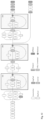
- FIG. 9 shows a histopathological image (HI) of tumor tissue of a patient suffering from cancer.
- FIG. 10 (a) illustrates a digital image of an HE stained, formalin-fixed, paraffin-embedded (FFPE) tumor tissue section.
- FIG. 10 (b) illustrates an output of the prediction tool, according to some embodiments.
- FIG. 10 (c) illustrates a digital image of an adjacent FFPE tumor tissue section stained with an antibody for indicating presence of TRK proteins using immunohistochemistry.
- FIG. 11 shows schematically a process for determining a probability value in practice, according to some embodiments.
- FIG. 12 shows the layer structure of the artificial neural network, according to some embodiments.
- FIG. 13 shows a receiver operating characteristic curve (ROC curve), according to some embodiments.
- FIG. 14 a precision recall curve, according to some embodiments.
- FIG. 1 shows schematically a system 1 for determining a probability value from patient data, according to some embodiments.
- system 1 comprises an input component 10 , a processor 20 , and an output component 30 .
- the input component 10 may accept patient data and may forward the patient data to the processor 20 (depicted by the arrow between the input component 10 and the processor 20 ).
- the processor 20 may determine a probability value based on the patient data.
- the processor may be optionally configured to compare the probability value with a predefined threshold value and thereby determining a comparison result.
- the probability value and/or the comparison result may be transmitted from the processor 20 to the output component 30 (depicted by the arrow between the processor 20 and the output component 30 ).
- output component 30 may display the probability value and/or the comparison result.
- the input component 10 , the processor 20 and the output component 30 can be components of one or more computer systems.
- FIG. 2 shows a detailed schematic of a computer system 1 for determining a probability value from patient data, according to some embodiments of the present disclosure.
- a computer system of exemplary implementations of the present disclosure may be referred to as a computer and may comprise, include, or be embodied in one or more fixed or portable electronic devices.
- the computer may include one or more of each of a number of components such as, for example, the processor 20 connected to a memory 50 (e.g., storage device).
- a memory 50 e.g., storage device
- the processor 20 may be composed of one or more processors alone or in combination with one or more memories.
- the processor 20 may generally be any piece of computer hardware that is capable of processing information such as, for example, data, computer programs, and/or other suitable electronic information.
- the processor 20 may be composed of a collection of electronic circuits some of which may be packaged as an integrated circuit or multiple interconnected integrated circuits (an integrated circuit at times more commonly referred to as a “chip”).
- the processor 20 may be configured to execute computer programs, which may be stored onboard the processor 20 or otherwise stored in the memory 50 of the same or another computer.
- the processor 20 may be a number of processors, a multi-core processor, or some other type of processor, depending on the particular implementation. Further, the processor 20 may be implemented using a number of heterogeneous processor systems in which a main processor is present with one or more secondary processors on a single chip. In some embodiments, the processor 20 may be a symmetric multi-processor system containing multiple processors of the same type. In some embodiments, the processor 20 may be embodied as or otherwise include one or more ASICs, FPGAs, or the like. Thus, although the processor 20 may be capable of executing a computer program to perform one or more functions, the processor 20 of various embodiments may be capable of performing one or more functions without the aid of a computer program. In either instance, the processor 20 may be appropriately programmed to perform functions or operations according to example embodiments of the present disclosure.
- the memory 50 may generally be any piece of computer hardware that is capable of storing information such as, for example, data, computer programs (e.g., computer-readable program code 60 ), and/or other suitable information either on a temporary basis and/or a permanent basis.
- the memory may include volatile and/or non-volatile memory, and may be fixed or removable. Examples of suitable memory include random access memory (RAM), read-only memory (ROM), a hard drive, a flash memory, a thumb drive, a removable computer diskette, an optical disk, a magnetic tape or some combination of the above.
- Optical disks may include compact disk-read only memory (CD-ROM), compact disk-read/write (CD-R/W), DVD, Blu-ray disk or the like.
- the memory may be referred to as a computer-readable storage medium.
- the computer-readable storage medium is a non-transitory device capable of storing information, and is distinguishable from computer-readable transmission media such as electronic transitory signals capable of carrying information from one location to another.
- Computer-readable medium as described herein may generally refer to a computer-readable storage medium or computer-readable transmission medium.
- the processing unit 20 may also be connected to one or more interfaces for displaying, transmitting and/or receiving information.
- the interfaces may include one or more communications interfaces and/or one or more user interfaces.
- the communications interface(s) may be configured to transmit and/or receive information, such as to and/or from other computer(s), network(s), database(s) or the like.
- the communications interface may be configured to transmit and/or receive information by physical (wired) and/or wireless communications links.
- the communications interface(s) may include interface(s) 41 to connect to a network, such as using technologies such as cellular telephone, Wi-Fi, satellite, cable, digital subscriber line (DSL), fiber optics, and the like.
- the communications interface(s) may include one or more short-range communications interfaces 42 configured to connect devices using short-range communications technologies such as NFC, RFID, Bluetooth, Bluetooth LE, ZigBee, infrared (e.g., IrDA) or the like.
- short-range communications technologies such as NFC, RFID, Bluetooth, Bluetooth LE, ZigBee, infrared (e.g., IrDA) or the like.
- the user interfaces may include a display 30 .
- the display may be configured to present or otherwise display information to a user, suitable examples of which include a liquid crystal display (LCD), light-emitting diode display (LED), plasma display panel (PDP), or the like.
- the user input interface(s) 11 may be wired or wireless, and may be configured to receive information from a user into the computer system 1 , such as for processing, storage, and/or display. Suitable examples of user input interfaces include a microphone, image or video capture device, keyboard or keypad, joystick, touch-sensitive surface (separate from or integrated into a touchscreen) or the like.
- the user interfaces may include automatic identification and data capture (AIDC) technology 12 for machine-readable information. This may include barcode, radio frequency identification (RFID), magnetic stripes, optical character recognition (OCR), integrated circuit card (ICC), and the like.
- the user interfaces may further include one or more interfaces for communicating with peripherals such as printers and the like.
- program code instructions may be stored in memory and executed by a processor that is programmed to implement functions of the systems, subsystems, tools, and their respective elements described herein.
- any suitable program code instructions may be loaded onto a computer or other programmable apparatus from a computer-readable storage medium to produce a particular machine, such that the particular machine becomes a means for implementing the functions specified herein.
- These program code instructions may also be stored in a computer-readable storage medium that can direct a computer, processor, or other programmable apparatus to function in a particular manner to thereby generate a particular machine or particular article of manufacture.
- the instructions stored in the computer-readable storage medium may produce an article of manufacture, where the article of manufacture becomes a means for implementing functions described herein.
- the program code instructions may be retrieved from a computer-readable storage medium and loaded into a computer, processor, or other programmable apparatus to configure the computer, processor, or other programmable apparatus to execute operations to be performed on or by the computer, processor, or other programmable apparatus.
- Retrieval, loading, and execution of the program code instructions may be performed sequentially such that one instruction is retrieved, loaded, and executed at a time. In some embodiments, retrieval, loading, and/or execution may be performed in parallel such that multiple instructions are retrieved, loaded, and/or executed together. Execution of the program code instructions may produce a computer-implemented process such that the instructions executed by the computer, processing circuitry or other programmable apparatus provide operations for implementing functions described herein.
- the method steps may comprise:
- FIG. 4 shows a method for determining a probability value, according to some embodiments.
- FIG. 5 shows a method for determining a probability value, according to some embodiments.
- the method steps may comprise:
- FIGS. 6 and 7 illustrate schematically an exemplary convolutional neural network which can be used to determine a probability value, according to some embodiments.
- FIG. 6 illustrates various layers within a CNN.
- an exemplary CNN used to identify within patient data one or more characteristics of an NTRK oncogenic fusion can receive input 102 describing the red, green, and blue (RGB) components of an input histopathological image.
- the input 102 can be processed by multiple convolutional layers (e.g., convolutional layer 104 , convolutional layer 106 .
- the output from the multiple convolutional layers may optionally be processed by a set of fully connected layers 108 and 110 . Neurons in a fully connected layer have full connections to all activations in the previous layer.
- the output from the fully connected layers 108 can be used to generate an output result 110 from the network.
- the output result can be the probability value indicating the probability of a patient suffering from cancer caused by an NTRK oncogenic fusion.
- the activations within the fully connected layers 108 can be computed using matrix multiplication instead of convolution. Not all CNN implementations make use of fully connected layers.
- the convolutional layer 106 can generate output for the CNN.
- the convolutional layers may be sparsely connected, which differs from traditional neural network configuration found in the fully connected layers 108 .
- Traditional neural network layers are fully connected, such that every output unit interacts with every input unit.
- the convolutional layers may be sparsely connected because the output of the convolution of a field is input (instead of the respective state value of each of the nodes in the field) to the nodes of the subsequent layer, as illustrated.
- the kernels associated with the convolutional layers may perform convolution operations, the output of which may be sent to the next layer.
- the dimensionality reduction performed within the convolutional layers is one aspect that enables the CNN to process large images.
- FIG. 7 illustrates computation stages within a convolutional layer of a CNN, according to some embodiments.
- Input 112 to a convolutional layer 114 of a CNN can be processed in three stages of the convolutional layer 114 .
- the three stages can include a convolution stage 116 , a detector stage 118 , and a pooling stage 120 .
- the convolution layer 114 can then output data to a successive convolutional layer.
- the final convolutional layer of the network can generate output feature map data or provide input to a fully connected layer, for example, to generate a classification or regression value.
- the linear activations can be processed by a detector stage 118 .
- each linear activation may be processed by a non-linear activation function.
- the non-linear activation function increases the nonlinear properties of the overall network without affecting the receptive fields of the convolution layer.
- the pooling stage 120 may use a pooling function that may replace the output of the convolutional layer 106 with a summary statistic of the nearby outputs.
- the pooling function can be used to introduce translation invariance into the neural network, such that small translations to the input do not change the pooled outputs. Invariance to local translation can be useful in scenarios where the presence of a feature in the input data is more important than the precise location of the feature.
- Various types of pooling functions can be used during the pooling stage 120 , including max pooling, average pooling, and l2-norm pooling.
- a CNN implementation may not include a pooling stage.
- a CNN implementation without a pooling stage may substitute an additional convolution stage having an increased stride relative to previous convolution stages.
- the output from the convolutional layer 114 can then be processed by the next layer 122 .
- the next layer 122 can be an additional convolutional layer or one of the fully connected layers 108 .
- the first convolutional layer 104 of FIG. 6 can output to the second convolutional layer 106
- the second convolutional layer can output to a first layer of the fully connected layers 108 .
- FIG. 8 shows training and deployment of a neural network, according to some embodiments. Once a given network has been structured for a task the neural network may be trained using a training dataset 1102 .
- the initial weights may be chosen randomly or by pre-training using a deep belief network.
- the training cycle may then be performed in either a supervised or unsupervised manner.
- Supervised learning is a learning method in which training is performed as a mediated operation, such as when the training dataset 1102 includes input paired with the desired output for the input, or where the training dataset includes input having known output and the output of the neural network is manually graded.
- the network may process the inputs and compare the resulting outputs against a set of expected or desired outputs. Errors are then propagated back through the system.
- the training framework 1104 may adjust the weights that control the untrained neural network 1106 .
- the training framework 1104 may provide tools to monitor how well the untrained neural network 1106 is converging towards a model suitable to generating correct answers based on known input data.
- the training process may occur repeatedly as the weights of the network are adjusted to refine the output generated by the neural network.
- the training process can continue until the neural network reaches a statistically desired accuracy associated with a trained neural network 1108 .
- the trained neural network 1108 can then be deployed to implement any number of machine learning operations.
- a new set of patient data 1112 can be inputted into the trained neural network 1108 to determine a probability value, the probability value indicating the probability of a patient suffering from cancer caused by a mutation of a neurotrophic receptor tyrosine kinase (NTRK) gene.
- NTRK neurotrophic receptor tyrosine kinase
- FIG. 9 is an example of a histopathological image (HI) of tumor tissue of a patient suffering from cancer.
- FIG. 10 illustrates of the performance of the prediction model and an example of the way to represent its output, according to some embodiments.
- FIG. 10 (a) illustrates a digital image of an HE stained, formalin-fixed, paraffin-embedded (FFPE) tumor tissue section.
- the tissue 200 contains holes 210 .
- Such an image may serve as input to the prediction model.
- the details of the tissue revealed by the HE stain (which result in different colors and intensities with in the tissue section) are omitted for clarity. However, these reveal to the trained person only the position, shape, and thus type of cells, not their molecular composition.
- FIG. 10 (b) illustrates an output example of the prediction tool.
- a digital image is generated corresponding to the layout of the tissue scan in FIG. 10 (a).
- the tissue scan is usually overlaid by a pseudo-colored image in which the color or the intensity of a color corresponds to the probability that an area of the tissue shows a tumor cancer caused by an NTRK oncogenic fusion (here only black/white images are shown).
- the colors are chosen to represent the typical colors (eg brownish or redish) of immunohistochemistry dyes to generate a ‘pseudo IHC image’. Colors are indicated in the figure by areas which are dotted (indicating a predicted weak staining intensity), cross hatched (moderately stained), or thickly hatched (strongly stained).
- FIG. 10 (c) illustrates a digital image of an adjacent FFPE tumor tissue section stained with an antibody for indicating presence of TRK proteins using immunohistochemistry. Note that the digital image corresponds very well to the predictions made by the prediction model as shown in FIG. 10 (b), even though the molecular assay was conducted on an adjacent tissue section.
- predicted images such as shown in FIG. 10 (b) turn from HE colors to pseudo IHC colors in (almost) real time when zooming in to one zone or moving across the tissue. This is enabled by edge analysis, and enables the system according to the present invention to process information locally and respond more quickly to situations. This feature also allows our tool to work in telepathology settings, even in mobile computing devices such as smartphones.
- the prediction model is embedded in a website for convenient installation on routine computer devices.
- IHC can be performed for example with the research use only panTRK monoclonal antibody EPR17341 (Abcam, Cambridge, MA), see Hechtman et al., The American Journal of Surgical Pathology (2017), 41 (11), 1547-1551, PMID 28719467, using for example a Ventana Autostainer Discovery Ultra equipment.
- FIG. 11 shows a process for using the present invention in practice, according to some embodiments.
- the starting point of the process is a patient suffering from cancer.
- One aim of the process may be to find out whether the cancer is caused by a mutation of a neurotrophic receptor tyrosine kinase (NTRK) gene.
- NTRK neurotrophic receptor tyrosine kinase
- patient data 1112 may be collected/retrieved.
- the patient data 1112 may comprise at least one histopathological image (HI) of tumor tissue of the patient.
- the patient data may be inputted into a prediction model 1108 which is trained by supervised learning.
- NRRK neurotrophic receptor tyrosine kinase
- trained prediction model 1108 may be configured to determine a probability value p from the inputted patient data 1112 .
- the probability value may indicates the probability of the patient suffering from cancer caused by a mutation of a neurotrophic receptor tyrosine kinase (NTRK) gene.
- NTRK neurotrophic receptor tyrosine kinase
- the probability value p may be compared with a predefined threshold value t.
- the probability value p is smaller than the predefined threshold value t (p ⁇ t: “No”), then it may be unlikely that the patient suffers from cancer caused by a mutation of a neurotrophic receptor tyrosine kinase (NTRK) gene. Other causes for the cancer may have to be identified; as such, the process ends.
- NRRK neurotrophic receptor tyrosine kinase
- the probability value p is equal to or greater than the predefined threshold value t (p ⁇ t: “Yes”), then it may be likely that the patient suffers from cancer caused by a mutation of a neurotrophic receptor tyrosine kinase (NTRK) gene.
- a subsequent step 1120 may be initiated.
- further investigations may be initiated in order to verify that the cancer is caused by a mutation of a neurotrophic receptor tyrosine kinase (NTRK) gene. This can be done e.g. by a genetic analysis of the tumor tissue. The results of the further investigations can be used to optimize (further supervised learning) the prediction model 1108 .
- the patient can be treated with a Trk inhibitor.
- the likelihood of the cancer being caused by a mutation of a neurotrophic receptor tyrosine kinase (NTRK) gene may be considered to be so high that the patient is treated directly (without any further genetic analysis of the tumor tissue) with a Trk inhibitor.
- FIGS. 12 to 14 illustrate an artificial neural network was set up for the prediction of probability values, each probability value indicating the probability of a subject patient suffering from cancer of the thyroid gland caused by an NTRK oncogenic fusion.
- FIG. 12 shows the layer structure of the artificial neural network, according to some embodiments.
- the network is provided by IBM under the name Inception ResNet v2 (https://developer.ibm.com/exchanges/models/all/max-inception-resnet-v2/).
- the network pre-trained on ImageNet was used with transfer learning (in Keras+Tensorflow).
- FIG. 13 shows the results in the form of a receiver operating characteristic curve (ROC curve).
- TRAIN refers to the training data set
- VAL refers to the validation data set
- TEST refers to the testing data set.
- the training data set was used to train the prediction model
- the validation data set was used to validate the prediction model
- the testing data set was used for prediction purposes.
- AUC means area under the curve.
- FIG. 14 shows the result in the form of a precision recall curve.
- TRAIN refers to the training data set
- VAL refers to the validation data set
- TEST refers to the testing data set.
- the training data set was used to train the prediction model
- the validation data set was used to validate the prediction model
- the testing data set was used for prediction purposes.
- AUC means area under the curve.
Landscapes
- Engineering & Computer Science (AREA)
- Health & Medical Sciences (AREA)
- General Health & Medical Sciences (AREA)
- Medical Informatics (AREA)
- Public Health (AREA)
- Biomedical Technology (AREA)
- Theoretical Computer Science (AREA)
- Physics & Mathematics (AREA)
- Data Mining & Analysis (AREA)
- Primary Health Care (AREA)
- Epidemiology (AREA)
- General Physics & Mathematics (AREA)
- Molecular Biology (AREA)
- Life Sciences & Earth Sciences (AREA)
- Artificial Intelligence (AREA)
- Biophysics (AREA)
- Computational Linguistics (AREA)
- Evolutionary Computation (AREA)
- Computing Systems (AREA)
- General Engineering & Computer Science (AREA)
- Mathematical Physics (AREA)
- Software Systems (AREA)
- Radiology & Medical Imaging (AREA)
- Nuclear Medicine, Radiotherapy & Molecular Imaging (AREA)
- Databases & Information Systems (AREA)
- Pathology (AREA)
- Computer Vision & Pattern Recognition (AREA)
- Quality & Reliability (AREA)
- Multimedia (AREA)
- Investigating Or Analysing Biological Materials (AREA)
- Apparatus Associated With Microorganisms And Enzymes (AREA)
- Measuring Or Testing Involving Enzymes Or Micro-Organisms (AREA)
Applications Claiming Priority (4)
| Application Number | Priority Date | Filing Date | Title |
|---|---|---|---|
| EP19173832 | 2019-05-10 | ||
| EP19173832 | 2019-05-10 | ||
| EP19173832.7 | 2019-05-10 | ||
| PCT/EP2020/061665 WO2020229152A1 (en) | 2019-05-10 | 2020-04-28 | Identification of candidate signs indicative of an ntrk oncogenic fusion |
Publications (2)
| Publication Number | Publication Date |
|---|---|
| US20220223261A1 US20220223261A1 (en) | 2022-07-14 |
| US12217851B2 true US12217851B2 (en) | 2025-02-04 |
Family
ID=66483934
Family Applications (1)
| Application Number | Title | Priority Date | Filing Date |
|---|---|---|---|
| US17/595,191 Active 2041-07-27 US12217851B2 (en) | 2019-05-10 | 2020-04-28 | Identification of candidate signs indicative of an NTRK oncogenic fusion |
Country Status (6)
| Country | Link |
|---|---|
| US (1) | US12217851B2 (https=) |
| EP (1) | EP3966830A1 (https=) |
| JP (1) | JP7518097B2 (https=) |
| CN (1) | CN113785365A (https=) |
| CA (1) | CA3139352A1 (https=) |
| WO (1) | WO2020229152A1 (https=) |
Families Citing this family (15)
| Publication number | Priority date | Publication date | Assignee | Title |
|---|---|---|---|---|
| EP4248356A1 (en) | 2020-11-20 | 2023-09-27 | Bayer Aktiengesellschaft | Representation learning |
| US11791035B2 (en) * | 2021-03-09 | 2023-10-17 | PAIGE.AI, Inc. | Systems and methods for artificial intelligence powered molecular workflow verifying slide and block quality for testing |
| WO2023194090A1 (en) | 2022-04-08 | 2023-10-12 | Bayer Aktiengesellschaft | Multiple instance learning considering neighborhood aggregations |
| US20250265832A1 (en) | 2022-04-26 | 2025-08-21 | Bayer Aktiengesellschaft | Multiple-instance learning based on regional embeddings |
| WO2023213623A1 (en) | 2022-05-03 | 2023-11-09 | Bayer Aktiengesellschaft | Dynamic sampling strategy for multiple-instance learning |
| EP4471710B1 (de) | 2023-05-30 | 2025-12-17 | Bayer Aktiengesellschaft | Erkennen von artefakten in synthetischen medizinischen aufnahmen |
| EP4475070B1 (de) | 2023-06-05 | 2026-04-22 | Bayer Aktiengesellschaft | Erkennen von artefakten in synthetischen medizinischen aufnahmen |
| EP4492324A1 (de) | 2023-07-12 | 2025-01-15 | Bayer AG | Erkennen von artefakten in synthetischen medizinischen aufnahmen |
| US20250045926A1 (en) | 2023-07-25 | 2025-02-06 | Bayer Aktiengesellschaft | Detection of artifacts in synthetic images |
| EP4560648A1 (en) | 2023-11-22 | 2025-05-28 | Bayer AG | Generating synthetic training data |
| EP4567715A1 (en) | 2023-12-06 | 2025-06-11 | Bayer Aktiengesellschaft | Generating synthetic representations |
| WO2025119803A1 (en) | 2023-12-06 | 2025-06-12 | Bayer Aktiengesellschaft | Generating synthetic medical representations |
| EP4571650A1 (en) | 2023-12-12 | 2025-06-18 | Bayer AG | Generating synthetic images |
| EP4575997A1 (en) | 2023-12-18 | 2025-06-25 | Bayer Aktiengesellschaft | Generating synthetic images |
| WO2025190827A1 (en) | 2024-03-15 | 2025-09-18 | Bayer Aktiengesellschaft | Generation of a synthetic medical image |
Citations (9)
| Publication number | Priority date | Publication date | Assignee | Title |
|---|---|---|---|---|
| US20050262031A1 (en) | 2003-07-21 | 2005-11-24 | Olivier Saidi | Systems and methods for treating, diagnosing and predicting the occurrence of a medical condition |
| US20090252391A1 (en) * | 2005-08-22 | 2009-10-08 | National Center Of Neurology And Psychiatry | Method for assisting in diagnosis of cerebral diseases and apparatus thereof |
| US20170316567A1 (en) * | 2016-04-28 | 2017-11-02 | Canon Kabushiki Kaisha | Image processing apparatus and image processing method |
| US9922421B1 (en) * | 2016-11-06 | 2018-03-20 | Hadassa Degani | Diffusion ellipsoid mapping of tissue |
| US20180130204A1 (en) * | 2016-11-06 | 2018-05-10 | Dde Mri Solutions Ltd. | Diffusion ellipsoid mapping of tissue |
| WO2019084697A1 (en) | 2017-11-06 | 2019-05-09 | University Health Network | Platform, device and process for annotation and classification of tissue specimens using convolutional neural network |
| US20200327659A1 (en) * | 2019-04-10 | 2020-10-15 | International Business Machines Corporation | Image analysis and annotation |
| US20200342600A1 (en) * | 2018-01-08 | 2020-10-29 | Progenics Pharmaceuticals, Inc. | Systems and methods for rapid neural network-based image segmentation and radiopharmaceutical uptake determination |
| US12002544B2 (en) * | 2010-11-30 | 2024-06-04 | The Chinese University Of Hong Kong | Determining progress of chromosomal aberrations over time |
Family Cites Families (5)
| Publication number | Priority date | Publication date | Assignee | Title |
|---|---|---|---|---|
| US6754380B1 (en) * | 2003-02-14 | 2004-06-22 | The University Of Chicago | Method of training massive training artificial neural networks (MTANN) for the detection of abnormalities in medical images |
| US20080032321A1 (en) * | 2006-08-07 | 2008-02-07 | General Electric Company | System and methods for analyzing images of tissue samples |
| CN109478231A (zh) * | 2016-04-01 | 2019-03-15 | 20/20基因系统股份有限公司 | 帮助区别良性和恶性放射线照相明显肺结节的方法和组合物 |
| WO2018156133A1 (en) | 2017-02-23 | 2018-08-30 | Google Llc | Method and system for assisting pathologist identification of tumor cells in magnified tissue images |
| US11107189B2 (en) | 2017-04-07 | 2021-08-31 | Intel Corporation | Methods and systems using improved convolutional neural networks for image processing |
-
2020
- 2020-04-28 EP EP20720468.6A patent/EP3966830A1/en active Pending
- 2020-04-28 US US17/595,191 patent/US12217851B2/en active Active
- 2020-04-28 CN CN202080033711.2A patent/CN113785365A/zh active Pending
- 2020-04-28 JP JP2021566992A patent/JP7518097B2/ja active Active
- 2020-04-28 WO PCT/EP2020/061665 patent/WO2020229152A1/en not_active Ceased
- 2020-04-28 CA CA3139352A patent/CA3139352A1/en active Pending
Patent Citations (9)
| Publication number | Priority date | Publication date | Assignee | Title |
|---|---|---|---|---|
| US20050262031A1 (en) | 2003-07-21 | 2005-11-24 | Olivier Saidi | Systems and methods for treating, diagnosing and predicting the occurrence of a medical condition |
| US20090252391A1 (en) * | 2005-08-22 | 2009-10-08 | National Center Of Neurology And Psychiatry | Method for assisting in diagnosis of cerebral diseases and apparatus thereof |
| US12002544B2 (en) * | 2010-11-30 | 2024-06-04 | The Chinese University Of Hong Kong | Determining progress of chromosomal aberrations over time |
| US20170316567A1 (en) * | 2016-04-28 | 2017-11-02 | Canon Kabushiki Kaisha | Image processing apparatus and image processing method |
| US9922421B1 (en) * | 2016-11-06 | 2018-03-20 | Hadassa Degani | Diffusion ellipsoid mapping of tissue |
| US20180130204A1 (en) * | 2016-11-06 | 2018-05-10 | Dde Mri Solutions Ltd. | Diffusion ellipsoid mapping of tissue |
| WO2019084697A1 (en) | 2017-11-06 | 2019-05-09 | University Health Network | Platform, device and process for annotation and classification of tissue specimens using convolutional neural network |
| US20200342600A1 (en) * | 2018-01-08 | 2020-10-29 | Progenics Pharmaceuticals, Inc. | Systems and methods for rapid neural network-based image segmentation and radiopharmaceutical uptake determination |
| US20200327659A1 (en) * | 2019-04-10 | 2020-10-15 | International Business Machines Corporation | Image analysis and annotation |
Non-Patent Citations (7)
| Title |
|---|
| Arunachalam, H. B. et al., Apr. 17, 2019, "Viable and necrotic tumor assessment from whole slide images of osteosarcoma using machine-learning and deep-learning models," PLOS ONE, 14(4):e0210706. |
| Coudray, N. et al., Oct. 2018, "Classification and mutation prediction from non-small cell lung cancer histopathology images using deep learning," Nature Medicine, 24:1559-1567. |
| David, J. L. et al., 2018, "Infantile NTRK-associated Mesenchymal Tumors," Pediatric and Developmental Pathology, 21(1):68-78. no date available. |
| Davis, J. L. et al. (2017). "Infantile NTRK-associated Mesenchymal Tumors" Pediatric and Developmental Pathology, vol. 21(1), Jul. 6, 2017, pp. 68-78. |
| Extended European Search Report dated Nov. 18, 2019 for European Application No. 19173832.7, 11 pages. |
| Penault-Llorca, F. et al., May 9, 2019, "Testing algorithm for identification of patients with TRK fusion cancer," J Clin. Pathol., 72:460-467. |
| Safoora Deihimi et al, BRCA2, EGFR, and NTRK mutations in mismatch repair-deficient colorectal cancers with MSH2 or MLH1 mutations, 2017, Oncotarget vol. 8 (Year: 2017). * |
Also Published As
| Publication number | Publication date |
|---|---|
| WO2020229152A1 (en) | 2020-11-19 |
| CA3139352A1 (en) | 2020-11-19 |
| JP2022531961A (ja) | 2022-07-12 |
| JP7518097B2 (ja) | 2024-07-17 |
| US20220223261A1 (en) | 2022-07-14 |
| CN113785365A (zh) | 2021-12-10 |
| EP3966830A1 (en) | 2022-03-16 |
Similar Documents
| Publication | Publication Date | Title |
|---|---|---|
| US12217851B2 (en) | Identification of candidate signs indicative of an NTRK oncogenic fusion | |
| Schneider et al. | Integration of deep learning-based image analysis and genomic data in cancer pathology: A systematic review | |
| JP2022527264A (ja) | 病理組織スライド画像からのバイオマーカーの判定方法 | |
| Kim et al. | A deep learning approach for rapid mutational screening in melanoma | |
| van der Logt et al. | Fully automated fluorescent in situ hybridization (FISH) staining and digital analysis of HER2 in breast cancer: a validation study | |
| US20250272839A1 (en) | Machine-learning-enabled predictive biomarker discovery and patient stratification using standard-of-care data | |
| Singla et al. | Artificial Intelligence: Exploring utility in detection and typing of fungus with futuristic application in fungal cytology | |
| Marion et al. | Deep learning discovers new morphological features while predicting genetic alterations from histopathology of papillary thyroid carcinoma | |
| Jones et al. | Peri‐tumoural stroma collagen organization of invasive ductal carcinoma assessed by polarized light microscopy differs between OncotypeDX risk group | |
| Hahn et al. | Points to consider from the ESTP Pathology 2.0 Working Group: overview on spatial omics technologies supporting drug discovery and development | |
| Feng et al. | An accurate regression of developmental stages for breast cancer based on transcriptomic biomarkers | |
| WO2023213623A1 (en) | Dynamic sampling strategy for multiple-instance learning | |
| US11367004B2 (en) | Real-world evidence of diagnostic testing and treatment patterns in U.S. breast cancer patients with implications for treatment biomarkers from RNA-sequencing data | |
| Dia et al. | Computational analysis of whole slide images predicts PD-L1 expression and progression-free survival in immunotherapy-treated non-small cell lung cancer patients | |
| Yuan et al. | Anchored-fusion enables targeted fusion search in bulk and single-cell RNA sequencing data | |
| Abe et al. | Evaluation of a Deep Learning Model for Metastatic Squamous Cell Carcinoma Prediction From Whole Slide Images | |
| Emerson et al. | Quantitative assessment of tissue biomarkers and construction of a model to predict outcome in breast cancer using multiple imputation | |
| WO2023208663A1 (en) | Multiple-instance learning based on regional embeddings | |
| Dawood et al. | Buyer Beware: confounding factors and biases abound when predicting omics-based biomarkers from histological images | |
| KR20230130536A (ko) | 병리 슬라이드 이미지를 분석하는 방법 및 장치 | |
| Du et al. | Optimal transport method-based gene filter (GF) denoising algorithm for enhancing spatially resolved transcriptomics data | |
| Pan et al. | A Deep Learning Framework Integrating Tumor Microenvironmental Features Accurately Predicts Multiple Driver Gene Mutations in Lung Cancer Pathology Images | |
| Cuello et al. | Detection of NTRK gene fusions in solid tumors: recommendations from a Latin American group of oncologists and pathologists | |
| Komura et al. | Deep texture representations as a universal encoder for pan-cancer histology | |
| Bairavi et al. | An Ensemble AI Model for RET Alteration Detection Using H&E Images as a Putative Screening Tool for More Efficient Genomic Alteration Detection |
Legal Events
| Date | Code | Title | Description |
|---|---|---|---|
| FEPP | Fee payment procedure |
Free format text: ENTITY STATUS SET TO UNDISCOUNTED (ORIGINAL EVENT CODE: BIG.); ENTITY STATUS OF PATENT OWNER: LARGE ENTITY |
|
| STPP | Information on status: patent application and granting procedure in general |
Free format text: DOCKETED NEW CASE - READY FOR EXAMINATION |
|
| STPP | Information on status: patent application and granting procedure in general |
Free format text: NON FINAL ACTION MAILED |
|
| STPP | Information on status: patent application and granting procedure in general |
Free format text: RESPONSE TO NON-FINAL OFFICE ACTION ENTERED AND FORWARDED TO EXAMINER |
|
| STPP | Information on status: patent application and granting procedure in general |
Free format text: NOTICE OF ALLOWANCE MAILED -- APPLICATION RECEIVED IN OFFICE OF PUBLICATIONS |
|
| STPP | Information on status: patent application and granting procedure in general |
Free format text: PUBLICATIONS -- ISSUE FEE PAYMENT VERIFIED |
|
| STCF | Information on status: patent grant |
Free format text: PATENTED CASE |