US11873266B2 - Methods of treating or controlling cytotoxic cerebral edema consequent to an ischemic stroke - Google Patents
Methods of treating or controlling cytotoxic cerebral edema consequent to an ischemic stroke Download PDFInfo
- Publication number
- US11873266B2 US11873266B2 US17/443,361 US202117443361A US11873266B2 US 11873266 B2 US11873266 B2 US 11873266B2 US 202117443361 A US202117443361 A US 202117443361A US 11873266 B2 US11873266 B2 US 11873266B2
- Authority
- US
- United States
- Prior art keywords
- edema
- treated
- compound
- condition
- controlled
- Prior art date
- Legal status (The legal status is an assumption and is not a legal conclusion. Google has not performed a legal analysis and makes no representation as to the accuracy of the status listed.)
- Active, expires
Links
- 238000000034 method Methods 0.000 title claims abstract description 358
- 208000032382 Ischaemic stroke Diseases 0.000 title claims abstract description 25
- 208000006752 brain edema Diseases 0.000 title claims description 185
- GTKIGDZXPDCIKR-UHFFFAOYSA-N 2-phenylbenzamide Chemical class NC(=O)C1=CC=CC=C1C1=CC=CC=C1 GTKIGDZXPDCIKR-UHFFFAOYSA-N 0.000 claims abstract description 54
- 208000006011 Stroke Diseases 0.000 claims description 51
- 150000003839 salts Chemical group 0.000 claims description 20
- 238000001990 intravenous administration Methods 0.000 claims description 16
- 230000002537 thrombolytic effect Effects 0.000 claims description 15
- 125000002023 trifluoromethyl group Chemical group FC(F)(F)* 0.000 claims description 14
- 125000002252 acyl group Chemical group 0.000 claims description 11
- 125000001309 chloro group Chemical group Cl* 0.000 claims description 11
- 125000001246 bromo group Chemical group Br* 0.000 claims description 7
- 125000001476 phosphono group Chemical group [H]OP(*)(=O)O[H] 0.000 claims description 7
- 125000001153 fluoro group Chemical group F* 0.000 claims description 4
- 102000010637 Aquaporins Human genes 0.000 abstract description 96
- 108010063290 Aquaporins Proteins 0.000 abstract description 94
- 206010030113 Oedema Diseases 0.000 abstract description 89
- 108010036280 Aquaporin 4 Proteins 0.000 abstract description 85
- XLYOFNOQVPJJNP-UHFFFAOYSA-N water Substances O XLYOFNOQVPJJNP-UHFFFAOYSA-N 0.000 abstract description 73
- 229910001868 water Inorganic materials 0.000 abstract description 71
- 210000004556 brain Anatomy 0.000 abstract description 69
- 206010021143 Hypoxia Diseases 0.000 abstract description 55
- 230000007954 hypoxia Effects 0.000 abstract description 55
- 208000037265 diseases, disorders, signs and symptoms Diseases 0.000 abstract description 45
- 239000003112 inhibitor Substances 0.000 abstract description 45
- 108010036221 Aquaporin 2 Proteins 0.000 abstract description 42
- 208000031225 myocardial ischemia Diseases 0.000 abstract description 40
- 206010019280 Heart failures Diseases 0.000 abstract description 38
- 238000003556 assay Methods 0.000 abstract description 38
- 201000010099 disease Diseases 0.000 abstract description 37
- 206010010904 Convulsion Diseases 0.000 abstract description 34
- 206010015037 epilepsy Diseases 0.000 abstract description 31
- 238000011282 treatment Methods 0.000 abstract description 30
- 208000005735 Water intoxication Diseases 0.000 abstract description 28
- 230000001404 mediated effect Effects 0.000 abstract description 28
- 208000001380 Diabetic Ketoacidosis Diseases 0.000 abstract description 25
- 208000032612 Glial tumor Diseases 0.000 abstract description 25
- 206010018338 Glioma Diseases 0.000 abstract description 25
- 206010019663 Hepatic failure Diseases 0.000 abstract description 25
- 201000009906 Meningitis Diseases 0.000 abstract description 25
- 208000034388 Mountain sickness acute Diseases 0.000 abstract description 25
- 208000018315 acute mountain sickness Diseases 0.000 abstract description 25
- 208000007386 hepatic encephalopathy Diseases 0.000 abstract description 25
- 208000015181 infectious disease Diseases 0.000 abstract description 25
- 208000007903 liver failure Diseases 0.000 abstract description 25
- 231100000835 liver failure Toxicity 0.000 abstract description 25
- 208000030159 metabolic disease Diseases 0.000 abstract description 25
- 210000000278 spinal cord Anatomy 0.000 abstract description 25
- 206010007559 Cardiac failure congestive Diseases 0.000 abstract description 21
- 208000014674 injury Diseases 0.000 abstract description 20
- 208000010125 myocardial infarction Diseases 0.000 abstract description 18
- 208000032253 retinal ischemia Diseases 0.000 abstract description 18
- 230000000638 stimulation Effects 0.000 abstract description 18
- 230000008733 trauma Effects 0.000 abstract description 18
- 206010002329 Aneurysm Diseases 0.000 abstract description 17
- 206010021036 Hyponatraemia Diseases 0.000 abstract description 17
- 208000007201 Myocardial reperfusion injury Diseases 0.000 abstract description 17
- 238000009593 lumbar puncture Methods 0.000 abstract description 17
- 230000005486 microgravity Effects 0.000 abstract description 17
- 230000008439 repair process Effects 0.000 abstract description 17
- 230000002107 myocardial effect Effects 0.000 abstract description 16
- 208000019695 Migraine disease Diseases 0.000 abstract description 14
- 206010040047 Sepsis Diseases 0.000 abstract description 14
- 230000036571 hydration Effects 0.000 abstract description 14
- 238000006703 hydration reaction Methods 0.000 abstract description 14
- 208000008795 neuromyelitis optica Diseases 0.000 abstract description 14
- 208000020406 Creutzfeldt Jacob disease Diseases 0.000 abstract description 13
- 208000003407 Creutzfeldt-Jakob Syndrome Diseases 0.000 abstract description 13
- 208000010859 Creutzfeldt-Jakob disease Diseases 0.000 abstract description 13
- 230000005856 abnormality Effects 0.000 abstract description 13
- 206010000269 abscess Diseases 0.000 abstract description 13
- 210000003169 central nervous system Anatomy 0.000 abstract description 13
- 201000008191 cerebritis Diseases 0.000 abstract description 13
- 208000002296 eclampsia Diseases 0.000 abstract description 13
- 206010014599 encephalitis Diseases 0.000 abstract description 13
- 230000004410 intraocular pressure Effects 0.000 abstract description 13
- 206010025135 lupus erythematosus Diseases 0.000 abstract description 13
- 201000010183 Papilledema Diseases 0.000 abstract description 12
- 206010038886 Retinal oedema Diseases 0.000 abstract description 12
- 201000011195 retinal edema Diseases 0.000 abstract description 12
- 238000011321 prophylaxis Methods 0.000 abstract description 3
- 102000011899 Aquaporin 2 Human genes 0.000 abstract 1
- 102000012002 Aquaporin 4 Human genes 0.000 abstract 1
- 206010048962 Brain oedema Diseases 0.000 description 157
- 150000001875 compounds Chemical class 0.000 description 149
- 210000004027 cell Anatomy 0.000 description 92
- 102100037276 Aquaporin-4 Human genes 0.000 description 84
- 102100034414 Aquaporin-2 Human genes 0.000 description 41
- 230000000694 effects Effects 0.000 description 35
- 206010063036 Spinal cord oedema Diseases 0.000 description 32
- 206010019196 Head injury Diseases 0.000 description 31
- RJMUSRYZPJIFPJ-UHFFFAOYSA-N niclosamide Chemical compound OC1=CC=C(Cl)C=C1C(=O)NC1=CC=C([N+]([O-])=O)C=C1Cl RJMUSRYZPJIFPJ-UHFFFAOYSA-N 0.000 description 30
- 229940125904 compound 1 Drugs 0.000 description 28
- 229960001920 niclosamide Drugs 0.000 description 28
- IAZDPXIOMUYVGZ-UHFFFAOYSA-N Dimethylsulphoxide Chemical compound CS(C)=O IAZDPXIOMUYVGZ-UHFFFAOYSA-N 0.000 description 27
- -1 e.g. Chemical compound 0.000 description 26
- 241000699670 Mus sp. Species 0.000 description 23
- 229940125898 compound 5 Drugs 0.000 description 22
- 206010041549 Spinal cord compression Diseases 0.000 description 21
- 230000008859 change Effects 0.000 description 21
- 241000699666 Mus <mouse, genus> Species 0.000 description 19
- 239000003814 drug Substances 0.000 description 19
- 201000007309 middle cerebral artery infarction Diseases 0.000 description 19
- 208000020339 Spinal injury Diseases 0.000 description 18
- 241001465754 Metazoa Species 0.000 description 16
- 201000008284 inappropriate ADH syndrome Diseases 0.000 description 16
- 238000012360 testing method Methods 0.000 description 16
- 210000001519 tissue Anatomy 0.000 description 16
- 230000008081 blood perfusion Effects 0.000 description 15
- 208000028329 epileptic seizure Diseases 0.000 description 15
- 230000002490 cerebral effect Effects 0.000 description 14
- 229940126214 compound 3 Drugs 0.000 description 14
- 230000009885 systemic effect Effects 0.000 description 14
- 206010008089 Cerebral artery occlusion Diseases 0.000 description 13
- 206010016807 Fluid retention Diseases 0.000 description 13
- 210000003128 head Anatomy 0.000 description 13
- UUUHXMGGBIUAPW-UHFFFAOYSA-N 1-[1-[2-[[5-amino-2-[[1-[5-(diaminomethylideneamino)-2-[[1-[3-(1h-indol-3-yl)-2-[(5-oxopyrrolidine-2-carbonyl)amino]propanoyl]pyrrolidine-2-carbonyl]amino]pentanoyl]pyrrolidine-2-carbonyl]amino]-5-oxopentanoyl]amino]-3-methylpentanoyl]pyrrolidine-2-carbon Chemical compound C1CCC(C(=O)N2C(CCC2)C(O)=O)N1C(=O)C(C(C)CC)NC(=O)C(CCC(N)=O)NC(=O)C1CCCN1C(=O)C(CCCN=C(N)N)NC(=O)C1CCCN1C(=O)C(CC=1C2=CC=CC=C2NC=1)NC(=O)C1CCC(=O)N1 UUUHXMGGBIUAPW-UHFFFAOYSA-N 0.000 description 12
- 102000004270 Peptidyl-Dipeptidase A Human genes 0.000 description 12
- 108090000882 Peptidyl-Dipeptidase A Proteins 0.000 description 12
- 239000002333 angiotensin II receptor antagonist Substances 0.000 description 12
- 229940125364 angiotensin receptor blocker Drugs 0.000 description 12
- 239000002876 beta blocker Substances 0.000 description 12
- 230000002401 inhibitory effect Effects 0.000 description 11
- 208000010496 Heart Arrest Diseases 0.000 description 10
- FAPWRFPIFSIZLT-UHFFFAOYSA-M Sodium chloride Chemical compound [Na+].[Cl-] FAPWRFPIFSIZLT-UHFFFAOYSA-M 0.000 description 10
- 239000011780 sodium chloride Substances 0.000 description 10
- 208000030886 Traumatic Brain injury Diseases 0.000 description 9
- 229940079593 drug Drugs 0.000 description 9
- 229960002378 oftasceine Drugs 0.000 description 9
- 230000035939 shock Effects 0.000 description 9
- 230000001988 toxicity Effects 0.000 description 9
- 231100000419 toxicity Toxicity 0.000 description 9
- 238000013417 toxicology model Methods 0.000 description 9
- 102100027221 CD81 antigen Human genes 0.000 description 8
- 101000914479 Homo sapiens CD81 antigen Proteins 0.000 description 8
- 230000009471 action Effects 0.000 description 8
- BQRGNLJZBFXNCZ-UHFFFAOYSA-N calcein am Chemical compound O1C(=O)C2=CC=CC=C2C21C1=CC(CN(CC(=O)OCOC(C)=O)CC(=O)OCOC(C)=O)=C(OC(C)=O)C=C1OC1=C2C=C(CN(CC(=O)OCOC(C)=O)CC(=O)OCOC(=O)C)C(OC(C)=O)=C1 BQRGNLJZBFXNCZ-UHFFFAOYSA-N 0.000 description 8
- 208000019425 cirrhosis of liver Diseases 0.000 description 8
- 208000035475 disorder Diseases 0.000 description 8
- 125000000623 heterocyclic group Chemical group 0.000 description 8
- 230000005764 inhibitory process Effects 0.000 description 8
- 208000028867 ischemia Diseases 0.000 description 8
- 238000002595 magnetic resonance imaging Methods 0.000 description 8
- 238000012423 maintenance Methods 0.000 description 8
- 239000011734 sodium Substances 0.000 description 8
- 125000001424 substituent group Chemical group 0.000 description 8
- 238000002560 therapeutic procedure Methods 0.000 description 8
- 230000009529 traumatic brain injury Effects 0.000 description 8
- 239000002536 vasopressin receptor antagonist Substances 0.000 description 8
- DGAQECJNVWCQMB-PUAWFVPOSA-M Ilexoside XXIX Chemical compound C[C@@H]1CC[C@@]2(CC[C@@]3(C(=CC[C@H]4[C@]3(CC[C@@H]5[C@@]4(CC[C@@H](C5(C)C)OS(=O)(=O)[O-])C)C)[C@@H]2[C@]1(C)O)C)C(=O)O[C@H]6[C@@H]([C@H]([C@@H]([C@H](O6)CO)O)O)O.[Na+] DGAQECJNVWCQMB-PUAWFVPOSA-M 0.000 description 7
- 230000017531 blood circulation Effects 0.000 description 7
- 230000003788 cerebral perfusion Effects 0.000 description 7
- 230000006378 damage Effects 0.000 description 7
- 239000002934 diuretic Substances 0.000 description 7
- 229940030606 diuretics Drugs 0.000 description 7
- 229910052757 nitrogen Inorganic materials 0.000 description 7
- 125000004433 nitrogen atom Chemical group N* 0.000 description 7
- 239000008194 pharmaceutical composition Substances 0.000 description 7
- 229910052708 sodium Inorganic materials 0.000 description 7
- PQSUYGKTWSAVDQ-ZVIOFETBSA-N Aldosterone Chemical compound C([C@@]1([C@@H](C(=O)CO)CC[C@H]1[C@@H]1CC2)C=O)[C@H](O)[C@@H]1[C@]1(C)C2=CC(=O)CC1 PQSUYGKTWSAVDQ-ZVIOFETBSA-N 0.000 description 6
- PQSUYGKTWSAVDQ-UHFFFAOYSA-N Aldosterone Natural products C1CC2C3CCC(C(=O)CO)C3(C=O)CC(O)C2C2(C)C1=CC(=O)CC2 PQSUYGKTWSAVDQ-UHFFFAOYSA-N 0.000 description 6
- BSYNRYMUTXBXSQ-UHFFFAOYSA-N Aspirin Chemical compound CC(=O)OC1=CC=CC=C1C(O)=O BSYNRYMUTXBXSQ-UHFFFAOYSA-N 0.000 description 6
- KXDAEFPNCMNJSK-UHFFFAOYSA-N Benzamide Chemical compound NC(=O)C1=CC=CC=C1 KXDAEFPNCMNJSK-UHFFFAOYSA-N 0.000 description 6
- 208000009447 Cardiac Edema Diseases 0.000 description 6
- 206010008190 Cerebrovascular accident Diseases 0.000 description 6
- 208000018652 Closed Head injury Diseases 0.000 description 6
- FBPFZTCFMRRESA-KVTDHHQDSA-N D-Mannitol Chemical compound OC[C@@H](O)[C@@H](O)[C@H](O)[C@H](O)CO FBPFZTCFMRRESA-KVTDHHQDSA-N 0.000 description 6
- LTMHDMANZUZIPE-AMTYYWEZSA-N Digoxin Natural products O([C@H]1[C@H](C)O[C@H](O[C@@H]2C[C@@H]3[C@@](C)([C@@H]4[C@H]([C@]5(O)[C@](C)([C@H](O)C4)[C@H](C4=CC(=O)OC4)CC5)CC3)CC2)C[C@@H]1O)[C@H]1O[C@H](C)[C@@H](O[C@H]2O[C@@H](C)[C@H](O)[C@@H](O)C2)[C@@H](O)C1 LTMHDMANZUZIPE-AMTYYWEZSA-N 0.000 description 6
- LFQSCWFLJHTTHZ-UHFFFAOYSA-N Ethanol Chemical compound CCO LFQSCWFLJHTTHZ-UHFFFAOYSA-N 0.000 description 6
- 208000001953 Hypotension Diseases 0.000 description 6
- 229930195725 Mannitol Natural products 0.000 description 6
- 125000002777 acetyl group Chemical group [H]C([H])([H])C(*)=O 0.000 description 6
- 229960001138 acetylsalicylic acid Drugs 0.000 description 6
- 239000002253 acid Substances 0.000 description 6
- 229960002478 aldosterone Drugs 0.000 description 6
- DKNWSYNQZKUICI-UHFFFAOYSA-N amantadine Chemical compound C1C(C2)CC3CC2CC1(N)C3 DKNWSYNQZKUICI-UHFFFAOYSA-N 0.000 description 6
- 229960003805 amantadine Drugs 0.000 description 6
- 238000002399 angioplasty Methods 0.000 description 6
- 125000006615 aromatic heterocyclic group Chemical group 0.000 description 6
- 230000000386 athletic effect Effects 0.000 description 6
- 229940125717 barbiturate Drugs 0.000 description 6
- 229940097320 beta blocking agent Drugs 0.000 description 6
- DEGAKNSWVGKMLS-UHFFFAOYSA-N calcein Chemical compound O1C(=O)C2=CC=CC=C2C21C1=CC(CN(CC(O)=O)CC(O)=O)=C(O)C=C1OC1=C2C=C(CN(CC(O)=O)CC(=O)O)C(O)=C1 DEGAKNSWVGKMLS-UHFFFAOYSA-N 0.000 description 6
- 230000018044 dehydration Effects 0.000 description 6
- 238000006297 dehydration reaction Methods 0.000 description 6
- 238000011161 development Methods 0.000 description 6
- 235000005911 diet Nutrition 0.000 description 6
- 230000000378 dietary effect Effects 0.000 description 6
- LTMHDMANZUZIPE-PUGKRICDSA-N digoxin Chemical compound C1[C@H](O)[C@H](O)[C@@H](C)O[C@H]1O[C@@H]1[C@@H](C)O[C@@H](O[C@@H]2[C@H](O[C@@H](O[C@@H]3C[C@@H]4[C@]([C@@H]5[C@H]([C@]6(CC[C@@H]([C@@]6(C)[C@H](O)C5)C=5COC(=O)C=5)O)CC4)(C)CC3)C[C@@H]2O)C)C[C@@H]1O LTMHDMANZUZIPE-PUGKRICDSA-N 0.000 description 6
- 229960005156 digoxin Drugs 0.000 description 6
- LTMHDMANZUZIPE-UHFFFAOYSA-N digoxine Natural products C1C(O)C(O)C(C)OC1OC1C(C)OC(OC2C(OC(OC3CC4C(C5C(C6(CCC(C6(C)C(O)C5)C=5COC(=O)C=5)O)CC4)(C)CC3)CC2O)C)CC1O LTMHDMANZUZIPE-UHFFFAOYSA-N 0.000 description 6
- 239000012530 fluid Substances 0.000 description 6
- 208000000122 hyperventilation Diseases 0.000 description 6
- 230000000870 hyperventilation Effects 0.000 description 6
- 230000036543 hypotension Effects 0.000 description 6
- 230000002631 hypothermal effect Effects 0.000 description 6
- 230000001965 increasing effect Effects 0.000 description 6
- 239000007924 injection Substances 0.000 description 6
- 238000002347 injection Methods 0.000 description 6
- 239000000594 mannitol Substances 0.000 description 6
- 235000010355 mannitol Nutrition 0.000 description 6
- 230000004060 metabolic process Effects 0.000 description 6
- 206010027599 migraine Diseases 0.000 description 6
- 230000037000 normothermia Effects 0.000 description 6
- 230000003204 osmotic effect Effects 0.000 description 6
- 230000002285 radioactive effect Effects 0.000 description 6
- 239000012857 radioactive material Substances 0.000 description 6
- 230000004083 survival effect Effects 0.000 description 6
- 230000001225 therapeutic effect Effects 0.000 description 6
- 206010033712 Papilloedema Diseases 0.000 description 5
- 125000004429 atom Chemical group 0.000 description 5
- 230000015572 biosynthetic process Effects 0.000 description 5
- 230000008499 blood brain barrier function Effects 0.000 description 5
- 210000001218 blood-brain barrier Anatomy 0.000 description 5
- 230000009172 bursting Effects 0.000 description 5
- 125000003917 carbamoyl group Chemical group [H]N([H])C(*)=O 0.000 description 5
- 210000004978 chinese hamster ovary cell Anatomy 0.000 description 5
- 238000002474 experimental method Methods 0.000 description 5
- 238000012188 high-throughput screening assay Methods 0.000 description 5
- 238000003384 imaging method Methods 0.000 description 5
- 210000003657 middle cerebral artery Anatomy 0.000 description 5
- 230000000926 neurological effect Effects 0.000 description 5
- 230000010410 reperfusion Effects 0.000 description 5
- 210000002966 serum Anatomy 0.000 description 5
- 238000000149 argon plasma sintering Methods 0.000 description 4
- 230000000903 blocking effect Effects 0.000 description 4
- 238000006243 chemical reaction Methods 0.000 description 4
- 210000001723 extracellular space Anatomy 0.000 description 4
- 239000007850 fluorescent dye Substances 0.000 description 4
- 230000002727 hyperosmolar Effects 0.000 description 4
- 238000000338 in vitro Methods 0.000 description 4
- 238000001727 in vivo Methods 0.000 description 4
- 230000003447 ipsilateral effect Effects 0.000 description 4
- 238000011813 knockout mouse model Methods 0.000 description 4
- 230000036470 plasma concentration Effects 0.000 description 4
- 108090000623 proteins and genes Proteins 0.000 description 4
- 230000009103 reabsorption Effects 0.000 description 4
- 230000009467 reduction Effects 0.000 description 4
- 230000002829 reductive effect Effects 0.000 description 4
- 238000011160 research Methods 0.000 description 4
- 230000004044 response Effects 0.000 description 4
- 230000008961 swelling Effects 0.000 description 4
- 125000002861 (C1-C4) alkanoyl group Chemical group 0.000 description 3
- WEVYAHXRMPXWCK-UHFFFAOYSA-N Acetonitrile Chemical compound CC#N WEVYAHXRMPXWCK-UHFFFAOYSA-N 0.000 description 3
- 108090001004 Aquaporin 1 Proteins 0.000 description 3
- 206010014418 Electrolyte imbalance Diseases 0.000 description 3
- 150000001413 amino acids Chemical class 0.000 description 3
- 239000001961 anticonvulsive agent Substances 0.000 description 3
- QVGXLLKOCUKJST-UHFFFAOYSA-N atomic oxygen Chemical compound [O] QVGXLLKOCUKJST-UHFFFAOYSA-N 0.000 description 3
- 210000004369 blood Anatomy 0.000 description 3
- 239000008280 blood Substances 0.000 description 3
- 125000002915 carbonyl group Chemical group [*:2]C([*:1])=O 0.000 description 3
- 125000003178 carboxy group Chemical group [H]OC(*)=O 0.000 description 3
- 230000001413 cellular effect Effects 0.000 description 3
- 231100000433 cytotoxic Toxicity 0.000 description 3
- 230000001472 cytotoxic effect Effects 0.000 description 3
- 238000012217 deletion Methods 0.000 description 3
- 230000037430 deletion Effects 0.000 description 3
- 239000003085 diluting agent Substances 0.000 description 3
- 230000001787 epileptiform Effects 0.000 description 3
- 239000000819 hypertonic solution Substances 0.000 description 3
- 238000001294 liquid chromatography-tandem mass spectrometry Methods 0.000 description 3
- 238000002552 multiple reaction monitoring Methods 0.000 description 3
- 239000001301 oxygen Substances 0.000 description 3
- 229910052760 oxygen Inorganic materials 0.000 description 3
- 125000001997 phenyl group Chemical group [H]C1=C([H])C([H])=C(*)C([H])=C1[H] 0.000 description 3
- 239000013641 positive control Substances 0.000 description 3
- 102000004169 proteins and genes Human genes 0.000 description 3
- 210000001525 retina Anatomy 0.000 description 3
- 238000005556 structure-activity relationship Methods 0.000 description 3
- 230000000982 vasogenic effect Effects 0.000 description 3
- 125000004178 (C1-C4) alkyl group Chemical group 0.000 description 2
- 125000004214 1-pyrrolidinyl group Chemical group [H]C1([H])N(*)C([H])([H])C([H])([H])C1([H])[H] 0.000 description 2
- ZNPREQSETWFCPD-UHFFFAOYSA-N 5-chloro-n-[3-fluoro-5-(trifluoromethyl)phenyl]-2-hydroxybenzamide Chemical compound OC1=CC=C(Cl)C=C1C(=O)NC1=CC(F)=CC(C(F)(F)F)=C1 ZNPREQSETWFCPD-UHFFFAOYSA-N 0.000 description 2
- 208000010444 Acidosis Diseases 0.000 description 2
- 102000004888 Aquaporin 1 Human genes 0.000 description 2
- 108090000976 Aquaporin 5 Proteins 0.000 description 2
- 201000004569 Blindness Diseases 0.000 description 2
- 239000006144 Dulbecco’s modified Eagle's medium Substances 0.000 description 2
- 241000282412 Homo Species 0.000 description 2
- 206010061216 Infarction Diseases 0.000 description 2
- 206010027202 Meningitis bacterial Diseases 0.000 description 2
- 206010027417 Metabolic acidosis Diseases 0.000 description 2
- NBIIXXVUZAFLBC-UHFFFAOYSA-N Phosphoric acid Chemical compound OP(O)(O)=O NBIIXXVUZAFLBC-UHFFFAOYSA-N 0.000 description 2
- 241000700159 Rattus Species 0.000 description 2
- 241001362551 Samba Species 0.000 description 2
- 208000003443 Unconsciousness Diseases 0.000 description 2
- 206010047571 Visual impairment Diseases 0.000 description 2
- 208000027418 Wounds and injury Diseases 0.000 description 2
- 230000001154 acute effect Effects 0.000 description 2
- 125000004453 alkoxycarbonyl group Chemical group 0.000 description 2
- 125000000217 alkyl group Chemical group 0.000 description 2
- 125000004397 aminosulfonyl group Chemical group NS(=O)(=O)* 0.000 description 2
- 238000004458 analytical method Methods 0.000 description 2
- 229940125681 anticonvulsant agent Drugs 0.000 description 2
- 238000013459 approach Methods 0.000 description 2
- 230000036525 aquaresis Effects 0.000 description 2
- 201000009904 bacterial meningitis Diseases 0.000 description 2
- 239000000872 buffer Substances 0.000 description 2
- 210000000269 carotid artery external Anatomy 0.000 description 2
- 210000004004 carotid artery internal Anatomy 0.000 description 2
- 230000030833 cell death Effects 0.000 description 2
- 125000004093 cyano group Chemical group *C#N 0.000 description 2
- 125000004122 cyclic group Chemical group 0.000 description 2
- 230000007423 decrease Effects 0.000 description 2
- 239000008367 deionised water Substances 0.000 description 2
- 229910021641 deionized water Inorganic materials 0.000 description 2
- 239000010432 diamond Substances 0.000 description 2
- 238000010828 elution Methods 0.000 description 2
- 238000003366 endpoint assay Methods 0.000 description 2
- 230000006870 function Effects 0.000 description 2
- 238000002523 gelfiltration Methods 0.000 description 2
- 125000005843 halogen group Chemical group 0.000 description 2
- 210000001320 hippocampus Anatomy 0.000 description 2
- 239000000815 hypotonic solution Substances 0.000 description 2
- 230000006872 improvement Effects 0.000 description 2
- 230000007574 infarction Effects 0.000 description 2
- 230000003960 inflammatory cascade Effects 0.000 description 2
- 238000001802 infusion Methods 0.000 description 2
- 238000007917 intracranial administration Methods 0.000 description 2
- 239000007928 intraperitoneal injection Substances 0.000 description 2
- 150000002500 ions Chemical class 0.000 description 2
- 210000003734 kidney Anatomy 0.000 description 2
- 208000006443 lactic acidosis Diseases 0.000 description 2
- 239000002171 loop diuretic Substances 0.000 description 2
- 238000004519 manufacturing process Methods 0.000 description 2
- 238000005259 measurement Methods 0.000 description 2
- 239000000178 monomer Substances 0.000 description 2
- 125000004573 morpholin-4-yl group Chemical group N1(CCOCC1)* 0.000 description 2
- 238000010172 mouse model Methods 0.000 description 2
- 210000005036 nerve Anatomy 0.000 description 2
- QJGQUHMNIGDVPM-UHFFFAOYSA-N nitrogen group Chemical group [N] QJGQUHMNIGDVPM-UHFFFAOYSA-N 0.000 description 2
- 210000001328 optic nerve Anatomy 0.000 description 2
- 230000003287 optical effect Effects 0.000 description 2
- AICOOMRHRUFYCM-ZRRPKQBOSA-N oxazine, 1 Chemical compound C([C@@H]1[C@H](C(C[C@]2(C)[C@@H]([C@H](C)N(C)C)[C@H](O)C[C@]21C)=O)CC1=CC2)C[C@H]1[C@@]1(C)[C@H]2N=C(C(C)C)OC1 AICOOMRHRUFYCM-ZRRPKQBOSA-N 0.000 description 2
- 230000037361 pathway Effects 0.000 description 2
- 230000035699 permeability Effects 0.000 description 2
- 239000002953 phosphate buffered saline Substances 0.000 description 2
- 125000000587 piperidin-1-yl group Chemical group [H]C1([H])N(*)C([H])([H])C([H])([H])C([H])([H])C1([H])[H] 0.000 description 2
- 239000000902 placebo Substances 0.000 description 2
- 229940068196 placebo Drugs 0.000 description 2
- 125000000020 sulfo group Chemical group O=S(=O)([*])O[H] 0.000 description 2
- 230000009261 transgenic effect Effects 0.000 description 2
- 102000035160 transmembrane proteins Human genes 0.000 description 2
- 108091005703 transmembrane proteins Proteins 0.000 description 2
- 230000032258 transport Effects 0.000 description 2
- 208000029257 vision disease Diseases 0.000 description 2
- 230000004393 visual impairment Effects 0.000 description 2
- 125000004169 (C1-C6) alkyl group Chemical group 0.000 description 1
- ANRDLYIUCBDNFE-UHFFFAOYSA-N 2-hydroxy-5-methyl-n-[2-methyl-5-(trifluoromethyl)phenyl]benzamide Chemical compound CC1=CC=C(O)C(C(=O)NC=2C(=CC=C(C=2)C(F)(F)F)C)=C1 ANRDLYIUCBDNFE-UHFFFAOYSA-N 0.000 description 1
- ORFPVWOTYFSKQM-UHFFFAOYSA-N 2-hydroxy-5-methyl-n-[4-methyl-3-(trifluoromethyl)phenyl]benzamide Chemical compound CC1=CC=C(O)C(C(=O)NC=2C=C(C(C)=CC=2)C(F)(F)F)=C1 ORFPVWOTYFSKQM-UHFFFAOYSA-N 0.000 description 1
- FDOMRHBSZBWMJE-UHFFFAOYSA-N 5-bromo-2-hydroxy-n-[2-methoxy-5-(trifluoromethyl)phenyl]benzamide Chemical compound COC1=CC=C(C(F)(F)F)C=C1NC(=O)C1=CC(Br)=CC=C1O FDOMRHBSZBWMJE-UHFFFAOYSA-N 0.000 description 1
- JOPSYWSTEFXKKP-UHFFFAOYSA-N 5-bromo-2-hydroxy-n-[3-methoxy-5-(trifluoromethyl)phenyl]benzamide Chemical compound FC(F)(F)C1=CC(OC)=CC(NC(=O)C=2C(=CC=C(Br)C=2)O)=C1 JOPSYWSTEFXKKP-UHFFFAOYSA-N 0.000 description 1
- ODVTXYZSTFULIY-UHFFFAOYSA-N 5-bromo-n-(4-tert-butyl-5-cyano-1,3-thiazol-2-yl)-2-hydroxybenzamide Chemical compound S1C(C#N)=C(C(C)(C)C)N=C1NC(=O)C1=CC(Br)=CC=C1O ODVTXYZSTFULIY-UHFFFAOYSA-N 0.000 description 1
- QHCLUJCBAITGSH-UHFFFAOYSA-N 5-bromo-n-[2-chloro-5-(trifluoromethyl)phenyl]-2-hydroxybenzamide Chemical compound OC1=CC=C(Br)C=C1C(=O)NC1=CC(C(F)(F)F)=CC=C1Cl QHCLUJCBAITGSH-UHFFFAOYSA-N 0.000 description 1
- PRRVJTMMAVOABR-UHFFFAOYSA-N 5-bromo-n-[3-bromo-5-(trifluoromethyl)phenyl]-2-hydroxybenzamide Chemical compound OC1=CC=C(Br)C=C1C(=O)NC1=CC(Br)=CC(C(F)(F)F)=C1 PRRVJTMMAVOABR-UHFFFAOYSA-N 0.000 description 1
- MDHIVQPZQXDHMS-UHFFFAOYSA-N 5-bromo-n-[4-chloro-3-(trifluoromethyl)phenyl]-2-hydroxybenzamide Chemical compound OC1=CC=C(Br)C=C1C(=O)NC1=CC=C(Cl)C(C(F)(F)F)=C1 MDHIVQPZQXDHMS-UHFFFAOYSA-N 0.000 description 1
- NFAZRXHBYWNSFD-UHFFFAOYSA-N 5-bromo-n-[4-cyano-3-(trifluoromethyl)phenyl]-2-hydroxybenzamide Chemical compound OC1=CC=C(Br)C=C1C(=O)NC1=CC=C(C#N)C(C(F)(F)F)=C1 NFAZRXHBYWNSFD-UHFFFAOYSA-N 0.000 description 1
- KYOCESCAKLLYKQ-UHFFFAOYSA-N 5-bromo-n-[5-bromo-4-(trifluoromethyl)-1,3-thiazol-2-yl]-2-hydroxybenzamide Chemical compound OC1=CC=C(Br)C=C1C(=O)NC1=NC(C(F)(F)F)=C(Br)S1 KYOCESCAKLLYKQ-UHFFFAOYSA-N 0.000 description 1
- LCCUGNWKZAGAKR-UHFFFAOYSA-N 5-chloro-2-hydroxy-n-[2-methyl-3-(trifluoromethyl)phenyl]benzamide Chemical compound C1=CC=C(C(F)(F)F)C(C)=C1NC(=O)C1=CC(Cl)=CC=C1O LCCUGNWKZAGAKR-UHFFFAOYSA-N 0.000 description 1
- RSXRBYFMTAUUBD-UHFFFAOYSA-N 5-chloro-2-hydroxy-n-[2-methyl-5-(trifluoromethyl)phenyl]benzamide Chemical compound CC1=CC=C(C(F)(F)F)C=C1NC(=O)C1=CC(Cl)=CC=C1O RSXRBYFMTAUUBD-UHFFFAOYSA-N 0.000 description 1
- HZYNKSDQPBZDHY-UHFFFAOYSA-N 5-chloro-2-hydroxy-n-[2-methylsulfanyl-5-(trifluoromethyl)phenyl]benzamide Chemical compound CSC1=CC=C(C(F)(F)F)C=C1NC(=O)C1=CC(Cl)=CC=C1O HZYNKSDQPBZDHY-UHFFFAOYSA-N 0.000 description 1
- NBNLMULFBPSZTG-UHFFFAOYSA-N 5-chloro-2-hydroxy-n-[2-nitro-5-(trifluoromethyl)phenyl]benzamide Chemical compound OC1=CC=C(Cl)C=C1C(=O)NC1=CC(C(F)(F)F)=CC=C1[N+]([O-])=O NBNLMULFBPSZTG-UHFFFAOYSA-N 0.000 description 1
- WVDXIAKNJZARGN-UHFFFAOYSA-N 5-chloro-2-hydroxy-n-[4-methoxy-3-(trifluoromethyl)phenyl]benzamide Chemical compound C1=C(C(F)(F)F)C(OC)=CC=C1NC(=O)C1=CC(Cl)=CC=C1O WVDXIAKNJZARGN-UHFFFAOYSA-N 0.000 description 1
- NOTUJGXOCZBNTL-UHFFFAOYSA-N 5-chloro-2-hydroxy-n-[4-methyl-3-(trifluoromethyl)phenyl]benzamide Chemical compound C1=C(C(F)(F)F)C(C)=CC=C1NC(=O)C1=CC(Cl)=CC=C1O NOTUJGXOCZBNTL-UHFFFAOYSA-N 0.000 description 1
- JRHPJUCMRNXATL-UHFFFAOYSA-N 5-chloro-n-[2-chloro-5-(trifluoromethyl)phenyl]-2-hydroxybenzamide Chemical compound OC1=CC=C(Cl)C=C1C(=O)NC1=CC(C(F)(F)F)=CC=C1Cl JRHPJUCMRNXATL-UHFFFAOYSA-N 0.000 description 1
- PFIZSXCOWCLQNU-UHFFFAOYSA-N 5-chloro-n-[2-fluoro-3-(trifluoromethyl)phenyl]-2-hydroxybenzamide Chemical compound OC1=CC=C(Cl)C=C1C(=O)NC1=CC=CC(C(F)(F)F)=C1F PFIZSXCOWCLQNU-UHFFFAOYSA-N 0.000 description 1
- MZECJNJNGITFBT-UHFFFAOYSA-N 5-chloro-n-[2-fluoro-5-(trifluoromethyl)phenyl]-2-hydroxybenzamide Chemical compound OC1=CC=C(Cl)C=C1C(=O)NC1=CC(C(F)(F)F)=CC=C1F MZECJNJNGITFBT-UHFFFAOYSA-N 0.000 description 1
- XOJZVNXJYRVDTM-UHFFFAOYSA-N 5-chloro-n-[4-fluoro-3-(trifluoromethyl)phenyl]-2-hydroxybenzamide Chemical compound OC1=CC=C(Cl)C=C1C(=O)NC1=CC=C(F)C(C(F)(F)F)=C1 XOJZVNXJYRVDTM-UHFFFAOYSA-N 0.000 description 1
- 108091006112 ATPases Proteins 0.000 description 1
- 102000057290 Adenosine Triphosphatases Human genes 0.000 description 1
- 206010001497 Agitation Diseases 0.000 description 1
- 102000004392 Aquaporin 5 Human genes 0.000 description 1
- 229940121720 Aquaporin inhibitor Drugs 0.000 description 1
- 102100023771 Aquaporin-1 Human genes 0.000 description 1
- 102100037280 Aquaporin-5 Human genes 0.000 description 1
- 208000037260 Atherosclerotic Plaque Diseases 0.000 description 1
- 241000894006 Bacteria Species 0.000 description 1
- 208000003174 Brain Neoplasms Diseases 0.000 description 1
- 208000014644 Brain disease Diseases 0.000 description 1
- 241000242722 Cestoda Species 0.000 description 1
- 102000034534 Cotransporters Human genes 0.000 description 1
- 108020003264 Cotransporters Proteins 0.000 description 1
- 206010067276 Cytotoxic oedema Diseases 0.000 description 1
- 208000016192 Demyelinating disease Diseases 0.000 description 1
- 206010012689 Diabetic retinopathy Diseases 0.000 description 1
- QXNVGIXVLWOKEQ-UHFFFAOYSA-N Disodium Chemical group [Na][Na] QXNVGIXVLWOKEQ-UHFFFAOYSA-N 0.000 description 1
- 208000003098 Ganglion Cysts Diseases 0.000 description 1
- 208000010412 Glaucoma Diseases 0.000 description 1
- 102000001284 I-kappa-B kinase Human genes 0.000 description 1
- 108060006678 I-kappa-B kinase Proteins 0.000 description 1
- 241000124008 Mammalia Species 0.000 description 1
- 108010052285 Membrane Proteins Proteins 0.000 description 1
- 102000018697 Membrane Proteins Human genes 0.000 description 1
- 102000006386 Myelin Proteins Human genes 0.000 description 1
- 108010083674 Myelin Proteins Proteins 0.000 description 1
- FQGVUOFJCURFRI-UHFFFAOYSA-N N-[3,5-bis(trifluoromethyl)phenyl]-2-hydroxy-5-pyridin-2-ylbenzamide Chemical compound OC1=CC=C(C=2N=CC=CC=2)C=C1C(=O)NC1=CC(C(F)(F)F)=CC(C(F)(F)F)=C1 FQGVUOFJCURFRI-UHFFFAOYSA-N 0.000 description 1
- CHILCFMQWMQVAL-UHFFFAOYSA-N N-[3,5-bis(trifluoromethyl)phenyl]-5-chloro-2-hydroxybenzamide Chemical compound OC1=CC=C(Cl)C=C1C(=O)NC1=CC(C(F)(F)F)=CC(C(F)(F)F)=C1 CHILCFMQWMQVAL-UHFFFAOYSA-N 0.000 description 1
- 102000003945 NF-kappa B Human genes 0.000 description 1
- 108010057466 NF-kappa B Proteins 0.000 description 1
- 229910003251 Na K Inorganic materials 0.000 description 1
- 208000036110 Neuroinflammatory disease Diseases 0.000 description 1
- 206010033799 Paralysis Diseases 0.000 description 1
- CWRVKFFCRWGWCS-UHFFFAOYSA-N Pentrazole Chemical compound C1CCCCC2=NN=NN21 CWRVKFFCRWGWCS-UHFFFAOYSA-N 0.000 description 1
- CXOFVDLJLONNDW-UHFFFAOYSA-N Phenytoin Chemical compound N1C(=O)NC(=O)C1(C=1C=CC=CC=1)C1=CC=CC=C1 CXOFVDLJLONNDW-UHFFFAOYSA-N 0.000 description 1
- 206010057430 Retinal injury Diseases 0.000 description 1
- 229940124639 Selective inhibitor Drugs 0.000 description 1
- XUIMIQQOPSSXEZ-UHFFFAOYSA-N Silicon Chemical compound [Si] XUIMIQQOPSSXEZ-UHFFFAOYSA-N 0.000 description 1
- 208000032851 Subarachnoid Hemorrhage Diseases 0.000 description 1
- 208000005400 Synovial Cyst Diseases 0.000 description 1
- KJADKKWYZYXHBB-XBWDGYHZSA-N Topiramic acid Chemical compound C1O[C@@]2(COS(N)(=O)=O)OC(C)(C)O[C@H]2[C@@H]2OC(C)(C)O[C@@H]21 KJADKKWYZYXHBB-XBWDGYHZSA-N 0.000 description 1
- 206010053648 Vascular occlusion Diseases 0.000 description 1
- 206010067275 Vasogenic cerebral oedema Diseases 0.000 description 1
- GXBMIBRIOWHPDT-UHFFFAOYSA-N Vasopressin Natural products N1C(=O)C(CC=2C=C(O)C=CC=2)NC(=O)C(N)CSSCC(C(=O)N2C(CCC2)C(=O)NC(CCCN=C(N)N)C(=O)NCC(N)=O)NC(=O)C(CC(N)=O)NC(=O)C(CCC(N)=O)NC(=O)C1CC1=CC=CC=C1 GXBMIBRIOWHPDT-UHFFFAOYSA-N 0.000 description 1
- 102000004136 Vasopressin Receptors Human genes 0.000 description 1
- 108090000643 Vasopressin Receptors Proteins 0.000 description 1
- 102100026383 Vasopressin-neurophysin 2-copeptin Human genes 0.000 description 1
- 102000002852 Vasopressins Human genes 0.000 description 1
- 108010004977 Vasopressins Proteins 0.000 description 1
- KZNIFDHTCVXINA-UHFFFAOYSA-N [2-[[2,5-bis(trifluoromethyl)phenyl]carbamoyl]-4-chlorophenyl] acetate Chemical compound CC(=O)OC1=CC=C(Cl)C=C1C(=O)NC1=CC(C(F)(F)F)=CC=C1C(F)(F)F KZNIFDHTCVXINA-UHFFFAOYSA-N 0.000 description 1
- SZERUUDEGZNTMV-UHFFFAOYSA-N [2-[[3,5-bis(trifluoromethyl)phenyl]carbamoyl]-4-chlorophenyl] acetate Chemical compound CC(=O)OC1=CC=C(Cl)C=C1C(=O)NC1=CC(C(F)(F)F)=CC(C(F)(F)F)=C1 SZERUUDEGZNTMV-UHFFFAOYSA-N 0.000 description 1
- JQUKBXCYLVEOLZ-UHFFFAOYSA-N [4-chloro-2-[[2-chloro-5-(trifluoromethyl)phenyl]carbamoyl]phenyl] acetate Chemical compound CC(=O)OC1=CC=C(Cl)C=C1C(=O)NC1=CC(C(F)(F)F)=CC=C1Cl JQUKBXCYLVEOLZ-UHFFFAOYSA-N 0.000 description 1
- 238000002835 absorbance Methods 0.000 description 1
- 230000009102 absorption Effects 0.000 description 1
- 238000010521 absorption reaction Methods 0.000 description 1
- 238000009825 accumulation Methods 0.000 description 1
- BZKPWHYZMXOIDC-UHFFFAOYSA-N acetazolamide Chemical compound CC(=O)NC1=NN=C(S(N)(=O)=O)S1 BZKPWHYZMXOIDC-UHFFFAOYSA-N 0.000 description 1
- 229960000571 acetazolamide Drugs 0.000 description 1
- 239000013543 active substance Substances 0.000 description 1
- 150000003855 acyl compounds Chemical class 0.000 description 1
- 230000010933 acylation Effects 0.000 description 1
- 238000005917 acylation reaction Methods 0.000 description 1
- 208000026935 allergic disease Diseases 0.000 description 1
- 229910000147 aluminium phosphate Inorganic materials 0.000 description 1
- 125000003277 amino group Chemical group 0.000 description 1
- 210000004727 amygdala Anatomy 0.000 description 1
- 238000010171 animal model Methods 0.000 description 1
- 230000003556 anti-epileptic effect Effects 0.000 description 1
- KBZOIRJILGZLEJ-LGYYRGKSSA-N argipressin Chemical compound C([C@H]1C(=O)N[C@@H](CCC(N)=O)C(=O)N[C@@H](CC(N)=O)C(=O)N[C@@H](CSSC[C@@H](C(N[C@@H](CC=2C=CC(O)=CC=2)C(=O)N1)=O)N)C(=O)N1[C@@H](CCC1)C(=O)N[C@@H](CCCN=C(N)N)C(=O)NCC(N)=O)C1=CC=CC=C1 KBZOIRJILGZLEJ-LGYYRGKSSA-N 0.000 description 1
- 150000004945 aromatic hydrocarbons Chemical class 0.000 description 1
- 125000003118 aryl group Chemical group 0.000 description 1
- 230000001174 ascending effect Effects 0.000 description 1
- 210000001130 astrocyte Anatomy 0.000 description 1
- 230000002238 attenuated effect Effects 0.000 description 1
- 230000037396 body weight Effects 0.000 description 1
- 230000007177 brain activity Effects 0.000 description 1
- 230000006931 brain damage Effects 0.000 description 1
- 231100000874 brain damage Toxicity 0.000 description 1
- 230000005978 brain dysfunction Effects 0.000 description 1
- 201000008247 brain infarction Diseases 0.000 description 1
- 208000029028 brain injury Diseases 0.000 description 1
- 239000002775 capsule Substances 0.000 description 1
- KXDHJXZQYSOELW-UHFFFAOYSA-N carbonic acid monoamide Natural products NC(O)=O KXDHJXZQYSOELW-UHFFFAOYSA-N 0.000 description 1
- 150000001732 carboxylic acid derivatives Chemical class 0.000 description 1
- 210000004413 cardiac myocyte Anatomy 0.000 description 1
- 238000000423 cell based assay Methods 0.000 description 1
- 210000000170 cell membrane Anatomy 0.000 description 1
- 210000003710 cerebral cortex Anatomy 0.000 description 1
- 206010008118 cerebral infarction Diseases 0.000 description 1
- 239000003795 chemical substances by application Substances 0.000 description 1
- 230000001684 chronic effect Effects 0.000 description 1
- 210000003703 cisterna magna Anatomy 0.000 description 1
- 238000011260 co-administration Methods 0.000 description 1
- 238000011443 conventional therapy Methods 0.000 description 1
- 239000003179 convulsant agent Substances 0.000 description 1
- 210000004351 coronary vessel Anatomy 0.000 description 1
- 230000034994 death Effects 0.000 description 1
- 230000003247 decreasing effect Effects 0.000 description 1
- 230000002950 deficient Effects 0.000 description 1
- 239000003599 detergent Substances 0.000 description 1
- 201000010064 diabetes insipidus Diseases 0.000 description 1
- 238000009792 diffusion process Methods 0.000 description 1
- LOKCTEFSRHRXRJ-UHFFFAOYSA-I dipotassium trisodium dihydrogen phosphate hydrogen phosphate dichloride Chemical compound P(=O)(O)(O)[O-].[K+].P(=O)(O)([O-])[O-].[Na+].[Na+].[Cl-].[K+].[Cl-].[Na+] LOKCTEFSRHRXRJ-UHFFFAOYSA-I 0.000 description 1
- 239000002552 dosage form Substances 0.000 description 1
- 239000003596 drug target Substances 0.000 description 1
- 239000000975 dye Substances 0.000 description 1
- 239000012636 effector Substances 0.000 description 1
- 230000008030 elimination Effects 0.000 description 1
- 238000003379 elimination reaction Methods 0.000 description 1
- 150000002148 esters Chemical class 0.000 description 1
- OUZWUKMCLIBBOG-UHFFFAOYSA-N ethoxzolamide Chemical compound CCOC1=CC=C2N=C(S(N)(=O)=O)SC2=C1 OUZWUKMCLIBBOG-UHFFFAOYSA-N 0.000 description 1
- OTFFEFDWWIHCPV-UHFFFAOYSA-N ethyl 2-[(5-bromo-2-hydroxybenzoyl)amino]-4-(trifluoromethyl)-1,3-thiazole-5-carboxylate Chemical compound FC(F)(F)C1=C(C(=O)OCC)SC(NC(=O)C=2C(=CC=C(Br)C=2)O)=N1 OTFFEFDWWIHCPV-UHFFFAOYSA-N 0.000 description 1
- 230000000763 evoking effect Effects 0.000 description 1
- 238000000605 extraction Methods 0.000 description 1
- 125000002485 formyl group Chemical group [H]C(*)=O 0.000 description 1
- 239000012458 free base Substances 0.000 description 1
- ZZUFCTLCJUWOSV-UHFFFAOYSA-N furosemide Chemical compound C1=C(Cl)C(S(=O)(=O)N)=CC(C(O)=O)=C1NCC1=CC=CO1 ZZUFCTLCJUWOSV-UHFFFAOYSA-N 0.000 description 1
- 229960003883 furosemide Drugs 0.000 description 1
- 239000001963 growth medium Substances 0.000 description 1
- 150000004820 halides Chemical class 0.000 description 1
- 230000036541 health Effects 0.000 description 1
- 230000004217 heart function Effects 0.000 description 1
- 210000005003 heart tissue Anatomy 0.000 description 1
- 150000002390 heteroarenes Chemical class 0.000 description 1
- 125000001072 heteroaryl group Chemical group 0.000 description 1
- 238000012203 high throughput assay Methods 0.000 description 1
- 238000013537 high throughput screening Methods 0.000 description 1
- 230000013632 homeostatic process Effects 0.000 description 1
- 125000001183 hydrocarbyl group Chemical group 0.000 description 1
- 150000003840 hydrochlorides Chemical class 0.000 description 1
- 125000004435 hydrogen atom Chemical group [H]* 0.000 description 1
- 230000007062 hydrolysis Effects 0.000 description 1
- 238000006460 hydrolysis reaction Methods 0.000 description 1
- 150000002440 hydroxy compounds Chemical class 0.000 description 1
- 229940021223 hypertonic solution Drugs 0.000 description 1
- 210000003016 hypothalamus Anatomy 0.000 description 1
- 238000002513 implantation Methods 0.000 description 1
- 238000013383 initial experiment Methods 0.000 description 1
- 230000000977 initiatory effect Effects 0.000 description 1
- 238000003780 insertion Methods 0.000 description 1
- 230000037431 insertion Effects 0.000 description 1
- 230000003993 interaction Effects 0.000 description 1
- 230000002452 interceptive effect Effects 0.000 description 1
- 230000003834 intracellular effect Effects 0.000 description 1
- 208000037906 ischaemic injury Diseases 0.000 description 1
- 230000000302 ischemic effect Effects 0.000 description 1
- 238000012804 iterative process Methods 0.000 description 1
- PYZRQGJRPPTADH-UHFFFAOYSA-N lamotrigine Chemical compound NC1=NC(N)=NN=C1C1=CC=CC(Cl)=C1Cl PYZRQGJRPPTADH-UHFFFAOYSA-N 0.000 description 1
- 229960001848 lamotrigine Drugs 0.000 description 1
- 230000000670 limiting effect Effects 0.000 description 1
- 238000005567 liquid scintillation counting Methods 0.000 description 1
- 230000007774 longterm Effects 0.000 description 1
- 210000000210 loop of henle Anatomy 0.000 description 1
- 238000004949 mass spectrometry Methods 0.000 description 1
- 239000000463 material Substances 0.000 description 1
- 230000010534 mechanism of action Effects 0.000 description 1
- 239000002609 medium Substances 0.000 description 1
- 210000004379 membrane Anatomy 0.000 description 1
- 239000012528 membrane Substances 0.000 description 1
- 108020004999 messenger RNA Proteins 0.000 description 1
- 230000003278 mimic effect Effects 0.000 description 1
- 238000012544 monitoring process Methods 0.000 description 1
- 210000005012 myelin Anatomy 0.000 description 1
- 210000004165 myocardium Anatomy 0.000 description 1
- PKOFBDHYTMYVGJ-UHFFFAOYSA-N n-(4-sulfamoylphenyl)acetamide Chemical compound CC(=O)NC1=CC=C(S(N)(=O)=O)C=C1 PKOFBDHYTMYVGJ-UHFFFAOYSA-N 0.000 description 1
- WSGKPTJBOLFRBT-UHFFFAOYSA-N n-(4-tert-butyl-5-cyano-1,3-thiazol-2-yl)-5-chloro-2-hydroxybenzamide Chemical compound S1C(C#N)=C(C(C)(C)C)N=C1NC(=O)C1=CC(Cl)=CC=C1O WSGKPTJBOLFRBT-UHFFFAOYSA-N 0.000 description 1
- PPJCTPWXZVMOIC-UHFFFAOYSA-N n-[2,5-bis(trifluoromethyl)phenyl]-5-chloro-2-hydroxybenzamide Chemical compound OC1=CC=C(Cl)C=C1C(=O)NC1=CC(C(F)(F)F)=CC=C1C(F)(F)F PPJCTPWXZVMOIC-UHFFFAOYSA-N 0.000 description 1
- LDADUXCBYDZEHV-UHFFFAOYSA-N n-[3,5-bis(trifluoromethyl)phenyl]-2-hydroxy-5-(1,1,2,2,2-pentafluoroethyl)benzamide Chemical compound OC1=CC=C(C(F)(F)C(F)(F)F)C=C1C(=O)NC1=CC(C(F)(F)F)=CC(C(F)(F)F)=C1 LDADUXCBYDZEHV-UHFFFAOYSA-N 0.000 description 1
- QZVMECSGZDKZKK-UHFFFAOYSA-N n-[3,5-bis(trifluoromethyl)phenyl]-2-hydroxy-5-(2-methyl-1,3-thiazol-4-yl)benzamide Chemical compound S1C(C)=NC(C=2C=C(C(O)=CC=2)C(=O)NC=2C=C(C=C(C=2)C(F)(F)F)C(F)(F)F)=C1 QZVMECSGZDKZKK-UHFFFAOYSA-N 0.000 description 1
- WYTCDIFDARQDRB-UHFFFAOYSA-N n-[3,5-bis(trifluoromethyl)phenyl]-2-hydroxy-5-(2-phenylethynyl)benzamide Chemical compound OC1=CC=C(C#CC=2C=CC=CC=2)C=C1C(=O)NC1=CC(C(F)(F)F)=CC(C(F)(F)F)=C1 WYTCDIFDARQDRB-UHFFFAOYSA-N 0.000 description 1
- PFQRQJQHMMZZPJ-UHFFFAOYSA-N n-[3,5-bis(trifluoromethyl)phenyl]-2-hydroxy-5-(2-trimethylsilylethynyl)benzamide Chemical compound C[Si](C)(C)C#CC1=CC=C(O)C(C(=O)NC=2C=C(C=C(C=2)C(F)(F)F)C(F)(F)F)=C1 PFQRQJQHMMZZPJ-UHFFFAOYSA-N 0.000 description 1
- IUVMIBDWVHWWEQ-UHFFFAOYSA-N n-[3,5-bis(trifluoromethyl)phenyl]-2-hydroxy-5-(trifluoromethyl)benzamide Chemical compound OC1=CC=C(C(F)(F)F)C=C1C(=O)NC1=CC(C(F)(F)F)=CC(C(F)(F)F)=C1 IUVMIBDWVHWWEQ-UHFFFAOYSA-N 0.000 description 1
- CSFFILQKGKHUJA-UHFFFAOYSA-N n-[3,5-bis(trifluoromethyl)phenyl]-2-hydroxy-5-phenylbenzamide Chemical compound OC1=CC=C(C=2C=CC=CC=2)C=C1C(=O)NC1=CC(C(F)(F)F)=CC(C(F)(F)F)=C1 CSFFILQKGKHUJA-UHFFFAOYSA-N 0.000 description 1
- MLSHOVOUZVUGJR-UHFFFAOYSA-N n-[3,5-bis(trifluoromethyl)phenyl]-2-hydroxy-5-pyrrol-1-ylbenzamide Chemical compound OC1=CC=C(N2C=CC=C2)C=C1C(=O)NC1=CC(C(F)(F)F)=CC(C(F)(F)F)=C1 MLSHOVOUZVUGJR-UHFFFAOYSA-N 0.000 description 1
- FQAFWZQDELNPCV-UHFFFAOYSA-N n-[3,5-bis(trifluoromethyl)phenyl]-2-hydroxy-5-pyrrol-1-ylsulfonylbenzamide Chemical compound OC1=CC=C(S(=O)(=O)N2C=CC=C2)C=C1C(=O)NC1=CC(C(F)(F)F)=CC(C(F)(F)F)=C1 FQAFWZQDELNPCV-UHFFFAOYSA-N 0.000 description 1
- HTLFUJPWRCDPCB-UHFFFAOYSA-N n-[3,5-bis(trifluoromethyl)phenyl]-2-hydroxy-5-thiophen-3-ylbenzamide Chemical compound OC1=CC=C(C2=CSC=C2)C=C1C(=O)NC1=CC(C(F)(F)F)=CC(C(F)(F)F)=C1 HTLFUJPWRCDPCB-UHFFFAOYSA-N 0.000 description 1
- CQYBWCFJCUVBBB-UHFFFAOYSA-N n-[3,5-bis(trifluoromethyl)phenyl]-5-(2,2-dicyanoethenyl)-2-hydroxybenzamide Chemical compound OC1=CC=C(C=C(C#N)C#N)C=C1C(=O)NC1=CC(C(F)(F)F)=CC(C(F)(F)F)=C1 CQYBWCFJCUVBBB-UHFFFAOYSA-N 0.000 description 1
- XCYIRIBXPKNVSQ-UHFFFAOYSA-N n-[3,5-bis(trifluoromethyl)phenyl]-5-(dimethylsulfamoyl)-2-hydroxybenzamide Chemical compound CN(C)S(=O)(=O)C1=CC=C(O)C(C(=O)NC=2C=C(C=C(C=2)C(F)(F)F)C(F)(F)F)=C1 XCYIRIBXPKNVSQ-UHFFFAOYSA-N 0.000 description 1
- LNDZSWKYVTVGBW-UHFFFAOYSA-N n-[3,5-bis(trifluoromethyl)phenyl]-5-cyano-2-hydroxybenzamide Chemical compound OC1=CC=C(C#N)C=C1C(=O)NC1=CC(C(F)(F)F)=CC(C(F)(F)F)=C1 LNDZSWKYVTVGBW-UHFFFAOYSA-N 0.000 description 1
- OFFVIKCTFZRWOQ-UHFFFAOYSA-N n-[3,5-bis(trifluoromethyl)phenyl]-5-ethynyl-2-hydroxybenzamide Chemical compound OC1=CC=C(C#C)C=C1C(=O)NC1=CC(C(F)(F)F)=CC(C(F)(F)F)=C1 OFFVIKCTFZRWOQ-UHFFFAOYSA-N 0.000 description 1
- PJKOSKKLDHPIGI-UHFFFAOYSA-N n-[3,5-bis(trifluoromethyl)phenyl]-5-fluoro-2-hydroxybenzamide Chemical compound OC1=CC=C(F)C=C1C(=O)NC1=CC(C(F)(F)F)=CC(C(F)(F)F)=C1 PJKOSKKLDHPIGI-UHFFFAOYSA-N 0.000 description 1
- 210000004498 neuroglial cell Anatomy 0.000 description 1
- 230000003959 neuroinflammation Effects 0.000 description 1
- 230000002314 neuroinflammatory effect Effects 0.000 description 1
- 230000007971 neurological deficit Effects 0.000 description 1
- 235000015097 nutrients Nutrition 0.000 description 1
- 239000013307 optical fiber Substances 0.000 description 1
- 239000006186 oral dosage form Substances 0.000 description 1
- 238000007911 parenteral administration Methods 0.000 description 1
- 230000008506 pathogenesis Effects 0.000 description 1
- 210000003516 pericardium Anatomy 0.000 description 1
- 210000004303 peritoneum Anatomy 0.000 description 1
- 239000000546 pharmaceutical excipient Substances 0.000 description 1
- 229960002036 phenytoin Drugs 0.000 description 1
- 230000026731 phosphorylation Effects 0.000 description 1
- 238000006366 phosphorylation reaction Methods 0.000 description 1
- 230000004962 physiological condition Effects 0.000 description 1
- 238000007747 plating Methods 0.000 description 1
- 230000012495 positive regulation of renal sodium excretion Effects 0.000 description 1
- 239000002243 precursor Substances 0.000 description 1
- 238000002360 preparation method Methods 0.000 description 1
- 238000004321 preservation Methods 0.000 description 1
- 238000012545 processing Methods 0.000 description 1
- 239000000651 prodrug Substances 0.000 description 1
- 229940002612 prodrug Drugs 0.000 description 1
- 230000002035 prolonged effect Effects 0.000 description 1
- 230000005855 radiation Effects 0.000 description 1
- 230000000306 recurrent effect Effects 0.000 description 1
- 230000008263 repair mechanism Effects 0.000 description 1
- 230000025508 response to water Effects 0.000 description 1
- 230000004243 retinal function Effects 0.000 description 1
- 238000004366 reverse phase liquid chromatography Methods 0.000 description 1
- 230000002441 reversible effect Effects 0.000 description 1
- 238000012552 review Methods 0.000 description 1
- 230000011664 signaling Effects 0.000 description 1
- 229910052710 silicon Inorganic materials 0.000 description 1
- 239000010703 silicon Substances 0.000 description 1
- 239000004590 silicone sealant Substances 0.000 description 1
- 239000002356 single layer Substances 0.000 description 1
- 210000003625 skull Anatomy 0.000 description 1
- 239000000243 solution Substances 0.000 description 1
- 239000012453 solvate Substances 0.000 description 1
- 208000020431 spinal cord injury Diseases 0.000 description 1
- 238000007619 statistical method Methods 0.000 description 1
- 239000000126 substance Substances 0.000 description 1
- 229940124530 sulfonamide Drugs 0.000 description 1
- 150000003456 sulfonamides Chemical class 0.000 description 1
- KQKPFRSPSRPDEB-UHFFFAOYSA-N sumatriptan Chemical compound CNS(=O)(=O)CC1=CC=C2NC=C(CCN(C)C)C2=C1 KQKPFRSPSRPDEB-UHFFFAOYSA-N 0.000 description 1
- 229960003708 sumatriptan Drugs 0.000 description 1
- 239000000725 suspension Substances 0.000 description 1
- 239000012730 sustained-release form Substances 0.000 description 1
- 239000003826 tablet Substances 0.000 description 1
- 210000001103 thalamus Anatomy 0.000 description 1
- 229940124597 therapeutic agent Drugs 0.000 description 1
- KJAMZCVTJDTESW-UHFFFAOYSA-N tiracizine Chemical compound C1CC2=CC=CC=C2N(C(=O)CN(C)C)C2=CC(NC(=O)OCC)=CC=C21 KJAMZCVTJDTESW-UHFFFAOYSA-N 0.000 description 1
- 229960004394 topiramate Drugs 0.000 description 1
- 238000011144 upstream manufacturing Methods 0.000 description 1
- 210000002700 urine Anatomy 0.000 description 1
- 238000010200 validation analysis Methods 0.000 description 1
- 238000011311 validation assay Methods 0.000 description 1
- 208000021331 vascular occlusion disease Diseases 0.000 description 1
- 229960003726 vasopressin Drugs 0.000 description 1
- 230000035899 viability Effects 0.000 description 1
- UBQNRHZMVUUOMG-UHFFFAOYSA-N zonisamide Chemical compound C1=CC=C2C(CS(=O)(=O)N)=NOC2=C1 UBQNRHZMVUUOMG-UHFFFAOYSA-N 0.000 description 1
- 229960002911 zonisamide Drugs 0.000 description 1
Images


Classifications
-
- C—CHEMISTRY; METALLURGY
- C07—ORGANIC CHEMISTRY
- C07C—ACYCLIC OR CARBOCYCLIC COMPOUNDS
- C07C235/00—Carboxylic acid amides, the carbon skeleton of the acid part being further substituted by oxygen atoms
- C07C235/42—Carboxylic acid amides, the carbon skeleton of the acid part being further substituted by oxygen atoms having carbon atoms of carboxamide groups bound to carbon atoms of six-membered aromatic rings and singly-bound oxygen atoms bound to the same carbon skeleton
- C07C235/44—Carboxylic acid amides, the carbon skeleton of the acid part being further substituted by oxygen atoms having carbon atoms of carboxamide groups bound to carbon atoms of six-membered aromatic rings and singly-bound oxygen atoms bound to the same carbon skeleton with carbon atoms of carboxamide groups and singly-bound oxygen atoms bound to carbon atoms of the same non-condensed six-membered aromatic ring
- C07C235/58—Carboxylic acid amides, the carbon skeleton of the acid part being further substituted by oxygen atoms having carbon atoms of carboxamide groups bound to carbon atoms of six-membered aromatic rings and singly-bound oxygen atoms bound to the same carbon skeleton with carbon atoms of carboxamide groups and singly-bound oxygen atoms bound to carbon atoms of the same non-condensed six-membered aromatic ring with carbon atoms of carboxamide groups and singly-bound oxygen atoms, bound in ortho-position to carbon atoms of the same non-condensed six-membered aromatic ring
- C07C235/64—Carboxylic acid amides, the carbon skeleton of the acid part being further substituted by oxygen atoms having carbon atoms of carboxamide groups bound to carbon atoms of six-membered aromatic rings and singly-bound oxygen atoms bound to the same carbon skeleton with carbon atoms of carboxamide groups and singly-bound oxygen atoms bound to carbon atoms of the same non-condensed six-membered aromatic ring with carbon atoms of carboxamide groups and singly-bound oxygen atoms, bound in ortho-position to carbon atoms of the same non-condensed six-membered aromatic ring having the nitrogen atom of at least one of the carboxamide groups bound to a carbon atom of a six-membered aromatic ring
-
- A—HUMAN NECESSITIES
- A61—MEDICAL OR VETERINARY SCIENCE; HYGIENE
- A61K—PREPARATIONS FOR MEDICAL, DENTAL OR TOILETRY PURPOSES
- A61K31/00—Medicinal preparations containing organic active ingredients
- A61K31/16—Amides, e.g. hydroxamic acids
- A61K31/165—Amides, e.g. hydroxamic acids having aromatic rings, e.g. colchicine, atenolol, progabide
- A61K31/167—Amides, e.g. hydroxamic acids having aromatic rings, e.g. colchicine, atenolol, progabide having the nitrogen of a carboxamide group directly attached to the aromatic ring, e.g. lidocaine, paracetamol
-
- A—HUMAN NECESSITIES
- A61—MEDICAL OR VETERINARY SCIENCE; HYGIENE
- A61K—PREPARATIONS FOR MEDICAL, DENTAL OR TOILETRY PURPOSES
- A61K31/00—Medicinal preparations containing organic active ingredients
- A61K31/21—Esters, e.g. nitroglycerine, selenocyanates
- A61K31/215—Esters, e.g. nitroglycerine, selenocyanates of carboxylic acids
- A61K31/235—Esters, e.g. nitroglycerine, selenocyanates of carboxylic acids having an aromatic ring attached to a carboxyl group
- A61K31/24—Esters, e.g. nitroglycerine, selenocyanates of carboxylic acids having an aromatic ring attached to a carboxyl group having an amino or nitro group
-
- A—HUMAN NECESSITIES
- A61—MEDICAL OR VETERINARY SCIENCE; HYGIENE
- A61K—PREPARATIONS FOR MEDICAL, DENTAL OR TOILETRY PURPOSES
- A61K31/00—Medicinal preparations containing organic active ingredients
- A61K31/33—Heterocyclic compounds
- A61K31/395—Heterocyclic compounds having nitrogen as a ring hetero atom, e.g. guanethidine or rifamycins
- A61K31/535—Heterocyclic compounds having nitrogen as a ring hetero atom, e.g. guanethidine or rifamycins having six-membered rings with at least one nitrogen and one oxygen as the ring hetero atoms, e.g. 1,2-oxazines
- A61K31/5375—1,4-Oxazines, e.g. morpholine
-
- A—HUMAN NECESSITIES
- A61—MEDICAL OR VETERINARY SCIENCE; HYGIENE
- A61K—PREPARATIONS FOR MEDICAL, DENTAL OR TOILETRY PURPOSES
- A61K31/00—Medicinal preparations containing organic active ingredients
- A61K31/66—Phosphorus compounds
- A61K31/661—Phosphorus acids or esters thereof not having P—C bonds, e.g. fosfosal, dichlorvos, malathion or mevinphos
- A61K31/6615—Compounds having two or more esterified phosphorus acid groups, e.g. inositol triphosphate, phytic acid
-
- A—HUMAN NECESSITIES
- A61—MEDICAL OR VETERINARY SCIENCE; HYGIENE
- A61P—SPECIFIC THERAPEUTIC ACTIVITY OF CHEMICAL COMPOUNDS OR MEDICINAL PREPARATIONS
- A61P1/00—Drugs for disorders of the alimentary tract or the digestive system
- A61P1/16—Drugs for disorders of the alimentary tract or the digestive system for liver or gallbladder disorders, e.g. hepatoprotective agents, cholagogues, litholytics
-
- A—HUMAN NECESSITIES
- A61—MEDICAL OR VETERINARY SCIENCE; HYGIENE
- A61P—SPECIFIC THERAPEUTIC ACTIVITY OF CHEMICAL COMPOUNDS OR MEDICINAL PREPARATIONS
- A61P13/00—Drugs for disorders of the urinary system
- A61P13/12—Drugs for disorders of the urinary system of the kidneys
-
- A—HUMAN NECESSITIES
- A61—MEDICAL OR VETERINARY SCIENCE; HYGIENE
- A61P—SPECIFIC THERAPEUTIC ACTIVITY OF CHEMICAL COMPOUNDS OR MEDICINAL PREPARATIONS
- A61P25/00—Drugs for disorders of the nervous system
-
- A—HUMAN NECESSITIES
- A61—MEDICAL OR VETERINARY SCIENCE; HYGIENE
- A61P—SPECIFIC THERAPEUTIC ACTIVITY OF CHEMICAL COMPOUNDS OR MEDICINAL PREPARATIONS
- A61P25/00—Drugs for disorders of the nervous system
- A61P25/06—Antimigraine agents
-
- A—HUMAN NECESSITIES
- A61—MEDICAL OR VETERINARY SCIENCE; HYGIENE
- A61P—SPECIFIC THERAPEUTIC ACTIVITY OF CHEMICAL COMPOUNDS OR MEDICINAL PREPARATIONS
- A61P25/00—Drugs for disorders of the nervous system
- A61P25/08—Antiepileptics; Anticonvulsants
-
- A—HUMAN NECESSITIES
- A61—MEDICAL OR VETERINARY SCIENCE; HYGIENE
- A61P—SPECIFIC THERAPEUTIC ACTIVITY OF CHEMICAL COMPOUNDS OR MEDICINAL PREPARATIONS
- A61P27/00—Drugs for disorders of the senses
- A61P27/02—Ophthalmic agents
-
- A—HUMAN NECESSITIES
- A61—MEDICAL OR VETERINARY SCIENCE; HYGIENE
- A61P—SPECIFIC THERAPEUTIC ACTIVITY OF CHEMICAL COMPOUNDS OR MEDICINAL PREPARATIONS
- A61P3/00—Drugs for disorders of the metabolism
-
- A—HUMAN NECESSITIES
- A61—MEDICAL OR VETERINARY SCIENCE; HYGIENE
- A61P—SPECIFIC THERAPEUTIC ACTIVITY OF CHEMICAL COMPOUNDS OR MEDICINAL PREPARATIONS
- A61P3/00—Drugs for disorders of the metabolism
- A61P3/08—Drugs for disorders of the metabolism for glucose homeostasis
- A61P3/10—Drugs for disorders of the metabolism for glucose homeostasis for hyperglycaemia, e.g. antidiabetics
-
- A—HUMAN NECESSITIES
- A61—MEDICAL OR VETERINARY SCIENCE; HYGIENE
- A61P—SPECIFIC THERAPEUTIC ACTIVITY OF CHEMICAL COMPOUNDS OR MEDICINAL PREPARATIONS
- A61P3/00—Drugs for disorders of the metabolism
- A61P3/12—Drugs for disorders of the metabolism for electrolyte homeostasis
-
- A—HUMAN NECESSITIES
- A61—MEDICAL OR VETERINARY SCIENCE; HYGIENE
- A61P—SPECIFIC THERAPEUTIC ACTIVITY OF CHEMICAL COMPOUNDS OR MEDICINAL PREPARATIONS
- A61P39/00—General protective or antinoxious agents
-
- A—HUMAN NECESSITIES
- A61—MEDICAL OR VETERINARY SCIENCE; HYGIENE
- A61P—SPECIFIC THERAPEUTIC ACTIVITY OF CHEMICAL COMPOUNDS OR MEDICINAL PREPARATIONS
- A61P43/00—Drugs for specific purposes, not provided for in groups A61P1/00-A61P41/00
-
- A—HUMAN NECESSITIES
- A61—MEDICAL OR VETERINARY SCIENCE; HYGIENE
- A61P—SPECIFIC THERAPEUTIC ACTIVITY OF CHEMICAL COMPOUNDS OR MEDICINAL PREPARATIONS
- A61P7/00—Drugs for disorders of the blood or the extracellular fluid
- A61P7/10—Antioedematous agents; Diuretics
-
- A—HUMAN NECESSITIES
- A61—MEDICAL OR VETERINARY SCIENCE; HYGIENE
- A61P—SPECIFIC THERAPEUTIC ACTIVITY OF CHEMICAL COMPOUNDS OR MEDICINAL PREPARATIONS
- A61P9/00—Drugs for disorders of the cardiovascular system
- A61P9/04—Inotropic agents, i.e. stimulants of cardiac contraction; Drugs for heart failure
-
- A—HUMAN NECESSITIES
- A61—MEDICAL OR VETERINARY SCIENCE; HYGIENE
- A61P—SPECIFIC THERAPEUTIC ACTIVITY OF CHEMICAL COMPOUNDS OR MEDICINAL PREPARATIONS
- A61P9/00—Drugs for disorders of the cardiovascular system
- A61P9/10—Drugs for disorders of the cardiovascular system for treating ischaemic or atherosclerotic diseases, e.g. antianginal drugs, coronary vasodilators, drugs for myocardial infarction, retinopathy, cerebrovascula insufficiency, renal arteriosclerosis
-
- C—CHEMISTRY; METALLURGY
- C07—ORGANIC CHEMISTRY
- C07D—HETEROCYCLIC COMPOUNDS
- C07D295/00—Heterocyclic compounds containing polymethylene-imine rings with at least five ring members, 3-azabicyclo [3.2.2] nonane, piperazine, morpholine or thiomorpholine rings, having only hydrogen atoms directly attached to the ring carbon atoms
- C07D295/16—Heterocyclic compounds containing polymethylene-imine rings with at least five ring members, 3-azabicyclo [3.2.2] nonane, piperazine, morpholine or thiomorpholine rings, having only hydrogen atoms directly attached to the ring carbon atoms acylated on ring nitrogen atoms
- C07D295/18—Heterocyclic compounds containing polymethylene-imine rings with at least five ring members, 3-azabicyclo [3.2.2] nonane, piperazine, morpholine or thiomorpholine rings, having only hydrogen atoms directly attached to the ring carbon atoms acylated on ring nitrogen atoms by radicals derived from carboxylic acids, or sulfur or nitrogen analogues thereof
- C07D295/182—Radicals derived from carboxylic acids
- C07D295/185—Radicals derived from carboxylic acids from aliphatic carboxylic acids
-
- C—CHEMISTRY; METALLURGY
- C07—ORGANIC CHEMISTRY
- C07F—ACYCLIC, CARBOCYCLIC OR HETEROCYCLIC COMPOUNDS CONTAINING ELEMENTS OTHER THAN CARBON, HYDROGEN, HALOGEN, OXYGEN, NITROGEN, SULFUR, SELENIUM OR TELLURIUM
- C07F9/00—Compounds containing elements of Groups 5 or 15 of the Periodic System
- C07F9/02—Phosphorus compounds
- C07F9/06—Phosphorus compounds without P—C bonds
- C07F9/08—Esters of oxyacids of phosphorus
- C07F9/09—Esters of phosphoric acids
- C07F9/12—Esters of phosphoric acids with hydroxyaryl compounds
Definitions
- the present invention relates to the use of selective aquaporin inhibitors, e.g., of aquaporin-4 or aquaporin-2, e.g., certain phenylbenzamide compounds, for the prophylaxis, treatment and control of aquaporin-mediated conditions, e.g., diseases of water imbalance, for example edema (particularly edema of the brain and spinal cord, e.g., following trauma or ischemic stroke, as well as the edema associated with glioma, meningitis, acute mountain sickness, epileptic seizures, infections, metabolic disorders, hypoxia, water intoxication, hepatic failure, hepatic encephalopathy, diabetic ketoacidosis, abscess, eclampsia, Creutzfeldt-Jakob disease, and lupus cerebritis, as well as edema consequent to microgravity and/or radiation exposure, as well as edema consequent to invasive central nervous system procedures, e.g.
- Aquaporins are cell membrane proteins that act as molecular water channels to mediate the flow of water in and out of the cells. While there is some degree of passive diffusion or osmosis of water across cell membranes, the rapid and selective transport of water in and out of cells involves aquaporins. These water channels selectively conduct water molecules in and out of the cell, while blocking the passage of ions and other solutes, thereby preserving the membrane potential of the cell. Aquaporins are found in virtually all life forms, from bacteria to plants to animals. In humans, they are found in cells throughout the body.
- Cerebral edema is a major contributor to stroke damage, as it can result in increased intracerebral pressure (ICP), a corresponding decrease in cerebral perfusion, and potentially permanent or fatal brain damage. Edema also contributes to CNS damage in, for example, traumatic brain and spinal cord injury, glioma, meningitis, acute mountain sickness, epileptic seizures, infections, metabolic disorders, hypoxia, water intoxication, hepatic failure, hepatic encephalopathy, diabetic ketoacidosis, abscess, eclampsia, Creutzfeldt-Jakob disease, and lupus cerebritis. Patients surviving the period of maximal ICP, usually the three days following a stroke or traumatic brain injury, are likely to survive. Unfortunately, only a few treatment options are available for CE, and these are of limited efficacy.
- hyponatremia characterized by serum sodium levels ⁇ 135 mM, is the most common form of electrolyte imbalance with hospitals nationwide reporting an incidence of 15-20%.
- the associated fluid retention is symptomatic of heart failure (HF), liver cirrhosis, nephrotic disorder, and syndrome of inappropriate antidiuretic hormone secretion (SIADH).
- HF heart failure
- SIADH nephrotic disorder
- SIADH syndrome of inappropriate antidiuretic hormone secretion
- Various diuretics are used to treat congestion associated with HF.
- loop diuretics cause natriuresis by decreasing Na + and Cl ⁇ reabsorption from the urine.
- vasopressin receptor antagonists which inhibit water reabsorption by inhibiting the vasopressin-induced trafficking of AQP2.
- loop diuretics and vasopressin receptor antagonists act indirectly toward a desired physiological outcome.
- An ideal drug would block water reabsorption directly, thus minimizing potential side-effects caused by upstream effectors, but no such drugs are currently known.
- Epilepsy is a brain disorder characterized by recurrent seizures. Seizures occur because of disturbed brain activity resulting in some degree of temporary brain dysfunction. Seizures may cause uncontrollable shaking and loss of consciousness but, more commonly, a person experiencing a seizure stops moving or becomes unaware of what is happening. Anticonvulsants may be used to treat epilepsy, however anticonvulsants are not effective for all people with epilepsy.
- Ischemia is a condition characterized by an interruption or inadequate supply of blood to tissues.
- Retinal ischemia occurs due to a deficient supply of blood to the retina.
- Vascular occlusion, glaucoma, and diabetic retinopathy are associated with retinal ischemia and can produce retinal edema and ganglion cell death leading to visual impairment and blindness.
- AQP4 is expressed in the Müller cells in the retina. Due to relatively ineffectual treatment, retinal ischemia remains a common cause of visual impairment and blindness.
- Myocardial ischemia is a condition caused by a blockage or constriction of one more of the coronary arteries, such as can occur with atherosclerotic plaque occlusion or rupture.
- Myocardial infarction a heart attack, occurs when myocardial ischemia exceeds a critical threshold and overwhelms myocardial cellular repair mechanisms designed to maintain normal operating function and homeostasis. Myocardial infarction remains a leading cause of morbidity and mortality worldwide. Compounds effective in treating myocardial ischemia, myocardial ischemia/reperfusion injury, myocardial infarction, and congestive heart failure would be useful pharmaceuticals.
- Phenylbenzamide compounds are known as pharmaceuticals. Phenylbenzamides include compounds such niclosamide (5-chloro-N-(2-chloro-4-nitrophenyl)-2-hydroxybenzamide), an antihemintic agent used to treat tapeworms, but are not known to have any effect on aquaporins.
- US Patent Publication US 2010/0274051 A1 (the contents of which are incorporated herein by reference) describe certain phenylbenzamides as being useful to inhibit NF- ⁇ B via selective inhibition of IKK- ⁇ , while U.S. Pat. No. 7,626,042 (also incorporated herein by reference) discloses O-acyl derivatives of such compounds, while U.S. Pat. No.
- the invention provides the use of selective aquaporin inhibitors, e.g., of aquaporin-4 or aquaporin-2 for the prophylaxis, treatment and control of aquaporin-mediated conditions, e.g., diseases of water imbalance, for example edema (particularly edema of the brain and spinal cord, e.g., following trauma or ischemic stroke, as well as the edema associated with glioma, meningitis, acute mountain sickness, epileptic seizures, infections, metabolic disorders, water intoxication, hepatic failure, hepatic encephalopathy, diabetic ketoacidosis, abscess, eclampsia, Creutzfeldt-Jakob disease, and lupus cerebritis, as well as the edema consequent to microgravity and/or radiation exposure, as well as edema consequent to invasive central nervous system procedures, e.g., neurosurgery, endovascular clot removal, spinal tap, aneurys
- lactic acidosis due to hypoxia before the resuscitation period
- hyponatremia and excess fluid retention as well as diseases such as epilepsy, retinal ischemia and other diseases of the eye associated with abnormalities in intraocular pressure or tissue hydration, myocardial ischemia, myocardial ischemia/reperfusion injury, myocardial infarction, myocardial hypoxia, congestive heart failure, sepsis, and neuromyelitis optica, as well as migraines.
- the invention further provides the use of certain phenylbenzamides to inhibit aquaporins, particularly AQP4 and AQP2.
- the invention provides, inter alia, methods of treating or controlling a disease or condition mediated by an aquaporin, e.g., diseases or conditions of water imbalance and other diseases, for example,
- the invention further provides high throughput assays for identification of specific aquaporins, comprising measuring the response of an aquaporin-expressing cell population versus a control cell population to a hypertonic or hypotonic solution in the presence or absence of a test compound.
- the invention further provides a compound of formula
- FIG. 1 depicts results of aquaporin-4 ( FIG. 1 A ) and aquaporin-2 ( FIG. 1 B ) mediated cell volume change assay, and the inhibitory effect of Compound 3 (compound of formula 1a where R 1 , R 3 and R 5 are each chloro, and R 2 , R 4 and R 6 are H) against these aquaporins.
- FIG. 2 depicts specificity of Compound 3 towards AQP-1, AQP-2, AQP-4-M1, AQP-4-M23, and AQP-5.
- FIG. 3 depicts a Hummel-Dryer style assay for [3H]-labeled Compound 4 (compound of formula 1a where R 1 , R 3 and R 5 are each trifluoromethyl, and R 2 , R 4 and R 6 are H) binding to purified AQP4b.
- FIG. 4 depicts percent survival curves for the water toxicity mouse model using 0.76 mg/kg Compound 1 (compound of formula 1a where R 1 is chloro, R 3 and R 5 are each trifluoromethyl, and R 2 , R 4 and R 6 are H).
- FIG. 5 depicts inhibition of cerebral edema formation in a mouse water toxicity model determined by brain water content using Compound 1.
- a time course of edema formation is shown comparing no drug vs. Compound 1 at 0.76 mg/kg.
- the first time point at 5.67 min coincides with the scan slice at the middle of the brain during the first post-injection scan. Other time points are placed in a similar manner.
- the data is fitted to a single exponential equation:
- FIG. 7 depicts the calcein fluorescence end-point assay used for high throughput screening.
- FIG. 8 depicts hit validation using the Cell Bursting Aquaporin Assay; inset shows the structure of Compound 3.
- FIG. 9 depicts reduction in intracranial pressure (ICP) in the mouse water toxicity model with Compound 1 at 0.76 mg/kg.
- FIG. 10 depicts plasma and serum levels of Compound 1 converted from Compound 5 (compound of formula 1a where R 1 is chloro, R 3 and R 5 are each trifluoromethyl, R 2 and R 4 are H, and R 6 is P( ⁇ O)(O) 2 in disodium salt form).
- FIG. 11 depicts mouse middle cerebral artery occlusion (MCAo) model of ischemic stroke.
- FIG. 12 depicts relative change in hemispheric brain volume in the mouse middle cerebral artery occlusion (MCAo) model.
- FIG. 13 depicts neurological outcome following MCAo in mice treated with saline (no drug, •) or Compound 5 (o).
- Aquaporin-4 (AQP4) is upregulated in animal models of trauma, stroke and water intoxication as well as around human malignant brain tumors. Aquaporin-4 (AQP4) has been shown to play a critical role in the development of cerebral and spinal cord edema. AQP4 provides the primary route for water movement across the BBB and glia limitans. AQP4 knockout mice, without the APQ4 gene, have improved survival compared to wild-type mice in models of ischemic stroke, water toxicity, bacterial meningitis, and spinal cord compression.
- Cerebral edema is generally divided into 2 major categories: vasogenic and cytotoxic.
- Vasogenic cerebral edema may occur when a breach in the blood-brain barrier (BBB) allows water and solutes to diffuse into the brain.
- BBB blood-brain barrier
- AQP4-null mice have increased brain edema in a model of subarachnoid hemorrhage, suggesting that AQP4 may be required for the clearance of water collected in intercellular space.
- cytotoxic cerebral edema may be initiated by ischemia which results in reduced plasma osmolality rather than a disrupted BBB.
- Ischemia may to a drop in ATP levels which is thought to slow the Na—K ATPase pump resulting in an uptake of Na + and Cl ⁇ through leakage pathways.
- the net effect may be a cellular osmotic imbalance, drawing H 2 O into cells—astrocytes more so than neurons—and leading to increased ICP.
- Mouse models for ischemic stroke, water toxicity, bacterial meningitis, and spinal-cord compression fall into this category. In these models, AQP4-null mice have reduced CE pointing to AQP4 as the central pathway for water movement into the brain during the formation of cytotoxic CE.
- cytotoxic and vasogenic edema are not sharply divided categories; an injury that initially causes cytotoxic edema may be followed later, e.g., within the next hours to days, by vasogenic edema. This may suggest different treatments for cerebral edema at different times.
- AQP4 is expressed in the Müller cells in the retina. Studies have implicated Müller cells in the pathogenesis of retinal injury after ischemia. It has been reported that AQP4 deletion in mice conferred significant preservation of retinal function and architecture after retinal ischemia.
- AQP4 is reportedly found in mammalian hearts. It has been reported that AQP4 expression in the human heart is present at both the mRNA and protein level. Water accumulates in the myocardium as a result of ischemia, when ischemic tissue becomes hyperosmolar and attracts water from the capillary lumen. The water is transported into the myocardial cells, for example, into cardiomyocytes. Reperfusion delivers normoosmolar blood to the hyperosmolar cells, which leads to further cell swelling, which may even involve cells outside the risk area. This water accumulation leads to a pronounced depression of cardiac function, and aggravates effects of shortage in oxygen and nutrient supply.
- Myocardial ischemia/reperfusion injury refers to damage caused by ischemia followed by reperfusion in the heart. It has been reported that AQP4 knockout mice had reduced infarct size after both ex vivo ischemia-reperfusion and after in vivo ischemia without reperfusion. It was concluded that the AQP4 knockout genotype conferred increased tolerance to ischemic injury.
- NMO Neuromyelitis optica
- a feature of NMO is the presence of serum antibodies directed against extracellular epitopes on AQP4. It has been reported that most, if not all, NMO patients are seropositive for AQP4 autoantibodies (NMO-IgG). It is thought that NMO-IgG binding to AQP4 in astrocytes initiates an inflammatory cascade and the consequent neuroinflammation and myelin loss produce neurological deficits. Blocking binding of those antibodies to AQP4 could prevent the initiation of the inflammatory cascade.
- the invention provides methods of treating edema mediated by aquaporin, e.g., AQP4, wherein the edema is consequent to hypoxia, e.g., general systemic hypoxia, e.g., hypoxia caused by an interruption of blood perfusion, for example wherein the edema is cerebral edema consequent to hypoxia caused by cardiac arrest, stroke, or other interruption of blood perfusion to the brain, or wherein the edema is cardiac edema consequent to cardiac ischemia or other interruption of blood flow to the heart.
- Hypoxia can lead to development of metabolic acidosis (e.g.
- lactic acidosis which in turn leads to edema, and the edema itself can then reduce blood perfusion, leading to cell death and poorer outcomes, particularly in tissues where swelling is physically constrained, for example within the skull or within the pericardium.
- This hypoxia is believed to be why, for example, patients who have been rescued from cardiac arrest may subsequently exhibit brain swelling, as well as damage to the cardiac tissue.
- Blocking aquaporin channels, e.g., AQP4 e.g., by administering an aquaporin-inhibiting compound as described herein, inhibits or controls this edema, thereby limiting further damage to the affected tissue.
- Aquaporin-2 is the primary route of water movement at the collecting duct in the kidney. Blocking this water channel would lower water reabsorption without incurring electrolyte imbalances or interfering with vasopressin receptor-mediated signaling. Evidence that an AQP2 blocker would not produce electrolyte imbalances, and instead be an effective treatment for hyponatremia, comes from patients with diabetes insipidus who lack functional AQP2. They exhibit chronic aquaresis but—if normal hydration is maintained—do not demonstrate any other consequence of their long term loss of AQP2 function.
- the invention thus provides the use of aquaporin inhibitors in controlling diseases or conditions of water imbalance, including edema, particularly edema of the brain and spinal cord, e.g., following trauma or ischemic stroke, as well as the edema associated with glioma, meningitis, acute mountain sickness, epileptic seizures, infections, metabolic disorders, hypoxia, water intoxication, hepatic failure, hepatic encephalopathy, hypoxia, and diabetic ketoacidosis by inhibiting water uptake through the BBB, and also useful in treating and controlling hyponatremia and excessive fluid retention, by inhibiting water uptake at the kidneys.
- diseases or conditions of water imbalance including edema, particularly edema of the brain and spinal cord, e.g., following trauma or ischemic stroke, as well as the edema associated with glioma, meningitis, acute mountain sickness, epileptic seizures, infections, metabolic disorders, hypoxia, water intoxication, hepatic failure, hepati
- This invention also provides the use of aquaporin inhibitors in controlling diseases or conditions including epilepsy, retinal ischemia and other diseases of the eye associated with abnormalities in intraocular pressure and/or tissue hydration, myocardial ischemia, myocardial ischemia/reperfusion injury, myocardial infarction, myocardial hypoxia, congestive heart failure, sepsis, neuromyelitis optica, and migraines.
- diseases or conditions including epilepsy, retinal ischemia and other diseases of the eye associated with abnormalities in intraocular pressure and/or tissue hydration, myocardial ischemia, myocardial ischemia/reperfusion injury, myocardial infarction, myocardial hypoxia, congestive heart failure, sepsis, neuromyelitis optica, and migraines.
- the invention provides a method (Method 1) of treating or controlling a disease or condition mediated by an aquaporin comprising administering to a patient in need thereof an effective amount of a phenylbenzamide compound, e.g., an effective amount of niclosamide (5-chloro-N-(2-chloro-4-nitrophenyl)-2-hydroxybenzamide) or a compound of Formula 1:
- a phenylbenzamide compound e.g., an effective amount of niclosamide (5-chloro-N-(2-chloro-4-nitrophenyl)-2-hydroxybenzamide) or a compound of Formula 1:
- X represents a connecting group whose number of atoms in the main chain is 2 to 5 (said connecting group may be substituted), A represents a hydrogen atom or an acyl group which may be substituted, or a C 1 to C 6 alkyl group which may be substituted, or A may bind to connecting group X to form a cyclic structure which may be substituted, E represents an aryl group which may be substituted or a heteroaryl group which may be substituted, ring Z represents an arene which may have one or more substituents in addition to the group represented by formula—O-A wherein A has the same meaning as that defined above and the group represented by formula—X-E wherein each of X and E has the same meaning as that defined above, or a heteroarene which may have one or more substituents in addition to the group represented by formula—O-A wherein A has the same meaning as that defined above and the group represented by formula—X-E wherein each of X and E has the same meaning as that defined above; in free
- the invention further provides a phenylbenzamide, e.g. niclosamide or a compound of Formula I or formula 1a as hereinbefore described, for use in treating or controlling a disease or condition mediated by an aquaporin, e.g., in any of Methods 1, 1.1, et seq.
- a phenylbenzamide e.g. niclosamide or a compound of Formula I or formula 1a as hereinbefore described, for use in treating or controlling a disease or condition mediated by an aquaporin, e.g., in any of Methods 1, 1.1, et seq.
- the invention further provides a phenylbenzamide, e.g. niclosamide or a compound of Formula I or formula 1a as hereinbefore described, in the manufacture of a medicament for treating or controlling a disease or condition mediated by an aquaporin, e.g., for use in any of Methods 1, 1.1, et seq.
- a phenylbenzamide e.g. niclosamide or a compound of Formula I or formula 1a as hereinbefore described
- the invention further provides a pharmaceutical composition
- a pharmaceutical composition comprising a phenylbenzamide, e.g. niclosamide or a compound of Formula I or formula 1a as hereinbefore described, in combination with a pharmaceutically acceptable diluent or carrier for use in treating or controlling a disease or condition mediated by an aquaporin, e.g., in any of Methods 1, 1.1, et seq.
- Phenylbenzamides e.g. of Formula I or formula 1a as hereinbefore described, may exist in free or salt form, e.g., as acid addition salts.
- acid addition salts e.g., as acid addition salts.
- pharmaceutically acceptable salts are known in the art and include salts which are physiologically acceptable at the dosage amount and form to be administered, for example hydrochlorides.
- acyl group examples include, for example, formyl, glyoxyloyl group, thioformyl group, carbamoyl group, thiocarbamoyl group, sulfamoyl group, sulfinamoyl group, carboxy group, sulfo group, phosphono group, and groups represented by the following formulas:
- R a1 and R b1 may be the same or different and represent a hydrocarbon group or a heterocyclic group, or R a1 and R b1 combine to each other, together with the nitrogen atom to which they bind, to form a cyclic amino group.
- Acyl includes physiologically hydrolysable and acceptable acyl group. Examples of the acyl group of “an acyl group which may be substituted” as used herein, e.g., in relation to “A” include similar groups to the acyl group in the aforementioned definition.
- A is a group selected from the following substituent group ⁇ : [Substituent group ⁇ ] a hydrocarbon-carbonyl group which may be substituted, a heterocyclic ring-carbonyl group which may be substituted, a hydrocarbon-oxy-carbonyl group which may be substituted, a hydrocarbon-sulfonyl group which may be substituted, a sulfamoyl group which may be substituted, a sulfo group which may be substituted, a phosphono group which may be substituted, and a carbamoyl group which may be substituted.
- physiologically hydrolysable and acceptable acyl as used herein, e.g., in relation to “A” or “R 6 ” in compounds of Formula I or formula 1a, is meant a residue of an acid, for example a carboxylic acid, a carbamic acid or a phosphoric acid (e.g., optionally substituted carbonyl such as acetyl or the residue of an amino acid, optionally substituted carbamoyl, e.g. (morpholin-4-yl)carbonyl, or optionally substituted phosphono e.g., dibenzylphosphono), linked to an oxygen, e.g., as depicted in Formula 1 or formula 1a above, e.g.
- an acid for example a carboxylic acid, a carbamic acid or a phosphoric acid (e.g., optionally substituted carbonyl such as acetyl or the residue of an amino acid, optionally substituted carbamoyl, e.g. (morpholin
- acyl compounds may be prepared by conventional means, e.g., by acylation of a compound of Formula 1 or formula 1a, wherein A or R 6 is H, with the desired acid or acid halide.
- acylated compounds examples include acylated compounds and methods of making them, e.g., in US 2010/0274051 A1, U.S. Pat. No. 7,700,655, and in U.S. Pat. No. 7,626,042, each incorporated herein by reference.
- patient includes human or non-human (i.e., animal) patient.
- the invention encompasses both human and nonhuman.
- the invention encompasses nonhuman.
- the term encompasses human.
- fairly rapid with respect to onset of action means that the time it takes after a compound is administered for a response to be observed is 30 minutes or less, for example 20 minutes or less, for example or 15 minutes or less, for example 10 minutes or less, for example 5 minutes or less, for example 1 minute or less.
- Phenylbenzamides e.g. of Formula 1 or formula 1a as hereinbefore described for use in the methods of the invention may be used as a sole therapeutic agent, but may also be used in combination or for co-administration with other active agents, for example in conjunction with conventional therapies for cerebral edema, stroke, traumatic brain injury, glioma, meningitis, acute mountain sickness, infection, metabolic disorder, hypoxia, water intoxication, hepatic failure, hepatic encephalopathy, diabetic ketoacidosis, abscess, eclampsia, Creutzfeldt-Jakob disease, lupus cerebritis, edema of the optic nerve, hyponatremia, fluid retention, epilepsy, retinal ischemia or other diseases of the eye associated with abnormalities in intraocular pressure and/or tissue hydration, myocardial ischemia, myocardial ischemia/reperfusion injury, myocardial infarction, myocardial hypoxia, congestive heart
- the invention provides a method (Method 2) of treating or controlling edema, e.g. edema of the brain or spinal cord, e.g., cerebral edema, e.g. cerebral edema consequent to head trauma, ischemic stroke, glioma, meningitis, acute mountain sickness, epileptic seizures, infections, metabolic disorders, hypoxia, water intoxication, hepatic failure, hepatic encephalopathy, diabetic ketoacidosis, abscess, eclampsia, Creutzfeldt-Jakob disease, or lupus cerebritis, as well as edema consequent to microgravity and/or radiation exposure, as well as edema consequent to invasive central nervous system procedures, e.g., neurosurgery, endovascular clot removal, spinal tap, aneurysm repair, or deep brain stimulation or, e.g., retinal edema or, e.g., spinal a, e
- the invention provides a method (Method 3) of treating or controlling a condition selected from hyponatremia and excessive fluid retention, e.g., consequent to heart failure (HF), for example congestive heart failure, liver cirrhosis, nephrotic disorder, or syndrome of inappropriate antidiuretic hormone secretion (SIADH) comprising administering an effective amount of an inhibitor of AQP2, e.g., a compound binding to AQP2, e.g., to a patient in need thereof, e.g., wherein the inhibitor of AQP2 is selected from phenylbenzamides, e.g. niclosamide or a compound of Formula I or formula 1a as hereinbefore described, e.g., any of the compounds identified in Methods 1-1.28 above, for example
- HF heart failure
- SIADH syndrome of inappropriate antidiuretic hormone secretion
- the invention provides a method (Method 4) of treating or controlling a condition selected from epilepsy, retinal ischemia or other diseases of the eye associated with abnormalities in intraocular pressure and/or tissue hydration, myocardial ischemia, myocardial ischemia/reperfusion injury, myocardial infarction, myocardial hypoxia, congestive heart failure, sepsis, neuromyelitis optica, or migraines comprising administering an effective amount of an inhibitor of AQP4, e.g, a compound binding to AQP4, to a patient in need thereof, e.g., wherein the inhibitor of AQP4 is selected from phenylbenzamides, e.g. niclosamide or a compound of Formula I or formula 1a as hereinbefore described, e.g., any of the compounds identified in Method 1-1.28 above, for example
- the invention provides a method (Method 5) of treating or controlling a disease or condition mediated by an aquaporin comprising administering to a patient in need thereof a phenylbenzamide, e.g. niclosamide or a compound of Formula I or formula 1a as hereinbefore described, e.g., any of the compounds identified in Method 1-1.28 above, in an amount effective to inhibit the aquaporin, for example
- the invention provides a method (Method 6) of inhibiting an aquaporin comprising contacting the aquaporin with an effective amount of a phenylbenzamide, e.g. niclosamide or a compound of Formula I or formula 1a as hereinbefore described, e.g., any of the compounds identified in Method 1-1.28 above, for example
- the invention provides a method (Method 7) to inhibit an aquaporin in a patient suffering from a disease or condition mediated by an aquaporin comprising administering an effective amount of a phenylbenzamide, e.g. niclosamide or a compound of Formula I or formula 1a as hereinbefore described, e.g., any of the compounds identified in Method 1-1.28 above, to inhibit the aquaporin.
- a phenylbenzamide e.g. niclosamide
- a compound of Formula I or formula 1a as hereinbefore described, e.g., any of the compounds identified in Method 1-1.28 above, to inhibit the aquaporin.
- the invention provides a pharmaceutical composition comprising a phenylbenzamide, e.g. niclosamide or a compound of Formula I or formula 1a as hereinbefore described, e.g., any of the compounds identified in Methods 1-1.28 above, for use (Use 8) to inhibit an aquaporin in a patient suffering from a disease or condition mediated by the aquaporin.
- a pharmaceutical composition comprising a phenylbenzamide, e.g. niclosamide or a compound of Formula I or formula 1a as hereinbefore described, e.g., any of the compounds identified in Methods 1-1.28 above, for use (Use 8) to inhibit an aquaporin in a patient suffering from a disease or condition mediated by the aquaporin.
- a pharmaceutical composition comprising a phenylbenzamide, e.g. niclosamide or a compound of Formula I or formula 1a as hereinbefore described, e.g., any of the compounds identified in Methods
- the invention provides use (Use 9) of a phenylbenzamide, e.g. niclosamide or a compound of Formula I or formula 1a as hereinbefore described, e.g., any of the compounds identified in Methods 1-1.28 above, in the manufacture of a medicament for treating or controlling a disease or condition mediated by an aquaporin wherein the medicament comprises the phenylbenzamide in an amount effective to inhibit the aquaporin.
- a phenylbenzamide e.g. niclosamide or a compound of Formula I or formula 1a as hereinbefore described, e.g., any of the compounds identified in Methods 1-1.28 above
- a dose or method of administration of the dose of the present invention is not particularly limited. Dosages employed in practicing the present invention will of course vary depending, e.g. on the particular disease or condition to be treated, the particular compound used, the mode of administration, and the therapy desired.
- the compounds may be administered by any suitable route, including orally, parenterally, transdermally, or by inhalation. In stroke or other severely debilitating diseases or conditions, for example where the patient may be unconscious or unable to swallow, an IV infusion or IV bolus may be preferred. In general, satisfactory results, e.g. for the treatment of diseases as hereinbefore set forth are indicated to be obtained on oral administration at dosages of the order from about 0.01 to 15.0 mg/kg.
- an indicated daily dosage for oral administration will accordingly be in the range of from about 0.75 to 1000 mg per day, conveniently administered once, or in divided doses 2 to 3 times, daily or in sustained release form.
- Unit dosage forms for oral administration thus for example may comprise from about 0.2 to 75 or 150 mg, e.g. from about 0.2 or 2.0 to 50, 75, 100, 125, 150 or 200 mg of a Compound of the Invention, together with a pharmaceutically acceptable diluent or carrier therefor.
- the dose When the medicament is used via injection (subcutaneously, intramuscularly or intravenously) the dose may be 0.25 to 500 mg per day by bolus or if IV by bolus or infusion.
- compositions comprising compounds of Formula I or formula 1a may be prepared using conventional diluents or excipients and techniques known in the galenic art.
- oral dosage forms may include tablets, capsules, solutions, suspensions and the like.
- the invention provides a method, e.g., Method 10, for identification of specific aquaporin inhibitors, comprising measuring the response of an aquaporin-expressing cell population versus a control cell population to a hypertonic or hypotonic solution in the presence or absence of a test compound.
- Method 10 for identification of specific aquaporin inhibitors, comprising measuring the response of an aquaporin-expressing cell population versus a control cell population to a hypertonic or hypotonic solution in the presence or absence of a test compound.
- the invention provides, e.g., Method 10, for identification of specific aquaporin inhibitors, comprising measuring the response of an aquaporin-expressing cell population versus a control cell population to a hypertonic or hypotonic solution in the presence or absence of a test compound.
- the invention provides, e.g.,
- Structure activity relationships are determined by assaying analogues of selected hits to guide chemistry for the preparation of new molecules to be tested for improved potency.
- a quantitative kinetic assay the Aquaporin-Mediated Cell Volume Change Assa—in a 96-well multiplate reader. It detects changes in light scattering by a monolayer of CHO cells expressing the desired AQP as they shrink when exposed to hypertonic solution (300 mOsm ⁇ 530 mOsm).
- FIG. 1 depicts the aquaporin-mediated cell volume change assay with AQP4 expressing cells ( FIG. 1 A ) and AQP2 expressing cells ( FIG. 1 B ).
- the cells expressing aquaporins shrink more rapidly than control cells, due to enhanced water flow, which shrinkage can be inhibited by a compound that inhibits the aquaporin.
- FIG. 1 aquaporin-expressing cells are shown in the presence of DMSO (triangles) or in the presence of the test compound (here, Compound 3) at 10 ⁇ M (squares), along with CHO-CD81 expressing control cells in the presence of DMSO (diamonds). Each curve represents an average of 16 wells in the 96-well plate.
- FIG. 1 A when the AQP4b cells treated with DMSO are exposed to hypertonic shock, the cells show rapid shrinking, giving a rise in light scattering (increasing relative change in absorbance, Abs/Abs 0 ) followed by a decay as cells detach from the plate.
- the CHO-AQP4b cell line shows a 4.5-fold increase in the rate of shrinking compared to CHO-CD81 control cells (fitted to a double exponential model).
- CHO-AQP4b cells treated with the Compound 3 analogue at 10 ⁇ M (squares) show a slower rate of shrinking (55% inhibition) as seen by characteristic ‘unbending’ of the light scattering curve.
- FIG. 1 A when the AQP4b cells treated with DMSO are exposed to hypertonic shock, the cells show rapid shrinking, giving a rise in light scattering (increasing relative change in absorbance, Abs/Abs 0 ) followed by a decay as cells detach from the plate.
- FIG. 1 B depicts an experiment comparing CHO-AQP2 treated with DMSO or with Compound 3 at 10 ⁇ M. Aquaporin-2 has a lower intrinsic water permeability than AQP4 as observed here.
- CHO-AQP2 cell lines treated with DMSO FIG. 1 B , triangles
- FIG. 1 B show a 1.7-fold increase in the rate of shrinking compared to CHO-CD81 control cells (diamonds) also treated with DMSO (fitted to a double exponential model)
- FIG. 1 B CHO-AQP2 cells treated with Compound 3 at 10 ⁇ M (squares) show a slower rate of shrinking (81% inhibition), when comparing the relative change in Abs (Abs/Abs 0 ) ( FIG. 1 B ).
- Compound 3 is capable of significantly inhibiting AQP2 and AQP4 activity, e.g. by greater than 50%, at concentrations of 10 ⁇ M.
- the specificity of the compounds is tested against the most closely related of the 13 known aquaporins: AQP1, AQP2, AQP5 and both splice variants of AQP4 (A and B).
- a stable CHO cell line is created for each of the above aquaporins and the inhibition of water permeability using the Aquaporin-Mediated Cell Volume Change Assay with 10 ⁇ M Compound 3 is tested.
- Compound 3 inhibits AQP2 and 4, while it poorly inhibits AQP1 and 5 ( FIG. 2 ).
- FIG. 3 shows the elution profile of [ 3 H]-Compound 4 from the gel filtration column with the elution positions of tetrameric and monomeric AQP4b indicated.
- the rise in [ 3 H]-Compound 4 from a baseline value of 1 ⁇ M represents binding to each of these proteins.
- this assay reveals the presence of a small, albiet vanishing, amount of monomer.
- the relative affinities for Compound 4 are ⁇ 100 ⁇ M and less than 1 ⁇ M for tetramer and monomer, respectively.
- This assay shows relatively weak binding of Compound 4 to solubilized AQP4b; nevertheless, it clearly demonstrates that this phenylbenzamide directly interacts with AQP4b.
- mice The in vivo efficacies of the compounds are tested using the mouse water toxicity model, where a mouse is injected with water at 20% of its body weight.
- Manley, G. T. et al. Aquaporin -4 deletion in mice reduces brain edema after acute water intoxication and ischemic stroke . Nat Med 6, 159-163 (2000); Gullans, S. R. & Verbalis, J. G. Control of brain volume during hyperosmolar and hypoosmolar conditions . Annual Review of Medicine 44, 289-301 (1993).
- the resulting euvolemic hyponatremia rapidly leads to CE, making this a practical model to test an inhibitor of the CNS aquaporin, AQP4b.
- mice The ability of mice to survive H 2 O toxicity is determined in three experiments using 10-12 mice each (16-19 weak old male/female). Deionized water is prepared for injection with either 0.39 mg/kg phenylbenzamide (placebo) or 0.76 mg/kg with test compound.
- mice are also tested for the ability to reduce CE in mice exposed to the water shock by examining brain water content.
- Mice are treated with a water bolus as described above, then sacrificed at 30 minutes.
- mice treated with 0.76 mg/kg and 7.6 mg/kg Compound 1 show an 11.2% and 15.9% reduction in CE, respectively ( FIG. 5 ).
- Mouse Water Toxicity Model Brain Volume by Magnetic Resonance Imaging (MRI): MRI is used to measure changes in brain volume in response to water shock, using the water toxicity model. As described for the survival and brain water content studies above, mice are injected, IP, with a water bolus alone or water bolus and test compound at 0.76 mg/kg, and changes in brain volume as detected by MRI are monitored. Mouse brain volumes are assessed using MRI scans collected with a 9.4T Bruker Biospec MRI scanner at the Case Center for Imaging Research at Case Western Reserve University. This imaging method is found to provide sufficient contrast and resolution to sensitively detect changes in total brain volume in the mouse water toxicity model for cerebral edema.
- MRI Magnetic Resonance Imaging
- Each scan contains twenty-five 0.7 mm contiguous imaging slices of which 14-15 slices contain a portion of the brain.
- the cross sectional area of the brain in each imaging slice is measured by manual region-of-interest selection using ImageJ. Brain volumes are then calculated for each scan by summing the individual cross sectional brain areas and multiplying by the slice thickness (0.7 mm).
- Treatment with Compound 1 at 0.76 mg/kg reduces the rate of CE development from 0.081 to 0.032 min ⁇ 1 (or 2.5-fold) fit to a single exponential model ( FIG. 6 ). Also, the extent of CE during the period of observation is reduced ( FIG. 6 ). Moreover, plasma levels in the same assay are found to range between 0.03-0.06 ⁇ g as determined by LC-MS/MS (performed at Lerner Center, Cleveland Clinic, Cleveland, OH) and are sufficient to show efficacy in this model for CE.
- the brain volume by magnetic resonance imaging experiment is also conducted with phenylbenzamide (0.39 mg/kg) and Compound 4 (0.83 mg/kg).
- Compound 4 reduces the rate of CE development from 0.081 to 0.022 min ⁇ 1 (Table 1).
- Phenylbenzamide fails to show reduction in the rate of CE in mice (Table 1).
- hypotonic shock in a 384 well plate format, we return osmolality to normal (300 mOSM) by adding 2 ⁇ concentrated phosphate buffered saline supplemented to 2 ⁇ M with a nonfluorescent acetoxymethyl derivative of calcein (calcein-AM) to each well.
- Intact cells take up calcein-AM and convert it to the fluorescent dye calcein—giving a quantitative measure of the remaining intact cells. Burst cells do not convert the precursor to the dye. Water uptake by AQP4-expressing cells is relatively rapid, with most test cells bursting within 4 min of hypotonic shock, whereas most cells expressing CD81 remain viable after 8 min. Intracellular conversion of calcein-AM provides a strong and easily detectable signal at 535 nM in our assay ( FIG. 7 ).
- Calcein fluorescence end-point assay Cells are seeded 24 hr before assay to reach 100% confluence. Culture medium was replaced with H 2 O for 5:30 min (osmotic shock). Osmolality is then normalized with the addition of 2 ⁇ PBS plus 2 ⁇ M calcein-AM. Cells are then incubated at 37° C. for an additional 30 min and fluorescence measured on a plate-reader. Rows 1-22 are seeded with CHO-AQP4 cells, and rows 23-24, with CHO-CD81 cells (384 well plate). Note, all plate edges are discarded.
- Relative Fluorescence Intensity is calculated as the fluorescence intensity (FI) of each well divided by the mean FT of AQP4 cells treated with DMSO (control). Criteria for a successful assay: coefficients of variation (CVs) ⁇ 15%, and Z-factors >0.5. Statistical analysis shows that 5.5 min of osmotic shock provides the optimal signal-to-noise ratio.
- the signal for the CD81 cells is ca. 5 ⁇ higher than the signal for the APQ4 cells, because by 5.5 mins, most of the AQP4 cells have burst, while most of the CD81 cells remain intact. Inhibition of AQP4 would therefore be expected to provide a higher signal, more like the CD81 cells.
- This assay is applied in a pilot screen of the MicroSource GenPlus 960 and the Maybridge DiversityTM 20k libraries (approximately 21,000 compounds tested, each compound at 10-20 ⁇ M).
- Hits from the HTS are validated using the same assay using a different plating arrangement.
- FIG. 8 we show this validation assay used to examine Compound 3.
- CHO cells expressing CD81 are seeded in lanes 2-3 as a control, and CHO cells expressing AQP4, in lanes 4-23.
- Cells are treated with 0.1% DMSO in 10% FBS, DMEM (even numbered columns) or 10 ⁇ M Compound 1 (odd number columns) in 0.1% DMSO, 10% FBS, DMEM for 30 minutes.
- the cells are shocked with H 2 O for 5:30 minutes, then osmolality returned to 300 mOSM in the presence of 1 ⁇ M calcein-AM, as described above.
- the cells are incubated at 37° C. for 30 minutes and the relative fluorescence measured (ex 495/em 535 nM) on a fluorescence multiplate reader.
- ICP is monitored using a Samba 420 Sensor, pressure transducer, with a Samba 202 control unit (Harvard Apparatus, Holliston, MA).
- This ICP monitoring system consists of a 0.42 mm silicon sensor element mounted on an optical fiber.
- a 20-gauge syringe needle is implanted through the cisterna magna to a depth of ⁇ 1 cm. The needle then acts as a guide for insertion of the Samba Sensor and the site of implantation and the open end of the needle are sealed with 100% silicone sealant.
- a baseline ICP reading is established followed by a water bolus IP injection (20% weight of animal) with or without Compound 1. ICP is monitored until the animal expires from the water load.
- MRM multiple reaction monitoring
- Samples are analyzed by tandem LC-MS/MS using C18 reversed-phase chromatography and mass analysis with a triple-quadrapole mass spectrometer.
- the LC method is sufficient to separate Compound 1 from Compound 3 and subsequent MRM gave reliable quantitation with a linear response from 0.004-0.4 ng of Compound 1 for its most abundant daughter ion.
- the dashed line in FIG. 10 is the relative effective plasma concentration of Compound 1 observed in the mouse water toxicity model.
- the solubility of Compound 1 in water is 3.8 ⁇ g/ml.
- the solubility of Compound 5 in water is 1 mg/ml.
- Initial experiments show rapid bioconversion of Compound 5 to Compound 1 when added to mouse plasma in vitro. Less than 5 minutes at 20° C. is sufficient to render Compound 5 undetectable.
- Compound 1 is undetectable in plasma samples taken from mice injected IP with Compound 5. Instead, Compound 1 is detected at a concentration consistent with good bioavailability and near-complete conversion of Compound 5.
- doses of 10 mg/kg and IP injection volumes in saline (0.5 ml for a 30 g mouse), that give serum concentrations of Compound 1 in excess of 400 ng/ml ( FIG. 10 ) can be used.
- Key PK parameters for Compound 5 are: rate of absorption 0.12 min ⁇ 1 ; rate of elimination 0.017 min ⁇ 1 .
- MCA middle cerebral artery
- MCAo intraluminal monofilament model of middle cerebral artery occlusion
- ECA external carotid artery
- ICA internal carotid artery
- the resulting cessation of blood flow gives rise to subsequent brain infarction in the MCA territory (Longa, E. Z. et al., Reversible Middle Cerebral Artery Occlusion Without Craniectomy in Rats , Stroke, 20, 84-91 (1989)).
- FIG. 11 shows a single slice from a T2-weighted MR image depicting the center of the brain showing cerebral cortex, hippocampus, thalamus, amygdala and hypothalamus for a “Normal” mouse (left panels) and a mouse which receives MCAo for one hour followed by 24 hours of reperfusion (right panels). Dashed lines mark the midline of the brain and show a large shift in the MCAo brain due to cerebral edema. Solid line highlights the region of infarct in the MCAo brain.
- mice are scored for neurological outcome on a simple 5 point scale described in Manley, G. T. et al., Aquaporin -4 Deletion in Mice Reduces Brain Edema After Acute Water Intoxication and Ischemic Stroke , Nature Medicine, 6, 159-163 (2000).
- An improvement in neurological outcome is observed for animals given Compound 5.
Abstract
Description
-
- edema of the brain or spinal cord, e.g., cerebral edema, e.g. cerebral edema consequent to head trauma, ischemic stroke, glioma, meningitis, acute mountain sickness, epileptic seizures, infections, metabolic disorders, hypoxia (including general systemic hypoxia and hypoxia due to cardiac arrest), water intoxication, hepatic failure, hepatic encephalopathy, diabetic ketoacidosis, abscess, eclampsia, Creutzfeldt-Jakob disease, lupus cerebritis, or invasive central nervous system procedures, e.g., neurosurgery, endovascular clot removal, spinal tap, aneurysm repair, or deep brain stimulation or, e.g., spinal cord edema consequent to spinal cord trauma, e.g., spinal cord compression; or cerebral and/or optical nerve edema consequent to microgravity and/or radiation exposure; or
- retinal edema; or
- hyponatremia or excessive fluid retention, e.g., consequent to heart failure (HF), liver cirrhosis, nephrotic disorder, or syndrome of inappropriate antidiuretic hormone secretion (SIADH); or
- epilepsy, retinal ischemia or other diseases of the eye associated with abnormalities in intraocular pressure and/or tissue hydration, myocardial ischemia, myocardial ischemia/reperfusion injury, myocardial infarction, myocardial hypoxia, congestive heart failure, sepsis, or neuromyelitis optica;
- or migraines,
comprising administering to a patient in need thereof an effective amount of an aquaporin inhibitor, e.g., an inhibitor of AQP2 or AQP4, for example a phenylbenzamide, e.g., niclosamide or a compound of formula (I) as described in US 2010/0274051 A1 or U.S. Pat. No. 7,700,655, e.g., a compound of general formulae (I), (I-1), (I-2), (I-3), and (I-4) as set forth therein, e.g., selected from Compound Nos. 1-223 as set forth in US 2010/0274051 or Compound Nos. 301-555 as set forth in U.S. Pat. No. 7,700,655, or a compound of formula (I) as described in U.S. Pat. No. 7,626,042, e.g., selected from Compound Nos. 1-151 as set forth therein; for example a compound of formula 1a:

-
- wherein R1, R2, R3, R4, and R5 are selected from H, halo, halogenated C1-4 alkyl (e.g., trifluoromethyl), and cyano; and R6 is selected from H and physiologically hydrolysable and acceptable acyl groups,
- in free or pharmaceutically acceptable salt form.

-
- V/V0=Vi+dVmax(1−e(−kt)); where V/V0=relative brain volume, Vi=initial relative brain volume, dVmax=maximum change in relative brain volume, k=first order rate constant (min−1), and t=time in minutes.

wherein X represents a connecting group whose number of atoms in the main chain is 2 to 5 (said connecting group may be substituted), A represents a hydrogen atom or an acyl group which may be substituted, or a C1 to C6 alkyl group which may be substituted, or A may bind to connecting group X to form a cyclic structure which may be substituted, E represents an aryl group which may be substituted or a heteroaryl group which may be substituted, ring Z represents an arene which may have one or more substituents in addition to the group represented by formula—O-A wherein A has the same meaning as that defined above and the group represented by formula—X-E wherein each of X and E has the same meaning as that defined above, or a heteroarene which may have one or more substituents in addition to the group represented by formula—O-A wherein A has the same meaning as that defined above and the group represented by formula—X-E wherein each of X and E has the same meaning as that defined above; in free or pharmaceutically acceptable salt form, including solvate or hydrate forms; e.g.
- 1.1.
Method 1 wherein the compound of Formula I is selected from the compounds of formula (I) as described in US 2010/0274051 A1 or U.S. Pat. No. 7,700,655, e.g., a compound of general formulas (I), (I-1), (I-2), (I-3), and (I-4) as set forth therein, e.g., selected from Compound Nos. 1-223 as set forth in US 2010/0274051 or Compound Nos. 301-555 as set forth in U.S. Pat. No. 7,700,655. - 1.2. Method 1.1 wherein the compound of Formula I is selected from the free or pharmaceutically acceptable salt forms of:
- N-[3,5-bis(trifluoromethyl)phenyl]-5-fluoro-2-hydroxybenzamide,
- N-[3,5-bis(trifluoromethyl)phenyl]-5-cyano-2-hydroxybenzamide,
- N-[3,5-bis(trifluoromethyl)phenyl]-2-hydroxy-5-(trifluoromethyl)benzamide,
- N-[3,5-bis(trifluoromethyl)phenyl]-2-hydroxy-5-(1,1,2,2,2-pentafluoroethyl)benzamide,
- N-[3,5-bis(trifluoromethyl)phenyl]-5-(2,2-dicyanoethen-1-yl)-2-hydroxybenzamide,
- N-[3,5-bis(trifluoromethyl)phenyl]-5-ethynyl-2-hydroxybenzamide,
- N-[3,5-bis(trifluoromethyl)phenyl]-2-hydroxy-5-(phenylethynyl)benzamide,
- N-[3,5-bis(trifluoromethyl)phenyl]-2-hydroxy-5-[(trimethylsilyl)ethynyl]benzamide,
- N-[3,5-bis(trifluoromethyl)phenyl]-4-hydroxybiphenyl-3-carboxamide,
- N-[3,5-bis(trifluoromethyl)phenyl]-2-hydroxy-5-(3-thienyl)benzamide,
- N-[3,5-bis(trifluoromethyl)phenyl]-2-hydroxy-5-(1-pyrrolyl)benzamide,
- N-[3,5-bis(trifluoromethyl)phenyl]-2-hydroxy-5-(2-methylthiazol-4-yl)benzamide,
- N-[3,5-bis(trifluoromethyl)phenyl]-2-hydroxy-5-(2-pyridyl)benzamide,
- N-[3,5-bis(trifluoromethyl)phenyl]-5-dimethylsulfamoyl-2-hydroxybenzamide,
- N-[3,5-bis(trifluoromethyl)phenyl]-2-hydroxy-5-(pyrrole-1-sulfonyl)benzamide,
- N-[2,5-bis(trifluoromethyl)phenyl]-5-chloro-2-hydroxybenzamide,
- N-(2,5-bis(trifluoromethyl)phenyl-5-bromo-2-hydroxybenzamide,
- 2-acetoxy-N-[2,5-bis(trifluoromethyl)phenyl]-5-chlorobenzamide,
- 2-acetoxy-N-[3,5-bis(trifluoromethyl)phenyl]-5-chlorobenzamide,
- 5-chloro-N-[2-fluoro-3-(trifluoromethyl)phenyl]-2-hydroxybenzamide,
- 5-chloro-N-[2-fluoro-5-(trifluoromethyl)phenyl]-2-hydroxybenzamide,
- 5-chloro-N-[2-chloro-5-(trifluoromethyl)phenyl]-2-hydroxybenzamide,
- 5-bromo-N-[2-chloro-5-(trifluoromethyl)phenyl]-2-hydroxybenzamide,
- 2-acetoxy-5-chloro-N-[2-chloro-5-(trifluoromethyl)phenyl]benzamide,
- 5-chloro-N-[3-fluoro-5-(trifluoromethyl)phenyl]-2-hydroxybenzamide,
- 5-bromo-N-[3-bromo-5-(trifluoromethyl)phenyl]-2-hydroxybenzamide,
- 5-chloro-N-[3-fluoro-5-(trifluoromethyl)phenyl]-2-hydroxybenzamide,
- 5-chloro-N-[4-fluoro-3-(trifluoromethyl)phenyl]-2-hydroxybenzamide,
- 5-bromo-N-[4-chloro-3-(trifluoromethyl)phenyl]-2-hydroxybenzamide,
- 5-chloro-2-hydroxy-N-[2-nitro-5-(trifluoromethyl)phenyl]benzamide,
- 5-bromo-N-[4-cyano-3-(trifluoromethyl)phenyl]-2-hydroxybenzamide,
- 5-chloro-2-hydroxy-N-[2-methyl-3-(trifluoromethyl)phenyl]benzamide,
- 5-chloro-2-hydroxy-N-[2-methyl-5-(trifluoromethyl)phenyl]benzamide,
- 2-hydroxy-5-methyl-N-[2-methyl-5-(trifluoromethyl)phenyl]benzamide,
- 5-chloro-2-hydroxy-N-[4-methyl-3-(trifluoromethyl)phenyl]benzamide,
- 2-hydroxy-5-methyl-N-[4-methyl-3-(trifluoromethyl)phenyl]benzamide,
- 5-bromo-2-hydroxy-N-[2-methoxy-5-(trifluoromethyl)phenyl]benzamide,
- 5-chloro-2-hydroxy-N-[2-methoxy-5-trifluoromethyl)phenyl]benzamide,
- 5-bromo-2-hydroxy-N-[3-methoxy-5-(trifluoromethyl)phenyl]benzamide,
- 5-chloro-2-hydroxy-N-[4-methoxy-3-(trifluoromethyl)phenyl]benzamide,
- 5-chloro-2-hydroxy-N-[2-methylsulfanyl-5-(trifluoromethyl)phenyl]benzamide,
- 5-chloro-2-hydroxy-N-[2-(1-pyrrolidino)-5-(trifluoromethyl)phenyl]benzamide,
- 5-chloro-2-hydroxy-N-[2″-morpholino-5-(trifluoromethyl)phenyl]benzamide,
- 5-bromo-N-[5-bromo-4-(trifluoromethyl)thiazol-2-yl]-2-hydroxybenzamide,
- 5-chloro-N-{5-cyano-4-[(1,1-dimethyl)ethyl]thiazol-2-yl}-2-hydroxybenzamide,
- 5-bromo-N-{5-cyano-4-[(1,1-dimethyl)ethyl]thiazol-2-yl}-2-hydroxybenzamide,
- 2-(5-bromo-2-hydroxybenzoyl)amino-4 (trifluoromethyl)thiazol-5-carboxylic acid ethylester.
- 1.3.
Method 1 wherein A is C1-4 acyl (e.g. acetyl). - 1.4.
Method 1 wherein the compound is a compound of formula I as described in U.S. Pat. No. 7,626,042, for example of formula I-1, e.g. any of Compound Nos. 1-151 as described in U.S. Pat. No. 7,626,042. - 1.5. Method 1.4 wherein A is C1-4 acyl (e.g. acetyl).
- 1.6.
Method 1 wherein A is the residue of an amino acid. - 1.7.
Method 1 wherein A is a 5 to 6-membered non-aromatic heterocyclic ring-carbonyl group, for example a 5 to 6-membered non-aromatic heterocyclic ring-carbonyl group which comprises at least one nitrogen atom as ring-constituting atoms (ring forming atoms) of said heterocyclic ring and binds to the carbonyl group at the nitrogen atom, e.g., wherein said 5 to 6-membered non-aromatic heterocyclic ring is selected from 1-pyrrolidinyl group, piperidino group, morpholino group, and 1-piperazinyl group, and said heterocyclic ring may be substituted with one or more substituents, e.g., independently selected from an alkyl group, an alkyl-oxy-carbonyl group, and a carboxy group; for example wherein A is (morpholin-4-yl)carbonyl. - 1.8.
Method 1 wherein A is a N,N-di-substituted carbamoyl group, wherein two substituents of said carbamoyl group may combine to each other, together with the nitrogen atom to which they bind, to form a nitrogen-containing heterocyclic group which may be substituted. - 1.9.
Method 1 wherein A is (morpholin-4-yl)carbonyl. - 1.10.
Method 1 wherein A is a phosphono group, which may be substituted, e.g., dibenzyl phosphono, or unsubstituted. - 1.11. Method 1 wherein the compound of Formula I is a compound of formula 1a:

-
- wherein R1, R2, R3, R4, and R5 are selected from H, halo, halogenated C1-4 alkyl (e.g., trifluoromethyl), and cyano; and R6 is selected from H and physiologically hydrolysable and acceptable acyl, e.g., wherein R6 is A as hereinbefore defined in any of Methods 1-1.9;
- in free, pharmaceutically acceptable salt form.
- 1.12. Method 1.11 wherein the compound of Formula I is a compound of formula 1a wherein R1 is selected from trifluoromethyl, chloro, fluoro, and bromo; R3 and R5 are the same or different and selected from trifluoromethyl, chloro, fluoro, and bromo; and R2 and R4 are both H.
- 1.13. Method 1.12 wherein the compound of Formula I is a compound of formula 1a wherein R1 is selected from chloro and bromo; R3 and R5 are both trifluoromethyl; and R2, R4 and R6 are all H, e.g., wherein the compound of formula 1a is selected from:

- 1.14. Method 1.11 or 1.12 wherein R6 is H.
- 1.15. Method 1.11 or 1.12 wherein R6 is acetyl.
- 1.16. Method 1.11 or 1.12 wherein the compound of Formula I is a compound of formula 1a wherein R1 is selected from chloro and bromo; R3 and R5 are both trifluoromethyl; and R2 and R4 are H and R6 is acetyl, e.g., wherein the compound of formula 1a is selected from:

- 1.17. Method 1.13 wherein the compound of formula 1a is
Compound 1

- 1.18. Method 1.12 wherein the compound of Formula I is a compound of formula 1a wherein R1, R3 and R5 are each chloro, and R2, R4 and R6 are each H (Compound 3).
- 1.19. Method 1.12 wherein the compound of Formula I is a compound of formula 1a wherein R1, R3 and R5 are each trifluoromethyl, and R2, R4 and R6 are each H (Compound 4).
- 1.20. Method 1.11 or 1.12 wherein the compound of Formula I is a compound of formula 1a, and R6 is C1-4 acyl (e.g. acetyl).
- 1.21. Method 1.11 or 1.12 wherein the compound of Formula I is a compound of formula 1a, and R6 is the residue of an amino acid.
- 1.22. Method 1.11 or 1.12 wherein the compound of Formula I is a compound of formula 1a, and R6 is a 5 to 6-membered non-aromatic heterocyclic ring-carbonyl group, for example a 5 to 6-membered non-aromatic heterocyclic ring-carbonyl group which comprises at least one nitrogen atom as ring-constituting atoms (ring forming atoms) of said heterocyclic ring and binds to the carbonyl group at the nitrogen atom, e.g., wherein said 5 to 6-membered non-aromatic heterocyclic ring is selected from 1-pyrrolidinyl group, piperidino group, morpholino group, and 1-piperazinyl group, and said heterocyclic ring may be substituted with one or more substituents, e.g., independently selected from an alkyl group, an alkyl-oxy-carbonyl group, and a carboxy group; for example wherein R6 is (morpholin-4-yl)carbonyl.
- 1.23. Method 1.11 or 1.12 wherein the compound of Formula I is a compound of formula 1a, and R6 is a N,N-di-substituted carbamoyl group, wherein two substituents of said carbamoyl group may combine to each other, together with the nitrogen atom to which they bind, to form a nitrogen-containing heterocyclic group which may be substituted.
- 1.24. Method 1.11 or 1.12 wherein the compound of Formula I is a compound of formula 1a, and R6 is a (morpholin-4-yl)carbonyl group.
- 1.25. Method 1.11 or 1.12 wherein the compound of Formula I is a compound of formula 1a, and R6 is a phosphono group, which may be substituted, e.g. dibenzylphosphono, or unsubstituted.
- 1.26. Method 1.25 wherein the compound of formula 1a is selected from:

- 1.27. Method 1.26 wherein the compound of formula 1a is
Compound 5

- 1.28.
Method 1 wherein the phenylbenzamide compound is niclosamide or the compound shown below

- 1.29. Any of
Method 1, et seq. wherein the aquaporin is AQP4. - 1.30. Any of
Method 1, et seq. wherein the condition to be treated or controlled is selected from edema, e.g. edema of the brain or spinal cord, e.g., cerebral edema, e.g. cerebral edema consequent to head trauma, ischemic stroke, glioma, meningitis, acute mountain sickness, epileptic seizures, infections, metabolic disorders, water intoxication, hepatic failure, hepatic encephalopathy, or diabetic ketoacidosis or, e.g., spinal cord edema, e.g., spinal cord edema consequent to spinal cord trauma, e.g., spinal cord compression. - 1.31. Method 1.30 further comprising a treatment selected from one or more of the following: optimal head and neck positioning to facilitate venous outflow, e.g.
head elevation 30°; avoidance of dehydration; systemic hypotension; maintenance of normothermia or hypothermia; aggressive measures; osmotherapy, e.g., using mannitol or hypertonic saline; hyperventilation; therapeutic pressor therapy to enhance cerebral perfusion; administration of barbiturates to reduce cerebral metabolism (CMO2); hemicraniectomy; administration of aspirin; administration of amantadine; intravenous thrombolysis (e.g. using rtPA); mechanical clot removal; angioplasty; and/or stents. - 1.32. Any of
Method 1, et seq. wherein the patient is at elevated risk of cerebral edema, e.g., due to head trauma, ischemic stroke, glioma, meningitis, acute mountain sickness, epileptic seizure, infection, metabolic disorder, water intoxication, hepatic failure, hepatic encephalopathy, or diabetic ketoacidosis. - 1.33. Method 1.30 wherein the patient has suffered a stroke, head injury, or spinal injury.
- 1.34. Method 1.33 wherein the patient has suffered a stroke, head injury or spinal injury within 12 hours, e.g. within 6 hours, preferably within 3 hours of commencing treatment.
- 1.35. Method 1.30 wherein the patient is at elevated risk of suffering a stroke, head injury or spinal injury, e.g., in combat or in an athletic competition.
- 1.36. Any of
Method 1, et seq. wherein the patient already has cerebral edema. - 1.37. Any of
Method 1, et seq. wherein the condition to be treated or controlled is cerebral edema consequent to a stroke or a traumatic brain injury. - 1.38. Any of
Method 1, et seq. wherein the condition to be treated or controlled is cerebral edema consequent to a middle cerebral artery stroke. - 1.39. Any of
Method 1, et seq. wherein the condition to be treated or controlled is cerebral edema consequent to closed head trauma. - 1.40. Any of Methods 1-1.32 wherein the condition to be treated or controlled is cerebral edema consequent to an epileptic seizure.
- 1.41. Any of Methods 1-1.32 wherein the condition to be treated or controlled is cerebral edema consequent to an infection.
- 1.42. Any of Methods 1-1.32 wherein the condition to be treated or controlled is cerebral edema consequent to a metabolic disorder.
- 1.43. Any of Methods 1-1.32 wherein the condition to be treated or controlled is cerebral edema consequent to glioma.
- 1.44. Any of Methods 1-1.32 wherein the condition to be treated or controlled is cerebral edema consequent to meningitis, acute mountain sickness, or water intoxication.
- 1.45. Any of Methods 1-1.32 wherein the condition to be treated or controlled is cerebral edema consequent to hepatic failure, hepatic encephalopathy, or diabetic ketoacidosis.
- 1.46. Any of Methods 1-1.31 wherein the condition to be treated or controlled is cerebral edema consequent to an abscess.
- 1.47. Any of Methods 1-1.31 wherein the condition to be treated or controlled is cerebral edema consequent to eclampsia.
- 1.48. Any of Methods 1-1.31 wherein the condition to be treated or controlled is cerebral edema consequent to Creutzfeldt-Jakob disease.
- 1.49. Any of Methods 1-1.31 wherein the condition to be treated or controlled is cerebral edema consequent to lupus cerebritis.
- 1.50. Any of Methods 1-1.31 wherein the condition to be treated or controlled is edema consequent to hypoxia, e.g., general systemic hypoxia, e.g., hypoxia caused by an interruption of blood perfusion, for example wherein the edema is cerebral edema consequent to hypoxia caused by cardiac arrest, stroke, or other interruption of blood perfusion to the brain, or wherein the edema is cardiac edema consequent to cardiac ischemia or other interruption of blood flow to the heart.
- 1.51. Any of Methods 1-1.31 wherein the condition to be treated or controlled is cerebral and/or optic nerve edema consequent to microgravity and/or radiation exposure, e.g., exposure from space flight or from working with radioactive materials or from working in radioactive areas.
- 1.52. Any of Methods 1-1.31 wherein the condition to be treated or controlled is cerebral edema consequent to an invasive central nervous system procedures, e.g., neurosurgery, endovascular clot removal, spinal tap, aneurysm repair, or deep brain stimulation.
- 1.53. Method 1.51 or 1.52 wherein the patient is at elevated risk of edema, e.g., due to microgravity and/or radiation exposure, neurosurgery, endovascular clot removal, spinal tap, aneurysm repair, or deep brain stimulation.
- 1.54. Method 1.51 or 1.52 wherein the patient already has edema.
- 1.55. Any of
Methods 1, et seq. wherein the edema is cytotoxic cerebral edema or is primarily cytotoxic cerebral edema. - 1.56. Any of Methods 1-1.45 or 1.50 wherein the edema is cytotoxic cerebral edema or is primarily cytotoxic cerebral edema.
- 1.57. Any of Methods 1-1.30 wherein the condition to be treated or controlled is spinal cord edema, e.g., spinal cord edema consequent to a spinal cord trauma, e.g., spinal cord compression.
- 1.58. Method 1.57 wherein the condition to be treated or controlled is spinal cord edema consequent to spinal cord compression.
- 1.59. Any of Methods 1-1.30 wherein the condition to be treated or controlled is retinal edema.
- 1.60. Any of Methods 1-1.29 wherein the condition to be treated or controlled is epilepsy.
- 1.61. Any of Methods 1-1.29 wherein the condition to be treated or controlled is retinal ischemia or other diseases of the eye associated with abnormalities in intraocular pressure and/or tissue hydration.
- 1.62. Any of Methods 1-1.29 wherein the condition to be treated or controlled is myocardial ischemia.
- 1.63. Any of Methods 1-1.29 wherein the condition to be treated or controlled is myocardial ischemia/reperfusion injury.
- 1.64. Any of Methods 1-1.29 wherein the condition to be treated or controlled is myocardial infarction.
- 1.65. Any of Methods 1-1.29 wherein the condition to be treated or controlled is myocardial hypoxia.
- 1.66. Any of Methods 1-1.29 wherein the condition to be treated or controlled is congestive heart failure.
- 1.67. Any of Methods 1-1.29 wherein the condition to be treated or controlled is sepsis.
- 1.68. Any of Methods 1-1.29 wherein the condition to be treated or controlled is a migraine.
- 1.69. Any of Methods 1-1.28 wherein the aquaporin is AQP2.
- 1.70. Any of Methods 1-1.28 or 1.69 wherein the condition to be treated is hyponatremia or excessive fluid retention, e.g., consequent to heart failure (HF), for example congestive heart failure, liver cirrhosis, nephrotic disorder, or syndrome of inappropriate antidiuretic hormone secretion (SIADH).
- 1.71. Any of Methods 1-1.28 or 1.69-1.70 further comprising one or more of restriction of dietary sodium, fluid and/or alcohol; and/or administration of one or more diuretics, vasopressin receptor antagonists, angiotensin converting enzyme (ACE) inhibitors, aldosterone inhibitors, angiotensin receptor blockers (ARBs), beta-adrenergic antagonists (beta-blockers), and/or digoxin.
- 1.72. Any of
Method 1, et seq. wherein the niclosamide or the compound of Formula I or formula 1a inhibits aquaporin activity, e.g., AQP2 and/or AQP4 activity, by at least 50% at concentrations of 10 micromolar or less, for example inhibits APQ2 and/or AQP4 activity by at least 50% at concentrations of 10 micromolar or less in an aquaporin-mediated cell volume change assay, e.g., is active in any of the assays ofMethod 10, et seq. infra. - 1.73. Any of Methods 1-1.29 wherein the condition to be treated or controlled is neuromyelitis optica.
- 1.74. Any of
Method 1, et seq. wherein the niclosamide or the compound of Formula I or formula 1a is administered orally. - 1.75. Any of
Method 1, et seq. wherein the niclosamide or the compound of Formula I or formula 1a is administered parenterally. - 1.76. Method 1.75 wherein the niclosamide or the compound of Formula I or formula 1a is administered intravenously.
- 1.77. Any of
Method 1, et seq. wherein the patient is human. - 1.78. Any of
Method 1, et seq. wherein the onset of action of any of the compounds identified in Methods 1-1.28 is fairly rapid.

wherein Ra1 and Rb1 may be the same or different and represent a hydrocarbon group or a heterocyclic group, or Ra1 and Rb1 combine to each other, together with the nitrogen atom to which they bind, to form a cyclic amino group. Acyl includes physiologically hydrolysable and acceptable acyl group. Examples of the acyl group of “an acyl group which may be substituted” as used herein, e.g., in relation to “A” include similar groups to the acyl group in the aforementioned definition. “A” is a group selected from the following substituent group ω:
[Substituent group ω] a hydrocarbon-carbonyl group which may be substituted, a heterocyclic ring-carbonyl group which may be substituted, a hydrocarbon-oxy-carbonyl group which may be substituted, a hydrocarbon-sulfonyl group which may be substituted, a sulfamoyl group which may be substituted, a sulfo group which may be substituted, a phosphono group which may be substituted, and a carbamoyl group which may be substituted. By the term “physiologically hydrolysable and acceptable acyl” as used herein, e.g., in relation to “A” or “R6” in compounds of Formula I or formula 1a, is meant a residue of an acid, for example a carboxylic acid, a carbamic acid or a phosphoric acid (e.g., optionally substituted carbonyl such as acetyl or the residue of an amino acid, optionally substituted carbamoyl, e.g. (morpholin-4-yl)carbonyl, or optionally substituted phosphono e.g., dibenzylphosphono), linked to an oxygen, e.g., as depicted in
-
- 2.1.
Method 2 further comprising a treatment selected from one or more of the following: optimal head and neck positioning to facilitate venous outflow, e.g.head elevation 30°; avoidance of dehydration; systemic hypotension; maintenance of normothermia or hypothermia; aggressive measures; osmotherapy, e.g., using mannitol or hypertonic saline; hyperventilation; therapeutic pressor therapy to enhance cerebral perfusion; administration of barbiturates to reduce of cerebral metabolism (CMO2); hemicraniectomy; administration of aspirin; administration of amantadine; intravenous thrombolysis (e.g. using rtPA); mechanical clot removal; angioplasty; and/or stents. - 2.2.
Method 2 or 2.1 wherein the patient is at elevated risk of cerebral edema, e.g., due to head trauma, ischemic stroke, glioma, meningitis, acute mountain sickness epileptic seizure, infection, metabolic disorder, water intoxication, hepatic failure, hepatic encephalopathy, or diabetic ketoacidosis. - 2.3.
Method 2, 2.1, or 2.2 wherein the patient has suffered a stroke, head injury, or spinal injury. - 2.4. Any of
Method 2, et seq. wherein the patient has suffered a stroke, head injury or spinal injury within 12 hours, e.g. within 6 hours, preferably within 3 hours of commencing treatment. - 2.5. Any of
Method 2, et seq. wherein the patient is at elevated risk of suffering a stroke, head injury or spinal injury, e.g., in combat or in an athletic competition. - 2.6. Any of
Method 2, et seq. wherein the patient already has cerebral edema. - 2.7. Any of
Method 2, et seq. wherein the condition to be treated or controlled is cerebral edema consequent to a stroke or a traumatic brain injury. - 2.8. Any of
Method 2, et seq. wherein the condition to be treated or controlled is cerebral edema consequent to a middle cerebral artery stroke. - 2.9. Any of
Method 2, et seq. wherein the condition to be treated or controlled is cerebral edema consequent to a closed head trauma. - 2.10. Any of Methods 2-2.2 wherein the condition to be treated or controlled is cerebral edema consequent to an epileptic seizure.
- 2.11. Any of Methods 2-2.2 wherein the condition to be treated or controlled is cerebral edema consequent to an infection.
- 2.12. Any of Methods 2-2.2 wherein the condition to be treated or controlled is cerebral edema consequent to a metabolic disorder.
- 2.13. Any of Methods 2-2.2 wherein the condition to be treated or controlled is cerebral edema consequent to glioma.
- 2.14. Any of Methods 2-2.2 wherein the condition to be treated or controlled is cerebral edema consequent to meningitis, acute mountain sickness, or water intoxication.
- 2.15. Any of Methods 2-2.2 wherein the condition to be treated or controlled is cerebral edema consequent to hepatic failure, hepatic encephalopathy, or diabetic ketoacidosis.
- 2.16.
Method 2 or 2.1 wherein the condition to be treated or controlled is cerebral edema consequent to an abscess. - 2.17.
Method 2 or 2.1 wherein the condition to be treated or controlled is cerebral edema consequent to eclampsia. - 2.18.
Method 2 or 2.1 wherein the condition to be treated or controlled is cerebral edema consequent to Creutzfeldt-Jakob disease. - 2.19.
Method 2 or 2.1 wherein the condition to be treated or controlled is cerebral edema consequent to lupus cerebritis. - 2.20.
Method 2 or 2.1 wherein the condition to be treated or controlled is cerebral and/or optic nerve edema consequent to microgravity exposure, e.g., exposure from space flight or from working with radioactive materials or from working in radioactive areas. - 2.21.
Method 2 or 2.1 wherein the condition to be treated or controlled is cerebral edema consequent to invasive central nervous system procedures, e.g., neurosurgery, endovascular clot removal, spinal tap, aneurysm repair, or deep brain stimulation. - 2.22. Method 2.20 or 2.21 wherein the patient is at elevated risk of edema, e.g., due to microgravity and/or radiation exposure, neurosurgery, endovascular clot removal, spinal tap, aneurysm repair, or deep brain stimulation.
- 2.23. Method 2.20 or 2.21 wherein the patient already has edema.
- 2.24. Any of
Methods 2, et seq. wherein the edema is cytotoxic cerebral edema or is primarily cytotoxic cerebral edema. - 2.25. Any of Methods 2-2.15, et seq. wherein the edema is cytotoxic cerebral edema or is primarily cytotoxic cerebral edema.
- 2.26.
Method 2 wherein the condition to be treated or controlled is spinal cord edema, e.g., spinal cord edema consequent to spinal cord trauma, e.g., spinal cord compression. - 2.27. Method 2.26 wherein the condition to be treated or controlled is spinal cord edema consequent to spinal cord compression.
- 2.28.
Method 2 wherein the condition to be treated or controlled is retinal edema. - 2.29. Any of
Method 2, et seq. wherein the AQP4 inhibitor inhibits AQP4 activity by at least 50% at concentrations of 10 micromolar or less, for example inhibits AQP4 activity by at least 50% at concentrations of 10 micromolar or less in an aquaporin-mediated cell volume change assay, e.g., is active in any of the assays ofMethod 10, et seq. infra. - 2.30. Any of
Method 2, et seq. wherein the duration of treatment with an AQP4 inhibitor is less than 21 days, e.g., less than 2 weeks, e.g., one week or less. - 2.31. Any of
Method 2, et seq. wherein the AQP4 inhibitor is administered orally. - 2.32. Any of
Method 2, et seq. wherein the AQP4 inhibitor is administered parenterally. - 2.33. Method 2.32 wherein the AQP4 inhibitor is administered intravenously.
- 2.34. Any of
Method 2, et seq. wherein the patient is human. - 2.35. Any of
Method 2, et seq. wherein the onset of action of any of the compounds identified in Methods 1-1.28 is fairly rapid. - 2.36. Any of
Method 2, et seq. wherein the edema is consequent to hypoxia, e.g., general systemic hypoxia, e.g., hypoxia caused by an interruption of blood perfusion, for example wherein the edema is cerebral edema consequent to hypoxia caused by cardiac arrest or other interruption of blood perfusion to the brain.
- 2.1.
-
- 3.1.
Method 3 further comprising one or more of restriction of dietary sodium, fluid and/or alcohol; and/or administration of one or more diuretics, vasopressin receptor antagonists, angiotensin converting enzyme (ACE) inhibitors, aldosterone inhibitors, angiotensin receptor blockers (ARBs), beta-adrenergic antagonists (beta-blockers), and/or digoxin. - 3.2. Any of
Method 3, et seq. wherein the AQP2 inhibitor inhibits AQP2 activity by at least 50% at concentrations of 10 micromolar or less, for example inhibits APQ2 activity by at least 50% at concentrations of 10 micromolar or less in an aquaporin-mediated cell volume change assay, e.g., is active in any of the assays ofMethod 10, et seq. infra. - 3.3. Any of
Method 3, et seq. wherein the AQP2 inhibitor is administered orally. - 3.4. Any of
Method 3, et seq. wherein the AQP2 inhibitor is administered parenterally. - 3.5. Method 3.4 wherein the AQP2 inhibitor is administered intravenously.
- 3.6. Any of
Method 3, et seq. wherein the patient is human. - 3.7. Any of
Method 3, et seq. wherein the onset of action of any of the compounds identified in Methods 1-1.28 is fairly rapid.
- 3.1.
-
- 4.1.
Method 4 wherein the condition to be treated or controlled is retinal ischemia or other diseases of the eye associated with abnormalities in intraocular pressure and/or tissue hydration. - 4.2.
Method 4 wherein the condition to be treated or controlled is myocardial ischemia. - 4.3.
Method 4 wherein the condition to be treated or controlled is myocardial ischemia/reperfusion injury. - 4.4.
Method 4 wherein the condition to be treated or controlled is myocardial infarction. - 4.5.
Method 4 wherein the condition to be treated or controlled is myocardial hypoxia. - 4.6.
Method 4 wherein the condition to be treated or controlled is congestive heart failure. - 4.7.
Method 4 wherein the condition to be treated or controlled is sepsis. - 4.8.
Method 4 wherein the condition to be treated or controlled is neuromyelitis optica. - 4.9.
Method 4 wherein the condition to be treated or controlled is a migraine. - 4.10. Any of
Method 4, et seq. wherein the AQP4 inhibitor inhibits AQP4 activity by at least 50% at concentrations of 10 micromolar or less, for example inhibits APQ4 activity by at least 50% at concentrations of 10 micromolar or less in an aquaporin-mediated cell volume change assay, e.g., is active in any of the assays ofMethod 10, et seq. infra. - 4.11. Any of
Method 4, et seq. wherein the AQP4 inhibitor is administered orally. - 4.12. Any of
Method 4, et seq. wherein the AQP4 inhibitor is administered parenterally. - 4.13. Method 4.12 wherein the AQP4 inhibitor is administered intravenously.
- 4.14. Any of
Method 4, et seq. wherein the patient is human. - 4.15. Any of
Method 4, et seq. wherein the onset of action of any of the compounds identified in Methods 1-1.28 is fairly rapid.
- 4.1.
-
- 5.1.
Method 5 wherein the aquaporin is AQP4. - 5.2.
Method 5 or 5.1 wherein the condition to be treated or controlled is selected from edema, e.g. edema of the brain or spinal cord, e.g., cerebral edema, e.g. cerebral edema consequent to head trauma, ischemic stroke, glioma, meningitis, acute mountain sickness, epileptic seizures, infections, metabolic disorders, water intoxication, hepatic failure, hepatic encephalopathy, or diabetic ketoacidosis or, e.g., spinal cord edema, e.g., spinal cord edema consequent to spinal cord trauma, e.g., spinal cord compression. - 5.3. Method 5.2 further comprising a treatment selected from one or more of the following: optimal head and neck positioning to facilitate venous outflow, e.g.
head elevation 30°; avoidance of dehydration; systemic hypotension; maintenance of normothermia or hypothermia; aggressive measures; osmotherapy, e.g., using mannitol or hypertonic saline; hyperventilation; therapeutic pressor therapy to enhance cerebral perfusion; administration of barbiturates to reduce of cerebral metabolism (CMO2); hemicraniectomy; administration of aspirin; administration of amantadine; intravenous thrombolysis (e.g. using rtPA); mechanical clot removal; angioplasty; and/or stents. - 5.4. Any of
Method 5, et seq. wherein the patient is at elevated risk of cerebral edema, e.g., due to head trauma, ischemic stroke, glioma, meningitis, acute mountain sickness, epileptic seizure, infection, metabolic disorder, water intoxication, hepatic failure, hepatic encephalopathy, or diabetic ketoacidosis. - 5.5. Any of
Method 5, et seq. wherein the patient has suffered a stroke, head injury, or spinal injury. - 5.6. Any of
Method 5, et seq. wherein the patient has suffered a stroke, head injury or spinal injury within 12 hours, e.g. within 6 hours, preferably within 3 hours of commencing treatment. - 5.7. Any of
Method 5, et seq. wherein the patient is at elevated risk of suffering a stroke, head injury or spinal injury, e.g., in combat or in an athletic competition. - 5.8. Any of
Method 5, et seq. wherein the patient already has cerebral edema. - 5.9. Any of
Method 5, et seq. wherein the condition to be treated or controlled is cerebral edema consequent to a stroke or a traumatic brain injury. - 5.10. Any of
Method 5, et seq. wherein the condition to be treated or controlled is cerebral edema consequent to a middle cerebral artery stroke. - 5.11. Any of
Method 5, et seq. wherein the condition to be treated or controlled is cerebral edema consequent to a closed head trauma. - 5.12. Any of Methods 5-5.4 wherein the condition to be treated or controlled is cerebral edema consequent to an epileptic seizure.
- 5.13. Any of Methods 5-5.4 wherein the condition to be treated or controlled is cerebral edema consequent to an infection.
- 5.14. Any of Methods 5-5.4 wherein the condition to be treated or controlled is cerebral edema consequent to a metabolic disorder.
- 5.15. Any of Methods 5-5.4 wherein the condition to be treated or controlled is cerebral edema consequent to glioma.
- 5.16. Any of Methods 5-5.4 wherein the condition to be treated or controlled is cerebral edema consequent to meningitis, acute mountain sickness, or water intoxication.
- 5.17. Any of Methods 5-5.4 wherein the condition to be treated or controlled is cerebral edema consequent to hepatic failure, hepatic encephalopathy, or diabetic ketoacidosis.
- 5.18. Any of Methods 5-5.3 wherein the condition to be treated or controlled is cerebral edema consequent to an abscess.
- 5.19. Any of Methods 5-5.3 wherein the condition to be treated or controlled is cerebral edema consequent to eclampsia.
- 5.20. Any of Methods 5-5.3 wherein the condition to be treated or controlled is cerebral edema consequent to Creutzfeldt-Jakob disease.
- 5.21. Any of Methods 5-5.3 wherein the condition to be treated or controlled is cerebral edema consequent to lupus cerebritis.
- 5.22. Any of Methods 5-5.3 wherein the condition to be treated or controlled is edema consequent to hypoxia, e.g., general systemic hypoxia, e.g., hypoxia caused by an interruption of blood perfusion, for example wherein the edema is cerebral edema consequent to hypoxia caused by cardiac arrest, stroke, or other interruption of blood perfusion to the brain, or wherein the edema is cardiac edema consequent to cardiac ischemia or other interruption of blood flow to the heart.
- 5.23. Any of Methods 5-5.3 wherein the condition to be treated or controlled is cerebral and/or optic nerve edema consequent to microgravity and/or radiation exposure, e.g., exposure from space flight or from working with radioactive materials or from working in radioactive areas.
- 5.24. Any of Methods 5-5.3 wherein the condition to be treated or controlled is cerebral edema consequent to invasive central nervous system procedures, e.g., neurosurgery, endovascular clot removal, spinal tap, aneurysm repair, or deep brain stimulation.
- 5.25. Method 5.23 or 5.24 wherein the patient is at elevated risk of edema, e.g., due to microgravity and/or radiation exposure, neurosurgery, endovascular clot removal, spinal tap, aneurysm repair, or deep brain stimulation.
- 5.26. Method 5.23 or 5.24 wherein the patient already has edema.
- 5.27. Any of
Methods 5, et seq. wherein the edema is cytotoxic cerebral edema or is primarily cytotoxic cerebral edema. - 5.28. Any of Methods 5-5.17 or 5.22 wherein the edema is cytotoxic cerebral edema or is primarily cytotoxic cerebral edema.
- 5.29.
Method 5 or 5.1 wherein the condition to be treated or controlled is spinal cord edema, e.g., spinal cord edema consequent to spinal cord trauma, e.g., spinal cord compression. - 5.30. Method 5.29 wherein the condition to be treated or controlled is spinal cord edema consequent to spinal cord compression.
- 5.31. Any of Methods 5-5.2 wherein the condition to be treated or controlled is retinal edema.
- 5.32.
Method 5 or 5.1 wherein the condition to be treated or controlled is epilepsy. - 5.33.
Method 5 or 5.1 wherein the condition to be treated or controlled is retinal ischemia or other diseases of the eye associated with abnormalities in intraocular pressure and/or tissue hydration. - 5.34.
Method 5 or 5.1 wherein the condition to be treated or controlled is myocardial ischemia. - 5.35.
Method 5 or 5.1 wherein the condition to be treated or controlled is myocardial ischemia/reperfusion injury. - 5.36.
Method 5 or 5.1 wherein the condition to be treated or controlled is myocardial infarction. - 5.37.
Method 5 or 5.1 wherein the condition to be treated or controlled is myocardial hypoxia. - 5.38.
Method 5 or 5.1 wherein the condition to be treated or controlled is congestive heart failure. - 5.39.
Method 5 or 5.1 wherein the condition to be treated or controlled is sepsis. - 5.40.
Method 5 or 5.1 wherein the condition to be treated or controlled is a migraine. - 5.41.
Method 5 wherein the aquaporin is AQP2. - 5.42.
Method 5 or 5.41 wherein the condition to be treated is hyponatremia or excessive fluid retention, e.g., consequent to heart failure (HF), for example congestive heart failure, liver cirrhosis, nephrotic disorder, or syndrome of inappropriate antidiuretic hormone secretion (SIADH). - 5.43. Method 5.42 further comprising one or more of restriction of dietary sodium, fluid and/or alcohol; and/or administration of one or more diuretics, vasopressin receptor antagonists, angiotensin converting enzyme (ACE) inhibitors, aldosterone inhibitors, angiotensin receptor blockers (ARBs), beta-adrenergic antagonists (beta-blockers), and/or digoxin.
- 5.44. Any of
Method 5, et seq. wherein the compound of Formula I or formula 1a inhibits aquaporin activity, e.g., AQP2 and/or AQP4 activity, by at least 50% at concentrations of 10 micromolar or less, for example inhibits APQ2 and/or AQP4 activity by at least 50% at concentrations of 10 micromolar or less in an aquaporin-mediated cell volume change assay, e.g., is active in any of the assays ofMethod 10, et seq. infra. - 5.45. Any of
Method 5, et seq. wherein the duration of treatment with the phenylbenzamide is less than 21 days, e.g., less than 2 weeks, e.g., one week or less. - 5.46. Any of
Method 5, et seq. wherein the niclosamide or the compound of Formula I or formula 1a is administered orally. - 5.47. Any of
Method 5, et seq. wherein the niclosamide or the compound of Formula I or formula 1a is administered parenterally. - 5.48. Method 5.47 wherein the niclosamide or the compound of Formula I or formula 1a is administered intravenously.
- 5.49. Any of
Method 5, et seq. wherein the patient is human. - 5.50. Any of
Method 5, et seq. wherein the onset of action of any of the compounds identified in Methods 1-1.28 is fairly rapid.
- 5.1.
-
- 6.1.
Method 6 wherein the aquaporin is inhibited in vivo. - 6.2.
Method 6 wherein the aquaporin is inhibited in vitro. - 6.3. Any of
Methods 6, et seq. wherein the aquaporin is AQP4. - 6.4. Any of
Method 6, et seq. wherein the aquaporin is AQP2. - 6.5. Any of
Method 6, et seq. wherein the compound of Formula I or formula 1a inhibits aquaporin activity, e.g., AQP2 and/or AQP4 activity, by at least 50% at concentrations of 10 micromolar or less, for example inhibits APQ2 and/or AQP4 activity by at least 50% at concentrations of 10 micromolar or less in an aquaporin-mediated cell volume change assay, e.g., is active in any of the assays ofMethod 10, et seq. infra. - 6.6. Method 6.1 wherein the niclosamide or the compound of Formula I or formula 1a is administered orally.
- 6.7. Method 6.1 wherein the niclosamide or the compound of Formula I or formula 1a is administered parenterally.
- 6.8. Method of 6.7 wherein the niclosamide or the compound of Formula I or formula 1a is administered intravenously.
- 6.1.
-
- 7.1.
Method 7 wherein the aquaporin is AQP4. - 7.2.
Method 7 or 7.1 wherein the condition to be treated or controlled is selected from edema, e.g. edema of the brain or spinal cord, e.g., cerebral edema, e.g. cerebral edema consequent to head trauma, ischemic stroke, glioma, meningitis, acute mountain sickness, epileptic seizure, infection, metabolic disorder, water intoxication, hepatic failure, hepatic encephalopathy, or diabetic ketoacidosis or, e.g., spinal cord edema, e.g., spinal cord edema consequent to spinal cord trauma, e.g., spinal cord compression. - 7.3. Method 7.2 further comprising a treatment selected from one or more of the following: optimal head and neck positioning to facilitate venous outflow, e.g.
head elevation 30°; avoidance of dehydration; systemic hypotension; maintenance of normothermia or hypothermia; aggressive measures; osmotherapy, e.g., using mannitol or hypertonic saline; hyperventilation; therapeutic pressor therapy to enhance cerebral perfusion; administration of barbiturates to reduce of cerebral metabolism (CMO2); hemicraniectomy; administration of aspirin; administration of amantadine; intravenous thrombolysis (e.g. using rtPA); mechanical clot removal; angioplasty; and/or stents. - 7.4. Any of
Method 7, et seq. wherein the patient is at elevated risk of cerebral edema, e.g., due to head trauma, ischemic stroke, glioma, meningitis, acute mountain sickness, epileptic seizure, infection, metabolic disorder, water intoxication, hepatic failure, hepatic encephalopathy, or diabetic ketoacidosis. - 7.5. Any of
Method 7, et seq. wherein the patient has suffered a stroke, head injury, or spinal injury. - 7.6. Any of
Method 7, et seq. wherein the patient has suffered a stroke, head injury or spinal injury within 12 hours, e.g. within 6 hours, preferably within 3 hours of commencing treatment. - 7.7. Any of
Method 7, et seq. wherein the patient is at elevated risk of suffering a stroke, head injury or spinal injury, e.g., in combat or in an athletic competition. - 7.8. Any of
Method 7, et seq. wherein the patient already has cerebral edema. - 7.9. Any of
Method 7, et seq. wherein the condition to be treated or controlled is cerebral edema consequent to a stroke or a traumatic brain injury. - 7.10. Any of
Method 7, et seq. wherein the condition to be treated or controlled is cerebral edema consequent to a middle cerebral artery stroke. - 7.11. Any of
Method 7, et seq. wherein the condition to be treated or controlled is cerebral edema consequent to a closed head trauma. - 7.12. Any of Methods 7-7.4 wherein the condition to be treated or controlled is cerebral edema consequent to an epileptic seizure.
- 7.13. Any of Methods 7-7.4 wherein the condition to be treated or controlled is cerebral edema consequent an infection.
- 7.14. Any of Methods 7-7.4 wherein the condition to be treated or controlled is cerebral edema consequent to a metabolic disorder.
- 7.15. Any of Methods 7-7.4 wherein the condition to be treated or controlled is cerebral edema consequent to glioma.
- 7.16. Any of Methods 7-7.4 wherein the condition to be treated or controlled is cerebral edema consequent to meningitis, acute mountain sickness, or water intoxication.
- 7.17. Any of Methods 7-7.4 wherein the condition to be treated or controlled is cerebral edema consequent to hepatic failure, hepatic encephalopathy, or diabetic ketoacidosis.
- 7.18. Any of Methods 7-7.3 wherein the condition to be treated or controlled is cerebral edema consequent to an abscess.
- 7.19. Any of Methods 7-7.3 wherein the condition to be treated or controlled is cerebral edema consequent to eclampsia.
- 7.20. Any of Methods 7-7.3 wherein the condition to be treated or controlled is cerebral edema consequent to Creutzfeldt-Jakob disease.
- 7.21. Any of Methods 7-7.3 wherein the condition to be treated or controlled is cerebral edema consequent to lupus cerebritis.
- 7.22. Any of Methods 7-7.3 wherein the condition to be treated or controlled is edema consequent to hypoxia, e.g., general systemic hypoxia, e.g., hypoxia caused by an interruption of blood perfusion, for example wherein the edema is cerebral edema consequent to hypoxia caused by cardiac arrest, stroke, or other interruption of blood perfusion to the brain, or wherein the edema is cardiac edema consequent to cardiac ischemia or other interruption of blood flow to the heart.
- 7.23. Any of Methods 7-7.3 wherein the condition to be treated or controlled is cerebral and/or optical nerve edema consequent to microgravity and/or radiation exposure, e.g., exposure from space flight or from working with radioactive materials or from working in radioactive areas.
- 7.24. Any of Methods 7-7.3 wherein the condition to be treated or controlled is cerebral edema consequent to invasive central nervous system procedures, e.g., neurosurgery, endovascular clot removal, spinal tap, aneurysm repair, or deep brain stimulation.
- 7.25. Method 7.23 or 7.24 wherein the patient is at elevated risk of edema, e.g., due to microgravity and/or radiation exposure, neurosurgery, endovascular clot removal, spinal tap, aneurysm repair, or deep brain stimulation.
- 7.26. Method 7.23 or 7.24 wherein the patient already has edema.
- 7.27. Any of
Methods 7, et seq. wherein the edema is cytotoxic cerebral edema or is primarily cytotoxic cerebral edema. - 7.28. Any of Methods 7-7.17 or 7.22 wherein the edema is cytotoxic cerebral edema or is primarily cytotoxic cerebral edema.
- 7.29. Any of Methods 7-7.2 wherein the condition to be treated or controlled is spinal cord edema, e.g., spinal cord edema consequent to spinal cord trauma, e.g., spinal cord compression.
- 7.30. Method 7.29 wherein the condition to be treated or controlled is spinal cord edema consequent to spinal cord compression.
- 7.31. Any of Methods 7-7.2 wherein the condition to be treated or controlled is retinal edema.
- 7.32.
Method 7 or 7.1 wherein the condition to be treated or controlled is epilepsy. - 7.33.
Method 7 or 7.1 wherein the condition to be treated or controlled is retinal ischemia or other diseases of the eye associated with abnormalities in intraocular pressure and/or tissue hydration. - 7.34.
Method 7 or 7.1 wherein the condition to be treated or controlled is myocardial ischemia. - 7.35.
Method 7 or 7.1 wherein the condition to be treated or controlled is myocardial ischemia/reperfusion injury. - 7.36.
Method 7 or 7.1 wherein the condition to be treated or controlled is myocardial infarction. - 7.37.
Method 7 or 7.1 wherein the condition to be treated or controlled is myocardial hypoxia. - 7.38.
Method 7 or 7.1 wherein the condition to be treated or controlled is congestive heart failure. - 7.39.
Method 7 or 7.1 wherein the condition to be treated or controlled is sepsis. - 7.40.
Method 7 or 7.1 wherein the condition to be treated or controlled is a migraine. - 7.41.
Method 7 wherein the aquaporin is AQP2. - 7.42.
Method 7 or 7.41 wherein the condition to be treated is hyponatremia or excessive fluid retention, e.g., consequent to heart failure (HF), for example congestive heart failure, liver cirrhosis, nephrotic disorder, or syndrome of inappropriate antidiuretic hormone secretion (SIADH). - 7.43. Method 7.42 further comprising one or more of restriction of dietary sodium, fluid and/or alcohol; and/or administration of one or more diuretics, vasopressin receptor antagonists, angiotensin converting enzyme (ACE) inhibitors, aldosterone inhibitors, angiotensin receptor blockers (ARBs), beta-adrenergic antagonists (beta-blockers), and/or digoxin.
- 7.44. Any of
Method 7, et seq. wherein the compound of Formula I or formula 1a inhibits aquaporin activity, e.g., AQP2 and/or AQP4 activity, by at least 50% at concentrations of 10 micromolar or less, for example inhibits APQ2 and/or AQP4 activity by at least 50% at concentrations of 10 micromolar or less in an aquaporin-mediated cell volume change assay, e.g., is active in any of the assays ofMethod 10, et seq. infra. - 7.45. Any of
Method 7, et seq. wherein the duration of treatment with the phenylbenzamide is less than 21 days, e.g., less than 2 weeks, e.g., one week or less. - 7.46. Any of
Method 7, et seq. wherein the niclosamide or compound of Formula I or formula 1a is administered orally. - 7.47. Any of
Method 7, et seq. wherein the niclosamide or compound of Formula I or formula 1a is administered parenterally. - 7.48. Method 7.47 wherein the niclosamide or compound of Formula I or formula 1a is administered intravenously.
- 7.49. Any of
Method 7, et seq. wherein the patient is human. - 7.50. Any of
Method 7, et seq. wherein the onset of action of any of the compounds identified in Methods 1-1.28 is fairly rapid.
- 7.1.
-
- 8.1.
Use 8 wherein the aquaporin is AQP4. - 8.2.
Use 8 or 8.1 wherein the condition to be treated or controlled is selected from edema, e.g. edema of the brain or spinal cord, e.g., cerebral edema, e.g. cerebral edema consequent to head trauma, ischemic stroke, glioma, meningitis, acute mountain sickness, epileptic seizure, infection, metabolic disorder, water intoxication, hepatic failure, hepatic encephalopathy, or diabetic ketoacidosis or, e.g., spinal cord edema, e.g., spinal cord edema consequent to spinal cord trauma, e.g., spinal cord compression. - 8.3. Use 8.2 further comprising a treatment selected from one or more of the following: optimal head and neck positioning to facilitate venous outflow, e.g.
head elevation 30°; avoidance of dehydration; systemic hypotension; maintenance of normothermia or hypothermia; aggressive measures; osmotherapy, e.g., using mannitol or hypertonic saline; hyperventilation; therapeutic pressor therapy to enhance cerebral perfusion; administration of barbiturates to reduce of cerebral metabolism (CMO2); hemicraniectomy; administration of aspirin; administration of amantadine; intravenous thrombolysis (e.g. using rtPA); mechanical clot removal; angioplasty; and/or stents. - 8.4. Any of
Use 8, et seq. wherein the patient is at elevated risk of cerebral edema, e.g., due to head trauma, ischemic stroke, glioma, meningitis, acute mountain sickness, epileptic seizure, infection, metabolic disorder, water intoxication, hepatic failure, hepatic encephalopathy, or diabetic ketoacidosis. - 8.5. Any of
Use 8, et seq. wherein the patient has suffered a stroke, head injury, or spinal injury. - 8.6. Any of
Use 8, et seq. wherein the patient has suffered a stroke, head injury or spinal injury within 12 hours, e.g. within 6 hours, preferably within 3 hours of commencing treatment. - 8.7. Any of
Use 8, et seq. wherein the patient is at elevated risk of suffering a stroke, head injury or spinal injury, e.g., in combat or in an athletic competition. - 8.8. Any of
Use 8, et seq. wherein the patient already has cerebral edema. - 8.9. Any of
Use 8, et seq. wherein the condition to be treated or controlled is cerebral edema consequent to a stroke or a traumatic brain injury. - 8.10. Any of
Use 8, et seq. wherein the condition to be treated or controlled is cerebral edema consequent to a middle cerebral artery stroke. - 8.11. Any of
Use 8, et seq. wherein the condition to be treated or controlled is cerebral edema consequent to a closed head trauma. - 8.12. Any of Uses 8-8.4 wherein the condition to be treated or controlled is cerebral edema consequent to an epileptic seizure.
- 8.13. Any of Uses 8-8.4 wherein the condition to be treated or controlled is cerebral edema consequent to an infection.
- 8.14. Any of Uses 8-8.4 wherein the condition to be treated or controlled is cerebral edema consequent to a metabolic disorder.
- 8.15. Any of Uses 8-8.4 wherein the condition to be treated or controlled is cerebral edema consequent to glioma.
- 8.16. Any of Uses 8-8.4 wherein the condition to be treated or controlled is cerebral edema consequent to meningitis, acute mountain sickness, or water intoxication.
- 8.17. Any of Uses 8-8.4 wherein the condition to be treated or controlled is cerebral edema consequent to hepatic failure, hepatic encephalopathy, or diabetic ketoacidosis.
- 8.18. Any of the Uses 8-8.3 wherein the condition to be treated or controlled is cerebral edema consequent to an abscess.
- 8.19. Any of the Uses 8-8.3 wherein the condition to be treated or controlled is cerebral edema consequent to eclampsia.
- 8.20. Any of Uses 8-8.3 wherein the condition to be treated or controlled is cerebral edema consequent to Creutzfeldt-Jakob disease.
- 8.21. Any of Uses 8-8.3 wherein the condition to be treated or controlled is cerebral edema consequent lupus cerebritis.
- 8.22. Any of Uses 8-8.3 wherein the condition to be treated or controlled is edema consequent to hypoxia, e.g., general systemic hypoxia, e.g., hypoxia caused by an interruption of blood perfusion, for example wherein the edema is cerebral edema consequent to hypoxia caused by cardiac arrest, stroke, or other interruption of blood perfusion to the brain, or wherein the edema is cardiac edema consequent to cardiac ischemia or other interruption of blood flow to the heart.
- 8.23. Any of Uses 8-8.3 wherein the condition to be treated or controlled is cerebral and/or optic nerve edema consequent to microgravity and/or radiation exposure, e.g., exposure from space flight or from working with radioactive materials or from working in radioactive areas.
- 8.24. Any of Uses 8-8.3 wherein the condition to be treated or controlled is cerebral edema consequent to invasive central nervous system procedures, e.g., neurosurgery, endovascular clot removal, spinal tap, aneurysm repair, or deep brain stimulation.
- 8.25. Use 8.23 or 8.24 wherein the patient is at elevated risk of edema, e.g., due to microgravity exposure and/or radiation, neurosurgery, endovascular clot removal, spinal tap, aneurysm repair, or deep brain stimulation.
- 8.26. Use 8.23 or 8.24 wherein the patient already has edema.
- 8.27. Any of
Uses 8, et seq. wherein the edema is cytotoxic cerebral edema or is primarily cytotoxic cerebral edema. - 8.28. Any of Uses 8-8.17 or 8.22 wherein the edema is cytotoxic cerebral edema or is primarily cytotoxic cerebral edema.
- 8.29. Any of Uses 8-8.2 wherein the condition to be treated or controlled is spinal cord edema, e.g., spinal cord edema consequent to spinal cord trauma, e.g., spinal cord compression.
- 8.30. Use 8.29 wherein the condition to be treated or controlled is spinal cord edema consequent to spinal cord compression.
- 8.31. Any of Uses 8-8.2 wherein the condition to be treated or controlled is retinal edema.
- 8.32.
Use 8 or 8.1 wherein the condition to be treated or controlled is epilepsy. - 8.33.
Use 8 or 8.1 wherein the condition to be treated or controlled is retinal ischemia or other diseases of the eye associated with abnormalities in intraocular pressure and/or tissue hydration. - 8.34.
Use 8 or 8.1 wherein the condition to be treated or controlled is myocardial ischemia. - 8.35.
Use 8 or 8.1 wherein the condition to be treated or controlled is myocardial ischemia/reperfusion injury. - 8.36.
Use 8 or 8.1 wherein the condition to be treated or controlled is myocardial infarction. - 8.37.
Use 8 or 8.1 wherein the condition to be treated or controlled is myocardial hypoxia. - 8.38.
Use 8 or 8.1 wherein the condition to be treated or controlled is congestive heart failure. - 8.39.
Use 8 or 8.1 wherein the condition to be treated or controlled is sepsis. - 8.40.
Use 8 or 8.1 wherein the condition to be treated or controlled is a migraine. - 8.41.
Use 8 wherein the aquaporin is AQP2. - 8.42.
Use 8 or 8.41 wherein the condition to be treated is hyponatremia or excessive fluid retention, e.g., consequent to heart failure (HF), for example congestive heart failure, liver cirrhosis, nephrotic disorder, or syndrome of inappropriate antidiuretic hormone secretion (SIADH). - 8.43. Use 8.42 further comprising one or more of restriction of dietary sodium, fluid and/or alcohol; and/or administration of one or more diuretics, vasopressin receptor antagonists, angiotensin converting enzyme (ACE) inhibitors, aldosterone inhibitors, angiotensin receptor blockers (ARBs), beta-adrenergic antagonists (beta-blockers), and/or digoxin.
- 8.44. Any of
Use 8, et seq. wherein the compound of Formula I or formula 1a inhibits aquaporin activity, e.g., AQP2 and/or AQP4 activity, by at least 50% at concentrations of 10 micromolar or less, for example inhibits APQ2 and/or AQP4 activity by at least 50% at concentrations of 10 micromolar or less in an aquaporin-mediated cell volume change assay, e.g., is active in any of the assays ofMethod 10, et seq. infra. - 8.45. Any of
Use 8, et seq. wherein the duration of treatment with the phenylbenzamide is less than 21 days, e.g., less than 2 weeks, e.g., one week or less. - 8.46. Any of
Use 8, et seq. wherein the pharmaceutical composition is administered orally. - 8.47. Any of
Use 8, et seq. wherein the pharmaceutical composition is administered parenterally. - 8.48. Use 8.47 wherein the pharmaceutical composition is administered intravenously.
- 8.49. Any of
Use 8, et seq. wherein the patient is human. - 8.50. Any of
Use 8, et seq. wherein the onset of action of the pharmaceutical composition is fairly rapid.
- 8.1.
-
- 9.1.
Use 9 wherein the aquaporin is AQP4. - 9.2. Any of
Use 9 or 9.1 wherein the condition to be treated or controlled is selected from edema, e.g. edema of the brain or spinal cord, e.g., cerebral edema, e.g. cerebral edema consequent to head trauma, ischemic stroke, glioma, meningitis, acute mountain sickness, epileptic seizure, infection, metabolic disorder, water intoxication, hepatic failure, hepatic encephalopathy, or diabetic ketoacidosis or, e.g., spinal cord edema, e.g., spinal cord edema consequent to spinal cord trauma, e.g., spinal cord compression. - 9.3. Use 9.2 further comprising a treatment selected from one or more of the following: optimal head and neck positioning to facilitate venous outflow, e.g.
head elevation 30°; avoidance of dehydration; systemic hypotension; maintenance of normothermia or hypothermia; aggressive measures; osmotherapy, e.g., using mannitol or hypertonic saline; hyperventilation; therapeutic pressor therapy to enhance cerebral perfusion; administration of barbiturates to reduce of cerebral metabolism (CMO2); hemicraniectomy; administration of aspirin; administration of amantadine; intravenous thrombolysis (e.g. using rtPA); mechanical clot removal; angioplasty; and/or stents. - 9.4. Any of
Use 9, et seq. wherein the patient is at elevated risk of cerebral edema, e.g., due to head trauma, ischemic stroke, glioma, meningitis, acute mountain sickness, epileptic seizure, infection, metabolic disorder, water intoxication, hepatic failure, hepatic encephalopathy, or diabetic ketoacidosis. - 9.5. Any of
Use 9, et seq. wherein the patient has suffered a stroke, head injury, or spinal injury. - 9.6. Any of
Use 9, et seq. wherein the patient has suffered a stroke, head injury or spinal injury within 12 hours, e.g. within 6 hours, preferably within 3 hours of commencing treatment. - 9.7. Any of
Use 9, et seq. wherein the patient is at elevated risk of suffering a stroke, head injury or spinal injury, e.g., in combat or in an athletic competition. - 9.8. Any of
Use 9, et seq. wherein the patient already has cerebral edema. - 9.9. Any of
Use 9, et seq. wherein the condition to be treated or controlled is cerebral edema consequent to a stroke or a traumatic brain injury. - 9.10. Any of
Use 9, et seq. wherein the condition to be treated or controlled is cerebral edema consequent to a middle cerebral artery stroke. - 9.11. Any of
Use 9, et seq. wherein the condition to be treated or controlled is cerebral edema consequent to a closed head trauma. - 9.12. Any of Uses 9-9.4 wherein the condition to be treated or controlled is cerebral edema consequent to an epileptic seizure.
- 9.13. Any of Uses 9-9.4 wherein the condition to be treated or controlled is cerebral edema consequent to an infection.
- 9.14. Any of Uses 9-9.4 wherein the condition to be treated or controlled is cerebral edema consequent to a metabolic disorder.
- 9.15. Any of Uses 9-9.4 wherein the condition to be treated or controlled is cerebral edema consequent to glioma.
- 9.16. Any of Uses 9-9.4 wherein the condition to be treated or controlled is cerebral edema consequent to meningitis, acute mountain sickness, or water intoxication.
- 9.17. Any of Uses 9-9.4 wherein the condition to be treated or controlled is cerebral edema consequent to hepatic failure, hepatic encephalopathy, or diabetic ketoacidosis.
- 9.18. Any of Uses 9-9.3 wherein the condition to be treated or controlled is cerebral edema consequent to an abscess.
- 9.19. Any of Uses 9-9.3 wherein the condition to be treated or controlled is cerebral edema consequent to eclampsia.
- 9.20. Any of Uses 9-9.3 wherein the condition to be treated or controlled is cerebral edema consequent to Creutzfeldt-Jakob disease.
- 9.21. Any of Uses 9-9.3 wherein the condition to be treated or controlled is cerebral edema consequent to lupus cerebritis.
- 9.22. Any of Uses 9-9.3 wherein the condition to be treated or controlled is edema consequent to hypoxia, e.g., general systemic hypoxia, e.g., hypoxia caused by an interruption of blood perfusion, for example wherein the edema is cerebral edema consequent to hypoxia caused by cardiac arrest, stroke, or other interruption of blood perfusion to the brain, or wherein the edema is cardiac edema consequent to cardiac ischemia or other interruption of blood flow to the heart.
- 9.23. Any of Uses 9-9.3 wherein the condition to be treated or controlled is cerebral and/or optic nerve edema consequent to microgravity and/or radiation exposure, e.g., exposure from space flight or from working with radioactive materials or from working in radioactive areas.
- 9.24. Any of Uses 9-9.3 wherein the condition to be treated or controlled is cerebral edema consequent to invasive central nervous system procedures, e.g., neurosurgery, endovascular clot removal, spinal tap, aneurysm repair, or deep brain stimulation.
- 9.25. Use 9.24 or 9.25 wherein the patient is at elevated risk of edema, e.g., due to microgravity and/or radiation exposure, neurosurgery, endovascular clot removal, spinal tap, aneurysm repair, or deep brain stimulation.
- 9.26. Use 9.24 or 9.25 wherein the patient already has edema.
- 9.27. Any of
Uses 9, et seq. wherein the edema is cytotoxic cerebral edema or is primarily cytotoxic cerebral edema. - 9.28. Any of Uses 9-9.17 or 9.22 wherein the edema is cytotoxic cerebral edema or is primarily cytotoxic cerebral edema.
- 9.29. Any of Uses 9-9.2 wherein the condition to be treated or controlled is spinal cord edema, e.g., spinal cord edema consequent to spinal cord trauma, e.g., spinal cord compression.
- 9.30. Use 9.29 wherein the condition to be treated or controlled is spinal cord edema consequent to spinal cord compression.
- 9.31. Any of Uses 9-9.2 wherein the condition to be treated or controlled is retinal edema.
- 9.32.
Use 9 or 9.1 wherein the condition to be treated or controlled is epilepsy. - 9.33.
Use 9 or 9.1 wherein the condition to be treated or controlled is retinal ischemia or other diseases of the eye associated with abnormalities in intraocular pressure and/or tissue hydration. - 9.34.
Use 9 or 9.1 wherein the condition to be treated or controlled is myocardial ischemia. - 9.35.
Use 9 or 9.1 wherein the condition to be treated or controlled is myocardial ischemia/reperfusion injury. - 9.36.
Use 9 or 9.1 wherein the condition to be treated or controlled is myocardial infarction. - 9.37.
Use 9 or 9.1 wherein the condition to be treated or controlled is myocardial hypoxia. - 9.38.
Use 9 or 9.1 wherein the condition to be treated or controlled is congestive heart failure. - 9.39.
Use 9 or 9.1 wherein the condition to be treated or controlled is sepsis. - 9.40.
Use 9 or 9.1 wherein the condition to be treated or controlled is a migraine. - 9.41.
Use 9 wherein the aquaporin is AQP2. - 9.42.
Use 9 or 9.41 wherein the condition to be treated is hyponatremia or excessive fluid retention, e.g., consequent to heart failure (HF), for example congestive heart failure, liver cirrhosis, nephrotic disorder, or syndrome of inappropriate antidiuretic hormone secretion (SIADH). - 9.43. Use 9.42 further comprising one or more of restriction of dietary sodium, fluid and/or alcohol; and/or administration of one or more diuretics, vasopressin receptor antagonists, angiotensin converting enzyme (ACE) inhibitors, aldosterone inhibitors, angiotensin receptor blockers (ARBs), beta-adrenergic antagonists (beta-blockers), and/or digoxin.
- 9.44. Any of
Use 9, et seq. wherein the compound of Formula I or formula 1a inhibits aquaporin activity, e.g., AQP2 and/or AQP4 activity, by at least 50% at concentrations of 10 micromolar or less, for example inhibits APQ2 and/or AQP4 activity by at least 50% at concentrations of 10 micromolar or less in an aquaporin-mediated cell volume change assay, e.g., is active in any of the assays ofMethod 10, et seq. infra. - 9.45. Any of
Use 9, et seq. wherein the duration of treatment with the phenylbenzamide is less than 21 days, e.g., less than 2 weeks, e.g., one week or less. - 9.46. Any of
Use 9, et seq. wherein the medicament is formulated for oral administration. - 9.47. Any of
Use 9, et seq. wherein the medicament is formulated for parenteral administration. - 9.48. Use 9.47 wherein the medicament is formulated for intravenous administration.
- 9.1.
-
- 10.1.
Method 10 wherein the aquaporin-expressing cell population expresses AQP2 or AQP4. - 10.2. Any of
Method 10 or 10.1 wherein the cells are mammalian, e.g., transgenic CHO cells. - 10.3. Any of
Method 10, et seq. wherein the control cells express a transgenic transmembrane protein other than an aquaporin, e.g., CD81. - 10.4. Any of
Method 10, et seq. wherein the cells are exposed to a hypotonic environment for a period of time and at a concentration which causes most of the aquaporin-expressing cell population to burst in the absence of test compound, but not the control cell population, e.g. 3-8 minutes in water. - 10.5. Method 10.4 wherein cell bursting is measured by a fluorescent signal that is produced by viable cells but not by burst cells, e.g., conversion of acetoxymethyl calcein (calcein-AM) to the fluorescent dye calcein.
- 10.6. Method 10.4 or 10.5 wherein the cells are exposed to a hypotonic environment, e.g., deionized water, for a period of 3-8 minutes, and then returned to normotonic environment (e.g. ca. 300 mOSM) then the proportion of viable cells is measured.
- 10.7. Any of
Method 10, et seq. wherein the measurement of viability is the ability of the cells to convert acetoxymethyl calcein (calcein-AM) to the fluorescent dye calcein. - 10.8. Any of the foregoing Methods 10.4, et seq. wherein a compound of Formula I as hereinbefore described, e.g., of formula 1a, is used as a positive control, which inhibits the aquaporin-expressing cell population from bursting in a hypotonic environment.
- 10.9. Any of the foregoing Methods 10.4-10.8 wherein a test compound is identified as having aquaporin-inhibitory activity when the aquaporin-expressing cell population is identified viable, e.g., by the ability of the cells to convert acetoxymethyl calcein (calcein-AM) to the fluorescent dye calcein, following an exposure in a presence of test compound to a hypotonic environment that renders the aquaporin-expressing cell population non-viable in the absence of test compound.
- 10.10. Any of
Method 10 or 10.1-10.3 wherein the cells are exposed to a hypertonic environment for a period of time and at a concentration sufficient to cause the aquaporin-expressing cell population to shrink in the absence of test compound, e.g. 3-8 minutes at ca. 530 mOsm. - 10.11. Method 10.9 wherein the shrinkage of the cells is measured by light scattering.
- 10.12. Any of Method 10.10, et seq. wherein the hypertonic environment is approximately 530 mOsm and the normotonic environment is approximately 300 mOsm.
- 10.13. Any of the foregoing Methods 10.10, et seq. wherein a compound of Formula I as hereinbefore described, e.g., of formula 1a, is used as a positive control, which inhibits the aquaporin-expressing cell population from shrinking in a hypertonic environment.
- 10.14. Any of the foregoing Methods 10.10, et seq. wherein a test compound is identified as inhibiting aquaporin activity by inhibiting the aquaporin-expressing cell population from shrinking in a hypertonic environment.
- 10.15. Any of the foregoing methods wherein a test compound is identified as inhibiting aquaporin activity.
- 10.16. Any of
Method 10, et seq. wherein the aquaporin-expressing cell population expresses AQP2. - 10.17. Any of
Method 10, et seq. wherein the aquaporin-expressing cell population expresses AQP4. - 10.18. Any of
Method 10, et seq. wherein the test compound is a phenylbenzamide, e.g., of Formula I as hereinbefore described, e.g., of formula 1a.
- 10.1.
| TABLE 1 |
| Efficacy of compounds on CE formation in the mouse |
| water toxicity model |
| AQP inhibition | Cerebral Edema | |
| Compound | Cell-Based Assay (%) | Rate by MRI (min−1) |
| No | 0 | 0.081 |
| | 47.9 | 0.032 |
| Phenylbenzamide | 4.5 | 0.096 |
| | 38.9 | 0.022 |
For no drug and
| TABLE 2 |
| Statistics for endpoint ‘calcein’ assay in FIG. 7; |
| 5:30 min time point shown: |
| Mean | StDev | CV | Z′ | S/B | ||
| AQP4 | 581618 | 66311 | 11% | 0.629 | 5.0 | |
| CD81 | 2910106 | 221240 | 8% | |||
Claims (25)


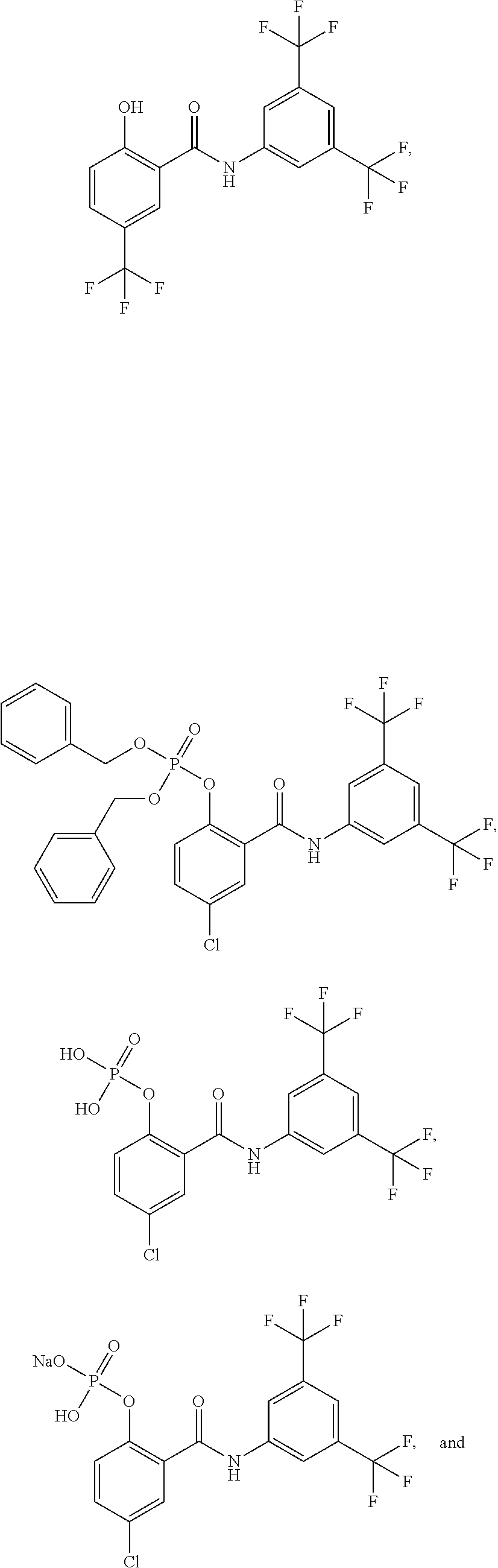


Priority Applications (1)
| Application Number | Priority Date | Filing Date | Title |
|---|---|---|---|
| US17/443,361 US11873266B2 (en) | 2012-05-08 | 2021-07-26 | Methods of treating or controlling cytotoxic cerebral edema consequent to an ischemic stroke |
Applications Claiming Priority (7)
| Application Number | Priority Date | Filing Date | Title |
|---|---|---|---|
| US201261644268P | 2012-05-08 | 2012-05-08 | |
| US201261651778P | 2012-05-25 | 2012-05-25 | |
| US201361799606P | 2013-03-15 | 2013-03-15 | |
| PCT/US2013/040194 WO2013169939A2 (en) | 2012-05-08 | 2013-05-08 | New methods |
| US201414398947A | 2014-11-04 | 2014-11-04 | |
| US15/982,644 US11084778B2 (en) | 2012-05-08 | 2018-05-17 | Methods of treating cardiac edema, neuromyelitis optica, and hyponatremia |
| US17/443,361 US11873266B2 (en) | 2012-05-08 | 2021-07-26 | Methods of treating or controlling cytotoxic cerebral edema consequent to an ischemic stroke |
Related Parent Applications (1)
| Application Number | Title | Priority Date | Filing Date |
|---|---|---|---|
| US15/982,644 Continuation US11084778B2 (en) | 2012-05-08 | 2018-05-17 | Methods of treating cardiac edema, neuromyelitis optica, and hyponatremia |
Publications (2)
| Publication Number | Publication Date |
|---|---|
| US20220081391A1 US20220081391A1 (en) | 2022-03-17 |
| US11873266B2 true US11873266B2 (en) | 2024-01-16 |
Family
ID=49551452
Family Applications (4)
| Application Number | Title | Priority Date | Filing Date |
|---|---|---|---|
| US14/398,947 Active US9994514B2 (en) | 2012-05-08 | 2013-05-08 | Methods of treating cerebral edema and spinal cord edema |
| US14/752,839 Active US9573885B2 (en) | 2012-05-08 | 2015-06-27 | Methods of treating cerebral edema |
| US15/982,644 Active US11084778B2 (en) | 2012-05-08 | 2018-05-17 | Methods of treating cardiac edema, neuromyelitis optica, and hyponatremia |
| US17/443,361 Active 2033-07-20 US11873266B2 (en) | 2012-05-08 | 2021-07-26 | Methods of treating or controlling cytotoxic cerebral edema consequent to an ischemic stroke |
Family Applications Before (3)
| Application Number | Title | Priority Date | Filing Date |
|---|---|---|---|
| US14/398,947 Active US9994514B2 (en) | 2012-05-08 | 2013-05-08 | Methods of treating cerebral edema and spinal cord edema |
| US14/752,839 Active US9573885B2 (en) | 2012-05-08 | 2015-06-27 | Methods of treating cerebral edema |
| US15/982,644 Active US11084778B2 (en) | 2012-05-08 | 2018-05-17 | Methods of treating cardiac edema, neuromyelitis optica, and hyponatremia |
Country Status (14)
| Country | Link |
|---|---|
| US (4) | US9994514B2 (en) |
| EP (1) | EP2846787B1 (en) |
| JP (2) | JP6238969B2 (en) |
| KR (1) | KR102200176B1 (en) |
| CN (2) | CN104602682B (en) |
| AU (2) | AU2013259526B2 (en) |
| BR (1) | BR112014027983B1 (en) |
| CA (1) | CA2872012C (en) |
| ES (1) | ES2699646T3 (en) |
| HK (1) | HK1208363A1 (en) |
| IL (1) | IL235512B (en) |
| MX (1) | MX363473B (en) |
| RU (1) | RU2671495C2 (en) |
| WO (1) | WO2013169939A2 (en) |
Families Citing this family (24)
| Publication number | Priority date | Publication date | Assignee | Title |
|---|---|---|---|---|
| CN104602682B (en) | 2012-05-08 | 2018-12-14 | 埃罗米克斯公司 | For treating the compound of the disease of aquaporin mediation |
| ES2834131T3 (en) * | 2013-11-06 | 2021-06-16 | Aeromics Inc | New prodrug salts |
| EP3218354A4 (en) * | 2014-11-13 | 2018-06-13 | Aeromics, Inc. | Novel methods |
| US10525021B2 (en) | 2014-11-18 | 2020-01-07 | Rutgers, The State University Of New Jersey | Mitochondrial uncouplers for treatment of metabolic diseases and cancer |
| CZ2014915A3 (en) * | 2014-12-16 | 2016-02-24 | Univerzita Karlova v Praze, Farmaceutická fakulta v Hradci Králové | Substituted derivative of phosphorus oxyacids, use thereof and pharmaceutical composition containing it |
| US10246733B2 (en) | 2015-06-04 | 2019-04-02 | Case Western University | Assay and method for quantitating carbonic anhydrase activity and assessing red blood hemolysis |
| CN105030747B (en) * | 2015-07-03 | 2017-11-17 | 厦门大学 | Niclosamidum is preparing the application in preventing and treating near-sighted medicine |
| CN109562137A (en) | 2015-09-01 | 2019-04-02 | 第波生物公司 | For treating the method and composition for reacting the related patient's condition with abnormal inflammatory |
| CN115093443A (en) | 2016-05-13 | 2022-09-23 | 埃罗米克斯公司 | Crystal |
| CN109415363B (en) | 2016-05-18 | 2021-07-20 | 罗格斯,新泽西州立大学 | Novel mitochondrial uncoupling agents for the treatment of metabolic diseases and cancer |
| WO2018053807A1 (en) * | 2016-09-23 | 2018-03-29 | 深圳市中医院 | Use of niclosamide ethanolamine salt in preparing medicine for type 1 diabetes |
| TWI631944B (en) * | 2016-10-14 | 2018-08-11 | 長庚大學 | Uses of niclosamide and derivatives thereof |
| CA3063524A1 (en) | 2017-05-19 | 2018-11-22 | Trudell Medical International | Positive expiratory pressure device |
| JP6895165B2 (en) * | 2017-06-06 | 2021-06-30 | 知和 松浦 | Biomarker containing α1-antichymotrypsin |
| WO2019067458A1 (en) * | 2017-09-26 | 2019-04-04 | Deha, Llc | Compositions and methods for channelopathy disorders-targeting fluid movement across membranes |
| CN108203719A (en) * | 2017-12-26 | 2018-06-26 | 吉林医药学院 | For treating the micromolecular compound of neuromyelitis optica and its high-throughput screening method |
| USD874064S1 (en) | 2018-05-18 | 2020-01-28 | Trudell Medical International | Mask |
| USD903097S1 (en) | 2018-05-18 | 2020-11-24 | Trudell Medical International | Mask |
| USD893806S1 (en) | 2018-11-09 | 2020-08-18 | Trudell Medical Internationl | Mask and shroud |
| USD902947S1 (en) * | 2019-03-25 | 2020-11-24 | Apple Inc. | Electronic device with graphical user interface |
| CN112107568B (en) * | 2019-06-19 | 2022-03-25 | 北京龙嘉博创医药科技有限公司 | Diaryl amide compound and application thereof |
| CN110790787B (en) * | 2019-11-12 | 2022-06-14 | 广东药科大学 | Water-soluble prodrug, preparation method thereof and application thereof as medicine |
| US10980756B1 (en) | 2020-03-16 | 2021-04-20 | First Wave Bio, Inc. | Methods of treatment |
| BR112023002129A2 (en) * | 2020-08-05 | 2023-04-18 | Jiangsu Simcere Pharm Co Ltd | PHARMACEUTICAL COMPOSITION OF AQUAPORIN INHIBITORS AND METHOD OF PREPARING THE SAME |
Citations (93)
| Publication number | Priority date | Publication date | Assignee | Title |
|---|---|---|---|---|
| US3331874A (en) | 1962-05-29 | 1967-07-18 | Herbert C Stecker | Bistrifluoromethyl anilides |
| US3332996A (en) | 1961-03-25 | 1967-07-25 | Cassella Farbwerke Mainkur Ag | Trifluoromethyl-substituted salicylanilides |
| EP0338415A2 (en) | 1988-04-19 | 1989-10-25 | Hoechst Aktiengesellschaft | Use of sulphonylureas in the treatment of oedema |
| US5137871A (en) | 1989-07-28 | 1992-08-11 | Regents Of The University Of California | Treatment to reduce edema for brain and musculature injuries |
| US5324749A (en) | 1990-04-02 | 1994-06-28 | Boehringer Mannheim Gmbh | Pharmaceutical aqueous solution of 4-[2-(benzene-sulphonylamino)-ethyl]-phenoxyacetic acid |
| US5486530A (en) | 1991-04-27 | 1996-01-23 | Boehringer Mannheim Gmbh | Use of torasemide for the treatment of brain oedemas |
| US5519035A (en) | 1993-07-02 | 1996-05-21 | Cornell Research Foundation, Inc. | Treatment of stroke or in anticipation of the occurrence of brain ischemia |
| US5741671A (en) | 1991-12-12 | 1998-04-21 | The Johns Hopkins University | Isolation cloning and expression of transmembrane water channel aquaporin 1(AQP1) |
| US5858702A (en) | 1991-12-13 | 1999-01-12 | The Johns Hopkins University | Isolation, cloning and expression of transmembrane water channel Aquaporin 5 (AQP5) |
| WO1999007382A1 (en) | 1997-08-06 | 1999-02-18 | Smithkline Beecham Corporation | Macrophage scavenger receptor antagonists for use in the treatment of cardiovascular diseases |
| US5905090A (en) | 1998-04-29 | 1999-05-18 | Italfarmaco S.P.A. | Analogues of the active metabolite of leflunomide |
| WO2001023399A1 (en) | 1999-09-30 | 2001-04-05 | Pfizer Products Inc. | Compounds for the treatment of ischemia |
| US20010046993A1 (en) | 1999-12-10 | 2001-11-29 | Takafumi Ikeda | 5-memberd heteroaryl substituted 1,4-dihydropyridine compounds as bradykinin antagonists |
| US20020061311A1 (en) | 1996-11-18 | 2002-05-23 | Susan Haas | Methods and compositions for inducing oral tolerance in mammals |
| WO2002049632A1 (en) | 2000-12-18 | 2002-06-27 | Institute Of Medicinal Molecular Design. Inc. | Inhibitors against the production and release of inflammatory cytokines |
| US6500809B1 (en) | 1999-11-12 | 2002-12-31 | Neuron Therapeutics, Inc. | Hyperoncotic artificial cerebrospinal fluid and method of treating neural tissue edema therewith |
| CA2108794C (en) | 1991-04-27 | 2003-06-24 | Tim Boelke | Use of torasemide for the treatment of brain oedemas |
| US20030215889A1 (en) | 2002-03-20 | 2003-11-20 | Simard J. Marc | Non-selective cation channel in neural cells and methods for treating brain swelling |
| WO2004006858A2 (en) | 2002-07-15 | 2004-01-22 | Myriad Genetics, Inc | Compounds, compositions, and methods employing same |
| EP1510210A1 (en) | 2002-06-05 | 2005-03-02 | Institute of Medicinal Molecular Design, Inc. | Immunity-related protein kinase inhibitors |
| EP1512397A1 (en) | 2002-06-06 | 2005-03-09 | Institute of Medicinal Molecular Design, Inc. | O-substituted hydroxyaryl derivatives |
| EP1514544A1 (en) | 2002-06-06 | 2005-03-16 | Institute of Medicinal Molecular Design, Inc. | Antiallergic |
| US20050085734A1 (en) | 2003-10-15 | 2005-04-21 | Tehrani Amir J. | Heart failure patient treatment and management device |
| EP1535609A1 (en) | 2002-06-10 | 2005-06-01 | Institute of Medicinal Molecular Design, Inc. | Nf-kb activation inhibitors |
| EP1555018A1 (en) | 2002-06-11 | 2005-07-20 | Institute of Medicinal Molecular Design, Inc. | Remedies for neurodegenerative diseases |
| US20050182012A1 (en) | 2003-12-02 | 2005-08-18 | Mcevoy Leslie M. | NF-kappaB oligonucleotide decoy molecules |
| US20060014811A1 (en) | 2002-06-10 | 2006-01-19 | Susumu Muto | Medicament for treatment of cancer |
| WO2006036278A2 (en) | 2004-09-18 | 2006-04-06 | University Of Maryland, Baltimore | THERAPEUTIC AGENTS TARGETING THE NCCa-ATP CHANNEL AND METHODS OF USE THEREOF |
| EP1649852A1 (en) | 2003-07-16 | 2006-04-26 | Institute of Medicinal Molecular Design, Inc. | Chromatosis remedies |
| US20060100257A1 (en) | 2002-06-05 | 2006-05-11 | Susumu Muto | Inhibitors against the activation of ap-1 and nfat |
| US20060111409A1 (en) | 2002-06-05 | 2006-05-25 | Susumu Muto | Medicament for treatment of diabetes |
| WO2006074127A2 (en) | 2005-01-04 | 2006-07-13 | Novartis Ag | Biomarkers for identifying efficacy of tegaserod in patients with chronic constipation |
| US20060167110A1 (en) | 1995-01-13 | 2006-07-27 | Blume Cherly D | Methods for treating cerebrovascular disease by administering desmethylselegiline |
| WO2007084464A2 (en) | 2006-01-17 | 2007-07-26 | The University Of North Carolina At Chapel Hill | Water channel blockers and methods of use thereof |
| US20070254956A1 (en) | 2003-10-29 | 2007-11-01 | Koichi Shudo | Medicament for Therapeutic and/or Preventive Treatment of Restenosis or Reocclusion After Vascular Recanalization Operation |
| US20070281978A1 (en) | 2006-06-01 | 2007-12-06 | Niigata University | Inhibitors of aquaporin 4, methods and uses thereof |
| WO2007143689A2 (en) | 2006-06-06 | 2007-12-13 | Genentech, Inc. | Compositions and methods for modulating vascular development |
| WO2008046014A1 (en) | 2006-10-12 | 2008-04-17 | Remedy Pharmaceuticals, Inc. | Treatment of alzheimer's disease using compounds that reduce the activity of non-selective ca++- activated atp-sensitive cation channels regulated by sur1 receptors |
| WO2008052190A2 (en) | 2006-10-26 | 2008-05-02 | Flynn Gary A | Aquaporin modulators and methods of using them for the treatment of edema and fluid imbalance |
| WO2008060705A2 (en) | 2006-06-06 | 2008-05-22 | Genentech, Inc. | Anti-dll4 antibodies and methods using same |
| US20080125488A1 (en) | 2003-05-01 | 2008-05-29 | Leverve Xavier M | Lactate Containing Pharmaceutical Composition and Uses Thereof |
| US20080171719A1 (en) | 2006-11-28 | 2008-07-17 | Alcon Manufacturing, Ltd. | RNAi-MEDIATED INHIBITION OF AQUAPORIN 1 FOR TREATMENT OF IOP-RELATED CONDITIONS |
| WO2008089212A1 (en) | 2007-01-16 | 2008-07-24 | Ipintl, Llc | Novel composition for treating metabolic syndrome |
| WO2008089103A2 (en) | 2007-01-12 | 2008-07-24 | University Of Maryland, Baltimore | Targeting ncca-atp channel for organ protection following ischemic episode |
| WO2008098160A1 (en) | 2007-02-09 | 2008-08-14 | University Of Maryland, Baltimore | Antagonists of a non-selective cation channel in neural cells |
| WO2008100636A2 (en) | 2007-02-17 | 2008-08-21 | President And Fellows Of Harvard College | Compositions and method for tissue preservation |
| US20080214486A1 (en) | 2006-11-28 | 2008-09-04 | Alcon Manufacturing, Ltd. | RNAi-MEDIATED INHIBITION OF AQUAPORIN 4 FOR TREATMENT OF IOP-RELATED CONDITIONS |
| WO2008133884A2 (en) | 2007-04-23 | 2008-11-06 | Combinatorx, Incorporated | Methods and compositions for the treatment of neurodegenerative disorders |
| WO2009002832A2 (en) | 2007-06-22 | 2008-12-31 | University Of Maryland, Baltimore | Inhibitors of ncca-atp channels for therapy |
| WO2009006555A2 (en) | 2007-07-02 | 2009-01-08 | Yu, Ming | Methods, composition, targets for combinational cancer treatments |
| WO2009054439A1 (en) | 2007-10-23 | 2009-04-30 | Institute Of Medicinal Molecular Design, Inc. | Pai-1 production inhibitor |
| WO2009073711A1 (en) | 2007-12-04 | 2009-06-11 | Remedy Pharmaceuticals, Inc. | Improved formulations and methods for lyophilization and lyophilates provided thereby |
| WO2009074809A1 (en) | 2007-12-13 | 2009-06-18 | University Of Dundee | Sigma ligands and ikk / nf - kb inhibitors for medical treatment |
| US20090163545A1 (en) | 2007-12-21 | 2009-06-25 | University Of Rochester | Method For Altering The Lifespan Of Eukaryotic Organisms |
| WO2009097443A2 (en) | 2008-01-29 | 2009-08-06 | Remedy Pharmaceuticals, Inc. | Liquid formulations of compounds active at sulfonylurea receptors |
| US20090239919A1 (en) | 2008-03-08 | 2009-09-24 | Richard Delarey Wood | Glutamate receptor modulators and therapeutic agents |
| US20090239868A1 (en) | 2007-10-23 | 2009-09-24 | Institute Of Medical Molecular Design, Inc. | Inhibitor of pai-1 production |
| US7601745B2 (en) | 2004-09-27 | 2009-10-13 | 4Sc Ag | Heterocyclic NF-kB inhibitors |
| WO2009139925A1 (en) | 2008-05-16 | 2009-11-19 | Panacea Pharmaceuticals, Inc. | Methods for the treatment of brain edema |
| US20100016381A1 (en) | 2005-04-28 | 2010-01-21 | Takeda Pharmaceutical Company Limited | Stable emulsion composition |
| US7671058B2 (en) | 2006-06-21 | 2010-03-02 | Institute Of Medicinal Molecular Design, Inc. | N-(3,4-disubstituted phenyl) salicylamide derivatives |
| WO2010033560A2 (en) | 2008-09-16 | 2010-03-25 | University Of Maryland, Baltimore | Sur1 inhibitors for therapy |
| WO2010048273A2 (en) | 2008-10-21 | 2010-04-29 | President And Fellows Of Harvard College | Methods and compounds for treatment of neurodegenerative disorders |
| US20100125090A1 (en) | 2003-10-08 | 2010-05-20 | Vertex Pharmaceuticals Incorporated | Modulators of ATP-Binding Cassette Transporters |
| US20100184856A1 (en) | 2009-01-12 | 2010-07-22 | Allan Sy Lau | Novel Compounds and Uses Thereof for Treating Inflammation and Modulating Immune Responses |
| US20100190796A1 (en) | 2006-11-16 | 2010-07-29 | The Regents Of The University Of California | Methods for identifying inhibitors of solute transporters |
| US7872048B2 (en) | 2004-09-18 | 2011-01-18 | University Of Maryland, Baltimore | Methods for treating spinal cord injury with a compound that inhibits a NCCa-ATP channel |
| US20110052678A1 (en) | 2010-11-05 | 2011-03-03 | Shantha Totada R | Method for treating age related macular degeneration |
| US8003610B2 (en) | 2003-02-06 | 2011-08-23 | Queen's University Of Belfast | Bradykinin B2 receptor antagonist peptide from amphibian skin |
| WO2011112791A1 (en) | 2010-03-10 | 2011-09-15 | Corthera, Inc | Modulating aquaporins with relaxin |
| WO2012012347A2 (en) | 2010-07-19 | 2012-01-26 | Remedy Pharmaceuticals, Inc. | Methods of intravenous administration of glyburide and other drugs |
| US20120039805A1 (en) | 2009-02-20 | 2012-02-16 | Pangea Biosciences, Inc. | Therapeutics And Methods For Treating Neoplastic Diseases Comprising Determining The Level Of Caveolin-1 And/Or Caveolin-2 In A Stromal Cell Sample |
| US20120183600A1 (en) | 2007-01-16 | 2012-07-19 | Chien-Hung Chen | Novel composition for treating metabolic syndrome and other conditions |
| US20120196875A1 (en) | 2010-07-28 | 2012-08-02 | Boehringer Ingelheim International Gmbh | Pharmaceutical compositions for treatment of respiratory and inflammatory diseases |
| US20120238623A1 (en) | 2011-03-14 | 2012-09-20 | Chandraratna Roshantha A | Inflammation and Autoimmune Disorder Treatment using RARa Selective Agonists |
| RU2461375C1 (en) | 2011-05-19 | 2012-09-20 | Учреждение Российской академии медицинских наук Научно-исследовательский институт онкологии Сибирского отделения Российской академии медицинских наук (НИИ онкологии СО РАМН) | Method of combination treatment of patients with locally advanced stomach cancer |
| US20120245094A1 (en) | 2008-12-22 | 2012-09-27 | Oyvind Jacobsen | Compounds, compositions and use |
| WO2012150857A1 (en) | 2011-05-02 | 2012-11-08 | Vereniging Voor Christelijk Hoger Onderwijs, Wetenschappelijk Onderzoek En Patiëntenzorg | Protection against endothelial barrier dysfunction through inhibition of the tyrosine kinase abl-related gene (arg) |
| WO2013052844A1 (en) | 2011-10-07 | 2013-04-11 | Pulmatrix, Inc. | Methods for treating and diagnosing respiratory tract infections |
| WO2013152313A1 (en) | 2012-04-05 | 2013-10-10 | The Regents Of The University Of California | Compositions and methods for treating cancer and diseases and conditions responsive to growth factor inhibition |
| WO2013165606A1 (en) | 2012-05-04 | 2013-11-07 | The United States Of America, As Represented By The Secretary, Department Of Health And Human Services | Modulators of the relaxin receptor 1 |
| US8980952B2 (en) | 2002-03-20 | 2015-03-17 | University Of Maryland, Baltimore | Methods for treating brain swelling with a compound that blocks a non-selective cation channel |
| WO2015037659A1 (en) | 2013-09-13 | 2015-03-19 | 株式会社医薬分子設計研究所 | Aqueous solution formulation, and manufacturing method for same |
| WO2015069961A1 (en) | 2013-11-06 | 2015-05-14 | Aeromics, Llc | Novel prodrug salts |
| US20150133405A1 (en) | 2012-05-08 | 2015-05-14 | Aeromics, Llc | Methods |
| US20150166589A1 (en) | 2013-12-13 | 2015-06-18 | Vertex Pharmaceuticals Incorporated | Prodrugs of pyridone amides useful as modulators of sodium channels |
| WO2016077787A1 (en) | 2014-11-13 | 2016-05-19 | Aeromics, Llc | Novel methods |
| WO2016196113A1 (en) | 2015-05-29 | 2016-12-08 | University Of Maryland, Baltimore | Methods of reducing or preventing intimal damage caused by mechanical stimulation of endothelial cells |
| WO2017062765A1 (en) | 2015-10-07 | 2017-04-13 | Remedy Pharmaceuticals, Inc. | Methods of treating injuries or conditions related to cns edema |
| WO2017197371A1 (en) | 2016-05-13 | 2017-11-16 | Aeromics, Inc. | Crystals |
| WO2018023035A1 (en) | 2016-07-29 | 2018-02-01 | Biogen Chesapeake Llc | Methods of medical treatment with sur1-trpm4 channel inhibitors |
| US10004703B2 (en) | 2006-10-12 | 2018-06-26 | Biogen Chesapeake Llc | Treatment of alzheimer's disease using compounds that reduce the activity of non-selective CA++ activated ATP-sensitive cation channels regulated by SUR1 channels |
| WO2022028459A1 (en) | 2020-08-05 | 2022-02-10 | Jiangsu Simcere Pharmaceutical Co., Ltd. | Pharmaceutical composition of aquaporin inhibitor and preparation method thereof |
Family Cites Families (2)
| Publication number | Priority date | Publication date | Assignee | Title |
|---|---|---|---|---|
| US5137817A (en) | 1990-10-05 | 1992-08-11 | Amoco Corporation | Apparatus and method for electroporation |
| KR20050019739A (en) * | 2002-06-11 | 2005-03-03 | 가부시키가이샤 이야쿠 분지 셋케이 겐쿠쇼 | Remedies for neurodegenerative diseases |
-
2013
- 2013-05-08 CN CN201380033198.7A patent/CN104602682B/en active Active
- 2013-05-08 JP JP2015511669A patent/JP6238969B2/en active Active
- 2013-05-08 RU RU2014148984A patent/RU2671495C2/en active
- 2013-05-08 MX MX2014013639A patent/MX363473B/en unknown
- 2013-05-08 CA CA2872012A patent/CA2872012C/en active Active
- 2013-05-08 BR BR112014027983-7A patent/BR112014027983B1/en active IP Right Grant
- 2013-05-08 WO PCT/US2013/040194 patent/WO2013169939A2/en active Application Filing
- 2013-05-08 AU AU2013259526A patent/AU2013259526B2/en active Active
- 2013-05-08 KR KR1020147033957A patent/KR102200176B1/en active IP Right Grant
- 2013-05-08 EP EP13788050.6A patent/EP2846787B1/en active Active
- 2013-05-08 CN CN201811415090.5A patent/CN109512805B/en active Active
- 2013-05-08 US US14/398,947 patent/US9994514B2/en active Active
- 2013-05-08 ES ES13788050T patent/ES2699646T3/en active Active
-
2014
- 2014-11-05 IL IL235512A patent/IL235512B/en active IP Right Grant
-
2015
- 2015-06-27 US US14/752,839 patent/US9573885B2/en active Active
- 2015-09-17 HK HK15109093.0A patent/HK1208363A1/en unknown
-
2017
- 2017-10-31 JP JP2017210529A patent/JP6570597B2/en active Active
-
2018
- 2018-05-14 AU AU2018203357A patent/AU2018203357C1/en active Active
- 2018-05-17 US US15/982,644 patent/US11084778B2/en active Active
-
2021
- 2021-07-26 US US17/443,361 patent/US11873266B2/en active Active
Patent Citations (144)
| Publication number | Priority date | Publication date | Assignee | Title |
|---|---|---|---|---|
| US3332996A (en) | 1961-03-25 | 1967-07-25 | Cassella Farbwerke Mainkur Ag | Trifluoromethyl-substituted salicylanilides |
| US3331874A (en) | 1962-05-29 | 1967-07-18 | Herbert C Stecker | Bistrifluoromethyl anilides |
| EP0338415A2 (en) | 1988-04-19 | 1989-10-25 | Hoechst Aktiengesellschaft | Use of sulphonylureas in the treatment of oedema |
| US5137871A (en) | 1989-07-28 | 1992-08-11 | Regents Of The University Of California | Treatment to reduce edema for brain and musculature injuries |
| US5324749A (en) | 1990-04-02 | 1994-06-28 | Boehringer Mannheim Gmbh | Pharmaceutical aqueous solution of 4-[2-(benzene-sulphonylamino)-ethyl]-phenoxyacetic acid |
| US5486530A (en) | 1991-04-27 | 1996-01-23 | Boehringer Mannheim Gmbh | Use of torasemide for the treatment of brain oedemas |
| CA2108794C (en) | 1991-04-27 | 2003-06-24 | Tim Boelke | Use of torasemide for the treatment of brain oedemas |
| US5741671A (en) | 1991-12-12 | 1998-04-21 | The Johns Hopkins University | Isolation cloning and expression of transmembrane water channel aquaporin 1(AQP1) |
| US5858702A (en) | 1991-12-13 | 1999-01-12 | The Johns Hopkins University | Isolation, cloning and expression of transmembrane water channel Aquaporin 5 (AQP5) |
| US5519035A (en) | 1993-07-02 | 1996-05-21 | Cornell Research Foundation, Inc. | Treatment of stroke or in anticipation of the occurrence of brain ischemia |
| US20060167110A1 (en) | 1995-01-13 | 2006-07-27 | Blume Cherly D | Methods for treating cerebrovascular disease by administering desmethylselegiline |
| US20020061311A1 (en) | 1996-11-18 | 2002-05-23 | Susan Haas | Methods and compositions for inducing oral tolerance in mammals |
| US6255298B1 (en) | 1997-08-06 | 2001-07-03 | Smithkline Beecham Corporation | Macrophage scavenger receptor antagonists for use in the treatment of cardiovascular diseases |
| WO1999007382A1 (en) | 1997-08-06 | 1999-02-18 | Smithkline Beecham Corporation | Macrophage scavenger receptor antagonists for use in the treatment of cardiovascular diseases |
| US5905090A (en) | 1998-04-29 | 1999-05-18 | Italfarmaco S.P.A. | Analogues of the active metabolite of leflunomide |
| WO2001023399A1 (en) | 1999-09-30 | 2001-04-05 | Pfizer Products Inc. | Compounds for the treatment of ischemia |
| US6500809B1 (en) | 1999-11-12 | 2002-12-31 | Neuron Therapeutics, Inc. | Hyperoncotic artificial cerebrospinal fluid and method of treating neural tissue edema therewith |
| US20010046993A1 (en) | 1999-12-10 | 2001-11-29 | Takafumi Ikeda | 5-memberd heteroaryl substituted 1,4-dihydropyridine compounds as bradykinin antagonists |
| WO2002049632A1 (en) | 2000-12-18 | 2002-06-27 | Institute Of Medicinal Molecular Design. Inc. | Inhibitors against the production and release of inflammatory cytokines |
| EP1352650A1 (en) | 2000-12-18 | 2003-10-15 | Institute of Medicinal Molecular Design, Inc. | Inhibitors against the production and release of inflammatory cytokines |
| US20100274051A1 (en) | 2000-12-18 | 2010-10-28 | Institute Of Medicinal Molecular Design. Inc. | Inflammatory cytokine release inhibitor |
| US20040259877A1 (en) | 2000-12-18 | 2004-12-23 | Susumu Muto | Inhibitors against the production and release of inflammatory cytokines |
| US8097759B2 (en) | 2000-12-18 | 2012-01-17 | Institute Of Medicinal Molecular Design, Inc. | Inflammatory cytokine release inhibitor |
| US8263657B2 (en) | 2000-12-18 | 2012-09-11 | Institute Of Medicinal Molecular Design, Inc. | Inhibitors against the production and release of inflammatory cytokines |
| US8980952B2 (en) | 2002-03-20 | 2015-03-17 | University Of Maryland, Baltimore | Methods for treating brain swelling with a compound that blocks a non-selective cation channel |
| US20030215889A1 (en) | 2002-03-20 | 2003-11-20 | Simard J. Marc | Non-selective cation channel in neural cells and methods for treating brain swelling |
| US20060019958A1 (en) | 2002-06-05 | 2006-01-26 | Susumu Muto | Immunity-related protein kinase inhibitors |
| EP1510210A1 (en) | 2002-06-05 | 2005-03-02 | Institute of Medicinal Molecular Design, Inc. | Immunity-related protein kinase inhibitors |
| US20060111409A1 (en) | 2002-06-05 | 2006-05-25 | Susumu Muto | Medicament for treatment of diabetes |
| US20060100257A1 (en) | 2002-06-05 | 2006-05-11 | Susumu Muto | Inhibitors against the activation of ap-1 and nfat |
| US7700655B2 (en) | 2002-06-06 | 2010-04-20 | Institute Of Medicinal Molecular Design, Inc. | Antiallergic agents |
| EP1514544A1 (en) | 2002-06-06 | 2005-03-16 | Institute of Medicinal Molecular Design, Inc. | Antiallergic |
| EP1512397A1 (en) | 2002-06-06 | 2005-03-09 | Institute of Medicinal Molecular Design, Inc. | O-substituted hydroxyaryl derivatives |
| US7626042B2 (en) | 2002-06-06 | 2009-12-01 | Institute Of Medicinal Molecular Design, Inc. | O-substituted hydroxyaryl derivatives |
| US20100113770A1 (en) | 2002-06-06 | 2010-05-06 | Institute Of Medicinal Molecular Design, Inc. | O-substituted hydroxyaryl derivatives |
| US20060094718A1 (en) | 2002-06-06 | 2006-05-04 | Susumu Muto | O-substituted hydroxyaryl derivatives |
| US20060014811A1 (en) | 2002-06-10 | 2006-01-19 | Susumu Muto | Medicament for treatment of cancer |
| EP1535609A1 (en) | 2002-06-10 | 2005-06-01 | Institute of Medicinal Molecular Design, Inc. | Nf-kb activation inhibitors |
| US20080234233A1 (en) | 2002-06-11 | 2008-09-25 | Institute Of Medicinal Molecular Design Inc. | Medicament for treatment of neurodegenerative diseases |
| EP1555018A1 (en) | 2002-06-11 | 2005-07-20 | Institute of Medicinal Molecular Design, Inc. | Remedies for neurodegenerative diseases |
| US20060035944A1 (en) | 2002-06-11 | 2006-02-16 | Susumu Muto | Remedies for neurodegenerative diseases |
| US20050187300A1 (en) | 2002-07-15 | 2005-08-25 | Myriad Genetics, Incorporated | Compounds, compositions, and methods employing same |
| WO2004006858A2 (en) | 2002-07-15 | 2004-01-22 | Myriad Genetics, Inc | Compounds, compositions, and methods employing same |
| US8003610B2 (en) | 2003-02-06 | 2011-08-23 | Queen's University Of Belfast | Bradykinin B2 receptor antagonist peptide from amphibian skin |
| US20080125488A1 (en) | 2003-05-01 | 2008-05-29 | Leverve Xavier M | Lactate Containing Pharmaceutical Composition and Uses Thereof |
| EP1649852A1 (en) | 2003-07-16 | 2006-04-26 | Institute of Medicinal Molecular Design, Inc. | Chromatosis remedies |
| US20100125090A1 (en) | 2003-10-08 | 2010-05-20 | Vertex Pharmaceuticals Incorporated | Modulators of ATP-Binding Cassette Transporters |
| US20050085734A1 (en) | 2003-10-15 | 2005-04-21 | Tehrani Amir J. | Heart failure patient treatment and management device |
| US20070254956A1 (en) | 2003-10-29 | 2007-11-01 | Koichi Shudo | Medicament for Therapeutic and/or Preventive Treatment of Restenosis or Reocclusion After Vascular Recanalization Operation |
| US20050182012A1 (en) | 2003-12-02 | 2005-08-18 | Mcevoy Leslie M. | NF-kappaB oligonucleotide decoy molecules |
| US7872048B2 (en) | 2004-09-18 | 2011-01-18 | University Of Maryland, Baltimore | Methods for treating spinal cord injury with a compound that inhibits a NCCa-ATP channel |
| WO2006036278A2 (en) | 2004-09-18 | 2006-04-06 | University Of Maryland, Baltimore | THERAPEUTIC AGENTS TARGETING THE NCCa-ATP CHANNEL AND METHODS OF USE THEREOF |
| US7601745B2 (en) | 2004-09-27 | 2009-10-13 | 4Sc Ag | Heterocyclic NF-kB inhibitors |
| US20090118350A1 (en) | 2005-01-04 | 2009-05-07 | Mclean Leeanne | Biomarkers for Identifying Efficacy of Tegaserod in Patients with Chronic Constipation |
| WO2006074127A2 (en) | 2005-01-04 | 2006-07-13 | Novartis Ag | Biomarkers for identifying efficacy of tegaserod in patients with chronic constipation |
| US20100016381A1 (en) | 2005-04-28 | 2010-01-21 | Takeda Pharmaceutical Company Limited | Stable emulsion composition |
| WO2007084464A2 (en) | 2006-01-17 | 2007-07-26 | The University Of North Carolina At Chapel Hill | Water channel blockers and methods of use thereof |
| US7659312B2 (en) | 2006-06-01 | 2010-02-09 | Niigata University | Inhibitors of Aquaporin 4, methods and uses thereof |
| US20070281978A1 (en) | 2006-06-01 | 2007-12-06 | Niigata University | Inhibitors of aquaporin 4, methods and uses thereof |
| WO2008060705A2 (en) | 2006-06-06 | 2008-05-22 | Genentech, Inc. | Anti-dll4 antibodies and methods using same |
| WO2007143689A2 (en) | 2006-06-06 | 2007-12-13 | Genentech, Inc. | Compositions and methods for modulating vascular development |
| US7671058B2 (en) | 2006-06-21 | 2010-03-02 | Institute Of Medicinal Molecular Design, Inc. | N-(3,4-disubstituted phenyl) salicylamide derivatives |
| US10004703B2 (en) | 2006-10-12 | 2018-06-26 | Biogen Chesapeake Llc | Treatment of alzheimer's disease using compounds that reduce the activity of non-selective CA++ activated ATP-sensitive cation channels regulated by SUR1 channels |
| WO2008046014A1 (en) | 2006-10-12 | 2008-04-17 | Remedy Pharmaceuticals, Inc. | Treatment of alzheimer's disease using compounds that reduce the activity of non-selective ca++- activated atp-sensitive cation channels regulated by sur1 receptors |
| US7906555B2 (en) | 2006-10-26 | 2011-03-15 | Arizona Board Of Regents On Behalf Of The University Of Arizona | Aquaporin modulators and methods of using them for the treatment of edema and fluid imbalance |
| US20110172195A1 (en) | 2006-10-26 | 2011-07-14 | Arizona Board Of Reg. On Behalf Of The Univ. Of Az | Aquaporin modulators and methods of using them for the treatment of edema and fluid imbalance |
| WO2008052190A2 (en) | 2006-10-26 | 2008-05-02 | Flynn Gary A | Aquaporin modulators and methods of using them for the treatment of edema and fluid imbalance |
| US20080221169A1 (en) | 2006-10-26 | 2008-09-11 | Arizona Board Of Reg. On Behalf Of The Univ. Of Az | Aquaporin modulators and methods of using them for the treatment of edema and fluid imbalance |
| US20100190796A1 (en) | 2006-11-16 | 2010-07-29 | The Regents Of The University Of California | Methods for identifying inhibitors of solute transporters |
| US20080214486A1 (en) | 2006-11-28 | 2008-09-04 | Alcon Manufacturing, Ltd. | RNAi-MEDIATED INHIBITION OF AQUAPORIN 4 FOR TREATMENT OF IOP-RELATED CONDITIONS |
| US20080171719A1 (en) | 2006-11-28 | 2008-07-17 | Alcon Manufacturing, Ltd. | RNAi-MEDIATED INHIBITION OF AQUAPORIN 1 FOR TREATMENT OF IOP-RELATED CONDITIONS |
| WO2008089103A2 (en) | 2007-01-12 | 2008-07-24 | University Of Maryland, Baltimore | Targeting ncca-atp channel for organ protection following ischemic episode |
| US20120183600A1 (en) | 2007-01-16 | 2012-07-19 | Chien-Hung Chen | Novel composition for treating metabolic syndrome and other conditions |
| US20080176822A1 (en) | 2007-01-16 | 2008-07-24 | Chien-Hung Chen | Novel composition for treating metabolic syndrome |
| WO2008089212A1 (en) | 2007-01-16 | 2008-07-24 | Ipintl, Llc | Novel composition for treating metabolic syndrome |
| WO2008098160A1 (en) | 2007-02-09 | 2008-08-14 | University Of Maryland, Baltimore | Antagonists of a non-selective cation channel in neural cells |
| WO2008100636A2 (en) | 2007-02-17 | 2008-08-21 | President And Fellows Of Harvard College | Compositions and method for tissue preservation |
| US20120282591A1 (en) | 2007-02-17 | 2012-11-08 | United States Government As Represented By The Department Of Veterans Affairs | Compositions and Methods for Tissue Preservation |
| WO2008133884A2 (en) | 2007-04-23 | 2008-11-06 | Combinatorx, Incorporated | Methods and compositions for the treatment of neurodegenerative disorders |
| WO2009002832A2 (en) | 2007-06-22 | 2008-12-31 | University Of Maryland, Baltimore | Inhibitors of ncca-atp channels for therapy |
| WO2009006555A2 (en) | 2007-07-02 | 2009-01-08 | Yu, Ming | Methods, composition, targets for combinational cancer treatments |
| US20090239868A1 (en) | 2007-10-23 | 2009-09-24 | Institute Of Medical Molecular Design, Inc. | Inhibitor of pai-1 production |
| WO2009054439A1 (en) | 2007-10-23 | 2009-04-30 | Institute Of Medicinal Molecular Design, Inc. | Pai-1 production inhibitor |
| EP2201946A1 (en) | 2007-10-23 | 2010-06-30 | Institute of Medicinal Molecular Design, Inc. | Pai-1 production inhibitor |
| US8277845B2 (en) | 2007-12-04 | 2012-10-02 | Remedy Pharmaceuticals, Inc. | Formulations and methods for lyophilization and lyophilates provided thereby |
| WO2009073711A1 (en) | 2007-12-04 | 2009-06-11 | Remedy Pharmaceuticals, Inc. | Improved formulations and methods for lyophilization and lyophilates provided thereby |
| WO2009074809A1 (en) | 2007-12-13 | 2009-06-18 | University Of Dundee | Sigma ligands and ikk / nf - kb inhibitors for medical treatment |
| US20090163545A1 (en) | 2007-12-21 | 2009-06-25 | University Of Rochester | Method For Altering The Lifespan Of Eukaryotic Organisms |
| US20170216321A1 (en) | 2008-01-29 | 2017-08-03 | Sven Jacobson | Liquid formulations of compounds active at sulfonylurea receptors |
| WO2009097443A2 (en) | 2008-01-29 | 2009-08-06 | Remedy Pharmaceuticals, Inc. | Liquid formulations of compounds active at sulfonylurea receptors |
| US20090239919A1 (en) | 2008-03-08 | 2009-09-24 | Richard Delarey Wood | Glutamate receptor modulators and therapeutic agents |
| WO2009139925A1 (en) | 2008-05-16 | 2009-11-19 | Panacea Pharmaceuticals, Inc. | Methods for the treatment of brain edema |
| WO2010033560A2 (en) | 2008-09-16 | 2010-03-25 | University Of Maryland, Baltimore | Sur1 inhibitors for therapy |
| WO2010048273A2 (en) | 2008-10-21 | 2010-04-29 | President And Fellows Of Harvard College | Methods and compounds for treatment of neurodegenerative disorders |
| US20120010178A1 (en) | 2008-10-21 | 2012-01-12 | President And Fellows Of Harvard College | Methods and compounds for treatment of neurodegenerative disorders |
| US20120245094A1 (en) | 2008-12-22 | 2012-09-27 | Oyvind Jacobsen | Compounds, compositions and use |
| US20100184856A1 (en) | 2009-01-12 | 2010-07-22 | Allan Sy Lau | Novel Compounds and Uses Thereof for Treating Inflammation and Modulating Immune Responses |
| US20120039805A1 (en) | 2009-02-20 | 2012-02-16 | Pangea Biosciences, Inc. | Therapeutics And Methods For Treating Neoplastic Diseases Comprising Determining The Level Of Caveolin-1 And/Or Caveolin-2 In A Stromal Cell Sample |
| WO2010101648A1 (en) | 2009-03-06 | 2010-09-10 | Wood Richard D | Glutamate receptor modulators and therapeutic agents |
| WO2011112791A1 (en) | 2010-03-10 | 2011-09-15 | Corthera, Inc | Modulating aquaporins with relaxin |
| US8946293B2 (en) | 2010-07-19 | 2015-02-03 | Remedy Pharmaceuticals, Inc. | Methods of intravenous administration of glyburide and other drugs |
| WO2012012347A2 (en) | 2010-07-19 | 2012-01-26 | Remedy Pharmaceuticals, Inc. | Methods of intravenous administration of glyburide and other drugs |
| US20120196875A1 (en) | 2010-07-28 | 2012-08-02 | Boehringer Ingelheim International Gmbh | Pharmaceutical compositions for treatment of respiratory and inflammatory diseases |
| US20110052678A1 (en) | 2010-11-05 | 2011-03-03 | Shantha Totada R | Method for treating age related macular degeneration |
| US20120238623A1 (en) | 2011-03-14 | 2012-09-20 | Chandraratna Roshantha A | Inflammation and Autoimmune Disorder Treatment using RARa Selective Agonists |
| WO2012150857A1 (en) | 2011-05-02 | 2012-11-08 | Vereniging Voor Christelijk Hoger Onderwijs, Wetenschappelijk Onderzoek En Patiëntenzorg | Protection against endothelial barrier dysfunction through inhibition of the tyrosine kinase abl-related gene (arg) |
| RU2461375C1 (en) | 2011-05-19 | 2012-09-20 | Учреждение Российской академии медицинских наук Научно-исследовательский институт онкологии Сибирского отделения Российской академии медицинских наук (НИИ онкологии СО РАМН) | Method of combination treatment of patients with locally advanced stomach cancer |
| WO2013052844A1 (en) | 2011-10-07 | 2013-04-11 | Pulmatrix, Inc. | Methods for treating and diagnosing respiratory tract infections |
| WO2013152313A1 (en) | 2012-04-05 | 2013-10-10 | The Regents Of The University Of California | Compositions and methods for treating cancer and diseases and conditions responsive to growth factor inhibition |
| WO2013165606A1 (en) | 2012-05-04 | 2013-11-07 | The United States Of America, As Represented By The Secretary, Department Of Health And Human Services | Modulators of the relaxin receptor 1 |
| US11084778B2 (en) | 2012-05-08 | 2021-08-10 | Aeromics, Inc. | Methods of treating cardiac edema, neuromyelitis optica, and hyponatremia |
| US20150133405A1 (en) | 2012-05-08 | 2015-05-14 | Aeromics, Llc | Methods |
| US9573885B2 (en) | 2012-05-08 | 2017-02-21 | Aeromics, Inc. | Methods of treating cerebral edema |
| US9994514B2 (en) | 2012-05-08 | 2018-06-12 | Aeromics, Inc. | Methods of treating cerebral edema and spinal cord edema |
| US20150342967A1 (en) | 2012-05-08 | 2015-12-03 | Aeromics, Llc | Methods |
| WO2015037659A1 (en) | 2013-09-13 | 2015-03-19 | 株式会社医薬分子設計研究所 | Aqueous solution formulation, and manufacturing method for same |
| US20160220680A1 (en) | 2013-09-13 | 2016-08-04 | Akiko Itai | Aqueous solution formulation and method for manufacturing same |
| WO2015069948A1 (en) | 2013-11-06 | 2015-05-14 | Aeromics, Llc | Novel methods |
| US11071744B2 (en) | 2013-11-06 | 2021-07-27 | Aeromics, Inc. | Prodrug salts |
| US20160346302A1 (en) | 2013-11-06 | 2016-12-01 | Aeromics, Inc. | Novel methods |
| US20220143048A1 (en) | 2013-11-06 | 2022-05-12 | Aeromics, Inc. | Novel prodrug salts |
| US20160264604A1 (en) | 2013-11-06 | 2016-09-15 | Aeromics, Inc. | Novel prodrug salts |
| US20210275549A1 (en) | 2013-11-06 | 2021-09-09 | Aeromics, Inc. | Novel formulations |
| WO2015069961A1 (en) | 2013-11-06 | 2015-05-14 | Aeromics, Llc | Novel prodrug salts |
| WO2015069956A2 (en) | 2013-11-06 | 2015-05-14 | Aeromics, Llc | Novel formulations |
| US9827253B2 (en) | 2013-11-06 | 2017-11-28 | Aeromics, Inc. | Prodrug salts |
| US10894055B2 (en) | 2013-11-06 | 2021-01-19 | Aeromics, Inc. | Pharmaceutical compositions, methods of making pharmaceutical compositions, and kits comprising 2-{[3,5-bis(trifluoromethyl)phenyl]carbamoyl}4-chlorophenyl dihydrogen phosphate |
| US20190307779A1 (en) | 2013-11-06 | 2019-10-10 | Aeromics, Inc. | Novel formulations |
| US9949991B2 (en) | 2013-11-06 | 2018-04-24 | Aeromics, Inc. | Methods of treating aquaporin-mediated conditions |
| US10258636B2 (en) | 2013-11-06 | 2019-04-16 | Aeromics, Inc. | Prodrug salts |
| US20160279155A1 (en) | 2013-11-06 | 2016-09-29 | Aeromics, Inc. | Novel formulations |
| US20180169118A1 (en) | 2013-11-06 | 2018-06-21 | Aeromics, Inc. | Novel prodrug salts |
| US20150166589A1 (en) | 2013-12-13 | 2015-06-18 | Vertex Pharmaceuticals Incorporated | Prodrugs of pyridone amides useful as modulators of sodium channels |
| US20180042873A1 (en) | 2014-11-13 | 2018-02-15 | Aeromics, Inc. | Novel methods |
| WO2016077787A1 (en) | 2014-11-13 | 2016-05-19 | Aeromics, Llc | Novel methods |
| US20180155727A1 (en) | 2015-05-29 | 2018-06-07 | University Of Maryland, Baltimore | Methods of reducing or preventing intimal damage caused by mechanical stimulation of endothelial cells |
| WO2016196113A1 (en) | 2015-05-29 | 2016-12-08 | University Of Maryland, Baltimore | Methods of reducing or preventing intimal damage caused by mechanical stimulation of endothelial cells |
| WO2017062765A1 (en) | 2015-10-07 | 2017-04-13 | Remedy Pharmaceuticals, Inc. | Methods of treating injuries or conditions related to cns edema |
| US20190185496A1 (en) | 2016-05-13 | 2019-06-20 | Aeromics, Inc. | Crystals |
| WO2017197371A1 (en) | 2016-05-13 | 2017-11-16 | Aeromics, Inc. | Crystals |
| US11117909B2 (en) | 2016-05-13 | 2021-09-14 | Aeromics, Inc. | Crystals |
| US20220204536A1 (en) | 2016-05-13 | 2022-06-30 | Aeromics, Inc. | Crystals |
| WO2018023035A1 (en) | 2016-07-29 | 2018-02-01 | Biogen Chesapeake Llc | Methods of medical treatment with sur1-trpm4 channel inhibitors |
| WO2022028459A1 (en) | 2020-08-05 | 2022-02-10 | Jiangsu Simcere Pharmaceutical Co., Ltd. | Pharmaceutical composition of aquaporin inhibitor and preparation method thereof |
Non-Patent Citations (275)
| Title |
|---|
| Abir-Awan, M. et al., "Inhibitors of Mammalian Aquaporin Water Channels," International Journal of Molecular Sciences, 2019, 20, 1589, 22 pages, doi:10.3390/ijms20071589. |
| Advisory Action dated Dec. 16, 2016, issued in U.S. Appl. No. 14/398,947, 3 pages. |
| Aeromics, "Aeromics and Simcere Announce Collaboration and License Agreement for AER-271 in Greater China," dated Nov. 2019, 4 pages, retrieved on Aug. 5, 2020, from: https://www.aeromics.com/pr-110519-simcere. |
| Aeromics, "Aeromics Initiates Phase 1 Clinical Trial of CNS Edema Inhibitor AER-271 in Healthy Human Volunteers," dated Jul. 9, 2018, 4 pages, retrieved on Aug. 5, 2020, from: https://www.aeromics.com/press-releases/aeromics-inc-initiates-phase-1-trial-of-aer-271-in-healthy-human-volunteers. |
| Aeromics, "Aeromics, Inc. Appoints Joseph Schindler, M.D. as Chief Medical Officer," dated Jan. 2020, 1 page, retrieved on Sep. 23, 2020, from: https://www.aeromics.com/pr-010720-schindler. |
| Aeromics, "Aeromics, Inc. Appoints Nobel Laureate Peter Agre to Its Board of Directors," dated Sep. 2019, 4 pages, retrieved on Aug. 5, 2020, from: https://www.aeromics.com/pr-090919-p-agre-director. |
| Aeromics, "Aeromics, Inc. Appoints Pharmaceutical and Biotech Veterans Mark Day, Ph.D. and Thomas Zindrick, J.D. to Its Board of Directors," dated Aug. 16, 2018, 7 pages, retrieved on Aug. 5, 2020, from: https://www.aeromics.com/press-releases/aeromics-inc-appoints-pharmaceutical-and-biotech-veterans-mark-day-phd-and-thomas-zindrick-jd-to-its-board-of-directors. |
| Aeromics, Anti-Edema Therapy for Patients Affected by Disabling and Life-Threatening Severe Ischemic Stroke, 7 pages, retrieved on Aug. 5, 2020, from: https://www.aeromics.com/. |
| Alexander, J. et al., "Administration of the Soluble Complement Inhibitor, Crry-Ig, Reduces Inflammation and Aquaporin 4 Expression in Lupus Cerebritis," Biochimica et Biophysica Acta, 2003, 1639, 169-176. |
| Amiry-Moghaddam, M. et al. "Anchoring of Aquaporin-4 in Brain: Molecular Mechanisms and Implications for the Physiology and Pathophysiology of Water Transport," Neuroscience, 2004, 129, 999-1010. |
| Amiry-Moghaddam, M. et al., "Alpha-Syntrophin Deletion Removes the Perivascular But Not Endothelial Pool of Aquaporin-4 at the Blood-Brain Barrier and Delays the Development of Brain Edema in an Experimental Model of Acute Hyponatremia," The FASEB Journal, 2004, 18, 542-544. |
| Aoki, K. et al., "Enhanced Expression of Aquaporin 4 in Human Brain with Infarction," Acta Neuropathologica, 2003, 106, 121-124. |
| Aoki-Yoshino, K. et al., "Enhanced Expression of Aquaporin 4 in Human Brain with Inflammatory Diseases," Acta Neuropathologica, 2005, 110, 281-288. |
| Ayasoufi, K. et al., "Aquaporin 4 Blockade Improves Survival of Murine Heart Allografts Subjected to Prolonged Cold Ischemia," American Journal of Transplantation, 2018, 18, 1238-1246. |
| Banamine (Flunixin Meglumine), retrieved from https://web.archive.org/web/20110711140225/http://www.banamine.com/research/FlunixinMeglumine.asp, 1 page, face of article states: Jul. 11, 2011. |
| Bao, W. et al., "Selective mGluR5 Receptor Antagonist or Agonist Provides Neuroprotection in a Rat Model of Focal Cerebral Ischemia," Brain Research, 2001, 922, 173-179. |
| Baraldo, M. et al., "Steroid-free and Steroid Withdrawal Protocols in Heart Transplantation: The Review of Literature," Transplant International, 2014, 515-529. |
| Bardutzky, J. et al., "Antiedema Therapy in Ischemic Stroke," Stroke, 2007, 38, 3084-3094. |
| Beeton, C. et al., "Induction and Monitoring of Active Delayed Type Hypersensitivity (DTH) in Rats," JoVe. 6. http://www.jove.com/index/Details.stp?ID=237, doi:10.3791/237, copyright 2007, 2 pages. |
| Benga, O. et al., "Brain Water Channel Proteins in Health and Disease," Molecular Aspects of Medicine, 2012, 33, 562-578. |
| Bereczki, D. et al., "Cochrane Report: A Systematic Review of Mannitol Therapy for Acute Ischemic Stroke and Cerebral Parenchymal Hemorrhage," Stroke, 2000, 31, 2719-2722. |
| Bhattacharyya, S. et al., "Specific Effects of BCL10 Serine Mutations on Phosphory lations in Canonical and Noncanonical Pathways of NF-κB Activation Following Carrageenan," The American Journal of Physiology—Gastrointesintal and Liver Physiology, 2011, 301, G475-G486. |
| Binder, D. et al., "Aquaporin-4 and Epilepsy," GLIA, 2012, 60, 1203-1214. |
| Binder, D. et al., "Increased Seizure Duration and Slowed Potassium Kinetics in Mice Lacking Aquaporin-4 Water Channels," GLIA, 2006, 53, 631-636. |
| Binder, D. et al., "Increased Seizure Threshold in Mice Lacking Aquaporin-4 Water Channels," NeuroReport, 2004, 15 (2), 259-262. |
| Bloch, O. et al., "Aquaporin-4 Gene Deletion in Mice Increases Focal Edema Associated with Staphylococcal Brain Abscess," Journal of Neurochemistry, 2005, 95, 254-262. |
| Brown, L. et al., "In Vitro and In Vivo Hydrolysis of Salicy lanilide N-Methylcarbamate and 4-Bipheny N-Methylcarbamate," Journal of Pharmaceutical Sciences, 1972, 61 (6), 858-860. |
| Caraci, F. et al., "Metabotropic Glutamate Receptors in Neurodegeneration/Neuroprotection: Still a Hot Topic?," Neurochemistry International, 2012, 61, 559-565. |
| Cernak, I., "Animal Models of Head Trauma," NeuroRx, 2005, 2, 410-422. |
| Chen, C. et al., "Animal Models of Nervous System Diseases," Jilin Science and Technology Press, 2007, pp. 252-254. |
| Chiamulera, C. et al., "Activation of Metabotropic Receptors has a Neuroprotective Effect in a Rodent Model of Focal Ischaemia," European Journal of Pharmacology, 1992, 216, 335-336. |
| Colucci, M. et al., "In Vitro Clot Lysis as a Potential Indicator of Thrombus Resistance to Fibrinolysis—Study in Healthy Subjects and Correlation with Blood Fibrinolytic Parameters," Thrombosis and Haemostasis, 1997, 77 (4), 725-729. |
| Cooper, G. et al., "Transport of Volatile Solutes Through AQP1," Journal of Physiology, 2002, 542.1, 17-29. |
| Costanzo, M., "The International Society of Heart and Lung Transplantation Guidelines for the Care of Heart Transplant Recipients," The Journal of Heart and Lung Transplantation, 2010, 29 (8), 914-956. |
| CRASH Trial, "Effect of Intravenous Corticosteroids on Death within 14 Days in 10008 Adults with Clinically Significant Head Injury (MRC CRASH Trial): Randomised Placebo Controlled Trial," Lancet, 2004, 264, 1321-1328. |
| Cummins, E. et al., "Prolyl Hydroxylase-1 Negatively Regulates IκB Kinase-β, Giving Insight into Hypoxia-induced NFκB Activity," Proceedings of the National Academy of Sciences, 2006, 103 (48), 18154-18159. |
| Da, T. et al., "Aquaporin-4 Gene Disruption in Mice Protects Against Impaired Retinal Function and Cell Death after Ischemia," Investigative Ophthalmology & Visual Science, 2004, 45 (12), 4477-4483. |
| Davis, M. et al., "State of the Art: Cardiac Transplantation," Trends in Cardiovascular Medicine, 2014, 24, 341-349. |
| Dearden, N. et al., "Effect of High-Dose Dexamethasone on Outcome from Severe Head Injury," Journal of Neurosurgery, 1986, 64, 81-88. |
| Di Renzo, G. et al., "Why have Ionotropic and Metabotropic Glutamate Antagonists Failed in Stroke Therapy?," in New Strategies in Stroke Intervention, L. Annunziato, Ed., Humana Press, 2009, pp. 13-25. |
| Dibas, A. et al., "Changes in Ocular Aquaporin-4 (AQP4) Expression Following Retinal Injury," Molecular Vision, 2008, 14, 1770-1783. |
| Ding, F., Neurobiology, Science Press, 2007, pp. 421-423. |
| Ding, T. et al., "Knockdown a Water Channel Protein, Aquaporin-4 Induced Glioblastoma Cell Apoptosis," PLoS ONE, 2013, 8 (8), e66751, 9 pages, doi: 10.1371/journal.pone.0066751. |
| Dixon, C. et al., "A Controlled Cortical Impact Model of Traumatic Brain Injury in the Rat," Journal of Neuroscience Methods, 1991, 39, 253-262. |
| Dudek, F. et al., "Regulation of Brain Water: Is there a Role for Aquaporins in Epilepsy?," Epilepsy Currents, 2005, 5 (3), 104-106, retrieved on Jul. 22, 2015, from http://www.ncbi.nlm.nih.gov/pmc/articles/PMC1198631/. |
| Dvirnik, N. et al., "Steroids in Cardiac Surgery: A Systematic Review and Meta-analysis," British Journal of Anaesthesia, 2018, 120 (4), 657-667. |
| Eid, T. et al., "Loss of Perivascular Aquaporin 4 May Underlie Deficient Water and K+ Homeostasis in the Human Epileptogenic Hippocampus," Proceedings of the National Academy of Sciences, 2005, 102 (4), 1193-1198. |
| Eisenberg, H. et al., "High-Dose Barbiturate Control of Elevated Intracranial Pressure in Patients with Severe Head Injury," Journal of Neurosurgery, 1988, 69, 15-23. |
| Elati, C. et al., "Novel Synthesis of Fosphenytoin: Anti-Convulsant Prodrug," Synthetic Communications, 2008, 38, 2950-2957. |
| Engel, O. et al., "Modeling Stroke in Mice—Middle Cerebral Artery Occlusion with the Filament Model," Journal of Visualized Experiments, 2011, 47, e2423, 5 pages, doi: 10.3791/2423. |
| English-language translation of Abaturov, A. et al., "Molecular Mechanisms of Nonspecific Protection of the Respiratory Tract (Continuation, beginning in No. 3, 2006) 2. Pathophysiological Significance of Transcription Factors NF-KB," Zhurnal "Zdorovie Rebenka" (Journal "Child's Health"), 2007, 1 (4), 14 pages. |
| English-language translation of Nimer, S. et al., "Edema as a Basic Response of the Vascular Component of Brain Perfusion Systems in Traumatic Brain Injury," Problems of Health and Ecology, 2007, 10 pages. |
| Esteva-Font, C. et al., "Experimental Evaluation of Proposed Small-Molecule Inhibitors of Water Channel Aquaporin-1," Molecular Pharmacology, 2016, 89, 686-693. |
| Farinas, J. et al., "Plasma Membrane Water Permeability of Cultured Cells and Epithelia Measured by Light Microscopy with Spatial Filtering," The Journal of General Physiology, 1997, 110, 283-296. |
| Farr, G. et al., "Aquaporin-4 Inhibitor AER-270 and Its Prodrug AER-271 Reduce Cerebral Edema and Improve Outcomes in Two Models of CNS Injury," Abstract M1905, Annals of Neurology, 2014, 76 (Supplement 18), S126-S127. |
| Farr, G. et al., "Functionalized Phenylbenzamides Inhibit Aquaporin-4 Reducing Cerebral Edema and Improving Outcome in Two Models of CNS Injury," Neuroscience, 2019, accepted manuscript, https://doi.org/10.1016/j.neuroscience.2019.01.034, 46 pages. |
| Farr, G. et al., "Phenylbenzamide Derivatives AER-270/271 Inhibit Aquaporin-4 Reducing Cerebral Edema and Improving Outcome in Two Models of CNS Injury," Poster presented at 2014 American Neurological Association Annual Meeting, 1 page. |
| Fei, Z. et al., "Metabotropic Glutamate Receptor Antagonists and Agonists: Potential Neuroprotectors in Diffuse Brain Injury," Journal of Clinical Neuroscience, 2006, 13, 1023-1027. |
| Final Office Action dated Aug. 30, 2016, issued in U.S. Appl. No. 14/752,839, 17 pages. |
| Final Office Action dated Sep. 20, 2016, issued in U.S. Appl. No. 14/398,947, 41 pages. |
| Fischer, U. et al., "The Antioxidant N-Acetylcysteine Preserves Myocardial Function and Diminishes Oxidative Stress After Cardioplegic Arrest," The Journal of Thoracic and Cardiovascular Surgery, 2003, 126 (5), 1483-1488. |
| Fomovska, A. et al., "Salicylanilide Inhibitors of Toxoplasma gondii," NIH Public Access, Author Manuscript, available in PMC 2013, 40 pages, face of article states: Published in final edited form as: J Med Chem. Oct. 1, 20121; 55(19): 8375-8391. doi: 10.1021/jm3007596. |
| Frigeri, A et al., "Immunolocalization of the Mercurial—Insensitive Water Channel and Glycerol Intrinsic Protein in Epithelial Cell Plasma Membranes," Proceedings of the National Academy of Sciences, 1995, 92, 4328-4331. |
| Frigeri, A. et al., "Localization of MIWC and GLIP Water Channel Homologs in Neuromuscular, Epithelial and Glandular Tissues," Journal of Cell Science, 1995, 108, 2993-3002. |
| Fukuda, A. et al., "Aquaporin 4: A Player in Cerebral Edema and Neuroinflammation," Journal of Neuroinflammation, 2012, 9:279, 9 pages, doi: 10.1186/1742-2094-9-279. |
| Gerber, J. et al., "Mechanisms of Injury in Bacterial Meningitis," Current Opinion in Neurology, 2010, 23, 312-318. |
| Gomez-Cabrero, A et al., "IMD-0354 Targets Breast Cancer Stem Cells: A Novel Approach for an Adjuvant to Chemotherapy to Prevent Multidrug Resistance in a Murine Model," PLOS One, 2013, 8 (8), e73607, 14 pages, doi:10.1371/journal.pone.0073607. |
| Gotoh, O. et al., "Ischemic Brain Edema Following Occlusion of the Middle Cerebral Artery in the Rat. I: The Time Courses of the Brain Water, Sodium and Potassium Contents and Blood Brain Barrier Permeability to 125I-Albumin," Stroke, 1985, 16 (1), 101-109. |
| Gullans, S., "Control of Brain vol. During Hyperosmolar and Hypoosmolar Conditions," Annual Review of Medicine, 1993, 44, 289-301. |
| Gunnarson, E. et al., "Erythropoietin Modulation of Astrocyte Water Permeability as a Component of Neuroprotection," Proceedings of the National Academy of Sciences, 2009, 106 (5), 1602-1607. |
| Gunnarson, E. et al., "Identification of a Molecular Target for Glutamate Regulation of Astrocyte Water Permeability," Glia, 2008, 56, 587-596. |
| Guo, Q. et al., "Progesterone Administration Modulates AQP4 Expression and Edema After Traumatic Brain Injury in Male Rats," Experimental Neurology, 2006, 198, 469-478. |
| Hall, G. et al., "Inhibitor-κB Kinase-β Regulates LPS-Induced TNF-α Production in Cardiac Myocytes Through Modulation of NF-κB p65 Subunit Phosphorylation," American Journal of Heart Physiology—Heart and Circulatory Physiology, 2005, 289, H2103-H2111. |
| Haller, M. et al., "Steroid Avoidance or Withdrawal for Kidney Transplant Recipients (Review)," Cochrane Database of Systematic Reviews, 2016, Issue 8, Art. No. CD005632, Doi: 10.1002/14651858.CD005632.pub3, 167 pages. |
| Himadri, P. et al., "Role of Oxidative Stress and Inflammation in Hypoxia-Induced Cerebral Edema: A Molecular Approach," High Altitude Medicine & Biology, 2010, 11 (3), 231-244. |
| Hiroaki, Y. et al., "Implications of the Aquaporin-4 Structure on Array Formation and Cell Adhesion," Journal of Molecular Biology, 2006, 355, 628-639. |
| Ho, J. et al., "Crystal Structure of Human Aquaporin 4 at 1.8 Å and Its Mechanism of Conductance," Proceedings of the National Academy of Sciences, 2009, 106 (18), 7437-7442. |
| Hsu, M. et al., "Potential Role of the Glial Water Channel Aquaporin-4 in Epilepsy," Neuron Glia Biology, 2008, doi: 10.1017/S1740925X08000112, 11 pages. |
| Huang, Y. et al., "The Dual Role of AQP4 in Cytotoxic and Vasogenic Edema Following Spinal Cord Contusion and Its Possible Association with Energy Metabolism via COX5A," Frontiers in Neuroscience, 2019, 13:584, 15 pages, doi:10.3389/fnins.2019.00584. |
| Huber, V. et al., "Identification of Aquaporin 4 Inhibitors Using in vitro and in silico Methods," Bioorganic & Medicinal Chemistry, 2009, 17, 411-417. |
| Huber, V. et al., "Identification of Arylsulfonamides as Aquaporin 4 Inhibitors," Bioorganic & Medicinal Chemistry Letters, 2007, 17, 1270-1273. |
| Huber, V. et al., "Inhibition of Aquaporin 4 by Antiepileptic Drugs," Bioorganic & Medicinal Chemistry, 2009, 17, 418-424. |
| Igarashi, H. et al., "Pretreatment with a Novel Aquaporin 4 Inhibitor, TGN-020, Significantly Reduces Ischemic Cerebral Edema," Neurological Sciences, 2011, 32, 113-116. |
| Ikeshima-Kataoka, H., "Neuroimmunological Implications of AQP4 in Astrocytes," International Journal of Molecular Sciences, 2016, 17, 1306, 16 pages, doi: 10.3390/ijms17081306. |
| Illarionova, N. et al., "Functional and Molecular Interactions Between Aquaporins and Na,K-ATPase," Neuroscience, 2010, 168, 915-925. |
| Inayama, M. et al., "A Novel IκB Kinase-β Inhibitor Ameliorates Bleomycin-induced Pulmonary Fibrosis in Mice," American Journal of Respiratory and Critical Care Medicine, 2006, 173, 1016-1022. |
| International Preliminary Report on Patentability for International Application No. PCT/US2013/040194, dated Nov. 11, 2014, 8 pages. |
| International Search Report for International Application No. PCT/US2013/040194, dated Dec. 20, 2013, 5 pages. |
| International Search Report for International Application No. PCT/US2014/064432, dated Apr. 13, 2015, 5 pages. |
| International Search Report for International Application No. PCT/US2014/064441, dated Feb. 13, 2015, 4 pages. |
| International Search Report for International Application No. PCT/US2014/064447, dated Apr. 6, 2015, 5 pages. |
| International Search Report for International Application No. PCT/US2015/060731, dated Jan. 29, 2016, 4 pages. |
| International Search Report for International Application No. PCT/US2017/032563, dated Aug. 24, 2017, 3 pages. |
| Ito, H. et al., "Interleukin-1-β Induces the Expression of Aquaporin-4 Through a Nuclear Factor-κB Pathway in Rat Astrocytes," Journal of Neurochemistry, 2006, 99, 107-118. |
| Ito, J. et al., "Characterization of Edema by Diffusion-Weighted Imaging in Experimental Traumatic Brain Injury," Journal of Neurosurgery, 1996, 84, 97-103. |
| Janssens, S. et al., "TNF-alpha and Interferon-gamma Upregulate AQP4 Protein Expression in ARPE-19, a Human Retinal Pigmented Epithelial Cell Line," Acta Ophthalmologica, 2007, 85, Issue s240, abstract only, 2 pages, retrieved on Nov. 18, 2020, from: https://onlinelibrary.wiley.com/doi/abs/10.1111/j.1600-0420.2007.01063_3298.x. |
| Jeyaseelan, K. et al., "Aquaporins: A Promising Target for Drug Development," Expert Opinion on Therapeutic Targets, 2006, 10 (6), 889-909. |
| Johannesdottir, S. et al., "Use of Glucocorticoids and Risk of Venous Thromboembolism," JAMA Internal Medicine, 2013, 173 (9), 743-752. |
| Juenemann, M. et al., "Aquaporin-4 Autoantibodies Increase Vasogenic Edema Formation and Infarct Size in a Rat Stroke Model," BMC Immunology, 2015, 16:30, 7 pages, doi: 10.1186/s12865-015-0087-y. |
| Jung, J. et al., "Molecular Characterization of an Aquaporin cDNA from Brain: Candidate Osmoreceptor and Regulator of Water Balance," Proceedings of the National Academy of Sciences, 1994, 91, 13052-13056. |
| Jüttler, E. et al., "Clinical Review: Therapy for Refractory Intracranial Hypertension in Ischaemic Stroke," Critical Care, 2007, 11, 231, 14 pages, doi: 10.1186/cc6087. |
| Kalita, J. et al., "Current Status of Osmotherapy in Intracerebral Hemorrhage," Neurology India, 2003, 51 (1), 104-109. |
| Kamegawa, A. et al., "Two-dimensional Crystal Structure of Aquaporin-4 Bound to the Inhibitor Acetazolamide," Microscopy, 2016, 65 (2), 177-184. |
| Kamon, J. et al., "A Novel IKKα Inhibitor Stimulates Adiponectin Levels and Ameliorates Obesity-Linked Insulin Resistance," Biochemical and Biophysical Research Communications, 2004, 323, 242-248. |
| Katada, R. et al., "Greatly Improved Survival and Neuroprotection in Aquaporin-4-Knockout Mice Following Global Cerebral Ischemia," The FASEB Journal, 2014, 28, 10 pages, face of article states: published online Nov. 1, 2013, doi: 10.1096/fj.13-231274. |
| Kaufmann, A. et al., "Ischemic Core and Penumbra in Human Stroke," Stroke, 1999, 30, 93-99. |
| Ke, C. et al., "Heterogeneous Responses of Aquaporin-4 in Oedema Formation in a Replicated Severe Traumatic Brain Injury Model in Rats," Neuroscience Letters, 2001, 301, 21-24. |
| Kiening, K. et al., "Decreased Hemispheric Aquaporin-4 is Linked to Evolving Brain Edema Following Controlled Cortical Impact Injury in Rats," Neuroscience Letters, 2002, 324, 105-108. |
| Kim, H. et al., "Preoperative Corticosteroid Use and Early Postoperative Bronchial Anastomotic Complications after Lung Transplantation," The Korean Journal of Thoracic and Cardiovascular Surgery, 2018, 51, 384-389. |
| Kirby, A. et al., "The Reactivity of Phosphate Esters. Monoester Hydrolysis," Journal of the American Chemical Society, 1967, 89 (2), 415-423. |
| Kitchen, P. et al., "Targeting Aquaporin-4 Subcellular Localization to Treat Central Nervous System Edema," Cell, 2020, 181, 784-799. |
| Kochanek, P. et al., "Operation Brain Trauma Therapy: 2016 Update," Military Medicine, 2018, 183, 303-312. |
| Krave, U. et al., "Transient, Powerful Pressures are Generated in the Brain by a Rotational Acceleration Impulse to the Head," European Journal of Neuroscience, 2005, 21, 2876-2882. |
| Laird, M. et al., "Curcumin Attenuates Cerebral Edema Following Traumatic Brian Injury in Mice: A Possible Role for Aquaporin-4?," Journal of Neurochemistry, 2010, 113, 637-648. |
| Lea, P. et al., "Neuroprotective Activity of the mGluR5 Antagonists MPEP and MTEP Against Acute Excitotoxicity Differs and Does Not Reflect Actions at mGluR5 Receptors," British Journal of Pharmacology, 2005, 145 (4), 527-534. |
| Lee, D. et al., "Decreased Expression of the Glial Water Channel Aquaporin-4 in the Intrahippocampal Kainic Acid Model of Epileptogenesis," Experimental Neurology, 2012, doi: 10.1016/j.expneurolo.2012.02.002, 10 pages. |
| Lennikov, A. et al., "Amelioration of Endotoxin-induced Uveitis Treated with an IκB Kinase β Inhibitor in Rats," Molecular Vision, 2012, 18, 2586-2597. |
| Lennikov, A. et al., "Selective IKK2 Inhibitor IMD0354 Disrupts NF-KB Signaling to Suppress Corneal Inflammation and Angiogenesis," Angiogenesis, 2018, 21, 267-285. |
| Lennon, V. et al., "IgG Marker of Optic-Spinal Multiple Sclerosis Binds to the Aquaporin-4 Water Channel," The Journal of Experimental Medicine, 2005, 202 (4), 473-477. |
| Li, J. et al., "Synthesis and Biological Evaluation of a Water Soluble Phosphate Prodrug of 3-Aminopyridine-2-carboxaldehyde Thiosemicarbazone (3-AP)," Bioorganic & Medicinal Chemistry Letters, 1998, 8, 3159-3164. |
| Li, L. et al., "Greatly Attenuated Experimental Autoimmune Encephalomyelitis in Aquaporin-4 Knockout Mice," BMC Neuroscience, 2009, 10, 94, 5 pages, doi:10.1186/1471-2202-10-94. |
| Li, L. et al., "Proinflammatory Role of Aquaporin-4 in Autoimmune Neuroinflammation," The FASEB Journal, 2011, 25, 1556-1566. |
| Li, W. et al., "Curcumin by Down-Regulating NF-KB and Elevating Nrf2, Reduces Brain Edema and Neurological Dysfunction after Cerebral I/R," Microvascular Research, 2016, 106, 117-127. |
| Li, Y-R. et al., "Study of the Inhibitory Effects on TNF-α-induced NF-κB Activation of IMD0354 Analogs," Chemical Biology & Drug Design, 2017, 90, 1307-1311. |
| Loane, D. et al., "Activation of Metabotropic Glutamate Receptor 5 Modulates Microglial Reactivity and Neurotoxicity by Inhibiting NADPH Oxidase," The Journal of Biological Chemistry, 2009, 284 (23), 15629-15639. |
| Longa, E. et al., "Reversible Middle Cerebral Artery Occlusion Without Craniectomy in Rats," Stroke, 1989, 20, 84-91. |
| Ma, T. et al., "Generation and Phenotype of a Transgenic Knockout Mouse Lacking the Mercurial-Insensitive Water Channel Aquaporin-4," Journal of Clinical Investigation, 1997, 100 (5), 957-962. |
| Maddahi, A. et al., "The Role of Tumor Necrosis Factor-α and TNF-α Receptors in Cerebral Arteries Following Cerebral Ischemia in Rat," Journal of Neuroinflammation, 2011, 8:107, 13 pages, doi: 10.1186/1742-2094-8-107. |
| Manley, G. et al., "Aquaporin-4 Deletion in Mice Reduces Brain Edema After Acute Water Intoxication and Ischemic Stroke," Nature Medicine, 2000, 6 (2), 159-163. |
| Manley, G. et al., "New Insights into Water Transport and Edema in the Central Nervous System from Phenotype Analysis of Aquaporin-4 Null Mice," Neuroscience, 2004, 129, 983-991. |
| Marmarou, A. et al., "Traumatic Brain Edema in Diffuse and Focal Injury: Cellular or Vasogenic?," Acta Neurochirurgica, 2006 [Supplement], 96, 24-29. |
| Mcanally, K. et al., "Effect of Pre-transplantation Prednisone on Survival After Lung Transplantation," The Journal of Heart and Lung Transplantation, 2006, 25, 67-74. |
| Mehlhorn, U. et al., "Myocardial Fluid Balance," European Journal of Cardio-throacic Surgery, 2001, 20, 1220-1230. |
| Meli, E. et al., "Activation of mGlu1 but not mGlu5 Metabotropic Glutamate Receptors Contributes to Postischemic Neuronal Injury In Vitro and In Vivo," Pharmacology, Biochemistry and Behavior, 2002, 73, 439-446. |
| Migliati, E. et al., "Inhibition of Aquaporin-1 and Aquaporin-4 Water Permeability by a Derivative of the Loop Diuretic Bumetanide Acting at an Internal Pore-Occluding Binding Site," Molecular Pharmacology, 2009, 76 (1), 105-112. |
| Mola, M. et al., "Automated Cell-Based Assay for Screening of Aquaporin Inhibitors," Analytical Chemistry, 2009, 81, 8219-8229. |
| Mola, M. et al., "Automated Cell-Based Assay for Screening of Aquaporin Inhibitors," Author Manuscript, available in PMC 2010, 24 pages, face of article states: Published in final edited form as: Anal Chem. Oct. 1, 2009; 81(19): 8219-8229. doi: 10.1021/ac901526k. |
| Monai, H. et al., "Adrenergic Receptor Antagonism Induces Neuroprotection and Facilitates Recovery from Acute Ischemic Stroke," Proceedings of the National Academy of Sciences, 2019, 116 (22), 11010-11019. |
| Morimoto, Y. et al., "Acute Brain Swelling After Out-of-Hospital Cardiac Arrest: Pathogenesis and Outcome," Critical Care Medicine, 1993, 21 (1), 104-110. |
| Mudge, D., "Avoiding or Stopping Steroids in Kidney Transplant Recipients: Sounds Good But Does It Work?," Cochrane Database of Systematic Reviews, 2016, 8, ED000114, https://doi.org/10.1002/14651858.ED000114, 2 pages. |
| Neely, J. et al., "Syntrophin-Dependent Expression and Localization of Aquaporin-4 Water Channel Protein, " Proceedings of the National Academy of Sciences, 2001, 98 (24), 14108-14113. |
| Nicosia, M. et al., "Aquaporin 4 Blockade Alters T Cell Trafficking Through a Novel Mechanism of S1PR1 Regulation," The Journal of Immunology, 2018, 200 (1 Supplement) 55.33, abstract only, 4 pages, retrieved on Oct. 10, 2018, from: http://www.jimmunol.org/content/200/1_Supplement/55.33. |
| Nicosia, M. et al., "Aquaporin 4 Inhibition Alters Chemokine Receptor Expression and T Cell Trafficking," Scientific Reports, 2019, 9:7417, 11 pages, https://doi.org/10.1038/s41598-019-43884-2. |
| Nimer, S. et al., "Edema as a Basic Response of the Vascular Component of Brain Perfusion Systems in Traumatic Brain Injury," Problems of Health and Ecology, 2007, 61-65. |
| Nishikawa, S. et al., "A Molecular Targeting Against Nuclear Factor-κB, as a Chemotherapeutic Approach for Human Malignant Mesothelioma," Cancer Medicine, 2014, 3 (2), 416-425. |
| Non-Final Office Action dated Aug. 16, 2017, issued in U.S. Appl. No. 14/398,947, 24 pages. |
| Non-Final Office Action dated Dec. 18, 2015, issued in U.S. Appl. No. 14/398,947, 31 pages. |
| Non-Final Office Action dated Mar. 8, 2016, issued in U.S. Appl. No. 14/752,839, 23 pages. |
| Nordén, R. et al., "Activation of Host Antiviral RNA-sensing Factors Necessary for Herpes Simplex Virus Type 1-Activated Transcription of Host Cell Fucosyltransferase Genes FUT3, FUT5, and FUT6 and Subsequent Expression of sLex in Virus-Infected Cells," Glycobiology, 2009, 19 (7), 776-788. |
| Nordqvist, C. "Everything You Need to Know About Infections," Medical News Today, face of article states: Last update Tue Aug. 22, 2017, 11 pages retrieved on Nov. 26, 2018, from http://www.medicalnewstoday.com/articles/196271.php. |
| Notice of Allowability and Examiner-Initiated Interview Summary dated Feb. 15, 2018, in U.S. Appl. No. 14/398,947, 7 pages. |
| Notice of Allowance dated Feb. 2, 2018, in U.S. Appl. No. 14/398,947, 10 pages. |
| Notice of Allowance dated Jul. 19, 2017, issued in U.S. Appl. No. 15/034,274, 9 pages. |
| Notice of Allowance dated Oct. 7, 2016, in U.S. Appl. No. 14/752,839, 10 pages. |
| Ogawa, M. et al., "The Mechanism of Anti-Inflammatory Effects of Prostaglandin E2 Receptor 4 Activation in Murine Cardiac Transplantation, " Transplantation, 2009, 87, 1645-1653. |
| Onai, Y. et al., "Inhibition of IκB Phosphorylation in Cardiomyoctyes Attenuates Myocardial Ischemia/Reperfusion Injury," Cardiovascular Research, 2004, 63, 51-59. |
| Onai, Y. et al., "Inhibition of NF-κB improves Left Ventricular Remodeling and Cardiac Dysfunction after Myocardial Infarction," American Journal of Heart Physiology—Heart and Circulatory Physiology, 2007, 292, H530-H538. |
| Pallan, T. et al., "Glyburide in Treating Malignant Cerebral Edema. Blocking Sulfonyl Urea One (SUR1) Receptors," Journal of Vasuclar and Interventional Neurology, 2014, 22-24. |
| Papadopoulos, M. et al., "Aquaporin Water Channels in the Nervous System," NIH Public Access, Author Manuscript, available in PMC 2014, 28 pages, face of article states: Published in final edited form as Nat Rev Neurosci. Apr. 2013; 14(4): 265-277. doi: 10.1038/nrn3468. |
| Papadopoulos, M. et al., "Aquaporin-4 Facilitates Reabsorption of Excess Fluid in Vasogenic Brain Edema," The FASEB Journal, 2004, 18, 1291-1293. |
| Papadopoulos, M. et al., "Aquaporin-4 Facilitates Reabsorption of Excess Fluid in Vasogenic Brain Edema," The FASEB Journal, published online Jun. 18, 2004, doi: 10.1096/fj.04-1723jfe, 18 pages. |
| Papadopoulos, M. et al., "Aquaporin-4 Gene Disruption in Mice Reduces Brain Swelling and Mortality in Pneumococcal meningitis," The Journal of Biological Chemistry, 2005, 280 (14), 13906-13912. |
| Papadopoulos, M. et al., "Potential Utility of Aquaporin Modulators for Therapy of Brain Disorders," NIH Public Access, Author Manuscript, available in PMC 2013, 17 pages, face of article states: Published in final edited form as: Prog Brain Res. 2008; 170: 589-601. doi: 10.1016/S0079-6123(08)00446-9. |
| Pippione, A. et al., "4-Hydroxy-N-[3,5-bis(trifluoromethyl)phenyl]-1,2,5-thiadiazole-3- carboxamide: A Novel Inhibitor of the Canonical NF-kB Cascade," Medicinal Chemistry Communications, 2017, 8, 1850-1855. |
| Prescott, J. et al., "Targeting IKKβ in Cancer: Challenges and Opportunities for the Therapeutic Utilisation of IKKβ Inhibitors," Cells, 2018, 7, 115, 34 pages, doi: 10.3390/cells7090115. |
| Quick, A. et al., "Pregnancy-Induced Up-Regulation of Aquaporin-4 Protein in Brain and Its Role in Eclampsia," The FASEB Journal, 2005, 19, 170-175. |
| Rabinstein, A., "Treatment of Cerebral Edema," Neurologist, 2006, 12 (2), 59-73. |
| Rabolli, V. et al., "Critical Role of Aquaporins in Interleukin 1β (IL-1β)-induced Inflammation," The Journal of Biological Chemistry, 2014, 289 (20), 13937-13947. |
| Rabolli, V. et al., "Critical Role of Aquaporins in Interleukin 1β (IL-1β)-induced Inflammation," The Journal of Biological Chemistry, 2014, 289 (20), 13937-13947. |
| Ramiro, L. et al., "Circulating Aquaporin-4 as a Biomarker of Early Neurological Improvement in Stroke Patients: A Pilot Study," Neuroscience Letters, 2020, 714, 134580, 7 pages, https://doi.org/10.1016/j.neulet.2019.134580. |
| Rao, K. et al., "Aquaporin-4 Expression in Cultured Astrocytes after Fluid Percussion Injury," Journal of Neurotrauma, 2011, 28, 371-381. |
| Rao, K. et al., "Marked Potentiation of Cell Swelling by Cytokines in Ammonia-Sensitized Cultured Astrocytes," Journal of Neuroinflammation, 2010, 7 (66), 8 pages. |
| Raslan, A. et al., "Medical Management of Cerebral Edema," Neurosurgical Focus, 2007, 22 (5), E12, 12 pages. |
| Raslan, A. et al., "Medical Management of Cerebral Edema," retrieved on Jan. 2, 2015, from http://www.medscape.com/viewarticle559004_6, 17 pages, face of article states: Neurosurgery Focus, 2007, 22 (5), E12. |
| Requirement for Restriction/Election dated Aug. 13, 2015, issued in U.S. Appl. No. 14/398,947, 10 pages. |
| Requirement for Restriction/Election dated Sep. 22, 2015, issued in U.S. Appl. No. 14/752,839, 6 pages. |
| Restrepo, D. et al., "Essential Activation of Na+—H+ Exchange by [H+]; in HL-60 Cells," American Journal of Physiology, 1990, 259, C490-C502. |
| Restrepo, L. et al., "Osmotherapy: A Call to Arms," and Response, Stroke, 2001, 32, 811-812. |
| Roth, Pharmacology Notes, Proc (Baylor Univ Med. Cent), vol. 24(3), pp. 257-259, publ. 2011 (Year: 2011). * |
| Rutkovskiy, A. et al., "Aquaporin-4 in the Heart: Expression, Regulation and Functional Role in Ischemia," Basic Research in Cardiology, 2012, doi: 10.1007/s00395-012-0280-6, 13 pages. |
| Saadoun, S. et al., "AQP4 Gene Deletion in Mice Does Not Alter Blood-Brain Barrier Integrity or Brain Morphology," Neuroscience, 2009, 161, 764-772. |
| Saadoun, S. et al., "Aquaporin-4 in Brain and Spinal Cord Oedema," Neuroscience, 2010, 168, 1036-1046. |
| Saadoun, S. et al., "Greatly Improved Neurological Outcome After Spinal Cord Compression Injury in AQP4-Deficient Mice," Brain, 2008, 131, 1087-1098. |
| Saadoun, S. et al., "Involvement of Aquaporin-4 in Astroglial Cell Migration and Glial Scar Formation," Journal of Cell Science, 2005, 118, 5691-5698. |
| Sandercock, P. et al., "Corticosteroids for Acute Ischaemic Stroke (Review)," Cochrane Database of Systematic Reviews, 2011, Issue 9, Art. No. CD000064, doi: 10.1002/14651858.CD000064.pub2, 20 pages. |
| Sandha, J. et al., "Steroids Limit Myocardial Edema During Ex Vivo Perfusion of Hearts Donated After Circulatory Death," Annals of Thoracic Surgery, 2018, 105, 1763-1770. |
| Sandhu, H. et al., "Upregulation of Contractile Endothelin Type B Receptors by Lipid-soluble Cigarette Smoking Particles in Rat Cerebral Arteries via Activation of MAPK," Toxicology and Applied Pharmacology, 2010, 249, 25-32. |
| Schwartz, M. et al., "The University of Toronto Head Injury Treatment Study: A Prospective, Randomized Comparison of Pentobarbital and Mannitol," The Canadian Journal of Neurological Sciences, 1984, 11, 434-440. |
| Second Office Action issued in Chinese Patent Application No. 201380033198.7 dated Dec. 13, 2016, and English-language translation, 13 pages (8 pages Second Office Action, 5 pages translation). |
| Sepramaniam, S. et al. "MicroRNA 320a Functions as a Novel Endogenous Modulator of Aquaporins 1 and 4 as Well as a Potential Therapeutic Target in Cerebral Ischemia," The Journal of Biological Chemistry, 2010, 285 (38), 29223-29230. |
| Shi, Z. et al., "Aquaporin 4-Mediated Glutamate-Induced Astrocyte Swelling is Partially Mediated through Metabotropic Glutatmate Receptor 5 Activation," Frontiers in Cellular Neuroscience, 2017, 11:116, 12 pages, doi: 10.3389/fncel.2017.0016. |
| Silberstein, C. et al., "Membrane Organization and Function of M1 and M23 Isoforms of Aquaporin-4 in Epithelial Cells," American Journal of Physiology, Renal Physiology, 2004, 287, F501-F511. |
| Slater, J. et al., "Discriminating Between Preservation and Reperfusion Injury in Human Cardiac Allografts Using Heart Weight and Left Ventricular Mass," retrieved on Jul. 23, 2015, from https://circ.ahajournals.org/content/92/9/223.full, 8 pages, face of article states: Circulation, 1995, 92, 223-227. |
| Slivka, A. et al., "High Dose Methylprednisolone Treatment in Experimental Focal Cerebral Ischemia," Experimental Neurology, 2001, 167, 166-172. |
| Söderlund, C. et al., "Immunosuppressive Therapies after Heart Transplantation—The Balance between Under and Over-immunosuppression," Transplantation Reviews, 2015, 29, 181-189. |
| Sodium Phosphate, Cold Spring Harbor Protocols, doi: 10.1101/pdb.rec8303, face of document states: copyright 2006, 1 page, retrieved Aug. 31, 2017 from: http://cshprotocols.cshlp.org/content/2006/1/pdb.rec8303.full?text_only=true. |
| Solenov, E. et al., "Sevenfold-Reduced Osmotic Water Permeability in Primary Astrocyte Cultures from AQP-4-Deficient Mice, Measured by a Fluorescence Quenching Method," American Journal of Physiology, Cell Physiology, 2004, 286, C426-C432. |
| Stehlik, J. et al., "The Registry of the International Society for Heart and Lung Transplantation: 29th Official Adult Heart Transplant Report—2012," The Journal of Heart and Lung Transplantation, 2012, 31 (10), 1052-1064. |
| Steiner, T. et al., "Treatment Options for Large Hemispheric Stroke," Neurology, 2001, 57 (Supplement 2), S61-68. |
| Stella, V. et al., Eds., Biotechnology: Pharmaceutical Aspects; Prodrugs: Challenges and Rewards, Part 2, Springer, New York, New York, 2007, p. 161. |
| Stokum, J. et al., "Mechanisms of Astrocyte-Mediated Cerebral Edema," NIH Public Access, Author Manuscript, available in PMC 2016, 22 pages, face of article states: Published in final edited form as: Neurochem Res. Feb. 2015; 40 (2):317-328. doi:10.1007/s11064-014-1374- 3. |
| Stokum, J. et al., "Molecular Pathophysiology of Cerebral Edema," Journal of Cerebral Blood Flow & Metabolism, 2016, 36 (3), 513-538. |
| Stokum, J. et al., "SUR1-TRPM4 and AQP4 Form a Heteromultimeric Complex that Amplifies Ion/Water Osmotic Coupling and Drives Astrocyte Swelling," HHS Public Access, Author Manuscript, available in PMC 2019, 36 pages, face of article states: Published in final edited form as: Glia. Jan. 2018; 66(1): 108-125. doi:10.1002/glia.23231. |
| Stroop, R. et al., "Magnetic Resonance Imaging Studies with Cluster Algorithm for Characterization of Brain Edema after Controlled Cortical Impact Injury (CCII)," Acta Neurochirurgica, 1998 [Supplement], 71, 303-305. |
| Su, C. et al., "Endogenous Memory CD8 T Cells Directly Mediate Cardiac Allograft Rejection," American Journal of Transplantation, 2014, 14, 568-579. |
| Sui, H. et al., "Structural Basis of Water-Specific Transport Through the AQP1 Water Channel," Nature, 2001, 414 (20/27), 872-878. |
| Sun, M. et al., "Regulation of Aquaporin-4 in a Traumatic Brain Injury Model in Rats," Journal of Neurosurgery, 2003, 98, 565-569. |
| Suzuki, J. et al., "Novel IKB Kinase Inhibitors for Treatment of Nuclear Factor-κB-related Diseases," Expert Opinion on Investigational Drugs, 2011, 20 (3), 395-405. |
| Szabó, G. et al., "Systolic and Diastolic Properties and Myocardial Blood Flow in the Heterotopically Transplanted Rat Heart during Acute Cardiac Rejection," World Journal of Surgery, 2001, 25, 545-552. |
| Tait, M. et al. "Increased Brain Edema in AQP4 Null Mice in an Experimental Model of Subarachnoid Hemorrhage," Neuroscience, 2010, 167, 60-67. |
| Tajkhorshid, E. et al., "Control of the Selectivity of the Aquaporin Water Channel Family by Global Orientational Tuning," Science, 2002, 296, 525-530. |
| Tanaka, A. et al., "A New IκB Kinase β Inhibitor Prevents Human Breast Cancer Progression through Negative Regulation of Cell Cycle Transition," Cancer Research, 2006, 66 (1), 419-426. |
| Tanaka, A. et al., "A Novel NF-κB Inhibitor, IMD-0354, Suppresses Neoplastic Proliferation of Human Mast Cells with Constitutively Activated c-kit Receptors," Blood, 2005, 105 (6), 2324-2331. |
| Tanaka, A. et al., "Topical Application with a New NF-κB Inhibitor Improves Atopic Dermatitis in NC/NgaTnd Mice," Journal of Investigative Dermatology, 2007, 127, 855-863. |
| Tang, Z. et al., "Structure-Activity Relationship of Niclosamide Derivatives," Anticancer Research, 2017, 37, 2839-2843. |
| Taniguchi, M. et al., "Induction of Aquaporin-4 Water Channel mRNA After Focal Cerebral Ischemia in Rat," Molecular Brain Research, 2000, 78, 131-137. |
| Tanimura, Y. et al., "Acetazolamide Reversibly Inhibits Water Conduction by Aquaporin-4," Journal of Structural Biology, 2009, 166, 16-21. |
| Tenbroek, E. et al., "Randomized Controlled Studies on the Efficacy of Antiarthritic Agents in Inhibiting Cartilage Degeneration and Pain Associated with Progression of Osteoarthritis in the Rat," Arthritis Research & Therapy, 2016, 18:24, 29 pages, doi: 10.1186/s13075-016-0921-5. |
| Thiemermann, C., Editorial, "Inhibition of the Activation of Nuclear Factor Kappa B to Reduce Myocardial Reperfusion Injury and Infarct Size," Cardiovascular Research, 63, 2004, 8-10. |
| Thomalla, G. et al., "Prediction of Malignant Middle Cerebral Artery Infarction by Magnetic Resonance Imaging Within 6 Hours of Symptom Onset: A Prospective Multicenter Observational Study," Annals of Neurology, 2010, 68 (4), 435-445. |
| Townsend, R. et al., "A Highly Selective Inhibitor of IκB Kinase, BMS-345541, Augments Graft Survival Mediated by Suboptimal Immunosupression in a Murine Model of Cardiac Graft Rejection," Transplantation, 2004, 77 (7), 1090-1094. |
| U.S. Appl. No. 12/381,086, filed Mar. 6, 2009, Wood et al. |
| U.S. Appl. No. 14/398,947, filed Nov. 4, 2014, Pelletier et al. |
| U.S. Appl. No. 14/752,839, filed Jun. 27, 2015, Pelletier et al. |
| U.S. Appl. No. 16/296,663, filed Mar. 8, 2019, Pelletier et al. |
| U.S. Appl. No. 16/665,333, filed Oct. 28, 2019, Pelletier et al. |
| U.S. Appl. No. 17/099,435, filed Nov. 16, 2020, Pelletier et al. |
| U.S. Appl. No. 17/304,201, filed Jun. 16, 2021, Pelletier et al. |
| U.S. Appl. No. 17/460,061, filed Aug. 27, 2021, McGuirk et al. |
| Unterberg, A. et al., "Characterisation of Brain Edema Following ‘Controlled Cortical Impact Injury’ in Rats," Acta Neurochirurgica, 1997 [Supplement], 70, 106-108. |
| Unterberg, A. et al., "Edema and Brain Trauma," Neuroscience, 2004, 129, 1021-1029. |
| Van Hoek, A. et al., "Freeze-Fracture Analysis of Plasma Membranes of CHO Cells Stably Expressing Aquaporins 1-5," The Journal of Membrane Biology, 1998, 165, 243-254. |
| Verkman, A. et al., "Aquaporin-4: Orthogonal Array Assembly, CNS Functions, and Role in Neuromyelitis Optica," Acta Pharmacologica Sinica, NIH Public Access, Author Manuscript, available in PMC Mar. 19, 2013, 16 pages, article states: published in final edited form as: Acta Pharmacol Sinica, Jun. 2011, 32 (6), 702-710. doi:10.1038/aps.2011.27. |
| Verkman, A. et al., "Aquaporins: Important But Elusive Drug Targets," Nature Reviews Drug Discovery, published online 2014, 19 pages, doi: 10.1038/nrd4426. |
| Verkman, A. et al., "The Aquaporin-4 Water Channel as a Potential Drug Target in Neurological Disorders," HHS Public Access, Author Manuscript, available in PMC 2019, 21 pages, face of article states: Published in final edited form as: Expert Opin Ther Targets. Dec. 2017; 21(12): 1161-1170. doi:10.1080/14728222.2017.1398236. |
| Verkman, A. et al., "Three Distinct Roles of Aquaporin-4 in Brain Function Revealed by Knockout Mice," Biochmica et Biophysica Acta, 2006, doi:10.1016/j.bbamem.2006.02.018, 9 pages. |
| Verkman, A., "Aquaporins at a Glance," Journal of Cell Science, 2011, 124, 2107-2112. |
| Verkman, A., "Aquaporins in Clinical Medicine," NIH Public Access, Author Manuscript, available in PMC 2013, 16 pages, face of article states: Published in final edited form as Annu Rev Med. 2012 63: 303-316. doi: 10.1146/annurev-med-043010-193843. |
| Verkman, A., "Aquaporins," NIH Public Access, Author Manuscript, available in PMC 2014, 6 pages, face article states: Published in final edited form as: Curr Biol. Jan. 21, 2013; 23(2): R52-R55, doi: 10.1016/j.cub.2012.11.025. |
| Vizuete, M. et al., "Differential Upregulation of Aquaporin-4 mRNA Expression in Reactive Astrocytes After Brain Injury: Potential Role in Brain Edema," Neurobiology of Disease, 1999, 6, 245-258. |
| Wakatsuki, S. et al., "A Novel IKK Inhibitor Suppresses Heart Failure and Chronic Remodeling After Myocardial Ischemia Via MMP Alteration," Expert Opinion on Therapeutic Targets, 2008, 12 (12), 1469-1476. |
| Waljee, A. et al., "Short Term Use of Oral Corticosteroids and Related Harms Among Adults in the United States: Population Based Cohort Study," BMJ, 2017, 357:j1415, 8 pages, doi: 10.1136/bmj.j1415. |
| Wallisch, J. et al., "Aquaporin-4 Inhibitor AER-271 Blocks Early Cerebral Edema in Pediatric Rat Asphyxial Cardiac Arrest," Critical Care Medicine, 2016, 44 (12, Supplement), Abstract 718. |
| Wallisch, J. et al., "Aquaporin-4 Inhibitor AER-271 Blocks Edema and Improves Outcome in Pediatric Rat Cardiac Arrest," Critical Care Medicine, 2017, 46 (1, Supplement), Abstract 49. |
| Wallisch, J. et al., "Effect of the Novel Aquaporin-4 Antagonist AER-271 in Combined TBI Plus Hemorrhagic Shock in Mice," Critical Care Medicine, 2015, 43 (12, Supplement), Abstract 23. |
| Wallisch, J. et al., "Evaluation of AER-271 in the Controlled Cortical Impact Model of Traumatic Brain Injury: An OBTT Consortium Study," Journal of Neurotrauma, 2016, 33, A-61, Abstract PSA-137. |
| Wallisch, J. et al., "The Aquaporin-4 Inhibitor AER-271 Blocks Acute Cerebral Edema and Improves Early Outcome in a Pediatric Model of Asphyxial Cardiac Arrest," Pediatric Research, 2018, https://doi.org/10.1038/s41390-018-0215-5, 7 pages. |
| Wang, C. et al., "Mechanism of Aquaporin 4 (AQP 4) Up-regulation in Rat Cerebral Edema Under Hypobaric Hypoxia and the Preventative Effect of Puerarin," Life Sciences, 2018, 193, 270-281. |
| Wang, F. et al., "Aquaporins as Potential Drug Targets," Acta Pharmacologica Sinica, 2006, 27 (4), 395-401. |
| Wang, J-W. et al., "Activation of Metabotropic Glutamate Receptor 5 Reduces the Secondary Brain Injury After Traumatic Brain Injury in Rats," Biochemical and Biophysical Research Communications, 2013, 430, 1016-1021. |
| Wang, X. et al., "Pre-Ischemic Treadmill Training Alleviates Brain Damage Via GLT-1-Mediated Signal Pathway After Ischemic Stroke in Rats," Neuroscience, 2014, 274, 393-402. |
| Wang, Z-R. et al., "Regulating Effect of Activated NF-κB on Edema Induced by Traumatic Brain Injury of Rats," Asian Pacific Journal of Tropical Medicine, 2016, 9 (3), 274-277. |
| Whitlock, R. et al., "Methylprednisolone in Patients Undergoing Cardiopulmonary Bypass (SIRS): A Randomised, Double-blind, Placebo-Controlled Trial," Lancet, 2015, 386, 1243-1253. |
| Wick, W. et al., "Brain Edema in Neurooncology: Radiological Assessment and Management," Onkologie, 2004, 27, 261-266. |
| Wilson, M. et al., "The Cerebral Effects of Ascent to High Altitudes," Lancet Neurology, 2009, 8, 175-191. |
| Wong, L. et al., "Vasoporessin V2 Receptor Antagonists," Cardiovascular Research, 2001, 51, 391-402. |
| Written Opinion of the International Searching Authority for International Application No. PCT/US2014/064432, dated Apr. 13, 2015, 7 pages. |
| Written Opinion of the International Searching Authority for International Application No. PCT/US2014/064441, dated Feb. 13, 2015, 8 pages. |
| Written Opinion of the International Searching Authority for International Application No. PCT/US2014/064447, dated Apr. 6, 2015, 6 pages. |
| Written Opinion of the International Searching Authority for International Application No. PCT/US2015/060731, dated Jan. 29, 2016, 8 pages. |
| Written Opinion of the International Searching Authority for International Application No. PCT/US2017/032563, dated Aug. 24, 2017, 5 pages. |
| Yang, B. et al., "Lack of Aquaporin-4 Water Transport Inhibition by Antiepileptics and Arylsulfonamides," Bioorganic & Medicinal Chemistry, 2008, 16, 7489-7493. |
| Yang, B. et al., "The Mercurial Insensitive Water Channel (AQP-4) Forms Orthogonal Arrays in Stably Transfected Chinese Hamster Ovary Cells," The Journal of Biological Chemistry, 1996, 271 (9), 4577-4580. |
| Yang, M. et al., "Temporal Changes in Expression of Aquaporin3, −4, −5, and −8 in Rat Brains After Permanent Focal Cerebral Ischemia," Brain Research, 2009, 1290, 121-132. |
| Yang, N. et al., "Effect of (S)-4C3HPG on Brain Damage in the Acute Stage of Moderate Traumatic Brain Injury Model of Mice and Underlying Mechanism," Acta Physiologica Sinica, 2010, 62 (6), 555-559. |
| Yao, X. et al., "Mildly Reduced Brain Swelling and Improved Neurological Outcome in Aquaporin-4 Knockout Mice following Controlled Cortical Impact Brain Injury," Journal of Neurotrauma, 2015, 32, 1458-1464. |
| Young, E., Chapter 9 entitled "IKKβ as a Therapeutic Intervention Point for Diseases Related to Inflammation," pp. 255-296, in Anti-Inflammatory Drug Discovery, Levin, J et al., Eds., Royal Society of Chemistry, 2012. |
| Zador, Z. et al., "Aquaporins: Role in Cerebral Edema and Brain Water Balance," Progress in Brain Research, 2007, 161, Chapter 12, 185-194. |
| Zeidel, M. et al., "Ultrastructure, Pharmacologic Inhibition, and Transport Selectivity of Aquaporin Channel-Forming Integral Protein in Proteoliposomes," Biochemistry, 1994, 33, 1606-1615. |
| Zelenina, M. et al., "Water Permeability of Aquaporin-4 is Decreased by Protein Kinase C and Dopamine," American Journal of Physiology, Renal Physiology, 2002, 283, F309-F318. |
| Zeng, X-N. et al., "AQP4 Knockout Aggravates Ischemia/Reperfusion Injury in Mice," CNS Neuroscience & Therapeutics, 2012, 18, 388-394. |
| Zhang, D. et al., "Aquaporin Deletion in Mice Reduces Intraocular Pressure and Aqueous Fluid Production," The Journal of General Physiology, 2002, 119, 561-569. |
| Zhang, F. et al., "The Effect of Treadmill Training Pre-Exercise on Glutamate Receptor Expression in Rats After Cerebral Ischemia," International Journal of Molecular Sciences, 2010, 11, 2658-2669. |
| Zhang, Z-Y. et al., "Activation of mGluR5 Attenuates Microglial Activation and Neuronal Apoptosis in Early Brain Injury After Experimental Subarachnoid Hemorrhage in Rats," Neurochemical Research, 2015, 40, 1121-1132. |
| Zhao, F. et al., "Aquaporin-4 Deletion Ameliorates Hypoglycemia-induced BBB Permeability by Inhibiting Inflammatory Responses," Journal of Neuroinflammation, 2018, 15:157, 13 pages, https://doi.org/10.1186/s12974-018-1203-8. |
Also Published As
Similar Documents
| Publication | Publication Date | Title |
|---|---|---|
| US11873266B2 (en) | Methods of treating or controlling cytotoxic cerebral edema consequent to an ischemic stroke | |
| US9949991B2 (en) | Methods of treating aquaporin-mediated conditions |
Legal Events
| Date | Code | Title | Description |
|---|---|---|---|
| FEPP | Fee payment procedure |
Free format text: ENTITY STATUS SET TO UNDISCOUNTED (ORIGINAL EVENT CODE: BIG.); ENTITY STATUS OF PATENT OWNER: SMALL ENTITY |
|
| FEPP | Fee payment procedure |
Free format text: ENTITY STATUS SET TO SMALL (ORIGINAL EVENT CODE: SMAL); ENTITY STATUS OF PATENT OWNER: SMALL ENTITY |
|
| AS | Assignment |
Owner name: AEROMICS, INC., CONNECTICUT Free format text: ASSIGNMENT OF ASSIGNORS INTEREST;ASSIGNOR:AEROMICS, LLC;REEL/FRAME:057747/0316 Effective date: 20160323 Owner name: AEROMICS, LLC, OHIO Free format text: ASSIGNMENT OF ASSIGNORS INTEREST;ASSIGNORS:PELLETIER, MARC F.;FARR, GEORGE WILLIAM;MCGUIRK, PAUL ROBERT;AND OTHERS;SIGNING DATES FROM 20141208 TO 20141209;REEL/FRAME:057725/0789 |
|
| STPP | Information on status: patent application and granting procedure in general |
Free format text: DOCKETED NEW CASE - READY FOR EXAMINATION |
|
| STPP | Information on status: patent application and granting procedure in general |
Free format text: NON FINAL ACTION MAILED |
|
| STPP | Information on status: patent application and granting procedure in general |
Free format text: RESPONSE TO NON-FINAL OFFICE ACTION ENTERED AND FORWARDED TO EXAMINER |
|
| STPP | Information on status: patent application and granting procedure in general |
Free format text: NOTICE OF ALLOWANCE MAILED -- APPLICATION RECEIVED IN OFFICE OF PUBLICATIONS |
|
| AS | Assignment |
Owner name: AEROMICS, INC., CONNECTICUT Free format text: ASSIGNEE CHANGE OF ADDRESS;ASSIGNOR:AEROMICS, INC.;REEL/FRAME:065155/0526 Effective date: 20230813 |
|
| STPP | Information on status: patent application and granting procedure in general |
Free format text: PUBLICATIONS -- ISSUE FEE PAYMENT RECEIVED |
|
| STPP | Information on status: patent application and granting procedure in general |
Free format text: AWAITING TC RESP, ISSUE FEE PAYMENT VERIFIED |
|
| STPP | Information on status: patent application and granting procedure in general |
Free format text: PUBLICATIONS -- ISSUE FEE PAYMENT VERIFIED |
|
| STCF | Information on status: patent grant |
Free format text: PATENTED CASE |