US11826951B2 - Temperature-controlled multi-material overprinting - Google Patents
Temperature-controlled multi-material overprinting Download PDFInfo
- Publication number
- US11826951B2 US11826951B2 US17/011,767 US202017011767A US11826951B2 US 11826951 B2 US11826951 B2 US 11826951B2 US 202017011767 A US202017011767 A US 202017011767A US 11826951 B2 US11826951 B2 US 11826951B2
- Authority
- US
- United States
- Prior art keywords
- bioink
- resin
- container
- printing surface
- curing
- Prior art date
- Legal status (The legal status is an assumption and is not a legal conclusion. Google has not performed a legal analysis and makes no representation as to the accuracy of the status listed.)
- Active
Links
- 239000000463 material Substances 0.000 title claims description 8
- 239000011347 resin Substances 0.000 claims abstract description 156
- 229920005989 resin Polymers 0.000 claims abstract description 156
- 238000000034 method Methods 0.000 claims abstract description 84
- 238000007639 printing Methods 0.000 claims abstract description 82
- 229920000642 polymer Polymers 0.000 claims abstract description 26
- 239000003814 drug Substances 0.000 claims description 26
- 230000033001 locomotion Effects 0.000 claims description 22
- 238000012545 processing Methods 0.000 claims description 17
- 239000006096 absorbing agent Substances 0.000 claims description 15
- 239000000126 substance Substances 0.000 claims description 14
- 239000003999 initiator Substances 0.000 claims description 13
- 229940124597 therapeutic agent Drugs 0.000 claims description 12
- 229940079593 drug Drugs 0.000 claims description 11
- 230000001678 irradiating effect Effects 0.000 claims description 9
- 239000002121 nanofiber Substances 0.000 claims description 3
- 238000005507 spraying Methods 0.000 claims 1
- 230000008569 process Effects 0.000 abstract description 11
- 239000000470 constituent Substances 0.000 abstract description 5
- 210000004027 cell Anatomy 0.000 description 49
- 210000001519 tissue Anatomy 0.000 description 22
- 108010010803 Gelatin Proteins 0.000 description 21
- 235000010443 alginic acid Nutrition 0.000 description 21
- 229920000615 alginic acid Polymers 0.000 description 21
- 229920000159 gelatin Polymers 0.000 description 21
- 239000008273 gelatin Substances 0.000 description 21
- 229940014259 gelatin Drugs 0.000 description 21
- 235000019322 gelatine Nutrition 0.000 description 21
- 235000011852 gelatine desserts Nutrition 0.000 description 21
- CERQOIWHTDAKMF-UHFFFAOYSA-M Methacrylate Chemical compound CC(=C)C([O-])=O CERQOIWHTDAKMF-UHFFFAOYSA-M 0.000 description 20
- 229940063557 methacrylate Drugs 0.000 description 20
- FHVDTGUDJYJELY-UHFFFAOYSA-N 6-{[2-carboxy-4,5-dihydroxy-6-(phosphanyloxy)oxan-3-yl]oxy}-4,5-dihydroxy-3-phosphanyloxane-2-carboxylic acid Chemical compound O1C(C(O)=O)C(P)C(O)C(O)C1OC1C(C(O)=O)OC(OP)C(O)C1O FHVDTGUDJYJELY-UHFFFAOYSA-N 0.000 description 19
- 229940072056 alginate Drugs 0.000 description 19
- 239000000203 mixture Substances 0.000 description 17
- 238000010586 diagram Methods 0.000 description 14
- -1 living cells Substances 0.000 description 14
- 108010035532 Collagen Proteins 0.000 description 13
- 102000008186 Collagen Human genes 0.000 description 13
- 229920001436 collagen Polymers 0.000 description 13
- 229920001285 xanthan gum Polymers 0.000 description 13
- 239000000230 xanthan gum Substances 0.000 description 13
- 235000010493 xanthan gum Nutrition 0.000 description 13
- 229940082509 xanthan gum Drugs 0.000 description 13
- IISBACLAFKSPIT-UHFFFAOYSA-N bisphenol A Chemical compound C=1C=C(O)C=CC=1C(C)(C)C1=CC=C(O)C=C1 IISBACLAFKSPIT-UHFFFAOYSA-N 0.000 description 12
- 230000006870 function Effects 0.000 description 12
- 239000000178 monomer Substances 0.000 description 12
- 229920002678 cellulose Polymers 0.000 description 11
- 239000001913 cellulose Substances 0.000 description 11
- 238000001723 curing Methods 0.000 description 11
- 108010085895 Laminin Proteins 0.000 description 10
- 239000002245 particle Substances 0.000 description 10
- 238000001125 extrusion Methods 0.000 description 9
- 239000002562 thickening agent Substances 0.000 description 9
- 229920001577 copolymer Polymers 0.000 description 7
- 229920001046 Nanocellulose Polymers 0.000 description 6
- 229960000074 biopharmaceutical Drugs 0.000 description 6
- 229940106691 bisphenol a Drugs 0.000 description 6
- 230000005670 electromagnetic radiation Effects 0.000 description 6
- 238000005516 engineering process Methods 0.000 description 6
- 239000000017 hydrogel Substances 0.000 description 6
- 229920001223 polyethylene glycol Polymers 0.000 description 6
- 238000006116 polymerization reaction Methods 0.000 description 6
- 150000003384 small molecules Chemical class 0.000 description 6
- 102000009123 Fibrin Human genes 0.000 description 5
- 108010073385 Fibrin Proteins 0.000 description 5
- BWGVNKXGVNDBDI-UHFFFAOYSA-N Fibrin monomer Chemical compound CNC(=O)CNC(=O)CN BWGVNKXGVNDBDI-UHFFFAOYSA-N 0.000 description 5
- 108010049003 Fibrinogen Proteins 0.000 description 5
- 102000008946 Fibrinogen Human genes 0.000 description 5
- 239000002202 Polyethylene glycol Substances 0.000 description 5
- 239000012620 biological material Substances 0.000 description 5
- 229920001222 biopolymer Polymers 0.000 description 5
- 229950003499 fibrin Drugs 0.000 description 5
- 229940012952 fibrinogen Drugs 0.000 description 5
- 229920002674 hyaluronan Polymers 0.000 description 5
- 239000002609 medium Substances 0.000 description 5
- 108090000765 processed proteins & peptides Proteins 0.000 description 5
- KIUKXJAPPMFGSW-DNGZLQJQSA-N (2S,3S,4S,5R,6R)-6-[(2S,3R,4R,5S,6R)-3-Acetamido-2-[(2S,3S,4R,5R,6R)-6-[(2R,3R,4R,5S,6R)-3-acetamido-2,5-dihydroxy-6-(hydroxymethyl)oxan-4-yl]oxy-2-carboxy-4,5-dihydroxyoxan-3-yl]oxy-5-hydroxy-6-(hydroxymethyl)oxan-4-yl]oxy-3,4,5-trihydroxyoxane-2-carboxylic acid Chemical compound CC(=O)N[C@H]1[C@H](O)O[C@H](CO)[C@@H](O)[C@@H]1O[C@H]1[C@H](O)[C@@H](O)[C@H](O[C@H]2[C@@H]([C@@H](O[C@H]3[C@@H]([C@@H](O)[C@H](O)[C@H](O3)C(O)=O)O)[C@H](O)[C@@H](CO)O2)NC(C)=O)[C@@H](C(O)=O)O1 KIUKXJAPPMFGSW-DNGZLQJQSA-N 0.000 description 4
- 210000000988 bone and bone Anatomy 0.000 description 4
- 210000002889 endothelial cell Anatomy 0.000 description 4
- 238000011156 evaluation Methods 0.000 description 4
- 229960003160 hyaluronic acid Drugs 0.000 description 4
- 239000011261 inert gas Substances 0.000 description 4
- 229920001610 polycaprolactone Polymers 0.000 description 4
- 210000003491 skin Anatomy 0.000 description 4
- GJKGAPPUXSSCFI-UHFFFAOYSA-N 2-Hydroxy-4'-(2-hydroxyethoxy)-2-methylpropiophenone Chemical compound CC(C)(O)C(=O)C1=CC=C(OCCO)C=C1 GJKGAPPUXSSCFI-UHFFFAOYSA-N 0.000 description 3
- 229920000936 Agarose Polymers 0.000 description 3
- 239000004971 Cross linker Substances 0.000 description 3
- 102000016942 Elastin Human genes 0.000 description 3
- 108010014258 Elastin Proteins 0.000 description 3
- AEMRFAOFKBGASW-UHFFFAOYSA-N Glycolic acid Polymers OCC(O)=O AEMRFAOFKBGASW-UHFFFAOYSA-N 0.000 description 3
- UEEJHVSXFDXPFK-UHFFFAOYSA-N N-dimethylaminoethanol Chemical compound CN(C)CCO UEEJHVSXFDXPFK-UHFFFAOYSA-N 0.000 description 3
- 229920003171 Poly (ethylene oxide) Polymers 0.000 description 3
- 239000000654 additive Substances 0.000 description 3
- 229920001525 carrageenan Polymers 0.000 description 3
- 239000006143 cell culture medium Substances 0.000 description 3
- 238000004891 communication Methods 0.000 description 3
- 238000004132 cross linking Methods 0.000 description 3
- 229920002549 elastin Polymers 0.000 description 3
- 210000002950 fibroblast Anatomy 0.000 description 3
- 239000000499 gel Substances 0.000 description 3
- 210000003734 kidney Anatomy 0.000 description 3
- JUYQFRXNMVWASF-UHFFFAOYSA-M lithium;phenyl-(2,4,6-trimethylbenzoyl)phosphinate Chemical compound [Li+].CC1=CC(C)=CC(C)=C1C(=O)P([O-])(=O)C1=CC=CC=C1 JUYQFRXNMVWASF-UHFFFAOYSA-M 0.000 description 3
- 210000004185 liver Anatomy 0.000 description 3
- 229920000515 polycarbonate Polymers 0.000 description 3
- 239000004417 polycarbonate Substances 0.000 description 3
- 235000018102 proteins Nutrition 0.000 description 3
- 102000004169 proteins and genes Human genes 0.000 description 3
- 108090000623 proteins and genes Proteins 0.000 description 3
- 238000011160 research Methods 0.000 description 3
- 210000000329 smooth muscle myocyte Anatomy 0.000 description 3
- YBJHBAHKTGYVGT-ZKWXMUAHSA-N (+)-Biotin Chemical compound N1C(=O)N[C@@H]2[C@H](CCCCC(=O)O)SC[C@@H]21 YBJHBAHKTGYVGT-ZKWXMUAHSA-N 0.000 description 2
- 108091032973 (ribonucleotides)n+m Proteins 0.000 description 2
- PAYRUJLWNCNPSJ-UHFFFAOYSA-N Aniline Chemical compound NC1=CC=CC=C1 PAYRUJLWNCNPSJ-UHFFFAOYSA-N 0.000 description 2
- IJGRMHOSHXDMSA-UHFFFAOYSA-N Atomic nitrogen Chemical compound N#N IJGRMHOSHXDMSA-UHFFFAOYSA-N 0.000 description 2
- 102000004219 Brain-derived neurotrophic factor Human genes 0.000 description 2
- 108090000715 Brain-derived neurotrophic factor Proteins 0.000 description 2
- 108010012236 Chemokines Proteins 0.000 description 2
- 102000019034 Chemokines Human genes 0.000 description 2
- 229920001661 Chitosan Polymers 0.000 description 2
- 241000196324 Embryophyta Species 0.000 description 2
- 108010074604 Epoetin Alfa Proteins 0.000 description 2
- 108010008165 Etanercept Proteins 0.000 description 2
- 102000010834 Extracellular Matrix Proteins Human genes 0.000 description 2
- 108010037362 Extracellular Matrix Proteins Proteins 0.000 description 2
- 102000004864 Fibroblast growth factor 10 Human genes 0.000 description 2
- 108090001047 Fibroblast growth factor 10 Proteins 0.000 description 2
- 229920002148 Gellan gum Polymers 0.000 description 2
- 229920002581 Glucomannan Polymers 0.000 description 2
- 108010017213 Granulocyte-Macrophage Colony-Stimulating Factor Proteins 0.000 description 2
- 102100039620 Granulocyte-macrophage colony-stimulating factor Human genes 0.000 description 2
- 102000002265 Human Growth Hormone Human genes 0.000 description 2
- 108010000521 Human Growth Hormone Proteins 0.000 description 2
- 239000000854 Human Growth Hormone Substances 0.000 description 2
- 108010057186 Insulin Glargine Proteins 0.000 description 2
- COCFEDIXXNGUNL-RFKWWTKHSA-N Insulin glargine Chemical compound C([C@@H](C(=O)N[C@@H](CC(C)C)C(=O)N[C@H]1CSSC[C@H]2C(=O)N[C@H](C(=O)N[C@@H](CO)C(=O)N[C@H](C(=O)N[C@H](C(N[C@@H](CO)C(=O)N[C@@H](CC(C)C)C(=O)N[C@@H](CC=3C=CC(O)=CC=3)C(=O)N[C@@H](CCC(N)=O)C(=O)N[C@@H](CC(C)C)C(=O)N[C@@H](CCC(O)=O)C(=O)N[C@@H](CC(N)=O)C(=O)N[C@@H](CC=3C=CC(O)=CC=3)C(=O)N[C@@H](CSSC[C@H](NC(=O)[C@H](C(C)C)NC(=O)[C@H](CC(C)C)NC(=O)[C@H](CC=3C=CC(O)=CC=3)NC(=O)[C@H](CC(C)C)NC(=O)[C@H](C)NC(=O)[C@H](CCC(O)=O)NC(=O)[C@H](C(C)C)NC(=O)[C@H](CC(C)C)NC(=O)[C@H](CC=3NC=NC=3)NC(=O)[C@H](CO)NC(=O)CNC1=O)C(=O)NCC(=O)N[C@@H](CCC(O)=O)C(=O)N[C@@H](CCCNC(N)=N)C(=O)NCC(=O)N[C@@H](CC=1C=CC=CC=1)C(=O)N[C@@H](CC=1C=CC=CC=1)C(=O)N[C@@H](CC=1C=CC(O)=CC=1)C(=O)N[C@@H]([C@@H](C)O)C(=O)N1[C@@H](CCC1)C(=O)N[C@@H](CCCCN)C(=O)N[C@@H]([C@@H](C)O)C(=O)N[C@@H](CCCNC(N)=N)C(=O)N[C@@H](CCCNC(N)=N)C(O)=O)C(=O)NCC(O)=O)=O)CSSC[C@@H](C(N2)=O)NC(=O)[C@H](CCC(N)=O)NC(=O)[C@H](CCC(O)=O)NC(=O)[C@H](C(C)C)NC(=O)[C@@H](NC(=O)CN)[C@@H](C)CC)[C@@H](C)CC)[C@@H](C)O)NC(=O)[C@H](CCC(N)=O)NC(=O)[C@H](CC(N)=O)NC(=O)[C@@H](NC(=O)[C@@H](N)CC=1C=CC=CC=1)C(C)C)C1=CN=CN1 COCFEDIXXNGUNL-RFKWWTKHSA-N 0.000 description 2
- 108090000723 Insulin-Like Growth Factor I Proteins 0.000 description 2
- 102000004218 Insulin-Like Growth Factor I Human genes 0.000 description 2
- 102000048143 Insulin-Like Growth Factor II Human genes 0.000 description 2
- 108090001117 Insulin-Like Growth Factor II Proteins 0.000 description 2
- 108010005716 Interferon beta-1a Proteins 0.000 description 2
- 108010076876 Keratins Proteins 0.000 description 2
- 102000011782 Keratins Human genes 0.000 description 2
- OUYCCCASQSFEME-QMMMGPOBSA-N L-tyrosine Chemical compound OC(=O)[C@@H](N)CC1=CC=C(O)C=C1 OUYCCCASQSFEME-QMMMGPOBSA-N 0.000 description 2
- JVTAAEKCZFNVCJ-UHFFFAOYSA-N Lactic Acid Natural products CC(O)C(O)=O JVTAAEKCZFNVCJ-UHFFFAOYSA-N 0.000 description 2
- 108010025020 Nerve Growth Factor Proteins 0.000 description 2
- 102000015336 Nerve Growth Factor Human genes 0.000 description 2
- 108091034117 Oligonucleotide Proteins 0.000 description 2
- 239000004696 Poly ether ether ketone Substances 0.000 description 2
- 229920002732 Polyanhydride Polymers 0.000 description 2
- 229920000954 Polyglycolide Polymers 0.000 description 2
- 229920001710 Polyorthoester Polymers 0.000 description 2
- 239000004743 Polypropylene Substances 0.000 description 2
- AUNGANRZJHBGPY-SCRDCRAPSA-N Riboflavin Chemical compound OC[C@@H](O)[C@@H](O)[C@@H](O)CN1C=2C=C(C)C(C)=CC=2N=C2C1=NC(=O)NC2=O AUNGANRZJHBGPY-SCRDCRAPSA-N 0.000 description 2
- 108091027967 Small hairpin RNA Proteins 0.000 description 2
- 108020004459 Small interfering RNA Proteins 0.000 description 2
- 239000003570 air Substances 0.000 description 2
- 229960005261 aspartic acid Drugs 0.000 description 2
- 230000001580 bacterial effect Effects 0.000 description 2
- 229940077737 brain-derived neurotrophic factor Drugs 0.000 description 2
- 239000001506 calcium phosphate Substances 0.000 description 2
- 229910052799 carbon Inorganic materials 0.000 description 2
- 235000010418 carrageenan Nutrition 0.000 description 2
- 210000001612 chondrocyte Anatomy 0.000 description 2
- 238000011960 computer-aided design Methods 0.000 description 2
- 230000001276 controlling effect Effects 0.000 description 2
- 230000036461 convulsion Effects 0.000 description 2
- ZYGHJZDHTFUPRJ-UHFFFAOYSA-N coumarin Chemical compound C1=CC=C2OC(=O)C=CC2=C1 ZYGHJZDHTFUPRJ-UHFFFAOYSA-N 0.000 description 2
- VFLDPWHFBUODDF-FCXRPNKRSA-N curcumin Chemical compound C1=C(O)C(OC)=CC(\C=C\C(=O)CC(=O)\C=C\C=2C=C(OC)C(O)=CC=2)=C1 VFLDPWHFBUODDF-FCXRPNKRSA-N 0.000 description 2
- 201000010099 disease Diseases 0.000 description 2
- 208000037265 diseases, disorders, signs and symptoms Diseases 0.000 description 2
- 229920001971 elastomer Polymers 0.000 description 2
- SEACYXSIPDVVMV-UHFFFAOYSA-L eosin Y Chemical compound [Na+].[Na+].[O-]C(=O)C1=CC=CC=C1C1=C2C=C(Br)C(=O)C(Br)=C2OC2=C(Br)C([O-])=C(Br)C=C21 SEACYXSIPDVVMV-UHFFFAOYSA-L 0.000 description 2
- 210000002919 epithelial cell Anatomy 0.000 description 2
- 210000002744 extracellular matrix Anatomy 0.000 description 2
- 235000010492 gellan gum Nutrition 0.000 description 2
- 239000000216 gellan gum Substances 0.000 description 2
- 239000003349 gelling agent Substances 0.000 description 2
- 210000004907 gland Anatomy 0.000 description 2
- 150000004676 glycans Chemical class 0.000 description 2
- 239000003102 growth factor Substances 0.000 description 2
- 210000002216 heart Anatomy 0.000 description 2
- 102000043827 human Smooth muscle Human genes 0.000 description 2
- 108700038605 human Smooth muscle Proteins 0.000 description 2
- 210000005260 human cell Anatomy 0.000 description 2
- 239000000416 hydrocolloid Substances 0.000 description 2
- 210000003292 kidney cell Anatomy 0.000 description 2
- 210000004072 lung Anatomy 0.000 description 2
- 230000000813 microbial effect Effects 0.000 description 2
- 229940053128 nerve growth factor Drugs 0.000 description 2
- 210000002569 neuron Anatomy 0.000 description 2
- 108010044644 pegfilgrastim Proteins 0.000 description 2
- 229920001983 poloxamer Polymers 0.000 description 2
- 229920006260 polyaryletherketone Polymers 0.000 description 2
- 229920002530 polyetherether ketone Polymers 0.000 description 2
- 229920000139 polyethylene terephthalate Polymers 0.000 description 2
- 229920001282 polysaccharide Polymers 0.000 description 2
- 239000005017 polysaccharide Substances 0.000 description 2
- 229920002635 polyurethane Polymers 0.000 description 2
- 239000004814 polyurethane Substances 0.000 description 2
- 230000001105 regulatory effect Effects 0.000 description 2
- 229960004641 rituximab Drugs 0.000 description 2
- 239000004055 small Interfering RNA Substances 0.000 description 2
- 239000007921 spray Substances 0.000 description 2
- 238000003860 storage Methods 0.000 description 2
- 229920001059 synthetic polymer Polymers 0.000 description 2
- 230000008719 thickening Effects 0.000 description 2
- QORWJWZARLRLPR-UHFFFAOYSA-H tricalcium bis(phosphate) Chemical compound [Ca+2].[Ca+2].[Ca+2].[O-]P([O-])([O-])=O.[O-]P([O-])([O-])=O QORWJWZARLRLPR-UHFFFAOYSA-H 0.000 description 2
- 235000019731 tricalcium phosphate Nutrition 0.000 description 2
- 229940078499 tricalcium phosphate Drugs 0.000 description 2
- 229910000391 tricalcium phosphate Inorganic materials 0.000 description 2
- OUYCCCASQSFEME-UHFFFAOYSA-N tyrosine Natural products OC(=O)C(N)CC1=CC=C(O)C=C1 OUYCCCASQSFEME-UHFFFAOYSA-N 0.000 description 2
- 238000002604 ultrasonography Methods 0.000 description 2
- 230000002792 vascular Effects 0.000 description 2
- 239000013603 viral vector Substances 0.000 description 2
- LUEWUZLMQUOBSB-FSKGGBMCSA-N (2s,3s,4s,5s,6r)-2-[(2r,3s,4r,5r,6s)-6-[(2r,3s,4r,5s,6s)-4,5-dihydroxy-2-(hydroxymethyl)-6-[(2r,4r,5s,6r)-4,5,6-trihydroxy-2-(hydroxymethyl)oxan-3-yl]oxyoxan-3-yl]oxy-4,5-dihydroxy-2-(hydroxymethyl)oxan-3-yl]oxy-6-(hydroxymethyl)oxane-3,4,5-triol Chemical compound O[C@H]1[C@@H](O)[C@H](O)[C@@H](CO)O[C@H]1O[C@@H]1[C@@H](CO)O[C@@H](O[C@@H]2[C@H](O[C@@H](OC3[C@H](O[C@@H](O)[C@@H](O)[C@H]3O)CO)[C@@H](O)[C@H]2O)CO)[C@H](O)[C@H]1O LUEWUZLMQUOBSB-FSKGGBMCSA-N 0.000 description 1
- MZOFCQQQCNRIBI-VMXHOPILSA-N (3s)-4-[[(2s)-1-[[(2s)-1-[[(1s)-1-carboxy-2-hydroxyethyl]amino]-4-methyl-1-oxopentan-2-yl]amino]-5-(diaminomethylideneamino)-1-oxopentan-2-yl]amino]-3-[[2-[[(2s)-2,6-diaminohexanoyl]amino]acetyl]amino]-4-oxobutanoic acid Chemical compound OC[C@@H](C(O)=O)NC(=O)[C@H](CC(C)C)NC(=O)[C@H](CCCN=C(N)N)NC(=O)[C@H](CC(O)=O)NC(=O)CNC(=O)[C@@H](N)CCCCN MZOFCQQQCNRIBI-VMXHOPILSA-N 0.000 description 1
- VNQXSTWCDUXYEZ-UHFFFAOYSA-N 1,7,7-trimethylbicyclo[2.2.1]heptane-2,3-dione Chemical compound C1CC2(C)C(=O)C(=O)C1C2(C)C VNQXSTWCDUXYEZ-UHFFFAOYSA-N 0.000 description 1
- YIYBRXKMQFDHSM-UHFFFAOYSA-N 2,2'-Dihydroxybenzophenone Chemical compound OC1=CC=CC=C1C(=O)C1=CC=CC=C1O YIYBRXKMQFDHSM-UHFFFAOYSA-N 0.000 description 1
- KWVGIHKZDCUPEU-UHFFFAOYSA-N 2,2-dimethoxy-2-phenylacetophenone Chemical compound C=1C=CC=CC=1C(OC)(OC)C(=O)C1=CC=CC=C1 KWVGIHKZDCUPEU-UHFFFAOYSA-N 0.000 description 1
- PYMYPHUHKUWMLA-UHFFFAOYSA-N 2,3,4,5-tetrahydroxypentanal Chemical compound OCC(O)C(O)C(O)C=O PYMYPHUHKUWMLA-UHFFFAOYSA-N 0.000 description 1
- OXBLVCZKDOZZOJ-UHFFFAOYSA-N 2,3-Dihydrothiophene Chemical compound C1CC=CS1 OXBLVCZKDOZZOJ-UHFFFAOYSA-N 0.000 description 1
- XKZQKPRCPNGNFR-UHFFFAOYSA-N 2-(3-hydroxyphenyl)phenol Chemical class OC1=CC=CC(C=2C(=CC=CC=2)O)=C1 XKZQKPRCPNGNFR-UHFFFAOYSA-N 0.000 description 1
- NLGDWWCZQDIASO-UHFFFAOYSA-N 2-hydroxy-1-(7-oxabicyclo[4.1.0]hepta-1,3,5-trien-2-yl)-2-phenylethanone Chemical compound OC(C(=O)c1cccc2Oc12)c1ccccc1 NLGDWWCZQDIASO-UHFFFAOYSA-N 0.000 description 1
- XMLYCEVDHLAQEL-UHFFFAOYSA-N 2-hydroxy-2-methyl-1-phenylpropan-1-one Chemical compound CC(C)(O)C(=O)C1=CC=CC=C1 XMLYCEVDHLAQEL-UHFFFAOYSA-N 0.000 description 1
- ZWVHTXAYIKBMEE-UHFFFAOYSA-N 2-hydroxyacetophenone Chemical class OCC(=O)C1=CC=CC=C1 ZWVHTXAYIKBMEE-UHFFFAOYSA-N 0.000 description 1
- 238000010146 3D printing Methods 0.000 description 1
- JJTUDXZGHPGLLC-IMJSIDKUSA-N 4511-42-6 Chemical compound C[C@@H]1OC(=O)[C@H](C)OC1=O JJTUDXZGHPGLLC-IMJSIDKUSA-N 0.000 description 1
- NIXOWILDQLNWCW-UHFFFAOYSA-M Acrylate Chemical compound [O-]C(=O)C=C NIXOWILDQLNWCW-UHFFFAOYSA-M 0.000 description 1
- 108010088751 Albumins Proteins 0.000 description 1
- 102000009027 Albumins Human genes 0.000 description 1
- 229920000945 Amylopectin Polymers 0.000 description 1
- 108091023037 Aptamer Proteins 0.000 description 1
- 108090001008 Avidin Proteins 0.000 description 1
- 102100032367 C-C motif chemokine 5 Human genes 0.000 description 1
- 102100039398 C-X-C motif chemokine 2 Human genes 0.000 description 1
- OKTJSMMVPCPJKN-UHFFFAOYSA-N Carbon Chemical compound [C] OKTJSMMVPCPJKN-UHFFFAOYSA-N 0.000 description 1
- 229920002134 Carboxymethyl cellulose Polymers 0.000 description 1
- 108010076119 Caseins Proteins 0.000 description 1
- 102000053642 Catalytic RNA Human genes 0.000 description 1
- 108090000994 Catalytic RNA Proteins 0.000 description 1
- 229920002101 Chitin Polymers 0.000 description 1
- 102000012422 Collagen Type I Human genes 0.000 description 1
- 108010022452 Collagen Type I Proteins 0.000 description 1
- 102000004127 Cytokines Human genes 0.000 description 1
- 108090000695 Cytokines Proteins 0.000 description 1
- AUNGANRZJHBGPY-UHFFFAOYSA-N D-Lyxoflavin Natural products OCC(O)C(O)C(O)CN1C=2C=C(C)C(C)=CC=2N=C2C1=NC(=O)NC2=O AUNGANRZJHBGPY-UHFFFAOYSA-N 0.000 description 1
- 241000702421 Dependoparvovirus Species 0.000 description 1
- 229920002307 Dextran Polymers 0.000 description 1
- 102000009024 Epidermal Growth Factor Human genes 0.000 description 1
- 102400001329 Epiregulin Human genes 0.000 description 1
- 101800000155 Epiregulin Proteins 0.000 description 1
- 102000003974 Fibroblast growth factor 2 Human genes 0.000 description 1
- 108090000379 Fibroblast growth factor 2 Proteins 0.000 description 1
- 102000003972 Fibroblast growth factor 7 Human genes 0.000 description 1
- 108090000385 Fibroblast growth factor 7 Proteins 0.000 description 1
- 108010067306 Fibronectins Proteins 0.000 description 1
- 102000016359 Fibronectins Human genes 0.000 description 1
- 229920002527 Glycogen Polymers 0.000 description 1
- 229920002683 Glycosaminoglycan Polymers 0.000 description 1
- 102100034221 Growth-regulated alpha protein Human genes 0.000 description 1
- 241000696272 Gull adenovirus Species 0.000 description 1
- HTTJABKRGRZYRN-UHFFFAOYSA-N Heparin Chemical compound OC1C(NC(=O)C)C(O)OC(COS(O)(=O)=O)C1OC1C(OS(O)(=O)=O)C(O)C(OC2C(C(OS(O)(=O)=O)C(OC3C(C(O)C(O)C(O3)C(O)=O)OS(O)(=O)=O)C(CO)O2)NS(O)(=O)=O)C(C(O)=O)O1 HTTJABKRGRZYRN-UHFFFAOYSA-N 0.000 description 1
- 244000043261 Hevea brasiliensis Species 0.000 description 1
- 101000797762 Homo sapiens C-C motif chemokine 5 Proteins 0.000 description 1
- 101000889128 Homo sapiens C-X-C motif chemokine 2 Proteins 0.000 description 1
- 101001069921 Homo sapiens Growth-regulated alpha protein Proteins 0.000 description 1
- 101001055222 Homo sapiens Interleukin-8 Proteins 0.000 description 1
- 101000632261 Homo sapiens Semaphorin-3A Proteins 0.000 description 1
- 108010021625 Immunoglobulin Fragments Proteins 0.000 description 1
- 102000008394 Immunoglobulin Fragments Human genes 0.000 description 1
- 102100026720 Interferon beta Human genes 0.000 description 1
- 102100037850 Interferon gamma Human genes 0.000 description 1
- 108010047761 Interferon-alpha Proteins 0.000 description 1
- 102000006992 Interferon-alpha Human genes 0.000 description 1
- 108090000467 Interferon-beta Proteins 0.000 description 1
- 108010074328 Interferon-gamma Proteins 0.000 description 1
- 102000003814 Interleukin-10 Human genes 0.000 description 1
- 108090000174 Interleukin-10 Proteins 0.000 description 1
- 102000003815 Interleukin-11 Human genes 0.000 description 1
- 108090000177 Interleukin-11 Proteins 0.000 description 1
- 102000013462 Interleukin-12 Human genes 0.000 description 1
- 108010065805 Interleukin-12 Proteins 0.000 description 1
- 102000003816 Interleukin-13 Human genes 0.000 description 1
- 108090000176 Interleukin-13 Proteins 0.000 description 1
- 102000049772 Interleukin-16 Human genes 0.000 description 1
- 101800003050 Interleukin-16 Proteins 0.000 description 1
- 102000003810 Interleukin-18 Human genes 0.000 description 1
- 108090000171 Interleukin-18 Proteins 0.000 description 1
- 108010002350 Interleukin-2 Proteins 0.000 description 1
- 102000000588 Interleukin-2 Human genes 0.000 description 1
- 108010002386 Interleukin-3 Proteins 0.000 description 1
- 102000000646 Interleukin-3 Human genes 0.000 description 1
- 102000004388 Interleukin-4 Human genes 0.000 description 1
- 108090000978 Interleukin-4 Proteins 0.000 description 1
- 108010002616 Interleukin-5 Proteins 0.000 description 1
- 102000000743 Interleukin-5 Human genes 0.000 description 1
- 102000004889 Interleukin-6 Human genes 0.000 description 1
- 108090001005 Interleukin-6 Proteins 0.000 description 1
- 102000004890 Interleukin-8 Human genes 0.000 description 1
- 108090001007 Interleukin-8 Proteins 0.000 description 1
- 102100026236 Interleukin-8 Human genes 0.000 description 1
- 108010002335 Interleukin-9 Proteins 0.000 description 1
- 102000000585 Interleukin-9 Human genes 0.000 description 1
- 241000713666 Lentivirus Species 0.000 description 1
- 102000004083 Lymphotoxin-alpha Human genes 0.000 description 1
- 108090000542 Lymphotoxin-alpha Proteins 0.000 description 1
- PEEHTFAAVSWFBL-UHFFFAOYSA-N Maleimide Chemical compound O=C1NC(=O)C=C1 PEEHTFAAVSWFBL-UHFFFAOYSA-N 0.000 description 1
- 108010058846 Ovalbumin Proteins 0.000 description 1
- 208000034530 PLAA-associated neurodevelopmental disease Diseases 0.000 description 1
- RVGRUAULSDPKGF-UHFFFAOYSA-N Poloxamer Chemical compound C1CO1.CC1CO1 RVGRUAULSDPKGF-UHFFFAOYSA-N 0.000 description 1
- 229920001609 Poly(3,4-ethylenedioxythiophene) Polymers 0.000 description 1
- 229920001244 Poly(D,L-lactide) Polymers 0.000 description 1
- 229920000331 Polyhydroxybutyrate Polymers 0.000 description 1
- 101710098940 Pro-epidermal growth factor Proteins 0.000 description 1
- 102100027974 Semaphorin-3A Human genes 0.000 description 1
- 229920001800 Shellac Polymers 0.000 description 1
- 229920002125 Sokalan® Polymers 0.000 description 1
- 229920002472 Starch Polymers 0.000 description 1
- 108090000190 Thrombin Proteins 0.000 description 1
- 108090001012 Transforming Growth Factor beta Proteins 0.000 description 1
- 102000004887 Transforming Growth Factor beta Human genes 0.000 description 1
- 102400001320 Transforming growth factor alpha Human genes 0.000 description 1
- 101800004564 Transforming growth factor alpha Proteins 0.000 description 1
- 108060008682 Tumor Necrosis Factor Proteins 0.000 description 1
- 102000000852 Tumor Necrosis Factor-alpha Human genes 0.000 description 1
- 239000002253 acid Substances 0.000 description 1
- 150000007513 acids Chemical class 0.000 description 1
- 229960002964 adalimumab Drugs 0.000 description 1
- 230000000996 additive effect Effects 0.000 description 1
- 210000001789 adipocyte Anatomy 0.000 description 1
- 230000001919 adrenal effect Effects 0.000 description 1
- 125000003277 amino group Chemical group 0.000 description 1
- 210000004102 animal cell Anatomy 0.000 description 1
- 125000000129 anionic group Chemical group 0.000 description 1
- 150000001450 anions Chemical class 0.000 description 1
- 239000004410 anthocyanin Substances 0.000 description 1
- 235000010208 anthocyanin Nutrition 0.000 description 1
- 229930002877 anthocyanin Natural products 0.000 description 1
- 150000004636 anthocyanins Chemical class 0.000 description 1
- 239000000611 antibody drug conjugate Substances 0.000 description 1
- 229940049595 antibody-drug conjugate Drugs 0.000 description 1
- 239000003146 anticoagulant agent Substances 0.000 description 1
- 229940127219 anticoagulant drug Drugs 0.000 description 1
- 239000000074 antisense oligonucleotide Substances 0.000 description 1
- 238000012230 antisense oligonucleotides Methods 0.000 description 1
- 230000001174 ascending effect Effects 0.000 description 1
- 210000001130 astrocyte Anatomy 0.000 description 1
- 229940120638 avastin Drugs 0.000 description 1
- 229940003504 avonex Drugs 0.000 description 1
- 210000000227 basophil cell of anterior lobe of hypophysis Anatomy 0.000 description 1
- 230000008901 benefit Effects 0.000 description 1
- 239000000440 bentonite Substances 0.000 description 1
- 229910000278 bentonite Inorganic materials 0.000 description 1
- SVPXDRXYRYOSEX-UHFFFAOYSA-N bentoquatam Chemical compound O.O=[Si]=O.O=[Al]O[Al]=O SVPXDRXYRYOSEX-UHFFFAOYSA-N 0.000 description 1
- RWCCWEUUXYIKHB-UHFFFAOYSA-N benzophenone Chemical compound C=1C=CC=CC=1C(=O)C1=CC=CC=C1 RWCCWEUUXYIKHB-UHFFFAOYSA-N 0.000 description 1
- 239000012965 benzophenone Substances 0.000 description 1
- 229960000397 bevacizumab Drugs 0.000 description 1
- 102000023732 binding proteins Human genes 0.000 description 1
- 108091008324 binding proteins Proteins 0.000 description 1
- 230000000975 bioactive effect Effects 0.000 description 1
- 229920000249 biocompatible polymer Polymers 0.000 description 1
- 230000015572 biosynthetic process Effects 0.000 description 1
- 229960002685 biotin Drugs 0.000 description 1
- 235000020958 biotin Nutrition 0.000 description 1
- 239000011616 biotin Substances 0.000 description 1
- 210000001052 bipolar neuron Anatomy 0.000 description 1
- 229920001400 block copolymer Polymers 0.000 description 1
- 239000010836 blood and blood product Substances 0.000 description 1
- 210000004204 blood vessel Anatomy 0.000 description 1
- 229930006711 bornane-2,3-dione Natural products 0.000 description 1
- 210000004556 brain Anatomy 0.000 description 1
- 239000002041 carbon nanotube Substances 0.000 description 1
- 229910021393 carbon nanotube Inorganic materials 0.000 description 1
- 239000001768 carboxy methyl cellulose Substances 0.000 description 1
- 235000010948 carboxy methyl cellulose Nutrition 0.000 description 1
- 239000008112 carboxymethyl-cellulose Substances 0.000 description 1
- 210000004413 cardiac myocyte Anatomy 0.000 description 1
- 239000000679 carrageenan Substances 0.000 description 1
- 229940113118 carrageenan Drugs 0.000 description 1
- 210000000845 cartilage Anatomy 0.000 description 1
- 210000003321 cartilage cell Anatomy 0.000 description 1
- 239000005018 casein Substances 0.000 description 1
- BECPQYXYKAMYBN-UHFFFAOYSA-N casein, tech. Chemical compound NCCCCC(C(O)=O)N=C(O)C(CC(O)=O)N=C(O)C(CCC(O)=N)N=C(O)C(CC(C)C)N=C(O)C(CCC(O)=O)N=C(O)C(CC(O)=O)N=C(O)C(CCC(O)=O)N=C(O)C(C(C)O)N=C(O)C(CCC(O)=N)N=C(O)C(CCC(O)=N)N=C(O)C(CCC(O)=N)N=C(O)C(CCC(O)=O)N=C(O)C(CCC(O)=O)N=C(O)C(COP(O)(O)=O)N=C(O)C(CCC(O)=N)N=C(O)C(N)CC1=CC=CC=C1 BECPQYXYKAMYBN-UHFFFAOYSA-N 0.000 description 1
- 235000021240 caseins Nutrition 0.000 description 1
- 239000012952 cationic photoinitiator Substances 0.000 description 1
- 150000001768 cations Chemical class 0.000 description 1
- 210000003169 central nervous system Anatomy 0.000 description 1
- 239000003153 chemical reaction reagent Substances 0.000 description 1
- 239000004927 clay Substances 0.000 description 1
- 238000004140 cleaning Methods 0.000 description 1
- 229940096422 collagen type i Drugs 0.000 description 1
- 210000005237 collecting duct principal cell Anatomy 0.000 description 1
- 238000004040 coloring Methods 0.000 description 1
- 210000002777 columnar cell Anatomy 0.000 description 1
- 150000001875 compounds Chemical class 0.000 description 1
- 230000006835 compression Effects 0.000 description 1
- 238000007906 compression Methods 0.000 description 1
- 238000004590 computer program Methods 0.000 description 1
- 238000009833 condensation Methods 0.000 description 1
- 230000005494 condensation Effects 0.000 description 1
- 239000002322 conducting polymer Substances 0.000 description 1
- 229920001940 conductive polymer Polymers 0.000 description 1
- 239000000562 conjugate Substances 0.000 description 1
- 210000001608 connective tissue cell Anatomy 0.000 description 1
- 239000002826 coolant Substances 0.000 description 1
- 229960000956 coumarin Drugs 0.000 description 1
- 235000001671 coumarin Nutrition 0.000 description 1
- 239000004148 curcumin Substances 0.000 description 1
- 235000012754 curcumin Nutrition 0.000 description 1
- 229940109262 curcumin Drugs 0.000 description 1
- 125000001295 dansyl group Chemical group [H]C1=C([H])C(N(C([H])([H])[H])C([H])([H])[H])=C2C([H])=C([H])C([H])=C(C2=C1[H])S(*)(=O)=O 0.000 description 1
- 238000013461 design Methods 0.000 description 1
- VFLDPWHFBUODDF-UHFFFAOYSA-N diferuloylmethane Natural products C1=C(O)C(OC)=CC(C=CC(=O)CC(=O)C=CC=2C=C(OC)C(O)=CC=2)=C1 VFLDPWHFBUODDF-UHFFFAOYSA-N 0.000 description 1
- QLZHNIAADXEJJP-UHFFFAOYSA-L dioxido-oxo-phenyl-$l^{5}-phosphane Chemical compound [O-]P([O-])(=O)C1=CC=CC=C1 QLZHNIAADXEJJP-UHFFFAOYSA-L 0.000 description 1
- PODOEQVNFJSWIK-UHFFFAOYSA-N diphenylphosphoryl-(2,4,6-trimethoxyphenyl)methanone Chemical compound COC1=CC(OC)=CC(OC)=C1C(=O)P(=O)(C=1C=CC=CC=1)C1=CC=CC=C1 PODOEQVNFJSWIK-UHFFFAOYSA-N 0.000 description 1
- 210000005232 distal tubule cell Anatomy 0.000 description 1
- 238000009826 distribution Methods 0.000 description 1
- 238000012377 drug delivery Methods 0.000 description 1
- 229940126534 drug product Drugs 0.000 description 1
- 210000001671 embryonic stem cell Anatomy 0.000 description 1
- 229940073621 enbrel Drugs 0.000 description 1
- 210000003890 endocrine cell Anatomy 0.000 description 1
- 229960003388 epoetin alfa Drugs 0.000 description 1
- 229940089118 epogen Drugs 0.000 description 1
- 210000003238 esophagus Anatomy 0.000 description 1
- 229960000403 etanercept Drugs 0.000 description 1
- ZJXZSIYSNXKHEA-UHFFFAOYSA-N ethyl dihydrogen phosphate Chemical compound CCOP(O)(O)=O ZJXZSIYSNXKHEA-UHFFFAOYSA-N 0.000 description 1
- UKZQEOHHLOYJLY-UHFFFAOYSA-M ethyl eosin Chemical compound [K+].CCOC(=O)C1=CC=CC=C1C1=C2C=C(Br)C(=O)C(Br)=C2OC2=C(Br)C([O-])=C(Br)C=C21 UKZQEOHHLOYJLY-UHFFFAOYSA-M 0.000 description 1
- 235000010944 ethyl methyl cellulose Nutrition 0.000 description 1
- GATNOFPXSDHULC-UHFFFAOYSA-N ethylphosphonic acid Chemical compound CCP(O)(O)=O GATNOFPXSDHULC-UHFFFAOYSA-N 0.000 description 1
- 210000003499 exocrine gland Anatomy 0.000 description 1
- 210000001508 eye Anatomy 0.000 description 1
- 238000001413 far-infrared spectroscopy Methods 0.000 description 1
- GNBHRKFJIUUOQI-UHFFFAOYSA-N fluorescein Chemical group O1C(=O)C2=CC=CC=C2C21C1=CC=C(O)C=C1OC1=CC(O)=CC=C21 GNBHRKFJIUUOQI-UHFFFAOYSA-N 0.000 description 1
- 239000012949 free radical photoinitiator Substances 0.000 description 1
- 238000007306 functionalization reaction Methods 0.000 description 1
- 108020001507 fusion proteins Proteins 0.000 description 1
- 102000037865 fusion proteins Human genes 0.000 description 1
- 239000007789 gas Substances 0.000 description 1
- 230000002496 gastric effect Effects 0.000 description 1
- 210000005095 gastrointestinal system Anatomy 0.000 description 1
- 108700004892 gelatin methacryloyl Proteins 0.000 description 1
- 239000003168 generic drug Substances 0.000 description 1
- 229940046240 glucomannan Drugs 0.000 description 1
- PCHJSUWPFVWCPO-UHFFFAOYSA-N gold Chemical compound [Au] PCHJSUWPFVWCPO-UHFFFAOYSA-N 0.000 description 1
- 239000010931 gold Substances 0.000 description 1
- 229910052737 gold Inorganic materials 0.000 description 1
- 210000003652 golgi cell Anatomy 0.000 description 1
- 229920000591 gum Polymers 0.000 description 1
- 230000036541 health Effects 0.000 description 1
- 229910052734 helium Inorganic materials 0.000 description 1
- 239000001307 helium Substances 0.000 description 1
- SWQJXJOGLNCZEY-UHFFFAOYSA-N helium atom Chemical compound [He] SWQJXJOGLNCZEY-UHFFFAOYSA-N 0.000 description 1
- 210000003958 hematopoietic stem cell Anatomy 0.000 description 1
- 229920000669 heparin Polymers 0.000 description 1
- 229960002897 heparin Drugs 0.000 description 1
- 210000003494 hepatocyte Anatomy 0.000 description 1
- 229940022353 herceptin Drugs 0.000 description 1
- 210000003630 histaminocyte Anatomy 0.000 description 1
- 229940048921 humira Drugs 0.000 description 1
- 229940099552 hyaluronan Drugs 0.000 description 1
- KIUKXJAPPMFGSW-MNSSHETKSA-N hyaluronan Chemical compound CC(=O)N[C@H]1[C@H](O)O[C@H](CO)[C@@H](O)C1O[C@H]1[C@H](O)[C@@H](O)[C@H](O[C@H]2[C@@H](C(O[C@H]3[C@@H]([C@@H](O)[C@H](O)[C@H](O3)C(O)=O)O)[C@H](O)[C@@H](CO)O2)NC(C)=O)[C@@H](C(O)=O)O1 KIUKXJAPPMFGSW-MNSSHETKSA-N 0.000 description 1
- 229910052588 hydroxylapatite Inorganic materials 0.000 description 1
- 239000001866 hydroxypropyl methyl cellulose Substances 0.000 description 1
- 235000010979 hydroxypropyl methyl cellulose Nutrition 0.000 description 1
- 229920003088 hydroxypropyl methyl cellulose Polymers 0.000 description 1
- UFVKGYZPFZQRLF-UHFFFAOYSA-N hydroxypropyl methyl cellulose Chemical compound OC1C(O)C(OC)OC(CO)C1OC1C(O)C(O)C(OC2C(C(O)C(OC3C(C(O)C(O)C(CO)O3)O)C(CO)O2)O)C(CO)O1 UFVKGYZPFZQRLF-UHFFFAOYSA-N 0.000 description 1
- 210000003016 hypothalamus Anatomy 0.000 description 1
- 230000001900 immune effect Effects 0.000 description 1
- 210000000987 immune system Anatomy 0.000 description 1
- 229960000598 infliximab Drugs 0.000 description 1
- 230000000266 injurious effect Effects 0.000 description 1
- 229960002869 insulin glargine Drugs 0.000 description 1
- 210000001439 intercalated cell Anatomy 0.000 description 1
- 229960004461 interferon beta-1a Drugs 0.000 description 1
- 210000001153 interneuron Anatomy 0.000 description 1
- FZWBNHMXJMCXLU-BLAUPYHCSA-N isomaltotriose Chemical compound O[C@@H]1[C@@H](O)[C@H](O)[C@@H](CO)O[C@@H]1OC[C@@H]1[C@@H](O)[C@H](O)[C@@H](O)[C@@H](OC[C@@H](O)[C@@H](O)[C@H](O)[C@@H](O)C=O)O1 FZWBNHMXJMCXLU-BLAUPYHCSA-N 0.000 description 1
- 210000002510 keratinocyte Anatomy 0.000 description 1
- 210000001865 kupffer cell Anatomy 0.000 description 1
- 210000004561 lacrimal apparatus Anatomy 0.000 description 1
- 210000002664 langerhans' cell Anatomy 0.000 description 1
- 229940060975 lantus Drugs 0.000 description 1
- 210000000265 leukocyte Anatomy 0.000 description 1
- 125000003473 lipid group Chemical group 0.000 description 1
- 238000001459 lithography Methods 0.000 description 1
- 210000005229 liver cell Anatomy 0.000 description 1
- 210000000210 loop of henle Anatomy 0.000 description 1
- 229940076783 lucentis Drugs 0.000 description 1
- 210000002540 macrophage Anatomy 0.000 description 1
- 210000005075 mammary gland Anatomy 0.000 description 1
- 238000004519 manufacturing process Methods 0.000 description 1
- 108010082117 matrigel Proteins 0.000 description 1
- 230000007246 mechanism Effects 0.000 description 1
- 210000002752 melanocyte Anatomy 0.000 description 1
- 210000000716 merkel cell Anatomy 0.000 description 1
- 210000002901 mesenchymal stem cell Anatomy 0.000 description 1
- 239000002184 metal Substances 0.000 description 1
- 229910052751 metal Inorganic materials 0.000 description 1
- DCUFMVPCXCSVNP-UHFFFAOYSA-N methacrylic anhydride Chemical compound CC(=C)C(=O)OC(=O)C(C)=C DCUFMVPCXCSVNP-UHFFFAOYSA-N 0.000 description 1
- 125000002496 methyl group Chemical group [H]C([H])([H])* 0.000 description 1
- 229920003087 methylethyl cellulose Polymers 0.000 description 1
- 210000000274 microglia Anatomy 0.000 description 1
- 210000000110 microvilli Anatomy 0.000 description 1
- 238000002156 mixing Methods 0.000 description 1
- 238000012986 modification Methods 0.000 description 1
- 230000004048 modification Effects 0.000 description 1
- 210000002161 motor neuron Anatomy 0.000 description 1
- 210000002894 multi-fate stem cell Anatomy 0.000 description 1
- 210000003078 multipolar neuron Anatomy 0.000 description 1
- 210000003205 muscle Anatomy 0.000 description 1
- 210000000663 muscle cell Anatomy 0.000 description 1
- 210000002346 musculoskeletal system Anatomy 0.000 description 1
- 210000001087 myotubule Anatomy 0.000 description 1
- WVFLGSMUPMVNTQ-UHFFFAOYSA-N n-(2-hydroxyethyl)-2-[[1-(2-hydroxyethylamino)-2-methyl-1-oxopropan-2-yl]diazenyl]-2-methylpropanamide Chemical compound OCCNC(=O)C(C)(C)N=NC(C)(C)C(=O)NCCO WVFLGSMUPMVNTQ-UHFFFAOYSA-N 0.000 description 1
- LKQUCICFTHBFAL-UHFFFAOYSA-N n-benzylbenzamide Chemical compound C=1C=CC=CC=1C(=O)NCC1=CC=CC=C1 LKQUCICFTHBFAL-UHFFFAOYSA-N 0.000 description 1
- OHDXDNUPVVYWOV-UHFFFAOYSA-N n-methyl-1-(2-naphthalen-1-ylsulfanylphenyl)methanamine Chemical compound CNCC1=CC=CC=C1SC1=CC=CC2=CC=CC=C12 OHDXDNUPVVYWOV-UHFFFAOYSA-N 0.000 description 1
- 239000002159 nanocrystal Substances 0.000 description 1
- 239000002105 nanoparticle Substances 0.000 description 1
- 229920003052 natural elastomer Polymers 0.000 description 1
- 229920001194 natural rubber Polymers 0.000 description 1
- 229940071846 neulasta Drugs 0.000 description 1
- 210000004498 neuroglial cell Anatomy 0.000 description 1
- 229910052757 nitrogen Inorganic materials 0.000 description 1
- 210000001331 nose Anatomy 0.000 description 1
- 108020004707 nucleic acids Proteins 0.000 description 1
- 102000039446 nucleic acids Human genes 0.000 description 1
- 150000007523 nucleic acids Chemical class 0.000 description 1
- 239000002773 nucleotide Substances 0.000 description 1
- 125000003729 nucleotide group Chemical group 0.000 description 1
- 210000004248 oligodendroglia Anatomy 0.000 description 1
- 210000000056 organ Anatomy 0.000 description 1
- 230000008520 organization Effects 0.000 description 1
- 210000000963 osteoblast Anatomy 0.000 description 1
- 210000002997 osteoclast Anatomy 0.000 description 1
- 210000004409 osteocyte Anatomy 0.000 description 1
- 230000002188 osteogenic effect Effects 0.000 description 1
- 229940092253 ovalbumin Drugs 0.000 description 1
- 230000002611 ovarian Effects 0.000 description 1
- 210000001711 oxyntic cell Anatomy 0.000 description 1
- 238000004806 packaging method and process Methods 0.000 description 1
- 210000000496 pancreas Anatomy 0.000 description 1
- 230000000849 parathyroid Effects 0.000 description 1
- 239000001814 pectin Substances 0.000 description 1
- 235000010987 pectin Nutrition 0.000 description 1
- 229920001277 pectin Polymers 0.000 description 1
- HQQSBEDKMRHYME-UHFFFAOYSA-N pefloxacin mesylate Chemical compound [H+].CS([O-])(=O)=O.C1=C2N(CC)C=C(C(O)=O)C(=O)C2=CC(F)=C1N1CCN(C)CC1 HQQSBEDKMRHYME-UHFFFAOYSA-N 0.000 description 1
- 229960001373 pegfilgrastim Drugs 0.000 description 1
- XYJRXVWERLGGKC-UHFFFAOYSA-D pentacalcium;hydroxide;triphosphate Chemical compound [OH-].[Ca+2].[Ca+2].[Ca+2].[Ca+2].[Ca+2].[O-]P([O-])([O-])=O.[O-]P([O-])([O-])=O.[O-]P([O-])([O-])=O XYJRXVWERLGGKC-UHFFFAOYSA-D 0.000 description 1
- 239000000825 pharmaceutical preparation Substances 0.000 description 1
- 230000000144 pharmacologic effect Effects 0.000 description 1
- CMPQUABWPXYYSH-UHFFFAOYSA-N phenyl phosphate Chemical compound OP(O)(=O)OC1=CC=CC=C1 CMPQUABWPXYYSH-UHFFFAOYSA-N 0.000 description 1
- 125000002467 phosphate group Chemical group [H]OP(=O)(O[H])O[*] 0.000 description 1
- 238000000016 photochemical curing Methods 0.000 description 1
- 230000004962 physiological condition Effects 0.000 description 1
- 210000004560 pineal gland Anatomy 0.000 description 1
- 230000001817 pituitary effect Effects 0.000 description 1
- 239000013612 plasmid Substances 0.000 description 1
- 210000004180 plasmocyte Anatomy 0.000 description 1
- 229920003023 plastic Polymers 0.000 description 1
- 239000004033 plastic Substances 0.000 description 1
- 210000001778 pluripotent stem cell Anatomy 0.000 description 1
- 210000000557 podocyte Anatomy 0.000 description 1
- 229960000502 poloxamer Drugs 0.000 description 1
- 229920001432 poly(L-lactide) Polymers 0.000 description 1
- 229920000117 poly(dioxanone) Polymers 0.000 description 1
- 239000005015 poly(hydroxybutyrate) Substances 0.000 description 1
- 229920000218 poly(hydroxyvalerate) Polymers 0.000 description 1
- 229920001606 poly(lactic acid-co-glycolic acid) Polymers 0.000 description 1
- 229920003229 poly(methyl methacrylate) Polymers 0.000 description 1
- 239000002745 poly(ortho ester) Substances 0.000 description 1
- 229920002627 poly(phosphazenes) Polymers 0.000 description 1
- 229920001515 polyalkylene glycol Polymers 0.000 description 1
- 229920000767 polyaniline Polymers 0.000 description 1
- 229920001230 polyarylate Polymers 0.000 description 1
- 239000004632 polycaprolactone Substances 0.000 description 1
- 229920000728 polyester Polymers 0.000 description 1
- 229920000570 polyether Polymers 0.000 description 1
- 239000005020 polyethylene terephthalate Substances 0.000 description 1
- 230000000379 polymerizing effect Effects 0.000 description 1
- 239000004926 polymethyl methacrylate Substances 0.000 description 1
- 229920001184 polypeptide Polymers 0.000 description 1
- 229920001155 polypropylene Polymers 0.000 description 1
- 229920001451 polypropylene glycol Polymers 0.000 description 1
- 229920000128 polypyrrole Polymers 0.000 description 1
- 229920000123 polythiophene Polymers 0.000 description 1
- 102000004196 processed proteins & peptides Human genes 0.000 description 1
- 210000000512 proximal kidney tubule Anatomy 0.000 description 1
- 230000002685 pulmonary effect Effects 0.000 description 1
- 210000000449 purkinje cell Anatomy 0.000 description 1
- 210000002763 pyramidal cell Anatomy 0.000 description 1
- 239000004172 quinoline yellow Substances 0.000 description 1
- 235000012752 quinoline yellow Nutrition 0.000 description 1
- IZMJMCDDWKSTTK-UHFFFAOYSA-N quinoline yellow Chemical compound C1=CC=CC2=NC(C3C(C4=CC=CC=C4C3=O)=O)=CC=C21 IZMJMCDDWKSTTK-UHFFFAOYSA-N 0.000 description 1
- 229940051201 quinoline yellow Drugs 0.000 description 1
- 150000003254 radicals Chemical class 0.000 description 1
- 229960003876 ranibizumab Drugs 0.000 description 1
- 229940116176 remicade Drugs 0.000 description 1
- 210000004994 reproductive system Anatomy 0.000 description 1
- PYWVYCXTNDRMGF-UHFFFAOYSA-N rhodamine B Chemical group [Cl-].C=12C=CC(=[N+](CC)CC)C=C2OC2=CC(N(CC)CC)=CC=C2C=1C1=CC=CC=C1C(O)=O PYWVYCXTNDRMGF-UHFFFAOYSA-N 0.000 description 1
- 239000002151 riboflavin Substances 0.000 description 1
- 235000019192 riboflavin Nutrition 0.000 description 1
- 229960002477 riboflavin Drugs 0.000 description 1
- 108091092562 ribozyme Proteins 0.000 description 1
- 239000010979 ruby Substances 0.000 description 1
- 229910001750 ruby Inorganic materials 0.000 description 1
- 210000003079 salivary gland Anatomy 0.000 description 1
- 150000003839 salts Chemical class 0.000 description 1
- 210000004116 schwann cell Anatomy 0.000 description 1
- 210000001732 sebaceous gland Anatomy 0.000 description 1
- 239000004065 semiconductor Substances 0.000 description 1
- 210000001044 sensory neuron Anatomy 0.000 description 1
- 239000004208 shellac Substances 0.000 description 1
- 229940113147 shellac Drugs 0.000 description 1
- ZLGIYFNHBLSMPS-ATJNOEHPSA-N shellac Chemical compound OCCCCCC(O)C(O)CCCCCCCC(O)=O.C1C23[C@H](C(O)=O)CCC2[C@](C)(CO)[C@@H]1C(C(O)=O)=C[C@@H]3O ZLGIYFNHBLSMPS-ATJNOEHPSA-N 0.000 description 1
- 235000013874 shellac Nutrition 0.000 description 1
- 210000002363 skeletal muscle cell Anatomy 0.000 description 1
- 210000004927 skin cell Anatomy 0.000 description 1
- 210000001057 smooth muscle myoblast Anatomy 0.000 description 1
- 239000007787 solid Substances 0.000 description 1
- 210000002325 somatostatin-secreting cell Anatomy 0.000 description 1
- 210000000952 spleen Anatomy 0.000 description 1
- 239000008107 starch Substances 0.000 description 1
- 235000019698 starch Nutrition 0.000 description 1
- 210000004500 stellate cell Anatomy 0.000 description 1
- 210000000130 stem cell Anatomy 0.000 description 1
- 239000000758 substrate Substances 0.000 description 1
- 125000001273 sulfonato group Chemical group [O-]S(*)(=O)=O 0.000 description 1
- 230000001502 supplementing effect Effects 0.000 description 1
- 239000000725 suspension Substances 0.000 description 1
- 210000000106 sweat gland Anatomy 0.000 description 1
- 239000004149 tartrazine Substances 0.000 description 1
- 235000012756 tartrazine Nutrition 0.000 description 1
- UJMBCXLDXJUMFB-GLCFPVLVSA-K tartrazine Chemical compound [Na+].[Na+].[Na+].[O-]C(=O)C1=NN(C=2C=CC(=CC=2)S([O-])(=O)=O)C(=O)C1\N=N\C1=CC=C(S([O-])(=O)=O)C=C1 UJMBCXLDXJUMFB-GLCFPVLVSA-K 0.000 description 1
- 229960000943 tartrazine Drugs 0.000 description 1
- 230000002123 temporal effect Effects 0.000 description 1
- KKEYFWRCBNTPAC-UHFFFAOYSA-L terephthalate(2-) Chemical compound [O-]C(=O)C1=CC=C(C([O-])=O)C=C1 KKEYFWRCBNTPAC-UHFFFAOYSA-L 0.000 description 1
- 230000002381 testicular Effects 0.000 description 1
- ZRKFYGHZFMAOKI-QMGMOQQFSA-N tgfbeta Chemical compound C([C@H](NC(=O)[C@H](C(C)C)NC(=O)CNC(=O)[C@H](CCC(O)=O)NC(=O)[C@H](CCCNC(N)=N)NC(=O)[C@H](CC(N)=O)NC(=O)[C@H](CC(C)C)NC(=O)[C@H]([C@@H](C)O)NC(=O)[C@H](CCC(O)=O)NC(=O)[C@H]([C@@H](C)O)NC(=O)[C@H](CC(C)C)NC(=O)CNC(=O)[C@H](C)NC(=O)[C@H](CO)NC(=O)[C@H](CCC(N)=O)NC(=O)[C@@H](NC(=O)[C@H](C)NC(=O)[C@H](C)NC(=O)[C@@H](NC(=O)[C@H](CC(C)C)NC(=O)[C@@H](N)CCSC)C(C)C)[C@@H](C)CC)C(=O)N[C@@H]([C@@H](C)O)C(=O)N[C@@H](C(C)C)C(=O)N[C@@H](CC=1C=CC=CC=1)C(=O)N[C@@H](C)C(=O)N1[C@@H](CCC1)C(=O)N[C@@H]([C@@H](C)O)C(=O)N[C@@H](CC(N)=O)C(=O)N[C@@H](CCC(O)=O)C(=O)N[C@@H](C)C(=O)N[C@@H](CC=1C=CC=CC=1)C(=O)N[C@@H](CCCNC(N)=N)C(=O)N[C@@H](C)C(=O)N[C@@H](CC(C)C)C(=O)N1[C@@H](CCC1)C(=O)N1[C@@H](CCC1)C(=O)N[C@@H](CCCNC(N)=N)C(=O)N[C@@H](CCC(O)=O)C(=O)N[C@@H](CCCNC(N)=N)C(=O)N[C@@H](CO)C(=O)N[C@@H](CCCNC(N)=N)C(=O)N[C@@H](CC(C)C)C(=O)N[C@@H](CC(C)C)C(O)=O)C1=CC=C(O)C=C1 ZRKFYGHZFMAOKI-QMGMOQQFSA-N 0.000 description 1
- 230000001225 therapeutic effect Effects 0.000 description 1
- 125000003396 thiol group Chemical class [H]S* 0.000 description 1
- 229960004072 thrombin Drugs 0.000 description 1
- 210000001685 thyroid gland Anatomy 0.000 description 1
- 210000003014 totipotent stem cell Anatomy 0.000 description 1
- 231100000331 toxic Toxicity 0.000 description 1
- 230000002588 toxic effect Effects 0.000 description 1
- 210000003437 trachea Anatomy 0.000 description 1
- 229960000575 trastuzumab Drugs 0.000 description 1
- 241001430294 unidentified retrovirus Species 0.000 description 1
- 210000003242 unipolar neuron Anatomy 0.000 description 1
- 230000000007 visual effect Effects 0.000 description 1
- XLYOFNOQVPJJNP-UHFFFAOYSA-N water Substances O XLYOFNOQVPJJNP-UHFFFAOYSA-N 0.000 description 1
- 210000002268 wool Anatomy 0.000 description 1
Images


Classifications
-
- B—PERFORMING OPERATIONS; TRANSPORTING
- B29—WORKING OF PLASTICS; WORKING OF SUBSTANCES IN A PLASTIC STATE IN GENERAL
- B29C—SHAPING OR JOINING OF PLASTICS; SHAPING OF MATERIAL IN A PLASTIC STATE, NOT OTHERWISE PROVIDED FOR; AFTER-TREATMENT OF THE SHAPED PRODUCTS, e.g. REPAIRING
- B29C64/00—Additive manufacturing, i.e. manufacturing of three-dimensional [3D] objects by additive deposition, additive agglomeration or additive layering, e.g. by 3D printing, stereolithography or selective laser sintering
- B29C64/10—Processes of additive manufacturing
- B29C64/106—Processes of additive manufacturing using only liquids or viscous materials, e.g. depositing a continuous bead of viscous material
- B29C64/124—Processes of additive manufacturing using only liquids or viscous materials, e.g. depositing a continuous bead of viscous material using layers of liquid which are selectively solidified
- B29C64/129—Processes of additive manufacturing using only liquids or viscous materials, e.g. depositing a continuous bead of viscous material using layers of liquid which are selectively solidified characterised by the energy source therefor, e.g. by global irradiation combined with a mask
-
- B—PERFORMING OPERATIONS; TRANSPORTING
- B29—WORKING OF PLASTICS; WORKING OF SUBSTANCES IN A PLASTIC STATE IN GENERAL
- B29C—SHAPING OR JOINING OF PLASTICS; SHAPING OF MATERIAL IN A PLASTIC STATE, NOT OTHERWISE PROVIDED FOR; AFTER-TREATMENT OF THE SHAPED PRODUCTS, e.g. REPAIRING
- B29C64/00—Additive manufacturing, i.e. manufacturing of three-dimensional [3D] objects by additive deposition, additive agglomeration or additive layering, e.g. by 3D printing, stereolithography or selective laser sintering
- B29C64/10—Processes of additive manufacturing
- B29C64/188—Processes of additive manufacturing involving additional operations performed on the added layers, e.g. smoothing, grinding or thickness control
-
- B—PERFORMING OPERATIONS; TRANSPORTING
- B29—WORKING OF PLASTICS; WORKING OF SUBSTANCES IN A PLASTIC STATE IN GENERAL
- B29C—SHAPING OR JOINING OF PLASTICS; SHAPING OF MATERIAL IN A PLASTIC STATE, NOT OTHERWISE PROVIDED FOR; AFTER-TREATMENT OF THE SHAPED PRODUCTS, e.g. REPAIRING
- B29C64/00—Additive manufacturing, i.e. manufacturing of three-dimensional [3D] objects by additive deposition, additive agglomeration or additive layering, e.g. by 3D printing, stereolithography or selective laser sintering
- B29C64/20—Apparatus for additive manufacturing; Details thereof or accessories therefor
- B29C64/205—Means for applying layers
-
- B—PERFORMING OPERATIONS; TRANSPORTING
- B29—WORKING OF PLASTICS; WORKING OF SUBSTANCES IN A PLASTIC STATE IN GENERAL
- B29C—SHAPING OR JOINING OF PLASTICS; SHAPING OF MATERIAL IN A PLASTIC STATE, NOT OTHERWISE PROVIDED FOR; AFTER-TREATMENT OF THE SHAPED PRODUCTS, e.g. REPAIRING
- B29C64/00—Additive manufacturing, i.e. manufacturing of three-dimensional [3D] objects by additive deposition, additive agglomeration or additive layering, e.g. by 3D printing, stereolithography or selective laser sintering
- B29C64/20—Apparatus for additive manufacturing; Details thereof or accessories therefor
- B29C64/227—Driving means
- B29C64/232—Driving means for motion along the axis orthogonal to the plane of a layer
-
- B—PERFORMING OPERATIONS; TRANSPORTING
- B29—WORKING OF PLASTICS; WORKING OF SUBSTANCES IN A PLASTIC STATE IN GENERAL
- B29C—SHAPING OR JOINING OF PLASTICS; SHAPING OF MATERIAL IN A PLASTIC STATE, NOT OTHERWISE PROVIDED FOR; AFTER-TREATMENT OF THE SHAPED PRODUCTS, e.g. REPAIRING
- B29C64/00—Additive manufacturing, i.e. manufacturing of three-dimensional [3D] objects by additive deposition, additive agglomeration or additive layering, e.g. by 3D printing, stereolithography or selective laser sintering
- B29C64/20—Apparatus for additive manufacturing; Details thereof or accessories therefor
- B29C64/227—Driving means
- B29C64/236—Driving means for motion in a direction within the plane of a layer
-
- B—PERFORMING OPERATIONS; TRANSPORTING
- B29—WORKING OF PLASTICS; WORKING OF SUBSTANCES IN A PLASTIC STATE IN GENERAL
- B29C—SHAPING OR JOINING OF PLASTICS; SHAPING OF MATERIAL IN A PLASTIC STATE, NOT OTHERWISE PROVIDED FOR; AFTER-TREATMENT OF THE SHAPED PRODUCTS, e.g. REPAIRING
- B29C64/00—Additive manufacturing, i.e. manufacturing of three-dimensional [3D] objects by additive deposition, additive agglomeration or additive layering, e.g. by 3D printing, stereolithography or selective laser sintering
- B29C64/20—Apparatus for additive manufacturing; Details thereof or accessories therefor
- B29C64/227—Driving means
- B29C64/241—Driving means for rotary motion
-
- B—PERFORMING OPERATIONS; TRANSPORTING
- B29—WORKING OF PLASTICS; WORKING OF SUBSTANCES IN A PLASTIC STATE IN GENERAL
- B29C—SHAPING OR JOINING OF PLASTICS; SHAPING OF MATERIAL IN A PLASTIC STATE, NOT OTHERWISE PROVIDED FOR; AFTER-TREATMENT OF THE SHAPED PRODUCTS, e.g. REPAIRING
- B29C64/00—Additive manufacturing, i.e. manufacturing of three-dimensional [3D] objects by additive deposition, additive agglomeration or additive layering, e.g. by 3D printing, stereolithography or selective laser sintering
- B29C64/20—Apparatus for additive manufacturing; Details thereof or accessories therefor
- B29C64/245—Platforms or substrates
-
- B—PERFORMING OPERATIONS; TRANSPORTING
- B29—WORKING OF PLASTICS; WORKING OF SUBSTANCES IN A PLASTIC STATE IN GENERAL
- B29C—SHAPING OR JOINING OF PLASTICS; SHAPING OF MATERIAL IN A PLASTIC STATE, NOT OTHERWISE PROVIDED FOR; AFTER-TREATMENT OF THE SHAPED PRODUCTS, e.g. REPAIRING
- B29C64/00—Additive manufacturing, i.e. manufacturing of three-dimensional [3D] objects by additive deposition, additive agglomeration or additive layering, e.g. by 3D printing, stereolithography or selective laser sintering
- B29C64/20—Apparatus for additive manufacturing; Details thereof or accessories therefor
- B29C64/255—Enclosures for the building material, e.g. powder containers
-
- B—PERFORMING OPERATIONS; TRANSPORTING
- B29—WORKING OF PLASTICS; WORKING OF SUBSTANCES IN A PLASTIC STATE IN GENERAL
- B29C—SHAPING OR JOINING OF PLASTICS; SHAPING OF MATERIAL IN A PLASTIC STATE, NOT OTHERWISE PROVIDED FOR; AFTER-TREATMENT OF THE SHAPED PRODUCTS, e.g. REPAIRING
- B29C64/00—Additive manufacturing, i.e. manufacturing of three-dimensional [3D] objects by additive deposition, additive agglomeration or additive layering, e.g. by 3D printing, stereolithography or selective laser sintering
- B29C64/20—Apparatus for additive manufacturing; Details thereof or accessories therefor
- B29C64/264—Arrangements for irradiation
-
- B—PERFORMING OPERATIONS; TRANSPORTING
- B33—ADDITIVE MANUFACTURING TECHNOLOGY
- B33Y—ADDITIVE MANUFACTURING, i.e. MANUFACTURING OF THREE-DIMENSIONAL [3-D] OBJECTS BY ADDITIVE DEPOSITION, ADDITIVE AGGLOMERATION OR ADDITIVE LAYERING, e.g. BY 3-D PRINTING, STEREOLITHOGRAPHY OR SELECTIVE LASER SINTERING
- B33Y80/00—Products made by additive manufacturing
-
- C—CHEMISTRY; METALLURGY
- C12—BIOCHEMISTRY; BEER; SPIRITS; WINE; VINEGAR; MICROBIOLOGY; ENZYMOLOGY; MUTATION OR GENETIC ENGINEERING
- C12M—APPARATUS FOR ENZYMOLOGY OR MICROBIOLOGY; APPARATUS FOR CULTURING MICROORGANISMS FOR PRODUCING BIOMASS, FOR GROWING CELLS OR FOR OBTAINING FERMENTATION OR METABOLIC PRODUCTS, i.e. BIOREACTORS OR FERMENTERS
- C12M33/00—Means for introduction, transport, positioning, extraction, harvesting, peeling or sampling of biological material in or from the apparatus
-
- B—PERFORMING OPERATIONS; TRANSPORTING
- B29—WORKING OF PLASTICS; WORKING OF SUBSTANCES IN A PLASTIC STATE IN GENERAL
- B29K—INDEXING SCHEME ASSOCIATED WITH SUBCLASSES B29B, B29C OR B29D, RELATING TO MOULDING MATERIALS OR TO MATERIALS FOR MOULDS, REINFORCEMENTS, FILLERS OR PREFORMED PARTS, e.g. INSERTS
- B29K2105/00—Condition, form or state of moulded material or of the material to be shaped
- B29K2105/0005—Condition, form or state of moulded material or of the material to be shaped containing compounding ingredients
- B29K2105/0035—Medical or pharmaceutical agents
-
- B—PERFORMING OPERATIONS; TRANSPORTING
- B29—WORKING OF PLASTICS; WORKING OF SUBSTANCES IN A PLASTIC STATE IN GENERAL
- B29K—INDEXING SCHEME ASSOCIATED WITH SUBCLASSES B29B, B29C OR B29D, RELATING TO MOULDING MATERIALS OR TO MATERIALS FOR MOULDS, REINFORCEMENTS, FILLERS OR PREFORMED PARTS, e.g. INSERTS
- B29K2105/00—Condition, form or state of moulded material or of the material to be shaped
- B29K2105/06—Condition, form or state of moulded material or of the material to be shaped containing reinforcements, fillers or inserts
- B29K2105/12—Condition, form or state of moulded material or of the material to be shaped containing reinforcements, fillers or inserts of short lengths, e.g. chopped filaments, staple fibres or bristles
- B29K2105/122—Condition, form or state of moulded material or of the material to be shaped containing reinforcements, fillers or inserts of short lengths, e.g. chopped filaments, staple fibres or bristles microfibres or nanofibers
- B29K2105/124—Nanofibers
-
- B—PERFORMING OPERATIONS; TRANSPORTING
- B33—ADDITIVE MANUFACTURING TECHNOLOGY
- B33Y—ADDITIVE MANUFACTURING, i.e. MANUFACTURING OF THREE-DIMENSIONAL [3-D] OBJECTS BY ADDITIVE DEPOSITION, ADDITIVE AGGLOMERATION OR ADDITIVE LAYERING, e.g. BY 3-D PRINTING, STEREOLITHOGRAPHY OR SELECTIVE LASER SINTERING
- B33Y10/00—Processes of additive manufacturing
-
- B—PERFORMING OPERATIONS; TRANSPORTING
- B33—ADDITIVE MANUFACTURING TECHNOLOGY
- B33Y—ADDITIVE MANUFACTURING, i.e. MANUFACTURING OF THREE-DIMENSIONAL [3-D] OBJECTS BY ADDITIVE DEPOSITION, ADDITIVE AGGLOMERATION OR ADDITIVE LAYERING, e.g. BY 3-D PRINTING, STEREOLITHOGRAPHY OR SELECTIVE LASER SINTERING
- B33Y30/00—Apparatus for additive manufacturing; Details thereof or accessories therefor
-
- B—PERFORMING OPERATIONS; TRANSPORTING
- B33—ADDITIVE MANUFACTURING TECHNOLOGY
- B33Y—ADDITIVE MANUFACTURING, i.e. MANUFACTURING OF THREE-DIMENSIONAL [3-D] OBJECTS BY ADDITIVE DEPOSITION, ADDITIVE AGGLOMERATION OR ADDITIVE LAYERING, e.g. BY 3-D PRINTING, STEREOLITHOGRAPHY OR SELECTIVE LASER SINTERING
- B33Y40/00—Auxiliary operations or equipment, e.g. for material handling
- B33Y40/20—Post-treatment, e.g. curing, coating or polishing
-
- B—PERFORMING OPERATIONS; TRANSPORTING
- B33—ADDITIVE MANUFACTURING TECHNOLOGY
- B33Y—ADDITIVE MANUFACTURING, i.e. MANUFACTURING OF THREE-DIMENSIONAL [3-D] OBJECTS BY ADDITIVE DEPOSITION, ADDITIVE AGGLOMERATION OR ADDITIVE LAYERING, e.g. BY 3-D PRINTING, STEREOLITHOGRAPHY OR SELECTIVE LASER SINTERING
- B33Y70/00—Materials specially adapted for additive manufacturing
Definitions
- the present invention is directed to the field of three-dimensional (3D) bioprinting. More particularly, embodiments of the invention are directed to systems and methods which utilize digital light processing (DLP) and multiple bioink resin tanks containing one or more photopolymerizable resins to provide for multi-material overprinting on a printing surface.
- DLP digital light processing
- bioink resin tanks containing one or more photopolymerizable resins
- a method of three-dimensional (3D) bioprinting includes immersing a printing platform or surface into a first bioink resin, curing one or more layer of the first bioink resin onto the printing platform or surface, and removing the printing platform or surface from the first bioink resin.
- the process is repeated with a second bioink resin such that the second bioink resin is cured on top of the one or more layer of first bioink resin, and can be further repeated with a third or even fourth bioink resin.
- varying the composition of one or more or each bioink resin such as living cell type or polymer, complex, multilayered tissues can be engineered.
- a system for three-dimensional (3D) bioprinting is also disclosed which is capable of performing the above method.
- the system includes a printing platform or surface, first and second container, one or more source of electromagnetic radiation, one or more actuator capable of horizontal, vertical, and/or rotary movement of the printing platform or surface and/or first container and/or second container and/or one or more source of electromagnetic radiation, and a controller capable of sending commands to the one or more source of electromagnetic radiation and one or more actuator.
- Embodiments include Aspect 1, which is a method of three-dimensional (3D) bioprinting comprising immersing a printing platform or surface into a first bioink resin, curing one or more layer of the first bioink resin on the printing platform or surface, and removing the printing platform or surface from the first bioink resin.
- Aspect 1 is a method of three-dimensional (3D) bioprinting comprising immersing a printing platform or surface into a first bioink resin, curing one or more layer of the first bioink resin on the printing platform or surface, and removing the printing platform or surface from the first bioink resin.
- Aspect 2 is a method of three-dimensional (3D) bioprinting comprising mechanically lowering a printing platform or surface into a first container irradiating the printing platform or surface with electromagnetic energy, and mechanically raising the printing platform or surface from the first container.
- Aspect 3 is the method of Aspect 1, further comprising immersing the printing platform or surface into a second bioink resin; and curing one or more layer of the second bioink resin.
- Aspect 4 is the method of Aspect 2, further comprising mechanically lowering the printing platform or surface into a second container.
- Aspect 5 is the method of Aspect 4, further comprising irradiating the printing platform or surface with electromagnetic energy.
- Aspect 6 is the method of any one of Aspects 2, 4, or 5, wherein the first container comprises a first bioink resin and the second container comprises a second bioink resin.
- Aspect 7 is the method of Aspect 6, wherein the electromagnetic energy is capable of curing one or more layer of the first bioink resin and/or second bioink resin.
- Aspect 8 is the method of any of Aspects 1-7, wherein the first bioink resin is the same as the second bioink resin.
- Aspect 9 is the method of any of Aspects 1-8, wherein the first bioink resin is different from the second bioink resin.
- Aspect 10 is the method of any of Aspects 1-9, wherein curing one or more layer of the first and/or second bioink resin comprises polymerizing one or more monomer within the first and/or second bioink resin.
- Aspect 11 is the method of any of Aspects 1-10, further comprising irradiating the printing platform or surface or a portion thereof with electromagnetic energy to initiate curing of the first and/or second bioink resin and/or polymerization of the one or more monomer.
- Aspect 12 is the method of any of Aspects 1-11, wherein the electromagnetic energy comprises light.
- Aspect 13 is the method of any of Aspects 1-12, wherein curing one or more layer of the first and/or second bioink resin comprises irradiating the first and/or second bioink resin with light from one or more digital light processing (DLP) projector.
- DLP digital light processing
- Aspect 14 is the method of any of Aspects 1-13, wherein the light is ultraviolet, visible, or infrared.
- Aspect 15 is the method of any of Aspects 1-14, wherein curing one or more layer of the first and/or second bioink resin comprises photopolymerization of a monomer to form one or more polymer.
- Aspect 16 is the method of any of Aspects 1-15, wherein the one or more polymer is a biopolymer.
- Aspect 17 is the method of any of Aspects 1-16, wherein the one or more polymer is a synthetic polymer.
- Aspect 18 is the method of any of Aspects 1-17, wherein the polymer formed from the first bioink resin is the same as the polymer formed from the second bioink resin.
- Aspect 19 is the method of any of Aspects 1-18, wherein the polymer formed from the first bioink resin is different from the polymer formed from the second bioink resin.
- Aspect 20 is the method of any of Aspects 1-19, wherein photopolymerization is initiated by one or more photo-initiator present in the first and/or second bioink resin.
- Aspect 21 is the method of any of Aspects 1-20, wherein photopolymerization is controlled by one or more photo-absorber present in the first and/or second bioink resin.
- Aspect 22 is the method of any of Aspects 1-21, wherein the first bioink resin and/or second bioink resin comprises one or more living cells.
- Aspect 23 is the method of any of Aspects 1-22, wherein the living cells of the first bioink resin are the same as the living cells of the second bioink resin.
- Aspect 24 is the method of any of Aspects 1-23, wherein the living cells of the first bioink resin are different from the living cells of the second bioink resin.
- Aspect 25 is the method of any of Aspects 1-24, wherein the first bioink resin and/or second bioink resin comprises one or more therapeutic agents or particles.
- Aspect 26 is the method of any of Aspects 1-25, wherein the one or more therapeutic agents or particles of the first bioink resin are the same as the one or more therapeutic agents or particles of the second bioink resin.
- Aspect 27 is the method of any of Aspects 1-26, wherein the one or more therapeutic agents or particles of the first bioink resin are different from the one or more therapeutic agents or particles of the second bioink resin.
- Aspect 28 is the method of any of Aspects 1-27, wherein the one or more therapeutic agents or particles comprise a biologic.
- Aspect 29 is the method of any of Aspects 1-28, wherein the one or more therapeutic agents or particles comprise a small molecule.
- Aspect 30 is the method of any of Aspects 1-29, further comprising immersing the printing platform or surface into a third bioink resin and curing one or more layer of the third bioink resin.
- Aspect 31 is the method of any of Aspects 1-30, wherein the immersing and removing of the printing platform or surface or mechanically lowering and mechanically raising of the printing platform or surface is by way of one or more actuator.
- Aspect 32 is the method of any of Aspects 1-31, wherein the one or more actuator is/are capable of rotary and/or vertical motion.
- Aspect 33 is the method of any of Aspects 1-32, further comprising consecutively positioning the first bioink resin and second bioink resin or first container and second container below the printing platform or surface.
- Aspect 34 is the method of any of Aspects 1-33, wherein the consecutive positioning is by way of one or more actuator.
- Aspect 35 is the method of any of Aspects 1-34, wherein the one or more actuator is capable of rotary and/or horizontal motion.
- Aspect 36 is the method of any of Aspects 1-35, wherein immersing the printing platform or surface into the first bioink resin comprises mechanically lowering the printing platform or surface into the first container.
- Aspect 37 is the method of any of Aspects 1-36, wherein curing the one or more layer of the first bioink resin on the printing platform or surface comprises irradiating the printing platform or surface with electromagnetic energy.
- Aspect 38 is the method of any of Aspects 1-37, wherein removing the printing platform or surface from the first bioink resin comprises mechanically raising the printing platform or surface from the first container.
- Aspect 39 is the method of any of Aspects 1-38, wherein immersing the printing platform or surface into the second bioink resin comprises mechanically lowing the printing platform or surface into the second container.
- Aspect 40 is the method of any of Aspects 1-39, wherein the first container is mechanically removed and the second container is mechanically positioned below the printing platform or surface prior to lowering.
- Aspect 41 is the method of any of Aspects 1-40, wherein the printing platform or surface is inflatable and optionally further comprising inflating or deflating the printing platform or surface.
- Aspect 42 is the method of any of Aspects 1-41, further comprising controlling the temperature of the printing platform or surface or first and second bioink resin or first and second container by way of one or more temperature control unit.
- Aspect 43 is a system for three-dimensional (3D) bioprinting comprising a printing platform or surface, first and second containers, one or more source of electromagnetic radiation, one or more actuator capable of horizontal, vertical, and/or rotary movement of the printing platform or surface and/or the first container and/or second container and/or the one or more source of electromagnetic radiation, and a controller capable of sending commands to the one or more source of electromagnetic radiation and the one or more actuator.
- 3D three-dimensional
- Aspect 44 is the system of Aspect 43, or a method, wherein the system is configured to perform or the method comprises the steps of:
- Aspect 45 is the system or method of Aspect 44, wherein the system is configured to perform or the method comprises the steps of any of steps a through h consecutively, such as steps a and b, steps a through c, steps a through d, steps a through e, steps a through f, steps a through g, or steps a thorough h.
- Aspect 46 is the system of Aspect 43, wherein the one or more actuator is configured to raise and lower the printing platform or surface by way of vertical and/or rotary motion and position the first container or second container below the printing platform or surface by way of horizontal and/or rotary motion.
- Aspect 47 is the system of any of Aspects 43-46, wherein the one or more actuator comprises one or more linear or rotary actuators.
- Aspect 48 is the system of any of Aspects 43-47, wherein the one or more actuators are pneumatic, hydraulic, electric and/or mechanical actuators.
- Aspect 49 is the system of Any of Aspects 43-48, wherein the system is configured to perform the method of any preceding claim.
- Aspect 50 is the system of any of Aspects 43-49, wherein the one or more source of electromagnetic energy is one or more light source, optionally one or more digital light processing (DLP) projector.
- the one or more source of electromagnetic energy is one or more light source, optionally one or more digital light processing (DLP) projector.
- DLP digital light processing
- Aspect 51 is the system of any of Aspects 43-50, wherein the controller comprises one or more processor and a non-transitory computer readable storage medium comprising one or more 3D files having instructions capable of being read by the processor.
- Aspect 52 is the system of any of Aspects 43-51, wherein the electromagnetic energy is light, such as UV, visible, or infrared light.
- Aspect 53 is the system of any of Aspects 43-52, wherein the one or more digital light processing (DLP) projector are positioned or positionable above, below, and/or at one or more sides of the printing platform or surface and/or first and/or second container.
- DLP digital light processing
- Aspect 54 is the system of any of Aspects 43-53, wherein the printing platform or surface is capable of inflation or deflation.
- Aspect 55 is the system of any of Aspects 43-54, further comprising a tank of compressed air or inert gas or air or inert gas compression mechanism.
- Aspect 56 is the system of any of Aspects 43-55, further comprising one or more temperature control unit in operable communication with the printing platform or surface and/or first and/or second containers and/or a platform or stage holding the first and/or second containers.
- Aspect 57 is the system or method of any of Aspects 1-56, which incorporates structure or methods step(s) for extruding bioink.
- FIG. 1 is a schematic diagram showing a print surface and the progression of different bioinks photocured on the surface with a side projector according to an embodiment.
- FIG. 2 A is a schematic diagram showing a top view of an embodiment of a multi-bioink resin tank system for 3D bio-overprinting.
- FIG. 2 B is a schematic diagram showing a top view of another embodiment of a multi-bioink resin tank system for 3D bio-overprinting.
- FIG. 3 A is a schematic diagram showing a side view of an embodiment of a multi-bioink resin tank system for 3D bio-overprinting with one projector.
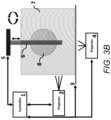
- FIG. 3 B is a schematic diagram showing a side view of an embodiment of a multi-bioink resin tank system for 3D bio-overprinting with two projectors.
- FIG. 4 is a schematic diagram showing multi-projector printing and the progression of different bioinks photocured on the printing surface according to an embodiment.
- FIG. 5 is a schematic diagram of a print surface with an extrusion function according to an embodiment.
- DLP digital light processing
- computed axial lithography is a volumetric additive manufacturing process which relies on DLP to deliver a computed 3D exposure dose to a rotating volume of photopolymerizable resin, thereby producing a 3D object.
- a “resin” or “bioink resin” is a reagent containing one or more photopolymerizable monomers, photo-initiators, photo-absorbers, living cells, and other constituents which together contribute to the formation of bioprinted tissue.
- DLP/CAL printing or “DLP/CAL” is a process which incorporates DLP and CAL and two or more bioink resin tanks to form multi-layered tissue structures.
- printing platform As used herein, “printing platform”, “printing surface”, “overprinting surface”, and the like are used interchangeably to indicate a substrate upon which one or more bioink resin is cured.
- a temperature controlled multi-bioink resin tank system for 3D bio-overprinting through DLP/CAL includes two or more tunable temperature controlled bioink resin tanks which are used consecutively to form a multi-layered tissue which is overprinted on a surface based on instructions stored in 3D files or software and tasks executed by a controller.
- the multi-layer tissue is formed by overprinting on a printing surface submerged in the bioink resin tank through one or more DLP projectors which cure the resin on the surface, which projectors can be positioned from the top, side and/or beneath the bioink resin tank or any angle therebetween.
- the one or more DLP projectors can be stationary or can be moveable by one or more actuators.
- the overprinting surface can be an inflatable, soft, or solid 3D shape, or combination thereof, and can include a rubber polymer, plastic, or metal, or combination thereof.
- the overprinting surface can be moveable by one or more actuators capable of rotary or vertical movement, and can have an extrusion function.
- the extrusion function can be implemented for example by way of one or more ports or orifices disposed in communication with one or more print surface and channels, such as the orifice being disposed on the top of the print surface and the channel(s) being disposed underneath the print surface.
- the channels can deliver one or more bioink, such as extrudable bioinks, including gels or polymers, which are capable of being extruded, for example into a photopolymerizable gel.
- one or more materials can be dispensed by way of a sprayer, or other device for dispensing materials without direct contact with the printed structures and/or the print surface.
- a sprayer or other device for dispensing materials without direct contact with the printed structures and/or the print surface.
- an air-brush type sprayer can be used that can spray additional cells, drugs, nanofibers, etc. between layers or around the printed structures.
- Such dispensing/extrusion function shown in FIG. 5 , will be elaborated on further below.
- the two or more tunable temperature controlled bioink resin tanks can be used consecutively as a solution rinse step or solution absorb step during the process.
- the two or more tunable temperature controlled bioink resin tanks can mechanically rotate into position below the overprinting surface which is raised and lowered in one or more or each bioink resin tank to form a multi-layered tissue through DLP/CAL printing.
- the two or more bioink resin tanks can have tunable temperature control for biologics, cells, biopolymers, synthetic polymers, therapeutics or any combination thereof.
- the temperature control for one or more or each bioink resin tank can be in the range of 4 to 60 degrees Celsius.
- the temperature controlled bioink resin tank(s) can include a condensation absorber.
- the two or more bioink resin tanks can include one or more bioinks with one or more natural or synthetic monomers capable of polymerization by way of electromagnetic irradiation, one or more types of living cells, one or more therapeutic agents or particles, and one or more photo-initiators or photo-absorbers.
- the completed bio-overprinting tissue structure can be removed from the surface by way of deflation according to some embodiments.
- the completed bio-overprinting tissue structure can also be used as a bioreactor with inflating or deflating mechanical forces on the bioprinted tissue according to some embodiments.
- the system can include a clean chamber function using positive pressure of filtered air or inert gas
- the temperature controlled multi-bioink resin tank system includes a controller and one or more DLP/CAL projector, as well as one or more actuators to control motion of a print platform or surface, the bioink resin tanks, and/or one or more DLP/CAL projector such that different bioink resins can be cured on the print surface consecutively to form a multi-layered tissue on the print surface.
- the controller commands the one or more DLP/CAL projector and the actuators to allow for bioprinting on the print surface.
- the electromagnetic energy from the DLP/CAL projector can have a wavelength ranging from 100 nm to 2,500 nm, including from 250 nm to 450 nm, or from 300 nm to 425 nm, or from 330 nm to 420 nm, or from 350 nm to 390 nm, or from 365 nm to 405 nm, or from 330 and 460 nm, or from 370 and 440 nm, or from 405 nm to 500 nm, or from 500 nm to 800 nm, or from 700 nm to 2,500 nm.
- the electromagnetic energy from the DLP/CAL projector can include UV light with wavelengths ranging from 100 nm to 400 nm, including UV-A light (320-400 nm), UV-B light (290-320 nm), and UV-C light (100-290 nm).
- the electromagnetic energy can include infrared light with wavelengths ranging from 780 nm to 1 mm, including IR-A (780 nm-1.4 ⁇ m), IR-B (1.4-3 ⁇ m) and IR-C, also known as far-IR (3 ⁇ m-1 mm).
- the electromagnetic energy can include visible light with wavelengths ranging from 400 nm to 700 nm.
- the intensity of the electromagnetic energy from the DLP/CAL projector can range from 0.1-40 J/cm 2 such as from 0.1-1 J/cm 2 , 1-5 J/cm 2 , 5-10 J/cm 2 , 10-15 J/cm 2 , 15-20 J/cm 2 , 20-25 J/cm 2 , 25-30 J/cm 2 , 30-35 J/cm 2 , or 35-40 J/cm 2 .
- the frequency of the electromagnetic energy used for irradiation can be varied. For example, ultraviolet light has frequencies that range from 8 ⁇ 10 14 Hz to 3 ⁇ 10 16 Hz. If infrared light is used, the frequency can range from 300 GHz to 450 THz.
- the source of the electromagnetic energy from the DLP/CAL projector can be a laser, LED, incandescent, or fluorescent light source.
- the laser can be a solid-state laser, gas laser, excimer laser, dye laser, or semiconductor laser.
- the controller can perform operations and processes described or depicted herein through one or more computer processor.
- the one or more computer processor can issue commands to the DLP/CAL projector to irradiate the overprinting surface with a specified wavelength, intensity, and location.
- the one or more computer processor can issue commands to the actuators to control horizontal, vertical, or rotary motion of the bioink resin tanks, overprinting surface, or one or more DLP/CAL projector as well as inflation and deflation of the print surface (if such inflatable surface is implemented).
- the one or more computer processor commands a sequence of movements such that a different bioink from one or more or each tank can be used to photopolymerize a layer on the overprinting surface.
- the one or more computer processor commands the DLP/CAL projector and actuators through instructions programmed on one or more computer files which are read by the one or more computer processors.
- Embodiments can include a non-transitory computer readable storage medium comprising one or more computer files (e.g. “3D files”) comprising a set of computer-executable instructions for performing one or more of the processes and operations described herein and/or depicted in the drawings, such as irradiation of the overprinting surface by the DLP/CAL projector or movement of the actuators.
- the 3D files can be created through Computer Aided Design (CAD) software, or equivalent 3D modeling software, which allow a user to determine the 3D structure of the overprinted tissue by creating tissue structure models.
- CAD Computer Aided Design
- the files can be stored contiguously or non-contiguously on the computer-readable medium.
- embodiments include a computer program product comprising the computer files, either in the form of the computer-readable medium comprising the computer files and, optionally, made available to a consumer through packaging, or alternatively made available to a consumer through electronic distribution.
- a “computer-readable medium” includes any kind of computer memory such as floppy disks, conventional hard disks, CD-ROMS, Flash ROMS, non-volatile ROM, electrically erasable programmable read-only memory (EEPROM), and RAM.
- the terms “computer-executable instructions”, “code”, “software”, “program”, “application”, “software code”, “computer readable code”, “software module”, “module” and “software program” are used interchangeably to mean software instructions that are executable by a processor.
- the computer-executable instructions can be organized into routines, subroutines, procedures, objects, methods, functions, or any other organization of computer-executable instructions that is known or becomes known to a skilled artisan in light of this disclosure, where the computer-executable instructions are configured to direct a computer or other data processing device to perform one or more of the specified processes and operations described herein.
- the computer-executable instructions can be written in any suitable programming language, non-limiting examples of which include C, C++, C#, Objective C, Swift, Ruby/Ruby on Rails, Visual Basic, Java, Python, Perl, PUP, and JavaScript.
- files comprising the set of computer-executable instructions can be stored in computer-readable memory on a single computer or distributed across multiple computers.
- files comprising the set of computer-executable instructions can be stored in computer-readable memory on a single computer or distributed across multiple computers.
- a skilled artisan will further appreciate, in light of this disclosure, how the invention can be implemented, in addition to software, using hardware or firmware. As such, as used herein, the operations of the invention can be implemented in a system comprising any combination of software, hardware, or firmware.
- Embodiments of the invention include one or more computers or devices loaded with a set of the computer-executable instructions described herein.
- the computers or devices can be a general-purpose computer, a special-purpose computer, or other programmable data processing apparatus to produce a particular machine, such that the one or more computers or devices are instructed and configured to carry out the processes and operations described herein.
- the computer or device performing the specified processes and operations can comprise at least one processing element such as a central processing unit (i.e. processor) and a form of computer-readable memory which can include random-access memory (RAM) or read-only memory (ROM).
- RAM random-access memory
- ROM read-only memory
- the computer-executable instructions can be embedded in computer hardware or stored in the computer-readable memory such that the computer or device can be directed to perform one or more of the processes and operations depicted in the drawings and/or described herein.
- the computer-executable instructions can provide an operator interface for the system controller which can be a graphical user interface (GUI) which can be presented on a display of a general-purpose or special-purpose computer with a processor, computer-readable memory, and standard I/O interfaces such as a universal serial bus (USB) port and a serial port, a disk drive, a CD-ROM drive, and/or one or more user interface devices including a keyboard, keypad, mouse, control panel, touch screen display, microphone, etc. which allow a user to operate or program the controller.
- GUI graphical user interface
- the actuators controlling the movement of system components can include rotary actuators, such as for mechanical rotation of the bioink resin tanks or the overprinting surface, or linear actuators such as for mechanical raising and lowering of the overprinting surface from and to the interior of the bioink resin tanks.
- the actuators can be pneumatic, hydraulic, electric or mechanical actuators.
- the system can include one or more temperature control units which are capable of regulating the temperature of the bioink resin tanks.
- the temperature of the overprinting surface is independently controlled through a temperature control unit.
- the temperature control units can include a heater in communication with the bioink resin tanks, or a heat sink such as circulating coolant.
- the temperature control units are capable of regulating the temperature of the bioink resin tanks in the range of 4 to 60 degrees Celsius.
- the system can include one or more tanks of pressurized air or inert gas such as nitrogen or helium for inflation of the overprinting surface or for cleaning any component of the system.
- pressurized air or inert gas such as nitrogen or helium
- the system and its components can be powered via 60V or 120V AC current, or other voltages according to the single-phase voltage standard that is used in particular countries or regions. In general, this can be in the range of 100-127 volts or 220-240 volts.
- One or more bioink resin tank can include one or more natural, semi-synthetic or synthetic monomers which, upon irradiation with electromagnetic energy for example from one or more DLP/CAL projector, can form a polymer such as any biodegradable or non-biodegradable, optionally “biocompatible polymer,” which when introduced into a living system is compatible with living tissue and/or the living system (e.g., not substantially toxic, injurious, or leading to immunological rejection).
- Any target polymer can be used, including any monomers, co-polymers, and block co-polymers used for preparing any such target polymer.
- One or more bioink resin tank can include one or more natural monomers which, upon irradiation with electromagnetic energy for example from one or more DLP/CAL projector, form a polymer or biopolymer such as gelatin, alginate, fibrin, hyaluronic acid (or hyaluronan), agarose, decellularized extracellular matrix, silk, natural rubber, glycogens, chitin, amylopectin, cellulose, alginate, wool, amber, keratin, collagen, starch, DNA, shellac, growth factors, Matrigel®, poly (acrylic acid), polypeptide-DNA, anticoagulants (including heparin and coumarin), polysaccharide, dextran, casein, albumin, ovalbumin, fibronectin, keratin, pectin, or elastin.
- Particular examples of photopolymerizable bioinks that can be used include gelatin methacrylate, collagen methacrylate, and PEGDA.
- One or more bioink resin tank can include one or more semi-synthetic or synthetic monomers which, upon irradiation with electromagnetic energy for example from one or more DLP/CAL projector, can form a polymer or biopolymer, for example, including polymethylmethacrylate, polyaryletherketones (PAEKs), including polyetheretherketone (PEEK) and polyaryletherketone-etherketoneketone (PEKEKK), polyurethane, poly(L-lactide), poly(D,L-lactide), poly(L-co-D,L-lactide), polyglycolide, poly(lactide-co-glycolide), poly(hydroxylbutyrate), poly(hydroxyvalerate), tyrosine-derived polycarbonate, polyanhydride, polyorthoester, polyphosphazene, poly(dioxanone), poly( ⁇ -caprolactone), and polyglyconate, such as described in U.S. Pat. No. 9,662,821,
- Polymers for tissue engineering and/or drug delivery can also be used.
- Electrically conducting polymers such as polyaniline, polypyrrole, polythiophene, and their derivatives (mainly aniline oligomer and poly(3,4-ethylenedioxythiophene)), or combinations thereof and combinations with other polymers can be used.
- Polymers can include ⁇ -hydroxycarboxylic acids and copolymers thereof, including PGA, PLA and copolymers thereof, polyethylene oxide/polyethylene terephthalates, copolymers of lactic or glycolic acid, poly(alkylene glycols) of various molecular weights, biodegradable and biocompatible polycaprolactones, polyhydroxybutyrates and copolymers of polyesters, polycarbonates, polyanhydrides and poly(ortho esters), bisphenol-A based polyphosphoesters, including poly(bisphenol-A phenylphosphate), poly(bisphenol-A ethylphosphate), poly(bisphenol-A ethylphosphonate), poly(bisphenol-A phenylphosphonate), poly[bis(2-ethoxy)hydrophosphonic terephthalate], and copolymers of bisphenol-A based poly(phosphoesters), copolymers of polyethylene oxide/polyethylene terephthalate, polymers of tyrosine-derived di
- Bioinks that can be used include a double-network bioink comprising a non-crosslinkable thickener and a cross-linkable polyethylene glycol-based network that interpenetrate each other.
- Such bioinks can comprise (a) a biocompatible or non-biocompatible thickener; (b) a polyethylene glycol based crosslinkable network; a photoinitiator; and/or optionally, additives to impart different characteristics; wherein the thickener and polyethylene glycol based crosslinkable network form a structure comprising two interpenetrating networks.
- the bioink can comprise a thickener chosen from one or more of: polyethylene oxide; polypropylene oxide; nanofibrillar cellulose; nanocrystalline cellulose; gelatin; collagen; glucomannan; alginate; k-carrageenan; bentonite clay; and/or xanthan gum.
- the cross-linkable polyethylene glycol can comprise one or more reactive groups, including: acrylate; thiol; maleimide; and/or biotin.
- the polyethylene glycol crosslinker can exhibit structures or blends that are: linear; branched; 4-arm; 8-arm; and/or hyperbranched.
- the additives can comprise monoacrylate PEG with functionalization of the following: fluorescent groups such as fluorescein, rhodamine, or dansyl; sulfonate groups; amine groups; phosphate groups; lipid groups; and/or CNT binding.
- the photoinitiator can comprise one or more of Irgacure 2959; LAP; Eosin-Y; and/or Avidin.
- An exemplary double-network bioink composition can include (a) one or more thickener present in an amount ranging from 0.1% to 20% w/v of the composition, (b) one or more PEGDA crosslinker present in an amount ranging from 0.1% to 10% w/v of the composition, (c) one or more photoinitiator present in an amount ranging from 0.05% to 1% w/v of the composition, and (d) optionally one or more supplementary proteins present in an amount ranging from 0.01% to 10% w/v of the composition, with the remainder comprising water and salts.
- the mechanical stiffness, the diffusivity, the elasticity, binding capacity, etc. of the construct can be varied.
- the elasticity and stiffness of the cross-linked bioink can be controlled by changing the blending ratio between the thickener, cross-linking polymer, and the concentration of the photoinitiator.
- Bioinks also include biogum-hydrogel bioinks (for example comprising a xanthan gum-based bioink, and one or more biomaterials derived from mammalian, plant, microbial or synthetically derived hydrogels) and/or botanical gum-hydrogel bioinks (for example comprising a botanical gum-based bioink, and one or more biomaterials derived from mammalian, plant, microbial or synthetically derived hydrogels).
- the bioink compositions can be provided with cells, preferably human cells.
- the bioink compositions can comprise a ratio of biogum versus biomaterial, or a ratio of botanical gum versus biomaterial, in the interval from 5:95 to 95:5.
- the biomaterial hydrogel can be at least one of the following constituents for cross-linking purposes and/or to contribute to rheological properties of the bioink, such as hydrocolloids or thickening and gelling agents: collagen type I, collagen and its derivatives, gelatin methacryloyl, gelatin and its derivatives, fibrinogen, thrombin, elastin, alginates, agarose and its derivatives, glycosaminoglycans such as hyaluronic acid and its derivatives, chitosan, low and high methoxy pectin, gellan gum, diutan gum, glucomannan gum, carrageenans, nanofibrillated cellulose, microfibrillated cellulose, crystalline nanocellulose, carboxymethyl cellulose, methyl and hydroxypropylmethyl cellulose, bacterial nanocellulose, or a combination of these constituents.
- collagen type I collagen and its derivatives
- gelatin methacryloyl gelatin and its derivatives
- fibrinogen thro
- the bioink compositions can configured such that the xanthan-gum bioink has a concentration in the interval from 0.5 to 10% (w/v) or the botanical gums bioink has a concentration in the interval from 0.5 to 50% (w/v).
- the bioink comprises additional biopolymers for cross-linking purposes and/or to contribute to rheological properties of the bioink, such as hydrocolloids or thickening and gelling agents, for example, alginates, hyaluronic acid and its derivatives, agarose and its derivatives, chitosan, fibrin, gellan gum, silk nanofibrillated cellulose, microfibrillated cellulose, crystalline nanocellulose, bacterial nanocellulose, carrageenans, elastin, collagen and its derivatives as well as gelatin and its derivatives.
- the concentration of cells such as human or animal cells, is in the interval from 0.1 million/ml to 150 million/ml.
- the cells are of human or porcine origin, preferably human origin.
- the composition is provided in physiological conditions, such as having a pH-value for the composition in the interval from 5-8, preferably 7 and/or an osmolarity of the composition is in the interval from 275 to 300 mOsm/kg, preferably 295 mOsm/kg.
- bioinks which can be used include, but are not limited to, the following available from CELLINK (Gothenburg, Sweden):
- the monomers can be provided in the bioink resins at an appropriate concentration, such as 0.1% to 20% w/v or higher, such that they react to polymerize upon exposure of the bioink resin to light.
- One or more or each bioink resin tank can include one or more photo-initiators within the bioink(s) which can include, for example, free radical photoinitiators, cationic photoinitiators, and anionic photoinitiators.
- the photoinitiator forms a free radical, cation, or anion which subsequently reacts and catalyzes a polymerization reaction among the monomers within the bioink.
- photoinitiators include, but are not limited to benzophenone, benzoin-ether, 2-(dimethylamino)ethanol (DMAE), hydroxyacetophenones, 2-hydroxy-2-methyl-1-phenylpropan-1-one and, hydroxyl-phenyl-ketone, Irgacure® 2959, 2-hydroxy-4′-(2-hydroxyethoxy)-2-methylpropiophenone, (2-hydroxy-1-[4-(2-hydroxyethoxy) phenyl]-2-methyl-1-propanone; lithium phenyl-2,4,6-trimethylbenzoylphosphinate (LAP); (2,2′-azobis[2-methyl-N-(2-hydroxyethyl)propionamide]; 2-isocyanotoethyl methacrylate; benzoyl benzylamine; camphorquinone; thiol-norbornene (thiol-ene); riboflavin; lucirin-TPO; Rose Bengal/furfuryl
- One or more or each bioink resin tank can include one or more photo-absorbers within the bioink(s) which can include, for example, compounds such as tartrazine, curcumin, anthocyanin, quinoline yellow, as well as gold nanoparticles.
- the photo-absorber(s) is included in the bioink(s) to limit or control the polymerization reaction.
- the photo-initiators and/or photo-absorbers can be free within the bioink solution or can be bound to the one or more monomers, such as through covalent bonding.
- the photo-initiators and photo-absorbers can be added to the bioink resins at concentration ratios which produce a desired rate of polymerization.
- One or more or each bioink resin tank can include one or more types of living cells within the bioink(s) which can be or include, for example, cells from tissues such as liver, kidney, heart, lung, gastrointestinal, muscle, skin, bone, cartilage, vascularized tissues, blood vessels, ducts, ear, nose, esophagus, trachea, and eye.
- tissues such as liver, kidney, heart, lung, gastrointestinal, muscle, skin, bone, cartilage, vascularized tissues, blood vessels, ducts, ear, nose, esophagus, trachea, and eye.
- endothelial cells skin cells such as keratinocytes, melanocytes, Langerhans' cells, and Merkel cells
- connective tissue cells such as fibroblasts, mast cells, plasma cells, macrophages, adipocytes, and leukocytes
- bone tissue cells such as osteoblasts, osteoclasts, osteocytes, and osteoprogenitor (or osteogenic) cells
- cartilage cells such as chondrocytes and chondroblasts
- muscle cells such as smooth muscle cells, skeletal muscle cells, cardiac muscle cells, any cells having muscle fibers such as type I (slow twitch), type IIa and type IIb (fast twitch), nerve cells such as multipolar neurons, bipolar neurons, unipolar neurons, sensory neurons, interneurons, motor neurons, neurons of the brain (e.g.
- Golgi cells Purkinje cells, pyramidal cells
- glial cells such as oligodendrocytes, astrocytes, ependymal cells, Schwann cells, microglia, and satellite cells
- liver cells such as hepatocytes, biliary epithelial cells (cholangiocytes), stellate cells, Kupffer cells, and liver sinusoidal endothelial cells
- kidney cells such as glomerulus parietal cells, glomerulus podocytes, proximal tubule brush border cells, Loop of Henle thin segment cells, thick ascending limb cells, kidney distal tubule cells, collecting duct principal cells, collecting duct intercalated cells, and interstitial kidney cells
- pancreatic cells such as islets cells, alpha cells, beta cells, delta cells, PP cells, endocrine gland cells such as pancreatic cells, hypothalamus cells, pituitary cells, thyroid cells, parathyroid cells, adrenal cells, pineal body cells, and ovarian
- One or more or each bioink resin tank can include one or more therapeutic agents or particles within the bioink(s), which can have a molecular weight in the range of less than 900 Daltons for small molecules to up to 1000 kDa for biologics.
- concentration of the small molecule or biologic therapeutics can be chosen according to published studies which reveal effective concentrations, such as pre-clinical or clinical pharmacology data.
- the small molecules can be any small molecule used in the treatment of any disease, including diseases of the eye, ear, nose, throat, mouth, lung, heart, liver kidney, spleen, pancreas, gastrointestinal system, circulatory system, reproductive system, central nervous system, immune system, musculoskeletal system, and skin.
- the small molecules can be found or identified in publicly available drug references (see Remington, The Science and Practice of Pharmacy. Easton, Pa.: Mack Pub. Co., 1995; Goodman, L. S., Brunton, L. L., Chabner, B., & Knollmann, B. C. (2011). Goodman & Gilman's pharmacological basis of therapeutics. New York: McGraw-Hill; O'Neil, M. J. (2006).
- the Merck index An encyclopedia of chemicals, drugs, and biologicals. Whitehouse Station, N.J.: Merck; United States Pharmacopeia and National Formulary).
- Rockville, Md. United States Pharmacopeial Convention; 2016; and Center for Drug Evaluation and Research (U.S.), & Center for Drug Evaluation and Research (U.S.). (1985). Orange book: Approved drug products with therapeutic equivalence evaluations. Rockville, Md.: U.S. Dept. of Health and Human Services, Food and Drug Administration, Center for Drug Evaluation and Research, Office of Pharmaceutical Science, Office of Generic Drugs); these and any edition available at the time of this disclosure are hereby incorporated by reference herein in their entireties.
- the one or more therapeutic agents or particles within the bioink(s), can be or include, for example, a biologic such as any bioactive protein or peptide, such as growth factors which include, but are not limited to native or modified PDGF, FGF-2, EGF, epiregulin, TGF-alpha, keratinocyte growth factor, keratinocyte growth factor-2 (KGF-2), granulocyte-macrophage colony-stimulating factor (GM-CSF), TGF-beta, insulin-like growth factor I (IGF-I), insulin-like growth factor II (IGF-II), nerve growth factor (NGF), brain-derived neurotrophic factor (BDNF), and human growth hormone (HGH), cytokines which include but are not limited to IL-1 (IL-1 ⁇ and - ⁇ ), IL-2, IL-3, IL-4, IL-5, IL-6, IL-8, IL-9, IL-10, IL-11, IL-12, IL-13, IL-16, IL-18, IFN-
- the biologics can include any of the above, as well as, for example, an antibody, antibody fragment, antibody-drug conjugate, peptide, peptide-drug conjugate, protein, polypeptide, fusion protein, multivalent binding protein, blood and blood product, nucleic acid, nucleotide, oligonucleotide, antisense oligonucleotide, short interfering RNA (siRNA), micro-interfering RNA (miRNA); small, temporal RNA (stRNA); short, hairpin RNA (shRNA), aptamer, ribozyme, viral vector (e.g.
- Non-limiting examples of biologics products include adalimumab (Humira), rituximab (Rituxan), etanercept (Enbrel), trastuzumab (Herceptin), bevacizumab (Avastin), infliximab (Remicade), insulin glargine (Lantus), pegfilgrastim (Neulasta), interferon beta-1a (Avonex), ranibizumab (Lucentis), and epoetin alfa (Epogen). Additional biologics can be found in the U.S. Food and Drug Administration's Purple Book, which is publicly available on the U.S. FDA website and hereby incorporated by reference.
- the temperature controlled multi-bioink resin tank system is capable of building complex tissue structures, such as branched or unbranched vascular structures, branched or unbranched pulmonary structures, branched or unbranched duct-like structures, bladder-shaped structures, and layered structures.
- the temperature controlled multi-bioink resin tank system is capable of use in combination with other 3D bioprinting technologies, including extrusion, inkjet, acoustic/ultrasound, laser, and stereolithography technologies.
- the temperature controlled multi-bioink resin tank system can be used in a clean room to maintain sterile conditions or can be incorporated into clean chamber technology such as described in U.S. Patent Application Publication No. US 20180326665A1, incorporated herein by reference in its entirety.
- FIG. 1 is a schematic diagram showing a print surface and the progression of different bioinks photocured on the surface with a side projector according to an embodiment.
- the print surface ( 1 ) is vertically lowered into bioink resin tank one and the first section of the sphere is polymerized through DLP/CAL onto the print surface ( 2 ).
- the print surface is raised and resin tank two moves into position with the print surface.
- the print surface is lowered into bioink resin tank two and polymerized through DLP/CAL onto the previous bioprinted layer ( 3 ).
- the print surface is raised and resin tank three moves into position with the print surface.
- the print surface is lowered into bioink resin tank three and polymerized through DLP/CAL on the previous bioprinted layer ( 4 ).
- a three-cell type multi-layered bioprinted spherical structure is created on the print surface.
- FIG. 2 A is a schematic diagram showing a top view of an embodiment of a multi-bioink resin tank system for 3D bio-overprinting which includes a controller ( 1 ) that commands and DLP/CAL projector ( 2 ), vertical motion of a print surface ( 3 ), and horizontal rotation of a temperature-controlled platform or stage ( 4 ) with multiple temperature-controlled bioink resin tanks ( 5 , 6 , 7 ).
- One or more or each bioink resin tank can move into position with the DLP projector and under the print surface consecutively to form a multi-layered tissue on the inflated surface. Movement of one or more components of the system can be relative to one or more other component(s) of the system.
- one or more light source can move and/or one or more print surface can move, and/or one or more bioink tank can move, and/or one or more platform on which one or more bioink tank is positioned can move.
- FIG. 2 B is a schematic diagram showing a top view of an embodiment of a multi-bioink resin tank system for 3D bio-overprinting which includes a controller ( 1 ) which commands multiple functions of the system.
- the controller commands the side projector ( 2 ), the horizontal rotation of the temperature-controlled platform or stage ( 3 ), the vertical and circular motion of the print surface ( 4 ), and the multiple temperature-controlled bioink resin tanks with circular motion ( 5 ).
- One or more or each bioink resin tank moves into position with the projector and under the print surface consecutively to form a multi-layered tissue on the surface.
- FIG. 3 A is a schematic diagram showing a side view of an embodiment of a multi-bioink resin tank system for 3D bio-overprinting with one projector.
- the system includes a controller ( 1 ) that commands a DLP projector ( 2 ), the horizontal rotation of a temperature-controlled platform or stage ( 3 ) with multiple temperature-controlled bioink resin tanks ( 4 ), and the print surface ( 5 , 6 , 7 ) with vertical and circular rotational motion and an inflatable feature ( 7 ).
- the controller and the system allow for multi-layer multi-material bioprinting on the inflatable print surface ( 8 ).
- FIG. 3 B is a schematic diagram showing a side view of an embodiment of a multi-bioink resin tank system for 3D bio-overprinting with two projectors.
- the controller ( 1 ) commands multiple functions of the system.
- the controller commands the side projector ( 2 ), the horizontal rotation of the temperature-controlled platform or stage ( 3 ), the bottom projector ( 4 ), the vertical and circular motion of the print surface ( 5 , 6 ), and the temperature-controlled bioink resin tanks with circular motion ( 7 ).
- the system allows for photo-polymerization of a bioinks on the print surface ( 8 ).
- FIG. 4 is a schematic diagram showing multi-projector printing and the progression of different bioinks photocured on the surface.
- the print surface ( 1 ) is vertically lowered into bioink resin tank one and the first section is polymerized with the bottom projector onto the print surface ( 2 ).
- the print surface is raised and resin tank two moves into position with the print surface.
- the print surface is lowered into bioink resin tank two and polymerized with the side projector onto the previous bioprinted layer ( 3 ).
- FIG. 5 is a schematic diagram of a print surface with an extrusion function.
- the print surface ( 1 ) is vertically lowered into a bioink resin tank and a sphere is polymerized through DLP/CAL onto the print surface ( 2 ).
- the print surface extrusion function is initiated and a bioink is extruded out of the print surface and into the polymerized sphere at specific points.
- one or more materials can be dispensed by way of a sprayer, or other device for dispensing materials without direct contact with the printed structures and/or the print surface.
- an air-brush type sprayer can be used that can spray additional cells, drugs, nanofibers, etc. between layers or around the printed structures.
- a 3D file is created containing a model of a hollow Y-branched vascular tube (50 ⁇ 50 ⁇ 10 mm) with three layered sections. Each of the three sections are 2 mm in width and represent a different bioink that will be consecutively photopolymerized on the print surface that mimics the model.
- the mimicking surface is a rubber polymer that is inflated and vertically lowered into the temperature-controlled multi-bioink resin tanks and rotates in a circular motion.
- the bioprinting software controller recognizes each model section and makes adjustments to each tank position, the print surface, and the projector based on the model coordinates and bioprinting progress.
- Three individual bioink resin tanks are each set and maintained at 4 degrees C. during the bioprinting.
- Bioink resin tank one is loaded with a photopolymerizable collagen solution, a photo-initiator, a photo-absorber, and human smooth muscle cells.
- Bioink resin tank two is loaded with a photopolymerizable collagen solution, a photo-initiator, a photo-absorber, and human fibroblast cells.
- Bioink resin tank three is loaded with a photopolymerizable collagen solution, a photo-initiator, a photo-absorber, and human endothelial cells.
- the print surface is inflated and is vertically lowered into bioink resin tank one and polymerized through DLP/CAL onto the print surface until 2 mm thickness is achieved. Upon completion, the print surface is raised and resin tank two moves into position with the print surface.
- the print surface is lowered into bioink resin tank two and polymerized through DLP/CAL onto the previous bioprinted layer until 2 mm thickness is achieved.
- the print surface is raised and resin tank three moves into position with the print surface.
- the print surface is lowered into bioink resin tank three and polymerized through DLP/CAL on the previous bioprinted layer until 2 mm thickness is achieved.
- a three-cell type multi-layered bioprinted construct is engineered on the print surface. After printing is complete the print surface is deflated and the bioprinted multi-layered structure is rinsed and incubated with cell culture media.
- Example 2 Same as Example 1, but after printing is complete the print surface with the bioprinted multi-layered structure is rinsed and placed into cell culture media.
- the inflate and deflate function of the system is used as a mechanical force bioreactor on the bioprinted multi-layered structure.
- a 3D file is created containing a model of a sphere (50 ⁇ 50 ⁇ 10 mm) with three layered sections. Each of the three sections represents a different bioink that will be consecutively photopolymerized on the print surface.
- the print surface and bioink tank both rotate in a circular motion during the photo-curing which is achieved by the side projector.
- the bioprinting software controller recognizes each model section and makes adjustments to the print surface, the bioink tank positions, and the side projector based on the model coordinates and bioprinting progress.
- Bioink resin tank one is loaded with a photopolymerizable collagen solution, a photo-initiator, a photo-absorber, and human smooth muscle cells.
- Bioink resin tank two is loaded with a photopolymerizable collagen solution, a photo-initiator, a photo-absorber, and human fibroblast cells.
- Bioink resin tank three is loaded with a photopolymerizable collagen solution, a photo-initiator, a photo-absorber, and human endothelial cells.
- the print surface is vertically lowered into bioink resin tank one and the first section of the sphere is polymerized through DLP/CAL onto the print surface.
- the print surface Upon completion, the print surface is raised and resin tank two moves into position with the print surface. The print surface is lowered into bioink resin tank two and polymerized through DLP/CAL onto the previous bioprinted layer. Upon completion, the print surface is raised and resin tank three moves into position with the print surface. The print surface is lowered into bioink resin tank three and polymerized through DLP/CAL on the previous bioprinted layer.
- a three-cell type multi-layered bioprinted spherical structure is engineered on the print surface ( FIG. 1 ). After printing is complete the bioprinted multi-layered sphere is rinsed and incubated with cell culture media.
Abstract
Description
-
- a) positioning the first container below the printing platform or surface; and/or
- b) mechanically lowering the printing platform or surface into the first container; and/or
- c) irradiating the printing platform or surface with electromagnetic energy; and/or
- d) mechanically raising the printing platform or surface from the first container; and/or
- e) positioning the second container below the printing platform or surface; and/or
- f) mechanically lowering the printing platform or surface into the second container; and/or
- g) irradiating the printing platform or surface with electromagnetic energy; and/or
- h) mechanically raising the printing platform or surface from the second container.
| TABLE I |
| Exemplary Bioinks |
| Bioink | Components |
| CELLINK A | Alginate |
| CELLINK A-RGD | Alginate coupled with L-arginine- |
| Glycine-L-aspartic Acid peptide | |
| CELLINK Bioink | Alginate & Nanofibrillar cellulose |
| CELLINK BONE | CELLINK & Tricalcium phosphate |
| CELLINK FIBRIN | CELLINK & Fibrinogen |
| CELLINK LAMININK+ | CELLINK & Laminin blend |
| CELLINK LAMININK 111 | CELLINK & Laminin a1b1y1 |
| CELLINK LAMININK 121 | CELLINK & Laminin a1b2y1 |
| CELLINK LAMININK 411 | CELLINK & Laminin a4b1y1 |
| CELLINK LAMININK 521 | CELLINK & Laminin a5b2y1 |
| CELLINK RGD | CELLINK & Alginate coupled with L- |
| arginine-Glycine-L-aspartic Acid peptide | |
| CELLINK SKIN | CELLINK & |
| Coll | |
| 1 | |
| ColMA | Collagen methacrylate |
| Bio Conductink | Gelatin methacrylate & Carbon nanotubes |
| GelMA | Gelatin methacrylate |
| GelMA A | Gelatin methacrylate & Alginate |
| GelMA C | Gelatin methacrylate, Nanofibrillar |
| cellulose & Alginate | |
| GelMA HA | Gelatin methacrylate & Methacrylated |
| hyaluronic acid | |
| GelMA HIGH | High-concentration gelatin methacrylate. |
| GelXA | Gelatin methacrylate, Xanthan Gum & |
| Alginate | |
| GelXA BONE | Gelatin methacrylate, Xanthan Gum, |
| Alginate, Tricalcium phosphate & | |
| Hydroxyapatite | |
| GelXA FIBRIN | Gelatin methacrylate, Xanthan Gum, |
| Alginate & Fibrinogen | |
| GelXA LAMININK+ | Gelatin methacrylate, Xanthan Gum, |
| Alginate & Laminin blend | |
| GelXA LAMININK 111 | Gelatin methacrylate, Xanthan Gum, |
| Alginate & Laminin a1b1y1 | |
| GelXA LAMININK 121 | Gelatin methacrylate, Xanthan Gum, |
| Alginate & Laminin a1b2y1 | |
| GelXA LAMININK 411 | Gelatin methacrylate, Xanthan Gum, |
| Alginate & Laminin a4b1y1 | |
| GelXA LAMININK 521 | Gelatin methacrylate, Xanthan Gum, |
| Alginate & Laminin a5b2y1 | |
| GelXA SKIN | Gelatin methacrylate, Xanthan Gum, |
| Alginate & Fibrinogen | |
| GelXG | Gelatin methacrylate & Xanthan Gum |
| CELLINK PCL | Polycaprolactone |
| CELLINK PLURONICS | Poloxamer |
| CELLINK START | Polypropylene gel |
| CELLINK START X | CELLINK START. Photo-crosslinkable |
| polymer. | |
| CELLINK SUPPORT | Nanofibrillar cellulose |
| CELLINK XPLORE | Cellulose nanocrystals. Alginate. Coloring. |
Claims (15)
Priority Applications (2)
| Application Number | Priority Date | Filing Date | Title |
|---|---|---|---|
| US17/011,767 US11826951B2 (en) | 2019-09-06 | 2020-09-03 | Temperature-controlled multi-material overprinting |
| US17/554,789 US20220105676A1 (en) | 2019-09-06 | 2021-12-17 | Temperature-controlled multi-material overprinting |
Applications Claiming Priority (2)
| Application Number | Priority Date | Filing Date | Title |
|---|---|---|---|
| US201962897188P | 2019-09-06 | 2019-09-06 | |
| US17/011,767 US11826951B2 (en) | 2019-09-06 | 2020-09-03 | Temperature-controlled multi-material overprinting |
Related Child Applications (1)
| Application Number | Title | Priority Date | Filing Date |
|---|---|---|---|
| US17/554,789 Division US20220105676A1 (en) | 2019-09-06 | 2021-12-17 | Temperature-controlled multi-material overprinting |
Publications (2)
| Publication Number | Publication Date |
|---|---|
| US20210069964A1 US20210069964A1 (en) | 2021-03-11 |
| US11826951B2 true US11826951B2 (en) | 2023-11-28 |
Family
ID=74849833
Family Applications (2)
| Application Number | Title | Priority Date | Filing Date |
|---|---|---|---|
| US17/011,767 Active US11826951B2 (en) | 2019-09-06 | 2020-09-03 | Temperature-controlled multi-material overprinting |
| US17/554,789 Abandoned US20220105676A1 (en) | 2019-09-06 | 2021-12-17 | Temperature-controlled multi-material overprinting |
Family Applications After (1)
| Application Number | Title | Priority Date | Filing Date |
|---|---|---|---|
| US17/554,789 Abandoned US20220105676A1 (en) | 2019-09-06 | 2021-12-17 | Temperature-controlled multi-material overprinting |
Country Status (1)
| Country | Link |
|---|---|
| US (2) | US11826951B2 (en) |
Cited By (1)
| Publication number | Priority date | Publication date | Assignee | Title |
|---|---|---|---|---|
| US11931966B2 (en) | 2018-01-26 | 2024-03-19 | Cellink Bioprinting Ab | Systems and methods for optical assessments of bioink printability |
Families Citing this family (6)
| Publication number | Priority date | Publication date | Assignee | Title |
|---|---|---|---|---|
| WO2020077118A1 (en) | 2018-10-10 | 2020-04-16 | Cellink Ab | Double network bioinks |
| US11826951B2 (en) | 2019-09-06 | 2023-11-28 | Cellink Ab | Temperature-controlled multi-material overprinting |
| EP4098286A1 (en) * | 2021-05-31 | 2022-12-07 | Roquette Freres | 3d-bioprinting of cell-laden-collagen gellan gum interpenetrating network hydrogel |
| CN113787708A (en) * | 2021-07-07 | 2021-12-14 | 珠海市三绿实业有限公司 | Photosensitive resin 3D printing method |
| WO2023119288A1 (en) * | 2021-12-21 | 2023-06-29 | Ehud Giloh | An adjustable shape changing printing bed for additive manufacturing |
| CN114540275B (en) * | 2022-02-23 | 2023-06-13 | 合肥学院 | Skin biological printing ink and preparation method and application thereof |
Citations (124)
| Publication number | Priority date | Publication date | Assignee | Title |
|---|---|---|---|---|
| US5236416A (en) | 1991-05-23 | 1993-08-17 | Ivac Corporation | Syringe plunger position detection and alarm generation |
| US6103790A (en) | 1994-03-01 | 2000-08-15 | Elf Atochem S.A. | Cellulose microfibril-reinforced polymers and their applications |
| JP2000513258A (en) | 1996-06-28 | 2000-10-10 | ジョンソン・アンド・ジョンソン・メディカル・リミテッド | Use of oxidized cellulose and its complexes for chronic wound healing |
| US6509733B2 (en) | 2000-12-20 | 2003-01-21 | Caterpillar Inc | Fluid cylinder with embedded positioning sensor |
| US20030059708A1 (en) * | 2000-06-09 | 2003-03-27 | Tetsuya Yamamura | Resin composition and three-dimensional object |
| WO2004092672A2 (en) | 2003-04-10 | 2004-10-28 | Praxair S. T. Technology, Inc. | Amorphous carbon layer for heat exchangers |
| JP2005003610A (en) | 2003-06-13 | 2005-01-06 | Olympus Corp | Dispenser, and automatic analyzer equipped with the same |
| US20050056713A1 (en) | 2003-07-31 | 2005-03-17 | Tisone Thomas C. | Methods and systems for dispensing sub-microfluidic drops |
| US6942830B2 (en) | 2000-04-17 | 2005-09-13 | Envisiontec Gmbh | Device and method for the production of three-dimensional objects |
| US7052263B2 (en) | 2001-04-20 | 2006-05-30 | Envisiontec Gmbh | Apparatus for manufacturing a three-dimensional object |
| US7105357B1 (en) | 1998-05-27 | 2006-09-12 | Max-Planck-Gesellschaft Zur Forderung Der Wissenschaften E. V. | Method and device for processing extremely small substance quantities |
| US7122712B2 (en) | 2002-12-02 | 2006-10-17 | Lutri Thomas P | Surgical bandage and methods for treating open wounds |
| US7195472B2 (en) | 2001-04-23 | 2007-03-27 | Envisiontec Gmbh | Apparatus and method for the non-destructive separation of hardened material layers from a flat construction plane |
| WO2008055533A1 (en) | 2006-11-10 | 2008-05-15 | Envisiontec Gmbh | Continuous, generative method and apparatus for the production of a three-dimensional object |
| WO2008122661A1 (en) | 2007-04-10 | 2008-10-16 | Bioregeneration Gmbh | Process for the production of a structure comprising crystalline cellulose |
| US20080305012A1 (en) | 2005-12-21 | 2008-12-11 | Hans Camenisch | Method and Device For Checking Whether a Liquid Transfer Has Been Successful |
| US20090003696A1 (en) | 2007-06-29 | 2009-01-01 | Canon Kabushiki Kaisha | Image processing method and image processing apparatus |
| US20090022791A1 (en) | 2005-04-22 | 2009-01-22 | Kazuhiro Obae | Porous cellulose aggregate and molding composition thereof |
| WO2009053100A1 (en) | 2007-10-26 | 2009-04-30 | Envisiontec Gmbh | Process and freeform fabrication system for producing a three-dimensional object |
| US7636610B2 (en) | 2006-07-19 | 2009-12-22 | Envisiontec Gmbh | Method and device for producing a three-dimensional object, and computer and data carrier useful therefor |
| EP2199380A2 (en) | 2008-12-19 | 2010-06-23 | Sanyo Electric Co., Ltd. | Observation unit |
| US20100175759A1 (en) | 2005-11-30 | 2010-07-15 | Musashi Engineering, Inc. | Method of adjusting nozzle clearance of liquid application apparatus, and liquid application apparatus |
| US20100200752A1 (en) | 2009-02-06 | 2010-08-12 | Siliconfile Technologies Inc. | Image sensor capable of judging proximity to subject |
| US20100206224A1 (en) | 2007-09-24 | 2010-08-19 | Berner Fachhochschule fur Technik und informatik HTI | Device for the deposition of layers |
| US7783371B2 (en) | 2006-04-28 | 2010-08-24 | Envisiontec Gmbh | Device and method for producing a three-dimensional object by means of mask exposure |
| JP2010533855A (en) | 2007-07-19 | 2010-10-28 | ビオメリュー | Apolipoprotein AII assay method for in vitro diagnosis of colorectal cancer |
| US20110024699A1 (en) | 2009-07-28 | 2011-02-03 | National Taiwan University | Polymeric polymer containing poly(oxyethylene)-amine and application thereof to preparing silver nanoparticle |
| US7894921B2 (en) | 2006-04-28 | 2011-02-22 | Envisiontec Gmbh | Device and method for producing a three-dimensional object by means of mask exposure |
| US7892474B2 (en) | 2006-11-15 | 2011-02-22 | Envisiontec Gmbh | Continuous generative process for producing a three-dimensional object |
| EP1732746B1 (en) | 2004-05-07 | 2011-04-27 | Envisiontec GmbH | Process for producing a threedimensional object with improved separation of hardened material layers from a base plane |
| US20110151482A1 (en) | 2009-12-22 | 2011-06-23 | Emery Michael P | Automated developer for immuno-stained biological samples |
| WO2012051718A1 (en) | 2010-10-22 | 2012-04-26 | William Gelbart | Automated slide scanning system for a microscope |
| WO2012056109A2 (en) | 2010-10-27 | 2012-05-03 | Upm-Kymmene Corporation | Plant derived cell culture material |
| WO2012071578A2 (en) | 2010-11-24 | 2012-05-31 | Bc Genesis Llc | Pharmacology bioassays for drug discovery, toxicity evaluation and in vitro cancer research using a 3d nano-cellulose scaffold and living tissue |
| USRE43955E1 (en) | 2004-05-10 | 2013-02-05 | Envisiontec Gmbh | Process for the production of a three-dimensional object with resolution improvement by pixel-shift |
| WO2012024675A9 (en) | 2010-08-20 | 2013-02-07 | Case Western Reserve University | Continuous digital light processing additive manufacturing of implants |
| US8394313B2 (en) | 2004-05-07 | 2013-03-12 | Envisiontec Gmbh | Process for the production of a three-dimensional object with an improved separation of hardened material layers from a construction plane |
| JP2013181167A (en) | 2012-03-05 | 2013-09-12 | Dai Ichi Kogyo Seiyaku Co Ltd | Aqueous ink composition and writing instrument using the same |
| US20130309295A1 (en) | 2011-02-04 | 2013-11-21 | Paul Gatenholm | Biosynthetic functional cellulose (bc) fibers as surgical sutures and reinforcement of implants and growing tissue |
| US20140074274A1 (en) | 2012-09-07 | 2014-03-13 | Makerbot Industries, Llc | Three-dimensional printing of large objects |
| WO2014049204A1 (en) | 2012-09-25 | 2014-04-03 | Upm-Kymmene Corporation | Three-dimensional cell culturing |
| US8691974B2 (en) | 2009-09-28 | 2014-04-08 | Virginia Tech Intellectual Properties, Inc. | Three-dimensional bioprinting of biosynthetic cellulose (BC) implants and scaffolds for tissue engineering |
| EP2808671A1 (en) | 2012-01-26 | 2014-12-03 | Sharp Kabushiki Kaisha | Fluorescence information reading device and fluorescence information reading method |
| US8931880B2 (en) | 2010-10-21 | 2015-01-13 | Organovo, Inc. | Devices, systems, and methods for the fabrication of tissue |
| US20150013476A1 (en) | 2012-02-13 | 2015-01-15 | Thermo Fisher Scientific Oy | Electronic Pipette |
| US20150045928A1 (en) | 2013-08-07 | 2015-02-12 | Massachusetts Institute Of Technology | Automatic Process Control of Additive Manufacturing Device |
| US20150050719A1 (en) | 2012-05-08 | 2015-02-19 | Roche Diagnostic Operations, Inc. | Dispensing assembly |
| US20150072293A1 (en) | 2013-08-14 | 2015-03-12 | Eipi Systems, Inc. | Continuous liquid interphase printing |
| KR101502236B1 (en) | 2013-10-25 | 2015-03-12 | 한양대학교 산학협력단 | 3 dimensional chromatic confocal microscope, and method of generating information on depth of specimen using same |
| DE202015000178U1 (en) | 2015-01-08 | 2015-04-16 | Marius Becker | Influence of the melting and softening zone by Peltier element in 3D printers after the melt layer process |
| US20150105891A1 (en) | 2013-10-11 | 2015-04-16 | Advanced Solutions Life Sciences, Llc | System and workstation for the design, fabrication and assembly of bio-material constructs |
| US20150102351A1 (en) | 2005-12-02 | 2015-04-16 | Semiconductor Energy Laboratory Co., Ltd. | Semiconductor device, display device, and electronic device |
| WO2015066705A1 (en) | 2013-11-04 | 2015-05-07 | University Of Iowa Research Foundation | Bioprinter and methods of using same |
| CN103893825B (en) | 2014-02-24 | 2015-06-10 | 钟春燕 | Method for preparing bacterial cellulose compounded amnion extracellular matrix material containing collagen |
| US9073261B2 (en) | 2011-06-28 | 2015-07-07 | Global Filtration Systems | Apparatus and method for forming three-dimensional objects using linear solidification |
| WO2015101712A1 (en) | 2013-12-30 | 2015-07-09 | Upm-Kymmene Corporation | Biomedical device |
| EP2916158A1 (en) | 2014-03-06 | 2015-09-09 | European Molecular Biology Laboratory | Microscope |
| WO2015148646A2 (en) | 2014-03-25 | 2015-10-01 | Biobots, Inc. | Methods, devices, and systems for the fabrication of materials and tissues utilizing electromagnetic radiation |
| US20150290874A1 (en) * | 2014-04-15 | 2015-10-15 | Xyzprinting, Inc. | Three dimensional printing apparatus |
| WO2015164844A1 (en) | 2014-04-24 | 2015-10-29 | Vutara, Inc. | Super resolution microscopy |
| WO2015175457A1 (en) | 2014-05-12 | 2015-11-19 | Jonathan Allen Rowley | Ready-to-print cells and integrated devices |
| US20150375453A1 (en) | 2014-05-01 | 2015-12-31 | Musc Foundation For Research Development | Multidispensor cartesian robotic printer |
| US9315043B2 (en) | 2013-07-31 | 2016-04-19 | Organovo, Inc. | Automated devices, systems, and methods for the fabrication of tissue |
| WO2016073782A1 (en) | 2014-11-05 | 2016-05-12 | Organovo, Inc. | Engineered three-dimensional skin tissues, arrays thereof, and methods of making the same |
| WO2016091336A1 (en) | 2014-12-12 | 2016-06-16 | Ecole Polytechnique Federale De Lausanne (Epfl) | A method for building a structure containing living cells |
| WO2016092106A1 (en) | 2014-12-11 | 2016-06-16 | ETH Zürich | Graft scaffold for cartilage repair and process for making same |
| WO2016100856A1 (en) | 2014-12-18 | 2016-06-23 | Advanced Polymer Technology Ab | Cellulose nanofibrillar bionik for 3d bioprinting for cell culturing, tissue engineering and regenerative medicine applications |
| US20160236414A1 (en) | 2015-02-12 | 2016-08-18 | Arevo Inc. | Method to monitor additive manufacturing process for detection and in-situ correction of defects |
| US20160243618A1 (en) | 2013-11-15 | 2016-08-25 | Eos Gmbh Electro Optical Systems | Device for Producing a Three-Dimensional Object in Layers |
| US20160271870A1 (en) * | 2015-03-20 | 2016-09-22 | Chevron Phillips Chemical Company Lp | Phonon generation in bulk material for manufacturing |
| US20160271875A1 (en) * | 2015-03-20 | 2016-09-22 | Chevron Phillips Chemical Company Lp | System and method for writing an article of manufacture into bulk material |
| US20170031149A1 (en) | 2010-11-30 | 2017-02-02 | Robert Levin | Compact, high-resolution fluorescence and brightfield microscope and methods of use |
| WO2017040675A1 (en) | 2015-08-31 | 2017-03-09 | Cellink Ab | Clean chamber technology for 3d printers and bioprinters |
| US20170080641A1 (en) | 2014-02-20 | 2017-03-23 | Global Filtration Systems, A Dba Of Gulf Filtration Systems Inc. | Apparatus and method for forming three-dimensional objects using a tilting solidification substrate |
| US20170100899A1 (en) | 2011-01-31 | 2017-04-13 | Global Filtration Systems, A Dba Of Gulf Filtration Systems Inc. | Method and apparatus for making three-dimensional objects from multiple solidifiable materials |
| US9662821B2 (en) | 2008-12-30 | 2017-05-30 | Orthovita, Inc. | Bioactive composites of polymer and glass and method for making same |
| WO2017109394A1 (en) | 2015-12-23 | 2017-06-29 | Compagnie Generale Des Etablissements Michelin | Device for conveying additive manufacture container/plate assemblies |
| WO2017109395A1 (en) | 2015-12-23 | 2017-06-29 | Compagnie Generale Des Etablissements Michelin | Additive manufacturing facility with successive nested confinement chambers |
| WO2017115056A1 (en) | 2015-12-30 | 2017-07-06 | Lab Skin Creations | Method for manufacturing body substitutes by additive deposition |
| US20170210077A1 (en) * | 2014-08-12 | 2017-07-27 | Carbon, Inc | Three-Dimensional Printing Using Carriers with Release Mechanisms |
| US20170216498A1 (en) | 2015-04-07 | 2017-08-03 | Sichuan Revotek Co., Ltd. | Compositions for cell-based three dimensional printing |
| US9725613B2 (en) | 2013-05-24 | 2017-08-08 | Fundació Eurecat | Ink composition for inkjet printing |
| US20170225393A1 (en) | 2016-02-04 | 2017-08-10 | Global Filtration Systems, A Dba Of Gulf Filtration Systems Inc. | Apparatus and method for forming three-dimensional objects using two-photon absorption linear solidification |
| WO2017152142A1 (en) | 2016-03-03 | 2017-09-08 | Desktop Metal, Inc. | Additive manufacturing with metallic build materials |
| WO2017184839A1 (en) | 2016-04-20 | 2017-10-26 | The Brigham And Women's Hospital, Inc. | Systems and methods for in vivo multi-material bioprinting |
| WO2017210663A1 (en) | 2016-06-03 | 2017-12-07 | Paul Gatenholm | Preparation and applications of rgd conjugated polysaccharide bioinks with or without fibrin for 3d bioprinting of human skin with novel printing head for use as model for testing cosmetics and for transplantation |
| WO2017214592A1 (en) | 2016-06-09 | 2017-12-14 | Paul Gatenholm | Preparation of modified cellulose nanofibrils with extracellular matrix components as 3d bioprinting bioinks |
| US20180071740A1 (en) | 2015-05-29 | 2018-03-15 | Roche Diagnostics Operations, Inc. | Cartridge for dispensing particles and a reagent fluid |
| WO2018119989A1 (en) | 2016-12-30 | 2018-07-05 | 苏州聚复高分子材料有限公司 | Biological ink |
| CN108248020A (en) * | 2016-12-28 | 2018-07-06 | 西安科技大学 | A kind of horizontal DLP shadow casting techniques face exposure molding machine and method |
| EP3366458A1 (en) | 2016-11-15 | 2018-08-29 | EOS GmbH Electro Optical Systems | Transport unit and provision of a three-dimensional component |
| WO2018169965A1 (en) | 2017-03-13 | 2018-09-20 | The Texas A&M University System | Nanocomposite ionic-covalent entanglement reinforcement mechanism and hydrogel |
| US20180273904A1 (en) | 2015-10-02 | 2018-09-27 | Wake Forest University Health Sciences | Spontaneously beating cardiac organoid constructs and integrated body-on-chip apparatus containing the same |
| US20180281280A1 (en) | 2017-04-04 | 2018-10-04 | Allevi, Inc. | Multi-headed auto-calibrating bioprinter with heads that heat, cool, and crosslink |
| WO2018187380A1 (en) | 2017-04-03 | 2018-10-11 | Greene Nguyen Deborah Lynn | Use of engineered liver tissue constructs for modeling liver disorders |
| US20180297270A1 (en) | 2015-12-16 | 2018-10-18 | The Regents Of The University Of California | Technique for three-dimensional nanoprinting |
| US20180326666A1 (en) | 2017-05-12 | 2018-11-15 | Brett Kelly | System and method for computed axial lithography (cal) for 3d additive manufacturing |
| US20180341248A1 (en) | 2017-05-24 | 2018-11-29 | Relativity Space, Inc. | Real-time adaptive control of additive manufacturing processes using machine learning |
| US20180348247A1 (en) | 2017-06-01 | 2018-12-06 | Hitachi, Ltd. | Dispensing apparatus |
| US20180345563A1 (en) | 2017-06-02 | 2018-12-06 | Cellink Ab | 3D Printer and a Method for 3D Printing of a Construct |
| EP3415300A1 (en) | 2017-06-16 | 2018-12-19 | Cellink AB | Printbeds, 3d-printers, methods and computer programs for regulation of a temperature of a printbed |
| WO2018234837A1 (en) | 2017-06-23 | 2018-12-27 | 3Dhistech Kft. | Device for moving a microscope stage and microscope comprising such a device |
| US20180370116A1 (en) | 2017-06-27 | 2018-12-27 | University Of Florida Research Foundation, Inc. | Three-dimensional printing of reactive materials using intersecting jets |
| US20190016052A1 (en) | 2017-07-11 | 2019-01-17 | Daniel S. Clark | 5d part growing machine with volumetric display technology |
| WO2019043529A1 (en) | 2017-08-30 | 2019-03-07 | Ecole Polytechnique Federale De Lausanne (Epfl) | Methods and apparatus for three-dimensional fabrication by tomographic back projections |
| US10226894B2 (en) | 2012-10-31 | 2019-03-12 | Nederlandse Organisatie Voor Toegepast-Natuurwetenschappelijk Onderzoek Tno | Method and apparatus for making tangible products by layerwise manufacturing |
| US10272664B2 (en) | 2015-01-14 | 2019-04-30 | Xactiv, Inc. | Fabrication of 3D objects via multiple build platforms |
| WO2019109127A1 (en) | 2017-12-08 | 2019-06-13 | Inventia Life Science Pty Ltd | Bioprinter for fabricating 3d cell constructs |
| WO2019145795A2 (en) | 2018-01-26 | 2019-08-01 | Cellink Ab | Systems and methods for optical assessments of bioink printability |
| US20190344500A1 (en) | 2018-05-08 | 2019-11-14 | 9328-8082 Québec Inc. | Modular additive manufacturing system and related methods for continuous part production |
| WO2019246623A9 (en) | 2018-06-22 | 2020-02-06 | Allevi, Inc. | Systems and methods for improved dispensing, layering, and deposition of cross-linkable hydrogels |
| US20200070421A1 (en) | 2018-09-05 | 2020-03-05 | Carbon, Inc. | Robotic additive manufacturing system |
| WO2020077118A1 (en) | 2018-10-10 | 2020-04-16 | Cellink Ab | Double network bioinks |
| US20200122135A1 (en) | 2014-10-22 | 2020-04-23 | The Regents Of The University Of California | High Definition Microdroplet Printer |
| WO2020086941A1 (en) | 2018-10-25 | 2020-04-30 | Cellink Ab | Biogum and botanical gum hydrogel bioinks for the physiological 3d bioprinting of tissue constructs for in vitro culture and transplantation |
| US20200139623A1 (en) | 2018-11-06 | 2020-05-07 | 3DTech Oy | Modular systems and methods for performing additive manufacturing of objects |
| WO2020157077A2 (en) | 2019-01-28 | 2020-08-06 | Cellink Ab | A compact fluorescence microscope and a cell monitoring system |
| WO2020165322A1 (en) | 2019-02-15 | 2020-08-20 | Cellink Ab | Systems and methods for controlled dispensing of temperature-sensitive fluids in liquid handling and dispensing systems |
| WO2020176079A1 (en) | 2019-02-26 | 2020-09-03 | Cellink Ab | Systems and methods for real-time optoelectronic assessments of fluid volume in fluid dispensing systems |
| CN111618302A (en) | 2020-06-15 | 2020-09-04 | 佛山宇仁智能科技有限公司 | Metal electric melting additive device and method for double-material printing cavity part |
| WO2021231717A1 (en) | 2020-05-14 | 2021-11-18 | Contraline, Inc. | Biomaterial compositions and methods of delivery |
| WO2021243046A1 (en) | 2020-05-28 | 2021-12-02 | Contraline, Inc. | Systems and methods for removing biomaterial implants |
| US20220105676A1 (en) | 2019-09-06 | 2022-04-07 | Cellink Ab | Temperature-controlled multi-material overprinting |
| WO2022192768A1 (en) | 2021-03-12 | 2022-09-15 | Cellink Bioprinting Ab | 3d bioprinter with continuous workflow capability |
-
2020
- 2020-09-03 US US17/011,767 patent/US11826951B2/en active Active
-
2021
- 2021-12-17 US US17/554,789 patent/US20220105676A1/en not_active Abandoned
Patent Citations (157)
| Publication number | Priority date | Publication date | Assignee | Title |
|---|---|---|---|---|
| US5236416A (en) | 1991-05-23 | 1993-08-17 | Ivac Corporation | Syringe plunger position detection and alarm generation |
| US6103790A (en) | 1994-03-01 | 2000-08-15 | Elf Atochem S.A. | Cellulose microfibril-reinforced polymers and their applications |
| JP2000513258A (en) | 1996-06-28 | 2000-10-10 | ジョンソン・アンド・ジョンソン・メディカル・リミテッド | Use of oxidized cellulose and its complexes for chronic wound healing |
| US7105357B1 (en) | 1998-05-27 | 2006-09-12 | Max-Planck-Gesellschaft Zur Forderung Der Wissenschaften E. V. | Method and device for processing extremely small substance quantities |
| US6942830B2 (en) | 2000-04-17 | 2005-09-13 | Envisiontec Gmbh | Device and method for the production of three-dimensional objects |
| US20030059708A1 (en) * | 2000-06-09 | 2003-03-27 | Tetsuya Yamamura | Resin composition and three-dimensional object |
| US6509733B2 (en) | 2000-12-20 | 2003-01-21 | Caterpillar Inc | Fluid cylinder with embedded positioning sensor |
| US7052263B2 (en) | 2001-04-20 | 2006-05-30 | Envisiontec Gmbh | Apparatus for manufacturing a three-dimensional object |
| US7195472B2 (en) | 2001-04-23 | 2007-03-27 | Envisiontec Gmbh | Apparatus and method for the non-destructive separation of hardened material layers from a flat construction plane |
| US7122712B2 (en) | 2002-12-02 | 2006-10-17 | Lutri Thomas P | Surgical bandage and methods for treating open wounds |
| WO2004092672A2 (en) | 2003-04-10 | 2004-10-28 | Praxair S. T. Technology, Inc. | Amorphous carbon layer for heat exchangers |
| JP2005003610A (en) | 2003-06-13 | 2005-01-06 | Olympus Corp | Dispenser, and automatic analyzer equipped with the same |
| US20050056713A1 (en) | 2003-07-31 | 2005-03-17 | Tisone Thomas C. | Methods and systems for dispensing sub-microfluidic drops |
| US8394313B2 (en) | 2004-05-07 | 2013-03-12 | Envisiontec Gmbh | Process for the production of a three-dimensional object with an improved separation of hardened material layers from a construction plane |
| EP1732746B1 (en) | 2004-05-07 | 2011-04-27 | Envisiontec GmbH | Process for producing a threedimensional object with improved separation of hardened material layers from a base plane |
| USRE43955E1 (en) | 2004-05-10 | 2013-02-05 | Envisiontec Gmbh | Process for the production of a three-dimensional object with resolution improvement by pixel-shift |
| US20090022791A1 (en) | 2005-04-22 | 2009-01-22 | Kazuhiro Obae | Porous cellulose aggregate and molding composition thereof |
| US20100175759A1 (en) | 2005-11-30 | 2010-07-15 | Musashi Engineering, Inc. | Method of adjusting nozzle clearance of liquid application apparatus, and liquid application apparatus |
| US20150102351A1 (en) | 2005-12-02 | 2015-04-16 | Semiconductor Energy Laboratory Co., Ltd. | Semiconductor device, display device, and electronic device |
| US20080305012A1 (en) | 2005-12-21 | 2008-12-11 | Hans Camenisch | Method and Device For Checking Whether a Liquid Transfer Has Been Successful |
| US7894921B2 (en) | 2006-04-28 | 2011-02-22 | Envisiontec Gmbh | Device and method for producing a three-dimensional object by means of mask exposure |
| US7783371B2 (en) | 2006-04-28 | 2010-08-24 | Envisiontec Gmbh | Device and method for producing a three-dimensional object by means of mask exposure |
| US7636610B2 (en) | 2006-07-19 | 2009-12-22 | Envisiontec Gmbh | Method and device for producing a three-dimensional object, and computer and data carrier useful therefor |
| WO2008055533A1 (en) | 2006-11-10 | 2008-05-15 | Envisiontec Gmbh | Continuous, generative method and apparatus for the production of a three-dimensional object |
| US7892474B2 (en) | 2006-11-15 | 2011-02-22 | Envisiontec Gmbh | Continuous generative process for producing a three-dimensional object |
| WO2008122661A1 (en) | 2007-04-10 | 2008-10-16 | Bioregeneration Gmbh | Process for the production of a structure comprising crystalline cellulose |
| US20090003696A1 (en) | 2007-06-29 | 2009-01-01 | Canon Kabushiki Kaisha | Image processing method and image processing apparatus |
| JP2010533855A (en) | 2007-07-19 | 2010-10-28 | ビオメリュー | Apolipoprotein AII assay method for in vitro diagnosis of colorectal cancer |
| US20100206224A1 (en) | 2007-09-24 | 2010-08-19 | Berner Fachhochschule fur Technik und informatik HTI | Device for the deposition of layers |
| WO2009053100A1 (en) | 2007-10-26 | 2009-04-30 | Envisiontec Gmbh | Process and freeform fabrication system for producing a three-dimensional object |
| EP2199380A2 (en) | 2008-12-19 | 2010-06-23 | Sanyo Electric Co., Ltd. | Observation unit |
| US9662821B2 (en) | 2008-12-30 | 2017-05-30 | Orthovita, Inc. | Bioactive composites of polymer and glass and method for making same |
| US20100200752A1 (en) | 2009-02-06 | 2010-08-12 | Siliconfile Technologies Inc. | Image sensor capable of judging proximity to subject |
| US20110024699A1 (en) | 2009-07-28 | 2011-02-03 | National Taiwan University | Polymeric polymer containing poly(oxyethylene)-amine and application thereof to preparing silver nanoparticle |
| US8691974B2 (en) | 2009-09-28 | 2014-04-08 | Virginia Tech Intellectual Properties, Inc. | Three-dimensional bioprinting of biosynthetic cellulose (BC) implants and scaffolds for tissue engineering |
| US20110151482A1 (en) | 2009-12-22 | 2011-06-23 | Emery Michael P | Automated developer for immuno-stained biological samples |
| WO2012024675A9 (en) | 2010-08-20 | 2013-02-07 | Case Western Reserve University | Continuous digital light processing additive manufacturing of implants |
| US8931880B2 (en) | 2010-10-21 | 2015-01-13 | Organovo, Inc. | Devices, systems, and methods for the fabrication of tissue |
| WO2012051718A1 (en) | 2010-10-22 | 2012-04-26 | William Gelbart | Automated slide scanning system for a microscope |
| WO2012056109A2 (en) | 2010-10-27 | 2012-05-03 | Upm-Kymmene Corporation | Plant derived cell culture material |
| WO2012056110A2 (en) | 2010-10-27 | 2012-05-03 | Upm-Kymmene Corporation | Cell culture material based on microbial cellulose |
| JP2013541956A (en) | 2010-10-27 | 2013-11-21 | ユー ピー エム キュンメネ コーポレーション | Plant-derived cell culture material |
| FI123988B (en) | 2010-10-27 | 2014-01-31 | Upm Kymmene Corp | Cell Culture Materials |
| WO2012056111A2 (en) | 2010-10-27 | 2012-05-03 | Upm-Kymmene Corporation | Drug delivery compositions |
| EP2633032B1 (en) | 2010-10-27 | 2015-02-25 | UPM-Kymmene Corporation | Plant derived cell culture material |
| EP2975115A1 (en) | 2010-10-27 | 2016-01-20 | UPM-Kymmene Corporation | Plant derived cell culture material |
| WO2012071578A2 (en) | 2010-11-24 | 2012-05-31 | Bc Genesis Llc | Pharmacology bioassays for drug discovery, toxicity evaluation and in vitro cancer research using a 3d nano-cellulose scaffold and living tissue |
| US20170031149A1 (en) | 2010-11-30 | 2017-02-02 | Robert Levin | Compact, high-resolution fluorescence and brightfield microscope and methods of use |
| US20170100899A1 (en) | 2011-01-31 | 2017-04-13 | Global Filtration Systems, A Dba Of Gulf Filtration Systems Inc. | Method and apparatus for making three-dimensional objects from multiple solidifiable materials |
| US20130309295A1 (en) | 2011-02-04 | 2013-11-21 | Paul Gatenholm | Biosynthetic functional cellulose (bc) fibers as surgical sutures and reinforcement of implants and growing tissue |
| US9073261B2 (en) | 2011-06-28 | 2015-07-07 | Global Filtration Systems | Apparatus and method for forming three-dimensional objects using linear solidification |
| US20150246482A1 (en) | 2011-06-28 | 2015-09-03 | Global Filtration Systems, A Dba Of Gulf Filtration Systems Inc. | Apparatus and method for forming three-dimensional objects using linear solidification |
| EP2808671A1 (en) | 2012-01-26 | 2014-12-03 | Sharp Kabushiki Kaisha | Fluorescence information reading device and fluorescence information reading method |
| US20150013476A1 (en) | 2012-02-13 | 2015-01-15 | Thermo Fisher Scientific Oy | Electronic Pipette |
| JP2013181167A (en) | 2012-03-05 | 2013-09-12 | Dai Ichi Kogyo Seiyaku Co Ltd | Aqueous ink composition and writing instrument using the same |
| US20150050719A1 (en) | 2012-05-08 | 2015-02-19 | Roche Diagnostic Operations, Inc. | Dispensing assembly |
| US20140074274A1 (en) | 2012-09-07 | 2014-03-13 | Makerbot Industries, Llc | Three-dimensional printing of large objects |
| WO2014049204A1 (en) | 2012-09-25 | 2014-04-03 | Upm-Kymmene Corporation | Three-dimensional cell culturing |
| US10226894B2 (en) | 2012-10-31 | 2019-03-12 | Nederlandse Organisatie Voor Toegepast-Natuurwetenschappelijk Onderzoek Tno | Method and apparatus for making tangible products by layerwise manufacturing |
| US9725613B2 (en) | 2013-05-24 | 2017-08-08 | Fundació Eurecat | Ink composition for inkjet printing |
| US20170199507A1 (en) | 2013-07-31 | 2017-07-13 | Organovo, Inc. | Automated devices, systems, and methods for the fabrication of tissue |
| US9315043B2 (en) | 2013-07-31 | 2016-04-19 | Organovo, Inc. | Automated devices, systems, and methods for the fabrication of tissue |
| US20150045928A1 (en) | 2013-08-07 | 2015-02-12 | Massachusetts Institute Of Technology | Automatic Process Control of Additive Manufacturing Device |
| US20150072293A1 (en) | 2013-08-14 | 2015-03-12 | Eipi Systems, Inc. | Continuous liquid interphase printing |
| US20150105891A1 (en) | 2013-10-11 | 2015-04-16 | Advanced Solutions Life Sciences, Llc | System and workstation for the design, fabrication and assembly of bio-material constructs |
| KR101502236B1 (en) | 2013-10-25 | 2015-03-12 | 한양대학교 산학협력단 | 3 dimensional chromatic confocal microscope, and method of generating information on depth of specimen using same |
| US20160288414A1 (en) | 2013-11-04 | 2016-10-06 | University Of Iowa Research Foundation | Bioprinter and methods of using same |
| WO2015066705A1 (en) | 2013-11-04 | 2015-05-07 | University Of Iowa Research Foundation | Bioprinter and methods of using same |
| US20160243618A1 (en) | 2013-11-15 | 2016-08-25 | Eos Gmbh Electro Optical Systems | Device for Producing a Three-Dimensional Object in Layers |
| WO2015101712A1 (en) | 2013-12-30 | 2015-07-09 | Upm-Kymmene Corporation | Biomedical device |
| US20170080641A1 (en) | 2014-02-20 | 2017-03-23 | Global Filtration Systems, A Dba Of Gulf Filtration Systems Inc. | Apparatus and method for forming three-dimensional objects using a tilting solidification substrate |
| CN103893825B (en) | 2014-02-24 | 2015-06-10 | 钟春燕 | Method for preparing bacterial cellulose compounded amnion extracellular matrix material containing collagen |
| EP2916158A1 (en) | 2014-03-06 | 2015-09-09 | European Molecular Biology Laboratory | Microscope |
| US20170172765A1 (en) | 2014-03-25 | 2017-06-22 | Biobots, Inc. | Methods, devices, and systems for the fabrication of materials and tissues utilizing electromagnetic radiation |
| WO2015148646A2 (en) | 2014-03-25 | 2015-10-01 | Biobots, Inc. | Methods, devices, and systems for the fabrication of materials and tissues utilizing electromagnetic radiation |
| US20150290874A1 (en) * | 2014-04-15 | 2015-10-15 | Xyzprinting, Inc. | Three dimensional printing apparatus |
| WO2015164844A1 (en) | 2014-04-24 | 2015-10-29 | Vutara, Inc. | Super resolution microscopy |
| US20150375453A1 (en) | 2014-05-01 | 2015-12-31 | Musc Foundation For Research Development | Multidispensor cartesian robotic printer |
| US20170079262A1 (en) | 2014-05-12 | 2017-03-23 | Roosterbio, Inc. | Ready-to-print cells and integrated devices |
| WO2015175457A1 (en) | 2014-05-12 | 2015-11-19 | Jonathan Allen Rowley | Ready-to-print cells and integrated devices |
| US20170210077A1 (en) * | 2014-08-12 | 2017-07-27 | Carbon, Inc | Three-Dimensional Printing Using Carriers with Release Mechanisms |
| US20200122135A1 (en) | 2014-10-22 | 2020-04-23 | The Regents Of The University Of California | High Definition Microdroplet Printer |
| WO2016073782A1 (en) | 2014-11-05 | 2016-05-12 | Organovo, Inc. | Engineered three-dimensional skin tissues, arrays thereof, and methods of making the same |
| US20170348458A1 (en) | 2014-12-11 | 2017-12-07 | Eth Zurich | Graft scaffold for cartilage repair and process for making same |
| WO2016092106A1 (en) | 2014-12-11 | 2016-06-16 | ETH Zürich | Graft scaffold for cartilage repair and process for making same |
| JP2018501845A (en) | 2014-12-11 | 2018-01-25 | イーティーエッチ チューリッヒ | Graft scaffold for cartilage repair and manufacturing method thereof |
| WO2016091336A1 (en) | 2014-12-12 | 2016-06-16 | Ecole Polytechnique Federale De Lausanne (Epfl) | A method for building a structure containing living cells |
| US20200206385A1 (en) | 2014-12-18 | 2020-07-02 | Cellink Ab | Cellulose nanofibrillar bioink for 3d bioprinting for cell culturing, tissue engineering and regenerative medicine applications |
| US20200164103A1 (en) | 2014-12-18 | 2020-05-28 | Cellink Ab | Cellulose nanofibrillar bioink for 3d bioprinting for cell culturing, tissue engineering and regenerative medicine applications |
| US20170368225A1 (en) | 2014-12-18 | 2017-12-28 | Cellink Ab | Cellulose nanofibrillar bioink for 3d bioprinting for cell culturing, tissue engineering and regenerative medicine applications |
| US10675379B2 (en) | 2014-12-18 | 2020-06-09 | Cellink Ab | Cellulose nanofibrillar bioink for 3D bioprinting for cell culturing, tissue engineering and regenerative medicine applications |
| WO2016100856A1 (en) | 2014-12-18 | 2016-06-23 | Advanced Polymer Technology Ab | Cellulose nanofibrillar bionik for 3d bioprinting for cell culturing, tissue engineering and regenerative medicine applications |
| DE202015000178U1 (en) | 2015-01-08 | 2015-04-16 | Marius Becker | Influence of the melting and softening zone by Peltier element in 3D printers after the melt layer process |
| US10272664B2 (en) | 2015-01-14 | 2019-04-30 | Xactiv, Inc. | Fabrication of 3D objects via multiple build platforms |
| US20160236414A1 (en) | 2015-02-12 | 2016-08-18 | Arevo Inc. | Method to monitor additive manufacturing process for detection and in-situ correction of defects |
| US20160271870A1 (en) * | 2015-03-20 | 2016-09-22 | Chevron Phillips Chemical Company Lp | Phonon generation in bulk material for manufacturing |
| US20160271875A1 (en) * | 2015-03-20 | 2016-09-22 | Chevron Phillips Chemical Company Lp | System and method for writing an article of manufacture into bulk material |
| US20170216498A1 (en) | 2015-04-07 | 2017-08-03 | Sichuan Revotek Co., Ltd. | Compositions for cell-based three dimensional printing |
| US20180071740A1 (en) | 2015-05-29 | 2018-03-15 | Roche Diagnostics Operations, Inc. | Cartridge for dispensing particles and a reagent fluid |
| US20180326665A1 (en) | 2015-08-31 | 2018-11-15 | Cellink Ab | Clean Chamber Technology for 3D Printers and Bioprinters |
| WO2017040675A1 (en) | 2015-08-31 | 2017-03-09 | Cellink Ab | Clean chamber technology for 3d printers and bioprinters |
| US20180273904A1 (en) | 2015-10-02 | 2018-09-27 | Wake Forest University Health Sciences | Spontaneously beating cardiac organoid constructs and integrated body-on-chip apparatus containing the same |
| US20180297270A1 (en) | 2015-12-16 | 2018-10-18 | The Regents Of The University Of California | Technique for three-dimensional nanoprinting |
| WO2017109394A1 (en) | 2015-12-23 | 2017-06-29 | Compagnie Generale Des Etablissements Michelin | Device for conveying additive manufacture container/plate assemblies |
| WO2017109395A1 (en) | 2015-12-23 | 2017-06-29 | Compagnie Generale Des Etablissements Michelin | Additive manufacturing facility with successive nested confinement chambers |
| WO2017115056A1 (en) | 2015-12-30 | 2017-07-06 | Lab Skin Creations | Method for manufacturing body substitutes by additive deposition |
| US20170225393A1 (en) | 2016-02-04 | 2017-08-10 | Global Filtration Systems, A Dba Of Gulf Filtration Systems Inc. | Apparatus and method for forming three-dimensional objects using two-photon absorption linear solidification |
| WO2017152142A1 (en) | 2016-03-03 | 2017-09-08 | Desktop Metal, Inc. | Additive manufacturing with metallic build materials |
| WO2017184839A1 (en) | 2016-04-20 | 2017-10-26 | The Brigham And Women's Hospital, Inc. | Systems and methods for in vivo multi-material bioprinting |
| EP3463822A1 (en) | 2016-06-03 | 2019-04-10 | Cellink AB | Preparation and applications of rgd conjugated polysaccharide bioinks with or without fibrin for 3d bioprinting of human skin with novel printing head for use as model for testing cosmetics and for transplantation |
| US20190160203A1 (en) | 2016-06-03 | 2019-05-30 | Cellink Ab | Preparation and applications of rgd conjugated polysaccharide bioinks with or without fibrin for 3d bioprinting of human skin with novel printing head for use as model for testing cosmetics and for transplantation |
| WO2017210663A1 (en) | 2016-06-03 | 2017-12-07 | Paul Gatenholm | Preparation and applications of rgd conjugated polysaccharide bioinks with or without fibrin for 3d bioprinting of human skin with novel printing head for use as model for testing cosmetics and for transplantation |
| US20190209738A1 (en) | 2016-06-09 | 2019-07-11 | Paul Gatenholm | Preparation and applications of modified cellulose nanofibrils with extracellular matrix components as 3d bioprinting bioinks to control cellular fate processes such as adhesion, proliferation and differentiation |
| JP7053503B2 (en) | 2016-06-09 | 2022-04-12 | セリンク エービー | Preparation of Cellulose Nanofibrils Modified with Extracellular Matrix Components as 3D Bioprinting Bioinks |
| WO2017214592A1 (en) | 2016-06-09 | 2017-12-14 | Paul Gatenholm | Preparation of modified cellulose nanofibrils with extracellular matrix components as 3d bioprinting bioinks |
| EP3469004A1 (en) | 2016-06-09 | 2019-04-17 | Cellink AB | Preparation of modified cellulose nanofibrils with extracellular matrix components as 3d bioprinting bioinks |
| EP3366458A1 (en) | 2016-11-15 | 2018-08-29 | EOS GmbH Electro Optical Systems | Transport unit and provision of a three-dimensional component |
| CN108248020A (en) * | 2016-12-28 | 2018-07-06 | 西安科技大学 | A kind of horizontal DLP shadow casting techniques face exposure molding machine and method |
| WO2018119989A1 (en) | 2016-12-30 | 2018-07-05 | 苏州聚复高分子材料有限公司 | Biological ink |
| US20200071550A1 (en) | 2017-03-13 | 2020-03-05 | The Texas A&M University System | Nanocomposite Ionic-Covalent Entanglement Reinforcement Mechanism and Hydrogel |
| WO2018169965A1 (en) | 2017-03-13 | 2018-09-20 | The Texas A&M University System | Nanocomposite ionic-covalent entanglement reinforcement mechanism and hydrogel |
| WO2018187380A1 (en) | 2017-04-03 | 2018-10-11 | Greene Nguyen Deborah Lynn | Use of engineered liver tissue constructs for modeling liver disorders |
| US20180281280A1 (en) | 2017-04-04 | 2018-10-04 | Allevi, Inc. | Multi-headed auto-calibrating bioprinter with heads that heat, cool, and crosslink |
| US20180326666A1 (en) | 2017-05-12 | 2018-11-15 | Brett Kelly | System and method for computed axial lithography (cal) for 3d additive manufacturing |
| US20180341248A1 (en) | 2017-05-24 | 2018-11-29 | Relativity Space, Inc. | Real-time adaptive control of additive manufacturing processes using machine learning |
| US20180348247A1 (en) | 2017-06-01 | 2018-12-06 | Hitachi, Ltd. | Dispensing apparatus |
| US20180345563A1 (en) | 2017-06-02 | 2018-12-06 | Cellink Ab | 3D Printer and a Method for 3D Printing of a Construct |
| EP3415300A1 (en) | 2017-06-16 | 2018-12-19 | Cellink AB | Printbeds, 3d-printers, methods and computer programs for regulation of a temperature of a printbed |
| WO2018234837A1 (en) | 2017-06-23 | 2018-12-27 | 3Dhistech Kft. | Device for moving a microscope stage and microscope comprising such a device |
| US20180370116A1 (en) | 2017-06-27 | 2018-12-27 | University Of Florida Research Foundation, Inc. | Three-dimensional printing of reactive materials using intersecting jets |
| US20190016052A1 (en) | 2017-07-11 | 2019-01-17 | Daniel S. Clark | 5d part growing machine with volumetric display technology |
| WO2019043529A1 (en) | 2017-08-30 | 2019-03-07 | Ecole Polytechnique Federale De Lausanne (Epfl) | Methods and apparatus for three-dimensional fabrication by tomographic back projections |
| WO2019109127A1 (en) | 2017-12-08 | 2019-06-13 | Inventia Life Science Pty Ltd | Bioprinter for fabricating 3d cell constructs |
| WO2019145795A2 (en) | 2018-01-26 | 2019-08-01 | Cellink Ab | Systems and methods for optical assessments of bioink printability |
| US20210031456A1 (en) | 2018-01-26 | 2021-02-04 | Cellink Ab | Systems and methods for optical assessments of bioink printability |
| US20190344500A1 (en) | 2018-05-08 | 2019-11-14 | 9328-8082 Québec Inc. | Modular additive manufacturing system and related methods for continuous part production |
| WO2019246623A9 (en) | 2018-06-22 | 2020-02-06 | Allevi, Inc. | Systems and methods for improved dispensing, layering, and deposition of cross-linkable hydrogels |
| US20200070421A1 (en) | 2018-09-05 | 2020-03-05 | Carbon, Inc. | Robotic additive manufacturing system |
| WO2020077118A1 (en) | 2018-10-10 | 2020-04-16 | Cellink Ab | Double network bioinks |
| US11186736B2 (en) | 2018-10-10 | 2021-11-30 | Cellink Ab | Double network bioinks |
| US20210179871A1 (en) | 2018-10-10 | 2021-06-17 | Cellink Ab | Double network bioinks |
| WO2020086941A1 (en) | 2018-10-25 | 2020-04-30 | Cellink Ab | Biogum and botanical gum hydrogel bioinks for the physiological 3d bioprinting of tissue constructs for in vitro culture and transplantation |
| JP7312761B2 (en) | 2018-10-25 | 2023-07-21 | ビコ グループ アー・ベー | Biogum and vegetable gum hydrogel bioinks for physiological 3D bioprinting of tissue constructs for in vitro culture and transplantation |
| US20210001009A1 (en) | 2018-10-25 | 2021-01-07 | Cellink Ab | Biogum and botanical gum hydrogel bioinks for the physiological 3d bioprinting of tissue constructs for in vitro culture and transplantation |
| EP3799571A1 (en) | 2018-10-25 | 2021-04-07 | Cellink AB | Biogum and botanical gum hydrogel bioinks for the physiological 3d bioprinting of tissue constructs for in vitro culture and transplantation |
| WO2020094913A1 (en) | 2018-11-06 | 2020-05-14 | 3DTech Oy | Modular systems and methods for performing additive manufacturing of objects |
| US20200139623A1 (en) | 2018-11-06 | 2020-05-07 | 3DTech Oy | Modular systems and methods for performing additive manufacturing of objects |
| WO2020157077A2 (en) | 2019-01-28 | 2020-08-06 | Cellink Ab | A compact fluorescence microscope and a cell monitoring system |
| WO2020165322A1 (en) | 2019-02-15 | 2020-08-20 | Cellink Ab | Systems and methods for controlled dispensing of temperature-sensitive fluids in liquid handling and dispensing systems |
| EP3931532A1 (en) | 2019-02-26 | 2022-01-05 | Cellink AB | Systems and methods for real-time optoelectronic assessments of fluid volume in fluid dispensing systems |
| WO2020176079A1 (en) | 2019-02-26 | 2020-09-03 | Cellink Ab | Systems and methods for real-time optoelectronic assessments of fluid volume in fluid dispensing systems |
| US20220161499A1 (en) | 2019-02-26 | 2022-05-26 | Cellink Ab | Systems and methods for real-time optoelectronic assessments of fluid volume in fluid dispensing systems |
| US20220105676A1 (en) | 2019-09-06 | 2022-04-07 | Cellink Ab | Temperature-controlled multi-material overprinting |
| WO2021231717A1 (en) | 2020-05-14 | 2021-11-18 | Contraline, Inc. | Biomaterial compositions and methods of delivery |
| WO2021243046A1 (en) | 2020-05-28 | 2021-12-02 | Contraline, Inc. | Systems and methods for removing biomaterial implants |
| CN111618302A (en) | 2020-06-15 | 2020-09-04 | 佛山宇仁智能科技有限公司 | Metal electric melting additive device and method for double-material printing cavity part |
| WO2022192768A1 (en) | 2021-03-12 | 2022-09-15 | Cellink Bioprinting Ab | 3d bioprinter with continuous workflow capability |
Non-Patent Citations (206)
| Title |
|---|
| (Abushall, Hany et al.) Co-Pending Application No. PCT/US19/19664, filed Feb. 26, 2019, Specification, Claims, Figures. |
| (Abushall, Hany et al.) Co-Pending European Application No. 19917298.2, filed Aug. 5, 2021, Specification, Claims, Figures (See WO 2020/176079). |
| (Abushall, Hany et al.) Co-Pending U.S. Appl. No. 17/434,321, filed Aug. 26, 2021, Specification, Claims, and Figures. |
| (Boyer, Christen et al.) Co-pending U.S. Appl. No. 17/554,789, filed Dec. 17, 2021, Specification, Claims, Figures. |
| (Gatenholm, Erik et al.) Co-pending International Application No. PCT/EP2020/052062, filed Jan. 28, 2020, Specification, Claims, Figures, 49 pages (See WO2020157077). |
| (Gatenholm, Paul) Co-pending European Application No. 17807642.8 filed Jan. 3, 2019, Specification, Claims, Figures. |
| (Gatenholm, Paul) Co-pending European Patent Application No. 17811137.3, filed Jan. 2, 2019, Claims (Attached), Specification, and Figures (See PCT/US17/036895). |
| (Gatenholm, Paul) Co-pending International Patent Application No. PCT/US15/66755 (APT-004-PCT) filed Dec. 18, 2015, published as WO 2016/100856 dated Jun. 23, 2016, Specification, Claims, Figures. |
| (Gatenholm, Paul) Co-pending International Patent Application No. PCT/US17/035861 filed Jun. 3, 2017, published as WO 2017/210663 dated Dec. 7, 2017, Specification, Claims, Figures. |
| (Gatenholm, Paul) Co-pending International Patent Application No. PCT/US17/036895, filed Jun. 9, 2017, which published as WO 2017/214592 dated Dec. 14, 2017, Specification, Claims, Figures. |
| (Gatenholm, Paul) Co-pending Japanese Application No. 2018-564332, filed on Dec. 7, 2018, Specification, Claims, and Figures (see PCT/US17/36895). |
| (Gatenholm, Paul) Co-pending Japanese Application No. 2019-516082 filed Nov. 30, 2018, Specification, Claims, Figures (see PCT/US17/35861). |
| (Gatenholm, Paul) Co-pending U.S. Appl. No. 15/537,154, filed Jun. 16, 2017, Specification, Claims, Figures. |
| (Gatenholm, Paul) Co-pending U.S. Appl. No. 16/306,436, filed Nov. 30, 2018, Specification, Claims, Figures. |
| (Gatenholm, Paul) Co-pending U.S. Appl. No. 16/307,852, filed Dec. 6, 2018, Specification, Claims, Figures. |
| (Gatenholm, Paul) Co-pending U.S. Appl. No. 16/777,146, filed Jan. 30, 2020, Specification, Claims, Figures. |
| (Gatenholm, Paul) Co-pending U.S. Appl. No. 16/799,062, filed Feb. 24, 2020, Specification, Claims, Figures. |
| (Gatenholm, Paul) Co-pending U.S. Appl. No. 18/455,533, filed Aug. 24, 2023, Specification, Claims, Figures. |
| (Martinez, Hector et al.) Co-Pending Application No. PCT/US22/20148, filed Mar. 14, 2022, Specification, Claims, Figures. |
| (Martinez, Hector et al.) Co-pending International Application No. PCT/EP2020/053721, filed Feb. 13, 2020, Specification, Claims, Figures, 46 pages (See WO2020/165322). |
| (Martinez, Hector et al.) Co-pending International Application No. PCT/US19/55684, filed Oct. 10, 2019, Specification, Claims, Figures. |
| (Martinez, Hector et al.) Co-Pending U.S. Appl. No. 17/048,755, filed Oct. 15, 2020, Specification, Claims, Figures. |
| (Martinez, Hector et al.) Co-Pending U.S. Appl. No. 18/550,279, filed Sep. 12, 2023, Specification, Claims, and Figures. |
| (Redwan, Adel Itadele Namro et al.) Co-pending International Application No. PCT/US19/58025, filed Oct. 25, 2019, Specification, Claims, Figures. |
| (Redwan, Adel Itedale Namro et al.) Co-pending European Patent Application No. 19874873.3 filed Dec. 29, 2020, Specification and Figures (See PCT/US2019/058025) and amended claims (3 pages). |
| (Redwan, Adel Itedale Namro et al.) Co-pending Japanese Application No. 2022-035620, filed Mar. 8, 2022, Specification, Claims, Figures (see PCT/US19/58025). |
| (Redwan, Adel Itedale Namro et al.) Co-pending Japanese Application No. 2023-064856, filed Apr. 12, 2023, Specification, Claims, Figures (see PCT/US19/58025). |
| (Redwan, Adel Itedale Namro et al.) Co-pending Korean Application No. 10-2020-7031999 filed Nov. 5, 2020, Specification, Claims, and Figures (See PCT/2019/058025). |
| (Redwan, Adel Itedale Namro et al.) Co-pending Korean Application No. 10-2022-7017436 filed May 24, 2022, Specification, Claims, and Figures (See PCT/2019/058025 for English Translation). |
| (Redwan, Adel Itedale Namro et al.) Co-Pending U.S. Appl. No. 16/979,452, filed Sep. 9, 2020, Specification, Claims, Figures. |
| (Redwan, Adele Itedale Namro et al.) Co-pending Japanese Application No. 2020-549630, filed Sep. 15, 2020, Specification, Claims, Figures (see PCT/US19/58025). |
| (Thayer, Patrick et al.) Co-pending Application No. PCT/IB2019/000215, filed Jan. 25, 2019, Specification, Claims, and Figures. |
| (Thayer, Patrick et al.) Co-Pending U.S. Appl. No. 16/964,899 filed Jul. 24, 2020, Specification, Claims, Figures. |
| Ahadian et al. "Bioconjugated Hydrogels for Tissue Engineering and Regenerative Medicine," Bioconjuoate Chem. Jul. 15, 2015 (Jul. 15, 2015) vol. 26, Iss. 10, pp. 1984-2001. |
| Andrade et al. "Improving the Affinity of Fibroblasts for Bacterial Cellulose Using.—Carbohydrate-Binding Modules Fused to RGD," Journal of Biomedical Materials Research. Jan. 22, 2009 (Jan. 22, 2009) vol. 92, Iss. 1, pp. 9-17. |
| Apelgren, P. et al., PLoS ONE, "Chondrocytes and stem cells in 3D-bioprinted structures create human cartilage in vivo", published Dec. 13, 2017, vol. 12, No. 12, 16 pages. |
| Backdahl, H., Esguerra, M., Delbro, D., Risberg, B., and Gatenholm, P., Engineering microporosity in bacterial cellulose scaffolds, Journal of Tissue Engineering and Regenerative Medicine, 2 (6), 320-330 (2008). |
| Bodin, A. et al., "Modification of Nanocellulose with a Xyloglucan-RGD Conjugate Enhances Adhesion and Proliferation of Endothelial Cells: Implications for Tissue Engineering", Biomacromolecules 2007, 8, 3697-3704, 8 pages. |
| Bovine Collagen Solution, Sigma Aldrich, 2020, https://www.sigmaaldrich.com/catalog/product/aldrich/804614?lang=en®ion=US. (International Search Report of PCT/US2019/055684 dated Jan. 28, 2020 indicates this reference was retrieved as early as Jan. 6, 2020. Retrieved Apr. 14, 2020.). |
| Center for Drug Evaluation and Research (U.S.)., & Center for Drug Evaluation and Research (U.S.). (1985). Orange book: Approved drug products with therapeutic equivalence evaluations. Rockville, Md.: U.S. Dept. of Health and Human Services, Food and Drug Administration, Center for Drug Evaluation and Research, Office of Pharmaceutical Science, Office of Generic Drugs. |
| Chang, R. et al., "Direct Cell Writing of 3D Microorgan for In Vitro Pharmacokinetic Model", Tissue Engineering: Part C, vol. 14, No. 2, 2008, 11 pages. |
| Co-pending Application No. PCT/IB2019/000215, International Preliminary Report on Patentability, 14 pages. |
| Co-pending Application No. PCT/IB2019/000215, International Search Report and Written Opinion dated Feb. 9, 2019 and Written Opinion dated Jul. 4, 2019, 20 pages. |
| Co-Pending Application No. PCT/US19/19664, International Search Report and Written Opinion dated Jun. 6, 2019, 11 pages. |
| Co-Pending Application No. PCT/US22/20148, International Search Report and Written Opinion dated Jun. 29, 2022, 13 pages. |
| Co-pending European Application No. 17807642.8, Response to Jul. 3, 2020 Communication pursuant to Rules 70(2) and 70a(2) EPC filed Jan. 13, 2021, 12 pages. |
| Co-Pending European Application No. 19917298.2, Amended Claims filed Mar. 25, 2022, 13 pages. |
| Co-Pending European Application No. 19917298.2, Extended European Search Report dated Sep. 8, 2022, 9 pages. |
| Co-pending European Patent Application No. 15871191.1 filed Jul. 18, 2017. |
| Co-pending European Patent Application No. 15871191.1, File History, dated Nov. 2019 to Feb. 2020, 30 pages. |
| Co-pending European Patent Application No. 15871191.1, File History, dated Oct. 2018 to Jul. 2019, 28 pages. |
| Co-pending European Patent Application No. 15871191.1, Letter and Communication pursuant to Rule 114(2) EPC, dated Jun. 12, 2018, 7 pages. |
| Co-pending European Patent Application No. 15871191.1, Supplemental Search and Opinion, dated Sep. 18, 2018, 8 pages. |
| Co-pending European Patent Application No. 17807642.8 Communication Pursuant to Rule 164(1) dated Jan. 27, 2020, 19 pages. |
| Co-pending European Patent Application No. 17807642.8 Communication Pursuant to Rules 161(2) and 162 EPC dated Feb. 1, 2019, 4 pages. |
| Co-pending European Patent Application No. 17807642.8 Response to Jan. 27, 2020 Communication Pursuant to Rule 164(1) filed Apr. 8, 2020, 3 pages. |
| Co-pending European Patent Application No. 17807642.8, Extended European Search Report dated Jun. 16, 2020, 21 pages. |
| Co-pending European Patent Application No. 17811137.3, Communication under Article 94(3) EPC dated Dec. 10, 2020, 6 pages. |
| Co-pending European Patent Application No. 17811137.3, Extended European Search Report dated Apr. 2, 2020, 9 pages. |
| Co-pending European Patent Application No. 17811137.3, Response to Apr. 1, 2020 Communication pursuant to Rules 70(2) and 70a(2) EPC filed Oct. 30, 2020, 11 pages. |
| Co-pending European Patent Application No. 17811137.3, Response to Dec. 10, 2020 Communication under Article 94(3) EPC, filed Jun. 21, 2021, 15 pages. |
| Co-pending European Patent Application No. 19874873.3, Communication pursuant to Article 94(3) EPC, dated Dec. 8, 2022, 6 pages. |
| Co-pending European Patent Application No. 19874873.3, Extended European Search Report, dated Feb. 8, 2022, 10 pages. |
| Co-pending European Patent Application No. 19874873.3, Response to Dec. 8, 2022 Communication pursuant to Article 94(3) EPC, dated Jun. 19, 2023, 20 pages. |
| Co-pending European Patent Application No. 19874873.3, Response to the Feb. 25, 2022 Communication Pursuant to Rules 70(2) and 70a(2) EPC, dated Sep. 7, 2022, 11 pages. |
| Co-pending International Application No. PCT/EP2020/052062, International Search Report and Written Opinion, dated Aug. 24, 2020, 19 pages. |
| Co-pending International Application No. PCT/EP2020/053721, International Search Report and Written Opinion, dated May 18, 2020, 11 pages. |
| Co-pending International Application No. PCT/US19/55684, International Search Report and Written Opinion dated Jan. 28, 2020, 8 pages. |
| Co-pending International Application No. PCT/US19/58025, International Search Report and Written Opinion dated Feb. 6, 2020, 10 pages. |
| Co-pending International Patent Application No. PCT/US15/66755, International Preliminary Report on Patentability dated Jun. 20, 2017, 6 pages. |
| Co-pending International Patent Application No. PCT/US15/66755, International Search Report and Written Opinion dated Apr. 28, 2016, 8 pages. |
| Co-pending International Patent Application No. PCT/US17/035861 filed Jun. 3, 2017, International Preliminary Report on Patentability dated Dec. 4, 2018, 11 pages. |
| Co-pending International Patent Application No. PCT/US17/035861 International Search Report and Written Opinion dated Aug. 17, 2017, 14 pages. |
| Co-pending International Patent Application No. PCT/US17/036895, International Preliminary Report on Patentability dated Aug. 17, 2017, 7 pages. |
| Co-pending International Patent Application No. PCT/US17/036895, International Search Report and Written Opinion dated Sep. 6, 2017, 9 pages. |
| Co-pending International Patent Application No. PCT/US2017/035861 filed Jun. 3, 2017, International Search Report and Written Opinion dated Aug. 17, 2017. |
| Co-pending Japanese Application No. 2018-564332 Certificate of Patent 7053503, date of registration Apr. 4, 2022, 2 pages. |
| Co-pending Japanese Application No. 2018-564332, Office Action dated May 14, 2021, 9 pages and English Translation, 10 pages. |
| Co-pending Japanese Application No. 2018-564332, Response to May 14, 2021 Office Action filed Oct. 13, 2021, 6 pages and English Translation of Amended Claims, 5 pages. |
| Co-pending Japanese Application No. 2019-516082, Decision to Grant dated Oct. 26, 2022, 4 pages. |
| Co-pending Japanese Application No. 2019-516082, Final Office Action dated Dec. 16, 2021 (3 pages) and English Translation (4 pages). |
| Co-pending Japanese Application No. 2019-516082, First Office Action dated Mar. 1, 2021 (3 pages) and English Translation (4 pages). |
| Co-pending Japanese Application No. 2019-516082, Response to Dec. 16, 2021 Final Office Action, dated Jun. 17, 2022 (9 pages) and English Translation (9 pages). |
| Co-pending Japanese Application No. 2019-516082, Response to First Office Action filed Aug. 2, 2021 (9 pages) and English Version (8 pages). |
| Co-pending Japanese Application No. 2020-549630, Decision of Rejection dated Dec. 12, 2022 (1 page) and English Translation (2 pages). |
| Co-pending Japanese Application No. 2020-549630, English Version of Claims filed Nov. 27, 2020, 3 pages. |
| Co-pending Japanese Application No. 2020-549630, Office Action dated Apr. 18, 2022 (4 pages) and English Translation (5 pages). |
| Co-pending Japanese Application No. 2020-549630, Office Action dated Aug. 18, 2022 (3 pages) and English Translation (4 pages). |
| Co-pending Japanese Application No. 2020-549630, Office Action dated Dec. 9, 2021 (4 pages) and English Translation (4 pages). |
| Co-pending Japanese Application No. 2020-549630, Office Action dated Jun. 21, 2021 (6 pages) and English Translation (7 pages). |
| Co-pending Japanese Application No. 2020-549630, Response to Apr. 18, 2022 Office Action, dated Jul. 15, 2022 (6 pages) and English Version (6 pages). |
| Co-pending Japanese Application No. 2020-549630, Response to Aug. 18, 2022 Office Action dated Nov. 4, 2022 (7 pages) and English Translation of the claims (3 pages). |
| Co-pending Japanese Application No. 2020-549630, Response to Dec. 12, 2022 Decision of Rejection, dated Apr. 12, 2023 (7 pages) and English Translation of the Amended Claims (3 pages). |
| Co-pending Japanese Application No. 2020-549630, Response to Dec. 9, 2021 Office Action, filed Mar. 9, 2022 (6 pages) and English Translation of the Amended Claims (4 pages). |
| Co-pending Japanese Application No. 2020-549630, Response to Jun. 21, 2021 Office Action, filed Nov. 19, 2021 (9 pages) and English Translation of the Amended Claims (4 pages). |
| Co-pending Japanese Application No. 2022-035620, Voluntary Amendment dated Oct. 24, 2022 (5 pages) and English Translation of the Claims (10 pages). |
| Co-pending Japanese Application No. 2023-064856, Voluntary Amendment and Request for Examination dated May 11, 2023 (52 pages) and English Translation of the Amended Claims (4 pages). |
| Co-pending Korean Application No. 10-2020-7031999 Office Action dated Aug. 25, 2021 (10 pages) with English translation (9 pages). |
| Co-pending Korean Application No. 10-2020-7031999 Office Action dated Feb. 23, 2022 (4 pages) with English translation (3 pages). |
| Co-pending Korean Application No. 10-2020-7031999 Response to Aug. 25, 2021 Office Action, filed Nov. 25, 2021 (25 pages) with English translation of the amended claims (5 pages). |
| Co-pending Korean Application No. 10-2020-7031999 Voluntary Amendment filed Apr. 2, 2021 (15 pages) with English version of the amended claims (5 pages). |
| Co-pending Korean Application No. 10-2022-7017436 , Remarks and Claims as filed (11 pages) dated May 24, 2022, with English version (9 pages). |
| Co-pending U.S. App. No. 15/537,154 Supplemental Notice of Allowance dated May 5, 2020, 3 pages. |
| Co-pending U.S. Appl. No. 15/537,154 Non-Final Office Action dated Feb. 27, 2019, 6 pages. |
| Co-pending U.S. Appl. No. 15/537,154 Notice of Allowance dated Apr. 28, 2020, 6 pages. |
| Co-pending U.S. Appl. No. 15/537,154 Official Interview Summary dated Mar. 13, 2019, 3 pages. |
| Co-pending U.S. Appl. No. 15/537,154 Preliminary Amendment filed Jun. 16, 2017, 7 pages. |
| Co-pending U.S. Appl. No. 15/537,154 Response to Feb. 27, 2019 Non-Final Office Action, filed Mar. 18, 2019, 6 pages. |
| Co-pending U.S. Appl. No. 15/537,154 Response to Oct. 18, 2018 Restriction Requirement, dated Dec. 18, 2018, 7 pages. |
| Co-pending U.S. Appl. No. 15/537,154 Restriction Requirement dated Oct. 18, 2018, 8 pages. |
| Co-pending U.S. Appl. No. 16/306,436, Final Office Action dated May 17, 2022, 17 pages. |
| Co-pending U.S. Appl. No. 16/306,436, Non-Final Office Action dated Nov. 18, 2021, 13 pages. |
| Co-pending U.S. Appl. No. 16/306,436, Preliminary Amendment filed Nov. 30, 2018, 5 pages. |
| Co-pending U.S. Appl. No. 16/306,436, Response to Jun. 28, 2021 Restriction Requirement, filed Aug. 27, 2021, 5 pages. |
| Co-pending U.S. Appl. No. 16/306,436, Response to Nov. 18, 2021 Non-Final Office Action, dated Feb. 17, 2022, 7 pages. |
| Co-pending U.S. Appl. No. 16/306,436, Restriction Requirement dated Jun. 28, 2021, 11 pages. |
| Co-pending U.S. Appl. No. 16/307,852, Election of Species Requirement, dated Oct. 5, 2022, 7 pages. |
| Co-pending U.S. Appl. No. 16/307,852, Final Office Action, dated Jun. 9, 2023, 14 pages. |
| Co-pending U.S. Appl. No. 16/307,852, Non-Final Office Action, dated Feb. 6, 2023, 15 pages. |
| Co-pending U.S. Appl. No. 16/307,852, Preliminary Amendment filed Dec. 6, 2018, 8 pages. |
| Co-pending U.S. Appl. No. 16/307,852, Response to Apr. 26, 2022 Restriction Requirement, dated Aug. 22, 2022, 5 pages. |
| Co-pending U.S. Appl. No. 16/307,852, Response to Feb. 6, 2023 Non-Final Office Action, dated May 5, 2023, 7 pages. |
| Co-pending U.S. Appl. No. 16/307,852, Response to Oct. 5, 2022 Election of Species Requirement, dated Dec. 5, 2022, 5 pages. |
| Co-pending U.S. Appl. No. 16/307,852, Restriction Requirement, dated Apr. 26, 2022, 9 pages. |
| Co-pending U.S. Appl. No. 16/777,146, Preliminary Amendment, filed Jan. 30, 2020, 5 pages. |
| Co-pending U.S. Appl. No. 16/777,146, Response to Aug. 4, 2023 Restriction Requirement, dated Oct. 4, 2023, 5 pages. |
| Co-pending U.S. Appl. No. 16/777,146, Restriction Requirement dated Aug. 4, 2023, 6 pages. |
| Co-pending U.S. Appl. No. 16/799,062, Non-Final Office Action dated Mar. 10, 2023, 7 pages. |
| Co-pending U.S. Appl. No. 16/799,062, Notice of Allowance dated May 24, 2023, 7 pages. |
| Co-pending U.S. Appl. No. 16/799,062, Response to Jan. 5, 2023 Restriction Requirement dated Jan. 18, 2023, 2 pages. |
| Co-pending U.S. Appl. No. 16/799,062, Response to Mar. 10, 2023 Non-Final Office Action, dated Mar. 15, 2023, 6 pages. |
| Co-pending U.S. Appl. No. 16/799,062, Restriction Requirement dated Jan. 5, 2023, 6 pages. |
| Co-Pending U.S. Appl. No. 16/964,899 Final Office Action, dated Oct. 6, 2022, 22 pages. |
| Co-Pending U.S. Appl. No. 16/964,899 Non-Final Office Action, dated Apr. 14, 2022, 34 pages. |
| Co-Pending U.S. Appl. No. 16/964,899 Preliminary Amendment filed Jul. 24, 2020, 6 pages. |
| Co-Pending U.S. Appl. No. 16/964,899 Response to Non-Final Office Action, dated Sep. 12, 2022, 7 pages. |
| Co-Pending U.S. Appl. No. 16/964,899 Response to Oct. 6, 2022 Final Office Action, dated Jan. 9, 2023, 10 pages. |
| Co-Pending U.S. Appl. No. 16/979,452, Final Office Action dated Jul. 13, 2021, 18 pages. |
| Co-Pending U.S. Appl. No. 16/979,452, Final Office Action dated Jun. 3, 2022, 15 pages. |
| Co-Pending U.S. Appl. No. 16/979,452, Final Office Action dated Mar. 23, 2023, 16 pages. |
| Co-Pending U.S. Appl. No. 16/979,452, Non-Final Office Action dated Dec. 16, 2021, 16 pages. |
| Co-Pending U.S. Appl. No. 16/979,452, Non-Final Office Action dated Mar. 30, 2021, 16 pages. |
| Co-Pending U.S. Appl. No. 16/979,452, Non-Final Office Action dated Oct. 26, 2022, 14 pages. |
| Co-Pending U.S. Appl. No. 16/979,452, Non-Final Office Action dated Oct. 3, 2023, 15 pages. |
| Co-Pending U.S. Appl. No. 16/979,452, Preliminary Amendment filed Sep. 9, 2020, 8 pages. |
| Co-Pending U.S. Appl. No. 16/979,452, Response to Dec. 16, 2021 Non-Final Office Action, dated May 16, 2022, 15 pages. |
| Co-Pending U.S. Appl. No. 16/979,452, Response to Jul. 13, 2021 Final Office Action, filed Nov. 15, 2021, 8 pages. |
| Co-Pending U.S. Appl. No. 16/979,452, Response to Jun. 3, 2022 Final Office Action, dated Sep. 6, 2022, 11 pages. |
| Co-Pending U.S. Appl. No. 16/979,452, Response to Mar. 10, 2021 Restriction Requirement dated Mar. 17, 2021, 3 pages. |
| Co-Pending U.S. Appl. No. 16/979,452, Response to Mar. 23, 2023 Final Office Action, dated Aug. 23, 2023, 9 pages. |
| Co-Pending U.S. Appl. No. 16/979,452, Response to Mar. 30, 2021 Non-Final Office Action dated Jun. 24, 2021, 8 pages. |
| Co-Pending U.S. Appl. No. 16/979,452, Response to Oct. 26, 2022 Non-Final Office Action, dated Feb. 23, 2023, 8 pages. |
| Co-Pending U.S. Appl. No. 16/979,452, Restriction Requirement dated Mar. 10, 2021, 11 pages. |
| Co-Pending U.S. Appl. No. 17/048,755, Non-Final Office Action dated Jun. 17, 2021, 11 pages. |
| Co-Pending U.S. Appl. No. 17/048,755, Notice of Allowance dated Aug. 11, 2021, 10 pages. |
| Co-Pending U.S. Appl. No. 17/048,755, Preliminary Amendment filed Oct. 19, 2020, 7 pages. |
| Co-Pending U.S. Appl. No. 17/048,755, Response to Jun. 17, 2021 Non-Final Office Action dated Jul. 2, 2021, 8 pages. |
| Co-Pending U.S. Appl. No. 17/048,755, Response to May 13, 2021 Restriction Requirement, filed May 20, 2021, 2 pages. |
| Co-Pending U.S. Appl. No. 17/048,755, Restriction Requirement, dated May 13, 2021, 7 pages. |
| Co-Pending U.S. Appl. No. 17/434,321, Preliminary Amendment filed Aug. 26, 2021, 7 pages. |
| Co-pending U.S. Appl. No. 17/554,789, Final Office Action dated Oct. 2, 2023, 12 pages. |
| Co-pending U.S. Appl. No. 17/554,789, Non-Final Office Action dated Feb. 9, 2023, 11 pages. |
| Co-pending U.S. Appl. No. 17/554,789, Response to Feb. 9, 2023 Non-Final Office Action, dated Aug. 9, 2023, 9 pages. |
| D. Gethin, A. Rees et al., "Studies on the 3D Printing of Nanocellulose Structures", Advances in Printing and Media Technology, vol. XLI(I), A2, (2014), 91-95. |
| Fine et al. "The Effect of Transforming Growth Factor-beta on Cell Proliferation and Collagen Formation by Lung Fibroblasts." JBC (1987), 262(8), p. 3897-3902. |
| Fink, Helen et al. Bacterial cellulose modified with xyloglucan bearing the adhesion peptide RGD promotes endothelial cell adhesion and metabolism—a promising modification for vascular grafts, Journal of Tissue Engineering and Regenerative Medicine, vol. 5, No. 6, Jun. 1, 2011, pp. 454-463. |
| Gatenholm P. et al. Bacteria fabricate 3D scaffolds for organ regeneration, Symposium 13: Biomedical research. New Biotechnology, Jul. 2014, vol. 31S, p. S52. |
| Goodman, L. S., Brunton, L. L., Chabner, B., & Knollmann, B. C. (2011). Goodman & Gilman'spharmacological basis of therapeutics. New York: McGraw-Hill. |
| Guerreiro, Susana G. et al. Neonatal Human Dermal Fibroblasts Immobilized in RGD-Alginate Induce Angiogenesis. Cell Transplantation, 23, 2014, 945-957. |
| Halper, J.; Kjaer, M. (2014) Chapter 3: Progress in Heritable Soft Connective Tissue Diseases. Advances in Experimental Medicine and Biology, vol. 802, 26 pages. |
| Helenius G, H. Bäckdahl, A. Bodin, U. Nanmark, P. Gatenholm, B. Risberg, In vivo Biocompatibility of Bacterial Cellulose, J. Biomed. Mater. Res. A., 76, 431-438, 2005. |
| Hinton et al. "Three-dimensional printing of complex biological structures by freeform reversible embedding of suspended hydrogels." Sci. Adv. (Oct. 2015), 1, e1500758, plus appended supplementary information. (2015), 25 pages. |
| Huh et al. "From 3D Cell Culture to Organs-on-Chips," Trends Cell Biol. Dec. 1, 2011 (Dec. 1, 2011), vol. 21,155.12, pp. 745-754. |
| J.A. Rowley, G. Madlambayan, D.J Mooney, Alginate hydrogels as synthetic extracellular matrix materials, Biomaterials 20 (1999), 45-53. |
| Jia et al. "Engineering Alginate as a Bioink for Bioprinting," Acta Biomater. Oct. 1, 2015 (Oct. 1, 2015), vol. 10, Iss. 10, pp. 4323-4331. |
| Johnson, H. Y. Chung et al. Bio-ink properties and printability for extrusion printing living cells. Biomater. Sci. 2013, 1, 763-773. |
| Koch et al. "Skin Tissue Generation by Laser Cell Printing." Biotechnology and Bioengineering (2012); 109, 1855-1863. |
| Kumar, A. et al. Carbohydrate Polymers, "Application of xanthan gum as polysaccharide in tissue engineering: A review", published online Oct. 5, 2017, vol. 180 pp. 128-144. |
| Kunt, Emrah Deniz "Microfactory Concept with Bilevel Modularity" Graduate School of Engineering and Natural Sciences, Sabanci University, Fall 2011, 194 pages. |
| Kuzmenko, Y, S. Saemfors, D. Haegg, and P. Gatenholm, Universal method for protein bioconjugation with nanocellulose scaffolds for increased cell adhesion. Mater. Sci. Eng., C,2013. 33(8): p. 4599-4607. |
| L.Nimeskern, et al., "Mechanical evaluation of bacterial nanocellulose as an implant material for ear cartilage replacement", Journal of the Mechanical Behaviour of Biomedical Materials, 22 (2013), 12-21. |
| Lee, K. Y. and Mooney, D. J. Alginate: Properties and biomedical applications. Progress in Polymer Science, 37, 2012, 106-126. |
| Markstedt et al. "3D Bioprinting Human Chondrocytes with Nanocellulose-Alginate Bioink—for Cartilage Tissue Engineering Applications," Bio Macromolecules, Mar. 25, 2015 (Mar. 25, 2015) vol. 16, Iss. 5, pp. 1489-1496. |
| Martinez Avila, Hector et al . . . 3D bioprinting of human chondrocyte-laden nanocellulose hydrogels for patient-specific auricular cartilage regeneration. Bioprinting. vol 1-2, Mar. 1, 2016, pp. 22-35. |
| Martinez, Hector Avila, S. Schwarz, E.M. Feldmann, A. Mantas, A. Von Bomhard, P. Gatenholm, and N. Rotter, Biocompatibility evaluation of densified bacterial nanocellulose hydrogel as an implant material for auricular cartilage regeneration. Appl. Microbiol. Biotechnol., 2014. 98(17): p. 7423-7435. |
| Memic, Adnan et al. "Bioprinting technologies for disease modeling", Biotechnol Lett (2017) 39:1279-1290, 12 pages. |
| Michael, S. et al. Tissue Engineered Skin Substitutes Created by Laser-Assisted Bioprinting Form Skin-Like Structures in the Dorsal Skin Fold Chamber in Mice. PLOS. Mar. 4, 2013; vol. 8, No. 3, pp. 1-12; doi:10.1371/journal.pone.0057741. |
| Murphy S. V et al. 3D bioprinting of tissues and organs. Nature Biotechnology, Aug. 2014, vol. 32, No. 8, p. 773-785. |
| Nakamura et al. "Biomatrices and Biomaterials for Future Developments of Bioprinting and Biofabrication," Biofabrication, Mar. 10, 2010 (Mar. 10, 2010) vol. 2, Iss. 1, pp. 1-6. |
| O'Neil, M. J. (2006). The Merck index: An encyclopedia of chemicals, drugs, and biologicals. Whitehouse Station, N.J. Merck. |
| Panwar et al. "Current Status of Bioinks for Micro-Extrusion-Based 3D Bioprinting Molecules," Molecules, May 25, 2016 (May 25, 2016) vol. 21, Iss. 6, pp. 1-26. |
| Petersen N, Gatenholm, P., Bacterial cellulose-based materials and medical devices: current state and perspectives, Applied Microbiology and Biotechnology, 91, 1277, 2011. |
| Qing, Gao et al. Coxial nozzle-assisted 3D bioprinting with built-in microchannels for nutrients delivery. Biomaterials, 61, 2015, 203-215. |
| Remington, The Science and Practice of Pharmacy. Easton, Pa.: Mack Pub. Co., 1995. |
| Rutz et al. "A Multi-Material Bioink Method for 3D Printing Tunable, Cell-Compatible—Hydrogels," Adv Mater. Mar. 4, 2015 (Mar. 4, 2015), vol. 27, Iss. 9, pp. 1-18. |
| Salas, C et al. Nanocellulose properties and applications in colloids and interfaces. Current Opinion in Colloid and Interface Science. Oct. 30, 2014, vol. 19, No. 5, pp. 383-396. |
| Schuurman, W. et al., Macromolecular Bioscience, "Gelatin-Methacrylamide Hydrogels as Potential Biomaterials for Fabrication of Tissue-Engineered Cartilage Constructs", 2013, vol. 13, pp. 551-561. |
| Shariati et al., (2015) Hydrogels for Cell Encapsulation and Bioprinting. In: Turksen K. (eds) Bioprinting in Regenerative Medicine. Stem Cell Biology and Regenerative Medicine. Springer, Cham., pp. 89-108. |
| Teelahti, Toimi "Implementing Additive Manufacturing in Microfactories." M.Sc. Thesis, Tampere University of Technology, 2014, 71 pages. |
| Tuan, R. S. et al., "Skin and Bones (and Cartilage): The Dermal Fibroblast Connection", NIH Public Access (pp. 1-5) of Nat Rev Rheumatol. 2009, 5(9): 471-472, 5 pages. |
| Turksen, K. (editor) Bioprinting in Regenerative Medicine. Stem Cell Biology and Regenerative Medicine. Springer, Cham., 2015, 148 pages. |
| United States Pharmacopeia and National Formulary. Rockville, MD: United States Pharmacopeial Convention; 2016. |
| Ventola C.L. Medical Applications for 3D Printing: Current and Projected Uses. P&T, Oct. 2014, vol. 39 No. 10, p. 704-711. |
| Xu, Mingen et al. An cell-assembly derived physiological 3D model of the metabolic syndrome, based on adipose-derived stromal cells and a gelatin/alginate/fibrinogen matrix. Biomaterials 31 (2010) 3868-3877. |
| Zhao, Yu et al. "Three-dimensional printing of Hela cells for cervical tumor model in vitro", Biofabrication, 6, 2014, 035001, 10 pages. |
| Zhou, Y; The Application of Ultrasound in 3D Bio-Printing. Molecules. May 5, 2016, vol. 21 No. 590; pp. 1-25. |
Cited By (1)
| Publication number | Priority date | Publication date | Assignee | Title |
|---|---|---|---|---|
| US11931966B2 (en) | 2018-01-26 | 2024-03-19 | Cellink Bioprinting Ab | Systems and methods for optical assessments of bioink printability |
Also Published As
| Publication number | Publication date |
|---|---|
| US20220105676A1 (en) | 2022-04-07 |
| US20210069964A1 (en) | 2021-03-11 |
Similar Documents
| Publication | Publication Date | Title |
|---|---|---|
| US11826951B2 (en) | Temperature-controlled multi-material overprinting | |
| Mondschein et al. | Polymer structure-property requirements for stereolithographic 3D printing of soft tissue engineering scaffolds | |
| Bedir et al. | 3D bioprinting applications in neural tissue engineering for spinal cord injury repair | |
| JP7339889B2 (en) | Additive manufacturing using recombinant collagen-containing formulations | |
| Xu et al. | Advances in the research of bioinks based on natural collagen, polysaccharide and their derivatives for skin 3D bioprinting | |
| Choi et al. | Recent advances in photo-crosslinkable hydrogels for biomedical applications | |
| Saroia et al. | A review on biocompatibility nature of hydrogels with 3D printing techniques, tissue engineering application and its future prospective | |
| Kim et al. | Current status of three-dimensional printing inks for soft tissue regeneration | |
| Annabi et al. | 25th anniversary article: Rational design and applications of hydrogels in regenerative medicine | |
| Klotz et al. | Gelatin-methacryloyl hydrogels: towards biofabrication-based tissue repair | |
| Cui et al. | Direct human cartilage repair using three-dimensional bioprinting technology | |
| Singh et al. | Advances in bioprinting using additive manufacturing | |
| Yang et al. | Emerging 3D bioprinting applications in plastic surgery | |
| Ansari et al. | Biomaterials for repair and regeneration of the cartilage tissue | |
| KR20230004525A (en) | Collagen-based formulations for use as soft tissue fillers and/or implants | |
| Später et al. | In vitro and in vivo analysis of adhesive, anti-inflammatory, and proangiogenic properties of novel 3D printed hyaluronic acid glycidyl methacrylate hydrogel scaffolds for tissue engineering | |
| Pereira et al. | Photopolymerizable hydrogels in regenerative medicine and drug delivery | |
| Selim et al. | Three-dimensional engineered peripheral nerve: toward a new era of patient-specific nerve repair solutions | |
| Huh et al. | Three-dimensional bioprinting for tissue engineering | |
| Bin Rashid et al. | 3D bioprinting in the era of 4th industrial revolution–insights, advanced applications, and future prospects | |
| Alheib et al. | 3D bioprinting: A step forward in creating engineered human tissues and organs | |
| Politakos | Block Copolymers in 3D/4D Printing: Advances and Applications as Biomaterials | |
| Zaccarelli et al. | 3D printed biopolymers for medical applications and devices | |
| Pountos et al. | 3D Bioprinting | |
| KR102640278B1 (en) | Bio ink composition and Method for producing three-dimensional adipose and skin complex Tissue-like structure using the same |
Legal Events
| Date | Code | Title | Description |
|---|---|---|---|
| FEPP | Fee payment procedure |
Free format text: ENTITY STATUS SET TO UNDISCOUNTED (ORIGINAL EVENT CODE: BIG.); ENTITY STATUS OF PATENT OWNER: LARGE ENTITY |
|
| STPP | Information on status: patent application and granting procedure in general |
Free format text: DOCKETED NEW CASE - READY FOR EXAMINATION |
|
| AS | Assignment |
Owner name: CELLINK AB, SWEDEN Free format text: ASSIGNMENT OF ASSIGNORS INTEREST;ASSIGNORS:BOYER, CHRISTEN J.;MARTINEZ, HECTOR;GATENHOLM, ERIK;SIGNING DATES FROM 20200213 TO 20200218;REEL/FRAME:053908/0207 |
|
| STPP | Information on status: patent application and granting procedure in general |
Free format text: NON FINAL ACTION MAILED |
|
| STPP | Information on status: patent application and granting procedure in general |
Free format text: RESPONSE TO NON-FINAL OFFICE ACTION ENTERED AND FORWARDED TO EXAMINER |
|
| STPP | Information on status: patent application and granting procedure in general |
Free format text: NON FINAL ACTION MAILED |
|
| STPP | Information on status: patent application and granting procedure in general |
Free format text: FINAL REJECTION MAILED |
|
| STPP | Information on status: patent application and granting procedure in general |
Free format text: DOCKETED NEW CASE - READY FOR EXAMINATION |
|
| STPP | Information on status: patent application and granting procedure in general |
Free format text: NON FINAL ACTION MAILED |
|
| STPP | Information on status: patent application and granting procedure in general |
Free format text: RESPONSE TO NON-FINAL OFFICE ACTION ENTERED AND FORWARDED TO EXAMINER |
|
| STPP | Information on status: patent application and granting procedure in general |
Free format text: NOTICE OF ALLOWANCE MAILED -- APPLICATION RECEIVED IN OFFICE OF PUBLICATIONS |
|
| STPP | Information on status: patent application and granting procedure in general |
Free format text: PUBLICATIONS -- ISSUE FEE PAYMENT VERIFIED |
|
| STCF | Information on status: patent grant |
Free format text: PATENTED CASE |
|
| AS | Assignment |
Owner name: BICO GROUP AB, SWEDEN Free format text: CHANGE OF NAME;ASSIGNOR:CELLINK AB;REEL/FRAME:066045/0256 Effective date: 20210812 |