US11726159B2 - B0 and B1 correction anti-respectively - Google Patents
B0 and B1 correction anti-respectively Download PDFInfo
- Publication number
- US11726159B2 US11726159B2 US17/343,014 US202117343014A US11726159B2 US 11726159 B2 US11726159 B2 US 11726159B2 US 202117343014 A US202117343014 A US 202117343014A US 11726159 B2 US11726159 B2 US 11726159B2
- Authority
- US
- United States
- Prior art keywords
- cest
- saturation
- pixel point
- frequency
- real
- Prior art date
- Legal status (The legal status is an assumption and is not a legal conclusion. Google has not performed a legal analysis and makes no representation as to the accuracy of the status listed.)
- Active, expires
Links
- 238000012937 correction Methods 0.000 title claims abstract description 60
- 238000003384 imaging method Methods 0.000 claims abstract description 114
- 238000000034 method Methods 0.000 claims abstract description 49
- 239000000126 substance Substances 0.000 claims abstract description 9
- 238000012546 transfer Methods 0.000 claims abstract description 5
- 229920006395 saturated elastomer Polymers 0.000 claims description 54
- 238000004364 calculation method Methods 0.000 claims description 34
- XLYOFNOQVPJJNP-UHFFFAOYSA-N water Substances O XLYOFNOQVPJJNP-UHFFFAOYSA-N 0.000 claims description 33
- 238000006243 chemical reaction Methods 0.000 claims description 14
- 238000009987 spinning Methods 0.000 claims description 9
- 238000004422 calculation algorithm Methods 0.000 description 12
- 210000004556 brain Anatomy 0.000 description 10
- 238000002595 magnetic resonance imaging Methods 0.000 description 8
- 230000000694 effects Effects 0.000 description 7
- 230000008569 process Effects 0.000 description 5
- 238000012545 processing Methods 0.000 description 5
- 238000004590 computer program Methods 0.000 description 4
- 125000004435 hydrogen atom Chemical group [H]* 0.000 description 4
- 238000005516 engineering process Methods 0.000 description 3
- UFHFLCQGNIYNRP-UHFFFAOYSA-N Hydrogen Chemical compound [H][H] UFHFLCQGNIYNRP-UHFFFAOYSA-N 0.000 description 2
- 125000003368 amide group Chemical group 0.000 description 2
- 230000008901 benefit Effects 0.000 description 2
- 230000008859 change Effects 0.000 description 2
- 210000001652 frontal lobe Anatomy 0.000 description 2
- 230000006870 function Effects 0.000 description 2
- 229910052739 hydrogen Inorganic materials 0.000 description 2
- 239000001257 hydrogen Substances 0.000 description 2
- 238000005070 sampling Methods 0.000 description 2
- 230000035508 accumulation Effects 0.000 description 1
- 238000009825 accumulation Methods 0.000 description 1
- 230000002238 attenuated effect Effects 0.000 description 1
- 238000004891 communication Methods 0.000 description 1
- 239000002872 contrast media Substances 0.000 description 1
- 238000011161 development Methods 0.000 description 1
- 238000010586 diagram Methods 0.000 description 1
- 230000005284 excitation Effects 0.000 description 1
- 238000002474 experimental method Methods 0.000 description 1
- 238000013213 extrapolation Methods 0.000 description 1
- 238000005259 measurement Methods 0.000 description 1
- 230000007246 mechanism Effects 0.000 description 1
- 238000012986 modification Methods 0.000 description 1
- 230000004048 modification Effects 0.000 description 1
- 238000007781 pre-processing Methods 0.000 description 1
- 238000000079 presaturation Methods 0.000 description 1
- 238000011160 research Methods 0.000 description 1
- 238000012552 review Methods 0.000 description 1
Images


Classifications
-
- G—PHYSICS
- G01—MEASURING; TESTING
- G01R—MEASURING ELECTRIC VARIABLES; MEASURING MAGNETIC VARIABLES
- G01R33/00—Arrangements or instruments for measuring magnetic variables
- G01R33/20—Arrangements or instruments for measuring magnetic variables involving magnetic resonance
- G01R33/44—Arrangements or instruments for measuring magnetic variables involving magnetic resonance using nuclear magnetic resonance [NMR]
- G01R33/48—NMR imaging systems
- G01R33/54—Signal processing systems, e.g. using pulse sequences ; Generation or control of pulse sequences; Operator console
- G01R33/56—Image enhancement or correction, e.g. subtraction or averaging techniques, e.g. improvement of signal-to-noise ratio and resolution
- G01R33/5605—Image enhancement or correction, e.g. subtraction or averaging techniques, e.g. improvement of signal-to-noise ratio and resolution by transferring coherence or polarization from a spin species to another, e.g. creating magnetization transfer contrast [MTC], polarization transfer using nuclear Overhauser enhancement [NOE]
-
- G—PHYSICS
- G01—MEASURING; TESTING
- G01R—MEASURING ELECTRIC VARIABLES; MEASURING MAGNETIC VARIABLES
- G01R33/00—Arrangements or instruments for measuring magnetic variables
- G01R33/20—Arrangements or instruments for measuring magnetic variables involving magnetic resonance
- G01R33/44—Arrangements or instruments for measuring magnetic variables involving magnetic resonance using nuclear magnetic resonance [NMR]
- G01R33/48—NMR imaging systems
- G01R33/54—Signal processing systems, e.g. using pulse sequences ; Generation or control of pulse sequences; Operator console
- G01R33/56—Image enhancement or correction, e.g. subtraction or averaging techniques, e.g. improvement of signal-to-noise ratio and resolution
- G01R33/565—Correction of image distortions, e.g. due to magnetic field inhomogeneities
- G01R33/56527—Correction of image distortions, e.g. due to magnetic field inhomogeneities due to chemical shift effects
-
- G—PHYSICS
- G01—MEASURING; TESTING
- G01R—MEASURING ELECTRIC VARIABLES; MEASURING MAGNETIC VARIABLES
- G01R33/00—Arrangements or instruments for measuring magnetic variables
- G01R33/20—Arrangements or instruments for measuring magnetic variables involving magnetic resonance
- G01R33/44—Arrangements or instruments for measuring magnetic variables involving magnetic resonance using nuclear magnetic resonance [NMR]
- G01R33/48—NMR imaging systems
- G01R33/483—NMR imaging systems with selection of signals or spectra from particular regions of the volume, e.g. in vivo spectroscopy
- G01R33/485—NMR imaging systems with selection of signals or spectra from particular regions of the volume, e.g. in vivo spectroscopy based on chemical shift information [CSI] or spectroscopic imaging, e.g. to acquire the spatial distributions of metabolites
-
- G—PHYSICS
- G01—MEASURING; TESTING
- G01R—MEASURING ELECTRIC VARIABLES; MEASURING MAGNETIC VARIABLES
- G01R33/00—Arrangements or instruments for measuring magnetic variables
- G01R33/20—Arrangements or instruments for measuring magnetic variables involving magnetic resonance
- G01R33/44—Arrangements or instruments for measuring magnetic variables involving magnetic resonance using nuclear magnetic resonance [NMR]
- G01R33/48—NMR imaging systems
- G01R33/58—Calibration of imaging systems, e.g. using test probes, Phantoms; Calibration objects or fiducial markers such as active or passive RF coils surrounding an MR active material
-
- G—PHYSICS
- G06—COMPUTING; CALCULATING OR COUNTING
- G06T—IMAGE DATA PROCESSING OR GENERATION, IN GENERAL
- G06T5/00—Image enhancement or restoration
- G06T5/50—Image enhancement or restoration using two or more images, e.g. averaging or subtraction
-
- G—PHYSICS
- G01—MEASURING; TESTING
- G01R—MEASURING ELECTRIC VARIABLES; MEASURING MAGNETIC VARIABLES
- G01R33/00—Arrangements or instruments for measuring magnetic variables
- G01R33/20—Arrangements or instruments for measuring magnetic variables involving magnetic resonance
- G01R33/44—Arrangements or instruments for measuring magnetic variables involving magnetic resonance using nuclear magnetic resonance [NMR]
- G01R33/48—NMR imaging systems
- G01R33/54—Signal processing systems, e.g. using pulse sequences ; Generation or control of pulse sequences; Operator console
- G01R33/56—Image enhancement or correction, e.g. subtraction or averaging techniques, e.g. improvement of signal-to-noise ratio and resolution
- G01R33/565—Correction of image distortions, e.g. due to magnetic field inhomogeneities
- G01R33/56563—Correction of image distortions, e.g. due to magnetic field inhomogeneities caused by a distortion of the main magnetic field B0, e.g. temporal variation of the magnitude or spatial inhomogeneity of B0
-
- G—PHYSICS
- G01—MEASURING; TESTING
- G01R—MEASURING ELECTRIC VARIABLES; MEASURING MAGNETIC VARIABLES
- G01R33/00—Arrangements or instruments for measuring magnetic variables
- G01R33/20—Arrangements or instruments for measuring magnetic variables involving magnetic resonance
- G01R33/44—Arrangements or instruments for measuring magnetic variables involving magnetic resonance using nuclear magnetic resonance [NMR]
- G01R33/48—NMR imaging systems
- G01R33/54—Signal processing systems, e.g. using pulse sequences ; Generation or control of pulse sequences; Operator console
- G01R33/56—Image enhancement or correction, e.g. subtraction or averaging techniques, e.g. improvement of signal-to-noise ratio and resolution
- G01R33/565—Correction of image distortions, e.g. due to magnetic field inhomogeneities
- G01R33/5659—Correction of image distortions, e.g. due to magnetic field inhomogeneities caused by a distortion of the RF magnetic field, e.g. spatial inhomogeneities of the RF magnetic field
-
- G—PHYSICS
- G06—COMPUTING; CALCULATING OR COUNTING
- G06T—IMAGE DATA PROCESSING OR GENERATION, IN GENERAL
- G06T2207/00—Indexing scheme for image analysis or image enhancement
- G06T2207/10—Image acquisition modality
- G06T2207/10016—Video; Image sequence
-
- G—PHYSICS
- G06—COMPUTING; CALCULATING OR COUNTING
- G06T—IMAGE DATA PROCESSING OR GENERATION, IN GENERAL
- G06T2207/00—Indexing scheme for image analysis or image enhancement
- G06T2207/10—Image acquisition modality
- G06T2207/10072—Tomographic images
- G06T2207/10088—Magnetic resonance imaging [MRI]
-
- G—PHYSICS
- G06—COMPUTING; CALCULATING OR COUNTING
- G06T—IMAGE DATA PROCESSING OR GENERATION, IN GENERAL
- G06T2207/00—Indexing scheme for image analysis or image enhancement
- G06T2207/30—Subject of image; Context of image processing
- G06T2207/30004—Biomedical image processing
- G06T2207/30016—Brain
Definitions
- the disclosure relates to the technical field of magnetic resonance imaging (MRI), in particular to a Chemical Exchange Saturation Transfer (CEST) imaging correction method, device, and a computer-readable storage medium.
- MRI magnetic resonance imaging
- CEST Chemical Exchange Saturation Transfer
- MRI involves the exertion of radio frequency (RF) pulses having a specific frequency on a human body in a magnetostatic field so that hydrogen protons in the human body are excited to cause a magnetic resonance phenomenon; after the emission of pulses is stopped, protons produce magnetic resonance (MR) signals during the relaxation process; MR images are produced after the processes of the receiving of MR signals, space encoding and image reconstruction.
- RF radio frequency
- CEST has become a hot topic in the MRI field.
- CEST is used for the imaging of solute molecules whose concentration is a plurality of magnitudes less than that of water molecules and which are free in water.
- solute molecules form a set and are called a solute pool, while free water molecules are called a water pool.
- a pre-saturated RF pulse is exerted not only on the resonance frequency of the solute pool, but is instead exerted over many frequency points on a frequency axis in a certain range.
- An MRI signal of the water pool is collected at each frequency point after saturation.
- the MRI signal has a significant attenuation compared with the signal on which the pre-saturated pulse is not exerted.
- the information of the CEST contrast agent is quantitatively analyzed according to the attenuation, and then important physiochemical parameters or structural information relating to an imaging area is obtained.
- the space coverage ratio and the MR signal acquisition speed must satisfy certain conditions. In addition, it is also required that the final CEST image should have no ghost.
- the B1 field represents the intensity of an RF pulse when a magnetic resonance coil emits the RF pulse. Because of a high dielectric constant of the tissue and the non-uniformity of the emitting field of the coil, the RF B1 field is non-uniform in space. As the magnetic field intensity of a current magnetic resonance spectrometer increases, the non-uniformity of the RF B1 field worsens. In current common MRI methods, such as parallel imaging and CEST imaging, image reconstruction and correction are required for the distribution of the RF B1 field.
- the B0 field represents the induced magnetic field intensity at a point in space during MRI and is closely related to the nucleus spinning frequency.
- the offset frequency of the B0 field is used to represent the difference between the RF frequency and the nucleus spinning frequency at the time of RF excitation.
- a B0 field offset will influence the measurement of the distribution of the B1 field. Especially when the B0 field offset is large, magnetic resonance signals are related to B0 and B1 under the off-resonance effect.
- the present disclosure provides a CEST imaging correction method to improve the robustness and speed of a CEST imaging correction
- the present disclosure further provides a CEST imaging correction device to improve the robustness and speed of a CEST imaging correction
- the present disclosure further provides a computer-readable storage medium to improve the robustness and speed of a CEST imaging correction.
- a CEST imaging correction method comprises:
- f(m) is the frequency of the pre-saturated RF pulse used for collecting the m th CEST image
- ⁇ is the gyromagnetic ratio of a spinning nucleus
- ⁇ B0(n) is the magnetic field deviation of the corresponding voxel of the n th pixel point in each CEST image relative to the main magnetic field
- ⁇ B0(n) is the frequency deviation of the corresponding water resonance frequency of the corresponding voxel of the n th pixel point in each CEST image relative to the main magnetic field
- f real (m,n) is the corresponding real saturation frequency of the n th pixel point in the m th CEST image
- n and m are positive integers
- M is the total number of times CEST images are collected
- N is the total number of pixel points in each CEST image.
- B1(m) is the intensity of the pre-saturated RF pulse used for collecting the m th CEST image
- B1map(n) is the conversion coefficient between the corresponding real saturation B1 field intensity of the n th pixel point in the m th CEST image and the system B1 field intensity
- the system B1 field intensity is the intensity of the pre-saturated RF pulse used for collecting the m th CEST image
- B1 real (m,n) is the corresponding real saturation intensity of the n th pixel point in the m th CEST image
- m and n are positive integers
- M is the total number of times CEST images are collected
- N is the total number of pixel points in each CEST image.
- comparing the corresponding real saturation frequency and real saturation B1 field intensity of each pixel point with the target saturation frequency and target saturation B1 field intensity, respectively, and selecting a plurality of pixel points as interpolated pixel points according to the comparison results comprises:
- f real (m,n) is the corresponding real saturation frequency of the n th pixel point in the m th CEST image
- B1 real (m,n) is the corresponding real saturation intensity of the n th pixel point in the m th CEST image
- f target is the target saturation frequency
- B1 target is the target saturation B1 field intensity
- m and n are positive integers
- M is the total number of times CEST images are collected
- N is the total number of pixel points in each CEST image
- the value of P is determined by a preset interpolation algorithm.
- a CEST imaging correction device comprises:
- a real saturation frequency and intensity calculation module configured, for each pixel point in each CEST image, to calculate the corresponding real saturation frequency of the pixel point according to the predetermined frequency deviation of the water resonance frequency of the corresponding voxel of the pixel point relative to the corresponding water resonance frequency of the main magnetic field and the frequency of the pre-saturated RF pulse used for collecting the CEST image, and calculate the corresponding real saturation B1 field intensity of the pixel point according to the predetermined conversion coefficient between the real saturation B1 field intensity of the corresponding voxel of the pixel point and the system B1 field intensity and the intensity of the pre-saturated RF pulse used for collecting the CEST image when a plurality of CEST images in an imaging area are collected, wherein the system B1 field intensity is equal to the intensity of the pre-saturated RF pulse used for collecting the CEST image;
- an interpolation module (processors, processing circuitry, processors executing machine readable instructions, etc.), configured, for pixel points corresponding to the same voxel in an imaging area in all CEST images, to compare the corresponding real saturation frequency and real saturation B1 field intensity of each pixel point with the target saturation frequency and target saturation B1 field intensity, respectively, select a plurality of pixel points as interpolated pixel points according to the comparison results, and perform interpolation calculations for the pixel values of the selected interpolated pixel points in the CEST images to obtain the pixel value of the corresponding voxel with the target saturation frequency and target saturation B1 field intensity.
- f(m) is the frequency of the pre-saturated RF pulse used for collecting the m th CEST image
- ⁇ is the gyromagnetic ratio of a spinning nucleus
- ⁇ B0(n) is the magnetic field deviation of the corresponding voxel of the n th pixel point in each CEST image relative to the main magnetic field
- ⁇ B0(n) is the frequency deviation of the corresponding water resonance frequency of the corresponding voxel of the n th pixel point in each CEST image relative to the main magnetic field
- f real (m,n) is the corresponding real saturation frequency of the n th pixel point in the m th CEST image
- n and m are positive integers
- M is the total number of times CEST images are collected
- N is the total number of pixel points in each CEST image.
- B1(m) is the intensity of the pre-saturated RF pulse used for collecting the m th CEST image
- B1map(n) is the conversion coefficient between the corresponding real saturation B1 field intensity of the n th pixel point in the m th CEST image and the system B1 field intensity
- the system B1 field intensity is the intensity of the pre-saturated RF pulse used for collecting the m th CEST image
- B1 real (m,n) is the corresponding real saturation intensity of the n th pixel point in the m th CEST image
- m and n are positive integers
- M is the total number of times CEST images are collected
- N is the total number of pixel points in each CEST image.
- the interpolation module comparing the corresponding real saturation frequency and real saturation B1 field intensity of each pixel point with the target saturation frequency and target saturation B1 field intensity, respectively, and selecting a plurality of pixel points as interpolated pixel points according to the comparison results comprises:
- f real (m,n) is the corresponding real saturation frequency of the n th pixel point in the m th CEST image
- B1 real (m,n) is the corresponding real saturation intensity of the n th pixel point in the m th CEST image
- f target is the target saturation frequency
- B1 target is the target saturation B1 field intensity
- m and n are positive integers
- M is the total number of times CEST images are collected
- N is the total number of pixel points in each CEST image
- the value of P is determined by a preset interpolation algorithm.
- a computer-readable storage medium stores a computer program and the computer program performs the steps of any of the above-mentioned CEST imaging correction methods when executed by one or more processors, processing circuitry, etc.
- a CEST imaging correction device comprises at least one processor and a memory
- an application which can be executed by the at least one processor to enable the at least one processor to perform the steps of the above-mentioned CEST imaging method, is stored in the memory.
- the corresponding real saturation frequency of each pixel point is compared with the target saturation frequency
- the corresponding real saturation B1 field intensity of each pixel point is compared with the target saturation B1 field intensity
- a plurality of pixel points are selected as interpolated pixel points according to the comparison results.
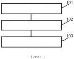
- FIG. 1 illustrates a flowchart of a CEST imaging correction method in one embodiment of the present disclosure.
- FIG. 2 illustrates a flowchart of a CEST imaging correction method in another embodiment of the present disclosure.
- FIG. 3 illustrates a method of selecting interpolated pixel points in one embodiment of the present disclosure.
- FIG. 4 illustrates the interpolation of four interpolated pixel points in one embodiment of the present disclosure.
- FIG. 5 illustrates the interpolation of six interpolated pixel points in one embodiment of the present disclosure.
- FIG. 6 illustrates an example of only a B0 field correction for CEST imaging in an embodiment of the present disclosure.
- FIG. 7 illustrates an example of a correction of a brain CEST image by using the B0 field correction method for CEST imaging in an embodiment of the present disclosure.
- FIG. 8 illustrates an example of a B1 field correction for CEST imaging after a B0 field correction in an embodiment of the present disclosure.
- FIG. 9 illustrates an example of a correction of a brain CEST image by using the method of first correcting the B0 field and then the B1 field for CEST imaging in an embodiment of the present disclosure.
- FIG. 10 illustrates another example of a correction of a brain CEST image by using the method of first correcting the B0 field and then the B1 field for CEST imaging in an embodiment of the present disclosure.
- FIG. 11 illustrates an example of a correction of a brain CEST image by using the method of simultaneously correcting the B0 field and the B1 field in an embodiment of the present disclosure.
- FIG. 12 shows the B0 map adopted in the examples shown in FIGS. 6 to 11 in an embodiment of the present disclosure.
- FIG. 13 shows the B1 map adopted in the examples shown in FIGS. 7 to 11 in an embodiment of the present disclosure.
- FIG. 14 shows the structure of a CEST imaging correction device in one embodiment of the present disclosure.
- FIG. 15 shows the structure of a CEST imaging correction device in another embodiment of the present disclosure.
- FIG. 1 illustrates a flowchart of a CEST imaging correction method in one embodiment of the present disclosure and the method comprises the following steps:
- Step 101 Collect a plurality of CEST images in an imaging area.
- Step 102 For each pixel point in each CEST image, calculate the corresponding real saturation frequency of the pixel point according to the predetermined frequency deviation of the water resonance frequency of the corresponding voxel of the pixel point relative to the corresponding water resonance frequency of the main magnetic field and the frequency of the pre-saturated RF pulse used for collecting the CEST image, and calculate the corresponding real saturation B1 field intensity of the pixel point according to the predetermined conversion coefficient between the real saturation B1 field intensity of the corresponding voxel of the pixel point and the system B1 field intensity and the intensity of the pre-saturated RF pulse used for collecting the CEST image, wherein the system B1 field intensity is equal to the intensity of the pre-saturated RF pulse used for collecting the CEST image.
- the real saturation frequency is the saturation frequency relative to the water resonance frequency.
- f(m) is the frequency of the pre-saturated RF pulse used for collecting the m th CEST image
- ⁇ is the gyromagnetic ratio of a spinning nucleus
- ⁇ B0(n) is the magnetic field deviation of the corresponding voxel of the n th pixel point in each CEST image relative to the main magnetic field
- ⁇ B0(n) is the frequency deviation of the corresponding water resonance frequency of the corresponding voxel of the n th pixel point in each CEST image relative to the main magnetic field
- f real (m,n) is the corresponding real saturation frequency of the n th pixel point in the m th CEST image
- n and m are positive integers
- M is the total number of times CEST images are collected
- N is the total number of pixel points in each CEST image.
- B1(m) is the intensity of the pre-saturated RF pulse used for collecting the m th CEST image
- B1map(n) is the conversion coefficient between the corresponding real saturation B1 field intensity of the n th pixel point in the m th CEST image and the system B1 field intensity
- the system B1 field intensity is the intensity of the pre-saturated RF pulse used for collecting the m th CEST image
- B1 real (m,n) is the corresponding real saturation intensity of the n th pixel point in the m th CEST image
- m and n are positive integers
- M is the total number of times CEST images are collected
- N is the total number of pixel points in each CEST image.
- Step 103 For pixel points corresponding to the same voxel in an imaging area in all CEST images, comparing the corresponding real saturation frequency and real saturation B1 field intensity of each pixel point with the target saturation frequency and target saturation B1 field intensity, respectively, selecting a plurality of pixel points as interpolated pixel points according to the comparison results, and performing interpolation calculations for the pixel values of the selected interpolated pixel points in the CEST images to obtain the pixel value of the corresponding voxel with the target saturation frequency and target saturation B1 field intensity.
- a value may be interpolated from the selected (or all) CEST images to approximate the value of the same voxel of the CEST image having both the target saturation frequency and the target saturation b1 field intensity.
- comparing the corresponding real saturation frequency and real saturation B1 field intensity of each pixel point with the target saturation frequency and target saturation B1 field intensity, respectively, and selecting a plurality of pixel points as interpolated pixel points according to the comparison results comprises:
- Step 1031 Pre-establish a 2-dimensional coordinate system, with the X-axis representing f real and the Y-axis representing B1 real .
- Step 1032 Denote the n th pixel point in all CEST images as (f real (m,n), B1 real (m,n)) respectively in the coordinate system, meanwhile denoting (f target ,B1 target ) in the coordinate system (in this case, there are M+1 points in the coordinate system), and then finding P points which are the closest to (f target ,B1 target ) but are not in a line from M pixel points (f real (m,n), B1 real (m,n)), wherein the value of P is determined by a preset interpolation algorithm,
- f real (m,n) is the corresponding real saturation frequency of the n th pixel point in the m th CEST image
- B1 real (m,n) is the corresponding real saturation intensity of the n th pixel point in the m th CEST image
- f target is the target saturation frequency
- B1 target is the target saturation B1 field intensity
- m and n are positive integers
- M is the total number of times CEST images are collected
- N is the total number of pixel points in each CEST image.
- comparing the corresponding real saturation frequency and real saturation B1 field intensity of each pixel point with the target saturation frequency and target saturation B1 field intensity, respectively, and selecting a plurality of pixel points as interpolated pixel points according to the comparison results comprises:
- Step 1033 Pre-establish a 2-dimensional coordinate system, with the X-axis representing f real and the Y-axis representing B Leal.
- Step 1034 Denote the n th pixel point in all CEST images as (f real (m,n), B1 real (m,n)) respectively in the coordinate system, and meanwhile denoting (f target ,B1 target ) in the coordinate system (in this case, there are M+1 points in the coordinate system).
- Step 1035 For each pixel point (f real (m,n),B1 real (m,n)), let the pixel point and every other P ⁇ 1 pixel points form a polygon of P sides according to the 2-dimensional coordinates (f real (m,n),B1 real (m,n)).
- Step 1036 Find the polygon of P sides which is the closest to (f target ,B1 target ) and using P vertexes of the polygon of P sides as interpolated pixel points,
- f real (m,n) is the corresponding real saturation frequency of the n th pixel point in the m th CEST image
- B1 real (m,n) is the corresponding real saturation intensity of the n th pixel point in the m th CEST image
- f target is the target saturation frequency
- B1 target is the target saturation B1 field intensity
- m and n are positive integers
- M is the total number of times CEST images are collected
- N is the total number of pixel points in each CEST image
- the value of P is determined by a preset interpolation algorithm.
- the corresponding real saturation frequency of each pixel point is compared with the target saturation frequency
- the corresponding real saturation B1 field intensity of each pixel point is compared with the target saturation B1 field intensity
- a plurality of pixel points are selected as interpolated pixel points according to the comparison results.
- FIG. 2 is a flowchart of a CEST imaging correction method in another embodiment of the present disclosure, the method comprising the following steps:
- Step 201 Pre-obtain the B0 map for describing the frequency deviation ⁇ B0(n) of the water resonance frequency of each voxel in an imaging area relative to the corresponding water resonance frequency of the main magnetic field, and the B1 map for describing the conversion coefficient B1map(n) between the real saturation B1 field intensity of each voxel in an imaging area and the system B1 field intensity.
- the collection of the B0 map and the B1 map is a mature technology.
- n represents the n th voxel in an imaging area
- n is a positive integer, 1 ⁇ n ⁇ N
- N is the total number of voxels in an imaging area.
- the system B1 field intensity is the intensity of the pre-saturated RF pulse used for collecting the CEST image.
- Step 202 Preset a target saturation frequency f target and a target saturation B1 field intensity B1 target , and set the number of times CEST images are collected and the frequency and intensity of the pre-saturated RF pulse used each time a CEST image is collected.
- the target saturation frequency is f target
- the target saturation B1 field intensity is B1 target .
- f target is the resonance frequency of the solute pool or the contralateral frequency of the resonance frequency.
- a plurality of f target or/and a plurality of B1 target will be set in this case.
- the frequency and intensity of the pre-saturated RF pulse adopted each time a CEST image is collected are set, for the frequency, the chemical shift frequency +f of the solute pool relative to the water pool and the contralateral frequency ⁇ f need to be set at least once, and then other frequencies are set according to a fixed or variable frequency change step ⁇ f; when the frequency of the pre-saturated RF pulse is set, the intensity of the pre-saturated RF pulse is set.
- the target saturation B1 field intensity B1 target is set once, and then other intensities are set according to the preset fixed or variable intensity change step ⁇ B1.
- Step 203 Emit pre-saturated RF pulses having the corresponding frequencies and intensities to an imaging area according to the set frequency and intensity of the pre-saturated RF pulse used each time a CEST image is collected to obtain a plurality of CEST images.
- Step 204 Perform Step 205 for each pixel point in each CEST image.
- Each CEST image contains N pixel points and each pixel point corresponds to one voxel in an imaging area.
- f(m) is the frequency of the pre-saturated RF pulse used for collecting the m th CEST image
- B1(m) is the intensity of the pre-saturated RF pulse used for collecting the m th CEST image
- m and n are positive integers
- M is the total number of times CEST images are collected
- N is the total number of pixel points in each CEST image, namely, the total number of voxels in an imaging area.
- Step 206 When calculating the corresponding f real (m, n) and B1 real (m,n) of all pixel points in all CEST images, perform Steps 207 and 208 for each voxel in an imaging area:
- Step 207 Let the current voxel be the n th voxel in an imaging area, then find the interpolated pixel point of the n th voxel in an imaging area from the n th pixel point in all CEST images according to f target , B1 target , and f real (m,n) and B1 real (m,n) of the n th pixel point in all CEST images.
- n th pixel point in all CEST images is denoted as (f real (m,n), B1 real (m,n)) respectively in the coordinate system, and meanwhile (f target ,B1 target ) is denoted in the coordinate system.
- M+1 points in the coordinate system there are M+1 points in the coordinate system.
- P points which are the closest to (f target ,B1 target ) but are not in a line are found from M pixel points (f real (m,n), B1 real (m,n)), and P points are interpolated pixel points.
- the value of P is determined by a preset interpolation algorithm. In practical applications, the value of P may vary with the interpolation algorithms adopted. Usually, the larger the value of P is, the better the interpolation effect. To achieve a good interpolation effect, it is usually required that P ⁇ 3.
- M pixel points in the same position in all CEST images namely, the pixel points corresponding to the same voxel in an imaging area in all CEST images, for example, are the n th pixel point in the first CEST image, the n th pixel point in the second CEST image, the n th pixel point in the third CEST image, . . . , the n th pixel point in the (M ⁇ 1) th CEST image, and the n th pixel point in the M th CEST image, wherein 1 ⁇ n ⁇ N.
- the 2-dimensional coordinates (f real (m,n),B1 real (m,n)) of five n th pixel points can be obtained, wherein 1 ⁇ m ⁇ 5 and the five n th pixel points correspond to A, B, C, D and E, respectively in FIG.
- Step 208 when an interpolation calculation needs to be performed, the triangle which the point (f target ,B1 target ) is closest to is found according to the coordinates (f real (m,n),B1 real (m,n)) of three vertexes of each triangle, and the pixel values of the three vertexes of the triangle in the CEST image are used for an interpolation or extrapolation calculation to obtain the pixel value of the n th voxel in an imaging area with f target and B1 target .
- point Y (f target , B1 target ) is closest to the triangle formed by points A, B and C, and then points A, B and C are used as interpolated pixel points for an interpolation calculation.
- FIG. 5 shows the interpolation of six irregularly sampled interpolated pixel points (f real (m,n),B1 real (m,n)), wherein points A, B, C, D, E and F are the pixel points in the same position in six CEST images, and interpolation calculations are performed for the pixel values of the six points to obtain the pixel values of the corresponding voxel at point Y (f target ,B1 target ).
- Step 208 Use the preset interpolation algorithm to perform interpolation calculations for the pixel values of all found interpolated pixel points in the CEST image to obtain the pixel value of the n th voxel in an imaging area with f target and B1 target .
- CEST imaging is only limited to amide proton transfer (APT) imaging. It should be noted that only an example is given here and the scope of the present disclosure is not limited to APT-CEST imaging.
- APT imaging is a branch of CEST imaging technology the purpose of which is to detect amide protons whose chemical shift frequency is 3.5 ppm.
- APT imaging at least three collection processes are required, and the frequencies of the RF pulses used in three collection processes are: a saturation frequency ⁇ 3.5 ppm, a saturation frequency 3.5 ppm, and an unsaturation frequency.
- a common method is the six point method and the frequencies of RF pulses used during six collection processes are ⁇ 3 ppm, ⁇ 3.5 ppm and ⁇ 4 ppm.
- the method of simultaneously correcting the B0 field and the B1 field is compared with the method of correcting only the B0 field and the method of first correcting the B0 field and then correcting the B1 field.
- FIG. 6 shows an example of only a B0 field correction for CEST imaging.
- a 2-dimensional coordinate system is pre-established, with the X-axis representing f real , and the Y-axis representing B1.
- the n th voxels in the collected CEST images are respectively denoted as (f real (m,n),B1) in the coordinate system. Since the target saturation frequency f target is consistent with the resonance frequency in FIG. 6 and only a B0 field correction is performed in FIG. 6 , only the distance between f real (m,n) and f target needs to be considered in this case. Therefore, FIG.
- two points which are the closest to point Y are found from three points A, B and C according to the difference between f real (m,n) and f target .
- point A (f target ⁇ f ⁇ B0(n), B1 target ) and point B (f target ⁇ B0(n), B1 target ) are the two points closest to point Y (f target , B1 target ).
- Interpolation calculations are performed according to the pixel values of the two points A and B in the CEST image to obtain the pixel value of the n th voxel in an imaging area with f target and B1 target .
- FIG. 7 shows an example of a correction of a brain CEST image by using the B0 field correction method for CEST imaging. It can be seen that the image value of the corrected brain cross-sections arranged above is lower than the image value of the corrected brain cross-sections arranged below. For example, in FIG. 7 , the image value of the cross-section indicated by an arrow is significantly lower than the image value of the cross-sections below. This is caused by the non-uniformity of the B1 field because only the B0 field is corrected but the B1 field is not corrected in this method.
- point Y3 (f target , (B1 target + ⁇ B1)*B1map(n)) and point Y2 (f target , B1 target *B1map(n)) are the two points closest to point (f target , B1 target ), and then interpolation calculations are performed according to the interpolation calculation results of these two points after the B0 field correction to obtain the pixel value of the n th voxel in an imaging area with f target and B1 target .
- FIG. 9 shows an example of a correction of a brain CEST image by using the method of first correcting the B0 field and then the B1 field for CEST imaging.
- four interpolation points are used.
- An over-correction phenomenon can be seen.
- the image value of the cross-section indicated by the arrow in FIG. 9 is higher than the image values of the cross-sections below. This may be caused by a small number of interpolation points.
- FIG. 10 shows another example of a correction of a brain CEST image by using the method of first correcting the B0 field and then the B1 field for CEST imaging.
- nine interpolation points are used. It can be seen that the over-corrected area around the frontal lobe of the sections arranged above is reduced, compared with FIG. 9 .
- a good correction effect is provided in the present example, but CEST images need to be collected 18 times, which is two times larger than in the common six point method.
- FIG. 11 shows an example of a correction of a brain CEST image by using the method of simultaneously correcting the B0 field and the B1 field.
- five interpolation points are used. It can be seen that the over-corrected area around the frontal lobe of the sections arranged above is reduced, compared with FIG. 9 .
- the correction effect is similar to the one in FIG. 10 , but CEST images need to be collected 18 times, which is twice larger than in the common six point method.
- FIG. 12 shows the B0 map adopted in the examples shown in FIGS. 6 to 11
- FIG. 13 shows the B1 map adopted in FIGS. 7 to 11 .
- the time spent in the above-mentioned examples is compared.
- the time starts from the collection of CEST images and ends when the final corrected image is obtained.
- the time spent in the example shown in FIG. 6 is 2:50 (2 minutes and 50 seconds)
- the time spent in the example shown in FIG. 9 is 3:10 (3 minutes and 10 seconds)
- the time spent in the example shown in FIG. 10 is 6:15 (6 minutes and 15 seconds)
- the time spent in the example shown in FIG. 11 is 3:30 (3 minutes and 30 seconds). It can be seen that the method (the example shown in FIG. 11 ) provided by the present disclosure requires almost half the time to achieve the same correction effect, compared with the example shown in FIG. 10 .
- FIG. 14 shows the structure of a CEST imaging correction device 140 in one embodiment of the present disclosure, the device mainly comprising:
- a real saturation frequency and intensity calculation module (one or more processors, processing circuitry, processors executing machine readable instructions, etc.) 141 , configured, for each pixel point in each CEST image, to calculate the corresponding real saturation frequency of the pixel point according to the predetermined frequency deviation of the water resonance frequency of the corresponding voxel of the pixel point relative to the corresponding water resonance frequency of the main magnetic field and the frequency of the pre-saturated RF pulse used for collecting the CEST image, and calculate the corresponding real saturation B1 field intensity of the pixel point according to the predetermined conversion coefficient between the real saturation B1 field intensity of the corresponding voxel of the pixel point and the system B1 field intensity and the intensity of the pre-saturated RF pulse used for collecting the CEST image when a plurality of CEST images in an imaging area are collected, and
- an interpolation module (one or more processors, processing circuitry, processors executing machine readable instructions, etc.) 142 , configured, for pixel points corresponding to the same voxel in an imaging area in all CEST images, to compare the corresponding real saturation frequency and real saturation B1 field intensity of each pixel point with the target saturation frequency and target saturation B1 field intensity, respectively, according to the corresponding real saturation frequency and target saturation B1 field intensity calculated by the real saturation frequency and intensity calculation module 141 , select a plurality of pixel points as interpolated pixel points according to the comparison results, and perform interpolation calculations for the pixel values of the selected interpolated pixel points in the CEST images to obtain the pixel value of the corresponding voxel with the target saturation frequency and target saturation B1 field intensity.
- an interpolation module one or more processors, processing circuitry, processors executing machine readable instructions, etc.
- f(m) is the frequency of the pre-saturated RF pulse used for collecting the m th CEST image
- ⁇ is the gyromagnetic ratio of a spinning nucleus
- ⁇ B0(n) is the magnetic field deviation of the corresponding voxel of the n th pixel point in each CEST image relative to the main magnetic field
- ⁇ B0(n) is the frequency deviation of the corresponding water resonance frequency of the corresponding voxel of the n th pixel point in each CEST image relative to the main magnetic field
- f real (m,n) is the corresponding real saturation frequency of the n th pixel point in the m th CEST image
- n and m are positive integers
- M is the total number of times CEST images are collected
- N is the total number of pixel points in each CEST image.
- B1(m) is the intensity of the pre-saturated RF pulse used for collecting the m th CEST image
- B1map(n) is the conversion coefficient between the corresponding real saturation B1 field intensity of the n th pixel point in the m th CEST image and the system B1 field intensity
- the system B1 field intensity is the intensity of the pre-saturated RF pulse used for collecting the m th CEST image
- B1 real (m,n) is the corresponding real saturation intensity of the n th pixel point in the m th CEST image
- m and n are positive integers
- M is the total number of times CEST images are collected
- N is the total number of pixel points in each CEST image.
- the interpolation module 142 comparing the corresponding real saturation frequency and real saturation B1 field intensity of each pixel point with the target saturation frequency and target saturation B1 field intensity respectively according to the corresponding real saturation frequency and real saturation B1 field intensity calculated by the real saturation frequency and intensity calculation module 141 for each pixel point comprises:
- f real (m,n) is the corresponding real saturation frequency of the n th pixel point in the m th CEST image
- B1 real (m,n) is the corresponding real saturation intensity of the n th pixel point in the m th CEST image
- f target is the target saturation frequency
- B1 target is the target saturation B1 field intensity
- m and n are positive integers
- M is the total number of times CEST images are collected
- N is the total number of pixel points in each CEST image
- the value of P is determined by a preset interpolation algorithm.
- FIG. 15 shows the structure of a CEST imaging correction device 150 in another embodiment of the present disclosure, the device mainly comprising a processor 151 and a memory 152 , wherein
- an application which can be executed by the processor 151 to enable the processor 151 to perform the steps of the CEST imaging correction method, namely, Steps 101 to 103 or Steps 201 to 208 , is stored in the memory 152 .
- a non-transitory computer-readable storage medium is provided in one embodiment of the present disclosure, a computer program is stored in the non-transitory computer-readable storage medium, and the computer program performs the steps of the CEST imaging correction method, namely, Steps 101 to 103 or Steps 201 to 208 when executed by at least one processor.
- Machine-readable instructions are stored in the non-transitory computer-readable storage medium, and when the machine-readable instructions are executed by at least processor, the at least one processor will execute any of the methods described in the previous embodiments.
- a system or device equipped with a non-transitory computer-readable storage medium can be provided.
- Software program codes which can realize the function in any of the above-mentioned embodiments are stored in the readable storage medium and the computer or one or more processors of the system or device can read out and execute the machine-readable instructions stored in the non-transitory computer-readable storage medium.
- program codes read from the non-transitory computer-readable storage medium can themselves realize the function in any of the above-mentioned embodiments. Therefore, machine-readable codes and the non-transitory computer-readable storage medium where machine-readable codes are stored constitute a part of the present disclosure.
- Embodiments of non-transitory computer-readable storage media include floppy disks, hard disks, magneto-optical disks, compact disks (for example, compact disks read-only memory (CD-ROM)), compact disks—recordable (CD-R), compact disks—rewritable (CD-RW), digital video disks—read only memory (DVD-ROM), digital versatile disks—random access memory (DVD-RAM), digital versatile disks—recordable (DVD-RW), digital versatile disks—rewritable (DVD+RW)), magnetic tape, non-volatile memory cards, and read-only memory (ROM).
- program codes can be downloaded from the server computer or cloud over a communication network.
Landscapes
- Physics & Mathematics (AREA)
- General Physics & Mathematics (AREA)
- High Energy & Nuclear Physics (AREA)
- Condensed Matter Physics & Semiconductors (AREA)
- Engineering & Computer Science (AREA)
- Spectroscopy & Molecular Physics (AREA)
- Theoretical Computer Science (AREA)
- Optics & Photonics (AREA)
- Health & Medical Sciences (AREA)
- General Health & Medical Sciences (AREA)
- Nuclear Medicine, Radiotherapy & Molecular Imaging (AREA)
- Radiology & Medical Imaging (AREA)
- Signal Processing (AREA)
- Magnetic Resonance Imaging Apparatus (AREA)
Abstract
Description
f real(m,n)=f(m)−γΔB0(n)
B1real(m,n)=B1(m)*B1map(n)
f real(m,n)=f(m)−γΔB0(n)
B1real(m,n)=B1(m)*B1map(n)
| Reference | |
| numeral | Meaning |
| 101-103 | Step |
| 201-208 | |
| 140 | CEST imaging correction device in one |
| embodiment of the |
|
| 141 | Real saturation frequency and |
| calculation module | |
| 142 | |
| 150 | CEST imaging correction device in another |
| embodiment of the |
|
| 151 | |
| 152 | Memory |
| 101-103 | Step |
| 201-208 | |
| 140 | CEST imaging correction device in one |
| embodiment of the |
|
| 141 | Real saturation frequency and |
| calculation module | |
| 142 | |
| 150 | CEST imaging correction device in another |
| embodiment of the |
|
| 151 | |
| 152 | Memory |
f real(m,n)=f(m)−γΔB0(n)
B1real(m,n)=B1(m)*B1map(n)
f real(m,n)=f(m)−γΔB0(n)
B1real(m,n)=B1(m)*B1map(n)
Claims (11)
freal(m,n)=f(m)−γΔB0(n), wherein:
B1real(m,n)=B1(m)*B1map(n), wherein:
freal(m,n)=f(m)−γΔB0(n), wherein:
B1real(m,n)=B1(m)*B1map(n), wherein:
Applications Claiming Priority (2)
| Application Number | Priority Date | Filing Date | Title |
|---|---|---|---|
| CN202010526773.9 | 2020-06-10 | ||
| CN202010526773.9A CN113777545A (en) | 2020-06-10 | 2020-06-10 | Chemical exchange saturation transfer imaging correction method and device and readable storage medium |
Publications (2)
| Publication Number | Publication Date |
|---|---|
| US20210389404A1 US20210389404A1 (en) | 2021-12-16 |
| US11726159B2 true US11726159B2 (en) | 2023-08-15 |
Family
ID=78824286
Family Applications (1)
| Application Number | Title | Priority Date | Filing Date |
|---|---|---|---|
| US17/343,014 Active 2042-01-28 US11726159B2 (en) | 2020-06-10 | 2021-06-09 | B0 and B1 correction anti-respectively |
Country Status (2)
| Country | Link |
|---|---|
| US (1) | US11726159B2 (en) |
| CN (1) | CN113777545A (en) |
Families Citing this family (2)
| Publication number | Priority date | Publication date | Assignee | Title |
|---|---|---|---|---|
| EP3511728A1 (en) * | 2018-01-12 | 2019-07-17 | Koninklijke Philips N.V. | Single-point dixon method for fat-water separation in chemical exchange saturation transfer magnetic resonance imaging |
| CN115561691B (en) * | 2022-09-28 | 2023-07-21 | 深圳市联影高端医疗装备创新研究院 | Radio frequency emission field correction method, device, computer equipment and storage medium |
Citations (1)
| Publication number | Priority date | Publication date | Assignee | Title |
|---|---|---|---|---|
| US11307271B2 (en) * | 2018-01-12 | 2022-04-19 | Koninklijke Philips N.V. | MRI method for determining a magnetic field map from a B0 reference scan and a WASSR scan |
Family Cites Families (3)
| Publication number | Priority date | Publication date | Assignee | Title |
|---|---|---|---|---|
| CN104997511A (en) * | 2015-06-01 | 2015-10-28 | 中国科学院深圳先进技术研究院 | CESTR measuring method and system for magnetic resonance chemical exchange saturation transfer imaging |
| EP3575814A1 (en) * | 2018-05-29 | 2019-12-04 | Koninklijke Philips N.V. | Motion detection in cest magnetic resonance imaging based on z-spectrum analysis |
| CN110780249B (en) * | 2019-11-21 | 2020-08-11 | 中国科学院武汉物理与数学研究所 | Magnetic resonance imaging method using adiabatic radio frequency pulses to measure radio frequency B1 field distribution |
-
2020
- 2020-06-10 CN CN202010526773.9A patent/CN113777545A/en active Pending
-
2021
- 2021-06-09 US US17/343,014 patent/US11726159B2/en active Active
Patent Citations (1)
| Publication number | Priority date | Publication date | Assignee | Title |
|---|---|---|---|---|
| US11307271B2 (en) * | 2018-01-12 | 2022-04-19 | Koninklijke Philips N.V. | MRI method for determining a magnetic field map from a B0 reference scan and a WASSR scan |
Non-Patent Citations (1)
| Title |
|---|
| Phillip Zhe Sun et al., Correction for Artifacts Induced by B0 and B1 Field Inhomogeneities in pH-Sensitive Chemical Exchange Saturation Transfer (CEST) Imaging (Year: 2007). * |
Also Published As
| Publication number | Publication date |
|---|---|
| US20210389404A1 (en) | 2021-12-16 |
| CN113777545A (en) | 2021-12-10 |
Similar Documents
| Publication | Publication Date | Title |
|---|---|---|
| Wang et al. | Model‐based T 1 mapping with sparsity constraints using single‐shot inversion‐recovery radial FLASH | |
| Veraart et al. | Comprehensive framework for accurate diffusion MRI parameter estimation | |
| US11726159B2 (en) | B0 and B1 correction anti-respectively | |
| Ebel et al. | Assessment of 3D proton MR echo‐planar spectroscopic imaging using automated spectral analysis | |
| US11029381B2 (en) | Method for varying undersampling dimension for accelerating multiple-acquisition magnetic resonance imaging and device for the same | |
| US9791531B2 (en) | Establishing a magnetic resonance system actuation sequence | |
| Shi et al. | Parallel imaging and compressed sensing combined framework for accelerating high‐resolution diffusion tensor imaging using inter‐image correlation | |
| US7777488B2 (en) | Methods for arbitrary shape selective excitation summed spectroscopy and applications of same | |
| US20050212517A1 (en) | Reduction of susceptibility artifacts in subencoded single-shot magnetic resonance imaging | |
| Windischberger et al. | Robust field map generation using a triple‐echo acquisition | |
| Keating et al. | Real‐time dynamic frequency and shim correction for single‐voxel magnetic resonance spectroscopy | |
| US9404987B2 (en) | Method for correcting image distortion and system, and magnetic resonance imaging equipment | |
| Ye et al. | Noncontrast‐enhanced magnetic resonance angiography and venography imaging with enhanced angiography | |
| Hancu et al. | Distortion correction in diffusion‐weighted imaging of the breast: Performance assessment of prospective, retrospective, and combined (prospective+ retrospective) approaches | |
| US20170108571A1 (en) | Epi ghost correction involving sense | |
| CN1820208A (en) | Shimming of mri scanner involving fat suppression and/or black blood preparation | |
| Benkhedah et al. | Evaluation of adaptive combination of 30‐channel head receive coil array data in 23 N a MR imaging | |
| Chen et al. | Self‐calibrating wave‐encoded variable‐density single‐shot fast spin echo imaging | |
| US20200300949A1 (en) | Chemical exchange saturation transfer - magnetic resonance imaging (cest-mri) sequence generating method, apparatus and readable storage medium | |
| Wang et al. | Analytical three‐point Dixon method: With applications for spiral water–fat imaging | |
| Hermann et al. | Magnetic resonance fingerprinting for simultaneous renal T1 and T2* mapping in a single breath‐hold | |
| Benkert et al. | Hybrid T2‐and T1‐weighted radial acquisition for free‐breathing abdominal examination | |
| Vegh et al. | Selective channel combination of MRI signal phase | |
| US20100303320A1 (en) | Systems, methods and machine readable programs for enhanced fat/water separation in magnetic resonance imaging | |
| Gaggl et al. | High‐resolution reduced field of view diffusion tensor imaging using spatially selective RF pulses |
Legal Events
| Date | Code | Title | Description |
|---|---|---|---|
| FEPP | Fee payment procedure |
Free format text: ENTITY STATUS SET TO UNDISCOUNTED (ORIGINAL EVENT CODE: BIG.); ENTITY STATUS OF PATENT OWNER: LARGE ENTITY |
|
| STPP | Information on status: patent application and granting procedure in general |
Free format text: DOCKETED NEW CASE - READY FOR EXAMINATION |
|
| AS | Assignment |
Owner name: SIEMENS HEALTHCARE GMBH, GERMANY Free format text: ASSIGNMENT OF ASSIGNORS INTEREST;ASSIGNOR:SIEMENS HEALTHINEERS LTD.;REEL/FRAME:058198/0607 Effective date: 20210726 Owner name: SIEMENS HEALTHCARE GMBH, GERMANY Free format text: ASSIGNMENT OF ASSIGNORS INTEREST;ASSIGNOR:LIEBIG, PATRICK, MR.;REEL/FRAME:058198/0516 Effective date: 20211109 Owner name: SIEMENS HEALTHINEERS LTD., CHINA Free format text: ASSIGNMENT OF ASSIGNORS INTEREST;ASSIGNOR:HSU, YI-CHENG;REEL/FRAME:058198/0458 Effective date: 20210714 |
|
| STPP | Information on status: patent application and granting procedure in general |
Free format text: PUBLICATIONS -- ISSUE FEE PAYMENT VERIFIED |
|
| STCF | Information on status: patent grant |
Free format text: PATENTED CASE |
|
| AS | Assignment |
Owner name: SIEMENS HEALTHINEERS AG, GERMANY Free format text: ASSIGNMENT OF ASSIGNORS INTEREST;ASSIGNOR:SIEMENS HEALTHCARE GMBH;REEL/FRAME:066267/0346 Effective date: 20231219 |