US11666423B2 - Dental implant replica for a dental model manufactured with 3D additive technology - Google Patents
Dental implant replica for a dental model manufactured with 3D additive technology Download PDFInfo
- Publication number
- US11666423B2 US11666423B2 US16/317,907 US201716317907A US11666423B2 US 11666423 B2 US11666423 B2 US 11666423B2 US 201716317907 A US201716317907 A US 201716317907A US 11666423 B2 US11666423 B2 US 11666423B2
- Authority
- US
- United States
- Prior art keywords
- replica
- dental
- dental model
- proximal end
- hole
- Prior art date
- Legal status (The legal status is an assumption and is not a legal conclusion. Google has not performed a legal analysis and makes no representation as to the accuracy of the status listed.)
- Active, expires
Links
Images


Classifications
-
- A—HUMAN NECESSITIES
- A61—MEDICAL OR VETERINARY SCIENCE; HYGIENE
- A61C—DENTISTRY; APPARATUS OR METHODS FOR ORAL OR DENTAL HYGIENE
- A61C13/00—Dental prostheses; Making same
- A61C13/34—Making or working of models, e.g. preliminary castings, trial dentures; Dowel pins [4]
-
- A—HUMAN NECESSITIES
- A61—MEDICAL OR VETERINARY SCIENCE; HYGIENE
- A61C—DENTISTRY; APPARATUS OR METHODS FOR ORAL OR DENTAL HYGIENE
- A61C13/00—Dental prostheses; Making same
- A61C13/0003—Making bridge-work, inlays, implants or the like
- A61C13/0006—Production methods
- A61C13/0013—Production methods using stereolithographic techniques
-
- A—HUMAN NECESSITIES
- A61—MEDICAL OR VETERINARY SCIENCE; HYGIENE
- A61C—DENTISTRY; APPARATUS OR METHODS FOR ORAL OR DENTAL HYGIENE
- A61C13/00—Dental prostheses; Making same
- A61C13/0003—Making bridge-work, inlays, implants or the like
- A61C13/0006—Production methods
- A61C13/0019—Production methods using three dimensional printing
-
- A—HUMAN NECESSITIES
- A61—MEDICAL OR VETERINARY SCIENCE; HYGIENE
- A61C—DENTISTRY; APPARATUS OR METHODS FOR ORAL OR DENTAL HYGIENE
- A61C9/00—Impression cups, i.e. impression trays; Impression methods
- A61C9/002—Means or methods for correctly replacing a dental model, e.g. dowel pins; Dowel pin positioning means or methods
-
- A—HUMAN NECESSITIES
- A61—MEDICAL OR VETERINARY SCIENCE; HYGIENE
- A61C—DENTISTRY; APPARATUS OR METHODS FOR ORAL OR DENTAL HYGIENE
- A61C8/00—Means to be fixed to the jaw-bone for consolidating natural teeth or for fixing dental prostheses thereon; Dental implants; Implanting tools
-
- B—PERFORMING OPERATIONS; TRANSPORTING
- B29—WORKING OF PLASTICS; WORKING OF SUBSTANCES IN A PLASTIC STATE IN GENERAL
- B29C—SHAPING OR JOINING OF PLASTICS; SHAPING OF MATERIAL IN A PLASTIC STATE, NOT OTHERWISE PROVIDED FOR; AFTER-TREATMENT OF THE SHAPED PRODUCTS, e.g. REPAIRING
- B29C35/00—Heating, cooling or curing, e.g. crosslinking or vulcanising; Apparatus therefor
Definitions
- the present invention of a digital dental implant replica for a dental model manufactured with 3D additive technology relates to a digital dental implant replica to simulate the position of the dental implant in a dental model which has been produced by means of 3D printing processes or CAD/CAM systems, providing greater precision and maneuverability with respect to analog systems in the orientation of the implant in the dental model due to a simple retention system which is easy to use and detachable. It likewise refers to a system made up of the replica and the dental model.
- the digital dental implant replica is a compact body which preferably begins in a cylindrical shape and preferably ends in a conical shape, although another fifteen geometric shapes are possible.
- the replica is formed by an implant connection, an initial body and a final body.
- the digital dental implant replica is especially designed to be used with dental models which have been digitally processed using CAD-CAM technology and 3D additive technology (3D printing) after scanning the mouth of the patient.
- CAD-CAM technology and 3D additive technology (3D printing) after scanning the mouth of the patient.
- 3D printing 3D additive technology
- the present invention is especially intended be applied in the dental industry, and more specifically the area of prosthetic dentistry.
- Replicas of dental implants are used for the production of dental models which reproduce the position of the implant in the patient's mouth.
- Known in the state of the art are analog replicas for dental models in which a negative is first taken of the jaw, using impression material, and by means of impression posts the position of the implant is reproduced. Once the paste solidifies, the impression tray is removed and an impression is produced. The replica is inserted, joined to the impression post through the connection of the replica, positioning it, and it is subsequently filled with plaster. After hardening, the model of the patient's mouth is obtained with the replica incorporated.
- the positioning of the replica becomes somewhat complex and does not allow the decoupling thereof without causing serious internal tensions in the dental model.
- a replica which incorporates the use of a standard sleeve which allows for the coupling and decoupling of the replica in the physical dental model, yet it is hardly advantageous in that it does not contain information which would make it possible for it to be inserted in a dental model produced by means of 3D printing or CAD/CAM systems.
- digital dental scanning has significantly improved the precision with which the position of the patient's implant is obtained. This has allowed for the incorporation of other systems which provide greater precision in the entire design and manufacturing process, as well as for the appearance of new elements which provide characteristics with advantages over the traditionally used analog replicas.
- analog dental implant replicas for analog dental models which are cylindrical with an outer threading on the lower end thereof by which they are attached to the physical model of the mouth by means screwing the same to a fastening nut.
- This is a complex attachment system which requires the use of tools for the positioning and tightening of said replicas, in addition to oversized assembly which is necessary, given that it requires a nut with an outer diameter greater than that of the replica.
- implant replicas which have a cylindrical or conical shape specially designed to be inserted in CAD/CAM models.
- the geometry of the replica includes on the lower part thereof a minimum of two cut surfaces, which allow the replica and the model to fit together on the intermediate plane between the two areas into which the replica is divided, which may cause it to be poorly set by basing much of its support on this surface which divides both areas of the replica as well as on the lower plane.
- dental implant replicas to be used in digital replicas of dental models manufactured with 3D additive technology or 3D printing, which allows for the manufacturing of the dental model after the patient's mouth has been scanned, thereby avoiding the previously described method using plaster.
- these dental models manufactured by means of additive technology, are manufactured by adding layers of material which are placed one on top of the other until the dental model is formed, they have the disadvantage that, in the manufacturing of inclined surfaces and, therefore, of holes with an inclination with respect to the base, micro-steps (each micro-step at the height of the layer of added printing material) are created, due to the varying heights of the added layers on those surfaces which should be continuous and/or curved.
- Analog dental implant replicas in the state of the art may not be used in dental models made with 3D additive technology since they cannot be adapted to the constructive limitations of this type of manufacturing and, therefore, are not able to achieve the necessary precision.
- the present invention relates to a digital dental implant replica according to claim 1 .
- the digital implant replica object of the present invention, is a dental implant replica for a hole of a dental model manufactured with 3D additive technology, and made in three dimensions, and to be placed on the longitudinal axis of the hole of said dental model, the replica being made up of a body with a proximal end and a distal end which determine between them a distal segment and a proximal segment, in which:
- the positioning means for positioning the replica in the dental model have said curved surface to adapt to the surfaces of the hole, which comprises micro-steps, and specifically to the bottom of the hole where, instead of a perfectly right angle, there is a curved groove formed by micro-steps produced by additive manufacturing.
- This curve on the end allows the necessary precision to be achieved when the replica is inserted in the 3D oral model, meaning great precision is achieved in mounting the replica in the dental model. This is so because, since said curve does not exist, when the hole of the dental model is inclined with respect to the base of said model, the imperfections caused by the micro-steps at the bottom of the hole would prevent the replica from being set in its definitive position, thus preventing its approximation to the real situation. Specifically, since the replica cannot be inserted into the support position, it would be partially outside the hole and would therefore be less precise.
- the angular and axial positioning means are preferably a flat surface, rising from a cross-sectioned surface of the body of revolution of the replica, while the additional height-positioning means are a notch located in the body of the replica and which corresponds, once the replica is installed, to a protrusion in the hole of the dental model.
- this notch is complementary to the curved surface at the proximal end which allows the replica to be placed at the correct height in the hole and prevent said curved surface from coming into contact with the corner formed by the surfaces at the bottom of the hole in the dental model.
- the replica For the connection of the replica to the dental model, the replica comprises an inner threading on the distal end thereof, wherein a fastening screw will be housed.
- connection means for connecting the replica to a dental prosthesis are a threaded perforation for the connection of the abutments or prosthetic attachments.
- connection means for connecting the replica to the dental model comprise connection elements for connecting to removable fastening means, said removable fastening means preferably being a screw inserted in the lower part of the dental model, in a cavity, until it butts against the inner surface of the dental model and being screwed into the inside of the replica, wherein the threading is located as a connection element. Thanks to these connection means, a detachable system is created between the implant replica and the dental model, according to claim 10 .
- the shape of the implant replica is complementary to the shape of the hole of the dental model in which it will be housed, such that said dental or oral model comprises a surface that is theoretically flat, but which in reality is not, due to the aforementioned limitations of 3D additive manufacturing which forms micro-steps on the inclined surfaces of the hole, and which the curved surface for vertical positioning of the implant replica avoids in order to achieve greater precision in the vertical positioning of the implant replica on the base of the hole.
- the foregoing is complemented by the flat surface in order to allow for the introduction of the implant replica, which coincides with the surface derived from sectioning the body of the replica and allows for the angular positioning thereof and, additionally, a protrusion which is housed in the notch of the implant replica.
- the assembly of the replica and the dental model forms the detachable system which allows for the fastening of the replica to the dental model in a simple way, thanks to the structure of the replica which, once inserted in the hole of the dental model, prevents it from moving vertically and/or axially or rotationally, facilitating the insertion of the connection or fitting means for connecting or fitting the replica to the dental model.
- object of the present invention on models manufactured with additive technology, it is first necessary to print the oral model in order to later place the replica in the same.
- Said oral model has holes created by the information stored in a CAD-CAM library.
- digital replicas object of the present invention
- the method followed would be similar to the previous one, in other words, based on CAM dental software, milling strategies are created for the oral model in order to mill said model with the holes where the replicas will be inserted.
- the material with which milling may be done is not limited to any specific material, and could be, for example, urethane or plaster, nor are there any limitations with regard to dimension or shape. This milling must be done on a five-axis machine.
- the work method would consist of:
- the CAD design will be made of said oral model by a prosthodontist.
- Said prosthodontist by means of a computer program or CAD software, will make the model of the patient's dental structure, in other words, designing what will be the patient's dental prosthesis, which is subsequently milled using metal, zicronium or polyether ether ketone (PEEK), as well as the design of the oral model, meaning the reproduction of the patient's mouth to later be printed in 3D.
- the file of said 3D oral model will have the holes in the complementary shape of the digital replica, object of the present invention, which is created from the data in the CAD libraries, designed exclusively for said digital replicas. As was previously mentioned, the hole will have the necessary skin or surface area to ensure said hole has the outer shape of the digital replica, such that after the oral model is printed, the digital replica, object of the present invention, is perfectly placed.
- a three-dimensional dental model is achieved which reproduces the patient's mouth with the hole or holes arranged wherein the dental implant or implants are located, such that said hole or holes allow for the perfect placement of the digital replica or replicas to be inserted.
- the hole, the replica and the scanbody are associated with one another through the CAD libraries which have been designed for said purpose.
- FIG. 1 shows a perspective view of the digital implant replica.
- FIG. 2 shows an elevation view of the replica.
- FIG. 3 shows a perspective view of the replica.
- FIG. 4 shows an elevation view of the replica.
- FIG. 5 shows a cross section of the proximal segment of an asymmetric implant replica.
- FIG. 6 shows the shows the cross section of the physical model wherein one can see the geometry of the predesigned hole in which the replica is inserted.
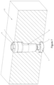
- FIG. 7 shows a partially sectioned perspective view with the replica inserted in the hole of the physical dental model (sectioned) and in which it may be seen how the dental model and the implant replica fit together.
- FIG. 8 shows a partial cross-sectional elevation view with the replica inserted in the physical dental model, in which the through-hole of the replica and the incorporation of the setting screw are shown.
- FIG. 9 shows a cross-sectional perspective view in which the replica, the dental model and the setting screw are seen.
- the replica 1 will be introduced in a dental model 10 of a patient to simulate the position of the dental implant in said dental model 10 , the implant replica 1 having been produced by means of 3D printing processes or CAD/CAM systems, in order to therefore provide greater precision and maneuverability in the mounting and orientation of the implant in the dental model 1 due to a simple retention system which is easy to use and detachable.
- the dental implant replica 1 Prior to the introduction of the dental implant replica 1 , it is necessary to have the dental model 10 of the patient wherein the replica 1 will be placed. Therefore, a scanning process of the element to be restored is carried out, obtaining a file with the virtual dental model 10 of the patient which includes the data of an intraoral scanbody, which subsequently, and by means of CAD-CAM libraries, allows a hole 11 to be created in the dental model 10 , intended to receive the replica 1 in order to be coupled to said hole 11 .
- This dental model 10 will be manufactured by means of 3D printing or additive technology. In this way, a highly precise personalized physical dental model 10 is obtained and incorporates a hole 11 adapted to the replica 1 .
- the geometry of the hole 11 is such that the replica of the implant 1 can be easily removed, constituting a detachable system which has a great advantage.
- the positioning of the replica 1 is done by means of retention mechanisms which impede the rotation thereof and guarantee its correct setting in the dental model 10 .
- the hole 11 envisaged for the insertion of the replica 1 is divided into three longitudinal zones.
- the first zone 11 a corresponds to the upper part of the digital replica 1 , and has a diameter, or outer measurement, adapted to the diameter or external measurement of the connection of the replica 1 .
- the second longitudinal zone 11 b preferably begins with a cylindrical section, or another geometric shape such as conical, and preferably continues with a conical shape, or another possible geometric shape, such as cylindrical, said second zone finishing in a curved surface 12 which serves as a vertical detention surface for the replica 1 .
- the assembly of the first zone 11 a and the second zone 11 b of the dental model 10 fit to the geometry of the replica 1 , to the distal segments 1 b and proximal segments 1 a .
- the third longitudinal zone 11 c is equivalent to the connection area of the replica 1 and has a cylindrical shape with a variable diameter to fit to the geometry of the fastening means, preferably a screw 9 , which will couple the replica 1 to the dental model
- the replica is preferably designed as a body of revolution with a proximal end and a distal end which determine between them a distal segment or section 1 a and a proximal segment or section 1 b , the preferred shape of the body being a cylindrical-conical element, or another geometric combination which allows for the height-positioning thereof with respect to the longitudinal axis, to fix the piece at a specific height, as well as an axial and/or angular positioning, to fix the piece at a specific angular position.
- the body of revolution of the replica 1 is divided in two longitudinal segments or sections: a distal segment 1 a with preferably cylindrical geometry and which includes the connection 6 of the replica 1 , and a proximal segment 1 b with a cross section that is preferably variable and which includes elements that make it possible to position the replica 1 .
- Said replica incorporates a perforation 4 , preferably with a through internal threading, although it is not necessary for it to be through, on the proximal end 1 b thereof, and which serves both for the connection of the prosthesis to the replica 1 through the connection means 6 of the distal end as part of the lower fastening, through the proximal end, of the replica 1 to the dental model 10 .
- the cross section perpendicular to the longitudinal axis 8 has an increasing or constant section from the proximal end of the replica, close to the free end of the proximal segment 1 b , approximately to the place where the proximal segment 1 b and the distal segment 1 a are joined.
- the radius of the replica R shown in FIG. 5 , a cross section of FIG. 4 , with respect to the longitudinal axis 8 , is preferably between 1 mm and 3 mm, based on the system used for the dental implant.
- the length of the replica which varies based on the system of the dental implant, is between 5 mm and 20 mm.
- the replica 1 comprises on the proximal segment 1 b thereof a vertical plane 2 , or vertical sectioned surface 2 of the body of the replica 1 , which impedes the axial rotation or movement of the replica 1 with respect to the longitudinal axis 8 .
- the shortest distance between the sectioned vertical surface of the body 2 and the longitudinal axis 8 is less than the radius of the aforementioned replica.
- it has a concave notch 3 which unavoidably fits with its convex equivalent 13 located in the second longitudinal zone 11 b of the dental model 10 .
- the proximal segment 1 b of the replica ends at the proximal end and preferably at a curved surface 5 which constitutes the support surface with the detention surface 12 in the dental model 10 , guaranteeing the correct height-positioning of the replica 1 with respect to the longitudinal axis 8 .
- the replica 1 is positioned with respect to the predefined hole 11 in the dental model 10 , the same is blocked in the dental model 10 by means of the insertion of a fastening screw 9 in the lower area of the hole 11 of the model 10 impeding any movement of the replica 1 in a longitudinal direction.
- the design allows the professional to initially position the fastening screw 9 without the need of additional tools which could make the procedure difficult, and moreover, it allows the professional to make the correct final adjustment by means of a screwdriver.
- the fastening screw 9 allows the digital replica to be adjusted against the protruding surface 7 , located in the third longitudinal zone 11 c of the dental model 10 , and which limits the advance of the screw 9 in the interior of the dental model 10 .
- the blocking of the replica 1 by means of a fastening screw 9 allows the professional to do any type of check up or work with the replica 1 without the risk of unintentionally moving it.
- the insertion of the replica 1 in the dental model 10 is done from the upper part of the model 10 , coupling the vertical surface 2 which impedes the rotation of the replica 1 in the second zone 11 b of the hole 11 , and fitting the concave notch 3 of the replica 1 to the convex notch 13 in the upper end of said second zone 11 b of the model 10 until reaching the curved surface 12 , in the lower end of said second zone 11 b , where the proximal end 5 of the replica is supported.
- the replica 1 is fastened to the dental model 10 by means of the fastening screw 9 and as was previously described.
- the distal end of the distal segment 1 b of the replica 1 incorporates a connection 6 and a perforation 4 with an inner threading, both being designed based on the type of dental implant which will be positioned in the replica.
- the positioning of the profile of the connection 6 is directly related to the sectioned surface 2 of the body of the replica 1 , such that if the connection 6 of the replica 1 is an outer hexagon (as shown in the figures), the sectioned surface 2 of the replica is parallel to one of the faces of the hexagon of the connection 6 .
- the present invention provides great advantages in the precision of the orientation due to the fit between the replica 1 and the hole 11 of the dental model 10 by means of the curved surface 5 of the proximal segment 1 b of the replica 1 to the model 10 , the concave notch 3 and the anti-rotational plane 2 , which guarantees great precision in the longitudinal, axial and rotational positioning of the replica 1 in the hole 11 of the model 10 .
- the replica 1 is mounted in the dental model 10 , on the prosthetic connection 6 , the abutments or attachments will be connected to the replica 1 .
- the figures show a system formed by a replica 1 and a dental model 10 according to that which was previously described.
Abstract
Description
-
- The proximal segment comprises means for positioning the replica in the dental model which comprise a curved surface on the proximal end for the positioning of the height of the replica in the hole, and
- The distal segment comprises means for connecting the replica to a dental prosthesis.
-
- Angular and axial positioning means which prevent the rotation of the replica on the axis,
- Additional positioning means for height which help place the replica at the correct height in the hole, and
- Connection means for connecting to the dental model placed at the proximal end.
-
- Placing the scanbody or scanbodies in the implants in the patient's mouth.
- Scanning the patient's mouth using a scanner, preferably an intraoral scanner, which reads the position of the scanbody or scanbodies in the patient's mouth.
- Creating a 3-dimensional (3D) file of the position of the implant or positions of the implants after the scan, based on the position of the scanbody or scanbodies in the mouth.
Claims (12)
Applications Claiming Priority (3)
| Application Number | Priority Date | Filing Date | Title |
|---|---|---|---|
| ES201630920U ES1162258Y (en) | 2016-07-15 | 2016-07-15 | DIGITAL REPLICA OF DENTAL IMPLANT FOR DENTAL MODEL MANUFACTURED WITH ADDITIVE 3D TECHNOLOGY |
| ESES20163092U | 2016-07-15 | ||
| PCT/ES2017/070510 WO2018011452A1 (en) | 2016-07-15 | 2017-07-13 | Dental implant replica for a dental model manufactured with 3d additive technology |
Publications (2)
| Publication Number | Publication Date |
|---|---|
| US20210290350A1 US20210290350A1 (en) | 2021-09-23 |
| US11666423B2 true US11666423B2 (en) | 2023-06-06 |
Family
ID=56551042
Family Applications (1)
| Application Number | Title | Priority Date | Filing Date |
|---|---|---|---|
| US16/317,907 Active 2040-06-20 US11666423B2 (en) | 2016-07-15 | 2017-07-13 | Dental implant replica for a dental model manufactured with 3D additive technology |
Country Status (5)
| Country | Link |
|---|---|
| US (1) | US11666423B2 (en) |
| EP (1) | EP3473209B1 (en) |
| ES (2) | ES1162258Y (en) |
| PL (1) | PL3473209T3 (en) |
| WO (1) | WO2018011452A1 (en) |
Families Citing this family (3)
| Publication number | Priority date | Publication date | Assignee | Title |
|---|---|---|---|---|
| ES1162258Y (en) * | 2016-07-15 | 2016-10-27 | Mangrane Esteban Xam-Mar | DIGITAL REPLICA OF DENTAL IMPLANT FOR DENTAL MODEL MANUFACTURED WITH ADDITIVE 3D TECHNOLOGY |
| LU100319B1 (en) | 2017-06-20 | 2018-12-20 | Laboratoire Dentaire Hornbeck Jacques S A R L | Mounting element for production of a dental prosthesis, a prosthesis and method of manufacturing same |
| KR102552919B1 (en) * | 2022-11-24 | 2023-07-07 | (주)덴탈릭스 | Digital Lab Analog for Implant 3D Printing Models |
Citations (8)
| Publication number | Priority date | Publication date | Assignee | Title |
|---|---|---|---|---|
| WO2005053564A2 (en) | 2003-12-01 | 2005-06-16 | Materialise N.V. | Method for manufacturing a prosthesis made prior to implant placement |
| FR2889050A1 (en) | 2005-07-28 | 2007-02-02 | Gerard Scortecci | Dental implant analog for prosthetic armature machining device, has shaft integrated to model and with inner passage guiding machining tool, where shaft has demountable part with body and screw applied on both sides of shaft |
| WO2011034781A2 (en) | 2009-09-15 | 2011-03-24 | 3M Innovative Properties Company | Method of making dental implant model and articles |
| US20110294093A1 (en) * | 2010-05-26 | 2011-12-01 | Straumann Holding Ag | Implant analog |
| US20130216980A1 (en) * | 2012-02-21 | 2013-08-22 | Allen R. Boronkay | Dental models and related methods |
| US20150230897A1 (en) * | 2014-02-18 | 2015-08-20 | Lev Bederak | Dental implants - replicas of customized abutment and implant analogs |
| EP3067009A1 (en) | 2015-03-09 | 2016-09-14 | Medentika GmbH | Laboratory analogue for insertion into a cavity of a print model |
| US20210290350A1 (en) * | 2016-07-15 | 2021-09-23 | Esteban Xam-Mar Mangrane | Dental implant replica for a dental model manufactured with 3d additive technology |
Family Cites Families (1)
| Publication number | Priority date | Publication date | Assignee | Title |
|---|---|---|---|---|
| WO2014081843A1 (en) * | 2012-11-20 | 2014-05-30 | Advanced Implant Intellectual Properties, Llc | Universal aligning adaptor system and methods |
-
2016
- 2016-07-15 ES ES201630920U patent/ES1162258Y/en active Active
-
2017
- 2017-07-13 WO PCT/ES2017/070510 patent/WO2018011452A1/en active Search and Examination
- 2017-07-13 EP EP17772082.8A patent/EP3473209B1/en active Active
- 2017-07-13 ES ES17772082T patent/ES2935980T3/en active Active
- 2017-07-13 PL PL17772082.8T patent/PL3473209T3/en unknown
- 2017-07-13 US US16/317,907 patent/US11666423B2/en active Active
Patent Citations (9)
| Publication number | Priority date | Publication date | Assignee | Title |
|---|---|---|---|---|
| WO2005053564A2 (en) | 2003-12-01 | 2005-06-16 | Materialise N.V. | Method for manufacturing a prosthesis made prior to implant placement |
| FR2889050A1 (en) | 2005-07-28 | 2007-02-02 | Gerard Scortecci | Dental implant analog for prosthetic armature machining device, has shaft integrated to model and with inner passage guiding machining tool, where shaft has demountable part with body and screw applied on both sides of shaft |
| WO2011034781A2 (en) | 2009-09-15 | 2011-03-24 | 3M Innovative Properties Company | Method of making dental implant model and articles |
| US20110294093A1 (en) * | 2010-05-26 | 2011-12-01 | Straumann Holding Ag | Implant analog |
| US20130216980A1 (en) * | 2012-02-21 | 2013-08-22 | Allen R. Boronkay | Dental models and related methods |
| US20190254792A1 (en) * | 2012-02-21 | 2019-08-22 | Align Technology, Inc. | Dental models and related methods |
| US20150230897A1 (en) * | 2014-02-18 | 2015-08-20 | Lev Bederak | Dental implants - replicas of customized abutment and implant analogs |
| EP3067009A1 (en) | 2015-03-09 | 2016-09-14 | Medentika GmbH | Laboratory analogue for insertion into a cavity of a print model |
| US20210290350A1 (en) * | 2016-07-15 | 2021-09-23 | Esteban Xam-Mar Mangrane | Dental implant replica for a dental model manufactured with 3d additive technology |
Non-Patent Citations (2)
| Title |
|---|
| International Search Report for PCT/ES2017/070510 dated Dec. 13, 2017 [PCT/ISA/210]. |
| Written Opinion for PCT/ES2017/070510 dated Dec. 13, 2017 [PCT/ISA/237]. |
Also Published As
| Publication number | Publication date |
|---|---|
| EP3473209A1 (en) | 2019-04-24 |
| WO2018011452A1 (en) | 2018-01-18 |
| ES1162258U (en) | 2016-08-05 |
| US20210290350A1 (en) | 2021-09-23 |
| EP3473209B1 (en) | 2022-11-16 |
| ES1162258Y (en) | 2016-10-27 |
| PL3473209T3 (en) | 2023-04-03 |
| ES2935980T3 (en) | 2023-03-13 |
Similar Documents
| Publication | Publication Date | Title |
|---|---|---|
| EP3348226B1 (en) | Jigs for placing dental implant analogs | |
| US9848836B2 (en) | Method of creating an accurate bone and soft-tissue digital dental model | |
| KR102302292B1 (en) | Molds for custom dental implant abutments and impression posts | |
| EP2921131B1 (en) | Method for manufacturing dental implant components | |
| US11666423B2 (en) | Dental implant replica for a dental model manufactured with 3D additive technology | |
| JP2010207276A (en) | Wax model table for abutment of dental implant | |
| US11426262B2 (en) | Digital design and manufacturing process for denture abutment luting bar | |
| JP2016533229A (en) | Dental implant replica | |
| US20080171301A1 (en) | Depth gauge for use in dental implants | |
| ES2885198T3 (en) | Digitizable dental implant analog | |
| EP3538018B1 (en) | Dental prostheses | |
| JP6104251B2 (en) | Lab analog with indexing for insertion into plastic models with corresponding counter indexing | |
| ES2761589T3 (en) | Method for designing fixation abutments to fix dentures to the lower jaw and / or the upper jaw | |
| RU2575144C2 (en) | Method for providing individual denture mated with dental retainer | |
| TWM551479U (en) | Emulated implant for assisting 3D scanning dental implant |
Legal Events
| Date | Code | Title | Description |
|---|---|---|---|
| FEPP | Fee payment procedure |
Free format text: ENTITY STATUS SET TO UNDISCOUNTED (ORIGINAL EVENT CODE: BIG.); ENTITY STATUS OF PATENT OWNER: SMALL ENTITY |
|
| AS | Assignment |
Owner name: TALLADIUM ESPANA, S.L., SPAIN Free format text: ASSIGNMENT OF ASSIGNORS INTEREST;ASSIGNOR:XAM-MAR MANGRANE, ESTEBAN;REEL/FRAME:055954/0020 Effective date: 20210119 |
|
| FEPP | Fee payment procedure |
Free format text: ENTITY STATUS SET TO SMALL (ORIGINAL EVENT CODE: SMAL); ENTITY STATUS OF PATENT OWNER: SMALL ENTITY |
|
| STPP | Information on status: patent application and granting procedure in general |
Free format text: DOCKETED NEW CASE - READY FOR EXAMINATION |
|
| STPP | Information on status: patent application and granting procedure in general |
Free format text: NON FINAL ACTION MAILED |
|
| STPP | Information on status: patent application and granting procedure in general |
Free format text: RESPONSE TO NON-FINAL OFFICE ACTION ENTERED AND FORWARDED TO EXAMINER |
|
| STPP | Information on status: patent application and granting procedure in general |
Free format text: FINAL REJECTION MAILED |
|
| STPP | Information on status: patent application and granting procedure in general |
Free format text: RESPONSE AFTER FINAL ACTION FORWARDED TO EXAMINER |
|
| STPP | Information on status: patent application and granting procedure in general |
Free format text: ADVISORY ACTION MAILED |
|
| STPP | Information on status: patent application and granting procedure in general |
Free format text: DOCKETED NEW CASE - READY FOR EXAMINATION |
|
| STCF | Information on status: patent grant |
Free format text: PATENTED CASE |