US11649289B2 - Anti-ICOS and anti-PD-1 antibody combination therapy - Google Patents
Anti-ICOS and anti-PD-1 antibody combination therapy Download PDFInfo
- Publication number
- US11649289B2 US11649289B2 US16/322,538 US201716322538A US11649289B2 US 11649289 B2 US11649289 B2 US 11649289B2 US 201716322538 A US201716322538 A US 201716322538A US 11649289 B2 US11649289 B2 US 11649289B2
- Authority
- US
- United States
- Prior art keywords
- icos
- cancer
- cell
- antibody
- cells
- Prior art date
- Legal status (The legal status is an assumption and is not a legal conclusion. Google has not performed a legal analysis and makes no representation as to the accuracy of the status listed.)
- Active, expires
Links
- 238000002648 combination therapy Methods 0.000 title description 2
- 206010028980 Neoplasm Diseases 0.000 claims abstract description 349
- 241000282414 Homo sapiens Species 0.000 claims abstract description 199
- 210000001744 T-lymphocyte Anatomy 0.000 claims abstract description 161
- 201000011510 cancer Diseases 0.000 claims abstract description 90
- 238000000034 method Methods 0.000 claims abstract description 70
- 239000003795 chemical substances by application Substances 0.000 claims abstract description 62
- 230000001965 increasing effect Effects 0.000 claims abstract description 40
- 230000035945 sensitivity Effects 0.000 claims abstract description 16
- 210000004027 cell Anatomy 0.000 claims description 201
- 239000000556 agonist Substances 0.000 claims description 117
- 229960002621 pembrolizumab Drugs 0.000 claims description 86
- 229960005386 ipilimumab Drugs 0.000 claims description 75
- 229960003301 nivolumab Drugs 0.000 claims description 23
- 208000032839 leukemia Diseases 0.000 claims description 22
- 206010025323 Lymphomas Diseases 0.000 claims description 20
- 201000001441 melanoma Diseases 0.000 claims description 18
- 239000002246 antineoplastic agent Substances 0.000 claims description 17
- 208000017604 Hodgkin disease Diseases 0.000 claims description 16
- 206010006187 Breast cancer Diseases 0.000 claims description 15
- 208000002154 non-small cell lung carcinoma Diseases 0.000 claims description 15
- 206010035226 Plasma cell myeloma Diseases 0.000 claims description 14
- 208000026310 Breast neoplasm Diseases 0.000 claims description 12
- 208000010747 Hodgkins lymphoma Diseases 0.000 claims description 12
- 208000015914 Non-Hodgkin lymphomas Diseases 0.000 claims description 12
- 208000034578 Multiple myelomas Diseases 0.000 claims description 11
- 208000031261 Acute myeloid leukaemia Diseases 0.000 claims description 10
- 208000010839 B-cell chronic lymphocytic leukemia Diseases 0.000 claims description 10
- 208000031422 Lymphocytic Chronic B-Cell Leukemia Diseases 0.000 claims description 10
- 229940034982 antineoplastic agent Drugs 0.000 claims description 10
- 208000008770 Multiple Hamartoma Syndrome Diseases 0.000 claims description 9
- 230000003308 immunostimulating effect Effects 0.000 claims description 9
- 208000021519 Hodgkin lymphoma Diseases 0.000 claims description 8
- 208000033776 Myeloid Acute Leukemia Diseases 0.000 claims description 8
- 206010033128 Ovarian cancer Diseases 0.000 claims description 8
- 206010009944 Colon cancer Diseases 0.000 claims description 7
- 206010061535 Ovarian neoplasm Diseases 0.000 claims description 7
- 206010060862 Prostate cancer Diseases 0.000 claims description 7
- 208000032852 chronic lymphocytic leukemia Diseases 0.000 claims description 7
- 201000003444 follicular lymphoma Diseases 0.000 claims description 7
- 208000032791 BCR-ABL1 positive chronic myelogenous leukemia Diseases 0.000 claims description 6
- 208000010833 Chronic myeloid leukaemia Diseases 0.000 claims description 6
- 201000002847 Cowden syndrome Diseases 0.000 claims description 6
- 206010051066 Gastrointestinal stromal tumour Diseases 0.000 claims description 6
- 208000033761 Myelogenous Chronic BCR-ABL Positive Leukemia Diseases 0.000 claims description 6
- 208000006664 Precursor Cell Lymphoblastic Leukemia-Lymphoma Diseases 0.000 claims description 6
- 208000006265 Renal cell carcinoma Diseases 0.000 claims description 6
- 208000027585 T-cell non-Hodgkin lymphoma Diseases 0.000 claims description 6
- 208000024313 Testicular Neoplasms Diseases 0.000 claims description 6
- 201000011243 gastrointestinal stromal tumor Diseases 0.000 claims description 6
- 208000014018 liver neoplasm Diseases 0.000 claims description 6
- 208000024893 Acute lymphoblastic leukemia Diseases 0.000 claims description 5
- 208000014697 Acute lymphocytic leukaemia Diseases 0.000 claims description 5
- 208000011691 Burkitt lymphomas Diseases 0.000 claims description 5
- 208000000461 Esophageal Neoplasms Diseases 0.000 claims description 5
- 208000008839 Kidney Neoplasms Diseases 0.000 claims description 5
- 206010058467 Lung neoplasm malignant Diseases 0.000 claims description 5
- 206010027406 Mesothelioma Diseases 0.000 claims description 5
- 206010030155 Oesophageal carcinoma Diseases 0.000 claims description 5
- 206010061902 Pancreatic neoplasm Diseases 0.000 claims description 5
- 208000007452 Plasmacytoma Diseases 0.000 claims description 5
- 208000000236 Prostatic Neoplasms Diseases 0.000 claims description 5
- 206010038389 Renal cancer Diseases 0.000 claims description 5
- 208000005718 Stomach Neoplasms Diseases 0.000 claims description 5
- 206010057644 Testis cancer Diseases 0.000 claims description 5
- 201000004101 esophageal cancer Diseases 0.000 claims description 5
- 206010017758 gastric cancer Diseases 0.000 claims description 5
- 208000005017 glioblastoma Diseases 0.000 claims description 5
- 201000009277 hairy cell leukemia Diseases 0.000 claims description 5
- 201000010982 kidney cancer Diseases 0.000 claims description 5
- 201000005202 lung cancer Diseases 0.000 claims description 5
- 208000020816 lung neoplasm Diseases 0.000 claims description 5
- 208000015486 malignant pancreatic neoplasm Diseases 0.000 claims description 5
- 201000002528 pancreatic cancer Diseases 0.000 claims description 5
- 208000008443 pancreatic carcinoma Diseases 0.000 claims description 5
- 201000011549 stomach cancer Diseases 0.000 claims description 5
- 201000003120 testicular cancer Diseases 0.000 claims description 5
- 206010005003 Bladder cancer Diseases 0.000 claims description 4
- 206010014733 Endometrial cancer Diseases 0.000 claims description 4
- 206010014759 Endometrial neoplasm Diseases 0.000 claims description 4
- 208000031637 Erythroblastic Acute Leukemia Diseases 0.000 claims description 4
- 208000036566 Erythroleukaemia Diseases 0.000 claims description 4
- 206010039491 Sarcoma Diseases 0.000 claims description 4
- 206010042971 T-cell lymphoma Diseases 0.000 claims description 4
- 208000007097 Urinary Bladder Neoplasms Diseases 0.000 claims description 4
- 208000008383 Wilms tumor Diseases 0.000 claims description 4
- 208000021841 acute erythroid leukemia Diseases 0.000 claims description 4
- 201000010536 head and neck cancer Diseases 0.000 claims description 4
- 208000014829 head and neck neoplasm Diseases 0.000 claims description 4
- 206010073071 hepatocellular carcinoma Diseases 0.000 claims description 4
- 201000009410 rhabdomyosarcoma Diseases 0.000 claims description 4
- 201000005112 urinary bladder cancer Diseases 0.000 claims description 4
- 208000036762 Acute promyelocytic leukaemia Diseases 0.000 claims description 3
- 201000007815 Bannayan-Riley-Ruvalcaba syndrome Diseases 0.000 claims description 3
- 206010008342 Cervix carcinoma Diseases 0.000 claims description 3
- 208000001333 Colorectal Neoplasms Diseases 0.000 claims description 3
- 208000012609 Cowden disease Diseases 0.000 claims description 3
- 206010014967 Ependymoma Diseases 0.000 claims description 3
- 208000006168 Ewing Sarcoma Diseases 0.000 claims description 3
- 208000002966 Giant Cell Tumor of Bone Diseases 0.000 claims description 3
- 206010018338 Glioma Diseases 0.000 claims description 3
- 208000005726 Inflammatory Breast Neoplasms Diseases 0.000 claims description 3
- 206010021980 Inflammatory carcinoma of the breast Diseases 0.000 claims description 3
- 208000022010 Lhermitte-Duclos disease Diseases 0.000 claims description 3
- 208000000172 Medulloblastoma Diseases 0.000 claims description 3
- 208000035490 Megakaryoblastic Acute Leukemia Diseases 0.000 claims description 3
- 208000003445 Mouth Neoplasms Diseases 0.000 claims description 3
- 206010029260 Neuroblastoma Diseases 0.000 claims description 3
- 208000033755 Neutrophilic Chronic Leukemia Diseases 0.000 claims description 3
- 208000037581 Persistent Infection Diseases 0.000 claims description 3
- 208000004337 Salivary Gland Neoplasms Diseases 0.000 claims description 3
- 206010061934 Salivary gland cancer Diseases 0.000 claims description 3
- 208000000389 T-cell leukemia Diseases 0.000 claims description 3
- 208000028530 T-cell lymphoblastic leukemia/lymphoma Diseases 0.000 claims description 3
- 208000006105 Uterine Cervical Neoplasms Diseases 0.000 claims description 3
- 206010047741 Vulval cancer Diseases 0.000 claims description 3
- 208000037833 acute lymphoblastic T-cell leukemia Diseases 0.000 claims description 3
- 208000013593 acute megakaryoblastic leukemia Diseases 0.000 claims description 3
- 208000020700 acute megakaryocytic leukemia Diseases 0.000 claims description 3
- 201000011143 bone giant cell tumor Diseases 0.000 claims description 3
- 201000010881 cervical cancer Diseases 0.000 claims description 3
- 201000010903 chronic neutrophilic leukemia Diseases 0.000 claims description 3
- 201000004653 inflammatory breast carcinoma Diseases 0.000 claims description 3
- 201000007270 liver cancer Diseases 0.000 claims description 3
- 201000008968 osteosarcoma Diseases 0.000 claims description 3
- 206010044412 transitional cell carcinoma Diseases 0.000 claims description 3
- 201000005102 vulva cancer Diseases 0.000 claims description 3
- 206010003571 Astrocytoma Diseases 0.000 claims description 2
- 201000010915 Glioblastoma multiforme Diseases 0.000 claims description 2
- 208000024770 Thyroid neoplasm Diseases 0.000 claims description 2
- 208000029742 colonic neoplasm Diseases 0.000 claims description 2
- 201000002510 thyroid cancer Diseases 0.000 claims description 2
- 208000001894 Nasopharyngeal Neoplasms Diseases 0.000 claims 1
- 206010061306 Nasopharyngeal cancer Diseases 0.000 claims 1
- 230000014509 gene expression Effects 0.000 abstract description 97
- 210000003289 regulatory T cell Anatomy 0.000 abstract description 30
- 210000003162 effector t lymphocyte Anatomy 0.000 abstract description 27
- 230000003247 decreasing effect Effects 0.000 abstract description 13
- 102000053646 Inducible T-Cell Co-Stimulator Human genes 0.000 description 329
- 108700013161 Inducible T-Cell Co-Stimulator Proteins 0.000 description 329
- 238000011282 treatment Methods 0.000 description 163
- 241000699670 Mus sp. Species 0.000 description 139
- 230000000694 effects Effects 0.000 description 114
- 230000027455 binding Effects 0.000 description 108
- 210000003819 peripheral blood mononuclear cell Anatomy 0.000 description 88
- 125000003275 alpha amino acid group Chemical group 0.000 description 54
- 241000699666 Mus <mouse, genus> Species 0.000 description 49
- 210000004369 blood Anatomy 0.000 description 47
- 239000008280 blood Substances 0.000 description 47
- 101000914514 Homo sapiens T-cell-specific surface glycoprotein CD28 Proteins 0.000 description 43
- 102100027213 T-cell-specific surface glycoprotein CD28 Human genes 0.000 description 43
- 108090000623 proteins and genes Proteins 0.000 description 43
- 102100035360 Cerebellar degeneration-related antigen 1 Human genes 0.000 description 39
- 102100037850 Interferon gamma Human genes 0.000 description 39
- 108010074328 Interferon-gamma Proteins 0.000 description 38
- 101100519207 Mus musculus Pdcd1 gene Proteins 0.000 description 37
- 102000005962 receptors Human genes 0.000 description 37
- 108020003175 receptors Proteins 0.000 description 37
- 230000006870 function Effects 0.000 description 36
- 239000003112 inhibitor Substances 0.000 description 35
- 230000004614 tumor growth Effects 0.000 description 35
- 238000003556 assay Methods 0.000 description 34
- 230000005764 inhibitory process Effects 0.000 description 34
- 239000011324 bead Substances 0.000 description 33
- 230000000638 stimulation Effects 0.000 description 33
- 229940045513 CTLA4 antagonist Drugs 0.000 description 31
- 102100034980 ICOS ligand Human genes 0.000 description 31
- 239000000427 antigen Substances 0.000 description 31
- 108091007433 antigens Proteins 0.000 description 31
- 102000036639 antigens Human genes 0.000 description 31
- 238000007799 mixed lymphocyte reaction assay Methods 0.000 description 31
- 102000004169 proteins and genes Human genes 0.000 description 31
- 230000004083 survival effect Effects 0.000 description 31
- 101000889276 Homo sapiens Cytotoxic T-lymphocyte protein 4 Proteins 0.000 description 30
- 230000004913 activation Effects 0.000 description 30
- 230000006698 induction Effects 0.000 description 30
- 102000004127 Cytokines Human genes 0.000 description 29
- 108090000695 Cytokines Proteins 0.000 description 29
- 101001019455 Homo sapiens ICOS ligand Proteins 0.000 description 28
- 102100039498 Cytotoxic T-lymphocyte protein 4 Human genes 0.000 description 26
- 235000018102 proteins Nutrition 0.000 description 26
- 239000012636 effector Substances 0.000 description 24
- 108091008874 T cell receptors Proteins 0.000 description 23
- 102000016266 T-Cell Antigen Receptors Human genes 0.000 description 23
- 230000004044 response Effects 0.000 description 23
- 230000000670 limiting effect Effects 0.000 description 22
- 210000001616 monocyte Anatomy 0.000 description 22
- 230000003442 weekly effect Effects 0.000 description 22
- 101100463133 Caenorhabditis elegans pdl-1 gene Proteins 0.000 description 21
- 101001042104 Homo sapiens Inducible T-cell costimulator Proteins 0.000 description 21
- 241001529936 Murinae Species 0.000 description 21
- 230000001419 dependent effect Effects 0.000 description 21
- 239000003446 ligand Substances 0.000 description 21
- 238000011002 quantification Methods 0.000 description 20
- 230000011664 signaling Effects 0.000 description 20
- 102100022153 Tumor necrosis factor receptor superfamily member 4 Human genes 0.000 description 19
- 238000004519 manufacturing process Methods 0.000 description 19
- 108020004414 DNA Proteins 0.000 description 18
- 101710165473 Tumor necrosis factor receptor superfamily member 4 Proteins 0.000 description 18
- 102000043396 human ICOS Human genes 0.000 description 18
- 230000001404 mediated effect Effects 0.000 description 18
- 108060003951 Immunoglobulin Proteins 0.000 description 17
- 102000018358 immunoglobulin Human genes 0.000 description 17
- 108091032973 (ribonucleotides)n+m Proteins 0.000 description 16
- 238000004458 analytical method Methods 0.000 description 16
- 230000016396 cytokine production Effects 0.000 description 16
- 238000011161 development Methods 0.000 description 16
- 230000018109 developmental process Effects 0.000 description 16
- 238000001727 in vivo Methods 0.000 description 16
- 108090000765 processed proteins & peptides Proteins 0.000 description 16
- 238000009097 single-agent therapy Methods 0.000 description 16
- 102000017420 CD3 protein, epsilon/gamma/delta subunit Human genes 0.000 description 15
- 108050005493 CD3 protein, epsilon/gamma/delta subunit Proteins 0.000 description 15
- 102100030086 Receptor tyrosine-protein kinase erbB-2 Human genes 0.000 description 15
- 230000006052 T cell proliferation Effects 0.000 description 15
- 230000000259 anti-tumor effect Effects 0.000 description 15
- 230000003389 potentiating effect Effects 0.000 description 15
- 210000001519 tissue Anatomy 0.000 description 15
- 206010065553 Bone marrow failure Diseases 0.000 description 14
- 102000001301 EGF receptor Human genes 0.000 description 14
- 108060006698 EGF receptor Proteins 0.000 description 14
- LFQSCWFLJHTTHZ-UHFFFAOYSA-N Ethanol Chemical compound CCO LFQSCWFLJHTTHZ-UHFFFAOYSA-N 0.000 description 14
- 230000006044 T cell activation Effects 0.000 description 14
- 108060008682 Tumor Necrosis Factor Proteins 0.000 description 14
- 102000000852 Tumor Necrosis Factor-alpha Human genes 0.000 description 14
- 235000001014 amino acid Nutrition 0.000 description 14
- 230000002401 inhibitory effect Effects 0.000 description 13
- 229940102223 injectable solution Drugs 0.000 description 13
- 239000000463 material Substances 0.000 description 13
- 230000003285 pharmacodynamic effect Effects 0.000 description 13
- 102000003814 Interleukin-10 Human genes 0.000 description 12
- 108090000174 Interleukin-10 Proteins 0.000 description 12
- 108010002350 Interleukin-2 Proteins 0.000 description 12
- 102000000588 Interleukin-2 Human genes 0.000 description 12
- 230000006023 anti-tumor response Effects 0.000 description 12
- 230000030833 cell death Effects 0.000 description 12
- 231100000673 dose–response relationship Toxicity 0.000 description 12
- 238000005516 engineering process Methods 0.000 description 12
- 238000011534 incubation Methods 0.000 description 12
- 230000003993 interaction Effects 0.000 description 12
- 239000002609 medium Substances 0.000 description 12
- 150000007523 nucleic acids Chemical class 0.000 description 12
- -1 pET41 Substances 0.000 description 12
- 230000035755 proliferation Effects 0.000 description 12
- WYWHKKSPHMUBEB-UHFFFAOYSA-N tioguanine Chemical compound N1C(N)=NC(=S)C2=C1N=CN2 WYWHKKSPHMUBEB-UHFFFAOYSA-N 0.000 description 12
- 108091000080 Phosphotransferase Proteins 0.000 description 11
- RJURFGZVJUQBHK-UHFFFAOYSA-N actinomycin D Natural products CC1OC(=O)C(C(C)C)N(C)C(=O)CN(C)C(=O)C2CCCN2C(=O)C(C(C)C)NC(=O)C1NC(=O)C1=C(N)C(=O)C(C)=C2OC(C(C)=CC=C3C(=O)NC4C(=O)NC(C(N5CCCC5C(=O)N(C)CC(=O)N(C)C(C(C)C)C(=O)OC4C)=O)C(C)C)=C3N=C21 RJURFGZVJUQBHK-UHFFFAOYSA-N 0.000 description 11
- 230000000903 blocking effect Effects 0.000 description 11
- 230000022131 cell cycle Effects 0.000 description 11
- 210000004443 dendritic cell Anatomy 0.000 description 11
- 239000013604 expression vector Substances 0.000 description 11
- 239000000203 mixture Substances 0.000 description 11
- 102000020233 phosphotransferase Human genes 0.000 description 11
- 238000006467 substitution reaction Methods 0.000 description 11
- 230000001225 therapeutic effect Effects 0.000 description 11
- 238000002560 therapeutic procedure Methods 0.000 description 11
- 238000007792 addition Methods 0.000 description 10
- 102000025171 antigen binding proteins Human genes 0.000 description 10
- 108091000831 antigen binding proteins Proteins 0.000 description 10
- 230000037396 body weight Effects 0.000 description 10
- 230000000139 costimulatory effect Effects 0.000 description 10
- 238000004132 cross linking Methods 0.000 description 10
- 239000003814 drug Substances 0.000 description 10
- 238000000338 in vitro Methods 0.000 description 10
- BASFCYQUMIYNBI-UHFFFAOYSA-N platinum Chemical compound [Pt] BASFCYQUMIYNBI-UHFFFAOYSA-N 0.000 description 10
- 230000008685 targeting Effects 0.000 description 10
- 210000003171 tumor-infiltrating lymphocyte Anatomy 0.000 description 10
- MZOFCQQQCNRIBI-VMXHOPILSA-N (3s)-4-[[(2s)-1-[[(2s)-1-[[(1s)-1-carboxy-2-hydroxyethyl]amino]-4-methyl-1-oxopentan-2-yl]amino]-5-(diaminomethylideneamino)-1-oxopentan-2-yl]amino]-3-[[2-[[(2s)-2,6-diaminohexanoyl]amino]acetyl]amino]-4-oxobutanoic acid Chemical compound OC[C@@H](C(O)=O)NC(=O)[C@H](CC(C)C)NC(=O)[C@H](CCCN=C(N)N)NC(=O)[C@H](CC(O)=O)NC(=O)CNC(=O)[C@@H](N)CCCCN MZOFCQQQCNRIBI-VMXHOPILSA-N 0.000 description 9
- UHDGCWIWMRVCDJ-CCXZUQQUSA-N Cytarabine Chemical compound O=C1N=C(N)C=CN1[C@H]1[C@@H](O)[C@H](O)[C@@H](CO)O1 UHDGCWIWMRVCDJ-CCXZUQQUSA-N 0.000 description 9
- AOJJSUZBOXZQNB-TZSSRYMLSA-N Doxorubicin Chemical compound O([C@H]1C[C@@](O)(CC=2C(O)=C3C(=O)C=4C=CC=C(C=4C(=O)C3=C(O)C=21)OC)C(=O)CO)[C@H]1C[C@H](N)[C@H](O)[C@H](C)O1 AOJJSUZBOXZQNB-TZSSRYMLSA-N 0.000 description 9
- 102100029205 Low affinity immunoglobulin gamma Fc region receptor II-b Human genes 0.000 description 9
- 102000004473 OX40 Ligand Human genes 0.000 description 9
- 108010042215 OX40 Ligand Proteins 0.000 description 9
- 108091008611 Protein Kinase B Proteins 0.000 description 9
- 230000010056 antibody-dependent cellular cytotoxicity Effects 0.000 description 9
- 238000003501 co-culture Methods 0.000 description 9
- 208000037265 diseases, disorders, signs and symptoms Diseases 0.000 description 9
- 238000009169 immunotherapy Methods 0.000 description 9
- 230000007246 mechanism Effects 0.000 description 9
- 208000028564 B-cell non-Hodgkin lymphoma Diseases 0.000 description 8
- 210000001266 CD8-positive T-lymphocyte Anatomy 0.000 description 8
- GHASVSINZRGABV-UHFFFAOYSA-N Fluorouracil Chemical compound FC1=CNC(=O)NC1=O GHASVSINZRGABV-UHFFFAOYSA-N 0.000 description 8
- 101100456320 Homo sapiens NR3C2 gene Proteins 0.000 description 8
- 101001012157 Homo sapiens Receptor tyrosine-protein kinase erbB-2 Proteins 0.000 description 8
- 101710093458 ICOS ligand Proteins 0.000 description 8
- 241001465754 Metazoa Species 0.000 description 8
- 102100033810 RAC-alpha serine/threonine-protein kinase Human genes 0.000 description 8
- 230000018199 S phase Effects 0.000 description 8
- 210000003719 b-lymphocyte Anatomy 0.000 description 8
- 239000000872 buffer Substances 0.000 description 8
- 230000007423 decrease Effects 0.000 description 8
- 201000010099 disease Diseases 0.000 description 8
- 229940088597 hormone Drugs 0.000 description 8
- 239000005556 hormone Substances 0.000 description 8
- 238000005259 measurement Methods 0.000 description 8
- 210000000822 natural killer cell Anatomy 0.000 description 8
- 230000001105 regulatory effect Effects 0.000 description 8
- 229960004641 rituximab Drugs 0.000 description 8
- 230000019491 signal transduction Effects 0.000 description 8
- 210000004881 tumor cell Anatomy 0.000 description 8
- 102000014914 Carrier Proteins Human genes 0.000 description 7
- 208000009329 Graft vs Host Disease Diseases 0.000 description 7
- 102000009465 Growth Factor Receptors Human genes 0.000 description 7
- 108010009202 Growth Factor Receptors Proteins 0.000 description 7
- 101000917826 Homo sapiens Low affinity immunoglobulin gamma Fc region receptor II-a Proteins 0.000 description 7
- 101000917824 Homo sapiens Low affinity immunoglobulin gamma Fc region receptor II-b Proteins 0.000 description 7
- ZDZOTLJHXYCWBA-VCVYQWHSSA-N N-debenzoyl-N-(tert-butoxycarbonyl)-10-deacetyltaxol Chemical compound O([C@H]1[C@H]2[C@@](C([C@H](O)C3=C(C)[C@@H](OC(=O)[C@H](O)[C@@H](NC(=O)OC(C)(C)C)C=4C=CC=CC=4)C[C@]1(O)C3(C)C)=O)(C)[C@@H](O)C[C@H]1OC[C@]12OC(=O)C)C(=O)C1=CC=CC=C1 ZDZOTLJHXYCWBA-VCVYQWHSSA-N 0.000 description 7
- 238000011495 NanoString analysis Methods 0.000 description 7
- 102000004022 Protein-Tyrosine Kinases Human genes 0.000 description 7
- 108090000412 Protein-Tyrosine Kinases Proteins 0.000 description 7
- 102100037422 Receptor-type tyrosine-protein phosphatase C Human genes 0.000 description 7
- 206010050283 Tumour ulceration Diseases 0.000 description 7
- 230000000735 allogeneic effect Effects 0.000 description 7
- 238000013459 approach Methods 0.000 description 7
- 108091008324 binding proteins Proteins 0.000 description 7
- 210000001185 bone marrow Anatomy 0.000 description 7
- 230000008859 change Effects 0.000 description 7
- 239000012829 chemotherapy agent Substances 0.000 description 7
- 230000001472 cytotoxic effect Effects 0.000 description 7
- 229940079593 drug Drugs 0.000 description 7
- 238000000684 flow cytometry Methods 0.000 description 7
- SDUQYLNIPVEERB-QPPQHZFASA-N gemcitabine Chemical compound O=C1N=C(N)C=CN1[C@H]1C(F)(F)[C@H](O)[C@@H](CO)O1 SDUQYLNIPVEERB-QPPQHZFASA-N 0.000 description 7
- 208000024908 graft versus host disease Diseases 0.000 description 7
- 230000001900 immune effect Effects 0.000 description 7
- 230000028993 immune response Effects 0.000 description 7
- 230000036039 immunity Effects 0.000 description 7
- 239000007924 injection Substances 0.000 description 7
- 238000002347 injection Methods 0.000 description 7
- 201000002364 leukopenia Diseases 0.000 description 7
- 238000002794 lymphocyte assay Methods 0.000 description 7
- 230000015654 memory Effects 0.000 description 7
- 201000000050 myeloid neoplasm Diseases 0.000 description 7
- 102000039446 nucleic acids Human genes 0.000 description 7
- 108020004707 nucleic acids Proteins 0.000 description 7
- 238000001543 one-way ANOVA Methods 0.000 description 7
- 230000037361 pathway Effects 0.000 description 7
- 239000008188 pellet Substances 0.000 description 7
- 238000002360 preparation method Methods 0.000 description 7
- 102000027426 receptor tyrosine kinases Human genes 0.000 description 7
- 108091008598 receptor tyrosine kinases Proteins 0.000 description 7
- RCINICONZNJXQF-MZXODVADSA-N taxol Chemical compound O([C@@H]1[C@@]2(C[C@@H](C(C)=C(C2(C)C)[C@H](C([C@]2(C)[C@@H](O)C[C@H]3OC[C@]3([C@H]21)OC(C)=O)=O)OC(=O)C)OC(=O)[C@H](O)[C@@H](NC(=O)C=1C=CC=CC=1)C=1C=CC=CC=1)O)C(=O)C1=CC=CC=C1 RCINICONZNJXQF-MZXODVADSA-N 0.000 description 7
- NRUKOCRGYNPUPR-QBPJDGROSA-N teniposide Chemical compound COC1=C(O)C(OC)=CC([C@@H]2C3=CC=4OCOC=4C=C3[C@@H](O[C@H]3[C@@H]([C@@H](O)[C@@H]4O[C@@H](OC[C@H]4O3)C=3SC=CC=3)O)[C@@H]3[C@@H]2C(OC3)=O)=C1 NRUKOCRGYNPUPR-QBPJDGROSA-N 0.000 description 7
- FDKXTQMXEQVLRF-ZHACJKMWSA-N (E)-dacarbazine Chemical compound CN(C)\N=N\c1[nH]cnc1C(N)=O FDKXTQMXEQVLRF-ZHACJKMWSA-N 0.000 description 6
- 206010000830 Acute leukaemia Diseases 0.000 description 6
- 108010006654 Bleomycin Proteins 0.000 description 6
- COVZYZSDYWQREU-UHFFFAOYSA-N Busulfan Chemical compound CS(=O)(=O)OCCCCOS(C)(=O)=O COVZYZSDYWQREU-UHFFFAOYSA-N 0.000 description 6
- CURLTUGMZLYLDI-UHFFFAOYSA-N Carbon dioxide Chemical compound O=C=O CURLTUGMZLYLDI-UHFFFAOYSA-N 0.000 description 6
- DLGOEMSEDOSKAD-UHFFFAOYSA-N Carmustine Chemical compound ClCCNC(=O)N(N=O)CCCl DLGOEMSEDOSKAD-UHFFFAOYSA-N 0.000 description 6
- 230000006820 DNA synthesis Effects 0.000 description 6
- 108010092160 Dactinomycin Proteins 0.000 description 6
- FBOZXECLQNJBKD-ZDUSSCGKSA-N L-methotrexate Chemical compound C=1N=C2N=C(N)N=C(N)C2=NC=1CN(C)C1=CC=C(C(=O)N[C@@H](CCC(O)=O)C(O)=O)C=C1 FBOZXECLQNJBKD-ZDUSSCGKSA-N 0.000 description 6
- 102100033467 L-selectin Human genes 0.000 description 6
- 102100029193 Low affinity immunoglobulin gamma Fc region receptor III-A Human genes 0.000 description 6
- 101710099301 Low affinity immunoglobulin gamma Fc region receptor III-A Proteins 0.000 description 6
- 229930012538 Paclitaxel Natural products 0.000 description 6
- 101710100968 Receptor tyrosine-protein kinase erbB-2 Proteins 0.000 description 6
- 229940122803 Vinca alkaloid Drugs 0.000 description 6
- RJURFGZVJUQBHK-IIXSONLDSA-N actinomycin D Chemical compound C[C@H]1OC(=O)[C@H](C(C)C)N(C)C(=O)CN(C)C(=O)[C@@H]2CCCN2C(=O)[C@@H](C(C)C)NC(=O)[C@H]1NC(=O)C1=C(N)C(=O)C(C)=C2OC(C(C)=CC=C3C(=O)N[C@@H]4C(=O)N[C@@H](C(N5CCC[C@H]5C(=O)N(C)CC(=O)N(C)[C@@H](C(C)C)C(=O)O[C@@H]4C)=O)C(C)C)=C3N=C21 RJURFGZVJUQBHK-IIXSONLDSA-N 0.000 description 6
- 230000001154 acute effect Effects 0.000 description 6
- 230000001270 agonistic effect Effects 0.000 description 6
- 229940024606 amino acid Drugs 0.000 description 6
- 150000001413 amino acids Chemical class 0.000 description 6
- 210000000481 breast Anatomy 0.000 description 6
- 201000008275 breast carcinoma Diseases 0.000 description 6
- 238000004113 cell culture Methods 0.000 description 6
- 230000003915 cell function Effects 0.000 description 6
- JCKYGMPEJWAADB-UHFFFAOYSA-N chlorambucil Chemical compound OC(=O)CCCC1=CC=C(N(CCCl)CCCl)C=C1 JCKYGMPEJWAADB-UHFFFAOYSA-N 0.000 description 6
- 229960004316 cisplatin Drugs 0.000 description 6
- DQLATGHUWYMOKM-UHFFFAOYSA-L cisplatin Chemical compound N[Pt](N)(Cl)Cl DQLATGHUWYMOKM-UHFFFAOYSA-L 0.000 description 6
- 238000011284 combination treatment Methods 0.000 description 6
- 150000001875 compounds Chemical class 0.000 description 6
- 239000012228 culture supernatant Substances 0.000 description 6
- 229960000684 cytarabine Drugs 0.000 description 6
- STQGQHZAVUOBTE-VGBVRHCVSA-N daunorubicin Chemical compound O([C@H]1C[C@@](O)(CC=2C(O)=C3C(=O)C=4C=CC=C(C=4C(=O)C3=C(O)C=21)OC)C(C)=O)[C@H]1C[C@H](N)[C@H](O)[C@H](C)O1 STQGQHZAVUOBTE-VGBVRHCVSA-N 0.000 description 6
- 150000004141 diterpene derivatives Chemical class 0.000 description 6
- 229960003668 docetaxel Drugs 0.000 description 6
- VJJPUSNTGOMMGY-MRVIYFEKSA-N etoposide Chemical compound COC1=C(O)C(OC)=CC([C@@H]2C3=CC=4OCOC=4C=C3[C@@H](O[C@H]3[C@@H]([C@@H](O)[C@@H]4O[C@H](C)OC[C@H]4O3)O)[C@@H]3[C@@H]2C(OC3)=O)=C1 VJJPUSNTGOMMGY-MRVIYFEKSA-N 0.000 description 6
- 239000012091 fetal bovine serum Substances 0.000 description 6
- 229960002949 fluorouracil Drugs 0.000 description 6
- 239000012634 fragment Substances 0.000 description 6
- 229960005277 gemcitabine Drugs 0.000 description 6
- 239000001963 growth medium Substances 0.000 description 6
- 210000002865 immune cell Anatomy 0.000 description 6
- GURKHSYORGJETM-WAQYZQTGSA-N irinotecan hydrochloride (anhydrous) Chemical compound Cl.C1=C2C(CC)=C3CN(C(C4=C([C@@](C(=O)OC4)(O)CC)C=4)=O)C=4C3=NC2=CC=C1OC(=O)N(CC1)CCC1N1CCCCC1 GURKHSYORGJETM-WAQYZQTGSA-N 0.000 description 6
- 210000004698 lymphocyte Anatomy 0.000 description 6
- 201000007924 marginal zone B-cell lymphoma Diseases 0.000 description 6
- 208000021937 marginal zone lymphoma Diseases 0.000 description 6
- SGDBTWWWUNNDEQ-LBPRGKRZSA-N melphalan Chemical compound OC(=O)[C@@H](N)CC1=CC=C(N(CCCl)CCCl)C=C1 SGDBTWWWUNNDEQ-LBPRGKRZSA-N 0.000 description 6
- GLVAUDGFNGKCSF-UHFFFAOYSA-N mercaptopurine Chemical compound S=C1NC=NC2=C1NC=N2 GLVAUDGFNGKCSF-UHFFFAOYSA-N 0.000 description 6
- 229960002450 ofatumumab Drugs 0.000 description 6
- 102000004196 processed proteins & peptides Human genes 0.000 description 6
- 230000000284 resting effect Effects 0.000 description 6
- 210000002966 serum Anatomy 0.000 description 6
- 239000000243 solution Substances 0.000 description 6
- 210000000952 spleen Anatomy 0.000 description 6
- 239000006228 supernatant Substances 0.000 description 6
- 230000001629 suppression Effects 0.000 description 6
- 230000002195 synergetic effect Effects 0.000 description 6
- 206010043554 thrombocytopenia Diseases 0.000 description 6
- UCFGDBYHRUNTLO-QHCPKHFHSA-N topotecan Chemical compound C1=C(O)C(CN(C)C)=C2C=C(CN3C4=CC5=C(C3=O)COC(=O)[C@]5(O)CC)C4=NC2=C1 UCFGDBYHRUNTLO-QHCPKHFHSA-N 0.000 description 6
- 208000029729 tumor suppressor gene on chromosome 11 Diseases 0.000 description 6
- 239000013598 vector Substances 0.000 description 6
- OZFAFGSSMRRTDW-UHFFFAOYSA-N (2,4-dichlorophenyl) benzenesulfonate Chemical compound ClC1=CC(Cl)=CC=C1OS(=O)(=O)C1=CC=CC=C1 OZFAFGSSMRRTDW-UHFFFAOYSA-N 0.000 description 5
- VDABVNMGKGUPEY-UHFFFAOYSA-N 6-carboxyfluorescein succinimidyl ester Chemical compound C=1C(O)=CC=C2C=1OC1=CC(O)=CC=C1C2(C1=C2)OC(=O)C1=CC=C2C(=O)ON1C(=O)CCC1=O VDABVNMGKGUPEY-UHFFFAOYSA-N 0.000 description 5
- STQGQHZAVUOBTE-UHFFFAOYSA-N 7-Cyan-hept-2t-en-4,6-diinsaeure Natural products C1=2C(O)=C3C(=O)C=4C(OC)=CC=CC=4C(=O)C3=C(O)C=2CC(O)(C(C)=O)CC1OC1CC(N)C(O)C(C)O1 STQGQHZAVUOBTE-UHFFFAOYSA-N 0.000 description 5
- 102100021569 Apoptosis regulator Bcl-2 Human genes 0.000 description 5
- MLDQJTXFUGDVEO-UHFFFAOYSA-N BAY-43-9006 Chemical compound C1=NC(C(=O)NC)=CC(OC=2C=CC(NC(=O)NC=3C=C(C(Cl)=CC=3)C(F)(F)F)=CC=2)=C1 MLDQJTXFUGDVEO-UHFFFAOYSA-N 0.000 description 5
- 102100032912 CD44 antigen Human genes 0.000 description 5
- 108010019670 Chimeric Antigen Receptors Proteins 0.000 description 5
- CMSMOCZEIVJLDB-UHFFFAOYSA-N Cyclophosphamide Chemical compound ClCCN(CCCl)P1(=O)NCCCO1 CMSMOCZEIVJLDB-UHFFFAOYSA-N 0.000 description 5
- 206010011906 Death Diseases 0.000 description 5
- 239000012591 Dulbecco’s Phosphate Buffered Saline Substances 0.000 description 5
- 108010087819 Fc receptors Proteins 0.000 description 5
- 102000009109 Fc receptors Human genes 0.000 description 5
- 108091007911 GSKs Proteins 0.000 description 5
- 102000038624 GSKs Human genes 0.000 description 5
- 101000971171 Homo sapiens Apoptosis regulator Bcl-2 Proteins 0.000 description 5
- 101000868273 Homo sapiens CD44 antigen Proteins 0.000 description 5
- 101000738771 Homo sapiens Receptor-type tyrosine-protein phosphatase C Proteins 0.000 description 5
- 102100021317 Inducible T-cell costimulator Human genes 0.000 description 5
- 208000031671 Large B-Cell Diffuse Lymphoma Diseases 0.000 description 5
- 206010027480 Metastatic malignant melanoma Diseases 0.000 description 5
- 108091028043 Nucleic acid sequence Proteins 0.000 description 5
- 102000004243 Tubulin Human genes 0.000 description 5
- 108090000704 Tubulin Proteins 0.000 description 5
- 230000033115 angiogenesis Effects 0.000 description 5
- 239000005557 antagonist Substances 0.000 description 5
- 239000000074 antisense oligonucleotide Substances 0.000 description 5
- 238000012230 antisense oligonucleotides Methods 0.000 description 5
- VSRXQHXAPYXROS-UHFFFAOYSA-N azanide;cyclobutane-1,1-dicarboxylic acid;platinum(2+) Chemical compound [NH2-].[NH2-].[Pt+2].OC(=O)C1(C(O)=O)CCC1 VSRXQHXAPYXROS-UHFFFAOYSA-N 0.000 description 5
- 230000008901 benefit Effects 0.000 description 5
- 230000015572 biosynthetic process Effects 0.000 description 5
- OYVAGSVQBOHSSS-UAPAGMARSA-O bleomycin A2 Chemical class N([C@H](C(=O)N[C@H](C)[C@@H](O)[C@H](C)C(=O)N[C@@H]([C@H](O)C)C(=O)NCCC=1SC=C(N=1)C=1SC=C(N=1)C(=O)NCCC[S+](C)C)[C@@H](O[C@H]1[C@H]([C@@H](O)[C@H](O)[C@H](CO)O1)O[C@@H]1[C@H]([C@@H](OC(N)=O)[C@H](O)[C@@H](CO)O1)O)C=1N=CNC=1)C(=O)C1=NC([C@H](CC(N)=O)NC[C@H](N)C(N)=O)=NC(N)=C1C OYVAGSVQBOHSSS-UAPAGMARSA-O 0.000 description 5
- VSJKWCGYPAHWDS-FQEVSTJZSA-N camptothecin Chemical compound C1=CC=C2C=C(CN3C4=CC5=C(C3=O)COC(=O)[C@]5(O)CC)C4=NC2=C1 VSJKWCGYPAHWDS-FQEVSTJZSA-N 0.000 description 5
- 230000004663 cell proliferation Effects 0.000 description 5
- 238000002512 chemotherapy Methods 0.000 description 5
- 230000000295 complement effect Effects 0.000 description 5
- 108091008034 costimulatory receptors Proteins 0.000 description 5
- 231100000433 cytotoxic Toxicity 0.000 description 5
- 229960000640 dactinomycin Drugs 0.000 description 5
- 238000001514 detection method Methods 0.000 description 5
- AAKJLRGGTJKAMG-UHFFFAOYSA-N erlotinib Chemical compound C=12C=C(OCCOC)C(OCCOC)=CC2=NC=NC=1NC1=CC=CC(C#C)=C1 AAKJLRGGTJKAMG-UHFFFAOYSA-N 0.000 description 5
- 229960005420 etoposide Drugs 0.000 description 5
- XGALLCVXEZPNRQ-UHFFFAOYSA-N gefitinib Chemical compound C=12C=C(OCCCN3CCOCC3)C(OC)=CC2=NC=NC=1NC1=CC=C(F)C(Cl)=C1 XGALLCVXEZPNRQ-UHFFFAOYSA-N 0.000 description 5
- 201000005787 hematologic cancer Diseases 0.000 description 5
- 102000046492 human ICOSLG Human genes 0.000 description 5
- 230000002519 immonomodulatory effect Effects 0.000 description 5
- 208000021039 metastatic melanoma Diseases 0.000 description 5
- 229960000485 methotrexate Drugs 0.000 description 5
- 230000035772 mutation Effects 0.000 description 5
- 102000037979 non-receptor tyrosine kinases Human genes 0.000 description 5
- 108091008046 non-receptor tyrosine kinases Proteins 0.000 description 5
- 229960001592 paclitaxel Drugs 0.000 description 5
- 238000002638 palliative care Methods 0.000 description 5
- 229910052697 platinum Inorganic materials 0.000 description 5
- 238000000528 statistical test Methods 0.000 description 5
- 239000000126 substance Substances 0.000 description 5
- 229960001278 teniposide Drugs 0.000 description 5
- 238000012360 testing method Methods 0.000 description 5
- 229960003087 tioguanine Drugs 0.000 description 5
- 229960000303 topotecan Drugs 0.000 description 5
- 230000001960 triggered effect Effects 0.000 description 5
- JXLYSJRDGCGARV-CFWMRBGOSA-N vinblastine Chemical compound C([C@H](C[C@]1(C(=O)OC)C=2C(=CC3=C([C@]45[C@H]([C@@]([C@H](OC(C)=O)[C@]6(CC)C=CCN([C@H]56)CC4)(O)C(=O)OC)N3C)C=2)OC)C[C@@](C2)(O)CC)N2CCC2=C1NC1=CC=CC=C21 JXLYSJRDGCGARV-CFWMRBGOSA-N 0.000 description 5
- 229960004528 vincristine Drugs 0.000 description 5
- OGWKCGZFUXNPDA-UHFFFAOYSA-N vincristine Natural products C1C(CC)(O)CC(CC2(C(=O)OC)C=3C(=CC4=C(C56C(C(C(OC(C)=O)C7(CC)C=CCN(C67)CC5)(O)C(=O)OC)N4C=O)C=3)OC)CN1CCC1=C2NC2=CC=CC=C12 OGWKCGZFUXNPDA-UHFFFAOYSA-N 0.000 description 5
- OGWKCGZFUXNPDA-XQKSVPLYSA-N vincristine Chemical compound C([N@]1C[C@@H](C[C@]2(C(=O)OC)C=3C(=CC4=C([C@]56[C@H]([C@@]([C@H](OC(C)=O)[C@]7(CC)C=CCN([C@H]67)CC5)(O)C(=O)OC)N4C=O)C=3)OC)C[C@@](C1)(O)CC)CC1=C2NC2=CC=CC=C12 OGWKCGZFUXNPDA-XQKSVPLYSA-N 0.000 description 5
- IJGRMHOSHXDMSA-UHFFFAOYSA-N Atomic nitrogen Chemical compound N#N IJGRMHOSHXDMSA-UHFFFAOYSA-N 0.000 description 4
- 102100022005 B-lymphocyte antigen CD20 Human genes 0.000 description 4
- 201000009030 Carcinoma Diseases 0.000 description 4
- 239000006144 Dulbecco’s modified Eagle's medium Substances 0.000 description 4
- 102100025137 Early activation antigen CD69 Human genes 0.000 description 4
- 102000001398 Granzyme Human genes 0.000 description 4
- 108060005986 Granzyme Proteins 0.000 description 4
- 102100034458 Hepatitis A virus cellular receptor 2 Human genes 0.000 description 4
- 101000897405 Homo sapiens B-lymphocyte antigen CD20 Proteins 0.000 description 4
- 101000934374 Homo sapiens Early activation antigen CD69 Proteins 0.000 description 4
- 101001068133 Homo sapiens Hepatitis A virus cellular receptor 2 Proteins 0.000 description 4
- 101000851370 Homo sapiens Tumor necrosis factor receptor superfamily member 9 Proteins 0.000 description 4
- VEXZGXHMUGYJMC-UHFFFAOYSA-N Hydrochloric acid Chemical compound Cl VEXZGXHMUGYJMC-UHFFFAOYSA-N 0.000 description 4
- 108010073807 IgG Receptors Proteins 0.000 description 4
- 102000009490 IgG Receptors Human genes 0.000 description 4
- 102000037982 Immune checkpoint proteins Human genes 0.000 description 4
- 108091008036 Immune checkpoint proteins Proteins 0.000 description 4
- 102000004388 Interleukin-4 Human genes 0.000 description 4
- 108090000978 Interleukin-4 Proteins 0.000 description 4
- 239000005411 L01XE02 - Gefitinib Substances 0.000 description 4
- 239000005551 L01XE03 - Erlotinib Substances 0.000 description 4
- 239000005511 L01XE05 - Sorafenib Substances 0.000 description 4
- NAVMQTYZDKMPEU-UHFFFAOYSA-N Targretin Chemical compound CC1=CC(C(CCC2(C)C)(C)C)=C2C=C1C(=C)C1=CC=C(C(O)=O)C=C1 NAVMQTYZDKMPEU-UHFFFAOYSA-N 0.000 description 4
- 210000000068 Th17 cell Anatomy 0.000 description 4
- 102100036856 Tumor necrosis factor receptor superfamily member 9 Human genes 0.000 description 4
- 108091008605 VEGF receptors Proteins 0.000 description 4
- 102100033177 Vascular endothelial growth factor receptor 2 Human genes 0.000 description 4
- 208000033559 Waldenström macroglobulinemia Diseases 0.000 description 4
- 230000008484 agonism Effects 0.000 description 4
- 229940100198 alkylating agent Drugs 0.000 description 4
- 239000002168 alkylating agent Substances 0.000 description 4
- 239000001569 carbon dioxide Substances 0.000 description 4
- 229910002092 carbon dioxide Inorganic materials 0.000 description 4
- 229960004562 carboplatin Drugs 0.000 description 4
- 229960005243 carmustine Drugs 0.000 description 4
- 230000005754 cellular signaling Effects 0.000 description 4
- 229960004630 chlorambucil Drugs 0.000 description 4
- 230000000875 corresponding effect Effects 0.000 description 4
- 229960004397 cyclophosphamide Drugs 0.000 description 4
- 229940127089 cytotoxic agent Drugs 0.000 description 4
- 229960003901 dacarbazine Drugs 0.000 description 4
- 229960000975 daunorubicin Drugs 0.000 description 4
- 230000034994 death Effects 0.000 description 4
- 231100000517 death Toxicity 0.000 description 4
- 239000003085 diluting agent Substances 0.000 description 4
- 229960004679 doxorubicin Drugs 0.000 description 4
- 210000003743 erythrocyte Anatomy 0.000 description 4
- 210000003527 eukaryotic cell Anatomy 0.000 description 4
- 230000003325 follicular Effects 0.000 description 4
- 229960002584 gefitinib Drugs 0.000 description 4
- 230000012010 growth Effects 0.000 description 4
- 208000024200 hematopoietic and lymphoid system neoplasm Diseases 0.000 description 4
- 206010022000 influenza Diseases 0.000 description 4
- 238000011081 inoculation Methods 0.000 description 4
- 238000002955 isolation Methods 0.000 description 4
- 230000007774 longterm Effects 0.000 description 4
- 201000007919 lymphoplasmacytic lymphoma Diseases 0.000 description 4
- 230000036210 malignancy Effects 0.000 description 4
- 229960001924 melphalan Drugs 0.000 description 4
- 210000003071 memory t lymphocyte Anatomy 0.000 description 4
- 229960001428 mercaptopurine Drugs 0.000 description 4
- 206010061289 metastatic neoplasm Diseases 0.000 description 4
- 201000005962 mycosis fungoides Diseases 0.000 description 4
- 230000000770 proinflammatory effect Effects 0.000 description 4
- 238000010186 staining Methods 0.000 description 4
- 210000000130 stem cell Anatomy 0.000 description 4
- 238000003786 synthesis reaction Methods 0.000 description 4
- 229960000575 trastuzumab Drugs 0.000 description 4
- 238000002255 vaccination Methods 0.000 description 4
- 210000003462 vein Anatomy 0.000 description 4
- 229960003048 vinblastine Drugs 0.000 description 4
- GBABOYUKABKIAF-IELIFDKJSA-N vinorelbine Chemical compound C1N(CC=2C3=CC=CC=C3NC=22)CC(CC)=C[C@H]1C[C@]2(C(=O)OC)C1=CC([C@]23[C@H]([C@@]([C@H](OC(C)=O)[C@]4(CC)C=CCN([C@H]34)CC2)(O)C(=O)OC)N2C)=C2C=C1OC GBABOYUKABKIAF-IELIFDKJSA-N 0.000 description 4
- 229960002066 vinorelbine Drugs 0.000 description 4
- NFGXHKASABOEEW-UHFFFAOYSA-N 1-methylethyl 11-methoxy-3,7,11-trimethyl-2,4-dodecadienoate Chemical compound COC(C)(C)CCCC(C)CC=CC(C)=CC(=O)OC(C)C NFGXHKASABOEEW-UHFFFAOYSA-N 0.000 description 3
- KDCGOANMDULRCW-UHFFFAOYSA-N 7H-purine Chemical compound N1=CNC2=NC=NC2=C1 KDCGOANMDULRCW-UHFFFAOYSA-N 0.000 description 3
- 108020000948 Antisense Oligonucleotides Proteins 0.000 description 3
- BFYIZQONLCFLEV-DAELLWKTSA-N Aromasine Chemical compound O=C1C=C[C@]2(C)[C@H]3CC[C@](C)(C(CC4)=O)[C@@H]4[C@@H]3CC(=C)C2=C1 BFYIZQONLCFLEV-DAELLWKTSA-N 0.000 description 3
- 210000005236 CD8+ effector T cell Anatomy 0.000 description 3
- 108091007914 CDKs Proteins 0.000 description 3
- 102000003915 DNA Topoisomerases Human genes 0.000 description 3
- 108090000323 DNA Topoisomerases Proteins 0.000 description 3
- 102000004190 Enzymes Human genes 0.000 description 3
- 108090000790 Enzymes Proteins 0.000 description 3
- HKVAMNSJSFKALM-GKUWKFKPSA-N Everolimus Chemical compound C1C[C@@H](OCCO)[C@H](OC)C[C@@H]1C[C@@H](C)[C@H]1OC(=O)[C@@H]2CCCCN2C(=O)C(=O)[C@](O)(O2)[C@H](C)CC[C@H]2C[C@H](OC)/C(C)=C/C=C/C=C/[C@@H](C)C[C@@H](C)C(=O)[C@H](OC)[C@H](O)/C(C)=C/[C@@H](C)C(=O)C1 HKVAMNSJSFKALM-GKUWKFKPSA-N 0.000 description 3
- 208000012766 Growth delay Diseases 0.000 description 3
- 241000282412 Homo Species 0.000 description 3
- 101100005713 Homo sapiens CD4 gene Proteins 0.000 description 3
- 101001059454 Homo sapiens Serine/threonine-protein kinase MARK2 Proteins 0.000 description 3
- 102000003816 Interleukin-13 Human genes 0.000 description 3
- 108090000176 Interleukin-13 Proteins 0.000 description 3
- 108090001005 Interleukin-6 Proteins 0.000 description 3
- 102000004889 Interleukin-6 Human genes 0.000 description 3
- 239000005089 Luciferase Substances 0.000 description 3
- 102000029749 Microtubule Human genes 0.000 description 3
- 108091022875 Microtubule Proteins 0.000 description 3
- 206010028116 Mucosal inflammation Diseases 0.000 description 3
- 201000010927 Mucositis Diseases 0.000 description 3
- 206010028813 Nausea Diseases 0.000 description 3
- BELBBZDIHDAJOR-UHFFFAOYSA-N Phenolsulfonephthalein Chemical compound C1=CC(O)=CC=C1C1(C=2C=CC(O)=CC=2)C2=CC=CC=C2S(=O)(=O)O1 BELBBZDIHDAJOR-UHFFFAOYSA-N 0.000 description 3
- 108010089430 Phosphoproteins Proteins 0.000 description 3
- 102000007982 Phosphoproteins Human genes 0.000 description 3
- 102100040678 Programmed cell death protein 1 Human genes 0.000 description 3
- 101710089372 Programmed cell death protein 1 Proteins 0.000 description 3
- 102000001253 Protein Kinase Human genes 0.000 description 3
- 239000012980 RPMI-1640 medium Substances 0.000 description 3
- 108091005682 Receptor kinases Proteins 0.000 description 3
- 102000014400 SH2 domains Human genes 0.000 description 3
- 108050003452 SH2 domains Proteins 0.000 description 3
- 102000000395 SH3 domains Human genes 0.000 description 3
- 108050008861 SH3 domains Proteins 0.000 description 3
- 102100028904 Serine/threonine-protein kinase MARK2 Human genes 0.000 description 3
- JXLYSJRDGCGARV-WWYNWVTFSA-N Vinblastine Natural products O=C(O[C@H]1[C@](O)(C(=O)OC)[C@@H]2N(C)c3c(cc(c(OC)c3)[C@]3(C(=O)OC)c4[nH]c5c(c4CCN4C[C@](O)(CC)C[C@H](C3)C4)cccc5)[C@@]32[C@H]2[C@@]1(CC)C=CCN2CC3)C JXLYSJRDGCGARV-WWYNWVTFSA-N 0.000 description 3
- 206010047700 Vomiting Diseases 0.000 description 3
- 230000003213 activating effect Effects 0.000 description 3
- 125000000539 amino acid group Chemical group 0.000 description 3
- 239000012491 analyte Substances 0.000 description 3
- 208000007502 anemia Diseases 0.000 description 3
- 230000001093 anti-cancer Effects 0.000 description 3
- 230000000340 anti-metabolite Effects 0.000 description 3
- 229940100197 antimetabolite Drugs 0.000 description 3
- 239000002256 antimetabolite Substances 0.000 description 3
- 230000005975 antitumor immune response Effects 0.000 description 3
- 230000006907 apoptotic process Effects 0.000 description 3
- 230000003115 biocidal effect Effects 0.000 description 3
- 229960001561 bleomycin Drugs 0.000 description 3
- 229960002092 busulfan Drugs 0.000 description 3
- 229940127093 camptothecin Drugs 0.000 description 3
- 238000002619 cancer immunotherapy Methods 0.000 description 3
- 230000010261 cell growth Effects 0.000 description 3
- 238000001516 cell proliferation assay Methods 0.000 description 3
- 238000012512 characterization method Methods 0.000 description 3
- 210000001072 colon Anatomy 0.000 description 3
- 238000004624 confocal microscopy Methods 0.000 description 3
- 206010012818 diffuse large B-cell lymphoma Diseases 0.000 description 3
- 238000010790 dilution Methods 0.000 description 3
- 239000012895 dilution Substances 0.000 description 3
- 239000000975 dye Substances 0.000 description 3
- 229960001433 erlotinib Drugs 0.000 description 3
- 229960000255 exemestane Drugs 0.000 description 3
- 238000002474 experimental method Methods 0.000 description 3
- 239000012458 free base Substances 0.000 description 3
- 238000002825 functional assay Methods 0.000 description 3
- 210000004602 germ cell Anatomy 0.000 description 3
- 230000036541 health Effects 0.000 description 3
- 230000002489 hematologic effect Effects 0.000 description 3
- 210000003958 hematopoietic stem cell Anatomy 0.000 description 3
- 230000003054 hormonal effect Effects 0.000 description 3
- 102000027596 immune receptors Human genes 0.000 description 3
- 108091008915 immune receptors Proteins 0.000 description 3
- 239000002955 immunomodulating agent Substances 0.000 description 3
- 230000001939 inductive effect Effects 0.000 description 3
- 230000008595 infiltration Effects 0.000 description 3
- 238000001764 infiltration Methods 0.000 description 3
- CDAISMWEOUEBRE-GPIVLXJGSA-N inositol Chemical compound O[C@H]1[C@H](O)[C@@H](O)[C@H](O)[C@H](O)[C@@H]1O CDAISMWEOUEBRE-GPIVLXJGSA-N 0.000 description 3
- 230000031261 interleukin-10 production Effects 0.000 description 3
- 239000007928 intraperitoneal injection Substances 0.000 description 3
- 229960004768 irinotecan Drugs 0.000 description 3
- 210000000265 leukocyte Anatomy 0.000 description 3
- 210000003563 lymphoid tissue Anatomy 0.000 description 3
- 239000003550 marker Substances 0.000 description 3
- 239000012528 membrane Substances 0.000 description 3
- 230000008172 membrane trafficking Effects 0.000 description 3
- 230000001394 metastastic effect Effects 0.000 description 3
- 210000004688 microtubule Anatomy 0.000 description 3
- 230000011278 mitosis Effects 0.000 description 3
- 210000004877 mucosa Anatomy 0.000 description 3
- 206010028537 myelofibrosis Diseases 0.000 description 3
- 230000008693 nausea Effects 0.000 description 3
- 230000001613 neoplastic effect Effects 0.000 description 3
- 208000004235 neutropenia Diseases 0.000 description 3
- 210000001672 ovary Anatomy 0.000 description 3
- 230000002093 peripheral effect Effects 0.000 description 3
- 229960002087 pertuzumab Drugs 0.000 description 3
- 229960003531 phenolsulfonphthalein Drugs 0.000 description 3
- 230000026731 phosphorylation Effects 0.000 description 3
- 238000006366 phosphorylation reaction Methods 0.000 description 3
- 238000011533 pre-incubation Methods 0.000 description 3
- 238000002203 pretreatment Methods 0.000 description 3
- 208000025638 primary cutaneous T-cell non-Hodgkin lymphoma Diseases 0.000 description 3
- 230000037452 priming Effects 0.000 description 3
- 230000000861 pro-apoptotic effect Effects 0.000 description 3
- 108700042226 ras Genes Proteins 0.000 description 3
- 150000003839 salts Chemical class 0.000 description 3
- 239000000523 sample Substances 0.000 description 3
- 238000012216 screening Methods 0.000 description 3
- 238000011519 second-line treatment Methods 0.000 description 3
- 150000003384 small molecules Chemical class 0.000 description 3
- 229960003787 sorafenib Drugs 0.000 description 3
- 210000004988 splenocyte Anatomy 0.000 description 3
- 238000007619 statistical method Methods 0.000 description 3
- 230000004936 stimulating effect Effects 0.000 description 3
- 210000000225 synapse Anatomy 0.000 description 3
- 230000002381 testicular Effects 0.000 description 3
- 229960001612 trastuzumab emtansine Drugs 0.000 description 3
- 238000011269 treatment regimen Methods 0.000 description 3
- 229950007217 tremelimumab Drugs 0.000 description 3
- 238000011870 unpaired t-test Methods 0.000 description 3
- VBEQCZHXXJYVRD-GACYYNSASA-N uroanthelone Chemical compound C([C@@H](C(=O)N[C@H](C(=O)N[C@@H](CS)C(=O)N[C@@H](CC(N)=O)C(=O)N[C@@H](CS)C(=O)N[C@H](C(=O)N[C@@H]([C@@H](C)CC)C(=O)NCC(=O)N[C@@H](CC=1C=CC(O)=CC=1)C(=O)N[C@@H](CO)C(=O)NCC(=O)N[C@@H](CC(O)=O)C(=O)N[C@@H](CCCNC(N)=N)C(=O)N[C@@H](CS)C(=O)N[C@@H](CCC(N)=O)C(=O)N[C@@H]([C@@H](C)O)C(=O)N[C@@H](CCCNC(N)=N)C(=O)N[C@@H](CC(O)=O)C(=O)N[C@@H](CC(C)C)C(=O)N[C@@H](CCCNC(N)=N)C(=O)N[C@@H](CC=1C2=CC=CC=C2NC=1)C(=O)N[C@@H](CC=1C2=CC=CC=C2NC=1)C(=O)N[C@@H](CCC(O)=O)C(=O)N[C@@H](CC(C)C)C(=O)N[C@@H](CCCNC(N)=N)C(O)=O)C(C)C)[C@@H](C)O)NC(=O)[C@H](CO)NC(=O)[C@H](CC(O)=O)NC(=O)[C@H](CC(C)C)NC(=O)[C@H](CO)NC(=O)[C@H](CCC(O)=O)NC(=O)[C@@H](NC(=O)[C@H](CC=1NC=NC=1)NC(=O)[C@H](CCSC)NC(=O)[C@H](CS)NC(=O)[C@@H](NC(=O)CNC(=O)CNC(=O)[C@H](CC(N)=O)NC(=O)[C@H](CC(C)C)NC(=O)[C@H](CS)NC(=O)[C@H](CC=1C=CC(O)=CC=1)NC(=O)CNC(=O)[C@H](CC(O)=O)NC(=O)[C@H](CC=1C=CC(O)=CC=1)NC(=O)[C@H](CO)NC(=O)[C@H](CO)NC(=O)[C@H]1N(CCC1)C(=O)[C@H](CS)NC(=O)CNC(=O)[C@H]1N(CCC1)C(=O)[C@H](CC=1C=CC(O)=CC=1)NC(=O)[C@H](CO)NC(=O)[C@@H](N)CC(N)=O)C(C)C)[C@@H](C)CC)C1=CC=C(O)C=C1 VBEQCZHXXJYVRD-GACYYNSASA-N 0.000 description 3
- 230000008673 vomiting Effects 0.000 description 3
- HBUBKKRHXORPQB-FJFJXFQQSA-N (2R,3S,4S,5R)-2-(6-amino-2-fluoro-9-purinyl)-5-(hydroxymethyl)oxolane-3,4-diol Chemical compound C1=NC=2C(N)=NC(F)=NC=2N1[C@@H]1O[C@H](CO)[C@@H](O)[C@@H]1O HBUBKKRHXORPQB-FJFJXFQQSA-N 0.000 description 2
- FOVRGQUEGRCWPD-UHFFFAOYSA-N (5aR)-9t-beta-D-Glucopyranosyloxy-5t-(4-hydroxy-3,5-dimethoxy-phenyl)-(5ar,8at)-5,8,8a,9-tetrahydro-5aH-furo[3',4';6,7]naphtho[2,3-d][1,3]dioxol-6-on Natural products COC1=C(O)C(OC)=CC(C2C3=CC=4OCOC=4C=C3C(OC3C(C(O)C(O)C(CO)O3)O)C3C2C(OC3)=O)=C1 FOVRGQUEGRCWPD-UHFFFAOYSA-N 0.000 description 2
- CPKVUHPKYQGHMW-UHFFFAOYSA-N 1-ethenylpyrrolidin-2-one;molecular iodine Chemical compound II.C=CN1CCCC1=O CPKVUHPKYQGHMW-UHFFFAOYSA-N 0.000 description 2
- FWBHETKCLVMNFS-UHFFFAOYSA-N 4',6-Diamino-2-phenylindol Chemical compound C1=CC(C(=N)N)=CC=C1C1=CC2=CC=C(C(N)=N)C=C2N1 FWBHETKCLVMNFS-UHFFFAOYSA-N 0.000 description 2
- YVCVYCSAAZQOJI-JHQYFNNDSA-N 4'-demethylepipodophyllotoxin Chemical compound COC1=C(O)C(OC)=CC([C@@H]2C3=CC=4OCOC=4C=C3[C@@H](O)[C@@H]3[C@@H]2C(OC3)=O)=C1 YVCVYCSAAZQOJI-JHQYFNNDSA-N 0.000 description 2
- YWLXLRUDGLRYDR-ZHPRIASZSA-N 5beta,20-epoxy-1,7beta,10beta,13alpha-tetrahydroxy-9-oxotax-11-ene-2alpha,4alpha-diyl 4-acetate 2-benzoate Chemical compound O([C@H]1[C@H]2[C@@](C([C@H](O)C3=C(C)[C@@H](O)C[C@]1(O)C3(C)C)=O)(C)[C@@H](O)C[C@H]1OC[C@]12OC(=O)C)C(=O)C1=CC=CC=C1 YWLXLRUDGLRYDR-ZHPRIASZSA-N 0.000 description 2
- ZCYVEMRRCGMTRW-UHFFFAOYSA-N 7553-56-2 Chemical compound [I] ZCYVEMRRCGMTRW-UHFFFAOYSA-N 0.000 description 2
- 206010069754 Acquired gene mutation Diseases 0.000 description 2
- 230000007730 Akt signaling Effects 0.000 description 2
- 201000004384 Alopecia Diseases 0.000 description 2
- 206010073478 Anaplastic large-cell lymphoma Diseases 0.000 description 2
- 102100022014 Angiopoietin-1 receptor Human genes 0.000 description 2
- 102100029822 B- and T-lymphocyte attenuator Human genes 0.000 description 2
- 102000051485 Bcl-2 family Human genes 0.000 description 2
- 108700038897 Bcl-2 family Proteins 0.000 description 2
- 101150013553 CD40 gene Proteins 0.000 description 2
- 241000282836 Camelus dromedarius Species 0.000 description 2
- 102000019034 Chemokines Human genes 0.000 description 2
- 108010012236 Chemokines Proteins 0.000 description 2
- 241000193449 Clostridium tetani Species 0.000 description 2
- 108050006400 Cyclin Proteins 0.000 description 2
- 102000016736 Cyclin Human genes 0.000 description 2
- 102000053602 DNA Human genes 0.000 description 2
- 229940123780 DNA topoisomerase I inhibitor Drugs 0.000 description 2
- 229940124087 DNA topoisomerase II inhibitor Drugs 0.000 description 2
- 102400001368 Epidermal growth factor Human genes 0.000 description 2
- 101800003838 Epidermal growth factor Proteins 0.000 description 2
- 108010021472 Fc gamma receptor IIB Proteins 0.000 description 2
- 102000018233 Fibroblast Growth Factor Human genes 0.000 description 2
- 108050007372 Fibroblast Growth Factor Proteins 0.000 description 2
- 102000016621 Focal Adhesion Protein-Tyrosine Kinases Human genes 0.000 description 2
- 108010067715 Focal Adhesion Protein-Tyrosine Kinases Proteins 0.000 description 2
- 102000012673 Follicle Stimulating Hormone Human genes 0.000 description 2
- 108010079345 Follicle Stimulating Hormone Proteins 0.000 description 2
- 206010064147 Gastrointestinal inflammation Diseases 0.000 description 2
- WHUUTDBJXJRKMK-UHFFFAOYSA-N Glutamic acid Natural products OC(=O)C(N)CCC(O)=O WHUUTDBJXJRKMK-UHFFFAOYSA-N 0.000 description 2
- 239000000579 Gonadotropin-Releasing Hormone Substances 0.000 description 2
- 108010017213 Granulocyte-Macrophage Colony-Stimulating Factor Proteins 0.000 description 2
- 102100039620 Granulocyte-macrophage colony-stimulating factor Human genes 0.000 description 2
- 208000017891 HER2 positive breast carcinoma Diseases 0.000 description 2
- 208000032843 Hemorrhage Diseases 0.000 description 2
- 101000753291 Homo sapiens Angiopoietin-1 receptor Proteins 0.000 description 2
- 101000864344 Homo sapiens B- and T-lymphocyte attenuator Proteins 0.000 description 2
- 101001057504 Homo sapiens Interferon-stimulated gene 20 kDa protein Proteins 0.000 description 2
- 101001055144 Homo sapiens Interleukin-2 receptor subunit alpha Proteins 0.000 description 2
- 101000946889 Homo sapiens Monocyte differentiation antigen CD14 Proteins 0.000 description 2
- 101000713602 Homo sapiens T-box transcription factor TBX21 Proteins 0.000 description 2
- 101000851007 Homo sapiens Vascular endothelial growth factor receptor 2 Proteins 0.000 description 2
- 241000701024 Human betaherpesvirus 5 Species 0.000 description 2
- 241000701044 Human gammaherpesvirus 4 Species 0.000 description 2
- 206010020631 Hypergammaglobulinaemia benign monoclonal Diseases 0.000 description 2
- 101710205775 Inducible T-cell costimulator Proteins 0.000 description 2
- 108030003815 Inositol 3-kinases Proteins 0.000 description 2
- 102100026878 Interleukin-2 receptor subunit alpha Human genes 0.000 description 2
- OUYCCCASQSFEME-QMMMGPOBSA-N L-tyrosine Chemical compound OC(=O)[C@@H](N)CC1=CC=C(O)C=C1 OUYCCCASQSFEME-QMMMGPOBSA-N 0.000 description 2
- 239000002136 L01XE07 - Lapatinib Substances 0.000 description 2
- 208000032004 Large-Cell Anaplastic Lymphoma Diseases 0.000 description 2
- 108060001084 Luciferase Proteins 0.000 description 2
- 108091054455 MAP kinase family Proteins 0.000 description 2
- 102000043136 MAP kinase family Human genes 0.000 description 2
- 108700018351 Major Histocompatibility Complex Proteins 0.000 description 2
- 208000025205 Mantle-Cell Lymphoma Diseases 0.000 description 2
- 102100035877 Monocyte differentiation antigen CD14 Human genes 0.000 description 2
- 241000699660 Mus musculus Species 0.000 description 2
- 101100407308 Mus musculus Pdcd1lg2 gene Proteins 0.000 description 2
- 201000003793 Myelodysplastic syndrome Diseases 0.000 description 2
- 206010067387 Myelodysplastic syndrome transformation Diseases 0.000 description 2
- 208000014767 Myeloproliferative disease Diseases 0.000 description 2
- 108091034117 Oligonucleotide Proteins 0.000 description 2
- 108700020796 Oncogene Proteins 0.000 description 2
- 108091008606 PDGF receptors Proteins 0.000 description 2
- 229930040373 Paraformaldehyde Natural products 0.000 description 2
- 102000035195 Peptidases Human genes 0.000 description 2
- 108091005804 Peptidases Proteins 0.000 description 2
- 208000027190 Peripheral T-cell lymphomas Diseases 0.000 description 2
- 108090000430 Phosphatidylinositol 3-kinases Proteins 0.000 description 2
- 102000003993 Phosphatidylinositol 3-kinases Human genes 0.000 description 2
- NBIIXXVUZAFLBC-UHFFFAOYSA-N Phosphoric acid Chemical compound OP(O)(O)=O NBIIXXVUZAFLBC-UHFFFAOYSA-N 0.000 description 2
- 102000011653 Platelet-Derived Growth Factor Receptors Human genes 0.000 description 2
- 229920000153 Povidone-iodine Polymers 0.000 description 2
- 241000288906 Primates Species 0.000 description 2
- 108700030875 Programmed Cell Death 1 Ligand 2 Proteins 0.000 description 2
- 102100024213 Programmed cell death 1 ligand 2 Human genes 0.000 description 2
- 208000033766 Prolymphocytic Leukemia Diseases 0.000 description 2
- 239000004365 Protease Substances 0.000 description 2
- 108090000315 Protein Kinase C Proteins 0.000 description 2
- 102000003923 Protein Kinase C Human genes 0.000 description 2
- 101710138747 Receptor-type tyrosine-protein phosphatase C Proteins 0.000 description 2
- 208000009527 Refractory anemia Diseases 0.000 description 2
- 208000033501 Refractory anemia with excess blasts Diseases 0.000 description 2
- 206010072684 Refractory cytopenia with unilineage dysplasia Diseases 0.000 description 2
- 102000034527 Retinoid X Receptors Human genes 0.000 description 2
- 108010038912 Retinoid X Receptors Proteins 0.000 description 2
- 239000006146 Roswell Park Memorial Institute medium Substances 0.000 description 2
- 206010040047 Sepsis Diseases 0.000 description 2
- 101000857870 Squalus acanthias Gonadoliberin Proteins 0.000 description 2
- 208000031673 T-Cell Cutaneous Lymphoma Diseases 0.000 description 2
- 208000031672 T-Cell Peripheral Lymphoma Diseases 0.000 description 2
- 102100036840 T-box transcription factor TBX21 Human genes 0.000 description 2
- 210000000662 T-lymphocyte subset Anatomy 0.000 description 2
- NKANXQFJJICGDU-QPLCGJKRSA-N Tamoxifen Chemical compound C=1C=CC=CC=1C(/CC)=C(C=1C=CC(OCCN(C)C)=CC=1)/C1=CC=CC=C1 NKANXQFJJICGDU-QPLCGJKRSA-N 0.000 description 2
- 241000202349 Taxus brevifolia Species 0.000 description 2
- CBPNZQVSJQDFBE-FUXHJELOSA-N Temsirolimus Chemical compound C1C[C@@H](OC(=O)C(C)(CO)CO)[C@H](OC)C[C@@H]1C[C@@H](C)[C@H]1OC(=O)[C@@H]2CCCCN2C(=O)C(=O)[C@](O)(O2)[C@H](C)CC[C@H]2C[C@H](OC)/C(C)=C/C=C/C=C/[C@@H](C)C[C@@H](C)C(=O)[C@H](OC)[C@H](O)/C(C)=C/[C@@H](C)C(=O)C1 CBPNZQVSJQDFBE-FUXHJELOSA-N 0.000 description 2
- 210000000447 Th1 cell Anatomy 0.000 description 2
- 101710183280 Topoisomerase Proteins 0.000 description 2
- 239000000365 Topoisomerase I Inhibitor Substances 0.000 description 2
- 239000000317 Topoisomerase II Inhibitor Substances 0.000 description 2
- 102100040245 Tumor necrosis factor receptor superfamily member 5 Human genes 0.000 description 2
- 102100029823 Tyrosine-protein kinase BTK Human genes 0.000 description 2
- 102000009484 Vascular Endothelial Growth Factor Receptors Human genes 0.000 description 2
- 210000001015 abdomen Anatomy 0.000 description 2
- 230000001594 aberrant effect Effects 0.000 description 2
- 229930183665 actinomycin Natural products 0.000 description 2
- 239000004480 active ingredient Substances 0.000 description 2
- 229960000548 alemtuzumab Drugs 0.000 description 2
- 229940045714 alkyl sulfonate alkylating agent Drugs 0.000 description 2
- 150000008052 alkyl sulfonates Chemical class 0.000 description 2
- 230000029936 alkylation Effects 0.000 description 2
- 238000005804 alkylation reaction Methods 0.000 description 2
- 231100000360 alopecia Toxicity 0.000 description 2
- 239000004037 angiogenesis inhibitor Substances 0.000 description 2
- 229940121369 angiogenesis inhibitor Drugs 0.000 description 2
- 208000022531 anorexia Diseases 0.000 description 2
- 229940044684 anti-microtubule agent Drugs 0.000 description 2
- 230000005809 anti-tumor immunity Effects 0.000 description 2
- 230000005875 antibody response Effects 0.000 description 2
- 229940045719 antineoplastic alkylating agent nitrosoureas Drugs 0.000 description 2
- 229940041181 antineoplastic drug Drugs 0.000 description 2
- 230000001640 apoptogenic effect Effects 0.000 description 2
- 238000002820 assay format Methods 0.000 description 2
- 229960002938 bexarotene Drugs 0.000 description 2
- 230000004071 biological effect Effects 0.000 description 2
- 230000000740 bleeding effect Effects 0.000 description 2
- 229940112129 campath Drugs 0.000 description 2
- 230000022534 cell killing Effects 0.000 description 2
- 239000006285 cell suspension Substances 0.000 description 2
- 229960005395 cetuximab Drugs 0.000 description 2
- 230000001684 chronic effect Effects 0.000 description 2
- 238000002591 computed tomography Methods 0.000 description 2
- 239000000562 conjugate Substances 0.000 description 2
- 230000002596 correlated effect Effects 0.000 description 2
- 230000004940 costimulation Effects 0.000 description 2
- 230000009260 cross reactivity Effects 0.000 description 2
- 201000007241 cutaneous T cell lymphoma Diseases 0.000 description 2
- 125000000151 cysteine group Chemical group N[C@@H](CS)C(=O)* 0.000 description 2
- 210000001151 cytotoxic T lymphocyte Anatomy 0.000 description 2
- 230000003013 cytotoxicity Effects 0.000 description 2
- 231100000135 cytotoxicity Toxicity 0.000 description 2
- GYOZYWVXFNDGLU-XLPZGREQSA-N dTMP Chemical compound O=C1NC(=O)C(C)=CN1[C@@H]1O[C@H](COP(O)(O)=O)[C@@H](O)C1 GYOZYWVXFNDGLU-XLPZGREQSA-N 0.000 description 2
- 238000007405 data analysis Methods 0.000 description 2
- 206010061428 decreased appetite Diseases 0.000 description 2
- 230000003111 delayed effect Effects 0.000 description 2
- 238000012217 deletion Methods 0.000 description 2
- 230000037430 deletion Effects 0.000 description 2
- 238000000432 density-gradient centrifugation Methods 0.000 description 2
- 230000004069 differentiation Effects 0.000 description 2
- 239000012470 diluted sample Substances 0.000 description 2
- QLBHNVFOQLIYTH-UHFFFAOYSA-L dipotassium;2-[2-[bis(carboxymethyl)amino]ethyl-(carboxylatomethyl)amino]acetate Chemical compound [K+].[K+].OC(=O)CN(CC([O-])=O)CCN(CC(O)=O)CC([O-])=O QLBHNVFOQLIYTH-UHFFFAOYSA-L 0.000 description 2
- 231100000371 dose-limiting toxicity Toxicity 0.000 description 2
- 210000002889 endothelial cell Anatomy 0.000 description 2
- 229940116977 epidermal growth factor Drugs 0.000 description 2
- 210000002919 epithelial cell Anatomy 0.000 description 2
- 229940082789 erbitux Drugs 0.000 description 2
- 229960005167 everolimus Drugs 0.000 description 2
- 229940126864 fibroblast growth factor Drugs 0.000 description 2
- 229960000961 floxuridine Drugs 0.000 description 2
- ODKNJVUHOIMIIZ-RRKCRQDMSA-N floxuridine Chemical compound C1[C@H](O)[C@@H](CO)O[C@H]1N1C(=O)NC(=O)C(F)=C1 ODKNJVUHOIMIIZ-RRKCRQDMSA-N 0.000 description 2
- 229940028334 follicle stimulating hormone Drugs 0.000 description 2
- 108020001507 fusion proteins Proteins 0.000 description 2
- 102000037865 fusion proteins Human genes 0.000 description 2
- 208000018925 gastrointestinal mucositis Diseases 0.000 description 2
- 230000004547 gene signature Effects 0.000 description 2
- BRZYSWJRSDMWLG-CAXSIQPQSA-N geneticin Chemical compound O1C[C@@](O)(C)[C@H](NC)[C@@H](O)[C@H]1O[C@@H]1[C@@H](O)[C@H](O[C@@H]2[C@@H]([C@@H](O)[C@H](O)[C@@H](C(C)O)O2)N)[C@@H](N)C[C@H]1N BRZYSWJRSDMWLG-CAXSIQPQSA-N 0.000 description 2
- XLXSAKCOAKORKW-AQJXLSMYSA-N gonadorelin Chemical compound C([C@@H](C(=O)NCC(=O)N[C@@H](CC(C)C)C(=O)N[C@@H](CCCNC(N)=N)C(=O)N1[C@@H](CCC1)C(=O)NCC(N)=O)NC(=O)[C@H](CO)NC(=O)[C@H](CC=1C2=CC=CC=C2NC=1)NC(=O)[C@H](CC=1N=CNC=1)NC(=O)[C@H]1NC(=O)CC1)C1=CC=C(O)C=C1 XLXSAKCOAKORKW-AQJXLSMYSA-N 0.000 description 2
- 229940035638 gonadotropin-releasing hormone Drugs 0.000 description 2
- 210000003128 head Anatomy 0.000 description 2
- 229920000669 heparin Polymers 0.000 description 2
- ZFGMDIBRIDKWMY-PASTXAENSA-N heparin Chemical compound CC(O)=N[C@@H]1[C@@H](O)[C@H](O)[C@@H](COS(O)(=O)=O)O[C@@H]1O[C@@H]1[C@@H](C(O)=O)O[C@@H](O[C@H]2[C@@H]([C@@H](OS(O)(=O)=O)[C@@H](O[C@@H]3[C@@H](OC(O)[C@H](OS(O)(=O)=O)[C@H]3O)C(O)=O)O[C@@H]2O)CS(O)(=O)=O)[C@H](O)[C@H]1O ZFGMDIBRIDKWMY-PASTXAENSA-N 0.000 description 2
- 229940022353 herceptin Drugs 0.000 description 2
- 102000043321 human CTLA4 Human genes 0.000 description 2
- 230000005931 immune cell recruitment Effects 0.000 description 2
- 230000005746 immune checkpoint blockade Effects 0.000 description 2
- 210000000987 immune system Anatomy 0.000 description 2
- 230000002163 immunogen Effects 0.000 description 2
- 229940121354 immunomodulator Drugs 0.000 description 2
- 230000001024 immunotherapeutic effect Effects 0.000 description 2
- 229960000367 inositol Drugs 0.000 description 2
- NOESYZHRGYRDHS-UHFFFAOYSA-N insulin Chemical compound N1C(=O)C(NC(=O)C(CCC(N)=O)NC(=O)C(CCC(O)=O)NC(=O)C(C(C)C)NC(=O)C(NC(=O)CN)C(C)CC)CSSCC(C(NC(CO)C(=O)NC(CC(C)C)C(=O)NC(CC=2C=CC(O)=CC=2)C(=O)NC(CCC(N)=O)C(=O)NC(CC(C)C)C(=O)NC(CCC(O)=O)C(=O)NC(CC(N)=O)C(=O)NC(CC=2C=CC(O)=CC=2)C(=O)NC(CSSCC(NC(=O)C(C(C)C)NC(=O)C(CC(C)C)NC(=O)C(CC=2C=CC(O)=CC=2)NC(=O)C(CC(C)C)NC(=O)C(C)NC(=O)C(CCC(O)=O)NC(=O)C(C(C)C)NC(=O)C(CC(C)C)NC(=O)C(CC=2NC=NC=2)NC(=O)C(CO)NC(=O)CNC2=O)C(=O)NCC(=O)NC(CCC(O)=O)C(=O)NC(CCCNC(N)=N)C(=O)NCC(=O)NC(CC=3C=CC=CC=3)C(=O)NC(CC=3C=CC=CC=3)C(=O)NC(CC=3C=CC(O)=CC=3)C(=O)NC(C(C)O)C(=O)N3C(CCC3)C(=O)NC(CCCCN)C(=O)NC(C)C(O)=O)C(=O)NC(CC(N)=O)C(O)=O)=O)NC(=O)C(C(C)CC)NC(=O)C(CO)NC(=O)C(C(C)O)NC(=O)C1CSSCC2NC(=O)C(CC(C)C)NC(=O)C(NC(=O)C(CCC(N)=O)NC(=O)C(CC(N)=O)NC(=O)C(NC(=O)C(N)CC=1C=CC=CC=1)C(C)C)CC1=CN=CN1 NOESYZHRGYRDHS-UHFFFAOYSA-N 0.000 description 2
- 230000003834 intracellular effect Effects 0.000 description 2
- 229910052740 iodine Inorganic materials 0.000 description 2
- 239000011630 iodine Substances 0.000 description 2
- 210000003734 kidney Anatomy 0.000 description 2
- 229960004891 lapatinib Drugs 0.000 description 2
- BCFGMOOMADDAQU-UHFFFAOYSA-N lapatinib Chemical compound O1C(CNCCS(=O)(=O)C)=CC=C1C1=CC=C(N=CN=C2NC=3C=C(Cl)C(OCC=4C=C(F)C=CC=4)=CC=3)C2=C1 BCFGMOOMADDAQU-UHFFFAOYSA-N 0.000 description 2
- 230000021633 leukocyte mediated immunity Effects 0.000 description 2
- 108020001756 ligand binding domains Proteins 0.000 description 2
- 239000007788 liquid Substances 0.000 description 2
- 210000001165 lymph node Anatomy 0.000 description 2
- 230000000527 lymphocytic effect Effects 0.000 description 2
- 238000013507 mapping Methods 0.000 description 2
- 208000020968 mature T-cell and NK-cell non-Hodgkin lymphoma Diseases 0.000 description 2
- 201000000248 mediastinal malignant lymphoma Diseases 0.000 description 2
- ANZJBCHSOXCCRQ-FKUXLPTCSA-N mertansine Chemical compound CO[C@@H]([C@@]1(O)C[C@H](OC(=O)N1)[C@@H](C)[C@@H]1O[C@@]1(C)[C@@H](OC(=O)[C@H](C)N(C)C(=O)CCS)CC(=O)N1C)\C=C\C=C(C)\CC2=CC(OC)=C(Cl)C1=C2 ANZJBCHSOXCCRQ-FKUXLPTCSA-N 0.000 description 2
- 208000037819 metastatic cancer Diseases 0.000 description 2
- 239000011325 microbead Substances 0.000 description 2
- 201000005328 monoclonal gammopathy of uncertain significance Diseases 0.000 description 2
- 208000016586 myelodysplastic syndrome with excess blasts Diseases 0.000 description 2
- 210000003739 neck Anatomy 0.000 description 2
- 230000001338 necrotic effect Effects 0.000 description 2
- 229910052757 nitrogen Inorganic materials 0.000 description 2
- 229920002866 paraformaldehyde Polymers 0.000 description 2
- 210000005259 peripheral blood Anatomy 0.000 description 2
- 239000011886 peripheral blood Substances 0.000 description 2
- 229920001184 polypeptide Polymers 0.000 description 2
- 239000013641 positive control Substances 0.000 description 2
- 208000017805 post-transplant lymphoproliferative disease Diseases 0.000 description 2
- 229960001621 povidone-iodine Drugs 0.000 description 2
- 230000002265 prevention Effects 0.000 description 2
- 238000009117 preventive therapy Methods 0.000 description 2
- 210000001236 prokaryotic cell Anatomy 0.000 description 2
- 230000009465 prokaryotic expression Effects 0.000 description 2
- 230000002062 proliferating effect Effects 0.000 description 2
- 108060006633 protein kinase Proteins 0.000 description 2
- 150000003212 purines Chemical class 0.000 description 2
- 238000003127 radioimmunoassay Methods 0.000 description 2
- 210000000664 rectum Anatomy 0.000 description 2
- 208000023933 refractory anemia with excess blasts in transformation Diseases 0.000 description 2
- 230000021014 regulation of cell growth Effects 0.000 description 2
- 230000010076 replication Effects 0.000 description 2
- 238000011160 research Methods 0.000 description 2
- 239000000837 restrainer Substances 0.000 description 2
- 102000003702 retinoic acid receptors Human genes 0.000 description 2
- 108090000064 retinoic acid receptors Proteins 0.000 description 2
- 238000012552 review Methods 0.000 description 2
- 206010039073 rheumatoid arthritis Diseases 0.000 description 2
- CDAISMWEOUEBRE-UHFFFAOYSA-N scyllo-inosotol Natural products OC1C(O)C(O)C(O)C(O)C1O CDAISMWEOUEBRE-UHFFFAOYSA-N 0.000 description 2
- 230000028327 secretion Effects 0.000 description 2
- 239000000333 selective estrogen receptor modulator Substances 0.000 description 2
- 229940095743 selective estrogen receptor modulator Drugs 0.000 description 2
- QFJCIRLUMZQUOT-HPLJOQBZSA-N sirolimus Chemical compound C1C[C@@H](O)[C@H](OC)C[C@@H]1C[C@@H](C)[C@H]1OC(=O)[C@@H]2CCCCN2C(=O)C(=O)[C@](O)(O2)[C@H](C)CC[C@H]2C[C@H](OC)/C(C)=C/C=C/C=C/[C@@H](C)C[C@@H](C)C(=O)[C@H](OC)[C@H](O)/C(C)=C/[C@@H](C)C(=O)C1 QFJCIRLUMZQUOT-HPLJOQBZSA-N 0.000 description 2
- DAEPDZWVDSPTHF-UHFFFAOYSA-M sodium pyruvate Chemical compound [Na+].CC(=O)C([O-])=O DAEPDZWVDSPTHF-UHFFFAOYSA-M 0.000 description 2
- 230000037439 somatic mutation Effects 0.000 description 2
- 241000894007 species Species 0.000 description 2
- 230000003393 splenic effect Effects 0.000 description 2
- 238000010561 standard procedure Methods 0.000 description 2
- UCSJYZPVAKXKNQ-HZYVHMACSA-N streptomycin Chemical compound CN[C@H]1[C@H](O)[C@@H](O)[C@H](CO)O[C@H]1O[C@@H]1[C@](C=O)(O)[C@H](C)O[C@H]1O[C@@H]1[C@@H](NC(N)=N)[C@H](O)[C@@H](NC(N)=N)[C@H](O)[C@H]1O UCSJYZPVAKXKNQ-HZYVHMACSA-N 0.000 description 2
- 230000002483 superagonistic effect Effects 0.000 description 2
- 230000020382 suppression by virus of host antigen processing and presentation of peptide antigen via MHC class I Effects 0.000 description 2
- 238000002198 surface plasmon resonance spectroscopy Methods 0.000 description 2
- 238000002626 targeted therapy Methods 0.000 description 2
- 229960000235 temsirolimus Drugs 0.000 description 2
- 238000012546 transfer Methods 0.000 description 2
- 238000011830 transgenic mouse model Methods 0.000 description 2
- 102000035160 transmembrane proteins Human genes 0.000 description 2
- 108091005703 transmembrane proteins Proteins 0.000 description 2
- 150000004654 triazenes Chemical class 0.000 description 2
- 229960001005 tuberculin Drugs 0.000 description 2
- 230000006433 tumor necrosis factor production Effects 0.000 description 2
- 230000003827 upregulation Effects 0.000 description 2
- 229940124676 vascular endothelial growth factor receptor Drugs 0.000 description 2
- 230000035899 viability Effects 0.000 description 2
- 230000003612 virological effect Effects 0.000 description 2
- XLYOFNOQVPJJNP-UHFFFAOYSA-N water Substances O XLYOFNOQVPJJNP-UHFFFAOYSA-N 0.000 description 2
- 238000001262 western blot Methods 0.000 description 2
- 229940055760 yervoy Drugs 0.000 description 2
- JNSWIYCWZPFQQF-JGVFFNPUSA-N (2r,3s)-3-(carboxyamino)-2-hydroxy-3-phenylpropanoic acid Chemical compound OC(=O)[C@H](O)[C@@H](NC(O)=O)C1=CC=CC=C1 JNSWIYCWZPFQQF-JGVFFNPUSA-N 0.000 description 1
- FPVKHBSQESCIEP-UHFFFAOYSA-N (8S)-3-(2-deoxy-beta-D-erythro-pentofuranosyl)-3,6,7,8-tetrahydroimidazo[4,5-d][1,3]diazepin-8-ol Natural products C1C(O)C(CO)OC1N1C(NC=NCC2O)=C2N=C1 FPVKHBSQESCIEP-UHFFFAOYSA-N 0.000 description 1
- LKJPYSCBVHEWIU-KRWDZBQOSA-N (R)-bicalutamide Chemical compound C([C@@](O)(C)C(=O)NC=1C=C(C(C#N)=CC=1)C(F)(F)F)S(=O)(=O)C1=CC=C(F)C=C1 LKJPYSCBVHEWIU-KRWDZBQOSA-N 0.000 description 1
- VSJKWCGYPAHWDS-HXUWFJFHSA-N (r)-camptothecin Chemical compound C1=CC=C2C=C(CN3C4=CC5=C(C3=O)COC(=O)[C@@]5(O)CC)C4=NC2=C1 VSJKWCGYPAHWDS-HXUWFJFHSA-N 0.000 description 1
- GOJUJUVQIVIZAV-UHFFFAOYSA-N 2-amino-4,6-dichloropyrimidine-5-carbaldehyde Chemical group NC1=NC(Cl)=C(C=O)C(Cl)=N1 GOJUJUVQIVIZAV-UHFFFAOYSA-N 0.000 description 1
- ZZVDXRCAGGQFAK-UHFFFAOYSA-N 2h-oxazaphosphinine Chemical class N1OC=CC=P1 ZZVDXRCAGGQFAK-UHFFFAOYSA-N 0.000 description 1
- WEVYNIUIFUYDGI-UHFFFAOYSA-N 3-[6-[4-(trifluoromethoxy)anilino]-4-pyrimidinyl]benzamide Chemical compound NC(=O)C1=CC=CC(C=2N=CN=C(NC=3C=CC(OC(F)(F)F)=CC=3)C=2)=C1 WEVYNIUIFUYDGI-UHFFFAOYSA-N 0.000 description 1
- WUIABRMSWOKTOF-OYALTWQYSA-N 3-[[2-[2-[2-[[(2s,3r)-2-[[(2s,3s,4r)-4-[[(2s,3r)-2-[[6-amino-2-[(1s)-3-amino-1-[[(2s)-2,3-diamino-3-oxopropyl]amino]-3-oxopropyl]-5-methylpyrimidine-4-carbonyl]amino]-3-[(2r,3s,4s,5s,6s)-3-[(2r,3s,4s,5r,6r)-4-carbamoyloxy-3,5-dihydroxy-6-(hydroxymethyl)ox Chemical compound OS([O-])(=O)=O.N([C@H](C(=O)N[C@H](C)[C@@H](O)[C@H](C)C(=O)N[C@@H]([C@H](O)C)C(=O)NCCC=1SC=C(N=1)C=1SC=C(N=1)C(=O)NCCC[S+](C)C)[C@@H](O[C@H]1[C@H]([C@@H](O)[C@H](O)[C@H](CO)O1)O[C@@H]1[C@H]([C@@H](OC(N)=O)[C@H](O)[C@@H](CO)O1)O)C=1NC=NC=1)C(=O)C1=NC([C@H](CC(N)=O)NC[C@H](N)C(N)=O)=NC(N)=C1C WUIABRMSWOKTOF-OYALTWQYSA-N 0.000 description 1
- IMXHGCRIEAKIBU-UHFFFAOYSA-N 4-[6-[4-(methoxycarbonylamino)phenyl]-4-(4-morpholinyl)-1-pyrazolo[3,4-d]pyrimidinyl]-1-piperidinecarboxylic acid methyl ester Chemical compound C1=CC(NC(=O)OC)=CC=C1C1=NC(N2CCOCC2)=C(C=NN2C3CCN(CC3)C(=O)OC)C2=N1 IMXHGCRIEAKIBU-UHFFFAOYSA-N 0.000 description 1
- FWMNVWWHGCHHJJ-SKKKGAJSSA-N 4-amino-1-[(2r)-6-amino-2-[[(2r)-2-[[(2r)-2-[[(2r)-2-amino-3-phenylpropanoyl]amino]-3-phenylpropanoyl]amino]-4-methylpentanoyl]amino]hexanoyl]piperidine-4-carboxylic acid Chemical compound C([C@H](C(=O)N[C@H](CC(C)C)C(=O)N[C@H](CCCCN)C(=O)N1CCC(N)(CC1)C(O)=O)NC(=O)[C@H](N)CC=1C=CC=CC=1)C1=CC=CC=C1 FWMNVWWHGCHHJJ-SKKKGAJSSA-N 0.000 description 1
- NMUSYJAQQFHJEW-UHFFFAOYSA-N 5-Azacytidine Natural products O=C1N=C(N)N=CN1C1C(O)C(O)C(CO)O1 NMUSYJAQQFHJEW-UHFFFAOYSA-N 0.000 description 1
- NMUSYJAQQFHJEW-KVTDHHQDSA-N 5-azacytidine Chemical compound O=C1N=C(N)N=CN1[C@H]1[C@H](O)[C@H](O)[C@@H](CO)O1 NMUSYJAQQFHJEW-KVTDHHQDSA-N 0.000 description 1
- HFEKDTCAMMOLQP-RRKCRQDMSA-N 5-fluorodeoxyuridine monophosphate Chemical compound O1[C@H](COP(O)(O)=O)[C@@H](O)C[C@@H]1N1C(=O)NC(=O)C(F)=C1 HFEKDTCAMMOLQP-RRKCRQDMSA-N 0.000 description 1
- RGBUBXPAZXBNMI-UHFFFAOYSA-N 6,8,11-trihydroxy-1-methoxy-7,8,9,10-tetrahydrotetracene-5,12-dione;hydrochloride Chemical compound Cl.C1C(O)CCC2=C1C(O)=C1C(=O)C(C=CC=C3OC)=C3C(=O)C1=C2O RGBUBXPAZXBNMI-UHFFFAOYSA-N 0.000 description 1
- YXHLJMWYDTXDHS-IRFLANFNSA-N 7-aminoactinomycin D Chemical compound C[C@H]1OC(=O)[C@H](C(C)C)N(C)C(=O)CN(C)C(=O)[C@@H]2CCCN2C(=O)[C@@H](C(C)C)NC(=O)[C@H]1NC(=O)C1=C(N)C(=O)C(C)=C2OC(C(C)=C(N)C=C3C(=O)N[C@@H]4C(=O)N[C@@H](C(N5CCC[C@H]5C(=O)N(C)CC(=O)N(C)[C@@H](C(C)C)C(=O)O[C@@H]4C)=O)C(C)C)=C3N=C21 YXHLJMWYDTXDHS-IRFLANFNSA-N 0.000 description 1
- 108700012813 7-aminoactinomycin D Proteins 0.000 description 1
- HBAQYPYDRFILMT-UHFFFAOYSA-N 8-[3-(1-cyclopropylpyrazol-4-yl)-1H-pyrazolo[4,3-d]pyrimidin-5-yl]-3-methyl-3,8-diazabicyclo[3.2.1]octan-2-one Chemical class C1(CC1)N1N=CC(=C1)C1=NNC2=C1N=C(N=C2)N1C2C(N(CC1CC2)C)=O HBAQYPYDRFILMT-UHFFFAOYSA-N 0.000 description 1
- 208000030507 AIDS Diseases 0.000 description 1
- 208000023761 AL amyloidosis Diseases 0.000 description 1
- BUROJSBIWGDYCN-GAUTUEMISA-N AP 23573 Chemical compound C1C[C@@H](OP(C)(C)=O)[C@H](OC)C[C@@H]1C[C@@H](C)[C@H]1OC(=O)[C@@H]2CCCCN2C(=O)C(=O)[C@](O)(O2)[C@H](C)CC[C@H]2C[C@H](OC)/C(C)=C/C=C/C=C/[C@@H](C)C[C@@H](C)C(=O)[C@H](OC)[C@H](O)/C(C)=C/[C@@H](C)C(=O)C1 BUROJSBIWGDYCN-GAUTUEMISA-N 0.000 description 1
- KVLFRAWTRWDEDF-IRXDYDNUSA-N AZD-8055 Chemical compound C1=C(CO)C(OC)=CC=C1C1=CC=C(C(=NC(=N2)N3[C@H](COCC3)C)N3[C@H](COCC3)C)C2=N1 KVLFRAWTRWDEDF-IRXDYDNUSA-N 0.000 description 1
- 102100034540 Adenomatous polyposis coli protein Human genes 0.000 description 1
- 108010054404 Adenylyl-sulfate kinase Proteins 0.000 description 1
- 108010029445 Agammaglobulinaemia Tyrosine Kinase Proteins 0.000 description 1
- 239000012103 Alexa Fluor 488 Substances 0.000 description 1
- 239000012114 Alexa Fluor 647 Substances 0.000 description 1
- 102400000068 Angiostatin Human genes 0.000 description 1
- 108010079709 Angiostatins Proteins 0.000 description 1
- 108090000672 Annexin A5 Proteins 0.000 description 1
- 102000004121 Annexin A5 Human genes 0.000 description 1
- 244000303258 Annona diversifolia Species 0.000 description 1
- 235000002198 Annona diversifolia Nutrition 0.000 description 1
- 238000011725 BALB/c mouse Methods 0.000 description 1
- 206010004446 Benign prostatic hyperplasia Diseases 0.000 description 1
- 108091003079 Bovine Serum Albumin Proteins 0.000 description 1
- 208000003174 Brain Neoplasms Diseases 0.000 description 1
- 206010055113 Breast cancer metastatic Diseases 0.000 description 1
- 102100028990 C-X-C chemokine receptor type 3 Human genes 0.000 description 1
- 102100031658 C-X-C chemokine receptor type 5 Human genes 0.000 description 1
- 102100027207 CD27 antigen Human genes 0.000 description 1
- 210000004366 CD4-positive T-lymphocyte Anatomy 0.000 description 1
- 102000008203 CTLA-4 Antigen Human genes 0.000 description 1
- 101150077124 CXCL10 gene Proteins 0.000 description 1
- KLWPJMFMVPTNCC-UHFFFAOYSA-N Camptothecin Natural products CCC1(O)C(=O)OCC2=C1C=C3C4Nc5ccccc5C=C4CN3C2=O KLWPJMFMVPTNCC-UHFFFAOYSA-N 0.000 description 1
- GAGWJHPBXLXJQN-UORFTKCHSA-N Capecitabine Chemical compound C1=C(F)C(NC(=O)OCCCCC)=NC(=O)N1[C@H]1[C@H](O)[C@H](O)[C@@H](C)O1 GAGWJHPBXLXJQN-UORFTKCHSA-N 0.000 description 1
- GAGWJHPBXLXJQN-UHFFFAOYSA-N Capecitabine Natural products C1=C(F)C(NC(=O)OCCCCC)=NC(=O)N1C1C(O)C(O)C(C)O1 GAGWJHPBXLXJQN-UHFFFAOYSA-N 0.000 description 1
- 102000003952 Caspase 3 Human genes 0.000 description 1
- 108090000397 Caspase 3 Proteins 0.000 description 1
- 208000005024 Castleman disease Diseases 0.000 description 1
- 102000053642 Catalytic RNA Human genes 0.000 description 1
- 108090000994 Catalytic RNA Proteins 0.000 description 1
- 102000000844 Cell Surface Receptors Human genes 0.000 description 1
- 108010001857 Cell Surface Receptors Proteins 0.000 description 1
- 231100000023 Cell-mediated cytotoxicity Toxicity 0.000 description 1
- 206010057250 Cell-mediated cytotoxicity Diseases 0.000 description 1
- 108010014231 Chemokine CXCL9 Proteins 0.000 description 1
- 102000016937 Chemokine CXCL9 Human genes 0.000 description 1
- 102000009410 Chemokine receptor Human genes 0.000 description 1
- 108050000299 Chemokine receptor Proteins 0.000 description 1
- 108091007741 Chimeric antigen receptor T cells Proteins 0.000 description 1
- 108010066551 Cholestenone 5 alpha-Reductase Proteins 0.000 description 1
- 208000006332 Choriocarcinoma Diseases 0.000 description 1
- PTOAARAWEBMLNO-KVQBGUIXSA-N Cladribine Chemical compound C1=NC=2C(N)=NC(Cl)=NC=2N1[C@H]1C[C@H](O)[C@@H](CO)O1 PTOAARAWEBMLNO-KVQBGUIXSA-N 0.000 description 1
- 206010053567 Coagulopathies Diseases 0.000 description 1
- 101100007328 Cocos nucifera COS-1 gene Proteins 0.000 description 1
- 108091026890 Coding region Proteins 0.000 description 1
- 108010047041 Complementarity Determining Regions Proteins 0.000 description 1
- 241000699679 Cricetulus migratorius Species 0.000 description 1
- 108010024986 Cyclin-Dependent Kinase 2 Proteins 0.000 description 1
- 108010025464 Cyclin-Dependent Kinase 4 Proteins 0.000 description 1
- 108010025468 Cyclin-Dependent Kinase 6 Proteins 0.000 description 1
- 102100036239 Cyclin-dependent kinase 2 Human genes 0.000 description 1
- 102100036252 Cyclin-dependent kinase 4 Human genes 0.000 description 1
- 102100026804 Cyclin-dependent kinase 6 Human genes 0.000 description 1
- 102000003903 Cyclin-dependent kinases Human genes 0.000 description 1
- 108090000266 Cyclin-dependent kinases Proteins 0.000 description 1
- 231100001074 DNA strand break Toxicity 0.000 description 1
- 206010012735 Diarrhoea Diseases 0.000 description 1
- BWGNESOTFCXPMA-UHFFFAOYSA-N Dihydrogen disulfide Chemical compound SS BWGNESOTFCXPMA-UHFFFAOYSA-N 0.000 description 1
- MWWSFMDVAYGXBV-RUELKSSGSA-N Doxorubicin hydrochloride Chemical compound Cl.O([C@H]1C[C@@](O)(CC=2C(O)=C3C(=O)C=4C=CC=C(C=4C(=O)C3=C(O)C=21)OC)C(=O)CO)[C@H]1C[C@H](N)[C@H](O)[C@H](C)O1 MWWSFMDVAYGXBV-RUELKSSGSA-N 0.000 description 1
- ZQZFYGIXNQKOAV-OCEACIFDSA-N Droloxifene Chemical compound C=1C=CC=CC=1C(/CC)=C(C=1C=C(O)C=CC=1)\C1=CC=C(OCCN(C)C)C=C1 ZQZFYGIXNQKOAV-OCEACIFDSA-N 0.000 description 1
- 241000255581 Drosophila <fruit fly, genus> Species 0.000 description 1
- 101100015729 Drosophila melanogaster drk gene Proteins 0.000 description 1
- KCXVZYZYPLLWCC-UHFFFAOYSA-N EDTA Chemical compound OC(=O)CN(CC(O)=O)CCN(CC(O)=O)CC(O)=O KCXVZYZYPLLWCC-UHFFFAOYSA-N 0.000 description 1
- 101150029707 ERBB2 gene Proteins 0.000 description 1
- 102400001047 Endostatin Human genes 0.000 description 1
- 108010079505 Endostatins Proteins 0.000 description 1
- 241000588724 Escherichia coli Species 0.000 description 1
- 208000032027 Essential Thrombocythemia Diseases 0.000 description 1
- 102000007317 Farnesyltranstransferase Human genes 0.000 description 1
- 108010007508 Farnesyltranstransferase Proteins 0.000 description 1
- 201000008808 Fibrosarcoma Diseases 0.000 description 1
- 238000012413 Fluorescence activated cell sorting analysis Methods 0.000 description 1
- 108010009306 Forkhead Box Protein O1 Proteins 0.000 description 1
- 102100035427 Forkhead box protein O1 Human genes 0.000 description 1
- 230000010337 G2 phase Effects 0.000 description 1
- 230000004668 G2/M phase Effects 0.000 description 1
- 241000251152 Ginglymostoma cirratum Species 0.000 description 1
- 102000019058 Glycogen Synthase Kinase 3 beta Human genes 0.000 description 1
- 108010051975 Glycogen Synthase Kinase 3 beta Proteins 0.000 description 1
- 108010069236 Goserelin Proteins 0.000 description 1
- 101150113453 Gsk3a gene Proteins 0.000 description 1
- 239000004010 HER dimerization inhibitor Substances 0.000 description 1
- 108010045100 HSP27 Heat-Shock Proteins Proteins 0.000 description 1
- 102100039165 Heat shock protein beta-1 Human genes 0.000 description 1
- 101000924577 Homo sapiens Adenomatous polyposis coli protein Proteins 0.000 description 1
- 101000916050 Homo sapiens C-X-C chemokine receptor type 3 Proteins 0.000 description 1
- 101000922405 Homo sapiens C-X-C chemokine receptor type 5 Proteins 0.000 description 1
- 101000914511 Homo sapiens CD27 antigen Proteins 0.000 description 1
- 101100166600 Homo sapiens CD28 gene Proteins 0.000 description 1
- 101000746373 Homo sapiens Granulocyte-macrophage colony-stimulating factor Proteins 0.000 description 1
- 101001056180 Homo sapiens Induced myeloid leukemia cell differentiation protein Mcl-1 Proteins 0.000 description 1
- 101000599940 Homo sapiens Interferon gamma Proteins 0.000 description 1
- 101001002657 Homo sapiens Interleukin-2 Proteins 0.000 description 1
- 101001002709 Homo sapiens Interleukin-4 Proteins 0.000 description 1
- 101001018097 Homo sapiens L-selectin Proteins 0.000 description 1
- 101000950669 Homo sapiens Mitogen-activated protein kinase 9 Proteins 0.000 description 1
- 101000581981 Homo sapiens Neural cell adhesion molecule 1 Proteins 0.000 description 1
- 101000904173 Homo sapiens Progonadoliberin-1 Proteins 0.000 description 1
- 101000690268 Homo sapiens Proline-rich AKT1 substrate 1 Proteins 0.000 description 1
- 101000979748 Homo sapiens Protein NDRG1 Proteins 0.000 description 1
- 101000579425 Homo sapiens Proto-oncogene tyrosine-protein kinase receptor Ret Proteins 0.000 description 1
- 101000944909 Homo sapiens Ribosomal protein S6 kinase alpha-1 Proteins 0.000 description 1
- 101000801234 Homo sapiens Tumor necrosis factor receptor superfamily member 18 Proteins 0.000 description 1
- 206010062904 Hormone-refractory prostate cancer Diseases 0.000 description 1
- 101150016644 ICOSLG gene Proteins 0.000 description 1
- 101150106931 IFNG gene Proteins 0.000 description 1
- 101150057269 IKBKB gene Proteins 0.000 description 1
- 101150034178 ISA3 gene Proteins 0.000 description 1
- 229940076838 Immune checkpoint inhibitor Drugs 0.000 description 1
- 108091008028 Immune checkpoint receptors Proteins 0.000 description 1
- 102000037978 Immune checkpoint receptors Human genes 0.000 description 1
- 206010053574 Immunoblastic lymphoma Diseases 0.000 description 1
- 108010021625 Immunoglobulin Fragments Proteins 0.000 description 1
- 102000008394 Immunoglobulin Fragments Human genes 0.000 description 1
- 108700005091 Immunoglobulin Genes Proteins 0.000 description 1
- 208000005531 Immunoglobulin Light-chain Amyloidosis Diseases 0.000 description 1
- 102000016844 Immunoglobulin-like domains Human genes 0.000 description 1
- 108050006430 Immunoglobulin-like domains Proteins 0.000 description 1
- 238000012404 In vitro experiment Methods 0.000 description 1
- 102100026539 Induced myeloid leukemia cell differentiation protein Mcl-1 Human genes 0.000 description 1
- 206010061218 Inflammation Diseases 0.000 description 1
- 102100023915 Insulin Human genes 0.000 description 1
- 108090001061 Insulin Proteins 0.000 description 1
- 102000000646 Interleukin-3 Human genes 0.000 description 1
- 108010002386 Interleukin-3 Proteins 0.000 description 1
- 108010002616 Interleukin-5 Proteins 0.000 description 1
- 241000581650 Ivesia Species 0.000 description 1
- 108010055717 JNK Mitogen-Activated Protein Kinases Proteins 0.000 description 1
- 102000019145 JUN kinase activity proteins Human genes 0.000 description 1
- 208000007766 Kaposi sarcoma Diseases 0.000 description 1
- 108010092694 L-Selectin Proteins 0.000 description 1
- ROHFNLRQFUQHCH-YFKPBYRVSA-N L-leucine Chemical compound CC(C)C[C@H](N)C(O)=O ROHFNLRQFUQHCH-YFKPBYRVSA-N 0.000 description 1
- KDXKERNSBIXSRK-YFKPBYRVSA-N L-lysine Chemical compound NCCCC[C@H](N)C(O)=O KDXKERNSBIXSRK-YFKPBYRVSA-N 0.000 description 1
- 108010001831 LDL receptors Proteins 0.000 description 1
- GUBGYTABKSRVRQ-QKKXKWKRSA-N Lactose Natural products OC[C@H]1O[C@@H](O[C@H]2[C@H](O)[C@@H](O)C(O)O[C@@H]2CO)[C@H](O)[C@@H](O)[C@H]1O GUBGYTABKSRVRQ-QKKXKWKRSA-N 0.000 description 1
- 241000282852 Lama guanicoe Species 0.000 description 1
- ROHFNLRQFUQHCH-UHFFFAOYSA-N Leucine Natural products CC(C)CC(N)C(O)=O ROHFNLRQFUQHCH-UHFFFAOYSA-N 0.000 description 1
- 208000035561 Leukaemic infiltration brain Diseases 0.000 description 1
- 101710122625 Low affinity immunoglobulin gamma Fc region receptor II Proteins 0.000 description 1
- 102100024640 Low-density lipoprotein receptor Human genes 0.000 description 1
- 201000003791 MALT lymphoma Diseases 0.000 description 1
- 102000019149 MAP kinase activity proteins Human genes 0.000 description 1
- 108040008097 MAP kinase activity proteins Proteins 0.000 description 1
- 241000282553 Macaca Species 0.000 description 1
- 241000282567 Macaca fascicularis Species 0.000 description 1
- 102000007651 Macrophage Colony-Stimulating Factor Human genes 0.000 description 1
- 108010046938 Macrophage Colony-Stimulating Factor Proteins 0.000 description 1
- 108010058398 Macrophage Colony-Stimulating Factor Receptor Proteins 0.000 description 1
- 102100028123 Macrophage colony-stimulating factor 1 Human genes 0.000 description 1
- 101710127797 Macrophage colony-stimulating factor 1 Proteins 0.000 description 1
- 206010064912 Malignant transformation Diseases 0.000 description 1
- 241000570861 Mandragora autumnalis Species 0.000 description 1
- 102100037809 Mitogen-activated protein kinase 9 Human genes 0.000 description 1
- 206010060880 Monoclonal gammopathy Diseases 0.000 description 1
- 101100059652 Mus musculus Cetn1 gene Proteins 0.000 description 1
- 101100059655 Mus musculus Cetn2 gene Proteins 0.000 description 1
- GXCLVBGFBYZDAG-UHFFFAOYSA-N N-[2-(1H-indol-3-yl)ethyl]-N-methylprop-2-en-1-amine Chemical compound CN(CCC1=CNC2=C1C=CC=C2)CC=C GXCLVBGFBYZDAG-UHFFFAOYSA-N 0.000 description 1
- 101150111783 NTRK1 gene Proteins 0.000 description 1
- 101150117329 NTRK3 gene Proteins 0.000 description 1
- 206010061309 Neoplasm progression Diseases 0.000 description 1
- 206010029155 Nephropathy toxic Diseases 0.000 description 1
- 102100027347 Neural cell adhesion molecule 1 Human genes 0.000 description 1
- 239000000020 Nitrocellulose Substances 0.000 description 1
- 101150056950 Ntrk2 gene Proteins 0.000 description 1
- 240000007817 Olea europaea Species 0.000 description 1
- 241000283973 Oryctolagus cuniculus Species 0.000 description 1
- 101100271175 Oryza sativa subsp. japonica AT10 gene Proteins 0.000 description 1
- 206010033109 Ototoxicity Diseases 0.000 description 1
- 102000004316 Oxidoreductases Human genes 0.000 description 1
- 108090000854 Oxidoreductases Proteins 0.000 description 1
- 229910019142 PO4 Inorganic materials 0.000 description 1
- 208000002774 Paraproteinemias Diseases 0.000 description 1
- 229930182555 Penicillin Natural products 0.000 description 1
- JGSARLDLIJGVTE-MBNYWOFBSA-N Penicillin G Chemical compound N([C@H]1[C@H]2SC([C@@H](N2C1=O)C(O)=O)(C)C)C(=O)CC1=CC=CC=C1 JGSARLDLIJGVTE-MBNYWOFBSA-N 0.000 description 1
- 102000004160 Phosphoric Monoester Hydrolases Human genes 0.000 description 1
- 108090000608 Phosphoric Monoester Hydrolases Proteins 0.000 description 1
- 208000021161 Plasma cell disease Diseases 0.000 description 1
- 206010035664 Pneumonia Diseases 0.000 description 1
- 229920000776 Poly(Adenosine diphosphate-ribose) polymerase Polymers 0.000 description 1
- 229920001213 Polysorbate 20 Polymers 0.000 description 1
- 208000004880 Polyuria Diseases 0.000 description 1
- 206010065857 Primary Effusion Lymphoma Diseases 0.000 description 1
- 206010036673 Primary amyloidosis Diseases 0.000 description 1
- 102100024028 Progonadoliberin-1 Human genes 0.000 description 1
- ONIBWKKTOPOVIA-UHFFFAOYSA-N Proline Natural products OC(=O)C1CCCN1 ONIBWKKTOPOVIA-UHFFFAOYSA-N 0.000 description 1
- 102100024091 Proline-rich AKT1 substrate 1 Human genes 0.000 description 1
- 208000004403 Prostatic Hyperplasia Diseases 0.000 description 1
- 108010029485 Protein Isoforms Proteins 0.000 description 1
- 102000001708 Protein Isoforms Human genes 0.000 description 1
- 102100024980 Protein NDRG1 Human genes 0.000 description 1
- 102100028286 Proto-oncogene tyrosine-protein kinase receptor Ret Human genes 0.000 description 1
- 101710186755 Putative pterin-4-alpha-carbinolamine dehydratase 1 Proteins 0.000 description 1
- CZPWVGJYEJSRLH-UHFFFAOYSA-N Pyrimidine Chemical compound C1=CN=CN=C1 CZPWVGJYEJSRLH-UHFFFAOYSA-N 0.000 description 1
- 239000012979 RPMI medium Substances 0.000 description 1
- 102000003901 Ras GTPase-activating proteins Human genes 0.000 description 1
- 108090000231 Ras GTPase-activating proteins Proteins 0.000 description 1
- 102000007056 Recombinant Fusion Proteins Human genes 0.000 description 1
- 108010008281 Recombinant Fusion Proteins Proteins 0.000 description 1
- 208000015634 Rectal Neoplasms Diseases 0.000 description 1
- 102000003861 Ribosomal protein S6 Human genes 0.000 description 1
- 108090000221 Ribosomal protein S6 Proteins 0.000 description 1
- 102100033536 Ribosomal protein S6 kinase alpha-1 Human genes 0.000 description 1
- 241000283984 Rodentia Species 0.000 description 1
- 101150099493 STAT3 gene Proteins 0.000 description 1
- 101100221606 Saccharomyces cerevisiae (strain ATCC 204508 / S288c) COS7 gene Proteins 0.000 description 1
- MTCFGRXMJLQNBG-UHFFFAOYSA-N Serine Natural products OCC(N)C(O)=O MTCFGRXMJLQNBG-UHFFFAOYSA-N 0.000 description 1
- 208000009359 Sezary Syndrome Diseases 0.000 description 1
- 208000021388 Sezary disease Diseases 0.000 description 1
- 208000000453 Skin Neoplasms Diseases 0.000 description 1
- 206010041067 Small cell lung cancer Diseases 0.000 description 1
- FAPWRFPIFSIZLT-UHFFFAOYSA-M Sodium chloride Chemical compound [Na+].[Cl-] FAPWRFPIFSIZLT-UHFFFAOYSA-M 0.000 description 1
- 102100039024 Sphingosine kinase 1 Human genes 0.000 description 1
- 108010090804 Streptavidin Proteins 0.000 description 1
- 241001147844 Streptomyces verticillus Species 0.000 description 1
- QAOWNCQODCNURD-UHFFFAOYSA-L Sulfate Chemical compound [O-]S([O-])(=O)=O QAOWNCQODCNURD-UHFFFAOYSA-L 0.000 description 1
- 101000996723 Sus scrofa Gonadotropin-releasing hormone receptor Proteins 0.000 description 1
- 108091005735 TGF-beta receptors Proteins 0.000 description 1
- 108010065917 TOR Serine-Threonine Kinases Proteins 0.000 description 1
- 102000013530 TOR Serine-Threonine Kinases Human genes 0.000 description 1
- 229940123237 Taxane Drugs 0.000 description 1
- 241001116498 Taxus baccata Species 0.000 description 1
- 208000005485 Thrombocytosis Diseases 0.000 description 1
- 108091023040 Transcription factor Proteins 0.000 description 1
- 102000040945 Transcription factor Human genes 0.000 description 1
- 102000004357 Transferases Human genes 0.000 description 1
- 108090000992 Transferases Proteins 0.000 description 1
- 102000016715 Transforming Growth Factor beta Receptors Human genes 0.000 description 1
- 102000004142 Trypsin Human genes 0.000 description 1
- 108090000631 Trypsin Proteins 0.000 description 1
- 101710162629 Trypsin inhibitor Proteins 0.000 description 1
- 229940122618 Trypsin inhibitor Drugs 0.000 description 1
- 108060008683 Tumor Necrosis Factor Receptor Proteins 0.000 description 1
- 102100033728 Tumor necrosis factor receptor superfamily member 18 Human genes 0.000 description 1
- 102000014384 Type C Phospholipases Human genes 0.000 description 1
- 108010079194 Type C Phospholipases Proteins 0.000 description 1
- 108010046308 Type II DNA Topoisomerases Proteins 0.000 description 1
- 102000007537 Type II DNA Topoisomerases Human genes 0.000 description 1
- 208000025865 Ulcer Diseases 0.000 description 1
- 102000005789 Vascular Endothelial Growth Factors Human genes 0.000 description 1
- 108010019530 Vascular Endothelial Growth Factors Proteins 0.000 description 1
- 241001416177 Vicugna pacos Species 0.000 description 1
- 241000863480 Vinca Species 0.000 description 1
- 101100175600 Zea mays SH2 gene Proteins 0.000 description 1
- 230000002159 abnormal effect Effects 0.000 description 1
- 239000002253 acid Substances 0.000 description 1
- 230000002378 acidificating effect Effects 0.000 description 1
- 230000009471 action Effects 0.000 description 1
- 239000012082 adaptor molecule Substances 0.000 description 1
- 102000035181 adaptor proteins Human genes 0.000 description 1
- 108091005764 adaptor proteins Proteins 0.000 description 1
- 230000001464 adherent effect Effects 0.000 description 1
- 239000000853 adhesive Substances 0.000 description 1
- 230000001070 adhesive effect Effects 0.000 description 1
- 239000002671 adjuvant Substances 0.000 description 1
- 208000020990 adrenal cortex carcinoma Diseases 0.000 description 1
- 208000007128 adrenocortical carcinoma Diseases 0.000 description 1
- 230000002411 adverse Effects 0.000 description 1
- 229940042992 afinitor Drugs 0.000 description 1
- 229940098174 alkeran Drugs 0.000 description 1
- 230000000172 allergic effect Effects 0.000 description 1
- 229910000147 aluminium phosphate Inorganic materials 0.000 description 1
- 229960003437 aminoglutethimide Drugs 0.000 description 1
- ROBVIMPUHSLWNV-UHFFFAOYSA-N aminoglutethimide Chemical compound C=1C=C(N)C=CC=1C1(CC)CCC(=O)NC1=O ROBVIMPUHSLWNV-UHFFFAOYSA-N 0.000 description 1
- 206010002022 amyloidosis Diseases 0.000 description 1
- 238000000540 analysis of variance Methods 0.000 description 1
- 229960002932 anastrozole Drugs 0.000 description 1
- YBBLVLTVTVSKRW-UHFFFAOYSA-N anastrozole Chemical compound N#CC(C)(C)C1=CC(C(C)(C#N)C)=CC(CN2N=CN=C2)=C1 YBBLVLTVTVSKRW-UHFFFAOYSA-N 0.000 description 1
- 239000003098 androgen Substances 0.000 description 1
- 229940030486 androgens Drugs 0.000 description 1
- 238000010171 animal model Methods 0.000 description 1
- 230000002280 anti-androgenic effect Effects 0.000 description 1
- 230000002424 anti-apoptotic effect Effects 0.000 description 1
- 229940124650 anti-cancer therapies Drugs 0.000 description 1
- 229940046836 anti-estrogen Drugs 0.000 description 1
- 230000001833 anti-estrogenic effect Effects 0.000 description 1
- 230000003432 anti-folate effect Effects 0.000 description 1
- 230000001946 anti-microtubular Effects 0.000 description 1
- 230000000118 anti-neoplastic effect Effects 0.000 description 1
- 230000001028 anti-proliverative effect Effects 0.000 description 1
- 230000000692 anti-sense effect Effects 0.000 description 1
- 239000000051 antiandrogen Substances 0.000 description 1
- 229940030495 antiandrogen sex hormone and modulator of the genital system Drugs 0.000 description 1
- 239000000611 antibody drug conjugate Substances 0.000 description 1
- 230000005888 antibody-dependent cellular phagocytosis Effects 0.000 description 1
- 229940049595 antibody-drug conjugate Drugs 0.000 description 1
- 238000011319 anticancer therapy Methods 0.000 description 1
- 229940127074 antifolate Drugs 0.000 description 1
- 210000000612 antigen-presenting cell Anatomy 0.000 description 1
- 230000000890 antigenic effect Effects 0.000 description 1
- 229940045686 antimetabolites antineoplastic purine analogs Drugs 0.000 description 1
- 239000003080 antimitotic agent Substances 0.000 description 1
- 239000002543 antimycotic Substances 0.000 description 1
- 229940045688 antineoplastic antimetabolites pyrimidine analogues Drugs 0.000 description 1
- PYMYPHUHKUWMLA-UHFFFAOYSA-N arabinose Natural products OCC(O)C(O)C(O)C=O PYMYPHUHKUWMLA-UHFFFAOYSA-N 0.000 description 1
- PYMYPHUHKUWMLA-WDCZJNDASA-N arabinose Chemical compound OC[C@@H](O)[C@@H](O)[C@H](O)C=O PYMYPHUHKUWMLA-WDCZJNDASA-N 0.000 description 1
- 239000003886 aromatase inhibitor Substances 0.000 description 1
- 229940046844 aromatase inhibitors Drugs 0.000 description 1
- 125000003118 aryl group Chemical group 0.000 description 1
- FZCSTZYAHCUGEM-UHFFFAOYSA-N aspergillomarasmine B Natural products OC(=O)CNC(C(O)=O)CNC(C(O)=O)CC(O)=O FZCSTZYAHCUGEM-UHFFFAOYSA-N 0.000 description 1
- 238000011948 assay development Methods 0.000 description 1
- 238000013096 assay test Methods 0.000 description 1
- 239000012298 atmosphere Substances 0.000 description 1
- 208000010668 atopic eczema Diseases 0.000 description 1
- 230000003190 augmentative effect Effects 0.000 description 1
- 229960002756 azacitidine Drugs 0.000 description 1
- 229960002170 azathioprine Drugs 0.000 description 1
- LMEKQMALGUDUQG-UHFFFAOYSA-N azathioprine Chemical compound CN1C=NC([N+]([O-])=O)=C1SC1=NC=NC2=C1NC=N2 LMEKQMALGUDUQG-UHFFFAOYSA-N 0.000 description 1
- 210000003651 basophil Anatomy 0.000 description 1
- 208000036815 beta tubulin Diseases 0.000 description 1
- SRBFZHDQGSBBOR-UHFFFAOYSA-N beta-D-Pyranose-Lyxose Natural products OC1COC(O)C(O)C1O SRBFZHDQGSBBOR-UHFFFAOYSA-N 0.000 description 1
- 229960000997 bicalutamide Drugs 0.000 description 1
- 229940108502 bicnu Drugs 0.000 description 1
- 238000004166 bioassay Methods 0.000 description 1
- 230000033228 biological regulation Effects 0.000 description 1
- 239000012472 biological sample Substances 0.000 description 1
- 239000000090 biomarker Substances 0.000 description 1
- OWMVSZAMULFTJU-UHFFFAOYSA-N bis-tris Chemical compound OCCN(CCO)C(CO)(CO)CO OWMVSZAMULFTJU-UHFFFAOYSA-N 0.000 description 1
- 210000003969 blast cell Anatomy 0.000 description 1
- 210000000601 blood cell Anatomy 0.000 description 1
- 210000001772 blood platelet Anatomy 0.000 description 1
- 210000000988 bone and bone Anatomy 0.000 description 1
- 210000004556 brain Anatomy 0.000 description 1
- 210000004899 c-terminal region Anatomy 0.000 description 1
- 238000004364 calculation method Methods 0.000 description 1
- 229940088954 camptosar Drugs 0.000 description 1
- 230000005907 cancer growth Effects 0.000 description 1
- 208000035269 cancer or benign tumor Diseases 0.000 description 1
- 238000009566 cancer vaccine Methods 0.000 description 1
- 229940022399 cancer vaccine Drugs 0.000 description 1
- 230000000711 cancerogenic effect Effects 0.000 description 1
- 229960004117 capecitabine Drugs 0.000 description 1
- 239000002775 capsule Substances 0.000 description 1
- 125000005708 carbonyloxy group Chemical group [*:2]OC([*:1])=O 0.000 description 1
- 125000003178 carboxy group Chemical group [H]OC(*)=O 0.000 description 1
- 231100000357 carcinogen Toxicity 0.000 description 1
- 239000003183 carcinogenic agent Substances 0.000 description 1
- 210000000182 cd11c+cd123- dc Anatomy 0.000 description 1
- 238000000423 cell based assay Methods 0.000 description 1
- 230000024245 cell differentiation Effects 0.000 description 1
- 230000007910 cell fusion Effects 0.000 description 1
- 230000006037 cell lysis Effects 0.000 description 1
- 239000008004 cell lysis buffer Substances 0.000 description 1
- 230000015861 cell surface binding Effects 0.000 description 1
- 238000002659 cell therapy Methods 0.000 description 1
- 230000023549 cell-cell signaling Effects 0.000 description 1
- 230000005890 cell-mediated cytotoxicity Effects 0.000 description 1
- 230000001413 cellular effect Effects 0.000 description 1
- 238000005119 centrifugation Methods 0.000 description 1
- 238000001311 chemical methods and process Methods 0.000 description 1
- 238000006243 chemical reaction Methods 0.000 description 1
- 239000003153 chemical reaction reagent Substances 0.000 description 1
- 210000004978 chinese hamster ovary cell Anatomy 0.000 description 1
- 230000002759 chromosomal effect Effects 0.000 description 1
- 210000000349 chromosome Anatomy 0.000 description 1
- 201000010902 chronic myelomonocytic leukemia Diseases 0.000 description 1
- 101150116749 chuk gene Proteins 0.000 description 1
- 229960002436 cladribine Drugs 0.000 description 1
- 208000013056 classic Hodgkin lymphoma Diseases 0.000 description 1
- 238000010367 cloning Methods 0.000 description 1
- 230000035602 clotting Effects 0.000 description 1
- 238000011260 co-administration Methods 0.000 description 1
- 238000011198 co-culture assay Methods 0.000 description 1
- 208000037966 cold tumor Diseases 0.000 description 1
- 201000010989 colorectal carcinoma Diseases 0.000 description 1
- 230000024203 complement activation Effects 0.000 description 1
- 239000002299 complementary DNA Substances 0.000 description 1
- 238000007906 compression Methods 0.000 description 1
- 230000006835 compression Effects 0.000 description 1
- 238000012937 correction Methods 0.000 description 1
- 229940088547 cosmegen Drugs 0.000 description 1
- 230000008878 coupling Effects 0.000 description 1
- 238000010168 coupling process Methods 0.000 description 1
- 238000005859 coupling reaction Methods 0.000 description 1
- 238000007821 culture assay Methods 0.000 description 1
- 238000012258 culturing Methods 0.000 description 1
- CCQPAEQGAVNNIA-UHFFFAOYSA-L cyclobutane-1,1-dicarboxylate(2-) Chemical compound [O-]C(=O)C1(C([O-])=O)CCC1 CCQPAEQGAVNNIA-UHFFFAOYSA-L 0.000 description 1
- PWOQRKCAHTVFLB-UHFFFAOYSA-N cyclophosphamide hydrate Chemical compound O.ClCCN(CCCl)P1(=O)NCCCO1 PWOQRKCAHTVFLB-UHFFFAOYSA-N 0.000 description 1
- 229960000978 cyproterone acetate Drugs 0.000 description 1
- UWFYSQMTEOIJJG-FDTZYFLXSA-N cyproterone acetate Chemical compound C1=C(Cl)C2=CC(=O)[C@@H]3C[C@@H]3[C@]2(C)[C@@H]2[C@@H]1[C@@H]1CC[C@@](C(C)=O)(OC(=O)C)[C@@]1(C)CC2 UWFYSQMTEOIJJG-FDTZYFLXSA-N 0.000 description 1
- UHDGCWIWMRVCDJ-ZAKLUEHWSA-N cytidine Chemical class O=C1N=C(N)C=CN1[C@H]1[C@H](O)[C@@H](O)[C@H](CO)O1 UHDGCWIWMRVCDJ-ZAKLUEHWSA-N 0.000 description 1
- 230000001461 cytolytic effect Effects 0.000 description 1
- 230000001086 cytosolic effect Effects 0.000 description 1
- 239000002254 cytotoxic agent Substances 0.000 description 1
- 231100000599 cytotoxic agent Toxicity 0.000 description 1
- 231100000050 cytotoxic potential Toxicity 0.000 description 1
- 229940107841 daunoxome Drugs 0.000 description 1
- 238000013461 design Methods 0.000 description 1
- 238000003745 diagnosis Methods 0.000 description 1
- 238000006471 dimerization reaction Methods 0.000 description 1
- 208000035475 disorder Diseases 0.000 description 1
- 238000010494 dissociation reaction Methods 0.000 description 1
- 230000005593 dissociations Effects 0.000 description 1
- 229930004069 diterpene Natural products 0.000 description 1
- 230000035619 diuresis Effects 0.000 description 1
- VSJKWCGYPAHWDS-UHFFFAOYSA-N dl-camptothecin Natural products C1=CC=C2C=C(CN3C4=CC5=C(C3=O)COC(=O)C5(O)CC)C4=NC2=C1 VSJKWCGYPAHWDS-UHFFFAOYSA-N 0.000 description 1
- 230000005782 double-strand break Effects 0.000 description 1
- 230000007783 downstream signaling Effects 0.000 description 1
- 229950004203 droloxifene Drugs 0.000 description 1
- 229940125436 dual inhibitor Drugs 0.000 description 1
- 229960004199 dutasteride Drugs 0.000 description 1
- JWJOTENAMICLJG-QWBYCMEYSA-N dutasteride Chemical compound O=C([C@H]1CC[C@H]2[C@H]3[C@@H]([C@]4(C=CC(=O)N[C@@H]4CC3)C)CC[C@@]21C)NC1=CC(C(F)(F)F)=CC=C1C(F)(F)F JWJOTENAMICLJG-QWBYCMEYSA-N 0.000 description 1
- 229940121647 egfr inhibitor Drugs 0.000 description 1
- 239000012039 electrophile Substances 0.000 description 1
- 201000003914 endometrial carcinoma Diseases 0.000 description 1
- 210000001163 endosome Anatomy 0.000 description 1
- 210000003979 eosinophil Anatomy 0.000 description 1
- 108060002566 ephrin Proteins 0.000 description 1
- 102000012803 ephrin Human genes 0.000 description 1
- 208000037828 epithelial carcinoma Diseases 0.000 description 1
- 230000008029 eradication Effects 0.000 description 1
- 229940011871 estrogen Drugs 0.000 description 1
- 239000000262 estrogen Substances 0.000 description 1
- 239000000328 estrogen antagonist Substances 0.000 description 1
- 102000015694 estrogen receptors Human genes 0.000 description 1
- 108010038795 estrogen receptors Proteins 0.000 description 1
- 125000001495 ethyl group Chemical group [H]C([H])([H])C([H])([H])* 0.000 description 1
- 238000013401 experimental design Methods 0.000 description 1
- 238000010195 expression analysis Methods 0.000 description 1
- 229960004039 finasteride Drugs 0.000 description 1
- DBEPLOCGEIEOCV-WSBQPABSSA-N finasteride Chemical compound N([C@@H]1CC2)C(=O)C=C[C@]1(C)[C@@H]1[C@@H]2[C@@H]2CC[C@H](C(=O)NC(C)(C)C)[C@@]2(C)CC1 DBEPLOCGEIEOCV-WSBQPABSSA-N 0.000 description 1
- 229960005304 fludarabine phosphate Drugs 0.000 description 1
- GIUYCYHIANZCFB-FJFJXFQQSA-N fludarabine phosphate Chemical compound C1=NC=2C(N)=NC(F)=NC=2N1[C@@H]1O[C@H](COP(O)(O)=O)[C@@H](O)[C@@H]1O GIUYCYHIANZCFB-FJFJXFQQSA-N 0.000 description 1
- MHMNJMPURVTYEJ-UHFFFAOYSA-N fluorescein-5-isothiocyanate Chemical compound O1C(=O)C2=CC(N=C=S)=CC=C2C21C1=CC=C(O)C=C1OC1=CC(O)=CC=C21 MHMNJMPURVTYEJ-UHFFFAOYSA-N 0.000 description 1
- 238000001943 fluorescence-activated cell sorting Methods 0.000 description 1
- 150000005699 fluoropyrimidines Chemical class 0.000 description 1
- 229960002074 flutamide Drugs 0.000 description 1
- MKXKFYHWDHIYRV-UHFFFAOYSA-N flutamide Chemical compound CC(C)C(=O)NC1=CC=C([N+]([O-])=O)C(C(F)(F)F)=C1 MKXKFYHWDHIYRV-UHFFFAOYSA-N 0.000 description 1
- 239000004052 folic acid antagonist Substances 0.000 description 1
- 230000005714 functional activity Effects 0.000 description 1
- 238000010230 functional analysis Methods 0.000 description 1
- BTCSSZJGUNDROE-UHFFFAOYSA-N gamma-aminobutyric acid Chemical compound NCCCC(O)=O BTCSSZJGUNDROE-UHFFFAOYSA-N 0.000 description 1
- 230000002496 gastric effect Effects 0.000 description 1
- 239000000499 gel Substances 0.000 description 1
- OKKDEIYWILRZIA-OSZBKLCCSA-N gemcitabine hydrochloride Chemical compound [H+].[Cl-].O=C1N=C(N)C=CN1[C@H]1C(F)(F)[C@H](O)[C@@H](CO)O1 OKKDEIYWILRZIA-OSZBKLCCSA-N 0.000 description 1
- 229940020967 gemzar Drugs 0.000 description 1
- 125000002686 geranylgeranyl group Chemical group [H]C([*])([H])/C([H])=C(C([H])([H])[H])/C([H])([H])C([H])([H])/C([H])=C(C([H])([H])[H])/C([H])([H])C([H])([H])/C([H])=C(C([H])([H])[H])/C([H])([H])C([H])([H])C([H])=C(C([H])([H])[H])C([H])([H])[H] 0.000 description 1
- 229960002989 glutamic acid Drugs 0.000 description 1
- 235000013922 glutamic acid Nutrition 0.000 description 1
- 239000004220 glutamic acid Substances 0.000 description 1
- ZDXPYRJPNDTMRX-UHFFFAOYSA-N glutamine Natural products OC(=O)C(N)CCC(N)=O ZDXPYRJPNDTMRX-UHFFFAOYSA-N 0.000 description 1
- PCHJSUWPFVWCPO-UHFFFAOYSA-N gold Chemical compound [Au] PCHJSUWPFVWCPO-UHFFFAOYSA-N 0.000 description 1
- 239000010931 gold Substances 0.000 description 1
- 229910052737 gold Inorganic materials 0.000 description 1
- XLXSAKCOAKORKW-UHFFFAOYSA-N gonadorelin Chemical compound C1CCC(C(=O)NCC(N)=O)N1C(=O)C(CCCN=C(N)N)NC(=O)C(CC(C)C)NC(=O)CNC(=O)C(NC(=O)C(CO)NC(=O)C(CC=1C2=CC=CC=C2NC=1)NC(=O)C(CC=1NC=NC=1)NC(=O)C1NC(=O)CC1)CC1=CC=C(O)C=C1 XLXSAKCOAKORKW-UHFFFAOYSA-N 0.000 description 1
- 229960003690 goserelin acetate Drugs 0.000 description 1
- 210000000224 granular leucocyte Anatomy 0.000 description 1
- 101150098203 grb2 gene Proteins 0.000 description 1
- 210000004837 gut-associated lymphoid tissue Anatomy 0.000 description 1
- 230000003394 haemopoietic effect Effects 0.000 description 1
- 201000003911 head and neck carcinoma Diseases 0.000 description 1
- 210000002443 helper t lymphocyte Anatomy 0.000 description 1
- 231100000844 hepatocellular carcinoma Toxicity 0.000 description 1
- 238000010562 histological examination Methods 0.000 description 1
- 239000000710 homodimer Substances 0.000 description 1
- 102000046157 human CSF2 Human genes 0.000 description 1
- 102000055229 human IL4 Human genes 0.000 description 1
- 210000005260 human cell Anatomy 0.000 description 1
- 210000004408 hybridoma Anatomy 0.000 description 1
- 229940088013 hycamtin Drugs 0.000 description 1
- 230000036571 hydration Effects 0.000 description 1
- 238000006703 hydration reaction Methods 0.000 description 1
- 150000003840 hydrochlorides Chemical class 0.000 description 1
- 230000002209 hydrophobic effect Effects 0.000 description 1
- 125000002887 hydroxy group Chemical group [H]O* 0.000 description 1
- 238000003384 imaging method Methods 0.000 description 1
- 125000002883 imidazolyl group Chemical group 0.000 description 1
- 238000007654 immersion Methods 0.000 description 1
- 229940126533 immune checkpoint blocker Drugs 0.000 description 1
- 229940126546 immune checkpoint molecule Drugs 0.000 description 1
- 238000011502 immune monitoring Methods 0.000 description 1
- 239000012274 immune-checkpoint protein inhibitor Substances 0.000 description 1
- 230000003053 immunization Effects 0.000 description 1
- 230000002871 immunocytoma Effects 0.000 description 1
- 238000010166 immunofluorescence Methods 0.000 description 1
- 230000002998 immunogenetic effect Effects 0.000 description 1
- 230000002434 immunopotentiative effect Effects 0.000 description 1
- 230000004957 immunoregulator effect Effects 0.000 description 1
- 238000000099 in vitro assay Methods 0.000 description 1
- 230000002779 inactivation Effects 0.000 description 1
- 238000010348 incorporation Methods 0.000 description 1
- 230000004054 inflammatory process Effects 0.000 description 1
- 208000037797 influenza A Diseases 0.000 description 1
- 230000000977 initiatory effect Effects 0.000 description 1
- 238000003780 insertion Methods 0.000 description 1
- 230000037431 insertion Effects 0.000 description 1
- 229940125396 insulin Drugs 0.000 description 1
- 102000006495 integrins Human genes 0.000 description 1
- 108010044426 integrins Proteins 0.000 description 1
- 230000002452 interceptive effect Effects 0.000 description 1
- 230000017306 interleukin-6 production Effects 0.000 description 1
- 238000010212 intracellular staining Methods 0.000 description 1
- 230000002601 intratumoral effect Effects 0.000 description 1
- 238000001990 intravenous administration Methods 0.000 description 1
- 229940084651 iressa Drugs 0.000 description 1
- 230000002147 killing effect Effects 0.000 description 1
- 210000003127 knee Anatomy 0.000 description 1
- 238000011813 knockout mouse model Methods 0.000 description 1
- 239000008101 lactose Substances 0.000 description 1
- 229960003881 letrozole Drugs 0.000 description 1
- HPJKCIUCZWXJDR-UHFFFAOYSA-N letrozole Chemical compound C1=CC(C#N)=CC=C1C(N1N=CN=C1)C1=CC=C(C#N)C=C1 HPJKCIUCZWXJDR-UHFFFAOYSA-N 0.000 description 1
- 229940063725 leukeran Drugs 0.000 description 1
- 231100001022 leukopenia Toxicity 0.000 description 1
- 210000004185 liver Anatomy 0.000 description 1
- 230000007762 localization of cell Effects 0.000 description 1
- 230000033001 locomotion Effects 0.000 description 1
- 238000001325 log-rank test Methods 0.000 description 1
- 238000004020 luminiscence type Methods 0.000 description 1
- 210000004072 lung Anatomy 0.000 description 1
- RVFGKBWWUQOIOU-NDEPHWFRSA-N lurtotecan Chemical compound O=C([C@]1(O)CC)OCC(C(N2CC3=4)=O)=C1C=C2C3=NC1=CC=2OCCOC=2C=C1C=4CN1CCN(C)CC1 RVFGKBWWUQOIOU-NDEPHWFRSA-N 0.000 description 1
- 201000011649 lymphoblastic lymphoma Diseases 0.000 description 1
- 208000025036 lymphosarcoma Diseases 0.000 description 1
- 239000006166 lysate Substances 0.000 description 1
- 229940124302 mTOR inhibitor Drugs 0.000 description 1
- 238000002595 magnetic resonance imaging Methods 0.000 description 1
- 238000012423 maintenance Methods 0.000 description 1
- 201000004792 malaria Diseases 0.000 description 1
- 230000036212 malign transformation Effects 0.000 description 1
- 230000003211 malignant effect Effects 0.000 description 1
- 210000004962 mammalian cell Anatomy 0.000 description 1
- 239000003628 mammalian target of rapamycin inhibitor Substances 0.000 description 1
- 201000006512 mast cell neoplasm Diseases 0.000 description 1
- 208000006971 mastocytoma Diseases 0.000 description 1
- 230000010534 mechanism of action Effects 0.000 description 1
- 238000002483 medication Methods 0.000 description 1
- 229960004296 megestrol acetate Drugs 0.000 description 1
- RQZAXGRLVPAYTJ-GQFGMJRRSA-N megestrol acetate Chemical compound C1=C(C)C2=CC(=O)CC[C@]2(C)[C@@H]2[C@@H]1[C@@H]1CC[C@@](C(C)=O)(OC(=O)C)[C@@]1(C)CC2 RQZAXGRLVPAYTJ-GQFGMJRRSA-N 0.000 description 1
- 210000001806 memory b lymphocyte Anatomy 0.000 description 1
- WFFQYWAAEWLHJC-UHFFFAOYSA-N mercaptopurine hydrate Chemical compound O.S=C1NC=NC2=C1NC=N2 WFFQYWAAEWLHJC-UHFFFAOYSA-N 0.000 description 1
- 229960005558 mertansine Drugs 0.000 description 1
- 239000002207 metabolite Substances 0.000 description 1
- 230000031864 metaphase Effects 0.000 description 1
- 208000011645 metastatic carcinoma Diseases 0.000 description 1
- 208000011575 metastatic malignant neoplasm Diseases 0.000 description 1
- BKBBTCORRZMASO-ZOWNYOTGSA-M methotrexate monosodium Chemical compound [Na+].C=1N=C2N=C(N)N=C(N)C2=NC=1CN(C)C1=CC=C(C(=O)N[C@@H](CCC(O)=O)C([O-])=O)C=C1 BKBBTCORRZMASO-ZOWNYOTGSA-M 0.000 description 1
- 229960003058 methotrexate sodium Drugs 0.000 description 1
- VDOCQQKGPJENHJ-UHFFFAOYSA-N methyl n-[4-[4-morpholin-4-yl-1-[1-(pyridin-3-ylmethyl)piperidin-4-yl]pyrazolo[3,4-d]pyrimidin-6-yl]phenyl]carbamate Chemical compound C1=CC(NC(=O)OC)=CC=C1C1=NC(N2CCOCC2)=C(C=NN2C3CCN(CC=4C=NC=CC=4)CC3)C2=N1 VDOCQQKGPJENHJ-UHFFFAOYSA-N 0.000 description 1
- 230000003278 mimic effect Effects 0.000 description 1
- 239000003226 mitogen Substances 0.000 description 1
- 230000035773 mitosis phase Effects 0.000 description 1
- 208000037524 mixed cellularity Hodgkin lymphoma Diseases 0.000 description 1
- 238000002156 mixing Methods 0.000 description 1
- 230000004048 modification Effects 0.000 description 1
- 238000012986 modification Methods 0.000 description 1
- 230000009149 molecular binding Effects 0.000 description 1
- 238000002625 monoclonal antibody therapy Methods 0.000 description 1
- 239000000178 monomer Substances 0.000 description 1
- 238000010172 mouse model Methods 0.000 description 1
- 229940124303 multikinase inhibitor Drugs 0.000 description 1
- 210000000066 myeloid cell Anatomy 0.000 description 1
- 229940090009 myleran Drugs 0.000 description 1
- DXASQZJWWGZNSF-UHFFFAOYSA-N n,n-dimethylmethanamine;sulfur trioxide Chemical group CN(C)C.O=S(=O)=O DXASQZJWWGZNSF-UHFFFAOYSA-N 0.000 description 1
- BLCLNMBMMGCOAS-UHFFFAOYSA-N n-[1-[[1-[[1-[[1-[[1-[[1-[[1-[2-[(carbamoylamino)carbamoyl]pyrrolidin-1-yl]-5-(diaminomethylideneamino)-1-oxopentan-2-yl]amino]-4-methyl-1-oxopentan-2-yl]amino]-3-[(2-methylpropan-2-yl)oxy]-1-oxopropan-2-yl]amino]-3-(4-hydroxyphenyl)-1-oxopropan-2-yl]amin Chemical compound C1CCC(C(=O)NNC(N)=O)N1C(=O)C(CCCN=C(N)N)NC(=O)C(CC(C)C)NC(=O)C(COC(C)(C)C)NC(=O)C(NC(=O)C(CO)NC(=O)C(CC=1C2=CC=CC=C2NC=1)NC(=O)C(CC=1NC=NC=1)NC(=O)C1NC(=O)CC1)CC1=CC=C(O)C=C1 BLCLNMBMMGCOAS-UHFFFAOYSA-N 0.000 description 1
- 229930014626 natural product Natural products 0.000 description 1
- 229940086322 navelbine Drugs 0.000 description 1
- 231100000417 nephrotoxicity Toxicity 0.000 description 1
- 230000007694 nephrotoxicity Effects 0.000 description 1
- 230000007935 neutral effect Effects 0.000 description 1
- 230000003472 neutralizing effect Effects 0.000 description 1
- 210000000440 neutrophil Anatomy 0.000 description 1
- 229940080607 nexavar Drugs 0.000 description 1
- 229960002653 nilutamide Drugs 0.000 description 1
- XWXYUMMDTVBTOU-UHFFFAOYSA-N nilutamide Chemical compound O=C1C(C)(C)NC(=O)N1C1=CC=C([N+]([O-])=O)C(C(F)(F)F)=C1 XWXYUMMDTVBTOU-UHFFFAOYSA-N 0.000 description 1
- 210000002445 nipple Anatomy 0.000 description 1
- 229920001220 nitrocellulos Polymers 0.000 description 1
- 229940127082 non-receptor tyrosine kinase inhibitor Drugs 0.000 description 1
- 230000009871 nonspecific binding Effects 0.000 description 1
- 238000010899 nucleation Methods 0.000 description 1
- 230000000269 nucleophilic effect Effects 0.000 description 1
- 239000002773 nucleotide Substances 0.000 description 1
- 125000003729 nucleotide group Chemical group 0.000 description 1
- 238000011580 nude mouse model Methods 0.000 description 1
- ZWLPBLYKEWSWPD-UHFFFAOYSA-N o-toluic acid Chemical compound CC1=CC=CC=C1C(O)=O ZWLPBLYKEWSWPD-UHFFFAOYSA-N 0.000 description 1
- 230000003287 optical effect Effects 0.000 description 1
- 238000005457 optimization Methods 0.000 description 1
- 231100000262 ototoxicity Toxicity 0.000 description 1
- 230000002611 ovarian Effects 0.000 description 1
- 230000002018 overexpression Effects 0.000 description 1
- 210000002741 palatine tonsil Anatomy 0.000 description 1
- 238000002559 palpation Methods 0.000 description 1
- 210000000496 pancreas Anatomy 0.000 description 1
- 244000052769 pathogen Species 0.000 description 1
- 230000001575 pathological effect Effects 0.000 description 1
- 229940049954 penicillin Drugs 0.000 description 1
- 229960002340 pentostatin Drugs 0.000 description 1
- FPVKHBSQESCIEP-JQCXWYLXSA-N pentostatin Chemical compound C1[C@H](O)[C@@H](CO)O[C@H]1N1C(N=CNC[C@H]2O)=C2N=C1 FPVKHBSQESCIEP-JQCXWYLXSA-N 0.000 description 1
- 210000001986 peyer's patch Anatomy 0.000 description 1
- 238000002823 phage display Methods 0.000 description 1
- 230000000144 pharmacologic effect Effects 0.000 description 1
- NBIIXXVUZAFLBC-UHFFFAOYSA-K phosphate Chemical compound [O-]P([O-])([O-])=O NBIIXXVUZAFLBC-UHFFFAOYSA-K 0.000 description 1
- 239000010452 phosphate Substances 0.000 description 1
- 208000031223 plasma cell leukemia Diseases 0.000 description 1
- 239000013612 plasmid Substances 0.000 description 1
- 210000004180 plasmocyte Anatomy 0.000 description 1
- 229940063179 platinol Drugs 0.000 description 1
- 150000003057 platinum Chemical class 0.000 description 1
- 230000010287 polarization Effects 0.000 description 1
- 229920000729 poly(L-lysine) polymer Polymers 0.000 description 1
- 208000030761 polycystic kidney disease Diseases 0.000 description 1
- 102000040430 polynucleotide Human genes 0.000 description 1
- 108091033319 polynucleotide Proteins 0.000 description 1
- 239000002157 polynucleotide Substances 0.000 description 1
- 235000010486 polyoxyethylene sorbitan monolaurate Nutrition 0.000 description 1
- 239000000256 polyoxyethylene sorbitan monolaurate Substances 0.000 description 1
- 239000003910 polypeptide antibiotic agent Substances 0.000 description 1
- 239000002243 precursor Substances 0.000 description 1
- 229960005205 prednisolone Drugs 0.000 description 1
- OIGNJSKKLXVSLS-VWUMJDOOSA-N prednisolone Chemical compound O=C1C=C[C@]2(C)[C@H]3[C@@H](O)C[C@](C)([C@@](CC4)(O)C(=O)CO)[C@@H]4[C@@H]3CCC2=C1 OIGNJSKKLXVSLS-VWUMJDOOSA-N 0.000 description 1
- 229960004618 prednisone Drugs 0.000 description 1
- XOFYZVNMUHMLCC-ZPOLXVRWSA-N prednisone Chemical compound O=C1C=C[C@]2(C)[C@H]3C(=O)C[C@](C)([C@@](CC4)(O)C(=O)CO)[C@@H]4[C@@H]3CCC2=C1 XOFYZVNMUHMLCC-ZPOLXVRWSA-N 0.000 description 1
- 210000004986 primary T-cell Anatomy 0.000 description 1
- 150000003141 primary amines Chemical class 0.000 description 1
- 208000003476 primary myelofibrosis Diseases 0.000 description 1
- 230000008569 process Effects 0.000 description 1
- 239000000047 product Substances 0.000 description 1
- 239000000092 prognostic biomarker Substances 0.000 description 1
- 208000037821 progressive disease Diseases 0.000 description 1
- 230000002035 prolonged effect Effects 0.000 description 1
- 230000001737 promoting effect Effects 0.000 description 1
- 230000000644 propagated effect Effects 0.000 description 1
- 230000000069 prophylactic effect Effects 0.000 description 1
- 125000002572 propoxy group Chemical group [*]OC([H])([H])C(C([H])([H])[H])([H])[H] 0.000 description 1
- 210000002307 prostate Anatomy 0.000 description 1
- 238000000159 protein binding assay Methods 0.000 description 1
- 230000012846 protein folding Effects 0.000 description 1
- 230000002685 pulmonary effect Effects 0.000 description 1
- IGFXRKMLLMBKSA-UHFFFAOYSA-N purine Chemical compound N1=C[N]C2=NC=NC2=C1 IGFXRKMLLMBKSA-UHFFFAOYSA-N 0.000 description 1
- 239000002213 purine nucleotide Substances 0.000 description 1
- 229940117820 purinethol Drugs 0.000 description 1
- 150000003230 pyrimidines Chemical class 0.000 description 1
- DRYRBWIFRVMRPV-UHFFFAOYSA-N quinazolin-4-amine Chemical compound C1=CC=C2C(N)=NC=NC2=C1 DRYRBWIFRVMRPV-UHFFFAOYSA-N 0.000 description 1
- 230000005855 radiation Effects 0.000 description 1
- 238000001959 radiotherapy Methods 0.000 description 1
- 108010077182 raf Kinases Proteins 0.000 description 1
- 102000009929 raf Kinases Human genes 0.000 description 1
- 229960004622 raloxifene Drugs 0.000 description 1
- GZUITABIAKMVPG-UHFFFAOYSA-N raloxifene Chemical compound C1=CC(O)=CC=C1C1=C(C(=O)C=2C=CC(OCCN3CCCCC3)=CC=2)C2=CC=C(O)C=C2S1 GZUITABIAKMVPG-UHFFFAOYSA-N 0.000 description 1
- 238000002708 random mutagenesis Methods 0.000 description 1
- ZAHRKKWIAAJSAO-UHFFFAOYSA-N rapamycin Natural products COCC(O)C(=C/C(C)C(=O)CC(OC(=O)C1CCCCN1C(=O)C(=O)C2(O)OC(CC(OC)C(=CC=CC=CC(C)CC(C)C(=O)C)C)CCC2C)C(C)CC3CCC(O)C(C3)OC)C ZAHRKKWIAAJSAO-UHFFFAOYSA-N 0.000 description 1
- 230000009467 reduction Effects 0.000 description 1
- 230000002829 reductive effect Effects 0.000 description 1
- 230000025053 regulation of cell proliferation Effects 0.000 description 1
- 230000008439 repair process Effects 0.000 description 1
- 230000004043 responsiveness Effects 0.000 description 1
- 210000001995 reticulocyte Anatomy 0.000 description 1
- 102000027483 retinoid hormone receptors Human genes 0.000 description 1
- 108091008679 retinoid hormone receptors Proteins 0.000 description 1
- 108091092562 ribozyme Proteins 0.000 description 1
- 229960001302 ridaforolimus Drugs 0.000 description 1
- 239000012146 running buffer Substances 0.000 description 1
- 238000005070 sampling Methods 0.000 description 1
- 238000009094 second-line therapy Methods 0.000 description 1
- 238000012163 sequencing technique Methods 0.000 description 1
- 239000004017 serum-free culture medium Substances 0.000 description 1
- 229960002930 sirolimus Drugs 0.000 description 1
- 238000002741 site-directed mutagenesis Methods 0.000 description 1
- 201000000849 skin cancer Diseases 0.000 description 1
- 208000000587 small cell lung carcinoma Diseases 0.000 description 1
- 239000011780 sodium chloride Substances 0.000 description 1
- 229940054269 sodium pyruvate Drugs 0.000 description 1
- YEENEYXBHNNNGV-XEHWZWQGSA-M sodium;3-acetamido-5-[acetyl(methyl)amino]-2,4,6-triiodobenzoate;(2r,3r,4s,5s,6r)-2-[(2r,3s,4s,5r)-3,4-dihydroxy-2,5-bis(hydroxymethyl)oxolan-2-yl]oxy-6-(hydroxymethyl)oxane-3,4,5-triol Chemical compound [Na+].CC(=O)N(C)C1=C(I)C(NC(C)=O)=C(I)C(C([O-])=O)=C1I.O[C@H]1[C@H](O)[C@@H](CO)O[C@]1(CO)O[C@@H]1[C@H](O)[C@@H](O)[C@H](O)[C@@H](CO)O1 YEENEYXBHNNNGV-XEHWZWQGSA-M 0.000 description 1
- 206010041823 squamous cell carcinoma Diseases 0.000 description 1
- 102000009076 src-Family Kinases Human genes 0.000 description 1
- 108010087686 src-Family Kinases Proteins 0.000 description 1
- 210000002784 stomach Anatomy 0.000 description 1
- 229960005322 streptomycin Drugs 0.000 description 1
- 230000008093 supporting effect Effects 0.000 description 1
- 238000001356 surgical procedure Methods 0.000 description 1
- 230000002459 sustained effect Effects 0.000 description 1
- 208000024891 symptom Diseases 0.000 description 1
- 229940095374 tabloid Drugs 0.000 description 1
- 229960001603 tamoxifen Drugs 0.000 description 1
- 229940120982 tarceva Drugs 0.000 description 1
- 229940099419 targretin Drugs 0.000 description 1
- DKPFODGZWDEEBT-QFIAKTPHSA-N taxane Chemical class C([C@]1(C)CCC[C@@H](C)[C@H]1C1)C[C@H]2[C@H](C)CC[C@@H]1C2(C)C DKPFODGZWDEEBT-QFIAKTPHSA-N 0.000 description 1
- 150000004579 taxol derivatives Chemical class 0.000 description 1
- RCINICONZNJXQF-XAZOAEDWSA-N taxol® Chemical compound O([C@@H]1[C@@]2(CC(C(C)=C(C2(C)C)[C@H](C([C@]2(C)[C@@H](O)C[C@H]3OC[C@]3(C21)OC(C)=O)=O)OC(=O)C)OC(=O)[C@H](O)[C@@H](NC(=O)C=1C=CC=CC=1)C=1C=CC=CC=1)O)C(=O)C1=CC=CC=C1 RCINICONZNJXQF-XAZOAEDWSA-N 0.000 description 1
- 229940063683 taxotere Drugs 0.000 description 1
- QFJCIRLUMZQUOT-UHFFFAOYSA-N temsirolimus Natural products C1CC(O)C(OC)CC1CC(C)C1OC(=O)C2CCCCN2C(=O)C(=O)C(O)(O2)C(C)CCC2CC(OC)C(C)=CC=CC=CC(C)CC(C)C(=O)C(OC)C(O)C(C)=CC(C)C(=O)C1 QFJCIRLUMZQUOT-UHFFFAOYSA-N 0.000 description 1
- 150000003505 terpenes Chemical class 0.000 description 1
- 235000007586 terpenes Nutrition 0.000 description 1
- 125000000999 tert-butyl group Chemical group [H]C([H])([H])C(*)(C([H])([H])[H])C([H])([H])[H] 0.000 description 1
- 210000001541 thymus gland Anatomy 0.000 description 1
- 210000001685 thyroid gland Anatomy 0.000 description 1
- 230000036962 time dependent Effects 0.000 description 1
- 230000008467 tissue growth Effects 0.000 description 1
- 229960005026 toremifene Drugs 0.000 description 1
- XFCLJVABOIYOMF-QPLCGJKRSA-N toremifene Chemical compound C1=CC(OCCN(C)C)=CC=C1C(\C=1C=CC=CC=1)=C(\CCCl)C1=CC=CC=C1 XFCLJVABOIYOMF-QPLCGJKRSA-N 0.000 description 1
- 230000001988 toxicity Effects 0.000 description 1
- 231100000419 toxicity Toxicity 0.000 description 1
- 230000002110 toxicologic effect Effects 0.000 description 1
- 231100000027 toxicology Toxicity 0.000 description 1
- 239000003053 toxin Substances 0.000 description 1
- 231100000765 toxin Toxicity 0.000 description 1
- 238000013518 transcription Methods 0.000 description 1
- 230000035897 transcription Effects 0.000 description 1
- 238000001890 transfection Methods 0.000 description 1
- 238000013519 translation Methods 0.000 description 1
- 230000032258 transport Effects 0.000 description 1
- 150000004684 trihydrates Chemical class 0.000 description 1
- 239000012588 trypsin Substances 0.000 description 1
- 239000002753 trypsin inhibitor Substances 0.000 description 1
- 102000003298 tumor necrosis factor receptor Human genes 0.000 description 1
- 230000005751 tumor progression Effects 0.000 description 1
- OUYCCCASQSFEME-UHFFFAOYSA-N tyrosine Natural products OC(=O)C(N)CC1=CC=C(O)C=C1 OUYCCCASQSFEME-UHFFFAOYSA-N 0.000 description 1
- 229940121358 tyrosine kinase inhibitor Drugs 0.000 description 1
- 239000005483 tyrosine kinase inhibitor Substances 0.000 description 1
- 125000001493 tyrosinyl group Chemical group [H]OC1=C([H])C([H])=C(C([H])=C1[H])C([H])([H])C([H])(N([H])[H])C(*)=O 0.000 description 1
- 230000036269 ulceration Effects 0.000 description 1
- 238000002604 ultrasonography Methods 0.000 description 1
- 241000701447 unidentified baculovirus Species 0.000 description 1
- 210000003932 urinary bladder Anatomy 0.000 description 1
- 229960005486 vaccine Drugs 0.000 description 1
- 239000012646 vaccine adjuvant Substances 0.000 description 1
- 229940124931 vaccine adjuvant Drugs 0.000 description 1
- KDQAABAKXDWYSZ-PNYVAJAMSA-N vinblastine sulfate Chemical compound OS(O)(=O)=O.C([C@H](C[C@]1(C(=O)OC)C=2C(=CC3=C([C@]45[C@H]([C@@]([C@H](OC(C)=O)[C@]6(CC)C=CCN([C@H]56)CC4)(O)C(=O)OC)N3C)C=2)OC)C[C@@](C2)(O)CC)N2CCC2=C1NC1=CC=CC=C21 KDQAABAKXDWYSZ-PNYVAJAMSA-N 0.000 description 1
- CILBMBUYJCWATM-PYGJLNRPSA-N vinorelbine ditartrate Chemical compound OC(=O)[C@H](O)[C@@H](O)C(O)=O.OC(=O)[C@H](O)[C@@H](O)C(O)=O.C1N(CC=2C3=CC=CC=C3NC=22)CC(CC)=C[C@H]1C[C@]2(C(=O)OC)C1=CC([C@]23[C@H]([C@@]([C@H](OC(C)=O)[C@]4(CC)C=CCN([C@H]34)CC2)(O)C(=O)OC)N2C)=C2C=C1OC CILBMBUYJCWATM-PYGJLNRPSA-N 0.000 description 1
- 229960002166 vinorelbine tartrate Drugs 0.000 description 1
- GBABOYUKABKIAF-IWWDSPBFSA-N vinorelbinetartrate Chemical compound C1N(CC=2C3=CC=CC=C3NC=22)CC(CC)=C[C@H]1C[C@]2(C(=O)OC)C1=CC(C23[C@H]([C@@]([C@H](OC(C)=O)[C@]4(CC)C=CCN([C@H]34)CC2)(O)C(=O)OC)N2C)=C2C=C1OC GBABOYUKABKIAF-IWWDSPBFSA-N 0.000 description 1
- 238000005406 washing Methods 0.000 description 1
- 238000011816 wild-type C57Bl6 mouse Methods 0.000 description 1
- 238000002424 x-ray crystallography Methods 0.000 description 1
Images


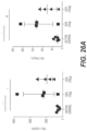


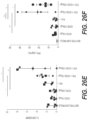


Classifications
-
- C—CHEMISTRY; METALLURGY
- C07—ORGANIC CHEMISTRY
- C07K—PEPTIDES
- C07K16/00—Immunoglobulins [IGs], e.g. monoclonal or polyclonal antibodies
- C07K16/18—Immunoglobulins [IGs], e.g. monoclonal or polyclonal antibodies against material from animals or humans
- C07K16/28—Immunoglobulins [IGs], e.g. monoclonal or polyclonal antibodies against material from animals or humans against receptors, cell surface antigens or cell surface determinants
- C07K16/2803—Immunoglobulins [IGs], e.g. monoclonal or polyclonal antibodies against material from animals or humans against receptors, cell surface antigens or cell surface determinants against the immunoglobulin superfamily
- C07K16/2818—Immunoglobulins [IGs], e.g. monoclonal or polyclonal antibodies against material from animals or humans against receptors, cell surface antigens or cell surface determinants against the immunoglobulin superfamily against CD28 or CD152
-
- A—HUMAN NECESSITIES
- A61—MEDICAL OR VETERINARY SCIENCE; HYGIENE
- A61P—SPECIFIC THERAPEUTIC ACTIVITY OF CHEMICAL COMPOUNDS OR MEDICINAL PREPARATIONS
- A61P35/00—Antineoplastic agents
-
- A—HUMAN NECESSITIES
- A61—MEDICAL OR VETERINARY SCIENCE; HYGIENE
- A61K—PREPARATIONS FOR MEDICAL, DENTAL OR TOILETRY PURPOSES
- A61K39/00—Medicinal preparations containing antigens or antibodies
- A61K2039/505—Medicinal preparations containing antigens or antibodies comprising antibodies
-
- A—HUMAN NECESSITIES
- A61—MEDICAL OR VETERINARY SCIENCE; HYGIENE
- A61K—PREPARATIONS FOR MEDICAL, DENTAL OR TOILETRY PURPOSES
- A61K39/00—Medicinal preparations containing antigens or antibodies
- A61K2039/505—Medicinal preparations containing antigens or antibodies comprising antibodies
- A61K2039/507—Comprising a combination of two or more separate antibodies
-
- A—HUMAN NECESSITIES
- A61—MEDICAL OR VETERINARY SCIENCE; HYGIENE
- A61K—PREPARATIONS FOR MEDICAL, DENTAL OR TOILETRY PURPOSES
- A61K39/00—Medicinal preparations containing antigens or antibodies
- A61K2039/545—Medicinal preparations containing antigens or antibodies characterised by the dose, timing or administration schedule
-
- C—CHEMISTRY; METALLURGY
- C07—ORGANIC CHEMISTRY
- C07K—PEPTIDES
- C07K2317/00—Immunoglobulins specific features
- C07K2317/20—Immunoglobulins specific features characterized by taxonomic origin
- C07K2317/24—Immunoglobulins specific features characterized by taxonomic origin containing regions, domains or residues from different species, e.g. chimeric, humanized or veneered
-
- C—CHEMISTRY; METALLURGY
- C07—ORGANIC CHEMISTRY
- C07K—PEPTIDES
- C07K2317/00—Immunoglobulins specific features
- C07K2317/30—Immunoglobulins specific features characterized by aspects of specificity or valency
-
- C—CHEMISTRY; METALLURGY
- C07—ORGANIC CHEMISTRY
- C07K—PEPTIDES
- C07K2317/00—Immunoglobulins specific features
- C07K2317/30—Immunoglobulins specific features characterized by aspects of specificity or valency
- C07K2317/33—Crossreactivity, e.g. for species or epitope, or lack of said crossreactivity
-
- C—CHEMISTRY; METALLURGY
- C07—ORGANIC CHEMISTRY
- C07K—PEPTIDES
- C07K2317/00—Immunoglobulins specific features
- C07K2317/70—Immunoglobulins specific features characterized by effect upon binding to a cell or to an antigen
- C07K2317/73—Inducing cell death, e.g. apoptosis, necrosis or inhibition of cell proliferation
- C07K2317/732—Antibody-dependent cellular cytotoxicity [ADCC]
-
- C—CHEMISTRY; METALLURGY
- C07—ORGANIC CHEMISTRY
- C07K—PEPTIDES
- C07K2317/00—Immunoglobulins specific features
- C07K2317/70—Immunoglobulins specific features characterized by effect upon binding to a cell or to an antigen
- C07K2317/74—Inducing cell proliferation
-
- C—CHEMISTRY; METALLURGY
- C07—ORGANIC CHEMISTRY
- C07K—PEPTIDES
- C07K2317/00—Immunoglobulins specific features
- C07K2317/70—Immunoglobulins specific features characterized by effect upon binding to a cell or to an antigen
- C07K2317/75—Agonist effect on antigen
-
- C—CHEMISTRY; METALLURGY
- C07—ORGANIC CHEMISTRY
- C07K—PEPTIDES
- C07K2317/00—Immunoglobulins specific features
- C07K2317/90—Immunoglobulins specific features characterized by (pharmaco)kinetic aspects or by stability of the immunoglobulin
- C07K2317/92—Affinity (KD), association rate (Ka), dissociation rate (Kd) or EC50 value
Definitions
- the present invention relates generally to immunotherapy in the treatment of human disease. More specifically, the present invention relates to the use of immunomodulators in the treatment of cancer.
- Cancer immunity is a multistep process that is tightly regulated by a series of negative immune checkpoint and positive co-stimulatory receptors that when effectively triggered can achieve antitumor response (Mellman, I., et al. (2011) Cancer Immunotherapy Comes of Age.
- tumors have established various mechanisms to circumvent immune clearance by altering the responsiveness of the immune infiltrate. In some instances, tumors will be highly dependent on a single mechanism, and in these cases, there is the potential to achieve significant clinical activity with single agent immunomodulatory therapy (Hoos, A. (2016). Development of immuno-oncology drugs—from CTLA4 to PD1 to the next generations. Nat Rev Drug Discov. 15(4), 235-47).
- combination therapy will likely be required for durable efficacy across a wide range of tumor types. Therefore, new immune targeted therapies are needed to improve the treatment of all cancers.
- H2L5 IgG4PE is a potent anti-ICOS agonist antibody
- A H2L5 IgG4PE binding to dimeric human ICOS
- B native ICOS-L binding to dimeric human ICOS
- C Binding of H2L5 IgG4PE (20 ug/mL) to CD4 + and CD8 + T cells from healthy donor PBMCs
- D Induction of AKT signaling in Ba/F3-ICOS expressing cell line after treatment with H2L5 IgG4PE
- E Binding of H2L5 IgG4PE (green) to isolated and activated CD4 + T cells as imaged by confocal microscopy.
- FIG. 2 H2L5 IgG4PE induces potent T cell activation, proliferation and function in a TCR-dependent manner
- A PBMCs from healthy subjects treated with either anti-CD28, IgG4PE isotype control or H2L5 IgG4PE (ICOS) alone in bound format at (10pg/mL) for 24 hrs
- B PBMCs prestimulated with anti-CD3 antibody followed by treatment with soluble anti-CD28, IgG4PE isotype control or H2L5 IgG4PE (ICOS) alone in bound format at (10pg/mL) for 24 hrs
- C PBMCs prestimulated with anti-CD3 antibody followed by treatment with anti-CD28, IgG4PE isotype control or H2L5 IgG4PE (ICOS) in bound format at (10pg/mL) for 24 hrs
- D PBMCs from healthy subjects treated with (12.5 ug/mL) of bound H2L5 IgG4PE and
- FIG. 3 Fc ⁇ R-mediated crosslinking is required for optimal H2L5 agonist function
- A Isolated CD4 + T cells from healthy subjects treated with indicated concentrations of H2L5 IgG4PE for 60 hrs
- B PBMCs from a healthy subject treated with soluble H2L5 IgG4PE (ICOS IgG4PE) or H2L5 Fc-disabled at (10 ug/mL) for 3.5 days
- C Modified mixed-lymphocyte reaction (MLR) with anti-CD3 antibody followed by treatment with soluble H2L5 IgG4PE or H2L5 Fc-disabled antibody at (10 ug/mL)
- D Modified mixed-lymphocyte reaction with CEFT peptide followed by treatment with soluble H2L5 IgG4PE (ICOS IgG4PE) or H2L5 Fc-disabled antibody at (11.1 ug/mL)
- E Co-culture of isolated T cells and monocytes from the
- FIG. 4 H2L5 isotype is critical to avoid Fc ⁇ R-mediated ICOS + cell depletion
- A PBMCs from healthy subjects treated with soluble H2L5 of varying isotypes at 5 ug/mL for 6 days. Proliferation as measured by CFSE dilution relative to isotype control (Fold change).
- B PBMCs from healthy subjects, with or without depletion of NK cells; treated with soluble H2L5 of varying isotypes at (5 ug/mL) for 6 days.
- C Treatment with soluble H2L5 of varying isotypes for 6 hrs. Fold change relative to isotype control
- D PBMCs from healthy subjects with or without NK cell depletion treated with soluble H2L5 of varying isotypes (10 ug/mL) for 24 hrs.
- FIG. 5 Anti-mouse ICOS agonist antibody results in anti-tumor responses in syngeneic mouse tumor models
- A Mice with EMT6 murine breast carcinoma tumors treated with indicated doses of 7E.17G9 antibody twice weekly for 3 weeks.
- mice with minimally detectable or non-detectable tumors at study endpoint (B) Mice with CT26 tumors treated with 7E.17G9 antibody twice weekly for 3 weeks (C) Percentage of CD4+ CD62L+ CD44+ (Effector Memory) and CD4+ CD62L ⁇ CD44+ (Central Memory) T cells in the blood of mice with EMT6 tumors 48 hrs following the third dose of 7E.17G9 antibody (D) The CD8:Treg ratio score as measured by Nanostring analysis from mice with EMT6 tumors 48 hrs following the third dose of 7E.17G9.
- FIG. 6 anti-mouse ICOS agonist antibody induces an IFN- ⁇ signature and PDL-1 increase in tumors.
- A Soluble IFN ⁇ from the blood of mice with EMT6 tumors 48 hrs following the third dose with 7E.17G9 antibody
- B Nanostring gene expression analysis from EMT6 tumors 48 hrs following the third dose of 7E.17G9 antibody
- C Ifng,
- D PDL-1 and
- E PD-1 gene expression from EMT6 tumors following treatment with 7E.17G9 antibody
- F Percentage of PD-1 + CD4 + T cells in the blood of mice with EMT6 tumors 48 hrs following the third dose of 7E.17G9 antibody.
- FIG. 7 Synergistic activity of anti-mouse ICOS agonist antibody in combination with anti-PD-1 in mouse tumor models
- A Mice with CT26 tumors treated with 7E.17G9 (100 ⁇ g), anti-PD1 (200 ⁇ g) or the combination of 7E.17G9 and anti-PD1 dosed concomitantly, twice weekly for 3 weeks
- B Mice with EMT6 tumors treated with 7E.17G9 (10 ⁇ g), anti-PD1 (200 ⁇ g) or the combination of 7E.17G9 and anti-PD1 dosed concomitantly, twice weekly for 3 weeks
- C CD8 + ICOS + T cells from the blood of CT26 tumor bearing mice 72 hrs after the first, second and third dose of anti-PD1 antibody (200 ⁇ g)
- D CD8 + ICOS + T cells from the tumors of CT26 tumor bearing mice 72 hrs after the first, second and third dose of anti-PD1 antibody
- E CD4 + FoxP3 + ICOS + T
- FIG. 8 Expression of ICOS and PDL-1 are closely associated in human tumors and concurrent targeting augments T cell function
- A Unbiased clustering analysis of PD-L1 associated gene expression in tumors from the TCGA database
- B Selected tumor types showing the association of expression of ICOS, ICOS-L and PDL-1
- C Modified MLR from healthy subject cells treated with soluble H2L5 IgG4PE (11 ⁇ g/mL) alone or in combination with Keytruda (11 ⁇ g/mL) for 4 days
- D Disseminated tumors from patients with NSCLC treated with anti-CD3 and H2L5 IgG4PE (10 ⁇ g/mL) for 24 hrs.
- FIG. 9 H2L5 IgG4PE binding stoichiometry and ligand competition (A) H2L5 IgG4PE antibody binds to human ICOS receptor with a 1:2 stoichiometry (B) H2L5 IgG4PE partially competes for binding to ICOS receptor with human ICOS-L
- FIG. 10 H2L5 IgG4PE induces phospho-AKT (A) Treatment with H2L5 IgG4PE antibody at (20 ug/mL) for 1 hr in Ba/F3 cells with and without expression of human ICOS receptor (B) Treatment with H2L5 IgG4PE at (10 ug/mL) in primary CD4 + T cells (B) Treatment with H2L5 IgG4PE at (10 ug/mL) in primary CD4 + T cells.
- FIG. 11 Membrane trafficking of H2L5 IgG4PE following binding. H2L5 IgG4PE after binding to primary CD4 + T cells in co-culture with monocyte-derived human dendritic cells. Timecourse indicates time following addition of H2L5 IgG4PE to cells.
- FIG. 12 Overnight activation with anti-CD3 antibody induces ICOS receptor expression (A) Percentage of healthy donor CD4 + and (B) CD8 + cells which are positive for ICOS expression following 24 hour stimulation with anti-CD3 antibody
- FIG. 13 Characterization of an anti-mouse ICOS agonist antibody.
- A Anti-mouse ICOS agonist antibody (7E.17G9) induces IFN ⁇ production in disseminated mouse splenocytes cultured ex vivo
- B Percentage of apoptotic/necrotic blood CD45 + cells 48 hours after the 3 rd dose of 7E.17G9 in mice with EMT-6 tumors
- C No significant change in CD4 + or
- D CD8 + T cell percentage 48 hours after the 3 rd dose of 7E.17G9 in mice with EMT-6 tumors
- FIG. 14 Anti-mouse ICOS agonist antibody 7E.17G9 induces T cell proliferation in vivo.
- A Percentage of proliferating CD8 +
- B CD4 + Tregs
- C CD4 + effector cells 48 hours after the 3 rd dose of 7E.17G9 in mice with EMT-6 tumors
- FIG. 15 Anti-mouse ICOS agonist antibody 7E.17G9 does not significantly induce other cytokines in vivo.
- A Levels of TNF ⁇
- B IL-2
- C IL-10 in the blood of mice with EMT-6 tumors 48 hours after the 3 rd dose of 7E.17G9
- FIG. 16 Another anti-mouse ICOS agonist antibody leads to anti-tumor response and synergy with anti-PD1 blocking antibody in syngeneic mouse tumor models.
- A Treatment with anti-mouse ICOS agonist antibody (C398.4A) results in increased survival of mice with CT-26 and
- B EMT-6 tumors alone and synergistically in combination with anti-PD1 blocking antibody
- FIG. 17 H2L5 hIgG4PE in combination with ipilimumab results increased proinflammatory cytokine production as compared to single antibody treatment in PBMC pre-stimulation assay.
- FIG. 18 H2L5 hIgG4PE in combination with pembrolizumab results increased proinflammatory cytokine production as compared to single antibody treatment in PBMC pre-stimulation assay.
- FIG. 19 H2L5 hIgG4PE plus ipilimumab combination induces increased proinflamatory cytokine production in a modified MLR assay with CEFT peptide and pre-incubation.
- FIG. 20 H2L5 hIgG4PE plus pembrolizumab combination induces increased proinflamatory cytokine production in a modified MLR assay with CEFT peptide and pre-incubation.
- FIG. 21 H2L5 hIgG4PE anti-ICOS agonist mAb alone and in combination with pembrolizumab results in tumor growth inhibition in a human PBMC A2058 Melanoma mouse tumor model.
- FIG. 22 Anti-ICOS murine surrogate mAb results in significant tumor growth inhibition and increased survival in combination with an anti-PD1 murine surrogate mAb in the CT26 mouse tumor model.
- FIG. 23 Anti-ICOS murine surrogate mAb results in significant tumor growth inhibition and increased survival in combination with an anti-PD1 murine surrogate mAb in the EMT6 mouse tumor model.
- FIG. 24 ICOS agonist mAb treatment induces clonal expansion of T EM cells and redistribution to the periphery
- FIG. 25 ICOS agonist mAb increases T cell function, infiltration to tumors and antitumor response
- (b) Heatmap of RNA expression as measured by Nanostring for indicated immune genes from CT26 tumors harvested 48 hours post 2 nd dose of respective treatment (a-b) In instances where ⁇ 5 tumors are represented there was not sufficient tumor material and/or RNA quality available to perform Nanostring analysis.
- FIG. 26 ICOS agonist mAb induces PD1/PDL-1 expression and synergized with anti-PD1
- N 10 mice per treatment group.
- MLR Modified mixed-lymphocyte reaction
- FIG. 28 H2L5 induces potent effector T cell activation and function in a TCR-dependent manner
- FIG. 29 H2L5 induces T cell mobilization and effector memory T cell redistribution in vivo
- AlexaFlour488-labeled H2L5 IgG4PE added at 3 ⁇ g/mL to co-culture cells on ice then moved to 37 deg C. for indicated timepoints.
- Arrows indicate T cell activated in response to H2L5 treatment, polarization and mobilization towards neighbouring dendritic cell.
- FIG. 30 Characterization of an anti-mouse ICOS antibody.
- A Anti-mouse ICOS agonist antibody (7E.17G9) induces IFN ⁇ production in disseminated mouse splenocytes cultured ex vivo
- B Percentage of apoptotic/necrotic blood CD45 + cells 48 hours after the 3 rd dose of 7E.17G9 in mice with EMT-6 tumors. Bars represent mean and error bars standard deviation. Horizontal lines represent mean values. Each symbol represents an individual mouse.
- FIG. 31 Anti-mouse ICOS agonist antibody 7E.17G9 does not significantly induce other cytokines in vivo.
- A Levels of TNF ⁇
- B IL-2 and
- FIG. 32 Anti-mouse ICOS agonist antibody 7E.17G9 induces T cell infiltration and induction of chemokines.
- FIG. 33 H2L5 IgG4PE epitope binding
- H2L5 IgG4PE partially competes for binding to ICOS receptor with human ICOS-L.
- FIG. 34 H2L5 IgG4PE induces phospho-AKT (a) Treatment with H2L5IgG4PE antibody at (20 ug/mL) for 1 hr in Ba/F3 cells with and without expression of human ICOS receptor (b) Treatment with H2L5 IgG4PE at (10 ug/mL) in primary CD4 + Tcells.
- FIG. 35 Human IgG1 istoype anti-ICOS agonist results in T cell depletion through ADCC.
- PBMCs from healthy subjects treated with soluble H2L5 of varying isotypes at 5 ug/mL for 6 days.
- Proliferation as measured by CFSE dilution relative to isotype control Fold change.
- PBMCs from healthy subjects, with or without depletion of NK cells treated with soluble H2L5 of varying isotypes at (5 ug/mL) for 6 days.
- FIG. 36 Membrane trafficking of H2L5 IgG4PE following binding. H2L5 IgG4PE after binding to primary CD4 + T cells. Timecourse indicates time following addition of H2L5 IgG4PE to cells. Green is Alex488 labeled H5L5 antibody and blue it nuclear DAPI stain.
- FIG. 37 Increase in spleen size in mice following treatment with H2L5antibody.
- FIG. 40 H2L5 IgG4PE binding to human and cynomolgus monkey ICOS.
- FIG. 41 Changes in cytokine levels from healthy human donor PBMC in response to treatment with anti-CD3 mAb plus indicated treatments below.
- FIGS. 42 A- 42 C ICOS expression in whole blood and tumor.
- FIGS. 43 A- 43 B H2L5 hIgG4PE binds to ICOS and induces signaling via AKT phosphorylation.
- FIGS. 44 A- 44 C H2L5 hIgG4PE induces T cell activation and Th differentiation in vitro.
- FIGS. 45 A- 45 B Induction of cytokine secretion and increased T cell proliferation by H2L5 hIgG4PE.
- FIGS. 46 A- 46 B Anti-tumor activity with mouse anti-ICOS antibody.
- FIGS. 47 A- 47 D ICOS induces T cell activation, proliferation and memory changes in vivo.
- FIGS. 48 A- 48 B Increase in TIL, IFN- ⁇ gene signature, higher PD-L1 expression with ICOS mAb in vivo.
- FIGS. 49 A- 49 B Significant increase in IFN ⁇ secretion with H2L5 hIgG4PE and pembrolizumab combination.
- FIGS. 50 A- 50 B Synergistic anti-tumor activity of ICOS+PD1 combination.
- FIGS. 51 A- 51 C Increase in immune cells, cytotoxicity and IFN ⁇ levels with ICOS+PD1 in vivo.
- FIGS. 52 A- 52 B ICOS, ICOS-L and PD-L1 abundance in tumors.
- the present invention provides methods for increasing expression of ICOS an effector T cell comprising contacting said efffector T cell with an anti-PD-1 antibody.
- the present invention provides methods for decreasing expression of ICOS on a regulatory T cell comprising contacting said regulatory T cell with an anti-PD-1 antibody.
- the present invention provides methods for increasing sensitivity to an agent directed to ICOS in a human, the methods comprising administering to the human an anti-PD1 antibody.
- methods for treating cancer in a human in need thereof comprising administering an anti-PD-1 antibody and an anti-ICOS antibody to said human, wherein the anti-PD-1 antibody increases T cell sensitivity to the ICOS antibody.
- in vitro methods for increasing expression of ICOS on an effector T cell, the method comprising contacting the effector T cell with an anti-PD-1 antibody, thereby increasing the expression of said ICOS on said effector T cell.
- in vitro methods for decreasing expression of ICOS on a regulatory T cell, the method comprising contacting the regulatory T cell with an anti-PD-1 antibody, thereby decreasing expression of said ICOS on said regulatory T cell.
- an anti-PD-1 antibody for use in treating cancer in a human in need thereof, wherein the anti-PD-1 antibody is administered with an anti-ICOS antibody in said human.
- administration of the anti-PD-1 antibody increases T cell sensitivity to the ICOS antibody in said human.
- an anti-ICOS antibody for use in treating cancer in a human in need thereof, wherein the anti-ICOS antibody is administered with an anti-PD-1 antibody in said human.
- administration of the anti-PD-1 antibody increases T cell sensitivity to the ICOS antibody in said human.
- an anti-PD-1 antibody and an anti-ICOS antibody are provided for use in treating cancer in a human.
- administration of the anti-PD-1 antibody increases T cell sensitivity to the ICOS antibody in said human.
- an anti-PD-1 antibody and an anti-ICOS antibody are provided for use in treating sepsis in a human.
- an anti-PD-1 antibody and an anti-ICOS antibody are provided for use in treating chronic infection in a human.
- ICOS means any Inducible T-cell costimulator protein.
- Pseudonyms for ICOS include AILIM; CD278; CVID1, JTT-1 or JTT-2, MGC39850, or 8F4.
- ICOS is a CD28-superfamily costimulatory molecule that is expressed on activated T cells. The protein encoded by this gene belongs to the CD28 and CTLA-4 cell-surface receptor family. It forms homodimers and plays an important role in cell-cell signaling, immune responses, and regulation of cell proliferation.
- Human ICOS is a 199 amino acid protein (Accession No.: UniProtKB-Q9Y6W8 (ICOS_HUMAN).
- increasing expression of ICOS means increasing the number of ICOS expressed on a single T cell and/or increasing the number of ICOS-expressing or ICOS-positive T cells in a population of cells.
- decreasing expression of ICOS means decreasing the number of ICOS expressed on a single T cell and/or decreasing the number of ICOS-expressing or ICOS-positive T cells in a population of cells.
- the T cell is an effector T cell.
- the T cell is a regulatory T cell.
- ICOS-L B7RP-1/B7-H2
- B7-1 nor B7-2 ligands for CD28 and CTLA4
- ICOS-L has been shown to bind weakly to both CD28 and CTLA-4 (Yao S et al., “B7-H2 is a costimulatory ligand for CD28 in human”, Immunity, 34(5); 729-40 (2011)).
- Expression of ICOS appears to be restricted to T cells. ICOS expression levels vary between different T cell subsets and on T cell activation status.
- ICOS expression has been shown on resting TH17, T follicular helper (TFH) and regulatory T (Treg) cells; however, unlike CD28; it is not highly expressed on na ⁇ ve T H 1 and T H 2 effector T cell populations (Paulos C M et al., “The inducible costimulator (ICOS) is critical for the development of human Th17 cells”, Sci Transl Med, 2(55); 55ra78 (2010)).
- ICOS expression is highly induced on CD4+ and CD8+ effector T cells following activation through TCR engagement (Wakamatsu E, et al., “Convergent and divergent effects of costimulatory molecules in conventional and regulatory CD4+ T cells”, Proc Natal Acad Sci USA, 110(3); 1023-8 (2013)).
- Co-stimulatory signalling through ICOS receptor only occurs in T cells receiving a concurrent TCR activation signal (Sharpe A H and Freeman G J. “The B7-CD28 Superfamily”, Nat. Rev Immunol, 2(2); 116-26 (2002)).
- ICOS In activated antigen specific T cells, ICOS regulates the production of both T H 1 and T H 2 cytokines including IFN- ⁇ , TNF- ⁇ , IL-10, IL-4, IL-13 and others. ICOS also stimulates effector T cell proliferation, albeit to a lesser extent than CD28 (Sharpe A H and Freeman G J. “The B7-CD28 Superfamily”, Nat. Rev Immunol, 2(2); 116-26 (2002)). Antibodies to ICOS and methods of using in the treatment of disease are described, for instance, in WO 2012/131004, US20110243929, and US20160215059. US20160215059 is incorporated by reference herein. Combination treatment of anti-CTLA4 antibodies and ICOS-ligand and anti-ICOS antibodies are described in US 2012251556.
- the ICOS antibodies of the present invention comprise any one or a combination of the following CDRs:
- CDRH1 DYAMH (SEQ ID NO: 2)
- CDRH2 LISIYSDHTNYNQKFQG (SEQ ID NO: 3)
- CDRH3 NNYGNYGWYFDV (SEQ ID NO: 4)
- CDRL1 SASSSVSYMH (SEQ ID NO: 5)
- CDRL2 DTSKLAS (SEQ ID NO: 6)
- CDRL3 FQGSGYPYT
- the anti-ICOS antibodies of the present invention comprise a heavy chain variable region having at least 90% sequence identity to SEQ ID NO:7.
- the ICOS binding proteins of the present invention may comprise a heavy chain variable region having about 85%, 86%, 87%, 88%, 89%, 90%, 91%, 92%, 93%, 94%, 95%, 96%, 97%, 98%, 99%, or 100% sequence identity to SEQ ID NO:7.
- V H Humanized Heavy Chain
- H2 Variable Region
- the ICOS antibody comprises CDRL1 (SEQ ID NO:4), CDRL2 (SEQ ID NO:5), and CDRL3 (SEQ ID NO:6) in the light chain variable region having the amino acid sequence set forth in SEQ ID NO:8.
- ICOS binding proteins of the present invention comprising the humanized light chain variable region set forth in SEQ ID NO:8 are designated as “L5.”
- an ICOS binding protein of the present invention comprising the heavy chain variable region of SEQ ID NO:7 and the light chain variable region of SEQ ID NO:8 can be designated as H2L5 herein.
- the ICOS binding proteins of the present invention comprise a light chain variable region having at least 90% sequence identity to the amino acid sequence set forth in SEQ ID NO:8.
- the ICOS binding proteins of the present invention may comprise a light chain variable region having about 85%, 86%, 87%, 88%, 89%, 90%, 91%, 92%, 93%, 94%, 95%, 10 96%, 97%, 98%, 99%, or 100% sequence identity to SEQ ID NO:8.
- V L Humanized Light Chain
- L5 Variable Region
- CDRs or minimum binding units may be modified by at least one amino acid substitution, deletion or addition, wherein the variant antigen binding protein substantially retains the biological characteristics of the unmodified protein, such as an antibody comprising SEQ ID NO:7 and SEQ ID NO:8.
- CDR H1, H2, H3, L1, L2, L3 may be modified alone or in combination with any other CDR, in any permutation or combination.
- a CDR is modified by the substitution, deletion or addition of up to 3 amino acids, for example 1 or 2 amino acids, for example 1 amino acID
- the modification is a substitution, particularly a conservative substitution, for example as shown in Table 1 below.
- the subclass of an antibody determines secondary effector functions, such as complement activation or Fc receptor (FcR) binding and antibody dependent cell cytotoxicity (ADCC) (Huber, et al., Nature 229(5284): 419-20 (1971); Brunhouse, et al., Mol Immunol 16(11): 907-17 (1979)).
- FcR complement activation or Fc receptor
- ADCC antibody dependent cell cytotoxicity
- the effector functions of the antibodies can be taken into account.
- hIgG1 antibodies have a relatively long half life, are very effective at fixing complement, and they bind to both Fc ⁇ RI and Fc ⁇ RII.
- human IgG4 antibodies have a shorter half life, do not fix complement and have a lower affinity for the FcRs.
- the ICOS antibody is an IgG4 isotype.
- the ICOS antibody comprises an IgG4 Fc region comprising the replacement S228P and L235E may have the designation IgG4PE.
- ICOS-L and “ICOS Ligand” are used interchangeably and refer to the membrane bound natural ligand of human ICOS.
- ICOS ligand is a protein that in humans is encoded by the ICOSLG gene.
- ICOSLG has also been designated as CD275 (cluster of differentiation 275).
- Pseudonyms for ICOS-L include B7RP-1 and B7-H2.
- the protein Programmed Death 1 is an inhibitory member of the CD28 family of receptors, that also includes CD28, CTLA-4, ICOS and BTLA.
- PD-1 is expressed on activated B cells, T cells, and myeloid cells (Agata et al., supra; Okazaki et al. (2002) Curr. Opin. Immunol 14:391779-82; Bennett et al. (2003) J Immunol 170:711-8)
- the initial members of the family, CD28 and ICOS were discovered by functional effects on augmenting T cell proliferation following the addition of monoclonal antibodies (Hutloff et al. (1999) Nature 397:263-266; Hansen et al.
- PD-1 was discovered through screening for differential expression in apototic cells (Ishida et al. (1992) EMBO J 11:3887-95)
- CTLA-4, and BTLA were discovered through screening for differential expression in cytotoxic T lymphocytes and TH1 cells, respectively.
- CD28, ICOS and CTLA-4 all have an unpaired cysteine residue allowing for homodimerization.
- PD-1 is suggested to exist as a monomer, lacking the unpaired cysteine residue characteristic in other CD28 family members.
- PD-1 antibodies and methods of using in treatment of disease are described in U.S. Pat. Nos.
- Opdivo/nivolumab is a fully human monoclonal antibody marketed by Bristol Myers Squibb directed against the negative immunoregulatory human cell surface receptor PD-1 (programmed death-1 or programmed cell death-1/PCD-1) with immunopotentiation activity.
- Nivolumab binds to and blocks the activation of PD-1, an Ig superfamily transmembrane protein, by its ligands PD-L1 and PD-L2, resulting in the activation of T-cells and cell-mediated immune responses against tumor cells or pathogens.
- Activated PD-1 negatively regulates T-cell activation and effector function through the suppression of P13k/Akt pathway activation.
- nivolumab Other names for nivolumab include: BMS-936558, MDX-1106, and ONO-4538.
- the amino acid sequence for nivolumab and methods of using and making are disclosed in U.S. Pat. No. 8,008,449.
- KEYTRUDA/pembrolizumab is an anti-PD-1 antibodies marketed for the treatment of lung cancer by Merck.
- the amino acid sequence of pembrolizumab and methods of using are disclosed in U.S. Pat. No. 8,168,757.
- CD134 also known as OX40
- OX40 is a member of the TNFR-superfamily of receptors which is not constitutively expressed on resting na ⁇ ve T cells, unlike CD28.
- OX40 is a secondary costimulatory molecule, expressed after 24 to 72 hours following activation; its ligand, OX40L, is also not expressed on resting antigen presenting cells, but is following their activation. Expression of OX40 is dependent on full activation of the T cell; without CD28, expression of OX40 is delayed and of fourfold lower levels.
- OX40/OX40-ligand (OX40 Receptor)/(OX40L) are a pair of costimulatory molecules critical for T cell proliferation, survival, cytokine production, and memory cell generation.
- OX40 signaling blocks the inhibitory function of CD4 + CD25 + naturally occurring regulatory T cells and the OX40/OX40L pair plays a critical role in the global regulation of peripheral immunity versus tolerance.
- OX-40 antibodies, OX-40 fusion proteins and methods of using them are disclosed in U.S. Pat. Nos. 7,504,101; 7,758,852; 7,858,765; 7,550,140; 7,960,515; and 9,006,399 and international publications: WO 2003082919; WO 2003068819; WO 2006063067; WO 2007084559; WO 2008051424; WO2012027328; and WO2013028231.
- T cell immunoglobulin and mucin domain-containing molecule 3 is an immunoglobulin (Ig) superfamily member, expressed on Th1 cells.
- Ig immunoglobulin
- TIM3 has been shown to play a role in modulating the immune response of Th1 cells, and reducing inflammation in a number of conditions.
- TIM3 is also expressed on cancer cells, and on cancer stem cells (CSCs), which are cells that can give rise to additional cancer cells.
- CSCs cancer stem cells
- CTLA-4 is a T cell surface molecule that was originally identified by differential screening of a murine cytolytic T cell cDNA library (Brunet et al., Nature 328:267-270(1987)). CTLA-4 is also a member of the immunoglobulin (Ig) superfamily; CTLA-4 comprises a single extracellular Ig domain. CTLA-4 transcripts have been found in T cell populations having cytotoxic activity, suggesting that CTLA-4 might function in the cytolytic response (Brunet et al., supra; Brunet et al., Immunol. Rev. 103-(21-36 (1988)).
- researchers have reported the cloning and mapping of a gene for the human counterpart of CTLA-4 (Dariavach et al., Eur.
- Suitable anti-CTLA4 antibodies for use in the methods of the invention include, without limitation, anti-CTLA4 antibodies, human anti-CTLA4 antibodies, mouse anti-CTLA4 antibodies, mammalian anti-CTLA4 antibodies, humanized anti-CTLA4 antibodies, monoclonal anti-CTLA4 antibodies, polyclonal anti-CTLA4 antibodies, chimeric anti-CTLA4 antibodies, ipilimumab, tremelimumab, anti-CD28 antibodies, anti-CTLA4 adnectins, anti-CTLA4 domain antibodies, single chain anti-CTLA4 fragments, heavy chain anti-CTLA4 fragments, light chain anti-CTLA4 fragments, inhibitors of CTLA4 that agonize the co-stimulatory pathway, the antibodies disclosed in PCT Publication No.
- CTLA-4 antibodies are described in U.S. Pat. Nos. 5,811,097, 5,855,887, 6,051,227, and 6,984,720; in PCT Publication Nos. WO 01/14424 and WO 00/37504; and in U.S. Publication Nos. US 2002/0039581 and US 2002/086014.
- Other anti-CTLA-4 antibodies that can be used in a method of the present invention include, for example, those disclosed in: WO 98/42752; U.S.
- agonist refers to an antigen binding protein including but not limited to an antibody, which upon contact with a co-signalling receptor causes one or more of the following (1) stimulates or activates the receptor, (2) enhances, increases or promotes, induces or prolongs an activity, function or presence of the receptor and/or (3) enhances, increases, promotes or induces the expression of the receptor.
- Agonist activity can be measured in vitro by various assays know in the art such as, but not limited to, measurement of cell signalling, cell proliferation, immune cell activation markers, cytokine production. Agonist activity can also be measured in vivo by various assays that measure surrogate end points such as, but not limited to the measurement of T cell proliferation or cytokine production.
- Antagonist refers to an antigen binding protein including but not limited to an antibody, which upon contact with a co-signalling receptor causes one or more of the following (1) attenuates, blocks or inactivates the receptor and/or blocks activation of a receptor by its natural ligand, (2) reduces, decreases or shortens the activity, function or presence of the receptor and/or (3) reduces, decrease, abrogates the expression of the receptor.
- Antagonist activity can be measured in vitro by various assays know in the art such as, but not limited to, measurement of an increase or decrease in cell signalling, cell proliferation, immune cell activation markers, cytokine production.
- Antagonist activity can also be measured in vivo by various assays that measure surrogate end points such as, but not limited to the measurement of T cell proliferation or cytokine production.
- cross competes for binding refers to any agent such as an antibody that will compete for binding to a target with any of the agents of the present invention.
- Competition for binding between two antibodies can be tested by various methods known in the art including Flow cytometry, Meso Scale Discovery and ELISA. Binding can be measured directly, meaning two or more binding proteins can be put in contact with a co-signalling receptor and bind may be measured for one or each. Alternatively, binding of molecules or interest can be tested against the binding or natural ligand and quantitatively compared with each other.
- binding protein refers to antibodies and other protein constructs, such as domains, which are capable of binding to and antigen.
- antibody is used herein in the broadest sense to refer to molecules with an immunoglobulin-like domain (for example IgG, IgM, IgA, IgD or IgE) and includes monoclonal, recombinant, polyclonal, chimeric, human, humanized, multispecific antibodies, including bispecific antibodies, and heteroconjugate antibodies; a single variable domain (e.g., V H , V HH , V L , domain antibody (dAbTM)), antigen binding antibody fragments, Fab, F(ab′) 2 , Fv, disulphide linked Fv, single chain Fv, disulphide-linked scFv, diabodies, TANDABSTM, etc. and modified versions of any of the foregoing.
- immunoglobulin-like domain for example IgG, IgM, IgA, IgD or IgE
- a single variable domain e.g., V H , V HH , V L , domain antibody (dAbTM)
- Alternative antibody formats include alternative scaffolds in which the one or more CDRs of the antigen binding protein can be arranged onto a suitable non-immunoglobulin protein scaffold or skeleton, such as an affibody, a SpA scaffold, an LDL receptor class A domain, an avimer or an EGF domain.
- a suitable non-immunoglobulin protein scaffold or skeleton such as an affibody, a SpA scaffold, an LDL receptor class A domain, an avimer or an EGF domain.
- domain refers to a folded protein structure which retains its tertiary structure independent of the rest of the protein. Generally domains are responsible for discrete functional properties of proteins and in many cases may be added, removed or transferred to other proteins without loss of function of the remainder of the protein and/or of the domain.
- single variable domain refers to a folded polypeptide domain comprising sequences characteristic of antibody variable domains. It therefore includes complete antibody variable domains such as V H , V HH and V L and modified antibody variable domains, for example, in which one or more loops have been replaced by sequences which are not characteristic of antibody variable domains, or antibody variable domains which have been truncated or comprise N- or C-terminal extensions, as well as folded fragments of variable domains which retain at least the binding activity and specificity of the full-length domain.
- a single variable domain is capable of binding an antigen or epitope independently of a different variable region or domain.
- a “domain antibody” or “dAb(TM)” may be considered the same as a “single variable domain”.
- a single variable domain may be a human single variable domain, but also includes single variable domains from other species such as rodent nurse shark and Camelid V HH dAbsTM.
- Camelid V HH are immunoglobulin single variable domain polypeptides that are derived from species including camel, llama, alpaca, dromedary, and guanaco, which produce heavy chain antibodies naturally devoid of light chains.
- Such V HH domains may be humanized according to standard techniques available in the art, and such domains are considered to be “single variable domains”.
- V H includes camelid V HH domains.
- An antigen binding fragment may be provided by means of arrangement of one or more CDRs on non-antibody protein scaffolds.
- Protein Scaffold as used herein includes but is not limited to an immunoglobulin (Ig) scaffold, for example an IgG scaffold, which may be a four chain or two chain antibody, or which may comprise only the Fc region of an antibody, or which may comprise one or more constant regions from an antibody, which constant regions may be of human or primate origin, or which may be an artificial chimera of human and primate constant regions.
- Ig immunoglobulin
- the protein scaffold may be an Ig scaffold, for example an IgG, or IgA scaffold.
- the IgG scaffold may comprise some or all the domains of an antibody (i.e. CH1, CH2, CH3, V H , V L ).
- the antigen binding protein may comprise an IgG scaffold selected from IgG1, IgG2, IgG3, IgG4 or IgG4PE.
- the scaffold may be IgG1.
- the scaffold may consist of, or comprise, the Fc region of an antibody, or is a part thereof.
- Affinity is the strength of binding of one molecule, e.g. an antigen binding protein of the invention, to another, e.g. its target antigen, at a single binding site.
- the binding affinity of an antigen binding protein to its target may be determined by equilibrium methods (e.g. enzyme-linked immunoabsorbent assay (ELISA) or radioimmunoassay (RIA)), or kinetics (e.g. BIACORETM analysis).
- ELISA enzyme-linked immunoabsorbent assay
- RIA radioimmunoassay
- kinetics e.g. BIACORETM analysis
- BiacoreTM methods described in Example 5 may be used to measure binding affinity.
- Avidity is the sum total of the strength of binding of two molecules to one another at multiple sites, e.g. taking into account the valency of the interaction.
- the molecule such as an antigen binding protein or nucleic acid
- the molecule is removed from the environment in which it may be found in nature.
- the molecule may be purified away from substances with which it would normally exist in nature.
- the mass of the molecule in a sample may be 95% of the total mass.
- expression vector means an isolated nucleic acid which can be used to introduce a nucleic acid of interest into a cell, such as a eukaryotic cell or prokaryotic cell, or a cell free expression system where the nucleic acid sequence of interest is expressed as a peptide chain such as a protein.
- Such expression vectors may be, for example, cosmids, plasmids, viral sequences, transposons, and linear nucleic acids comprising a nucleic acid of interest.
- Expression vectors within the scope of the disclosure may provide necessary elements for eukaryotic or prokaryotic expression and include viral promoter driven vectors, such as CMV promoter driven vectors, e.g., pcDNA3.1, pCEP4, and their derivatives, Baculovirus expression vectors, Drosophila expression vectors, and expression vectors that are driven by mammalian gene promoters, such as human Ig gene promoters.
- viral promoter driven vectors such as CMV promoter driven vectors, e.g., pcDNA3.1, pCEP4, and their derivatives
- Baculovirus expression vectors e.g., pcDNA3.1, pCEP4, and their derivatives
- Baculovirus expression vectors e.g., pcDNA3.1, pCEP4, and their derivatives
- Baculovirus expression vectors e.g., pcDNA3.1, pCEP4
- Drosophila expression vectors e.g., pcDNA3.1
- recombinant host cell means a cell that comprises a nucleic acid sequence of interest that was isolated prior to its introduction into the cell.
- the nucleic acid sequence of interest may be in an expression vector while the cell may be prokaryotic or eukaryotic.
- exemplary eukaryotic cells are mammalian cells, such as but not limited to, COS-1, COS-7, HEK293, BHK21, CHO, BSC-1, HepG2, 653, SP2/0, NS0, 293, HeLa, myeloma, lymphoma cells or any derivative thereof.
- the eukaryotic cell is a HEK293, NS0, SP2/0, or CHO cell.
- a recombinant cell according to the disclosure may be generated by transfection, cell fusion, immortalization, or other procedures well known in the art.
- a nucleic acid sequence of interest, such as an expression vector, transfected into a cell may be extrachromasomal or stably integrated into the chromosome of the cell.
- a “chimeric antibody” refers to a type of engineered antibody which contains a naturally-occurring variable region (light chain and heavy chains) derived from a donor antibody in association with light and heavy chain constant regions derived from an acceptor antibody.
- a “humanized antibody” refers to a type of engineered antibody having its CDRs derived from a non-human donor immunoglobulin, the remaining immunoglobulin-derived parts of the molecule being derived from one or more human immunoglobulin(s).
- framework support residues may be altered to preserve binding affinity (see, e.g., Queen et al. Proc. Natl Acad Sci USA, 86:10029-10032 (1989), Hodgson, et al., Bio/Technology, 9:421 (1991)).
- a suitable human acceptor antibody may be one selected from a conventional database, e.g., the KABATTM database, Los Alamos database, and Swiss Protein database, by homology to the nucleotide and amino acid sequences of the donor antibody.
- a human antibody characterized by a homology to the framework regions of the donor antibody (on an amino acid basis) may be suitable to provide a heavy chain constant region and/or a heavy chain variable framework region for insertion of the donor CDRs.
- a suitable acceptor antibody capable of donating light chain constant or variable framework regions may be selected in a similar manner. It should be noted that the acceptor antibody heavy and light chains are not required to originate from the same acceptor antibody.
- the prior art describes several ways of producing such humanized antibodies—see, for example, EP-A-0239400 and EP-A-054951.
- Fully human antibody includes antibodies having variable and constant regions (if present) derived from human germline immunoglobulin sequences.
- the human sequence antibodies of the invention may include amino acid residues not encoded by human germline immunoglobulin sequences (e.g., mutations introduced by random or site-specific mutagenesis in vitro or by somatic mutation in vivo).
- Fully human antibodies comprise amino acid sequences encoded only by polynucleotides that are ultimately of human origin or amino acid sequences that are identical to such sequences.
- antibodies encoded by human immunoglobulin-encoding DNA inserted into a mouse genome produced in a transgenic mouse are fully human antibodies since they are encoded by DNA that is ultimately of human origin.
- human immunoglobulin-encoding DNA can be rearranged (to encode an antibody) within the mouse, and somatic mutations may also occur.
- Antibodies encoded by originally human DNA that has undergone such changes in a mouse are fully human antibodies as meant herein.
- the use of such transgenic mice makes it possible to select fully human antibodies against a human antigen.
- fully human antibodies can be made using phage display technology wherein a human DNA library is inserted in phage for generation of antibodies comprising human germline DNA sequence.
- donor antibody refers to an antibody that contributes the amino acid sequences of its variable regions, CDRs, or other functional fragments or analogs thereof to a first immunoglobulin partner.
- the donor therefore, provides the altered immunoglobulin coding region and resulting expressed altered antibody with the antigenic specificity and neutralising activity characteristic of the donor antibody.
- acceptor antibody refers to an antibody that is heterologous to the donor antibody, which contributes all (or any portion) of the amino acid sequences encoding its heavy and/or light chain framework regions and/or its heavy and/or light chain constant regions to the first immunoglobulin partner.
- a human antibody may be the acceptor antibody.
- V H and V L are used herein to refer to the heavy chain variable region and light chain variable region respectively of an antigen binding protein.
- CDRs are defined as the complementarity determining region amino acid sequences of an antigen binding protein. These are the hypervariable regions of immunoglobulin heavy and light chains. There are three heavy chain and three light chain CDRs (or CDR regions) in the variable portion of an immunoglobulin. Thus, “CDRs” as used herein refers to all three heavy chain CDRs, all three light chain CDRs, all heavy and light chain CDRs, or at least two CDRs.
- the minimum overlapping region using at least two of the Kabat, Chothia, AbM and contact methods can be determined to provide the “minimum binding unit”.
- the minimum binding unit may be a sub-portion of a CDR.
- methods for increasing expression of ICOS on an effector T cell comprising contacting said effector T cell with an anti-PD-1 antibody.
- the anti-PD-1 antibody is selected from selected from pembrolizumab and nivolumab.
- the effector T cell is CD4+ and/or CD8+.
- methods are provided for decreasing expression of ICOS on a regulatory T cell comprising contacting said regulatory T cell with an anti-PD-1 antibody.
- the anti-PD-1 antibody is selected from selected from pembrolizumab and nivolumab.
- the regulatory T cell is CD4+.
- the effector T cell is a tumor infiltrating T cell.
- the regulatory T cell is a tumor infiltrating T cell.
- the effector T cell is a circulating T cell.
- the regulatory T cell is a circulating T cell.
- methods for increasing sensitivity to an agent directed to ICOS in a human comprising administering to the human an anti-PD-1 antibody.
- the anti-PD-1 antibody is selected from pembrolizumab and nivolumab.
- the agent directed to ICOS is an ICOS agonist.
- the agent directed to ICOS is an agonist antibody directed to ICOS.
- the agonist antibody directed to ICOS comprises one or more of: CDRH1 as set forth in SEQ ID NO: 1; CDRH2 as set forth in SEQ ID NO:2; CDRH3 as set forth in SEQ ID NO:3; CDRL1 as set forth in SEQ ID NO:4; CDRL2 as set forth in SEQ ID NO:5 and/or CDRL3 as set forth in SEQ ID NO:6 or a direct equivalent of each CDR wherein a direct equivalent has no more than two amino acid substitutions in said CDR.
- methods of treating cancer in a human in need thereof comprising administering an anti-PD-1 antibody and an anti-ICOS antibody to the human are provided, wherein the anti-PD-1 antibody increases T cell sensitivity to the ICOS antibody.
- the anti-PD-1 antibody is selected from selected from pembrolizumab and nivolumab.
- the T cell is CD4 + .
- the T cell is CD8 + .
- the anti-ICOS antibody is an agonist antibody directed to ICOS.
- the agonist antibody directed to ICOS comprises one or more of: CDRH1 as set forth in SEQ ID NO:1; CDRH2 as set forth in SEQ ID NO:2; CDRH3 as set forth in SEQ ID NO:3; CDRL1 as set forth in SEQ ID NO:4; CDRL2 as set forth in SEQ ID NO:5 and/or CDRL3 as set forth in SEQ ID NO:6 or a direct equivalent of each CDR wherein a direct equivalent has no more than two amino acid substitutions in said CDR.
- the agonist antibody directed to ICOS or antigen binding portion thereof comprises a V H domain comprising an amino acid sequence at least 90% identical to the amino acid sequence set forth in SEQ ID NO:7; and a V L domain comprising an amino acid sequence at least 90% identical to the amino acid sequence as set forth in SEQ ID NO:8.
- the agonist antibody directed to ICOS or antigen binding portion thereof comprises a V H domain comprising an amino acid sequence set forth in SEQ ID NO:7; and a V L domain comprising the amino acid sequence set forth in SEQ ID NO:8.
- the agonist antibody direct to ICOS or antigen binding portion thereof of comprises a hIgG4PE scaffold.
- in vitro methods for increasing expression of ICOS on an effector T cell, the method comprising contacting the effector T cell with an anti-PD-1 antibody, thereby increasing the expression of said ICOS on said effector T cell.
- the anti-PD-1 antibody is selected from pembrolizumab and nivolumab.
- the effector T cell is CD4+ and/or CD8 + .
- the regulatory T cell is CD4+.
- in vitro methods for decreasing expression of ICOS on a regulatory T cell, the method comprising contacting the regulatory T cell with an anti-PD-1 antibody, thereby decreasing expression of said ICOS on said regulatory T cell.
- the anti-PD-1 antibody is selected from pembrolizumab and nivolumab.
- the effector T cell is CD4+ and/or CD8+.
- the regulatory T cell is CD4+.
- an anti-PD-1 antibody for use in treating cancer in a human in need thereof, wherein the anti-PD-1 antibody is administered with an anti-ICOS antibody in said human.
- administration of the anti-PD-1 antibody increases T cell sensitivity to the ICOS antibody in said human.
- administration of the anti-PD-1 antibody increases expression of ICOS in said human.
- the anti-PD-1 antibody is selected from selected from pembrolizumab and nivolumab.
- the anti-ICOS antibody is an agonist antibody directed to ICOS.
- the agonist antibody directed to ICOS comprises one or more of: CDRH1 as set forth in SEQ ID NO: 1; CDRH2 as set forth in SEQ ID NO:2; CDRH3 as set forth in SEQ ID NO:3; CDRL1 as set forth in SEQ ID NO:4; CDRL2 as set forth in SEQ ID NO:5 and/or CDRL3 as set forth in SEQ ID NO:6 or a direct equivalent of each CDR wherein a direct equivalent has no more than two amino acid substitutions in said CDR.
- the agonist antibody directed to ICOS or antigen binding portion thereof comprises a V H domain comprising an amino acid sequence at least 90% identical to the amino acid sequence set forth in SEQ ID NO:7; and a V L domain comprising an amino acid sequence at least 90% identical to the amino acid sequence as set forth in SEQ ID NO:8.
- the agonist antibody directed to ICOS or antigen binding portion thereof comprises a V H domain comprising an amino acid sequence set forth in SEQ ID NO:7; and a V L domain comprising the amino acid sequence set forth in SEQ ID NO:8.
- the agonist antibody directed to ICOS or antigen binding portion thereof of comprises a hIgG4PE scaffold.
- an anti-ICOS antibody for use in treating cancer in a human in need thereof, wherein the anti-ICOS antibody is administered with an anti-PD-1 antibody in said human.
- administration of the anti-PD-1 antibody increases T cell sensitivity to the ICOS antibody in said human.
- administration of the anti-PD-1 antibody increases expression of ICOS in said human.
- the anti-PD-1 antibody is selected from selected from pembrolizumab and nivolumab.
- the anti-ICOS antibody is an agonist antibody directed to ICOS.
- the agonist antibody directed to ICOS comprises one or more of: CDRH1 as set forth in SEQ ID NO: 1; CDRH2 as set forth in SEQ ID NO:2; CDRH3 as set forth in SEQ ID NO:3; CDRL1 as set forth in SEQ ID NO:4; CDRL2 as set forth in SEQ ID NO:5 and/or CDRL3 as set forth in SEQ ID NO:6 or a direct equivalent of each CDR wherein a direct equivalent has no more than two amino acid substitutions in said CDR.
- the agonist antibody directed to ICOS or antigen binding portion thereof comprises a V H domain comprising an amino acid sequence at least 90% identical to the amino acid sequence set forth in SEQ ID NO:7; and a V L domain comprising an amino acid sequence at least 90% identical to the amino acid sequence as set forth in SEQ ID NO:8.
- the agonist antibody directed to ICOS or antigen binding portion thereof comprises a V H domain comprising an amino acid sequence set forth in SEQ ID NO:7; and a V L domain comprising the amino acid sequence set forth in SEQ ID NO:8.
- the agonist antibody directed to ICOS or antigen binding portion thereof of comprises a hIgG4PE scaffold.
- an anti-PD-1 antibody and an anti-ICOS antibody are provided for use in treating cancer in a human.
- administration of the anti-PD-1 antibody increases T cell sensitivity to the ICOS antibody in said human.
- administration of the anti-PD-1 antibody increases expression of ICOS in said human.
- the anti-PD-1 antibody is administered prior to administration of the anti-ICOS antibody.
- the anti-PD-1 antibody is selected from selected from pembrolizumab and nivolumab.
- the anti-ICOS antibody is an agonist antibody directed to ICOS.
- the agonist antibody directed to ICOS comprises one or more of: CDRH1 as set forth in SEQ ID NO:1; CDRH2 as set forth in SEQ ID NO:2; CDRH3 as set forth in SEQ ID NO:3; CDRL1 as set forth in SEQ ID NO:4; CDRL2 as set forth in SEQ ID NO:5 and/or CDRL3 as set forth in SEQ ID NO:6 or a direct equivalent of each CDR wherein a direct equivalent has no more than two amino acid substitutions in said CDR.
- the agonist antibody directed to ICOS or antigen binding portion thereof comprises a V H domain comprising an amino acid sequence at least 90% identical to the amino acid sequence set forth in SEQ ID NO:7; and a V L domain comprising an amino acid sequence at least 90% identical to the amino acid sequence as set forth in SEQ ID NO:8.
- the agonist antibody directed to ICOS or antigen binding portion thereof comprises a V H domain comprising an amino acid sequence set forth in SEQ ID NO:7; and a V L domain comprising the amino acid sequence set forth in SEQ ID NO:8.
- the agonist antibody directed to ICOS or antigen binding portion thereof of comprises a hIgG4PE scaffold.
- an anti-PD-1 antibody and an anti-ICOS antibody are provided for use in treating sepsis in a human.
- administration of the anti-PD-1 antibody increases T cell sensitivity to the ICOS antibody in said human.
- administration of the anti-PD-1 antibody increases expression of ICOS in said human.
- the anti-PD-1 antibody is selected from selected from pembrolizumab and nivolumab.
- the anti-ICOS antibody is an agonist antibody directed to ICOS.
- the agonist antibody directed to ICOS comprises one or more of: CDRH1 as set forth in SEQ ID NO: 1; CDRH2 as set forth in SEQ ID NO:2; CDRH3 as set forth in SEQ ID NO:3; CDRL1 as set forth in SEQ ID NO:4; CDRL2 as set forth in SEQ ID NO:5 and/or CDRL3 as set forth in SEQ ID NO:6 or a direct equivalent of each CDR wherein a direct equivalent has no more than two amino acid substitutions in said CDR.
- the agonist antibody directed to ICOS or antigen binding portion thereof comprises a V H domain comprising an amino acid sequence at least 90% identical to the amino acid sequence set forth in SEQ ID NO:7; and a V L domain comprising an amino acid sequence at least 90% identical to the amino acid sequence as set forth in SEQ ID NO:8.
- the agonist antibody directed to ICOS or antigen binding portion thereof comprises a V H domain comprising an amino acid sequence set forth in SEQ ID NO:7; and a V L domain comprising the amino acid sequence set forth in SEQ ID NO:8.
- the agonist antibody directed to ICOS or antigen binding portion thereof of comprises a hIgG4PE scaffold.
- an anti-PD-1 antibody and an anti-ICOS antibody are provided for use in treating chronic infection in a human.
- administration of the anti-PD-1 antibody increases T cell sensitivity to the ICOS antibody in said human.
- administration of the anti-PD-1 antibody increases expression of ICOS in said human.
- the anti-PD-1 antibody is selected from selected from pembrolizumab and nivolumab.
- the anti-ICOS antibody is an agonist antibody directed to ICOS.
- the agonist antibody directed to ICOS comprises one or more of: CDRH1 as set forth in SEQ ID NO:1; CDRH2 as set forth in SEQ ID NO:2; CDRH3 as set forth in SEQ ID NO:3; CDRL1 as set forth in SEQ ID NO:4; CDRL2 as set forth in SEQ ID NO:5 and/or CDRL3 as set forth in SEQ ID NO:6 or a direct equivalent of each CDR wherein a direct equivalent has no more than two amino acid substitutions in said CDR.
- the agonist antibody directed to ICOS or antigen binding portion thereof comprises a V H domain comprising an amino acid sequence at least 90% identical to the amino acid sequence set forth in SEQ ID NO:7; and a V L domain comprising an amino acid sequence at least 90% identical to the amino acid sequence as set forth in SEQ ID NO:8.
- the agonist antibody directed to ICOS or antigen binding portion thereof comprises a V H domain comprising an amino acid sequence set forth in SEQ ID NO:7; and a V L domain comprising the amino acid sequence set forth in SEQ ID NO:8.
- the agonist antibody directed to ICOS or antigen binding portion thereof of comprises a hIgG4PE scaffold.
- the methods of the present invention further comprise administering an anti-CTLA4 antibody to the human.
- the anti-CTLA4 antibody is ipilimumab.
- the anti-PD-1 antibody and the anti-ICOS antibody are administered to the human simultaneously.
- the methods comprise administering at least one additional neoplastic agent to the human and/or at least one immunostimulatory agent.
- the cancer is selected from head and neck cancer, breast cancer, lung cancer, colon cancer, ovarian cancer, prostate cancer, gliomas, glioblastoma, astrocytomas, glioblastoma multiforme, Bannayan-Zonana syndrome, Cowden disease, Lhermitte-Duclos disease, inflammatory breast cancer, Wilm's tumor, Ewing's sarcoma, Rhabdomyosarcoma, ependymoma, medulloblastoma, kidney cancer, liver cancer, melanoma, pancreatic cancer, sarcoma, osteosarcoma, giant cell tumor of bone, thyroid cancer, lymphoblastic T cell leukemia, Chronic myelogenous leukemia, Chronic lymphocytic leukemia, Hairy-cell leukemia, acute lymphoblastic leukemia, acute myelogenous leukemia, AML, Chronic neutrophilic leukemia, Acute lymphoblastic T cell leukemia, plasma
- the methods of the present invention further comprise administering at least one neo-plastic agent and/or at least one immunostimulatory agent to said human.
- the human has a solid tumor.
- the tumor is selected from head and neck cancer, gastric cancer, melanoma, renal cell carcinoma (RCC), esophageal cancer, non-small cell lung carcinoma, prostate cancer, colorectal cancer, ovarian cancer and pancreatic cancer.
- the human has a liquid tumor such as diffuse large B cell lymphoma (DLBCL), multiple myeloma, chronic lyphomblastic leukemia (CLL), follicular lymphoma, acute myeloid leukemia and chronic myelogenous leukemia.
- DLBCL diffuse large B cell lymphoma
- CLL chronic lyphomblastic leukemia
- follicular lymphoma acute myeloid leukemia and chronic myelogenous leukemia.
- the present disclosure also relates to a method for treating or lessening the severity of a cancer selected from: brain (gliomas), glioblastomas, Bannayan-Zonana syndrome, Cowden disease, Lhermitte-Duclos disease, breast, inflammatory breast cancer, Wilm's tumor, Ewing's sarcoma, Rhabdomyosarcoma, ependymoma, medulloblastoma, colon, head and neck, kidney, lung, liver, melanoma, ovarian, pancreatic, prostate, sarcoma, osteosarcoma, giant cell tumor of bone, thyroid, lymphoblastic T-cell leukemia, chronic myelogenous leukemia, chronic lymphocytic leukemia, hairy-cell leukemia, acute lymphoblastic leukemia, acute myelogenous leukemia, chronic neutrophilic leukemia, acute lymphoblastic T-cell leukemia, plasmacytoma, immunoblastic large cell leuk
- treating means: (1) to ameliorate or prevent the condition of one or more of the biological manifestations of the condition, (2) to interfere with (a) one or more points in the biological cascade that leads to or is responsible for the condition or (b) one or more of the biological manifestations of the condition, (3) to alleviate one or more of the symptoms, effects or side effects associated with the condition or treatment thereof, or (4) to slow the progression of the condition or one or more of the biological manifestations of the condition.
- Prophylactic therapy is also contemplated thereby.
- prevention is not an absolute term.
- prevention is understood to refer to the prophylactic administration of a drug to substantially diminish the likelihood or severity of a condition or biological manifestation thereof, or to delay the onset of such condition or biological manifestation thereof.
- Prophylactic therapy is appropriate, for example, when a subject is considered at high risk for developing cancer, such as when a subject has a strong family history of cancer or when a subject has been exposed to a carcinogen.
- cancer As used herein, the terms “cancer,” “neoplasm,” and “tumor” are used interchangeably and, in either the singular or plural form, refer to cells that have undergone a malignant transformation that makes them pathological to the host organism.
- Primary cancer cells can be readily distinguished from non-cancerous cells by well-established techniques, particularly histological examination.
- the definition of a cancer cell includes not only a primary cancer cell, but any cell derived from a cancer cell ancestor. This includes metastasized cancer cells, and in vitro cultures and cell lines derived from cancer cells.
- a “clinically detectable” tumor is one that is detectable on the basis of tumor mass; e.g., by procedures such as computed tomography (CT) scan, magnetic resonance imaging (MRI), X-ray, ultrasound or palpation on physical examination, and/or which is detectable because of the expression of one or more cancer-specific antigens in a sample obtainable from a patient.
- CT computed tomography
- MRI magnetic resonance imaging
- X-ray X-ray
- ultrasound or palpation e.g., ultrasound or palpation on physical examination
- Tumors may be a hematopoietic (or hematologic or hematological or blood-related) cancer, for example, cancers derived from blood cells or immune cells, which may be referred to as “liquid tumors.”
- liquid tumors include leukemias such as chronic myelocytic leukemia, acute myelocytic leukemia, chronic lymphocytic leukemia and acute lymphocytic leukemia; plasma cell malignancies such as multiple myeloma, MGUS and Waldenstrom's macroglobulinemia; lymphomas such as non-Hodgkin's lymphoma, Hodgkin's lymphoma; and the like.
- the cancer may be any cancer in which an abnormal number of blast cells or unwanted cell proliferation is present or that is diagnosed as a hematological cancer, including both lymphoid and myeloid malignancies.
- Myeloid malignancies include, but are not limited to, acute myeloid (or myelocytic or myelogenous or myeloblastic) leukemia (undifferentiated or differentiated), acute promyeloid (or promyelocytic or promyelogenous or promyeloblastic) leukemia, acute myelomonocytic (or myelomonoblastic) leukemia, acute monocytic (or monoblastic) leukemia, erythroleukemia and megakaryocytic (or megakaryoblastic) leukemia.
- leukemias may be referred together as acute myeloid (or myelocytic or myelogenous) leukemia (AML).
- Myeloid malignancies also include myeloproliferative disorders (MPD) which include, but are not limited to, chronic myelogenous (or myeloid) leukemia (CML), chronic myelomonocytic leukemia (CMML), essential thrombocythemia (or thrombocytosis), and polcythemia vera (PCV).
- CML chronic myelogenous leukemia
- CMML chronic myelomonocytic leukemia
- PCV polcythemia vera
- Myeloid malignancies also include myelodysplasia (or myelodysplastic syndrome or MDS), which may be referred to as refractory anemia (RA), refractory anemia with excess blasts (RAEB), and refractory anemia with excess blasts in transformation (RAEBT); as well as myelofibrosis (MFS) with or without agnogenic myeloid metaplasia.
- myelodysplasia or myelodysplastic syndrome or MDS
- MDS myelodysplasia
- RA refractory anemia
- RAEB refractory anemia with excess blasts
- RAEBT refractory anemia with excess blasts in transformation
- MFS myelofibrosis
- Hematopoietic cancers also include lymphoid malignancies, which may affect the lymph nodes, spleens, bone marrow, peripheral blood, and/or extranodal sites.
- Lymphoid cancers include B-cell malignancies, which include, but are not limited to, B-cell non-Hodgkin's lymphomas (B-NHLs).
- B-NHLs may be indolent (or low-grade), intermediate-grade (or aggressive) or high-grade (very aggressive).
- Indolent Bcell lymphomas include follicular lymphoma (FL); small lymphocytic lymphoma (SLL); marginal zone lymphoma (MZL) including nodal MZL, extranodal MZL, splenic MZL and splenic MZL with villous lymphocytes; lymphoplasmacytic lymphoma (LPL); and mucosa-associated-lymphoid tissue (MALT or extranodal marginal zone) lymphoma.
- FL follicular lymphoma
- SLL small lymphocytic lymphoma
- MZL marginal zone lymphoma
- LPL lymphoplasmacytic lymphoma
- MALT mucosa-associated-lymphoid tissue
- Intermediate-grade B-NHLs include mantle cell lymphoma (MCL) with or without leukemic involvement, diffuse large cell lymphoma (DLBCL), follicular large cell (or grade 3 or grade 3B) lymphoma, and primary mediastinal lymphoma (PML).
- MCL mantle cell lymphoma
- DLBCL diffuse large cell lymphoma
- follicular large cell or grade 3 or grade 3B lymphoma
- PML primary mediastinal lymphoma
- High-grade B-NHLs include Burkitt's lymphoma (BL), Burkitt-like lymphoma, small non-cleaved cell lymphoma (SNCCL) and lymphoblastic lymphoma.
- B-NHLs include immunoblastic lymphoma (or immunocytoma), primary effusion lymphoma, HIV associated (or AIDS related) lymphomas, and post-transplant lymphoproliferative disorder (PTLD) or lymphoma.
- B-cell malignancies also include, but are not limited to, chronic lymphocytic leukemia (CLL), prolymphocytic leukemia (PLL), Waldenstrom's macroglobulinemia (WM), hairy cell leukemia (HCL), large granular lymphocyte (LGL) leukemia, acute lymphoid (or lymphocytic or lymphoblastic) leukemia, and Castleman's disease.
- CLL chronic lymphocytic leukemia
- PLL prolymphocytic leukemia
- WM Waldenstrom's macroglobulinemia
- HCL hairy cell leukemia
- LGL large granular lymphocyte
- LAman's disease Castleman's disease.
- NHL may also include T-cell non-Hodgkin's lymphoma s(T-NHLs), which include, but are not limited to T-cell non-Hodgkin's lymphoma not otherwise specified (NOS), peripheral T-cell lymphoma (PTCL), anaplastic large cell lymphoma (ALCL), angioimmunoblastic lymphoid disorder (AILD), nasal natural killer (NK) cell/T-cell lymphoma, gamma/delta lymphoma, cutaneous T cell lymphoma, mycosis fungoides, and Sezary syndrome.
- T-NHLs T-cell non-Hodgkin's lymphoma s
- T-NHLs T-cell non-Hodgkin's lymphoma not otherwise specified
- PTCL peripheral T-cell lymphoma
- ALCL anaplastic large cell lymphoma
- angioimmunoblastic lymphoid disorder IL-associated lymphoid disorder
- NK
- Hematopoietic cancers also include Hodgkin's lymphoma (or disease) including classical Hodgkin's lymphoma, nodular sclerosing Hodgkin's lymphoma, mixed cellularity Hodgkin's lymphoma, lymphocyte predominant (LP) Hodgkin's lymphoma, nodular LP Hodgkin's lymphoma, and lymphocyte depleted Hodgkin's lymphoma.
- Hematopoietic cancers also include plasma cell diseases or cancers such as multiple myeloma (MM) including smoldering MM, monoclonal gammopathy of undetermined (or unknown or unclear) significance (MGUS), plasmacytoma (bone, extramedullary), lymphoplasmacytic lymphoma (LPL), Waldenström's Macroglobulinemia, plasma cell leukemia, and primary amyloidosis (AL).
- MM multiple myeloma
- MGUS monoclonal gammopathy of undetermined (or unknown or unclear) significance
- MGUS monoclonal gammopathy of undetermined (or unknown or unclear) significance
- plasmacytoma bone, extramedullary
- LPL lymphoplasmacytic lymphoma
- Waldenström's Macroglobulinemia plasma cell leukemia
- plasma cell leukemia and primary amyloidosis
- AL primary amyloidosis
- Hematopoietic cancers may also
- Tissues which include hematopoietic cells referred herein to as “hematopoietic cell tissues” include bone marrow; peripheral blood; thymus; and peripheral lymphoid tissues, such as spleen, lymph nodes, lymphoid tissues associated with mucosa (such as the gut-associated lymphoid tissues), tonsils, Peyer's patches and appendix, and lymphoid tissues associated with other mucosa, for example, the bronchial linings.
- any anti-neoplastic agent that has activity versus a susceptible tumor being treated may be co-administered in the treatment of cancer in the present invention.
- examples of such agents can be found in Cancer Principles and Practice of Oncology by V. T. Devita and S. Hellman (editors), 6 th edition (Feb. 15, 2001), Lippincott Williams & Wilkins Publishers.
- a person of ordinary skill in the art would be able to discern which combinations of agents would be useful based on the particular characteristics of the drugs and the cancer involved.
- Typical anti-neoplastic agents useful in the present invention include, but are not limited to, anti-microtubule agents such as diterpenoids and vinca alkaloids; platinum coordination complexes; alkylating agents such as nitrogen mustards, oxazaphosphorines, alkylsulfonates, nitrosoureas, and triazenes; antibiotic agents such as anthracyclins, actinomycins and bleomycins; topoisomerase II inhibitors such as epipodophyllotoxins; antimetabolites such as purine and pyrimidine analogues and anti-folate compounds; topoisomerase I inhibitors such as camptothecins; hormones and hormonal analogues; signal transduction pathway inhibitors; non-receptor tyrosine kinase angiogenesis inhibitors; immunotherapeutic agents; proapoptotic agents; and cell cycle signaling inhibitors.
- anti-microtubule agents such as diterpenoids and vinca alkaloids
- anti-neoplastic agents including any chemotherapeutic agents, immuno-modulatory agents or immune-modulators and immunostimulatory adjuvants.
- Anti-microtubule or anti-mitotic agents are phase specific agents active against the microtubules of tumor cells during M or the mitosis phase of the cell cycle.
- anti-microtubule agents include, but are not limited to, diterpenoids and vinca alkaloids.
- Diterpenoids which are derived from natural sources, are phase specific anti-cancer agents that operate at the G2/M phases of the cell cycle. It is believed that the diterpenoids stabilize the ⁇ -tubulin subunit of the microtubules, by binding with this protein. Disassembly of the protein appears then to be inhibited with mitosis being arrested and cell death following.
- diterpenoids examples include, but are not limited to, paclitaxel and its analog docetaxel.
- Paclitaxel 5 ⁇ ,20-epoxy-1,2 ⁇ ,4,7 ⁇ ,10 ⁇ ,13 ⁇ -hexa-hydroxytax-11-en-9-one 4,10-diacetate 2-benzoate 13-ester with (2R,3S)—N-benzoyl-3-phenylisoserine; is a natural diterpene product isolated from the Pacific yew tree Taxus brevifolia and is commercially available as an injectable solution TAXOL®. It is a member of the taxane family of terpenes. It was first isolated in 1971 by Wani et al. J. Am. Chem, Soc., 93:2325. 1971), who characterized its structure by chemical and X-ray crystallographic methods.
- Paclitaxel has been approved for clinical use in the treatment of refractory ovarian cancer in the United States (Markman et al., Yale Journal of Biology and Medicine, 64:583, 1991; McGuire et al., Ann. Intem, Med., 111:273, 1989) and for the treatment of breast cancer (Holmes et al., J. Nat. Cancer Inst., 83:1797, 1991.) It is a potential candidate for treatment of neoplasms in the skin (Einzig et. al., Proc. Am. Soc. Clin. Oncol., 20:46) and head and neck carcinomas (Forastire et. al., Sem. Oncol., 20:56, 1990).
- the compound also shows potential for the treatment of polycystic kidney disease (Woo et. al., Nature, 368:750. 1994, lung cancer and malaria.
- Treatment of patients with paclitaxel results in bone marrow suppression (multiple cell lineages, Ignoff, R. J. et. al, Cancer Chemotherapy Pocket Guide, 1998) related to the duration of dosing above a threshold concentration (50 nM) (Kearns, C. M. et. al., Seminars in Oncology, 3(6) p.16-23, 1995).
- Docetaxel (2R,3S)—N-carboxy-3-phenylisoserine,N-tert-butyl ester, 13-ester with 5 ⁇ -20-epoxy-1,2 ⁇ ,4,7 ⁇ ,10 ⁇ ,13 ⁇ -hexahydroxytax-11-en-9-one 4-acetate 2-benzoate, trihydrate; is commercially available as an injectable solution as TAXOTERE®.
- Docetaxel is indicated for the treatment of breast cancer.
- Docetaxel is a semisynthetic derivative of paclitaxel q.v., prepared using a natural precursor, 10-deacetyl-baccatin III, extracted from the needle of the European Yew tree. The dose limiting toxicity of docetaxel is neutropenia.
- Vinca alkaloids are phase specific anti-neoplastic agents derived from the periwinkle plant. Vinca alkaloids act at the M phase (mitosis) of the cell cycle by binding specifically to tubulin. Consequently, the bound tubulin molecule is unable to polymerize into microtubules. Mitosis is believed to be arrested in metaphase with cell death following. Examples of vinca alkaloids include, but are not limited to, vinblastine, vincristine, and vinorelbine.
- Vinblastine vincaleukoblastine sulfate
- VELBAN® an injectable solution.
- Myelosuppression is the dose limiting side effect of vinblastine.
- Vincristine vincaleukoblastine, 22-oxo-, sulfate
- ONCOVIN® an injectable solution.
- Vincristine is indicated for the treatment of acute leukemias and has also found use in treatment regimens for Hodgkin's and non-Hodgkin's malignant lymphomas.
- Alopecia and neurologic effects are the most common side effect of vincristine and to a lesser extent myelosupression and gastrointestinal mucositis effects occur.
- Vinorelbine 3′,4′-didehydro-4‘-deoxy-C’-norvincaleukoblastine [R—(R*,R*)-2,3-dihydroxybutanedioate (1:2)(salt)], commercially available as an injectable solution of vinorelbine tartrate (NAVELBINE®), is a semisynthetic vinca alkaloID Vinorelbine is indicated as a single agent or in combination with other chemotherapeutic agents, such as cisplatin, in the treatment of various solid tumors, particularly non-small cell lung, advanced breast, and hormone refractory prostate cancers. Myelosuppression is the most common dose limiting side effect of vinorelbine.
- Platinum coordination complexes are non-phase specific anti-cancer agents, which are interactive with DNA.
- the platinum complexes enter tumor cells, undergo, aquation and form intra- and interstrand crosslinks with DNA causing adverse biological effects to the tumor.
- Examples of platinum coordination complexes include, but are not limited to, cisplatin and carboplatin.
- Cisplatin cis-diamminedichloroplatinum
- PLATINOL® an injectable solution.
- Cisplatin is primarily indicated in the treatment of metastatic testicular and ovarian cancer and advanced bladder cancer.
- the primary dose limiting side effects of cisplatin are nephrotoxicity, which may be controlled by hydration and diuresis, and ototoxicity.
- Carboplatin platinum, diammine [1,1-cyclobutane-dicarboxylate(2-)-O,O′], is commercially available as PARAPLATIN® as an injectable solution.
- Carboplatin is primarily indicated in the first and second line treatment of advanced ovarian carcinoma. Bone marrow suppression is the dose limiting toxicity of carboplatin.
- Alkylating agents are non-phase anti-cancer specific agents and strong electrophiles. Typically, alkylating agents form covalent linkages, by alkylation, to DNA through nucleophilic moieties of the DNA molecule such as phosphate, amino, sulfhydryl, hydroxyl, carboxyl, and imidazole groups. Such alkylation disrupts nucleic acid function leading to cell death.
- alkylating agents include, but are not limited to, nitrogen mustards such as cyclophosphamide, melphalan, and chlorambucil; alkyl sulfonates such as busulfan; nitrosoureas such as carmustine; and triazenes such as dacarbazine.
- Cyclophosphamide 2-[bis(2-chloroethyl)amino]tetrahydro-2H-1,3,2-oxazaphosphorine 2-oxide monohydrate, is commercially available as an injectable solution or tablets as CYTOXAN®. Cyclophosphamide is indicated as a single agent or in combination with other chemotherapeutic agents, in the treatment of malignant lymphomas, multiple myeloma, and leukemias. Alopecia, nausea, vomiting and leukopenia are the most common dose limiting side effects of cyclophosphamide.
- Melphalan 4-[bis(2-chloroethyl)amino]-L-phenylalanine, is commercially available as an injectable solution or tablets as ALKERAN®. Melphalan is indicated for the palliative treatment of multiple myeloma and non-resectable epithelial carcinoma of the ovary. Bone marrow suppression is the most common dose limiting side effect of melphalan.
- Chlorambucil 4-[bis(2-chloroethyl)amino]benzenebutanoic acid, is commercially available as LEUKERAN® tablets. Chlorambucil is indicated for the palliative treatment of chronic lymphatic leukemia, and malignant lymphomas such as lymphosarcoma, giant follicular lymphoma, and Hodgkin's disease. Bone marrow suppression is the most common dose limiting side effect of chlorambucil.
- Busulfan 1,4-butanediol dimethanesulfonate, is commercially available as MYLERAN® TABLETS. Busulfan is indicated for the palliative treatment of chronic myelogenous leukemia. Bone marrow suppression is the most common dose limiting side effects of busulfan.
- Carmustine 1,3-[bis(2-chloroethyl)-1-nitrosourea, is commercially available as single vials of lyophilized material as BiCNU®.
- Carmustine is indicated for the palliative treatment as a single agent or in combination with other agents for brain tumors, multiple myeloma, Hodgkin's disease, and non-Hodgkin's lymphomas. Delayed myelosuppression is the most common dose limiting side effects of carmustine.
- dacarbazine 5-(3,3-dimethyl-1-triazeno)-imidazole-4-carboxamide, is commercially available as single vials of material as DTIC-Dome®.
- dacarbazine is indicated for the treatment of metastatic malignant melanoma and in combination with other agents for the second line treatment of Hodgkin's Disease. Nausea, vomiting, and anorexia are the most common dose limiting side effects of dacarbazine.
- Antibiotic anti-neoplastics are non-phase specific agents, which bind or intercalate with DNA. Typically, such action results in stable DNA complexes or strand breakage, which disrupts ordinary function of the nucleic acids leading to cell death.
- antibiotic anti-neoplastic agents include, but are not limited to, actinomycins such as dactinomycin, anthrocyclins such as daunorubicin and doxorubicin; and bleomycins.
- Dactinomycin also know as Actinomycin D, is commercially available in injectable form as COSMEGEN®. Dactinomycin is indicated for the treatment of Wilm's tumor and rhabdomyosarcoma. Nausea, vomiting, and anorexia are the most common dose limiting side effects of dactinomycin.
- Daunorubicin (8S-cis-)-8-acetyl-10-[(3-amino-2,3,6-trideoxy-a-L-lyxo-hexopyranosyl)oxy]-7,8,9,10-tetrahydro-6,8,11-trihydroxy-1-methoxy-5,12 naphthacenedione hydrochloride, is commercially available as a liposomal injectable form as DAUNOXOME® or as an injectable as CERUBIDINE®. Daunorubicin is indicated for remission induction in the treatment of acute nonlymphocytic leukemia and advanced HIV associated Kaposi's sarcoma. Myelosuppression is the most common dose limiting side effect of daunorubicin.
- Doxorubicin (8S, 10S)-10-[(3-amino-2,3,6-trideoxy-a-L-lyxo-hexopyranosyl)oxy]-8-glycoloyl, 7,8,9,10-tetrahydro-6,8,11-trihydroxy-1-methoxy-5,12 naphthacenedione hydrochloride, is commercially available as an injectable form as RUBEX® or ADRIAMYCIN RDF®.
- Doxorubicin is primarily indicated for the treatment of acute lymphoblastic leukemia and acute myeloblastic leukemia, but is also a useful component in the treatment of some solid tumors and lymphomas. Myelosuppression is the most common dose limiting side effect of doxorubicin.
- Bleomycin a mixture of cytotoxic glycopeptide antibiotics isolated from a strain of Streptomyces verticillus , is commercially available as BLENOXANE®. Bleomycin is indicated as a palliative treatment, as a single agent or in combination with other agents, of squamous cell carcinoma, lymphomas, and testicular carcinomas. Pulmonary and cutaneous toxicities are the most common dose limiting side effects of bleomycin.
- Topoisomerase II inhibitors include, but are not limited to, epipodophyllotoxins.
- Epipodophyllotoxins are phase specific anti-neoplastic agents derived from the mandrake plant. Epipodophyllotoxins typically affect cells in the S and G 2 phases of the cell cycle by forming a ternary complex with topoisomerase II and DNA causing DNA strand breaks. The strand breaks accumulate and cell death follows. Examples of epipodophyllotoxins include, but are not limited to, etoposide and teniposide.
- Etoposide 4′-demethyl-epipodophyllotoxin 9[4,6-0-(R)-ethylidene- ⁇ -D-glucopyranoside]
- VePESID® an injectable solution or capsules
- VP-16 an injectable solution or capsules
- Etoposide is indicated as a single agent or in combination with other chemotherapy agents in the treatment of testicular and non-small cell lung cancers. Myelosuppression is the most common side effect of etoposide. The incidence of leucopenia tends to be more severe than thrombocytopenia.
- Teniposide 4′-demethyl-epipodophyllotoxin 9[4,6-0-(R)-thenylidene- ⁇ -D-glucopyranoside], is commercially available as an injectable solution as VUMON® and is commonly known as VM-26.
- Teniposide is indicated as a single agent or in combination with other chemotherapy agents in the treatment of acute leukemia in children. Myelosuppression is the most common dose limiting side effect of teniposide. Teniposide can induce both leucopenia and thrombocytopenia.
- Antimetabolite neoplastic agents are phase specific anti-neoplastic agents that act at S phase (DNA synthesis) of the cell cycle by inhibiting DNA synthesis or by inhibiting purine or pyrimidine base synthesis and thereby limiting DNA synthesis. Consequently, S phase does not proceed and cell death follows.
- antimetabolite anti-neoplastic agents include, but are not limited to, fluorouracil, methotrexate, cytarabine, mecaptopurine, thioguanine, and gemcitabine. 5-fluorouracil, 5-fluoro-2,4-(1H,3H) pyrimidinedione, is commercially available as fluorouracil.
- 5-fluorouracil leads to inhibition of thymidylate synthesis and is also incorporated into both RNA and DNA. The result typically is cell death.
- 5-fluorouracil is indicated as a single agent or in combination with other chemotherapy agents in the treatment of carcinomas of the breast, colon, rectum, stomach and pancreas. Myelosuppression and mucositis are dose limiting side effects of 5-fluorouracil.
- Other fluoropyrimidine analogs include 5-fluoro deoxyuridine (floxuridine) and 5-fluorodeoxyuridine monophosphate.
- Cytarabine 4-amino-1- ⁇ -D-arabinofuranosyl-2 (1H)-pyrimidinone, is commercially available as CYTOSAR-U® and is commonly known as Ara-C. It is believed that cytarabine exhibits cell phase specificity at S-phase by inhibiting DNA chain elongation by terminal incorporation of cytarabine into the growing DNA chain. Cytarabine is indicated as a single agent or in combination with other chemotherapy agents in the treatment of acute leukemia. Other cytidine analogs include 5-azacytidine and 2′,2′-difluorodeoxycytidine (gemcitabine). Cytarabine induces leucopenia, thrombocytopenia, and mucositis.
- Mercaptopurine 1,7-dihydro-6H-purine-6-thione monohydrate, is commercially available as PURINETHOL®.
- Mercaptopurine exhibits cell phase specificity at S-phase by inhibiting DNA synthesis by an as of yet unspecified mechanism.
- Mercaptopurine is indicated as a single agent or in combination with other chemotherapy agents in the treatment of acute leukemia. Myelosuppression and gastrointestinal mucositis are expected side effects of mercaptopurine at high doses.
- a useful mercaptopurine analog is azathioprine.
- Thioguanine 2-amino-1,7-dihydro-6H-purine-6-thione, is commercially available as TABLOID®.
- Thioguanine exhibits cell phase specificity at S-phase by inhibiting DNA synthesis by an as of yet unspecified mechanism.
- Thioguanine is indicated as a single agent or in combination with other chemotherapy agents in the treatment of acute leukemia.
- Myelosuppression including leucopenia, thrombocytopenia, and anemia, is the most common dose limiting side effect of thioguanine administration. However, gastrointestinal side effects occur and can be dose limiting.
- Other purine analogs include pentostatin, erythrohydroxynonyladenine, fludarabine phosphate, and cladribine.
- Gemcitabine 2′-deoxy-2′, 2′-difluorocytidine monohydrochloride (P-isomer), is commercially available as GEMZAR®.
- Gemcitabine exhibits cell phase specificity at S-phase and by blocking progression of cells through the G1/S boundary.
- Gemcitabine is indicated in combination with cisplatin in the treatment of locally advanced non-small cell lung cancer and alone in the treatment of locally advanced pancreatic cancer.
- Myelosuppression including leucopenia, thrombocytopenia, and anemia, is the most common dose limiting side effect of gemcitabine administration.
- Methotrexate N-[4[[(2,4-diamino-6-pteridinyl) methyl]methylamino] benzoyl]-L-glutamic acid, is commercially available as methotrexate sodium. Methotrexate exhibits cell phase effects specifically at S-phase by inhibiting DNA synthesis, repair and/or replication through the inhibition of dyhydrofolic acid reductase which is required for synthesis of purine nucleotides and thymidylate.
- Methotrexate is indicated as a single agent or in combination with other chemotherapy agents in the treatment of choriocarcinoma, meningeal leukemia, non-Hodgkin's lymphoma, and carcinomas of the breast, head, neck, ovary and bladder.
- Myelosuppression (leucopenia, thrombocytopenia, and anemia) and mucositis are expected side effect of methotrexate administration.
- Camptothecins including, camptothecin and camptothecin derivatives are available or under development as Topoisomerase I inhibitors. Camptothecins cytotoxic activity is believed to be related to its Topoisomerase I inhibitory activity. Examples of camptothecins include, but are not limited to irinotecan, topotecan, and the various optical forms of 7-(4-methylpiperazino-methylene)-10,11-ethylenedioxy-20-camptothecin described below.
- Irinotecan HCl (4S)-4,11-diethyl-4-hydroxy-9-[(4-piperidinopiperidino) carbonyloxy]-1H-pyrano[3′,4′,6,7]indolizino[1,2-b]quinoline-3,14(4H,12H)-dione hydrochloride, is commercially available as the injectable solution CAMPTOSAR®.
- Irinotecan is a derivative of camptothecin which binds, along with its active metabolite SN-38, to the topoisomerase I-DNA complex. It is believed that cytotoxicity occurs as a result of irreparable double strand breaks caused by interaction of the topoisomerase I:DNA:irintecan or SN-38 ternary complex with replication enzymes. Irinotecan is indicated for treatment of metastatic cancer of the colon or rectum. The dose limiting side effects of irinotecan HCl are myelosuppression, including neutropenia, and GI effects, including diarrhea.
- Topotecan HCl (S)-10-[(dimethylamino)methyl]-4-ethyl-4,9-dihydroxy-1H-pyrano[3′,4′,6,7]indolizino[1,2-b]quinoline-3,14-(4H,12H)-dione monohydrochloride, is commercially available as the injectable solution HYCAMTIN®.
- Topotecan is a derivative of camptothecin which binds to the topoisomerase I-DNA complex and prevents religation of singles strand breaks caused by Topoisomerase I in response to torsional strain of the DNA molecule.
- Topotecan is indicated for second line treatment of metastatic carcinoma of the ovary and small cell lung cancer.
- the dose limiting side effect of topotecan HCl is myelosuppression, primarily neutropenia.
- Rituximab is a chimeric monoclonal antibody which is sold as RITUXAN® and MABTHERA®.
- Rituximab binds to CD20 on B cells and causes cell apoptosis.
- Rituximab is administered intravenously and is approved for treatment of rheumatoid arthritis and B-cell non-Hodgkin's lymphoma.
- Ofatumumab is a fully human monoclonal antibody which is sold as ARZERRA®.
- Ofatumumab binds to CD20 on B cells and is used to treat chronic lymphocytic leukemia CLL; a type of cancer of the white blood cells) in adults who are refractory to treatment with fludarabine (Fludara) and alemtuzumab Campath).
- Trastuzumab (HEREPTIN®) is a humanized monoclonal antibody that binds to the HER2 receptor. It original indication is HER2 positive breast cancer.
- Cetuximab (ERBITUX®) is a chimeric mouse human antibody that inhibits epidermal growth factor receptor (EGFR).
- mTOR inhibitors include but are not limited to rapamycin (FK506) and rapalogs, RAD001 or everolimus (Afinitor), CCI-779 or temsirolimus, AP23573, AZD8055, WYE-354, WYE-600, WYE-687 and Pp121.
- Bexarotene is sold as Targretin® and is a member of a subclass of retinoids that selectively activate retinoid X receptors (RXRs). These retinoid receptors have biologic activity distinct from that of retinoic acid receptors (RARs).
- RXRs retinoid X receptors
- RARs retinoic acid receptors
- the chemical name is 4-[1-(5,6,7,8-tetrahydro-3,5,5,8,8-pentamethyl-2-naphthalenyl) ethenyl] benzoic acID Bexarotene is used to treat cutaneous T-cell lymphoma CTCL, a type of skin cancer) in people whose disease could not be treated successfully with at least one other medication.
- Sorafenib marketed as Nexavar® is in a class of medications called multikinase inhibitors. Its chemical name is 4-[4-[[4-chloro-3-(trifluoromethyl)phenyl]carbamoylamino] phenoxy]-N-methyl-pyridine-2-carboxamide. Sorafenib is used to treat advanced renal cell carcinoma (a type of cancer that begins in the kidneys). Sorafenib is also used to treat unresectable hepatocellular carcinoma (a type of liver cancer that cannot be treated with surgery).
- erbB inhibitors examples include lapatinib, erlotinib, and gefitinib.
- Lapatinib, N-(3-chloro-4- ⁇ [(3-fluorophenyl)methyl]oxy ⁇ phenyl)-6-[5-( ⁇ [2-(methylsulfonyl)ethyl]aminoImethyl)-2-furanyl]-4-quinazolinamine (represented by formula II, as illustrated), is a potent, oral, small-molecule, dual inhibitor of erbB-1 and erbB-2 (EGFR and HER2) tyrosine kinases that is approved in combination with capecitabine for the treatment of HER2-positive metastatic breast cancer.
- the free base, HCl salts, and ditosylate salts of the compound of formula (II) may be prepared according to the procedures disclosed in WO 99/35146, published Jul. 15, 1999; and WO 02/02552 published Jan. 10, 2002.
- the free base and HCl salt of erlotinib may be prepared, for example, according to U.S. Pat. No. 5,747,498, Example 20.
- Gefitinib which is commercially available under the trade name IRESSA® (Astra-Zenenca) is an erbB-1 inhibitor that is indicated as monotherapy for the treatment of patients with locally advanced or metastatic non-small-cell lung cancer after failure of both platinum-based and docetaxel chemotherapies.
- the free base, HCl salts, and diHCl salts of gefitinib may be prepared according to the procedures of International Patent Application No. PCT/GB96/00961, filed Apr. 23, 1996, and published as WO 96/33980 on Oct. 31, 1996.
- camptothecin derivative of formula A following, currently under development, including the racemic mixture (R,S) form as well as the R and S enantiomers:
- Hormones and hormonal analogues are useful compounds for treating cancers in which there is a relationship between the hormone(s) and growth and/or lack of growth of the cancer.
- hormones and hormonal analogues useful in cancer treatment include, but are not limited to, adrenocorticosteroids such as prednisone and prednisolone which are useful in the treatment of malignant lymphoma and acute leukemia in children; aminoglutethimide and other aromatase inhibitors such as anastrozole, letrazole, vorazole, and exemestane useful in the treatment of adrenocortical carcinoma and hormone dependent breast carcinoma containing estrogen receptors; progestrins such as megestrol acetate useful in the treatment of hormone dependent breast cancer and endometrial carcinoma; estrogens, androgens, and anti-androgens such as flutamide, nilutamide, bicalutamide, cyproterone acetate and 5 ⁇ -reductases
- GnRH gonadotropin-releasing hormone
- LH leutinizing hormone
- FSH follicle stimulating hormone
- Signal transduction pathway inhibitors are those inhibitors, which block or inhibit a chemical process which evokes an intracellular change. As used herein this change is cell proliferation or differentiation.
- Signal tranduction inhibitors useful in the present invention include inhibitors of receptor tyrosine kinases, non-receptor tyrosine kinases, SH2/SH3domain blockers, serine/threonine kinases, phosphotidyl inositol-3 kinases, myo-inositol signaling, and Ras oncogenes.
- protein tyrosine kinases catalyse the phosphorylation of specific tyrosyl residues in various proteins involved in the regulation of cell growth.
- protein tyrosine kinases can be broadly classified as receptor or non-receptor kinases.
- Receptor tyrosine kinases are transmembrane proteins having an extracellular ligand binding domain, a transmembrane domain, and a tyrosine kinase domain. Receptor tyrosine kinases are involved in the regulation of cell growth and are generally termed growth factor receptors. Inappropriate or uncontrolled activation of many of these kinases, i.e. aberrant kinase growth factor receptor activity, for example by over-expression or mutation, has been shown to result in uncontrolled cell growth. Accordingly, the aberrant activity of such kinases has been linked to malignant tissue growth. Consequently, inhibitors of such kinases could provide cancer treatment methods.
- Growth factor receptors include, for example, epidermal growth factor receptor (EGFr), platelet derived growth factor receptor (PDGFr), erbB2, erbB4, vascular endothelial growth factor receptor (VEGFr), tyrosine kinase with immunoglobulin-like and epidermal growth factor homology domains (TIE-2), insulin growth factor-I (IGFI) receptor, macrophage colony stimulating factor Cfms), BTK, ckit, cmet, fibroblast growth factor (FGF) receptors, Trk receptors (TrkA, TrkB, and TrkC), ephrin (eph) receptors, and the RET protooncogene.
- EGFr epidermal growth factor receptor
- PDGFr platelet derived growth factor receptor
- erbB2 erbB4
- VEGFr vascular endothelial growth factor receptor
- TIE-2 vascular endothelial growth factor receptor
- IGFI insulin growth factor-
- inhibitors of growth receptors include ligand antagonists, antibodies, tyrosine kinase inhibitors and anti-sense oligonucleotides.
- Growth factor receptors and agents that inhibit growth factor receptor function are described, for instance, in Kath, John C., Exp. Opin. Ther. Patents (2000) 10(6):803-818; Shawver et al DDT Vol 2, No. 2 Feb. 1997; and Lofts, F. J. et al, “Growth factor receptors as targets”, New Molecular Targets for Cancer Chemotherapy, ed. Workman, Paul and Kerr, David, CRC press 1994, London.
- Non-receptor tyrosine kinases which are not growth factor receptor kinases are termed non-receptor tyrosine kinases.
- Non-receptor tyrosine kinases useful in the present invention include cSrc, Lck, Fyn, Yes, Jak, cAbl, FAK (Focal adhesion kinase), Brutons tyrosine kinase, and Bcr-Abl.
- Such non-receptor kinases and agents which inhibit non-receptor tyrosine kinase function are described in Sinh, S. and Corey, S. J., (1999) Journal of Hematotherapy and Stem Cell Research 8 (5): 465-80; and Bolen, J. B., Brugge, J. S., (1997) Annual review of Immunology. 15: 371-404.
- SH2/SH3 domain blockers are agents that disrupt SH2 or SH3 domain binding in a variety of enzymes or adaptor proteins including, PI3-K p85 subunit, Src family kinases, adaptor molecules (She, Crk, Nck, Grb2) and Ras-GAP.
- SH2/SH3 domains as targets for anti-cancer drugs are discussed in Smithgall, T. E. (1995), Journal of Pharmacological and Toxicological Methods. 34(3) 125-32.
- Inhibitors of Serine/Threonine Kinases including MAP kinase cascade blockers which include blockers of Raf kinases (rafk), Mitogen or Extracellular Regulated Kinase (MEKs), and Extracellular Regulated Kinases (ERKs); and Protein kinase C family member blockers including blockers of PKCs (alpha, beta, gamma, epsilon, mu, lambda, iota, zeta).
- IkB kinase family IKKa, IKKb
- PKB family kinases AKT kinase family members
- TGF beta receptor kinases TGF beta receptor kinases.
- Serine/Threonine kinases and inhibitors thereof are described in Yamamoto, T., Taya, S., Kaibuchi, K., (1999), Journal of Biochemistry. 126 (5) 799-803; Brodt, P, Samani, A., and Navab, R. (2000), Biochemical Pharmacology, 60. 1101-1107; Massague, J., Weis-Garcia, F. (1996) Cancer Surveys. 27:41-64; Philip, P. A., and Harris, A. L. (1995), Cancer Treatment and Research. 78: 3-27, Lackey, K. et al Bioorganic and Medicinal Chemistry Letters, (10), 2000, 223-226; U.S. Pat. No. 6,268,391; and Martinez-Iacaci, L., et al, Int. J. Cancer (2000), 88(1), 44-52.
- Inhibitors of Phosphotidyl inositol-3 Kinase family members including blockers of PI3-kinase, ATM, DNA-PK, and Ku are also useful in the present invention.
- Such kinases are discussed in Abraham, R. T. (1996), Current Opinion in Immunology. 8 (3) 412-8; Canman, C. E., Lim, D. S. (1998), Oncogene 17 (25) 3301-3308; Jackson, S. P. (1997), International Journal of Biochemistry and Cell Biology. 29 (7):935-8; and Zhong, H. et al, Cancer res, (2000) 60(6), 1541-1545.
- Myo-inositol signaling inhibitors such as phospholipase C blockers and Myoinositol analogues.
- signal inhibitors are described in Powis, G., and Kozikowski A., (1994 New Molecular Targets for Cancer Chemotherapy ed., Paul Workman and David Kerr, CRC press 1994, London.
- Ras Oncogene inhibitors include inhibitors of farnesyltransferase, geranyl-geranyl transferase, and CAAX proteases as well as anti-sense oligonucleotides, ribozymes and immunotherapy. Such inhibitors have been shown to block ras activation in cells containing wild type mutant ras, thereby acting as antiproliferation agents.
- Ras oncogene inhibition is discussed in Scharovsky, O. G., Rozados, V. R., Gervasoni, S. I. Matar, P. (2000), Journal of Biomedical Science. 7(4 292-8; Ashby, M. N. (1998), Current Opinion in Lipidology. 9 (2) 99-102; and Bennett, C. F. and Cowsert, L. M. BioChim. Biophys. Acta, (1999) 1489(1):19-30.
- antibody antagonists to receptor kinase ligand binding may also serve as signal transduction inhibitors.
- This group of signal transduction pathway inhibitors includes the use of humanized antibodies to the extracellular ligand binding domain of receptor tyrosine kinases.
- Imclone C225 EGFR specific antibody see Green, M. C. et al, Monoclonal Antibody Therapy for Solid Tumors, Cancer Treat.
- Herceptin® erbB2 antibody see Tyrosine Kinase Signalling in Breast cancer:erbB Family Receptor Tyrosine Kniases, Breast cancer Res., 2000, 2(3), 176-183
- 2CB VEGFR2 specific antibody see Brekken, R. A. et al, Selective Inhibition of VEGFR2 Activity by a monoclonal Anti-VEGF antibody blocks tumor growth in mice, Cancer Res. (2000) 60, 5117-5124.
- Non-receptor kinase angiogenesis inhibitors may also find use in the present invention.
- Inhibitors of angiogenesis related VEGFR and TIE2 are discussed above in regard to signal transduction inhibitors (both receptors are receptor tyrosine kinases).
- Angiogenesis in general is linked to erbB2/EGFR signaling since inhibitors of erbB2 and EGFR have been shown to inhibit angiogenesis, primarily VEGF expression.
- the combination of an erbB2/EGFR inhibitor with an inhibitor of angiogenesis makes sense.
- non-receptor tyrosine kinase inhibitors may be used in combination with the EGFR/erbB2 inhibitors of the present invention.
- anti-VEGF antibodies which do not recognize VEGFR (the receptor tyrosine kinase), but bind to the ligand; small molecule inhibitors of integrin (alpha v beta 3 ) that will inhibit angiogenesis; endostatin and angiostatin (non-RTK) may also prove useful in combination with the disclosed erb family inhibitors.
- VEGFR the receptor tyrosine kinase
- small molecule inhibitors of integrin alpha v beta 3
- endostatin and angiostatin non-RTK
- Agents used in immunotherapeutic regimens may also be useful in combination with the compounds of formula (I).
- immunologic strategies to generate an immune response against erbB2 or EGFR. These strategies are generally in the realm of tumor vaccinations.
- the efficacy of immunologic approaches may be greatly enhanced through combined inhibition of erbB2/EGFR signaling pathways using a small molecule inhibitor. Discussion of the immunologic/tumor vaccine approach against erbB2/EGFR are found in Reilly R T et al. (2000), Cancer Res. 60: 3569-3576; and Chen Y, Hu D, Eling D J, Robbins J, and Kipps T J. (1998), Cancer Res. 58: 1965-1971.
- Agents used in proapoptotic regimens may also be used in the combination of the present invention.
- Members of the Bcl-2 family of proteins block apoptosis. Upregulation of bcl-2 has therefore been linked to chemoresistance.
- EGF epidermal growth factor
- Trastuzumab (HEREPTIN®) is a humanized monoclonal antibody that binds to the HER2 receptor. It original indication is HER2 positive breast cancer.
- Trastuzumab emtansine (trade name Kadcyla) is an antibody-drug conjugate consisting of the monoclonal antibody trastuzumab (Herceptin) linked to the cytotoxic agent mertansine (DM1). Trastuzumab alone stops growth of cancer cells by binding to the HER2/neu receptor, whereas mertansine enters cells and destroys them by binding to tubulin. Because the monoclonal antibody targets HER2, and HER2 is only over-expressed in cancer cells, the conjugate delivers the toxin specifically to tumor cells. The conjugate is abbreviated T-DM1.
- Cetuximab (ERBITUX®) is a chimeric mouse human antibody that inhibits epidermal growth factor receptor (EGFR).
- Pertuzumab (also called 2C4, trade name Omnitarg) is a monoclonal antibody. The first of its class in a line of agents called “HER dimerization inhibitors”. By binding to HER2, it inhibits the dimerization of HER2 with other HER receptors, which is hypothesized to result in slowed tumor growth. Pertuzumab is described in WO01/00245 published Jan. 4, 2001.
- Rituximab is a chimeric monoclonal antibody which is sold as RITUXAN® and MABTHERA®.
- Rituximab binds to CD20 on B cells and causes cell apoptosis.
- Rituximab is administered intravenously and is approved for treatment of rheumatoid arthritis and B-cell non-Hodgkin's lymphoma.
- Ofatumumab is a fully human monoclonal antibody which is sold as ARZERRA®.
- Ofatumumab binds to CD20 on B cells and is used to treat chronic lymphocytic leukemia (CLL; a type of cancer of the white blood cells) in adults who are refractory to treatment with fludarabine (Fludara) and alemtuzumab (Campath).
- CLL chronic lymphocytic leukemia
- Fludara fludarabine
- alemtuzumab Campath
- Cell cycle signalling inhibitors inhibit molecules involved in the control of the cell cycle.
- cyclin dependent kinases A family of protein kinases called cyclin dependent kinases CDKs) and their interaction with a family of proteins termed cyclins controls progression through the eukaryotic cell cycle. The coordinate activation and inactivation of different cyclin/CDK complexes is necessary for normal progression through the cell cycle.
- cyclin dependent kinases including CDK2, CDK4, and CDK6 and inhibitors for the same are described in, for instance, Rosania et al, Exp. Opin. Ther. Patents (2000) 10(2):215-230.
- immunostimulatory agent refers to any agent that can stimulate the immune system.
- immunostimulatory agents include, but are not limited to, vaccine adjuvants.
- Inducible T cell Co-Stimulator is a T cell restricted co-stimulatory receptor belonging to the CD28/CTLA immunoglobulin super family whose expression is highly induced on CD4+ and CD8+ T cells upon T cell receptor (TCR) engagement.
- TCR T cell receptor
- Antibody-induced ICOS agonism elicited potent T cell activation and antitumor responses alone and in combination with PD-1 blockade.
- the ICOS agonist antibody described herein offers a promising next-generation immunotherapy agent for the treatment of cancer.
- the first-in-class ICOS agonist antibody described herein is a complementary and synergistic immunomodulatory approach together with immune checkpoint inhibition, which offers the potential to enhance the magnitude and duration of antitumor responses in patients whose tumors are already sensitive to current immunotherapy approaches as well as potentially expanding the population of patients and range of tumor types that respond to immunotherapy.
- ICOS is a co-stimulatory receptor with structural and functional relation to the CD28/CTLA-4-Ig superfamily (Hutloff, A., et al. (1999) ICOS is an inducible T-cell co-stimulator structurally and functionally related to CD28. Nature 397(6716), 263-266). Activation of ICOS occurs through binding by ICOS-L (B7RP-1/B7-H2). Neither B7-1 nor B7-2 (ligands for CD28 and CTLA4) bind or activate ICOS. However, ICOS-L has been shown to bind to both CD28 and CTLA-4 (Yao, S., et al. (2011).
- B7-H2 is a costimulatory ligand for CD28 in human. Immunity 34(5), 729-40). Expression of ICOS is primarily restricted to T cells, varying between different T cell subsets and dependent upon activation status. ICOS expression has been shown on resting T H 17, T follicular helper (T FH ) and regulatory T (T reg ) cells, however, unlike CD28; it is not highly expressed on na ⁇ ve T H 1 and T H 2 effector T cell populations (Paulos, C M., et al. (2010) The inducible costimulator (ICOS) is critical for the development of human Th17 cells. Sci Transl Med. 2(55), 55ra78).
- ICOS expression is highly induced on CD4+ and CD8+ effector T cells following activation through TCR engagement (Wakamatsu, E., et al. (2013) Convergent and divergent effects of costimulatory molecules in conventional and regulatory CD4+ T cells. Proc Natl Acad Sci USA 110(3), 1023-8). Co-stimulatory signaling through ICOS only occurs in T cells receiving a concurrent TCR activation signal (Hutloff, A., et al. (1999) ICOS is an inducible T-cell co-stimulator structurally and functionally related to CD28. Nature 397(6716), 263-266; Sharpe, A H. and Freeman, G J. (2002) The B7-CD28 Superfamily. Nat. Rev Immunol.
- ICOS In activated, antigen-specific T cells, ICOS regulates the production of both T H 1 and T H 2 cytokines including IFN- ⁇ , TNF- ⁇ , IL-10, IL-4, IL-13 and others and has been shown to stimulate effector T cell proliferation, albeit to a lesser extent than CD28 (Sharpe, A H. and Freeman, G J. (2002) The B7-CD28 Superfamily. Nat. Rev Immunol. 2(2), 116-26). Additionally, ICOS plays a crucial role in the survival and expansion of effector T cells and T regs, both in the steady state and during antigen stimulated immune responses (Burmeister Y, et al. (2008) ICOS controls the pool size of effector-memory and regulatory T cells. J Immunol. 180(2), 774-82).
- ipilimumab changes the ICOS + T effector :T reg ratio, reversing an abundance of T regs pre-treatment to a significant abundance of T effectors following treatment
- CTLA-4 blockade increases IFN-gamma producing CD4+ICOS hi cells to shift the ratio of effector to regulatory T cells in cancer patients.
- Clin Cancer Res. 16(13), 3485-94 Therefore, ICOS+ T effector cells are a positive predictive biomarker of ipilimumab response which points to the potential advantage of activating this population of cells with an agonist ICOS antibody.
- ICOS has also recently been shown to be critical for the development and function of unique CD4 + helper T cell subsets including T H 17 T cells (Paulos, C M., et al. (2010) The inducible costimulator (ICOS) is critical for the development of human Th17 cells. Sci Transl Med. 2(55), 55ra78) and circulating T follicular helper-like (TFH) cells (Bentebibel, S E., et al (2013) Induction of ICOS+CXCR3+CXCR5+ TH cells correlates with antibody responses to influenza vaccination.
- ICOS was shown to mediate the production of memory B cell and antibody responses following influenza vaccination.
- the co-stimulatory function of ICOS on T H 17 cells was shown to be distinct among various co-stimulatory receptors which were tested, including CD28, OX40 and 4-1BB (Guedan, S., et al. (2014) ICOS-based chimeric antigen receptors program bipolar TH17/TH1 cells. Blood 124(7), 1070-80).
- This population of ICOS + T H 17 cells showed impressive anti-tumor activity in a mesothelioma mouse tumor model when used as the donor cells in an engineered chimeric antigen receptor (CAR) approach (Paulos, C M., Carpenito, C., Plesa, G., Suhoski, M M., Varela-Rohena, A., Golovina, T N., Carroll, R G., Riley, J L., June, C H. (2010) The inducible costimulator (ICOS) is critical for the development of human Th17 cells. Sci Transl Med. 2(55), 55ra78).
- CAR chimeric antigen receptor
- T H 17-polarized T cells which were transduced with a CAR vector containing a portion of the ICOS intracellular domain showed superior anti-tumor activity as compared to CD28 and 4-1BB domain containing CARs in the same mesothelioma model.
- ICOS T H 17 CAR T cells showed significantly increased expansion and prolonged duration in vivo as compared to CD28 and 4-1BB domain containing CARs, indicating that ICOS is critical for the proliferation and survival of this population (Guedan, S., et al. (2014) ICOS-based chimeric antigen receptors program bipolar TH17/TH1 cells. Blood 124(7), 1070-80).
- ICOS-based chimeric antigen receptors program bipolar TH17/TH1 cells. Blood 124(7), 1070-80).
- a novel agonist antibody targeted to human ICOS offering the first potent and selective therapeutic against this important co-stimulatory T cell receptor.
- the monoclonal antibody (mAb) H2L5 is a humanized, IgG4PE antibody against human ICOS. It binds to human ICOS with an affinity of 1.34 nM ( FIG. 1 A ), which is approximately 17-fold higher than the native ICOS-L/CD275 ( FIG. 1 B ). H2L5 was also found to bind with equivalent affinity to ICOS from cynomolgous macaques (0.95 nM). However, no cross-reactive binding was observed to murine ICOS. Importantly, H2L5 also had no detectable binding to either human CD28 or CTLA-4 protein, the two nearest structurally related proteins (Hutloff, A., et al.
- ICOS is an inducible T-cell co-stimulator structurally and functionally related to CD28. Nature 397(6716), 263-266). This is in contrast to the native human ICOSL which has been shown to bind to both CTLA-4 and CD28 (Yao, S., et al (2011). B7-H2 is a costimulatory ligand for CD28 in human. Immunity 34(5), 729-40).
- the cross-reactivity of the native ICOSL reduces the therapeutic applicability of a recombinant version of this protein, and underscores the need for a potent and ICOS-selective agonistic mAb.
- Endogenous human ICOS is expressed as a disulfide-linked homodimer and as such a human Fc-fusion version of the recombinant ICOS protein was used for binding studies to mimic the dimeric native state of the protein (Chattopadhyay, K., et al. (2006) Structural basis of inducible costimulator ligand costimulatory function: determination of the cell surface oligomeric state and functional mapping of the receptor binding site of the protein. J Immunol. 177(6), 3920-9). Using a quantitative BIAcore target capture approach, H2L5 was found to bind with a 1:2 stoichiometry to ICOS ( FIG. 9 A ).
- H2L5 was shown to partially compete with ICOSL binding to ICOS as shown in an MSD competition assay where a 10-fold excess of H2L5 resulted in approximately 50% inhibition of ICOSL binding, indicating an overlapping but unique binding site for H2L5 as compared to ICOSL ( FIG. 9 B ).
- H2L5 binding was also evaluated by flow cytometry using na ⁇ ve or activated CD4+ and CD8+ T cells isolated from healthy human donors. Activated PBMCs from every human donor tested showed significant binding of H2L5 to both CD4 + and CD8 + T cell populations ( FIG. 1 C ).
- ICOS has previously been shown to activate AKT in response to ICOSL binding in human T cells (Okamoto, N., et al. (2003) PI3-kinase and MAP-kinase signaling cascades in AILIM/ICOS- and CD28-costimulated T-cells have distinct functions between cell proliferation and IL-10 production. Biochem Biophys Res Commun. 310(3), 691-702).
- a stable mouse Ba/F3 cell line was engineered to express high levels of human ICOS. Using this cell line H2L5 showed a concentration-dependent increase in phospho-AKT levels ( FIG. 1 D ).
- H2L5 also resulted in phosphorylation of other proteins downstream of AKT, such as GSK3 ⁇ and ribosomal protein S6 in a concentration and time dependent manner in the Ba/F3-ICOS expressing cell line (Table 2). Signaling in this cell line was dependent upon ICOS expression as no AKT activation was observed in the parental Ba/F3 cell line ( FIG. 10 A ).
- H2L5 was also tested in activated CD3+ T cells isolated from healthy human donors. Pre-activated primary human CD4+ T cells showed a similar increase in phospho-AKT (Ser473) in response to treatment indicating that H2L5 is able to signal through ICOS via activation of the AKT pathway in primary human T cells ( FIG. 10 B ).
- H2L5 binding and the resulting membrane trafficking was evaluated by confocal microscopy on activated primary human CD3 + T cells.
- H2L5 mAb was labelled with an AlexaFluor488 dye to image the kinetics of H2L5 cell localization in a time-course following binding. Strikingly, it was found that the H2L5 mAb:ICOS receptor complex rapidly polarized following binding.
- the caps containing H2L5 mAb:ICOS receptor were crescent shaped foci resembling an immune synapse on the surface of T cells. In instances where T cells were in cell:cell contact, H2L5 accumulated at the point of contact ( FIG. 1 E ).
- a key, desired activity of an anti-ICOS agonist mAb is to co-stimulate the proliferation of activated effector CD4 + and cytotoxic CD8 + T cells in the context of simultaneous TCR engagement (i.e., not acting as a super-agonist).
- the ability of H2L5 to co-stimulate T cells was evaluated in assays wherein H2L5 was administered either alone or in combination with an anti-CD3 antibody in primary human PBMC from 10 healthy donors. Cytokine release (IL-2, IL-10, IL-6, TNF- ⁇ and IFN ⁇ ) was used as a measure of T cell activation in response to H2L5 binding.
- H2L5 induced levels of IL-2, IL-10, TNF- ⁇ in 3, 5 and 2 out of 10 donors, respectively, as compared to the isotype control IgG4.
- H2L5 did not significantly increase IL 6 production above control IgG4 levels in any donor tested using this assay format.
- H2L5 significantly increased the percentage of CD4+ and CD8+ T cells expression of the T cell activation marker CD69 and OX40 ( FIG. 2 D ).
- Cytokine analysis from the cell culture supernatants of healthy subject PMBC showed that H2L5 antibody induced Th1, Th2 and Th17 cytokines IFN ⁇ , TNF- ⁇ , IL-17a, IL-10, IL-6 and to a lesser extent IL-2, IL-5 and IL-13 ( FIG. 2 F ).
- PBMC were also isolated from patients with cancers including non-small cell lung carcinoma (NSCLC), head and neck and melanoma and treated with H2L5 antibody. H2L5 induced significant IFN ⁇ production in all cancer PBMC samples tested ( FIG. 2 G ).
- Fc ⁇ R-mediated crosslinking has been shown to be critical for the optimal function of agonist antibodies targeting other immune receptors (Dahal, L N., et al. (2015) Fc ⁇ R requirements leading to successful immunotherapy. Immunol Rev. 268(1), 104-22).
- Fc ⁇ R-dependent crosslinking was also important for the agonist function of H2L5.
- H2L5 in the immobilized format induced significantly greater levels of IFN ⁇ as compared to the soluble antibody ( FIG. 3 A ).
- EC50 values of IFN ⁇ induction for both donors were 3.8 and 4.5 ⁇ g/mL, respectively, for immobilized format and were 6.0 and 4.3 ⁇ g/mL, respectively, for the soluble format.
- H2L5 was next evaluated in an activated human PBMC assay, in which Fc ⁇ R-expressing cells (monocytes, B cells, etc.) are present.
- Fc ⁇ R-expressing cells monocytes, B cells, etc.
- the CDR binding domains of H2L5 were re-cloned onto an Fc-disabled isotype backbone which cannot efficiently engage Fc ⁇ R (Bartholomew, M., et al. (1995).
- H2L5 IgG4PE resulted in a greater than 2-fold induction of IFN ⁇ whereas the Fc-disabled version of H2L5 had no cytokine induction activity as compared to isotype control ( FIG. 3 B ).
- the IgG4PE and Fc-disabled versions of H2L5 were also tested in a modified mixed lymphocyte reaction (MLR) where isolated CD3 + T cells from one human donor were co-cultured with the matured dendritic cells from a separate human donor, resulting in an allogenic response. H2L5 was then added to the T cell:dendritic cell co-culture together with the anti-CD3 antibody.
- MLR mixed lymphocyte reaction
- the H2L5 IgG4PE mAb provided a greater than 2-fold induction of IFN ⁇ whereas the Fc-disabled H2L5 mAb had no activity as compared to isotype control ( FIG. 3 C ).
- a CEFT peptide mixture was added together with H2L5 instead of anti-CD3 to induce a more physiologically relevant TCR activation.
- the CEFT MLR assay also resulted in induction of IFN ⁇ only when H2L5 was in an IgG4PE format ( FIG. 3 D ).
- a CD4+ T cell/CD14+ monocyte donor-matched co-culture assay was utilized across three separate donors to determine whether Fc ⁇ R-expressing monocytes increased the agonist potential of soluble H2L5. Similar to the MLR assay format, H2L5 only induced IFN ⁇ when tested as an IgG4PE isotype, as the Fc-disabled showed no significant cytokine induction as compared to isotype control. The addition of monocytes resulted in a significant increase in H2L5 IgG4PE induced cytokine production as compared to T cells alone. The optimal ratio of Tcells:monocytes was found to be 1:2 for achieving H2L5-induced cytokine production and T cell proliferation.
- IgG1 isotype format would be suitable for therapeutic design as this isotype allows for maximal Fc ⁇ R binding and our observation that Fc ⁇ R binding was shown to be critical for the full agonist potential of the H2L5 mAb.
- the H2L5 CDRs were cloned as different human IgG isotypes (IgG1, IgG2, IgG4PE, IgG Fc-disabled) and subsequently tested in functional assays using healthy donor PBMCs.
- H2L5 binding was determined against human Fc ⁇ R I, Fc ⁇ R IIa (H131), Fc ⁇ R IIa (R131), Fc ⁇ R IIb, Fc ⁇ R IIIa (V158) and Fc ⁇ R IIIa (F158).
- SPR surface plasmon resonance
- H2L5 IgG4PE was shown to retain binding to human Fc ⁇ RIIb but had very little or no binding to Fc ⁇ RIIIa and significantly reduced binding affinities for Fc ⁇ RI and IIa, whereas H2L5 IgG2 and IgG Fc-disabled formats showed little-no binding to any to Fc ⁇ R (Table 3).
- the different H2L5 antibody variants were tested in a 6 day proliferation assay with different PBMC donors.
- the H2L5 IgG1 antibody demonstrated a greater than 1.3-fold decrease in proliferation as compared to the IgG1 isotype control in CD4 + cells from 8/12 donors and CD8 + cells from 6/12 of donors, respectively.
- H2L5 IgG1 The inhibitory effect of H2L5 IgG1 was further demonstrated to be dependent upon NK cells in the PMBC mixture, as removal of NK cells eliminated the proliferation inhibition of H2L5 IgG1 on both CD4 + and CD8 + cells ( FIG. 4 B ).
- the H2L5 antibodies were also tested in a reporter assay which detects signaling induced by activation of Fc ⁇ RIIIa, the primary activatory Fc ⁇ R responsible for Antibody-Dependent Cellular Cytotoxicity (ADCC) in humans.
- ADCC Antibody-Dependent Cellular Cytotoxicity
- H2L5 IgG1 induced cell death in greater than 15% of total PBMC (predominantly due to a loss of CD4+ cells) while neither H2L5 IgG4PE or Fc-disabled resulted in any significant increase in cell death as compared to isotype control treatment ( FIG. 4 D ). Similar to the PBMC proliferation assay the cell death inhibition induced by H2L5 IgG1 was shown to be dependent upon NK cells in the PBMC pool ( FIG. 4 D ). An anti-CD52 antibody known to induce ADCC-mediated cell killing was included as a positive control for both of these assays and showed strong signaling of Fc ⁇ RIIIa ( FIG. 4 C ) and death ( FIG. 4 D ) of PBMCs.
- H2L5 exhibits no binding cross-reactivity for murine ICOS. For this reason a suitable murine surrogate ICOS agonist antibody was required to perform studies in immune competent mouse tumor models.
- An anti-mICOS antibody (7E.17G9 mIgG2a) was shown to induce IFN ⁇ in an in vitro splenocyte assay ( FIG. 13 A ).
- This antibody demonstrated no significant increase in CD45 + cell death or decrease in circulating CD4 + or CD8 + T cell percentage when dosed in EMT6 tumor bearing mice ( FIGS. 13 B- 13 D ).
- the antibody provided a dose-dependent increase in the proliferation of circulating T cells, as would be expected of an ICOS agonist antibody ( FIG. 14 ).
- 7E.17G9 was determined to be a suitable murine surrogate ICOS agonist antibody for use in mouse tumor models.
- 7E.17G9 demonstrated sustained anti-tumor responses in 10 and 30% of mice with EMT6 breast carcinoma tumors at 10 ⁇ g and 100 ⁇ g, respectively, when administered twice weekly for 6 total doses ( FIG. 5 A ) and in 20 and 40% of mice with CT26 colorectal carcinoma tumors at 10 ⁇ g and 100 ⁇ g, respectively ( FIG. 5 B ).
- the anti-tumor activity was associated with an increase in circulating effector memory T cells, a corresponding decrease in central memory cells ( FIG. 5 C ) and a significant increase in the CD8/Treg score ratio was also observed in 7E.17G9 treated tumors ( FIG. 5 D ).
- Murine ICOS Agonist Induces an IFN- ⁇ Signature and PDL-1 Increase in Tumors.
- 7E.17G9 resulted in increased in IFN ⁇ levels in the blood of tumor bearing mice ( FIG. 6 A ). No significant increases in other cytokines (IL-2, IL-10, TNF- ⁇ ) were observed ( FIG. 15 ).
- the tumors of treated mice were analyzed by Nanostring to determine the gene changes associated with ICOS agonist treatment.
- One of the most noticeable changes in response to 7E.17G9 was an increase in IFN- ⁇ associated genes ( FIGS. 6 B- 6 C ).
- PD-L1 a known IFN- ⁇ responsive gene, was also shown to increase significantly in the tumors of 7E.17G9 treated mice ( FIG. 6 D ). Additionally, PD-1 was induced in the tumors of treated mice ( FIG. 6 E ) as well as on the circulating CD4 + T cells ( FIG. 6 F ).
- ICOS Inducible Co-Stimulator
- FIG. 7 C Treatment with the PD-1 antibody resulted in a statistically significant increase in ICOS expression on circulating CD8 + T cells. This effect was also observed in the tumors of PD-1 treated mice; albeit to a lesser extent than on circulating T cells ( FIG. 7 D ). Interestingly, while ICOS receptor levels were induced on CD8 + T cells, the expression of ICOS was decreased on CD4 + Tregs ( FIG. 7 E ).
- FIGS. 7 F- 7 H show ICOS expression on CD8 + , T effector , and T reg tumor infiltrating lymphocytes (TILs) after PD-1 antibody treatment.
- Pembrolizumab (Keytruda®) is an antibody that blocks the interaction between PD-1 and its ligands, PD-L1 and PD-L2 (Garon, E B., et al. (2015) Pembrolizumab for the treatment of Non-Small Cell Lung Cancer. N Engl J Med 372:2018-2028).
- H2L5 IgG4PE antibody was therefore tested alone or in combination with Keytruda in a modified allogeneic human MLR assay.
- the modified MLR assay combined treatment with H2L5 and pembrolizumab resulted in increased IFN ⁇ levels as compared to either agent alone, in 2 ⁇ 3 of the donor pairs tested ( FIG. 8 C ).
- the H2L5 hIgG4PE antibody is a potent and selective ICOS agonist, which effectively induces effector T cell proliferation and cytokine release.
- agonistic antibodies against other co-stimulatory immune receptors including the TNFR super family targets CD27, CD40, OX40 and 4-1BB as well as GITR and CD28.
- An emerging body of literature supports the idea that Fc ⁇ R-mediated crosslinking is critical to achieving antibody-induced agonist function against some of these receptors (White et al. 2011; Bartholomaeus, P., et al.
- the optimal isotype for achieving agonist function is an engineered form of IgG4.
- the particular IgG4 used here incorporates the mutations S228P and L235E (EU numbering) relative to the native human IgG4. These specific amino acid changes stabilize the native IgG4 hinge to de-risk heterogeneous exchange with native IgG4 (Aalberse, R C and Schuurman, J. (2002) IgG4 Breaking the Rules.
- IgG4PE isoform retains functional binding to the inhibitory Fc ⁇ R, Fc ⁇ RIIb, important for modulating agonist activity against several stimulatory immune receptors (White et al., 2011 for CD40; Bartholomaeus, P., et al. (2014) Cell contact-dependent priming and Fc interaction with CD32+ immune cells contribute to the TGN1412-triggered cytokine response. J Immunol.
- H2L5 IgG4PE is able to effectively crosslink Fc ⁇ R and induce agonist function through ICOS while avoiding significant Fc ⁇ R-mediated cell killing as was observed with the IgG1 version of the H2L5 antibody ( FIG. 4 ).
- ICOS expression on regulatory T cells is a factor which must be considered when developing an ICOS agonist antibody for the treatment of cancer, since it would be undesirable to stimulate the proliferation and/or function of this T cell population. This risk may be mitigated by either targeting tumor types where T regs are rare in the tumor microenvironment or by combining H2L5 with other agents that are known to have a Treg inhibitory function (i.e. anti-CTLA-4 or OX-40 antibodies, etc.).
- Treg inhibitory function i.e. anti-CTLA-4 or OX-40 antibodies, etc.
- mice treated with anti-PD1 antibody the percentage of ICOS + regulatory T cells decreases in the tumor microenvironment ( FIG. 7 E ). This suggests that co-administration or prior administration of a PD-1 blocking antibody together with H2L5 IgG4PE antibody may limit the potential for activation of Tregs by the ICOS agonist.
- the ability of an anti-PD1 antibody to induce ICOS expression on CD8 + and CD4 + effector populations makes patients who have previously received PD-1 therapy an attractive population for treatment with an ICOS agonist antibody.
- the novel ICOS agonist antibody described here offers a promising next generation cancer immunotherapy both alone in combination with other agents. Due to the immune boosting mechanism of the ICOS agonist it is possible that this antibody may have activity across a broad range of tumor types and clinical settings, including current areas of high unmet need such as 1) tumors with low baseline levels of immune infiltrate “cold tumors” not responsive to CTLA4 and PD-1/PDL-1 immune checkpoint antibodies alone, where an ICOS agonist may boost the quantity and/or function of the infiltrate 2) in tumors refractory to treatment with tumor targeted modalities such as chemotherapy, radiation or targeted therapy known to be immunogenic and thus may induce ICOS expression (Zitvogel, L., et al.
- an ICOS/PD-1 combination both in tumors which already express PDL-1 as well as in tumors which are negative for PDL-1 but may be induced in response to ICOS activation.
- the applicability of an ICOS agonist is not limited to combination with a PD-1 antibody.
- an ICOS agonist antibody offers a unique opportunity to provide therapeutic benefit specifically in patients with the greatest need.
- the 422.2 murine monoclonal antibody was generated using standard hybridoma technology by immunizing 2 balb/c mice 4 times (intraperitoneally) biweekly, with recombinant human ICOS-Fc.
- the recombinant protein used human IgG1 as the Fc tag and was produced at the Olive lab using COS7 cells.
- Ba/F3-ICOS cells were obtained from the lab of Daniel Olive, Institut Paoli-Calmettes, INSERM (Marseille, France) and cultured in murine IL-3 (R&D Systems, Minneapolis, Minn.), Geneticin (ThermoFisher, Waltham, Mass.) and 10% FBS supplemented Dulbecco's Modified Eagle's Medium (DMEM). All patient material was obtained with the appropriate informed written consent for each donor and in accordance with the GSK human biological sample management (HBSM) policy and SOP. Whole blood in sodium heparin tubes (BD Biosciences) and surgically resected tumor tissues from cancer patients were obtained from Avaden Biosciences (Seattle) shipped overnight by post.
- HBSM human biological sample management
- PBMC Primary T cells or PBMC from healthy human donors were purified from whole blood collected in sodium heparin tubes at the GSK on-site blood donation units with appropriate consent and in accordance with the GSK HBSM policy. PBMC were isolated by density gradient centrifugation through Histopaque. Further isolation of T-cells was performed by negative isolation using DynabeadsTM UntouchedTM Human T cell kit (Life Technologies) or RosetteSep human CD4 or CD8 T cell enrichment kits (StemCell) for binding and functional assays where indicated.
- DynabeadsTM UntouchedTM Human T cell kit Life Technologies
- RosetteSep human CD4 or CD8 T cell enrichment kits StemCell
- Isolated T-cells were pre-activated with plate-bound anti-CD3 (clone OKT3, eBioscience) and anti-CD28 (clone CD28.2, eBioscience) for 48-96 hrs to upregulate ICOS expression.
- Mice, Tumor Challenge and Treatment 6-8 week old female BALB/c mice (Harlan/Envigo) were utilized for in-vivo studies in compliance with the USDA Laboratory Animal Welfare Act, in a fully accredited AAALAC facility and in accordance with procedures in IACUC protocol #AUP0606.
- 5.0 ⁇ 10 4 cells/mouse CT26 mouse colon carcinoma or 1 ⁇ 10 5 EMT6 mouse mammary carcinoma (ATCC) tumor cells were inoculated sub-cutaneously into the right flank.
- Palpable tumors were measured using a digital caliper with tumor volume calculated as 0.52 ⁇ Length ⁇ Width 2 .
- mice bearing CT26 tumors 100 mm 3
- mice bearing CT26 tumors 100 mm 3
- isotype control IgG2a 200 ⁇ g
- anti-PD-1 RMP1-14, 200 ⁇ g
- H2L5 IgG4PE binding to rabbit Fc-tagged recombinant human or Cynomolgus ICOS was determined using a BiacoreTM T200 (GE HealthcareTM).
- the ICOS binding data was fitted to a 1:1 kinetics model using the T200 data analysis software.
- Cell surface binding of H2L5IgG4PE to both freshly isolated unactivated and CD3/CD28 activated CD4 and CD8 T cells was determined via detection of anti-human IgG, kappa light chain FITC (Sigma) binding to H2L5 IgG4PE by flow cytometry.
- Antibodies The following anti-human antibodies were used for flow cytometry analysis, CD4 (RPA-T4, BD Biosciences), CD8 (RPA-T8, Biolegend); CD69 (FN50, Biolegend), OX40 (ACT-35, eBioscience); Ki67 (B56, BD Biosciences); ICOS (ISA3, eBioscience); pAKT (S473, #4060 and T308, #13038), total Akt (#9272), pGSK3P (#5558), total GSK3P (#12456), pS6 (S235/236, #2211 and S240/244, #5364), total S6 (#2317), and pERK (#9101) (all from Cell Signaling Technology) for western.
- CD4 RPA-T4, BD Biosciences
- CD8 RPA-T8, Biolegend
- CD69 FN50, Biolegend
- OX40 ACT-35, eBioscience
- Ki67 B56, BD Biosciences
- CD3 145-2C11, BD Biosciences
- CD4 RM4-5,BD Biosciences
- CD8 53-6.7, BD Biosciences
- CD25 PC61, BD Biosciences
- CD44 IM7, Biolegend
- CD62L MEL14, BD Biosciences
- Foxp3 Fjk-16s, eBioscience
- ICOS C398.4a, Biolegend
- Ki67 16A8, Biolegend
- PBMC or NK depleted PBMC were activated with plate-bound anti-CD3 and anti-CD28 antibodies.
- Cells were incubated with anti-ICOS antibodies (H2L5 IgG1, H2L5 IgG4PE and H2L5 Fc-disabled) or control antibodies at 10 ⁇ g/mL final concentration for 24 hours.
- Cells were stained with anti-CD8 and CD4 antibodies followed by incubation with NIR Live/Dead dye. Stained cells were analyzed by flow cytometry (FACSCanto, BD Biosciences) to determine the level of T-cell killing based on NIR Live/Dead cell dye staining.
- H2L5 IgG4PE was tested in human PBMC assays either in a plate-bound format with concurrent CD3 stimulation using freshly isolated PBMC or in a soluble format in CD3/CD28 pre-stimulated PBMC as described earlier.
- PBMC peripheral blood mononuclear cells
- ICOS ICOS agonist antibodies or its isotype controls.
- 10 ⁇ g/mL soluble Pembrolizumab was used in in-vitro assays to study effects of combination. Cytokine concentrations in supernatants from these assays were measured using human special order multiplex meso-scale detection kits (MSD, Rockville, Md.).
- Human monocytes were isolated from whole blood of healthy human donors, using CD14 MicroBeads (Miltenyi Biotec, San Diego, Calif.) for the T cell-monocyte mixed culture assays. T cell and monocytes were donor matched. CD3/CD28 pre-stimulated T cells and monocytes were mixed at 2:1 ratio in AIM-V serum-free media and cultured together with anti-CD3 dynabeads, 100 IU of recombinant human IL-2 and 100 ng/ml of M-CSF (Peprotech) prior to incubating with soluble H2L5 IgG4PE or other control antibodies at 37° C. for 4 days.
- human Fc block B564220 (BD biosciences) or anti-CD32 mAb (MCA1075EL, Clone AT10) (AbD serotec) were used to test the role of Fc ⁇ R cross linking.
- MLR mixed lymphocyte reaction
- monocytes Lonza, Switzerland
- IL-4 Pepro Tech
- LGM-3 media Lonza
- the mDC-T cell (1:10 ratio) mix was treated with 10 ⁇ g/mL soluble H2L5 IgG4PE Fc-disabled or the isotype control antibodies either in the presence of anti-CD3 beads at a 1:10 bead to cell ratio (Life Technologies) or CEFT peptide mix (0.02 ⁇ g/mL) (JPT Peptide Technologies) for 4 days before collecting the supernatants for cytokine analysis by MSD.
- Primary patient tumors were dissociated using GentleMACS (Miltenyi Biotec) tissue dissociator. TIL were expanded in IL-2 supplemented RPMI media (Baldan, V., et al.
- PBMCs were pre-stimulated overnight with immobilized anti-CD3 (0.3 ⁇ g/mL) and then incubated in a secondary stimulation for 24 hrs with immobilized H2L5 IgG4PE (up to 10 ⁇ g/mL) in the presence or absence of immobilized anti-CD3 (0.6 ⁇ g/mL).
- Cytokine concentrations in supernatants were measured by multiplex bead-based assays.
- anonymized leukocyte cones from healthy donors were obtained from the National Blood Service at Southampton General Hospital, UK and used within 4 hours. Use of human samples was approved by local ethical committees in accordance with the Declaration of Helsinki.
- PBMCs were isolated by density gradient centrifugation (Lymphoprep) and cultured in RPMI medium 1640 (Life Technologies) supplemented with glutamine (2 mM), sodium pyruvate (1 mM), penicillin, and streptomycin (100 IU/mL) at 37° C. in 5% CO 2 .
- Proliferation assays were performed as detailed previously (Hussain et al Blood 2014/5). Briefly, fresh PBMCs were labelled with 1 ⁇ M carboxyfluorescein succinimidyl ester (CFSE) and cultured at high density (1 ⁇ 10 7 /mL) for 48 hours prior to antibody stimulations.
- CFSE carboxyfluorescein succinimidyl ester
- PBMC stimulation cells were transferred into round-bottomed 96-well plates at 1 ⁇ 10 5 per well and stimulated with 1 ug/ml OKT3 (plate-bound) and 5ug/ml (soluble) H2L5 mAbs.
- OKT3 plate-bound
- 5ug/ml (soluble) H2L5 mAbs On day 6, cells were labelled with anti-CD8-e450 (SK-1, ebioscience) and anti-CD4-APC (RPA-T4, insight biotechnology), and proliferation assessed by CFSE dilution on a FACSCantolI flow cytometer (BD Biosciences).
- NK depletion was performed using CD56 micro beads (Miltenyi Biotec) according to the manufacturer's instructions post 48 hours high density culture.
- the cells were transferred to Poly-L-lysine coated coverslips for 15 minutes and then mounted on slides in ProLong Gold with DAPI (Invitrogen). Analysis of the samples was performed using a ZEISS LSM510 Meta Confocal microscope with a 63 ⁇ oil immersion lens. Western Blotting Ba/F3-ICOS cells were treated with H2L5 IgG4PE or an isotype control for up to 48 hrs.
- CD4+ T cells were prestimulated with CD3/CD28 Dynabeads® (ThermoFisher) at a cell-to-bead ratio of 1:20 for 48 hours, allowed to rest in the absence of stimulation for 24 hours, and then treated with isotype control antibody or H2L5 IgG4PE (10 ⁇ g/mL) in the presence of plate-bound anti-CD3 antibody.
- Cells were lysed with cell lysis buffer (Cell Signaling Technologies) containing protease and phosphatase inhibitors (Roche). 25-30 ⁇ g of protein was run on 4-12% Bis-Tris gels (Invitrogen) and transferred onto nitrocellulose membranes (Invitrogen).
- H2L5 IgG4PE 3 ⁇ g/mL cold labeled antibody
- ICOS-L Competition Assay MSD plates were incubated overnight at 4° C. with 10 ⁇ g/mL recombinant ICOS protein (R&D Systems) diluted in PBS. Plates were washed and blocked before adding isotype control or H2L5 IgG4PE in a 7-point dose curve. After overnight incubation and washes, the plates were incubated with 1 ⁇ g/mL human ICOS ligand (B7-H2) (R&D Systems) followed by incubation with 10 ⁇ g/mL biotinylated anti-human ICOS ligand (B7-H2) (R&D Systems) antibody.
- B7-H2 human ICOS ligand
- B7-H2 biotinylated anti-human ICOS ligand
- a murine anti-poly-histidine IgG was immobilised on a GLM biosensor chip by primary amine coupling. This surface was used as a capture surface for the poly-histidine tagged human Fc gamma receptors. Antibodies to be tested were used as the analyte and passed over at 1024 nM, 256 nM, 64 nM, 16 nM and 4 nM with an injection of 0 nM (i.e. buffer alone) used to double reference the binding curves. The murine anti-poly-histidine IgG surface was regenerated with 100 mM phosphoric acid between interactions. The run was carried out at 25° C. using HBS-EP as running buffer. Data were analysed for each receptor separately, setting a global R-max and using the equilibrium model inherent to the ProteOn analysis software.
- Fresh blood was obtained from GSK Health Center blood donors and was diluted 1:1 with phenol red free-10% RPMI1640 media. Diluted blood was layered on top of the density medium in a Uni-Sep Max 50m conical tube and centrifuge at 400 ⁇ g for 20 minutes at room temperature with BREAK OFF. The resulted white mononuclear layer (buffy coat) was carefully extracted into a new 50 mL conical tube through a 100 uM cell strainer. An equal volume of Phenol red free-10% RPMI1640 media was added to the buffy coat and centrifuged at 300 ⁇ g for 10 minutes at room temperature.
- the cell pellet was resuspended in 10m of red blood cell lysis solution (Sigma Aldrich) and incubated for 5 minutes at room temperature. Cells were washed once with media and centrifuged as previously described. Volume was brought to 40 ml with Phenol red free-10% RPMI1640 media and cells were counted using Vicell cell counter and viability analyzer (Beckman Coulter).
- Human monocytes were isolated using the plastic adherence method. Briefly, 20 million freshly isolated PBMC were cultured in a T-75 tissue culture flask in AIM-V media (Thermo Fisher) for 3 hours. Cells that do not bind to plastic were washed off. The adherent monocytes were cultured in a 37° C. 5% CO 2 incubator in AIM-V media supplemented with 1000U/ml of human GM-CSF (Calt #300-03, PeproTech) and 500 U/ml of human IL-4 (cat #200-04). After 7-10 days, the iDC cells were collected for co-culturing with T cells from a different donor in the allogeneic Mixed Lymphocyte Reaction assays.
- Human T cells were isolated directly from fresh human blood using a human T cell enrichment cocktail (Stem Cell Technologies).
- the RosetteSep Human T Cell Enrichment Cocktail (50 ⁇ L/mL) was added to whole blood and mixed well. After 20 minutes of incubation at room temperature, an equal volume of PBS+2% FBS was added with gentle mixing.
- the diluted sample was layered on top of the density medium and centrifuged for 20 minutes at 1200 ⁇ g at room temperature with the brake off.
- the enriched cells from the density medium: plasma interface were carefully poured into a new conical tube.
- the red blood cells were lysed with Red Blood Cell Lying Buffer (Sigma Aldrich) and the enriched cells were washed with PBS+2% FBS twice.
- the T cells were then resuspended in 40 ml of PBS+2% FBS and counted with a Vi-Cell cell counter.
- Freshly isolated human PBMCs were pre-stimulated with CD3/CD28 T cell expander DynaBeads at a bead to cell ratio of 1:20 in a T-75 tissue culture flask in AIM-V medium supplemented with 100 ng/ml of MCSF and 100IU/ml of IL-2 (PeproTech) at 37° C. After 48 hours, the pre-stimulation beads were magnetically removed and cells were washed, counted and re-stimulated with anti-CD3 DynaBeads and therapeutic antibodies in AIM-V medium supplemented with 100 IU/ml of IL-2 (PeproTech) in 96-well non-tissue culture treated round bottom plate. The seeding density was 100 k cells per 100 ul of medium per well. After incubating at 37° C. for 3.5 days, cell culture supernatants were collected for multiplex cytokine measurement by MSD.
- Monocyte-derived iDCs from a healthy human volunteer were mixed at a 1:10 ratio (iDC:T) with freshly isolated human T cells from a different donor and pre-incubated at 37° C. in AIM-V media in the presence of 0.02 ⁇ g/ml of a CEFT peptide mixture for 24 hours.
- Different groups of treatment antibodies were added directly to the wells, mixed and further incubated for an additional 4 days.
- Cell culture supernatants were collected for multiplex cytokine measurement by MSD analysis.
- IFN- ⁇ , IL-10, IL-2 and TNF- ⁇ cytokine levels in the tissue culture supernatant were determined using MSD human V-Plex customized kits. Samples were first diluted 1:200 in Diluent 2. Calibrators were also prepared in Diluent 2 following the manufacturer's recommendations. Diluted samples and calibrators were added to black MSD plates which were subsequently sealed with an adhesive plate seal and incubated at room temperature with shaking for 2 hours. After adding 25 ⁇ L of the detection antibody solution, which was freshly prepared in Diluent 2 to each well, the plate was re-sealed and incubated at room temperature with shaking for another 2 hours.
- the plates were washed 3 times with 150 ⁇ L/well of PBS plus 0.05% Tween-20 before adding 150 ⁇ l/well of freshly diluted 2 ⁇ read buffer and immediately read on a MESO QuickPlex reader. Data were analyzed using MSD Workbench software.
- MSD data was analyzed with Discovery Workbench software (MSD, version 4.0.9). Calibrators in the manufacturer's kit were included on each MSD plate to generate plate specific standard curves with R 2 value over 0.99 in all cases. The amounts of cytokine detected were back calculated based on the standard curve and the mean and standard deviation from three biological replicates were used to generate the graphs.
- H2L5 hIgG4PE single agent treatment resulted in induction of IFN- ⁇ as compared to isotype control in all pre-stimulation conditions tested.
- the magnitude of IFN- ⁇ induced by H2L5 hIgG4PE was inversely correlated with the strength of the pre-stimulation.
- the combination of H2L5 hIgG4PE together with ipilimumab demonstrated enhanced cytokine production as compared to either H2L5 HIGG4PE or ipilimumab alone in PBMCs that were weakly pre-stimulated.
- the combination effect was lost under plate-bound anti-CD3/anti-CD28 pre-stimulation conditions, which is considered a stronger pre-stimulation condition.
- the pre-stimulation condition using anti-CD3/anti-CD28 beads at a bead to cell ratio of 1:20 was chosen for all future PBMC assays. Results from four individual donors are summarized for anti-CTLA-4 combination in FIG. 17 and combination with anti-PD-1 in FIG. 18 .
- H2L5 hIgG4PE Results in Dose-Dependent Cytokine Induction in a PBMC Pre-Stimulation Assay
- H2L5 hIgG4PE The dose-dependent activity of H2L5 hIgG4PE was evaluated in human PBMCs pre-stimulated with anti-CD3/anti-CD28 beads at a pre-determined bead to cell ratio of 1:20.
- the anti-RSV IgG4PE and anti-ICOS 422.2 IgG1 Fc Disabled were included as controls.
- Eight concentrations of H2L5 HIGG4PE were tested (100, 30, 10, 3, 1, 0.3, 0.1, and 0.03 ⁇ g/ml). IFN- ⁇ , IL-10 and TNF- ⁇ were evaluated by MSD in the tissue culture supernatants of PBMC samples.
- H2L5 hIgG4PE but not isotype control IgG4 or Fc-Disabled 422.2, induced IFN- ⁇ , IL-10 and TNF- ⁇ production in a dose-dependent manner. These results were used to determine the concentration of H2L5 hIgG4PE to be used in combination studies.
- anti-CD3 beads were also added into the wells to provide a basal TCR stimuli to help prime the cells. Results demonstrated that anti-CD3 beads greatly increased the range of IFN- ⁇ induction. Although ipilimumab alone can induce IFN- ⁇ production in the absence of anti-CD3 beads, H2L5 hIgG4PE alone or the H2L5 HIGG4PE/ipilimumab combination only showed enhanced IFN- ⁇ production over corresponding controls in the presence of anti-CD3 beads.
- H2L5 hIgG4PE alone or in combination with ipilimumab was tested in an allogeneic human MLR assay in which T cells that were pre-incubated with monocyte-derived immature DCs from an unmatched donor in the presence of 0.02 ⁇ g/ml CEFT peptides for 1 day.
- the H2L5 hIgG4PE/ipilimumab combination resulted in a significant enhancement in IFN- ⁇ production as compared to either agent alone. Results were consistent across three donor pairs tested; however, modest variability was observed between donors ( FIG. 19 ).
- H2L5 hIgG4PE and pembrolizumab were also tested in the human allogeneic MLR assay described above.
- H2L5 hIg G4PE was tested alone and in combination with pembrolizumab at 10 ⁇ g/ml.
- the combination of H2L5 hIg G4PE and pembrolizumab resulted in increased IFN- ⁇ as compared to either agent alone.
- statistical significance was not reached due to high donor variability and significant activity of single agent anti-PD-1 treatment in some donors ( FIG. 20 ).
- ICOS is a costimulatory receptor that is weakly expressed on na ⁇ ve T cells and quickly upregulated in activated CD4+ and CD8+ T cells.
- the ligand for ICOS is ICOS-L (B7h, B7RP-1, CD275), which is expressed by professional APCs and by peripheral epithelial and endothelial cells following TNF- ⁇ stimulation.
- the ICOS:ICOS-L pathway provides a key costimulatory signal for T-cell proliferation and function. Due to its role in sustaining T-cell activation and effector functions, targeting ICOS by agonist antibodies could be a plausible approach to enhance anti-tumor immunity.
- PD-1 Programmed cell death-1
- B7-H1 is one of the ligands of PD-1
- T cells T cells
- epithelial cells endothelial cells
- tumor cells T cells
- Antibodies targeting the PD-1/PD-L1 axis have also shown clinical responses in multiple tumor types.
- the FDA recently approved pembrolizumab and nivolumab as second generation of the immune checkpoint blockers for the treatment of cancer.
- nivolumab the anti-PD-1 antibody from BMS, also showed clinical benefit in patients with metastatic melanoma with a response rate of 40% and an overall survival rate of 72.9% at 1 year.
- nivolumab was also FDA-approved for advanced or metastatic non-small cell lung cancer. As the PD-1 checkpoint blockade antibodies become the dominant cancer immune therapy in the clinic, it will be important to evaluate H2L5 hIgG4PE in combination with an anti-PD-1 antibody for their combined anti-tumor activity.
- PBMC activation assay was developed and used to evaluate the T cell stimulation activity of a panel of anti-ICOS agonist antibodies.
- the data generated from those studies supported the candidate selection of clone 422.2 with an IgG4PE isotype as H2L5 hIgG4PE.
- PBMC cells were pre-stimulated with plate bound anti-CD3 antibody at 1 ⁇ g/ml and anti-CD28 antibody at 3 ⁇ g/ml for 48 hours before they were harvested and re-stimulated with anti-CD3 and soluble ICOS antibodies that were being investigated.
- H2L5 hIgG4PE was shown to induce IFN- ⁇ production in a dose-dependent manner.
- H2L5 hIgG4PE single agent treatment resulted in IFN- ⁇ induction relative to isotype control in all pre-stimulation conditions tested.
- the magnitude of IFN- ⁇ induced by H2L5 hIgG4PE was inversely correlated with the strength of the pre-stimulation.
- the combination of H2L5 hIgG4PE together with ipilimumab demonstrated enhanced cytokine production as compared to either H2L5 hIgG4PE or ipilimumab alone in PBMCs that were weakly pre-stimulated. The combination effect was lost under plate-bound anti-CD3/anti-CD28 pre-stimulation conditions, which is considered a stronger pre-stimulation condition.
- H2L5 hIgG4PE and ipilimumab combination demonstrated a statistically significant increase in IFN- ⁇ production as compared to either antibody treatment alone.
- this assay was used to evaluate the dose response of H2L5 hIgG4PE.
- a total of 8 antibody concentrations were tested, which were 100, 30, 10, 3, 1, 0.3, 0.1 and 0.03 ⁇ g/ml.
- MLR assay A mixed lymphocytes reaction (MLR) assay was also developed to evaluate the combination effect of H2L5 hIg G4PE and checkpoint blocking antibodies.
- MLR assay is an ex vivo cellular immune assay in which primary monocyte-derived immature dendritic cells (iDCs) were mixed with T cells isolated from a different donor. The mismatch of major histocompatibility complex (MHC) molecules on the surface of iDC cells can initiate T cell stimulation in an allogeneic setting. In the clinic, the MLR assay is well-known for identifying the compatibility of tissue transplants between donors and recipients.
- MHC major histocompatibility complex
- H2L5 hIgG4PE alone or the H2L5 hIgG4PE/ipilimumab combination showed enhanced IFN- ⁇ production over corresponding controls in the presence of anti-CD3 beads.
- the TCR stimulus by DC cells alone may not be sufficient to induce ICOS expression on the surface of resting T cells that were freshly isolated from PBMCs.
- a 24 hour iDC and T cells pre-incubation step was added before the addition of therapeutic antibodies.
- the CEFT peptide mix was also added into the assay procedure to better prime the T cells and to elicit an antigen-specific response.
- the CEFT peptide pool consists of 27 peptides selected from defined HLA class I and II-restricted T-cell epitopes from human Cytomegalovirus (HHV-5; CMV), Epstein-Barr virus (HHV-4; EBV), Influenza A and Clostridium tetani . Considering the high vaccination frequency against Influenza and Clostridium tetani and the high prevalence of CMV and EBV in the general population, recall antigen responses were expected for a majority of the human samples.
- H2L5 hIgG4PE and pembrolizumab were also tested in the human allogeneic MLR assay described above.
- H2L5 hIgG4PE was tested alone and in combination with pembrolizumab at 10 ⁇ g/ml.
- the combination of H2L5 hIgG4PE and pembrolizumab resulted in increased IFN- ⁇ as compared to either agent alone.
- statistical significance was not reached due to high donor variability and significant activity of single agent anti-PD-1 treatment in some donors.
- H2L5 hIgG4PE demonstrated the superior combination activity of H2L5 hIgG4PE with two FDA-approved check point inhibitors, ipilimumab and pembrolizumab, when compared to mono-therapies in two human immune cell based assays.
- H2L5 hIgG4PE was shown to promote T cell activation and T H 1 skewing (e.g. IFN- ⁇ production) that is characteristic of productive anti-tumor immune responses.
- Example 3 Functional Activity of H2L5 hIgG4PE Alone and in Combination with Anti-PD1 and Anti-CTLA-4 Antibodies In Vivo
- A2058 were propagated according to ATCC protocol.
- H2L5 hIgG4PE was administered to human PBMC engrafted NSG mice implanted with A2058 melanoma tumors.
- mice were assigned into each study.
- a summary of the treatment regimen for dose response using human PBMC from donor #7129 is present in Table 4.
- H2L5 hIgG4PE was dosed at 0.04, 0.4, 1.2 and 4 mg/kg.
- Ipilimumab was dosed at 3 mg/kg and an Fc-Disabled variant of the anti-ICOS agonist was tested at 1 mg/kg.
- Test groups were evaluated relative to the vehicle and matched isotype control groups. Survivability analysis concluded on day 49 at termination of the study.
- H2L5 hIgG4PE monotherapy dosed at 0.04 mg/kg and 0.4 mg/kg.
- H2L5 hIgG4PE dosed in combination with ipilimumab or pembrolizumab with matched isotype controls.
- H2L5 hIgG4PE (0.04 or 0.4 mg/kg) and ipilimumab or IgG1 (3 mg/kg) or H2L5 hIgG4PE (0.04 or 0.4 mg/kg) and pembrolizumab or IgG4 (5 mg/kg) were dosed.
- H2L5 hIgG4PE and ipilimumab as well as the matched isotype controls were dosed twice weekly for 6 doses, pembrolizumab and isotype control were dosed every 5 days until end of the H2L5 hIgG4PE dose.
- H2L5 hIgG4PE was dosed at 0.004, 0.04, 0.4 and 1.2 mg/kg. Treatment groups were evaluated relative to the vehicle and isotype control groups. Treatment groups for vehicle, isotypes and H2L5 hIgG4PE alone and in combination with ipilimumab and pembrolizumab using human PBMC from donor number #6711 are shown in Table 5. Analysis concluded on day 59 at termination of the study.
- H2L5 hIgG4PE dosed at 0.01 and 0.04 mg/kg
- ipilimumab or pembrolizumab with matched isotype controls in the human PBMC engrafted NSG mouse using A2058 melanoma tumor model.
- a total of 13 groups with 10 mice per group were assigned into the study.
- Group 2 was the combined isotype control of humanized IgG1 and IgG4.
- H2L5 hIgG4PE was dosed at 0.01 mg/kg (Group12) and 0.04 mg/kg (Group13) as single agent.
- H2L5 hIgG4PE (0.01 and 0.04 mg/kg) and ipilimumab or IgG1 (3 mg/kg) or H2L5 hIgG4PE (0.01 and 0.04 mg/kg) and pembrolizumab or IgG4 (5 mg/kg) was dosed.
- H2L5 hIgG4PE and ipilimumab as well as the matched isotype controls were dosed twice weekly for 6 doses, pembrolizumab and isotype control was dosed every 5 days until end of the H2L5 hIgG4PE dose.
- a summary of treatment groups, using human PBMC from donor #4568, is presented in Table 6. Treatment groups were evaluated relative to the vehicle and isotype control groups. Survivability analysis was concluded on day 33 at termination of the study.
- mice in A2058 melanoma tumor model # mice/ Group Treatment1 Treatment2 group Dosing 1 Tumor + Vehicle 10 Twice a week for 6 doses huPBMC (donor #4568) 2 Tumor + Isotype control 10 IgG1 Twice a week for 6 doses huPBMC (IgG1 3 mg/kg + IgG4 every 5 days until the (donor #4568) IgG4 5 mg/kg) end of ICOS dose 3 Tumor + Ipilimumab 3 10 Twice a week for 6 doses huPBMC mg/kg + IgG4 IgG4 every 5 days until the (donor #4568) 5 mg/kg end of ICOS dose 4 Tumor + Pembrolizumab 5 10 IgG1 Twice a week for 6 doses huPBMC mg/kg + IgG1 Pembrolizumab every 5 days (donor #4568) 3 mg/kg until the end of ICOS dose 5 Tumor + H2
- the event for survival analysis was tumor volume >2000 mm 3 , tumor ulceration, mouse body weight loss>20%, moribund or found dead, whichever came first.
- the exact time to cut-off volume was estimated by fitting a linear line between log tumor volume and day of two observations, the first observation that exceed the cut-off volume and the one observation that immediately preceded the cut-off volume.
- Kaplan-Meier (KM) method was carried out to estimate the survival probability of different treatment groups at a given time. The median time to endpoint and its corresponding 95% confidence interval was reported. Whether or not KM survival curves are statistically different between any two groups was then tested by log-rank test.
- Tumor volume data from the last day in which there were 10 animals per group (i.e. before any animals were euthanized) was utilized to make tumor volume comparisons between the different treatment groups. Prior to the analysis, the tumor volume was natural log transformed due to the inequality of variance in the different treatment groups. ANOVA followed by pair-wise comparison were then carried out on the log transformed data.
- Graphpad Prism software was used to plot the tumor growth and body weight data.
- Control group Human PBMC (donor 7129) showed no effect on A2058 tumor growth in NSG mice.
- Ipilimumab treatment at 3 mg/kg demonstrated significant tumor growth inhibition (p ⁇ 0.03) as compared to vehicle control Group #1, however the statistical significance was lost (p ⁇ 0.22) when compared to the isotype control Group #2. This indicated the isotype antibody may affect tumor growth.
- H2L5 hIgG4PE treatment at 0.4 mg/kg demonstrated a trend of tumor growth inhibition and increased survivability of mice compared to other doses, although the affects were not statistically significant when compared to either vehicle or isotype control.
- Clinical Observations Loss of body weight in mice was observed during the study which was approximately 20% at the end of study. It has been reported that both GvHD and tumor burden can result in a drop in mice body weight, though in this study the body weight loss seemed to be more related to A2058 tumor since tumor bearing mice without PBMC engraftment (group #10) showed the same trend. Tumor ulceration was observed in multiple tumors during the study, including the isotype control group.
- mice Most mice were removed upon tumors reaching volumes >2000 mm 3 . Three mice were euthanized due to tumor ulceration, and three mice were euthanized due to body weight loss of >20%. Nine mice were found dead randomly across the groups, including two in the vehicle, and three total in the isotype control groups. These deaths were attributed to the susceptibility of the model for a Graft-versus-Host Disease state, and not treatment related since no pattern was observed with treatment groups compared to vehicle or isotype control groups.
- Control group A2058 tumor bearing mice with human PBMC treated with vehicle or isotype control antibodies developed tumors which grew as expected.
- Ipilimumab treatment at 3 mg/kg combined with IgG4 resultsed in significant tumor growth inhibition (p ⁇ 0.04) as compared to vehicle control Group #1.
- the statistical significance was lost (p ⁇ 0.23).
- Pembrolizumab treatment alone at 2.5 or 5 mg/kg showed observable tumor growth inhibition without statistical significance when compared to vehicle or isotype control group #12.
- Pembrolizumab combined with IgG1(Group #4) showed observable tumor growth inhibition without statistical significance, however a significant increase in survival was observed (p ⁇ 0.04) as compared to vehicle control Group #1.
- Statistical significance was lost (p ⁇ 0.4) when compared with isotype control Group #2.
- H2L5 hIgG4PE treatment alone at 0.4 mg/kg showed observable tumor growth inhibition without statistical significance as compared to vehicle or isotype control group #12.
- H2L5 hIgG4PE at 0.04 or 0.4 mg/kg combined with IgG1(Group #5 and 6) showed observable delay in tumor progression and mice survival but didn't reach statistical significance.
- Combination Treatment Combination of H2L5 hIgG4PE (0.04 or 0.4 mg/kg) with ipilimumab (3 mg/kg). Groups #8 and #9 showed no additional tumor growth inhibition as compared to Ipilimumab alone (Group #3).
- H2L5 hIgG4PE 0.04 or 0.4 mg/kg
- pembrolizumab 5 mg/kg
- Groups #10 and #11 demonstrated modest but insignificant tumor growth inhibition and mice survival compared to pembrolizumab monotherapy, Group #4, or H2L5 hIgG4PE monotherapy Groups #5 and #6.
- Clinical Observations Mice body weight loss observed during the study was approximately 20%. Tumor ulceration was apparent in multiple tumors during the study across the majority of group.
- Mouse Fates A total of 100 out of 160 mice were euthanized when tumor volumes reached >2000 mm 3 .
- mice 29 mice were euthanized due to tumor ulceration, 18 mice were found dead, 12 mice were euthanized due to body weight loss >20%, and one mouse was euthanized as moribund. Mice were found dead across the groups including the isotype control group #2. These deaths were attributed to the susceptibility of the model for a Graft-versus-Host Disease state, and not treatment related since no pattern was observed with treatment groups compared to the isotype control group. Efficacy study evaluating H2L5 hIgG4PE dosed in combination with ipilimumab or pembrolizumab ( FIG.
- Tumor Growth Delay Control group: A2058 tumor bearing mice with human PBMC treated with vehicle or isotype control antibodies developed tumors which grew as expected.
- Monotherapy Ipilimumab treatment at 3 mg/kg combined with IgG4 (Group #3) demonstrated significant tumor growth inhibition (p ⁇ 0.02) and significant increase in survival (p ⁇ 0.01) as compared to vehicle control Group #1. Compared to isotype control Group #2 however, the tumor growth inhibition did not reach significance (p ⁇ 0.13) while significant increase in mice survival remained (p ⁇ 0.04).
- Pembrolizumab treatment at 5 mg/kg combined with IgG1(Group #4) showed tumor growth inhibition without statistical significance as compared to vehicle or isotype control Group #2.
- H2L5 hIgG4PE treatment alone at 0.01 mg/kg or 0.04 mg/kg demonstrated significant tumor growth inhibition (p ⁇ 0.03) compared to vehicle control group #1
- H2L5 hIgG4PE dosed at 0.04 mg/kg also showed a significant increase in mice survival (p ⁇ 0.048) as compared to vehicle control group #1.
- tumor growth inhibition and survival did not reach statistical significance for groups #12 and #13.
- H2L5 hIgG4PE at 0.01 mg/kg combined with IgG1(Group #5) showed significant tumor growth inhibition (p ⁇ 0.03) and mice survival (p ⁇ 0.03) as compared to vehicle control group #1.
- H2L5 hIgG4PE with ipilimumab (0.01 mg/kg plus ipilimumab 3 mg/kg; Group #8) showed observable tumor growth inhibition and mice survival but failed to reach statistical significance.
- H2L5 hIgG4PE combination with ipilimumab (0.04 mg/kg plus ipilimumab 3 mg/kg; Group #9) demonstrated significant tumor growth inhibition (p ⁇ 0.00) and a significant increase in mice survival (p ⁇ 0.04) as compared to vehicle control group #1 or isotype control group #2 (p ⁇ 0.02). However, as compared to isotype control survival failed to reach statistical significance.
- Combination activity did not reach significance as compared to monotherapy ipilimumab group #3 or H2L5 hIgG4PE monotherapy groups.
- the tumor growth inhibition significance remained in the 0.04 mg/kg H2L5 hIgG4PE combination with pembrolizumab (p ⁇ 0.03).
- the survival benefit failed to reach statistical significance however.
- the combination failed to reach significance as compared to either monotherapy treatment group pembrolizumab group #3 or H2L5 hIgG4PE group #5 or #6.
- H2L5 hIgG4PE combined with pembrolizumab (0.01 or 0.04 mg/kg plus pembrolizumab 5 mg/kg) demonstrated an increase in tumor growth inhibition and mice survival but failed to reach statistical significance versus isotype control or monotherapies.
- mice A total of 91 mice were euthanized due to tumor size >2000 mm 3 , 34 mice were euthanized due to tumor ulcerations, and 5 mice were found dead. These deaths were attributed to the susceptibility of the model for a Graft-versus-Host Disease state.
- H2L5 hIgG4PE as a monotherapy and in combination with pembrolizumab as well as ipilimumab was evaluated in the human PBMC engrafted NSG mouse model with A2058 melanoma tumors.
- the Hu-PBMC NSG model was implanted with human cancer cell line A2058 subcutaneously to investigate the effect of human immunotherapeutic antibodies on tumor growth.
- the limitations of this model include onset of GvHD symptoms, loss of body weight, and frequent tumor ulcerations which prevent survival monitoring for longer period of time as is possible with syngeneic mouse tumor models.
- H2L5 hIgG4PE initial studies evaluating H2L5 hIgG4PE at doses ranging from 0.04 mg/kg to 4 mg/kg showed that doses in the lower range demonstrated modest tumor growth inhibition. Delay in tumor progression and increased survival of mice was observed in dose groups ranging from 0.04 to 0.4 mg/kg though not statistically significant when compared to the isotype control groups. Based on these studies, H2L5 hIgG4PE doses of 0.04 to 0.4 mg/kg were selected for further evaluation alone and in combination with pembrolizumab and ipilimumab in two studies with PBMC grafts from two different donors (donor numbers 4568 and 6711).
- Anti-PD1 therapy did not demonstrate statistically significant activity in this model as seen with the limited change in tumor growth and survival seen with pembrolizumab treated cohort compared to isotope treated cohort.
- Ipilimumab monotherapy showed a trend of tumor growth inhibition modestly better than pembrolizumab in both studies, and it showed statistically significant increase in survival versus isotype in the second combination study with the responsive PBMC donor 4568 (p ⁇ 0.04).
- the H2L5 hIgG4PE 0.01 mg/kg dose in combination with ipilimumab 3 mg/kg showed a significant increase in survival versus ipilimumab (p ⁇ 0.02), but not versus H2L5 hIgG4PE monotherapy.
- Example 4 Functional Activity of Anti-Murine ICOS Agonist Antibody Alone and in Combination with Anti-PD1 and Anti-CTLA-4 Antibodies In Vivo
- mice Female BALB/c mice from Harlan Sprague Dawley. Mice were 6-8 weeks old at the beginning of the study when they were inoculated.
- CT-26 cells (ATCC: CRL-2638) (3 ⁇ 10 6 cells; P-11) was thawed from ⁇ 140° C. and plated in RPMI with 10% FBS. Cells were subcultured 3 times over 10 days. Trypsin/EDTA was used to facilitate cell detachment from culture flask during subculturing. Cells were collected, washed twice, and re-suspended in RPMI without FBS at 5 ⁇ 10 5 cells/ml. Mice were inoculated subcutaneously with 0.1 ml cells (5 ⁇ 10 4 cells/mouse) on the right hind flank.
- Antibody Formulation and Preparation Antibodies were diluted from stock source vials to desired concentrations in sterile 0.9% saline on the day of dosing.
- Anti-ICOS agonist clone C398.4 was tested at 0.05 mg/kg and 0.5 mg/kg. Each dose was also tested with both anti-PD1 10 mg/kg and anti-CTLA-4 1 mg/kg.
- mice were inoculated on day 0. On day 11 body weight and tumor volume were measured. Mice were randomized into the 12 study groups shown in Table 7 with 10 mice/group based on tumor size. Randomization was done using Studylog Study Director software. Mice were dosed based on the study design chart twice weekly starting on randomization day and continuing for 6 total doses. Dosing was interperitoneal (IP) in 100ul volume of 0.9% saline vehicle. Tumor volume and body weight were measured 3 times per week throughout the study.
- IP interperitoneal
- mice were removed from study when tumors developed open ulcerations. Ulcerations were observed throughout the experiment, however scabbed over ulcerations alone were not an endpoint unless they formed open holes.
- the event for survival analysis is tumor volume of 2000 mm 3 or tumor ulceration, whichever came first.
- the exact time to cut-off volume was estimated by fitting a linear line between log tumor volume and day of two observations, the first observation that exceed the cut-off volume and the one observation that immediately preceded the cut-off volume.
- the Kaplan-Meier (KM) method was carried out to estimate the survival probability of different treatment groups at a given time. The median time to endpoint and its corresponding 95% confidence interval was reported. Whether or not KM survival curves are statistically different between any two groups was then tested by the log-rank test.
- Tumor volumes at 17 days after initial dosing between the different treatment groups were compared. Prior to the analysis, the tumor volume was natural log transformed due to the inequality of variance in the different treatment groups. ANOVA followed by pair-wise comparison were then carried out on the log transformed data.
- the raw p-value, as well as the false discovery rate (FDR) adjusted p-values, from the comparisons of days to events by survival analysis and the comparisons of log transformed tumor volume at day 10 between treatment groups are shown in the above table. Comparisons, using FDR adjusted p-values ⁇ 0.05, are declared to be statistically significant.
- mice were tumor free at study day 61 except 1 mouse in G7 which has a tumor volume of 579.16 mm 3 .
- the combination therapy groups particularly the high dose anti-ICOS and anti-PD1 combination (Group 12), demonstrated tumor growth inhibition and increased survival over monotherapy and isotype control groups, although statistical significance was not reached at Day 61.
- the isotype control for group 12 was the Rat IgG2a+Hamster IgG group 3.
- the monotherapy groups for comparison are; ICOS 10 ug (group 6) and PD1 200ug (group 8).
- a total of 5 mice remained as tumor free in group 12 compared to 1 in group 3, 1 in group 6 and 1 in group 8.
- the survival benefit was quantified by taking the day each mouse reached any of the pre-determined study endpoints. A number of mice were removed from study for open tumor ulcerations and not due to tumor burden.
- mice were removed due to tumor ulceration by day 31 which likely masked the survival and anti-tumor benefit that this combination provided.
- group 10 an increased number of mice were removed due to tumor ulceration by day 31 which likely masked the survival and anti-tumor benefit that this combination provided.
- 5 mice were removed for tumor ulcerations and only 2 for tumor burden reaching 2000 mm 3 . All tumors removed due to tumor ulceration where still at modest size when taken off study, and it is expected that tumor ulceration may have been the result of a therapy-induced anti-tumor immune response in these mice.
- Three mice remained tumor free in this group out to day 61.
- the 2 mice removed for tumor burden were the lowest number of mice removed for tumor burden of all groups.
- mice All procedures and euthanization criteria described in this document are in accordance with IACUC protocol AUP0606.
- tumor growth and total body weight were measured 3 times a week with a Fowler “ProMax” digital caliper for 4 weeks or longer.
- Antibodies were acquired from a commercial vendor and diluted to desired concentration in 0.9% saline.
- Dosing occurred biweekly, for a total of 6 doses and initiated on the day of randomization, designated as Day 0, when average tumor volume approximated 100 mm 3 , approximately 7 to 8 days after inoculation.
- mice were inoculated with EMT6 cells in order to generate enough mice with tumors in the desired size range for 13 groups of 10 mice each as shown in Table 8.
- Saline vehicle injected mice and isotype control groups served as controls for ICOS, PD1 and CTLA-4 mAb treated mice.
- the isotype control for ICOS (Hamster IgG) was dosed at 10 ug alone and in combination with the isotype for CTLA-4 (mouse IgG2b) or PD-1 (rat IgG2a).
- Monotherapy treatment groups for anti-CTLA-4 (9D9) and anti-PD-1 (RMP1-14) were dosed at 20 and 200 ug per mouse, respectively, and evaluated in combination with the ICOS isotype control.
- the C398.4 clone of ICOS agonist was dosed at 10 and 1 ug per mouse.
- Efficacy of the ICOS agonist was also evaluated at 10 and 1 ug per mouse dosed in combination with anti-CTLA-4 or anti-PD-1.
- An additional group of PD-1 and CTLA-4 at predescribed concentrations was included as a positive control comparator group.
- Statistical analysis of tumor volume was performed on day 13 post randomization. Survivability analysis included mice on study through day 60.
- EMT6 cells were thawed and cultured in cell culture flasks for eight days prior to inoculation. Cells were passed 3 times in this time. On the day of inoculation, the cells are harvested from the flask in complete medium. Cells are centrifuged and resuspended in Weymouth's (with 15% FBS). This step is repeated 3 times in Weymouth's media without FBS. Cell density and viability are checked via trypan blue exclusion. Cells are then diluted to desired density (1 ⁇ 10 6 cells per mL).
- the event for survival analysis was tumor volume of 2000 mm 3 or tumor ulceration, whichever came first.
- the exact time to cut-off volume was estimated by fitting a linear line between log tumor volume and day of two observations, the first observation that exceed the cut-off volume and the one observation that immediately preceded the cut-off volume.
- the Kaplan-Meier (KM) method was carried out to estimate the survival probability of different treatment groups at a given time. The median time to endpoint and its corresponding 95% confidence interval was reported. Whether or not KM survival curves were statistically different between any two groups was then tested by the log-rank test.
- Tumor volumes at 13 days after initial dosing between the different treatment groups were compared. Prior to the analysis, the tumor volume was natural log transformed due to the inequality of variance in the different treatment groups. ANOVA followed by pair-wise comparison were then carried out on the log transformed data. SAS 9.3 and R 3.0.2 Analysis Software was utilized.
- mice were inoculated and randomized into groups of ten based on treatment regimen 8 days later. Administration of therapeutics or controls began on randomization day (Day 0) and continued twice a week for 3 weeks.
- the saline treated group grew tumors at the expected rate relative to previous EMT-6 studies. All mice in the saline vehicle group were euthanized due to tumor size or ulceration by day 30. Treatment with hamster IgG alone or in combination with rat IgG2a or mouse IgG2b, resulted in no statistically significant change in average tumor growth or survival when compared to the saline vehicle group.
- the ICOS monotherapy groups demonstrated little observable change in average tumor growth as compared to isotype controls. However, the high dose ICOS treatment group (10ug) demonstrated an apparent trend towards more tumor growth inhibition than the low dose group. An effect that was comparable to the CTLA-4 monotherapy activity was observed. Monotherapy treatment with PD-1 mAb also resulted in some observable, but statistically insignificant reduction in average tumor volume at day 13. However, as with ICOS and CTLA-4 monotherapy, this did not result in increased survival when compared to that of the appropriate isotype groups.
- the ICOS antibody at both doses in combination with anti-CTLA-4 demonstrated little observable benefit in tumor growth inhibition or survival as compared to monotherapy treatment with either antibody.
- the dosing regimen of anti-PD-1 with 10 ug of anti-ICOS agonist led to an observable inhibition of tumor growth resulting in a decrease in tumor volume at day 13 when compared to isotype controls, although this difference was less obvious when compared to anti-PD-1 monotherapy.
- the combination did result in a total of five animals surviving beyond any in the anti-PD-1 monotherapy group, with three mice experiencing complete tumor regression as compared to none in the anti-PD-1 monotherapy group.
- mice enrolled in this study 12 remained alive at day 60 with 11 having achieved complete tumor regression.
- 111 were removed due to reaching a tumor size of 2000 mm 3 .
- the remaining seven mice were euthanized due to ulceration on the tumor. Occurances of ulceration were spread out among the groups.
- Groups 1 Saline
- 3 hamster IgG & rat IgG2a
- 4 hamster IgG
- 10 CTLA-4 with 1 ug ICOS
- Group 13 (CTLA-4+PD-1) showed two animals sacrificed due to ulceration.
- the remaining groups had no animals removed due to ulceration.
- Inducible T cell Co-Stimulator is a T cell-restricted co-stimulatory receptor whose expression is induced on activated and memory T cells upon T cell receptor (TCR) engagement.
- TCR T cell receptor
- humanized agonist monoclonal antibody to human ICOS capable of stimulating effector T cell activation, expansion and function.
- the first-in-class humanized ICOS agonist antibody described here offers the potential to expand the population of patients and range of tumor types that respond to cancer immunotherapy.
- Inducible T Cell Co-stimulator is a co-stimulatory receptor with structural and functional relation to the CD28/CTLA-4-Ig superfamily 1 .
- Expression of ICOS is primarily restricted to T cells, varying between different T cell subsets and their activation status.
- ICOS expression has been shown on resting T H 17, T follicular helper (TFH) and regulatory T (T reg ) cells.
- T H T follicular helper
- T reg regulatory T
- ICOS In activated, antigen-specific T cells, ICOS can regulate the production of both T H 1 and T H 2 cytokines and has been shown to stimulate effector T cell proliferation, albeit to a lesser extent than CD28 4 .
- ICOS plays a crucial role in the survival and expansion of effector T cells and T Reg , both in steady state and during antigen-stimulated immune responses 5 .
- ICOS has also recently been shown to be critical for the development and function of unique CD4 + helper T cell subsets including T H 17 cells 3, 6 and circulating T FH -like cells 7 .
- T EM effector memory T cell
- mice bearing EMT6 breast tumors were treated with the anti-murine ICOS mAb (7E.17G9 mIgG2b), an agent which demonstrated agonist potential ( FIG. 30 ).
- ICOS mAb 7E.17G9 mIgG2b
- significant changes were observed in the relative population of circulating memory T cells.
- the percentage of CD4 + and CD8 + na ⁇ ve ( FIG. 24 A ) and central memory (T CM ) ( FIG. 24 B ) T cells decreased in relation to isotype control treated mice whereas a corresponding increase in T EM cells was observed ( FIG. 24 C ).
- CD3 + , CD8 + and CD4 + T cells increased in both EMT6 and CT26 tumors, consistent with the expected pattern for increased tumor infiltrating lymphocytes (TIL) ( FIG. 25 A ,B).
- Increased CD4 + and CD8 + T cell infiltration following ICOS antibody treatment was further confirmed in EMT6 tumors by flow cytometry ( FIG. 32 A-B ).
- expression of the cytotoxicity genes (Prf1, Gzma, Gzmb and Gzmk) as well as Ifng and Icos all were increased in the tumors of ICOS mAb treated mice as compared to isotype control treatment ( FIG. 25 A ,B).
- ICOS Agonist Treatment Induces PD1I/PDL-1 in Tumors and Demonstrates Synergistic Activity in Combination with Anti-PD-1 PD-L1, a known IFN- ⁇ responsive gene, as well as PD-lincreased significantly in the tumors of ICOS mAb treated mice ( FIG. 26 A , B). Therefore, we tested whether combination with a PD-1 blocking antibody could augment the antitumor activity of the ICOS agonist mAb and overcome this potential resistance mechanism.
- the ICOS agonist mAb was dosed alone or in combination with anti-PD-1 antibody in both the EMT6 and the CT26 tumor models.
- FIG. 27 A which is approximately 17-fold higher than the native ICOS-L/CD275 interaction ( FIG. 27 B ).
- H2L5 was also found to bind with equivalent affinity to ICOS from cynomolgus macaques (0.95 nM) ( FIG. 39 ). However, no binding was observed to murine ICOS. Importantly, H2L5 also had no detectable binding to either human CD28 or CTLA-4, the two nearest structurally related proteins 1 . This is in contrast to the native human ICOS-L which has been shown to bind to both CTLA-4 and CD28 16 .
- ICOS has previously been shown to activate AKT in response to ICOS-L binding in human T cells 18 .
- H2L5 induced phosphorylation of AKT at S473 and T308 sites ( FIG. 27 D ) as well as other proteins downstream of AKT, such as GSK3 ⁇ and ribosomal protein S6 ( FIG. 40 ).
- Signaling in this cell line was dependent upon ICOS expression as no AKT activation was observed in the parental Ba/F3 cell line upon treatment with H2L5 ( FIG. 34 A ).
- Pre-activated primary human CD4+ T cells showed a similar increase in phospho-AKT in response to treatment with H2L5 ( FIG.
- H2L5 was first tested against isolated human CD4 + T cells in both a plate bound (immobilized antibody) format as well as in solution. H2L5 in the immobilized format induced significantly greater levels of IFN- ⁇ as compared to the soluble antibody ( FIG. 27 E ). H2L5 was next evaluated in an activated human PBMC assay, in which Fc ⁇ R-expressing cells (monocytes, B cells, etc.) are present.
- H2L5 IgG4PE resulted in a greater than 2-fold induction of IFN- ⁇ whereas the Fc-disabled version of H2L5 had no cytokine induction activity as compared to isotype control ( FIG. 27 F ).
- the IgG4PE and Fc-disabled versions of H2L5 were also tested in a modified mixed lymphocyte reaction (MLR).
- H2L5 IgG4PE mAb provided a greater than 2-fold induction of IFN- ⁇ whereas the Fc-disabled H2L5 mAb had no activity as compared to isotype control ( FIG. 27 G ).
- a CD4+ T cell/CD14+ monocyte donor-matched co-culture assay was utilized to determine whether Fc ⁇ R-expressing monocytes increased the agonist potential of soluble H2L5.
- H2L5 only induced IFN- ⁇ when tested as an IgG4PE isotype, as the Fc-disabled antibody showed no significant cytokine induction as compared to isotype control.
- monocytes resulted in a significant increase in H2L5 IgG4PE-induced cytokine production as compared to T cells alone.
- Interaction with CD32 has been shown to be critical for the agonistic activity of other immunomodulatory antibodies targeting TNF- ⁇ family receptors as well as CD28 21 .
- an anti-CD32 antibody was added to induce crosslinking of the CD32 protein on monocytes.
- the anti-CD32 mAb resulted in significant induction of cytokine production when added together with H2L5 IgG4PE.
- the H2L5 CDRs were cloned as different human IgG isotypes (IgG1, IgG2, IgG4PE, IgG Fc-disabled) and subsequently tested in functional assays using healthy donor PMBC.
- the H2L5 IgG1 antibody demonstrated decreased T cell proliferation in greater than 50% of donors tested.
- use of IgG2, IgG4PE or Fc-disabled isotype variants of H2L5 did not result in substantial inhibition of either CD4+ or CD8+ T cell proliferation in any donors tested ( FIG. 35 a ).
- H2L5 IgG1 The inhibitory effect of H2L5 IgG1 was further demonstrated to be due to antibody-dependent cellular cytotoxicity (ADCC) and dependent upon NK cells in the PMBC mixture ( FIG. 35 B-D ). These results indicate that IgG4PE is the optimal antibody isotype for achieving ICOS agonist function in human cells.
- H2L5 Induces Potent Effector T Cell Activation and Function in a TCR-Dependent Manner
- the ability of H2L5 to co-stimulate T cells was further evaluated in assays wherein H2L5 was administered either alone or in combination with an anti-CD3 antibody in primary human PBMC.
- H2L5 antibody was only able to induce IFN- ⁇ when PBMC were first activated then incubated with both H2L5 and anti-CD3 (assay system 3) ( FIG. 28 A ).
- H2L5 is not a super-agonist and co-stimulation through the TCR is required for productive H2L5 agonist function.
- an additional 4 healthy donor PBMC samples were tested to determine the ability of H2L5 to induce T cell activation, proliferation and cytokine production.
- the functional effects of H2L5 were evaluated with concurrent TCR engagement via anti-human CD3.
- H2L5 antibody induced T H1 , T H2 and T H17 cytokines IFN- ⁇ , TNF- ⁇ , IL-17a, IL-10, IL-6 and to a lesser extent IL-2, IL-5 and IL-13 ( FIG. 28 B and FIG. 41 ).
- H2L5 also significantly increased CD4+ and CD8+ T cell activation ( FIG. 28 C ) as well as proliferation ( FIG. 28 D ).
- treatment with H2L5 also led to significant increase in the expression of the T H1 transcription factor TBX21 (T-Bet) ( FIG. 28 E ) as well as GZMB ( FIG. 28 F ).
- PBMC peripheral blood mononuclear cells
- H2L5 induced significant IFN- ⁇ production in all but one cancer PBMC sample tested, whereas more modest increases in IL-2, TNF- ⁇ and IL-6 were observed in a proportion of the donors tested ( FIG. 28 G ).
- H2L5 antibody was further tested alone or in combination with Pembrolizumab in a modified allogeneic human MLR assay where combination treatment resulted in increased IFN ⁇ levels as compared to either agent alone, in 2 ⁇ 3 of the donor pairs tested.
- H2L5 Induces T Cell Mobilization and Effector Memory T Cell Migration to Peripheral Tissue
- H2L5 mAb was fluorescently labelled to image the kinetics of H2L5 cell localization and cell mobilization in a time-course following binding to primary human CD3 + T cells. Strikingly, it was found that the H2L5 mAb:ICOS receptor complex rapidly polarized following binding.
- the caps containing H2L5 mAb:ICOS receptor were crescent shaped foci resembling an immune synapse on the surface of T cells. In instances where T cells were in cell:cell contact, H2L5 accumulated at the point of contact ( FIG. 36 ).
- H2L5 mAb:ICOS complexes were rapidly co-localized to the polarized caps of activated T cells as well as the subsequent immune synapses formed upon T cell binding to dendritic cells ( FIG. 29 A ).
- H2L5 activated cells rapidly polarized and mobilized towards neighbouring dendritic cells ( FIG. 29 A ).
- ICOS induces human T cell mobilization and is co-located at the immune synapse following T cell activation.
- a significant decrease in SELL (L-Selectin) expression was observed on human T cells upon H2L5 treatment, similar to our finding in murine T cells in response to 7E.17G9 treatment ( FIG.
- H2L5 was also evaluated in a human PBMC engrafted NSG mouse model. This model induces a Graft-versus-Host Disease response and has been used to study effector and memory T cell activity 22 .
- the number of human T cells decreased in a dose-dependent manner ( FIG. 29 C ), while a corresponding increase in T cell activation was observed ( FIG. 29 D ).
- the Fc-disabled version of the H2L5 antibody showed similar, albeit weaker, trends than H2L5 IgG4PE suggesting that the disappearance of cells was not due to antibody-induced cell depletion but was likely an effect of T cell redistribution out of the blood ( FIG. 29 C ,D).
- H2L5 induced a dose-dependent decrease in naive ( FIG. 29 E ) and T CM CD4 + cells ( FIG. 29 F ) and an increase in T EM cells in the spleen ( FIG. 29 G ) of mice with human PBMC. Similar effects were also observed with CD8 + T cells ( FIG. 29 I ,J) in addition to an increase in CD8 + CD45RO ⁇ CD62L + cells, potentially indicating terminally differentiated effector cells in addition to naive cells ( FIG. 29 H ). These findings show that the human ICOS mAb H2L5 induces similar T EM cell expansion and functional induction as was observed with the murine ICOS agonist mAb in the syngeneic tumor models.
- T EM cells have increased effector function through increased expression of T H1 cytokines such as IFN- ⁇ as well as increased production of cytotoxic factors such as Granzyme B.
- ICOS antibody-activated T EM cells displayed increased tissue-homing to tumors with significant accumulation and infiltration resulting in antitumor responses.
- Effector memory T cells are characterized in part by their propensity for homing to peripheral tissues and exclusion from lymph nodes through down regulation of CD62L 27 .
- ICOS has been previously implicated in the direct down regulation of CD62L in a mouse model of T H2 -mediated airway inflammation.
- ICOS ⁇ / ⁇ mice had significantly less CD4 + cell accumulation in the lungs and instead had increased lymph node homing in part through the inability to down regulate CD62L 28 .
- T EM cells proliferate less and are skewed towards greater effector cytokine production, such as IL-4 and IFN- ⁇ 27, 29 .
- ICOS agonist antibody treatment induces down regulation of CD62L expression and increased homing to peripheral tissues.
- ICOS agonist treatment resulted in the preferential induction of IFN- ⁇ which is associated with T EM cells as opposed to the limited induction of IL-2 which is more commonly associated with T CM cells 27 .
- ICOS agonist treatment also demonstrated a skewing towards a T H1 phenotype with increased expression of the T H1 chemokine receptors CCR5, CXCR3 and CXCR6 30 in the tumors of treated mice, while the chemokine gene CX3CR1 which is important for T H2 recruitment and survival 31 was the most down regulated gene in ICOS treated tumors from both the EMT6 and CT26 tumor models.
- the clonality and degree of T cell infiltration in tumors has recently been shown to be an important positive predictor for immunotherapy outcomes in cancer 32 .
- the optimal isotype for achieving agonist function against human ICOS is an engineered form of IgG4 that incorporates the mutations S228P and L235E (EU numbering) relative to the native human IgG4. These specific amino acid changes stabilize the native IgG4 hinge to de-risk heterogeneous exchange with native IgG4 33, 34 and decrease binding to activating Fc ⁇ R and C1q thereby diminishing the cytotoxic potential of H2L5 that could result in depletion of ICOS-positive T cells through antibody-dependent or complement-dependent mechanisms, respectively.
- the IgG4PE isoform retains functional binding to the inhibitory Fc ⁇ R, Fc ⁇ RIIb, important for modulating agonist activity against several stimulatory immune receptors 19, 21, 34-36 , which may also be essential for optimal ICOS agonist activity and associated antitumor effects in humans.
- the novel ICOS agonist antibody described here offers a unique opportunity to provide therapeutic benefit specifically in patients with the greatest need, such as those with low baseline levels of immune infiltrate. These so-called “cold tumors” are not responsive to CTLA4 and PD-1/PDL-1 immune checkpoint antibodies alone where an ICOS agonist may boost the quantity and/or function of the infiltrate in these tumors.
- ICOS Inducible T cell costimulator
- CD4 and CD8 T cells are costimulatory receptors that are upregulated on activated CD4 and CD8 T cells and plays an important role in T cell survival, differentiation, regulation of memory and regulatory T cell pools and humoral responses
- ICOS is an inducible T-cell co-stimulator structurally and functionally related to CD28. Nature 397(6716), 263-266; Burmeister Y et al. (2008) ICOS controls the pool size of effector-memory and regulatory T cells. J Immunol. 180(2), 774-82).
- FIG. 42 A shows an example of gating strategy and ICOS expression on Foxp3+ and Foxp3-CD4 T cells populations in NSCLC tumor.
- ICOS median fluorescence intensity (MFI) was measured by flow cytometry from ICOS+ T cell populations in blood ( FIG. 42 B ) and tumor infiltrating lymphocytes (TIL) ( FIG. 42 C ) from human tumors evaluated by flow cytometry within 24-36 hrs of sampling from patients.
- TIL tumor infiltrating lymphocytes
- FIG. 43 A shows that H2L5 hIgG4PE binds to na ⁇ ve and CD3/CD28 activated CD4 and CD8 T cells.
- FIGS. 44 A- 44 B show that PBMC from healthy human donors ( FIG. 44 A ) or cancer patients ( FIG. 44 B ) treated with H2L5 hIgG4PE in the presence of anti-CD3 antibody for 24-72 hrs showed an increase in T cell activation by flow cytometry.
- FIG. 44 A shows that PBMC from healthy human donors ( FIG. 44 A ) or cancer patients ( FIG. 44 B ) treated with H2L5 hIgG4PE in the presence of anti-CD3 antibody for 24-72 hrs showed an increase in T cell activation by flow cytometry.
- FIGS. 45 A- 45 B show that PBMC from cancer patients treated with H2L5 hIgG4PE in the presence of anti-CD3 antibody for 72 hrs showed an increase in secretion of pro-inflammatory cytokines in the media ( FIG. 45 A ) and T cell proliferation as measured by Ki67+ T cells by flow cytometry ( FIG. 45 B ). As shown in FIGS.
- FIGS. 47 A- 47 C show that spleens from Balb/c mice harboring EMT6 tumors treated with 3 doses of 10 or 100 ⁇ g/mouse of anti-mouse ICOS antibody had increased T cell activation ( FIG. 47 A ), proliferation ( FIG. 47 B ) and effector memory population and a decreased central memory population ( FIG. 47 C ).
- RNA from EMT6 tumors analyzed using TCR sequencing FIG. 48 A
- Nanostring FIG. 48 B
- mouse pancancer immune panel showed greater TIL and a robust IFN ⁇ gene signature.
- FIGS. 49 A- 49 B significant IFN ⁇ induction was observed with H2L5 hIgG4PE alone and in combination with pembrolizumab in TIL expanded from freshly dissociated NSCLC samples ( FIG. 49 A ) and in Mixed Lymphocyte reaction (MLR) with CEFT peptide mix ( FIG. 49 B ).
- MLR Mixed Lymphocyte reaction
- FIGS. 51 A- 51 B show that a combination of ICOS and PD1 mouse surrogate antibodies showed synergistic effect on mouse survival in CT26 ( FIG. 50 A ) and EMT6 ( FIG. 50 B ) murine tumor models respectively.
- FIGS. 51 A- 51 B show gene expression analysis of CT26 mouse tumors treated with either anti-PD1 or anti-ICOS or the combination of the two antibodies, which show an increase in immune cells and related genes ( FIG. 51 A ) and Granzyme B expression ( FIG. 51 B ).
- FIG. 51 C shows that serum from the same mice showed a significant increase in IFN ⁇ levels in the combination group compared to the either monotherapy groups.
- FIG. 51 A- 51 B show that a combination of ICOS and PD1 mouse surrogate antibodies showed synergistic effect on mouse survival in CT26 ( FIG. 50 A ) and EMT6 ( FIG. 50 B ) murine tumor models respectively.
- FIGS. 51 A- 51 B show gene expression analysis of CT26 mouse tumors treated with either
- FIG. 52 A shows ICOS, ICOS-L and PD-L1 abundance in tumors of various histologies based on RNA-Seq data from TCGA (The Cancer Genome Atlas).
- FIG. 52 B shows IHC based analysis of tumor tissues providing correlation of ICOS with PD-L1, CD4, CD8 and Foxp3.
Landscapes
- Health & Medical Sciences (AREA)
- Chemical & Material Sciences (AREA)
- Immunology (AREA)
- Organic Chemistry (AREA)
- Medicinal Chemistry (AREA)
- Life Sciences & Earth Sciences (AREA)
- General Health & Medical Sciences (AREA)
- Proteomics, Peptides & Aminoacids (AREA)
- Nuclear Medicine, Radiotherapy & Molecular Imaging (AREA)
- Molecular Biology (AREA)
- Biophysics (AREA)
- Biochemistry (AREA)
- Chemical Kinetics & Catalysis (AREA)
- General Chemical & Material Sciences (AREA)
- Genetics & Genomics (AREA)
- Pharmacology & Pharmacy (AREA)
- Animal Behavior & Ethology (AREA)
- Public Health (AREA)
- Veterinary Medicine (AREA)
- Medicines Containing Antibodies Or Antigens For Use As Internal Diagnostic Agents (AREA)
- Medicines That Contain Protein Lipid Enzymes And Other Medicines (AREA)
- Peptides Or Proteins (AREA)
Abstract
Description
| (SEQ ID NO: 1) | |
| CDRH1: DYAMH | |
| (SEQ ID NO: 2) | |
| CDRH2: LISIYSDHTNYNQKFQG | |
| (SEQ ID NO: 3) | |
| CDRH3: NNYGNYGWYFDV | |
| (SEQ ID NO: 4) | |
| CDRL1: SASSSVSYMH | |
| (SEQ ID NO: 5) | |
| CDRL2: DTSKLAS | |
| (SEQ ID NO: 6) | |
| CDRL3: FQGSGYPYT |
| (SEQ ID NO: 7) | |
| QVQLVQSGAE VKKPGSSVKV SCKASGYTFT DYAMHWVRQA | |
| PGQGLEWMGL ISIYSDHTNY NQKFQGRVTI TADKSTSTAY | |
| MELSSLRSED TAVYYCGRNN YGNYGWYFDV WGQGTTVTVS | |
| S |
| (SEQ ID NO: 8) | |
| EIVLTQSPAT LSLSPGERAT LSCSASSSVS YMHWYQQKPG | |
| QAPRLLIYDT SKLASGIPAR FSGSGSGTDY TLTISSLEPE | |
| DFAVYYCFQG SGYPYTFGQG TKLEIK |
| TABLE 1 | |
| Side chain | Members |
| Hydrophobic | Met, Ala, Val, Leu, Ile |
| Neutral hydrophilic | Cys, Ser, Thr |
| Acidic | Asp, Glu |
| Basic | Asn, Gln, His, Lys, Arg |
| Residues that influence chain orientation | Gly, Pro |
| Aromatic | Trp, Tyr, Phe |

The free base, HCl salts, and ditosylate salts of the compound of formula (II) may be prepared according to the procedures disclosed in WO 99/35146, published Jul. 15, 1999; and WO 02/02552 published Jan. 10, 2002.

The free base and HCl salt of erlotinib may be prepared, for example, according to U.S. Pat. No. 5,747,498, Example 20.

Gefitinib, which is commercially available under the trade name IRESSA® (Astra-Zenenca) is an erbB-1 inhibitor that is indicated as monotherapy for the treatment of patients with locally advanced or metastatic non-small-cell lung cancer after failure of both platinum-based and docetaxel chemotherapies. The free base, HCl salts, and diHCl salts of gefitinib may be prepared according to the procedures of International Patent Application No. PCT/GB96/00961, filed Apr. 23, 1996, and published as WO 96/33980 on Oct. 31, 1996.

known by the chemical name “7-(4-methylpiperazino-methylene)-10,11-ethylenedioxy-20(R,S)-camptothecin (racemic mixture) or “7-(4-methylpiperazino-methylene)-10,11-ethylenedioxy-20(R)-camptothecin (R enantiomer) or “7-(4-methylpiperazino-methylene)-10,11-ethylenedioxy-20(S)-camptothecin (S enantiomer). Such compound as well as related compounds are described, including methods of making, in U.S. Pat. Nos. 6,063,923; 5,342,947; 5,559,235; 5,491,237 and pending U.S. patent application Ser. No. 08/977,217 filed Nov. 24, 1997.
-
- Comprehensive characterization of a first-in-class agonist antibody against human ICOS
- Antibody isotype and FcγR-mediated crosslinking are critical features required for optimal activity
- Antibody-mediated ICOS activation potentiates effector CD4+ and cytotoxic CD8+ T cell function
- Combining antibody-mediated ICOS agonism and PD-1 blockade results in increased immune responses and synergistic antitumor activity
Results
Development of a Potent and Selective ICOS Agonistic Monoclonal Antibody
| TABLE 2 |
| Summary of phospho-proteins (n = 24) investigated in |
| Ba/F3-ICOS cells treated with H2L5 IgG4PE antibody at 20 ug/mL |
| Observations | |||
| western | antibody | upon H2L5 | |
| phospho protein | blot | array | IgG4PE treatment |
| AKT (T308) | Y | Y | increase up to 48 |
| hours (WB only) | |||
| ART (S473) | Y | Y | increase from |
| 1-6 hour | |||
| ERK1/2 (T202/Y204) | Y | Y | no effect |
| p70 S6K (T389) | N | Y | no effect |
| S6 (S235/236) | Y | Y | increase at |
| 1 hour only | |||
| S6 (S240/244) | Y | N | no effect |
| GSR3α (S21) | Y | N | modest increase |
| from 1-6 hour | |||
| GSK3β (S9) | Y | Y | no effect |
| PRAS40 (T246) | Y | Y | no effect |
| 4EBP1 (T37/46) | Y | N | no effect |
| FOXO1/FOXO3a (T24/T32) | Y | N | no effect |
| mTOR (S2448) | N | Y | no effect |
| NDRG1 (T346) | Y | N | no effect |
| RSK1/2 (S221/227) | Y | N | no effect |
| Stat1 (Y701) | N | Y | no effect |
| Stat3 (Y705) | N | Y | no effect |
| AMPKα (T172) | N | Y | no effect |
| HSP27 (S78) | N | Y | no effect |
| Bad (S112) | N | Y | no effect |
| p53 (S15) | N | Y | no effect |
| p38 (T180/Y182) | N | Y | no effect |
| SAPK/JNK (T183/Y185) | N | Y | modest increase at |
| 48 hour only | |||
| PARP (D214) | N | Y | no effect |
| Caspase-3 (D175) | N | Y | no effect |
| TABLE 3 |
| Binding affinities of H2L5 Isotype variants (KD nM) |
| to human Fcγ Receptors as measured by BIAcore |
| FcγR IIa | FcγR IIa | FcγR IIIa | FcγR IIIa | |||
| FcγR I | H131 | R131 | FcγR IIb | V158 | F158 | |
| Antibodies | KD (nM) | KD (nM) | KD (nM) | KD (nM) | KD (nM) | KD (nM) |
| ICOS IgG1 WT | 60.8 | 405 | 662 | 1340 | 281 | 862 |
| ICOS IgG4PE | 645 | | 2500 | 1470 | NB | NB |
| ICOS Fc− | NB | NB | NB | NB | NB | NB |
| disabled | ||||||
| Fix Fc+ | 31.9 | 473 | 593 | 1450 | 203 | 718 |
| (wildtype | ||||||
| control) | ||||||
| Fix Fc − | NB | NB | NB | NB | NB | NB |
| (Fc disabled | ||||||
| control) | ||||||
| * NB (no binding) | ||||||
Experimental Procedures
Humanized H12L5 Antibody
H2L5 is a humanized variant of the murine monoclonal antibody clone 422.2 obtained from the lab of Daniel Olive, Institut Paoli-Calmettes, INSERM (Marseille, France). The 422.2 murine monoclonal antibody was generated using standard hybridoma technology by immunizing 2 balb/
Cell Lines and Primary Cell Cultures
Ba/F3-ICOS cells were obtained from the lab of Daniel Olive, Institut Paoli-Calmettes, INSERM (Marseille, France) and cultured in murine IL-3 (R&D Systems, Minneapolis, Minn.), Geneticin (ThermoFisher, Waltham, Mass.) and 10% FBS supplemented Dulbecco's Modified Eagle's Medium (DMEM). All patient material was obtained with the appropriate informed written consent for each donor and in accordance with the GSK human biological sample management (HBSM) policy and SOP. Whole blood in sodium heparin tubes (BD Biosciences) and surgically resected tumor tissues from cancer patients were obtained from Avaden Biosciences (Seattle) shipped overnight by post.
Primary T cells or PBMC from healthy human donors were purified from whole blood collected in sodium heparin tubes at the GSK on-site blood donation units with appropriate consent and in accordance with the GSK HBSM policy. PBMC were isolated by density gradient centrifugation through Histopaque. Further isolation of T-cells was performed by negative isolation using Dynabeads™ Untouched™ Human T cell kit (Life Technologies) or RosetteSep human CD4 or CD8 T cell enrichment kits (StemCell) for binding and functional assays where indicated. Isolated T-cells were pre-activated with plate-bound anti-CD3 (clone OKT3, eBioscience) and anti-CD28 (clone CD28.2, eBioscience) for 48-96 hrs to upregulate ICOS expression.
Mice, Tumor Challenge and Treatment
6-8 week old female BALB/c mice (Harlan/Envigo) were utilized for in-vivo studies in compliance with the USDA Laboratory Animal Welfare Act, in a fully accredited AAALAC facility and in accordance with procedures in IACUC protocol #AUP0606. 5.0×10⁴ cells/mouse CT26 mouse colon carcinoma or 1×10⁵ EMT6 mouse mammary carcinoma (ATCC) tumor cells were inoculated sub-cutaneously into the right flank. Palpable tumors were measured using a digital caliper with tumor volume calculated as 0.52×Length×Width2. Mice (n=10/treatment group) were randomized when the tumors reached 100 mm3 and received the mouse anti-ICOS (clone 7E.17G9) and/or mouse anti-PD1 (clone RMP1-14) antibodies or saline via intraperitoneal injection twice weekly starting on randomization day for a total of 6 doses. Similar experiments were also performed with mouse anti-ICOS clone C398.4a, an armenian hamster IgG1 and it's isotype control. Tumor measurement of greater than 2,000 mm3 for an individual mouse and/or development of open ulcerations resulted in mice being removed from study. In order to evaluate the expression of ICOS following treatment with anti-PD-1 monoclonal antibodies, mice bearing CT26 tumors (100 mm3),were dosed twice a week with isotype control (
Binding Studies
The binding affinity and kinetics of H2L5 IgG4PE binding to rabbit Fc-tagged recombinant human or Cynomolgus ICOS (generated in-house) was determined using a Biacore™ T200 (GE Healthcare™). The ICOS binding data was fitted to a 1:1 kinetics model using the T200 data analysis software. Cell surface binding of H2L5IgG4PE to both freshly isolated unactivated and CD3/CD28 activated CD4 and CD8 T cells was determined via detection of anti-human IgG, kappa light chain FITC (Sigma) binding to H2L5 IgG4PE by flow cytometry.
Antibodies
The following anti-human antibodies were used for flow cytometry analysis, CD4 (RPA-T4, BD Biosciences), CD8 (RPA-T8, Biolegend); CD69 (FN50, Biolegend), OX40 (ACT-35, eBioscience); Ki67 (B56, BD Biosciences); ICOS (ISA3, eBioscience); pAKT (S473, #4060 and T308, #13038), total Akt (#9272), pGSK3P (#5558), total GSK3P (#12456), pS6 (S235/236, #2211 and S240/244, #5364), total S6 (#2317), and pERK (#9101) (all from Cell Signaling Technology) for western. Following anti-mouse antibodies were used, CD3 (145-2C11, BD Biosciences); CD4 (RM4-5,BD Biosciences); CD8 (53-6.7, BD Biosciences); CD25 (PC61, BD Biosciences); CD44 (IM7, Biolegend); CD62L (MEL14, BD Biosciences); Foxp3 (Fjk-16s, eBioscience); ICOS (C398.4a, Biolegend); Ki67 (16A8, Biolegend). Apoptotis was measured using Annexin V kit with 7-AAD (Biolegend).
Functional Assays
H2L5 IgG4PE was tested in human PBMC assays either in a plate-bound format with concurrent CD3 stimulation using freshly isolated PBMC or in a soluble format in CD3/CD28 pre-stimulated PBMC as described earlier. For PBMC from cancer patients, an overnight rest step was included before treatment with plate bound anti-CD3 and ICOS agonist antibodies or its isotype controls. 10 μg/mL soluble Pembrolizumab was used in in-vitro assays to study effects of combination. Cytokine concentrations in supernatants from these assays were measured using human special order multiplex meso-scale detection kits (MSD, Rockville, Md.).
Human monocytes were isolated from whole blood of healthy human donors, using CD14 MicroBeads (Miltenyi Biotec, San Diego, Calif.) for the T cell-monocyte mixed culture assays. T cell and monocytes were donor matched. CD3/CD28 pre-stimulated T cells and monocytes were mixed at 2:1 ratio in AIM-V serum-free media and cultured together with anti-CD3 dynabeads, 100 IU of recombinant human IL-2 and 100 ng/ml of M-CSF (Peprotech) prior to incubating with soluble H2L5 IgG4PE or other control antibodies at 37° C. for 4 days. 20 μg/mL human Fc block (B564220) (BD biosciences) or anti-CD32 mAb (MCA1075EL, Clone AT10) (AbD serotec) were used to test the role of FcγR cross linking.
For the mixed lymphocyte reaction (MLR) assays, monocytes (Lonza, Switzerland) were grown in GM-CSF and IL-4 (Pepro Tech) supplemented LGM-3 media (Lonza) for 9 days for differentiating into mDCs and TNFα (R&D Systems) for an additional day before use in the MLR assay. The mDC-T cell (1:10 ratio) mix was treated with 10 μg/mL soluble H2L5 IgG4PE Fc-disabled or the isotype control antibodies either in the presence of anti-CD3 beads at a 1:10 bead to cell ratio (Life Technologies) or CEFT peptide mix (0.02 μg/mL) (JPT Peptide Technologies) for 4 days before collecting the supernatants for cytokine analysis by MSD.
Primary patient tumors were dissociated using GentleMACS (Miltenyi Biotec) tissue dissociator. TIL were expanded in IL-2 supplemented RPMI media (Baldan, V., et al. (2015) Efficient and reproducible generation of tumour-infitrating lymphocytes for renal cell carcinoma. 112, 1510-1518) before treating with anti-CD3 plus anti-ICOS agonist antibody.
In the cytokine release assay, overnight rested PBMCs were incubated with immobilized H2L5 IgG4PE (10 μg/mL) in the absence of anti-CD3 costimulation for 24 hrs. In other assay conditions, PBMCs were pre-stimulated overnight with immobilized anti-CD3 (0.3 μg/mL) and then incubated in a secondary stimulation for 24 hrs with immobilized H2L5 IgG4PE (up to 10 μg/mL) in the presence or absence of immobilized anti-CD3 (0.6 μg/mL). Cytokine concentrations in supernatants were measured by multiplex bead-based assays.
For PBMC assays testing different H2L5 isotypes, anonymized leukocyte cones from healthy donors were obtained from the National Blood Service at Southampton General Hospital, UK and used within 4 hours. Use of human samples was approved by local ethical committees in accordance with the Declaration of Helsinki. PBMCs were isolated by density gradient centrifugation (Lymphoprep) and cultured in RPMI medium 1640 (Life Technologies) supplemented with glutamine (2 mM), sodium pyruvate (1 mM), penicillin, and streptomycin (100 IU/mL) at 37° C. in 5% CO2.
Proliferation assays were performed as detailed previously (Hussain et al Blood 2014/5). Briefly, fresh PBMCs were labelled with 1 μM carboxyfluorescein succinimidyl ester (CFSE) and cultured at high density (1×10⁷/mL) for 48 hours prior to antibody stimulations. For the PBMC stimulation, cells were transferred into round-bottomed 96-well plates at 1×10⁵ per well and stimulated with 1 ug/ml OKT3 (plate-bound) and 5ug/ml (soluble) H2L5 mAbs. On
Immunofluorescence Studies
Un-stimulated and CD3/CD28 stimulated T cells were Fc blocked and then treated with 6 μg/mL cold labeled antibody (anti-ICOS or IgG4PE isotype control) on ice for 1 hr. Cells were washed in cold buffer and transferred to 37° C. for various times (0, 5, 15, 30 minutes and 1 hr) to allow protein trafficking before fixing with freshly prepared 4% paraformaldehyde (Sigma). Samples post 1 hr and 2 hrs of the initial pulse at 37° C. were re-pulsed with Alexa Fluor 647 labeled anti-ICOS for 30 minutes at 37° C., washed and fixed in paraformaldehyde. The cells were transferred to Poly-L-lysine coated coverslips for 15 minutes and then mounted on slides in ProLong Gold with DAPI (Invitrogen). Analysis of the samples was performed using a ZEISS LSM510 Meta Confocal microscope with a 63× oil immersion lens.
Western Blotting
Ba/F3-ICOS cells were treated with H2L5 IgG4PE or an isotype control for up to 48 hrs. CD4+ T cells were prestimulated with CD3/CD28 Dynabeads® (ThermoFisher) at a cell-to-bead ratio of 1:20 for 48 hours, allowed to rest in the absence of stimulation for 24 hours, and then treated with isotype control antibody or H2L5 IgG4PE (10 □g/mL) in the presence of plate-bound anti-CD3 antibody. Cells were lysed with cell lysis buffer (Cell Signaling Technologies) containing protease and phosphatase inhibitors (Roche). 25-30 μg of protein was run on 4-12% Bis-Tris gels (Invitrogen) and transferred onto nitrocellulose membranes (Invitrogen). Membranes were blocked using LI-COR Odyssey Blocking Buffer and subsequently immunoblotted using the primary and secondary antibodies and scanned on a LI-COR Odyssey imaging system.
FACS Analysis
Non-specific binding on activated T-cells were blocked by incubation with human or mouse Fc block (Miltenyi Biotec) as appropriate prior to the incubation with detection antibodies to cell surface markers conjugated to different fluorophores on ice for 30 minutes. For intracellular staining, the cells were fixed and permeabilized using the Transcription Factor Buffer set (BD biosciences). After compensation, data was acquired on FACS Canto II or Fortessa (BD biosciences) and analyzed with FACSDiva (BD) or Flowjo (Treestar) software.
Statistical Analysis
One way ANOVA with Sidak or Tukey correction was used. Data were analyzed with GraphPad Prism software (GraphPad) and p values of <0.05 were considered to be statistically significant. (* p value <0.05; ** p value <0.01; *** p value <0.005).
Binding Stoichiometry
Calculation of target capture level and receptor binding stoichiometry was calculated using the formula below:
Confocal Microscopy
CD3 stimulated T cells were treated with 3 μg/mL cold labeled antibody (H2L5 IgG4PE) on ice for 1 hr. Cells were then washed in cold buffer and held on ice. 200ul of Dendritic cells (at 1×10e6/ml) were plated in 8 well chamber coverslips and allowed to attach at RT for 1 hour. Coverslips were then placed on ice, the media removed and the H2L5 IgG4PE treated T-cells (200ul at 1×10e6) added and allowed to settle on ice for 15 minutes. Coverslips were then moved to the heated (37 deg C.) incubation chamber and imaged every 5 minutes with a 20× lens on a ZEISS LSM 780 Confocal to create the time lapse images following antibody labeled ICOS receptor movement over time.
ICOS-L Competition Assay
MSD plates were incubated overnight at 4° C. with 10 μg/mL recombinant ICOS protein (R&D Systems) diluted in PBS. Plates were washed and blocked before adding isotype control or H2L5 IgG4PE in a 7-point dose curve. After overnight incubation and washes, the plates were incubated with 1 μg/mL human ICOS ligand (B7-H2) (R&D Systems) followed by incubation with 10 μg/mL biotinylated anti-human ICOS ligand (B7-H2) (R&D Systems) antibody. Sulfo-tagged streptavidin at 10 μg/mL in
Fc Gamma Receptor Binding Method
The human Fc gamma receptor binding was assessed using the ProteOn™ XPR36 (BioRad™) biosensor instrument. A murine anti-poly-histidine IgG was immobilised on a GLM biosensor chip by primary amine coupling. This surface was used as a capture surface for the poly-histidine tagged human Fc gamma receptors. Antibodies to be tested were used as the analyte and passed over at 1024 nM, 256 nM, 64 nM, 16 nM and 4 nM with an injection of 0 nM (i.e. buffer alone) used to double reference the binding curves. The murine anti-poly-histidine IgG surface was regenerated with 100 mM phosphoric acid between interactions. The run was carried out at 25° C. using HBS-EP as running buffer. Data were analysed for each receptor separately, setting a global R-max and using the equilibrium model inherent to the ProteOn analysis software.
-
- A2058 human melanoma cell line: ATCC, Cat #CRL-11147, lot #59349362
- DPBS: ATCC, Cat #30-2200, Lot #63357436
- Dulbecco's Modified Eagle's Medium: ATCC, Cat #30-2002, Lot #62596471 Expiration: October-2015
- Fetal Bovine Serum: Sigma-Aldrich, Cat #12176c-1000 ml, lot #13G180R0H1, Expiration: July-2018
- 0.25% (w/v) Trypsin-0.53 mM EDTA: ATCC, Cat #30-2102, Lot #62420300
- Antibiotic-Antimycotic (100×): Life Technologies, Cat #15240-062
- T175 cell culture flask: Greiner bio-one, Cat #661175
- T75 cell culture flask: Greiner bio-one, Cat #658175
Medium: - A2058 complete growth medium: Dulbecco's Modified Eagle's Medium+10% FBS. Culture conditions: Atmosphere: Air, 95%; 5% carbon dioxide (CO2); Temperature: 37° C.
- Upon receipt of the cells:
- Pre-warm complete medium at 37° C.
- Thaw the cells quickly in 37° C. water bath. Wipe the tube with 70% ethanol and transfer cells to 15 ml tube filled with prewarmed complete medium.
- Centrifuge at 1200 rpm for 5 minutes to collect the cell pellet.
- Add the cells back to T75 flask filled with prewarmed complete medium and incubate at 37° C.
Subculture of the Cells: - Volumes are given for a 75 cm2 flask (For T175 cm2 flask, adjust the amount of dissociation and culture medium needed proportionally).
- Remove and discard culture medium.
- Briefly rinse the cell layer with DPBS to remove all traces of serum that contains trypsin inhibitor.
- Add 2.0 to 3.0 mL of Trypsin-EDTA solution to flask and observe cells under an inverted microscope until cell layer is dispersed (2-3 minutes).
- Note: To avoid clumping do not agitate the cells by hitting or shaking the flask while waiting for the cells to detach. Cells that are difficult to detach may be placed at 37° C. to facilitate dispersal.
- Add 10 mL of complete growth medium and aspirate cells by gently pipetting.
- Centrifuge at 1200 rpm for 5 minutes to collect the cell pellet, add 10 ml of complete growth medium
- Add appropriate aliquots of the cell suspension to new culture vessels. Incubate cultures at 37° C.
- Medium Renewal:
Every 2 to 3 days
Preparation of Tumor Cells for Mice Inoculation: - Wash cells with 1×DPBS, add
3m 1× Trypsin for 2-3 minutes. - Add complete growth media and collect the cell suspension in sterile conical centrifuge tube in the tissue culture hood.
- Centrifuge the cells at 1200 rpm for 5 minutes to obtain cell pellet.
- Wash cells with 1×DPBS solution, Centrifuge at 1200 rpm for 5 minutes to obtain cell pellet. Repeat the
washing 2 times. - Count the cells by hemocytometer for cell number and viability.
- Resuspend the cells in ice-cold PBS at concentrations for In Vivo inoculation (A2058, 2.5e7/ml, 2.5e6/100 μl/mouse).
Tumor Cell Line Inoculation to NSG Mice
Materials: - Mouse: NOD.Cg-Prkdcscid Il2rgtm1Wjl/SzJ. The Jackson Laboratory Stock: 005557 Female Age: 6 weeks
- 1 mL Tuberculin Syringes with Attached Needle 25 G ⅝: Becton Dickinson, Cat #305554
- PDI™ Alcohol Prep Pads: Professional Disposables, Cat #B339
- PDI™ Povidone-Iodine Prep Pad: Professional Disposables,
- Cat #B40600
- Preparation of mice
- Mice should be 6 weeks old.
- Allow 3-5 days acclimatization period after mice have arrived.
- Shave the mice on right hind flank
Preparation of the Injection - Clean and sterilize the inoculation area of the mice with iodine followed by ethanol pad
- Use 1-cc syringe and a 25-gauge needle
- Pull out the plunger, mix cells and add 100ul of cells to the back of syringe, carefully insert the plunger.
- Inject cells subcutaneously (s.c.) into the right hind flank of the mouse.
- Tumor Growth Assessment
- To measure a tumor, wet fur with 70% ethanol to make it easier to find tumor margins. Measure tumor size and body weight every 2-3 days.
- Tumor size is measured with a digital caliper, and the volume is determined as follow: Tumor volume (mm3)=(length)×(width)2/2
- Human PBMC Intravenous Administration
- Human PBMC administration can start 1 week after when the tumors have reached an average volume of approximately 100 mm3.
Materials: - Fresh Human PBMC: Allcells, cat #C-PB102-3B2
- 1 mL Tuberculin Syringes with Attached Needle 25 G ⅝: Becton Dickinson, Cat #305554
- PDI™ Alcohol Prep Pads: Professional Disposables, Cat #B339
- PDI™ Povidone-Iodine Prep Pad: Professional Disposables, Cat #B40600
- Gauze sponges: Covidien, cat #441211
- Mouse Tail Illuminator Restrainer: Braintree scientific, cat #MTI STD
- PBMC preparation
- Fresh human PBMC are purchased from Allcells by overnight shipment.
- Centrifuge the cells at 1400 rpm for 5 minutes to obtain cell pellet.
- Wash cells with 1×DPBS solution, Centrifuge at 1400 rpm for 5 minutes to obtain cell pellet.
- Resuspend the cells in ice-cold PBS at concentrations for In Vivo injection (20e7/ml).
- Use 1-cc syringe and a 25-gauge needle.
- Pull out the plunger, mix cells and add 100ul of cells to the back of syringe, carefully insert the plunger.
- Keep the cells on ice.
- Tail Vein injection
- Warm the mice with an incandescent lamp for 5 minutes
- Restrain the mice with tail illuminator restrainer.
- Rotate the tail slightly to visualize vein.
- Clean and sterilize injection site with iodine followed by ethanol pad
- Insert needle into the vein at a slight angle and inject the cells.
- Remove the needle and apply gentle compression with Gauze sponges until bleeding has stopped.
- Return animals to their cage and observe for 5-10 minutes to make sure that bleeding has not resumed.
Therapeutic Antibody Administration - 1-3 days post human PBMC injection, mice are administrated with antibodies by Intraperitoneal injection.
Materials: - Fully human IgG1 isotype control: Eureka therapeutics, cat #ET-901(preclinical grade) Lot #15-726 Expiration: February 2017
- Ipilimumab (Yervoy): Bristol-Myers Squibb NDC 0003-2327-11, lot #921873 Expiration: April 2015; lot #4H69490, Expiration: May 2016
- Fully human IgG4 isotype control: Eureka therapeutics, cat #ET-904(preclinical grade) Lot #15-726 Expiration: February 2017
- Anti human ICOS H2L5 hIgG4PE
- Pembrolizumab (Keytruda): Merck, NDC 0006-3026-02, lot #L010592, Expiration: Apr. 26 2016
- Intraperitoneal injection:
- Draw up, into the syringe and needle, 100 μl of to be administered.
- Line the bevel of the needle with the numbers on the syringe.
- Sufficiently restrain the animal with your non-dominant hand.
- Entry point for the needle: Draw an imaginary line across the abdomen just above the knees, the needle will be inserted along this line on the animal's right side and close to the midline. As this is a female, you can see that the point of entry is cranial to and slightly medial of the last nipple.
- Tilt the mouse with its head slightly toward the ground so that its head is lower than its hind end.
- Insert the needle into the abdomen at about a 30-degree angle.
- The shaft of the needle should enter to a depth of about half a centimeter.
- After injection, withdraw the needle and return the mouse to its cage.
- Blood and tumor sampling
- Materials:
- Microvette CB300 (Serum): Braintree Scientific, Cat #MV-CB300 16440
- Microvette CB300 (Hematology/Potassium EDTA): Braintree Scientific, Cat #MV-CB300 16444
- Blood:
- Mice were tail vein bled once a week.
- 30 μl of blood was collected in Microvette CB300 (Hematology/Potassium EDTA) for flow cytometry analysis.
- Another 30 μl of blood was collected in serum Microvette CB300 and incubated for 2 hours at room temperature to allow clotting, followed by centrifugation at 2000×g in order to collect serum. Serum was stored at −20 until further analysis.
Tumor: - Mice were euthanized when tumor size reached 2000 mm3. Tumors were collected and processed in the following procedure.
Experimental Design
All studies were prepared according to procedures listed above.
H2L5 hIgG4PE Dose Response
| TABLE 4 |
| Summary of Treatment Regimen for H2L5 |
| hIgG4PE Dose Response in Mice |
| # mice/ | ||||||
| | Treatment | 1 | | | Dosing | |
| 1 | Tumor + | Vehicle | 10 | Twice weekly | |
| (donor #7129) | for 3 | ||||
| 2 | Tumor + huPBMC | | 10 | Twice weekly | |
| (donor #7129) | Isotype | for 3 weeks | |||
| (3 mg/kg) | |||||
| 3 | Tumor + | Ipilimumab | 10 | Twice weekly | |
| (donor #7129) | (3 mg/kg) | for 3 | |||
| 4 | Tumor + huPBMC | | 10 | Twice weekly | |
| (donor #7129) | (4 mg/kg) | for 3 | |||
| 5 | Tumor + | H2L5 hIgG4PE | 10 | Twice weekly | |
| (donor #7129) | (0.04 mg/kg) | for 3 | |||
| 6 | Tumor + | H2L5 hIgG4PE | 10 | Twice weekly | |
| (donor #7129) | (0.4 mg/kg) | for 3 | |||
| 7 | Tumor + | H2L5 hIgG4PE | 10 | Twice weekly | |
| (donor #7129) | (1.2 mg/kg) | for 3 | |||
| 8 | Tumor + | H2L5 hIgG4PE | 10 | Twice weekly | |
| (donor #7129) | (4 mg/kg) | for 3 | |||
| 9 | Tumor + huPBMC | ICOS-Fc-dis- | 10 | Twice weekly | |
| (donor #7129) | abled | for 3 weeks | |||
| (1 mg/kg) | |||||
| 10 | Tumor (no PBMC) | Untreated | 7 | Twice weekly | |
| (donor #7129) | for 3 weeks | ||||
Efficacy and Pharmacodynamic (PD) Activity Study with H2L5 hIgG4PE in Combination with Ipilimumab and Pembrolizumab
Study Objectives:
| TABLE 5 |
| Treatment groups of mice in A2058 melanoma tumor model |
| # mice/ | ||||
| Group | Treatment 1 | Treatment 2 | group | Dosing |
| 1 | Tumor + | Vehicle | 10 | Twice a week for 6 doses |
| huPBMC | ||||
| (donor #6711) | ||||
| 2 | Tumor + | Isotype control | 10 | IgG1 Twice a week for 6 doses |
| huPBMC | (IgG1 3 mg/kg + | IgG4 every 5 days until the | ||
| (donor #6711) | IgG4 5 mg/kg) | end of ICOS dose | ||
| 3 | Tumor + | Ipilimumab 3 | 10 | Twice a week for 6 doses |
| huPBMC | mg/kg + IgG4 | IgG4 every 5 days until the | ||
| (donor #6711) | 5 mg/kg | end of ICOS dose | ||
| 4 | Tumor + | Pembrolizumab 5 | 10 | IgG1 Twice a week for 6 doses |
| huPBMC | mg/kg + IgG1 | Pembrolizumab every 5 days | ||
| (donor #6711) | 3 mg/kg | until the end of ICOS dose | ||
| 5 | Tumor + | H2L5 hIgG4PE 0.04 | 10 | IgG1 and ICOS Twice a week |
| huPBMC | mg/kg + IgG1 | for 6 doses | ||
| (donor #6711) | 3 mg/kg | |||
| 6 | Tumor + | H2L5 hIgG4PE 0.4 | 10 | IgG1 and ICOS Twice a week |
| huPBMC | mg/kg + IgG1 | for 6 doses | ||
| (donor #6711) | 3 mg/kg | |||
| 7 | Tumor + | Ipilimumab 3 mg/kg + | 10 | Ipilimumab Twice a week for 6 doses |
| huPBMC | Pembrolizumab | Pembrolizumab every 5 days until | ||
| (donor #6711) | 5 mg/kg | the end of ICOS dose | ||
| 8 | Tumor + | H2L5 hIgG4PE 0.04 | 10 | Ipilimumab and ICOS Twice a week |
| huPBMC | mg/kg + Ipilimumab | for 6 doses | ||
| (donor #6711) | 3 mg/kg | |||
| 9 | Tumor + | H2L5 hIgG4PE 0.4 | 10 | Ipilimumab and ICOS Twice a week |
| huPBMC | mg/kg + Ipilimumab | for 6 doses | ||
| (donor #6711) | 3 mg/kg | |||
| 10 | Tumor + | H2L5 hIgG4PE 0.04 | 10 | ICOS Twice a week for 6 doses |
| huPBMC | mg/kg + Pembrolizumab | Pembrolizumab every 5 days until | ||
| (donor #6711) | 5 mg/kg | the end of ICOS dose | ||
| 11 | Tumor + | H2L5 hIgG4PE 0.4 | 10 | ICOS Twice a week for 6 doses |
| huPBMC | mg/kg + Pembrolizumab | Pembrolizumab every 5 days until | ||
| (donor #6711) | 5 mg/kg | the end of ICOS dose | ||
| 12 | Tumor + | IgG4 5 mg/kg | 10 | Twice a week for 6 doses |
| huPBMC | ||||
| (donor #6711) | ||||
| 13 | Tumor + | Pembrolizumab | 10 | Pembrolizumab every 5 days until the end |
| huPBMC | 2.5 mg/kg | of ICOS dose | ||
| (donor #6711) | ||||
| 14 | Tumor + | Pembrolizumab | 10 | Pembrolizumab every 5 days until the end |
| huPBMC | 5 mg/kg | of ICOS dose | ||
| (donor #6711) | ||||
| 15 | Tumor + | H2L5 hIgG4PE 0.4 | 10 | ICOS Twice a week for 6 doses |
| huPBMC | mg/kg | |||
| (donor #6711) | ||||
| 16 | Tumor + | H2L5 hIgG4PE 0.4 | 10 | ICOS Twice a week for 6 doses |
| huPBMC | mg/kg + Pembrolizumab | Pembrolizumab every 5 days until the end | ||
| (donor #6711) | 5 mg/kg + Ipi | of ICOS dose | ||
| 17 | Tumor + | Vehicle | 10 | Twice a week for pharmacodynamic |
| huPBMC | activity, 5 mice harvested 24 hr post 2nd | |||
| (donor #6711) | dose and 5 mice harvested 24 hr post 4nd | |||
| dose | ||||
| 18 | Tumor + | Isotype Control (IgG4) | 10 | Twice a week for pharmacodynamic |
| huPBMC | 1.2 mg/kg | activity, 5 mice harvested 24 hr post 2nd | ||
| (donor #6711) | dose and 5 mice harvested 24 hr post 4nd | |||
| dose | ||||
| 19 | Tumor + | H2L5 hIgG4PE 0.004 | 10 | Twice a week for pharmacodynamic |
| huPBMC | mg/kg | activity, 5 mice harvested 24 hr post 2nd | ||
| (donor #6711) | dose and 5 mice harvested 24 hr post 4nd | |||
| dose | ||||
| 20 | Tumor + | H2L5 hIgG4PE 0.04 | 10 | Twice a week for pharmacodynamic |
| huPBMC | mg/kg | activity, 5 mice harvested 24 hr post 2nd | ||
| (donor #6711) | dose and 5 mice harvested 24 hr post 4nd | |||
| dose | ||||
| 21 | Tumor + | H2L5 hIgG4PE 0.4 | 10 | Twice a week for pharmacodynamic |
| huPBMC | mg/kg | activity, 5 mice harvested 24 hr post 2nd | ||
| (donor #6711) | dose and 5 mice harvested 24 hr post 4nd | |||
| dose | ||||
| 22 | Tumor + | H2L5 hIgG4PE 1.2 | 10 | Twice a week for pharmacodynamic |
| huPBMC | mg/kg | activity, 5 mice harvested 24 hr post 2nd | ||
| (donor #6711) | dose and 5 mice harvested 24 hr post 4nd | |||
| dose | ||||
Efficacy Study Evaluating H2L5 hIgG4PE Dosed in Combination with Ipilimumab or Pembrolizumab
| TABLE 6 |
| Treatment groups of mice in A2058 melanoma tumor model |
| # mice/ | ||||
| Group | Treatment1 | Treatment2 | group | Dosing |
| 1 | Tumor + | Vehicle | 10 | Twice a week for 6 doses |
| huPBMC | ||||
| (donor #4568) | ||||
| 2 | Tumor + | Isotype control | 10 | IgG1 Twice a week for 6 doses |
| huPBMC | (IgG1 3 mg/kg + | IgG4 every 5 days until the | ||
| (donor #4568) | IgG4 5 mg/kg) | end of ICOS dose | ||
| 3 | Tumor + | Ipilimumab 3 | 10 | Twice a week for 6 doses |
| huPBMC | mg/kg + IgG4 | IgG4 every 5 days until the | ||
| (donor #4568) | 5 mg/kg | end of ICOS dose | ||
| 4 | Tumor + | Pembrolizumab 5 | 10 | IgG1 Twice a week for 6 doses |
| huPBMC | mg/kg + IgG1 | Pembrolizumab every 5 days | ||
| (donor #4568) | 3 mg/kg | until the end of ICOS dose | ||
| 5 | Tumor + | H2L5 hIgG4PE 0.01 | 10 | IgG1 and ICOS Twice a week |
| huPBMC | mg/kg + IgG1 | for 6 doses | ||
| (donor #4568) | 3 mg/kg | |||
| 6 | Tumor + | H2L5 hIgG4PE 0.04 | 10 | IgG1 and ICOS Twice a week |
| huPBMC | mg/kg + IgG1 | for 6 doses | ||
| (donor #4568) | 3 mg/kg | |||
| 7 | Tumor + | Ipilimumab 3 | 10 | Ipilimumab Twice a week for 6 doses |
| huPBMC | mg/kg + Pembrolizumab | Pembrolizumab every 5 days until | ||
| (donor #4568) | 5 mg/kg | the end of ICOS dose | ||
| 8 | Tumor + | H2L5 hIgG4PE 0.01 | 10 | Ipilimumab and ICOS Twice a week |
| huPBMC | mg/kg + Ipilimumab | for 6 doses | ||
| (donor #4568) | 3 mg/kg | |||
| 9 | Tumor + | H2L5 hIgG4PE 0.04 | 10 | Ipilimumab and ICOS Twice a week |
| huPBMC | mg/kg + Ipilimumab | for 6 doses | ||
| (donor #4568) | 3 mg/kg | |||
| 10 | Tumor + | H2L5 hIgG4PE 0.01 | 10 | ICOS Twice a week for 6 doses |
| huPBMC | mg/kg + Pembrolizumab | Pembrolizumab every 5 days until | ||
| (donor #4568) | 5 mg/kg | the end of ICOS dose | ||
| 11 | Tumor + | H2L5 hIgG4PE 0.04 | 10 | ICOS Twice a week for 6 doses |
| huPBMC | mg/kg + Pembrolizumab | Pembrolizumab every 5 days until | ||
| (donor #4568) | 5 mg/kg | the end of ICOS dose | ||
| 12 | Tumor + | H2L5 hIgG4PE 0.01 | 10 | Twice a week for 6 doses |
| huPBMC | mg/kg | |||
| (donor #4568) | ||||
| 13 | Tumor + | H2L5 hIgG4PE 0.04 | 10 | Twice a week for 6 doses |
| huPBMC | mg/kg | |||
| (donor #4568) | ||||
Statistical Analysis
Ipilimumab treatment at 3 mg/kg (Group #3) demonstrated significant tumor growth inhibition (p<0.03) as compared to vehicle
H2L5 hIgG4PE treatment at 0.4 mg/kg demonstrated a trend of tumor growth inhibition and increased survivability of mice compared to other doses, although the affects were not statistically significant when compared to either vehicle or isotype control.
Clinical Observations:
Loss of body weight in mice was observed during the study which was approximately 20% at the end of study. It has been reported that both GvHD and tumor burden can result in a drop in mice body weight, though in this study the body weight loss seemed to be more related to A2058 tumor since tumor bearing mice without PBMC engraftment (group #10) showed the same trend. Tumor ulceration was observed in multiple tumors during the study, including the isotype control group.
Mouse Fates:
Most mice were removed upon tumors reaching volumes >2000 mm3. Three mice were euthanized due to tumor ulceration, and three mice were euthanized due to body weight loss of >20%. Nine mice were found dead randomly across the groups, including two in the vehicle, and three total in the isotype control groups. These deaths were attributed to the susceptibility of the model for a Graft-versus-Host Disease state, and not treatment related since no pattern was observed with treatment groups compared to vehicle or isotype control groups.
Pembrolizumab treatment alone at 2.5 or 5 mg/kg (
H2L5 hIgG4PE treatment alone at 0.4 mg/kg (Group #15) showed observable tumor growth inhibition without statistical significance as compared to vehicle or isotype
Combination Treatment:
Combination of H2L5 hIgG4PE (0.04 or 0.4 mg/kg) with ipilimumab (3 mg/kg).
Clinical Observations:
Mice body weight loss observed during the study was approximately 20%. Tumor ulceration was apparent in multiple tumors during the study across the majority of group.
Mouse Fates:
A total of 100 out of 160 mice were euthanized when tumor volumes reached >2000 mm3. 29 mice were euthanized due to tumor ulceration, 18 mice were found dead, 12 mice were euthanized due to body weight loss >20%, and one mouse was euthanized as moribund. Mice were found dead across the groups including the isotype
Efficacy study evaluating H2L5 hIgG4PE dosed in combination with ipilimumab or pembrolizumab (
Tumor Growth Delay:
Control group: A2058 tumor bearing mice with human PBMC treated with vehicle or isotype control antibodies developed tumors which grew as expected.
Monotherapy:
Ipilimumab treatment at 3 mg/kg combined with IgG4 (Group #3) demonstrated significant tumor growth inhibition (p<0.02) and significant increase in survival (p<0.01) as compared to vehicle
Pembrolizumab treatment at 5 mg/kg combined with IgG1(Group #4) showed tumor growth inhibition without statistical significance as compared to vehicle or isotype
H2L5 hIgG4PE treatment alone at 0.01 mg/kg or 0.04 mg/kg (
H2L5 hIgG4PE at 0.01 mg/kg combined with IgG1(Group #5) showed significant tumor growth inhibition (p<0.03) and mice survival (p<0.03) as compared to vehicle
Combination Treatment:
On the day of cell collection and inoculation, cell counts were done on Beckman Coulter Vi-cell XR and checked by hemacytometer. Cells were detached from flask with trypsin/EDTA and washed twice, first with RPMI+10% FBS and second with RPMI only and resuspended in 10 ml RPMI. 178×106 cells were collected in 20 ml RPMI with 98.8% viability. 1.685 ml cell suspension (15×106 cells total) was added to 28.315 ml RPMI.
15×106 cells/30 ml media=5×10⁵ cells/ml. This equates to 5×10⁴ cells/100ul.
Antibody Formulation and Preparation
Antibodies were diluted from stock source vials to desired concentrations in sterile 0.9% saline on the day of dosing. Anti-ICOS agonist clone C398.4 was tested at 0.05 mg/kg and 0.5 mg/kg. Each dose was also tested with both anti-PD1 10 mg/kg and anti-CTLA-4 1 mg/kg.
Experimental Protocol(s)
Tumor Monitoring and Dosing
| Antibody | Vendor | Catalog # | Lot | Clone |
| ICOS | Biolegend | 93108 | B205973 | C398.4 |
| PD1 | BioXcell | BE0146 | 5792-10/0815B | RMP1-14 |
| CTLA-4 | BioXcell | BE0164 | 5632-4/0715 | 9D9 |
| Mouse IgG2b | BioXcell | BE0086 | 4700/1014 | MCP-11 |
| Rat IgG2a | BioXcell | BE0089 | 5679-6/0815 | 2A3 |
| Hamster IgG | Biolegend | 92257 | B205974 | HTK888 |
All antibodies were diluted to desired concentrations in 0.9% saline and saline was used as a vehicle control.
Data Analysis
| TABLE 7 |
| Study Groups |
| Group No. | |
| 1 | |
| 2 | |
| 3 | |
| 4 | |
| 5 | |
| 6 | |
| 7 | CTLA-4 20 |
| 8 | |
| 9 | |
| 10 | |
| 11 | |
| 12 | |
| TABLE 8 |
| Study |
| Dosing | treatment |
| 1 | treatment 2 | n= | ||
| Group 1: 1 × 10⁵ cells per | | 10 | ||
| Group 2: 1 × 10⁵ cells per | | mIgG2b | 20 | 10 |
| 10 μg | ||||
| Group 3: 1 × 10⁵ cells per | | rIgG2a | 200 | 10 |
| 10 μg | ||||
| Group 4: 1 × 10⁵ cells per | | 10 | ||
| 10 μg | ||||
| Group 5: 1 × 10⁵ cells per | | 10 | ||
| Group 6: 1 × 10⁵ cells per | | 10 | ||
| Group 7: 1 × 10⁵ cells per | | Hamster IgG | 10 | |
| 10 μg | ||||
| Group 8: 1 × 10⁵ cells per | PD-1 200 | Hamster IgG | 10 | |
| 10 μg | ||||
| Group 9: 1 × 10⁵ cells per | | CTLA4 | 20 | 10 |
| Group 10: 1 × 10⁵ cells per | | CTLA4 | 20 | 10 |
| Group 11: 1 × 10⁵ cells per | | PD-1 200 μg | 10 | |
| Group 12: 1 × 10⁵ cells per | | PD-1 200 μg | 10 | |
| Group 13: 1 × 10⁵ cells per | | PD-1 200 | 10 | |
Drugs and Materials
Animals
Female Balb/c mice from 6 to 8 weeks of age were received from Harlan Sprague Dawley and housed in accordance with IACUC standards.
EMT6 Cells
| TABLE 9 |
| Therapeutic dilutions |
| starting | desired | dose/ | volume | add | Total | total | |||
| conc. | conc. | dilution | mouse | number | needed | stock | diluent | volume | |
| Rx | mg/mL | mg/mL | 1: | mg | of mice | mL | mL | mL | mL |
| mouse | 4.46 | 0.1 | 44.6 | 0.02 | 10 | 2 | 0.10 | 4.36 | 4.46 |
| IgG2b | |||||||||
| rat IgG2a | 6.92 | 1 | 6.92 | 0.2 | 10 | 2 | 0.40 | 2.37 | 2.77 |
| Hamster | 1.47 | 0.05 | 29.4 | 0.01 | 50 | 10 | 0.40 | 11.36 | 11.76 |
| IgG | |||||||||
| CTLA4 | 6.1 | 0.1 | 61 | 0.02 | 40 | 8 | 0.15 | 9 | 9.15 |
| PD-1 | 7.44 | 1 | 7.44 | 0.2 | 40 | 8 | 1.30 | 8.372 | 9.672 |
| | 5 | 0.05 | 100 | 0.01 | 30 | 6 | 0.10 | 9.9 | 10 |
| ICOS | 0.05 | 0.005 | 10 | 0.001 | 30 | 6 | 1.00 | 9 | 10 |
Data Analysis
Statistical Analysis
ICOS Agonist Treatment Induces Increased T Cell Homing, Tumor Infiltration and Antitumor Response
We harvested EMT6 breast or CT26 colorectal carcinoma tumors in mice following three doses of ICOS antibody to determine whether ICOS agonist mAb treatment also led to increased T cell homing to the tumor microenvironment. The gene signature for CD3+, CD8+ and CD4+ T cells increased in both EMT6 and CT26 tumors, consistent with the expected pattern for increased tumor infiltrating lymphocytes (TIL) (
ICOS Agonist Treatment Induces PD1I/PDL-1 in Tumors and Demonstrates Synergistic Activity in Combination with Anti-PD-1
PD-L1, a known IFN-γ responsive gene, as well as PD-lincreased significantly in the tumors of ICOS mAb treated mice (
Development of a Potent and Selective Anti-Human ICOS Agonist Monoclonal Antibody
To translate the findings of the ICOS agonist in mice to humans, we undertook the generation of an anti-human ICOS agonist monoclonal antibody. We developed H2L5, a humanized, IgG4PE antibody against human ICOS. H2L5 bound to human ICOS with an affinity of 1.34 nM (
H2L5 Induces Potent Effector T Cell Activation and Function in a TCR-Dependent Manner
The ability of H2L5 to co-stimulate T cells was further evaluated in assays wherein H2L5 was administered either alone or in combination with an anti-CD3 antibody in primary human PBMC. Three different incubation conditions were evaluated: 1) overnight culture of PBMC (naïve) followed by incubation of H2L5 for 24 hours; 2) overnight culture of PBMC in the presence of immobilized anti-CD3 (0.3 μg/mL) to stimulate signaling through the TCR followed by incubation with H2L5 for 24 hours, and;
3) overnight culture of PBMC in the presence of immobilized anti-CD3 (0.3 μg/mL) to stimulate signaling through the TCR followed by incubation with H2L5 and anti-CD3 for 24 hours. As a positive control, an agonist CD28 mAb was added to each condition. Under these assay conditions, H2L5 antibody was only able to induce IFN-γ when PBMC were first activated then incubated with both H2L5 and anti-CD3 (assay system 3) (
H2L5 Induces T Cell Mobilization and Effector Memory T Cell Migration to Peripheral Tissue
H2L5 mAb was fluorescently labelled to image the kinetics of H2L5 cell localization and cell mobilization in a time-course following binding to primary human CD3+ T cells. Strikingly, it was found that the H2L5 mAb:ICOS receptor complex rapidly polarized following binding. The caps containing H2L5 mAb:ICOS receptor were crescent shaped foci resembling an immune synapse on the surface of T cells. In instances where T cells were in cell:cell contact, H2L5 accumulated at the point of contact (
Discussion
Here we describe the immunological activity and antitumor potential of an anti-ICOS agonist antibody. We show that ICOS activation with both murine and human-specific agonist antibodies induces significant activation and clonal expansion of both CD4+ and CD8+ effector memory T cells. These TEM cells have increased effector function through increased expression of TH1 cytokines such as IFN-γ as well as increased production of cytotoxic factors such as Granzyme B. ICOS antibody-activated TEM cells displayed increased tissue-homing to tumors with significant accumulation and infiltration resulting in antitumor responses. Prior reports using ICOS−/− and ICOS-L−/− mice as well as blocking antibodies to ICOS-L have demonstrated the importance of ICOS for the expansion, survival and function of both CD4+ and CD8+ TEM cells in mice5, 23-25. Additionally, patients with common variable immune deficiency (CVID) which is characterized by a homozygous loss of ICOS have been found to have fewer memory T cells, specifically those which are CD62Llow 26. These studies point to the critical importance of ICOS for this population of memory T cells, a finding that has been confirmed by our studies using agonist antibodies targeting ICOS.
- 1. Hutloff, A., et al. ICOS is an inducible T-cell co-stimulator structurally and functionally related to CD28. Nature 397(6716), 263-266. (1999).
- 2. Fazilleau N. et al. Lymphoid reservoirs of antigen-specific memory T helper cells. Nat Immunol. 8(7):753-61. (2007).
- 3. Paulos, C. M. et al. The inducible costimulator (ICOS) is critical for the development of human Th17 cells. Sci Transl Med. 2(55), 55ra78. (2010).
- 4. Sharpe, A. H. and Freeman, G J. The B7-CD28 Superfamily. Nat. Rev Immunol. 2(2), 116-26. (2002)
- 5. Burmeister Y. et al. ICOS controls the pool size of effector-memory and regulatory T cells. J Immunol. 180(2), 774-82. (2008).
- 6. Guedan, S. et al. ICOS-based chimeric antigen receptors program bipolar TH17/TH1 cells. Blood 124(7), 1070-80. (2014).
- 7. Bentebibel, S. E. et al. Induction of ICOS+CXCR3+CXCR5+ TH cells correlates with antibody responses to influenza vaccination. Sci Transl Med. 5(176), 176ra32. (2013).
- 8. Ara, G. et al. Potent activity of soluble B7RP-1-Fc in therapy of murine tumors in syngeneic hosts. Int. J Cancer. 103(4); 501-7 (2003).
- 9. Fu, T., He, Q., Sharma, P. The ICOS/ICOS-L pathway is required for optimal antitumor responses mediated by anti-CTLA-4 therapy. Cancer Res. 71(16), 5445-54. (2011).
- 10. Fan X, et al. Engagement of the ICOS pathway markedly enhances efficacy of CTLA-4 blockade in cancer immunotherapy. J Exp Med. 211(4):715-25. (2014).
- 11. Liakou, C. I. et al. CTLA-4 blockade increases IFN-gamma producing CD4+ICOShi cells to shift the ratio of effector to regulatory T cells in cancer patients. Proc Natl Acad Sci USA. 105(39), 14987-92. (2008).
- 12. Di Giacomo, A. M. et al. Long-term survival and immunological parameters in metastatic melanoma patients who respond to
ipilimumab 10 mg/kg within an expanded access program. Cancer Immunol Immunother. 62(6); 1021-8. (2013). - 13. Carthon, B. C. et al. Preoperative CTLA-4 blockade: Tolerability and immune monitoring in the setting of a presurgical clinical trial. Clin Cancer Res. 16(10), 2861-71. (2010).
- 14. Vonderheide, R. H. et al. Tremelimumab in combination with exemestane in patients with advanced breast cancer and treatment-associated modulation of inducible costimulator expression on patient T cells. Clin Cancer Res. 16(13), 3485-94. (2010).
- 15. Bromley S K1, Mempel T R, Luster A D. Orchestrating the orchestrators: chemokines in control of T cell traffic. Nat Immunol. (9):970-80. (2008).
- 16. Yao, S. et al. B7-H2 is a costimulatory ligand for CD28 in human. Immunity 34(5), 729-40. (2011).
- 17. Chattopadhyay, K. et al. Structural basis of inducible costimulator ligand costimulatory function: determination of the cell surface oligomeric state and functional mapping of the receptor binding site of the protein. J Immunol. 177(6), 3920-9. (2006).
- 18. Okamoto, N. et al. PI3-kinase and MAP-kinase signaling cascades in AILIM/ICOS- and CD28-costimulated T-cells have distinct functions between cell proliferation and IL-10 production. Biochem Biophys Res Commun. 310(3), 691-702. (2003).
- 19. Dahal, L. N. et al. FcγR requirements leading to successful immunotherapy. Immunol Rev. 268(1), 104-22. (2015).
- 20. Bartholomew, M. et al. Functional analysis of the effects of a fully humanized anti-CD4 antibody on resting and activated human T cells. Immunology 85(1), 41-8 (1995).
- 21. Bartholomaeus, P. et al. Cell contact-dependent priming and Fc interaction with CD32+ immune cells contribute to the TGN1412-triggered cytokine response. J Immunol. 192(5), 2091-8. (2014).
- 22. van Rijn R. S. et al. A new xenograft model for graft-versus-host disease by intravenous transfer of human peripheral blood mononuclear cells in RAG2−/− γc−/− double-mutant mice. Blood 102: 2522-2531. (2003)
- 23. Mahajan S. et al. The role of ICOS in the development of CD4 T cell help and the reactivation of memory T cells. Eur J Immunol. 7(7):1796-808. (2007).
- 24. Schenk A. D et al. Effector functions of donor-reactive CD8 memory T cells are dependent on ICOS induced during division in cardiac grafts. Am J Transplant. (1):64-73. (2009).
- 25. Moore T. V. et al. Protective effector memory CD4 T cells depend on ICOS for survival. PLoS One. February 18; 6(2):e16529 (2011).
- 26. Takahashi N. et al. Impaired CD4 and CD8 effector function and decreased memory T cell populations in ICOS-deficient patients. J Immunol. 182(9):5515-27. (2009).
- 27. Sallusto F. et al. Two subsets of memory T lymphocytes with distinct homing potentials and effector functions. Nature. 401(6754):708-12. (1999).
- 28. Moore T. V. et al. Inducible costimulator controls migration of T cells to the lungs via down-regulation of CCR7 and CD62L. Am J Respir Cell Mol Biol. 45(4):843-50. (2011).
- 29. Hengel R. L. et al. Cutting edge: L-selectin (CD62L) expression distinguishes small resting memory CD4+ T cells that preferentially respond to recall antigen. J Immunol. 170(1):28-32. (2003)
- 30. Griffith J. W, Sokol C. L and Luster A. D. Chemokines and chemokine receptors: positioning cells for host defense and immunity. Annu Rev Immunol. 32:659-702. (2014).
- 31. Mionnet C. et al. CX3CR1 is required for airway inflammation by promoting T helper cell survival and maintenance in inflamed lung. Nat Med. 16(11):1305-12. (2010).
- 32. Tumeh P. C. et al. PD-1 blockade induces responses by inhibiting adaptive immune resistance. Nature. 515(7528):568-71. (2014).
- 33. Aalberse, R. C and Schuurman, J. IgG4 Breaking the Rules. Immunology 105.1, 9-19. (2002).
- 34. Schuurman J. and Parren P. W. Editorial overview: Special section: New concepts in antibody therapeutics: What's in store for antibody therapy? Curr Opin Immunol. (2016)
- 35. White A. L. et al. Interaction with FcγRIIB is critical for the agonistic activity of anti-CD40 monoclonal antibody. J Immunol. 187(4):1754-63. (2011).
- 36. Hussain K. et al. Upregulation of FcγRIIb on monocytes is necessary to promote the superagonist activity of TGN1412. Blood. 125(1):102-10. (2015)
- 37. Dahan R. et al. Therapeutic Activity of Agonistic, Human Anti-CD40 Monoclonal Antibodies Requires Selective FcγR Engagement. Cancer Cell. 29(6):820-31. (2016)
The results described herein in Example 5 were obtained with the following materials and methods.
Humanized H2L5
Antibody
H2L5 is a humanized variant of the murine monoclonal antibody clone 422.2 obtained from the lab of Daniel Olive, Institut Paoli-Calmettes, INSERM (Marseille, France). The 422.2 murine monoclonal antibody was generated using standard hybridoma technology by immunizing 2 BALB/c mice 4 times (intraperitoneally) biweekly, with recombinant human ICOS-Fc. The recombinant protein used human IgG1 as the Fc tag and was produced at the Olive lab using COS7 cells.
Cell Lines and Primary Cell Cultures
Ba/F3-ICOS cells were obtained from the lab of Daniel Olive, Institut Paoli-Calmettes, INSERM (Marseille, France) and cultured in murine IL-3 (R&D Systems), Geneticin (ThermoFisher) and 10% FBS supplemented Dulbecco's Modified Eagle's Medium (DMEM). All patient material was obtained with the appropriate informed written consent for each donor and in accordance with the GSK human biological sample management (HBSM) policy and SOP. Whole blood in sodium heparin tubes (BD Biosciences) and surgically resected tumor tissues from cancer patients were obtained from Avaden Biosciences (Seattle) shipped overnight by post.
The human biological samples in this study were sourced ethically and their research use was in accord with the terms of the informed consents. Primary T cells or PBMC from healthy human donors were purified from whole blood collected in sodium heparin tubes at the GSK on-site blood donation units with appropriate consent and in accordance with the GSK HBSM policy. PBMC were isolated by density gradient centrifugation through Histopaque. Further isolation of T-cells was performed by negative isolation using Dynabeads™ Untouched™ Human T cell kit (Life Technologies) or RosetteSep human CD4 or CD8 T cell enrichment kits (StemCell) for binding and functional assays where indicated. Isolated T cells were pre-activated with plate-bound anti-CD3 (clone OKT3, eBioscience) and anti-CD28 (clone CD28.2, eBioscience) for 48-96 hrs to up regulate ICOS expression.
Mice, Tumor Challenge and Treatment
All studies were conducted in accordance with the GSK Policy on the Care, Welfare and Treatment of Laboratory Animals and were reviewed the Institutional Animal Care and Use Committee either at GSK or by the ethical review process at the institution where the work was performed. 6-8 week old female BALB/c mice (Harlan/Envigo) were utilized for in vivo studies in a fully accredited AAALAC facility.
5.0×10⁴ cells/mouse CT26 mouse colon carcinoma or 1×10⁵ EMT6 mouse mammary carcinoma
(ATCC) tumor cells were inoculated sub-cutaneously into the right flank. Palpable tumors were measured using a digital caliper with tumor volume calculated as 0.52×Length×Width2. Mice (n=10/treatment group) were randomized when the tumors reached 100 mm3 and received the mouse anti-ICOS (clone 7E.17G9) and/or mouse anti-PD1 (clone RMP1-14) antibodies or saline via intraperitoneal injection twice weekly starting on randomization day for a total of 6 doses. Tumor measurement of greater than 2,000 mm3 for an individual mouse and/or development of open ulcerations resulted in mice being removed from study. In order to evaluate the expression of ICOS following treatment with anti-PD-1 monoclonal antibodies, mice bearing CT26 tumors (100 mm3), were dosed twice a week with isotype control (IgG2a 200 μg) or anti-PD-1 (RMP1-14, 200 μg) and harvested onday day
Binding Studies
The binding affinity and kinetics of H2L5 IgG4PE binding to rabbit Fc-tagged recombinant human or cynomolgus ICOS (generated in-house) was determined using a Biacore™ T200 (GE Healthcare™). The ICOS binding data was fitted to a 1:1 kinetics model using the T200 data analysis software. Cell surface binding of H2L5IgG4PE to both freshly isolated unactivated and CD3/CD28 activated CD4+ and CD8+ T cells was determined via detection of anti-human IgG, kappa light chain FITC (Sigma) binding to H2L5 IgG4PE by flow cytometry.
Antibodies
The following anti-human antibodies were used for flow cytometry analysis, CD4 (RPA-T4, BD Biosciences), CD8 (RPA-T8, Biolegend), CD69 (FN50, Biolegend), OX40 (ACT-35, eBioscience), Ki67 (B56, BD Biosciences), ICOS (ISA3, eBioscience), p-AKT (S473, #4060 and T308, #13038), totalAkt (#9272), pGSK3-a (#5558), total GSK3-a (#12456), pS6 (5235/236, #2211 and 5240/244, #5364), total
S6 (#2317), and pERK (#9101) (all from Cell Signaling Technology) for Western Blots. The following anti-mouse antibodies were used for flow cytometry analysis: CD3 (145-2C11, BD Biosciences), CD4 (RM4-5, BD Biosciences), CD8 (53-6.7, BD Biosciences), CD25 (PC61, BD Biosciences), CD44 (IM7, Biolegend), CD62L (MEL14, BD Biosciences), Foxp3 (Fjk-16s, eBioscience), ICOS (C398.4a, Biolegend), Ki67 (16A8, Biolegend). Apoptotis was measured using Annexin V kit with 7-AAD (Biolegend). For flow cytometry analysis from the human PMBC mouse model the following antibodies were used: CD45 (HI30, BD Biosciences), CD3 (UCHT1, Biolegend), CD4 (SK3, BD Biosciences), CD45RO (UCHL1, Biolegend), CD62L (SK11, BD Biosciences).
ADCC Assays
Whole PBMC or NK depleted PBMC were activated with plate-bound anti-CD3 and anti-CD28 antibodies. Cells were incubated with anti-ICOS antibodies (H2L5 IgG1, H2L5 IgG4PE and H2L5 Fc-disabled) or control antibodies at 10 μg/mL final concentration for 24 hours. Cells were stained with anti-CD8 and CD4 antibodies followed by incubation with NIR Live/Dead dye. Stained cells were analyzed by flow cytometry (FACSCanto, BD Biosciences) to determine the level of T-cell killing based on NIR Live/Dead cell dye staining.
In the FcγRIIIa engagement reporter bioassay (Promega), anti-CD3/CD28 pre-activated CD4+ T cells were incubated with the anti-ICOS and control antibodies for 45 minutes prior to the addition of Jurkat-FcγRIIIA-NFAT-luciferase effector cells at an E:T cell ratio of 6:1. ONE-GLO luciferase reagent was added to each well after 6 hrs of treatment and luminescence intensity measured to determine engagement between the target CD4+ T cells and the effector cells on an Envision plate reader (Perkin Elmer).
Functional Assays
H2L5 IgG4PE was tested in human PBMC assays either in a plate-bound format with concurrent CD3 stimulation using freshly isolated PBMC or in a soluble format in CD3/CD28 pre-stimulated PBMC as described earlier. For PBMC from cancer patients, an overnight rest step was included before treatment with plate bound anti-CD3 and ICOS agonist antibodies or its isotype controls. 10 μg/mL soluble Pembrolizumab was used in in vitro assays to study effects of combination. Cytokine concentrations in supernatants from these assays were measured using human special order multiplex meso-scale detection kits (Meso Scale Discovery).
Human monocytes were isolated from whole blood of healthy human donors, using CD14 MicroBeads (Miltenyi Biotec) for the T cell-monocyte mixed culture assays. T cell and monocytes were donor matched. CD3/CD28 pre-stimulated T cells and monocytes were mixed at 2:1 ratio in AIM-V serum-free media and cultured together with anti-CD3 dynabeads (Life Technologies), 100 IU of recombinant human IL-2 and 100 ng/ml of M-CSF (Peprotech) prior to incubating with soluble H2L5 IgG4PE or other control antibodies at 37° C. for 4 days. 20 μg/mL human Fc block (B564220) (BD biosciences) or anti-CD32 mAb (MCA1075EL, Clone AT10) (AbD serotec) were used to test the role of FcγR cross linking.
For the mixed lymphocyte reaction (MLR) assays, monocytes (Lonza, Switzerland) were grown in GM-CSF and IL-4 (Pepro Tech) supplemented LGM-3 media (Lonza) for 9 days for differentiating into mDCs and TNFα (R&D Systems) for an additional day before use in the MLR assay. The mDC-T cell (1:10 ratio) mix was treated with 10 μg/mL soluble H2L5 IgG4PE Fc-disabled or the isotype control antibodies either in the presence of anti-CD3 beads at a 1:10 bead to cell ratio (Life Technologies) or CEFT peptide mix (0.02 μg/mL) (JPT Peptide Technologies) for 4 days before collecting the supernatants for cytokine analysis by MSD.
Primary patient tumors were dissociated using GentleMACS (Miltenyi Biotec) tissue dissociator. TIL were expanded in IL-2 supplemented RPMI media (Baldan et al., 2015) before treating with anti-CD3 plus anti-ICOS agonist antibody.
In the cytokine release assay, overnight rested PMBC were incubated with immobilized H2L5 IgG4PE (10 μg/mL) in the absence of anti-CD3 costimulation for 24 hrs. In other assay conditions, PMBC were pre-stimulated overnight with immobilized anti-CD3 (0.3 μg/mL) and then incubated in a secondary stimulation for 24 hrs with immobilized H2L5 IgG4PE (up to 10 μg/mL) in the presence or absence of immobilized anti-CD3 (0.6 μg/mL). Cytokine concentrations in supernatants were measured by multiplex bead-based assays.
For PBMC assays testing different H2L5 isotypes, anonymized leukocyte cones from healthy donors were obtained from the National Blood Service at Southampton General Hospital, UK and used within 4 hours. Use of human samples was approved by local ethical committees in accordance with the Declaration of Helsinki. PMBC were isolated by density gradient centrifugation (Lymphoprep) and cultured in RPMI medium 1640 (Life Technologies) supplemented with glutamine (2 mM), sodium pyruvate (1 mM), penicillin, and streptomycin (100 IU/mL) at 37° C. in 5% CO2.
Proliferation assays were performed as detailed previously35. Briefly, fresh PMBC were labelled with 1 μM carboxyfluorescein succinimidyl ester (CFSE) and cultured at high density (1×10⁷/mL) for 48 hours prior to antibody stimulations. For the PBMC stimulation, cells were transferred into round-bottomed 96-well plates at 1×10⁵ per well and stimulated with 1 ug/ml OKT3 (plate-bound) and 5ug/ml (soluble) H2L5 mAbs. Onday 6, cells were labelled with anti-CD8-e450 (SK-1, eBioscience) and anti-CD4-APC (RPA-T4, Insight Biotechnology), and proliferation assessed by CFSE dilution on a FACSCantoII flow cytometer (BD Biosciences). NK depletion was performed using CD56 micro beads (Miltenyi Biotec) according to the manufacturer's instructions post 48 hours high density culture.
Human T Cell Gene Expression
Peripheral blood was obtained from healthy volunteer donors (n=6) at the Upper Providence Blood Donation Unit in accord with the terms of the informed consents and GSK Policy. T cells were purified by Immunomagnetic negative selection from whole blood using RosetteSep™ Human T Cell Enrichment Cocktail (Stemcell Technologies, 15061) and were routinely more than 95% CD3 as determined by flow cytometry. The cells were re-suspended (5×10⁶ cells/ml) in AIM-V culture media (Gibco, 12055-091) and incubated in 96-well plates (Falcon, BD351172) that were sequentially pre-coated with 0.6 μg/ml of mouse anti-human CD3 mAb (eBioscience, 16-0037-85) and 10 μg/ml of anti-human ICOS or corresponding isotype control mAbs-mouse IgG2ακ (eBioscience, 16-4724-85) and IgG4PE. After 24 hours of incubation at 37° C. and 5% CO2, cells were pelleted, suspended in RLT buffer (Qiagen), and stored at −80° C. for RNA isolation. Total RNA was extracted using the RNeasy Mini QIAcube Kit (Qiagen, 74116). RNA expression levels were determined by NanoString nCounter Analysis System. 50 ng of RNA was used in each reaction for gene signature using NanoString Human PanCancer Immune profiling CodeSet according to the manufacturer's instruction. Raw data was normalized using built-in positive controls and house-keeping genes (nCounter Expression Data Analysis Guide, NanoString). Heat map was generated using ArrayStudio (OmicSoft) and Graph for single gene expression was generated by GraphPad Prism (GraphPad Software).
Confocal Microscopy
CD3-stimulated T cells were treated with 3 μg/mL cold labeled antibody (H2L5 IgG4PE) on ice for 1 hr. Cells were then washed in cold buffer and held on ice. 200 μl of dendritic cells (at 1×10⁶/ml) were plated in 8 well chamber coverslips and allowed to attach at RT for 1 hour. Coverslips were then placed on ice, the media removed and the H2L5 IgG4PE treated T cells (200 μl at 1×10⁶) added and allowed to settle on ice for 15 minutes. Coverslips were then moved to the heated (37 deg C.) incubation chamber and imaged every 5 minutes with a 20× lens on a ZEISS LSM 780 confocal to create the time lapse images following antibody labeled ICOS receptor movement over time.
ICOS-L Competition Assay
MSD plates were incubated overnight at 4° C. with 10 μg/mL recombinant ICOS protein (R&D Systems) diluted in PBS. Plates were washed and blocked before adding isotype control or H2L5 IgG4PE in a7-point dose curve. After overnight incubation and washes, the plates were incubated with 1 μg/mL human ICOS ligand (B7-H2) (R&D Systems) followed by incubation with 10 μg/mL biotinylated anti-human ICOS ligand (B7-H2) (R&D Systems) antibody. Sulfo-tagged streptavidin at 10 μg/mL inDiluent 100 was used for detection of the biotinylated ligand. All incubations were at room temperature for one hour on a plate shaker unless otherwise noted. The plates were read immediately following MSD Read buffer addition on a MSD MESOQuick Plex SQ 120 and data analyzed on MSD workbench software (Meso Scale Discovery).
Human PBMC Mouse Model
Adult immunodeficient NSG (NOD/SCID/IL-2Rγnull) mice (Jackson Labs) were injected with human PBMC by intravenous injection via the tail vein. 1-3 days post human PBMC injection; mice were administrated isotype control or anti-human ICOS antibodies at doses ranging from 0.004 mg/kg to 1.2 mg/kg by intraperitoneal injection twice weekly for 3 weeks. Spleens and whole blood were collected post-euthanization at 24 hrs post 2nd or 4th-dose of antibodies. Splenocytes were isolated by mechanical dissociation followed by RBC lysis with LCK lysis buffer (Lonza) and antibody staining whereas whole blood was stained with the appropriate antibodies before RBC lysis with FACSlyse (BD Biosciences). All samples were evaluated by flow cytometry on FACScanto (BD) as described below.
Immunofluorescence Studies
Un-stimulated and CD3/CD28 stimulated T cells were Fc blocked and then treated with 6 μg/mL cold labeled antibody (anti-ICOS or IgG4PE isotype control) on ice for 1 hr. Cells were washed in cold buffer and transferred to 37° C. for various times (0, 5, 15, 30 minutes and 1 hr) to allow protein trafficking before fixing with freshly prepared 4% paraformaldehyde (Sigma). Samples post 1 hr and 2 hrs of the initial pulse at 37° C. were re-pulsed with Alexa Fluor 647 labeled anti-ICOS for 30 minutes at 37° C., washed and fixed in paraformaldehyde. The cells were transferred to Poly-L-lysine coated coverslips for 15 minutes and then mounted on slides in ProLong Gold with DAPI (Invitrogen). Analysis of the samples was performed using a ZEISS LSM510 Meta Confocal microscope with a 63× oil immersion lens.
Western Blotting
Ba/F3-ICOS cells were treated with H2L5 IgG4PE or an isotype control for up to 48 hrs. CD4+ T cells were prestimulated with CD3/CD28 Dynabeads® (ThermoFisher) at a cell-to-bead ratio of 1:20 for 48 hours, allowed to rest in the absence of stimulation for 24 hours, and then treated with isotype control antibody or H2L5 IgG4PE (10 μg/mL) in the presence of plate-bound anti-CD3 antibody. Cells were lysed with cell lysis buffer (Cell Signaling Technologies) containing protease and phosphatase inhibitors (Roche). 25-30 μg of protein was run on 4-12% Bis-Tris gels (Invitrogen) and transferred onto nitrocellulose membranes (Invitrogen). Membranes were blocked using LI-COR Odyssey Blocking Buffer and subsequently immunoblotted using the primary and secondary antibodies and scanned on a LI-COR Odyssey imaging system.
FACS Analysis
Non-specific binding on activated T-cells were blocked by incubation with human or mouse Fc block (Miltenyi Biotec) as appropriate prior to the incubation with detection antibodies to cell surface markers conjugated to different fluorophores on ice for 30 minutes. For intracellular staining, the cells were fixed and permeabilized using the Transcription Factor Buffer set (BD biosciences). After compensation, data were acquired on FACS Canto II or Fortessa (BD biosciences) and analyzed with FACSDiva (BD) or Flowjo (Treestar) software.
Statistical Analysis
One way ANOVA or Student's t-tests were used as specified in the figure legend. Data were analyzed with GraphPad Prism software (GraphPad) and p values of <0.05 were considered to be statistically significant. (* P value ≤0.05; ** P value ≤0.01; *** P value ≤0.005; ****P value <0.0001).
Non-clinical data evaluating ICOS agonist antibody activity in human and mouse model systems using different species-specific antibodies is presented herein. H2L5 IgG4PE is a selective anti-human ICOS agonist antibody. In mice, an ICOS surrogate antibody was utilized in immune competent mouse tumor models to evaluate anti-tumor activity of an ICOS antibody alone and in combination with other immuno-modulatory antibodies.
As shown in
As shown in
As shown in
Claims (10)
Priority Applications (1)
| Application Number | Priority Date | Filing Date | Title |
|---|---|---|---|
| US16/322,538 US11649289B2 (en) | 2016-08-04 | 2017-08-03 | Anti-ICOS and anti-PD-1 antibody combination therapy |
Applications Claiming Priority (6)
| Application Number | Priority Date | Filing Date | Title |
|---|---|---|---|
| US201662370891P | 2016-08-04 | 2016-08-04 | |
| US201662400344P | 2016-09-27 | 2016-09-27 | |
| US201662438464P | 2016-12-23 | 2016-12-23 | |
| US201762479558P | 2017-03-31 | 2017-03-31 | |
| PCT/IB2017/054764 WO2018025221A1 (en) | 2016-08-04 | 2017-08-03 | Anti-icos and anti-pd-1 antibody combination therapy |
| US16/322,538 US11649289B2 (en) | 2016-08-04 | 2017-08-03 | Anti-ICOS and anti-PD-1 antibody combination therapy |
Publications (2)
| Publication Number | Publication Date |
|---|---|
| US20190256599A1 US20190256599A1 (en) | 2019-08-22 |
| US11649289B2 true US11649289B2 (en) | 2023-05-16 |
Family
ID=59887322
Family Applications (1)
| Application Number | Title | Priority Date | Filing Date |
|---|---|---|---|
| US16/322,538 Active 2039-10-24 US11649289B2 (en) | 2016-08-04 | 2017-08-03 | Anti-ICOS and anti-PD-1 antibody combination therapy |
Country Status (3)
| Country | Link |
|---|---|
| US (1) | US11649289B2 (en) |
| EP (1) | EP3494140A1 (en) |
| WO (1) | WO2018025221A1 (en) |
Cited By (1)
| Publication number | Priority date | Publication date | Assignee | Title |
|---|---|---|---|---|
| US20220064299A1 (en) * | 2015-01-28 | 2022-03-03 | Glaxosmithkline Intellectual Property Development Limited | Icos binding proteins |
Families Citing this family (13)
| Publication number | Priority date | Publication date | Assignee | Title |
|---|---|---|---|---|
| GB201403775D0 (en) | 2014-03-04 | 2014-04-16 | Kymab Ltd | Antibodies, uses & methods |
| US11207393B2 (en) * | 2015-10-16 | 2021-12-28 | President And Fellows Of Harvard College | Regulatory T cell PD-1 modulation for regulating T cell effector immune responses |
| BR112018076281A2 (en) | 2016-06-20 | 2019-03-26 | Kymab Limited | immunocytocin, use of an immunocytocin, method, pharmaceutical composition, method for treating a proliferative disease in an animal, nucleic acid, vector, host and antibody or fragment thereof |
| US9567399B1 (en) | 2016-06-20 | 2017-02-14 | Kymab Limited | Antibodies and immunocytokines |
| CN117736325A (en) | 2016-08-09 | 2024-03-22 | 科马布有限公司 | Isolated antibodies and uses thereof |
| US11779604B2 (en) | 2016-11-03 | 2023-10-10 | Kymab Limited | Antibodies, combinations comprising antibodies, biomarkers, uses and methods |
| WO2018115859A1 (en) * | 2016-12-20 | 2018-06-28 | Kymab Limited | Multispecific antibody with combination therapy for immuno-oncology |
| WO2019122882A1 (en) | 2017-12-19 | 2019-06-27 | Kymab Limited | Bispecific antibody for icos and pd-l1 |
| GB201721338D0 (en) | 2017-12-19 | 2018-01-31 | Kymab Ltd | Anti-icos Antibodies |
| EP3870299A1 (en) * | 2018-10-26 | 2021-09-01 | Jounce Therapeutics, Inc. | Methods of treating cancer |
| CN110731968A (en) * | 2019-09-10 | 2020-01-31 | 南京鼓楼医院 | ICOS+CXCR3+Application of regulatory T cells in preparation of severe pneumonia prevention medicine |
| US20230140694A1 (en) * | 2020-04-14 | 2023-05-04 | GlaxoSmithKline Intellectual Property Developement Limited | Combination treatment for cancer involving anti-icos and anti-pd1 antibodies, optionally further involving anti-tim3 antibodies |
| WO2022098910A1 (en) * | 2020-11-04 | 2022-05-12 | Medimmune, Llc | Icos antibodies for treatment of lymphomas |
Citations (7)
| Publication number | Priority date | Publication date | Assignee | Title |
|---|---|---|---|---|
| US6632927B2 (en) * | 1989-12-21 | 2003-10-14 | Celltech Therapeutics Limited | Humanized antibodies |
| US7595048B2 (en) * | 2002-07-03 | 2009-09-29 | Ono Pharmaceutical Co., Ltd. | Method for treatment of cancer by inhibiting the immunosuppressive signal induced by PD-1 |
| US20150125463A1 (en) * | 2012-05-15 | 2015-05-07 | Bristol- Myers Squibb Company | Cancer immunotherapy by disrupting pd-1/pd-l1 signaling |
| US20160215059A1 (en) | 2015-01-28 | 2016-07-28 | Glaxosmithkline Intellectual Property Development Limited | Icos binding proteins |
| WO2017025871A1 (en) | 2015-08-07 | 2017-02-16 | Glaxosmithkline Intellectual Property Development Limited | Combination therapy comprising anti ctla-4 antibodies |
| US20200181275A1 (en) * | 2017-06-09 | 2020-06-11 | Glaxosmithkline Intellectual Property Development Limited | Combination therapy with icos agonist and ox40 agonist to treat cancer |
| US20200190194A1 (en) * | 2017-06-09 | 2020-06-18 | Glaxosmithkline Intellectual Property Development Limited | Combination therapy |
Family Cites Families (45)
| Publication number | Priority date | Publication date | Assignee | Title |
|---|---|---|---|---|
| JPS57106673A (en) | 1980-12-24 | 1982-07-02 | Chugai Pharmaceut Co Ltd | Dibenzo(b,f)(1,4)oxazepin derivative |
| GB8607679D0 (en) | 1986-03-27 | 1986-04-30 | Winter G P | Recombinant dna product |
| US5851795A (en) | 1991-06-27 | 1998-12-22 | Bristol-Myers Squibb Company | Soluble CTLA4 molecules and uses thereof |
| US5559235A (en) | 1991-10-29 | 1996-09-24 | Glaxo Wellcome Inc. | Water soluble camptothecin derivatives |
| US5342947A (en) | 1992-10-09 | 1994-08-30 | Glaxo Inc. | Preparation of water soluble camptothecin derivatives |
| US5681835A (en) | 1994-04-25 | 1997-10-28 | Glaxo Wellcome Inc. | Non-steroidal ligands for the estrogen receptor |
| US5491237A (en) | 1994-05-03 | 1996-02-13 | Glaxo Wellcome Inc. | Intermediates in pharmaceutical camptothecin preparation |
| GB9508538D0 (en) | 1995-04-27 | 1995-06-14 | Zeneca Ltd | Quinazoline derivatives |
| US5747498A (en) | 1996-05-28 | 1998-05-05 | Pfizer Inc. | Alkynyl and azido-substituted 4-anilinoquinazolines |
| US5855887A (en) | 1995-07-25 | 1999-01-05 | The Regents Of The University Of California | Blockade of lymphocyte down-regulation associated with CTLA-4 signaling |
| US6051227A (en) | 1995-07-25 | 2000-04-18 | The Regents Of The University Of California, Office Of Technology Transfer | Blockade of T lymphocyte down-regulation associated with CTLA-4 signaling |
| US5811097A (en) | 1995-07-25 | 1998-09-22 | The Regents Of The University Of California | Blockade of T lymphocyte down-regulation associated with CTLA-4 signaling |
| WO1998042752A1 (en) | 1997-03-21 | 1998-10-01 | Brigham And Women's Hospital Inc. | Immunotherapeutic ctla-4 binding peptides |
| GB9716557D0 (en) | 1997-08-06 | 1997-10-08 | Glaxo Group Ltd | Benzylidene-1,3-dihydro-indol-2-one derivatives having anti-cancer activity |
| GB9800569D0 (en) | 1998-01-12 | 1998-03-11 | Glaxo Group Ltd | Heterocyclic compounds |
| US6312700B1 (en) | 1998-02-24 | 2001-11-06 | Andrew D. Weinberg | Method for enhancing an antigen specific immune response with OX-40L |
| US7109003B2 (en) | 1998-12-23 | 2006-09-19 | Abgenix, Inc. | Methods for expressing and recovering human monoclonal antibodies to CTLA-4 |
| HRP20010551B1 (en) | 1998-12-23 | 2013-05-31 | Pfizer Inc. | Human monoclonal antibodies to ctla-4 |
| US6682736B1 (en) | 1998-12-23 | 2004-01-27 | Abgenix, Inc. | Human monoclonal antibodies to CTLA-4 |
| CA2589418A1 (en) | 1999-08-24 | 2001-03-01 | Medarex, Inc. | Human ctla-4 antibodies and their uses |
| US7605238B2 (en) | 1999-08-24 | 2009-10-20 | Medarex, Inc. | Human CTLA-4 antibodies and their uses |
| JP2003520828A (en) | 2000-01-27 | 2003-07-08 | ジェネティクス インスティテュート,エルエルシー | Antibodies to CTLA4 (CD152), conjugates containing the same, and uses thereof |
| TWI307339B (en) | 2000-06-30 | 2009-03-11 | Glaxo Group Ltd | Quinazoline ditosylate salt comprounds |
| EP1456652A4 (en) | 2001-11-13 | 2005-11-02 | Dana Farber Cancer Inst Inc | AGENTS MODULATING IMMUNE CELL ACTIVITY AND METHODS OF USE THEREOF |
| IL162111A0 (en) | 2001-12-22 | 2005-11-20 | 4Antibody Ag | Method for the generation of generically modified vertebrate precursor lymphocytes and use thereof for the production of heterologo |
| WO2003063792A2 (en) | 2002-01-30 | 2003-08-07 | The Brigham And Women's Hospital, Inc. | Compositions and methods related to tim-3, a th1-specific cell surface molecule |
| IL164376A0 (en) | 2002-04-03 | 2005-12-18 | Applied Research Systems | Ox4or binding agents, their preparation and pharmaceutical compositions containing them |
| ES2295639T3 (en) | 2002-06-13 | 2008-04-16 | Crucell Holland B.V. | OX40 RECEIVER AGONISTS = (= CD134) AND THERAPEUTIC USE DESCRIPTION. |
| ES2524694T3 (en) | 2002-10-17 | 2014-12-11 | Genmab A/S | Human monoclonal antibodies against CD20 |
| CN1753912B (en) | 2002-12-23 | 2011-11-02 | 惠氏公司 | Anti-PD-1 antibodies and uses thereof |
| US20090311280A1 (en) | 2004-12-09 | 2009-12-17 | La Jolla Institute For Allergy And Immunology | Novel tnf receptor regulatory domain |
| MX2007013978A (en) | 2005-05-09 | 2008-02-22 | Ono Pharmaceutical Co | MONOCLONAL ANTIBODIES TO PROGRAMMED DEATH 1 (PD-1) AND METHODS FOR CANCER TREATMENT USING ANTI-PD-1 ANTIBODIES ONLY OR IN COMBINATION WITH OTHER IMMUNOTHERAPEUTICS. |
| WO2007084559A2 (en) | 2006-01-13 | 2007-07-26 | Board Of Regents, The University Of Texas System | Methods to treat disease states by influencing the signaling of ox40-receptors and high throughput screening methods for identifying substances therefor |
| GB0620894D0 (en) | 2006-10-20 | 2006-11-29 | Univ Southampton | Human immune therapies using a CD27 agonist alone or in combination with other immune modulators |
| KR20100017514A (en) | 2007-05-07 | 2010-02-16 | 메디뮨 엘엘씨 | Anti-icos antibodies and their use in treatment of oncology, transplantation and autoimmune disease |
| KR101562580B1 (en) | 2007-06-18 | 2015-10-22 | 머크 샤프 앤 도메 비.브이. | Antibodies to human programmed death receptor PD-1 |
| DK2242771T3 (en) | 2007-12-14 | 2013-08-26 | Pfizer | BINDING MOLECULES FOR THE HUMAN OX40 RECEPTOR |
| WO2009114335A2 (en) | 2008-03-12 | 2009-09-17 | Merck & Co., Inc. | Pd-1 binding proteins |
| US9181342B2 (en) | 2008-09-12 | 2015-11-10 | Isis Innovation Limited | PD-1 specific antibodies and uses thereof |
| US8927697B2 (en) | 2008-09-12 | 2015-01-06 | Isis Innovation Limited | PD-1 specific antibodies and uses thereof |
| CN102264762B (en) | 2008-09-26 | 2018-03-27 | 达纳-法伯癌症研究公司 | Human anti-PD-1, PD-L1 and PD-L2 antibodies and uses thereof |
| CN102740887B (en) | 2009-09-30 | 2015-04-15 | 斯隆凯特林防癌纪念中心 | Combination immunotherapy for the treatment of cancer |
| WO2013028231A1 (en) | 2011-08-23 | 2013-02-28 | Board Of Regents, The University Of Texas System | Anti-ox40 antibodies and methods of using the same |
| AP2013006771A0 (en) | 2010-08-23 | 2013-03-31 | Boards Of Regents The University Of Texas System | Anti-OX40 antibodies and methods of using the same |
| SG193428A1 (en) | 2011-03-31 | 2013-10-30 | Inst Nat Sante Rech Med | Antibodies directed against icos and uses thereof |
-
2017
- 2017-08-03 EP EP17768229.1A patent/EP3494140A1/en not_active Ceased
- 2017-08-03 US US16/322,538 patent/US11649289B2/en active Active
- 2017-08-03 WO PCT/IB2017/054764 patent/WO2018025221A1/en not_active Ceased
Patent Citations (12)
| Publication number | Priority date | Publication date | Assignee | Title |
|---|---|---|---|---|
| US6632927B2 (en) * | 1989-12-21 | 2003-10-14 | Celltech Therapeutics Limited | Humanized antibodies |
| US7595048B2 (en) * | 2002-07-03 | 2009-09-29 | Ono Pharmaceutical Co., Ltd. | Method for treatment of cancer by inhibiting the immunosuppressive signal induced by PD-1 |
| US20150125463A1 (en) * | 2012-05-15 | 2015-05-07 | Bristol- Myers Squibb Company | Cancer immunotherapy by disrupting pd-1/pd-l1 signaling |
| US20160215059A1 (en) | 2015-01-28 | 2016-07-28 | Glaxosmithkline Intellectual Property Development Limited | Icos binding proteins |
| US20170174767A1 (en) * | 2015-01-28 | 2017-06-22 | Glaxosmithkline Intellectual Property Development Limited | Icos binding proteins |
| US9738718B2 (en) * | 2015-01-28 | 2017-08-22 | Glaxosmithkline Intellectual Property Development Limited | ICOS binding proteins |
| US9771424B2 (en) * | 2015-01-28 | 2017-09-26 | Glaxosmithkline Intellectual Property Development Limited | ICOS binding proteins |
| US20200123257A1 (en) * | 2015-01-28 | 2020-04-23 | Glaxosmithkline Intellectual Property Development Limited | Icos binding proteins |
| US11130811B2 (en) * | 2015-01-28 | 2021-09-28 | Glaxosmithkline Intellectual Property Development Limited | ICOS binding proteins |
| WO2017025871A1 (en) | 2015-08-07 | 2017-02-16 | Glaxosmithkline Intellectual Property Development Limited | Combination therapy comprising anti ctla-4 antibodies |
| US20200181275A1 (en) * | 2017-06-09 | 2020-06-11 | Glaxosmithkline Intellectual Property Development Limited | Combination therapy with icos agonist and ox40 agonist to treat cancer |
| US20200190194A1 (en) * | 2017-06-09 | 2020-06-18 | Glaxosmithkline Intellectual Property Development Limited | Combination therapy |
Non-Patent Citations (17)
| Title |
|---|
| Angevin et al. (2020) Journal of Clinical Oncology 38, No. 15_suppl (May 20, 2020) 6517-6517. * |
| Beiboer et al., J. Mol. Biol. (2000), 296: 833-849. * |
| Brodsky, et al., "CICON16: The Power of Combinations," Internet Citation, XP009501318, 1-6 (2016), URL:https://www.cancerresearch.org/blog/september-2016/cicon16-the-power-of-combinations. |
| Clinical Trials NCT02723955, 2016; https://clinicaltrials.gov/ct2/show/NCT02723955. |
| Curran, et al., "PD-1 and CTLA-4 combination blockade expands infiltrating T cells and reduces regulatory T and myeloid cells within B16 melanoma tumors," Proceedings of the Natl Academy of Sciences, 107(9):4275-4280 (2010). |
| Harvey, et al., Journal for ImmunoTherapy of Cancer, 3(Suppl 2):09 (2015). |
| Klimka et al., British Journal of Cancer (2000), 83: 252-260. * |
| Liakou, et al., "CTLA-4 blockade increases IFN Gamma producing CD4(+)ICOS(HI) cells to shift the ratio of effector to regulatory T cells in cancer patients," Proceedings Natl Academy Sciences PNAS, 105(39):14987-14992 (2008). |
| Mayes, et al., "Abstract IA22: Targeting inducible T cell co-stimulator (ICOS) prootes effector T cell function and antitumor response," Cancer Immunology Research, 4(11):IA22-IA22, 2326-6066 (2016). |
| Panka et al., 1988, Proc. Natl. Acad. Sci. USA, 85: 3080-3084. * |
| PIERLUIGI CARAMELLA, PAOLA VITA FINZI: "Reazione tra henzonitrilossìdo e propargil ammina", CHIMICA E L'INDUSTRIA., SOCIETA CHIMICA ITALIANO, MILANO., IT, vol. 48, no. 9, 1 September 1966 (1966-09-01), IT , pages 963 - 965, XP009501216, ISSN: 0009-4315 |
| Rader et al., Proc. Natl. Acad. Sci. USA (1998), 95: 8910-8915. * |
| Roussey, et al., "Targeted Blockade of the Programmed Cell Death-1 Receptor Improves Fungal Clearance in a Murine Model of Persistent Cryptococcal Lung Infection," Am. J. Respiratory and Critical Care Medicine, Online: URL:http://www.atsjournals.org/doi/abs/10.1164/ajrccm-conference.2017.195.1_MeetingAbstracts.A5255_XP009501216, Jan. 1, 2017. |
| Rudikoff et al. 1982, Proc. Natl. Acad. Sci. USA, , 79: 1979-1983. * |
| SAPNA YADAVILLI, TIANQIAN ZHANG, ASHLEIGH HAHN, LAURA M. SEESTALLER-WEHR, HONG SHI, YAO-BIN LIU, M.PHILLIP DEYOUNG, DAVID J. KILIA: "ICOS agonism induces potent immune activation and anti-tumor response in non-clinical models", CANCER RESEARCH, AMERICAN ASSOCIATION FOR CANCER RESEARCH, US, - 1 April 2017 (2017-04-01), US, pages Abstr. 1637, XP009501353, ISSN: 0008-5472, DOI: 10.1158/1538-7445.AM2017-1637 |
| Xu et al., Immunity (2000), 13: 37-45. * |
| Yadavilli, et al., "ICOS agonist induces potent immune activation and anti-tumor response in non-clinical models," Cancer Research, XP009501353 (2017). |
Cited By (1)
| Publication number | Priority date | Publication date | Assignee | Title |
|---|---|---|---|---|
| US20220064299A1 (en) * | 2015-01-28 | 2022-03-03 | Glaxosmithkline Intellectual Property Development Limited | Icos binding proteins |
Also Published As
| Publication number | Publication date |
|---|---|
| US20190256599A1 (en) | 2019-08-22 |
| EP3494140A1 (en) | 2019-06-12 |
| WO2018025221A1 (en) | 2018-02-08 |
Similar Documents
| Publication | Publication Date | Title |
|---|---|---|
| US11649289B2 (en) | Anti-ICOS and anti-PD-1 antibody combination therapy | |
| US20210155903A1 (en) | Combination Therapy | |
| JP6653782B2 (en) | Agonist ICOS binding protein | |
| US20190256608A1 (en) | Combination treatments and uses and methods thereof | |
| US20220098303A1 (en) | Combination treatments for cancer comprising belantamab mafodotin and an anti ox40 antibody and uses and methods thereof | |
| HK40014509A (en) | Icos binding proteins | |
| HK40016303A (en) | Icos binding proteins | |
| HK1240235B (en) | Agonistic icos binding proteins |
Legal Events
| Date | Code | Title | Description |
|---|---|---|---|
| AS | Assignment |
Owner name: GLAXOSMITHKLINE INTELLECTUAL PROPERTY DEVELOPMENT LIMITED, UNITED KINGDOM Free format text: ASSIGNMENT OF ASSIGNORS INTEREST;ASSIGNORS:BHATTACHARYA, SABYASACHI;BOJCZUK, PAUL M.;JACKSON, HEATHER L.;AND OTHERS;SIGNING DATES FROM 20170803 TO 20170814;REEL/FRAME:048217/0453 Owner name: GLAXOSMITHKLINE INTELLECTUAL PROPERTY DEVELOPMENT Free format text: ASSIGNMENT OF ASSIGNORS INTEREST;ASSIGNORS:BHATTACHARYA, SABYASACHI;BOJCZUK, PAUL M.;JACKSON, HEATHER L.;AND OTHERS;SIGNING DATES FROM 20170803 TO 20170814;REEL/FRAME:048217/0453 |
|
| FEPP | Fee payment procedure |
Free format text: ENTITY STATUS SET TO UNDISCOUNTED (ORIGINAL EVENT CODE: BIG.); ENTITY STATUS OF PATENT OWNER: LARGE ENTITY |
|
| STPP | Information on status: patent application and granting procedure in general |
Free format text: DOCKETED NEW CASE - READY FOR EXAMINATION |
|
| STPP | Information on status: patent application and granting procedure in general |
Free format text: NON FINAL ACTION MAILED |
|
| STPP | Information on status: patent application and granting procedure in general |
Free format text: RESPONSE TO NON-FINAL OFFICE ACTION ENTERED AND FORWARDED TO EXAMINER |
|
| STPP | Information on status: patent application and granting procedure in general |
Free format text: FINAL REJECTION MAILED |
|
| STPP | Information on status: patent application and granting procedure in general |
Free format text: NOTICE OF ALLOWANCE MAILED -- APPLICATION RECEIVED IN OFFICE OF PUBLICATIONS |
|
| STCB | Information on status: application discontinuation |
Free format text: ABANDONED -- FAILURE TO PAY ISSUE FEE |
|
| STPP | Information on status: patent application and granting procedure in general |
Free format text: NOTICE OF ALLOWANCE MAILED -- APPLICATION RECEIVED IN OFFICE OF PUBLICATIONS |
|
| STPP | Information on status: patent application and granting procedure in general |
Free format text: NOTICE OF ALLOWANCE MAILED -- APPLICATION RECEIVED IN OFFICE OF PUBLICATIONS |
|
| STPP | Information on status: patent application and granting procedure in general |
Free format text: PUBLICATIONS -- ISSUE FEE PAYMENT VERIFIED |
|
| STCF | Information on status: patent grant |
Free format text: PATENTED CASE |