CROSS REFERENCE
This application is a national stage application under 35 U.S.C. § 371 of International Application No. PCT/US2018/028963, filed Apr. 23, 2018, which claims priority to U.S. Provisional Patent Application Ser. No. 62/489,027, filed Apr. 24, 2017, the disclosures of which are incorporated by referenced herein in is their entirety.
FIELD OF THE INVENTION
The disclosure relates to antibody immune cell inhibitor fusion proteins comprising four polypeptide chains that form two antigen binding sites and at least two immune cell receptor binding sites that inhibit or diminish activation of an immune effector cell when bound to a target antigen. The disclosure also relates to antibody immune cell inhibitor fusion proteins comprising two polypeptide chains that form one antigen binding site and at least one immune cell receptor binding site that inhibit or diminish activation of an immune effector cell when bound to a target antigen. The disclosure further relates to pharmaceutical compositions and kits that comprise such antibody immune cell inhibitor fusion proteins, and methods of treatment using such proteins.
BACKGROUND
Autoimmune disorders are diseases caused by a dysfunction of the immune system, wherein the body produces an inappropriate immune response against its own tissues. As a result, the immune system creates B and T lymphocytes, autoantibodies, monocytes, NK cells, antigen presenting cells, and other immune factors that attack or facilitate immune responses to an individual's own cells, tissues, and/or organs.
Autoimmune diseases are among the most prevalent diseases in the United States. The National Institutes of Health (NIH) estimates that up to 23.5 million Americans suffer from an autoimmune disease, and that the prevalence of such diseases is rising. Some of the current treatments for autoimmune diseases include administration of corticosteroid drugs, non-steroidal anti-inflammatory drugs (NSAIDs), or more powerful immunosuppressant drugs such as cyclophosphamide, methotrexate, and azathioprine that suppress the immune response and stop the progression of the disease. Radiation of the lymph nodes and plasmapheresis (a procedure that removes the diseased cells and harmful molecules from a patient's blood circulation) are other ways of treating an autoimmune disease. However, these treatments often have devastating long-term side effects.
Primary Biliary Cholangitis (PBC) is an autoimmune disease of the liver characterized by T-lymphocyte mediated destruction of the intrahepatic bile ducts. The continuous attack on the bile duct epithelial cells leads to cholestasis, fibrosis, cirrhosis, liver failure, and death. Symptomatic patients generally become very ill in 3 to 5 years, and require a liver transplant soon thereafter. Ursodeoxycholic acid (UDCA) is the only FDA approved treatment for PBC, which ameliorates symptoms of the disease by primarily sequestering bile acid. Once symptoms manifest in an individual, life expectancy is less than 10 years, unless a liver transplant becomes available.
The hallmark diagnosis for PBC is through the detection of serum autoantibodies known as anti-mitochondrial antibodies (AMAs) found in >95% of PBC patients. Most AMAs are reported to react with dihydrolipoamide S-acetyltransferase (DLAT), the E2 subunit of the mitochondrial pyruvate dehydrogenase complex. Aberrant expression of DLAT or mimic antigens on biliary epithelial cells provide the target for an autoimmune attack in PBC.
SUMMARY
The disclosure provides an antibody immune cell inhibitor fusion protein comprising four polypeptide chains that form two antigen binding sites and at least two immune cell receptor binding sites; wherein two polypeptide chains have a structure represented by the formula II1-VL-CL-II2, and two polypeptide chains have a structure represented by the formula II3-VH-CH1-Fc-II4; wherein:
VL is an immunoglobulin light chain variable domain;
VH is an immunoglobulin heavy chain variable domain;
CL is an immunoglobulin light chain constant domain;
CH1 is the immunoglobulin CH1 heavy chain constant domain;
Fc is the immunoglobulin hinge region and CH2 and CH3 immunoglobulin heavy chain constant domains; and
II1, II2, II3, and II4 are each independently an immune cell inhibitor domain of an immunoglobulin superfamily member or are absent;
wherein at least one of II1, II2, II3, and II4 is an immune cell inhibitor domain of an immunoglobulin superfamily member; and wherein the antibody immune cell inhibitor fusion protein inhibits or diminishes activation of an immune effector cell only when bound to a target antigen at one or both of the antigen binding sites.
The disclosure also provides an antibody immune cell inhibitor fusion protein comprising four polypeptide chains that form two antigen binding sites and at least two immune cell receptor binding sites; wherein two polypeptide chains have a structure represented by the formula II1-L1-VL-CL-L2-II2, and two polypeptide chains have a structure represented by the formula II3-L3-VH-CH1-Fc-L4-II4; wherein:
VL is an immunoglobulin light chain variable domain;
VH is an immunoglobulin heavy chain variable domain;
CL is an immunoglobulin light chain constant domain;
CH1 is the immunoglobulin CH1 heavy chain constant domain;
Fc is the immunoglobulin hinge region and CH2 and CH3 immunoglobulin heavy chain constant domains;
L1, L2, L3 and L4 are each independently a linker domain or are absent; and
II1, II2, II3, and II4 are each independently an immune cell inhibitor domain of an immunoglobulin superfamily member or are absent;
wherein at least one of L1, L2, L3 and L4 is a linker domain and at least one of II1, II2, II3, and II4 is an immune cell inhibitor domain of an immunoglobulin superfamily member; and wherein the antibody immune cell inhibitor fusion protein inhibits or diminishes activation of an immune effector cell only when bound to a target antigen at one or both of the antigen binding sites.
The disclosure further provides an antibody immune cell inhibitor fusion protein comprising two polypeptide chains that form one antigen binding site and at least one immune cell receptor binding site; wherein one polypeptide chain has a structure represented by the formula II1-VL-CL-II2, and one polypeptide chain has a structure represented by the formula II3-VH-CH1-II4; wherein:
VL is an immunoglobulin light chain variable domain;
VH is an immunoglobulin heavy chain variable domain;
CL is an immunoglobulin light chain constant domain;
CH1 is the immunoglobulin CH1 heavy chain constant domain; and
II1, II2, II3, and II4 are each independently an immune cell inhibitor domain of an immunoglobulin superfamily member or are absent;
wherein at least one of II1, II2, II3, and II4 is an immune cell inhibitor domain of an immunoglobulin superfamily member; and wherein the antibody immune cell inhibitor fusion protein inhibits or diminishes activation of an immune effector cell only when bound to a target antigen at the antigen binding site.
The disclosure further provides an antibody immune cell inhibitor fusion protein comprising two polypeptide chains that form one antigen binding site and at least one immune cell receptor binding site; wherein one polypeptide chain has a structure represented by the formula II1-L1-VL-CL-L2-II2, and one polypeptide chain has a structure represented by the formula II3-L3-VH-CH1-L4-II4; wherein:
VL is an immunoglobulin light chain variable domain;
VH is an immunoglobulin heavy chain variable domain;
CL is an immunoglobulin light chain constant domain;
CH1 is the immunoglobulin CH1 heavy chain constant domain;
L1, L2, L3 and L4 are each independently a linker domain or are absent; and
II1, II2, II3, and II4 are each independently an immune cell inhibitor domain of an immunoglobulin superfamily member or are absent;
wherein at least one of L1, L2, L3 and L4 is a linker domain and at least one of II1, II2, II3, and II4 is an immune cell inhibitor domain of an immunoglobulin superfamily member; and wherein the antibody immune cell inhibitor fusion protein inhibits or diminishes activation of an immune effector cell only when bound to a target antigen at the antigen binding site.
Specific embodiments of the disclosure will become evident from the following more detailed description of certain embodiments and the claims.
BRIEF DESCRIPTION OF THE DRAWINGS
FIGS. 1A-IG show bio-layer interferometry results of PD-1 binding to PD-L1 fusion proteins. FIG. 1A illustrates that TPP-993 (PD2 Fab having no PD-L1 domain and no linker) did not bind to PD-1 Fc because TPP-993 lacks a PD-L1 domain. FIGS. 1B and 1D illustrate that TPP-994 (PD2 with PD-L1 fused to the C-terminus of the heavy chain) and TPP-999 (PD2 with PD-L1 fused to the C-terminus of the light chain) exhibited very little binding to PD-1 Fc. FIGS. 1C and 1E illustrate that TPP-995 (PD2 with PD-L1 fused to the N-terminus of the heavy chain) and TPP-1001 (PD2 with PD-L1 fused to the N-terminus of the light chain) both exhibited binding to PD-1 Fc. FIG. 1F illustrates that PD-L1 Fc (a dimeric protein having two PD-L1 proteins fused to a human Fc) bound PD-1 very strongly. FIG. 1G illustrates that PD-1 Fc did not bind to itself, demonstrating that the Fc portion of PD-L1 Fc is not interacting with the tips.
FIGS. 2A-2C show bio-layer interferometry results of mouse IgG1 chimera anti-DLAT antibodies to DLAT antigen. FIG. 2A illustrates slow on-rates and fast off-rates for the TPP-1003 PD2 antibody. FIGS. 2B and 2C illustrate slow off-rates for the TPP-1004 and TPP-1005 anti-DLAT antibodies.
FIGS. 3A-3E show bio-layer interferometry results of simultaneous binding of human IgG2/4 anti-DLAT antibody PD-L1 V-like domain fusions to DLAT antigen and PD-1 Fc. FIG. 3A illustrates that TPP-985 (human IgG2/4 PD5 antibody) bound to the DLAT antigen, but did not bind to PD-1 Fc. FIGS. 3B and 3D illustrate that TPP-986 and TPP-992 (fusions of the PD-L1 V-like domain to the N-terminus of anti-DLAT antibodies) both exhibited the ability to simultaneously bind the DLAT antigen and PD-1 Fc. FIG. 3C illustrates that TPP-990 (PD-L1 V-like domain fused to the C-terminus of an anti-DLAT antibody) bound the DLAT antigen, but did not bind PD-1 Fc. FIG. 3E illustrates that the PD-L1 Fc control bound to PD-1 Fc, but did not bind to the DLAT antigen.
FIGS. 4A-4F show human T-cell activation for targeted PD-L1 fusion inhibition. Representative histograms of one of three replicates are shown for each sample at 260 nM and 65 nM concentrations of antibody fusions or non-targeted PD-L1 Fc. FIG. 4A illustrates that NC beads exhibited no activation of T-cells, whereas the DLAT-aCD3 beads demonstrated clear activation of the pan T-cells. FIG. 4B illustrates that TPP-985 (an anti-DLAT IgG2-4 antibody with no PD-L1 variable-like domain) exhibited no significant inhibition of T-cell activation. FIG. 4C illustrates that TPP-986 (an anti-DLAT antibody with PD-L1 fused at the N-terminus of the heavy chain) exhibited significant inhibition of T-cell activation at both the 65 nM and 260 nM concentrations. FIG. 4D illustrates that TPP-992 (an anti-DLAT antibody with PD-L1 fused at the N-terminus of the light chain) exhibited the most significant inhibition of T-cell activation, with complete inhibition at the 260 nM concentration. FIG. 4E illustrates that the non-targeting PD-L1 Fc chimera exhibited no significant inhibition of T-cell activation. FIG. 4F illustrates that the titration of TPP-992 demonstrated a dose-dependent inhibition of T-cell activation with an EC50 of 17 nM±4.5 nM.
FIGS. 5A-5C show that targeting by the PD-L1 fusion is necessary for inhibition of T-cell activation. FIG. 5A illustrates a significant difference (** P value=0.0084) between the TPP-985 and TPP-992-competed sample versus the TPP-992 sample alone. FIG. 5B illustrates that there was no significant inhibition for any of the PD-L1 anti-DLAT fusions using TA2 beads that lack the DLAT antigen. FIG. 5C illustrates that TA2 bead activation showed no significant difference between samples.
FIGS. 6A-6D show the statistical analysis of the human T-cell activation results. FIG. 6A illustrates that at the 260 nM concentration of the anti-DLAT PD-L1 fusions tested, TPP-992 (anti-DLAT IgG2-4 with PD-L1 fused at the N-terminus of the light chain) demonstrated the greatest level of T-cell activation inhibition with no significant difference to the NC beads (P value=0.464). FIG. 6B illustrates the statistical analysis summary of all samples in the T-cell activation assay. FIG. 6C illustrates that at the 65 nM concentration, a lower level of T-cell activation inhibition was observed for all samples. TPP-992 demonstrated the most significant inhibition of T-cell activation. FIG. 6D illustrates the statistical analysis summary of T-cell activation inhibition for anti-DLAT antibody PD-L1 fusion proteins at the 65 nM concentration.
FIGS. 7A-7B illustrate a schematic for T-cell activation and inhibition assays using anti-DLAT antibody PD-L1 fusion proteins. FIG. 7A is a table listing the samples used in the T-cell activation and inhibition assays. FIG. 7B illustrates exemplary antibody immune cell inhibitor fusion proteins or fragments thereof.
FIG. 8 is a schematic illustrating DLAT-aCD3 beads, T-cell activation beads (TA2 beads), and negative control (NC) beads used in human T-cell activation assays. DLAT-aCD3 beads were prepared by conjugating anti-human CD3 antibody and human DLAT recombinant protein, TA2 beads were prepared by conjugating anti-human CD3 antibody-coated beads with human IgG Fc, and NC beads were prepared by conjugating human IgG Fc with uncoated beads to prepare a no-activation control reagent.
FIG. 9 depicts the binding affinity of TPP-992 (PD5 with PD-L1 V-like domain fused to the N-terminus of the light chain). TPP-992 had an affinity of 184 pM as determined using a single curve analysis by BIAcore.
FIG. 10 depicts an electrospray ionization-time of flight mass spectra for TPP-985 and TPP-992.
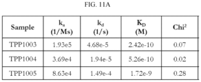
FIGS. 11A-11D illustrate the BIAcore kinetics results for TPP-1003, TPP-1004, and TPP-1005. FIG. 11A illustrates that TPP-1003 and TPP-1004 had pico-molar binding to the DLAT antigen and TPP-1005 had a single digit nano-molar affinity to the DLAT antigen. FIGS. 11B, 11C and 11D illustrate the BIAcore kinetics for TPP-1003, TPP-1004, and TPP 1005, respectively.
FIGS. 12A-12F show mouse T-cell activation for targeted PD-L1 fusion inhibition. Samples were gated to exclude the low Forward Scatter (FSC) and the FL-4+(SYTOX™ Red) dead cells. The same gate was utilized to analyze all samples. The histograms shown are gated on the live mouse T-cells. The decrease in signal of the FL-1 mean fluorescence measurement represents the activated T-cells that have expanded new generations of T-cells, with decreasing dye concentration in each respective generation. FIG. 12A illustrates complete inhibition of mouse T-cell activation for TPP-1986 at 500 nM with an incomplete inhibition observed for 100 nM. FIG. 12B illustrates that TPP-1694 had a significant difference compared to the NC beads, but showed no significant difference compared to TPP-1986 at 500 nM. A significant difference was observed at 100 nM for human TPP-1986 and TPP-992 versus mouse PD-L1-TPP-1694. FIG. 12C illustrates that TPP-1695 demonstrated a lower potency compared to the PD5 human PD-L1 fusion variants at 500 nM and 100 nM concentrations with similar results observed for TPP-1694. FIG. 12D illustrates complete inhibition of mouse T-cell activation for TPP-992 at 500 nM with an incomplete inhibition for 100 nM. FIGS. 12E and 12F illustrate that TPP-1697 (non-targeted isotype control with human PD-L1) and TPP-1004 (murine IgG1 PD5 with no PD-L1 variable-like domain present), showed no significant inhibition of mouse T-cell activation.
FIGS. 13A and 13B show the statistical analysis of the mouse T-cell activation results at 500 nM. FIG. 13A illustrates complete inhibition of TPP-1986 and TPP-992 fusions containing human PD-L1 demonstrated by a not significant difference compared to the NC beads. An expected decrease in the geometric mean fluorescence intensity (Geo MFI) of FL-1 representing the CFSE dye diffusion into progeny cells for the 1× beads, TPP-1697, and TPP-1004 was observed, illustrating activation of the murine T-cells with no significant differences between samples. FIG. 13B is a table of statistical analysis between samples at 500 nM concentrations using Tukey's multiple comparisons test.
FIGS. 14A and 14B show the statistical analysis of the mouse T-cell activation results at 500 nM. FIG. 14A illustrates complete inhibition of TPP-1986 and TPP-992 fusions containing human PD-L1 demonstrated by a not significant difference compared to the NC beads and incomplete inhibition of TPP-1504 containing hB7-H4. An expected decrease in the geometric mean fluorescence intensity (Geo MFI) of FL-1 representing the CFSE dye diffusion into progeny cells for the 1× beads, TPP-1697 and TPP-1505 was observed, illustrating activation of the murine T-cells. FIG. 14B is a table of statistical analysis between samples at 500 nM concentrations using Tukey's multiple comparisons test.
FIGS. 15A and 15B show the statistical analysis of the mouse T-cell activation results at 100 nM. FIG. 15A illustrates incomplete inhibition using fusions containing human PD-L1 compared to the NC beads. A significant difference of human versus murine PD-L1 containing fusions was observed, suggesting human PD-L1 results in greater inhibition of mouse T-cells than mouse PD-L1 V-like domain. An expected decrease in the geometric mean fluorescence intensity (Geo MFI) of FL-1 representing the CFSE dye diffusion into progeny cells for the 1× beads, TPP-1697, and TPP-1004 was observed, illustrating activation of the murine T-cells with no significant differences between samples. FIG. 15B is a table of statistical analysis between samples at 100 nM concentrations using Tukey's multiple comparisons test.
FIGS. 16A and 16B show the statistical analysis of the mouse T-cell activation results at 100 nM. FIG. 16A illustrates incomplete inhibition using fusions containing human PD-L1 and hB7-H4 as compared to the NC beads. An expected decrease in the geometric mean fluorescence intensity (Geo MFI) of FL-1 representing the CFSE dye diffusion into progeny cells for the 1× beads, TPP-1697 and TPP 1505 was observed illustrating activation of the murine T-cells. FIG. 16B is a table of statistical analysis between samples at 100 nM concentrations using Tukey's multiple comparisons test.
FIG. 17 illustrates that mouse PD-L1(TPP-1964) binds to human PD-1 with a faster on-rate than human PD-L1. PD-1 Fc (R&D Systems) was loaded onto anti-human IgG tips illustrated by the first observed deflection. The two fusions TPP-1964 (msPDL1) and TPP-1986 (huPDL1) bound PD-1 Fc. The increased slope observed for TPP-1964 (B1 sensor) suggests the mouse PD-L1 has a faster on-rate than the human PD-L1 on the TPP-1986 fusion (GI Fusion). This binding steps followed by the dissociation in PBS buffer.
FIGS. 18A-18D show the statistical analysis of the human T-cell activation results. FIG. 18A illustrates that at the 500 nM concentration of the anti-DLAT PD-L1 (TPP-1986), B7H4 (TPP-1504), HVEM (TPP-1506), B7H6 (TPP-1894) and CTLA4 (TPP-1898) fusions tested at day 7 inhibited T-cell activation. FIG. 18B illustrates the statistical analysis summary of all samples in the T-cell activation assay. FIG. 18C illustrates that at the 500 nM concentration of the anti-DLAT fusions only PDL-2 (TPP-2215), CD200 (TPP-2216), TIM-3 (TPP-2220) and PD-L1 (TTP-992) showed inhibition of T cell activation at day 4. FIG. 18D illustrates the statistical analysis summary of T-cell activation inhibition for anti-DLAT antibody fusion proteins.
FIGS. 19A and 19B illustrates human T-cell activation at 4 days for 500 nM concentration of PD-L1 Fab fusions. FIG. 19B illustrates the statistical analysis summary of T-cell activation inhibition for PD-L1 fab fusions. V=V-like domain of PDL1; V−C=V−+C-like domains of PDL1; HC=PDL1 fused to N-terminus of Heavy Chain and LC=PDL1 fused to N-terminus of Light Chain.
FIGS. 20A-20D show the statistical analysis of the mouse CD4+ T-cell activation results. FIG. 20A illustrates that at 500 nM concentration of the anti-DLAT PD-L1 (TPP-1984), PD-5 (TPP-1694), and CTLA4 (TPP-1898) fusions tested at day 7 inhibited T-cell activation. FIG. 20B illustrates the statistical analysis summary of all samples in the T-cell activation assay. FIG. 20C illustrates that at the 125 nM a lower level of T-cell activation inhibition was observed with TPP-1898 showing the highest level of inhibition. FIG. 20D illustrates the statistical analysis summary of T-cell activation inhibition for anti-DLAT antibody fusion proteins.
FIGS. 21A and 21B demonstrate the induction of regulatory T-cells (Tregs—CD4+, CD25+, FoxP3+) with the PD-L1 fusions anti-DLAT_PDL1 (labeled DLAT DRUG) and anti-VAP-1_PDL1 (labeled VAP-1 DRUG) combined with the respective activation beads. FIG. 21A illustrate flow cytometry results demonstrating low levels of CD25 for the no activation control (labeled NO BEADS) with 1.71% in quadrant 2 (Q2) representing the CD4+, CD25+ population. The histogram shown below the dot plot represents the intracellular staining with FoxP3 of the Q2 gated events. The activated T-cells (labeled DLAT) showed a significant increase in effector T-cells (Teff) in Q2, however a small percentage of these CD4+, CD25+ cells were Tregs (FoxP3+). FIG. 21B illustrates inhibition of the Teff population shown as 7% in Q2 with the addition of TPP-992 (labeled DLAT DRUG) to the DLAT activation beads, compared to 32% for DLAT beads only. In addition, from the Q2 population, the FoxP3 positive events went from 7% to 16% with the drug. Anti-VAP-1_PDL1 previously showed weaker inhibition in the T-cell activation assay using proliferation dye compared to the anti-DLAT_PDL1 (data not shown). This intermediate effect of anti-VAP-1 fusion was confirmed by the results of this assay; a higher percentage of Q2 events were observed (18%), with a lower induction of Tregs 12% compared to anti-DLAT_PDL1.
FIGS. 22A-22C illustrate histograms showing itration from 250 nM to 50 nM of anti-CD80_mPDL1 fusion incubated with CD80 activation beads for 4 days with human T-cells. The results demonstrate complete inhibition at 250 nM, partial inhibition at 100 nM and no significant inhibition at 50 nM. FIG. 22D shows the dilution of proliferation dye (CFSE) into the progeny T-cells observed by the multiple peaks on the histograms which indicate that the controls including no activation (labeled No Beads) had no activation and the activation control with the CD80 beads had significant activation. FIG. 22E is the graphical overview of the described results from above, the geometric mean of the CFSE is plotted vs the samples incubated.
FIG. 22F is the statiscal analysis of the results using Dunnett's multiple comparisons test.
FIGS. 23A-23C demonstrate the titration of anti-CD20_mPDL1 fusion with CD20 activation beads from 250 nM to 50 nM incubated for 4 days with human T-cells. The results demonstrate complete inhibition at 250 nM, partial inhibition at 100 nM, and less inhibition at 50 nM. FIG. 23D illustrates the dilution of proliferation dye (CFSE) into the progeny T-cells observed by the multiple peaks indicating that activation of the T-cells by the CD20 beads occurred. FIG. 23E represents the graphical overview of the geometric mean of the proliferation dye CFSE. FIG. 23F is the statistical analysis of the described data demonstrating that all of the concentrations of the anti-CD20_mPDL1 were statistically significantly different from the CD20 beads showing inhibition of T-cell activation.
DETAILED DESCRIPTION
The disclosure provides antibody immune cell inhibitor fusion proteins comprising four polypeptide chains that form two antigen binding sites and at least two immune cell receptor binding sites and that inhibit or diminish activation of an immune effector cell when bound to a target antigen. The disclosure also provides antibody immune cell inhibitor fusion proteins comprising two polypeptide chains that form one antigen binding site and at least one immune cell receptor binding site that inhibit or diminish activation of an immune effector cell when bound to a target antigen. The disclosure further provides pharmaceutical compositions and kits that comprise such antibody immune cell inhibitor fusion proteins, and methods of treatment using such proteins.
Standard recombinant DNA methodologies are used to construct the polynucleotides that encode the polypeptides that form the antibody immune cell inhibitor fusion proteins of the disclosure, incorporate these polynucleotides into recombinant expression vectors, and introduce such vectors into host cells. See e.g., Green and Sambrook, 2012, MOLECULAR CLONING: A LABORATORY MANUAL (Cold Spring Harbor Laboratory Press, 4th ed.). Enzymatic reactions and purification techniques may be performed according to manufacturer's protocols, as commonly accomplished in the art, or as described herein. Unless specific definitions are provided, the nomenclature utilized in connection with, and the laboratory procedures and techniques of, analytical chemistry, synthetic organic chemistry, and medicinal and pharmaceutical chemistry described herein are those well-known and commonly used in the art. Similarly, conventional techniques may be used for chemical syntheses, chemical analyses, pharmaceutical preparation, formulation, delivery, and treatment of patients.
1. General Definitions
As utilized in accordance with the present disclosure, the following terms, unless otherwise indicated, shall be understood to have the following meanings. Unless otherwise required by context, singular terms shall include pluralities and plural terms shall include the singular.
The term “antibody immune cell inhibitor fusion protein” as used herein refers to a non-naturally occurring (or recombinant) molecule which comprises four polypeptide chains that form two antigen binding sites and at least two immune cell receptor binding sites; wherein two polypeptide chains have a structure represented by the formula II1-VL-CL-II2, and two polypeptide chains have a structure represented by the formula II3-VH-CH1-Fc-II4; wherein:
VL is an immunoglobulin light chain variable domain;
VH is an immunoglobulin heavy chain variable domain;
CL is an immunoglobulin light chain constant domain;
CH1 is the immunoglobulin CH1 heavy chain constant domain;
Fe is the immunoglobulin hinge region and CH2 and CH3 immunoglobulin heavy chain constant domains; and
II1, II2, II3, and II4 are each independently an immune cell inhibitor domain of an immunoglobulin superfamily member or are absent;
wherein at least one of II1, II2, II3, and II4 is an immune cell inhibitor domain of an immunoglobulin superfamily member; and wherein the antibody immune cell inhibitor fusion protein inhibits or diminishes activation of an immune effector cell only when bound to a target antigen at one or both of the antigen binding sites.
The term “antibody immune cell inhibitor fusion protein” as used herein also refers to a non-naturally occurring (or recombinant) molecule which comprises four polypeptide chains that form two antigen binding sites and at least two immune cell receptor binding sites; wherein two polypeptide chains have a structure represented by the formula II1-L1-VL-CL-L2-II2, and two polypeptide chains have a structure represented by the formula II3-L3-VH-CH1-Fc-L4-II4; wherein:
VL is an immunoglobulin light chain variable domain;
VH is an immunoglobulin heavy chain variable domain;
CL is an immunoglobulin light chain constant domain;
CH1 is the immunoglobulin CH1 heavy chain constant domain;
Fc is the immunoglobulin hinge region and CH2 and CH3 immunoglobulin heavy chain constant domains;
L1, L2, L3 and L4 are each independently a linker domain or are absent; and
II1, II2, II3, and II4 are each independently an immune cell inhibitor domain of an immunoglobulin superfamily member or are absent;
wherein at least one of L1, L2, L3 and L4 is a linker domain and at least one of II1, II2, II3, and II4 is an immune cell inhibitor domain of an immunoglobulin superfamily member; and wherein the antibody immune cell inhibitor fusion protein inhibits or diminishes activation of an immune effector cell only when bound to a target antigen at one or both of the antigen binding sites.
The term “antibody immune cell inhibitor fusion protein” as used herein also refers to a non-naturally occurring (or recombinant) molecule which comprises two polypeptide chains that form one antigen binding site and at least one immune cell receptor binding site; wherein one polypeptide chain has a structure represented by the formula II1-VL-CL-II2, and one polypeptide chain has a structure represented by the formula II3-VH-CH1-II4, wherein:
VL is an immunoglobulin light chain variable domain;
VH is an immunoglobulin heavy chain variable domain;
CL is an immunoglobulin light chain constant domain;
CH1 is the immunoglobulin CH1 heavy chain constant domain;
II1, II2, II3, and II4 are each independently an immune cell inhibitor domain of an immunoglobulin superfamily member or are absent;
wherein at least one of II1, II2, II3, and II4 is an immune cell inhibitor domain of an immunoglobulin superfamily member; and wherein the antibody immune cell inhibitor fusion protein inhibits or diminishes activation of an immune effector cell only when bound to a target antigen at the antigen binding site.
The term “antibody immune cell inhibitor fusion protein” as used herein also refers to a non-naturally occurring (or recombinant) molecule which comprises two polypeptide chains that form one antigen binding site and at least one immune cell receptor binding site, wherein one polypeptide chain has a structure represented by the formula II1-L1-VL-CL-L2-II2, and one polypeptide chain has a structure represented by the formula II3-L3-VH-CH1-L4-II4, wherein:
VL is an immunoglobulin light chain variable domain;
VH is an immunoglobulin heavy chain variable domain;
CL is an immunoglobulin light chain constant domain;
CH1 is the immunoglobulin CH1 heavy chain constant domain;
L1, L2, L3 and L4 are each independently a linker domain or are absent; and
II1, II2, II3, and II4 are each independently an immune cell inhibitor domain of an immunoglobulin superfamily member or are absent;
wherein at least one of L1, L2, L3 and L4 is a linker domain and at least one of II1, II2, II3, and II4 is an immune cell inhibitor domain of an immunoglobulin superfamily member; and wherein the antibody immune cell inhibitor fusion protein inhibits or diminishes activation of an immune effector cell only when bound to a target antigen at the antigen binding site.
The term “antibody” as used herein refers to a protein that is capable of recognizing and specifically binding to an antigen. Ordinary or conventional mammalian antibodies comprise a tetramer, which is typically composed of two identical pairs of polypeptide chains, each pair consisting of one “light” chain (typically having a molecular weight of about 25 kDa) and one “heavy” chain (typically having a molecular weight of about 50-70 kDa). The terms “heavy chain” and “light chain,” as used herein, refer to any immunoglobulin polypeptide having sufficient variable domain sequence to confer specificity for a target antigen. The amino-terminal portion of each light and heavy chain typically includes a variable domain of about 100 to 110 or more amino acids that typically is responsible for antigen recognition. The variable domain may be subjected to further protein engineering to humanize the framework regions if the antibody was derived from a non-human source. The carboxyl-terminal portion of each chain typically defines a constant domain responsible for effector function. Thus, in a naturally occurring antibody, a full-length heavy chain immunoglobulin polypeptide includes a variable domain (VH) and three constant domains (CH1, CH2, and CH3) and a hinge region between CH1 and CH2, wherein the VH domain is at the amino-terminus of the polypeptide and the CH3 domain is at the carboxyl-terminus, and a full-length light chain immunoglobulin polypeptide includes a variable domain (VL) and a constant domain (CL), wherein the VL domain is at the amino-terminus of the polypeptide and the CL domain is at the carboxyl-terminus.
Within full-length light and heavy chains, the variable and constant domains typically are joined by a “J” region of about 12 or more amino acids, with the heavy chain also including a “D” region of about 10 more amino acids. The variable regions of each light/heavy chain pair typically form an antigen binding site. The variable domains of naturally occurring antibodies typically exhibit the same general structure of relatively conserved framework regions (FR) joined by three hypervariable regions, also called complementarity determining regions or CDRs. The CDRs from the two chains of each pair typically are aligned by the framework regions, which may enable binding to a specific epitope. From the amino-terminus to the carboxyl-terminus, both light and heavy chain variable domains typically comprise the domains FR1, CDR1, FR2, CDR2, FR3, CDR3, and FR4.
The term “IgG2/4” as used herein refers to the non-naturally occurring, protein-engineered, heavy chain developed for use in eculizumab (CH1-Hinge-CH2-CH3) and designed to reduce immune effector function and immune cell activation, minimize immunogenicity, and contribute to a longer half-life. This non-IgG 1, 2, 3 or 4 heavy chain is a chimera containing sequence elements of IgG4 and IgG2 including two disulfide bonds in the hinge and changes in the constant regions CH1 and CH2.
The term “human antibody” as used herein includes antibodies having variable and constant regions substantially corresponding to circulating human antibodies and human germline immunoglobulin sequences. In some embodiments, human antibodies are produced in non-human transgenic mammals, including, but not limited to, rodents, such as mice and rats, and lagomorphs, such as rabbits. In other embodiments, human antibodies are produced in hybridoma cells. In still other embodiments, human antibodies are produced recombinantly.
The term “antibody fragment” refers to a portion of an intact or full-length chain or an antibody, generally the target binding or variable region. Examples of antibody fragments include, but are not limited to, Fab, Fab′, F(ab′)2 and Fv fragments. As used herein, the term “functional fragment” is generally synonymous with “antibody fragment,” and with respect to antibodies, can refer to antibody fragments such as Fv, Fab, F(ab′)2.
The term “autoantibody” as used herein refers to a naturally obtained antibody produced by an individual, wherein the antibody recognizes and binds to an antigen derived from or which mimics an antigen derived from a human tissue. An autoantibody can facilitate an immune response against a self-tissue or self-antigen (i.e., antigens that are native to the individual, e.g., an antigen on a cell or tissue, or an endogenous peptide or protein). Autoantibodies frequently arise or are triggered by an infection with an infectious agent such as a virus, bacteria or parasite, where the infectious agent carried a structure that induces antibodies to the structure. The induced antibodies can become harmful in the infection aftermath where that inducing biologic structure is also found on naturally occurring human tissues, thus creating an aberrant and continuing autoantibody response in the aftermath of the infection. The autoantibody can be redesigned and repurposed, as described herein, from the natural autoantibody to modify the original structure found in a diseased individual. The autoantibody may be re-humanized and the heavy chain may be modified to alter the isotype or to otherwise modulate harmful secondary immune functions of the autoantibody. The modified autoantibody can be used to produce a therapeutic fusion protein to treat the disorder created by an aberrant autoantibody response. The autoantibody may also be re-engineered and/or re-derived from or into a single chain Camelid VHH antibody format.
The term “antigen” or “target antigen” as used herein refers to a molecule or a portion of a molecule that is capable of being recognized by and bound by an antibody or the antigen binding portion of the antibody immune cell inhibitor fusion proteins of the disclosure. The target antigen is capable of being used in an animal to produce antibodies capable of binding to an epitope of that antigen. A target antigen may have one or more epitopes. With respect to each target antigen recognized by an antibody or the antigen binding portion of the antibody immune cell inhibitor fusion protein, the antibody or the antigen binding portion of the fusion protein is capable of competing with an intact antibody that recognizes the target antigen.
The term “epitope” as used herein refers to a region or structural element of an antigen that is recognized and bound by an antibody or the antigen binding portion of the antibody immune cell inhibitor fusion protein of the disclosure. More precisely, the epitope is the specific structure that is bound by the CDRs of the antibody. Epitopes can comprise protein structural elements, carbohydrates or even portions of lipid structures found in membranes. An antibody or the antigen binding portion of the antibody immune cell inhibitor fusion protein is said to specifically bind an antigen when it preferentially recognizes its antigen target in a complex mixture of proteins and/or macromolecules. The term “specifically binds,” as used herein, refers to the ability of an antibody or antigen binding portion of the antibody immune cell inhibitor fusion protein to bind to an antigen containing an epitope with an Kd of at least about 1×10⁻⁶ M, 1×10⁻⁷ M, 1×10⁻⁸ M, 1×10⁻⁹ M, 1×10⁻¹⁰ M, 1×10⁻¹¹ M, 1×10⁻¹² M, or more, and/or to bind to an epitope with an affinity that is at least two-fold greater than its affinity for a nonspecific antigen.
The term “antigen binding site” as used herein refers to a site created on the surface of an antibody or the antigen binding portion of the antibody immune cell inhibitor fusion protein of the disclosure where an antigen or an epitope on an antigen is bound. The antigen binding site of an antibody or the antigen binding portion or surface of the antibody immune cell inhibitor fusion protein is typically described by reference to the loop structures created by complementarity determining regions (CDRs) of the antibody or antibody immune cell inhibitor fusion protein.
The term “ligand” as used herein refers to a chemical molecule or biological molecule that can bind readily to a receptor with a specific binding affinity constant. The ligand may be natural or synthetic.
The term “receptor” as used herein refers to a protein capable of interacting (binding) with a ligand. In some embodiments, such a protein is capable of transmitting information resulting from interaction with a ligand, into a cell.
In some circumstances, the terms ligand and receptor may be interchangeable because both the ligand and receptor may be surface bound proteins on different cells that interact with each other. Depending on which cell is the focal point, a receptor on one cell is a ligand of another receptor on a different cell however if the cell which is the focal point is reversed, so is the receptor ligand relationship. In some circumstances both interacting molecules are surface bound and the receptor-ligand relationship is not strict, but merely designates two different molecules interacting with another and causing cell signaling consequences.
The term “native Fc” as used herein refers to a molecule comprising the sequence of a non-antigen binding fragment resulting from digestion of an antibody or produced by other means, whether in monomeric or multimeric form, and can contain the hinge region. The original immunoglobulin source of the native Fc is preferably of human origin and can be any of the immunoglobulins. Native Fc molecules are made up of monomeric polypeptides that can be linked into dimeric or multimeric forms by covalent (i.e., disulfide bonds) and non-covalent association. The number of intermolecular disulfide bonds between monomeric subunits of native Fc molecules ranges from 1 to 4 depending on class (e.g., IgG, IgA, and IgE) or subclass (e.g., IgG1, IgG2, IgG3, IgA1, and IgGA2). One example of a native Fc is a disulfide-bonded dimer resulting from papain digestion of an IgG. The term “native Fc” as used herein is generic to the monomeric, dimeric, and multimeric forms.
The term “Fc variant” as used herein refers to a molecule or sequence that is modified from a native Fc but still comprises a binding site for the salvage receptor, FcRn (neonatal Fc receptor). Exemplary Fc variants, and their interaction with the salvage receptor, are known in the art. Thus, the term “Fc variant” can comprise a molecule or sequence that is humanized from a non-human native Fc. Furthermore, a native Fc comprises regions that can be removed or mutated to produce an Fc variant to alter certain residues that provide structural features or biological activity that are not required for the antibody immune cell inhibitor fusion proteins of the disclosure. Thus, the term “Fe variant” comprises a molecule or sequence that lacks one or more native Fc sites or residues, or in which one or more Fc sites or residues has been modified, that affect or are involved in: (1) disulfide bond formation, (2) incompatibility with a selected host cell, (3) N-terminal heterogeneity upon expression in a selected host cell, (4) glycosylation, (5) interaction with complement, (6) binding to an Fc receptor other than a salvage receptor, or (7) antibody-dependent cellular cytotoxicity (ADCC).
The term “Fc domain” as used herein encompasses native Fc and Fc variants and sequences as defined above. As with Fc variants and native Fc molecules, the term “Fc domain” includes molecules in monomeric or multimeric form, whether digested from whole antibody or produced by other means.
Modifications to the Fc Region
An antibody or antibody immune cell inhibitor fusion protein of the disclosure described herein can, in some embodiments, comprise a variant human Fc constant region that binds to human neonatal Fc receptor (FcRn) with greater affinity than that of the native human Fc constant region from which the variant human Fc constant region was derived. For example, the Fc constant region can comprise one or more (e.g., two, three, four, five, six, seven, or eight or more) amino acid substitutions relative to the native human Fc constant region from which the variant human Fc constant region was derived. The substitutions can increase the binding affinity of an IgG antibody or antibody immune cell inhibitor fusion protein containing the variant Fc constant region to FcRn at pH 6.0, while maintaining the pH dependence of the interaction. See, e.g., Hinton et al., 2004, J. Biol. Chem. 279(8): 6213-16; and Datta-Mannan et al., 2007, Drug Metab. Dispos. 35(1): 86-94. Methods for testing whether one or more substitutions in the Fc constant region of an antibody increases the affinity of the Fc constant region for FcRn at pH 6.0 (while maintaining pH dependence of the interaction) are known in the art. See, e.g., Datta-Mannan et al., 2007, J. Biol. Chem. 282(3): 1709-17; International Publication Nos. WO 98/23289 and WO 97/34631; and U.S. Pat. No. 6,277,375, the disclosures of each of which are incorporated herein by reference in their entirety.
Substitutions that enhance the binding affinity of an antibody Fc constant region for FcRn are known in the art and include, for example: (1) the M252Y/S254T/T256E triple substitution described by Dall'Acqua et al., 2006, J. Biol. Chem. 281(33): 23514-24; (2) the M428L or T250Q/M428L substitutions described in Hinton et al., 2004, J. Biol. Chem. 279(8): 6213-16, and Hinton et al., 2006, J. Immunol. 176(1): 346-56; and (3) the N434A or T307/E380A/N434A substitutions described in Petkova et al., 2006, Int. Immunol. 18(12): 1759-69, the disclosures of which are incorporated herein by reference in their entirety. The additional substitution pairings P257I/Q311I, P257I/N434H, and D376V/N434H are described in, for example, Datta-Mannan et al., 2007, J. Biol. Chem. 282(3): 1709-17, the disclosure of which is incorporated herein by reference in its entirety.
Many mutations to modify Fc biological properties have been identified and may be useful depending on the biology of the disease being treated. In some embodiments, the variant constant region has a substitution at EU amino acid residue 255 for valine. In some embodiments, the variant constant region has a substitution at EU amino acid residue 309 for asparagine. In some embodiments, the variant constant region has a substitution at EU amino acid residue 312 for isoleucine. In some embodiments, the variant constant region has a substitution at EU amino acid residue 386 for leucine.
In some embodiments, the variant Fc constant region comprises no more than 30 (e.g., no more than 29, 28, 27, 26, 25, 24, 23, 22, 21, 20, 19, 18, 17, 16, 15, 14, 13, 12, 11, 10, 9, 8, 7, 6, 5, 4, 3, or 2) amino acid substitutions, insertions, or deletions relative to the native constant region from which it was derived. In some embodiments, the variant Fc constant region comprises one or more amino acid substitutions selected from the group consisting of M252Y, S254T, T256E, N434S, M428L, V259I, T250I, and V308F. In some embodiments, the variant human Fc constant region comprises a methionine at position 428 and an asparagine at position 434, each in EU numbering. In some embodiments, the variant Fc constant region comprises a 428L/434S double substitution as described in, for example, U.S. Pat. No. 9,079,949.
In some embodiments, when the eculizumab heavy chain is used as the starting heavy chain, the 428L/434S mutations are shifted to 429L/435S as a result of the IgG2/4 chimerization engineering. Furthermore, the precise mutations in the eculizumab heavy chain are Met-429-Leu and Asn-435-Ser. See U.S. Pat. No. 9,079,949, the disclosure of which is hereby incorporated by reference in its entirety.
In some embodiments, the variant constant region comprises a substitution at amino acid position 237, 238, 239, 248, 250, 252, 254, 255, 256, 257, 258, 265, 270, 286, 289, 297, 298, 303, 305, 307, 308, 309, 311, 312, 314, 315, 317, 325, 332, 334, 360, 376, 380, 382, 384, 385, 386, 387, 389, 424, 428, 433, 434, or 436 (EU numbering) relative to the native human Fc constant region. In some embodiments, the substitution is selected from the group consisting of methionine for glycine at position 237; alanine for proline at position 238; lysine for serine at position 239; isoleucine for lysine at position 248; alanine, phenylalanine, isoleucine, methionine, glutamine, serine, valine, tryptophan, or tyrosine for threonine at position 250; phenylalanine, tryptophan, or tyrosine for methionine at position 252; threonine for serine at position 254; glutamic acid for arginine at position 255; aspartic acid, glutamic acid, or glutamine for threonine at position 256; alanine, glycine, isoleucine, leucine, methionine, asparagine, serine, threonine, or valine for proline at position 257; histidine for glutamic acid at position 258; alanine for aspartic acid at position 265; phenylalanine for aspartic acid at position 270; alanine, or glutamic acid for asparagine at position 286; histidine for threonine at position 289; alanine for asparagine at position 297; glycine for serine at position 298; alanine for valine at position 303; alanine for valine at position 305; alanine, aspartic acid, phenylalanine, glycine, histidine, isoleucine, lysine, leucine, methionine, asparagine, proline, glutamine, arginine, serine, valine, tryptophan, or tyrosine for threonine at position 307; alanine, phenylalanine, isoleucine, leucine, methionine, proline, glutamine, or threonine for valine at position 308; alanine, aspartic acid, glutamic acid, proline, or arginine for leucine or valine at position 309; alanine, histidine, or isoleucine for glutamine at position 311; alanine or histidine for aspartic acid at position 312; lysine or arginine for leucine at position 314; alanine or histidine for asparagine at position 315; alanine for lysine at position 317; glycine for asparagine at position 325; valine for isoleucine at position 332; leucine for lysine at position 334; histidine for lysine at position 360; alanine for aspartic acid at position 376; alanine for glutamic acid at position 380; alanine for glutamic acid at position 382; alanine for asparagine or serine at position 384; aspartic acid or histidine for glycine at position 385; proline for glutamine at position 386; glutamic acid for proline at position 387; alanine or serine for asparagine at position 389; alanine for serine at position 424; alanine, aspartic acid, phenylalanine, glycine, histidine, isoleucine, lysine, leucine, asparagine, proline, glutamine, serine, threonine, valine, tryptophan, or tyrosine for methionine at position 428; lysine for histidine at position 433; alanine, phenylalanine, histidine, serine, tryptophan, or tyrosine for asparagine at position 434; and histidine for tyrosine or phenylalanine at position 436 (all in EU numbering).
An “immune cell inhibitor domain” as used herein refers to an immunoglobulin domain of an immunoglobulin superfamily member that can cause suppression of immune responses upon binding to the specific receptor on an immune system cell, including inhibition of T-cells, B-cells, monocytes, and antigen presenting cells, inhibition of a particular immune cell function, including cytotoxicity, or any combination of the above responses. The immune cell inhibitor domains of the antibody immune cell inhibitor fusion proteins of the disclosure are referred to herein as follows: II1, which, when present, is fused (with or without an intervening linker) to the N-terminal end of the light chain; II2, which, when present, is fused (with or without an intervening linker) to the C-terminal end of the light chain; II3, which, when present, is fused (with or without an intervening linker) to the N-terminal end of the heavy chain; and II4, which, when present, (with or without an intervening linker) is fused to the C-terminal end of the heavy chain. Linkers may or may not be needed depending on the where the stop and start residues of protein fusions are chosen because often natural linkers are found between immunoglobulin domains.
The term “immunoglobulin superfamily member” as used herein refers to a class of proteins that are associated with the adhesion, binding, and recognition processes of cells. Exemplary members of the immunoglobulin superfamily include, but are not limited to, Programmed Death Ligand 1 (PD-L1), also known as B7 Homolog 1 (B7-H1), or CD274; Programmed Death Ligand 2 (PD-L2), also known as B7-DC; B7 Homolog 3 (B7-H3), as known as CD276; B7 Homolog 4 (B7-H4), also known as V-set Domain Containing T-cell Activation Inhibitor 1 (VTCN1), B7 Superfamily, Member 1 (B7S1), or B7x; Herpesvirus Entry Mediator (HVEM), also known as Herpesvirus Entry Mediator A (HVEA), Tumor Necrosis Factor Receptor Superfamily, Member 14 (TNFRSF14), or CD270; V-type Immunoglobulin Domain-Containing Suppressor of T-cell Activation; Chromosome 10 Open reading Frame 54 (C10orf54), also known as Death Domain 1-Alpha (DD1-Alpha); B7 Homolog 6 (B7-H6), also known as Natural Cytotoxicity Triggering Receptor 3 ligand 1; Human Endogenous Retrovirus-H Long Terminal Repeat-Associating 2 (HHLA2), also known as B7 Homolog 7 (B7-H7), B7 Homolog 5 (B7-H5), or HERV-H LTR-Associating 2; Cytotoxic T Lymphocyte-Associated 4 (CTLA-4), also known as CD152; CD200, also known as Membrane Glycoprotein MRC OX-2 or MOX2; Killer-cell Immunoglobulin-Like Receptor (KIR); T-cell Immunoglobulin and Mucin Domains-Containing Protein 3 (TIM-3), also known as Hepatitis A Virus Cellular Receptor 2 (HAVCR2); and Lymphocyte Activation Gene 3 (LAG3), also known as CD223.
The term “immune effector cell” as used herein refers to the cells of the immune system that mount immune responses to an antigen. Suitable effector cells include but are not limited to populations of antigen presenting cells, cytotoxic T-cells, and T helper cells that mediate cellular immunity. In addition to antigen-specific effector T-cells, the effector cell populations may include, but are not limited to, other cytotoxic immune cells against a selected antigen such as natural killer cells, lymphocytes, monocytes, macrophages, neutrophils, and eosinophils.
The term “linker” as used herein refers to one or more amino acid residues inserted between immunoglobulin domains and/or immune cell inhibitor domains of the antibody immune cell inhibitor fusion proteins of the disclosure. For example, a linker may be inserted between an immunoglobulin domain and an immune cell inhibitor domain, at the sequence level. The precise location of a domain transition can be determined by locating peptide stretches that do not form secondary structural elements such as beta-sheets or alpha-helices as demonstrated by experimental data or as can be assumed by techniques of modeling or secondary structure prediction. Linkers may or may not be needed depending on the where the stop and start residues of protein fusions are chosen because often natural linkers are found between immunoglobulin domains. The linkers of the antibody immune cell inhibitor fusion proteins of the disclosure are referred to herein as follows: L1, which, when present, is located on the light chain between the II1 and VL domains; L2, which, when present, is located on the light chain between the CL and II2 domains; L3, which, when present, is located on the heavy chain between the II3 and L3 domains; and L4, which, when present, is located on the heavy chain between the Fc and II4 domains. The linkers L1, L2, L3, and L4 are independent, but in some embodiments of the antibody immune cell inhibitor fusion proteins of the disclosure may have the same sequence and/or length.
The term “naturally occurring” as used herein and applied to an object refers to the fact that the object can be found in nature and has not been manipulated by man. For example, a polynucleotide or polypeptide that is present in an organism (including viruses) that can be isolated from a source in nature and that has not been intentionally modified by man is naturally occurring. Similarly, “non-naturally occurring” as used herein refers to a protein molecule that is not found in nature or that has been structurally modified through protein engineering and synthesized, manufactured, or produced by man using recombinant DNA technologies in appropriate cells such as, for example, CHO cells.
A “recombinant” molecule is one that has been prepared, expressed, created, or isolated by recombinant DNA technology means.
The term “fusion protein” as used herein refers to protein constructs comprising an immunoglobulin domain and an immune cell inhibitor protein. The immune cell inhibitor protein in some fusion proteins of the disclosure may not constitute the entire natural protein but may be limited to an active domain of the entire protein responsible for binding to a corresponding receptor on the surface of an immune function cell. In addition, an immunoglobulin domain in some fusion proteins of the disclosure may not constitute the entire natural immunoglobulin domain but may be limited to a portion of the natural immunoglobulin domain responsible for specifically binding a target antigen or epitope or conferring other properties of the natural immunoglobulin domain. Importantly, the fragment of the immune cell inhibitor protein or immunoglobulin domain in some fusion proteins of the disclosure would not be naturally occurring as the fragment, but may retain the same protein sequence for the fragment and incorporated into a therapeutic fusion protein.
The terms “inhibit” or “diminish” as used herein refer to a complete or partial arrest of immune effector cell activation.
The terms “substantially pure” or “substantially purified” as used herein refer to a compound or species that is the predominant species present (i.e., on a molar basis it is more abundant than any other individual species in the composition). In some embodiments, a substantially purified fraction is a composition wherein the species comprises at least about 50% (on a molar basis) of all macromolecular species present. In other embodiments, a substantially pure composition will comprise more than about 80%, 85%, 90%, 95%, or 99% of all macromolar species present in the composition. In still other embodiments, the species is purified to essential homogeneity (contaminant species cannot be detected in the composition by conventional detection methods) wherein the composition consists essentially of a single macromolecular species.
The phrases “biological property,” “biological characteristic,” and the term “activity” in reference to an antibody or an antibody immune cell inhibitor fusion protein of the disclosure are used interchangeably herein and include, but are not limited to, epitope affinity and specificity, ability to antagonize the activity of the antigen target (or targeted polypeptide), the in vivo stability of the antibody or antibody immune cell inhibitor fusion protein, and the immunogenic properties of the antibody or antibody immune cell inhibitor fusion protein. Other identifiable biological properties or characteristics of an antibody or an antibody immune cell inhibitor fusion protein include, for example, cross-reactivity, (i.e., with non-human homologs of the antigen target, or with other antigen targets or tissues, generally), and ability to preserve high expression levels of protein in mammalian cells. The aforementioned properties or characteristics can be observed or measured using art-recognized techniques.
A “neutralizing effect” of an antibody immune cell inhibitor fusion protein as used herein refers to a fusion protein of an antibody and an immune cell inhibitor ligand that is able to first, bind an antigen for which the antigen binding portion of the antibody immune cell inhibitor fusion protein specifically recognizes, then, through simultaneous binding of the fused immune cell inhibitor domain to a specific receptor, to block or substantially reduce an unwanted deleterious or autoimmune effector function carried out by the cell expressing the immune cell inhibitor receptor. As used herein, “substantially reduce” means at least about 60%, preferably at least about 70%, more preferably at least about 75%, even more preferably at least about 80%, still more preferably at least about 85%, most preferably at least about 90% reduction of the unwanted or autoimmune effector function of the cell carrying the ligand's receptor.
The term “neutralizing antibody” refers to an antibody that can neutralize the function of the protein or infectious agent the antibody specifically recognizes and binds. Typically, neutralizing antibodies refer to antibodies specific for viral, bacterial, or other infectious agents. Where therapeutic antibodies are used as drug candidates to treat human disease, neutralizing antibodies can arise as anti-drug antibodies (ADA) but have the undesirable function of neutralizing the therapeutic benefit of the therapeutic antibody. Neutralizing antibodies to the antigen binding portions of the fusion proteins of the present disclosure would represent an undesirable development.
The term “KD,” as used herein, refers to the dissociation constant (KD=[A]×[B]/[AB]) of the interaction between an antibody or an antibody immune cell inhibitor fusion protein of the disclosure and an antigen target and has the units of moles/liter. An antibody or antibody immune cell inhibitor fusion protein of the disclosure typically has a dissociation constant (KD) of 10−5 to 10−12 moles/liter or less, or 10−7 to 10−12 moles/liter or less, or 10−3 to 10−12 moles/liter, and/or with a binding affinity of at least 107 M−1, or at least 108 M−1, or at least 109 M−1, or at least 1012 M−1. Any KD value greater than 10−4 moles/liter is generally considered to indicate non-specific binding. Therefore, the lower the KD value, the greater the affinity. In some embodiments, a monovalent antibody or antibody immune cell inhibitor fusion protein of the disclosure will bind to a desired antigen with an affinity less than 500 nM, or less than 200 nM, or less than 10 nM, or less than 500 pM. High affinity or very strong binding is often associated with greater efficacy, but it is not always the case that the greater the affinity the greater the efficacy.
The dissociation constant (KD) can be determined, for example, by surface plasmon resonance (SPR). Generally, surface plasmon resonance analysis measures real-time binding interactions (both on rate and off rate) between a ligand (a target antigen on a biosensor matrix) and an analyte by surface plasmon resonance using, for example, the BIAcore system (Pharmacia Biosensor; Piscataway, N.J.). Surface plasmon analysis can also be performed by immobilizing the analyte and presenting the ligand. Specific binding of an antibody or an antibody immune cell inhibitor fusion protein of the disclosure to an antigen or antigenic determinant can also be determined in any suitable manner known in the art, including, for example, Scatchard analysis and/or competitive binding assays, such as radioimmunoassays (RIA), enzyme linked immunosorbent assays (ELISA), enzyme immunoassays (EIA), and sandwich competition assays.
The term “vector,” as used herein, refers to any molecule (e.g., nucleic acid, plasmid, or virus) that is used to transfer coding information to a host cell. One type of vector is a “plasmid,” which refers to a circular double-stranded DNA molecule into which additional DNA segments may be inserted. Another type of vector is a viral vector, wherein additional DNA segments may be inserted into the viral genome. Certain vectors are capable of autonomous replication in a host cell into which they are introduced (e.g., bacterial vectors having a bacterial origin of replication and episomal mammalian vectors). Other vectors (e.g., non-episomal mammalian vectors) can be integrated into the genome of a host cell upon introduction into the host cell and thereby are replicated along with the host genome. In addition, certain vectors are capable of directing the expression of genes to which they are operatively linked. Such vectors are referred to herein as “expression vectors.”
The term “operably linked” is used herein to refer to an arrangement of flanking sequences wherein the flanking sequences are configured or assembled so as to perform their usual function. Thus, a flanking sequence operably linked to a coding sequence may be capable of effecting the replication, transcription, and/or translation of the coding sequence. For example, a coding sequence is operably linked to a promoter when the promoter is capable of directing transcription of that coding sequence.
The term “host cell,” as used herein, refers to a cell into which an expression vector has been introduced. A host cell is intended to refer not only to the particular subject cell, but also to the progeny of such a cell. Because certain modifications may occur in succeeding generations due to either mutation or environmental influences, such progeny may not, in fact, be identical to the parent cell, but such cells are still included within the scope of the term “host cell” as used herein. A wide variety of host cell expression systems can be used to express the antibody immune cell inhibitor fusion proteins of the disclosure, including bacterial, yeast, baculoviral, and mammalian expression systems (as well as phage display expression systems).
Examples of cultured mammalian cell lines include Chinese Hamster ovary (CHO) simian cells such as COS, murine cell lines such as NS0, and human cell lines such as HEK and HeLa, which may be used to produce the antibody immune cell inhibitor fusion proteins of the disclosure. Vectors are transfected into the cells and the DNA may be integrated into the genome by homologous recombination in the case of stable transfection, or the cells may be transiently transfected. Examples of mammalian expression vectors include adenoviral vectors, the pSV and the pCMV series of plasmid vectors, vaccinia and retroviral vectors, as well as baculovirus. The promoters for cytomegalovirus (CMV) and simian virus 40 (SV40) are commonly used in mammalian expression vectors to drive gene expression. Non-viral promoters, such as the elongation factor (EF)-1 promoter, may also be used.
One embodiment of the disclosure provides nucleic acid molecules comprising nucleotide sequences encoding polypeptide chains that form an antibody immune cell inhibitor fusion protein of the disclosure. Another embodiment of the disclosure provides expression vectors comprising nucleic acid molecules comprising nucleotide sequences encoding polypeptide chains that form the antibody immune cell inhibitor fusion proteins of the disclosure. Yet another embodiment of the disclosure provides host cells that express such antibody immune cell inhibitor fusion proteins (i.e., comprising nucleic acid molecules or vectors encoding polypeptide chains that form such antibody immune cell inhibitor fusion proteins).
In some embodiments, the disclosure provides methods for preparing an antibody immune cell inhibitor fusion protein of the disclosure, wherein such methods comprise cultivating or maintaining a host cell under conditions such that the host cell produces or expresses such antibody immune cell inhibitor fusion proteins, and optionally further comprises isolating the antibody immune cell inhibitor fusion protein so produced.
A skilled artisan will be able to determine suitable variants of the polypeptide chains of the antibody immune cell inhibitor fusion proteins of the disclosure using well-known techniques. For example, one skilled in the art may identify suitable areas of a polypeptide chain that may be changed without destroying activity by targeting regions not believed to be important for activity. Alternatively, one skilled in the art can identify residues and portions of the molecules that are conserved among similar polypeptides. In addition, even areas that may be important for biological activity or for structure may be subject to conservative amino acid substitutions without destroying the biological activity or without adversely affecting the polypeptide structure.
The term “patient” as used herein includes human and animal subjects.
A “disorder” is any condition that would benefit from treatment using the antibody immune cell inhibitor fusion proteins of the disclosure. “Disorder” and “condition” are used interchangeably herein and include chronic and acute disorders or diseases, including those pathological conditions that predispose a patient to the disorder in question. A “T-cell mediated-condition” is a condition directly or indirectly effected by the T-cells of the immune system.
The terms “treatment” or “treat” as used herein refer to both therapeutic treatment and prophylactic or preventative measures. Those in need of treatment include those having a disorder as well as those prone to have the disorder or those in which the disorder is to be prevented.
The terms “pharmaceutical composition” or “therapeutic composition” as used herein refer to a compound or composition capable of inducing a desired therapeutic effect when properly administered to a patient. One embodiment of the disclosure provides a pharmaceutical composition comprising a pharmaceutically acceptable carrier and a therapeutically effective amount of at least one antibody immune cell inhibitor fusion protein of the disclosure.
The term “pharmaceutically acceptable carrier” or “physiologically acceptable carrier” as used herein refers to one or more formulation materials suitable for accomplishing or enhancing the delivery of one or more antibody immune cell inhibitor fusion proteins of the disclosure.
The terms “effective amount” and “therapeutically effective amount” when used in reference to a pharmaceutical composition comprising one or more antibody immune cell inhibitor fusion proteins of the disclosure refer to an amount or dosage sufficient to produce a desired therapeutic result. More specifically, a therapeutically effective amount is an amount of an antibody immune cell inhibitor fusion protein sufficient to inhibit, for some period of time, one or more of the clinically defined pathological processes associated with the condition being treated. The effective amount may vary depending on the specific antibody immune cell inhibitor fusion protein that is being used, and may also depend on a variety of factors and conditions related to the patient being treated and the severity of the disorder. For example, if the antibody immune cell inhibitor fusion protein is to be administered in vivo, factors such as the age, weight, and health of the patient as well as dose response curves and toxicity data obtained in preclinical animal work would be among those factors considered. The determination of an effective amount or therapeutically effective amount of a given pharmaceutical composition is within the ability of those skilled in the art.
2. Antibody Immune Cell Inhibitor Fusion Proteins
In one embodiment of the disclosure, the antibody immune cell inhibitor fusion protein comprises four polypeptide chains that form two antigen binding sites at least two immune cell receptor binding sites, wherein two polypeptide chains have a structure represented by the formula II1-VL-CL-II2, and two polypeptide chains have a structure represented by the formula II3-VH-CH1-Fc-II4; wherein:
VL is an immunoglobulin light chain variable domain;
VH is an immunoglobulin heavy chain variable domain;
CL is an immunoglobulin light chain constant domain;
CH1 is the immunoglobulin CH1 heavy chain constant domain;
Fc is the immunoglobulin hinge region and CH2 and CH3 immunoglobulin heavy chain constant domains; and
II1, II2, II3, and II4 are each independently an immune cell inhibitor domain of an immunoglobulin superfamily member or are absent;
wherein at least one of II1, II2, II3, and II4 is an immune cell inhibitor domain of an immunoglobulin superfamily member; and wherein the antibody immune cell inhibitor fusion protein inhibits or diminishes activation of an immune effector cell only when bound to a target antigen at one or both of the antigen binding sites.
In another embodiment of the disclosure, the antibody immune cell inhibitor fusion protein comprises four polypeptide chains that form two antigen binding sites and at least two immune cell receptor binding sites, wherein two polypeptide chains have a structure represented by the formula II1-L1-VL-CL-L2-II2, and two polypeptide chains have a structure represented by the formula II3-L3-VH-CH1-Fc-L4-II4; wherein:
VL is an immunoglobulin light chain variable domain;
VH is an immunoglobulin heavy chain variable domain;
CL is an immunoglobulin light chain constant domain;
CH1 is the immunoglobulin CH1 heavy chain constant domain;
Fc is the immunoglobulin hinge region and CH2 and CH3 immunoglobulin heavy chain constant domains;
L1, L2, L3 and L4 are each independently a linker domain or are absent; and
II1, II2, II3, and II4 are each independently an immune cell inhibitor domain of an immunoglobulin superfamily member or are absent;
wherein at least one of L1, L2, L3 and L4 is a linker domain and at least one of II1, II2, II3, and II4 is an immune cell inhibitor domain of an immunoglobulin superfamily member; and wherein the antibody immune cell inhibitor fusion protein inhibits or diminishes activation of an immune effector cell only when bound to a target antigen at one or both of the antigen binding sites.
In another embodiment of the disclosure, the antibody immune cell inhibitor fusion protein comprises two polypeptide chains that form one antigen binding site and at least one immune cell receptor binding site; wherein one polypeptide chain has a structure represented by the formula II1-VL-CL-II2, and one polypeptide chain has a structure represented by the formula II3-VH-CH1-II4; wherein:
VL is an immunoglobulin light chain variable domain;
VH is an immunoglobulin heavy chain variable domain;
CL is an immunoglobulin light chain constant domain;
CH1 is the immunoglobulin CH1 heavy chain constant domain; and
II1, II2, II3, and II4 are each independently an immune cell inhibitor domain of an immunoglobulin superfamily member or are absent;
wherein at least one of II1, II2, II3, and II4 is an immune cell inhibitor domain of an immunoglobulin superfamily member; and wherein the antibody immune cell inhibitor fusion protein inhibits or diminishes activation of an immune effector cell only when bound to a target antigen at the antigen binding site.
In another embodiment of the disclosure, the antibody immune cell inhibitor fusion protein comprises two polypeptide chains that form one antigen binding site and at least one immune cell receptor binding site; wherein one polypeptide chain has a structure represented by the formula II1-L1-VL-CL-L2-II2, and one polypeptide chain has a structure represented by the formula II3-L3-VH-CH1-L4-II4; wherein:
VL is an immunoglobulin light chain variable domain;
VH is an immunoglobulin heavy chain variable domain;
CL is an immunoglobulin light chain constant domain;
CH1 is the immunoglobulin CH1 heavy chain constant domain;
L1, L2, L3 and L4 are each independently a linker domain or are absent; and
II1, II2, II3, and II4 are each independently an immune cell inhibitor domain of an immunoglobulin superfamily member or are absent;
wherein at least one of L1, L2, L3 and L4 is a linker domain, and at least one of II1, II2, II3, and II4 is an immune cell inhibitor domain of an immunoglobulin superfamily member; and wherein the antibody immune cell inhibitor fusion protein inhibits or diminishes activation of an immune effector cell only when bound to a target antigen at the antigen binding site.
The antibody immune cell inhibitor fusion proteins of the disclosure may be prepared using domains or sequences obtained or derived from any human or non-human antibody, including, for example, human, murine, or humanized antibodies. In some antibody immune cell inhibitor fusion proteins of the disclosure, the VL, VH, CL, CH1, and/or Fc domains of the antibody immune cell inhibitor fusion protein may not constitute the entire natural immunoglobulin domain, provided, however, that the portion of the VL, VH, CL, CH1, and/or Fc domain used in the antibody immune cell inhibitor fusion protein is capable of functioning in the same manner as the full-length natural immunoglobulin domain. In other embodiments, the antibody immune cell inhibitor fusion proteins of the disclosure may further comprise additional VL, VH, CL, CH1, and/or Fc domains.
In some antibody immune cell inhibitor fusion proteins of the disclosure, one or more of II1, II2, II3, and II4 is an immune cell inhibitor domain. In some embodiments, at least one of the immune cell inhibitor domains can inhibit or diminish activation of an immune effector cell involved in the immune response to self-tissue. In other embodiments, at least one of the immune cell inhibitor domains can inhibit or diminish activation of an immune effector cell involved in the immune response to self-tissue when an autoantibody is bound to the antigen at the site of an ongoing disease process. In other embodiments, at least one of II1, II2, II3, and II4 is an immune cell inhibitor domain of an immunoglobulin domain superfamily member. In other embodiments, at least one of II1, II2, II3 and II4 is absent.
In some embodiments, the immune cell inhibitor domain is obtained or derived from a member of the immunoglobulin superfamily. In certain embodiments, the immune cell inhibitor domain comprises a Programmed Death Ligand 1 (PD-L1), B7 Homolog 1 (B7-H1), or CD274 domain; Programmed Death Ligand 2 (PD-L2) or B7-DC domain; B7 Homolog 3 (B7-H3) or CD276 domain; B7 Homolog 4 (B7-H4), V-set Domain-Containing T-cell Activation Inhibitor 1 (VTCN1), B7 Superfamily, Member 1 (B7S1), or B7x domain; Herpesvirus Entry Mediator (HVEM), Herpesvirus Entry Mediator A (HVEA), Tumor Necrosis Factor Receptor Superfamily, Member 14 (TNFRSF14), or CD270 domain (also known as Tumor necrosis factor receptor superfamily member 14); V-type Immunoglobulin Domain-Containing Suppressor of T-cell Activation domain; B7 Homolog 6 (B7-H6) or Natural cytotoxicity triggering receptor 3 ligand 1 domain; Human Endogenous Retrovirus-H Long Terminal Repeat-Associating 2 (HHLA2), B7 Homolog 7 (B7-H7), B7 Homolog 5 (B7-H5), or HERV-H LTR-associating protein 2 domain; Cytotoxic T-Lymphocyte-Associated protein 4 (CTLA-4) or CD152 domain; CD200, Membrane Glycoprotein MRC OX-2, or MOX2 domain; Killer-cell Immunoglobulin-like Receptor (KIR) domain; T-cell Immunoglobulin and Mucin Domains-Containing Protein 3 (TIM-3) or Hepatitis A Virus Cellular Receptor 2 (HAVCR2) domain; or Lymphocyte Activation Gene-3 (LAG3) or CD223 domain.
In some embodiments, the immune cell inhibitor domain comprises a PD-L1 extracellular domain (wherein PD-L1 is also known as CD274, B7-H, B7H1, PDCD1L1, or PDCD1LG1) or a PD-L2 extracellular domain (wherein PD-L2 is also known as CD273). PD-L1 when bound to Programmed Death 1 (PD-1) inhibitory receptor (also known as CD279) acts as an immune checkpoint inhibitor of B-cells, T-cells, monocytes, and antigen presenting cells. For example, an activated T-cell expresses PD-1 on its surface upon antigen recognition and produces interferons, which induce expression of PD-L1 in multiple tissues. Binding of PD-1 to its ligand limits T-cell activity. Under normal conditions, the PD-1/PD-L1 pathway prevents excessive stimulation and maintains the immune tolerance to self-antigens by negatively regulating the immune response (Riella et al., 2012, Am. J. Transplant. 12(10): 2575-87).
In one embodiment of the disclosure, the antibody immune cell inhibitor fusion protein comprises four polypeptide chains that form two antigen binding sites and at least two PD-1 binding sites; wherein two polypeptide chains have a structure represented by the formula II1-VL-CL-II2, and two polypeptide chains have a structure represented by the formula II3-VH-CH1-Fc-II4; wherein:
VL is an immunoglobulin light chain variable domain;
VH is an immunoglobulin heavy chain variable domain;
CL is an immunoglobulin light chain constant domain;
CH1 is the immunoglobulin CH1 heavy chain constant domain;
Fc is the immunoglobulin hinge region and CH2 and CH3 immunoglobulin heavy chain constant domains; and
II1, II2, II3, and II4 are each independently an immune cell inhibitor domain of an immunoglobulin superfamily member capable of binding PD-1 or are absent; wherein at least one of II1, II2, II3, and II4 is an immune cell inhibitor domain of an immunoglobulin superfamily member capable of binding PD-1; and wherein the antibody immune cell inhibitor fusion protein inhibits or diminishes activation of an immune effector cell only when bound to a target antigen at one or both of the antigen binding sites.
In another embodiment of the disclosure, the antibody immune cell inhibitor fusion protein comprises four polypeptide chains that form two antigen binding sites and at least two PD-1 binding sites; wherein two polypeptide chains have a structure represented by the formula II1-L1-VL-CL-L2-II2, and two polypeptide chains have a structure represented by the formula II3-L3-VH-CH1-Fc-L4-II4; wherein:
VL is an immunoglobulin light chain variable domain;
VH is an immunoglobulin heavy chain variable domain;
CL is an immunoglobulin light chain constant domain;
CH1 is the immunoglobulin CH1 heavy chain constant domain;
Fc is the immunoglobulin hinge region and CH2 and CH3 immunoglobulin heavy chain constant domains;
L1, L2, L3 and L4 are each independently a linker domain or are absent; and
II1, II2, II3, and II4 are each independently an immune cell inhibitor domain of an immunoglobulin superfamily member capable of binding PD-1 or are absent; wherein at least one of L1, L2, L3 and L4 is a linker domain and at least one of II1, II2, II3, and II4 is an immune cell inhibitor domain of an immunoglobulin superfamily member capable of binding PD-1; and wherein the antibody immune cell inhibitor fusion protein inhibits or diminishes activation of an immune effector cell only when bound to a target antigen at one or both of the antigen binding sites.
In another embodiment of the disclosure, the antibody immune cell inhibitor fusion protein comprises two polypeptide chains that form one antigen binding site and at least one PD-1 binding site; wherein one polypeptide chain has a structure represented by the formula II1-VL-CL-II2, and one polypeptide chain has a structure represented by the formula II3-VH-CH1-II4; wherein:
VL is an immunoglobulin light chain variable domain;
VH is an immunoglobulin heavy chain variable domain;
CL is an immunoglobulin light chain constant domain;
CH1 is the immunoglobulin CH1 heavy chain constant domain;
II1, II2, II3, and II4 are each independently an immune cell inhibitor domain of an immunoglobulin superfamily member capable of binding PD-1 or are absent; wherein at least one of II1, II2, II3, and II4 is an immune cell inhibitor domain of an immunoglobulin superfamily member capable of binding PD-1; and wherein the antibody immune cell inhibitor fusion protein inhibits or diminishes activation of an immune effector cell only when bound to a target antigen at the antigen binding site.
In another embodiment of the disclosure, the antibody immune cell inhibitor fusion protein comprises two polypeptide chains that form one antigen binding site and at least one PD-1 binding site; wherein one polypeptide chain has a structure represented by the formula II1-L1-VL-CL-L2-II2, and one polypeptide chain has a structure represented by the formula II3-L3-VH-CH1-L4-II4, wherein:
VL is an immunoglobulin light chain variable domain;
VH is an immunoglobulin heavy chain variable domain;
CL is an immunoglobulin light chain constant domain;
CH1 is the immunoglobulin CH1 heavy chain constant domain;
L1, L2, L3 and L4 are each independently a linker domain or are absent; and
II1, II2, II3, and II4 are each independently an immune cell inhibitor domain of an immunoglobulin superfamily member capable of binding PD-1 or are absent; wherein at least one of L1, L2, L3 and L4 is a linker domain and at least one of II1, II2, II3, and II4 is an immune cell inhibitor domain of an immunoglobulin superfamily member capable of binding PD-1; and wherein the antibody immune cell inhibitor fusion protein inhibits or diminishes activation of an immune effector cell only when bound to a target antigen at the antigen binding site.
In some embodiments, the VL, VH, CL, and CH1 domains of the antibody immune cell inhibitor fusion proteins of the disclosure form an autoantibody that recognizes and binds an antigen derived from or mimicking an antigen derived from a human tissue. In some embodiments, the autoantibody facilitates an immune response to a self-tissue.
In some embodiments of the disclosure, the antibody immune cell inhibitor fusion proteins of the disclosure are capable of specifically binding one or more antigen targets. In certain embodiments of the disclosure, the antibody immune cell inhibitor fusion protein is capable of specifically binding at least one antigen target that is Human serum albumin, Bone mineral (hydroxyapatite), Complement component 3d (C3d), Glutamic acid decarboxylase 65 (GAD65), Asialoglycoprotein receptor (ASGPR), Asialoglycoprotein receptor 2 (ASGPR2) Transferrin receptor protein 2 (TFR2), Solute carrier family 2, facilitated glucose (SLC2A2), Solute carrier organic anion transporter family member 1B1 (SLCO1B1), Solute carrier organic anion transporter family member 1B3 (SLCO1B3), Multidrug resistance-associated protein 6 (ABCC6), Major histocompatibility complex I (MHC I), Major histocompatibility complex II (MHC II), T-cell receptor (TCR), B-Cell receptor (BCR), Transforming growth factor beta, Cluster differentiation 4 (CD4), Cluster differentiation 8 (CD8), Cluster differentiation 11c (CD11c), cluster differentiation 14 (CD14), Fas ligand/Tumor necrosis factor super family 6 (Fas Ligand/TNFSF6), Fibrinogen (1F3), collagen type I, collagen type II, collagen type III, collagen type IV, Cystatin C, Cluster differentiation 133 (CD133), Cluster differentiation 10 (CD10), Cluster differentiation 13 (CD13), Cluster differentiation 20 (CD20), Cluster differentiation 80 (CD80), Neural cell adhesion molecule 1 (NCAM1), E-cadherin, Epithelial cell adhesion molecule (EPCAM), Epithelial Membrane Antigen (EMA), Transthyretin, Vascular adhesion protein 1, Lymphocyte function-associated antigen 3, or Dihydrolipoamide acetyltransferase. In some embodiments of the disclosure, the antibody immune cell inhibitor fusion protein is capable of inhibiting the function of one or more of the antigen targets.
In some embodiments, the antibody immune cell inhibitor fusion proteins of the disclosure comprise a linker joining at least one immune cell inhibitor domain to at least one immunoglobulin light chain or heavy chain or domain. In some antibody immune cell inhibitor fusion proteins, L1, when present, may be 5 to 30 amino acid residues in length; L2, when present, may be 5 to 30 amino acid residues in length; L3, when present, may be 5 to 30 amino acid residues in length; and L4, when present, may be 5 to 30 amino acid residues in length.
In some antibody immune cell inhibitor fusion proteins of the disclosure, at least one of L1, L2, L3 and L4 is absent.
The identity and sequence of amino acid residues in the linker may vary depending on the type of secondary structural element necessary to be achieved. For example, glycine, serine, and alanine are best for linkers having maximum flexibility. Some combination of glycine, proline, threonine, and serine are useful if a more rigid and extended linker is necessary. Any amino acid residue may be considered as a linker in combination with one or more other amino acid residues, which may be the same as or different as the first amino acid residue, to construct larger peptide linkers as necessary depending on the desired properties.
3. Antibody Immune Cell Inhibitor Fusion Protein Therapeutic Compositions and Administration Thereof
Therapeutic or pharmaceutical compositions comprising one or more antibody immune cell inhibitor fusion proteins of the disclosure are within the scope of the disclosure. Such therapeutic or pharmaceutical compositions can comprise a therapeutically effective amount of an antibody immune cell inhibitor fusion protein, or antibody immune cell inhibitor fusion protein-drug conjugate, in admixture with a pharmaceutically or physiologically acceptable formulation agent selected for suitability with the mode of administration.
Acceptable formulation materials are preferably nontoxic to recipients at the dosages and concentrations employed.
The pharmaceutical composition can contain formulation materials for modifying, maintaining, or preserving, for example, the pH, osmolarity, viscosity, clarity, color, isotonicity, odor, sterility, stability, rate of dissolution or release, adsorption, or penetration of the composition. Suitable formulation materials include, but are not limited to, amino acids (such as glycine, glutamine, asparagine, arginine, or lysine), antimicrobials, antioxidants (such as ascorbic acid, sodium sulfite, or sodium hydrogen-sulfite), buffers (such as borate, bicarbonate, Tris-HCl, citrates, phosphates, or other organic acids), bulking agents (such as mannitol or glycine), chelating agents (such as ethylenediamine tetraacetic acid (EDTA)), complexing agents (such as caffeine, polyvinylpyrrolidone, beta-cyclodextrin, or hydroxypropyl-beta-cyclodextrin), fillers, monosaccharides, disaccharides, and other carbohydrates (such as glucose, mannose, or dextrins), proteins (such as serum albumin, gelatin, or immunoglobulins), coloring, flavoring and diluting agents, emulsifying agents, hydrophilic polymers (such as polyvinylpyrrolidone), low molecular weight polypeptides, salt-forming counterions (such as sodium), preservatives (such as benzalkonium chloride, benzoic acid, salicylic acid, thimerosal, phenethyl alcohol, methylparaben, propylparaben, chlorhexidine, sorbic acid, or hydrogen peroxide), solvents (such as glycerin, propylene glycol, or polyethylene glycol), sugar alcohols (such as mannitol or sorbitol), suspending agents, surfactants or wetting agents (such as pluronics; PEG; sorbitan esters; polysorbates such as polysorbate 20 or polysorbate 80; triton; tromethamine; lecithin; cholesterol or tyloxapal), stability enhancing agents (such as sucrose or sorbitol), tonicity enhancing agents (such as alkali metal halides—preferably sodium or potassium chloride—or mannitol sorbitol), delivery vehicles, diluents, excipients and/or pharmaceutical adjuvants (see, e.g., REMINGTON'S PHARMACEUTICAL SCIENCES (18th Ed., A. R. Gennaro, ed., Mack Publishing Company 1990), and subsequent editions of the same, incorporated herein by reference in their entirety for any purpose).
The optimal pharmaceutical composition will be determined by a skilled artisan depending upon, for example, the intended route of administration, delivery format, and desired dosage. Such compositions can influence the physical state, stability, rate of in vivo release, and rate of in vivo clearance of the antibody immune cell inhibitor fusion proteins of the disclosure.
The primary vehicle or carrier in a pharmaceutical composition can be either aqueous or non-aqueous in nature. For example, a suitable vehicle or carrier for injection can be water, physiological saline solution, or artificial cerebrospinal fluid, possibly supplemented with other materials common in compositions for parenteral administration. Neutral buffered saline or saline mixed with serum albumin are further exemplary vehicles. Other exemplary pharmaceutical compositions comprise Tris buffer of about pH 7.0-8.5, or acetate buffer of about pH 4.0-5.5, which can further include sorbitol or a suitable substitute. In one embodiment of the disclosure, antibody immune cell inhibitor fusion protein compositions can be prepared for storage by mixing the selected composition having the desired degree of purity with optional formulation agents in the form of a lyophilized cake or an aqueous solution. Further, the antibody immune cell inhibitor fusion protein can be formulated as a lyophilizate using appropriate excipients such as sucrose.
The pharmaceutical compositions of the disclosure can be selected for parenteral delivery. Alternatively, the compositions can be selected for inhalation or for delivery through the digestive tract, such as orally. The preparation of such pharmaceutically acceptable compositions is within one of skill in the art.
The formulation components are present in concentrations that are acceptable to the site of administration. For example, buffers are used to maintain the composition at physiological pH or at a slightly lower pH, typically within a pH range of from about 5 to about 8.
When parenteral administration is contemplated, the therapeutic compositions for use in this disclosure can be in the form of a pyrogen-free, parenterally acceptable, aqueous solution comprising the desired antibody immune cell inhibitor fusion protein in a pharmaceutically acceptable vehicle. One suitable vehicle for parenteral injection is sterile distilled water in which an antibody immune cell inhibitor fusion protein of the disclosure is formulated as a sterile, isotonic solution, properly preserved. Yet another preparation can involve the formulation of the antibody immune cell inhibitor fusion protein with an agent, such as injectable microspheres, bio-erodible particles, polymeric compounds (such as polylactic acid or polyglycolic acid), beads, or liposomes, that provides for the controlled or sustained release of the product which can then be delivered via a depot injection. Hyaluronic acid can also be used, and this can have the effect of promoting sustained duration in the circulation. Other suitable means for the introduction of the antibody immune cell inhibitor fusion protein include implantable drug delivery devices.
Additional pharmaceutical compositions of the disclosure will be evident to those skilled in the art, including formulations involving antibody immune cell inhibitor fusion proteins in sustained- or controlled-delivery formulations. Techniques for formulating a variety of other sustained- or controlled-delivery means, such as liposome carriers, bio-erodible microparticles, or porous beads and depot injections, are also known to those skilled in the art. Additional examples of sustained-release preparations include semipermeable polymer matrices in the form of shaped articles, e.g., films, or microcapsules. Sustained release matrices can include polyesters, hydrogels, polylactides, copolymers of L-glutamic acid and gamma ethyl-L-glutamate, poly(2-hydroxyethyl-methacrylate), ethylene vinyl acetate, or poly-D(−)-3-hydroxybutyric acid. Sustained-release compositions can also include liposomes, which can be prepared by any of several methods known in the art.
Pharmaceutical compositions of the disclosure to be used for in vivo administration typically must be sterile. This can be accomplished by filtration through sterile filtration membranes. Where the composition is lyophilized, sterilization using this method can be conducted either prior to, or following, lyophilization and reconstitution. The composition for parenteral administration can be stored in lyophilized form or in a solution. In addition, parenteral compositions generally are placed into a container having a sterile access port, for example, an intravenous solution bag or vial having a stopper pierceable by a hypodermic injection needle.
Once the pharmaceutical composition has been formulated, it can be stored in sterile vials as a solution, suspension, gel, emulsion, solid, or as a dehydrated or lyophilized powder. Such formulations can be stored either in a ready-to-use form or in a form (e.g., lyophilized) requiring reconstitution prior to administration.
Also encompassed by the disclosure are kits for producing a single-dose administration unit. The kits can each contain both a first container having a dried protein and a second container having an aqueous formulation. Also included within the scope of this disclosure are kits containing single and multi-chambered pre-filled syringes (e.g., liquid syringes and lyosyringes).
The effective amount of pharmaceutical composition containing an antibody immune cell inhibitor fusion protein to be employed therapeutically will depend, for example, upon the therapeutic context and objectives. One skilled in the art will appreciate that the appropriate dosage levels for treatment will thus vary depending, in part, upon the molecule delivered, the indication for which the antibody immune cell inhibitor fusion protein is being used, the route of administration, and the size (body weight, body surface, or organ size) and condition (the age and general health) of the patient. Accordingly, the clinician can titer the dosage and modify the route of administration to obtain the optimal therapeutic effect. A typical dosage can range from about 0.1 mg/kg to up to about 100 mg/kg or more, depending on the factors mentioned above. In other embodiments, the dosage can range from 0.1 mg/kg up to about 100 mg/kg; or 1 mg/kg up to about 100 mg/kg; or 5 mg/kg, 10 mg/kg, 15 mg/kg, 20 mg/kg, 25 mg/kg, 30 mg/kg, 35 mg/kg, 40 mg/kg, 45 mg/kg, 50 mg/kg, 55 mg/kg, 60 mg/kg, 65 mg/kg, 70 mg/kg, 75 mg/kg, up to about 100 mg/kg.
Dosing frequency will depend upon the pharmacokinetic parameters of the antibody immune cell inhibitor fusion protein in the formulation being used. Typically, a clinician will administer the composition until a dosage is reached that achieves the desired effect. The composition can therefore be administered as a single dose, as two or more doses (which may or may not contain the same amount of the desired molecule) over time, or as a continuous infusion via an implantation device or catheter. Further refinement of the appropriate dosage is routinely made by those of ordinary skill in the art and is within the ambit of tasks routinely performed by them. Appropriate dosages can be ascertained through use of appropriate dose-response data.
The route of administration of the pharmaceutical composition is in accord with known methods, e.g., orally; through injection by intravenous, intraperitoneal, intracerebral (intraparenchymal), intracerebroventricular, intramuscular, intraocular, intraarterial, intraportal, or intralesional routes; by sustained release systems; or by implantation devices. Where desired, the compositions can be administered by bolus injection or continuously by infusion, or by implantation device.
The composition can also be administered locally via implantation of a membrane, sponge, or other appropriate material onto which the desired molecule has been absorbed or encapsulated. Where an implantation device is used, the device can be implanted into any suitable tissue or organ, and delivery of the desired molecule can be via diffusion, timed-release bolus, or continuous administration.
In some embodiments of the disclosure, the antibody immune cell inhibitor fusion proteins of the disclosure are used to treat a patient having Primary Biliary Cholangitis (PBC), Type 1 Diabetes, Goodpasture's syndrome, Amyloidosis, Ankylosing spondylitis, Anti-glomerular basement membrane/anti-tubular basement membrane nephritis, Antiphospholipid syndrome, Autoimmune hepatitis, Autoimmune oophoritis Autoimmune pancreatitis, Autoimmune retinopathy, Behcet's disease, Crohn's disease, Devic's disease, Lupus, Dressler's syndrome, Fibrosing alveolitis, Glomerulonephritis, Graves' disease, Guillain-Barre syndrome, IgA Nephropathy, IgG4-related sclerosing disease, Immune thrombocytopenic purpura (ITP), Microscopic polyangiitis (MPA), Mixed connective tissue disease (MCTD), Multiple sclerosis, Polyneuropathy, Organomegaly, Endocrinopathy, Monoclonal syndrome (POEMS), Polyarteritis nodosa, Rheumatoid arthritis, Schmidt syndrome, Scleritis, Scleroderma, Sjögren's syndrome Sperm & testicular autoimmunity, Stiff person syndrome (SPS), Takayasu's arteritis, Temporal arteritis, Giant cell arteritis, Thrombocytopenic purpura (TTP), Tolosa-Hunt syndrome (THS), Vasculitis or a T-cell mediated condition.
4. EXAMPLES
The Examples that follow are illustrative of specific embodiments of the disclosure, and various uses thereof. They are set forth for explanatory purposes only, and should not be construed as limiting the scope of the disclosure in any way.
Example 1. Construction of Anti-DLAT Fusion Antibody
An antibody immune inhibitor fusion protein was designed comprising a human autoimmune antibody to a self-antigen identified as driving the disease process known as Primary Biliary Cholangitis (PBC), previously considered cirrhosis. The self-antigen recognized by the autoimmune antibody was shown to be Pyruvate Dehydrogenase Complex subunit E2 (PDC-E2, also known as Dihydrolipoamide Acetyltransferase or DLAT). The original fusion protein fused the extracellular domain of Programmed Death Ligand 1 (PD-L1, also known as CD274, or B7 Homolog 1 (B7-H1)), to each of the N- and C-termini of an engineered autoantibody containing the V regions of the autoantibody and an engineered Fc variant known as IgG2/4 to minimize effector function. See Pascual et al., 1994, J. Immunol. 152(5): 2577-85; and Chen et al., 1998, J. Autoimmun. 11(2): 151-61. The PD-L1 domain was fused to the antibody to interact with Programmed Death 1 (PD-1, CD279) receptor on T-cells believed to be driving the disease process, thereby inhibiting activation of the T-cells and subsequent activation of other immune system actors such as antigen presenting cells, B-cells, and any associated excreted cytokines to thus ameliorate the immune response causing the disease.
Initially, multiple anti-DLAT antibodies were identified in the literature (Thomson et al., 1998, J. Hepatol. 28(4): 582-94). Two of them (designated PD2 and PD5) were initially selected for cloning as: (i) human full-length antibodies and were constructed with the autoantibody VH and VL regions fused with a non-naturally occurring engineered heavy chain including CH1-hinge-CH2-CH3 (SEQ ID NO: 35)(IgG2/4); and (ii) human CH Fabs. Each of these antibodies or antibody fragments were constructed with or without the variable-like domain of the extracellular domain (ECD) of PD-L1 linked to either the C-terminus or the N-terminus of the heavy and/or light chains. Upon review of the crystal structure of the PD-1/PD-L1 complex, it was determined that the variable-like domain of PD-L1 was the binding portion, and thus the variable-like domain of PD-L1 was fused to each of the N- and C-termini of the anti-DLAT antibody to determine which orientation was most effective for this particular antibody-immune inhibitor pair. See Zak et al., 2015, Structure 23(12): 2341-48.
Using Geneious 8.1.5 software, reference sequences (see Table 1) were imported and modified for construct design. Constructs were synthesized and cloned into suitable vectors and were sequence-verified prior to transient transfections. Protein expression was performed by transfection of constructs shown in Table 2 using the Invitrogen EXPI293F system. Protein A columns were used for purification of antibodies and antibody fusions or Nickel NTA was used for purification of the 6× His-tagged Fab proteins using commercial columns.
Sequences for the constructs are shown in Table 3. The sequences are shown as monomeric sequences, but all full-length antibodies expressed and purified were, of course, dimeric. The heavy chain of the antibody is shown in regular text, the light chain of the antibody is shown in underlined text, the linker is shown in italicized text, and the immune cell inhibitor domain of an immunoglobulin superfamily member are shown in bold text.
| TABLE 1 |
| |
| Reference Sequences |
| Reference Sequences |
Accession # |
Source |
| |
| Human PD-L1 Reference Protein Sequence |
Q9NZQ7 |
UniProt |
| Human PD-L1 Reference Protein Sequence |
NP_054862.1 |
NCBI |
| Murine PD-L1 Reference Protein Sequence |
NP_068693.1 |
NCBI |
| Murine PD-L1 Reference Protein Sequence |
Q9EP73 |
UniProt |
| Crystal Structure of human PD-1 and its ligand PD-L1 |
4ZQK |
Protein Data |
| |
|
Bank |
| PD-1 binding domain from human PD-L1 |
5C3T |
Protein Data |
| |
|
Bank |
| DWZ Heavy Chain Reference Protein Sequence |
AJ001167.1 |
GenBank |
| DWZ Light Chain Reference Protein Sequence |
AJ001168.1 |
GenBank |
| PD2 Heavy Chain Reference Protein Sequence |
AJ001171.1 |
GenBank |
| PD2 Light Chain Reference Protein Sequence |
AJ001172.1 |
GenBank |
| Human B7 Dendritic Cell Molecule (B7-DC), Programmed |
Q9BQ51 |
UniProt |
| cell death 1 ligand 2 (PD-L2) |
|
|
| Human B7-Homolog 3 (B7-H3), (CD276) |
Q5ZPR3 |
UniProt |
| Human B7-H4, V-set domain-containing T-cell activation |
Q7Z7D3 |
UniProt |
| inhibitor 1 (VTCN1) |
|
|
| Human B7-H5, V-type immunoglobulin domain-containing |
Q9H7M9 |
UniProt |
| suppressor of T-cell activation (VISTA) |
|
|
| Human B7-H6 (NCR3LG1), Natural cytotoxicity triggering |
Q68D85 |
UniProt |
| receptor 3 ligand 1 |
|
|
| Human B7-H7, HERV-H LTR-associating protein 2 |
Q9UM44 |
UniProt |
| (HHLA2) |
|
|
| Human Cytotoxic T-lymphocyte protein 4 (CTLA-4) |
P16410 |
UniProt |
| Human Tumor necrosis factor receptor superfamily |
Q92956 |
UniProt |
| member 14 (TNFRSF14), Herpesvirus Entry Mediator |
|
|
| (HVEM) |
|
|
| Human T-Cell Immunoglobulin Mucin Family Member 3 |
Q8TDQ0 |
UniProt |
| (TIM-3), Hepatitis A virus cellular receptor 2 (HAVCR2) |
| |
| TABLE 2 |
| |
| Construct Descriptions |
| Protein |
|
Transfection |
Molecular |
|
| ID |
Isotype |
Constructs |
Weight (kDa) |
Description |
| |
| TPP-985 |
Human IgG2/4 |
pME074 + pME090 |
144 |
PD5_IgG2-4 |
| TPP-986 |
Human IgG2/4 |
pME074 + pME095 |
172.8 |
N-Terminus_PDL1_HC_PD5 |
| TPP-992 |
Human IgG2/4 |
pME076 + pME090 |
172.8 |
N-terminus_PDL1_LC_PD5 |
| TPP-990 |
Human IgG2/4 |
pME075 + pME090 |
172.8 |
PD5_LC_C-terminus_PDL1 |
| TPP-996 |
Human IgG2/4 |
pME080 + pME092 |
174.4 |
N-Terminus_PDL1_HC_PD2 |
| TPP-1002 |
Human IgG2/4 |
pME082 + pME089 |
175 |
N-terminus_PDL1_LC_PD2 |
| TPP-1007 |
Human IgG2/4 |
pME091 + pME102 |
144 |
DWZ_IgG2-4 |
| TPP-1006 |
Human IgG2/4 |
pME089 + pME080 |
145.7 |
PD2_IgG2-4 |
| TPP-955 |
Human Fab |
pME071 + pME074 |
46.7 |
PD5_Human_Fab |
| TPP-983 |
Human Fab |
pME072 + pME074 |
61.1 |
PD5_LC_C-terminus linker |
| |
|
|
|
PDL1 |
| TPP-984 |
Human Fab |
pME073 + pME074 |
61.1 |
N-terminus_PDL1_LC_PD5 |
| TPP-991 |
Human Fab |
pME076 + pME071 |
61.1 |
N-terminus_PDL1_HC_PD5 |
| TPP-989 |
Human Fab |
pME075 + pME071 |
61.1 |
PD5_HC_C-terminus_PDL1 |
| TPP-993 |
Human Fab |
pME077 + pME080 |
48.4 |
PD2 Fab |
| TPP-994 |
Human Fab |
pME078 + pME080 |
62.8 |
PD2_HC_C-terminus_PDL1 |
| TPP-995 |
Human Fab |
pME079 + pME080 |
62.8 |
N-terminus_PDL1_HC_PD2 |
| TPP-999 |
Human Fab |
pME081 + pME077 |
62.8 |
PD2_LC_C-terminus_PDL1 |
| TPP-1001 |
Human Fab |
pME077 + pME082 |
62.8 |
N-terminus_PDL1_LC_PD2 |
| TPP-1003 |
Murine IgG1 |
pME083 + pME084 |
147.4 |
mIgG1 PD2 Ab |
| TPP-1004 |
Murine IgG1 |
pME085 + pME086 |
145.4 |
mIgG1 PD5 Ab |
| TPP-1005 |
Murine IgG1 |
pME087 + pME088 |
144.9 |
mIgG1 DWZ Ab |
| TPP-1008 |
N/A |
pME098 |
15.2 |
Human_PDL1_V-domain_N- |
| |
|
|
|
terminus |
| TPP-1009 |
N/A |
pME099 |
26 |
Human_PDL1_V_C_Domain |
| |
|
|
|
NP_054862.1 |
| TPP-1010 |
N/A |
pME100 |
13.9 |
Human_PDL1_V-domain |
| |
|
|
|
NP_054862.1 |
| TPP-1011 |
N/A |
pME101 |
14.4 |
Human_PDL1_V-domain_C- |
| |
|
|
|
Terminus |
| TPP-1986 |
Murine IgG1 Fc |
N/A |
173 |
N-terminus_PDL1_LC_PD5 |
| |
Silent |
|
|
|
| TPP-1694 |
Murine IgG1 |
pME085 + pADL0128 |
173.8 |
N- |
| |
|
|
|
terminus_mPDL1_LC_PD5 |
| TPP-1695 |
Murine IgG1 |
pME087 + pADL0129 |
173.3 |
N- |
| |
|
|
|
terminus_mPDL1_LC_DWZ |
| TPP-1697 |
Human IgG2/4 |
pADL0131 + pADL0132 |
172.8 |
N-terminus_PDL1_LC_12B4 |
| TPP-1504 |
Human IgG2/4 |
pADL0123 + pME090 |
199.7 |
N-terminus_B7- |
| |
|
|
|
H4_LC_PD5 |
| TPP-1505 |
Human IgG2/4 |
pADL0124 + pME090 |
184.3 |
N-terminus_B7- |
| |
|
|
|
H5_LC_PD5 |
| TPP-1506 |
Human IgG2/4 |
pADL0125 + pME090 |
182.8 |
N- |
| |
|
|
|
terminus_HVEM_LC_PD5 |
| TPP-1894 |
Human IgG2/4 |
pADL0141 + pME090 |
201.4 |
N-terminus_B7- |
| |
|
|
|
H6_LC_PD5 |
| TPP-1898 |
Human IgG2/4 |
pADL0145 + pME090 |
175.1 |
N-terminus_CTLA- |
| |
|
|
|
4_LC_PD5 |
| TPP-1507 |
Human IgG2/4 |
pADL0126 + pME090 |
170.8 |
N-terminus_PDL2_V- |
| |
|
|
|
like_domain_LC_PD5 |
| TPP-2215 |
Human IgG2/4 |
pADL0176 + pME090 |
193.2 |
N- |
| |
|
|
|
terminus_PDL2_LC_PD5 |
| TPP-2216 |
Human IgG2/4 |
pADL0177 + pME090 |
193 |
N- |
| |
|
|
|
terminus_CD200_LC_PD5 |
| TPP-2218 |
Human IgG2/4 |
pADL0179 + pME090 |
242.2 |
N-terminus_B7- |
| |
|
|
|
H3_LC_PD5 |
| TPP-2220 |
Human IgG2/4 |
pADL0181 + pME090 |
188.1 |
N-terminus_TIM- |
| |
|
|
|
3_LC_PD5 |
| |
| TABLE 3 |
| |
| Construct Sequences |
| SEQ |
|
|
|
| ID |
Sequence |
Construct |
|
| NO: |
Description |
Name |
Sequence |
| |
| 1 |
PD5 HC Fab |
pME071 |
QLQLQESGPGLVKPSETLSLTCIVSGGSISRNSYYWG |
| |
|
|
WIRQPPGKGLEWIGSIVYSGSTYHQPSLKSRVTIFLD |
| |
|
|
TSKNQFFLKLTSVTAADTAVYYCARGTRATTWPPPIG |
| |
|
|
YWGQGTLVTVSSASTKGPSVFPLAPSSKSTSGGTAAL |
| |
|
|
GCLVKDYFPEPVTVSWNSGALTSGVHTFPAVLQSSGL |
| |
|
|
YSLSSVVTVPSSSLGTQTYICNVNHKPSNTKVDKRVE |
| |
|
|
PKSC |
| |
| 2 |
PD5 HC PDL1 |
pME072 |
QLQLQESGPGLVKPSETLSLTCIVSGGSISRNSYYWG |
| |
Fab |
|
WIRQPPGKGLEWIGSIVYSGSTYHQPSLKSRVTIFLD |
| |
|
|
TSKNQFFLKLTSVTAADTAVYYCARGTRATTWPPPIG |
| |
|
|
YWGQGTLVTVSSASTKGPSVFPLAPSSKSTSGGTAAL |
| |
|
|
GCLVKDYFPEPVTVSWNSGALTSGVHTFPAVLQSSGL |
| |
|
|
YSLSSWTVPSSSLGTQTYICNVNHKPSNTKVDKRVE |
| |
|
|
PKSCGGSSRSSSSGGGGSGGGG FTVTVPKDLYVVEYG |
| |
|
|
SNMTIECKFPVEKQLDLAALIVYWEMEDKNIIQFVHG
|
| |
|
|
EEDLKVQHSSYRQRARLLKDQLSLGNAALQITDVKLQ
|
| |
|
|
DAGVYRCMISYGGADYKRITVKVNA
|
| |
| 3 |
PDL1 HC PD5 |
pME073 |
FTVTVPKDLYVVEYGSNMTIECKFPVEKQLDLAALIV
|
| |
Fab |
|
YWEMEDKNIIQFVHGEEDLKVQHSSYRQRARLLKDQL
|
| |
|
|
SLGNAALQITDVKLQDAGVYRCMISYGGADYKRITVK
|
| |
|
|
VNA GGSSRSSSSGGGGSGGGGQLQLQESGPGLVKPSE |
| |
|
|
TLSLTCIVSGGSISRNSYYWGWIRQPPGKGLEWIGSI |
| |
|
|
VYSGSTYHQPSLKSRVTIFLDTSKNQFFLKLTSVTAA |
| |
|
|
DTAVYYCARGTRATTWPPPIGYWGQGTLVTVSSASTK |
| |
|
|
GPSVFPLAPSSKSTSGGTAALGCLVKDYFPEPVTVSW |
| |
|
|
NSGALTSGVHTFPAVLQSSGLYSLSSVVTVPSSSLGT |
| |
|
|
QTYICNVNHKPSNTKVDKRVEPKSC |
| |
| 4 |
PD5 LC Fab |
pME074 |
QSVLTQPPSVSAAPGQKVTVSCFGSSSNIGNYFASWY
|
| |
Human Lamda LC |
|
QQLPGAAPRLLIYGNNERPSGIPDRFSGSKSGTSATL
|
| |
|
|
VITGLQTGDEAAYYCATWDSSLSAVVFGGGTKLTVLG
|
| |
|
|
QPKANPTVTLFPPSSEELQANKATLVCLISDFYPGAV
|
| |
|
|
TVAWKADGSPVKAGVETTKPSKQSNNKYAASSYLSLT
|
| |
|
|
PEQWKSHRSYSCQVTHEGSTVEKTVAPTECS
|
| |
| 5 |
PD5 LC PDL1 |
pME075 |
QSVLTQPPSVSAAPGQKVTVSCFGSSSNIGNYFASWY
|
| |
|
|
QQLPGAAPRLLIYGNNERPSGIPDRFSGSKSGTSATL
|
| |
|
|
VITGLQTGDEAAYYCATWDSSLSAVVFGGGTKLTVLG
|
| |
|
|
QPKANPTVTLFPPSSEELQANKATLVCLISDFYPGAV
|
| |
|
|
TVAWKADGSPVKAGVETTKPSKQSNNKYAASSYLSLT
|
| |
|
|
PEQWKSHRSYSCQVTHEGSTVEKTVAPTECS
GGSSRS
|
| |
|
|
SSSGGGGSGGGG
FTVTVPKDLYVVEYGSNMTIECKFP
|
| |
|
|
VEKQLDLAALIVYWEMEDKNIIQFVHGEEDLKVQHSS
|
| |
|
|
YRQRARLLKDQLSLGNAALQITDVKLQDAGVYRCMIS
|
| |
|
|
YGGADYKRITVKVNA
|
| |
| 6 |
PDL1 LC PD5 |
pME076 |
FTVTVPKDLYVVEYGSNMTIECKFPVEKQLDLAALIV
|
| |
|
|
YWEMEDKNIIQFVHGEEDLKVQHSSYRQRARLLKDQL
|
| |
|
|
SLGNAALQITDVKLQDAGVYRCMISYGGADYKRITVK
|
| |
|
|
VNA
GGSSRSSSSGGGGSGGGG
QSVLTQPPSVSAAPGQ
|
| |
|
|
KVTVSCFGSSSNIGNYFASWYQQLPGAAPRLLIYGNN
|
| |
|
|
ERPSGIPDRFSGSKSGTSATLVITGLQTGDEAAYYCA
|
| |
|
|
TWDSSLSAVVFGGGTKLTVLGQPKANPTVTLFPPSSE
|
| |
|
|
ELQANKATLVCLISDFYPGAVTVAWKADGSPVKAGVE
|
| |
|
|
TTKPSKQSNNKYAASSYLSLTPEQWKSHRSYSCQVTH
|
| |
|
|
EGSTVEKTVAPTECS
|
| |
| 7 |
PD2 HC Fab |
pME077 |
EVQLVESGGGLVKPGGSLRLSCAASGFTFGSYSMTWV |
| |
|
|
RQAPGKGLEWVSFISTVSTYIYYPDSVKGRFTISRDN |
| |
|
|
AKNSLYLQMNSLRAEDTAVYYCARVSGRGSAARDGEK |
| |
|
|
GTYYYYMDVWGKGTTVTVSSASTKGPSVFPLAPSSKS |
| |
|
|
TSGGTAALGCLVKDYFPEPVTVSWNSGALTSGVHTFP |
| |
|
|
AVLQSSGLYSLSSVVTVPSSSLGTQTYICNVNHKPSN |
| |
|
|
TKVDKRVEPKSC |
| |
| 8 |
PD2 HC PDL1 Fab |
pME078 |
EVQLVESGGGLVKPGGSLRLSCAASGFTFGSYSMTWV |
| |
|
|
RQAPGKGLEWVSFISTVSTYIYYPDSVKGRFTISRDN |
| |
|
|
AKNSLYLQMNSLRAEDTAVYYCARVSGRGSAARDGEK |
| |
|
|
GTYYYYMDVWGKGTTVTVSSASTKGPSVFPLAPSSKS |
| |
|
|
TSGGTAALGCLVKDYFPEPVTVSWNSGALTSGVHTFP |
| |
|
|
AVLQSSGLYSLSSVVTVPSSSLGTQTYICNVNHKPSN |
| |
|
|
TKVDKRVEPKSCGGSSRSSSSGGGGSGGGG FTVTVPK |
| |
|
|
DLYVVEYGSNMTIECKFPVEKQLDLAALIVYWEMEDK
|
| |
|
|
NIIQFVHGEEDLKVQHSSYRQRARLLKDQLSLGNAAL
|
| |
|
|
QITDVKLQDAGVYRCMISYGGADYKRITVKVNA
|
| |
| 9 |
PD2 HC PDL1 Fab |
pME079 |
FTVTVPKDLYVVEYGSNMTIECKFPVEKQLDLAALIV
|
| |
|
|
YWEMEDKNIIQFVHGEEDLKVQHSSYRQRARLLKDQL
|
| |
|
|
SLGNAALQITDVKLQDAGVYRCMISYGGADYKRITVK
|
| |
|
|
VNA GGSSRSSSSGGGGSGGGGEVQLVESGGGLVKPGG |
| |
|
|
SLRLSCAASGFTFGSYSMTWVRQAPGKGLEWVSFIST |
| |
|
|
VSTYIYYPDSVKGRFTISRDNAKNSLYLQMNSLRAED |
| |
|
|
TAVYYCARVSGRGSAARDGEKGTYYYYMDVWGKGTTV |
| |
|
|
TVSSASTKGPSVFPLAPSSKSTSGGTAALGCLVKDYF |
| |
|
|
PEPVTVSWNSGALTSGVHTFPAVLQSSGLYSLSSVVT |
| |
|
|
VPSSSLGTQTYICNVNHKPSNTKVDKRVEPKSC |
| |
| 10 |
PD2 Human Lambda |
pME080 |
SSELTQDPAVSVALGQTVTITCQGDSLRSYYASWYQQ
|
| |
LC |
|
KPGQAPVLVIFGKNNRPSGIPDRFSGSRSGNTASLTI
|
| |
|
|
TGAQAEDEADYFCDSRDSSANHWVFGGGTKLTVLQPK
|
| |
|
|
AAPSVTLFPPSSEELQANKATLVCLISDFYPGAVTVA
|
| |
|
|
WKADSSPVKAGVETTTPSKQSNNKYAASSYLSLTPEQ
|
| |
|
|
WKSHKSYSCQVTHEGSTVEKTVAPTECS
|
| |
| 11 |
PD2 LC PDL1 Fab |
pME081 |
SSELTQDPAVSVALGQTVTITCQGDSLRSYYASWYQQ
|
| |
|
|
KPGQAPVLVIFGKNNRPSGIPDRFSGSRSGNTASLTI
|
| |
|
|
TGAQAEDEADYFCDSRDSSANHWVFGGGTKLTVLQPK
|
| |
|
|
AAPSVTLFPPSSEELQANKATLVCLISDFYPGAVTVA
|
| |
|
|
WKADSSPVKAGVETTTPSKQSNNKYAASSYLSLTPEQ
|
| |
|
|
WKSHKSYSCQVTHEGSTVEKTVAPTECS
GGSSRSSSS
|
| |
|
|
GGGGSGGGG
FTVTVPKDLYVVEYGSNMTIECKFPVEK
|
| |
|
|
QLDLAALIVYWEMEDKNIIQFVHGEEDLKVQHSSYRQ
|
| |
|
|
RARLLKDQLSLGNAALQITDVKLQDAGVYRCMISYGG
|
| |
|
|
ADYKRITVKVNA
|
| |
| 12 |
PDL1 LC PD2 Fab |
pME082 |
FTVTVPKDLYVVEYGSNMTIECKFPVEKQLDLAALIV
|
| |
|
|
YWEMEDKNIIQFVHGEEDLKVQHSSYRQRARLLKDQL
|
| |
|
|
SLGNAALQITDVKLQDAGVYRCMISYGGADYKRITVK
|
| |
|
|
VNA
GGSSRSSSSGGGGSGGGG
SSELTQDPAVSVALGQ
|
| |
|
|
TVTITCQGDSLRSYYASWYQQKPGQAPVLVIFGKNNR
|
| |
|
|
PSGIPDRFSGSRSGNTASLTITGAQAEDEADYFCDSR
|
| |
|
|
DSSANHWVFGGGTKLTVLQPKAAPSVTLFPPSSEELQ
|
| |
|
|
ANKATLVCLISDFYPGAVTVAWKADSSPVKAGVETTT
|
| |
|
|
PSKQSNNKYAASSYLSLTPEQWKSHKSYSCQVTHEGS
|
| |
|
|
TVEKTVAPTECS
|
| |
| 13 |
PD2 HC Hu IgG2/4 |
pME089 |
EVQLVESGGGLVKPGGSLRLSCAASGFTFGSYSMTWV |
| |
|
|
RQAPGKGLEWVSFISTVSTYIYYPDSVKGRFTISRDN |
| |
|
|
AKNSLYLQMNSLRAEDTAVYYCARVSGRGSAARDGEK |
| |
|
|
GTYYYYMDVWGKGTTVTVSSASTKGPSVFPLAPCSRS |
| |
|
|
TSESTAALGCLVKDYFPEPVTVSWNSGALTSGVHTFP |
| |
|
|
AVLQSSGLYSLSSVVTVPSSNFGTQTYTCNVDHKPSN |
| |
|
|
TKVDKTVERKCCVECPPCPAPPVAGPSVFLFPPKPKD |
| |
|
|
TLMISRTPEVTCVVVDVSQEDPEVQFNWYVDGVEVHN |
| |
|
|
AKTKPREEQFNSTYRVVSVLTVLHQDWLNGKEYKCKV |
| |
|
|
SNKGLPSSIEKTISKAKGQPREPQVYTLPPSQEEMTK |
| |
|
|
NQVSLTCLVKGFYPSDIAVEWESNGQPENNYKTTPPV |
| |
|
|
LDSDGSFFLYSRLTVDKSRWQEGNVFSCSVMHEALHN |
| |
|
|
HYTQKSLSLSLGK |
| |
| 14 |
PD5 HC Hu IgG2/4 |
pME090 |
QLQLQESGPGLVKPSETLSLTCIVSGGSISRNSYYWG |
| |
|
|
WIRQPPGKGLEWIGSIVYSGSTYHQPSLKSRVTIFLD |
| |
|
|
TSKNQFFLKLTSVTAADTAVYYCARGTRATTWPPPIG |
| |
|
|
YWGQGTLVTVSSASTKGPSVFPLAPCSRSTSESTAAL |
| |
|
|
GCLVKDYFPEPVTVSWNSGALTSGVHTFPAVLQSSGL |
| |
|
|
YSLSSVVTVPSSNFGTQTYTCNVDHKPSNTKVDKTVE |
| |
|
|
RKCCVECPPCPAPPVAGPSVFLFPPKPKDTLMISRTP |
| |
|
|
EVTCVVVDVSQEDPEVQFNWYVDGVEVHNAKTKPREE |
| |
|
|
QFNSTYRVVSVLTVLHQDWLNGKEYKCKVSNKGLPSS |
| |
|
|
IEKTISKAKGQPREPQVYTLPPSQEEMTKNQVSLTCL |
| |
|
|
VKGFYPSDIAVEWESNGQPENNYKTTPPVLDSDGSFF |
| |
|
|
LYSRLTVDKSRWQEGNVFSCSVMHEALHNHYTQKSLS |
| |
|
|
LSLGK |
| |
| 15 |
DWZ HC Hu IgG2/4 |
pME091 |
EVQLLESGGDVVQPGGSLRLSCVASGFTLTNYVTAWV |
| |
|
|
RRRPGKGLEWISGMTHGAVATYYSDSVRGRFSSSRDT |
| |
|
|
SRSTLYLQMNDLTVEDTAIYYCAKSLHRSGTSLSYWG |
| |
|
|
QGTLVTVSSASTKGPSVFPLAPCSRSTSESTAALGCL |
| |
|
|
VKDYFPEPVTVSWNSGALTSGVHTFPAVLQSSGLYSL |
| |
|
|
SSVVTVPSSNFGTQTYTCNVDHKPSNTKVDKTVERKC |
| |
|
|
CVECPPCPAPPVAGPSVFLFPPKPKDTLMISRTPEVT |
| |
|
|
CVVVDVSQEDPEVQFNWYVDGVEVHNAKTKPREEQFN |
| |
|
|
STYRVVSVLTVLHQDWLNGKEYKCKVSNKGLPSSIEK |
| |
|
|
TISKAKGQPREPQVYTLPPSQEEMTKNQVSLTCLVKG |
| |
|
|
FYPSDIAVEWESNGQPENNYKTTPPVLDSDGSFFLYS |
| |
|
|
RLTVDKSRWQEGNVFSCSVMHEALHNHYTQKSLSLSL |
| |
|
|
GK |
| |
| 16 |
PDL1 HC PD2 |
pME092 |
FTVTVPKDLYVVEYGSNMTIECKFPVEKQLDLAALIV
|
| |
|
|
YWEMEDKNIIQFVHGEEDLKVQHSSYRQRARLLKDQL
|
| |
|
|
SLGNAALQITDVKLQDAGVYRCMISYGGADYKRITVK
|
| |
|
|
VNA GGSSRSSSSGGGGSGGGGEVQLVESGGGLVKPGG |
| |
|
|
SLRLSCAASGFTFGSYSMTWVRQAPGKGLEWVSFIST |
| |
|
|
VSTYIYYPDSVKGRFTISRDNAKNSLYLQMNSLRAED |
| |
|
|
TAVYYCARVSGRGSAARDGEKGTYYYYMDVWGKGTTV |
| |
|
|
TVSSASTKGPSVFPLAPCSRSTSESTAALGCLVKDYF |
| |
|
|
PEPVTVSWNSGALTSGVHTFPAVLQSSGLYSLSSVVT |
| |
|
|
VPSSNFGTQTYTCNVDHKPSNTKVDKTVERKCCVECP |
| |
|
|
PCPAPPVAGPSVFLFPPKPKDTLMISRTPEVTCVVVD |
| |
|
|
VSQEDPEVQFNWYVDGVEVHNAKTKPREEQFNSTYRV |
| |
|
|
VSVLTVLHQDWLNGKEYKCKVSNKGLPSSIEKTISKA |
| |
|
|
KGQPREPQVYTLPPSQEEMTKNQVSLTCLVKGFYPSD |
| |
|
|
IAVEWESNGQPENNYKTTPPVLDSDGSFFLYSRLTVD |
| |
|
|
KSRWQEGNVFSCSVMHEALHNHYTQKSLSLSLGK |
| |
| 17 |
PDL1 HC PD5 |
pME095 |
FTVTVPKDLYVVEYGSNMTIECKFPVEKQLDLAALIV
|
| |
|
|
YWEMEDKNIIQFVHGEEDLKVQHSSYRQRARLLKDQL
|
| |
|
|
SLGNAALQITDVKLQDAGVYRCMISYGGADYKRITVK
|
| |
|
|
VNA GGSSRSSSSGGGGSGGGGQLQLQESGPGLVKPSE |
| |
|
|
TLSLTCIVSGGSISRNSYYWGWIRQPPGKGLEWIGSI |
| |
|
|
VYSGSTYHQPSLKSRVTIFLDTSKNQFFLKLTSVTAA |
| |
|
|
DTAVYYCARGTRATTWPPPIGYWGQGTLVTVSSASTK |
| |
|
|
GPSVFPLAPCSRSTSESTAALGCLVKDYFPEPVTVSW |
| |
|
|
NSGALTSGVHTFPAVLQSSGLYSLSSVVTVPSSNFGT |
| |
|
|
QTYTCNVDHKPSNTKVDKTVERKCCVECPPCPAPPVA |
| |
|
|
GPSVFLFPPKPKDTLMISRTPEVTCVVVDVSQEDPEV |
| |
|
|
QFNWYVDGVEVHNAKTKPREEQFNSTYRVVSVLTVLH |
| |
|
|
QDWLNGKEYKCKVSNKGLPSSIEKTISKAKGQPREPQ |
| |
|
|
VYTLPPSQEEMTKNQVSLTCLVKGFYPSDIAVEWESN |
| |
|
|
GQPENNYKTTPPVLDSDGSFFLYSRLTVDKSRWQEGN |
| |
|
|
VFSCSVMHEALHNHYTQKSLSLSLGK |
| |
| 18 |
DWZ LC |
pME102 |
QSVLTQPPSASGTPGQNINISCSGTTSNIGGSNVDWY
|
| |
|
|
QHVPGTAPKLFIHSNNQRPSGVPARFSASKSGTSASL
|
| |
|
|
AISGLQSEDEADYYCATWDVRLLAYVFGSATEVTVLR
|
| |
|
|
HQPKANPTVTLFPPSSEELQANKATLVCLISDFYPGA
|
| |
|
|
VTVAWKADGSPVKAGVETTKPSKQSNNKYAASSYLSL
|
| |
|
|
TPEQWKSHRSYSCQVTHEGSTVEKTVAPTECS
|
| |
| 19 |
PD2 mIgG1 HC |
pME083 |
EVQLVESGGGLVKPGGSLRLSCAASGFTFGSYSMTWV |
| |
|
|
RQAPGKGLEWVSFISTVSTYIYYPDSVKGRFTISRDN |
| |
|
|
AKNSLYLQMNSLRAEDTAVYYCARVSGRGSAARDGEK |
| |
|
|
GTYYYYMDVWGKGTTVTVSSAKTTPPSVYPLAPGSAA |
| |
|
|
QTNSMVTLGCLVKGYFPEPVTVTWNSGSLSSGVHTFP |
| |
|
|
AVLQSDLYTLSSSVTVPSSTWPSQTVTCNVAHPASST |
| |
|
|
KVDKKIVPRDCGCKPCICTVPEVSSVFIFPPKPKDVL |
| |
|
|
TITLTPKVTCVVVDISKDDPEVQFSWFVDDVEVHTAQ |
| |
|
|
TKPREEQINSTFRSVSELPIMHQDWLNGKEFKCRVNS |
| |
|
|
AAFPAPIEKTISKTKGRPKAPQVYTIPPPKEQMAKDK |
| |
|
|
VSLTCMITNFFPEDITVEWQWNGQPAENYKNTQPIMD |
| |
|
|
TDGSYFVYSKLNVQKSNWEAGNTFTCSVLHEGLHNHH |
| |
|
|
TEKSLSHSPGK |
| |
| 20 |
PD2 mu kappa LC |
pME084 |
SSELTQDPAVSVALGQTVTITCQGDSLRSYYASWYQQ
|
| |
|
|
KPGQAPVLVIFGKNNRPSGIPDRFSGSRSGNTASLTI
|
| |
|
|
TGAQAEDEADYFCDSRDSSANHWVFGGGTKLTVLQPK
|
| |
|
|
RADAAPTVSIFPPSSEQLTSGGASVVCFLNNFYPKDI
|
| |
|
|
NVKWKIDGSERQNGVLNSWTDQDSKDSTYSMSSTLTL
|
| |
|
|
TKDEYERHNSYTCEATHKTSTSPIVKSFNRNEC
|
| |
| 21 |
PD5 mIgG1 HC |
pME085 |
QLQLQESGPGLVKPSETLSLTCIVSGGSISRNSYYWG |
| |
|
|
WIRQPPGKGLEWIGSIVYSGSTYHQPSLKSRVTIFLD |
| |
|
|
TSKNQFFLKLTSVTAADTAVYYCARGTRATTWPPPIG |
| |
|
|
YWGQGTLVTVSSAKTTPPSVYPLAPGSAAQTNSMVTL |
| |
|
|
GCLVKGYFPEPVTVTWNSGSLSSGVHTFPAVLQSDLY |
| |
|
|
TLSSSVTVPSSTWPSQTVTCNVAHPASSTKVDKKIVP |
| |
|
|
RDCGCKPCICTVPEVSSVFIFPPKPKDVLTITLTPKV |
| |
|
|
TCVVVDISKDDPEVQFSWFVDDVEVHTAQTKPREEQI |
| |
|
|
NSTFRSVSELPIMHQDWLNGKEFKCRVNSAAFPAPIE |
| |
|
|
KTISKTKGRPKAPQVYTIPPPKEQMAKDKVSLTCMIT |
| |
|
|
NFFPEDITVEWQWNGQPAENYKNTQPIMDTDGSYFVY |
| |
|
|
SKLNVQKSNWEAGNTFTCSVLHEGLHNHHTEKSLSHS |
| |
|
|
PGK |
| |
| 22 |
PD5 mu kappa LC |
pME086 |
QSVLTQPPSVSAAPGQKVTVSCFGSSSNIGNYFASWY
|
| |
|
|
QQLPGAAPRLLIYGNNERPSGIPDRFSGSKSGTSATL
|
| |
|
|
VITGLQTGDEAAYYCATWDSSLSAVVFGGGTKLTVLG
|
| |
|
|
QPRADAAPTVSIFPPSSEQLTSGGASVVCFLNNFYPK
|
| |
|
|
DINVKWKIDGSERQNGVLNSWTDQDSKDSTYSMSSTL
|
| |
|
|
TLTKDEYERHNSYTCEATHKTSTSPIVKSFNRNEC
|
| |
| 23 |
DWZ mIgG1 HC |
pME087 |
EVQLLESGGDVVQPGGSLRLSCVASGFTLTNYVTAWV |
| |
|
|
RRRPGKGLEWISGMTHGAVATYYSDSVRGRFSSSRDT |
| |
|
|
SRSTLYLQMNDLTVEDTAIYYCAKSLHRSGTSLSYWG |
| |
|
|
QGTLVTVSSAKTTPPSVYPLAPGSAAQTNSMVTLGCL |
| |
|
|
VKGYFPEPVTVTWNSGSLSSGVHTFPAVLQSDLYTLS |
| |
|
|
SSVTVPSSTWPSQTVTCNVAHPASSTKVDKKIVPRDC |
| |
|
|
GCKPCICTVPEVSSVFIFPPKPKDVLTITLTPKVTCV |
| |
|
|
VVDISKDDPEVQFSWFVDDVEVHTAQTKPREEQINST |
| |
|
|
FRSVSELPIMHQDWLNGKEFKCRVNSAAFPAPIEKTI |
| |
|
|
SKTKGRPKAPQVYTIPPPKEQMAKDKVSLTCMITNFF |
| |
|
|
PEDITVEWQWNGQPAENYKNTQPIMDTDGSYFVYSKL |
| |
|
|
NVQKSNWEAGNTFTCSVLHEGLHNHHTEKSLSHSPGK |
| |
| 24 |
DWZ mu kappa LC |
pME088 |
QSVLTQPPSASGTPGQNINISCSGTTSNIGGSNVDWY
|
| |
|
|
QHVPGTAPKLFIHSNNQRPSGVPARFSASKSGTSASL
|
| |
|
|
AISGLQSEDEADYYCATWDVRLLAYVFGSATEVTVLR
|
| |
|
|
HRADAAPTVSIFPPSSEQLTSGGASVVCFLNNFYPKD
|
| |
|
|
INVKWKIDGSERQNGVLNSWTDQDSKDSTYSMSSTLT
|
| |
|
|
LTKDEYERHNSYTCEATHKTSTSPIVKSFNRNEC
|
| |
| 25 |
PD5 mu Lambda LC |
N/A |
FTVTVPKDLYVVEYGSNMTIECKFPVEKQLDLAALIV
|
| |
|
|
YWEMEDKNIIQFVHGEEDLKVQHSSYRQRARLLKDQL
|
| |
|
|
SLGNAALQITDVKLQDAGVYRCMISYGGADYKRITVK
|
| |
|
|
VNAGGSSRSSSSGGGGSGGGG QSVLTQPPSVSAAPGQ |
| |
|
|
KVTVSCFGSSSNIGNYFASWYQQLPGAAPRLLIYGNN
|
| |
|
|
ERPSGIPDRFSGSKSGTSATLVITGLQTGDEAAYYCA
|
| |
|
|
TWDSSLSAVVFGGGTKLTVLGQPKSSPSVTLFPPSSE
|
| |
|
|
ELETNKATLVCTITDFYPGVVTVDWKVDGTPVTQGME
|
| |
|
|
TTQPSKQSNNKYMASSYLTLTARAWERHSSYSCQVTH
|
| |
|
|
EGHTVEKSLSRADCS |
| |
| 26 |
PD5 mu IgG1 Fc |
N/A |
QLQLQESGPGLVKPSETLSLTCIVSGGSISRNSYYWG |
| |
Silent™ |
|
WIRQPPGKGLEWIGSIVYSGSTYHQPSLKSRVTIFLD |
| |
|
|
TSKNQFFLKLTSVTAADTAVYYCARGTRATTWPPPIG |
| |
|
|
YWGQGTLVTVSSAKTTPPSVYPLAPGSAAQTNSMVTL |
| |
|
|
GCLVKGYFPEPVTVTWNSGSLSSGVHTFPAVLQSDLY |
| |
|
|
TLSSSVTVPSSTWPSQTVTCNVAHPASSTKVDKKIVP |
| |
|
|
RDCGCKPCICTVPEQSSVFIFPPKPKDVLMISLTPKV |
| |
|
|
TCVVVDISKDDPEVQFSWFVDDVEVHTAQTKPREEQI |
| |
|
|
NSTFRSVSELPILHQDWLNGKEFKCRVNSAAFPAPIE |
| |
|
|
KTISKTKGRPKAPQVYTIPPPKEQMAKDKVSLTCMIT |
| |
|
|
NFFPEDITVEWQWNGQPAENYKNTQPIMDTDGSYFVY |
| |
|
|
SKLNVQKSNWEAGNTFTCSVLHEGLHNHHTEKSLSHS |
| |
|
|
PGK |
| |
| 27 |
12B4_HC |
pADL0131 |
LEVQLQQSGAELVRPGSSVKISCKASGYVFSNYWMNW |
| |
|
|
VKQRPGQGLEWIGQIYPGDGDTNYNGKFKGKATLTAD |
| |
|
|
KSSSTAYMQLSSLTSEDSAVYFCARREIYFDYWGQGT |
| |
|
|
TVTVASTKGPSVFPLAPCSRSTSESTAALGCLVKDYF |
| |
|
|
PEPVTVSWNSGALTSGVHTFPAVLQSSGLYSLSSVVT |
| |
|
|
VPSSNFGTQTYTCNVDHKPSNTKVDKTVERKCCVECP |
| |
|
|
PCPAPPVAGPSVFLFPPKPKDTLMISRTPEVTCVVVD |
| |
|
|
VSQEDPEVQFNWYVDGVEVHNAKTKPREEQFNSTYRV |
| |
|
|
VSVLTVLHQDWLNGKEYKCKVSNKGLPSSIEKTISKA |
| |
|
|
KGQPREPQVYTLPPSQEEMTKNQVSLTCLVKGFYPSD |
| |
|
|
IAVEWESNGQPENNYKTTPPVLDSDGSFFLYSRLTVD |
| |
|
|
KSRWQEGNVFSCSVMHEALHNHYTQKSLSLSLGK |
| |
| 28 |
PDL1_LC_12B4 |
pADL0132 |
FTVTVPKDLYVVEYGSNMTIECKFPVEKQLDLAALIV
|
| |
|
|
YWEMEDKNIIQFVHGEEDLKVQHSSYRQRARLLKDQL
|
| |
|
|
SLGNAALQITDVKLQDAGVYRCMISYGGADYKRITVK
|
| |
|
|
VNA
GGSSRSSSSGGGGSGGGG
DILLTQSPAILSVSPG
|
| |
|
|
ERVSFSCRASQSIGTSIHWYQQRTNGSPRLLIEYASE
|
| |
|
|
SISGIPSRFSGSGSGTDFTLSINSVESEDFAYYYCQQ
|
| |
|
|
SNGWPITFGGGTKLEIKRTVAAPSVFIFPPSDEQLKS
|
| |
|
|
GTASVVCLLNNFYPREAKVQWKVDNALQSGNSQESVT
|
| |
|
|
EQDSKDSTYSLSSTLTLSKADYEKHKVYACEVTHQGL
|
| |
|
|
SSPVTKSFNRGEC
|
| |
| 29 |
Linker_PDL1_V- |
pME098 |
GGSSRSSSSGGGGSGGGG
FTVTVPKDLYVVEYGSNMT
|
| |
like_domain |
|
IECKFPVEKQLDLAALIVYWEMEDKNIIQFVHGEEDL
|
| |
|
|
KVQHSSYRQRARLLKDQLSLGNAALQITDVKLQDAGV
|
| |
|
|
YRCMISYGGADYKRITVKVNA
|
| |
| 30 |
PDL1_ECD |
pME099 |
FTVTVPKDLYVVEYGSNMTIECKFPVEKQLDLAALIV
|
| |
|
|
YWEMEDKNIIQFVHGEEDLKVQHSSYRQRARLLKDQL
|
| |
|
|
SLGNAALQITDVKLQDAGVYRCMISYGGADYKRITVK
|
| |
|
|
VNAPYNKINQRILVVDPVTSEHELTCQAEGYPKAEVI
|
| |
|
|
WTSSDHQVLSGKTTTTNSKREEKLFNVTSTLRINTTT
|
| |
|
|
NEIFYCTFRRLDPEENHTAELVIPELPLAHPPNER
|
| |
| 31 |
PDL1_V- |
pME100 |
FTVTVPKDLYVVEYGSNMTIECKFPVEKQLDLAALIV
|
| |
like_domain |
|
YWEMEDKNIIQFVHGEEDLKVQHSSYRQRARLLKDQL
|
| |
|
|
SLGNAALQITDVKLQDAGVYRCMISYGGADYKRITVK
|
| |
|
|
VNA
|
| |
| 32 |
PDL1_V- |
pME101 |
FTVTVPKDLYVVEYGSNMTIECKFPVEKQLDLAALIV
|
| |
like_domain_Linker_Hu |
|
YWEMEDKNIIQFVHGEEDLKVQHSSYRQRARLLKDQL
|
| |
IgG2/4 |
|
SLGNAALQITDVKLQDAGVYRCMISYGGADYKRITVK
|
| |
|
|
VNA GGSSRSSSSGGGGSGGGGASTKGPSVFPLAPCSR |
| |
|
|
STSESTAALGCLVKDYFPEPVTVSWNSGALTSGVHTF |
| |
|
|
PAVLQSSGLYSLSSVVTVPSSNFGTQTYTCNVDHKPS |
| |
|
|
NTKVDKTVERKCCVECPPCPAPPVAGPSVFLFPPKPK |
| |
|
|
DTLMISRTPEVTCVVVDVSQEDPEVQFNWYVDGVEVH |
| |
|
|
NAKTKPREEQFNSTYRVVSVLTVLHQDWLNGKEYKCK |
| |
|
|
VSNKGLPSSIEKTISKAKGQPREPQVYTLPPSQEEMT |
| |
|
|
KNQVSLTCLVKGFYPSDIAVEWESNGQPENNYKTTPP |
| |
|
|
VLDSDGSFFLYSRLTVDKSRWQEGNVFSCSVMHEALH |
| |
|
|
NHYTQKSLSLSLGK |
| |
| 33 |
mPDL1_LC_PD5 |
pADL0128 |
FTITAPKDLYVVEYGSNVTMECRFPVERELDLLALVV
|
| |
|
|
YWEKEDEQVIQFVAGEEDLKPQHSNFRGRASLPKDQL
|
| |
|
|
LKGNAALQITDVKLQDAGVYCCIISYGGADYKRITLK
|
| |
|
|
VNA
GGSSRSSSSGGGGSGGGG
QSVLTQPPSVSAAPGQ
|
| |
|
|
KVTVSCFGSSSNIGNYFASWYQQLPGAAPRLLIYGNN
|
| |
|
|
ERPSGIPDRFSGSKSGTSATLVITGLQTGDEAAYYCA
|
| |
|
|
TWDSSLSAVVFGGGTKLTVLGQPRADAAPTVSIFPPS
|
| |
|
|
SEQLTSGGASVVCFLNNFYPKDINVKWKIDGSERQNG
|
| |
|
|
VLNSWTDQDSKDSTYSMSSTLTLTKDEYERHNSYTCE
|
| |
|
|
ATHKTSTSPIVKSFNRNEC
|
| |
| 34 |
mPDL1_LC_DWZ |
pADL0129 |
FTITAPKDLYVVEYGSNVTMECRFPVERELDLLALVV
|
| |
|
|
YWEKEDEQVIQFVAGEEDLKPQHSNFRGRASLPKDQL
|
| |
|
|
LKGNAALQITDVKLQDAGVYCCIISYGGADYKRITLK
|
| |
|
|
VNA
GGSSRSSSSGGGGSGGGG
QSVLTQPPSASGTPGQ
|
| |
|
|
NINISCSGTTSNIGGSNVDWYQHVPGTAPKLFIHSNN
|
| |
|
|
QRPSGVPARFSASKSGTSASLAISGLQSEDEADYYCA
|
| |
|
|
TWDVRLLAYVFGSATEVTVLRHRADAAPTVSIFPPSS
|
| |
|
|
EQLTSGGASVVCFLNNFYPKDINVKWKIDGSERQNGV
|
| |
|
|
LNSWTDQDSKDSTYSMSSTLTLTKDEYERHNSYTCEA
|
| |
|
|
THKTSTSPIVKSFNRNEC
|
| |
| 35 |
N-terminus_CTLA- |
pADL0145 |
KAMHVAQPAVVLASSRGIASFVCEYASPGKATEVRVT
|
| |
4_LC_PD5 (TPP- |
|
VLRQADSQVTEVCAATYMMGNELTFLDDSICTGTSSG
|
| |
1898) |
|
NQVNLTIQGLRAMDTGLYICKVELMYPPPYYLGIGNG
|
| |
|
|
TQIYVIDPEPCPDSD
GGSSRSSSSGGGGSGGGG
QSVL
|
| |
|
|
TQPPSVSAAPGQKVTVSCFGSSSNIGNYFASWYQQLP
|
| |
|
|
GAAPRLLIYGNNERPSGIPDRFSGSKSGTSATLVITG
|
| |
|
|
LQTGDEAAYYCATWDSSLSAVVFGGGTKLTVLGQPRT
|
| |
|
|
VAAPSVFIFPPSDEQLKSGTASVVCLLNNFYPREAKV
|
| |
|
|
QWKVDNALQSGNSQESVTEQDSKDSTYSLSSTLTLSK
|
| |
|
|
ADYEKHKVYACEVTHQGLSSPVTKSFNRGEC
|
| |
| 36 |
N-terminus PDL2_V- |
pADL0126 |
FTVTVPKELYIIEHGSNVTLECNFDTGSHVNLGAITA
|
| |
like domain_LC_PD5 |
|
SLQKVENDTSPHRERATLLEEQLPLGKASFHIPQVQV
|
| |
|
|
RDEGQYQCIIIYGVAWDYKYLTLKVKA
GGSSRSSSSG
|
| |
|
|
GGGSGGGG
QSVLTQPPSVSAAPGQKVTVSCFGSSSNI
|
| |
|
|
GNYFASWYQQLPGAAPRLLIYGNNERPSGIPDRFSGS
|
| |
|
|
KSGTSATLVITGLQTGDEAAYYCATWDSSLSAVVFGG
|
| |
|
|
GTKLTVLGQPRTVAAPSVFIFPPSDEQLKSGTASVVC
|
| |
|
|
LLNNFYPREAKVQWKVDNALQSGNSQESVTEQDSKDS
|
| |
|
|
TYSLSSTLTLSKADYEKHKVYACEVTHQGLSSPVTKS
|
| |
|
|
FNRGEC
|
| |
| 37 |
N- |
pADL0176 |
FTVTVPKELYIIEHGSNVTLECNFDTGSHVNLGAITA
|
| |
terminus_PDL2_LC_PD5 |
|
SLQKVENDTSPHRERATLLEEQLPLGKASFHIPQVQV
|
| |
|
|
RDEGQYQCIIIYGVAWDYKYLTLKVKASYRKINTHIL
|
| |
|
|
KVPETDEVELTCQATGYPLAEVSWPNVSVPANTSHSR
|
| |
|
|
TPEGLYQVTSVLRLKPPPGRNFSCVFWNTHVRELTLA
|
| |
|
|
SIDLQSQMEPRTHPT
GGSSRSSSSGGGGSGGGG
QSVL
|
| |
|
|
TQPPSVSAAPGQKVTVSCFGSSSNIGNYFASWYQQLP
|
| |
|
|
GAAPRLLIYGNNERPSGIPDRFSGSKSGTSATLVITG
|
| |
|
|
LQTGDEAAYYCATWDSSLSAVVFGGGTKLTVLGQPRT
|
| |
|
|
VAAPSVFIFPPSDEQLKSGTASVVCLLNNFYPREAKV
|
| |
|
|
QWKVDNALQSGNSQESVTEQDSKDSTYSLSSTLTLSK
|
| |
|
|
ADYEKHKVYACEVTHQGLSSPVTKSFNRGEC
|
| |
| 38 |
N- |
pADL0177 |
QVQVVTQDEREQLYTPASLKCSLQNAQEALIVTWQKK
|
| |
terminus_CD200_LC_PD5 |
|
KAVSPENMVTFSENHGVVIQPAYKDKINITQLGLQNS
|
| |
|
|
TITFWNITLEDEGCYMCLFNTFGFGKISGTACLTVYV
|
| |
|
|
QPIVSLHYKFSEDHLNITCSATARPAPMVFWKVPRSG
|
| |
|
|
IENSTVTLSHPNGTTSVTSILHIKDPKNQVGKEVICQ
|
| |
|
|
VLHLGTVTDFKQTVNK
GGGSSRSSSSGGGGSGGGG
QS
|
| |
|
|
VLTQPPSVSAAPGQKVTVSCFGSSSNIGNYFASWYQQ
|
| |
|
|
LPGAAPRLLIYGNNERPSGIPDRFSGSKSGTSATLVI
|
| |
|
|
TGLQTGDEAAYYCATWDSSLSAVVFGGGTKLTVLGQP
|
| |
|
|
RTVAAPSVFIFPPSDEQLKSGTASVVCLLNNFYPREA
|
| |
|
|
KVQWKVDNALQSGNSQESVTEQDSKDSTYSLSSTLTL
|
| |
|
|
SKADYEKHKVYACEVTHQGLSSPVTKSFNRGEC
|
| |
| 39 |
N-terminus B7- |
pADL0179 |
LEVQVPEDPVVALVGTDATLCCSFSPEPGFSLAQLNL
|
| |
H3_LC_PD5 |
|
IWQLTDTKQLVHSFAEGQDQGSAYANRTALFPDLLAQ
|
| |
|
|
GNASLRLQRVRVADEGSFTCFVSIRDFGSAAVSLQVA
|
| |
|
|
APYSKPSMTLEPNKDLRPGDTVTITCSSYQGYPEAEV
|
| |
|
|
FWQDGQGVPLTGNVTTSQMANEQGLFDVHSILRVVLG
|
| |
|
|
ANGTYSCLVRNPVLQQDAHSSVTITPQRSPTGAVEVQ
|
| |
|
|
VPEDPVVALVGTDATLRCSFSPEPGFSLAQLNLIWQL
|
| |
|
|
TDTKQLVHSFTEGRDQGSAYANRTALFPDLLAQGNAS
|
| |
|
|
LRLQRVRVADEGSFTCFVSIRDFGSAAVSLQVAAPYS
|
| |
|
|
KPSMTLEPNKDLRPGDTVTITCSSYRGYPEAEVFWQD
|
| |
|
|
GQGVPLTGNVTTSQMANEQGLFDVHSVLRVVLGANGT
|
| |
|
|
YSCLVRNPVLQQDAHGSVTITGQPMTFPPEA
GGSSRS
|
| |
|
|
SSSGGGGSGGGG
QSVLTQPPSVSAAPGQKVTVSCFGS
|
| |
|
|
SSNIGNYFASWYQQLPGAAPRLLIYGNNERPSGIPDR
|
| |
|
|
FSGSKSGTSATLVITGLQTGDEAAYYCATWDSSLSAV
|
| |
|
|
VFGGGTKLTVLGQPRTVAAPSVFIFPPSDEQLKSGTA
|
| |
|
|
SVVCLLNNFYPREAKVQWKVDNALQSGNSQESVTEQD
|
| |
|
|
SKDSTYSLSSTLTLSKADYEKHKVYACEVTHQGLSSP
|
| |
|
|
VTKSFNRGEC
|
| |
| 40 |
N-terminus_TIM- |
pADL0181 |
SEVEYRAEVGQNAYLPCFYTPAAPGNLVPVCWGKGAC
|
| |
3_LC_PD5 |
|
PVFECGNVVLRTDERDVNYWTSRYWLNGDFRKGDVSL
|
| |
|
|
TIENVTLADSGIYCCRIQIPGIMNDEKFNLKLVIKPA
|
| |
|
|
KVTPAPTRQRDFTAAFPRMLTTRGHGPAETQTLGSLP
|
| |
|
|
DINLTQISTLANELRDSRLANDLRDSGATIRI
GGGSS
|
| |
|
|
RSSSSGGGGSGGGG
QSVLTQPPSVSAAPGQKVTVSCF
|
| |
|
|
GSSSNIGNYFASWYQQLPGAAPRLLIYGNNERPSGIP
|
| |
|
|
DRFSGSKSGTSATLVITGLQTGDEAAYYCATWDSSLS
|
| |
|
|
AVVFGGGTKLTVLGQPRTVAAPSVFIFPPSDEQLKSG
|
| |
|
|
TASVVCLLNNFYPREAKVQWKVDNALQSGNSQESVTE
|
| |
|
|
QDSKDSTYSLSSTLTLSKADYEKHKVYACEVTHQGLS
|
| |
|
|
SPVTKSFNRGEC
|
| |
| 41 |
N-terminus_B7- |
pADL0141 |
DLKVEMMAGGTQITPLNDNVTIFCNIFYSQPLNITSM
|
| |
H6_LC_PD5 |
|
GITWFWKSLTFDKEVKVFEFFGDHQEAFRPGAIVSPW
|
| |
|
|
RLKSGDASLRLPGIQLEEAGEYRCEVVVTPLKAQGTV
|
| |
|
|
QLEVVASPASRLLLDQVGMKENEDKYMCESSGFYPEA
|
| |
|
|
INITWEKQTQKFPHPIEISEDVITGPTIKNMDGTFNV
|
| |
|
|
TSCLKLNSSQEDPGTVYQCVVRHASLHTPLRSNFTLT
|
| |
|
|
AARHSLSETEKTDNF
SGGSSRSSSSGGGGSGGGG
QSV
|
| |
|
|
LTQPPSVSAAPGQKVTVSCFGSSSNIGNYFASWYQQL
|
| |
|
|
PGAAPRLLIYGNNERPSGIPDRFSGSKSGTSATLVIT
|
| |
|
|
GLQTGDEAAYYCATWDSSLSAVVFGGGTKLTVLGQPR
|
| |
|
|
TVAAPSVFIFPPSDEQLKSGTASVVCLLNNFYPREAK
|
| |
|
|
VQWKVDNALQSGNSQESVTEQDSKDSTYSLSSTLTLS
|
| |
|
|
KADYEKHKVYACEVTHQGLSSPVTKSFNRGEC
|
| |
| 42 |
N-terminus_B7- |
pADL0123 |
LIIGFGISGRHSITVTTVASAGNIGEDGILSCTFEPD
|
| |
H4_LC_PD5 |
|
IKLSDIVIQWLKEGVLGLVHEFKEGKDELSEQDEMFR
|
| |
|
|
GRTAVFADQVIVGNASLRLKNVQLTDAGTYKCYIITS
|
| |
|
|
KGKGNANLEYKTGAFSMPEVNVDYNASSETLRCEAPR
|
| |
|
|
WFPQPTVVWASQVDQGANFSEVSNTSFELNSENVTMK
|
| |
|
|
VVSVLYNVTINNTYSCMIENDIAKATGDIKVTESEIK
|
| |
|
|
RRSHLQLLNSKA
SGGSSRSSSSGGGGSGGGG
QSVLTQ
|
| |
|
|
PPSVSAAPGQKVTVSCFGSSSNIGNYFASWYQQLPGA
|
| |
|
|
APRLLIYGNNERPSGIPDRFSGSKSGTSATLVITGLQ
|
| |
|
|
TGDEAAYYCATWDSSLSAVVFGGGTKLTVLGQPRTVA
|
| |
|
|
APSVFIFPPSDEQLKSGTASVVCLLNNFYPREAKVQW
|
| |
|
|
KVDNALQSGNSQESVTEQDSKDSTYSLSSTLTLSKAD
|
| |
|
|
YEKHKVYACEVTHQGLSSPVTKSFNRGEC
|
| |
| 43 |
N-terminus_B7- |
pADL0124 |
FKVATPYSLYVCPEGQNVTLTCRLLGPVDKGHDVTFY
|
| |
H5_LC_PD5 |
|
KTWYRSSRGEVQTCSERRPIRNLTFQDLHLHHGGHQA
|
| |
|
|
ANTSHDLAQRHGLESASDHHGNFSITMRNLTLLDSGL
|
| |
|
|
YCCLVVEIRHHHSEHRVHGAMELQVQTGKDAPSNCVV
|
| |
|
|
YPSSSQDSENITAA
GGSSRSSSSGGGGSGGGG
QSVLT
|
| |
|
|
QPPSVSAAPGQKVTVSCFGSSSNIGNYFASWYQQLPG
|
| |
|
|
AAPRLLIYGNNERPSGIPDRFSGSKSGTSATLVITGL
|
| |
|
|
QTGDEAAYYCATWDSSLSAVVFGGGTKLTVLGQPRTV
|
| |
|
|
AAPSVFIFPPSDEQLKSGTASVVCLLNNFYPREAKVQ
|
| |
|
|
WKVDNALQSGNSQESVTEQDSKDSTYSLSSTLTLSKA
|
| |
|
|
DYEKHKVYACEVTHQGLSSPVTKSFNRGEC
|
| |
| 44 |
N- |
pADL0125 |
LPSCKEDEYPVGSECCPKCSPGYRVKEACGELTGTVC
|
| |
terminus_HVEM_LC_PD5 |
|
EPCPPGTYIAHLNGLSKCLQCQMCDPAMGLRASRNCS
|
| |
|
|
RTENAVCGCSPGHFCIVQDGDHCAACRAYATSSPGQR
|
| |
|
|
VQKGGTESQDTLCQNCPPGTFSPNGTLEECQHQTKCS
|
| |
|
|
WLVTKAGAGTSSSHWV
GGSSRSSSSGGGGSGGGG
QSV
|
| |
|
|
LTQPPSVSAAPGQKVTVSCFGSSSNIGNYFASWYQQL
|
| |
|
|
PGAAPRLLIYGNNERPSGIPDRFSGSKSGTSATLVIT
|
| |
|
|
GLQTGDEAAYYCATWDSSLSAVVFGGGTKLTVLGQPR
|
| |
|
|
TVAAPSVFIFPPSDEQLKSGTASVVCLLNNFYPREAK
|
| |
|
|
VQWKVDNALQSGNSQESVTEQDSKDSTYSLSSTLTLS
|
| |
|
|
KADYEKHKVYACEVTHQGLSSPVTKSFNRGEC
|
| |
| 45 |
Anti-CD20 HC |
Human |
QVQLQQPGAELVKPGASVKMSCKASGYTFTSYNMHWV |
| |
|
IgG1 Fc |
KQTPGRGLEWIGAIYPGNGDTSYNQKFKGKATLTADK |
| |
|
|
SSSTAYMQLSSLTSEDSAVYYCARSTYYGGDWYFNVW |
| |
|
|
GAGTTVTVSSASTKGPSVFPLAPSSKSTSGGTAALGC |
| |
|
|
LVKDYFPEPVTVSWNSGALTSGVHTFPAVLQSSGLYS |
| |
|
|
LSSVVTVPSSSLGTQTYICNVNHKPSNTKVDKKVEPK |
| |
|
|
SCDKTHTCPPCPAPEQSGGPSVFLFPPKPKDTLMISR |
| |
|
|
TPEVTCVVVDVSHEDPEVKFNWYVDGVEVHNAKTKPR |
| |
|
|
EEQYNSTYRVVSVLTVLHQDWLNGKEYKCKVSNKALP |
| |
|
|
APIEKTISKAKGQPREPQVYTLPPSRDELTKNQVSLT |
| |
|
|
CLVKGFYPSDIAVEWESNGQPENNYKTTPPVLDSDGS |
| |
|
|
FFLYSKLTVDKSRWQQGNVFSCSVMHEALHNHYTQKS |
| |
|
|
LSLSPGK |
| |
| 46 |
mPDL1_LC_Anti- |
Human |
FTITAPKDLYVVEYGSNVTMECRFPVERELDLLALVV
|
| |
CD20 |
kappa |
YWEKEDEQVIQFVAGEEDLKPQHSNFRGRASLPKDQL
|
| |
|
light |
LKGNAALQITDVKLQDAGVYCCIISYGGADYKRITLK
|
| |
|
chain |
VNA
GGSSRSSSSGGGGAGGGG
QIVLSQSPAILSASPG
|
| |
|
|
EKVTMTCRASSSVSYIHWFQQKPGSSPKPWIYATSNL
|
| |
|
|
ASGVPVRFSGSGSGTSYSLTISRVEAEDAATYYCQQW
|
| |
|
|
TSNPPTFGGGTKLEIKRTVAAPSVFIFPPSDEQLKSG
|
| |
|
|
TASVVCLLNNFYPREAKVQWKVDNALQSGNSQESVTE
|
| |
|
|
QDSKDSTYSLSSTLTLSKADYEKHKVYACEVTHQGLS
|
| |
|
|
SPVTKSFNRGEC
|
| |
| 47 |
Anti-CD80 HC |
Human |
QVQLQESGPGLVKPSETLSLTCAVSGGSISGGYGWGW |
| |
|
IgG1 Fc |
IRQPPGKGLEWIGSFYSSSGNTYYNPSLKSQVTISTD |
| |
|
|
TSKNQFSLKLNSMTAADTAVYYCVRDRLFSVVGMVYN |
| |
|
|
NWFDVWGPGVLVTVSSASTKGPSVFPLAPSSKSTSGG |
| |
|
|
TAALGCLVKDYFPEPVTVSWNSGALTSGVHTFPAVLQ |
| |
|
|
SSGLYSLSSVVTVPSSSLGTQTYICNVNHKPSNTKVD |
| |
|
|
KKVEPKSCDKTHTCPPCPAPEQSGGPSVFLFPPKPKD |
| |
|
|
TLMISRTPEVTCVVVDVSHEDPEVKFNWYVDGVEVHN |
| |
|
|
AKTKPREEQYNSTYRVVSVLTVLHQDWLNGKEYKCKV |
| |
|
|
SNKALPAPIEKTISKAKGQPREPQVYTLPPSRDELTK |
| |
|
|
NQVSLTCLVKGFYPSDIAVEWESNGQPENNYKTTPPV |
| |
|
|
LDSDGSFFLYSKLTVDKSRWQQGNVFSCSVMHEALHN |
| |
|
|
HYTQKSLSLSPGK |
| |
| 48 |
mPDL1_LC_Anti- |
Human |
FTITAPKDLYVVEYGSNVTMECRFPVERELDLLALVV
|
| |
CD80 |
lambda |
YWEKEDEQVIQFVAGEEDLKPQHSNFRGRASLPKDQL
|
| |
|
light |
LKGNAALQITDVKLQDAGVYCCIISYGGADYKRITLK
|
| |
|
chain |
VNA
GGSSRSSSSGGGGAGGGG
ESVLTQPPSVSGAPGQ
|
| |
|
|
KVTISCTGSTSNIGGYDLHWYQQLPGTAPKLLIYDIN
|
| |
|
|
KRPSGISDRFSGSKSGTAASLAITGLQTEDEADYYCQ
|
| |
|
|
SYDSSLNAQVFGGGTRLTVLGQPKAAPSVTLFPPSSE
|
| |
|
|
ELQANKATLVCLISDFYPGAVTVAWKADSSPVKAGVE
|
| |
|
|
TTTPSKQSNNKYAASSYLSLTPEQWKSHRSYSCQVTH
|
| |
|
|
EGSTVEKTVAPTECS
|
| |
Example 2. Construction of Anti-DLAT IgG1 Murine Antibodies
Three of the anti-DLAT antibodies referenced in Thomson et al., 1998, J. Hepatol. 28(4): 582-94, designated PD2, PD5, and DWZ, were selected for cloning as: (i) human full-length antibodies were constructed with the autoantibody VH-CH1 and VL-CL regions fused to a Fc variant region (IgG2/4); (ii) murine IgG1 Fc; (iii) PD5 as a murine IgG1 Fc variant with mutations to remove effector functions. Each of these antibodies was constructed with or without the human or mouse variable-like domain of the extracellular domain (ECD) of Programmed Death-Ligand 1 (PD-L1) linked to the N-terminus of the light chains. TPP-1986 was ordered from Absolute Antibody as a custom production consisting of their murine IgG1 Fc Silent isotype with PD5 variable region and human PD-L1 variable-like domain.
All murine light chain sequences are kappa light chains. The reference sequence sources for these antibodies are shown in Table 4, the sequences comprising the heavy and light chains are shown in Table 3 above, and the construct descriptions are shown in Table 5.
| TABLE 4 |
| |
| Reference Sequences |
| Reference Sequences |
Accession # |
Source |
| |
| DWZ Heavy Chain Reference Protein |
AJ001167.1 |
GenBank |
| Sequence |
|
|
| DWZ Light Chain Reference Protein |
AJ001168.1 |
GenBank |
| Sequence |
|
|
| PD2 Heavy Chain Reference Protein |
AJ001171.1 |
GenBank |
| Sequence |
|
|
| PD2 Light Chain Reference Protein |
AJ001172.1 |
GenBank |
| Sequence |
|
|
| PD5 Heavy Chain Reference Protein |
AJ001175.1 |
GenBank |
| Sequence |
|
|
| PD5 Light Chain Reference Protein |
AJ001176.1 |
GenBank |
| Sequence |
|
|
| Murine PD-L1 Reference Protein Sequence |
NP_068693.1 |
NCBI |
| Murine PD-L1 Reference Protein Sequence |
Q9EP73 |
Uniprot |
| |
| TABLE 5 |
| |
| Construct Descriptions |
| Protein |
|
Transfection |
Molecular |
|
| ID |
Isotype |
Constructs |
Weight (kDa) |
Description |
| |
| TPP-1003 |
Murine IgG1 |
pME083 + pME084 |
147.4 |
mIgG1 PD2 Ab |
| TPP-1004 |
Murine IgG1 |
pME085 + pME086 |
145.4 |
mIgG1 PD5 Ab |
| TPP-1005 |
Murine IgG1 |
pME087 + pME088 |
144.9 |
mIgG1 DWZ Ab |
| TPP-1986 |
Murine IgG1 |
N/A |
173 |
N- |
| |
Fc Silent |
|
|
terminus_PDL1_LC_PD5 |
| TPP-1694 |
Murine IgG1 |
pME085 + pADL0128 |
173.8 |
N- |
| |
|
|
|
terminus_mPDL1_LC_PD5 |
| TPP-1695 |
Murine IgG1 |
pME087 + pADL0129 |
173.8 |
N- |
| |
|
|
|
terminus_mPDL1_LC_DWZ |
| |
Example 3. Characterization of PD-L1 Targeted Fusions
Programmed Death 1 (PD-1) Fe chimera (R&D Systems, Catalog #1086-PD-050) was captured on the anti-human Fe tips (ForteBio, Catalog #18-5064) at 10 μg/mL in PBS, followed by binding to PD2 Fab fusion variants or Programmed Death Ligand 1 (PD-L1) Fe chimera (R&D Systems, Catalog #156-1B7-100) at 10 μg/mL or μg/mL in PBS, respectively. Final dissociation in PBS was performed in the last step on the Bio-Layer Interferometry (BLI) data.
TPP-993 did not bind to PD-1 Fc because TPP-993 lacks a PD-L1 domain (FIG. 1A). TPP-994 and TPP-999 exhibited very little binding to PD-1 Fc (FIGS. 1B and 1D) while TPP-995 and TPP-1001 both exhibited binding to PD-1 Fc (FIGS. 1C and E). PD-L1 Fc, a dimeric protein having two PD-L1 proteins fused to a human Fe, bound PD-1 very strongly to PD-1 Fc (FIG. 1F), but did not bind to itself, demonstrating that the Fc portion of PD-L1 Fc did not non-specifically interact with the tips (FIG. 1G).
Example 4. Anti-DLAT Antibodies Bind Recombinant DLAT Protein
The three anti-DLAT murine IgG1 antibodies described in Example 2 were diluted to 40 μg/mL in PBS and loaded onto anti-mouse IgG1 Fe tips (ForteBio, Catalog #18-5090). Human DLAT recombinant protein (ProSpec, Catalog #ENZ-082) was diluted into PBS starting at 200 nM with three-fold dilutions down to 2.5 nM. Binding was determined by the association of the DLAT antigen with the respective antibodies using Bio-Layer Interferometry (BLI) deflections the Octet Red (ForteBio). Finally, a dissociation step in PBS was performed to determine dissociation from the bound antibodies.
The on- and off-rates of the anti-DLAT murine IgG1 antibodies to DLAT antigen are shown in FIG. 2 . Slow on-rates and fast off-rates observed for TPP-1003 suggested a weak affinity, whereas a slow off-rate observed for TPP-1004 (FIG. 2B) and TPP-1005 (FIG. 2C) suggested a high affinity to the DLAT antigen.
Example 5. Simultaneous Binding of the DLAT Antigen and PD-1 Fc by the Anti-DLAT PD-L1 Fusions
As shown in Table 6 below and FIG. 3 , Octet Red (ForteBio) was utilized for Bio-Layer Interferometry (BLI) to determine binding of the anti-DLAT human IgG2/4 antibodies to antibody PD-L1 fusions to associate with the DLAT antigen followed by binding to PD-1 Fc. This assay was utilized to determine the ability of the targeted PD-L1 fusions to simultaneously bind the DLAT antigen by the antibody and PD-1 Fc by the PD-L1 variable-like domain.
Antibodies and antibody fusions were diluted to 16 μg/mL in PBS and loaded onto anti-human IgG Fe tips (ForteBio, Catalog #18-5064). Human DLAT recombinant protein (ProSpec, Catalog #ENZ-082) was diluted into PBS to 10 μg/mL. PD-L1 Fc chimera (R&D Systems, Catalog #156-B7-100) and PD-1 Fc chimera (R&D Systems, Catalog #1086-PD-050) were diluted to 12.5 μg/mL in PBS. Finally, a dissociation step in PBS was performed to determine dissociation from the bound antibodies.
Results are summarized in Table 6 below and shown in FIGS. 3A-E. The two fusions of the PD-L1 variable-like domain to the N-terminus of the anti-DLAT antibodies, TPP-986 (FIG. 3B) and TPP-992 (FIG. 3D), were able to simultaneously bind the DLAT antigen and the PD-1 Fc. The fusion proteins lacking PD-L1, or having PD-L1 fused to the antibody C-terminus (FIG. 3C) of the light chain did not bind well to PD-1 Fc for this particular combination of antibody and immune inhibitor.
| TABLE 6 |
| |
| Binding to DLAT Antigen and/or PD-1 Fc |
| |
Bound to |
|
| |
DLAT |
Bound to |
| Construct |
Antigen? |
PD-1 Fc? |
| |
| TPP-985, human IgG2/4 PD5 antibody |
Yes |
No |
| TPP-986, human IgG2/4 PD5 antibody, |
Yes |
Yes |
| N-terminus PD-L1 HC |
|
|
| TPP-990, human IgG2/4 PD5 antibody, |
Yes |
No |
| C-terminus PD-L1 LC |
|
|
| TPP-992, human IgG2/4 PD5 antibody, |
Yes |
Yes |
| N-terminus PD-L1 LC |
|
|
| PD-L1 Fc control |
No |
Yes |
| |
Example 6. Ability of the Constructs to Inhibit Human T-Cell Activation Bead Preparation
Dihydrolipoamide Acetyltransferase (DLAT) human T-cell activation beads were prepared by conjugating 120 μg anti-human CD3, clone UCHT1 (Stemcell Technologies, Catalog #60011) to 3 mL of Dynabeads M-450 Epoxy (ThermoFisher, Catalog #14011) for 18 hours rotating at room temperature in 0.1M sodium phosphate buffer, pH 8.0. See FIG. 8 for visual representations of the various bead constructs. The next day, the beads were washed and an excess of 546 μg of human DLAT recombinant protein antigen (ProSpec, Catalog #ENZ-082) was added to the anti-CD3-coated beads and incubated for an additional 24 hours at room temperature. The DLAT and anti-CD3-coated beads (“human DLAT-aCD3 Beads” or “DLAT beads”) (FIG. 8 ) were then washed and suspended to a concentration of 4×10⁸ beads/mL in PBS with 0.1% w/v Bovine Serum Albumin. 100 μL aliquots were prepared and stored at −20° C. These human DLAT-aCD3 Beads were constructed to facilitate T-cell activation.
As shown in FIG. 8 , T-cell activation beads (“TA2 beads”) were prepared by conjugating anti-CD3-coated beads (as prepared in making the human DLAT-aCD3 beads) with negative control irrelevant human IgG Fc (Bethyl Labs, Catalog #P80-104).
Negative control (“NC”) beads were prepared by conjugating human IgG Fc with uncoated beads for a no-activation control reagent. Bead conjugation was verified by flow cytometry prior to performing the T-cell activation assay.
Human T-cell Activation Assay
Human peripheral primary human pan-T-cells including CD4+ and CD8+ T-cells as well as some gamma/delta T-cell subsets (Stemcell Technologies, Catalog #70024) were stained with the CFSE Cell Proliferation Kit (ThermoFisher, Catalog #C34554) and then activated using DLAT-aCD3 beads, TA2 beads or NC beads, in the presence or absence of the anti-DLAT antibody or antibody PD-L1 fusions, TPP-985, TPP-986, TPP-992, or the PD-L1 Fc chimera as a non-targeted control (R&D Systems, Catalog #156-B7-100) at 260 nM or 65 nM in replicates of three. In addition, TPP-992 was titrated starting from 260 nM by two-fold dilutions down to 2 nM and combined with the DLAT-aCD3 beads as described. This assay was performed in a 96-well U-bottom tissue culture plate (Corning, Catalog #3799) with 1.5×10⁵ cells/well with a 1:1.5 bead to cell ratio for 4 days at 37° C. with 5% CO2. On the fourth day, flow cytometry was performed after staining with SYTOX™ Red Dead Cell stain (ThermoFisher, Catalog #S34859) following the manufacturer's protocol using the C6 Accuri Flow Cytometer with C6 Sampler (BD Biosciences) detectors FL-1 (CFSE) and FL-4 (SYTOX™ Red).
TPP-985 (human IgG2/4 PD5 antibody) bound to the DLAT antigen, but did not bind to PD-1 Fc (FIG. 3A). TPP-986 and TPP-992 (fusions of the PD-L1 V-like domain to the N-terminus of anti-DLAT antibodies) both exhibited the ability to simultaneously bind the DLAT antigen and PD-1 Fc (FIGS. 3B and 3D). TPP-990 (PD-L1 V-like domain fused to the C-terminus of an anti-DLAT antibody) bound the DLAT antigen, but did not bind PD-1 Fc (FIG. 3C). The PD-L1 Fc control bound to PD-1 Fc, but did not bind to the DLAT antigen (FIG. 3E).
As shown in FIG. 4 , the decreased signal in the FL-1 mean fluorescence measurement represents the activated T-cells that have expanded into new generations of T-cells, as demonstrated by the decreasing dye concentration in each respective generation (FIG. 4A). Samples were gated to exclude the low Forward Scatter (FSC) and the FL-4+(SYTOX™ Red) dead cells. The same gate was utilized to analyze all samples. The histograms shown in FIG. 4 were gated on the live T-cells. FIG. 4B illustrates that TPP-985, an anti-DLAT IgG2-4 with no PD-L1 variable-like domain present, showed no significant inhibition of T-cell activation.
In summary, FIGS. 4A-4F show human T-cell activation for targeted PD-L1 fusion inhibition. Representative histograms of one of three replicates are shown for each sample at 260 nM and 65 nM concentrations of antibody fusions or non-targeted PD-L1 Fc. FIG. 4A illustrates that NC beads exhibited no activation of T-cells, whereas the DLAT-aCD3 beads demonstrated clear activation of the pan T-cells. FIG. 4B illustrates that TPP-985 (an anti-DLAT IgG2/4 antibody with no PD-L1 variable-like domain) exhibited no significant inhibition of T-cell activation. FIG. 4C illustrates that TPP-986 (an anti-DLAT antibody with PD-L1 fused at the N-terminus of the heavy chain) exhibited significant inhibition of T-cell activation at both the 65 nM and 260 nM concentrations. FIG. 4D illustrates that TPP-992 (an anti-DLAT antibody with PD-L1 fused at the N-terminus of the light chain) exhibited the most significant inhibition of T-cell activation, with complete inhibition at the 260 nM concentration. FIG. 4E illustrates that the non-targeting PD-L1 Fc chimera exhibited no significant inhibition of T-cell activation. FIG. 4F illustrates that the titration of TPP-992 demonstrated a dose-dependent inhibition of T-cell activation with an EC50 of 17 nM±4.5 nM.
For this particular antibody immune inhibitor fusion protein, N-terminal fusion of PD-L1 is the most effective design. To reiterate, the results shown in FIG. 4C show that TPP-986, which is an anti-DLAT IgG2/4 fused with PD-L1 on the N-terminus of the heavy chain, showed significant inhibition of human T-cell activation at both the 65 nM and 260 nM concentrations. TPP-992, (FIG. 4D) which is an anti-DLAT IgG2/4 fused with PD-L1 on the N-terminus of the light chain, showed the most significant inhibition of human T-cell activation of all of the samples tested, with nearly complete inhibition at the 260 nM concentration.
FIGS. 22A-22C illustrate histograms showing titration from 250 nM to 50 nM of anti-CD80_mPDL1 fusion incubated with CD80 activation beads for 4 days with human T-cells. The results demonstrate complete inhibition at 250 nM, partial inhibition at 100 nM and no significant inhibition at 50 nM. FIG. 22D shows the dilution of proliferation dye (CFSE) into the progeny T-cells observed by the multiple peaks on the histograms which indicate that the controls including no activation (labeled No Beads) had no activation and the activation control with the CD80 beads had significant activation. FIG. 22E is the graphical overview of the described results from above, the geometric mean of the CFSE is plotted vs the samples incubated. FIG. 22F is the statiscal analysis of the results using Dunnett's multiple comparisons test.
FIGS. 23A-23C demonstrate the titration of anti-CD20 mPDL1 fusion with CD20 activation beads from 250 nM to 50 nM incubated for 4 days with human T-cells. The results demonstrate complete inhibition at 250 nM, partial inhibition at 100 nM, and less inhibition at 50 nM. FIG. 23D illustrates the dilution of proliferation dye (CFSE) into the progeny T-cells observed by the multiple peaks indicating that activation of the T-cells by the CD20 beads occurred. FIG. 23E represents the graphical overview of the geometric mean of the proliferation dye CFSE. FIG. 23F is the statistical analysis of the described data demonstrating that all of the concentrations of the anti-CD20_mPDL1 were statistically significantly different from the CD20 beads showing inhibition of T-cell activation.
Example 7. T-Cell Activation Assay Fusion Targeting Evaluation
To determine whether the PD-L1 fusion needed to bind to surface bound DLAT in order to inhibit T-cell activation, the T-cell activation assay described above was performed on a sample combining TPP-985 (an anti-DLAT construct having no PD-L1 variable-like domain) with TPP-992 (an anti-DLAT IgG2/4 fused with PD-L1 on the N-terminus of the light chain) at 260 nM and 31 nM, respectively, in three replicates. The T-cell activation beads (TA2) were also tested with all of the fusion variants to determine if binding to the DLAT antigen was necessary for inhibiting T-cell activation.
The results are shown in FIG. 5 . These results show that the anti-DLAT PD-L1 fusion must bind to the DLAT on the surface of the beads in order to effectively inhibit T-cell activation.
Example 8. Statistical Analysis of T-Cell Activation Results
Statistical analysis was performed using the average geometric mean fluorescence of FL-1 (CFSE) intensity using FlowJo X software with gating as described, using the GraphPad Prism 7 software to calculate P values by One-way ANOVA multiple comparisons test. This method compares all samples to each other, providing the P values between all samples in the assay. GraphPad Prism 7 software was also utilized for all graphs displayed in figures, and all P value calculations shown. Results are shown in FIG. 6 .
FIG. 6A shows that at the 260 nM concentration of the anti-DLAT PD-L1 fusions tested, TPP-992 (the anti-DLAT IgG2/4 construct with PD-L1 at the N-terminus of the light chain) demonstrated the greatest level of T-cell activation inhibition, with significantly greater activity inhibition than TPP-986 (the anti-DLAT IgG2/4 construct with PD-L1 at the N-terminus of the heavy chain). Both TPP-986 and TPP-992 showed significant inhibition of T-cell activation as compared to the human DLAT-aCD3 beads (referred to in the figure as DLAT Beads). TPP-992 was not significantly different than the no-activation NC beads, whereas TPP-986 was significantly different than the no-activation NC beads. FIG. 6B illustrates the statistical analysis summary of all samples in the T-cell activation assay.
No significant difference was observed when comparing the results with the DLAT-aCD3 beads in the presence or absence of the non-targeting PD-L1 Fc or TPP-985 (anti-DLAT IgG2/4 antibody with no PD-L1 fusion). At the 65 nM concentration, a lower level of T-cell activation inhibition was observed for all samples (see FIG. 6C). TPP-992 again demonstrated the most significant inhibition of T-cell activation, and showed significantly greater T-cell activation inhibition than the other fusion samples. In addition, the anti-DLAT PD-L1 fusion T-cell activation inhibition results demonstrated that TPP-986 was no longer significantly different from the PD-L1 Fc chimera at the 65 nM concentration. FIG. 6D illustrates the statistical analysis summary of T-cell activation inhibition for anti-DLAT antibody PD-L1 fusion proteins at the 65 nM concentration.
FIG. 18A illustrates that at 500 nM concentration of the anti-DLAT PD-L1 (TPP-1986), B7H4 (TPP-1504), HVEM (TPP-1506), B7H6 (TPP-1894) and CTLA4 (TPP-1898) fusions tested at day 7, inhibited T-cell activation. FIG. 18B illustrates the statistical analysis summary of all samples in the T-cell activation assay. FIG. 18C illustrates that at 500 nM concentration of the anti-DLAT PDL-2 (TPP-2215), CD200 (TPP-2216), TIM-3 (TPP-2220) and PD-5 (TTP-992) fusions showed inhibition of T cell activation at day 4. FIG. 18D illustrates the statistical analysis summary of T-cell activation inhibition for anti-DLAT antibody fusion proteins.
Example 9. Ability of the Constructs to Inhibit Mouse T-Cell Activation Bead Preparation
Dihydrolipoamide Acetyltransferase mouse T-cell activation beads (“mouse DLAT-aCD3 beads”) were prepared by conjugating 10 μg anti-mouse CD3, clone 145-2C11 (Stemcell Technologies, Catalog #60015) to 250 μL of Dynabeads M-450 Epoxy (ThermoFisher, Catalog #14011) for 24 hours rotating at room temperature in 0.1M sodium phosphate buffer, pH 8.0. The next day the mouse DLAT-aCD3 beads were washed and an excess of 50 μg of human DLAT recombinant protein (ProteinTech, Catalog #Ag22831) antigen was added to the mouse DLAT-aCD3 beads and incubated for an additional 48 hours at room temperature. The mouse DLAT-aCD3 beads were then washed and suspended to a concentration of 4×10⁷ beads/mL in PBS with EasySep Buffer (Stemcell Technologies, Catalog #20144) and 250 μL aliquots were prepared and stored at −20° C.
Negative control (“NC”) beads were previously prepared by conjugating human IgG Fc (Bethyl Labs, Catalog #P80-104) with uncoated beads for a no-activation control reagent. Bead conjugation was verified by flow cytometry prior to performing the mouse T-cell activation assay.
Mouse T-Cell Isolation from Spleens
Two six-week old female C57 mice were sacrificed and the spleens harvested into EasySep buffer (Stemcell Technologies, Catalog #20144). Splenocytes were harvested by dissociation of spleens by grinding between two frosted slides (ThermoFisher Catalog #6776214) into 5 mL EasySep buffer in a petri dish, filtered through a 40 μM Cell Strainer (Fisher Catalog #352340) into a 50 mL conical tube. The process was repeated for a second spleen and the combined cells were centrifuged at 300×g for 10 minutes. Splenocytes were adjusted to 1e8 cells/mL using EasySep buffer per Mouse T-cell Isolation Kit (Stemcell Technologies, Catalog #19851) protocol. Mouse T-cells were isolated following the protocol provided with the isolation kit using the Big Easy Magnet (Stemcell Technologies, Cat #18001).
Mouse T-Cell Activation Assay
Mouse T-cells isolated as described above were stained with 5 μM CFSE Cell Proliferation Kit (ThermoFisher, Catalog #C34554) dye in 10 mL EasySep buffer, incubated for 20 minutes in a 37° C. water bath, with sporadic mixing. 100 mL of TexMacs Media (Miltenyi Biosciences, Catalog #130-097-196) was added to stained cells and incubated for 5 minutes at 37° C. water bath to quench dye. Cells were centrifuged for 10 minutes at 400×g, supernatant discarded, and cell pellet was resuspended to 1.28e6 cells/mL with TexMacs Media supplemented with recombinant mouse IL-2 (Stemcell Technologies, Cat #78081). 3 μL of mouse DLAT-aCD3 beads were added to a 96-well U-bottom tissue culture plate (Corning, Catalog #3799) followed by monoclonal antibody PD-L1 fusions (mAb-PDL1) to a final concentration of 500 nM and 100 nM in replicates of three. The samples of antibody immune cell inhibitor fusion proteins evaluated in the murine T-cell activation assay include molecules are listed in Table 7 below. This assay was performed in a 96-well U-bottom tissue culture plate (Corning, Catalog #3799) with 1.3×10⁵ cells/well with a 1:1 bead to cell ratio for 4 days at 37° C. with 5% CO2. On the fourth day, flow cytometry was performed after staining with SYTOX™ Red Dead Cell stain (ThermoFisher, Catalog #534859) following the manufacturer's protocol using the C6 Accuri Flow Cytometer with C6 Sampler (BD Biosciences) detectors FL-1 (CFSE) and FL-4 (SYTOX™ Red).
| TABLE 7 |
| |
| Antibody Immune Cell Inhibitor Fusion Proteins |
| Evaluated in Mouse T cell Activation Assay |
| Protein ID |
Isotype |
Description |
| |
| TPP-1986 |
mIgG1 |
Human PDL1_Mouse PD5 |
| TPP-992 |
Human IgG2/4 |
Human PDL1_Human PD5 |
| TPP-1694 |
Mouse IgG1 |
Mouse PDL1_Mouse PD5 |
| TPP-1695 |
Mouse IgG1 |
Mouse PDLl_Mouse DWZ |
| TPP-1697 |
Human IgG2/4 |
Human PDL1_Human 12B4 Isotype control |
| TPP-1504 |
Human IgG2/4 |
Human B7-H4_Human PD5 |
| TPP-1505 |
Human IgG2/4 |
Human B7-H5_Human PD5 |
| TPP-1004 |
Mouse IgG1 |
Mouse PD5 |
| TTP-1984 |
Mouse IgG1 |
Mouse PDL1_Mouse PD5 |
| TTP-1985 |
Mouse IgG1 |
Mouse PDL1_Mouse 12b4 |
| TTP-1898 |
Human IgG2/4 |
CTLA4_PD5 |
| TTP-2246 |
Mouse IgG1 |
Mouse PD5 |
| |
| TABLE 8 |
| |
| Summary of Antibody Immune Cell Inhibitor Fusion Proteins |
| Evaluated in Mouse or Human T cell Activation Assays |
| Fusion to anti- | |
| DLAT (PD5) | Result of T-cell Activation Inhibition |
| |
| CTLA-4 | Inhibited human T-cells as potently as PD-L1 fusion in- |
| | vitro, TPP-1898 vs. DLAT beads (P Value = 0.0001). |
| | Complete inhibition observed for human & mouse T-cells. |
| (B7-H1) PD-L1 | Inhibited human & mouse T-cells, TPP-992 vs. DLAT |
| | beads (P Value = <0.0001). Fab fusion was not as potent |
| | as mab, full length PD-L1 ECD not as potent as V-like |
| | domain only when tested as Fabs. Complete inhibition |
| | observed for human & mouse T-cells. |
| HVEM (CD270) | Inhibited human T-cells as potently as PD-L1 fusion, TPP- |
| | 1506 vs. DLAT beads (P Value = 0.0053). Complete |
| | inhibition observed for human & mouse T-cells. |
| B7-H4 (VTCN1, | Inhibited mouse & human T-cells. Not as potent as PD-L1 |
| B7x) | fusion, but close to complete inhibition observed in 7 day |
| | human T-cell assay. TPP-1504 vs. DLAT beads (P value = |
| | .3416). Complete inhibition observed in mouse T-cell |
| | assay. |
| B7-H6 (NCR3LG1) | Not as potent as PD-L1 fusion, but close to complete |
| | inhibition observed in 7 day human T-cell assay. Inhibited |
| | human T-cells (Day 7 was not significant w 95% CI), TPP-1894 |
| | vs. DLAT beads (P Value = 0.202). Similar potency to |
| | HVEM fusion. |
| TIM-3 | Full length TIM-3 fusion expressed well. Significantly |
| | inhibited human T-cells, not as potent as PD-L1 fusion, |
| | IC50 ~500 nM in 4 day assay. TPP-2220 vs. DLAT beads |
| | (P Value = <0.0001). |
| (B7-DC) PD-L2 | Full length PD-L2 ECD fusion expressed well, V-like |
| | domain only did not express. Significantly inhibited |
| | human T-cells; TPP-2215 vs. DLAT beads (P Value = |
| | <0.0001). Not as potent as PD-L1 fusion. Low levels of |
| | inhibition observed, only slightly more potent than |
| | PDL2_Fc. |
| CD200 | Full length ECD of CD200 as a fusion expressed well, v- |
| | like domain did not express as fusion. Statistically |
| | significant TPP-2216 vs. DLAT beads (P value = 0.0338) |
| | inhibition of human T-cells as targeted & non-targeted |
| | fusion (CD200_Fc). Not as potent as PD-L1 or other |
| | fusions. |
| B7-H3 (CD276) | Full length B7-H3 fusion expressed well. No significant |
| | inhibition observed. |
| B7-H5 (VISTA) | Expressed well as a fusion. No significant inhibition |
| | observed in repeated assays. |
| B7-H7 (HHLA2) | Did not express as individual V-like domains fused to PD5 |
| | or as full length ECD fusion. |
| |
As shown in
FIG. 12A, TPP-1986 provided complete inhibition of mouse T-cell activation at 500 nM with incomplete inhibition observed for 100 nM.
FIG. 12B illustrates that TPP-1694 showed a significant difference as compared to the NC beads, but showed no significant difference as compared to TPP-1986 at 500 nM. A significant difference was observed at 100 nM for human TPP-1986 and TPP-992 versus mouse PD-L1-TPP-1694.
FIG. 12C illustrates that TPP-1695 demonstrated a lower potency as compared to the PD5 human PD-L1 fusion variants at 500 nM and 100 nM concentrations with similar results observed for TPP-1694.
FIG. 12D illustrates complete inhibition of mouse T-cell activation for TPP-992 at 500 nM with an incomplete inhibition for 100 nM.
FIGS. 12E and 12F illustrate that TPP-1697 (non-targeted isotype control with human PD-L1) and TPP-1004 (murine IgG1 PD5 with no PD-L1 variable-like domain present), showed no significant inhibition of T-cell activation.
FIGS. 13A and 13B show the statistical analysis of the mouse T-cell activation results at 500 nM. FIG. 13A illustrates complete inhibition of mouse T-cells with TPP-1986 and TPP-992 fusions containing human PD-L1 as demonstrated by no significant difference as compared to the NC beads. An expected decrease in the geometric mean fluorescence intensity (Geo MFI) of FL-1 representing the CFSE dye diffusion into progeny cells for the 1× beads, TPP-1697, TPP-1004, was observed, illustrating activation of the murine T-cells with no significant differences observed between samples. FIG. 13B is a table of statistical analysis between samples at 500 nM concentrations using Tukey's multiple comparisons test.
FIGS. 14A and 14B show the statistical analysis of the mouse T-cell activation results at 500 nM. FIG. 14A illustrates complete inhibition of mouse T-cells with TPP-TPP-992 with no statistically significant difference to TPP-1504 (hB7-H4) fusion and no significant difference as compared to the NC beads. An expected decrease in the geometric mean fluorescence intensity (Geo MFI) of FL-1 representing the CFSE dye diffusion into progeny cells for the 1× beads, TPP-1697, or NC beads, was observed, illustrating activation of the murine T-cells with no significant differences observed between samples. TPP-1505 (hB7-H5) fusion shows no significant difference to the 1× beads suggesting it did not inhibit T-cell activation. FIG. 14B is a table of statistical analysis between samples at 500 nM concentrations using Tukey's multiple comparisons test.
FIGS. 15 and 16 show the statistical analysis of the mouse T-cell activation results at 100 nM. FIGS. 15A and 16A illustrate incomplete inhibition using fusions containing human PD-L1 and hB7-H4 as compared to the NC beads. A significant difference of human versus murine PD-L1 containing fusions was observed, suggesting human PD-L1 results in greater inhibition of mouse T-cells than mouse PD-L1 V-like domain. An expected decrease in the geometric mean fluorescence intensity (Geo MFI) of FL-1 representing the CFSE dye diffusion into progeny cells for the 1× beads, TPP-1697, TPP-1004, and TPP-1505 was observed illustrating activation of the murine T-cells with no significant differences between samples. FIGS. 15B and 16B are tables of statistical analysis between samples at 100 nM concentrations using Tukey's multiple comparisons test.
FIGS. 20A-20C illustrate the results of mouse T-cell activation at 7 days gated on the CD4+ cells. FIG. 20A illustrates that at the 500 nM concentration of the anti-DLAT PD-L1 (TPP-1984) and CTLA4 (TPP-1898) fusions tested at day 7, inhibited mouse T-cell activation completely. FIG. 20B illustrates that at the 125 nM concentration a lower level of T-cell activation inhibition was observed with TPP-1984 but not TPP-1898 that maintains no significant difference to the NC beads. FIG. 20C illustrates the anti-DLAT CTLA4 (TPP-1898) completely inhibits mouse T-cell activation at a 32 nM concentration in the 7 day assay. 1× beads, TPP-1985, and TPP-2246 all show the expected decrease of signal representing the expected decrease in the geometric mean fluorescence intensity (Geo MFI) of FL-1 representing the CFSE dye diffusion into progeny cells was observed illustrating activation of the murine T-cells with no significant differences between samples. FIG. 20D illustrates the statistical analysis summary of T-cell activation inhibition for anti-DLAT antibody fusion proteins at the three concentrations tested. The statistical analysis demonstrates the anti-DLAT CTLA4 is significantly more potent than the untargeted CTLA4 (Abatacept)
FIGS. 21A and 21B demonstrate the induction of regulatory T-cells (Tregs—CD4+, CD25+, FoxP3+) with the PD-L1 fusions anti-DLAT_PDL1 (labeled DLAT DRUG) and anti-VAP-1_PDL1 (labeled VAP-1 DRUG) combined with the respective activation beads. FIG. 21A illustrate flow cytometry results demonstrating low levels of CD25 for the no activation control (labeled NO BEADS) with 1.71% in quadrant 2 (Q2) representing the CD4+, CD25+ population. The histogram shown below the dot plot represents the intracellular staining with FoxP3 of the Q2 gated events. The activated T-cells (labeled DLAT) showed a significant increase in effector T-cells (Teff) in Q2, however a small percentage of these CD4+, CD25+ cells were Tregs (FoxP3+). FIG. 21B illustrates inhibition of the Teff population shown as 7% in Q2 with the addition of TPP-992 (labeled DLAT DRUG) to the DLAT activation beads, compared to 32% for DLAT beads only. In addition, from the Q2 population, the FoxP3 positive events went from 7% to 16% with the drug. Anti-VAP-1_PDL1 previously showed weaker inhibition in the T-cell activation assay using proliferation dye compared to the anti-DLAT_PDL1 (data not shown). This intermediate effect of anti-VAP-1 fusion was confirmed by the results of this assay; a higher percentage of Q2 events were observed (18%), with a lower induction of Tregs 12% compared to anti-DLAT_PDL1.