US11518754B2 - Radiolabeled pharmaceuticals and methods of making and using same - Google Patents
Radiolabeled pharmaceuticals and methods of making and using same Download PDFInfo
- Publication number
- US11518754B2 US11518754B2 US16/486,444 US201816486444A US11518754B2 US 11518754 B2 US11518754 B2 US 11518754B2 US 201816486444 A US201816486444 A US 201816486444A US 11518754 B2 US11518754 B2 US 11518754B2
- Authority
- US
- United States
- Prior art keywords
- subject
- compound
- region
- sv2a
- brain
- Prior art date
- Legal status (The legal status is an assumption and is not a legal conclusion. Google has not performed a legal analysis and makes no representation as to the accuracy of the status listed.)
- Active
Links
- QRNOXYSOMKRYSH-JMSZLRHESA-N CC.CC.O=C1CC(c2ccccc2)CN1CC1=CC=[Y][Y]=C1 Chemical compound CC.CC.O=C1CC(c2ccccc2)CN1CC1=CC=[Y][Y]=C1 QRNOXYSOMKRYSH-JMSZLRHESA-N 0.000 description 2
- WARKZIWEQGMWLH-UHFFFAOYSA-N CC.CC.O=C1CC(c2ccccc2)CN1Cc1cccnc1 Chemical compound CC.CC.O=C1CC(c2ccccc2)CN1Cc1cccnc1 WARKZIWEQGMWLH-UHFFFAOYSA-N 0.000 description 2
- SZIPOZXVMURDTM-UHFFFAOYSA-N CC.CC.O=C1CC(c2ccccc2)CN1Cc1ccncc1 Chemical compound CC.CC.O=C1CC(c2ccccc2)CN1Cc1ccncc1 SZIPOZXVMURDTM-UHFFFAOYSA-N 0.000 description 2
- JFMPVSNHVBQEKI-UHFFFAOYSA-N CC.Cn1cncc1CN1CC(c2ccccc2)CC1=O Chemical compound CC.Cn1cncc1CN1CC(c2ccccc2)CC1=O JFMPVSNHVBQEKI-UHFFFAOYSA-N 0.000 description 2
- NIOLLRCFIZPHDL-UHFFFAOYSA-N Cc1cc(F)cc(C2CC(=O)N(Cc3cncn3C)C2)c1 Chemical compound Cc1cc(F)cc(C2CC(=O)N(Cc3cncn3C)C2)c1 NIOLLRCFIZPHDL-UHFFFAOYSA-N 0.000 description 2
- SHZXMDHRJGLNSF-UHFFFAOYSA-N Cc1ccc(C2CC(=O)N(Cc3ccncc3C)C2)cc1.Cc1cnccc1CN1CC(c2cccc(F)c2)CC1=O Chemical compound Cc1ccc(C2CC(=O)N(Cc3ccncc3C)C2)cc1.Cc1cnccc1CN1CC(c2cccc(F)c2)CC1=O SHZXMDHRJGLNSF-UHFFFAOYSA-N 0.000 description 2
- QBSJDTWHIGTATF-UHFFFAOYSA-N Cc1ccncc1CN1CC(c2cc(F)c(F)c(F)c2)CC1=O.Cc1ccncc1CN1CC(c2cc(F)cc(F)c2)CC1=O.Cc1ccncc1CN1CC(c2ccc(F)c(F)c2)CC1=O.O=C1CC(c2cc(F)cc(F)c2)CN1Cc1cccnc1.O=C1CC(c2ccc(F)c(F)c2)CN1Cc1cccnc1.O=C1CC(c2ccc(F)cc2)CN1Cc1cccnc1 Chemical compound Cc1ccncc1CN1CC(c2cc(F)c(F)c(F)c2)CC1=O.Cc1ccncc1CN1CC(c2cc(F)cc(F)c2)CC1=O.Cc1ccncc1CN1CC(c2ccc(F)c(F)c2)CC1=O.O=C1CC(c2cc(F)cc(F)c2)CN1Cc1cccnc1.O=C1CC(c2ccc(F)c(F)c2)CN1Cc1cccnc1.O=C1CC(c2ccc(F)cc2)CN1Cc1cccnc1 QBSJDTWHIGTATF-UHFFFAOYSA-N 0.000 description 2
- GGIDKZUOJHKNJB-UHFFFAOYSA-N Cc1ccncc1CN1CC(c2ccc(F)cc2)CC1=O.Cc1ccncc1CN1CC(c2cccc(F)c2)CC1=O.O=C1CC(c2cc(F)c(F)c(F)c2)CN1Cc1cccnc1.O=C1CC(c2cc(F)c(F)c(F)c2)CN1Cc1cnccc1F.O=C1CC(c2cccc(F)c2)CN1Cc1cccnc1 Chemical compound Cc1ccncc1CN1CC(c2ccc(F)cc2)CC1=O.Cc1ccncc1CN1CC(c2cccc(F)c2)CC1=O.O=C1CC(c2cc(F)c(F)c(F)c2)CN1Cc1cccnc1.O=C1CC(c2cc(F)c(F)c(F)c2)CN1Cc1cnccc1F.O=C1CC(c2cccc(F)c2)CN1Cc1cccnc1 GGIDKZUOJHKNJB-UHFFFAOYSA-N 0.000 description 2
- PGIHFXSZMCRJLW-UHFFFAOYSA-N Cc1cnccc1CN1CC(c2cc(F)c(F)c(F)c2)CC1=O.Cc1cnccc1CN1CC(c2cc(F)cc(F)c2)CC1=O.Cc1cnccc1CN1CC(c2ccc(F)c(F)c2)CC1=O Chemical compound Cc1cnccc1CN1CC(c2cc(F)c(F)c(F)c2)CC1=O.Cc1cnccc1CN1CC(c2cc(F)cc(F)c2)CC1=O.Cc1cnccc1CN1CC(c2ccc(F)c(F)c2)CC1=O PGIHFXSZMCRJLW-UHFFFAOYSA-N 0.000 description 2
- WCRIFPJSXHJSRC-UHFFFAOYSA-N Cc1cnccc1CN1CC(c2cc(F)c(F)c(F)c2)CC1=O.Cc1cnccc1CN1CC(c2cc(F)cc(F)c2)CC1=O.Cc1cnccc1CN1CC(c2ccc(F)c(F)c2)CC1=O.O=C1CC(c2cc(F)cc(F)c2)CN1Cc1ccncc1.O=C1CC(c2ccc(F)c(F)c2)CN1Cc1ccncc1.O=C1CC(c2ccc(F)cc2)CN1Cc1ccncc1 Chemical compound Cc1cnccc1CN1CC(c2cc(F)c(F)c(F)c2)CC1=O.Cc1cnccc1CN1CC(c2cc(F)cc(F)c2)CC1=O.Cc1cnccc1CN1CC(c2ccc(F)c(F)c2)CC1=O.O=C1CC(c2cc(F)cc(F)c2)CN1Cc1ccncc1.O=C1CC(c2ccc(F)c(F)c2)CN1Cc1ccncc1.O=C1CC(c2ccc(F)cc2)CN1Cc1ccncc1 WCRIFPJSXHJSRC-UHFFFAOYSA-N 0.000 description 2
- AUKIWQMFVYBEIS-UHFFFAOYSA-N Cc1cnccc1CN1CC(c2ccc(F)cc2)CC1=O.Cc1cnccc1CN1CC(c2cccc(F)c2)CC1=O.O=C1CC(c2cc(F)c(F)c(F)c2)CN1Cc1ccncc1.O=C1CC(c2cc(F)c(F)c(F)c2)CN1Cc1ccncc1F.O=C1CC(c2cccc(F)c2)CN1Cc1ccncc1 Chemical compound Cc1cnccc1CN1CC(c2ccc(F)cc2)CC1=O.Cc1cnccc1CN1CC(c2cccc(F)c2)CC1=O.O=C1CC(c2cc(F)c(F)c(F)c2)CN1Cc1ccncc1.O=C1CC(c2cc(F)c(F)c(F)c2)CN1Cc1ccncc1F.O=C1CC(c2cccc(F)c2)CN1Cc1ccncc1 AUKIWQMFVYBEIS-UHFFFAOYSA-N 0.000 description 2
- ATDWSDBZUNVXLW-SRRUHLEPSA-N C1CCOC1.C=CC(=O)OCC.C=Ic1c(F)cc(C2CNC(=O)C2)cc1F.CCC.CCOC(=O)CC(C[N+](=O)[O-])c1cc(F)c(I)c(F)c1.Cc1cnccc1CCl.Cc1cnccc1CN1CC(c2cc(F)c(I)c(F)c2)CC1=O.Cc1cnccc1CN1CC(c2cc(F)c(I=C3C(=O)OC4(CCCC4)OC3=O)c(F)c2)CC1=O.Cc1cnccc1CN1C[C@@H](c2cc(F)c(I)c(F)c2)CC1=O.Cc1cnccc1CN1C[C@@H](c2cc(F)c(I=C3C(=O)OC4(CCCC4)OC3=O)c(F)c2)CC1=O.Cc1cnccc1CN1C[C@H](c2cc(F)c(I)c(F)c2)CC1=O.ClCCl.N#Cc1cc(F)c(I)c(F)c1.N#Cc1cc(F)cc(F)c1.O=C1CC(c2cc(F)c(I)c(F)c2)CN1.[H]/C(C(=O)OCC)=C(/[H])c1cc(F)c(I)c(F)c1.[H]C(=O)c1cc(F)c(I)c(F)c1 Chemical compound C1CCOC1.C=CC(=O)OCC.C=Ic1c(F)cc(C2CNC(=O)C2)cc1F.CCC.CCOC(=O)CC(C[N+](=O)[O-])c1cc(F)c(I)c(F)c1.Cc1cnccc1CCl.Cc1cnccc1CN1CC(c2cc(F)c(I)c(F)c2)CC1=O.Cc1cnccc1CN1CC(c2cc(F)c(I=C3C(=O)OC4(CCCC4)OC3=O)c(F)c2)CC1=O.Cc1cnccc1CN1C[C@@H](c2cc(F)c(I)c(F)c2)CC1=O.Cc1cnccc1CN1C[C@@H](c2cc(F)c(I=C3C(=O)OC4(CCCC4)OC3=O)c(F)c2)CC1=O.Cc1cnccc1CN1C[C@H](c2cc(F)c(I)c(F)c2)CC1=O.ClCCl.N#Cc1cc(F)c(I)c(F)c1.N#Cc1cc(F)cc(F)c1.O=C1CC(c2cc(F)c(I)c(F)c2)CN1.[H]/C(C(=O)OCC)=C(/[H])c1cc(F)c(I)c(F)c1.[H]C(=O)c1cc(F)c(I)c(F)c1 ATDWSDBZUNVXLW-SRRUHLEPSA-N 0.000 description 1
- NOHNHYXLFQNJKC-UHFFFAOYSA-N C=CC(=O)OCC.C=Ic1cccc(C2CC(=O)N(Cc3ccncc3C)C2)c1.CCOC(=O)CC(C[N+](=O)[O-])c1cccc(I)c1.Cc1cnccc1CCl.Cc1cnccc1CN1CC(c2cccc(I)c2)CC1=O.O=C1CC(c2cccc(I)c2)CN1.O=C1CC(c2cccc(I=C3C(=O)OC4(CCCC4)OC3=O)c2)CN1.O=Cc1cccc(I)c1 Chemical compound C=CC(=O)OCC.C=Ic1cccc(C2CC(=O)N(Cc3ccncc3C)C2)c1.CCOC(=O)CC(C[N+](=O)[O-])c1cccc(I)c1.Cc1cnccc1CCl.Cc1cnccc1CN1CC(c2cccc(I)c2)CC1=O.O=C1CC(c2cccc(I)c2)CN1.O=C1CC(c2cccc(I=C3C(=O)OC4(CCCC4)OC3=O)c2)CN1.O=Cc1cccc(I)c1 NOHNHYXLFQNJKC-UHFFFAOYSA-N 0.000 description 1
- DRBBRLBKMJCOFS-UHFFFAOYSA-N C=CC(=O)OCC.CCOC(=O)CC(C[N+](=O)[O-])c1ccc(F)cc1.CO.Cc1cnccc1CN1CC(c2ccc(F)cc2)CC1=O.O=C1CC(c2ccc(F)cc2)CN1.O=C1CC(c2ccc(F)cc2)CN1Cc1cccnc1.O=C1CC(c2ccc(F)cc2)CN1Cc1ccncc1.O=Cc1ccc(F)cc1 Chemical compound C=CC(=O)OCC.CCOC(=O)CC(C[N+](=O)[O-])c1ccc(F)cc1.CO.Cc1cnccc1CN1CC(c2ccc(F)cc2)CC1=O.O=C1CC(c2ccc(F)cc2)CN1.O=C1CC(c2ccc(F)cc2)CN1Cc1cccnc1.O=C1CC(c2ccc(F)cc2)CN1Cc1ccncc1.O=Cc1ccc(F)cc1 DRBBRLBKMJCOFS-UHFFFAOYSA-N 0.000 description 1
- XRMMDMKQZWAMIY-UHFFFAOYSA-N C=CC(=O)OCC.CCOC(=O)CC(C[N+](=O)[O-])c1ccc(I)cc1.O=C1CC(c2ccc(I)cc2)CN1.O=C1CC(c2ccc(I=C3C(=O)OC4(CCCC4)OC3=O)cc2)CN1.O=C1CC(c2ccc(I=C3C(=O)OC4(OC3=O)C3CC5CC(C3)CC4C5)cc2)CN1.O=Cc1ccc(I)cc1 Chemical compound C=CC(=O)OCC.CCOC(=O)CC(C[N+](=O)[O-])c1ccc(I)cc1.O=C1CC(c2ccc(I)cc2)CN1.O=C1CC(c2ccc(I=C3C(=O)OC4(CCCC4)OC3=O)cc2)CN1.O=C1CC(c2ccc(I=C3C(=O)OC4(OC3=O)C3CC5CC(C3)CC4C5)cc2)CN1.O=Cc1ccc(I)cc1 XRMMDMKQZWAMIY-UHFFFAOYSA-N 0.000 description 1
- LWHWIYKKDCVGCB-UHFFFAOYSA-N C=CC(=O)OCC.CCOC(=O)CC(C[N+](=O)[O-])c1cccc(F)c1.CO.Cc1cnccc1CN1CC(c2cccc(F)c2)CC1=O.O=C1CC(c2cccc(F)c2)CN1.O=C1CC(c2cccc(F)c2)CN1Cc1cccnc1.O=C1CC(c2cccc(F)c2)CN1Cc1ccncc1.O=Cc1cccc(F)c1 Chemical compound C=CC(=O)OCC.CCOC(=O)CC(C[N+](=O)[O-])c1cccc(F)c1.CO.Cc1cnccc1CN1CC(c2cccc(F)c2)CC1=O.O=C1CC(c2cccc(F)c2)CN1.O=C1CC(c2cccc(F)c2)CN1Cc1cccnc1.O=C1CC(c2cccc(F)c2)CN1Cc1ccncc1.O=Cc1cccc(F)c1 LWHWIYKKDCVGCB-UHFFFAOYSA-N 0.000 description 1
- ZLNAIPGSRGNJKY-PGRWZSOISA-K Cc1ccc(S(=O)(=O)[O-])cc1.Cc1ccc([I+]c2cc(C3CC(=O)N(Cc4ccncc4C)C3)cc(F)c2F)cc1.Cc1cnccc1CN1CC(c2cc(F)c(F)c(F)c2)CC1=O.Cc1cnccc1CN1CC(c2cc(F)c(F)c(I=C3C(=O)OC4(CCCC4)OC3=O)c2)CC1=O.Cc1cnccc1CN1CC(c2cc(F)c(F)c([18F])c2)CC1=O.Cc1cnccc1CN1CC(c2cc(F)c(F)c([18F])c2)CC1=O.Cc1cnccc1CN1CC(c2cc(F)c([18F])c(F)c2)CC1=O.[18F-].[18F-].[18F-] Chemical compound Cc1ccc(S(=O)(=O)[O-])cc1.Cc1ccc([I+]c2cc(C3CC(=O)N(Cc4ccncc4C)C3)cc(F)c2F)cc1.Cc1cnccc1CN1CC(c2cc(F)c(F)c(F)c2)CC1=O.Cc1cnccc1CN1CC(c2cc(F)c(F)c(I=C3C(=O)OC4(CCCC4)OC3=O)c2)CC1=O.Cc1cnccc1CN1CC(c2cc(F)c(F)c([18F])c2)CC1=O.Cc1cnccc1CN1CC(c2cc(F)c(F)c([18F])c2)CC1=O.Cc1cnccc1CN1CC(c2cc(F)c([18F])c(F)c2)CC1=O.[18F-].[18F-].[18F-] ZLNAIPGSRGNJKY-PGRWZSOISA-K 0.000 description 1
- MCJGYXJLKFZTCJ-HXMUXDAESA-M Cc1cnccc1CN1CC(c2cc(F)c(I=C3C(=O)OC4(CCCC4)OC3=O)c(F)c2)CC1=O.Cc1cnccc1CN1CC(c2cc(F)c([18F])c(F)c2)CC1=O.[18F-] Chemical compound Cc1cnccc1CN1CC(c2cc(F)c(I=C3C(=O)OC4(CCCC4)OC3=O)c(F)c2)CC1=O.Cc1cnccc1CN1CC(c2cc(F)c([18F])c(F)c2)CC1=O.[18F-] MCJGYXJLKFZTCJ-HXMUXDAESA-M 0.000 description 1
- PUSDGZMSWZLZCZ-DSZLKCNKSA-J Cc1cnccc1CN1CC(c2cc(F)cc(I=C3C(=O)OC4(CCCC4)OC3=O)c2)CC1=O.Cc1cnccc1CN1CC(c2cc(F)cc([18F])c2)CC1=O.Cc1cnccc1CN1CC(c2ccc(I=C3C(=O)OC4(CCCC4)OC3=O)c(F)c2)CC1=O.Cc1cnccc1CN1CC(c2ccc([18F])c(F)c2)CC1=O.Cc1cnccc1CN1CC(c2cccc(I=C3C(=O)OC4(CCCC4)OC3=O)c2)CC1=O.Cc1cnccc1CN1CC(c2cccc([18F])c2)CC1=O.Cc1cnccc1CN1CC(c2cccc([18F])c2)CC1=O.ClCc1ccncc1.ClCc1ccncc1.O=C1CC(c2cccc(I=C3C(=O)OC4(CCCC4)OC3=O)c2)CN1.O=C1CC(c2cccc([18F])c2)CN1.O=C1CC(c2cccc([18F])c2)CN1Cc1ccncc1.[18F-].[18F-].[18F-].[18F-] Chemical compound Cc1cnccc1CN1CC(c2cc(F)cc(I=C3C(=O)OC4(CCCC4)OC3=O)c2)CC1=O.Cc1cnccc1CN1CC(c2cc(F)cc([18F])c2)CC1=O.Cc1cnccc1CN1CC(c2ccc(I=C3C(=O)OC4(CCCC4)OC3=O)c(F)c2)CC1=O.Cc1cnccc1CN1CC(c2ccc([18F])c(F)c2)CC1=O.Cc1cnccc1CN1CC(c2cccc(I=C3C(=O)OC4(CCCC4)OC3=O)c2)CC1=O.Cc1cnccc1CN1CC(c2cccc([18F])c2)CC1=O.Cc1cnccc1CN1CC(c2cccc([18F])c2)CC1=O.ClCc1ccncc1.ClCc1ccncc1.O=C1CC(c2cccc(I=C3C(=O)OC4(CCCC4)OC3=O)c2)CN1.O=C1CC(c2cccc([18F])c2)CN1.O=C1CC(c2cccc([18F])c2)CN1Cc1ccncc1.[18F-].[18F-].[18F-].[18F-] PUSDGZMSWZLZCZ-DSZLKCNKSA-J 0.000 description 1
- HELQGZSPDFVIJC-FDFKJZRMSA-M O=C1CC(c2cc(F)c(F)c(F)c2)CN1Cc1ccncc1F.O=C1CC(c2cc(F)c([18F])c(F)c2)CN1Cc1ccncc1F.[18F-] Chemical compound O=C1CC(c2cc(F)c(F)c(F)c2)CN1Cc1ccncc1F.O=C1CC(c2cc(F)c([18F])c(F)c2)CN1Cc1ccncc1F.[18F-] HELQGZSPDFVIJC-FDFKJZRMSA-M 0.000 description 1
Images


Classifications
-
- A—HUMAN NECESSITIES
- A61—MEDICAL OR VETERINARY SCIENCE; HYGIENE
- A61P—SPECIFIC THERAPEUTIC ACTIVITY OF CHEMICAL COMPOUNDS OR MEDICINAL PREPARATIONS
- A61P35/00—Antineoplastic agents
-
- C—CHEMISTRY; METALLURGY
- C07—ORGANIC CHEMISTRY
- C07D—HETEROCYCLIC COMPOUNDS
- C07D401/00—Heterocyclic compounds containing two or more hetero rings, having nitrogen atoms as the only ring hetero atoms, at least one ring being a six-membered ring with only one nitrogen atom
- C07D401/02—Heterocyclic compounds containing two or more hetero rings, having nitrogen atoms as the only ring hetero atoms, at least one ring being a six-membered ring with only one nitrogen atom containing two hetero rings
- C07D401/06—Heterocyclic compounds containing two or more hetero rings, having nitrogen atoms as the only ring hetero atoms, at least one ring being a six-membered ring with only one nitrogen atom containing two hetero rings linked by a carbon chain containing only aliphatic carbon atoms
-
- A—HUMAN NECESSITIES
- A61—MEDICAL OR VETERINARY SCIENCE; HYGIENE
- A61K—PREPARATIONS FOR MEDICAL, DENTAL OR TOILETRY PURPOSES
- A61K51/00—Preparations containing radioactive substances for use in therapy or testing in vivo
- A61K51/02—Preparations containing radioactive substances for use in therapy or testing in vivo characterised by the carrier, i.e. characterised by the agent or material covalently linked or complexing the radioactive nucleus
- A61K51/04—Organic compounds
- A61K51/041—Heterocyclic compounds
- A61K51/044—Heterocyclic compounds having nitrogen as a ring hetero atom, e.g. guanethidine, rifamycins
- A61K51/0446—Heterocyclic compounds having nitrogen as a ring hetero atom, e.g. guanethidine, rifamycins having five-membered rings with one nitrogen as the only ring hetero atom, e.g. sulpiride, succinimide, tolmetin, buflomedil
-
- A—HUMAN NECESSITIES
- A61—MEDICAL OR VETERINARY SCIENCE; HYGIENE
- A61K—PREPARATIONS FOR MEDICAL, DENTAL OR TOILETRY PURPOSES
- A61K51/00—Preparations containing radioactive substances for use in therapy or testing in vivo
- A61K51/02—Preparations containing radioactive substances for use in therapy or testing in vivo characterised by the carrier, i.e. characterised by the agent or material covalently linked or complexing the radioactive nucleus
- A61K51/04—Organic compounds
- A61K51/041—Heterocyclic compounds
- A61K51/044—Heterocyclic compounds having nitrogen as a ring hetero atom, e.g. guanethidine, rifamycins
- A61K51/0455—Heterocyclic compounds having nitrogen as a ring hetero atom, e.g. guanethidine, rifamycins having six-membered rings with one nitrogen as the only ring hetero atom
-
- A—HUMAN NECESSITIES
- A61—MEDICAL OR VETERINARY SCIENCE; HYGIENE
- A61P—SPECIFIC THERAPEUTIC ACTIVITY OF CHEMICAL COMPOUNDS OR MEDICINAL PREPARATIONS
- A61P43/00—Drugs for specific purposes, not provided for in groups A61P1/00-A61P41/00
-
- C—CHEMISTRY; METALLURGY
- C07—ORGANIC CHEMISTRY
- C07D—HETEROCYCLIC COMPOUNDS
- C07D403/00—Heterocyclic compounds containing two or more hetero rings, having nitrogen atoms as the only ring hetero atoms, not provided for by group C07D401/00
- C07D403/02—Heterocyclic compounds containing two or more hetero rings, having nitrogen atoms as the only ring hetero atoms, not provided for by group C07D401/00 containing two hetero rings
- C07D403/06—Heterocyclic compounds containing two or more hetero rings, having nitrogen atoms as the only ring hetero atoms, not provided for by group C07D401/00 containing two hetero rings linked by a carbon chain containing only aliphatic carbon atoms
-
- A—HUMAN NECESSITIES
- A61—MEDICAL OR VETERINARY SCIENCE; HYGIENE
- A61K—PREPARATIONS FOR MEDICAL, DENTAL OR TOILETRY PURPOSES
- A61K9/00—Medicinal preparations characterised by special physical form
- A61K9/0012—Galenical forms characterised by the site of application
- A61K9/0019—Injectable compositions; Intramuscular, intravenous, arterial, subcutaneous administration; Compositions to be administered through the skin in an invasive manner
-
- C—CHEMISTRY; METALLURGY
- C07—ORGANIC CHEMISTRY
- C07B—GENERAL METHODS OF ORGANIC CHEMISTRY; APPARATUS THEREFOR
- C07B2200/00—Indexing scheme relating to specific properties of organic compounds
- C07B2200/05—Isotopically modified compounds, e.g. labelled
Definitions
- AD Alzheimer's disease
- a ⁇ ⁇ -amyloid plaques
- tau protein aggregates i.e., ⁇ -amyloid plaques (a ⁇ ) and tau protein aggregates
- MCI mild cognitive impairment
- Parkinson's disease affects more than 10 million people worldwide, and causes enormous socioeconomic burden.
- PD Parkinson's disease
- the exact synaptic density dynamics during the developing process of PD is yet to be clarified, partly due to the lack of a non-invasive imaging tool.
- an increase in synaptic density in the striatum was observed in animal models at the early stages of PD (proposed to be due to a compensatory mechanism from dopaminergic denervation), while a decrease in synaptic density was observed at the later stages of PD with clinical manifestations.
- the synaptic decrease was also found outside of the nigrostriatal system and include non-dopamine neurons in the cortex.
- Stroke is a devastating disease that has high mortality and morbidity rates.
- the modified ranking scale which measures the degree of disability or dependence in the daily activities of the stroke patients.
- mRS modified ranking scale
- One of the pathological features of stroke is synaptic deficit, with recovery of motor and memory functions after stroke accompanied by the increase of synaptic density.
- Studies in cortical stroke models have found a correlation between motor recovery and the synaptophysin expression level, a biomarker of synaptic density.
- MS multiple sclerosis
- cognitive deficits have been reported in 40-65% of MS patients at all stages and subtypes. These cognitive deficits relate to demyelination associated with loss of synaptic density and brain atrophy. Postmortem brain pathology showed hippocampal demyelination in MS as well as significant synaptic density loss ( ⁇ 50%). Significant reduction of cortical axonal density was observed only in demyelinated areas of cortical gray matter, but synapse loss was present also in normal appearing gray matter.
- Autism spectrum disorder is a range of disorders that are characterized by social and communication deficits and repetitive behaviors. Recent research suggests that children and teens with autism have an abnormal “pruning” of synapses in the brain during development, which results in a surplus of brain synapses. Thus abnormal synaptic homeostasis represent a risk factor for ASD and therapies to correct these synaptic defects might be effective in the treatment of ASD.
- Epilepsy is a chronic brain disorder that affects nearly 3 million people in the United State.
- the hallmarks of epilepsy are spontaneous recurrent seizures and frequent comorbid conditions such as anxiety, depression, cognitive impairment, and sudden unexpected death.
- comorbid conditions such as anxiety, depression, cognitive impairment, and sudden unexpected death.
- antiepileptic drugs about 30% of the patients develop medically intractable epilepsy, with a particularly high (50-70%) rate of intractable cases among patients with temporal lobe epilepsy.
- surgery for epilepsy has been a long-standing practice for medically refractory patients, but are dependent on the preoperative localization of the seizure-onset zone.
- SV2A is the target for levetiracetam, a second generation antiepileptic drug, and its expression has been shown to be reduced by 30-50% in the anterior temporal neocortex of patients with temporal lobe epilepsy.
- a similar reduction in SV2A expression has also been seen in the hippocampus of temporal lobe epilepsy patients with hippocampal sclerosis and in tissues resected from epilepsy patients with focal cortical dysplasia and tuberous sclerosis.
- Traumatic brain injury is a devastating problem worldwide. Approximately 2 million head injuries occur each year in the US, which lead to over 50,000 deaths and approximately 80,000 individuals who survive with severe neurological dysfunction. A large portion of patients with severe injury develop cognitive dysfunction, as well as post-traumatic seizures with epilepsy that can last for years. Acute TBI has been shown to be associated with reorganization of synapses, and functional recovery from TBI has been indicated to accompany recovery in synaptic function.
- Diabetes mellitus is a group of chronic metabolic conditions, all of which are characterized by elevated blood glucose levels resulting from the body's inability to produce insulin or resistance to insulin action, or both. Diabetes prevalence is increasing rapidly. According to the 2016 data from the World Health Organization (WHO), an estimated 422 million adults globally are living with diabetes mellitus, and it is becoming one of the major health problems in developing countries. Diabetes-related complications, e.g., cardiovascular disease, kidney disease, neuropathy, blindness, and lower-extremity amputation are a significant cause of increased morbidity and mortality among people with diabetes, and result in a heavy economic burden. In the United States, the prevalence and incidence of diabetes have increased dramatically during the past 2 decades approaching epidemic proportions.
- WHO World Health Organization
- Type 1 Diabetes mellitus accounts for 5% to 10% of all cases of diabetes, and is characterized by a complete lack of insulin production caused by autoimmune beta-cell mass (BCM) destruction in the islets of Langerhans of the pancreas and subsequent deficient insulin secretion in response to hyperglycemia.
- BCM beta-cell mass
- a clinically viable means to measure BCM would be an invaluable tool for evaluating the physiological basis of therapeutic approaches to restore deficient insulin secretory capacity.
- SV2A is expressed in the pancreas associated with insulin-containing granules in neuroendocrine cells.
- the invention provides a compound of formula 1:
- each instance of Y is independently selected from the group consisting of N, CH and CR 1 , provided that 0-1 Y is N; each instance of R 1 and R 2 is independently selected from the group consisting of F, Cl, Br, I, C 1 -C 6 alkyl, C 1 -C 6 haloalkyl, CN, —N 3 and —NO 2 , provided that at least one instance of R 1 or R 2 is F; and m and n are independently selected from the group consisting of 0, 1, 2, 3, 4 and 5; or a salt, solvate, tautomer or enantiomer thereof, or any mixtures thereof.
- the compound is a compound of formula 2:
- the compound is selected from the group consisting of:
- the compound is a compound of formula 14, or a salt, solvate, tautomer or enantiomer thereof; or any mixtures thereof:
- the compound is selected from the group consisting of:
- F is 18 F; or a salt, solvate, tautomer or enantiomer thereof; or any mixtures thereof.
- the invention provides a compound of formula 26:
- each instance of R 1 is independently selected from the group consisting of F, Cl, Br, I, C 1 -C 6 alkyl, C 1 -C 6 haloalkyl, CN, —N 3 and —NO 2 , provided that at least one instance of R 1 is F; and n is selected from the group consisting of 1, 2, 3, 4 and 5, or a salt, solvate, tautomer or enantiomer thereof, or any mixtures thereof.
- the compound is 27, wherein at least one instance of F is 18 F, or a salt, solvate, tautomer or enantiomer thereof, or any mixtures thereof:
- the compound is in an enantiomerically enriched form. In various embodiments, the compound is a single enantiomer that is essentially free of the corresponding other enantiomer.
- the invention provides a pharmaceutical composition comprising the compound described above and at least one pharmaceutical excipient.
- the composition is formulated for intravenous delivery.
- the invention provides a method of decreasing the amount of unbound synaptic vesicle glycoprotein 2A (SV2A) in at least one region of the brain of a subject.
- the method comprises administering to the subject at least one compound or at least one pharmaceutical composition of the invention.
- the invention provides a method of detecting and/or measuring the amount of synaptic vesicle glycoprotein 2A (SV2A) in a subject's body.
- the method comprises administering to the subject at least one compound or at least one pharmaceutical composition of the invention.
- the method comprises detecting the at least one compound bound to the at least one region of the body of the subject, thereby detecting the amount of SV2A in the at least one region of the body of the subject.
- the invention provides a method of imaging at least one region of a subject's body.
- the method comprises administering to the subject at least at least one compound or at least one pharmaceutical composition of the invention.
- the method comprises detecting the at least one compound by positron emission tomography (PET) in at least one region of the body of the subject; thereby generating an image of the at least one region of the body of the subject.
- PET positron emission tomography
- the invention provides a method of detecting and/or measuring synaptic density in at least one region of a subject's body.
- the method comprises administering to the subject at least one at least one compound or at least one pharmaceutical composition of the invention.
- the method comprises detecting the at least one compound by PET in at least one region of the body of the subject.
- the method comprises determining a level of SV2A in the at least one region of the body of the subject.
- the invention provides a method of detecting a disease or disorder involving synaptic disruptions and/or synaptic abnormalities or a metabolic disease or disorder in a subject's body.
- the method comprises administering to the subject at least one compound or at least one pharmaceutical composition of the invention.
- the method comprises detecting the at least one compound by PET in at least one region of the body of the subject.
- the method comprises determining a level of SV2A in the at least one region of the body of the subject, and comparing it to a control reference from a control subject who is not affected by the disease or disorder.
- the disease or disorder is detected in the subject.
- the invention provides a method of monitoring and/or evaluating effectiveness of treatment and/or therapy for a disease or disorder involving synaptic disruptions and/or synaptic abnormalities a metabolic disease or disorder in a subject's body.
- the method comprises administering to the subject at least one compound or at least one pharmaceutical composition of the invention.
- the method comprises detecting the at least one compound by PET in at least one region of the body of the subject.
- the method comprises determining a level of SV2A in the at least one region of the body of the subject, and comparing it to a control reference from the subject before receiving the therapy and/or treatment.
- the treatment and/or therapy is at least partially effective for the subject.
- the disease or disorder is at least one selected from the group consisting of Alzheimer's disease (AD), Parkinson's disease (PD), multiple sclerosis (MS), autism, epilepsy, stroke, traumatic brain injury (TBI), schizophrenia, psychiatric disease, and diabetes.
- AD Alzheimer's disease
- PD Parkinson's disease
- MS multiple sclerosis
- autism epilepsy
- stroke stroke
- TBI traumatic brain injury
- schizophrenia schizophrenia, psychiatric disease, and diabetes.
- the disease or disorder is AD. In various embodiments, the AD is pre-clinical or prodromal AD. In various embodiments, the disease or disorder is PD. In various embodiments, the disease or disorder is MS. In various embodiments, the disease or disorder is autism. In various embodiments, the disease or disorder is epilepsy. In various embodiments, the disease or disorder is stroke. In various embodiments, the disease or disorder is TBI. In various embodiments, the disease or disorder is a psychiatric disease, such as but not limited to depression, schizophrenia, post-traumatic stress disorder (PTSD), and/or substance abuse disorder. In various embodiments, the disease or disorder is diabetes.
- the invention provides a method of detecting and/or measuring a seizure onset zone in a subject with epilepsy, stroke, traumatic brain injury, Parkinson's disease or autism.
- the method comprises administering to the subject at least one compound or at least one pharmaceutical composition of the invention.
- the method comprises detecting the at least one compound by PET in at least one region of the brain of the subject.
- the method comprises determining a level of SV2A in the at least one region of the brain of the subject.
- if an area within the at least one region has lower SV2A level than the rest of the at least one region the area is identified as a seizure onset zone.
- the invention provides a method of detecting an ischemic area in a subject with stroke.
- the method comprises administering to the subject at least one compound or at least one pharmaceutical composition of the invention.
- the method comprises detecting the at least one compound by PET in at least one region of the brain of the subject.
- the method comprises determining a level of SV2A in the at least one region of the brain of the subject. In yet other embodiments, if an area within the at least one region has lower SV2A level than the rest of the at least one region, the area is identified as being ischemic.
- the region of the subject's body is a region of the brain and the region of the brain comprises at least one selected from the group consisting of cingulate cortex, frontal cortex, insular cortex, nucleus accumbens, occipital cortex, temporal cortex, putamen, caudate nucleus, thalamus, cerebellum, hippocampus, globus pallidus, amygdala, brainstem, pons, and centrum semiovale.
- the region of the subject's body comprises a region of the pancreas.
- FIG. 1 A is a graph illustrating a time-activity course of regional radioactivity in the rhesus monkey brain after i.v. injection of ( ⁇ )- 18 F-3. Data are single measurements. X-axis is scan time; Y-axis is standardized uptake value (SUV).
- FIG. 1 B is a graph illustrating a time-activity course of regional radioactivity in the rhesus monkey brain after i.v. injection of ( ⁇ )- 18 F-4. Data are single measurements.
- FIG. 1 C is a graph illustrating a time-activity course of regional radioactivity in the rhesus monkey brain after i.v. injection of ( ⁇ ) 18 F-10. Data are mean ⁇ standard deviation of 2 measurements.
- FIG. 1 A is a graph illustrating a time-activity course of regional radioactivity in the rhesus monkey brain after i.v. injection of ( ⁇ ) 18 F-3. Data are single measurements. X-axis is scan time; Y-axis is standardized uptake value (
- FIG. 1 D is a graph illustrating a time-activity course of regional radioactivity in the rhesus monkey brain after i.v. injection of ( ⁇ )- 18 F-11. Data are single measurements.
- FIG. 1 E is a graph illustrating a time-activity course of regional radioactivity in the rhesus monkey brain after i.v. injection of ( ⁇ )- 18 F-12. Data are single measurements.
- FIG. 2 A is a graph illustrating a time-activity course of regional radioactivity in the rhesus monkey brain after i.v. injection of (R)- 11 C-3. Data are mean ⁇ standard deviation of 5 measurements. X-axis is scan time; Y-axis is standardized uptake value (SUV).
- FIG. 2 B is a graph illustrating a time-activity course of regional radioactivity in the rhesus monkey brain after i.v. injection of (R)- 18 F-3. Data are mean ⁇ standard deviation of 3 measurements.
- FIG. 2 C is a graph illustrating a time-activity course of regional radioactivity in the rhesus monkey brain after i.v. injection of (R)- 18 F-4. Data are single measurements.
- FIG. 1 is a graph illustrating a time-activity course of regional radioactivity in the rhesus monkey brain after i.v. injection of (R)- 18 F-4. Data are single measurements.
- FIG. 2 D is a graph illustrating a time-activity course of regional radioactivity in the rhesus monkey brain after i.v. injection of (R)- 18 F-5. Data are mean ⁇ standard deviation of 2 measurements.
- FIG. 2 E is a graph illustrating a time-activity course of regional radioactivity in the rhesus monkey brain after i.v. injection of (R)- 18 F-11. Data are mean ⁇ standard deviation of 3 measurements.
- FIG. 3 A is a graph illustrating the displacement effect of levetiracetam (30 mg/kg) on regional radioactivity in the rhesus monkey brain after i.v. injection of (R)- 18 F-3. Data are single measurements. The vertical dotted lines represent levetiracetam administration which was i.v. infused at 90-95 min post radiotracer injection.
- FIG. 3 B is a graph illustrating the displacement effect of levetiracetam (30 mg/kg) on regional radioactivity in the rhesus monkey brain after i.v. injection of ( ⁇ )- 18 F-4. Data are single measurements. The vertical dotted lines represent levetiracetam administration which was i.v. infused at 60-65 min post radiotracer injection.
- FIG. 3 A is a graph illustrating the displacement effect of levetiracetam (30 mg/kg) on regional radioactivity in the rhesus monkey brain after i.v. injection of (R)- 18 F-3. Data are single measurements. The vertical dotted lines represent levetiracetam administration
- FIG. 3 C is a graph illustrating the displacement effect of levetiracetam (30 mg/kg) on regional radioactivity in the rhesus monkey brain after i.v. injection of (R)- 18 F-4. Data are single measurements. The vertical dotted lines represent levetiracetam administration which was i.v. infused at 90-95 min post radiotracer injection.
- FIG. 3 D is a graph illustrating the displacement effect of levetiracetam (30 mg/kg) on regional radioactivity in the rhesus monkey brain after i.v. injection of (R)- 18 F-5. Data are single measurements. The vertical dotted lines represent levetiracetam administration which was i.v. infused at 90-95 min post radiotracer injection.
- FIG. 3 C is a graph illustrating the displacement effect of levetiracetam (30 mg/kg) on regional radioactivity in the rhesus monkey brain after i.v. injection of (R)- 18 F-4. Data are single measurements. The vertical dotted lines represent levetiracetam administration
- FIG. 3 E is a graph illustrating the displacement effect of levetiracetam (30 mg/kg) on regional radioactivity in the rhesus monkey brain after i.v. injection of ( ⁇ )- 18 F-11. Data are single measurements. The vertical dotted lines represent levetiracetam administration which was i.v. infused at 90-95 min post radiotracer injection.
- FIG. 3 F is a graph illustrating the displacement effect of levetiracetam (30 mg/kg) on regional radioactivity in the rhesus monkey brain after i.v. injection of ( ⁇ )- 18 F-12. Data are single measurements. The vertical dotted lines represent levetiracetam administration which was i.v. infused at 90-95 min post radiotracer injection.
- FIG. 4 A is a graph illustrating the blocking effect of levetiracetam (30 mg/kg) on regional radioactivity in the rhesus monkey brain after i.v. injection of (R)- 11 C-3. Data are single measurements. Levetiracetam was administered i.v. at 10 min before radiotracer injection.
- FIG. 4 B is a graph illustrating the blocking effect of (R)-3 (150 ⁇ g/kg) on regional radioactivity in the rhesus monkey brain after i.v. injection of (R)- 18 F-3. Data are single measurements. (R)-3 was administered i.v. at 10 min before radiotracer injection.
- FIG. 4 A is a graph illustrating the blocking effect of levetiracetam (30 mg/kg) on regional radioactivity in the rhesus monkey brain after i.v. injection of (R)- 11 C-3. Data are single measurements. Levetiracetam was administered i.v. at 10 min before radiotracer injection.
- FIG. 4 B is a graph illustrating
- FIG. 4 C is a graph illustrating the blocking effect of (R)-3 (150 jag g/kg) on regional radioactivity in the rhesus monkey brain after i.v. injection of (R)- 18 F-11. Data are single measurements. (R)-3 was administered i.v. at 10 min before radiotracer injection.
- FIG. 5 shows template MRI (top) and PET summation images of the rhesus monkey brain from 20-40 min after i.v. injection of (R)- 18 F-11 (middle), and blocking with (R)-3 (150 jag/kg, i.v.) (bottom).
- (R)-3 was administered i.v. at 10 min before radiotracer injection.
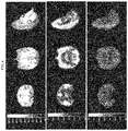
- FIG. 6 shows template MRI (top) and PET summation images of the rhesus monkey brain from 20-40 min after i.v. injection of (R)- 18 F-4 (middle), and (S)- 18 F-4 (bottom).
- FIG. 7 shows template MRI (top) and PET summation images of the rhesus monkey brain from 20-40 min after i.v. injection of (R)- 18 F-5 (middle), and from 140-160 min after i.v. injection of (R)- 18 F-5 (and from 50-70 min after i.v. injection of 30 mg/kg levetiracetam, bottom). Levetiracetam was given at 90-95 min after radiotracer injection.
- FIG. 8 A shows template MRI and PET summation images of baboon brain from 30-120 min after i.v. injection of (R)- 18 C-3.
- FIG. 8 B is a graph depicting time-activity curves of regional brain radioactivity in the insular cortex, frontal cortex, thalamus, and centrum semiovale and (R)- 18 C-3 plasma concentration. The solid lines show curve fitting with the 1-tissue (1T) compartment model.
- FIG. 8 C depicts Western blot analyses of twelve baboon brain regions.
- FIG. 8 D is a graph that depicts the correlation between regional SV2A density in vitro (by Western blot) and in vivo (by PET measurement). Data are from 12 baboon brain regions.
- FIG. 8 E is a graph that depicts the correlation between in vitro SV2A and in vitro synaptophysin (SYN) density in gray matter regions determined using Western blot analyses. Data are from 9 baboon brain regions.
- FIG. 8 F is a graph that depicts saturation studies of (R)- 11 C-3 that were performed for 12 baboon brain regions. Data are for the temporal cortex. Each measurement was performed in duplicate. Membranes were incubated with increasing concentrations of 3 for 30 min at 37° C. Nonspecific binding was determined as the residual binding measured in the presence of 1 mM levetiracetam. Specific binding was determined by subtraction of the nonspecific from the total binding.
- FIG. 8 G is a Scatchard plot from the transformed data of the temporal cortex in FIG.
- FIG. 8 H is a graph that depicts the correlation between regional SV2A density (B max ) measured in vitro using tissue homogenate binding with (R)- 11 C-3 and regional (R)- 11 C-3 binding measured in vivo in a baboon using PET. Data are from 12 brain regions.
- FIG. 8 I is a graph that depicts the correlation between regional SV2A density (B max ) measured in vitro using tissue homogenate binding and SV2A density using in vitro Western blot analyses (optical density, OD). Data are from 12 baboon brain regions.
- FIG. 8 H is a graph that depicts the correlation between regional SV2A density (B max ) measured in vitro using tissue homogenate binding with (R)- 11 C-3 and regional (R)- 11 C-3 binding measured in vivo in a baboon using PET. Data are from 12 brain regions.
- FIG. 8 I is a graph that depicts the correlation between regional SV2A density (B max ) measured in vitro
- FIG. 8 J is a low-power confocal microscopy image of 4′,6-diamidino-2-phenylindole (DAPI), SYN and SV2A in white matter and cortical gray matter of the baboon brain.
- the dotted white line indicates the border between the white matter (WM, left) and the gray matter (GM, right).
- FIG. 8 K is a high-power confocal microscopy image of DAPI, SYN and SV2A in the gray matter of the baboon brain from FIG. 8 J .
- Labeling for SYN and SV2A is evident as punctate staining in the neuropil, particularly surrounding neuronal cell bodies and proximal dendrites (arrow), but absent in neuronal cell bodies (white arrows). Nuclei are indicated by the DAPI stain.
- FIG. 9 A is a graph depicting the regional binding potential (BP ND ) values of (R)- 11 C-3 in selected brain regions of ten healthy control subjects and nine temporal lobe epilepsy (TLE) patients with mesial temporal sclerosis (MTS).
- FIG. 9 B is a graph depicting the regional standard uptake values (SUV) of 18 F-FDG in the ipsilateral and contralateral sides of selected brain regions of nine TLE patients with MTS. * indicates p ⁇ 0.05.
- FIG. 9 C are representative parametric (R)- 11 C-3 BP ND images of a medial temporal lobe epilepsy (MTLE) patient, with arrow showing a reduction in the hippocampus.
- MTLE medial temporal lobe epilepsy
- FIG. 10 A are representative coronal and axial images of MRI and (R)- 11 C-3 parametric PET (V T ) in a cognitive normal healthy control (HC) with a negative 11 C-PB PET scan. Arrow indicates the hippocampus region.
- FIG. 10 B are representative coronal and axial images of MRI and (R)- 11 C-3 parametric PET (V T ) in a mild cognitive impairment (MCI) patient with a positive 11 C-PIB PET scan. Arrow indicates the hippocampus region.
- FIG. 10 C are representative coronal and axial images of MRI and (R)- 11 C-3 parametric PET (V T ) in a mild cognitive impairment (MCI) patient with a positive 11 C-PB PET scan. Arrow indicates the hippocampus region.
- FIG. 10 B are representative coronal and axial images of MRI and (R)- 11 C-3 parametric PET (V T ) in a mild cognitive impairment (MCI) patient with a positive 11 C-PB PET scan. Arrow indicates the hippocampus region.
- FIG. 10 D are representative coronal and axial images of MRI and (R)- 11 C-3 parametric PET (V T ) in a mild AD dementia patient with a positive 11 C-PB PET scan. Arrow indicates the hippocampus region.
- FIG. 10 E are representative coronal and axial images of MRI and (R)- 11 C-3 parametric PET (V T ) in a mild AD dementia patient with a positive 11 C-PB PET scans. Arrow indicates the hippocampus region.
- FIG. 11 are parametric PET (V T ) images of (R)- 11 C-3 in a representative healthy control subject (top row) and a patient with Parkinson's disease (PD) (bottom row). Reductions in (R)- 11 C-3 V T are seen across all brain regions, with the most pronounced reductions in the cortical regions (19%-23% decreases), and in the substantia nigra and putamen (19% and 15% decreases, respectively).
- Synaptic vesicle (SV) proteins are abundant, hydrophobic, integral 12 transmembrane glycoproteins. They are expressed in three isoforms, SV2A, SV2B, and SV2C, with SV2A being the most abundant and ubiquitously distributed in all brain grey matter areas.
- the invention provides compounds that bind to SV2A and can be used as imaging biomarkers.
- the invention provides compounds and/or compositions that can bind to SV2A. In other embodiments, such compounds and/or compositions can be used for monitoring disease progression, recovery and treatment effects of disease-modifying therapies.
- the invention provides methods of using such compounds to image at least portions of the brain or other organs such as the pancreas, measure changes in SV2A binding as a surrogate for synaptic density, or to diagnose a neurodegenerative, neurological, psychiatric, and/or metabolic disease, such as but not limited to AD, Parkinson's disease (PD), multiple sclerosis (MS), autism, epilepsy, stroke, traumatic brain injury (TBI), schizophrenia, post-traumatic stress disorder (PTSD), depression, and diabetes, or any diseases/disorders where involvement of synaptic disruptions and/or abnormalities are present.
- a neurodegenerative, neurological, psychiatric, and/or metabolic disease such as but not limited to AD, Parkinson's disease (PD), multiple sclerosis (MS), autism, epilepsy, stroke, traumatic brain injury (TBI), schizophrenia, post-traumatic stress disorder (PTSD), depression, and diabetes, or any diseases/disorders where involvement of synaptic disruptions and/or abnormalities are present.
- PET imaging of the synaptic vesicle glycoprotein-2A provides an imaging biomarker for synaptic density that is useful in the early diagnosis of AD, and monitoring of disease progression and treatment efficacy of disease-modifying therapies.
- Synaptic dysfunction is involved in PD and regional synaptic density changes are characteristic in the pathogenesis of the disease.
- PET imaging of synaptic density in the brains of PD patients will not only reveal the disease progression, but also serve as a diagnosis or prognosis tool to facilitate more effective disease-modification interventions.
- measurement of synaptic density after stroke by PET imaging of SV2A provides an objective measure of disease progression and treatment efficacy.
- SV2A PET imaging can be used to monitor the progression of MS cognitive dysfunction, and treatment effect of disease-modifying therapies.
- measurement of synaptic density by PET imaging of SV2A provides a non-invasive method to diagnose ASD and monitor the efficacy of therapies targeting the correction of synaptic abnormality in ASD.
- PET imaging of SV2A provides an effective, non-invasive way to detect the seizure-onset zone in patients with epilepsy and aid in the treatment of this disease.
- detection of synaptic density and/or function by SV2A PET imaging offers a non-invasive and objective means to monitor TBI, recovery, and treatment efficacy of therapies.
- a number of psychiatric disorders such as depression, schizophrenia, post-traumatic stress disorder (PTSD), and substance abuse disorders, are believed to be associated with region-specific decreases in synaptic density.
- PET imaging of SV2A as a synaptic density biomarker is a valuable tool for the accurate diagnosis and treatment effect monitoring of these psychiatric disorders.
- non-invasive PET imaging of SV2A as a marker for endogenous pancreatic BCM may provide the needed tool for quantifying BCM in healthy subjects and type-1 diabetic patients, and for monitoring the treatment effects of therapeutic interventions.
- Standard techniques are used for biochemical and/or biological manipulations.
- the techniques and procedures are generally performed according to conventional methods in the art and various general references (e.g., Sambrook and Russell, 2012, Molecular Cloning, A Laboratory Approach, Cold Spring Harbor Press, Cold Spring Harbor, N.Y., and Ausubel et al., 2002, Current Protocols in Molecular Biology, John Wiley & Sons, N.Y.), which are provided throughout this document.
- an element means one element or more than one element.
- “About” as used herein when referring to a measurable value such as an amount, a temporal duration, and the like, is meant to encompass variations of ⁇ 20% or ⁇ 10%, more preferably ⁇ 5%, even more preferably ⁇ 1%, and still more preferably ⁇ 0.1% from the specified value, as such variations are appropriate to perform the disclosed methods.
- AD Alzheimer's Disease
- MCI mild cognitive impairment
- an analog As used herein, the terms “analog,” “analogue,” or “derivative” are meant to refer to a chemical compound or molecule made from a parent compound or molecule by one or more chemical reactions. As such, an analog can be a structure having a structure similar to that of the small molecules described herein or can be based on a scaffold of a small molecule described herein, but differing from it in respect to certain components or structural makeup, which may have a similar or opposite action metabolically.
- the term “detecting” may refer to the qualitative observation of the presence of a biomarker or to the quantitative measurement of the relative or total amount of that biomarker or to a determination of its concentration.
- the term detecting may be applied to a sample, to a subject or to a defined region in the body of a subject.
- binding refers to the adherence of molecules to one another, such as, but not limited to, enzymes or any other proteins to substrates, antibodies to antigens, DNA strands to their complementary strands. Binding occurs because the shape and chemical nature of parts of the molecule surfaces are complementary. A common metaphor is the “lock-and-key” used to describe how enzymes fit around their substrate.
- the “subject's body” or “the body of the subject” particularly in the context of a region of the subject's body refers to the physical corpus of the subject.
- a region of the body of the subject may be any portion thereof.
- the region of the subject's body may be the brain.
- the region of the subject's body may be the pancreas.
- a “disease” is a state of health of an animal, such as a human subject, wherein the animal cannot maintain homeostasis, and wherein if the disease is not ameliorated then the animal's health continues to deteriorate.
- detectable label is meant a composition that when linked to a molecule of interest renders the latter detectable, via spectroscopic, photochemical, radiological, biochemical, immunochemical, or chemical means.
- useful labels include radioactive isotopes, magnetic beads, metallic beads, colloidal particles, fluorescent dyes, electron-dense reagents, enzymes (for example, as commonly used in an ELISA), biotin, digoxigenin, or haptens.
- an “effective amount” of a compound is that amount of compound which is sufficient to allow effective measurement or imaging when provided to the patient by a particular method of administration.
- An appropriate effective amount in any individual case may be determined by one of ordinary skill in the art using routine experimentation.
- a first compound is essentially free of a second compound in a composition indicates that the ratio of the second compound to the first compound in the composition is about 10:90, 5:95, 4:96, 3:97, 2:98, 1:99, 0.5:99.5, 0.25:99.75, 0.1:99.9, 0.05:99.95, 0.025:99.975, 0.01:99.99 or 0:100.
- the “level” of one or more biomarkers means the absolute or relative amount or concentration of the biomarker in the sample as determined by detecting the biomarker.
- the level of a biomarker may vary across a region of interest, for example, the level of the biomarker may be higher or lower in a subset of the region of interest and this may be interpreted to imply various biological states.
- patient refers to any animal, or cells thereof whether in vitro or in situ, amenable to the methods described herein.
- the patient, subject or individual is a human.
- the term “pharmaceutically acceptable carrier” means a pharmaceutically acceptable material, composition or carrier, such as a liquid or solid filler, stabilizer, dispersing agent, suspending agent, diluent, excipient, thickening agent, solvent or encapsulating material, involved in carrying or transporting a compound useful within the invention within or to the patient such that it may perform its intended function.
- a pharmaceutically acceptable material, composition or carrier such as a liquid or solid filler, stabilizer, dispersing agent, suspending agent, diluent, excipient, thickening agent, solvent or encapsulating material, involved in carrying or transporting a compound useful within the invention within or to the patient such that it may perform its intended function.
- Such constructs are carried or transported from one organ, or portion of the body, to another organ, or portion of the body.
- Each carrier must be “acceptable” in the sense of being compatible with the other ingredients of the formulation, including the compound useful within the invention, and not injurious to the patient.
- materials that may serve as pharmaceutically acceptable carriers include: sugars, such as lactose, glucose and sucrose; starches, such as corn starch and potato starch; cellulose, and its derivatives, such as sodium carboxymethyl cellulose, ethyl cellulose and cellulose acetate; powdered tragacanth; malt; gelatin; talc; excipients, such as cocoa butter and suppository waxes; oils, such as peanut oil, cottonseed oil, safflower oil, sesame oil, olive oil, corn oil and soybean oil; glycols, such as propylene glycol; polyols, such as glycerin, sorbitol, mannitol and polyethylene glycol; esters, such as ethyl oleate and ethyl laurate; agar; buffering agents, such as magnesium hydroxide and aluminum hydroxide; surface active agents; alginic acid; pyrogen-free water; isotonic saline
- “pharmaceutically acceptable carrier” also includes any and all coatings, antibacterial and antifungal agents, and absorption delaying agents, and the like that are compatible with the activity of the compound useful within the invention, and are physiologically acceptable to the patient. Supplementary active compounds may also be incorporated into the compositions.
- the “pharmaceutically acceptable carrier” may further include a pharmaceutically acceptable salt of the compound useful within the invention.
- Other additional ingredients that may be included in the pharmaceutical compositions used in the practice of the invention are known in the art and described, for example in Remington's Pharmaceutical Sciences (Genaro, Ed., Mack Publishing Co., 1985, Easton, Pa.), which is incorporated herein by reference.
- pharmaceutically acceptable salt refers to a salt of the administered compounds prepared from pharmaceutically acceptable non-toxic acids, including inorganic acids or bases, organic acids or bases, solvates, hydrates, or clathrates thereof.
- substituted means that a carbon or hydrogen atom in the group to which the term is applied may be changed to another atom, such as but not limited to nitrogen, oxygen, sulfur or halogens, so long as the substitution produces a stable compound.
- ranges throughout this disclosure, various aspects of the invention can be presented in a range format. It should be understood that the description in range format is merely for convenience and brevity and should not be construed as an inflexible limitation on the scope of the invention. Accordingly, the description of a range should be considered to have specifically disclosed all the possible subranges as well as individual numerical values within that range. For example, description of a range such as from 1 to 6 should be considered to have specifically disclosed subranges such as from 1 to 3, from 1 to 4, from 1 to 5, from 2 to 4, from 2 to 6, from 3 to 6 and so forth, as well as individual numbers within that range, for example, 1, 2, 2.7, 3, 4, 5, 5.3, and 6. This applies regardless of the breadth of the range.
- the invention provides compounds according to formula 1 or a salt, solvate, stereoisomer (such as, in a non-limiting example, an enantiomer or diastereomer (also known as diastereoisomer), and any mixtures thereof, such as, in a non-limiting example, mixtures in any proportion of enantiomers and/or diastereomers thereof), tautomer and any mixtures thereof, and/or geometric isomer and any mixtures thereof:
- each instance of Y is independently selected from the group consisting of N, CH and CR 1 , provided that 0-1 Y is N; each instance of R 1 and R 2 is independently selected from the group consisting of F, Cl, Br, I, C 1 -C 6 alkyl (such as but not limited to CH 3 ), C 1 -C 6 haloalkyl (such as but not limited to CF 3 ), CN and —NO 2 , provided that at least one instance of R 1 or R 2 is F; and m and n are independently selected from the group consisting of 0, 1, 2, 3, 4 and 5.
- 1 encompasses multiple substitutions of R 1 , e.g. 1, 2, 3 or 4 instances of R 1 on each ring.
- the synthetic processes by which these compounds can be made are illustrated in Example 1. As illustrated in FIGS. 3 A- 3 F, 4 A- 4 C, and 5 and in Examples 2-4, these compounds bind SV2A, which may be used as a biomarker for synaptic density.
- the compounds of formula 1 comprise a detectable label.
- the detectable label is an isotope that can be detected by PET. In yet other embodiments, this isotope is 18 F. Synthetic methods for selectively generating the isotopically labeled compounds are described in Example 1.
- Compounds of the invention that possess at least one stereogenic center may be utilized as racemic mixtures or may be enriched in either the R or S enantiomer.
- the compounds are provided as isolated pure enantiomers. Illustrative methods of separating the enantiomers are discussed in Example 1 and Scheme 4.
- the compound of formula 1 is a compound of formula 2
- each instance of R 1 and R 2 is independently selected from the group consisting of F, Cl, Br, I, C 1 -C 6 alkyl (such as but not limited to CH 3 ), C 1 -C 6 haloalkyl (such as but not limited to CF 3 ), CN and —NO 2 , provided that at least one instance of R 1 or R 2 is F; and m and n are independently selected from the group consisting of 0, 1, 2, 3, 4 and 5; a salt, solvate, tautomer or enantiomer thereof, and any mixtures thereof.
- the compound of formula 1 or 2 is at least one selected from the group consisting of:
- F is 18 F, a salt, solvate, tautomer or enantiomer thereof, and any mixtures thereof.
- the compound of formula 1 or 2 is selected from the group consisting of: 1-((3-methylpyridin-4-yl)methyl)-4-(3,4,5-trifluorophenyl) pyrrolidin-2-one; 4-(3,4-difluorophenyl)-1-((3-methylpyridin-4-yl)methyl)pyrrolidin-2-one; 4-(3,5-difluorophenyl)-1-((3-methylpyridin-4-yl)methyl)pyrrolidin-2-one; 4-(4-fluorophenyl)-1-(pyridin-4-ylmethyl)pyrrolidin-2-one; 4-(3,5-difluorophenyl)-1-(pyridin-4-ylmethyl) pyrrolidin-2-one; 4-(3,4-difluorophenyl)-1-(pyridin-4-ylmethyl)pyrrolidin-2-one; 4-(3,4-difluorophenyl)-1-(pyridin-4
- the compound of formula 1 is a compound of formula 14:
- each instance of R 1 and R 2 is independently selected from the group consisting of F, Cl, Br, I, C 1 -C 6 alkyl (such as but not limited to CH 3 ), C 1 -C 6 haloalkyl (such as but not limited to CF 3 ), CN and —NO 2 , provided that at least one instance of R 1 or R 2 is F; and m and n are independently selected from the group consisting of 0, 1, 2, 3, 4 and 5; a salt, solvate, tautomer or enantiomer thereof, and any mixtures thereof.
- the compound of formula 1 or 14 is at least one selected from the group consisting of:
- F is 18 F, a salt, solvate, tautomer or enantiomer thereof, and any mixtures thereof.
- the compound of formula 1 or 14 is selected from the group consisting of: 1-((4-methylpyridin-3-yl)methyl)-4-(3,4,5-trifluorophenyl) pyrrolidin-2-one; 4-(3,4-difluorophenyl)-1-((4-methylpyridin-3-yl)methyl)pyrrolidin-2-one; 4-(3,5-difluorophenyl)-1-((4-methylpyridin-3-yl)methyl)pyrrolidin-2-one; 4-(4-fluoro phenyl)-1-(pyridin-3-ylmethyl)pyrrolidin-2-one; 4-(3,5-difluorophenyl)-1-(pyridin-3-ylmethyl)pyrrolidin-2-one; 4-(3,4-difluorophenyl)-1-(pyridin-3-ylmethyl)pyrrolidin-2-one; 1-(pyridin-3-ylmethyl)-4-(3,4,5-trifluoroph
- the invention of formula 1 is a compound of formula 26:
- each instance of R 1 is independently selected from the group consisting of F, Cl, Br, I, C 1 -C 6 alkyl (such as but not limited to CH 3 ), C 1 -C 6 haloalkyl (such as but not limited to CF 3 ), CN and —NO 2 , provided that at least one instance of R 1 is F; and n is selected from the group consisting of 1, 2, 3, 4 and 5, a salt, solvate, tautomer or enantiomer thereof, and any mixtures thereof.
- the compound of formula 1 or 26 is 27:
- F is 18 F, a salt, solvate, tautomer or enantiomer thereof, and any mixtures thereof.
- the compound of formula 1 or 26 is 4-(3,5-difluorophenyl)-1-((1-methyl-1H-imidazol-5-yl)methyl)pyrrolidin-2-one, a salt, solvate, tautomer or enantiomer thereof, and any mixtures thereof.
- Compounds described herein also include isotopically labeled compounds wherein one or more atoms is replaced by an atom having the same atomic number, but an atomic mass or mass number different from the atomic mass or mass number usually found in nature.
- isotopes suitable for inclusion in the compounds described herein include and are not limited to 2 H, 3 H, 11 C, 13 C, 14 C, 36 Cl, 18 F, 123 I, 125 I, 13 N, 15 N, 15 O, 17 O, 18 O, 32 P, and 35 S.
- isotopically labeled compounds are useful in drug and/or substrate tissue distribution studies.
- substitution with heavier isotopes such as deuterium affords greater metabolic stability (for example, increased in vivo half-life or reduced dosage requirements).
- substitution with positron emitting isotopes, such as C, 18 F, 15 O and 13 N is useful in Positron Emission Topography (PET) studies for examining target protein concentration and/or substrate receptor occupancy.
- Isotopically-labeled compounds are prepared by any suitable method or by processes using an appropriate isotopically-labeled reagent in place of the non-labeled reagent otherwise employed.
- any isotope of any atom as shown in the structures in various embodiments, 18 F may be substituted for F at any position or at multiple positions.
- the compounds described herein are labeled by other means, including, but not limited to, the use of chromophores or fluorescent moieties, bioluminescent labels, or chemiluminescent labels.
- salts embraces addition salts of free acids or bases that are useful within the methods of the invention.
- pharmaceutically acceptable salt refers to salts that possess toxicity profiles within a range that affords utility in pharmaceutical applications.
- the salts are pharmaceutically acceptable salts.
- Pharmaceutically unacceptable salts may nonetheless possess properties such as high crystallinity, which have utility in the practice of the present invention, such as for example utility in process of synthesis, purification or formulation of compounds useful within the methods of the invention.
- Suitable pharmaceutically acceptable acid addition salts may be prepared from an inorganic acid or from an organic acid.
- inorganic acids include sulfate, hydrogen sulfate, hydrochloric, hydrobromic, hydriodic, nitric, carbonic, sulfuric, and phosphoric acids (including hydrogen phosphate and dihydrogen phosphate).
- organic acids may be selected from aliphatic, cycloaliphatic, aromatic, araliphatic, heterocyclic, carboxylic and sulfonic classes of organic acids, examples of which include formic, acetic, propionic, succinic, glycolic, gluconic, lactic, malic, tartaric, citric, ascorbic, glucuronic, maleic, fumaric, pyruvic, aspartic, glutamic, benzoic, anthranilic, 4-hydroxybenzoic, phenylacetic, mandelic, embonic (or pamoic), methanesulfonic, ethanesulfonic, benzenesulfonic, pantothenic, sulfanilic, 2-hydroxyethanesulfonic, trifluoromethanesulfonic, p-toluenesulfonic, cyclohexylaminosulfonic, stearic, alginic, ⁇ -hydroxybutyric, sal
- Suitable pharmaceutically acceptable base addition salts of compounds of the invention include, for example, ammonium salts and metallic salts including alkali metal, alkaline earth metal and transition metal salts such as, for example, calcium, magnesium, potassium, sodium and zinc salts.
- Pharmaceutically acceptable base addition salts also include organic salts made from basic amines such as, for example, N,N′-dibenzylethylene-diamine, chloroprocaine, choline, diethanolamine, ethylenediamine, meglumine (or N-methylglucamine) and procaine. All of these salts may be prepared from the corresponding compound by reacting, for example, the appropriate acid or base with the compound.
- the invention provides a method of decreasing the amount of unbound synaptic vesicle glycoprotein 2A (SV2A) in at least one region of the body of a subject.
- the method comprises administering to a subject at least one compound of the invention, or a composition comprising the same.
- the compound is administered to the subject by any known means of administration, and optionally in a pharmaceutical composition further comprising at least one excipient as appropriate to deliver the compound to the intended region of the subject's body.
- the at least one region of the subject's body may the subject's brain or a portion thereof.
- the at least one region of the subject's body may be the pancreas.
- the invention provides a method of imaging at least one region of the body of a subject.
- the method comprises administering to a subject at least one compound of the invention, or a composition comprising the same, and detecting the at least one compound by positron emission tomography in at least one region of the body of the subject, thereby generating an image of the at least one region of the body of the subject.
- the compound is administered to the subject by any known means of administration, and optionally in a pharmaceutical composition further comprising at least one excipient as appropriate to deliver the compound to the subject's body.
- the compounds of the invention can be utilized as radiotracers for PET imaging.
- the region of the brain may be the cingulate cortex, frontal cortex, insular cortex, nucleus accumbens, occipital cortex, temporal cortex, putamen, caudate nucleus, thalamus, cerebellum, hippocampus, globus pallidus, amygdala, brainstem, pons, centrum semiovale, or any combinations thereof.
- FIGS. 5 - 7 , 8 A- 8 K, 9 A- 9 C, 10 A- 10 F, and 11 depict PET images and data acquired according to this method and procedures are discussed in Examples 3-5.
- a person of skill in the art is familiar with methods of PET imaging and may readily apply such knowledge in combination with the present disclosure to generate PET images and data using the compounds disclosed herein.
- the invention further provides a method of detecting and/or measuring synaptic loss in a subject.
- the method comprises administering to a subject at least one compound of the invention, or a composition comprising the same; and detecting and/or measuring the at least one compound by PET in at least one region of the brain of the subject.
- the detecting and/or measuring the at least one compound by PET is used to determine or evaluate the SV2A level in the at least one region of the brain of the subject.
- the amount of the least one compound measured and/or detected by PET is compared or correlated to a predetermined reference (which is known to have, or not to have, significant synaptic loss), wherein the amount of the least one compound measured and/or detected by PET is correlated with the SV2A level.
- a predetermined reference which is known to have, or not to have, significant synaptic loss
- the amount of the least one compound measured and/or detected by PET is correlated with the SV2A level.
- if the SV2A level in the subject is lower than in the reference known not to have significant synaptic loss, decreased synaptic density is detected in the subject.
- Validation of SV2A in combination with compounds of the invention as a biomarker of synaptic density is exemplified in Example 4.
- FIGS. 9 A- 9 C, 10 A- 10 F , and 11 depict PET detection of changes in synaptic density in the brain of patients with epilepsy, AD, and Parkinson's disease (PD).
- the compound is administered to the subject by any known means of administration, and optionally in a pharmaceutical composition further comprising at least one excipient as appropriate to deliver the compound to the subject's brain or other organs such as the pancreas.
- the compound may be detected using methods of PET familiar to a person of skill in the art and described and exemplified in Examples 2-5 and the Figures of the invention.
- the compounds of the invention bind to SV2A and can be detected by PET.
- Competitive binding assays demonstrating the affinity for SV2A possessed by compounds of the invention are exemplified in Example 2.
- a level of SV2A in a region of the brain or other organs such as the pancreas can be determined by calculating a distribution volume (V T ) and in turn binding potential (BP ND ). These values, along with the dissociation constant (K D ), can be used to calculate target protein density using equations familiar to a person of skill in the art. Further details are illustrated in Example 3-5 and FIGS. 8 A- 8 K, 9 A- 9 C, 10 A- 10 F, and 11 .
- Reference levels for SV2A can be calculated by determining typical values for groups of patients stratified by variables such as age by using the disclosed methods. Alternatively, reference levels can be determined by measuring the concentration of SV2A in vitro as described in Example 2.
- the invention provides a method of detecting a neurodegenerative, neurological, psychiatric, and/or metabolic disease, or any other diseases/disorders where involvement of synaptic disruptions and/or abnormalities is present, in a subject.
- the method comprises administering to a subject at least one compound of the invention, or a composition comprising the same; detecting and/or measuring the at least one compound by PET in at least one region of the subject's body; determining the SV2A level in the at least one region of the subject's body and comparing it to a reference (which is known to be derived from a subject who has, or does not have, the disease); wherein, if the SV2A level in the at least one region of the subject's body is lower than in the reference from a subject known not to have the disease, the disease is detected in the subject.
- a reference which is known to be derived from a subject who has, or does not have, the disease
- altered synaptic density is a leading indicator of cognitive impairment and AD, as exemplified in FIGS. 10 A- 10 F .
- the disease is in pre-clinical or prodromal stage.
- the manner in which the compounds are provided to the patients, as well as the methods for determining a level and reference level for SV2A in a region of the brain of the patient, are illustrated elsewhere herein, such as in Example 5 and FIGS. 9 A- 9 C, 10 A- 10 F, and 11 .
- the invention provides a method of detecting and/or measuring a seizure onset zone in a subject with epilepsy, stroke, traumatic brain injury, Parkinson's disease or autism.
- the method comprises administering to the subject at least one compound or at least one pharmaceutical composition of the invention; detecting the at least one compound by PET in at least one region of the brain of the subject and determining a level of SV2A in the at least one region of the brain of the subject. Because, PET detection allows spatial resolution of the level of SV2A in the region of interest, if an area within the at least one region has lower SV2A level than the rest of the at least one region, the area is identified as a seizure onset zone.
- the invention provides a method of detecting an ischemic area in a subject with stroke.
- the method comprises administering to the subject at least one compound or at least one pharmaceutical composition of the invention; detecting the at least one compound by PET in at least one region of the brain of the subject and determining a level of SV2A in the at least one region of the brain of the subject. Because, PET detection allows spatial resolution of the level of SV2A in the region of interest, if an area within the at least one region has lower SV2A level than the rest of the at least one region, the area is identified as being ischemic.
- Administration of the compounds and/or compositions of the present invention to a patient, preferably a mammal, more preferably a human, may be carried out using known procedures, at dosages and for periods of time effective to perform an imaging method contemplated in the invention.
- An effective amount of the compound necessary for adequate signal for imaging may vary according to factors such as the state of a disease or disorder in the patient; the age, sex, and weight of the patient; and the equipment used to detect the compound of the invention.
- One of ordinary skill in the art would be able to study the relevant factors and make the determination regarding the effective amount of the compound without undue experimentation.
- Actual dosage levels of the active ingredients in the pharmaceutical compositions of this invention may be varied so as to obtain an amount of the active ingredient that is effective to achieve successful imaging for a particular patient, composition, and mode of administration, without being toxic to the patient.
- compositions of the invention are formulated using one or more pharmaceutically acceptable excipients or carriers.
- pharmaceutical compositions of the invention comprise an effective amount of a compound of the invention and a pharmaceutically acceptable carrier.
- the carrier may be a solvent or dispersion medium containing, for example, water, ethanol, polyol (for example, glycerol, propylene glycol, and liquid polyethylene glycol, and the like), suitable mixtures thereof, and vegetable oils.
- the proper fluidity may be maintained, for example, by the use of a coating such as lecithin, by the maintenance of the required particle size in the case of dispersion and by the use of surfactants.
- Prevention of the action of microorganisms may be achieved by various antibacterial and antifungal agents, for example, parabens, chlorobutanol, phenol, ascorbic acid, thimerosal, and the like.
- isotonic agents for example, sugars, sodium chloride, or polyalcohols such as mannitol and sorbitol, in the composition.
- Prolonged absorption of the injectable compositions may be brought about by including in the composition an agent which delays absorption, for example, aluminum monostearate or gelatin.
- Compounds of the invention for administration may be in the range of from about 1 ⁇ g to about 10,000 mg, about 20 ⁇ g to about 9,500 mg, about 40 ⁇ g to about 9,000 mg, about 75 ⁇ g to about 8,500 mg, about 150 ⁇ g to about 7,500 mg, about 200 ⁇ g to about 7,000 mg, about 3050 ⁇ g to about 6,000 mg, about 500 ⁇ g to about 5,000 mg, about 750 ⁇ g to about 4,000 mg, about 1 mg to about 3,000 mg, about 10 mg to about 2,500 mg, about 20 mg to about 2,000 mg, about 25 mg to about 1,500 mg, about 30 mg to about 1,000 mg, about 40 mg to about 900 mg, about 50 mg to about 800 mg, about 60 mg to about 750 mg, about 70 mg to about 600 mg, about 80 mg to about 500 mg, and any and all whole or partial increments therebetween.
- the dose of a compound of the invention is from about 1 mg and about 2,500 mg. In certain embodiments, a dose of a compound of the invention used in compositions described herein is less than about 10,000 mg, or less than about 8,000 mg, or less than about 6,000 mg, or less than about 5,000 mg, or less than about 3,000 mg, or less than about 2,000 mg, or less than about 1,000 mg, or less than about 500 mg, or less than about 200 mg, or less than about 50 mg.
- a dose of a second compound as described herein is less than about 1,000 mg, or less than about 800 mg, or less than about 600 mg, or less than about 500 mg, or less than about 400 mg, or less than about 300 mg, or less than about 200 mg, or less than about 100 mg, or less than about 50 mg, or less than about 40 mg, or less than about 30 mg, or less than about 25 mg, or less than about 20 mg, or less than about 15 mg, or less than about 10 mg, or less than about 5 mg, or less than about 2 mg, or less than about 1 mg, or less than about 0.5 mg, and any and all whole or partial increments thereof.
- Formulations may be employed in admixtures with conventional excipients, i.e., pharmaceutically acceptable organic or inorganic carrier substances suitable for oral, parenteral, nasal, intravenous, subcutaneous, enteral, or any other suitable mode of administration, known to the art.
- the pharmaceutical preparations may be sterilized and if desired mixed with auxiliary agents, e.g., lubricants, preservatives, stabilizers, wetting agents, emulsifiers, salts for influencing osmotic pressure buffers, coloring, flavoring and/or aromatic substances and the like.
- routes of administration of any of the compositions of the invention include oral, nasal, rectal, intravaginal, parenteral, buccal, sublingual or topical.
- the compounds for use in the invention may be formulated for administration by any suitable route, such as for oral or parenteral, for example, transdermal, transmucosal (e.g., sublingual, lingual, (trans) buccal, (trans)urethral, vaginal (e.g., trans- and perivaginally), (intra)nasal and (trans)rectal), intravesical, intrapulmonary, intraduodenal, intragastrical, intrathecal, subcutaneous, intramuscular, intradermal, intra-arterial, intravenous, intrabronchial, inhalation, and topical administration.
- compositions and dosage forms include, for example, tablets, capsules, caplets, pills, gel caps, troches, dispersions, suspensions, solutions, syrups, granules, beads, transdermal patches, gels, powders, pellets, magmas, lozenges, creams, pastes, plasters, lotions, discs, suppositories, liquid sprays for nasal or oral administration, dry powder or aerosolized formulations for inhalation, compositions and formulations for intravesical administration and the like. It should be understood that the formulations and compositions that would be useful in the present invention are not limited to the particular formulations and compositions that are described herein.
- parenteral administration of a pharmaceutical composition includes any route of administration characterized by physical breaching of a tissue of a subject and administration of the pharmaceutical composition through the breach in the tissue.
- Parenteral administration thus includes, but is not limited to, administration of a pharmaceutical composition by injection of the composition, by application of the composition through a surgical incision, by application of the composition through a tissue-penetrating non-surgical wound, and the like.
- parenteral administration is contemplated to include, but is not limited to, subcutaneous, intravenous, intraperitoneal, intramuscular, intrasternal injection, and kidney dialytic infusion techniques.
- Formulations of a pharmaceutical composition suitable for parenteral administration comprise the active ingredient combined with a pharmaceutically acceptable carrier, such as sterile water or sterile isotonic saline. Such formulations may be prepared, packaged, or sold in a form suitable for bolus administration or for continuous administration. Injectable formulations may be prepared, packaged, or sold in unit dosage form, such as in ampules or in multidose containers containing a preservative. Formulations for parenteral administration include, but are not limited to, suspensions, solutions, emulsions in oily or aqueous vehicles, pastes, and implantable sustained-release or biodegradable formulations. Such formulations may further comprise one or more additional ingredients including, but not limited to, suspending, stabilizing, or dispersing agents.
- the active ingredient is provided in dry (i.e., powder or granular) form for reconstitution with a suitable vehicle (e.g., sterile pyrogen free water) prior to parenteral administration of the reconstituted composition.
- a suitable vehicle e.g., sterile pyrogen free water
- compositions may be prepared, packaged, or sold in the form of a sterile injectable aqueous or oily suspension or solution.
- This suspension or solution may be formulated according to the known art, and may comprise, in addition to the active ingredient, additional ingredients such as the dispersing agents, wetting agents, or suspending agents described herein.
- Such sterile injectable formulations may be prepared using a non-toxic parenterally-acceptable diluent or solvent, such as water or 1,3-butanediol, for example.
- Other acceptable diluents and solvents include, but are not limited to, Ringer's solution, isotonic sodium chloride solution, and fixed oils such as synthetic mono- or di-glycerides.
- compositions for sustained release or implantation may comprise pharmaceutically acceptable polymeric or hydrophobic materials such as an emulsion, an ion exchange resin, a sparingly soluble polymer, or a sparingly soluble salt.
- Additional dosage forms of this invention include dosage forms as described in U.S. Pat. Nos. 6,340,475; 6,488,962; 6,451,808; 5,972,389; 5,582,837; and 5,007,790. Additional dosage forms of this invention also include dosage forms as described in U.S. Patent Applications Nos. 2003/0147952; 2003/0104062; 2003/0104053; 2003/0044466; 2003/0039688; and 2002/0051820. Additional dosage forms of this invention also include dosage forms as described in PCT Applications Nos.
- reaction conditions including but not limited to reaction times, reaction size/volume, and experimental reagents, such as solvents, catalysts, pressures, atmospheric conditions, e.g., nitrogen atmosphere, and reducing/oxidizing agents, with art-recognized alternatives and using no more than routine experimentation, are within the scope of the present application.
- High resolution mass spectrometry results were determined on a LC-MS/MS system including a Thermo Scientific LTQ-Orbitrap XL mass spectrometer and a Waters nanoACQUITY ultra high performance liquid chromatography instrument.
- HPLC-MS was performed with an Agilent 1200RRLC/6110SQD SYSTEM.
- Radio-HPLC systems include a Shimadzu LC-20A pump, a Knauer K200 UV detector, Shimadzu SPD-M20A PDA or SPD-20A UV detector, and a Bioscan ⁇ -flow detector.
- [ 18 F]Fluoride was produced via the 18 O(p, n) 18 F nuclear reaction in a 16.5-MeV GE PETtrace cyclotron.
- H 2 18 O was obtained from Huayi Isotopes.
- Anion exchange CHROMAFIXTM cartridges PS-HCO 3
- SPE Solid-phase extraction
- the synthetic intermediate (4.5 g, 14.9 mmol) was dissolved in nitromethane (2.5 mL, 46.7 mmol) under argon and cooled at 5° C.
- 1,8-Diazabicyclo[5.4.0]undec-7-ene (DBU, 2.62 g, 17.2 mmol) was added dropwise, and the mixture was stirred at 5° C. for 1 h.
- the reaction was quenched with saturated NH 4 Cl (20 mL), acidified with aqueous HCl (2 M) until the pH of the mixture reached 2, and extracted with EtOAc (25 mL ⁇ 3).
- Compound 38 was prepared in procedures similar to those described above for 31, with (1r,3r,5r,7r)-spiro[adamantane-2,2′-[1,3]dioxane]-4′,6′-dione (SPI-Ad) added instead of 6,10-dioxaspiro[4.5]decane-7,9-dione (SPI-5) in the second stage of the reaction. Yield: 37%.
- 3,5-Difluoro-4-iodobenzonitrile (52): To a solution of 3,5-difluorobenzonitrile (51, 13.7 g, 98.5 mmol) in anhydrous tetrahydrofuran (THF, 130 mL) under argon and cooled at ⁇ 78° C. was added dropwise a solution of lithium diisopropylamide (55 mL, 2 M in THF), followed by a solution of iodine (26.3 g, 103.6 mmol) in anhydrous THF (75 mL). The reaction was slowly warmed to room temperature, stirred for 1 h, and then quenched with 10% sodium thiosulfite solution (100 mL).
- THF tetrahydrofuran
- the cyclotron produced aqueous 18 F-fluoride solution was transferred to a lead-shielded hot cell, where the 18 F-fluoride ion was trapped on a Chromafix 30-PS-HCO 3 18 F-separation cartridge.
- the trapped 18 F-fluoride in the cartridge was then eluted into the reaction vessel with a solution of 7 mg of Kryptofix® K 222 and 0.7 mg of K 2 CO 3 in 1 mL of CH 3 CN/water (1:0.4, v/v) or 2 mg of tetraethylammonium bicarbonate (TEAB) in 1 mL of CH 3 CN/water (7:3, v/v).
- the solvent was evaporated at 110° C.
- the column was eluted under isocratic conditions at a flow rate of 5 mL/min with a mixture of MeCN and 0.1 M aqueous ammonium formate with 0.5% acetic acid (v/v, pH 4.2).
- the eluent was monitored by a UV detector and a radioactivity detector.
- the fraction containing the radiolabeled product was collected, diluted with deionized (DI) water (50 mL), and passed through a C18 SepPak.
- the SepPak was washed with DI water (10 mL), and dried with air.
- the 18 F-labeled product was eluted off the SepPak with ethanol (1 mL), followed by USP saline (3 mL).
- the resulting solution was then passed through a sterile membrane filter (0.2 ⁇ m) for terminal sterilization and collected in a sterile vial pre-charged with 7 mL of saline (USP) to afford a formulated solution ready for dispensing and intravenous administration.
- a sterile membrane filter 0.2 ⁇ m
- USP saline
- One Sprague Dawley rat was euthanized with carbon dioxide inhalation.
- the brain was harvested, rapidly frozen on dry ice and then stored at ⁇ 80° C.
- the whole brain homogenates were obtained following published procedures.
- the final homogenate was suspended in 20 mM Tris-HCl buffer (pH 7.4) containing 250 mM of sucrose at a wet tissue concentration of 100 mg/mL and stored at ⁇ 80° C. until use.
- Nonspecific binding was defined as the residual binding observed in the presence of 1 mM levetiracetam (100 ⁇ L).
- the membrane-bound radioligand was recovered using rapid filtration through GF/B glass fiber filters (13 mm GD/X, Whatman Inc.) which were pre-soaked for at least 30 min in 0.5% polyethyleneimine (PEI).
- PEI polyethyleneimine
- the membranes were washed twice with 1 mL of ice-cooled binding buffer.
- the binding mixtures, filters and filtrates were counted using cross-calibrated gamma counters (1480 & 2480 WIZARD; Perkin-Elmer). Values of IC 50 were calculated using the GRAPHPAD PRISMTM software and converted to inhibition constants (K i ) by the Cheng-Prusoff equation.
- Assay compounds displayed moderate to high binding affinity to SV2A protein. Values of IC 50 and K for some example compounds are listed in Table 1.
- Plasma free fraction (fp) was measured in triplicate using the ultrafiltration method.
- PET scans of the brain were acquired using the FOCUS 220 PET scanner (Siemens Preclinical Solutions) with a reconstructed image resolution of approximately 1.5 mm.
- the radioligand was injected i.v. as a 3 min slow bolus by an infusion pump (PHD 22/2000; Harvard Apparatus).
- List-mode data were acquired for 120-180 min and binned into sinograms with an applicable number of frames.
- Scan data were reconstructed with a Fourier rebinning/filtered backprojection algorithm with corrections for attenuation, scanner normalization, radiation scatter, and randoms. Regions of interest were manually delineated on a single representative anatomic rhesus monkey MR image registered to a template image.
- the regions used in this study were amygdala, brain stem, caudate, centrum semiovale, cerebellum, cingulate cortex, frontal cortex, globus pallidus, insular cortex, nucleus accumbens, occipital cortex, pons, putamen, temporal cortex, and thalamus. Registration parameters were obtained to apply the regions of interest to individual PET scans, and regional time-activity curves were generated. Regional time-activity curves of the brain were analyzed with the 1-tissue-(1T) compartment model. Fit quality was compared with the 2-tissue model and the multilinear analysis method to calculate V T .
- Typical injected radioactivity amount was 0.5-5 mCi per injection, with typical injected mass amount of 1-10 ⁇ g.
- the 1T model produced suitable fits and reliable distribution volume (V T ) values.
- Multilinear analysis method 1 produced good fits with small differences in V T values from 1T.
- the 2-tissue compartment model produced comparable V T estimates, but with higher standard error than the 1T model.
- the 1T model was the method chosen for quantification of V T values.
- the regional V T values are shown in Table 2.
- Regional BP ND values were estimated using the centrum semiovale as the reference region and are provided in Table 3. Reversible and specific binding to SV2A was examined for 18 F-3, 18 F-4, 18 F-5, 18 F-11, and 18 F-12 in displacement studies with levetiracetam, where levetiracetam was i.v.
- FIG. 3 Specific binding to SV2A was further examined for 11 C-3, 18 F-3, and 18 F-11 in blocking studies, where levetiracetam or UCB-J ((R)-3) was i.v. infused before injection of the radiotracers. Uptake of radiotracers was all reduced to background levels, indicating blocking of specific binding by levetiracetam and UCB-J ( FIG. 4 ). Thus, the SV2A binding specificity of the examined radiotracers was demonstrated.
- PET data were reconstructed with a TrueX+TOF algorithm with corrections for attenuation, normalization, scatter and randoms.
- Regions of interest were manually delineated on a single representative anatomical baboon magnetic resonance image (MRI) registered to a template image. The included ROIs were brainstem, caudate nucleus, centrum semiovale, cerebellum, cingulate cortex, frontal cortex, globus pallidus, insular cortex, pons, putamen, temporal cortex and thalamus.
- MRI-to-PET registration parameters were obtained to apply the ROIs to individual PET scans, and regional time-activity curves (TACs) were generated.
- V T values were estimated using a one-tissue (1T) compartment model with the metabolite-corrected input function.
- the baboon was euthanized using an i.v. bolus injection of pentobarbital (86 mg/kg) and phenytoin (11 mg/kg).
- the brain was rapidly harvested, placed on ice and sliced in coronal sections ( ⁇ 5-10 mm thick).
- the left hemisphere sections were placed in 4% paraformaldehyde in 0.1 M phosphate buffer, pH 7.4 and stored at 4° C.
- the right hemisphere sections were snap-frozen on dry ice and stored at 80° C. Later, tissue was dissected from 12 brain regions, matching those examined with PET, for Western blot analyses and homogenate binding studies.
- Binding assays with baboon brain homogenate were performed in accordance with the above described methods for SV2A using 11 C-3 or (R)- 18 F-11.
- the homogenates were centrifuged at 24,000 g at 4° C. for 20 min and the pellets resuspended in fresh buffer A (1 mL). The membranes were then incubated at 37° C. for 15 min, followed by three cycles of washing using the same centrifugation protocol.
- the final pellets were resuspended in buffer A at a wet tissue concentration of 100 mg/mL and stored at ⁇ 80° C. Protein concentrations were determined using the bicinchoninic acid method (BCA) using bovine serum albumin (BSA) as standard.
- BCA bicinchoninic acid method
- BSA bovine serum albumin
- Nonspecific binding was defined as the residual binding observed in the presence of 1 mM levetiracetam (100 ⁇ L).
- the membrane-bound radioligand was recovered using rapid filtration through GF/B glass fiber filters (13 mm GD/X, Whatman Inc.) that were pre-soaked for more than 30 min in 0.5% polyethyleneimine (PEI).
- the membranes were washed twice with 1 ml of ice-cold buffer B.
- the binding mixtures, filters and filtrates were counted in a ⁇ -counter (Wizard; Perkin-Elmer).
- B max in the centrum semiovale was fit using a fixed K D value of 3.9 nM.
- Tissue of 12 brain regions was homogenized using a FastPrep instrument in lysis buffer containing, per mL: 200 ⁇ L of 10% sodium dodecyl sulfate (SDS), 100 ⁇ L of glycerol, 62.5 ⁇ L of 1 M Tris (pH 6.8), 100 ⁇ L of 10 ⁇ protease inhibitor in water, 100 ⁇ L of 10 ⁇ phosphatase inhibitor in water, and 437.5 ⁇ L of water.
- the protein concentrations were determined using the BCA method.
- Proteins were separated by gel electrophoresis and transferred to PVDF membranes by electroblotting (Mini-PROTEAN system; Bio-Rad Laboratories). The membranes were blocked in tris-buffered-saline-tween 20 (TBST, 0.1%, v/v) containing 5% BSA for 1 h at room temperature, and then incubated overnight at 4° C. with primary antibodies (rabbit anti-SV2A, ab32942, 1:10,000, Abcam; mouse anti-SYN, s5768, 1:500, Sigma-Aldrich; mouse anti- ⁇ -actin, ab6276, 1:5000, Abcam).
- TST tris-buffered-saline-tween 20
- the membranes were washed in TBST for ⁇ 15 min, before incubation for 1 h at room temperature with secondary antibodies conjugated with horseradish peroxidase (anti-rabbit, 711-035-152, 1:10,000 Jackson ImmunoResearch Laboratories; anti-mouse, 715-035-150, 1:10,000 Jackson ImmunoResearch Laboratories).
- Membranes were washed in TBST for ⁇ 15 min, before visualization of the immunoreactivity bands by chemiluminescence using Clarity Western ECL substrate (Bio-Rad). Chemiluminescent intensities were measured on a Typhoon Trio Variable Mode Imager (GE Healthcare Life Sciences).
- a quality control sample (2 ⁇ g, temporal cortex) was included. Background optical density was subtracted from each band to ensure measurement of antibody-specific signal.
- tissue sections were washed in TBST2 (TBS with 0.5% Triton-X-100) followed by incubation in 1M ethanolamine in 0.1 M phosphate buffer (pH 7.4) for quenching of aldehydes.
- Sections were washed again in TBST2, and then blocked for 1 h in 10% newborn calf serum+3% BSA in TBST2.
- the blocked sections were incubated with primary antibodies (anti-SV2A, 1:200; anti-SYN, 1:1000) overnight at room temperature.
- primary antibodies anti-SV2A, 1:200; anti-SYN, 1:1000
- secondary fluorescent antibodies goat anti-rabbit, Alexa Fluor 555, 1:1000, Abcam; goat anti-mouse, Alexa Fluor 488, 1:1000, Abcam
- the sections were mounted on gelatin coated slides using Vectashield with DAPI (Vector Laboratories). Images were acquired using a LEICA TCS SP5 confocal laser scanning microscope (Leica Microsystems).
- V T Regional volume of distribution
- Tissues were sampled from 12 regions of the baboon brain and analyzed by Western blotting and SV2A homogenate binding assays.
- SV2A can, like the gold standard SYN, be used as a marker of synaptic density
- regional densities of SV2A and SYN were compared using selective antibodies for SV2A, SYN, and the housekeeping protein 3-actin.
- SV2A and SYN signals were strong and specific in all gray matter regions, but absent or weak in the centrum semiovale ( FIG. 8 C ).
- the in vitro regional distribution of SV2A (optical density, OD) correlated well with in vivo PET-measured 11 C-3 V T ( FIG. 8 D ).
- FIG. 8 E there was an excellent linear correlation between SV2A and SYN across all gray matter regions analyzed ( FIG. 8 E ), indicating that SV2A can be used as an alternative to SYN for accurate synapse quantification.
- the regional SV2A B max ranged from 2.2 pmol/mg-protein (111 pmol/mL-brain tissue) in the pons to 19.9 pmol/mg-protein (918 pmol/mL-brain tissue) in the temporal cortex; specific binding could not be reliably detected in the centrum semiovale.
- the in vitro B max values also correlated well with the in vivo V T values measured in baboon ( FIG. 8 H ). There was excellent correlation between the in vitro B max derived from homogenate binding and the regional SV2A Western blot measurements ( FIG. 8 I ).
- FIGS. 8 J- 8 K To evaluate the cellular distribution of SV2A, the localization of SV2A and SYN was compared using selective antibodies. Immunoreactivity was evaluated in a section of the somatosensory cortex and the adjacent white matter using confocal microscopy ( FIGS. 8 J- 8 K ). There was considerable overlap between the staining for SYN and SV2A. SV2A and SYN staining was negligible in the white matter, but high in the gray matter ( FIG. 8 J ). SV2A and SYN staining was absent in neuronal cell bodies (white arrows in FIG. 8 K ), but high at the border of a proximal dendrite (yellow arrow in FIG. 8 K ). These data further support SV2A as an alternative to SYN for synapse quantification.
- Example 5 Use of SV2A PET Imaging to Detect Changes of Synaptic Density Under Disease Conditions
- PET imaging with (R)- 11 C-3 was performed in healthy human subjects and patients with various disorders to determine if SV2A imaging can be used as an in vivo biomarker to detect changes in synaptic density in human diseases.
- PET scans were carried out on the Siemens High Resolution Research Tomograph (HRRT) for 90-120 min after injection of the radiotracer with typical radioactivity dose of 5-20 mCi.
- HRRT Siemens High Resolution Research Tomograph
- Procedures for PET image acquisition, arterial blood sampling for metabolite analysis and input function measurement, PET image processing, drawing of regions of interest (ROIs), generation of brain regional time activity curves (TACs), and kinetic analysis of imaging data to derive binding parameters (V T and BP ND ) were similar to those described in Examples 3-4.
- PET imaging with (R)- 11 C-3 was carried out in ten healthy control subjects and nine patients of temporal lobe epilepsy (TLE) with mesial temporal sclerosis (MTS).
- TLE temporal lobe epilepsy
- MTS mesial temporal sclerosis
- (R)- 11 C-3 BP ND asymmetry indices were ⁇ 38 ⁇ 21% in the hippocampus, with very limited asymmetry in other brain regions.
- Reductions in (R)- 11 C-3 BP ND values were restricted to the sclerotic hippocampus when compared to healthy control subjects.
- PET imaging with (R)- 11 C-3 was performed in 10 patients with AD or MCI and 11 age-matched cognitively normal (CN) healthy subjects.
- CN subjects were all confirmed A ⁇ negative by 11 C-PiB PET imaging, i.e., without deposition of plaques.
- Hippocampal SV2A binding was correlated with a composite episodic memory score in the overall sample.
- Results from this study indicate that PET imaging of SV2A may provide a direct measure of synaptic density in AD/MCI and therefore holds promise as an in vivo biomarker for AD, and as an outcome measure in trials of disease-modifying therapies for AD, particularly those targeted at the preservation and restoration of synapses.
- FIG. 10 Shown in FIG. 10 are (R)- 11 C-3 parametric PET (V T ) images in CN subjects ( FIG. 10 A ), and patients with MCI ( FIGS. 10 B & 10 C ) and mild AD dementia ( FIGS. 10 D & 10 E ).
- the pseudo-color in PET images represents the intensity of (R)- 11 C-3 binding (V T ).
- Evident reduction of (R)- 11 C-3 binding in the hippocampus of patients with AD/MCI is noted as compared to CN (arrow denotes the right hippocampus).
- Various degrees of reduction can also be visualized in the temporoparietal cortex of patients with MCI ( FIGS. 10 B & 10 C ) and mild AD dementia ( FIG.
- PET imaging with (R)- 11 C-3 was conducted in two PD subjects (50 and 43 years old females with clinical onset 10 and 4 years ago). When compared with age-matched healthy control subjects (56 and 42 years old females), reductions in (R)- 11 C-3 V T were seen across all brain regions, with the most pronounced reductions in cortical regions (19-23% decreases), and in the substantia nigra (SN) and putamen (19% and 15% decreases, respectively). (R)- 11 C-3 BP ND values were also similarly reduced with decreases of 16%-20% in the cortical regions, and 33% and 16% decreases in the SN and putamen, respectively.
- FIG. 11 Shown in FIG. 11 are parametric (R)- 11 C-3 PET images (V T ) in a healthy control (top) and a PD patient (bottom).
Abstract
Description

wherein: each instance of Y is independently selected from the group consisting of N, CH and CR1, provided that 0-1 Y is N; each instance of R1 and R2 is independently selected from the group consisting of F, Cl, Br, I, C1-C6 alkyl, C1-C6 haloalkyl, CN, —N3 and —NO2, provided that at least one instance of R1 or R2 is F; and m and n are independently selected from the group consisting of 0, 1, 2, 3, 4 and 5; or a salt, solvate, tautomer or enantiomer thereof, or any mixtures thereof.

or a salt, solvate, tautomer or enantiomer thereof; or any mixtures thereof.


wherein at least one instance of F is 18F; or a salt, solvate, tautomer or enantiomer thereof; or any mixtures thereof.
In various embodiments, the compound is a compound of formula 14, or a salt, solvate, tautomer or enantiomer thereof; or any mixtures thereof:


wherein at least one instance of F is 18F; or a salt, solvate, tautomer or enantiomer thereof; or any mixtures thereof.

wherein: each instance of R1 is independently selected from the group consisting of F, Cl, Br, I, C1-C6 alkyl, C1-C6 haloalkyl, CN, —N3 and —NO2, provided that at least one instance of R1 is F; and n is selected from the group consisting of 1, 2, 3, 4 and 5, or a salt, solvate, tautomer or enantiomer thereof, or any mixtures thereof.


wherein:
each instance of Y is independently selected from the group consisting of N, CH and CR1, provided that 0-1 Y is N; each instance of R1 and R2 is independently selected from the group consisting of F, Cl, Br, I, C1-C6 alkyl (such as but not limited to CH3), C1-C6 haloalkyl (such as but not limited to CF3), CN and —NO2, provided that at least one instance of R1 or R2 is F; and m and n are independently selected from the group consisting of 0, 1, 2, 3, 4 and 5.

wherein:
each instance of R1 and R2 is independently selected from the group consisting of F, Cl, Br, I, C1-C6 alkyl (such as but not limited to CH3), C1-C6 haloalkyl (such as but not limited to CF3), CN and —NO2, provided that at least one instance of R1 or R2 is F; and m and n are independently selected from the group consisting of 0, 1, 2, 3, 4 and 5; a salt, solvate, tautomer or enantiomer thereof, and any mixtures thereof.


wherein at least one instance of F is 18F, a salt, solvate, tautomer or enantiomer thereof, and any mixtures thereof.

wherein:
each instance of R1 and R2 is independently selected from the group consisting of F, Cl, Br, I, C1-C6 alkyl (such as but not limited to CH3), C1-C6 haloalkyl (such as but not limited to CF3), CN and —NO2, provided that at least one instance of R1 or R2 is F; and m and n are independently selected from the group consisting of 0, 1, 2, 3, 4 and 5;
a salt, solvate, tautomer or enantiomer thereof, and any mixtures thereof.


wherein at least one instance of F is 18F, a salt, solvate, tautomer or enantiomer thereof, and any mixtures thereof.

wherein:
each instance of R1 is independently selected from the group consisting of F, Cl, Br, I, C1-C6 alkyl (such as but not limited to CH3), C1-C6 haloalkyl (such as but not limited to CF3), CN and —NO2, provided that at least one instance of R1 is F; and n is selected from the group consisting of 1, 2, 3, 4 and 5, a salt, solvate, tautomer or enantiomer thereof, and any mixtures thereof.

wherein at least one instance of F is 18F, a salt, solvate, tautomer or enantiomer thereof, and any mixtures thereof.


Preparation of 18F-Fluoride:
| TABLE 1 |
| Values of IC50 and Ki obtained from radioligand |
| competition binding assays |
| Compound | IC50 (nM) | Ki (nM) | ||
| (R)-3 | 1.621 | 0.27 | ||
| (S)-3 | 75.56 | 12.5 | ||
| (R)-4 | 12.58 | 2.08 | ||
| (S)-4 | 56.99 | 9.43 | ||
| (R)-5 | 3.524 | 0.58 | ||
| (S)-5 | 698.1 | 115.46 | ||
| (R)-11 | 26.85 | 4.44 | ||
| (S)-11 | 109.2 | 18.2 | ||
| (R)-12 | 26.8 | 4.43 | ||
| (S)-12 | 28.2 | 4.66 | ||
| TABLE 2 |
| Distribution volumes (VT)* in monkey brain regions calculated with 1T model. |
| Radiotracer |
| 18F-4 | 18F-5 | 18F-10 | 18F-11 | 18F-12 | 18F-3 | 11C-3 | ||
| n = 1 | n = 2 | n = 1 | n = 3 | n = 1 | n = 3 | n = 5 | ||
| Cingulate cortex | 18.77 | 45.0 ± 9.2 | 16.0 | 18.2 ± 2.2 | 8.5 | 47.9 ± 7.9 | 55.6 ± 9.9 |
| Frontal cortex | 18.30 | 43.0 ± 8.1 | 14.7 | 17.4 ± 2.2 | 8.0 | 46.2 ± 6.1 | 55.4 ± 8.2 |
| Insular cortex | 17.77 | 42.3 ± 9.1 | 14.9 | 16.8 ± 2.6 | 7.3 | 45.7 ± 5.9 | 54.6 ± 6.7 |
| Nucleus accumbens | 18.08 | 41.9 ± 7.8 | 14.4 | 16.4 ± 2.9 | 7.6 | 42.5 ± 5.5 | 53.9 ± 9.2 |
| Occipital cortex | 17.19 | 39.3 ± 10.7 | 15.5 | 15.7 ± 2.7 | 6.6 | 41.3 ± 6.6 | 52.9 ± 7.0 |
| Temporal cortex | 16.74 | 37.9 ± 9.4 | 14.0 | 14.7 ± 2.5 | 6.8 | 39.2 ± 5.6 | 50.5 ± 6.9 |
| Putamen | 14.90 | 32.8 ± 5.2 | 14.0 | 13.6 ± 2.3 | 6.5 | 34.3 ± 5.3 | 45.2 ± 3.0 |
| Caudate nucleus | 14.19 | 31.9 ± 4.8 | 13.8 | 13.2 ± 2.6 | 6.1 | 33.1 ± 3.4 | 44.8 ± 4.6 |
| Thalamus | 13.78 | 32.7 ± 1.9 | 12.0 | 13.2 ± 2.1 | 5.0 | 32.3 ± 7.3 | 40.3 ± 6.4 |
| Cerebellum | 12.84 | 26.1 ± 6.5 | 12.3 | 10.8 ± 2.6 | 4.4 | 27.6 ± 3.8 | 36.0 ± 5.2 |
| Hippocampus | 12.25 | 27.3 ± 4.0 | 11.3 | 10.7 ± 2.8 | 5.0 | 26.9 ± 1.7 | 34.4 ± 2.8 |
| Globus pallidus | 10.17 | 19.3 ± 2.4 | 11.5 | 8.8 ± 1.8 | 4.5 | 24.0 ± 6.3 | 27.9 ± 3.0 |
| Amygdala | 8.62 | 20.5 ± 6.0 | 9.8 | 8.7 ± 2.4 | 3.6 | 19.2 ± 4.3 | 24.4 ± 2.0 |
| Brainstem | 9.09 | 15.0 ± 4.0 | 10.2 | 6.6 ± 2.9 | 3.3 | 17.3 ± 2.1 | 23.5 ± 2.8 |
| Pons | 9.00 | 14.9 ± 4.1 | 10.5 | 6.4 ± 3.5 | 3.2 | 17.0 ± 2.6 | 23.4 ± 3.2 |
| Centrum semiovale | 5.21 | 8.3 ± 2.4 | 10.1 | 4.4 ± 1.5 | 3.5 | 9.5 ± 1.7 | 13.6 ± 2.8 |
| *Data are single or multiple (n) measurements represented as single value or mean ± SD, respectively. | |||||||
| SD is standard deviation. | |||||||
| TABLE 2 |
| Binding potential (BPND) values* in monkey brain regions calculated |
| from the VT values using centrum semiovale as reference region. |
| Radiotracer |
| 18F-4 | 18F-5 | 18F-10 | 18F-11 | 18F-12 | 18F-3 | 11C-3 | ||
| n = 1 | n = 2 | n = 1 | n = 3 | n = 1 | n = 3 | n = 5 | ||
| Cingulate cortex | 2.60 | 4.49 ± 0.50 | 0.59 | 3.49 ± 1.52 | 1.45 | 4.04 ± 0.38 | 3.17 ± 0.84 |
| Frontal cortex | 2.51 | 4.26 ± 0.57 | 0.46 | 3.29 ± 1.40 | 1.28 | 3.90 ± 0.54 | 3.18 ± 0.91 |
| Insular cortex | 2.41 | 4.16 ± 0.42 | 0.48 | 3.00 ± 1.21 | 1.09 | 3.51 ± 0.56 | 3.12 ± 0.81 |
| Nucleus accumbens | 2.47 | 3.09 ± 2.04 | 0.43 | 3.12 ± 1.26 | 1.18 | 3.85 ± 0.57 | 3.04 ± 0.69 |
| Occipital cortex | 2.30 | 3.75 ± 0.11 | 0.53 | 2.90 ± 1.50 | 0.90 | 3.35 ± 0.27 | 2.97 ± 0.65 |
| Temporal cortex | 2.21 | 3.60 ± 0.22 | 0.39 | 2.63 ± 1.33 | 0.95 | 3.14 ± 0.41 | 2.79 ± 0.61 |
| Putamen | 1.86 | 3.03 ± 0.55 | 0.39 | 2.21 ± 0.89 | 0.86 | 2.62 ± 0.46 | 2.42 ± 0.63 |
| Caudate nucleus | 1.72 | 2.09 ± 1.37 | 0.37 | 2.29 ± 0.88 | 0.75 | 2.54 ± 0.60 | 2.40 ± 0.72 |
| Thalamus | 1.65 | 3.08 ± 0.96 | 0.19 | 2.18 ± 0.77 | 0.42 | 2.53 ± 1.18 | 2.03 ± 0.62 |
| Cerebellum | 0.55 | 2.16 ± 0.15 | 0.22 | 1.59 ± 0.76 | 0.25 | 1.88 ± 0.42 | 1.68 ± 0.31 |
| Hippocampus | 1.35 | 2.36 ± 0.51 | 0.12 | 1.58 ± 0.59 | 0.44 | 1.99 ± 0.75 | 1.59 ± 0.39 |
| Globus pallidus | 0.95 | 1.04 ± 0.09 | 0.14 | 1.13 ± 0.61 | 0.29 | 1.08 ± 0.59 | 1.10 ± 0.28 |
| Amygdala | 0.66 | 1.70 ± 1.51 | −0.03 | 1.05 ± 0.29 | 0.04 | 1.65 ± 1.04 | 0.85 ± 0.34 |
| Brainstem | 0.75 | 0.59 ± 0.57 | 0.01 | 0.53 ± 0.42 | −0.06 | 0.84 ± 0.28 | 0.76 ± 0.21 |
| Pons | 0.73 | 0.80 ± 0.03 | 0.04 | 0.46 ± 0.51 | −0.09 | 0.80 ± 0.26 | 0.75 ± 0.21 |
| *Data are single or multiple (n) measurements represented as single value or mean ± SD, respectively. | |||||||
| SD is standard deviation. | |||||||
Claims (20)


Priority Applications (1)
| Application Number | Priority Date | Filing Date | Title |
|---|---|---|---|
| US16/486,444 US11518754B2 (en) | 2017-02-17 | 2018-02-15 | Radiolabeled pharmaceuticals and methods of making and using same |
Applications Claiming Priority (3)
| Application Number | Priority Date | Filing Date | Title |
|---|---|---|---|
| US201762460541P | 2017-02-17 | 2017-02-17 | |
| US16/486,444 US11518754B2 (en) | 2017-02-17 | 2018-02-15 | Radiolabeled pharmaceuticals and methods of making and using same |
| PCT/US2018/018388 WO2018152339A1 (en) | 2017-02-17 | 2018-02-15 | Radiolabeled pharmaceuticals and methods of making and using same |
Publications (2)
| Publication Number | Publication Date |
|---|---|
| US20200010447A1 US20200010447A1 (en) | 2020-01-09 |
| US11518754B2 true US11518754B2 (en) | 2022-12-06 |
Family
ID=63169630
Family Applications (1)
| Application Number | Title | Priority Date | Filing Date |
|---|---|---|---|
| US16/486,444 Active US11518754B2 (en) | 2017-02-17 | 2018-02-15 | Radiolabeled pharmaceuticals and methods of making and using same |
Country Status (3)
| Country | Link |
|---|---|
| US (1) | US11518754B2 (en) |
| EP (1) | EP3582771A4 (en) |
| WO (1) | WO2018152339A1 (en) |
Families Citing this family (1)
| Publication number | Priority date | Publication date | Assignee | Title |
|---|---|---|---|---|
| WO2022246118A2 (en) * | 2021-05-20 | 2022-11-24 | The Regents Of The University Of California | Pet imaging tracers |
Citations (3)
| Publication number | Priority date | Publication date | Assignee | Title |
|---|---|---|---|---|
| US20080292548A1 (en) | 2006-09-08 | 2008-11-27 | Lutz Lehmann | Compounds and methods for 18f labeled agents |
| WO2013038153A1 (en) | 2011-09-14 | 2013-03-21 | The University Court Of The University Of Aberdeen | 18f-labelled barbiturate compounds for use as positron emission imaging agents |
| US20130156700A1 (en) | 2011-11-30 | 2013-06-20 | Genentech, Inc. | Fluorine-18 and carbon-11 labeled radioligands for positron emission tomography (pet) imaging for lrrk2 |
-
2018
- 2018-02-15 WO PCT/US2018/018388 patent/WO2018152339A1/en unknown
- 2018-02-15 EP EP18753847.5A patent/EP3582771A4/en active Pending
- 2018-02-15 US US16/486,444 patent/US11518754B2/en active Active
Patent Citations (3)
| Publication number | Priority date | Publication date | Assignee | Title |
|---|---|---|---|---|
| US20080292548A1 (en) | 2006-09-08 | 2008-11-27 | Lutz Lehmann | Compounds and methods for 18f labeled agents |
| WO2013038153A1 (en) | 2011-09-14 | 2013-03-21 | The University Court Of The University Of Aberdeen | 18f-labelled barbiturate compounds for use as positron emission imaging agents |
| US20130156700A1 (en) | 2011-11-30 | 2013-06-20 | Genentech, Inc. | Fluorine-18 and carbon-11 labeled radioligands for positron emission tomography (pet) imaging for lrrk2 |
Non-Patent Citations (16)
| Title |
|---|
| Bretin , et al., "Biodistribution and Radiation Dosimetry for the Novel SV2A Radiotracer [(18)F]UCB-H: First-in-Human Study", Mol Imaging Biol. 17(4), Aug. 2015, 557-564. |
| Bretin , et al., "Preclinical radiation dosimetry for the novel SV2A radiotracer [18F]UCB-H", EJNMMI Res. 3(1), May 2013, 35. |
| Estrada , et al., "[(11)C]UCB-A, a novel PET tracer for synaptic vesicle protein 2A", Nucl Med Biol. 43(6), Jun. 2016, 325-332. |
| Extended European Search Report for European Patent Application No. 18753847.5 dated Nov. 30, 2020. |
| Finnema, et al., "Imaging synaptic density in the living human brain", Sci Transl Med. 8(348), Jul. 2016, 348ra96. |
| International Search Report and Written Opinion for PCT International Application No. PCT/US2018/018388 dated Apr. 30, 2018. |
| Menten-Dedoyart , et al., "Development and Validation of a New Mouse Model to Investigate the Role of SV2A in Epilepsy", PLoS One. 11(11), Nov. 2016, e0166525. |
| Mercier et al. Discovery of heterocyclic nonacetamide synaptic vesicle protein 2A (SV2A) ligands with single-digit nanomolar potency: opening avenues towards the first SV2A positron emission tomography (PET) ligands. 2014 ChemMedChem. 9: 693-698. (Year: 2014). * |
| Mercier, et al., "Discovery of heterocyclic nonacetamide synaptic vesicle protein 2A (SV2A) ligands with single-digit Tanomolar potency: opening avenues towards the first SV2A positron emission tomography (PET) ligands", ChemMedChem. 9(4), Apr. 2014, 693-698. |
| Morris , "Brain imaging: Synaptic density directly visualized in human brains", Nat Rev Neurol. 12(9):494, Sep. 2016, 494. |
| Nabulsi , et al., "[11 C]UCB-J: A novel PET tracer for imaging the synaptic vesicle glycoprotein 2A (SV2A)", J. Nucl. Med. vol. 55, supplement 1 355; URL:http://jnm.snmjournals.org/content/55/supplement_ 1 /355 [retrieved on Nov. 16, 2020], May 2014, Abstract only. |
| Nabulsi, et al., "Synthesis and Preclinical Evaluation of 11C-UCB-J as a PET Tracer for Imaging the Synaptic Vesicle Glycoprotein 2A in the Brain", J Nucl Med. 57(5), May 2016, 777-784. |
| Nicolas , et al., "Brivaracetam, a selective high-affinity synaptic vesicle protein 2A (SV2A) ligand with preclinical evidence of high brain permeability and fast onset of action", Epilepsia 57(2), Feb. 2016, 201-209. |
| Warnier, et al., "Enabling Efficient Positron Emission Tomography (PET) Imaging of Synaptic Vesicle Glycoprotein 2A (SV2A) with a Robust and One-Step Radiosynthesis of a Highly Potent 18 F-Labeled Ligand ([ 18 F]UCB-H)", J Med Chem. 59(19), Oct. 2016, 8955-8966. |
| Warnock , et al., "Evaluation of 18F-UCB-H as a novel PET tracer for synaptic vesicle protein 2A in the brain", J Nucl Med. 55(8), Aug. 2014, 1336-1341. |
| Zheng , et al., "Synthesis and evaluation of 18F-UCB-H, a novel PET imaging tracer for the synaptic vesicle protein 2A", Journal of Nuclear Medicine, vol. 55, Issue supplement 1; URL:http://jnm.snmjournals.org/content/55/supplement_1 /1792 [retrieved on Nov. 16, 2020], May 2014, Abstract only. |
Also Published As
| Publication number | Publication date |
|---|---|
| EP3582771A4 (en) | 2020-12-30 |
| EP3582771A1 (en) | 2019-12-25 |
| WO2018152339A1 (en) | 2018-08-23 |
| US20200010447A1 (en) | 2020-01-09 |
Similar Documents
| Publication | Publication Date | Title |
|---|---|---|
| US10335504B2 (en) | Heterocyclic molecules for biomedical imaging and therapeutic applications | |
| US7700616B2 (en) | Compounds and amyloid probes thereof for therapeutic and imaging uses | |
| US9452985B2 (en) | Tau imaging probe | |
| CA2438032C (en) | Benzothiazole derivative compounds, compositions and uses | |
| US20070053831A1 (en) | Methods for binding agents to b-amyloid plaques | |
| HUE035413T2 (en) | Diazacarbazole derivatives as tau-pet-ligands | |
| EP2218464A1 (en) | Compounds for non-invasive measurement of aggregates of amyloid peptides | |
| US9134328B2 (en) | Methods of using benzothiazole derivative compounds and compositions | |
| JPWO2005016888A1 (en) | Probes for amyloid-accumulating diseases, amyloid stains, therapeutic and preventive agents for amyloid-accumulate diseases, and diagnostic probes and stains for neurofibrillary tangles | |
| JP6912602B2 (en) | Ligand compounds of α7 nicotinic acetylcholine receptor and their applications | |
| EP1655287A1 (en) | Probe for diseases with amyloid accumulation, amyloid-staining agent, remedy and preventive for diseases with amyloid accumulation and diagnostic probe and staining agent for neurofibrillary change | |
| Ono et al. | In vivo visualization of propagating α-synuclein pathologies in mouse and marmoset models by a bimodal imaging probe, C05-05 | |
| US11518754B2 (en) | Radiolabeled pharmaceuticals and methods of making and using same | |
| Abrahamson et al. | Development of a PET radioligand selective for cerebral amyloid angiopathy | |
| US6617459B2 (en) | Azabicyclo derivatives and their use | |
| WO2022045093A1 (en) | NOVEL COMPOUND, α-SYNUCLEIN AGGREGATE BINDER, AND USE THEREOF | |
| US20050043523A1 (en) | Benzothiazole derivative compounds, compositions and uses | |
| CA3189243A1 (en) | Compositions and methods for improving neurological diseases and disorders | |
| JP2004067659A (en) | BENZIMIDAZOLE RING-CONTAINING COMPOUND AS DIAGNOSTIC PROBE OF tau-PROTEIN ACCUMULATIVE DISEASE | |
| US20220227728A1 (en) | Microtubule-associated protein Tau imaging compounds for Alzheimer's disease and precursors thereof | |
| Endo et al. | Imaging α-synuclein pathologies in animal models and patients with Parkinson’s and related diseases | |
| CN116251199A (en) | Small molecule probes that bind to alpha-synuclein aggregates and uses thereof | |
| WO2023083961A1 (en) | Dihydropyrrolo[3,4-c]pyrazole derivatives and their use in diagnosis | |
| JP2021102593A (en) | New compound imaging tau | |
| Maruyama et al. | Yukitsuka Kudo1, Nobuyuki Okamura2, Shozo Furumoto1, Manabu Tashiro3, Katsutoshi Furukawa4 |
Legal Events
| Date | Code | Title | Description |
|---|---|---|---|
| FEPP | Fee payment procedure |
Free format text: ENTITY STATUS SET TO UNDISCOUNTED (ORIGINAL EVENT CODE: BIG.); ENTITY STATUS OF PATENT OWNER: SMALL ENTITY |
|
| FEPP | Fee payment procedure |
Free format text: ENTITY STATUS SET TO SMALL (ORIGINAL EVENT CODE: SMAL); ENTITY STATUS OF PATENT OWNER: SMALL ENTITY |
|
| STPP | Information on status: patent application and granting procedure in general |
Free format text: DOCKETED NEW CASE - READY FOR EXAMINATION |
|
| STPP | Information on status: patent application and granting procedure in general |
Free format text: NON FINAL ACTION MAILED |
|
| STPP | Information on status: patent application and granting procedure in general |
Free format text: RESPONSE TO NON-FINAL OFFICE ACTION ENTERED AND FORWARDED TO EXAMINER |
|
| STPP | Information on status: patent application and granting procedure in general |
Free format text: NOTICE OF ALLOWANCE MAILED -- APPLICATION RECEIVED IN OFFICE OF PUBLICATIONS |
|
| STPP | Information on status: patent application and granting procedure in general |
Free format text: AWAITING TC RESP., ISSUE FEE NOT PAID |
|
| STPP | Information on status: patent application and granting procedure in general |
Free format text: NOTICE OF ALLOWANCE MAILED -- APPLICATION RECEIVED IN OFFICE OF PUBLICATIONS |
|
| STPP | Information on status: patent application and granting procedure in general |
Free format text: PUBLICATIONS -- ISSUE FEE PAYMENT VERIFIED |
|
| STCF | Information on status: patent grant |
Free format text: PATENTED CASE |